EP0333134A2 - Gentechnische Herstellung von Protein PP 15. - Google Patents
Gentechnische Herstellung von Protein PP 15. Download PDFInfo
- Publication number
- EP0333134A2 EP0333134A2 EP89104516A EP89104516A EP0333134A2 EP 0333134 A2 EP0333134 A2 EP 0333134A2 EP 89104516 A EP89104516 A EP 89104516A EP 89104516 A EP89104516 A EP 89104516A EP 0333134 A2 EP0333134 A2 EP 0333134A2
- Authority
- EP
- European Patent Office
- Prior art keywords
- dna
- cdna
- parts
- ecori
- genetically
- Prior art date
- Legal status (The legal status is an assumption and is not a legal conclusion. Google has not performed a legal analysis and makes no representation as to the accuracy of the status listed.)
- Granted
Links
Images

Classifications
-
- C—CHEMISTRY; METALLURGY
- C12—BIOCHEMISTRY; BEER; SPIRITS; WINE; VINEGAR; MICROBIOLOGY; ENZYMOLOGY; MUTATION OR GENETIC ENGINEERING
- C12N—MICROORGANISMS OR ENZYMES; COMPOSITIONS THEREOF; PROPAGATING, PRESERVING, OR MAINTAINING MICROORGANISMS; MUTATION OR GENETIC ENGINEERING; CULTURE MEDIA
- C12N15/00—Mutation or genetic engineering; DNA or RNA concerning genetic engineering, vectors, e.g. plasmids, or their isolation, preparation or purification; Use of hosts therefor
-
- C—CHEMISTRY; METALLURGY
- C07—ORGANIC CHEMISTRY
- C07K—PEPTIDES
- C07K14/00—Peptides having more than 20 amino acids; Gastrins; Somatostatins; Melanotropins; Derivatives thereof
- C07K14/435—Peptides having more than 20 amino acids; Gastrins; Somatostatins; Melanotropins; Derivatives thereof from animals; from humans
- C07K14/46—Peptides having more than 20 amino acids; Gastrins; Somatostatins; Melanotropins; Derivatives thereof from animals; from humans from vertebrates
- C07K14/47—Peptides having more than 20 amino acids; Gastrins; Somatostatins; Melanotropins; Derivatives thereof from animals; from humans from vertebrates from mammals
- C07K14/4701—Peptides having more than 20 amino acids; Gastrins; Somatostatins; Melanotropins; Derivatives thereof from animals; from humans from vertebrates from mammals not used
- C07K14/4715—Pregnancy proteins, e.g. placenta proteins, alpha-feto-protein, pregnancy specific beta glycoprotein
-
- A—HUMAN NECESSITIES
- A61—MEDICAL OR VETERINARY SCIENCE; HYGIENE
- A61P—SPECIFIC THERAPEUTIC ACTIVITY OF CHEMICAL COMPOUNDS OR MEDICINAL PREPARATIONS
- A61P37/00—Drugs for immunological or allergic disorders
- A61P37/02—Immunomodulators
- A61P37/06—Immunosuppressants, e.g. drugs for graft rejection
-
- A—HUMAN NECESSITIES
- A61—MEDICAL OR VETERINARY SCIENCE; HYGIENE
- A61K—PREPARATIONS FOR MEDICAL, DENTAL OR TOILETRY PURPOSES
- A61K38/00—Medicinal preparations containing peptides
Definitions
- the immunosuppressive protein PP 15 is described in DE-A 29 52 792 (US-A 4,348,316) with the following parameters: a) a carbohydrate content of 3.35 ⁇ 0.9%, consisting of 2.8 ⁇ 0.5% hexoses, 0.3 ⁇ 0.2% hexosamines, 0.05 ⁇ 0.05 fucose and 0.20 ⁇ 0 , 15% neuraminic acid; b) a sedimentation coefficient S 20.w0 of 2.9 ⁇ 0.2 S; c) a molecular weight determined in the ultracentrifuge of 30 700 ⁇ 3 200 (dimer); d) an extinction coefficient E 1cm 1% (280 nm) of 14.2 ⁇ 1.0 and e) electrophoretic mobility in the area of the albumin and f) an isoelectric point of 4.4 ⁇ 0.1; g) the amino acid composition amino acid Residues per 100 residues (mol%) Coefficient of variation (%) Lysine 4.74 3.30 Histidine 3.81 5.
- the molecular weight determination by means of SDS-polyacrylamide gel electrophoresis showed a molecular weight of approximately 15,000 d (monomer).
- the invention consequently relates to a method for the genetic engineering production of PP15, the mRNA required for this, the cDNA obtained therefrom, DNA structures and vectors containing all or part of this cDNA, cells transformed with such DNA, the polypeptide expressed by these cells and its use as Drug.
- the invention further relates to the amino acid sequence and to partial sequences of the amino acid sequence of PP15, specific antibodies obtained therewith, diagnostics and antibody columns produced from these antibodies, and polypeptide obtained with the aid of such columns.
- a further embodiment of the invention relates to diagnostics which contain PP15 encoding PP15 or their complementary DNA or RNA in whole or in part, and diagnostic methods with which body fluids and tissue are examined with the aid of such diagnostics. Further aspects of the invention are explained in more detail below or defined in the patent claims.
- Peptides A, B and C were synthesized by generally known methods and specific antibodies were generated in rabbits according to conventional methods. It was not possible to cDNA expression library containing ⁇ 1 x 10⁶ recombined lambda gt11 clones to locate positive clones. The antibodies against peptide A and peptide B precipitated PP15 in control experiments, while antibodies against peptide C did not react. As can be seen later, the peptide C is also not contained in the protein sequence of PP15 derived subsequently from the cDNA sequence, so that it must probably be assigned to accompanying proteins of PP15.
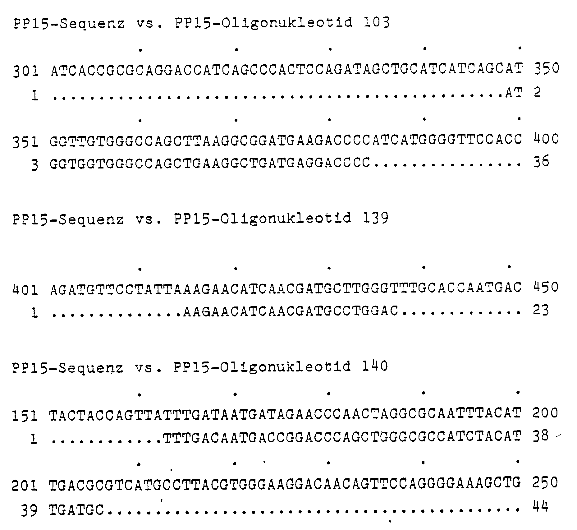
- the PP15 oligonucleotide-103 was then converted from the oligonucleotides coding for the PP15 oligopeptide A. 5′ATGGTGGTGG GCCAGCTGAA GGCTGATGAG GACCCC, correspondingly from the oligopeptide E the PP15 oligonucleotide 140 5′TTTGACAATG ACCGGACCCA GCTGGGCGCC ATCTACATTG ATGC and from the oligopeptide F a 64-fold degenerate PP15 oligonucleotide-139 selected according to statistical data from R. Lathe (J. Mol. Biol. (1985) 183, 1-12).
- a cDNA library which was produced from mRNA from mature, human placenta, was subjected to a screening.
- the mRNA was isolated from the placenta and the cDNA was made from it. This was provided with EcoRI ends and ligated into the EcoRI site of the phage vector Lambda gt10.
- Two clones (PP15 - 24 and PP15 - 28) were detected, which contain the entire cDNA of PP15.
- DNA sequencing was carried out according to methods known per se; the complete sequence of the PP15 cDNA (coding strand) is shown in Table 1.
- This cDNA is 894 base pairs (bp) long, has 99 bp untranslated sequence at the 5 'end, has an open reading frame of 381 bp and leaves 414 bp including eight bases poly (A) untranslated at the 3' end.
- nucleotide probes are marked by underlining in Table 1, and the amino acid sequence is also entered.
- the coding cDNA can be used by means of suitable expression systems to express PP15.
- the shape of the Modification of PP15 can be influenced. For example, there is no glycosylation in bacteria and a different one in yeast cells than in higher eukaryotic cells.
- polyclonal or monoclonal antibodies can be produced by conventional or genetic engineering methods, which can serve as antigens for the production of polyclonal or monoclonal antibodies.
- Such antibodies can be used not only for diagnostic purposes, but also for the production of antibody columns with which PP15 can be separated from solutions which contain it in addition to other proteins.
- the genomic clone coding for PP15 can also be isolated in a simple manner from a genomic bank, with the aid of which not only expression in eukaryotic cells is facilitated, but also further diagnostic statements can be made.
- EDTA sodium ethylenediamine tetraacetate
- SDS sodium dodecyl sulfate
- DTT dithiothreitol
- BSA bovine serum albumin
- RNA was derived from mature, human placenta (according to Chirgwin et al., Biochemistry 18 (1979) 5294-5299). About 10 g of placental tissue were ground in liquid nitrogen, suspended in 80 ml of 4 M guanidinium thiocyanate with 0.1 M mercaptoethanol and treated in a homogenizer (Ultraturrax) at 20,000 rpm for 90 seconds. The lysate was 15 min. Centrifuged at 7,000 rpm (Sorvall-GSA rotor) and the supernatant with 2 ml of 1 M acetic acid and 60 ml of ethanol abs. precipitated at -20 ° C overnight.
- RNA was used to obtain poly (A) -containing mRNA via oligo (dT) cellulose chromatography (Aviv and Leder, Proc. Natl. Acad. Sci. USA 69 (1973) 1408-1412) in 2 ml Pasteur pipettes separated in LiCl. About 5 mg of placental RNA was applied to the column in buffer 1 (500 mM LiCl, 20 mM Tris (pH 7.5), 1 mM EDTA, 0.1% SDS). While the poly (A) + RNA was bound to oligo (dT) cellulose, the poly (A) ⁇ RNA could be eluted again.
- buffer 1 500 mM LiCl, 20 mM Tris (pH 7.5), 1 mM EDTA, 0.1% SDS.
- RNA-RNA placenta mRNA
- buffer 3 5th mM Tris (pH 7.5), 1 mM EDTA, 0.05% SDS
- poly (A) RNA RNA was adjusted to buffer 1 and again chromatographed on oligo (dT) cellulose.
- the yield of placenta poly (A) +-RNA was about 4% of the RNA used after this second purification step.
- Placental mRNA containing poly (A) was checked for intactness in a 1.5% agarose gel prior to cDNA synthesis.
- placenta mRNA were dissolved in 65.5 ul H2O, 10 min. denatured at 70 ° C and chilled on ice.
- the cDNA synthesis was carried out in a 100 ul approach after adding 20 ul RT1 buffer (250 mM Tris (pH 8.2) at 42 ° C, 250 mM KCl, 30 mM MgCl2), 2.5 ul 20 mM dNTP (ie all four deoxynucleoside triphosphates), 1 ⁇ l oligo (dT) of 1 ⁇ g / ml, 1 ⁇ l 1 M DTT, 2 ⁇ l RNAsin (Boehringer Mannheim) and 8 ⁇ l reverse transcriptase (24 U / ⁇ l, Boehringer Mannheim) for 90 min. at 42 ° C.
- 20 ul RT1 buffer 250 mM Tris (pH 8.2) at 42 ° C, 250 mM KCl, 30 mM MgCl2
- 2.5 ul 20 mM dNTP ie all four deoxynucleoside triphosphates
- Double-stranded cDNA was synthesized according to Gubler and Hoffmann (Gene 25 (1983) 263-269). The synthesis was carried out immediately after the cDNA synthesis by adding 305.5 ul H2O, 80 ul RT2 buffer (100 mM Tris (pH 7.5), 25 mM MgCl2, 500 mM KCl, 50 mM DTT, 250 ⁇ g / ml BSA), 2 ⁇ l RNase H (2 U / ⁇ l), 2.5 ⁇ l E.
- the reaction mixture was after the addition of 55 ul 250 uM dNTP, 55 ul 10 mM Tris (pH 7.5), 10 mM MgCl2, 10 ug / ml BSA, 3 ul T4 DNA polymerase I (1 U / ul), 2 ul RNase H (2 U / ⁇ l) and 2 ⁇ l RNase A (2 ⁇ g / ml) for a further 30 min. incubated at 37 ° C to ensure the completeness of the synthesis on the second strand of DNA ("Repair Reaction").
- the EcoRI reaction mixture was applied in toto to a potassium acetate gradient (5-20% KOAc, 1 mM EDTA, 1 ⁇ l / ml ethidium bromide) and 3 h at 50,000 rpm and 20 ° C centrifuged (Beckman SW 65 rotor). The gradient was fractionated from below so that the first five fractions measured 500 ⁇ l and all the remaining 100 ⁇ l. The fractions were precipitated with 0.01 volume of acrylamide (2 mg / ml) and 2.5 volume of ethanol, washed once with 70% ethanol, dried and taken up in 5 ⁇ l of H2O.
- a potassium acetate gradient 5-20% KOAc, 1 mM EDTA, 1 ⁇ l / ml ethidium bromide
- the gradient was fractionated from below so that the first five fractions measured 500 ⁇ l and all the remaining 100 ⁇ l.
- the fractions were precipitated with 0.01 volume of acrylamide (2 mg / ml) and
- the dsDNA was incorporated into the EcoRI interface of the phage vector ⁇ gt10 (Vector Cloning Systems, San Diego, CA) in a 4 ⁇ l ligase mixture: 2 ⁇ l dsDNA, 1 ⁇ l ⁇ gt10 x EcoRI (1 ⁇ g / ml), 0.4 ⁇ l Ligase buffer, 0.5 ul H2O, 0.1 ul T4 DNA ligase. The mixture was incubated at 15 ° C. for 4 h.
- the ⁇ -lysogenic cell extracts E. coli NS 428 and NS 433 were followed by an "in vitro packaging" reaction of the ligase mixture at room temperature for 2 h (vector cloning Systems, San Diego, CA; Enquist and Sternberg, Methods in Enzymology 68 , (1979), 281-298).
- the reaction was stopped with 500 ul suspension medium (SM: 0.1 M NaCl, 8 mM MgSO4, 50 mM Tris (pH 7.5), 0.01% gelatin) and 2 drops of chloroform.
- the number of plaque forming units (PFU) in the placenta cDNA bank was determined using competent cells of the E. coli K 12 strain C600 HFL: it was 1 ⁇ 106 PFU.
- Oligonucleotide probes (PP15 oligonucleotide 103 and 140) and a pool of oligonucleotides (PP15 oligonucleotide pool 139) were synthesized for analysis of the placenta cDNA library. Their sequences were derived from the amino acid sequence of three bromocyanine fragments from PP15.
- the oligonucleotide sequences were labeled with T4 polynucleotide kinase in the presence of ( ⁇ -32P) ATP at the 5 'end (60 ⁇ Ci / 40 ⁇ l reaction mixture was used).
- the probes had a specific activity of 1 x 10⁸ Bq / ⁇ l or 1.5 x 10⁶ Bq / pMol.
- 1 x 10⁶ PFU of the placenta cDNA library were examined together with the PP15 oligonucleotide probes 103, 140 and 139.
- 3 x 10⁴ PFU with cells of the E. coli K 12 strain C 600 HFL in soft agar to 13.5 cm Petri dishes were plated out and incubated at 37 ° C. for 6 h. At this point in time, no confluent lysis had occurred. The plates were incubated overnight in the refrigerator and the phages were transferred to nitrocellulose filters (Schleicher & Schull, BA 85, Ref. No. 401124) (duplicates). Nitrocellulose filters and petri dishes were marked with an injection cannula to enable positive plaques to be assigned later.
- the petri dishes were stored in the cold room while the nitrocellulose filters were being processed.
- the DNA on the nitrocellulose filters was denatured by filtering for 5 min. were placed on filter paper (Whatman-M3) soaked with 1.5 M NaCl, 0.5 M NaOH.
- the filters were then renatured in the same way with 1.5 M NaCl, 0.5 M Tris (pH 8.0) and with 2 x SSPE (0.36 M NaCl, 16 mM NaOH, 20 mM NaH2PO4, 2 mM EDTA ) washed.
- the filters were then dried in vacuo at 80 ° C for 2 h.
- the filters were incubated overnight with the addition of 100,000-200,000 Bq of the labeled oligonucleotide / ml hybridization solution (such as prehybridization solution, but without herring sperm DNA) in beakers or in sealed polyethylene foils with gentle shaking.
- the hybridization temperature was 46 ° C for the oligonucleotide probe 139 and 52 ° C for the other probes.
- the nitrocellulose filters were washed with 6 x SSC, 0.05 M sodium pyrophosphate for one hour at room temperature and for a further hour at the respective hybridization temperature. The filters were dried and autoradiographed overnight. Signals on The X-ray film, which appeared on both duplicates, was assigned to the Petri dish and the region (approx.
- the phage clones PP15 - 24 and PP 15 - 28 were propagated and their DNA was extracted.
- the respective EcoRI fragment was isolated and ligated into the EcoRI site of the Bluescript M13 vector (Stratagene, San Diego, CA, USA) for restriction analyzes and sequence analyzes using the Sanger enzymatic dideoxy method.
- the sequence shows an open reading frame and codes for a protein with a maximum of 127 amino acids.
- PP15 has a calculated molecular weight of 14478 d (including methionine), which is in good agreement with the value mentioned in the patent DE-A 29 52 792.
- the vector pTrc99A (E. Amann et al. (1988) Gene 69, 301-315) was used to express the unfused, mature PP15 protein in E. coli.
- the DNA sequence of the PP15 cDNA on the initiation codon is:
- NcoI site can, however, be achieved by two base changes in the PP15 sequence: 5 ′ GAATGG 3 ′ after 5 ′ CCATGG 3 ′ by mutagenesis.
- the second amino acid (Gly) is not affected by this manipulation, since the second codon of the PP15 structural sequence begins with a "G".
- mutagenesis a 902 base pair EcoRI fragment of the PP15 cDNA clone PP15-28 was isolated and ligated into the mutagenesis vector pMa5-8 (FIG.), Likewise cut and dephosphorylated with EcoRI.
- the resulting plasmid pMa5-8-PP15 (with the correct orientation of the PP15 EcoRI insert in relation to the F1 origo) was then subjected to the "gapped duplex" mutagenesis protocol (Kramer et al. (1984) Nucl. Acids. Res. 12, 9441 -9456) using the following oligodeoxynucleotide: 5 ′ GGCTTGTCTCCCATGGTGGAGCGTCAC 3 ′
- pMc5-8-PP15-NcoI A clone that had the desired mutation was identified by restriction analysis and designated pMc5-8-PP15-NcoI.
- the 798 base pair NcoI-EcoRI fragment was isolated from this plasmid and ligated into the corresponding cut pTrc99A vector.
- the resulting plasmid pTrc99A-PP15 comprises 4918 base pairs and, after induction of the trc promoter, expresses the unfused, approximately 15 kD PP15 protein.
- F1-ORI origin of replication of phage f1; ORI: origin of replication of the ColE1 type; CAT: coding region for chloramphenicol acetyltransferase; AMP: coding region for ⁇ -lactamase.
- pMA5-8 carries an amber mutation in CAT (A at position 3409) and pMC5-8 an amber mutation in AMP (C at position 2238).
Landscapes
- Health & Medical Sciences (AREA)
- Chemical & Material Sciences (AREA)
- Life Sciences & Earth Sciences (AREA)
- Organic Chemistry (AREA)
- Engineering & Computer Science (AREA)
- Bioinformatics & Cheminformatics (AREA)
- Genetics & Genomics (AREA)
- Immunology (AREA)
- Zoology (AREA)
- General Health & Medical Sciences (AREA)
- Wood Science & Technology (AREA)
- Molecular Biology (AREA)
- Biomedical Technology (AREA)
- Medicinal Chemistry (AREA)
- Biotechnology (AREA)
- Biophysics (AREA)
- Biochemistry (AREA)
- General Engineering & Computer Science (AREA)
- Gastroenterology & Hepatology (AREA)
- Chemical Kinetics & Catalysis (AREA)
- Toxicology (AREA)
- Pregnancy & Childbirth (AREA)
- Gynecology & Obstetrics (AREA)
- Veterinary Medicine (AREA)
- Public Health (AREA)
- Animal Behavior & Ethology (AREA)
- Pharmacology & Pharmacy (AREA)
- Proteomics, Peptides & Aminoacids (AREA)
- Physics & Mathematics (AREA)
- Nuclear Medicine, Radiotherapy & Molecular Imaging (AREA)
- General Chemical & Material Sciences (AREA)
- Reproductive Health (AREA)
- Plant Pathology (AREA)
- Microbiology (AREA)
- Transplantation (AREA)
- Preparation Of Compounds By Using Micro-Organisms (AREA)
- Peptides Or Proteins (AREA)
- Micro-Organisms Or Cultivation Processes Thereof (AREA)
- Medicines That Contain Protein Lipid Enzymes And Other Medicines (AREA)
- Saccharide Compounds (AREA)
Abstract
Description
- Das immunsuppressiv wirkende Protein PP 15 ist in der DE-A 29 52 792 (US-A 4,348,316) mit folgenden Parametern beschrieben:
a) einem Kohlenhydratanteil von 3,35 ± 0,9 %, bestehend aus 2,8 ± 0,5 % Hexosen, 0,3 ± 0,2 % Hexosaminen, 0,05 ± 0,05 Fucose und 0,20 ± 0,15 % Neuraminsäure;
b) einem Sedimentationskoeffizienten S20.w⁰ von 2,9 ± 0,2 S;
c) einem in der Ultrazentrifuge bestimmtes Molekulargewicht von 30 700 ± 3 200 (Dimer);
d) einem Extinktionskoeffizienten E1cm 1 % (280 nm) von 14,2 ± 1,0 und
e) einer elektrophoretischen Beweglichkeit im Bereich des Albumins sowie
f) einem isoelektrischen Punkt von 4,4 ± 0,1;
g) der AminosäurezusammensetzungAminosäure Reste pro 100 Reste (Mol %) Variationskoeffizient (%) Lysin 4,74 3,30 Histidin 3,81 5,43 Arginin 1,62 3,43 Asparaginsäure 13,39 5,08 Threonin 3,85 5,35 Serin 6,38 2,81 Glutaminsäure 13,43 5,32 Prolin 4,35 14,25 Glycin 6,87 2,13 Alanin 6,51 8,26 Cystin 1/2 2,48 4,55 Valin 2,29 15,67 Aminosäure Rest pro 100 Reste (Mol %) Variationskoeffizient (%) Methionin 2,87 10,86 Isoleucin 8,39 8,18 Leucin 8,18 6,72 Tyrosin 2,09 8,49 Phenylalanin 6,27 2,27 Tryptophan 2,51 6,81 - Die Molekulargewichtsbestimmung mittels SDS-Polyacrylamid-Gelelektrophorese ergab ein Molekulargewicht von etwa 15000 d (Monomer).
- Wegen des durch die immunsuppressiven Eigenschaften hervorgerufenen therapeutischen und des diagnostischen Interesses ist eine gentechnische Herstellung dieses Proteins äußerst wünschenswert. Die Erfindung betrifft folglich ein Verfahren zur gentechnischen Herstellung von PP15, die dazu erforderliche mRNA, die daraus gewonnene cDNA, diese cDNA ganz oder teilweise enthaltende DNA-Strukturen und Vektoren, mit solcher DNA transformierte Zellen, das von diesen Zellen exprimierte Polypeptid und dessen Verwendung als Arzneimittel. Die Erfindung bezieht sich ferner auf die Aminosäuresequenz sowie auf Teilsequenzen der Aminosäuresequenz von PP15, damit gewonnene spezifische Antikörper, aus diesen Antikörpern hergestellte Diagnostika und Antikörpersäulen sowie mit Hilfe solcher Säulen gewonnenes Polypeptid. Eine weitere Ausgestaltung der Erfindung betrifft Diagnostika, die PP15 kodierende bzw. deren komplementäre DNA oder RNA ganz oder teilweise enthalten, und diagnostische Verfahren, mit denen Körperflüssigkeiten und Gewebe mit Hilfe solcher Diagnostika untersucht werden. Weitere Aspekte der Erfindung werden im folgenden näher erläutert bzw. in den Patentansprüchen definiert.
- Zunächst wurde versucht, mittels spezifischer Antikörper gegen PP15 in einer kommerziell erhältlichen cDNA-Expressionsbank aus mRNA von reifer humaner Plazenta (Fa. Genofit, Heidelberg) Klone nachzuweisen, die PP15 exprimieren. Da bekannt war, daß PP15 immunsuppressiv wirkt, folglich spezifische Antikörper nur schlecht, wenn überhaupt, herstellbar waren, wurden spezifische Antikörper gegen Teilpeptide hergestellt.
- Deshalb wurde das Protein PP15 durch Spaltung mittels Bromcyan, Trypsin oder Proteinase V8 in spezifische Fragmente zerlegt, die anschließend sequenziert wurden. Folgende Fragmente wurden erhalten:
- (A) M V V G Q L K A D E D P I M G F H Q M F
- (B) F R L A L H N F G
- (C) V S V Y A E A A E R
- (D) L S S L P F Q K I Q (H)
- (E) F D N D R T Q L G A I Y I D A S - L T - E
- (F) L L K N I N D A W T
- Peptide A, B und C wurden durch allgemein bekannte Methoden synthetisiert und nach üblichen Verfahren in Kaninchen spezifische Antikörper erzeugt. Es gelang nicht, in der o.g. cDNA-Expressionsbank, die ≧ 1 x 10⁶ rekombinierte Lambda gt11 Klone enthielt, positive Klone zu lokalisieren. Die Antikörper gegen Peptid A und Peptid B präzipitierten dabei in Kontrollversuchen PP15, während Antikörper gegen Peptid C nicht reagierten. Wie später ersichtlich, ist das Peptid C auch nicht in der nachfolgend aus der cDNA-Sequenz abgeleiteten Proteinsequenz von PP15 enthalten, so daß es wahrscheinlich Begleitproteinen von PP15 zugeordnet werden muß.
- Darauf wurden aus den für das PP15-Oligopeptid A kodierenden Oligonukleotiden das PP15-Oligonukleotid-103
5′ATGGTGGTGG GCCAGCTGAA GGCTGATGAG GACCCC,
entsprechend aus dem Oligopeptid E das PP15-Oligonukleotid-140
5′TTTGACAATG ACCGGACCCA GCTGGGCGCC ATCTACATTG ATGC
und aus dem Oligopeptid F ein 64-fach degeneriertes PP15-Oligonukleotid-139nach statistischen Daten von R. Lathe (J. Mol. Biol. (1985) 183, 1 - 12) ausgewählt.
- Mit diesen Oligonukleotidsonden wurde eine cDNA-Bank, die aus mRNA von reifer, humaner Plazenta hergestellt war, einem Screening unterworfen. Zunächst wurde aus der Plazenta die mRNA isoliert und daraus die cDNA hergestellt. Diese wurde mit EcoRI-Enden versehen und in die EcoRI-Schnittstelle des Phagenvektors Lambda gt10 ligiert. Es wurden 2 Klone (PP15 - 24 und PP15 - 28) nachgewiesen, die die gesamte cDNA von PP15 enthalten. Die DNA-Sequenzierung erfolgte nach an sich bekannten Methoden; die vollständige Sequenz der PP15 cDNA (kodierender Strang) ist in Tab. 1 gezeigt. Diese cDNA ist 894 Basenpaare (bp) lang, hat am 5′-Ende 99 bp untranslatierter Sequenz, besitzt einen offenen Leserahmen von 381 bp und läßt am 3′-Ende 414 bp einschließlich acht Basen Poly(A) untranslatiert.
- In der Tabelle 1 sind die Positionen der Nukleotidsonden durch Unterstreichen markiert, zusätzlich ist die Aminosäuresequenz eingetragen.
- Erfindungsgemäß kann die kodierende cDNA mittels geeigneter Expressionssysteme dazu benutzt werden, PP15 zu exprimieren. Durch die Auswahl des Wirtes kann weiterhin die Form der Modifikation von PP15 beeinflußt werden. So findet in Bakterien keine, in Hefezellen eine andere Glykosylierung als in höheren eukaryotischen Zellen statt.
- In Kenntnis der Aminosäuresequenz von PP15 ist es möglich, nach konventionellen oder gentechnischen Methoden Aminosäure-Teilsequenzen herzustellen, die als Antigene zur Herstellung von polyklonalen oder monoklonalen Antikörpern dienen können. Solche Antikörper können nicht nur zu diagnostischen Zwecken, sondern auch zur Herstellung von Antikörpersäulen dienen, mit denen PP15 aus Lösungen abgetrennt werden kann, die es neben anderen Proteinen enthalten.
- Mit Hilfe der cDNA bzw. Teilen davon kann man auch auf einfache Weise aus einer genomischen Bank den für PP15 codierenden genomischen Klon isolieren, mit dessen Hilfe nicht nur eine Expression in eukaryotischen Zellen erleichtert wird, sondern auch weitere diagnostische Aussagen getroffen werden können.
- Die Erfindung ist ferner in den Patentansprüchen definiert und in folgenden Beispielen weiter ausgeführt.
- Soweit nicht im Text erläutert, werden die folgenden Abkürzungen verwendet:
EDTA = Natrium-ethylendiamin-tetraacetat
SDS = Natrium-dodecylsulfat
DTT = Dithiothreitol
BSA = Rinderserumalbumin - RNA wurde aus reifer, humaner Plazenta (nach Chirgwin et al., Biochemistry 18 (1979) 5294-5299) gewonnen. Etwa 10 g Plazentagewebe wurden in flüssigem Stickstoff zermörsert, in 80 ml 4 M Guanidinium-Thiocyanat mit 0,1 M Mercaptoethanol suspendiert und 90 sec. bei 20 000 rpm in einem Homogenisator (Ultraturrax) behandelt. Das Lysat wurde 15 min. bei 7 000 rpm zentrifugiert (Sorvall-GSA Rotor) und der Überstand mit 2 ml 1 M Essigsäure und 60 ml Ethanol abs. bei -20°C über Nacht gefällt. Nach Sedimentation bei 6 000 rpm und -10°C für 10 min. wurden die Nucleinsäuren in 40 ml 7,5 M Guanidinium-Hydrochlorid (pH 7,0) vollständig gelöst und mit einer Mischung aus 1 ml 1 M Essigsäure und 20 ml Ethanol abs. gefällt. Zur Abtrennung der DNA wurde die Fällung ein weiteres Mal mit jeweils halben Volumina wiederholt. Die RNA wurde in 12 ml H₂O gelöst, mit einer Mischung aus 1,2 ml 4 M Kaliumacetat und 24 ml Ethanol abs. gefällt, sedimentiert und schließlich erneut in 10 ml H₂O (1 ml pro g Gewebe) aufgenommen.
- Die Plazenta-RNA wurde zur Gewinnung von Poly(A)-haltiger mRNA über Oligo(dT)-Cellulose-Chromatographie (Aviv and Leder, Proc. Natl. Acad. Sci. USA 69 (1973) 1408-1412) in 2 ml Pasteurpipetten in LiCl aufgetrennt. Etwa 5 mg Plazenta-RNA wurden in Puffer 1 (500 mM LiCl, 20 mM Tris (pH 7,5), 1 mM EDTA, 0,1 % SDS) auf die Säule aufgetragen. Während die Poly(A)⁺-RNA an Oligo(dT)-Cellulose gebunden wurde, konnte die Poly(A)⁻-RNA wieder eluiert werden. Nach einem Waschschritt mit Puffer 2 (100 mM LiCl, 20 mM Tris (pH 7,5), 1 mM EDTA, 0,1 % SDS) wurde die Poly(A)⁺-RNA (Plazenta-mRNA) mit Puffer 3 (5 mM Tris (pH 7,5), 1 mM EDTA, 0,05 % SDS) von der Säule eluiert.
- Zur weiteren Reinigung wurde die Poly(A)⁺-RNA auf Puffer 1 eingestellt und erneut über Oligo(dT)-Cellulose chromatographiert.
- Die Ausbeute an Plazenta Poly(A)⁺-RNA betrug nach diesem zweiten Reinigungsschritt etwa 4 % der eingesetzten RNA.
- Poly(A)-haltige Plazenta-mRNA wurde vor der cDNA-Synthese im 1,5 % Agarosegel auf Intaktheit geprüft.
- Danach wurden 4 µg Plazenta-mRNA in 65,5 µl H₂O gelöst, 10 min. bei 70°C denaturiert und auf Eis gekühlt.
- Die cDNA-Synthese erfolgte in einem 100 µl-Ansatz nach Zugabe von 20 µl RT₁-Puffer (250 mM Tris (pH 8,2) bei 42°C, 250 mM KCl, 30 mM MgCl₂), 2,5 µl 20 mM dNTP (d.h. aller vier Desoxynukleosidtriphosphate), 1 µl Oligo(dT) von 1 µg/ml, 1 µl 1 M DTT, 2 µl RNAsin (Boehringer Mannheim) und 8 µl Reverse Transcriptase (24 U/µl, Boehringer Mannheim) für 90 min. bei 42°C. Doppelsträngige cDNA (dsDNA) wurde nach Gubler and Hoffmann (Gene 25 (1983) 263-269) synthetisiert. Die Synthese erfolgte unmittelbar nach der cDNA-Synthese durch Zugabe von 305,5 µl H₂O, 80 µl RT₂-Puffer (100 mM Tris (pH 7,5), 25 mM MgCl₂, 500 mM KCl, 50 mM DTT, 250 µg/ml BSA), 2 µl RNase H (2 U/µl), 2,5 µl E. coli DNA Ligase (5 U/µl), 5 µl 15 mM β-NAD, und 5 µl DNA Polymerase I (5 U/µl) und Inkubation für 5 h bei 15°C. Durch Hitzeinaktivierung (70°C, 30 min.) wurde die Reaktion beendet.
- Der Reaktionsansatz wurde nach Zugabe von 55 µl 250 µM dNTP, 55 µl 10 mM Tris (pH 7,5), 10 mM MgCl₂, 10 µg/ml BSA, 3 µl T4 DNA-Polymerase I (1 U/µl), 2 µl RNase H (2 U/µl) und 2 µl RNase A (2 µg/ml) für weitere 30 min. bei 37°C inkubiert, um die Vollständigkeit der Synthese am zweiten DNA-Strang zu gewährleisten ("Repair Reaction").
- Zur Errichtung einer Plazenta-cDNA-Bank wurde die dsDNA mit EcoRI-Enden versehen, um sie in die EcoRI-Schnittstelle des Phagenvektors λgt10 ligieren zu können (T. Maniatis et al. (1982), Molecular Cloning, A Laboratory Manual, Cold Spring Harbor). Hierzu wurde die dsDNA
- a) mit EcoRI-Methylase behandelt, um interne EcoRI-Schnittstellen der dsDNA zu schützen,
- b) mit EcoRI-Linkern versehen, die
- c) danach mit EcoRI geschnitten wurden.
- Zu a):
Die Methylase-Reaktion der dsDNA erfolgte direkt im Anschluß an die "Repair Reaction" nach Zugabe von 25 µl 500 mM EDTA (pH 8,0), 60 µl Methylase-Puffer (100 mM NaOAc (pH 5,2), 2 mg S-Adenosyl-L-Methionin) und 2 µl EcoRI-Methylase (20 U/µl) durch Inkubation bei 37°C für 30 min.
Der Reaktionsansatz wurde mit Phenol extrahiert und die dsDNA mit 60 µl 4M NaOAc und 1300 µl Ethanol gefällt. Die dsDNA wurde zweimal mit 70 % Ethanol gewaschen, einmal mit Ether ausgeschüttelt und getrocknet. - Zu b):
Die EcoRI-methylierte dsDNA wurde in 88 µl H₂O gelöst und nach Zugabe von 10 µl Ligase-Puffer (500 mM Tris (pH 7,4), 100 mM MgCl₂, 100 mM DTT, 10 mM Spermidin, 10 mM ATP, 1 mg/ml BSA), 1 µl T4 DNA-Ligase (10 U/µl) mit 1 µl EcoRI Linkern (0,5 µg/µl) (pGGAATTCC bzw. pAGAATTCT) über Nacht bei 15°C ligiert. - Zu c):
Das Volumen des Ligase-Ansatzes wurde mit 6 µl H₂O, 12 µl 10x EcoRI-Puffer und 2 µl EcoRI (120 U/µl) auf 120 µl gebracht. Die EcoRI-Verdauung erfolgte für 2 h bei 37°C. - Zur Abtrennung aller nichtgebundenen EcoRI-Linker von der dsDNA wurde der EcoRI-Reaktionsansatz in toto auf einen Kaliumacetat-Gradienten (5-20 % KOAc, 1 mM EDTA, 1 µl/ml Ethidiumbromid) aufgetragen und 3 h bei 50 000 rpm und 20°C zentrifugiert (Beckman SW 65-Rotor).
Der Gradient wurde von unten so fraktioniert, daß die ersten fünf Fraktionen 500 µl maßen und alle restlichen 100 µl. Die Fraktionen wurden mit 0,01 Volumen Acrylamid (2 mg/ml) und 2,5 Volumen Ethanol gefällt, einmal mit 70 %igem Ethanol gewaschen, getrocknet und jeweils in 5 µl H₂O aufgenommen. - Zur Größenbestimmung der dsDNA wurden 1 µl jeder Fraktion im 1,5 % Agarose-Gel analysiert. Zusätzlich wurde mit 1 µl jeder Fraktion die Quantität an dsDNA bestimmt.
- Fraktionen mit dsDNA über 500 bp wurden vereinigt und die Probe so eingeengt, daß die Endkonzentration 27 µg/ml betrug.
- Der Einbau der dsDNA in die EcoRI-Schnittstelle des Phagenvektors λgt10 (Vector Cloning Systems, San Diego, CA) erfolgte in einem Ligaseansatz von 4 µl: 2 µl dsDNA, 1 µl λgt10 x EcoRI (1 µg/ml), 0,4 µl Ligase-Puffer, 0,5 µl H₂O, 0,1 µl T4 DNA-Ligase. Der Ansatz wurde 4 h bei 15°C inkubiert.
- Zur Etablierung der Plazenta-cDNA-Bank im Phagenvektor λgt10 folgte mit den λ-lysogenen Zellextrakten E. coli NS 428 und NS 433 eine "in vitro packaging"-Reaktion des Ligaseansatzes bei Raumtemperatur für 2 h (Vector Cloning Systems, San Diego, CA; Enquist and Sternberg, Methods in Enzymology 68, (1979), 281-298). Die Reaktion wurde mit 500 µl Suspensionsmedium (SM: 0,1 M NaCl, 8 mM MgSO₄, 50 mM Tris (pH 7,5), 0,01 % Gelatine) und 2 Tropfen Chloroform gestoppt.
- Die Zahl der "Plaque Forming Units" (PFU) der Plazenta-cDNA-Bank wurde mit kompetenten Zellen des E. coli-K 12-Stammes C600 HFL bestimmt: Sie betrug 1 x 10⁶ PFU.
- Oligonukleotidsonden (PP15-Oligonukleotid 103 und 140) und ein Pool von Oligonukleotiden (PP15-Oligonukleotidpool 139) wurden zur Analyse der Plazenta-cDNA-Bank synthetisiert. Ihre Sequenzen wurden abgeleitet von der Aminosäuresequenz dreier Bromcyanfragmente von PP15.
- Die Art des Aufbaus und die Verwendung der Sonden folgten im wesentlichen den Regeln von R. Lathe, a.a.O..
- Die Oligonukleotidsequenzen wurden mit T4 Polynukleotidkinase unter Anwesenheit von (γ-32P)ATP am 5′-Ende markiert (eingesetzt wurden 60 µCi/40 µl Reaktionsansatz). Die Sonden hatten eine spezifische Aktivität von 1 x 10⁸ Bq/µl bzw. 1,5 x 10⁶ Bq/pMol.
- 1 x 10⁶ PFU der Plazenta-cDNA-Bank wurden mit den PP15-Oligonukleotidsonden 103, 140 und 139 zusammen untersucht. Dazu wurden je 3 x 10⁴ PFU mit Zellen des E. coli K 12-Stammes C 600 HFL in Weich-Agar auf 13,5 cm Petrischalen ausplattiert und 6 h bei 37°C inkubiert. Zu diesem Zeitpunkt war noch keine konfluente Lyse eingetreten. Die Platten wurden über Nacht im Kühlschrank inkubiert und die Phagen auf Nitrocellulosefilter (Schleicher & Schüll, BA 85, Ref.-Nr. 401124) übertragen (Duplikate). Nitrocellulosefilter und Petrischalen wurden mit einer Injektionskanüle markiert, um eine spätere Zuordnung positiver Plaques zu ermöglichen. Die Petrischalen wurden während des Prozessierens der Nitrocellulosefilter im Kühlraum gelagert. Die auf den Nitrocellulosefiltern befindliche DNA wurde denaturiert, indem die Filter für 5 min. auf mit 1,5 M NaCl, 0,5 M NaOH-getränktes Filterpapier (Whatman-M3) gelegt wurden. Anschließend wurden die Filter auf die gleiche Art mit 1,5 M NaCl, 0,5 M Tris (pH 8,0) renaturiert und mit 2 x SSPE (0,36 M NaCl, 16 mM NaOH, 20 mM NaH₂PO₄, 2 mM EDTA) gewaschen. Die Filter wurden dann für 2 h bei 80°C im Vakuum getrocknet. Die Filter wurden für 4 h bei 65°C in 3xSSC, 0,1 % SDS (20 x SSC = 3 M NaCl, 0,3 M Na-Citrat) gewaschen und für 4 h bei 65°C vorhybridisiert (Prähybridisierungslösung: 0,6 M NaCl, 0,06 M Tris (pH 8,3), 6 mM EDTA, 0,2 % nichtionisches synthetisches Saccharose-Polymer (RFicoll), 0,2 % Polyvinylpyrrolidon 40, 0,2 % BSA, 0,1 % SDS, 50 µg/ml denaturierte Heringssperma-DNA). Die Filter wurden über Nacht unter Zusatz von 100 000-200 000 Bq des markierten Oligonukleotids/ml Hybridisierungslösung (wie Prähybridisierungslösung, jedoch ohne Heringssperma-DNA) in Bechergläsern bzw. in zugeschweißten Polyethylenfolien unter leichtem Schütteln inkubiert. Die Hybridisierungstemperatur betrug 46°C für die Oligonukleotidsonde 139 bzw. 52°C für die anderen Sonden. Die Nitrocellulosefilter wurden mit 6 x SSC, 0,05 M Natriumpyrophosphat eine Stunde bei Raumtemperatur und für eine weitere Stunde bei der jeweiligen Hybridisierungstemperatur gewaschen. Die Filter wurden getrocknet und über Nacht autoradiographiert. Signale auf dem Röntgenfilm, die auf beiden Duplikaten auftraten, wurden der Petrischale zugeordnet und die Region (ca. 50 Plaques) mit dem breiten Ende einer Pasteurpipette ausgestanzt und die Phagen in 1 ml SM-Puffer resuspendiert. Positive Phagen wurden über drei Runden vereinzelt, bis ein einzelner postiviter Klon vorlag.
- Je 1 x 10⁶ PFU der Plazenta-cDNA-Bank wurden dreimal untersucht. Erst beim dritten Screening wurden 2 Signale auf Duplikat-Filtern identifiziert. Die beiden Klone PP15 - 24 und PP15 - 28 enthalten die gesamte cDNA von PP15.
- Der Vergleich der Oligonukleotidsequenzen 103, 139 und 140 mit der ermittelten PP15-Sequenz ist in Tab. 2 zusammengefaßt.
- Die Phagenklone PP15 - 24 und PP 15 - 28 wurden vermehrt und ihre DNA jeweils extrahiert. Das jeweilige EcoRI-Fragment wurde isoliert und in die EcoRI-Stelle des Bluescript M13 Vektors (Stratagene, San Diego, CA, USA) für Restriktionsanlaysen sowie Sequenzanalysen mittels der enzymatischen Dideoxymethode nach Sanger einligiert. Die Sequenz zeigt einen offenen Leserahmen und kodiert für ein Protein mit maximal 127 Aminosäuren. PP15 besitzt ein errechnetes Molekulargewicht von 14478 d (einschließlich Methionin), was mit dem eingangs genannten Wert der Patentschrift DE-A 29 52 792 gut übereinstimmt.

- Der Vektor pTrc99A (E. Amann et al. (1988) Gene 69, 301-315) wurde zur Expression des unfusionierten, reifen PP15 Proteins in E.coli benutzt. Die DNA Sequenz der PP15 cDNA am Initiationscodon lautet:

- Da keine NcoI stelle am ATG vorhanden ist, kann diese DNA nicht direkt in den pTrc99A Expressionsvektor kloniert werden. Eine NcoI Stelle kann aber durch zwei Basenaustausche in der PP15 Sequenz: 5′ GAATGG 3′ nach 5′ CCATGG 3′ durch Mutagenese erzielt werden. Die zweite Aminosäure (Gly) ist von dieser Manipulation nicht betroffen, da das zweite Codon der PP15 Struktursequenz mit einem "G" beginnt. Zur Mutagenese wurde ein 902 Basenpaar großes EcoRI-Fragment des PP15 cDNA Klons PP15-28 isoliert und in das ebenfalls mit EcoRI geschnittenen und dephosphorylierten Mutagenesevektor pMa5-8 (Fig.) ligiert. Das resultierende Plasmid pMa5-8-PP15 (mit der korrekten Orientierung des PP15 EcoRI-Inserts in Relation zum F1-Origo) wurde dann dem "gapped duplex" Mutageneseprotokoll (Kramer et al. (1984) Nucl. Acids. Res. 12, 9441-9456) unterzogen, wobei das folgende Oligodesoxynukleotid verwendet wurde:
5′ GGCTTGTCTCCCATGGTGGAGCGTCAC 3′ - Ein Klon, der die gewünschte Mutation aufwies, wurde durch Restriktionsanalyse identifiziert und als pMc5-8-PP15-NcoI bezeichnet. Aus diesem Plasmid wurde das 798 Basenpaar große NcoI-EcoRI-Fragment isoliert und in den entsprechenden geschnittenen pTrc99A Vektor ligiert. Das resultierende Plasmid pTrc99A-PP15 umfaßt 4918 Basenpaare und exprimiert nach Induktion des trc Promoters das unfusionierte, ca. 15 kD große PP15 Protein.
- Karte der Plasmide pMAC5-8 (= pMA5-8 und pMC5-8).
F1-ORI: Replikationsursprung des Phagen f1;
ORI: Replikationsursprung vom ColE1-Typ;
CAT: Kodierende Region für Chloramphenicol-acetyltransferase;
AMP: Kodierende Region für β-Lactamase.
pMA5-8 trägt eine amber-Mutation in CAT (A bei Position 3409) und pMC5-8 eine amber-Mutation in AMP (C bei Position 2238).
Claims (15)
Applications Claiming Priority (2)
| Application Number | Priority Date | Filing Date | Title |
|---|---|---|---|
| DE3809119 | 1988-03-18 | ||
| DE3809119A DE3809119A1 (de) | 1988-03-18 | 1988-03-18 | Gentechnisch hergestelltes protein pp 15 |
Publications (3)
| Publication Number | Publication Date |
|---|---|
| EP0333134A2 true EP0333134A2 (de) | 1989-09-20 |
| EP0333134A3 EP0333134A3 (en) | 1990-06-06 |
| EP0333134B1 EP0333134B1 (de) | 1996-08-07 |
Family
ID=6350091
Family Applications (1)
| Application Number | Title | Priority Date | Filing Date |
|---|---|---|---|
| EP89104516A Expired - Lifetime EP0333134B1 (de) | 1988-03-18 | 1989-03-14 | Gentechnische Herstellung von Protein PP 15. |
Country Status (11)
| Country | Link |
|---|---|
| EP (1) | EP0333134B1 (de) |
| JP (2) | JPH029370A (de) |
| KR (1) | KR890014739A (de) |
| AT (1) | ATE141104T1 (de) |
| AU (1) | AU616248B2 (de) |
| DE (2) | DE3809119A1 (de) |
| DK (1) | DK131989A (de) |
| ES (1) | ES2090019T3 (de) |
| FI (1) | FI95146C (de) |
| GR (1) | GR3020755T3 (de) |
| PT (1) | PT90031B (de) |
Family Cites Families (1)
| Publication number | Priority date | Publication date | Assignee | Title |
|---|---|---|---|---|
| DE2952792A1 (de) * | 1979-12-31 | 1981-07-02 | Behringwerke Ag, 3550 Marburg | Neues protein (pp(pfeil abwaerts)15(pfeil abwaerts)) mit immunsuppressiver wirkung |
-
1988
- 1988-03-18 DE DE3809119A patent/DE3809119A1/de not_active Withdrawn
-
1989
- 1989-03-14 ES ES89104516T patent/ES2090019T3/es not_active Expired - Lifetime
- 1989-03-14 DE DE58909708T patent/DE58909708D1/de not_active Expired - Fee Related
- 1989-03-14 AT AT89104516T patent/ATE141104T1/de not_active IP Right Cessation
- 1989-03-14 EP EP89104516A patent/EP0333134B1/de not_active Expired - Lifetime
- 1989-03-16 FI FI891249A patent/FI95146C/fi not_active IP Right Cessation
- 1989-03-17 AU AU31431/89A patent/AU616248B2/en not_active Ceased
- 1989-03-17 PT PT90031A patent/PT90031B/pt not_active IP Right Cessation
- 1989-03-17 DK DK131989A patent/DK131989A/da not_active Application Discontinuation
- 1989-03-18 JP JP1067402A patent/JPH029370A/ja active Pending
- 1989-03-18 KR KR1019890003375A patent/KR890014739A/ko not_active Ceased
-
1996
- 1996-06-21 JP JP8161789A patent/JPH09131188A/ja active Pending
- 1996-08-08 GR GR960401980T patent/GR3020755T3/el unknown
Also Published As
| Publication number | Publication date |
|---|---|
| FI95146B (fi) | 1995-09-15 |
| DE58909708D1 (de) | 1996-09-12 |
| PT90031A (pt) | 1989-11-10 |
| DK131989D0 (da) | 1989-03-17 |
| AU616248B2 (en) | 1991-10-24 |
| DK131989A (da) | 1989-09-19 |
| DE3809119A1 (de) | 1989-10-05 |
| JPH09131188A (ja) | 1997-05-20 |
| ES2090019T3 (es) | 1996-10-16 |
| EP0333134B1 (de) | 1996-08-07 |
| GR3020755T3 (en) | 1996-11-30 |
| PT90031B (pt) | 1994-05-31 |
| FI891249A0 (fi) | 1989-03-16 |
| ATE141104T1 (de) | 1996-08-15 |
| FI891249L (fi) | 1989-09-19 |
| FI95146C (fi) | 1995-12-27 |
| EP0333134A3 (en) | 1990-06-06 |
| KR890014739A (ko) | 1989-10-25 |
| AU3143189A (en) | 1989-09-21 |
| JPH029370A (ja) | 1990-01-12 |
Similar Documents
| Publication | Publication Date | Title |
|---|---|---|
| DE3852255T2 (de) | Tumor-Nekrosisfaktor-Inhibitor-Protein und dessen Reinigung. | |
| DE69028671T2 (de) | Löslisches extrazellulares Fragment des menschlischen IFN-beta 2/IL-6-Rezeptors, seine Herstellung und diesen Fragment enthaltende pharmazeutische Mischung | |
| DE69330087T2 (de) | System zur lokalisierung und kommunikation mit mobilen fahrzeugen | |
| DE69017753T2 (de) | Tumor-Nekrosefaktor-Bindungsprotein II, seine Reinigung und spezifische Antikörper. | |
| DE69519454T2 (de) | Protein, das Interferon-Gamma Herstellung induziert und monoklonaler Antikörper dagegen | |
| DE69033949T2 (de) | Aus hausstaub isoliertes t-zellreaktives katzenprotein, und dessen verwendungen | |
| EP0471701B2 (de) | Neue proteine mit tnf-hemmender wirkung und ihre herstellung | |
| DE68910354T2 (de) | Menschliches mutantes Angiogenin (Angiogenese-Faktor mit überlegener Angiogenin-Aktivität), Gene dafür und Methode zur Expression. | |
| EP0393438A2 (de) | TNF-Rezeptor, TNF bindende Proteine und dafür kodierende DNAs | |
| DE68920931T2 (de) | Rekombinanter natürlicher Killerzellen-Aktivator. | |
| EP0236978B1 (de) | gentechnische Herstellung von Faktor XIIIa | |
| DE3851645T2 (de) | DNS, die ein Koagulationshemmendes Polypeptid codiert. | |
| EP0318703A1 (de) | Gentechnische Herstellung von anticoagulatorischem Protein PP4 | |
| DE3689977T2 (de) | Dns-sequenzen, rekombinante dns-moleküle und verfahren zur herstellung menschlicher lipocortinähnlicher polypeptide. | |
| DE69033756T2 (de) | Gene, die für MACIF Proteine kodieren, Expressionsvektoren mit diesem Genen, und Transformantenzellen mit diesem Proteinen | |
| DE69022901T2 (de) | Streptokinase-Proteine, entsprechende Gene, entsprechende Plasmide und Verfahren zu deren Herstellung. | |
| Day et al. | Purification and molecular-cloning of human apolipoprotein F | |
| EP0315081B1 (de) | Anticoagulatorisches Protein PP4-X, seine Herstellung und Verwendung | |
| EP0333134B1 (de) | Gentechnische Herstellung von Protein PP 15. | |
| DE3881698T2 (de) | Lectine, die beta-d-galactosid binden. | |
| DE4136513A1 (de) | Neues thrombininhibitorisches protein aus raubwanzen | |
| DE69522444T2 (de) | Fk506 binde-protein gen | |
| DE3913101A1 (de) | Tnf-(alpha) bindende proteine und dafuer kodierende dnas | |
| EP0610246B1 (de) | Neues thrombininhibitorisches protein aus zecken | |
| WO1990006362A1 (de) | Ancrod-proteine, ihre herstellung und verwendung |
Legal Events
| Date | Code | Title | Description |
|---|---|---|---|
| PUAI | Public reference made under article 153(3) epc to a published international application that has entered the european phase |
Free format text: ORIGINAL CODE: 0009012 |
|
| AK | Designated contracting states |
Kind code of ref document: A2 Designated state(s): AT BE CH DE ES FR GB GR IT LI LU NL SE |
|
| PUAL | Search report despatched |
Free format text: ORIGINAL CODE: 0009013 |
|
| AK | Designated contracting states |
Kind code of ref document: A3 Designated state(s): AT BE CH DE ES FR GB GR IT LI LU NL SE |
|
| 17P | Request for examination filed |
Effective date: 19901205 |
|
| 17Q | First examination report despatched |
Effective date: 19930222 |
|
| GRAH | Despatch of communication of intention to grant a patent |
Free format text: ORIGINAL CODE: EPIDOS IGRA |
|
| GRAA | (expected) grant |
Free format text: ORIGINAL CODE: 0009210 |
|
| AK | Designated contracting states |
Kind code of ref document: B1 Designated state(s): AT BE CH DE ES FR GB GR IT LI LU NL SE |
|
| PG25 | Lapsed in a contracting state [announced via postgrant information from national office to epo] |
Ref country code: GR Free format text: LAPSE BECAUSE OF FAILURE TO SUBMIT A TRANSLATION OF THE DESCRIPTION OR TO PAY THE FEE WITHIN THE PRESCRIBED TIME-LIMIT Effective date: 19960807 |
|
| REF | Corresponds to: |
Ref document number: 141104 Country of ref document: AT Date of ref document: 19960815 Kind code of ref document: T |
|
| REF | Corresponds to: |
Ref document number: 58909708 Country of ref document: DE Date of ref document: 19960912 |
|
| ET | Fr: translation filed | ||
| REG | Reference to a national code |
Ref country code: ES Ref legal event code: FG2A Ref document number: 2090019 Country of ref document: ES Kind code of ref document: T3 |
|
| ITF | It: translation for a ep patent filed | ||
| REG | Reference to a national code |
Ref country code: GR Ref legal event code: FG4A Free format text: 3020755 |
|
| GBT | Gb: translation of ep patent filed (gb section 77(6)(a)/1977) |
Effective date: 19961011 |
|
| REG | Reference to a national code |
Ref country code: ES Ref legal event code: FG2A Ref document number: 2090019 Country of ref document: ES Kind code of ref document: T3 |
|
| PG25 | Lapsed in a contracting state [announced via postgrant information from national office to epo] |
Ref country code: GB Effective date: 19970314 Ref country code: AT Free format text: LAPSE BECAUSE OF NON-PAYMENT OF DUE FEES Effective date: 19970314 |
|
| PG25 | Lapsed in a contracting state [announced via postgrant information from national office to epo] |
Ref country code: SE Effective date: 19970315 Ref country code: ES Free format text: LAPSE BECAUSE OF NON-PAYMENT OF DUE FEES Effective date: 19970315 |
|
| PG25 | Lapsed in a contracting state [announced via postgrant information from national office to epo] |
Ref country code: LU Free format text: LAPSE BECAUSE OF NON-PAYMENT OF DUE FEES Effective date: 19970331 Ref country code: LI Effective date: 19970331 Ref country code: CH Effective date: 19970331 Ref country code: BE Effective date: 19970331 |
|
| PLBE | No opposition filed within time limit |
Free format text: ORIGINAL CODE: 0009261 |
|
| STAA | Information on the status of an ep patent application or granted ep patent |
Free format text: STATUS: NO OPPOSITION FILED WITHIN TIME LIMIT |
|
| 26N | No opposition filed | ||
| BERE | Be: lapsed |
Owner name: BEHRINGWERKE A.G. Effective date: 19970331 |
|
| PG25 | Lapsed in a contracting state [announced via postgrant information from national office to epo] |
Ref country code: NL Effective date: 19971001 |
|
| REG | Reference to a national code |
Ref country code: GR Ref legal event code: MM2A Free format text: 3020755 |
|
| GBPC | Gb: european patent ceased through non-payment of renewal fee |
Effective date: 19970314 |
|
| REG | Reference to a national code |
Ref country code: CH Ref legal event code: PL |
|
| PG25 | Lapsed in a contracting state [announced via postgrant information from national office to epo] |
Ref country code: FR Free format text: LAPSE BECAUSE OF NON-PAYMENT OF DUE FEES Effective date: 19971128 |
|
| NLV4 | Nl: lapsed or anulled due to non-payment of the annual fee |
Effective date: 19971001 |
|
| PG25 | Lapsed in a contracting state [announced via postgrant information from national office to epo] |
Ref country code: DE Effective date: 19971202 |
|
| EUG | Se: european patent has lapsed |
Ref document number: 89104516.3 |
|
| REG | Reference to a national code |
Ref country code: FR Ref legal event code: ST |
|
| REG | Reference to a national code |
Ref country code: ES Ref legal event code: FD2A Effective date: 19990301 |
|
| PG25 | Lapsed in a contracting state [announced via postgrant information from national office to epo] |
Ref country code: IT Free format text: LAPSE BECAUSE OF NON-PAYMENT OF DUE FEES;WARNING: LAPSES OF ITALIAN PATENTS WITH EFFECTIVE DATE BEFORE 2007 MAY HAVE OCCURRED AT ANY TIME BEFORE 2007. THE CORRECT EFFECTIVE DATE MAY BE DIFFERENT FROM THE ONE RECORDED. Effective date: 20050314 |