EP0336030A1 - System zur Überwachung des Hebens und zur Ausübung des Trainings - Google Patents
System zur Überwachung des Hebens und zur Ausübung des Trainings Download PDFInfo
- Publication number
- EP0336030A1 EP0336030A1 EP88303082A EP88303082A EP0336030A1 EP 0336030 A1 EP0336030 A1 EP 0336030A1 EP 88303082 A EP88303082 A EP 88303082A EP 88303082 A EP88303082 A EP 88303082A EP 0336030 A1 EP0336030 A1 EP 0336030A1
- Authority
- EP
- European Patent Office
- Prior art keywords
- user
- belt
- lifting
- exercise
- muscle
- Prior art date
- Legal status (The legal status is an assumption and is not a legal conclusion. Google has not performed a legal analysis and makes no representation as to the accuracy of the status listed.)
- Withdrawn
Links
- 238000012549 training Methods 0.000 title claims abstract description 51
- 238000012544 monitoring process Methods 0.000 title claims description 28
- 210000003205 muscle Anatomy 0.000 claims abstract description 56
- 230000033001 locomotion Effects 0.000 claims abstract description 27
- 230000004044 response Effects 0.000 claims description 2
- 230000004118 muscle contraction Effects 0.000 abstract description 2
- 230000036640 muscle relaxation Effects 0.000 abstract 1
- 238000000034 method Methods 0.000 description 8
- 238000012806 monitoring device Methods 0.000 description 8
- 238000012360 testing method Methods 0.000 description 7
- 238000010586 diagram Methods 0.000 description 5
- 230000003183 myoelectrical effect Effects 0.000 description 4
- 238000000554 physical therapy Methods 0.000 description 3
- 230000001960 triggered effect Effects 0.000 description 3
- 210000000436 anus Anatomy 0.000 description 2
- 230000000712 assembly Effects 0.000 description 2
- 238000000429 assembly Methods 0.000 description 2
- 238000005266 casting Methods 0.000 description 2
- 239000004744 fabric Substances 0.000 description 2
- 210000002784 stomach Anatomy 0.000 description 2
- 210000001215 vagina Anatomy 0.000 description 2
- 208000008035 Back Pain Diseases 0.000 description 1
- 229920000742 Cotton Polymers 0.000 description 1
- 208000008930 Low Back Pain Diseases 0.000 description 1
- BQCADISMDOOEFD-UHFFFAOYSA-N Silver Chemical compound [Ag] BQCADISMDOOEFD-UHFFFAOYSA-N 0.000 description 1
- 230000036982 action potential Effects 0.000 description 1
- 230000006399 behavior Effects 0.000 description 1
- 238000005452 bending Methods 0.000 description 1
- 230000037396 body weight Effects 0.000 description 1
- 230000008859 change Effects 0.000 description 1
- 239000000470 constituent Substances 0.000 description 1
- 230000008878 coupling Effects 0.000 description 1
- 238000010168 coupling process Methods 0.000 description 1
- 238000005859 coupling reaction Methods 0.000 description 1
- 230000006378 damage Effects 0.000 description 1
- 238000002567 electromyography Methods 0.000 description 1
- 238000011156 evaluation Methods 0.000 description 1
- 206010016256 fatigue Diseases 0.000 description 1
- 230000003993 interaction Effects 0.000 description 1
- 239000004973 liquid crystal related substance Substances 0.000 description 1
- 239000000463 material Substances 0.000 description 1
- 230000013011 mating Effects 0.000 description 1
- 238000005259 measurement Methods 0.000 description 1
- 239000002991 molded plastic Substances 0.000 description 1
- 238000012545 processing Methods 0.000 description 1
- 230000002040 relaxant effect Effects 0.000 description 1
- 230000011664 signaling Effects 0.000 description 1
- 229910052709 silver Inorganic materials 0.000 description 1
- 239000004332 silver Substances 0.000 description 1
- 210000005070 sphincter Anatomy 0.000 description 1
- 229910001220 stainless steel Inorganic materials 0.000 description 1
- 239000010935 stainless steel Substances 0.000 description 1
- 230000000007 visual effect Effects 0.000 description 1
Images





Classifications
-
- A—HUMAN NECESSITIES
- A61—MEDICAL OR VETERINARY SCIENCE; HYGIENE
- A61B—DIAGNOSIS; SURGERY; IDENTIFICATION
- A61B5/00—Measuring for diagnostic purposes; Identification of persons
- A61B5/22—Ergometry; Measuring muscular strength or the force of a muscular blow
- A61B5/224—Measuring muscular strength
- A61B5/227—Measuring muscular strength of constricting muscles, i.e. sphincters
-
- A—HUMAN NECESSITIES
- A61—MEDICAL OR VETERINARY SCIENCE; HYGIENE
- A61B—DIAGNOSIS; SURGERY; IDENTIFICATION
- A61B5/00—Measuring for diagnostic purposes; Identification of persons
- A61B5/103—Measuring devices for testing the shape, pattern, colour, size or movement of the body or parts thereof, for diagnostic purposes
- A61B5/11—Measuring movement of the entire body or parts thereof, e.g. head or hand tremor or mobility of a limb
- A61B5/1107—Measuring contraction of parts of the body, e.g. organ or muscle
-
- A—HUMAN NECESSITIES
- A61—MEDICAL OR VETERINARY SCIENCE; HYGIENE
- A61B—DIAGNOSIS; SURGERY; IDENTIFICATION
- A61B2505/00—Evaluating, monitoring or diagnosing in the context of a particular type of medical care
- A61B2505/09—Rehabilitation or training
-
- A—HUMAN NECESSITIES
- A61—MEDICAL OR VETERINARY SCIENCE; HYGIENE
- A61B—DIAGNOSIS; SURGERY; IDENTIFICATION
- A61B2562/00—Details of sensors; Constructional details of sensor housings or probes; Accessories for sensors
- A61B2562/04—Arrangements of multiple sensors of the same type
- A61B2562/043—Arrangements of multiple sensors of the same type in a linear array
-
- A—HUMAN NECESSITIES
- A61—MEDICAL OR VETERINARY SCIENCE; HYGIENE
- A61B—DIAGNOSIS; SURGERY; IDENTIFICATION
- A61B5/00—Measuring for diagnostic purposes; Identification of persons
- A61B5/68—Arrangements of detecting, measuring or recording means, e.g. sensors, in relation to patient
- A61B5/6801—Arrangements of detecting, measuring or recording means, e.g. sensors, in relation to patient specially adapted to be attached to or worn on the body surface
- A61B5/6813—Specially adapted to be attached to a specific body part
- A61B5/6823—Trunk, e.g., chest, back, abdomen, hip
-
- A—HUMAN NECESSITIES
- A61—MEDICAL OR VETERINARY SCIENCE; HYGIENE
- A61B—DIAGNOSIS; SURGERY; IDENTIFICATION
- A61B5/00—Measuring for diagnostic purposes; Identification of persons
- A61B5/68—Arrangements of detecting, measuring or recording means, e.g. sensors, in relation to patient
- A61B5/6801—Arrangements of detecting, measuring or recording means, e.g. sensors, in relation to patient specially adapted to be attached to or worn on the body surface
- A61B5/683—Means for maintaining contact with the body
- A61B5/6831—Straps, bands or harnesses
Definitions
- the invention is directed to systems for monitoring the lifting motion and/or the exercise training of an individual.
- Such devices may comprise longitudinal belts that are wrapped from a person's waist over his or her shoulder, these devices monitor belt tension insuring that the user's back is being held upright, see US-A-3608541 US-A-4007733 and US-A-4055168.
- Other devices include conventional belts that are fitted with sensors for monitoring stomach sag, which indicates improper posture because of relaxation of the stomach muscles, see US-A-3582935 and US-A-3670320.
- US-A-3644919 discloses a signaling device indicating the improper position of a skier's legs during skiing.
- the amount of force exerted by a muscle is directly related to its enervation by virtue of the amplitude and frequency of constituent action potentials. Therefore it is possible to measure muscle force with electromyographic (EMG) techniques.
- EMG electromyographic
- IEMG integrated electromyography
- the myoelectric signal is rectified and time averaged to produce an accurate representation of the EMG signal energy which can be related to muscle force.
- a physical movement monitoring system for monitoring the lifting movement of a user characterised in that said system comprises a belt means adapted and constructed to be secured about a user's waist, at least one electromyographic sensor which produces a rectified and time averaged signal forming a muscle force signal, the sensor being provided with at least one electrode which is secured to the belt in such a manner that when the belt is worn the electrode is located adjacent to a user's lower back so that the sensor senses muscles force exerted by the lower back during a lifting operation.
- an exercise training system for alerting a user when an exercise is to begin and recording the intensity of the exercise, characterised in that the training system comprises an electromyographic sensor which produces a rectified and time averaged signal forming a muscle force signal, the sensor being provided with at least one electrode that is positioned adjacent to a user's muscle group for indicating muscle force of that muscle group, and a control means having a clock for measuring time intervals and an alerting means for alerting a user that an exercise period has started as determined by the clock for a predetermined time interval loaded into the control means, whereby a user in response to the alterting means contracts and relaxes a predetermined muscle group and the muscle force used in the exercise is sensed by the electromyographic sensor.
- a physical movement monitoring system for monitoring the lifting movement of a user characterised in that said system comprises a belt means adapted and constructed to be secured about a user's waist, a goniometer for measuring horizontal angles being mounted on the belt for measuring the lifting angle of a user's back, and generating an output signal related thereto.
- FIGS 1 to 3 illustrate the belt mouinted lift training system.
- Belt 10 is secured to just above the waist of a user in a conventional manner.
- the belt is provided with three electrodes 12, 14 and 16 which are electrically coupled to monitoring device 18 through wires (not shown in these figures).
- the elctrodes are secured to the belt so that as the belt is worn the elctrodes are located adjacent to a patient's lower back.
- the training and monitoring device is located in a pocket on the belt.
- Goniometer 20 is also mounted o the belt and is located so that it is positioned adjacent to a patient's side so that as a patient bends the goniometer can monitor the bending angle. It should be noted that by mounting the training and monitoring device so that it too is located on the patient's side, the goniometer can be located in the device rather than having a separate mounting location on the belt.
- the belt can be fabricated from a light weight elastomeric fabric and is designed to be worn just above the waist.
- the belt fastener or securing member can be made from hook and pile fasteners located at the adjoining ends of the belt.
- the electrodes themselves are silver element pads that serve as surface electrodes of an electromyographic sensor.
- the goniometer and the electrodes are connected to the monitoring device via wires located in the fabric that terminate in metallic snaps that can be coupled to mating snaps located in the training and monitoring device.
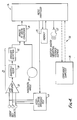
- FIG 4 is an electric block diagram of the training and monitoring device.
- the monitoring device comprises electromyographic sensor 22 which is operatively connected to control means 24 through an analog to digital converter 26.
- Goniometer 20 is also coupled to the control means through converter 26.
- the control means comprises a microprocessor unit acting also as an internal clock and is interfaced to an electronic memory 25 that forms a recording means.
- the microprocessor is coupled to a indicator means 27, which can be auditory and/or vibrational for indicating to the user a lifting condition which exceeds preset parameters programmed into the microprocessor.
- the myoelectric signals from the three electrodes are amplified by high gain differential amplifier 28, filtered by bandpass filter 30 and directed to envelope detector 32 which converts the raw EMG waveform of the myoelectric signals into an approximation of the total myoelectric energy which essentially comprises a muscle force signal.
- envelope detector 32 converts the raw EMG waveform of the myoelectric signals into an approximation of the total myoelectric energy which essentially comprises a muscle force signal.
- the resulting muscle force signal is an analog signal it is converted into a digital format acceptable to the microprocessor.
- the goniometer forms a horizontal angle signal that comprises a lifting angle signal that is also converted from an analog to a digital format before being directed to the microprocessor. It should be noted that goniometer measures the lumbar angle including anterior and/or left/right lateral angles.
- Figure 5 reflects the idealized behaviour of lumbar angle and EMG measurement under several lifting conditions.
- the EMG curves shown do not include components of inertia and body weight.
- Figure 5a and 5c are graphical presentations of lifting no loads in a back straight position and back bent position.
- the horizontal angle changes only slightly whereas in the back bent position the horizontal angle changes from nearly zero degrees to ninety degrees.
- EMG muscle force
- Figures 5b and 5d a load is lifted and although the lifting angle is identical to the no load sequence, the amount of muscle force required in each sequence varies considerably because of the lifting methodology.
- the back bent position the amount of muscle force required from the lower back tends to mirror the change in lifting angle whereas in the back straight position during the initial lifting motion the amount of lower back muscle force is considerably reduced because the legs are doing the lifting.
- a teacher programs the microprocessor via the compliance computer 38 with a set of lifting parameters which include limits as to muscle load and horizontal angle.
- the teacher can set up a system wherein a combination of the parameters triggers a feedback warning signal.
- a feedback warning signal For example in Figure 5c the user has taken an incorrect lifting angle but since the user is not lifting any load the indicator is not triggered. However in Figure 5d the user has taken an incorrect lifting position and is lifting a load, therefore the indicator is triggered.
- the present system gives the teacher the ability to program triggering parameters that are a combination of the lifting angle and muscle force required.
- the monitoring system is battery operated and located in a lockable housing so that after the teacher has programmed the microprocessor, the housing is locked and the battery cannot be tampered with by the user.
- the microprocessor is provided with interface 36 comprising a plug for coupling the microprocessor to compliance computer 38.
- the compliance computer can be an IBM PC compatible unit and is used to interrogate the memory so that a training session can be tabulated for evaluation by the teacher. In addition this interface can be used for programming the microprocessor with the programmed lifting parameters.
- the compliance computer is provided with monitor 42, input keyboard 44, and printed 46.
- the microprocessor periodically activates interelectrode impedance test 48 to check if electrode contact is sufficient.
- the test aplied a bipolar sinusoidal signal across the EMG inputs, the impedance is then measured by the microprocessor.
- the microprocessor can be provided with a testing system for testing battery voltage to insure proper voltage to the monitoring system. In the event that the contacts fail the impedance test or the battery has insufficient voltage the microprocessor signals the user through the indicator means and turns off the system.
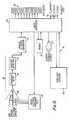
- Figures 7-12 are directed to an exercise training system which is similar to the lift training monitoring system. As can be seen in Figure 8 the circuitry is similar except that the exercise training system is provided with visual feedback display means 50 comprising a bar graph, and alerting means 52 comprising three light emitting diodes.
- visual feedback display means 50 comprising a bar graph
- alerting means 52 comprising three light emitting diodes.
- the auditory feedback element 27, which in the lifting training system is an indicating means, in this embodiment is used in conjunction with the visible display means and the alerting means to inform the patient audibly that these displays have been triggered.
- Bar graph 50 is a liquid crystal or light emitting diode display that is used for displaying muscle force used during use.
- the exercise training system is auto ranging with respect to the bar graph, the alogorithm for auto ranging the bar graph is disclosed in Figure 9.
- light emitting diode 54 lights up indicating to the user to contract the muscle group that is equipped with the electromyographic electrodes.
- the user keeps that muscle contracted until light emitting diode 54 is turned off, and light emitting diode 56 lights up indicating to the user to relax the muscle group. Contract/relax cycles are repeated as determined by the preprogrammed microprocessor.
- the intensity of the muscle contractions is fed back to the user by viewing bar graph 50 which indicates muscle force used.
- a physical therapist first applies the electromyographic electrodes to a patient adjacent to the muscle group to be exercised. Then the therapist programs the microprocessor via the compliance computer of the training system, by programming a time interval in which the exercise routine is to begin, the timed interval for contracting a muscle group and relaxing a muscle group, and the number of repetitions. The therapist then couples the unit to the electrode leads and the patient can then conduct his own physical therapy by using isometric exercises for contracting the desired muscle group for the required duration and repetitions and monitoring the intensity of the exercise on the bar graph.
- the exercise training system can be coupled to compliance computer 38 through interface 36, whuich can comprise a simple jack.
- the compliance computer is used to program the microprocessor and to tabulate the patient's performance with the exercise program by interrogating the electronic memory which recorded the exercise session. The therapist can then program into the microprocessor a new training routine based upon the patient's actual performance in the last training session.
- the compliance computer is also used to program the microprocessor.
- Figure 7 is a front view of the training and monitoring device which is relatively compact.
- the circuitry including the microprocessor, the electronic memory, and the elctromyographic processing circuitry are contained in housing 60.
- the device is provided with a start/stop switch 72 for overriding the exercise routine programmed into the microprocessor, and a third light emitting diode 73 indicates the device is not functioning correctly based upon its self testing, which is identical to the self testing of the lift training device.
- Figure 9 discloses a flow chart illustrating the method of auto ranging the bar graph display.
- the exercise parameters programmed into electronic memory via the compliance computer are read by the microprocessor and are used to initialize relevant variables.
- the auto ranging method then through subsequent EMG (muscle force) readings sets a continually updated top value and bottom value for the bar graph scale.
- the method then calculates a new EMG reading located between the top and bottom value as a ratio of the EMC range and as such displays this ratio by lighting up the correct number of bar graph display elements.
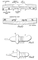
- Figure 10-12 disclose different devices for securing the electrodes of the electromyographic sensor to selected body location.
- the electrodes are secured to cotton gauze 74 that forms the inner liner of a cast for a limb.
- the monitoring housing and related circuitry because of its compact nature, can then be embedded in casting material 75 of the outer cast layer.
- the bar graph display is located at an angle to the housing to facilitate viewing by the patient.
- Figures 11 and 12 are directed to electrode mounting assemblies that were designed to be inserted into a naturally occurring body orifices. These assemblies are cylindrical and have three stainless steel electrode bands located about their circumference.
- the embodiment illustrated in Figure 11 comprises cylindrical member 80 which is inserted into a female vagina so that the female patient can monitor the exercise of associated vaginal muscles.
- the embodiment illustrated in Figure 12 comprises cylindrical member 81 and is inserted in a patient's anus for monitoring a patient's exercise of the anal sphincter muscles. Both units are made from injected moulded plastic, and are provided with depth guages 82 which can be adjustably positioned and fixed on the cylindrical members by the therapist.
- FIG. 13 An alternate embodiment of the lift training system is illustrated in Figure 13 and comprises a lift training system that is not provided with an electromyographic sensor. Instead only goniometer 20 is used to measure lumbar angle. Temperature sensor 90 and/or motion sensor 92 are also mounted on the belt and indicate the belt is being worn by a user. In this way, the actual usage of the lift training system is logged together with a log of incorrect lifting angle.
- the microprocessor is programmed with lifting parameters via the compliance computer that when exceeded trigger indicator means 27 to alert the user.
- the compliance computer is used to interrogate the electronic memory for evaluating and tabulating the results of the lift monitoring session.
- usage sensors 90 and 92 do not have to be aplied to converter 26 if the signals are already in digital form.
- the usage sensors can be used on the belt disclosed in Figures 1-3.
Landscapes
- Health & Medical Sciences (AREA)
- Life Sciences & Earth Sciences (AREA)
- Heart & Thoracic Surgery (AREA)
- Medical Informatics (AREA)
- Physics & Mathematics (AREA)
- Veterinary Medicine (AREA)
- Biophysics (AREA)
- Pathology (AREA)
- Engineering & Computer Science (AREA)
- Biomedical Technology (AREA)
- Public Health (AREA)
- General Health & Medical Sciences (AREA)
- Molecular Biology (AREA)
- Surgery (AREA)
- Animal Behavior & Ethology (AREA)
- Oral & Maxillofacial Surgery (AREA)
- Physiology (AREA)
- Dentistry (AREA)
- Physical Education & Sports Medicine (AREA)
- Rehabilitation Tools (AREA)
- Measurement Of The Respiration, Hearing Ability, Form, And Blood Characteristics Of Living Organisms (AREA)
- Measurement And Recording Of Electrical Phenomena And Electrical Characteristics Of The Living Body (AREA)
Priority Applications (2)
| Application Number | Priority Date | Filing Date | Title |
|---|---|---|---|
| CA000562841A CA1331489C (en) | 1988-03-29 | 1988-03-29 | Lifting monitoring and exercise training system |
| EP88303082A EP0336030A1 (de) | 1988-03-29 | 1988-04-06 | System zur Überwachung des Hebens und zur Ausübung des Trainings |
Applications Claiming Priority (2)
| Application Number | Priority Date | Filing Date | Title |
|---|---|---|---|
| CA000562841A CA1331489C (en) | 1988-03-29 | 1988-03-29 | Lifting monitoring and exercise training system |
| EP88303082A EP0336030A1 (de) | 1988-03-29 | 1988-04-06 | System zur Überwachung des Hebens und zur Ausübung des Trainings |
Publications (1)
| Publication Number | Publication Date |
|---|---|
| EP0336030A1 true EP0336030A1 (de) | 1989-10-11 |
Family
ID=25671806
Family Applications (1)
| Application Number | Title | Priority Date | Filing Date |
|---|---|---|---|
| EP88303082A Withdrawn EP0336030A1 (de) | 1988-03-29 | 1988-04-06 | System zur Überwachung des Hebens und zur Ausübung des Trainings |
Country Status (2)
| Country | Link |
|---|---|
| EP (1) | EP0336030A1 (de) |
| CA (1) | CA1331489C (de) |
Cited By (11)
| Publication number | Priority date | Publication date | Assignee | Title |
|---|---|---|---|---|
| ES2048676A2 (es) * | 1992-09-28 | 1994-03-16 | Univ Politecnica De Valencia I | Raquimetro extensometrico. |
| FR2715821A1 (fr) * | 1994-02-07 | 1995-08-11 | Univ Alsace | Dispositif d'aide à la rééducation fonctionnelle, notamment du rachis. |
| WO1996029929A1 (en) * | 1995-03-29 | 1996-10-03 | The University Of Queensland | Diagnosis of neuromuscular dysfunction |
| WO2006038822A1 (en) * | 2004-10-04 | 2006-04-13 | Industrial Research Limited | Angle and force measurement instrument |
| EP2383021A1 (de) | 2010-04-28 | 2011-11-02 | Technogym S.p.A. | Vorrichtung zur begleitenden Durchführung einer Fitnessübung |
| EP2722078A1 (de) * | 2012-10-22 | 2014-04-23 | Paul Chen | Muskeltrainingseinrichtung für den Oberkörper eines Benutzers |
| EP2722080A1 (de) * | 2012-10-22 | 2014-04-23 | Paul Chen | Muskeltrainingseinrichtung |
| EP2722079A1 (de) * | 2012-10-22 | 2014-04-23 | Paul Chen | Muskeltrainingsseinrichtung für den Unterkörper eines Benutzers |
| WO2015032006A1 (en) * | 2013-09-09 | 2015-03-12 | Shabah Abdo | Orthopedic support device and associated methods |
| US9409053B1 (en) | 2015-07-13 | 2016-08-09 | Bml Productions, Inc. | Exercise data collection system |
| CN116440470A (zh) * | 2023-03-31 | 2023-07-18 | 余姚市机器人研究中心 | 一种智能运动健身束带、控制方法及系统 |
Citations (5)
| Publication number | Priority date | Publication date | Assignee | Title |
|---|---|---|---|---|
| US4170225A (en) * | 1976-09-20 | 1979-10-09 | Somatronics, Inc. | Biofeedback device |
| EP0074407A1 (de) * | 1981-03-18 | 1983-03-23 | KONNO, Yoshio | Gerät zur Messung des AusmaBes von Muskelermüdung oder -schmerz |
| DE3442549A1 (de) * | 1984-11-22 | 1986-05-22 | Detlef 4630 Bochum Dick | Vorrichtung zur ueberwachung des beugewinkels von gelenken in der orthopaedie |
| US4655227A (en) * | 1985-06-06 | 1987-04-07 | Diagnospine Research Inc. | Equipment for the detection of mechanical injuries in the lumbar spine of a patient, using a mathematical model |
| US4702108A (en) * | 1985-06-24 | 1987-10-27 | Regents Of The Univ. Of Minnesota | Method and apparatus for measuring the isometric muscle strength of multiple muscle groups in the human body |
-
1988
- 1988-03-29 CA CA000562841A patent/CA1331489C/en not_active Expired - Fee Related
- 1988-04-06 EP EP88303082A patent/EP0336030A1/de not_active Withdrawn
Patent Citations (5)
| Publication number | Priority date | Publication date | Assignee | Title |
|---|---|---|---|---|
| US4170225A (en) * | 1976-09-20 | 1979-10-09 | Somatronics, Inc. | Biofeedback device |
| EP0074407A1 (de) * | 1981-03-18 | 1983-03-23 | KONNO, Yoshio | Gerät zur Messung des AusmaBes von Muskelermüdung oder -schmerz |
| DE3442549A1 (de) * | 1984-11-22 | 1986-05-22 | Detlef 4630 Bochum Dick | Vorrichtung zur ueberwachung des beugewinkels von gelenken in der orthopaedie |
| US4655227A (en) * | 1985-06-06 | 1987-04-07 | Diagnospine Research Inc. | Equipment for the detection of mechanical injuries in the lumbar spine of a patient, using a mathematical model |
| US4702108A (en) * | 1985-06-24 | 1987-10-27 | Regents Of The Univ. Of Minnesota | Method and apparatus for measuring the isometric muscle strength of multiple muscle groups in the human body |
Cited By (15)
| Publication number | Priority date | Publication date | Assignee | Title |
|---|---|---|---|---|
| ES2048676A2 (es) * | 1992-09-28 | 1994-03-16 | Univ Politecnica De Valencia I | Raquimetro extensometrico. |
| FR2715821A1 (fr) * | 1994-02-07 | 1995-08-11 | Univ Alsace | Dispositif d'aide à la rééducation fonctionnelle, notamment du rachis. |
| WO1996029929A1 (en) * | 1995-03-29 | 1996-10-03 | The University Of Queensland | Diagnosis of neuromuscular dysfunction |
| US5916172A (en) * | 1995-03-29 | 1999-06-29 | The University Of Queensland | Diagnosis of neuromuscular dysfunction |
| WO2006038822A1 (en) * | 2004-10-04 | 2006-04-13 | Industrial Research Limited | Angle and force measurement instrument |
| US7661309B2 (en) | 2004-10-04 | 2010-02-16 | Industrial Research Limited | Angle and force measurement instrument |
| EP2383021A1 (de) | 2010-04-28 | 2011-11-02 | Technogym S.p.A. | Vorrichtung zur begleitenden Durchführung einer Fitnessübung |
| US8911328B2 (en) | 2010-04-28 | 2014-12-16 | Technogym S.P.A. | Apparatus for the assisted performance of a fitness exercise |
| US9061170B2 (en) | 2010-04-28 | 2015-06-23 | Technogym S.P.A. | Apparatus for the assisted performance of a fitness exercise |
| EP2722078A1 (de) * | 2012-10-22 | 2014-04-23 | Paul Chen | Muskeltrainingseinrichtung für den Oberkörper eines Benutzers |
| EP2722080A1 (de) * | 2012-10-22 | 2014-04-23 | Paul Chen | Muskeltrainingseinrichtung |
| EP2722079A1 (de) * | 2012-10-22 | 2014-04-23 | Paul Chen | Muskeltrainingsseinrichtung für den Unterkörper eines Benutzers |
| WO2015032006A1 (en) * | 2013-09-09 | 2015-03-12 | Shabah Abdo | Orthopedic support device and associated methods |
| US9409053B1 (en) | 2015-07-13 | 2016-08-09 | Bml Productions, Inc. | Exercise data collection system |
| CN116440470A (zh) * | 2023-03-31 | 2023-07-18 | 余姚市机器人研究中心 | 一种智能运动健身束带、控制方法及系统 |
Also Published As
| Publication number | Publication date |
|---|---|
| CA1331489C (en) | 1994-08-16 |
Similar Documents
| Publication | Publication Date | Title |
|---|---|---|
| US5474083A (en) | Lifting monitoring and exercise training system | |
| US4817628A (en) | System and method for evaluating neurological function controlling muscular movements | |
| US6168569B1 (en) | Apparatus and method for relating pain and activity of a patient | |
| US7640804B2 (en) | Apparatus for measuring activity | |
| US5277197A (en) | Microprocessor controlled system for unsupervised EMG feedback and exercise training | |
| US4387723A (en) | Method and apparatus for determining the level of neuromuscular block in a patient | |
| US5131401A (en) | Method and apparatus for monitoring neuromuscular blockage | |
| US20090043230A1 (en) | Biomechanical monitoring apparatus | |
| US4396019A (en) | Vaginal myograph method and apparatus | |
| US3983865A (en) | Method and apparatus for myofunctional biofeedback | |
| US5038795A (en) | Muscle tonometer apparatus and method | |
| US20060047232A1 (en) | Back training device | |
| WO2009042818A1 (en) | Obtaining measurements of muscle reflexes for diagnosis of patient symptoms | |
| CA1331489C (en) | Lifting monitoring and exercise training system | |
| WO2020078486A1 (en) | A device for measuring muscle strength, a method for determining muscle function, a method for testing the muscle performance of athletes, a method for testing preparations for the treatment and prevention of sarcopenia and use thereof | |
| EP0474745A1 (de) | Bio-feedback-vorrichtung zur überwachung von muskelbewegungen | |
| CN114098735B (zh) | 可调节会阴收缩力计装置 | |
| JPH0515518A (ja) | 身体運動監視システム及び運動トレーニングシステム | |
| JPH0670899A (ja) | 神経筋トレーニングシステム | |
| EP1424938B1 (de) | Verfahren und vorrichtung zur selektiven und nichtinvasiven messung des ablaufs der skelettmuskelkontraktion | |
| WO2009013490A1 (en) | Biofeedback orthosis | |
| JPH01256934A (ja) | 持ち上げ動作観察及び運動訓練システム | |
| JP4399939B2 (ja) | 生体情報測定装置 | |
| Brass et al. | An improved method for muscle force assessment in neuromuscular disease | |
| CN211270736U (zh) | 一种防滑肌肉活动评估装置 |
Legal Events
| Date | Code | Title | Description |
|---|---|---|---|
| PUAI | Public reference made under article 153(3) epc to a published international application that has entered the european phase |
Free format text: ORIGINAL CODE: 0009012 |
|
| AK | Designated contracting states |
Kind code of ref document: A1 Designated state(s): AT BE CH DE ES FR GB GR IT LI LU NL SE |
|
| 17P | Request for examination filed |
Effective date: 19900329 |
|
| 17Q | First examination report despatched |
Effective date: 19930114 |
|
| RAP1 | Party data changed (applicant data changed or rights of an application transferred) |
Owner name: EMPI, INC. |
|
| STAA | Information on the status of an ep patent application or granted ep patent |
Free format text: STATUS: THE APPLICATION IS DEEMED TO BE WITHDRAWN |
|
| 18D | Application deemed to be withdrawn |
Effective date: 19950801 |