EP0341439A1 - Verfahren zur Erhöhung der Spezifizität in kompetitiven Immuntests - Google Patents
Verfahren zur Erhöhung der Spezifizität in kompetitiven Immuntests Download PDFInfo
- Publication number
- EP0341439A1 EP0341439A1 EP89106445A EP89106445A EP0341439A1 EP 0341439 A1 EP0341439 A1 EP 0341439A1 EP 89106445 A EP89106445 A EP 89106445A EP 89106445 A EP89106445 A EP 89106445A EP 0341439 A1 EP0341439 A1 EP 0341439A1
- Authority
- EP
- European Patent Office
- Prior art keywords
- hbc
- sample
- reducing agent
- cysteine
- immunoassay
- Prior art date
- Legal status (The legal status is an assumption and is not a legal conclusion. Google has not performed a legal analysis and makes no representation as to the accuracy of the status listed.)
- Granted
Links
- 238000000034 method Methods 0.000 title abstract description 18
- 238000002967 competitive immunoassay Methods 0.000 title description 2
- 239000003638 chemical reducing agent Substances 0.000 claims abstract description 44
- 238000003556 assay Methods 0.000 claims abstract description 34
- 238000003018 immunoassay Methods 0.000 claims abstract description 15
- 101710132601 Capsid protein Proteins 0.000 claims abstract description 14
- 238000001514 detection method Methods 0.000 claims abstract description 10
- 239000000523 sample Substances 0.000 claims description 41
- XUJNEKJLAYXESH-UHFFFAOYSA-N cysteine Natural products SCC(N)C(O)=O XUJNEKJLAYXESH-UHFFFAOYSA-N 0.000 claims description 35
- 235000018417 cysteine Nutrition 0.000 claims description 35
- 239000011859 microparticle Substances 0.000 claims description 18
- 239000007790 solid phase Substances 0.000 claims description 13
- DGVVWUTYPXICAM-UHFFFAOYSA-N β‐Mercaptoethanol Chemical compound OCCS DGVVWUTYPXICAM-UHFFFAOYSA-N 0.000 claims description 12
- VHJLVAABSRFDPM-QWWZWVQMSA-N dithiothreitol Chemical compound SC[C@@H](O)[C@H](O)CS VHJLVAABSRFDPM-QWWZWVQMSA-N 0.000 claims description 10
- 239000012472 biological sample Substances 0.000 claims description 8
- XUJNEKJLAYXESH-REOHCLBHSA-N L-Cysteine Chemical compound SC[C@H](N)C(O)=O XUJNEKJLAYXESH-REOHCLBHSA-N 0.000 claims description 5
- DZBUGLKDJFMEHC-UHFFFAOYSA-O acridine;hydron Chemical compound C1=CC=CC2=CC3=CC=CC=C3[NH+]=C21 DZBUGLKDJFMEHC-UHFFFAOYSA-O 0.000 claims description 5
- VHJLVAABSRFDPM-ZXZARUISSA-N dithioerythritol Chemical compound SC[C@H](O)[C@H](O)CS VHJLVAABSRFDPM-ZXZARUISSA-N 0.000 claims description 5
- LSNNMFCWUKXFEE-UHFFFAOYSA-M Bisulfite Chemical compound OS([O-])=O LSNNMFCWUKXFEE-UHFFFAOYSA-M 0.000 claims description 4
- 230000006872 improvement Effects 0.000 claims description 4
- 239000004816 latex Substances 0.000 claims description 4
- 229920000126 latex Polymers 0.000 claims description 4
- 239000011541 reaction mixture Substances 0.000 claims description 4
- 125000003396 thiol group Chemical group [H]S* 0.000 claims description 4
- 238000005406 washing Methods 0.000 claims description 2
- 239000011248 coating agent Substances 0.000 claims 2
- 238000000576 coating method Methods 0.000 claims 2
- 125000000151 cysteine group Chemical group N[C@@H](CS)C(=O)* 0.000 claims 1
- 208000002672 hepatitis B Diseases 0.000 abstract description 13
- 238000006243 chemical reaction Methods 0.000 abstract description 7
- 230000000694 effects Effects 0.000 description 20
- 239000003153 chemical reaction reagent Substances 0.000 description 19
- 230000005764 inhibitory process Effects 0.000 description 14
- LMDZBCPBFSXMTL-UHFFFAOYSA-N 1-ethyl-3-(3-dimethylaminopropyl)carbodiimide Chemical compound CCN=C=NCCCN(C)C LMDZBCPBFSXMTL-UHFFFAOYSA-N 0.000 description 12
- 230000002452 interceptive effect Effects 0.000 description 12
- XLYOFNOQVPJJNP-UHFFFAOYSA-N water Chemical compound O XLYOFNOQVPJJNP-UHFFFAOYSA-N 0.000 description 12
- 210000004369 blood Anatomy 0.000 description 11
- 239000008280 blood Substances 0.000 description 11
- 239000013642 negative control Substances 0.000 description 11
- 238000012360 testing method Methods 0.000 description 11
- 239000000243 solution Substances 0.000 description 9
- KCXVZYZYPLLWCC-UHFFFAOYSA-N EDTA Chemical compound OC(=O)CN(CC(O)=O)CCN(CC(O)=O)CC(O)=O KCXVZYZYPLLWCC-UHFFFAOYSA-N 0.000 description 8
- 229960001484 edetic acid Drugs 0.000 description 8
- 239000002245 particle Substances 0.000 description 8
- 230000009257 reactivity Effects 0.000 description 8
- 230000035945 sensitivity Effects 0.000 description 8
- 239000011324 bead Substances 0.000 description 7
- 208000015181 infectious disease Diseases 0.000 description 7
- 239000013641 positive control Substances 0.000 description 7
- 229920001213 Polysorbate 20 Polymers 0.000 description 6
- 238000002835 absorbance Methods 0.000 description 6
- 239000000256 polyoxyethylene sorbitan monolaurate Substances 0.000 description 6
- 235000010486 polyoxyethylene sorbitan monolaurate Nutrition 0.000 description 6
- 210000002966 serum Anatomy 0.000 description 6
- FAPWRFPIFSIZLT-UHFFFAOYSA-M Sodium chloride Chemical compound [Na+].[Cl-] FAPWRFPIFSIZLT-UHFFFAOYSA-M 0.000 description 5
- WBZKQQHYRPRKNJ-UHFFFAOYSA-L disulfite Chemical compound [O-]S(=O)S([O-])(=O)=O WBZKQQHYRPRKNJ-UHFFFAOYSA-L 0.000 description 5
- 239000003550 marker Substances 0.000 description 5
- 239000012528 membrane Substances 0.000 description 5
- 102000004190 Enzymes Human genes 0.000 description 4
- PXIPVTKHYLBLMZ-UHFFFAOYSA-N Sodium azide Chemical compound [Na+].[N-]=[N+]=[N-] PXIPVTKHYLBLMZ-UHFFFAOYSA-N 0.000 description 4
- 201000010099 disease Diseases 0.000 description 4
- 208000037265 diseases, disorders, signs and symptoms Diseases 0.000 description 4
- 239000002244 precipitate Substances 0.000 description 4
- 238000002360 preparation method Methods 0.000 description 4
- 230000009467 reduction Effects 0.000 description 4
- 108090000790 Enzymes Proteins 0.000 description 3
- 108010001336 Horseradish Peroxidase Proteins 0.000 description 3
- MHAJPDPJQMAIIY-UHFFFAOYSA-N Hydrogen peroxide Chemical compound OO MHAJPDPJQMAIIY-UHFFFAOYSA-N 0.000 description 3
- NQTADLQHYWFPDB-UHFFFAOYSA-N N-Hydroxysuccinimide Chemical compound ON1C(=O)CCC1=O NQTADLQHYWFPDB-UHFFFAOYSA-N 0.000 description 3
- DWAQJAXMDSEUJJ-UHFFFAOYSA-M Sodium bisulfite Chemical compound [Na+].OS([O-])=O DWAQJAXMDSEUJJ-UHFFFAOYSA-M 0.000 description 3
- HEMHJVSKTPXQMS-UHFFFAOYSA-M Sodium hydroxide Chemical compound [OH-].[Na+] HEMHJVSKTPXQMS-UHFFFAOYSA-M 0.000 description 3
- 239000003085 diluting agent Substances 0.000 description 3
- 239000012153 distilled water Substances 0.000 description 3
- 238000010828 elution Methods 0.000 description 3
- 208000006454 hepatitis Diseases 0.000 description 3
- 231100000283 hepatitis Toxicity 0.000 description 3
- 230000002401 inhibitory effect Effects 0.000 description 3
- 235000018102 proteins Nutrition 0.000 description 3
- 102000004169 proteins and genes Human genes 0.000 description 3
- 108090000623 proteins and genes Proteins 0.000 description 3
- 238000012216 screening Methods 0.000 description 3
- 235000010267 sodium hydrogen sulphite Nutrition 0.000 description 3
- 239000007787 solid Substances 0.000 description 3
- GEYOCULIXLDCMW-UHFFFAOYSA-N 1,2-phenylenediamine Chemical compound NC1=CC=CC=C1N GEYOCULIXLDCMW-UHFFFAOYSA-N 0.000 description 2
- 206010059193 Acute hepatitis B Diseases 0.000 description 2
- 108091003079 Bovine Serum Albumin Proteins 0.000 description 2
- LEVWYRKDKASIDU-QWWZWVQMSA-N D-cystine Chemical compound OC(=O)[C@H](N)CSSC[C@@H](N)C(O)=O LEVWYRKDKASIDU-QWWZWVQMSA-N 0.000 description 2
- XEEYBQQBJWHFJM-UHFFFAOYSA-N Iron Chemical compound [Fe] XEEYBQQBJWHFJM-UHFFFAOYSA-N 0.000 description 2
- 239000004793 Polystyrene Substances 0.000 description 2
- 239000012506 Sephacryl® Substances 0.000 description 2
- 229920002684 Sepharose Polymers 0.000 description 2
- 239000007983 Tris buffer Substances 0.000 description 2
- 208000037628 acute hepatitis B virus infection Diseases 0.000 description 2
- 230000005540 biological transmission Effects 0.000 description 2
- 230000036770 blood supply Effects 0.000 description 2
- 229940098773 bovine serum albumin Drugs 0.000 description 2
- 239000000872 buffer Substances 0.000 description 2
- 229960003067 cystine Drugs 0.000 description 2
- 238000002474 experimental method Methods 0.000 description 2
- 239000012458 free base Substances 0.000 description 2
- 108010074605 gamma-Globulins Proteins 0.000 description 2
- 230000002209 hydrophobic effect Effects 0.000 description 2
- 238000011534 incubation Methods 0.000 description 2
- 238000010915 one-step procedure Methods 0.000 description 2
- 229920002223 polystyrene Polymers 0.000 description 2
- 238000003127 radioimmunoassay Methods 0.000 description 2
- 238000011084 recovery Methods 0.000 description 2
- 238000013207 serial dilution Methods 0.000 description 2
- HRZFUMHJMZEROT-UHFFFAOYSA-L sodium disulfite Chemical compound [Na+].[Na+].[O-]S(=O)S([O-])(=O)=O HRZFUMHJMZEROT-UHFFFAOYSA-L 0.000 description 2
- 229940001584 sodium metabisulfite Drugs 0.000 description 2
- 235000010262 sodium metabisulphite Nutrition 0.000 description 2
- 239000001488 sodium phosphate Substances 0.000 description 2
- 229910000162 sodium phosphate Inorganic materials 0.000 description 2
- LENZDBCJOHFCAS-UHFFFAOYSA-N tris Chemical compound OCC(N)(CO)CO LENZDBCJOHFCAS-UHFFFAOYSA-N 0.000 description 2
- RYFMWSXOAZQYPI-UHFFFAOYSA-K trisodium phosphate Chemical compound [Na+].[Na+].[Na+].[O-]P([O-])([O-])=O RYFMWSXOAZQYPI-UHFFFAOYSA-K 0.000 description 2
- SXGZJKUKBWWHRA-UHFFFAOYSA-N 2-(N-morpholiniumyl)ethanesulfonate Chemical compound [O-]S(=O)(=O)CC[NH+]1CCOCC1 SXGZJKUKBWWHRA-UHFFFAOYSA-N 0.000 description 1
- GHCZTIFQWKKGSB-UHFFFAOYSA-N 2-hydroxypropane-1,2,3-tricarboxylic acid;phosphoric acid Chemical compound OP(O)(O)=O.OC(=O)CC(O)(C(O)=O)CC(O)=O GHCZTIFQWKKGSB-UHFFFAOYSA-N 0.000 description 1
- UMCMPZBLKLEWAF-BCTGSCMUSA-N 3-[(3-cholamidopropyl)dimethylammonio]propane-1-sulfonate Chemical compound C([C@H]1C[C@H]2O)[C@H](O)CC[C@]1(C)[C@@H]1[C@@H]2[C@@H]2CC[C@H]([C@@H](CCC(=O)NCCC[N+](C)(C)CCCS([O-])(=O)=O)C)[C@@]2(C)[C@@H](O)C1 UMCMPZBLKLEWAF-BCTGSCMUSA-N 0.000 description 1
- RYGMFSIKBFXOCR-UHFFFAOYSA-N Copper Chemical compound [Cu] RYGMFSIKBFXOCR-UHFFFAOYSA-N 0.000 description 1
- GUBGYTABKSRVRQ-WFVLMXAXSA-N DEAE-cellulose Chemical compound OC1C(O)C(O)C(CO)O[C@H]1O[C@@H]1C(CO)OC(O)C(O)C1O GUBGYTABKSRVRQ-WFVLMXAXSA-N 0.000 description 1
- 239000007987 MES buffer Substances 0.000 description 1
- 108020004511 Recombinant DNA Proteins 0.000 description 1
- QAOWNCQODCNURD-UHFFFAOYSA-N Sulfuric acid Chemical compound OS(O)(=O)=O QAOWNCQODCNURD-UHFFFAOYSA-N 0.000 description 1
- 208000036142 Viral infection Diseases 0.000 description 1
- VJHCJDRQFCCTHL-UHFFFAOYSA-N acetic acid 2,3,4,5,6-pentahydroxyhexanal Chemical compound CC(O)=O.OCC(O)C(O)C(O)C(O)C=O VJHCJDRQFCCTHL-UHFFFAOYSA-N 0.000 description 1
- DZBUGLKDJFMEHC-UHFFFAOYSA-N acridine Chemical class C1=CC=CC2=CC3=CC=CC=C3N=C21 DZBUGLKDJFMEHC-UHFFFAOYSA-N 0.000 description 1
- BFNBIHQBYMNNAN-UHFFFAOYSA-N ammonium sulfate Chemical compound N.N.OS(O)(=O)=O BFNBIHQBYMNNAN-UHFFFAOYSA-N 0.000 description 1
- 229910052921 ammonium sulfate Inorganic materials 0.000 description 1
- 235000011130 ammonium sulphate Nutrition 0.000 description 1
- 239000000427 antigen Substances 0.000 description 1
- 102000036639 antigens Human genes 0.000 description 1
- 108091007433 antigens Proteins 0.000 description 1
- 239000007864 aqueous solution Substances 0.000 description 1
- 238000000149 argon plasma sintering Methods 0.000 description 1
- 244000309466 calf Species 0.000 description 1
- 238000005119 centrifugation Methods 0.000 description 1
- 239000002738 chelating agent Substances 0.000 description 1
- 238000004587 chromatography analysis Methods 0.000 description 1
- 230000001684 chronic effect Effects 0.000 description 1
- 229910052802 copper Inorganic materials 0.000 description 1
- 239000010949 copper Substances 0.000 description 1
- 230000008878 coupling Effects 0.000 description 1
- 238000010168 coupling process Methods 0.000 description 1
- 238000005859 coupling reaction Methods 0.000 description 1
- 230000003247 decreasing effect Effects 0.000 description 1
- 238000000502 dialysis Methods 0.000 description 1
- 238000010790 dilution Methods 0.000 description 1
- 239000012895 dilution Substances 0.000 description 1
- 230000008034 disappearance Effects 0.000 description 1
- 150000002148 esters Chemical class 0.000 description 1
- 238000011156 evaluation Methods 0.000 description 1
- -1 for example Proteins 0.000 description 1
- 239000000499 gel Substances 0.000 description 1
- 238000002523 gelfiltration Methods 0.000 description 1
- 239000011521 glass Substances 0.000 description 1
- 239000003365 glass fiber Substances 0.000 description 1
- 230000003862 health status Effects 0.000 description 1
- 238000004128 high performance liquid chromatography Methods 0.000 description 1
- 230000028993 immune response Effects 0.000 description 1
- 230000002163 immunogen Effects 0.000 description 1
- 229940027941 immunoglobulin g Drugs 0.000 description 1
- 230000002779 inactivation Effects 0.000 description 1
- 239000012678 infectious agent Substances 0.000 description 1
- 230000002458 infectious effect Effects 0.000 description 1
- 230000003993 interaction Effects 0.000 description 1
- 229910052742 iron Inorganic materials 0.000 description 1
- 229920002521 macromolecule Polymers 0.000 description 1
- 229910021645 metal ion Inorganic materials 0.000 description 1
- 230000007935 neutral effect Effects 0.000 description 1
- 238000006386 neutralization reaction Methods 0.000 description 1
- 230000003647 oxidation Effects 0.000 description 1
- 238000007254 oxidation reaction Methods 0.000 description 1
- 230000002085 persistent effect Effects 0.000 description 1
- 239000008363 phosphate buffer Substances 0.000 description 1
- 230000003389 potentiating effect Effects 0.000 description 1
- 230000002829 reductive effect Effects 0.000 description 1
- 230000000717 retained effect Effects 0.000 description 1
- 230000000405 serological effect Effects 0.000 description 1
- 229910052708 sodium Inorganic materials 0.000 description 1
- 239000011734 sodium Substances 0.000 description 1
- 239000011780 sodium chloride Substances 0.000 description 1
- 238000012421 spiking Methods 0.000 description 1
- 239000012536 storage buffer Substances 0.000 description 1
- 239000000126 substance Substances 0.000 description 1
- 235000011149 sulphuric acid Nutrition 0.000 description 1
- 239000006228 supernatant Substances 0.000 description 1
- 208000024891 symptom Diseases 0.000 description 1
- 238000010998 test method Methods 0.000 description 1
- 239000012085 test solution Substances 0.000 description 1
- 238000000870 ultraviolet spectroscopy Methods 0.000 description 1
- 230000009385 viral infection Effects 0.000 description 1
- 210000002845 virion Anatomy 0.000 description 1
- 239000011800 void material Substances 0.000 description 1
Images





Classifications
-
- G—PHYSICS
- G01—MEASURING; TESTING
- G01N—INVESTIGATING OR ANALYSING MATERIALS BY DETERMINING THEIR CHEMICAL OR PHYSICAL PROPERTIES
- G01N33/00—Investigating or analysing materials by specific methods not covered by groups G01N1/00 - G01N31/00
- G01N33/48—Biological material, e.g. blood, urine; Haemocytometers
- G01N33/50—Chemical analysis of biological material, e.g. blood, urine; Testing involving biospecific ligand binding methods; Immunological testing
- G01N33/53—Immunoassay; Biospecific binding assay; Materials therefor
- G01N33/557—Immunoassay; Biospecific binding assay; Materials therefor using kinetic measurement, i.e. time rate of progress of an antigen-antibody interaction
-
- G—PHYSICS
- G01—MEASURING; TESTING
- G01N—INVESTIGATING OR ANALYSING MATERIALS BY DETERMINING THEIR CHEMICAL OR PHYSICAL PROPERTIES
- G01N33/00—Investigating or analysing materials by specific methods not covered by groups G01N1/00 - G01N31/00
- G01N33/48—Biological material, e.g. blood, urine; Haemocytometers
- G01N33/50—Chemical analysis of biological material, e.g. blood, urine; Testing involving biospecific ligand binding methods; Immunological testing
- G01N33/53—Immunoassay; Biospecific binding assay; Materials therefor
- G01N33/543—Immunoassay; Biospecific binding assay; Materials therefor with an insoluble carrier for immobilising immunochemicals
- G01N33/54393—Improving reaction conditions or stability, e.g. by coating or irradiation of surface, by reduction of non-specific binding, by promotion of specific binding
-
- G—PHYSICS
- G01—MEASURING; TESTING
- G01N—INVESTIGATING OR ANALYSING MATERIALS BY DETERMINING THEIR CHEMICAL OR PHYSICAL PROPERTIES
- G01N33/00—Investigating or analysing materials by specific methods not covered by groups G01N1/00 - G01N31/00
- G01N33/48—Biological material, e.g. blood, urine; Haemocytometers
- G01N33/50—Chemical analysis of biological material, e.g. blood, urine; Testing involving biospecific ligand binding methods; Immunological testing
- G01N33/53—Immunoassay; Biospecific binding assay; Materials therefor
- G01N33/5306—Improving reaction conditions, e.g. reduction of non-specific binding, promotion of specific binding
-
- G—PHYSICS
- G01—MEASURING; TESTING
- G01N—INVESTIGATING OR ANALYSING MATERIALS BY DETERMINING THEIR CHEMICAL OR PHYSICAL PROPERTIES
- G01N33/00—Investigating or analysing materials by specific methods not covered by groups G01N1/00 - G01N31/00
- G01N33/48—Biological material, e.g. blood, urine; Haemocytometers
- G01N33/50—Chemical analysis of biological material, e.g. blood, urine; Testing involving biospecific ligand binding methods; Immunological testing
- G01N33/53—Immunoassay; Biospecific binding assay; Materials therefor
- G01N33/576—Immunoassay; Biospecific binding assay; Materials therefor for hepatitis
- G01N33/5761—Hepatitis B
- G01N33/5762—Hepatitis B core antigen
-
- Y—GENERAL TAGGING OF NEW TECHNOLOGICAL DEVELOPMENTS; GENERAL TAGGING OF CROSS-SECTIONAL TECHNOLOGIES SPANNING OVER SEVERAL SECTIONS OF THE IPC; TECHNICAL SUBJECTS COVERED BY FORMER USPC CROSS-REFERENCE ART COLLECTIONS [XRACs] AND DIGESTS
- Y10—TECHNICAL SUBJECTS COVERED BY FORMER USPC
- Y10S—TECHNICAL SUBJECTS COVERED BY FORMER USPC CROSS-REFERENCE ART COLLECTIONS [XRACs] AND DIGESTS
- Y10S436/00—Chemistry: analytical and immunological testing
- Y10S436/82—Hepatitis associated antigens and antibodies
-
- Y—GENERAL TAGGING OF NEW TECHNOLOGICAL DEVELOPMENTS; GENERAL TAGGING OF CROSS-SECTIONAL TECHNOLOGIES SPANNING OVER SEVERAL SECTIONS OF THE IPC; TECHNICAL SUBJECTS COVERED BY FORMER USPC CROSS-REFERENCE ART COLLECTIONS [XRACs] AND DIGESTS
- Y10—TECHNICAL SUBJECTS COVERED BY FORMER USPC
- Y10S—TECHNICAL SUBJECTS COVERED BY FORMER USPC CROSS-REFERENCE ART COLLECTIONS [XRACs] AND DIGESTS
- Y10S436/00—Chemistry: analytical and immunological testing
- Y10S436/825—Pretreatment for removal of interfering factors from sample
Definitions
- Hepatitis B core antigen is a particle from the core of the hepatitis B virion (HBV), sometimes referred to as the Dane particle.
- HBV hepatitis B virion
- HBcAg is a potent immunogen which typically elicits a strong immune response persisting for years after HBV infection has cleared and is found in serum of patients with chronic HBV infections.
- anti-HBc hepatitis B core antigen
- NANB non-A, non-B hepatitis infections
- Immunoassays for the detection of anti-HBc are well known. Examples of such assays include the CORAB® radioimmunoassay and the CORZYME® enzyme immunoassay, both commercially available from Abbott Laboratories, North Chicago, Illinois. These assays are competitive immunoassays in which anti-HBc in a biological sample competes with a constant amount of anti-HBc which is labeled with a detectable label for a limited number of binding sites on a solid phase coated with HBcAg. The proportion of bound labeled anti-HBc is inversely proportional to the concentration of anti-HBc in the biological sample.
- the anti-HBc assays described above can be performed by one- or two-step methods.
- a sample and labeled anti-HBc are mixed together and allowed to compete for HBcAg attached to a solid phase.
- the HBcAg on the solid phase is first reacted with the test sample and then with labeled anti-HBc in a second step.
- the amount of labeled anti-HBc bound to the solid phase is measured, and a reduction in the amount of labeled antibody is indicative of an antibody positive sample which is able to block binding of the labeled antibody.
- False positives represent a problem in screening donor blood for anti-HBc because there are a large number of specimens which are positive only for anti-HBc with no other hepatitis marker present and no history or symptoms of disease. There is no simple way to confirm that these specimens are true positives. This is particularly troublesome when anti-HBc testing is used as a surrogate screening for NANB because the epidemiologic connection between the marker and the disease is not strong (85% of recipients of anti-HBc positive blood do not develop a known infection). Therefore, it is important to discriminate between anti-HBc positive specimens that relate to NANB and those which do not or are false positives.
- the improvement comprises the addition of a reducing agent such as sulfhydryl compounds including 2-mercaptoethanol (2ME), dithiothreitol (DTT), dithioerythritol (DTE), cysteine or other reducing agents such as bisulfite solutions especially a metabisulfite solution (MBS) or sodium bisulfite.
- a reducing agent such as sulfhydryl compounds including 2-mercaptoethanol (2ME), dithiothreitol (DTT), dithioerythritol (DTE), cysteine or other reducing agents
- bisulfite solutions especially a metabisulfite solution (MBS) or sodium bisulfite.
- the reducing agent can be introduced into the immunoassay in several ways.
- the sample may be treated with the chemical prior to the start of the assay.
- the sample, labelled reagent, solid phase and the reducing agent may be added simultaneously or in one of the diluents for the sample, labelled reagent or solid phase.
- the labelled reagent contains a reducing agent-sensitive component such as an enzyme, for example, horseradish peroxidase
- the reducing agent should not come in contact with the enzyme and is introduced in a separate step in a two-step assay.
- the sample and solid phase are incubated together before the addition of the labelled reagent in a second step.
- the reducing agent may be added in either the first or second step but is preferably added to the sample prior to addition of the solid phase or simultaneously with the sample and solid phase, and is most effective when added in the first incubation.
- a reducing agent to an assay for anti-HBc eliminates false positive specimens, especially those near the cut-off value, while having no significant effect on control values or true positive specimens. This results in a reduced number of samples that are reactive for anti-HBc only, i.e., with no other HBV markers or history of disease.
- This increased specificity for anti-HBc testing further increases specificity for blood bank surrogate testing for NANB, reducing the number of discarded blood units and saving donors from follow up clinical evaluation to determine their own health status and future rejection as blood donors.
- the interfering reactivity or factor which is being destroyed is not completely understood, it is a macromolecule with physical characteristics different from IgG.
- the interfering activity may be caused by a heterogenous factor, i.e., more than one molecule or a complex.
- the molecular weight of the interfering factor is greater than 500,000, it is extremely unstable in the presence of a reducing agent and is unstable below a pH of 4.5.
- This interfering factor is believed to be hydrophobic on the basis of its interaction with hydrophobic gels and is soluble in 30% (NH4)2SO4 and precipitates at 50%. It is partially retained on CM or DEAE-cellulose.
- the interfering factor eliminated by the reducing agent is IgM, since it is unlikely that there would be a significant number of low level IgM anti-HBc positive samples, and many samples containing the interfering factor failed to react in an IgM-capture assay such as Corzyme M (available from Abbott Laboratories, North Chicago, Illinois). Further, the biochemical evidence is that the interfering factor is differentially destroyed by MBS or cysteine and is unstable in the presence of other reducing agents to a different degree than IgM. For example, at the concentrations of reducing agents and under the conditions employed in the invention, IgM would not be completely destroyed, whereas, the interfering factor is totally eliminated.
- the interfering factor coelutes with IgM on Sephacryl S-300, the nonspecific activity is lost upon chromatography on Sepharose 6B while low levels of IgM-specific activity are recovered in the expected fractions. It is noteworthy that the inactivation of the interfering factor on Sepharose 6B in the absence of reducing agent. It appears therefore that not only reducing agents, but also gel filtration, causes disruption of a possible complex or aggregate which is responsible for the interfering activity.
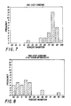
- the first population (I) is a group of selected anti-HBc positive sera which were defined as true positives because they possessed other markers of hepatitis B or were from subjects with a previous history of hepatitis B and whose reactivity was associated with gamma globulins.
- the second population (II) is a group of serum specimens from donors without other markers of hepatitis B or a history of hepatitis B and whose reactivity could not be clearly associated with gamma globulins.
- Group II specimens were weak positives when they were tested in a one-step anti-HBc assay, but usually exhibited stronger anti-HBc-like reactivity when tested with a two-step procedure.
- Group I positives were generally strongly reactive in both test procedures; randomly selected sera of this group were diluted so that they simulated the level of activity of group II positives in the study. Both groups I and II were tested by conditions described in detail below, with and without reducing agents.
- 0.1 ml samples in groups I and II were placed in duplicate reaction wells, along with negative and positive controls, and 10 ul of 0.3% 2 mercaptoethanol (2 ME) in H2O was added to one each of the duplicates, while 10 ul H2O was added to the other.
- 0.1 ml of 125I-labeled anti-HBc was added to all wells, followed by an HBcAg coated 1/4 inch polystyrene bead to all wells. Trays were gently shaken and allowed to incubate overnight at room temperature. All beads were washed 3 times with distilled water and transferred to test tubes for counting for 1 minute in a gamma scintillation counter.
- 0.1 ml of specimens in groups I and II were placed in duplicate in reaction wells, as in Example 1, but 0.1 ml of saline solution was added to each, followed by 10 ul of 0.5% NaHSO3 in H2O to one of the duplicates, and 10 ul H2O to the other duplicate. (0.2% Na in H2O can be substituted for the 0.5% NaHSO3.)
- An HBcAg coated-bead was added to each well, and the trays were gently shaken and incubated overnight at room temperature. After washing the beads, they were incubated with 0.2 ml of anti-HBc IgG conjugated with horseradish peroxidase (HRP) for 1 hour at 40°.
- HRP horseradish peroxidase
- Beads were again washed 3 times with distilled water and transferred to clean reaction tubes. Bead-bound antibody-HRP was detected by adding a solution of o-phenylenediamine (OPD) in citrate-phosphate buffer containing 0.02% hydrogen peroxide, to each bead for 30 minutes, stopping the reaction with H2SO4 and reading the degree of absorbance at A492.
- OPD o-phenylenediamine
- a significant reduction in color intensity was an indication of reactivity from a true or false antibody positive specimen, and positivity was calculated according to the following formula: 0.4 x NCV + 0.6 x
- Control Mean Value Specimens with absorbance values equal to or lower than the Cutoff Value were considered positive and specimens with values greater than the Cutoff Value were considered negative. Additionally, the degree (%) inhibition from negative control for each specimen, both with and without reducing reagent was calculated according to the formula of Example 1, but substituting A492 values for CPM, and the effect of the addition of reducing reagent to specimens was also evaluated according to the criteria described in Example 1.
- a microparticle chemiluminescence immunoassay for anti-HBc was performed as follows:
- Recombinant DNA hepatitis B core antigen (rHBcAg) was coupled to carboxylated polystyrene latex microparticles (Seragen Diagnostics, Indianapolis, Indiana, 0.188 um) using a two-step procedure.
- purified polyclonal human anti-HBc at 2.7 ug/mg latex particles in the presence of normal human serum (1.0 ul/mg) was coupled using 1-ethyl-3-(3-dimethylaminopropyl) carbodiimide (EDAC, 43 ug/mg) in 0.015 M MES buffer (2-[N-morpholino] ethanesulfonic acid), pH 4.8, for 2 hours at room temperature.
- Particles were washed three times as before and dispersed through a 25 gauge needle and stored in 0.01 M sodium phosphate, pH 7.2, containing 0.15 M NaCl, 0.1% bovine serum albumin, 0.01% Tween-20 and 0.1% sodium azide (Microparticle Diluent). Percent solids was determined by light scattering at 500 nanometers. Coated particles were stored concentrated (typically 5-10% solids) and diluted in storage buffer to a working concentration (0.036%) before each assay. In an alternative method, rHBcAg was coupled directly to the microparticles by the EDAC method. The anti-HBc undercoating method is preferred.
- NHS N-hydroxylsuccinimide
- Activated acridinium was added to dialyzed antibody (30:1 molar excess) and the reaction mixture stirred for 30 minutes at room temperature. After dialysis versus the same buffer, the preparation was centrifuged and the supernatant chromatographed on a Beckman TSK 250 HPLC column equilibrated with the same buffer. Individual fractions (1 ml) were analyzed by UV spectroscopy at 369 and 280 nm to determine incorparation of acridinium (typically 3:1).
- Conjugate was stored in concentrated fractions (approximately 100 ug/ml) at 2-8°C and diluted in 0.01 M sodium phosphate containing 14.8 g/L EDTA, 0.1% sodium azide, 0.4% Tween-20, 50% (V:V) normal calf serum and 2% (V:V) normal human serum, pH 6.3, before each assay (Conjugate Diluent).
- cysteine In water, cysteine oxidizes to cystine which is relatively insoluble and forms a white precipitate. The oxidation is enhanced by the presence of metal ions notably copper, iron and by a neutral or slightly alkaline pH. A procedure for preparing relatively stable solutions of cysteine is described below.
- Distilled water was deionized using a Barnstead Water-I Deionizer to a final resistance of 18 megaohms.
- Cysteine Sigma Chemical Co., Free Base
- EDTA EDTA
- DTT DTT
- the pH of the solution was between 4.5 and 5.0 and was not adjusted. Aliquots of the solution were stored in closed glass vials at room temperature and at 2-8°C.
- Stability was assessed by the lack of cystine precipitate which formed in aqueous solutions without the chelating agent (EDTA) and reductant (DTT). After two weeks no precipitate was evident at either temperature (Table II). Stability was also assessed by the effectiveness of this reagent over time to eliminate non-specific anti-HBc activity from two plasma samples (S9 and S14), which were similar in anti-HBc activity to the Group II samples described above, as shown in Table II. Two week stability was sufficient for practical utilization of the reagent in the microparticle assay described below.
- each reaction mixture was transferred to a microparticle capture/flow-through glass fiber filter funnel (Abbott Laboratories, North Chicago, Illinois) which was prewetted with 50 ul of 0.1% Tween-20. Each capture membrane was then washed twice with 50 ul of 0.1% Tween-20. 20 ul of the acridinium-labeled anti-HBc (375 ng/ml) was added to the surface of each capture membrane and incubated at room temperature for 20 minutes. The capture membranes were then washed twice with 100 ul and once with 50 ul of 0.1% Tween-20.
- Plasma samples designated 52, 71, 166 and 277, Panel 7 and negative and positive controls were tested according to the microparticle assay procedure of Example 3, except that 10 ul of 0.75 M cysteine (Sigma, free base) prepared fresh was used instead of the cysteine solution containing EDTA and DTT. The samples were run both with and without cysteine.
- Sample 277 is a truly negative sample.
- Samples 71 and 166 are borderline positive samples which are similar in anti-HBc activity to Group II samples, except that these are unaffected by treatment with the reducing agent cysteine, indicating that these are indeed borderline or discrepant samples.
- Samples 52 and 189 are borderline samples which are also similar to Group II samples, but in the presence of the reducing reagent these samples proved to be negative.
- Each sample was also tested with and without 26.3 ul of a sensitivity panel sample (Panel 5) equivalent to 1.23 PEI U/ml to determine the effect of spiking these samples with a true positive anti-HBc sample with and without cysteine. Results are shown in Table IV.
- Panel 7 produced 71.8% and 80.7% inhibition in the absence and presence of cysteine, respectively.
- the negative control spiked with an amount of Panel 5 equivalent to panel 7 gave 73.7% and 80.8% inhibition in the absence and presence of cysteine, respectively.
- the average percent inhibition in unspiked plasma samples dropped from 31.8% to 21.0% with addition of cysteine.
- the average percent inhibition of plasma samples spiked with the equivalent of panel 7 was 67.1% and 71.5% in the absence and presence of cysteine, respectively.
- Sensitivity panel samples which are known positive for anti-HBc (Panels 5, 6, 7, 8 and 9) were tested by the microparticle assay procedure of Example 3. Results are shown in Table V. There was no effect on assay sensitivity when the cysteine reagent was utilized.
- Plasma samples designated 25, 31, 76, 91, 177 and 189 were tested with a commercially available anti-HBc kit (Corab®, Abbott Laboratories, North Chicago, Illinois) with and without addition of 10 ul of a reducing agent, 0.5 M sodium metabisulfite. Also, as in Example 4, samples were tested with and without addition of 26.3 ul of a positive anti-HBc sample (Panel 5). Results are shown in Table VI. As can be seen from the Table, Sample 25 is a truly negative sample. Samples 31 and 91 are discrepant samples which are confirmed as negative upon addition of the reducing agent. Sample 177 does not respond to treatment with MBS indicating that this sample is an elevated negative. As can be seen from Table VI, all anti-HBc positive samples, including Panel 7 and samples which were spiked with Panel 5 were not affected by the addition of MBS.
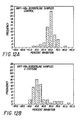
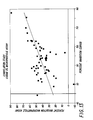
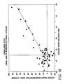
- Example 7 The plasma samples described in Example 7 were tested according to the microparticle assay procedure described in Example 3. Data are shown graphically in Figure 12. The correlation between the data of Example 7 and this Example 8 in the absence of metabisulfite and cysteine, respectively, are shown in Figure 13. The correlation between the data of Example 7 and this Example 8 in the presence of metabisulfite and cysteine, respectively, are shown in Figure 14.
- Example 209 The absorbance and anti-HBc activity of an anti-HBc positive sample (Sample 209) is shown in Figure 15. This sample displayed 86.3% and 85.6% inhibition when tested in the Corab assay in the presence and absence of metabisulfite, respectively. Briefly, 2 ml of recalcified Sample 209 was applied to a Sephacryl S-300 column (2.5 x 50 cm) equilibrated with PBS. 2.5 ml fractions were collected at a flow rate of 20 ml/hr. Fractions were assayed using the microparticle assay of Example 3 except that an approximately 5 fold increase in sensitivity was implemented by using increasing incubation time and decreasing the amount of anti-HBc conjugate.
- Example 9 3 ml of a borderline positive sample (Sample 9) was tested according to the procedure of Example 9. The sample displayed 56.8% and 5.2% inhibition when assayed by the Corab assay in the absence and presence of metabisulfite, respectively.
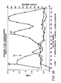
- the absorbance and anti-HBc activity elution profile of Sample 9 is shown in Fiqure 16.
- a single inhibitory peak was seen near the void volume of the column. This peak was not detected when assayed in the presence of cysteine, and was clearly resolved form the IgG protein peak. This non-IgG reactivity is not believed to be a result of Hepatitis B infection and is therefore nonspecific.
- the improved anti-HBc assay of the invention has many advantages. These include reduction in the number of borderline reactive anti-HBc-positive samples, including reduction in the number of samples which are positive for anti-HBc only with no other marker of hepatitis or disease history being present. By reducing the number of borderline or false positive samples in blood donor screening, the nation's blood supply will be enhanced and future donors will not be unnecessarily excluded.
Landscapes
- Health & Medical Sciences (AREA)
- Life Sciences & Earth Sciences (AREA)
- Immunology (AREA)
- Engineering & Computer Science (AREA)
- Chemical & Material Sciences (AREA)
- Urology & Nephrology (AREA)
- Hematology (AREA)
- Biomedical Technology (AREA)
- Molecular Biology (AREA)
- Analytical Chemistry (AREA)
- General Health & Medical Sciences (AREA)
- Cell Biology (AREA)
- Pathology (AREA)
- Food Science & Technology (AREA)
- Medicinal Chemistry (AREA)
- Physics & Mathematics (AREA)
- Biotechnology (AREA)
- Biochemistry (AREA)
- Microbiology (AREA)
- General Physics & Mathematics (AREA)
- Chemical Kinetics & Catalysis (AREA)
- Communicable Diseases (AREA)
- Measuring Or Testing Involving Enzymes Or Micro-Organisms (AREA)
- Peptides Or Proteins (AREA)
- Medicines Containing Antibodies Or Antigens For Use As Internal Diagnostic Agents (AREA)
- Investigating Or Analysing Biological Materials (AREA)
- Saccharide Compounds (AREA)
Priority Applications (1)
| Application Number | Priority Date | Filing Date | Title |
|---|---|---|---|
| AT89106445T ATE92633T1 (de) | 1988-05-11 | 1989-04-11 | Verfahren zur erhoehung der spezifizitaet in kompetitiven immuntests. |
Applications Claiming Priority (2)
| Application Number | Priority Date | Filing Date | Title |
|---|---|---|---|
| US193307 | 1980-10-02 | ||
| US19330788A | 1988-05-11 | 1988-05-11 |
Publications (2)
| Publication Number | Publication Date |
|---|---|
| EP0341439A1 true EP0341439A1 (de) | 1989-11-15 |
| EP0341439B1 EP0341439B1 (de) | 1993-08-04 |
Family
ID=22713085
Family Applications (1)
| Application Number | Title | Priority Date | Filing Date |
|---|---|---|---|
| EP89106445A Expired - Lifetime EP0341439B1 (de) | 1988-05-11 | 1989-04-11 | Verfahren zur Erhöhung der Spezifizität in kompetitiven Immuntests |
Country Status (9)
| Country | Link |
|---|---|
| US (2) | US5681695A (de) |
| EP (1) | EP0341439B1 (de) |
| JP (1) | JP2945025B2 (de) |
| KR (1) | KR900018679A (de) |
| AT (1) | ATE92633T1 (de) |
| AU (1) | AU613187B2 (de) |
| CA (1) | CA1335706C (de) |
| DE (1) | DE68907996T2 (de) |
| ES (1) | ES2059601T3 (de) |
Cited By (7)
| Publication number | Priority date | Publication date | Assignee | Title |
|---|---|---|---|---|
| WO1996036876A1 (en) * | 1995-05-19 | 1996-11-21 | Chiron Diagnostics Corporation | Stable protein solutions for diagnostics and method of making and using the same |
| WO1999006837A1 (en) * | 1997-07-30 | 1999-02-11 | Bionova Corporation | Method for detecting hbcag from hepatitis b virus |
| WO1999015901A1 (en) * | 1997-09-22 | 1999-04-01 | Chiron Corporation | Buffers for stabilizing antigens |
| US6391540B1 (en) | 1997-09-22 | 2002-05-21 | Chiron Corporation | Method for detecting antibodies in a sample |
| US6645732B2 (en) | 1996-11-29 | 2003-11-11 | Roche Diagnostics Gmbh | Antigen-specific IgG detection |
| US7176313B2 (en) | 2000-02-09 | 2007-02-13 | Daiichi Pharmaceutical Co., Ltd. | Anti acid-fast bacterial agent containing pyridonecarboxylic acids as active ingredient |
| EP1752768A4 (de) * | 2004-05-19 | 2007-11-28 | Advanced Life Science Inst Inc | Verfahren zum nachweis von hepatitis-b-virus |
Families Citing this family (10)
| Publication number | Priority date | Publication date | Assignee | Title |
|---|---|---|---|---|
| DE4428705A1 (de) * | 1994-08-12 | 1996-02-15 | Boehringer Mannheim Gmbh | Rekombinantes Antigen aus der NS3-Region des Hepatitis C Virus |
| AU751362B2 (en) * | 1998-04-17 | 2002-08-15 | Innogenetics N.V. | Improved immunodiagnostic assays using reducing agents |
| US20020064881A1 (en) * | 2000-11-30 | 2002-05-30 | Devlin William Jackson | Method for automatically storing and reprocessing patient specimen's in an automatic clinical analyzer |
| US6723851B2 (en) | 2001-10-31 | 2004-04-20 | Quest Diagnostics Investment Incorporated | Chemiluminescent compounds and use thereof |
| US7674632B1 (en) * | 2001-12-10 | 2010-03-09 | Zeus Scientific, Inc | Method and composition for homogeneous multiplexed microparticle-based assay |
| US20060134647A1 (en) * | 2004-12-17 | 2006-06-22 | Sylwia Karwowska | Method for elimination of false reactivities in immunoassay for Hepatitis A virus |
| US8112229B2 (en) * | 2007-05-31 | 2012-02-07 | Abbott Laboratories | Method for determining the order of execution of assays of a sample in a laboratory automation system |
| JP5163883B2 (ja) * | 2008-05-30 | 2013-03-13 | 東ソー株式会社 | B型肝炎ウイルスコア抗原又はそれに対する抗体の測定方法 |
| JP7209498B2 (ja) * | 2017-09-15 | 2023-01-20 | 富士レビオ株式会社 | B型肝炎ウイルスコア抗体の免疫測定方法 |
| CN113777312B (zh) * | 2021-09-03 | 2024-02-02 | 普十生物科技(北京)有限公司 | 乙肝抗体片段的制备方法、乙肝抗体片段、试剂盒及应用 |
Citations (6)
| Publication number | Priority date | Publication date | Assignee | Title |
|---|---|---|---|---|
| FR2383446A1 (fr) * | 1977-03-11 | 1978-10-06 | Abbott Lab | Reactifs et procedes pour la mise en evidence de l'anticorps antinoyaux de dane |
| GB2016687A (en) * | 1978-03-20 | 1979-09-26 | Abbott Lab | Sugar Coated Reagents for Solid Phase Immunoassay |
| EP0012699A1 (de) * | 1978-12-18 | 1980-06-25 | Merck & Co. Inc. | Verfahren zur Bestimmung von Hepatitis-B-Kern-Antikörpern in biologischen Flüssigkeiten |
| GB2052059A (en) * | 1979-06-19 | 1981-01-21 | Siber G | Immunological reagnet and test |
| EP0163312A2 (de) * | 1984-06-01 | 1985-12-04 | Daiichi Pure Chemicals Co. Ltd. | Immunotest |
| WO1987003690A1 (en) * | 1985-12-10 | 1987-06-18 | Murex Corporation | Particle-bound binding component immunoassay |
Family Cites Families (4)
| Publication number | Priority date | Publication date | Assignee | Title |
|---|---|---|---|---|
| US4102996A (en) * | 1977-04-20 | 1978-07-25 | Merck & Co., Inc. | Method of preparing hepatitis B core antigen |
| US4234564A (en) * | 1977-12-19 | 1980-11-18 | Merck & Co., Inc. | Hepatitis B core antigen composition |
| AU8746582A (en) * | 1981-09-02 | 1983-03-10 | Biogen N.V. | Hepatitis b virus e type antigen |
| US4683136A (en) * | 1985-03-06 | 1987-07-28 | Scripps Clinic And Research Foundation | Proteinaceous antigens with conformation-independent and conformation-dependent determinants |
-
1989
- 1989-04-11 EP EP89106445A patent/EP0341439B1/de not_active Expired - Lifetime
- 1989-04-11 AT AT89106445T patent/ATE92633T1/de not_active IP Right Cessation
- 1989-04-11 DE DE89106445T patent/DE68907996T2/de not_active Expired - Lifetime
- 1989-04-11 ES ES89106445T patent/ES2059601T3/es not_active Expired - Lifetime
- 1989-05-10 CA CA000599324A patent/CA1335706C/en not_active Expired - Lifetime
- 1989-05-10 AU AU34641/89A patent/AU613187B2/en not_active Ceased
- 1989-05-10 KR KR1019890006227A patent/KR900018679A/ko not_active Ceased
- 1989-05-10 JP JP1117182A patent/JP2945025B2/ja not_active Expired - Lifetime
-
1994
- 1994-12-15 US US08/357,369 patent/US5681695A/en not_active Expired - Lifetime
-
1996
- 1996-12-09 US US08/762,037 patent/US6150113A/en not_active Expired - Lifetime
Patent Citations (6)
| Publication number | Priority date | Publication date | Assignee | Title |
|---|---|---|---|---|
| FR2383446A1 (fr) * | 1977-03-11 | 1978-10-06 | Abbott Lab | Reactifs et procedes pour la mise en evidence de l'anticorps antinoyaux de dane |
| GB2016687A (en) * | 1978-03-20 | 1979-09-26 | Abbott Lab | Sugar Coated Reagents for Solid Phase Immunoassay |
| EP0012699A1 (de) * | 1978-12-18 | 1980-06-25 | Merck & Co. Inc. | Verfahren zur Bestimmung von Hepatitis-B-Kern-Antikörpern in biologischen Flüssigkeiten |
| GB2052059A (en) * | 1979-06-19 | 1981-01-21 | Siber G | Immunological reagnet and test |
| EP0163312A2 (de) * | 1984-06-01 | 1985-12-04 | Daiichi Pure Chemicals Co. Ltd. | Immunotest |
| WO1987003690A1 (en) * | 1985-12-10 | 1987-06-18 | Murex Corporation | Particle-bound binding component immunoassay |
Non-Patent Citations (5)
| Title |
|---|
| BIOLOGICAL ABSTRACTS, vol. 70, no. 10, 1980, abstract no. 64854, Biological Abstracts, Inc., Philadelphia, PA, US; R.A. DEYO et al.: "Interference by rheumatoid factor with the detection of C-reactive protein by the latex agglutination method", & J. RHEUMATOL. 7(3): 279-287. 1980 * |
| BIOLOGICAL ABSTRACTS, vol. 72, no. 5, 1981, abstract no. 32079, Biological Abstracts, Inc., Philadelphia, PA, US; G.L. ANGARANO et al.: "Serological diagnosis of virus hepatitis A: radioimmunoassay for the detection of specific immunoglobulin M anti-hepatitis A virus by 3 methods", & BOLL. SOC. ITAL. BIOL. SPER. 56(20): 2083-2089. 1980 * |
| BIOLOGICAL ABSTRACTS, vol. 80, no. 3, 1985, abstract no. 23240, Biological Abstracts, Inc., Philadelphia, PA, US; H. SHIRAISHI et al.: "Rheumatoid factor-like reactants in sera proven to transmit non-A, non-B hepatitis. A potential source of false-positive reactions in non-A, non-B assays", & HEPATOLOGY (BALTIMORE) 5(2); 181-187, 1985 * |
| CLIN. CHEM., vol. 29, no. 8, 1983, pages 1474-1479; I. WEEKS et al.: "Acridinium esters as high-specific-activity labels in immunoassay" * |
| JOURNAL OF IMMUNOLOGICAL METHODS, vol. 28, 1979, pages 13-23, Elsevier/North-Holland Biomedical Press; C.L. CAMBIASO et al.: "Particle counting immunoassay (PACIA). III. Automated determination of circulating immune complexes by inhibition of an agglutinating factor of mouse serum" * |
Cited By (11)
| Publication number | Priority date | Publication date | Assignee | Title |
|---|---|---|---|---|
| WO1996036876A1 (en) * | 1995-05-19 | 1996-11-21 | Chiron Diagnostics Corporation | Stable protein solutions for diagnostics and method of making and using the same |
| US5817525A (en) * | 1995-05-19 | 1998-10-06 | Chiron Diagnostics Corporation | Stable protein solutions for diagnostics and method of making and using the same |
| US6645732B2 (en) | 1996-11-29 | 2003-11-11 | Roche Diagnostics Gmbh | Antigen-specific IgG detection |
| WO1999006837A1 (en) * | 1997-07-30 | 1999-02-11 | Bionova Corporation | Method for detecting hbcag from hepatitis b virus |
| US6153392A (en) * | 1997-07-30 | 2000-11-28 | Bionova Corporation | Devices and methods comprising an HBcAg from hepatitis B virus |
| WO1999015901A1 (en) * | 1997-09-22 | 1999-04-01 | Chiron Corporation | Buffers for stabilizing antigens |
| US6261764B1 (en) | 1997-09-22 | 2001-07-17 | Chiron Corporation | Buffers for stabilizing antigens |
| US6391540B1 (en) | 1997-09-22 | 2002-05-21 | Chiron Corporation | Method for detecting antibodies in a sample |
| US6537745B2 (en) | 1997-09-22 | 2003-03-25 | Chiron Corporation | Buffers for stabilizing antigens |
| US7176313B2 (en) | 2000-02-09 | 2007-02-13 | Daiichi Pharmaceutical Co., Ltd. | Anti acid-fast bacterial agent containing pyridonecarboxylic acids as active ingredient |
| EP1752768A4 (de) * | 2004-05-19 | 2007-11-28 | Advanced Life Science Inst Inc | Verfahren zum nachweis von hepatitis-b-virus |
Also Published As
| Publication number | Publication date |
|---|---|
| AU613187B2 (en) | 1991-07-25 |
| KR900018679A (ko) | 1990-12-22 |
| JP2945025B2 (ja) | 1999-09-06 |
| DE68907996D1 (de) | 1993-09-09 |
| ATE92633T1 (de) | 1993-08-15 |
| US6150113A (en) | 2000-11-21 |
| JPH01321365A (ja) | 1989-12-27 |
| CA1335706C (en) | 1995-05-30 |
| AU3464189A (en) | 1989-11-16 |
| EP0341439B1 (de) | 1993-08-04 |
| DE68907996T2 (de) | 1994-01-05 |
| ES2059601T3 (es) | 1994-11-16 |
| US5681695A (en) | 1997-10-28 |
Similar Documents
| Publication | Publication Date | Title |
|---|---|---|
| EP0341439B1 (de) | Verfahren zur Erhöhung der Spezifizität in kompetitiven Immuntests | |
| CA1148859A (en) | Simultaneous assay of two hepatitis viruses using a solid phase | |
| JP2636331B2 (ja) | 抗原特異的な抗体の一段階測定法およびそれに適する試薬 | |
| JPS632348B2 (de) | ||
| EP0722087B1 (de) | Mess-system unter verwendung von vollblut | |
| JP3623657B2 (ja) | 非特異反応抑制剤、免疫測定試薬及び免疫測定方法 | |
| JPWO2001092885A1 (ja) | 免疫学的ラテックス比濁分析方法及びその試薬 | |
| WO2001092885A1 (fr) | Methode immunologique de turbidimetrie provoquee par du latex et reactif pour la mise en oeuvre de ladite methode | |
| Eble et al. | Differential diagnosis of acute viral hepatitis using rapid, fully automated immunoassays | |
| PT95669A (pt) | Processo para reduzir a incidencia de respostas positivas falsas em ensaios imunologicos utilizando imunoglobulina polimerizada | |
| Ukkonen et al. | Enzyme-immunoassay in the detection of hepatitis B surface antigen | |
| EP0080108B1 (de) | Diagnostisches Reagent und dessen Verwendung | |
| CA1206876A (en) | Method for measuring igg in a neonatal foal or calf and in colostrum | |
| JPH0792455B2 (ja) | 免疫学的試験用水性溶媒 | |
| EP0139316B1 (de) | Verfahren zur immunochemischen Bestimmung von Hepatitis-B-Core-Antigenen | |
| JP2000346841A (ja) | Pivka−iiの測定方法 | |
| JP7622350B2 (ja) | 高感度な抗hbv抗体の検出方法 | |
| WO1993020445A1 (en) | IMMUNOASSAY FOR DETECTING HCV IgM ANTIBODY | |
| Mushahwar | A biotin/avidin solid-phase sandwich enzyme immunoassay for the antibody to hepatitis B surface antigen (anti-HBs) | |
| Atherton et al. | A sensitive screening test for the simultaneous detection of hepatitis B surface antigen and antibody | |
| Galanti et al. | Immunoassay of hepatitis B surface antigen by particle counting after pepsin digestion | |
| Nath et al. | Enzyme‐linked immunoassay for hepatitis B surface antigen An evaluation of commercial test kits | |
| AU664179B2 (en) | Immunoassay for detecting HCV IgM antibody | |
| Ohori et al. | An antigen/antibody system specific for an epidemic non‐A, non‐B hepatitis in patients of a cardiovascular surgical unit | |
| Nath et al. | A simple technique for enhancing the sensitivity of a commercial solid‐phase radioimmunoassay for hepatitis B surface antigen (HBsAg) |
Legal Events
| Date | Code | Title | Description |
|---|---|---|---|
| PUAI | Public reference made under article 153(3) epc to a published international application that has entered the european phase |
Free format text: ORIGINAL CODE: 0009012 |
|
| AK | Designated contracting states |
Kind code of ref document: A1 Designated state(s): AT BE CH DE ES FR GB IT LI NL |
|
| 17P | Request for examination filed |
Effective date: 19900305 |
|
| 17Q | First examination report despatched |
Effective date: 19911204 |
|
| GRAA | (expected) grant |
Free format text: ORIGINAL CODE: 0009210 |
|
| RAP1 | Party data changed (applicant data changed or rights of an application transferred) |
Owner name: ABBOTT LABORATORIES |
|
| AK | Designated contracting states |
Kind code of ref document: B1 Designated state(s): AT BE CH DE ES FR GB IT LI NL |
|
| REF | Corresponds to: |
Ref document number: 92633 Country of ref document: AT Date of ref document: 19930815 Kind code of ref document: T |
|
| REF | Corresponds to: |
Ref document number: 68907996 Country of ref document: DE Date of ref document: 19930909 |
|
| ET | Fr: translation filed | ||
| ITF | It: translation for a ep patent filed | ||
| PLBI | Opposition filed |
Free format text: ORIGINAL CODE: 0009260 |
|
| 26 | Opposition filed |
Opponent name: BOEHRINGER MANNHEIM GMBH PATENTABTEILUNG Effective date: 19940503 |
|
| NLR1 | Nl: opposition has been filed with the epo |
Opponent name: BOEHRINGER MANNHEIM GMBH |
|
| REG | Reference to a national code |
Ref country code: ES Ref legal event code: FG2A Ref document number: 2059601 Country of ref document: ES Kind code of ref document: T3 |
|
| NLT2 | Nl: modifications (of names), taken from the european patent patent bulletin |
Owner name: ABBOTT LABORATORIES |
|
| PLBO | Opposition rejected |
Free format text: ORIGINAL CODE: EPIDOS REJO |
|
| APAC | Appeal dossier modified |
Free format text: ORIGINAL CODE: EPIDOS NOAPO |
|
| PLBQ | Unpublished change to opponent data |
Free format text: ORIGINAL CODE: EPIDOS OPPO |
|
| PLAB | Opposition data, opponent's data or that of the opponent's representative modified |
Free format text: ORIGINAL CODE: 0009299OPPO |
|
| APAE | Appeal reference modified |
Free format text: ORIGINAL CODE: EPIDOS REFNO |
|
| R26 | Opposition filed (corrected) |
Opponent name: BOEHRINGER MANNHEIM GMBH PATENTABTEILUNG Effective date: 19940503 |
|
| APAC | Appeal dossier modified |
Free format text: ORIGINAL CODE: EPIDOS NOAPO |
|
| NLR1 | Nl: opposition has been filed with the epo |
Opponent name: BOEHRINGER MANNHEIM GMBH PATENTABTEILUNG |
|
| PLAB | Opposition data, opponent's data or that of the opponent's representative modified |
Free format text: ORIGINAL CODE: 0009299OPPO |
|
| R26 | Opposition filed (corrected) |
Opponent name: ROCHE DIAGNOSTICS GMBH Effective date: 19940503 |
|
| NLR1 | Nl: opposition has been filed with the epo |
Opponent name: ROCHE DIAGNOSTICS GMBH |
|
| APAC | Appeal dossier modified |
Free format text: ORIGINAL CODE: EPIDOS NOAPO |
|
| PLBN | Opposition rejected |
Free format text: ORIGINAL CODE: 0009273 |
|
| STAA | Information on the status of an ep patent application or granted ep patent |
Free format text: STATUS: OPPOSITION REJECTED |
|
| 27O | Opposition rejected |
Effective date: 20000912 |
|
| NLR2 | Nl: decision of opposition | ||
| REG | Reference to a national code |
Ref country code: GB Ref legal event code: IF02 |
|
| APAH | Appeal reference modified |
Free format text: ORIGINAL CODE: EPIDOSCREFNO |
|
| PGFP | Annual fee paid to national office [announced via postgrant information from national office to epo] |
Ref country code: GB Payment date: 20080317 Year of fee payment: 20 |
|
| REG | Reference to a national code |
Ref country code: CH Ref legal event code: PFA Owner name: ABBOTT LABORATORIES Free format text: ABBOTT LABORATORIES# #ABBOTT PARK/IL (US) -TRANSFER TO- ABBOTT LABORATORIES# #ABBOTT PARK/IL (US) |
|
| PGFP | Annual fee paid to national office [announced via postgrant information from national office to epo] |
Ref country code: CH Payment date: 20080425 Year of fee payment: 20 Ref country code: DE Payment date: 20080430 Year of fee payment: 20 Ref country code: ES Payment date: 20080409 Year of fee payment: 20 |
|
| PGFP | Annual fee paid to national office [announced via postgrant information from national office to epo] |
Ref country code: AT Payment date: 20080318 Year of fee payment: 20 |
|
| PGFP | Annual fee paid to national office [announced via postgrant information from national office to epo] |
Ref country code: IT Payment date: 20080419 Year of fee payment: 20 Ref country code: BE Payment date: 20080507 Year of fee payment: 20 |
|
| PGFP | Annual fee paid to national office [announced via postgrant information from national office to epo] |
Ref country code: NL Payment date: 20080325 Year of fee payment: 20 |
|
| PGFP | Annual fee paid to national office [announced via postgrant information from national office to epo] |
Ref country code: FR Payment date: 20080403 Year of fee payment: 20 |
|
| REG | Reference to a national code |
Ref country code: CH Ref legal event code: PL |
|
| BE20 | Be: patent expired |
Owner name: *ABBOTT LABORATORIES Effective date: 20090411 |
|
| REG | Reference to a national code |
Ref country code: GB Ref legal event code: PE20 Expiry date: 20090410 |
|
| PG25 | Lapsed in a contracting state [announced via postgrant information from national office to epo] |
Ref country code: NL Free format text: LAPSE BECAUSE OF EXPIRATION OF PROTECTION Effective date: 20090411 |
|
| NLV7 | Nl: ceased due to reaching the maximum lifetime of a patent |
Effective date: 20090411 |
|
| REG | Reference to a national code |
Ref country code: ES Ref legal event code: FD2A Effective date: 20090413 |
|
| PG25 | Lapsed in a contracting state [announced via postgrant information from national office to epo] |
Ref country code: ES Free format text: LAPSE BECAUSE OF EXPIRATION OF PROTECTION Effective date: 20090413 |
|
| PG25 | Lapsed in a contracting state [announced via postgrant information from national office to epo] |
Ref country code: GB Free format text: LAPSE BECAUSE OF EXPIRATION OF PROTECTION Effective date: 20090410 |