EP0345750A2 - Polyfunktionelle Protease - Google Patents
Polyfunktionelle Protease Download PDFInfo
- Publication number
- EP0345750A2 EP0345750A2 EP89110277A EP89110277A EP0345750A2 EP 0345750 A2 EP0345750 A2 EP 0345750A2 EP 89110277 A EP89110277 A EP 89110277A EP 89110277 A EP89110277 A EP 89110277A EP 0345750 A2 EP0345750 A2 EP 0345750A2
- Authority
- EP
- European Patent Office
- Prior art keywords
- leu
- proteasome
- protease
- cleaving
- optimum
- Prior art date
- Legal status (The legal status is an assumption and is not a legal conclusion. Google has not performed a legal analysis and makes no representation as to the accuracy of the status listed.)
- Granted
Links
Images


Classifications
-
- C—CHEMISTRY; METALLURGY
- C12—BIOCHEMISTRY; BEER; SPIRITS; WINE; VINEGAR; MICROBIOLOGY; ENZYMOLOGY; MUTATION OR GENETIC ENGINEERING
- C12N—MICROORGANISMS OR ENZYMES; COMPOSITIONS THEREOF; PROPAGATING, PRESERVING, OR MAINTAINING MICROORGANISMS; MUTATION OR GENETIC ENGINEERING; CULTURE MEDIA
- C12N9/00—Enzymes; Proenzymes; Compositions thereof; Processes for preparing, activating, inhibiting, separating or purifying enzymes
- C12N9/14—Hydrolases (3)
- C12N9/48—Hydrolases (3) acting on peptide bonds (3.4)
- C12N9/50—Proteinases, e.g. Endopeptidases (3.4.21-3.4.25)
- C12N9/64—Proteinases, e.g. Endopeptidases (3.4.21-3.4.25) derived from animal tissue
-
- C—CHEMISTRY; METALLURGY
- C11—ANIMAL OR VEGETABLE OILS, FATS, FATTY SUBSTANCES OR WAXES; FATTY ACIDS THEREFROM; DETERGENTS; CANDLES
- C11D—DETERGENT COMPOSITIONS; USE OF SINGLE SUBSTANCES AS DETERGENTS; SOAP OR SOAP-MAKING; RESIN SOAPS; RECOVERY OF GLYCEROL
- C11D3/00—Other compounding ingredients of detergent compositions covered in group C11D1/00
- C11D3/16—Organic compounds
- C11D3/38—Products with no well-defined composition, e.g. natural products
- C11D3/386—Preparations containing enzymes, e.g. protease or amylase
-
- C—CHEMISTRY; METALLURGY
- C07—ORGANIC CHEMISTRY
- C07K—PEPTIDES
- C07K16/00—Immunoglobulins [IG], e.g. monoclonal or polyclonal antibodies
- C07K16/18—Immunoglobulins [IG], e.g. monoclonal or polyclonal antibodies against material from animals or humans
- C07K16/28—Immunoglobulins [IG], e.g. monoclonal or polyclonal antibodies against material from animals or humans against receptors, cell surface antigens or cell surface determinants
- C07K16/30—Immunoglobulins [IG], e.g. monoclonal or polyclonal antibodies against material from animals or humans against receptors, cell surface antigens or cell surface determinants from tumour cells
-
- C—CHEMISTRY; METALLURGY
- C07—ORGANIC CHEMISTRY
- C07K—PEPTIDES
- C07K16/00—Immunoglobulins [IG], e.g. monoclonal or polyclonal antibodies
- C07K16/40—Immunoglobulins [IG], e.g. monoclonal or polyclonal antibodies against enzymes
-
- C—CHEMISTRY; METALLURGY
- C12—BIOCHEMISTRY; BEER; SPIRITS; WINE; VINEGAR; MICROBIOLOGY; ENZYMOLOGY; MUTATION OR GENETIC ENGINEERING
- C12N—MICROORGANISMS OR ENZYMES; COMPOSITIONS THEREOF; PROPAGATING, PRESERVING, OR MAINTAINING MICROORGANISMS; MUTATION OR GENETIC ENGINEERING; CULTURE MEDIA
- C12N9/00—Enzymes; Proenzymes; Compositions thereof; Processes for preparing, activating, inhibiting, separating or purifying enzymes
- C12N9/14—Hydrolases (3)
- C12N9/48—Hydrolases (3) acting on peptide bonds (3.4)
- C12N9/50—Proteinases, e.g. Endopeptidases (3.4.21-3.4.25)
- C12N9/58—Proteinases, e.g. Endopeptidases (3.4.21-3.4.25) derived from fungi
- C12N9/60—Proteinases, e.g. Endopeptidases (3.4.21-3.4.25) derived from fungi from yeast
-
- C—CHEMISTRY; METALLURGY
- C12—BIOCHEMISTRY; BEER; SPIRITS; WINE; VINEGAR; MICROBIOLOGY; ENZYMOLOGY; MUTATION OR GENETIC ENGINEERING
- C12N—MICROORGANISMS OR ENZYMES; COMPOSITIONS THEREOF; PROPAGATING, PRESERVING, OR MAINTAINING MICROORGANISMS; MUTATION OR GENETIC ENGINEERING; CULTURE MEDIA
- C12N9/00—Enzymes; Proenzymes; Compositions thereof; Processes for preparing, activating, inhibiting, separating or purifying enzymes
- C12N9/14—Hydrolases (3)
- C12N9/48—Hydrolases (3) acting on peptide bonds (3.4)
- C12N9/50—Proteinases, e.g. Endopeptidases (3.4.21-3.4.25)
- C12N9/64—Proteinases, e.g. Endopeptidases (3.4.21-3.4.25) derived from animal tissue
- C12N9/6402—Proteinases, e.g. Endopeptidases (3.4.21-3.4.25) derived from animal tissue from non-mammals
-
- C—CHEMISTRY; METALLURGY
- C12—BIOCHEMISTRY; BEER; SPIRITS; WINE; VINEGAR; MICROBIOLOGY; ENZYMOLOGY; MUTATION OR GENETIC ENGINEERING
- C12N—MICROORGANISMS OR ENZYMES; COMPOSITIONS THEREOF; PROPAGATING, PRESERVING, OR MAINTAINING MICROORGANISMS; MUTATION OR GENETIC ENGINEERING; CULTURE MEDIA
- C12N9/00—Enzymes; Proenzymes; Compositions thereof; Processes for preparing, activating, inhibiting, separating or purifying enzymes
- C12N9/14—Hydrolases (3)
- C12N9/48—Hydrolases (3) acting on peptide bonds (3.4)
- C12N9/50—Proteinases, e.g. Endopeptidases (3.4.21-3.4.25)
- C12N9/64—Proteinases, e.g. Endopeptidases (3.4.21-3.4.25) derived from animal tissue
- C12N9/6421—Proteinases, e.g. Endopeptidases (3.4.21-3.4.25) derived from animal tissue from mammals
-
- A—HUMAN NECESSITIES
- A61—MEDICAL OR VETERINARY SCIENCE; HYGIENE
- A61K—PREPARATIONS FOR MEDICAL, DENTAL OR TOILETRY PURPOSES
- A61K38/00—Medicinal preparations containing peptides
-
- Y—GENERAL TAGGING OF NEW TECHNOLOGICAL DEVELOPMENTS; GENERAL TAGGING OF CROSS-SECTIONAL TECHNOLOGIES SPANNING OVER SEVERAL SECTIONS OF THE IPC; TECHNICAL SUBJECTS COVERED BY FORMER USPC CROSS-REFERENCE ART COLLECTIONS [XRACs] AND DIGESTS
- Y10—TECHNICAL SUBJECTS COVERED BY FORMER USPC
- Y10S—TECHNICAL SUBJECTS COVERED BY FORMER USPC CROSS-REFERENCE ART COLLECTIONS [XRACs] AND DIGESTS
- Y10S435/00—Chemistry: molecular biology and microbiology
- Y10S435/8215—Microorganisms
- Y10S435/911—Microorganisms using fungi
- Y10S435/94—Saccharomyces
- Y10S435/942—Saccharomyces cerevisiae
Definitions
- the present invention relates to polyfunctional proteases, and more particularly to polyfunctional proteases as novel intracellular proteases.
- the polyfunctional protease which locally present in the cytoplasm as an inactive protease, was separated from the rat livers by the present inventor and reported as to its fnctions, molecular structure, distribution, locality, etc (The Journal of Biological Chemistry, 261(32), pp 15197-15203; ibid. pp 15204-152-7 (1986); Kagaku and Seibutsu 25(8), pp 489-490 (1987); Tanpaku Kakusan Kouso 32(7), pp 955-961 (1987); etc.).
- protease is assumed to have a molecular weight of about 750,000, which is exceptionally large for a protease.
- the protease was a giant protein complex in the form of heteropolymer and has in one molecule a plurality of catalytic sites which are different in substrate specificity and independent of one another. From this point of view the protease is referred to as a "polyfunctional protease". When observed electron microscopically, the molecule of this enzyme is found to be of an annular particle structure. Therefore, the protease is also referred to as "proteasome" (Nature, 94, 192-194 (1988)). This protease is assumed to be an enzyme responsible for a non-lysosomal proteolytic pathway.
- An object of the present invention is to provide novel polyfunctional proteases, i.e., human, chicken, Xenopus laevis and yeast (S. cerevisiae) polyfunctional proteases (hereinafter referred to as "proteasomes").
- Another object of the invention is to provide anti-proteasome antibodies useful for purifying and determining the respective proteasomes and antigens for preparing the antibodies.
- Still another object of the invention is to provide methods of immunologically purifying or determining the proteasome with use of the antibody, and techniques for screening and/or diagnosing cancers by determining the proteasome in the body fluid.
- the present invention provides a human proteasome characterized by the following enzymological and physicochemical properties.
- the absorption value of the ultraviolet absorption spectrum, the sedimentation constant given by the sedimentation velocity method, the diffusion coefficient determined by the quasi-elastic light scattering method, the amino acid contents (mole %) and other measurments obtained by physical methods herein given are all experimental values with a standard deviation of up to 5%.
- the present invention further provides a chicken polyfuctional protease characterized by the following enzymological and physicochemical properties.
- the present invention further provides a Xenopus polyfuctional protease characterized by the following enzymological and physicochemical properties.
- the present invention further provides a yeast polyfuctional protease characterized by the following enzymological and physicochemical properties.
- the present invention further provides antibodies characterized in that they are specifically reactive with the respective proteasomes only individually, i.e., an anti-human proteasome antibody, anti-chicken proteasome antibody, anti-Xenopus laevis proteasome antibody and anti-yeast proteasome antibody.
- the proteasome of the invention is a complex of proteases which differ in substrate specificity, has activity to cleave proteins at any of acid, neutral and basic sites and can therefore be said to be the most powerful protease in the history of research on proteases. Moreover, the expression of its activity is controlled intramolecularly, and the proteasome is activated as by the plasmin in the blood and is therefore useful as a fibrinolytic agent.
- the proteasome of the invention is usable also as an additive for detergents and cosmetics.
- the anti-proteasome antibody provided by the present invention and specific to the proteasome is useful for purifying and assaying the proteasome.
- the proteasome can be immunologically purified and assayed with use of the antibody.
- cancers can be screened and/or diagonosed by determining the proteasome in the body fluid with the antibody.
- proteasomes of the invention can be prepared by the processes to be described below in detail.
- the proteasome of the invention can be obtained from a cytoplasmic fraction of the animal or microorganism from which it is to be derived, by usual purification and isolation methods.
- the proteasome is widely distributed in various cells and tissues, and the origin cell is not limited specifically. In humans, the proteasome content is high in organs having high metabolic activity, especially in the liver. Accordingly it is desirable to use cells of such organs as the origin. Further the cells to be used may be derived from various established cell lines.
- cytoplasm fraction from origin cells is known and can be accomplished by the usual cell fractionation method.
- advantageously usable as the cytoplasm fraction is a supernatant obtained by centrifuging a homogenate of origin cells to remove the cell fraction including the cell membrane. nucleus, mitochondrion, lysosome and microsome.
- the present proteasome can be isolated from the cytoplasm fraction and purified by various procedures utilizing the physicochemical and immunochemical properties of the proteasome (see, for example, "Biochemical Data Book II," pp. 1175-1259, first edition, first print, June 23, 1980, published by Tokyo Kagaku Dojin Co., Ltd.). More specifically, useful methods include, for example, treatment with a protein precipitating agent, ultrafiltration, molecular sieve chromatography (gel filtration), adsorption chromatography involving ion exchange, partition chromatography and like liquid chromatographic methods, centrifuging, electrophoresis, dialysis and combinations of such methods.
- the present proteasome is present in an inactive form within the cells and in the extracted fraction, but is irreversibly activated during purification.
- the enzyme is very unstable and difficult to isolate and purify when activated, and must therefore be maintained in the inactive form at all times.
- the enzyme can be maintained in the inactive form, for example, by incorporating in the separation solvent 20% glycerol. and 10 mM 2-mercapto ethanol or 1 mM dithiothreitol.
- proteasome of the invention obtained as a homogeneous substance by the isolation and purification procedure is identified by the foregoing enzymological and physicochemical properties.
- the present proteasome thus obtained can be used as an immunogen for preparing an antibody (anti-proteasome antibody) which is specifically reactive with the proteasome.
- the anti-proteasome antibody is identified as an antibody which specifically recognizes only the type of proteasome of the same origin and is characterized in that the proteasome is not cross-reactive with proteasomes derived from other origins.
- This antibody can be prepared, for example, from the proteasome of the invention serving as an immunogen.
- the proteasome to be used as the immunogen needs to be as homogeneous as the present proteasome, whereas if the antibody is a monoclonal antibody, the homogeneous proteasome of the invention need not always be used but other immunogens containing the proteasome can be used.
- the antibody can be prepared from the proteasome as the immunogen by a usual method, for example, by administering the immunogen to a mammal for immunization to produce the desired antibody (polyclonal antibody) in the living body and collecting the antibody, or by fusing the plasmacyte (immunized cell) of the immunized mammal with a plasmacytoma cell compatible therewith to prepare a hybridoma, selecting a clone producing the desired antibody (monoclonal antibody) therefrom and incubating the clone.
- a usual method for example, by administering the immunogen to a mammal for immunization to produce the desired antibody (polyclonal antibody) in the living body and collecting the antibody, or by fusing the plasmacyte (immunized cell) of the immunized mammal with a plasmacytoma cell compatible therewith to prepare a hybridoma, selecting a clone producing the desired antibody (monoclonal antibody)
- the mammal to be immunized is not limited specifically, it is desirable to select a suitable animal in view of the compatibility of the plasmacyte with the plasmacytoma to be used for cell fusion in the case where the antibody specific to the proteasome of the invention is to be prepared with use of hybridoma.
- the mouse, rat or the like is advantageous to use.
- the mammal can be immunized by a usual method, for example, by giving the immunogen intraveneously, intracutaneously, subcutaneously, intraperitoneally or otherwise. More specifically, the immunogen is given several times (booster), in combination with a usual adjuvant at a overall dose of about 200 to about 250 u.g,body in the case of a mouse.
- a usual adjuvant at a overall dose of about 200 to about 250 u.g,body in the case of a mouse.
- the polyclonal antibody can be collected by separating off the serum of the immunized mammal one to two weeks after the final administration.
- Suitable immunized cells for use in preparing the monoclonal antibody include the spleen cell removed from the mammal about three days after the final administration.
- Examples of immunized cells for use as parent cells to be fused with such immunized cells are those derived from known established cell lines, e.g. myeloma cells such as P3 (p3/X63-Ag8) [Nature, 256, 495-497 (1975)], P3-U1 [Current Topics in Microbiology and Immunology, 81, 1-7 (1978)], NS-1 [Eur.J.
- Methods of preparing monoclonal antibodies from hybridomas are known; for example, the method of Milstein et al. can be resorted to (Method in Emzymology, Vol. 73, p.3, (1981)).
- the fusion reaction can be conducted by a suitable method, such as a method wherein polyethylene glycol (PEG), hemagglutinating virus of Japan (HVJ) or the like is used, or an electrical fusion method.
- PEG polyethylene glycol
- HVJ hemagglutinating virus of Japan
- the hydridoma method is-not limitative; also usable is a method wherein the immunized cell is immortalized as by Epstein-Barr virus.
- the hybridoma or immortalized cells thus obtained is monocloned by the usual limiting dilution method to select the desired antibody producing clone.
- the desired antibody producing clone can be selected by various methods generally used for detecting antibodies, for example, by the EIISA method (Meth. Enzymol., 70, 419-439 (1980)), plaque method, spot method, agglutination method and radioimmunoassay (RIA) ("Hybridoma Methods and Monoclonal Antibodies,” published by R & D Planning Co., Ltd., pp. 30-53, March 5, 1982).
- the immunogen can be utilized for the screening procedure.
- the resulting cells producing the monoclonal antibody can be subcultured with a usual medium and preserved for a prolonged period of time in liquid nitrogen.
- the antibody can be collected from the clone by incubating the clone in a usual manner and preparing a supernatant from the culture, or by administering the clone to a mammal compatible therewith for proliferation and collecting the ascitic fluid from the mammal.
- the former method is suitable for preparing the antibody with a high purity, while the latter method is suited to quantity production.
- the antibody thus obtained can be purified by a usual method such as salting out, gel filtration or affinity chromatography.
- proteasome determining technique is very useful for screening and/or diagnosing cancers.
- the determination of the proteasome in the body fluid for screening and/or diagnosing cancers can be accomplished basically by usual methods of immunoassays such as RIA and enzymatic immunoassay.
- immunoassays such as RIA and enzymatic immunoassay.
- the procedures for practicing these methods include the competitive method, sandwich method, agglutination method, etc.
- body fluids to be assayed are blood, tissue fluid, lympth, pleural fluid, ascites fluid, amniotic fluid, gastric juice, urine, pancreatic juice, cerebrospinal fluid, saliva and the like.
- blood especially serum and plasma are especially desirable.
- Cancers can be screened or diagnosed advantageously by comparing the proteasome level of the subject thus determined with the corresponding level of the normal person.
- kits for determining the proteasome in the body fluid can be screened or diagnosed conveniently using a kit for determining the proteasome in the body fluid.
- the kit comprises an antibody reagent consisting essentially of the anti-proteasome antibody.
- a stabilizer such as glycerol, bovine or like serum protein and/or a preservative can be admixed with the reagent.
- the buffer is not an essential component of the kit, various buffers can be added to the reagent for use in the determination for adjusting the reagent to a pH of about 4 to 8.
- the protease activity was determined using [ 3 H]methyl-casein according to the method of Tanaka et al. (J.B.C.. 261(32), pp 15197-15203 (1986)).
- the protease activity can be easily and more sensitively determined by evaluating the lytic activity of fluorescent peptide substrate such as Suc-Leu-Leu-Val-Tyr-MCA.
- the process for preparing the proteasome was practiced at a temperature of not more than 4° C using as buffer 50 mM Tris HCI buffer (pH 7.5) containing 1mM dithiothreitol and 20% glycerol unless otherwise specified.
- a 200 g quantity of human liver tissue was homogenized with a buffer (50 mM Tris HCI buffer, pH 7.5, containing 0.1 mM EDTA, 1 mM dithiothreitol and 0.25 M sucrose) in 3 times the volume of the tissue using a homogenizer (Potter Elvehjem homogenizer), and centrifuged (105000 x g, 60 mins), giving a cytoplasm fraction as supernatant.
- a buffer 50 mM Tris HCI buffer, pH 7.5, containing 0.1 mM EDTA, 1 mM dithiothreitol and 0.25 M sucrose
- the liver supernatant fraction obtained above was admixed with 800 ml of Fast Flow Q-Sepharose (product of Pharmacia Fine Chemicals) equilibrated with the buffer, and the mixture was stirred by a magnetic stirrer at 4 C for 1 hour.
- the resulting resin was transferred into a Buechner funnel, washed and applied to a column (5 cm in diameter, 60 cm in length). Subsequently the column was washed with the buffer until the absorbance of the washing reduced to below 0.2.
- the specimen adsorbed on the column was eluted with a linear gradient of 0 to 0.8 M NaCI (2 t, 10 ml of fraction). The protease activity was recovered in the fraction at about 0.4 M NaCI.
- the active fraction obtained was precipitated with polyethylene glycol 6000 (terminal concentration: 15%) and the precipitate was dissolved in a small amount (20 ml) of the buffer.
- the solution was applied to a Biogel A-1.5m column (product of Japan Bio Rad Laboratories, Co., Ltd., 4 cm in diameter, 80 cm in length) and eluted with the buffer (the flow rate was 26 ml/min, and the eluate was collected in 10 ml of fraction).
- the active fraction of a single peak was collected .
- the fraction obtained was dialyzed against 10 mM potassium phosphate buffer (pH 6.8) containing 1 mM of dithiothreitol and 20% glycerol, applied to a 40 ml column of Hydroxy Apatite (product of Japan Bio Rad Laboratories, Co., Ltd.) equilibrated with the same buffer.
- the column was then wash d with the same buffer, and the specimen adsorbed on the column was eluted with a linear gradient of 10 to 300 mM phosphate (400 ml, 4 ml fraction). The activity was recovered in the fraction at about 220 mM phosphate.
- the active fraction was applied to a 20 ml column of Heparin-Sepharose CL-6B (product of Pharmacia LKB Biotechnology Inc.) equilibrated with the buffer. The column was washed with the buffer to the base line absorbance, followed by elution with a linear gradient from 0 to 0.4 M KCI (400 ml, 4 ml fraction). The activity was recovered in the fraction at about 100 mM KCI.
- the active fraction obtained above was applied to Mono-Q Column (product of Pharmacia LKB Biotechnology Inc., 1 cm in diameter and 10 cm in length) equilibrated with 25 mM Tris HCI buffer (pH 7.5) containing 1 mM dithiothreitol and 20% glycerol and eluted by high-performance liquid chromatography (FPLC-System, product of Pharmacia LKB Biotechnology Inc.) with a linear gradient from 0 to 0.8 M KCI (the flow rate was 1 ml;mim).
- Tris HCI buffer pH 7.5
- FPLC-System product of Pharmacia LKB Biotechnology Inc.
- the desired homogeneous human proteasome was thus recovered in the fraction at about 340 mM KCI.
- the desired proteasome was prepared from chicken liver tissues in the same manner as in Example 1.
- the xenopus proteasome and the yeast proteasome were obtained from the ovary of Xenopus laevis and S. cerevisiae, respectively.
- a 5 mg quantity of each of the above proteasomes was emulsified in.a specified amount of a complete Freund's adjuvant (manufactured by Difco).
- the emulsion was administered pareterally to the foot-pad and back of female white rabbits weighing 2.5 kg. Four weeks later, one-half of the above injected dose was given as a booster dose. One more week later, a booster dose of the emulsion was administered again to immunize the test animal.
- IgG fraction was separated from the serum by protein A-Sepharose affinity chromatography (FEBS Lett., 28, 73-76 (1972)) to obtain the antibody against the proteasome.
- the monoclonal antibody against the proteasome of the present invention obtained above was prepared as follows.
- a 500 ⁇ l quantity of a phosphate buffer saline (PBS, pH 7.4) containing 100 ⁇ g of the proteasome was admixed with the same equal amount of a Freund's adjuvant.
- the dispersion obtained was subcutaneously administered to BALB/c mouse at a dose of 20 u.g of the proteasome.
- the same dispersion was similarly administered.
- 200 ⁇ g of the purified proteasome in physiological saline was subcutaneously administered.
- the spleen was removed from the test animal, and spleen cells were washed three times with RPMI-1640 medium.
- mice myeloma cell line P3UI (Current Topics in Microbiology and Immunology, 81, 1-7 (1978)) was washed in the same manner as above, and then the myeloma cells (4 x 10 7 cells)were admixed with the above spleen cells (2 x 10 8 cells) in a 500 ml-centrifuge tube. After centrifugation at 200G for 5 minutes, the supernatant was removed using a Pasteur pipette.
- RPMI-1640 medium 37° C containing 15% FCS, 0.05 g/t streptomycin, 60000 U/t potassium penicillin G, 54 mg/l gentamycin and 1 mM pyruvate (hereinafter referred to as "complete RPMI medium"), and the mixture was centrifuged at 200g for 5 minutes. The pellets separated from the supernatant were dispersed in the complete RPMI medium maintained at 37° C in an amount of 2 x 10 6 spleen cells/ml.
- a 1 ml quantity of the dispersion was placed into each of 24 wells of a microplate (manufactured by Costar Corporation) and incubated at 37° C in an incubator in the presence of 5% C0 2 gas. After 24 hours, 1 ml of the complete RPMI medium containing 1.0 x 10 -4 M hypoxanthine, 4.0 x 10- 7 aminopterin and 1.6 x 10- 5 M thymidine (hereinafter referred to as "HAT medium”) was placed into the well.
- HAT medium complete RPMI medium containing 1.0 x 10 -4 M hypoxanthine, 4.0 x 10- 7 aminopterin and 1.6 x 10- 5 M thymidine
- the hybridomas thus obtained were cloned by limiting dilution. More specifically, the culture was diluted so that 2.5 x 10 cellsrml of the hybridoma and 4 x 10 8 BALB/c mouse thymus cells/mi were contained in complete RPMI medium. The dilution was placed into 96 wells of a microplate so that 5 hybndomas were present in each well, followed by incubation. The grown hybridomas were further diluted to 0.5 hybridoma/well in the same manner as above, and cloning was done.
- the clone No. OAL HMWP-1 was incubated in a complete RPMI medium at 37° C for 48 hours in an incubator in the presence of 5% CO 2 gas. The culture was centrifuged at 3000 rpm for 10 minutes to separate a supernatant containing the desired monoclonal antibody.
- the monoclonal antibody obtained in (2) belongs to IgG 1 class, which was identified using Rat Monoclonal Typing Kit (product of Miles Scientific Laboratories Inc.).
- RPMI-1640 1 x 10 7 cells of OAL HMWP-1 were dispersed. The dispersion was administered intraperitoneally to nude mice or nude rats. After 2 to 3 weeks was recovered the ascites fluid accumulated with a human proteasome antibody content of about 0.2 to 5 mg/ml.
- PBS was added to each of the culture supernatants obtained in (2) to prepare 2-fold dilution.
- the diluted supernatant was applied to a protein A column to prepare the anti-human proteansome antibody as purified.
- the purified anti-human proteasome antibody obtained in (4) was admixed with 50 mM PBS (pH 7.4) containing 0.15 M NaCI and 0.05% NaN 3 to a protein concentration of 5 ⁇ g/ml.
- Polyethylene beads (10000 beads, 4 mm in diameter, manufactured by Sekisui Kagaku Kogyo Co., Ltd.) were thoroughly washed with 0.001 N aqueous solution of NaOH containing 30% ethanol. The beads were then washed with 0.001 N aqueous solution of HCI and thoroughly washed with distilled water.
- a 100 ⁇ g quantity of the purified antibody obtained in (4) was dissolved in 0.1 ml of 0.1 M borate buffer (pH 8.2), and thereto 1 mCi Na 125
- a solution of 2mliml of lodogen (product of Piece Chemical Company) in 20 ⁇ l of dichloromethane was placed into a glass tube, and the solvent was evaporated off in a stream of nitrogen gas to dry the tube.
- the above antibody solution was placed into this glass tube, and the mixture was allowed to stand for 5 minutes with ice-cooling.
- the reaction mixture obtained was transferred to another test tube and the reaction was concluded.
- abeled antibody was also favourably prepared by the Chloramine T method (Nature, 194. 496 (1962)) and Bolton-Hunter method (Biochem. J., 89, 114 (1963)).
- a 10 mg quantity of the anti-human proteasome antibody obtained in (4) was dissolved in 1 ml of 0.1 M phosphate buffer (pH 6.8), and the same buffer containing 10 mg/ml of peroxidase was added to the solution. The mixture was gently stirred and thereto 50 ⁇ l of 1% glutal aldehyde solution was added dropwise. The resulting mixture was allowed to react at room temperature for 2 hours and dialyzed against physiological saline all day long at 4° C, giving a peroxidase-labeled antibody.
- the periodate crosslink method J. Histochem. Cytochem., 22, 1084-1091 (1974)
- the maleimide crosslink method J. Histochem. Cytochem., 78, 235-237 (1975)
- the isocyanate crosslink method J. Histochem. Cytochem., 21, 233-240 (1973)
- the benzoquinone crosslink method Ann. Immunol., 127c, 197-208 (1976)
- enzymes such as ⁇ -G-galactosidase, alkaline phosphatase, malate dehydrogenase, glucose-6-phosphate dehydrogenase, glucose oxidase, acetylcholinesterase, glucoamyrase and lysozyme can be used instead of peroxidase to obtain the desired labeled antibody.
- a 10 mg quantity of the purified anti-human proteasome antibody obtained in (4) was dissolved in 1 ml of 0.05 M carbonate buffer (pH 9.5) and the same buffer containing 100 ⁇ g/ml of FITC was added dropwise to the solution. The mixture was stirred at room temperature for one hour, and then dialyzed against 0.005M phosphate buffer all day long. The dialyzate was purified by means of ion-exchange using DEAE-Sepharose to give FITC-labeled human proteasome antibody.
- RITC tetramethyl rhodamine isothiocyanate
- proteasomes of the present invention obtained in Examples 1 and 2 were tested for enzymatic activity, physiological properties, shape and immunological properties, - and subjected to amino acid analysis, electrophoresis and chromatography. The results of these tests are shown in the accompanying drawings, in which:
- the proteasome is present in the inactive form in cells and activated when treated with heat, trypsin, etc.
- the present proteasome was tested for activation using poly-L-lysine.
- the activity was expressed in decomposition rate (%/h). The result of the test (the test was repeated three times) was given in mean ⁇ S.D.
- the test was conducted using 0.1 M Tris HCI buffer as a buffer and pH was determined directly by means of a pH meter.
- the proteasome activity (maximum decomposition rate) at varying pH values was determined according to the above (i).
- the concentration of the synthetic substrate was 0.2 mM and the reaction time was 10 minutes.
- To the reaction system were added 100 ⁇ l of 10% SDS for conclusion of the reaction and then 2 ml of 0.1 M Tris HCI (pH 9.0). MCA, MNA and NA thus released were determined using a fluorophotometer.
- the diagram reveals that all of the proteasomes have enzymatic activity against three types of the synthetic substrates which carry in the C-terminal a hydrophobic amino acid which is acidic, basic or neutral.
- the optimal pH for each of the synthetic substrates was as follows:
- protease inhibitors tested and their concentrations were as follows:
- the sedimentation constant(S 20,W ), the diffusion coefficient(D2o,w) the isoelectric point (pl) and the molecular ellipiticity([ ⁇ ] 220nm ) for every proteasome were detemined according to the sedimentation volecity method, the quasi-elastic light scattering method, the isoelectric focusing (IEF) method and the circular dichroism method, respectively. These methods are proposed by Tanaka et al. in J. Biol. Chem., 261, 15204-15207 (1986).
- the molecular weight (M.W.) was calculated from the sedimentation constant and diffusion coefficient shown in Table 3 according to the Svedberg's equation.
- the Storkes radius (Rh) was calculated from the diffusion coefficient in the same manner.
- the calculated values and absorption spectrum values (maximum absorption ( ⁇ max ) and at 280 nm) are shown in Table 4.
- the human protease was dissolved in a buffer to a concentration of 50 ⁇ g/ml.
- the test solution was added dropwise onto the supporting membrane and then thereonto 1-3% uranyl acetate (pH 4.5) was added to conduct negative staining.
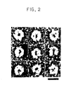
- the proteasome was then observed with an electron microscope (H7000, manufactured by Hitachi, Ltd., x50000).
- Figure 2 reveals that the human proteasome was observed in the form of a symmetric disk having a cavity in the center. This observation substantially corresponded to the above physical properties.
- a 10 LLg portion of the proteasome was placed into a center well on an agar plate, 7 ⁇ l portions of the antibodies were placed in side wells on the same plate. Each of the proteasomes was allowed to react with the antibodies in a humidifier at room temperature for 2 days. The plate was washed by being immersed in a phosphate buffer saline (PBS) with gently stirring for four days, and then the preciptin arc was protein- stained with Coomassie Brilliant Blue (CBB).
- PBS phosphate buffer saline
- CBB Coomassie Brilliant Blue
- the antibodies in the side wells were as follows:
- FIG. 3 reveals that each of the antibodies obtained in Example 3-(1) exhibited reactivity specific only to the corresponding proteasome, and did not exhibit cross-reactivity with any of the other proteasomes. Therefore, the proteasomes are evidently different from one another.
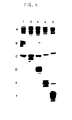
- the subunit component of each proteasome was evaluated in immunological reactivity according to immuno blotting analysis as follows.
- a 35 ⁇ g quantity of each of the proteasome was electrophoresed according to SDS-PAGE (see to ⁇ Electrophoresis of Proteasome> described below), then electrically transferred onto Durapore Membrane (product of Japan Milipore Ltd.) using Semi Drug Elactroblotter (product of Sartorius).
- the membrane was treated with 3% bovine serum, and then with 10 I lg/ml of the corresponding anti-proteasome antibody (primary antibody). Subsequently the membrane treated was allowed to react with 0.25 ⁇ Ci/ml of 125 1- protein A as a secondary antibody.
- the reaction product was analysed by autoradiography.
- A, B, C, D, E and F show the result of protein-staining with CBB in the absence of the primary antibody, the result obtained using the anti-human proteasome antibody, the result obtained using the anti-rat proteasome antibody, the result obtained using the anti-chicken proteasome antibody, the result obtained using the anti-Xenopus proteasome antibody and the result obtained using the anti-yeast proteasome antibody, respectively.
- the lanes shows the following specimens:
- Figure 4 reveals that the subunit component of the proteasome was strongly reactive with the polyclonal antibody against the enzyme derived from the same species, but limitedly reactive with the polyclonal antibodies from other species. It was also obverved that some subunit are cross-reactive with the antibody from the other species. Therefore, the result shows that the partial structure of the subunit was reatained evolutionarily.
- the monoclonal antibodies in the order of descending reactivity were No. 2-17, No. 4-3, No. 2-21 and No. 2-24.
- FIG 6 A, B, C, D and E show the result of protein staining with CBB in the absence of the primary antibody, the result obtained using the human monoclonal antibody No. 2-17, the result obtained using the human monoclonal antibody No. 2-21, the result obtained using the human monoclonal antibody No. 2-24 and the result obtained using the human monoclonal antibody No. 4-3, respectively.
- the lanes represent the following specimens:
- Figure 6 reveals that all kinds of the monoclonal anitbodies are strongly reactive with the subunit component having a molecular weight of 30000.
- the monoclonal antibodies Nos. 2-17 and 4-3 were cross-reactive with the rat proteasome component of the same molecular weight, and the monoclonal antibodies Nos. 2-17 and 2-24 were cross-reactive with the chicken proteasome. This observation clarifies that, although these monoclonal antibodies are reactive with subunits components similar to them, they differ from one another in epitopes.
- the monoclonal antibodies as such are important as kits useful for immunological identification.
- proteasome was electrophoresed on polyacrylamide gel according to Laemmli's method (Nature, 227, pp 680-685 (1970)) in the presence or in the absence of SDS (5% polyacrylamide gel, 35 ⁇ g of proteasome)
- each proteasome was electrophoresed as a single band substantiating the homogeneity thereof and the protease activity.
- the electrophoresis patterns are similar to one another regardless of the presence of a sulfhydryl reducing agent (2-mercaptoethanol). From this result, it is speculated that no disulfide bond exists in the proteasome.
- Figures 9 (1), (2), (3), (4), (5) and (6) show the results obtained from the human proteasome, the rat proteasome, the chicken proteasome, the Xenopus proteasome, the yeast proteasome and blank, respectively.
- the diagram reveals that the proteasomes of the present invention are distinguishable from one another in elution pattern.
- the monoclonal antibody prepared according to Example 3-(2) was adjusted to 5 ⁇ g/ml with 50 mM PBS-0.05% NaN 3 .
- a 100 ⁇ l quantity of the mixture was placed into each of 96 wells of a plate for ELISA (InterMed Nunc-Immuno plate Maxisorp), and the mixture was left to stand overnight at 4° C to immobilize the monoclonal antibody. Subsequently the plate was washed with Dulbecco's PBS.
- a 300 ⁇ l quantity of 50 mM pBS-0.05% NaN 3 -1% BSA was further palced into each of the wells in order to remove non-specific adsorbate. The plate was then left to stand overnight at 4 C to immobilize the antibody on the plate.
- a 100 ⁇ l quantity of 50 mM PBS-0.05% NaN 3 -0.1% BSA-1.0% glycerol-0.02% Tween 20 was placed into each of wells of the plate having the antibody as immobilized. With addition of 10 ⁇ l of serum specimen, the mixture was incubated with shaking at room temperature for 2 hours.
- reaction mixture was washed with Dulbecco's PBS-0.02% Tween 20, and 100 ⁇ l of the anti-human proteasome polyclonal antibody (10,000-foid diluted with the assay buffer) was placed into each of the wells, followed by incubation with shaking at room temperature for 2 hours.
- reaction mixture was washed with Dulbecco's PBS-0.02% Tween 20 and 100 ⁇ l of a diluent of peroxidase-labeled anti-rabbit immunoglobulin antisera (Zymed HRPO(product of Zymed Laboratories, Inc.)-labeled goat anti-rabbit IgG, IgM) was added to the mixture, followed by incubation with shaking at room temperature for 2 hours.
- peroxidase-labeled anti-rabbit immunoglobulin antisera Zymed HRPO(product of Zymed Laboratories, Inc.)-labeled goat anti-rabbit IgG, IgM
- reaction mixture obtained was washed with Dulbecco's PBS-0.02% Tween 20 and into each of the wells was placed 100 ⁇ l of a solution of 2.5 mg/ml of o-phenylenediamine in 0.015% H 2 0 2 citrate buffer (pH 5.0). The mixture was allowed to stand at room temperature for 10 minutes. Thereto a 100 ⁇ l of 2N H 2 SO 4 . was added to stop the reaction, followed by determination at 492 nm using a microplate reader (manufactured by Titertek Multiskan).
- the above table shows that the proteasome was detected in patients suffering from malignant diseases in higher level than in healthy adults.
- the proteasome is assumed to effuse into blood. Therfore, the detection of the proteasome in serum was considered important in diagnosis of various malignant cancers.
Landscapes
- Chemical & Material Sciences (AREA)
- Health & Medical Sciences (AREA)
- Life Sciences & Earth Sciences (AREA)
- Organic Chemistry (AREA)
- Engineering & Computer Science (AREA)
- Genetics & Genomics (AREA)
- Wood Science & Technology (AREA)
- Bioinformatics & Cheminformatics (AREA)
- Zoology (AREA)
- Biomedical Technology (AREA)
- Molecular Biology (AREA)
- Biochemistry (AREA)
- General Health & Medical Sciences (AREA)
- Medicinal Chemistry (AREA)
- Microbiology (AREA)
- Biotechnology (AREA)
- General Engineering & Computer Science (AREA)
- Immunology (AREA)
- Biophysics (AREA)
- Proteomics, Peptides & Aminoacids (AREA)
- Mycology (AREA)
- Oil, Petroleum & Natural Gas (AREA)
- Chemical Kinetics & Catalysis (AREA)
- Cell Biology (AREA)
- Enzymes And Modification Thereof (AREA)
- Peptides Or Proteins (AREA)
- Preparation Of Compounds By Using Micro-Organisms (AREA)
Applications Claiming Priority (2)
| Application Number | Priority Date | Filing Date | Title |
|---|---|---|---|
| JP63142526A JPH01309686A (ja) | 1988-06-08 | 1988-06-08 | 多機能プロテアーゼ |
| JP142526/88 | 1988-06-08 |
Publications (3)
| Publication Number | Publication Date |
|---|---|
| EP0345750A2 true EP0345750A2 (de) | 1989-12-13 |
| EP0345750A3 EP0345750A3 (de) | 1992-06-03 |
| EP0345750B1 EP0345750B1 (de) | 1995-04-26 |
Family
ID=15317407
Family Applications (1)
| Application Number | Title | Priority Date | Filing Date |
|---|---|---|---|
| EP89110277A Expired - Lifetime EP0345750B1 (de) | 1988-06-08 | 1989-06-07 | Polyfunktionelle Protease |
Country Status (7)
| Country | Link |
|---|---|
| US (1) | US5425942A (de) |
| EP (1) | EP0345750B1 (de) |
| JP (1) | JPH01309686A (de) |
| KR (1) | KR910001039A (de) |
| DE (1) | DE68922340T2 (de) |
| DK (1) | DK277689A (de) |
| ES (1) | ES2070868T3 (de) |
Cited By (7)
| Publication number | Priority date | Publication date | Assignee | Title |
|---|---|---|---|---|
| WO1992007270A1 (en) * | 1990-10-12 | 1992-04-30 | Pro-Soma Sarl | Diagnostic method |
| WO1992007269A1 (en) * | 1990-10-11 | 1992-04-30 | Pro-Soma S.A.R.L. | Diagnostic method |
| EP0519536A1 (de) * | 1991-06-21 | 1992-12-23 | Pro-Soma Sarl | Diagnostisches Verfahren |
| US5635605A (en) * | 1988-12-15 | 1997-06-03 | Amdl, Inc. | Method for detecting the presence of ring shaped particle tumor marker |
| WO1998006811A1 (en) * | 1996-08-16 | 1998-02-19 | The Procter & Gamble Company | Detergent compositions comprising antibody controlled proteolytic activity |
| WO1998042829A1 (de) * | 1997-03-21 | 1998-10-01 | MAX-PLANCK-Gesellschaft zur Förderung der Wissenschaften e.V. | Verfahren zur reinigung und kristallisierung von proteasom |
| EP0922088A1 (de) * | 1996-08-16 | 1999-06-16 | The Procter & Gamble Company | Waschmittelzusammensetzungen mit antikörper-kontrollierter enzymatischer aktivität |
Families Citing this family (3)
| Publication number | Priority date | Publication date | Assignee | Title |
|---|---|---|---|---|
| US5872206A (en) * | 1994-10-06 | 1999-02-16 | The General Hospital Corporation | Compositions and methods for interfering wtih hepatitis B virus infection |
| DE10232902A1 (de) * | 2002-07-19 | 2004-01-29 | Bayer Cropscience Ag | Verfahren zum Identifizieren von Inhibitoren des 20S und 26S Proteasoms |
| US8530151B2 (en) * | 2002-09-03 | 2013-09-10 | Charles S. Cobbs | Localization of human cytomegalovirus nucleic acids and proteins in human cancer cells |
Family Cites Families (2)
| Publication number | Priority date | Publication date | Assignee | Title |
|---|---|---|---|---|
| RO57973A (de) * | 1969-06-13 | 1975-03-15 | ||
| FR2233038A1 (en) * | 1973-05-30 | 1975-01-10 | Innerfield Irving | Thrombosis treatment - by oral administration of a protease |
-
1988
- 1988-06-08 JP JP63142526A patent/JPH01309686A/ja active Pending
-
1989
- 1989-06-07 ES ES89110277T patent/ES2070868T3/es not_active Expired - Lifetime
- 1989-06-07 DK DK277689A patent/DK277689A/da not_active Application Discontinuation
- 1989-06-07 EP EP89110277A patent/EP0345750B1/de not_active Expired - Lifetime
- 1989-06-07 DE DE68922340T patent/DE68922340T2/de not_active Expired - Fee Related
- 1989-06-08 KR KR1019890007967A patent/KR910001039A/ko not_active Ceased
-
1993
- 1993-03-05 US US08/027,595 patent/US5425942A/en not_active Expired - Fee Related
Cited By (10)
| Publication number | Priority date | Publication date | Assignee | Title |
|---|---|---|---|---|
| US5635605A (en) * | 1988-12-15 | 1997-06-03 | Amdl, Inc. | Method for detecting the presence of ring shaped particle tumor marker |
| WO1992007269A1 (en) * | 1990-10-11 | 1992-04-30 | Pro-Soma S.A.R.L. | Diagnostic method |
| US5635345A (en) * | 1990-10-11 | 1997-06-03 | Akzo Nobel N.V. | Method for the diagnosis of HIV using antibodies to prosomes |
| WO1992007270A1 (en) * | 1990-10-12 | 1992-04-30 | Pro-Soma Sarl | Diagnostic method |
| EP0519536A1 (de) * | 1991-06-21 | 1992-12-23 | Pro-Soma Sarl | Diagnostisches Verfahren |
| US5472848A (en) * | 1991-06-21 | 1995-12-05 | Pro Soma S.A.R.L. | Methods to aid in the diagnosis of multiple sclerosis |
| WO1998006811A1 (en) * | 1996-08-16 | 1998-02-19 | The Procter & Gamble Company | Detergent compositions comprising antibody controlled proteolytic activity |
| WO1998007819A1 (en) * | 1996-08-16 | 1998-02-26 | The Procter & Gamble Company | Detergent compositions comprising antibody controlled proteolytic activity |
| EP0922088A1 (de) * | 1996-08-16 | 1999-06-16 | The Procter & Gamble Company | Waschmittelzusammensetzungen mit antikörper-kontrollierter enzymatischer aktivität |
| WO1998042829A1 (de) * | 1997-03-21 | 1998-10-01 | MAX-PLANCK-Gesellschaft zur Förderung der Wissenschaften e.V. | Verfahren zur reinigung und kristallisierung von proteasom |
Also Published As
| Publication number | Publication date |
|---|---|
| KR910001039A (ko) | 1991-01-30 |
| DK277689D0 (da) | 1989-06-07 |
| JPH01309686A (ja) | 1989-12-14 |
| ES2070868T3 (es) | 1995-06-16 |
| EP0345750B1 (de) | 1995-04-26 |
| DK277689A (da) | 1989-12-09 |
| DE68922340D1 (de) | 1995-06-01 |
| US5425942A (en) | 1995-06-20 |
| DE68922340T2 (de) | 1995-10-26 |
| EP0345750A3 (de) | 1992-06-03 |
Similar Documents
| Publication | Publication Date | Title |
|---|---|---|
| Chao et al. | Identification of tissue kallikrein in brain and in the cell-free translation product encoded by brain mRNA. | |
| US4696895A (en) | Monoclonal anti-protein C antibody, its preparation and use thereof | |
| US4894442A (en) | Monoclonal antibodies that bind to alpha-acid glycoprotein | |
| US5425942A (en) | Polyfunctinal protease | |
| DK171646B1 (da) | Renset human-makrofag-migrationsinhiberingsfaktor (human-MIF) og dens enkeltproteiner, fremgangsmådetil fremstilling deraf, monoklonale antistoffer mod human-MIF og fremgangsmåde til den fremstilling,hybridomacellelinier, der producerer monoklonale antistoffer mod human-MIF, deres anvendelse og fremgangsmåde til deres fremstilling samt farmaceutiske præparater indeholdende renset human-MIF eller | |
| US5670328A (en) | Monoclonal antibodies to human pulmonary surfactant apoprotein D and use thereof | |
| US5316914A (en) | Method for determining human collagen peptides by way of enzyme immunoassay | |
| EP0131834A2 (de) | Monoklonaler Antikörper mit Spezifität gegen nur einen Typ eines Isozyms der schweren Kette des menschlichen Herzmyosins | |
| KR960002740B1 (ko) | 안티-트롬빈-결합물질 모노클로날 항체, 이를 생산하는 하이브리도마 및 모노클로날 항체를 이용한 트롬빈-결합물질의 정제법 및 측정법 | |
| EP0175310A1 (de) | Ein im wesentlichen reines für rheumatoide Arthritis spezifisches Protein und ein Antikörper dagegen | |
| EP0401370A1 (de) | Enzymimmuntest für menschliches typiv-collagen gemäss dem sandwichverfahren | |
| CA2281262C (en) | Anti-human medullasin monoclonal antibody, process for producing the same and immunoassay using the same | |
| JPS63258898A (ja) | ヒト▲膵▼ホスホリパ−ゼa▲下2▼に対するモノクロ−ナル抗体、その製造法、該モノクロ−ナル抗体産生ハイブリド−マおよび該モノクロ−ナル抗体を用いたヒト▲膵▼ホスホリパ−ゼa▲下2▼の測定方法 | |
| JP3076640B2 (ja) | ヒト92kDaゼラチナーゼの免疫学的定量法 | |
| EP0465652B1 (de) | Antikörper gegen die schwere kette eines glatt-muskel-myosins | |
| JPH02276591A (ja) | Anpのc端側を認識するモノクローナル抗体 | |
| Bose-Bierne et al. | Production and characterization of four monoclonal antibodies against porcine pancreatic colipase | |
| US5576182A (en) | Anti-mucus glycoprotein monoclonal antibody | |
| US4637984A (en) | Monoclonal anti-ornithine decarboxylase antibody and method of producing same | |
| JP2779193B2 (ja) | 抗ヒト組織因子モノクローナル抗体 | |
| Becker et al. | Entamoeba histolytica: localization of a 30-kDa cysteine proteinase using a monoclonal antibody | |
| US5747652A (en) | Antibody to smooth muscle myosin heavy chains | |
| EP0207170A1 (de) | Verfahren zur herstellung menschlicher krebsspezifischer monoklonaler antikörper | |
| JPH0977799A (ja) | ヒト−マクロファージ遊走阻止因子(ヒト−mif)に対するモノクローナル抗体および該抗体を産生するハイブリドーマ | |
| Jensenius | VH and L‐chain allotype determinants of rabbit IgG and IgA estimated by a semi‐automatic, modified Farr‐type radioimmunoassay |
Legal Events
| Date | Code | Title | Description |
|---|---|---|---|
| PUAI | Public reference made under article 153(3) epc to a published international application that has entered the european phase |
Free format text: ORIGINAL CODE: 0009012 |
|
| AK | Designated contracting states |
Kind code of ref document: A2 Designated state(s): CH DE ES FR GB IT LI NL SE |
|
| PUAL | Search report despatched |
Free format text: ORIGINAL CODE: 0009013 |
|
| AK | Designated contracting states |
Kind code of ref document: A3 Designated state(s): CH DE ES FR GB IT LI NL SE |
|
| 17P | Request for examination filed |
Effective date: 19920810 |
|
| 17Q | First examination report despatched |
Effective date: 19930517 |
|
| GRAA | (expected) grant |
Free format text: ORIGINAL CODE: 0009210 |
|
| AK | Designated contracting states |
Kind code of ref document: B1 Designated state(s): CH DE ES FR GB IT LI NL SE |
|
| ITF | It: translation for a ep patent filed | ||
| REF | Corresponds to: |
Ref document number: 68922340 Country of ref document: DE Date of ref document: 19950601 |
|
| ET | Fr: translation filed | ||
| REG | Reference to a national code |
Ref country code: ES Ref legal event code: FG2A Ref document number: 2070868 Country of ref document: ES Kind code of ref document: T3 |
|
| PLBE | No opposition filed within time limit |
Free format text: ORIGINAL CODE: 0009261 |
|
| STAA | Information on the status of an ep patent application or granted ep patent |
Free format text: STATUS: NO OPPOSITION FILED WITHIN TIME LIMIT |
|
| 26N | No opposition filed | ||
| PGFP | Annual fee paid to national office [announced via postgrant information from national office to epo] |
Ref country code: FR Payment date: 19970527 Year of fee payment: 9 |
|
| PGFP | Annual fee paid to national office [announced via postgrant information from national office to epo] |
Ref country code: SE Payment date: 19970529 Year of fee payment: 9 Ref country code: GB Payment date: 19970529 Year of fee payment: 9 |
|
| PGFP | Annual fee paid to national office [announced via postgrant information from national office to epo] |
Ref country code: ES Payment date: 19970606 Year of fee payment: 9 |
|
| PGFP | Annual fee paid to national office [announced via postgrant information from national office to epo] |
Ref country code: NL Payment date: 19970630 Year of fee payment: 9 |
|
| PGFP | Annual fee paid to national office [announced via postgrant information from national office to epo] |
Ref country code: DE Payment date: 19970725 Year of fee payment: 9 |
|
| PGFP | Annual fee paid to national office [announced via postgrant information from national office to epo] |
Ref country code: CH Payment date: 19970930 Year of fee payment: 9 |
|
| PG25 | Lapsed in a contracting state [announced via postgrant information from national office to epo] |
Ref country code: GB Free format text: LAPSE BECAUSE OF NON-PAYMENT OF DUE FEES Effective date: 19980607 |
|
| PG25 | Lapsed in a contracting state [announced via postgrant information from national office to epo] |
Ref country code: SE Free format text: LAPSE BECAUSE OF NON-PAYMENT OF DUE FEES Effective date: 19980608 Ref country code: ES Free format text: LAPSE BECAUSE OF EXPIRATION OF PROTECTION Effective date: 19980608 |
|
| PG25 | Lapsed in a contracting state [announced via postgrant information from national office to epo] |
Ref country code: LI Free format text: LAPSE BECAUSE OF NON-PAYMENT OF DUE FEES Effective date: 19980630 Ref country code: CH Free format text: LAPSE BECAUSE OF NON-PAYMENT OF DUE FEES Effective date: 19980630 |
|
| PG25 | Lapsed in a contracting state [announced via postgrant information from national office to epo] |
Ref country code: NL Free format text: LAPSE BECAUSE OF NON-PAYMENT OF DUE FEES Effective date: 19990101 |
|
| GBPC | Gb: european patent ceased through non-payment of renewal fee |
Effective date: 19980607 |
|
| REG | Reference to a national code |
Ref country code: CH Ref legal event code: PL |
|
| PG25 | Lapsed in a contracting state [announced via postgrant information from national office to epo] |
Ref country code: FR Free format text: LAPSE BECAUSE OF NON-PAYMENT OF DUE FEES Effective date: 19990226 |
|
| EUG | Se: european patent has lapsed |
Ref document number: 89110277.4 |
|
| NLV4 | Nl: lapsed or anulled due to non-payment of the annual fee |
Effective date: 19990101 |
|
| PG25 | Lapsed in a contracting state [announced via postgrant information from national office to epo] |
Ref country code: DE Free format text: LAPSE BECAUSE OF NON-PAYMENT OF DUE FEES Effective date: 19990401 |
|
| REG | Reference to a national code |
Ref country code: FR Ref legal event code: ST |
|
| REG | Reference to a national code |
Ref country code: ES Ref legal event code: FD2A Effective date: 20000201 |
|
| PG25 | Lapsed in a contracting state [announced via postgrant information from national office to epo] |
Ref country code: IT Free format text: LAPSE BECAUSE OF NON-PAYMENT OF DUE FEES;WARNING: LAPSES OF ITALIAN PATENTS WITH EFFECTIVE DATE BEFORE 2007 MAY HAVE OCCURRED AT ANY TIME BEFORE 2007. THE CORRECT EFFECTIVE DATE MAY BE DIFFERENT FROM THE ONE RECORDED. Effective date: 20050607 |