EP0352761B1 - Antigenkonstrukte von "Major Histocompatibility Complex" Klasse I Antigenen mit spezifischen Trägermolekülen, ihre Herstellung und Verwendung - Google Patents
Antigenkonstrukte von "Major Histocompatibility Complex" Klasse I Antigenen mit spezifischen Trägermolekülen, ihre Herstellung und VerwendungInfo
- Publication number
- EP0352761B1 EP0352761B1 EP89113741A EP89113741A EP0352761B1 EP 0352761 B1 EP0352761 B1 EP 0352761B1 EP 89113741 A EP89113741 A EP 89113741A EP 89113741 A EP89113741 A EP 89113741A EP 0352761 B1 EP0352761 B1 EP 0352761B1
- Authority
- EP
- European Patent Office
- Prior art keywords
- antigens
- class
- specific carrier
- antigenic constructs
- hla
- Prior art date
- Legal status (The legal status is an assumption and is not a legal conclusion. Google has not performed a legal analysis and makes no representation as to the accuracy of the status listed.)
- Expired - Lifetime
Links
Images


Classifications
-
- C—CHEMISTRY; METALLURGY
- C12—BIOCHEMISTRY; BEER; SPIRITS; WINE; VINEGAR; MICROBIOLOGY; ENZYMOLOGY; MUTATION OR GENETIC ENGINEERING
- C12N—MICROORGANISMS OR ENZYMES; COMPOSITIONS THEREOF; PROPAGATING, PRESERVING, OR MAINTAINING MICROORGANISMS; MUTATION OR GENETIC ENGINEERING; CULTURE MEDIA
- C12N15/00—Mutation or genetic engineering; DNA or RNA concerning genetic engineering, vectors, e.g. plasmids, or their isolation, preparation or purification; Use of hosts therefor
- C12N15/09—Recombinant DNA-technology
- C12N15/11—DNA or RNA fragments; Modified forms thereof; Non-coding nucleic acids having a biological activity
-
- B—PERFORMING OPERATIONS; TRANSPORTING
- B82—NANOTECHNOLOGY
- B82Y—SPECIFIC USES OR APPLICATIONS OF NANOSTRUCTURES; MEASUREMENT OR ANALYSIS OF NANOSTRUCTURES; MANUFACTURE OR TREATMENT OF NANOSTRUCTURES
- B82Y5/00—Nanobiotechnology or nanomedicine, e.g. protein engineering or drug delivery
-
- A—HUMAN NECESSITIES
- A61—MEDICAL OR VETERINARY SCIENCE; HYGIENE
- A61K—PREPARATIONS FOR MEDICAL, DENTAL OR TOILETRY PURPOSES
- A61K47/00—Medicinal preparations characterised by the non-active ingredients used, e.g. carriers or inert additives; Targeting or modifying agents chemically bound to the active ingredient
- A61K47/50—Medicinal preparations characterised by the non-active ingredients used, e.g. carriers or inert additives; Targeting or modifying agents chemically bound to the active ingredient the non-active ingredient being chemically bound to the active ingredient, e.g. polymer-drug conjugates
- A61K47/51—Medicinal preparations characterised by the non-active ingredients used, e.g. carriers or inert additives; Targeting or modifying agents chemically bound to the active ingredient the non-active ingredient being chemically bound to the active ingredient, e.g. polymer-drug conjugates the non-active ingredient being a modifying agent
- A61K47/68—Medicinal preparations characterised by the non-active ingredients used, e.g. carriers or inert additives; Targeting or modifying agents chemically bound to the active ingredient the non-active ingredient being chemically bound to the active ingredient, e.g. polymer-drug conjugates the non-active ingredient being a modifying agent the modifying agent being an antibody, an immunoglobulin or a fragment thereof, e.g. an Fc-fragment
- A61K47/6891—Pre-targeting systems involving an antibody for targeting specific cells
- A61K47/6897—Pre-targeting systems with two or three steps using antibody conjugates; Ligand-antiligand therapies
- A61K47/6898—Pre-targeting systems with two or three steps using antibody conjugates; Ligand-antiligand therapies using avidin- or biotin-conjugated antibodies
-
- C—CHEMISTRY; METALLURGY
- C07—ORGANIC CHEMISTRY
- C07K—PEPTIDES
- C07K14/00—Peptides having more than 20 amino acids; Gastrins; Somatostatins; Melanotropins; Derivatives thereof
- C07K14/435—Peptides having more than 20 amino acids; Gastrins; Somatostatins; Melanotropins; Derivatives thereof from animals; from humans
- C07K14/705—Receptors; Cell surface antigens; Cell surface determinants
- C07K14/70503—Immunoglobulin superfamily
- C07K14/70539—MHC-molecules, e.g. HLA-molecules
-
- C—CHEMISTRY; METALLURGY
- C07—ORGANIC CHEMISTRY
- C07K—PEPTIDES
- C07K16/00—Immunoglobulins [IG], e.g. monoclonal or polyclonal antibodies
-
- C—CHEMISTRY; METALLURGY
- C07—ORGANIC CHEMISTRY
- C07K—PEPTIDES
- C07K17/00—Carrier-bound or immobilised peptides; Preparation thereof
- C07K17/02—Peptides being immobilised on, or in, an organic carrier
-
- C—CHEMISTRY; METALLURGY
- C12—BIOCHEMISTRY; BEER; SPIRITS; WINE; VINEGAR; MICROBIOLOGY; ENZYMOLOGY; MUTATION OR GENETIC ENGINEERING
- C12N—MICROORGANISMS OR ENZYMES; COMPOSITIONS THEREOF; PROPAGATING, PRESERVING, OR MAINTAINING MICROORGANISMS; MUTATION OR GENETIC ENGINEERING; CULTURE MEDIA
- C12N15/00—Mutation or genetic engineering; DNA or RNA concerning genetic engineering, vectors, e.g. plasmids, or their isolation, preparation or purification; Use of hosts therefor
- C12N15/09—Recombinant DNA-technology
- C12N15/63—Introduction of foreign genetic material using vectors; Vectors; Use of hosts therefor; Regulation of expression
-
- C—CHEMISTRY; METALLURGY
- C12—BIOCHEMISTRY; BEER; SPIRITS; WINE; VINEGAR; MICROBIOLOGY; ENZYMOLOGY; MUTATION OR GENETIC ENGINEERING
- C12P—FERMENTATION OR ENZYME-USING PROCESSES TO SYNTHESISE A DESIRED CHEMICAL COMPOUND OR COMPOSITION OR TO SEPARATE OPTICAL ISOMERS FROM A RACEMIC MIXTURE
- C12P21/00—Preparation of peptides or proteins
-
- A—HUMAN NECESSITIES
- A61—MEDICAL OR VETERINARY SCIENCE; HYGIENE
- A61K—PREPARATIONS FOR MEDICAL, DENTAL OR TOILETRY PURPOSES
- A61K38/00—Medicinal preparations containing peptides
-
- C—CHEMISTRY; METALLURGY
- C07—ORGANIC CHEMISTRY
- C07K—PEPTIDES
- C07K2319/00—Fusion polypeptide
-
- C—CHEMISTRY; METALLURGY
- C07—ORGANIC CHEMISTRY
- C07K—PEPTIDES
- C07K2319/00—Fusion polypeptide
- C07K2319/01—Fusion polypeptide containing a localisation/targetting motif
- C07K2319/02—Fusion polypeptide containing a localisation/targetting motif containing a signal sequence
Definitions
- the invention relates to antigen constructs which result from the linkage of "Major Histocompatibility Complex” (MHC) class I antigens with specific carrier molecules.
- MHC Major Histocompatibility Complex
- Tissue rejection is the most potent T cell mediated immune response known. In individuals of the same species, they are caused by allogeneic class I and class II MHC antigen differences. In the case of organ transfers, for example, the allogeneic determinants of the MHC antigens of the donor tissue, which may be present, are recognized as foreign by allospecific T cells of the recipient, a T cell immune response is induced and there is a rejection reaction if no immunosuppressive therapy has been initiated or no longer is sufficient.
- MHC class I antigens are glycoproteins which are expressed on the surface of all nucleated cells. They consist of a heavy chain, which is encoded by MHC class I genes, and a light chain, the ⁇ 2-microglobulin, which is non-covalently associated with the heavy chain.
- the extracellular part of the heavy chain is folded into three domains, of which the first two domains (alpha1 and alpha2) when Comparison of the amino acid sequences of previously known Class I MHC antigens from different individuals have a pronounced polymorphism. They serve the antigen presentation and carry the allogeneic determinants.
- the third extracellular domain shows a more conserved sequence.
- the association with ⁇ 2-microglobulin is essential for correct folding of the heavy chain and for the transport of the molecule to the cell surface.
- target cell-specific carriers for example preferably monoclonal antibodies (mAb), but also polyclonal antibodies or molecules binding to cellular receptors, can be covalently coupled to the N- or C-terminal end of an allogeneic MHC class I molecule, without this to disadvantageously change the allogeneic determinants.
- the MHC class I molecule is specific to the target cells brought, which leads to the activation of allospecific T cells and thus to the destruction of the target cells by allospecific cytotoxic T cells.
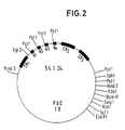
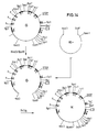
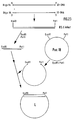
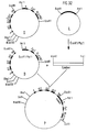
- N-terminal end of the MHC class I molecule is on the one directed towards the cell Side of the alpha1 and alpha2 domains is, while the allogeneic determinants are on the side of the alpha1 and alpha2 domains facing away from the cell (Fig. 1, Fig. 34).
- NP- nitrophenol
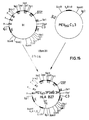
- human IgG CF (ab ′) 2 gene (2) a human IgG CF (ab ′) 2 gene (2) and an HLA B27w gene (3).
- (1) and (2) are examples of the specific carrier fraction - here a MAK against NP -, while (3) stands for an HLA class I antigen.
- the above-mentioned construct is expressed and secreted by those myeloma cells which contain a human ⁇ 2-microglobulin and a light chain of an immunoglobulin, the V gene of which forms an NP binding site with V H B / 1-8, such as the mouse -Myeloma cell J 558 L (Oi, VT, Morrison, SL, Herzenberg, LA, Berg, P .: Immunoglobulin gene expression in transformed lymphoid cells. Proc. Natl. Acad. Sci. USA 80 , 825, 1983).
- the mAK / HLA B27w fusion product can be provided with any desired specificity for which a specific or selective mAK exists.
- Examples 1 to 13 show the construction of an HLA B27 / MAK fusion gene with the HLA B27 portion at the 3 ′ end of the monoclonal antibody
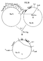
- a human IgG3 C gene was isolated from a human gene bank in EMBL3 phages (Frischholz, A.-M., Lehrach, H., Proustka, A., Murray, N .: Lambda replacement vectors carrying polylinker sequences. J. Mol. Biol. 170 , 827-842 (1983) and Seemann, GHA, Rein, RS, Brown, CS, Ploegh, HL: Gene conversion-like mechanisms may generate polymorphism in human class I genes.
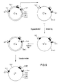
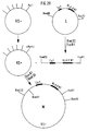
- the 54.1.24 clone was subjected to complete HindIII and partial Pst1 restriction digestion. This creates restriction fragments that contain the C H1 exon and one, two or three "hinge exons". These fragments were made from an agarose gel cut out and cloned into a pUC19 vector cut with HindIII and Pst1 (FIG. 3).
- the plasmid clone with the C H1 and three "hinge exons" was then cleaved with BamH1 and Asp718, the interfaces were filled in and religated with T4 ligase (Fig. 4). This deletes the pUC19 polylinker between the XbaI and the Sst1 interface.
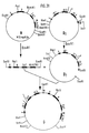
- HLA B27w gene was isolated from a genomic library (Frischholz, A.-M., loc. Cit.) And Seemann, GHA, loc. Cit. Cloned in EMBL3 bacteriophages and characterized by restriction mapping and nucleotide sequence analysis (Maxam, A., Gilbert, W .: Sequencing and labeled DNA with base specific chemical cleavage. Meth. Enzymol. 65 , 499-560 (1986) and Sanger, F., Nicklen, S., Coulson, AR: DNA sequencing with chain terminating inhibitors. Proc. Natl. Acad. Sci USA 74 , 5463-5467 (1977)) (Fig. 5).
- the HLA B27w gene was then digested with the restriction enzymes Sst1 and BglII and subcloned into the Sst1 and BamHI sites of pUC19. Plasmid clones with subfragments A, B and C (Fig. 5) were isolated.
- the plasmid with subfragment A was cleaved completely with Sst1 and partially with SmaI and after separation on an agarose gel, the fragment A '(FIG. 6) was cloned in a pUC19 plasmid cleaved with HindII and Sst1.
- the plasmid with subfragment B was digested with XbaI and the resulting XbaI insert (B ′) was cloned into an XbaI-cleaved pUC19 plasmid (FIG. 7).
- the plasmid with the subfragment C was cleaved completely with HindIII and partially with Sst1 and the fragment which adjoins fragment A in the HLA B27w gene (C ') after separation on an agarose gel and isolated in the bluescript cleaved with HindIII and Sst1 Phasmid vector KS + (Stratagene, LaJolla, CA, USA) cloned (Fig. 8).
- Single-strand phages were prepared and purified from the KS + phasmid vector C ′ by infection with helper phages VCS-M13 (Stratagene, Cat # 200251) (Stratagene: Bluescript Exo / Mung DNA sequencing system: Instruction Manual).
- VCS-M13 Helper phages VCS-M13 (Stratagene, Cat # 200251) (Stratagene: Bluescript Exo / Mung DNA sequencing system: Instruction Manual).
- the plasmid with the fragment A ' was cleaved with Sst1 and ligated with the C ⁇ fragment generated by a complete HindIII and partial Sst1 cleavage of the phasmid clone C ⁇ and isolated after separation on an agarose gel. After 30 minutes of ligation at 14 ° C., the non-ligated ends were filled in with T4 polymerase and then ligated again. Restriction mapping was used to identify plasmid D (FIG. 10) in which fragment A 'is connected to fragment C ⁇ via the Sst1 site in the alpha2 exon.
- the plasmid with the fragment D was cleaved with XbaI and ligated with the fragment B ′, which had been cut out from the plasmid B ′ with XbaI and purified after separation on an agarose gel (FIG. 11). With the help of nucleotide sequence analyzes (17), a plasmid was identified (E) in which the B ′ fragment is ligated to the fragment D in the correct 5′-3 ′ orientation.
- the fragment E was cut out from the plasmid E by cleavage with EcoRI and HindIII, the ends were filled with T4 polymerase and, after separation, purified on an agarose gel. This purified fragment E was then ligated to the plasmid containing the IgG3 F (ab ′) 2 3H fragment after it had been cleaved with XbaI and the XbaI ends had been filled in with T4 polymerase (FIG. 12).
- the clone containing the plasmid F was identified by restriction mapping, in which the modified HLA B27w gene in the correct 5′-3 ′ orientation is fused with the F (ab ′) 2 3H gene.
- the fragment F was cut out with HindIII and EcoRI in order to place a polylinker in front of the 5 ′ end of the fragment F.
- the HindIII and XbaI ends were filled in with T4 polymerase and cloned into a pUC19, the Sst1 was cleaved and the Sst1 ends were filled in with T4 polymerase.
- the clone with the plasmid G was identified by restriction analyzes, which has the pUC19 polylinker 5 ′ from the fragment F. (Fig. 13).
- the plasmid G was digested with HindIII and EcoRI, the insert was isolated with the IgG F (ab ′) 2 HLA B27w fusion gene and into a Bluescript digested with HindIII and EcoRI KS + phasmid vector (Stratagene: Bluescript Exo / Mung DNA sequencing system: Instruction Manual) cloned (Fig. 14).
- the plasmid H resulting from this cloning was then cleaved with BamHI and the insert was cloned into the BamHI-cleaved eukaryotic expression vector pEV H (Simon, T., Rajewsky, K., Nucl. Acids Res. 16 , 354, (1988)), which contains the IgG H promoter / enhancer sequences and the V H gene derived from the NP-specific mouse mAK B / 1-8 (FIG. 15) (Neuberger, MN: EMBO Journal 2 , 1375-1378 (1983)).
- the plasmid I was identified by restriction analysis, in which the IgG 3 F (ab ′) 2 HLA B27w fusion gene was cloned in the correct 5′-3 ′ orientation behind the V H gene.
- the mAK / HLA B27w fusion gene now has intact 5 'and 3' ends with all the signals required for expression in eukaryotic cells.
- the construct is expressed and secreted in every myeloma cell which contains a human ⁇ 2-microglobulin and a light chain of an immunoglobulin, the V gene of which forms an NP binding site with V H B / 1-8, such as the mouse -Myeloma cell J 558L (Oi, VT, Morrison, SL, Herzenberg, LA, Berg, P., Proc. Natl. Acad. Sci. USA 80 , 825 (1983)).
- Examples 14 to 17 show the construction of an HLA B27 / MAK fusion gene with the HLA B27 portion at the 5 'end of the monoclonal antibody
- HLA B27w gene was isolated from a genomic library (Frischholz et al., Supra and Seemann, GHA, supra) cloned into EMBL3 bacteriophages and characterized by restriction mapping and nucleotide sequence analysis (Maxam et al., Supra) and (Sanger et al., Supra ) (Fig. 5).
- the HLA B27w gene was then digested with the restriction enzymes Sst1 and BglII and subcloned into the Sst1 and BamH1 sites of pUC19. Plasmid clones with subfragments A, B and C (Fig. 5) were isolated.
- the plasmid with the HLA B27 subclone C was partially digested with Pst1. The overhanging 3'-ends of the Pst1 sites were removed with T caution polymerase and dGTP added and religated with T relig ligase.
- the plasmid clone C1 which contains no Pst1 site in the intron between the alpha2 and alpha3 exon, was identified by restriction analysis (FIG. 16).
- the plasmid clone C1 was partially cleaved with Sst1 and completely with HindIII, the C1′-fragment was isolated and cloned into a Sst1 and HindIII cleaved double-stranded M13 mp18 vector.
- the M13 clone C1 'with the C1' fragment was identified by determining the nucleic acid sequence of the insert (Fig. 17).
- Single-strand phages were isolated from the C1 ′ M13 mp18 phage in the bacterial strain CJ236 according to the protocol of the Bio-Rad Muta-Gene M13 mutagenesis kit, which contained uraciles.
- An oligonucleotide (oligonucleotide III) with the sequence 5 ′ GCGCGCTGGAGCGTCTC 3 ′ was hybridized to these single-strand phages with the addition and the second strand was synthesized by T4 polymerase, dNTP and T4 ligase.
- the mutant clone C2 was identified by restriction analysis of the M13 mp18 double-stranded DNAs and confirmed by nucleic acid sequence analysis (FIG. 18).
- the mutagenesis destroyed the Pst1 restriction site in the alpha2 exon without changing the reading frame or the encoded amino acid sequence.
- the plasmid clone with subfragment B was digested with XbaI and the resulting XbaI insert (B ′) was cloned into a pUC19 plasmid cleaved by XbaI (FIG. 7).
- the plasmid clone with fragment A was completely cleaved with HindII and partially with SmaI and religated. Restriction analysis identified clone A 'in which part of the pUC19 polylinker was deleted (FIG. 21).
- the plasmid clone A was partially digested with Sst1 and ligated with the C2 fragment generated by Sst1 cleavage of the plasmid clone C2 and isolated after separation on an agarose gel.
- the plasmid D 1 was identified by restriction mapping (FIG. 22), in which the fragment A is connected to the fragment C2 via the Sst1 site in the alpha2 exon.
- the 5'-end of the HLA B27w gene is thus complete. Construction of the linker: Two oligonucleotides were synthesized: Oligonucleotide Va: Oligonucleotide Vb: The two oligonucleotides were hybridized with one another.
- the immunoglobulin V gene was developed by P.T. Jones et al. (Jones, P.T., Dear, P.H., Foote, J., Neuberger, M.S. Winter, G., Nature 321: 522, (1986)) using oligonucleotides. It contains a Pst1 restriction site in the 5 'region of the clone and is cloned as a HindIII / BamHI fragment in an M13 mp8 vector whose Pst1 site had been destroyed by cleavage, removal of the overhanging ends and religation (FIG. 24).
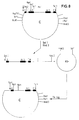
- a human IgG3 C gene was isolated from a human gene bank in EMBL3 phages (Frischholz et al., Loc. Cit. And Seemann et al., Loc. Cit.) And subcloned as a 3.1 kb HindIII / SphI fragment into the plasmid vector pUC19 (clone 54.1. 24) (Fig. 2).
- the plasmid clone 54.1.24 was cleaved with HindII and Asp718, the overhanging ends of the Asp718 interface were removed with T4 polymerase and religated with T4 ligase.
- the clone 54.1.24 Delta Pol was identified by restriction analysis and nucleic acid sequence determination and, apart from SphI, PstI, Sst1 and EcoRI, no longer contains any restriction sites 3 ′ of the human IgG3 C gene (FIG. 25).
- the plasmid clone 54.1.24 Delta Pol was digested with Bg12 and SphI. The overhanging ends were removed with T4 polymerase and religated with T4 ligase. By restriction analysis and nucleic acid sequence determination, the clone I was identified, which only contains the CH1 exon of the human IgG3 C gene (Fig. 26).
- the plasmid clone I was cleaved with Pst1 and the overhanging ends were removed with T4 polymerase.
- the B ′ insert cut out with XbaI and filled in with T4 polymerase to make the ends smooth was ligated into the resulting smooth ends.
- the clone K was identified by restriction analysis and nucleic acid sequence determination (FIG. 27), which contains a human IgG3C H 1 exon and a 3 ′ end of an HLA class I gene.
- the plasmid clone K was cleaved with HindIII and EcoRI, the overhanging ends removed and the insert ligated into a pUC19 plasmid cleaved with Sst1, the ends of which had also been smoothed.
- the clone L was identified, which carries the polylinker of the pUC19 vector 5 'of the C H 1 exon (Fig. 28).
- the plasmid clone L was cleaved with EcoRI and HindIII, the insert was purified and ligated into a HindIII and EcoRI cleaved KS+ vector (Stratagene; Bluescript Exo / Mung DNA Sequencing System), whose PstI cleavage site had previously been destroyed by cleavage with PstI, T4 polymerase treatment and religation.
- the clone M was identified from which the human C H 1 exon with the HLA class I 3 'end can be cut out by a BamHI cleavage (FIG. 29).
- Double-stranded DNA was produced from the M13 mp8 clone V and cleaved with BamHI.
- the KS+ clone M was cleaved with BamHI and the insert M was cleaned.
- the M fragment was ligated into the BamHI-cloned V clone and identified by means of nucleic acid sequence determination of the M13 clone N, which contains an intact IgG3 gene (FIG. 30).
- Double-stranded DNA was produced from M13 clone N, cleaved with EcoRI and the insert purified.
- the plasmid clone D 1 was cleaved with EcoRI and ligated to the fragment N.
- the phage clone O was isolated, in which the fragment N is cloned in the correct orientation in the clone D 1 (Fig. 31).
- the plasmid clone O was subjected to a complete PstI cleavage and a partial EcoRI cleavage and ligated with the linker fragment cut from the plasmid vector L with EcoRI and Pst1.
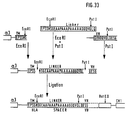
- the plasmid clone P contains the complete HLA B27w mAK fusion gene (Fig. 32, Fig. 33). This fusion gene can be expressed and secreted in human cells alone or in mouse cells together with the human beta2 microglobulin gene, even if an immunoglobulin light chain is present in the expression cells.
- alpha 1, alpha 2 and alpha 3 mean the class I MHC antigen chain domains.
- the arrows point to the alpha helices that carry the allode determinants.
- CM should represent the cell membrane, C the cell.
- EcoRI etc. stands for the cleavage with the respective restriction endonuclease or for the corresponding interface.
- BamHI / Asp718 means a restriction site destroyed by religation after filling.
- TM means transmembrane region.
- 3′NT means 3′-non-translated IgH p / E means immunoglobulin heavy chain promoter / enhancer * means: incomplete digestion
- DS-DNA double-stranded DNA
- SS-DNA single strand of DNA
- s.CTL syngeneic cytotoxic T lymphocyte
- TcR T cell receptor a.
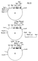
- MHC class I means: allogeneic MHC class I antigen taa means: tumor-associated antigen stc means: syngeneic tumor cell
Landscapes
- Health & Medical Sciences (AREA)
- Chemical & Material Sciences (AREA)
- Life Sciences & Earth Sciences (AREA)
- Organic Chemistry (AREA)
- Engineering & Computer Science (AREA)
- Genetics & Genomics (AREA)
- General Health & Medical Sciences (AREA)
- Molecular Biology (AREA)
- Biophysics (AREA)
- Medicinal Chemistry (AREA)
- Biochemistry (AREA)
- Bioinformatics & Cheminformatics (AREA)
- Immunology (AREA)
- Zoology (AREA)
- General Engineering & Computer Science (AREA)
- Biotechnology (AREA)
- Proteomics, Peptides & Aminoacids (AREA)
- Wood Science & Technology (AREA)
- Biomedical Technology (AREA)
- Nanotechnology (AREA)
- Pharmacology & Pharmacy (AREA)
- Microbiology (AREA)
- Medical Informatics (AREA)
- Epidemiology (AREA)
- Veterinary Medicine (AREA)
- Public Health (AREA)
- Gastroenterology & Hepatology (AREA)
- Toxicology (AREA)
- Physics & Mathematics (AREA)
- Cell Biology (AREA)
- Plant Pathology (AREA)
- Crystallography & Structural Chemistry (AREA)
- Animal Behavior & Ethology (AREA)
- General Chemical & Material Sciences (AREA)
- Chemical Kinetics & Catalysis (AREA)
- Medicines Containing Antibodies Or Antigens For Use As Internal Diagnostic Agents (AREA)
- Peptides Or Proteins (AREA)
- Preparation Of Compounds By Using Micro-Organisms (AREA)
- Micro-Organisms Or Cultivation Processes Thereof (AREA)
- Medicines That Contain Protein Lipid Enzymes And Other Medicines (AREA)
Description
- Die Erfindung betrifft Antigenkonstrukte, die aus der Verknüpfung von "Major Histocompatibility Complex" (MHC) Klasse I Antigenen mit spezifischen Trägermolekülen resultieren.
- Gewebeabstoßungsreaktionen sind die stärksten T-Zell-vermittelten Immunreaktionen, die bekannt sind. Bei Individuen derselben Spezies werden sie durch allogene Unterschiede der Klasse I und Klasse II MHC Antigene verursacht. Bei Organübertragungen beispielsweise werden die gegebenenfalls vorhandenen allogenen Determinanten der MHC Antigene des Spendergewebes von allospezifischen T-Zellen des Empfängers als fremd erkannt, eine T-Zell Immunantwort wird induziert und es kommt zur Abstoßungsreaktion, sofern keine immunsuppressive Therapie eingeleitet wurde bzw. eine solche nicht mehr ausreicht.
- Ferner ist bekannt, daß MHC Klasse I Antigene Glykoproteine sind, die auf der Oberfläche aller kernhaltigen Zellen ausgeprägt werden. Sie bestehen aus einer schweren Kette, die von MHC Klasse I Genen kodiert wird, und einer leichten Kette, dem β₂-Microglobulin, das nicht-kovalent mit der schweren Kette assoziiert ist. Der extrazelluläre Teil der schweren Kette ist in drei Domänen gefaltet, von denen die ersten beiden Domänen (alpha₁ und alpha₂) beim Vergleich der Aminosäuresequenzen bislang bekannter Klasse I MHC Antigene verschiedener Individuen einen ausgeprägten Polymorphismus aufweisen. Sie dienen der Antigenpräsentation und tragen die allogenen Determinanten. Die dritte extrazelluläre Domäne zeigt eine konserviertere Sequenz. Die Assoziation mit β₂-Microglobulin ist für eine korrekte Faltung der schweren Kette und für den Transport des Moleküls auf die Zelloberfläche essentiell.
- Die Isolation und Charakterisierung mutierter MHC Klasse I Antigene bei der Maus zeigte, daß für die Induktion einer Abstoßungsreaktion bereits wenige Aminosäureunterschiede auf der alpha₁ und alpha₂ Domäne zwischen Spender und Empfänger genügen (Nathenson et al., Ann. Rev. Immunol.,1986, 4, 471-502). Auch beim Menschen wurde gezeigt, daß geringe Unterschiede zwischen Spender und Empfänger zur Abstoßung eines Transplantates führen (Dausset, J., Rapaport, F.T., Legrand, L., Colombani, J., Marcelli-Barge, A.: Skin allograft survival in 238 human subjects: Role of specific relationships at the four gene sites of the first and the second HL-A loci., Histocompatibility Testing (1970) S. 381-397, Terasaki P.L. (Ed.)). Aus dem vorstehend Gesagten stellte sich die Aufgabe, die spezifische Induzierbarkeit und Stärke der zellulären Immunantwort bei der Gewebeabstoßungsreaktion zu nutzen, um ausgewählte Zielzellen zu schädigen oder zu zerstören.
- Es wurde gefunden, daß sich Zielzell-spezifische Träger, z.B. vorzugsweise monoklonale Antikörper (mAK), aber auch polyklonale Antikörper oder an zellständige Rezeptoren bindende Moleküle kovalent an das N- oder C-terminale Ende eines allogenen MHC Klasse I Moleküls koppeln lassen, ohne dadurch die allogenen Determinanten nachteilig zu verändern. Mit Hilfe dieses Zielzell-spezifischen Trägers wird das MHC Klasse I Molekül spezifisch auf die Zielzellen gebracht, was zur Aktivierung allospezifischer T-Zellen und damit zur Zerstörung der Zielzellen durch allospezifische zytotoxische T-Zellen führt. Eine Erklärung für das Gelingen der Kopplung eines Zielzell-spezifischen Trägers an das N- oder C-terminale Ende eines MHC Klasse I Moleküls unter Erhalt der allogenen Determinanten liegt darin, daß das N-terminale Ende des MHC Klasse I Moleküls auf der zur Zelle gerichteten Seite der alpha₁- und alpha₂-Domänen liegt, während sich die allogenen Determinanten auf der von der Zelle abgewandten Seite der alpha₁- und alpha₂- Domänen befinden (Fig. 1, Fig. 34).
- Aufgrund des großen Polymorphismus der MHC Klasse I Antigene in der menschlichen Bevölkerung gelingt es, mit Hilfe von nur zwei verschiedenen ausgewählten MHC Klasse I Molekülen, z.B. HLA B27w und HLA B27k bei nahezu 100% der Population eine Abstoßungsreaktion zu induzieren. HLA B27w und HLA B27k sind zwei durch zytotoxische T-Lymphozyten definierte Subtypen der serologisch definierten HLA B27 Spezifität. In der kaukasoiden Bevölkerung prägen ca. 7% der Individuen HLA B27w und ca. 1% HLA B27k aus. Durch die Verwendung beispielsweise beider HLA B27 Subtypen zur Allogenisierung entsprechend dieser Erfindung können nahezu 100% der kaukasoiden Population behandelt werden. Man kann jedoch gemäß der Erfindung jedes beliebige MHC-Klasse I Antigen kovalent an die betreffenden spezifischen Träger koppeln, falls o.g. Antigene im betreffenden Empfänger nicht zur Aktivierung allospezifischer T-Zellen und anschließender Schädigung bzw. Zerstörung der Zielzellen führen. Als Zielzellen können im Körper nicht gewünschte und/oder krankmachende Zellen wie z.B. Tumorzellen gelten. Die erfindungsgemäßen Antigenkonstrukte sind demzufolge geeignet für die Tumortherapie. Jedoch auch andere Erkrankungen, die durch Zellen bzw. deren Produkte verursacht sind und durch Eliminierung dieser Zellen günstig beeinflußt werden, können mit den erfindungsgemäßen MHC Klasse I Antigenkonstrukten therapiert werden. Die Wirkungsweise der in den Beispielgruppen I und II beschriebenen Hybridmolekülen beruht darauf, daß diese aufgrund der Spezifität des Antikörperanteils an ein zellständiges Antigen binden können. Durch den HLA B27 Anteil des Fusionsmoleküls wird die Oberfläche der Zielzelle mit einem allogenen MHC Klasse I Molekül maskiert. Diese allogenen Klasse I Moleküle können dann von syngenen, allospezifischen, zytotoxischen T-Zellen erkannt werden, was zur Zerstörung der Zielzellen durch die allospezifischen zytotoxischen T-Zellen führt. Die Erfindung betrifft demgemäß
- a) MHC Klasse I Antigene, die N- oder C-terminal mit spezifischen Trägern verknüpft sind, wobei die Verknüpfung kovalent bewirkt wird und wobei die spezifischen Träger selektiv an Zielzellen binden, sie vorzugsweise monoklonale, aber auch polyklonale Antikörper bedeuten, sie jedoch ganz allgemein Rezeptor-bindende Moleküle sind, die an die jeweiligen Zellrezeptoren binden,
- b) Verfahren zur Herstellung der MHC Klasse I Antigenkonstrukte, und
- c) die Verwendung der unter a) und b) genannten MHC Klasse I Antigenkonstrukte zur Herstellung eines Arzneimittels zur Schädigung oder Eliminierung von Zielzellen.
- Die Erfindung ist ferner in den nachfolgenden Beispielen und den Patentansprüchen beschrieben, wobei sie aber nicht darauf beschränkt werden darf.
- Die nachstehend aufgeführten Beispiele 1 - 17 beschreiben ein erfindungsgemäßes Konstrukt aus dem Nitrophenol (NP-) spezifischen Maus mAK B/1-8 VH-Gen (1), einem humanen IgG C-F (ab′)₂ Gen (2) und einem HLA B27w Gen (3). (1) und (2) sind dabei beispielhaft für den spezifischen Trägeranteil - hier ein mAK gegen NP - zu sehen, während (3) für ein HLA Klasse I Antigen steht.
- Das o.g. Konstrukt wird nach entsprechender Transformation von solchen Myelomzellen ausgeprägt und sezerniert, die ein humanes β₂-Microglobulin und eine leichte Kette eines Immunglobulins enthalten, deren V-Gen mit VH B/1-8 eine NP-Bindungsstelle bildet, wie etwa die Maus-Myelomzelle J 558 L (Oi, V.T., Morrison, S.L., Herzenberg, L.A.,Berg, P.: Immunoglobulin gene expression in transformed lymphoid cells. Proc. Natl. Acad. Sci. USA 80, 825, 1983). Durch Austausch des VH Gens der schweren Kette und Verwendung einer entsprechenden leichten Kette kann das mAK/HLA B27w Fusionsprodukt mit jeder gewünschten Spezifität versehen werden, für die ein spezifischer oder selektiver mAK existiert.
- Ein humanes IgG₃ C-Gen wurde aus einer humanen Genbank in EMBL3 Phagen isoliert (Frischauf, A.-M., Lehrach, H., Proustka, A., Murray, N.: Lambda replacement vectors carrying polylinker sequences. J. Mol. Biol. 170, 827-842 (1983) und Seemann, G.H.A., Rein, R.S., Brown, C.S., Ploegh, H.L.: Gene conversion-like mechanisms may generate polymorphism in human class I genes. The EMBO Journal 5, 547-552 (1986)) und als 3,1 kb großes HindIII/SPH I Fragment in den Plasmidvektor pUC 19 subkloniert (Klon 54.1.24) (Fig. 2).
- Alle in diesen und in den folgenden Beispielen verwendeten Techniken wurden, wenn nicht anders angezeigt, aus Maniatis, T., Fritsch, E.F., Lehrach, H. und Frischauf, A.-M. Laboratory Manual EMBL (1982), Heidelberg; Sambrook, J.: Molecular Cloning: A laboratory manual (1982), Cold Spring Harbour Laboratory, entnommen.
- Der 54.1.24 Klon wurde einem vollständigen HindIII und einer partiellen Pst1 Restriktionsverdauung unterzogen. Dabei entstehen unter anderem Restriktionsfragmente, die das CH1 Exon und ein, zwei oder drei "Hinge-Exons" enthalten. Diese Fragmente wurden aus einem Agarosegel ausgeschnitten und in einen mit HindIII und Pst1 geschnittenen pUC19 Vektor kloniert (Fig. 3).
- Der Plasmidklon mit dem CH1 und drei "Hinge-Exons" (F(ab′)₂ 3H) wurde dann mit BamH1 und Asp718 gespalten, die Schnittstellen wurden aufgefüllt und mit T₄ Ligase religiert (Fig. 4). Dadurch wird der pUC19 Polylinker zwischen der XbaI und der Sst1 Schnittstelle deletiert.
- Ein HLA B27w Gen wurde aus einer in EMBL3 Bakteriophagen klonierten genomischen Genbank (Frischauf, A.-M., a.a.O.) und Seemann, G.H.A., a.a.O.) isoliert und durch Restriktionskartierung und Nukleotidsequenzanalyse charakterisiert (Maxam, A., Gilbert, W.: Sequencing and labeled DNA with base specific chemical cleavage. Meth. Enzymol. 65, 499-560 (1986) und Sanger, F., Nicklen, S., Coulson, A.R.: DNA sequencing with chain terminating inhibitors. Proc. Natl. Acad. Sci. USA 74, 5463-5467 (1977)) (Fig.5).
- Das HLA B27w Gen wurde dann mit den Restriktionsenzymen Sst1 und BglII verdaut und in die Sst1- bzw. BamHI-Schnittstellen von pUC19 subkloniert. Plasmidklone mit den Subfragmenten A, B und C (Fig. 5) wurden isoliert.
- Das Plasmid mit dem Subfragment A wurde mit Sst1 vollständig und mit SmaI partiell gespalten und nach Auftrennung auf einem Agarosegel das Fragment A′ (Fig. 6) in einem mit HindII und Sst1 gespaltenen pUC19 Plasmid kloniert.
- Das Plasmid mit dem Subfragment B wurde XbaI verdaut und das entstehende XbaI Insert (B′) in einen XbaI gespaltenen pUC19 Plasmid kloniert (Fig. 7).
- Das Plasmid mit dem Subfragment C wurde mit HindIII vollständig und mit Sst1 partiell gespalten und das Fragment, das sich im HLA B27w Gen an das Fragment A anschließt, (C′) nach Auftrennung auf einem Agarosegel isoliert und in den mit HindIII und Sst1 gespaltenen Bluescript Phasmidvektor KS+ (Stratagene, LaJolla, CA, USA) kloniert (Fig. 8).
- Vom KS+ Phasmidvektor C′ wurden durch Infektion mit Helferphagen VCS-M13 (Stratagene, Cat # 200251) Einzelstrangphagen präpariert und gereinigt (Stratagene: Bluescript Exo/Mung DNA sequencing system: Instruction Manual). An diese Einzelstränge wurde ein synthetisches Oligonukleotid hybridisiert (I = 5′CCTTACCTCATCTCAGG3′) und der Rest des zweiten Stranges mit Hilfe von Klenow Polymerase synthetisiert. Nach Transformation der auf diese Weise erzeugten Doppelstrang Phasmide in XL-Blue Bakterien wurden von den entstandenen Plasmidklonen wieder durch Infektion mit Helferphagen Einzelstrangphagen erzeugt und mit Hilfe eines Oligonukleotidprimers II (5′TGAGGGCTCCTGCTT3′) die Nukleotidsequenz bestimmt (Sanger, F. et al., a.a.O.). Ein Klon, bei dem das Codon TGG (Aminosäure 274) am 3′ Ende des alpha3 Exons zu einem Stop Codon (TGA) mutiert war, wurde identifiziert (C˝) (Fig. 9).
- Das Plasmid mit dem Fragment A′ wurde mit Sst1 gespalten und mit dem durch eine vollständige HindIII und partielle Sst1 Spaltung des Phasmidklones C˝ erzeugten und nach Auftrennung auf einem Agarosegel isolierten C˝ Fragment ligiert. Nach 30 Min. Ligation bei 14°C wurden die nicht ligierten Enden mit T₄-Polymerase aufgefüllt und anschließend noch einmal ligiert. Durch Restriktionskartierung wurde das Plasmid D identifiziert (Fig. 10), bei dem das Fragment A′ mit dem Fragment C˝ über die Sst1-Schnittstelle im alpha2 Exon verbunden ist.
- Das Plasmid mit dem Fragment D wurde mit XbaI gespalten und mit dem Fragment B′, das mit XbaI aus dem Plasmid B′ ausgeschnitten und nach Auftrennung auf einem Agarosegel gereinigt worden war, ligiert (Fig. 11). Mit Hilfe von Nukleotidsequenzanalysen (17) wurde ein Plasmid identifiziert (E), bei dem das B′ Fragment in der richtigen 5′-3′ Orientierung an das Fragment D ligiert ist.
- Das Fragment E wurde durch eine Spaltung mit EcoRI und HindIII aus dem Plasmid E ausgeschnitten, die Enden mit T₄-Polymerase aufgefüllt und nach Auftrennung auf einem Agarosegel gereinigt. Dieses gereinigte Fragment E wurde dann mit dem Plasmid, der das IgG3 F(ab′)₂ 3H Fragment enthält, ligiert, nachdem dieses mit XbaI gespalten und die XbaI-Enden mit T₄-Polymerase aufgefüllt worden waren (Fig. 12). Durch Restriktionskartierung wurde der Klon, der das Plasmid F enthielt, identifiziert, bei dem das modifizierte HLA B27w Gen in der richtigen 5′-3′ Orientierung mit dem F(ab′)₂ 3H Gen fusioniert ist.
- Das Fragment F wurde mit HindIII und EcoRI ausgeschnitten, um vor das 5′ Ende des Fragmentes F einen Polylinker zu setzen. Die HindIII und XbaI-Enden wurden mit T₄-Polymerase aufgefüllt und in einen pUC19 kloniert, der Sst1 gespalten und dessen Sst1 Enden mit T₄ Polymerase aufgefüllt waren. Durch Restriktionsanalysen wurde der Klon mit dem Plasmid G identifiziert, das 5′ vom Fragment F den pUC19 Polylinker besitzt. (Fig. 13).
- Das Plasmid G wurde mit HindIII und EcoRI gespalten, das Insert mit dem IgG F(ab′)₂ HLA B27w Fusionsgen isoliert und in einen mit HindIII und EcoRI gespaltenen Bluescript KS+ Phasmidvektor (Stratagene: Bluescript Exo/Mung DNA sequencing system: Instruction Manual) kloniert (Fig.14).
- Das aus dieser Klonierung resultierende Plasmid H wurde dann mit BamHI gespalten und das Insert in den mit BamHI gespaltenen eukaryontischen Expressionsvektor pEVH kloniert (Simon, T., Rajewsky, K., Nucl. Acids Res. 16, 354, (1988)), der die IgG H Promotor/Enhancer Sequenzen und das vom NP-spezifischen Maus mAK B/1-8 stammende VH Gen enthält (Fig. 15) (Neuberger, M.N.: EMBO Journal 2, 1375-1378 (1983)). Durch Restriktionsanalyse wurde das Plasmid I identifiziert, bei dem das IgG 3 F(ab′)₂ HLA B27w Fusionsgen in der korrekten 5′-3′ Orientierung hinter das VH Gen kloniert ist.
- Das mAK/HLA B27w Fusionsgen besitzt nun intakte 5′ und 3′ Enden mit allen für die Expression in eukaryontischen Zellen erforderlichen Signalen. Das Konstrukt wird, wie eingangs gesagt, in jeder Myelomzelle ausgeprägt und sezerniert, die ein humanes β₂-Microglobulin und eine leichte Kette eines Immunglobulins enthält, deren V Gen mit VH B/1-8 eine NP-Bindungsstelle bildet, wie z.B. die Maus-Myelomzelle J 558L (Oi, V.T., Morrison, S.L., Herzenberg, L.A., Berg, P., Proc. Natl. Acad. Sci. USA 80, 825 (1983)).
- Ein HLA B27w Gen wurde aus einer in EMBL3 Bakteriophagen klonierten genomischen Genbank (Frischauf et al., a.a.O. und Seemann, G.H.A., a.a.O.) isoliert und durch Restriktionskartierung und Nukleotidsequenzanalyse charakterisiert (Maxam et al., a.a.O.) und (Sanger et al., a.a.O.) (Fig. 5).
- Das HLA B27w Gen wurde dann mit den Restriktionsenzymen Sst1 und BglII verdaut und in die Sst1 bzw. BamH1-Schnittstellen von pUC19 subkloniert. Plasmidklone mit den Subfragmenten A, B und C (Fig. 5) wurden isoliert.
- Das Plasmid mit dem HLA B27 Subklon C wurde mit Pst1 partiell gespalten. Mit T₄-Polymerase wurden unter Zugaben von dGTP die überhängenden 3′-Enden der Pst1-Schnittstellen entfernt und mit T₄-Ligase religiert. Durch Restriktionsanalyse wurde der Plasmidklon C1 identifiziert, der keine Pst1-Schnittstelle im Intron zwischen dem alpha2-und alpha3-Exon enthält (Fig. 16).
- Der Plasmidklon C1 wurde mit Sst1 partiell und mit HindIII vollständig gespalten, das C1′-Fragment isoliert und in einen Sst1 und HindIII gespaltenen doppelsträngigen M13 mp18 Vektor kloniert. Der M13 Klon C1′ mit dem C1′ Fragment wurde durch Bestimmung der Nukleinsäuresequenz des Inserts identifiziert (Fig. 17).
- Von dem C1′ M13 mp18 Phagen wurden in dem Bakterienstamm CJ236 Einzelstrangphagen nach dem Protokoll des Bio-Rad Muta-Gene M13 Mutagenese Kits isoliert, die Uracile enthielten. An diese Einzelstrangphagen wurde ein Oligonukleotid (Oligonukleotid III) mit der Sequenz 5′ GCGCGCTGGAGCGTCTC3′ unter der Zugabe hybridisiert und von T₄-Polymerase, dNTP und T₄-Ligase der zweite Strang synthetisiert.
- Nach Infektion des Bakterienstammes MV 1190 wurde der mutierte Klon C2 durch Restriktionsanalyse der M13 mp18 Doppelstrang DNAs identifiziert und durch Nukleinsäuresequenzanalyse bestätigt (Fig. 18). Durch die Mutagenese wurde die Pst1 Restriktionsschnittstelle im alpha2-Exon zerstört, ohne das Leseraster oder die kodierte Aminosäuresequenz zu verändern.
- Vom M13 Klon C2 wurden im Bakterienstamm CJ236 wiederum Einzelstrangphagen produziert und mit dem Oligonukleotid IV (Oligonukleotid IV = 5′GGGGACGGTGGAATTCGAAGACGGCTC3′) hybridisiert. Dann wurde mit T₄-Polymerase, T₄-Ligase und dNTP der zweite Strang synthetisiert. Nach Transformation in MV 1190 Bakterien wurde der M13 mp18 Klon C2′ durch Restriktionsanalyse identifiziert und durch Nukleotidsequenzanalyse die korrekte Mutation verifiziert (Fig.19). Durch diese Mutagenese wurde in das TM Exon des HLA B27 Gens eine EcoRI und eine AsuII-Schnittstelle eingeführt und die Aminosäure 279 von Glutamin in Asparagin umgewandelt (Fig. 20).
- Der Plasmidklon mit dem Subfragment B wurde XbaI verdaut und das entstehende XbaI-Insert (B′) in einen durch XbaI gespaltenen pUC19 Plasmid kloniert (Fig. 7).
- Der Plasmidklon mit dem Fragment A wurde mit HindII vollständig und SmaI partiell gespalten und religiert. Durch Restriktionsanalyse wurde der Klon A′ identifiziert, bei dem ein Teil des pUC19 Polylinkers deletiert ist (Fig. 21).
- Der Plasmidklon A′ wurde partiell mit Sst1 gespalten und mit dem durch eine Sst1-Spaltung des Plasmidklones C2 erzeugten und nach Auftrennung auf einem Agarosegel isolierten C2-Fragment ligiert. Durch Restriktionskartierung wurde das Plasmid D₁ identifiziert (Fig. 22), bei dem das Fragment A mit dem Fragment C2 über die Sst1-Schnittstelle im alpha2-Exon verbunden ist. Das 5′-Ende des HLA B27w Gens ist damit vollständig.
Konstruktion des Linkers:
Es wurden zwei Oligonukleotide synthetisiert:
Oligonukleotid Va:
Oligonukleotid Vb:
Die beiden Oligonukleotide wurden miteinander hybridisiert. Dabei entstanden doppelsträngige DNA Fragmente, mit einer EcoRI Restriktionsschnittstelle am einen und einer Pst1 Restriktionschnittstelle am anderen Ende. Diese Fragmente wurden mit EcoRI und Pst1 gespalten und in einen EcoRI und Pst1 gespaltenen pUC19 Plasmidvektor kloniert (Fig. 23). Der Plasmidklon L wurde durch Restriktionsanalyse identifiziert und durch Nukleotidsequenzanalyse verifiziert. - Das Immunglobulin V-Gen wurde von P.T. Jones et al. (Jones, P.T., Dear, P.H., Foote, J., Neuberger, M.S. Winter, G., Nature 321: 522, (1986)) mit Hilfe von Oligonukleotiden synthetisiert. Es enthält eine Pst1-Restriktionsschnittstelle im 5′ Bereich des Klons und ist als HindIII/BamHI Fragment in einen M13 mp8 Vektor kloniert, dessen Pst1-Schnittstelle durch Spaltung, Entfernung der überhängenden Enden und Religation zerstört worden war (Fig. 24).
- Ein humanes IgG3 C-Gen wurde aus einer humanen Genbank in EMBL3 Phagen isoliert (Frischauf et al., a.a.O. und Seemann et al., a.a.O.) und als 3,1 kb großes HindIII/SphI Fragment in den Plasmidvektor pUC19 subkloniert (Klon 54.1.24) (Fig. 2).
- Der Plasmidklon 54.1.24 wurde mit HindII und Asp718 gespalten, die überhängenden Enden der Asp718 Schnittstelle mit T₄-Polymerase entfernt und mit T₄-Ligase religiert. Durch Restriktionsanalyse und Nukleinsäuresequenzbestimmung wurde der Klon 54.1.24 Delta Pol identifiziert, der außer SphI, PstI, Sst1 und EcoRI keine Restriktionsschnittstellen 3′ des humanen IgG3 C-Gens mehr enthält (Fig. 25).
- Der Plasmidklon 54.1.24 Delta Pol wurde mit Bg12 und SphI verdaut. Die überhängenden Enden wurden mit T₄-Polymerase entfernt und mit T₄-Ligase religiert. Durch Restriktionsanalyse und Nukleinsäuresequenzbestimmung wurde der Klon I identifiziert, der nur noch das CH₁-Exon des humanen IgG3 C-Gens enthält (Fig. 26).
- Der Plasmidklon I wurde mit Pst1 gespalten und die überhängenden Enden mit T₄-Polymerase entfernt. In die so entstandenen glatten Enden wurde das mit XbaI ausgeschnittene und mit T₄-Polymerase zu glatten Enden aufgefüllte B′ Insert ligiert. Durch Restriktionsanalyse und Nukleinsäuresequenzbestimmung wurde der Klon K identifiziert (Fig. 27), der ein humanes IgG₃CH1 Exon und ein 3′ Ende eines HLA Klasse I Gens enthält.
- Der Plasmidklon K wurde mit HindIII und EcoRI gespalten, die überhängenden Enden entfernt und das Insert in einen mit Sst1 gespaltenen pUC19 Plasmid, dessen Enden ebenfalls glatt gemacht worden waren, ligiert. Der Klon L wurde identifiziert, der den Polylinker des pUC19 Vektors 5′ vom CH1 Exon trägt (Fig. 28).
- Der Plasmidklon L wurde mit EcoRI und HindIII gespalten, das Insert gereinigt und in einen HindIII und EcoRI gespaltenen KS⁺ Vektor (Stratagene; Bluescript Exo/Mung DNA Sequencing System) ligiert, dessen PstI-Schnittstelle zuvor durch Spaltung mit PstI, T₄-Polymerasebehandlung und Religation zerstört worden war. Der Klon M wurde identifiziert, aus dem man das humane CH1 Exon mit dem HLA Klasse I 3′ Ende durch eine BamHI-Spaltung ausschneiden kann (Fig. 29).
- Vom M13 mp8 Klon V wurde doppelsträngige DNA hergestellt und mit BamHI gespalten. Der KS⁺ Klon M wurde mit BamHI gespalten und das Insert M gereinigt. Das M-Fragment wurde in den BamHI-gespaltenen Klon V ligiert und mit Hilfe von Nukleinsäuresequenzbestimmung der M13 Klon N identifiziert, der ein intaktes IgG3-Gen enthält (Fig. 30).
- Vom M13 Klon N wurde Doppelstrang DNA hergestellt, mit EcoRI gespalten und das Insert gereinigt. Der Plasmidklon D₁ wurde mit EcoRI gespalten und mit dem Fragment N ligiert. Der Phagenklon O wurde isoliert, bei dem das Fragment N in der richtigen Orientierung in den Klon D₁ kloniert ist (Fig. 31).
- Der Plasmidklon O wurde einer vollständigen PstI-Spaltung und einer partiellen EcoRI-Spaltung unterworfen und mit dem mit EcoRI und Pst1 aus dem Plasmidvektor L ausgeschnittenen Linkerfragment ligiert. Der Plasmidklon P enthält das vollständige HLA B27w mAK Fusionsgen (Fig. 32, Fig. 33). Dieses Fusionsgen kann in humanen Zellen allein oder in Mauszellen zusammen mit dem humanen beta₂ Microglobulin-Gen ausgeprägt und sezerniert werden, wenn auch eine Immunglobulin leichte Kette in den Expressionszellen vorhanden ist.
- alpha 1, alpha 2 und alpha 3 bedeuten die Domänen der Klasse I MHC Antigenkette. Die Pfeile zeigen auf die alpha-Helices, die die Allodeterminanten tragen. CM soll die Zellmembran darstellen, C die Zelle.
- EcoRI etc. steht für die Spaltung mit der jeweiligen Restriktionsendonuklease bzw. für die entsprechende Schnittstelle. BamHI / Asp718 bedeutet eine durch Religation nach Auffüllen zerstörte Restriktionsschnittstelle.
- TM bedeutet Transmembranregion.
- 3′NT bedeutet 3′-nichttranslatiert
IgH p/E bedeutet Immunglobulin Schwere Kette-Promoter/Enhancer
* bedeutet: unvollständige Verdauung
DS-DNA bedeutet: Doppelstrang DNA
SS-DNA bedeutet: Einzelstrang DNA - s.CTL bedeutet: syngener zytotoxischer T-Lymphozyt
TcR bedeutet: T-Zell Rezeptor
a.MHC class I bedeutet: allogenes MHC Klasse I Antigen
t.a.a. bedeutet: Tumor-assoziiertes Antigen
s.t.c. bedeutet: syngene Tumorzelle
Claims (12)
- Antigenkonstrukte, dadurch gekennzeichnet, daß "Major Histocompatibility Complex" (MHC) Klasse I Antigene am C- oder N-terminalen Ende mit spezifischen Trägermolekülen kovalent verbunden sind.
- Antigenkonstrukte nach Anspruch 1, dadurch gekennzeichnet, daß "Major Histocompatibility Complex" (MHC) Klasse I Antigene am aminoterminalen Ende mit spezifischen Trägermolekülen kovalent verbunden sind.
- Antigenkonstrukte nach Anspruch 1, dadurch gekennzeichnet, daß "Major Histocompatibility Complex" (MHC) Klasse I Antigene am C-terminalen Ende mit spezifischen Trägermolekülen kovalent verbunden sind.
- Antigenkonstrukte nach Anspruch 1, 2 oder 3, dadurch gekennzeichnet, daß jeweils ein MHC Klasse I Antigen mit einem spezifischen Trägermolekül kovalent verbunden sind.
- Antigenkonstrukte nach Anspruch 1, 2, 3 oder 4, dadurch gekennzeichnet, daß die spezifischen Trägermoleküle CD4 Domänen sind.
- Antigenkonstrukte nach Anspruch 1, 2, 3 oder 4, dadurch gekennzeichnet, daß die spezifischen Trägermoleküle monoklonale Antikörper sind.
- Antigenkonstrukte nach Anspruch 6, dadurch gekennzeichnet, daß die monoklonalen Antikörper im konstanten Teil der schweren Kette verkürzt sind.
- Antigenkonstrukte nach Anspruch 1, 2, 3, 4, 5, 6 oder 7, dadurch gekennzeichnet, daß das MHC Klasse I Antigen HLA B27w oder HLA B27k ist.
- Antigenkonstrukte nach Anspruch 1, 2, 3, 4, 5, 6, 7 oder 8, dadurch gekennzeichnet, daß sie gentechnisch durch Fusion der für sie kodierenden DNA's hergestellt werden.
- Verfahren zur Herstellung von Antigenkonstrukten nach Anspruch 1, 2, 3, 4, 5, 6, 7 oder 8 dadurch gekennzeichnet, daß die erforderlichen Genteile in Form ihrer DNA fusioniert, mit geeigneten Regulationssequenzen versehen und in geeigneten Expressionssystemen exprimiert werden.
- Verwendung der Antigenkonstrukte nach einem der Ansprüche 1 bis 11 zur Herstellung eines Arzneimittels zur Allogenisierung von Zielzellen.
- Arzneimittel, die Antigenkonstrukte nach einem der Ansprüche 1 bis 9 enthalten.
Applications Claiming Priority (2)
| Application Number | Priority Date | Filing Date | Title |
|---|---|---|---|
| DE3825615A DE3825615A1 (de) | 1988-07-28 | 1988-07-28 | Antigenkonstrukte von "major histocompatibility complex" klasse i antigenen mit spezifischen traegermolekuelen, ihre herstellung und verwendung |
| DE3825615 | 1988-07-28 |
Publications (3)
| Publication Number | Publication Date |
|---|---|
| EP0352761A2 EP0352761A2 (de) | 1990-01-31 |
| EP0352761A3 EP0352761A3 (de) | 1991-09-04 |
| EP0352761B1 true EP0352761B1 (de) | 1995-10-04 |
Family
ID=6359731
Family Applications (1)
| Application Number | Title | Priority Date | Filing Date |
|---|---|---|---|
| EP89113741A Expired - Lifetime EP0352761B1 (de) | 1988-07-28 | 1989-07-25 | Antigenkonstrukte von "Major Histocompatibility Complex" Klasse I Antigenen mit spezifischen Trägermolekülen, ihre Herstellung und Verwendung |
Country Status (12)
| Country | Link |
|---|---|
| US (2) | US6548067B1 (de) |
| EP (1) | EP0352761B1 (de) |
| JP (1) | JP2812726B2 (de) |
| KR (1) | KR970010760B1 (de) |
| AT (1) | ATE128732T1 (de) |
| AU (1) | AU625065B2 (de) |
| DE (2) | DE3825615A1 (de) |
| DK (1) | DK371089A (de) |
| ES (1) | ES2080055T3 (de) |
| FI (1) | FI100973B (de) |
| GR (1) | GR3017915T3 (de) |
| PT (1) | PT91294B (de) |
Cited By (4)
| Publication number | Priority date | Publication date | Assignee | Title |
|---|---|---|---|---|
| US8268964B2 (en) | 2007-03-26 | 2012-09-18 | Dako Denmark A/S | MHC peptide complexes and uses thereof in infectious diseases |
| US9139809B2 (en) | 2009-01-08 | 2015-09-22 | Albert Einstein College Of Medicine Of Yeshiva University | Bacterial vaccines with cell wall-associated ceramide-like glycolipids and uses thereof |
| US9404916B2 (en) | 2008-09-20 | 2016-08-02 | University College Cardiff Consultants Limited | Use of a protein kinase inhibitor to detect immune cells, such as T cells |
| US9603922B2 (en) | 2007-02-21 | 2017-03-28 | Vaccinex, Inc. | Modulation of NKT cell activity with antigen-loaded CD1d molecules |
Families Citing this family (53)
| Publication number | Priority date | Publication date | Assignee | Title |
|---|---|---|---|---|
| US5525338A (en) * | 1992-08-21 | 1996-06-11 | Immunomedics, Inc. | Detection and therapy of lesions with biotin/avidin conjugates |
| DE3825615A1 (de) * | 1988-07-28 | 1990-02-01 | Behringwerke Ag | Antigenkonstrukte von "major histocompatibility complex" klasse i antigenen mit spezifischen traegermolekuelen, ihre herstellung und verwendung |
| DE3920358A1 (de) | 1989-06-22 | 1991-01-17 | Behringwerke Ag | Bispezifische und oligospezifische, mono- und oligovalente antikoerperkonstrukte, ihre herstellung und verwendung |
| DE4106389A1 (de) * | 1991-02-28 | 1992-09-03 | Behringwerke Ag | Fusionsproteine zur prodrug-aktivierung, ihre herstellung und verwendung |
| US7241595B2 (en) | 1989-10-20 | 2007-07-10 | Sanofi-Aventis Pharma Deutschland Gmbh | Glycosyl-etoposide prodrugs, a process for preparation thereof and the use thereof in combination with functionalized tumor-specific enzyme conjugates |
| SE9002484L (sv) | 1990-07-20 | 1992-01-21 | Kabi Pharmacia Ab | Nya substituerade polyetrar |
| US5885570A (en) * | 1991-01-23 | 1999-03-23 | The General Hospital Corporation | Induction of tolerance with modified immunogens |
| JP3105629B2 (ja) † | 1991-04-23 | 2000-11-06 | サングスタット メディカル コーポレイション | 特異的結合ペアのメンバーの細胞活性調節接合体 |
| DE4133791A1 (de) * | 1991-10-11 | 1993-04-15 | Behringwerke Ag | Monoklonale antikoerper gegen tumorassoziierte antigene, verfahren zu ihrer herstellung und ihre verwendung |
| JPH05246889A (ja) * | 1992-03-05 | 1993-09-24 | Seitai Chiyousetsu Kenkyusho:Kk | 制癌方法および制癌剤 |
| US6030797A (en) * | 1992-10-08 | 2000-02-29 | Dade Behring Marburg Gmbh | Monoclonal antibodies against tumor-associated antigens, processes for the preparation thereof and the use thereof |
| EP0870040A2 (de) * | 1995-12-29 | 1998-10-14 | Chiron Corporation | Genabgabevehikel-zielgerichtete ligande |
| US6458354B1 (en) | 1996-03-28 | 2002-10-01 | The Johns Hopkins University | Molecular complexes which modify immune responses |
| CA2250166A1 (en) | 1996-03-28 | 1997-10-02 | The Johns Hopkins University | Soluble divalent and multivalent heterodimeric analogs of proteins |
| US6211342B1 (en) * | 1996-07-18 | 2001-04-03 | Children's Hospital Medical Center | Multivalent MHC complex peptide fusion protein complex for stimulating specific T cell function |
| EP0920328A1 (de) * | 1996-08-23 | 1999-06-09 | Massachusetts Institute Of Technology | Allogene histokompabilitätkomplexe als mediatoren der zellzerstörung |
| US6268411B1 (en) | 1997-09-11 | 2001-07-31 | The Johns Hopkins University | Use of multivalent chimeric peptide-loaded, MHC/ig molecules to detect, activate or suppress antigen-specific T cell-dependent immune responses |
| US6248564B1 (en) | 1997-08-29 | 2001-06-19 | Harvard University | Mutant MHC class I molecules |
| GB2339782A (en) * | 1998-06-05 | 2000-02-09 | Philip Michael Savage | Chimeric protein complexes comprising HLA class I antigens |
| AU2002301994B2 (en) * | 1998-06-05 | 2008-05-15 | Alexis Biotech Limited | Method for producing cytotoxic T-cells |
| US7264965B2 (en) | 1998-06-05 | 2007-09-04 | Alexis Biotech Limited | Method for producing or enhancing a T-cell response against a target cell using a complex comprising an HLA class I molecule and an attaching means |
| US20020051783A1 (en) * | 1998-06-05 | 2002-05-02 | Savage Philip Michael | Method for producing or enhancing a T-cell response against a target cell using a complex comprising an HLA class I molecule and an attaching means |
| US7521197B2 (en) | 1998-06-05 | 2009-04-21 | Alexis Biotech Limited | Method for producing cytotoxic T-cells |
| US6197591B1 (en) * | 1998-09-14 | 2001-03-06 | Pfizer Inc. | Streptomyces avermitilis regulatory genes for increased avermectin production |
| DE60142475D1 (de) * | 2000-03-27 | 2010-08-12 | Technion Res And Dev Of Founda | Einkettige haupthistokompatibilitätskomplexe der klasse i (mhc-i), kodierende konstrukte und methoden ihrer erzeugung |
| US20040191260A1 (en) | 2003-03-26 | 2004-09-30 | Technion Research & Development Foundation Ltd. | Compositions capable of specifically binding particular human antigen presenting molecule/pathogen-derived antigen complexes and uses thereof |
| PL211872B1 (pl) | 2000-03-31 | 2012-07-31 | Purdue Research Foundation | Kompozycja farmaceutyczna |
| US20030166277A1 (en) * | 2000-04-12 | 2003-09-04 | University Of Rochester | Targeted vaccine delivery systems |
| ES2747357T3 (es) * | 2001-03-14 | 2020-03-10 | Dako Denmark As | Construcciones de moléculas MHC y sus usos para el diagnóstico y terapia |
| US8022190B2 (en) | 2001-06-19 | 2011-09-20 | Technion Research & Development Foundation Ltd. | Immuno-molecules containing viral proteins, compositions thereof and methods of using |
| US20030017134A1 (en) * | 2001-06-19 | 2003-01-23 | Technion Research And Development Foundation Ltd. | Methods and pharmaceutical compositions for immune deception, particularly useful in the treatment of cancer |
| DE60334474D1 (de) * | 2002-07-12 | 2010-11-18 | Univ Johns Hopkins | Reagenzien und verfahren zum eingriff in einzigartige klonotype lymphozyten-rezeptoren |
| US9809654B2 (en) | 2002-09-27 | 2017-11-07 | Vaccinex, Inc. | Targeted CD1d molecules |
| WO2004087058A2 (en) * | 2003-03-28 | 2004-10-14 | Vaccinex, Inc. | Targeted mhc class i alpha3 vaccine delivery systems |
| US20050042218A1 (en) * | 2003-07-10 | 2005-02-24 | Vaccinex, Inc. | MHC class I - peptide-antibody conjugates with modified beta2-microglobulin |
| GB2408507B (en) * | 2003-10-06 | 2005-12-14 | Proimmune Ltd | Chimeric MHC protein and oligomer thereof for specific targeting |
| GB2409456B (en) * | 2003-10-30 | 2006-01-04 | Proimmune Ltd | Oligomeric receptor ligand pair member complexes |
| JP2007536522A (ja) * | 2004-05-07 | 2007-12-13 | ベックマン コールター インコーポレーティッド | Ctlの媒介による抗原提示細胞の溶解を検出するためのmhc架橋系 |
| EP1982176A1 (de) * | 2006-01-30 | 2008-10-22 | Dako Denmark A/S | Quantifizierung antigenspezifischer t-zellen in vollblut mit hoher geschwindigkeit mittels durchflusszytometrie |
| US7977457B2 (en) * | 2006-05-19 | 2011-07-12 | Teva Pharmaceutical Industries Ltd. | Fusion proteins, uses thereof and processes for producing same |
| GB2442048B (en) * | 2006-07-25 | 2009-09-30 | Proimmune Ltd | Biotinylated MHC complexes and their uses |
| EP3620465B1 (de) * | 2007-07-03 | 2025-02-19 | Dako Denmark A/S | Verbesserte verfahren zur herstellung, markierung und verwendung von mhc-multimeren |
| US10611818B2 (en) | 2007-09-27 | 2020-04-07 | Agilent Technologies, Inc. | MHC multimers in tuberculosis diagnostics, vaccine and therapeutics |
| DK2254592T3 (da) | 2008-02-28 | 2019-09-09 | Dako Denmark As | MHC-multimerer til Borrelia-diagnostik og sygdom |
| US10722562B2 (en) | 2008-07-23 | 2020-07-28 | Immudex Aps | Combinatorial analysis and repair |
| WO2010037402A1 (en) | 2008-10-02 | 2010-04-08 | Dako Denmark A/S | Molecular vaccines for infectious disease |
| US11992518B2 (en) | 2008-10-02 | 2024-05-28 | Agilent Technologies, Inc. | Molecular vaccines for infectious disease |
| EA201400046A1 (ru) * | 2011-06-22 | 2014-07-30 | Ф. Хоффманн-Ля Рош Аг | Удаление клеток-мишеней с помощью циркулирующих вирусспецифических цитотоксических т-клеток с использованием содержащих гкгс класса i комплексов |
| JP2015537043A (ja) * | 2012-11-30 | 2015-12-24 | ロシュ グリクアート アーゲー | 多機能タンパク質を含むがん細胞標的化mhcクラスiを用いた循環性ウイルス特異的細胞傷害性t細胞によるがん細胞の除去 |
| CA2889788A1 (en) | 2012-12-21 | 2014-06-26 | F. Hoffmann-La Roche Ag | Disulfide-linked multivalent mhc class i comprising multi-function proteins |
| US9371352B2 (en) | 2013-02-08 | 2016-06-21 | Vaccinex, Inc. | Modified glycolipids and methods of making and using the same |
| US10294454B2 (en) | 2016-08-24 | 2019-05-21 | General Electric Company | Methods and kits for cell activation |
| US12258373B2 (en) | 2018-12-17 | 2025-03-25 | Immudex Aps | Panel comprising Borrelia MHC multimers |
Family Cites Families (13)
| Publication number | Priority date | Publication date | Assignee | Title |
|---|---|---|---|---|
| WO1979000160A1 (en) * | 1977-09-28 | 1979-04-05 | Nat Res Dev | Improvements in or relating to immunological preparations |
| DE3063736D1 (en) * | 1979-03-27 | 1983-07-21 | Nat Res Dev | Improvements in or relating to immunological preparations |
| DE3531301A1 (de) * | 1985-09-02 | 1987-03-05 | Behringwerke Ag | Monoklonale antikoerper gegen tumorassoziierte glykoproteine, verfahren zu ihrer herstellung sowie ihre verwendung |
| DE3329184A1 (de) * | 1983-08-12 | 1985-02-21 | Behringwerke Ag, 3550 Marburg | Monoklonale antikoerper mit spezifitaet fuer membran-assoziierte antigene |
| US4894443A (en) * | 1984-02-08 | 1990-01-16 | Cetus Corporation | Toxin conjugates |
| DE3545576A1 (de) * | 1985-11-28 | 1987-07-02 | Behringwerke Ag | Hla-b 27, dafuer codierende genomische dna und ihre verwendung |
| US5258498A (en) * | 1987-05-21 | 1993-11-02 | Creative Biomolecules, Inc. | Polypeptide linkers for production of biosynthetic proteins |
| EP0334300A1 (de) | 1988-03-21 | 1989-09-27 | Neorx Corporation | Verwendung von monoklonalen Antikörpern und deren Konjugaten als Leitgruppen zum Transport von sensibilisierten Effektorzellen an Tumorstellen |
| US5130297A (en) * | 1988-06-23 | 1992-07-14 | Anergen, Inc. | Conjugates useful in ameliorating autoimmunity MHC-II-peptide |
| US5194425A (en) * | 1988-06-23 | 1993-03-16 | Anergen, Inc. | Mhc-mediated toxic conjugates useful in ameliorating autoimmunity |
| DE3825615A1 (de) * | 1988-07-28 | 1990-02-01 | Behringwerke Ag | Antigenkonstrukte von "major histocompatibility complex" klasse i antigenen mit spezifischen traegermolekuelen, ihre herstellung und verwendung |
| US5225538A (en) * | 1989-02-23 | 1993-07-06 | Genentech, Inc. | Lymphocyte homing receptor/immunoglobulin fusion proteins |
| US5734023A (en) * | 1991-11-19 | 1998-03-31 | Anergen Inc. | MHC class II β chain/peptide complexes useful in ameliorating deleterious immune responses |
-
1988
- 1988-07-28 DE DE3825615A patent/DE3825615A1/de not_active Withdrawn
-
1989
- 1989-07-25 ES ES89113741T patent/ES2080055T3/es not_active Expired - Lifetime
- 1989-07-25 EP EP89113741A patent/EP0352761B1/de not_active Expired - Lifetime
- 1989-07-25 AT AT89113741T patent/ATE128732T1/de not_active IP Right Cessation
- 1989-07-25 DE DE58909458T patent/DE58909458D1/de not_active Expired - Fee Related
- 1989-07-26 FI FI893575A patent/FI100973B/fi not_active IP Right Cessation
- 1989-07-27 PT PT91294A patent/PT91294B/pt not_active IP Right Cessation
- 1989-07-27 KR KR1019890010629A patent/KR970010760B1/ko not_active Expired - Fee Related
- 1989-07-27 AU AU39005/89A patent/AU625065B2/en not_active Ceased
- 1989-07-27 DK DK371089A patent/DK371089A/da unknown
- 1989-07-28 JP JP1196476A patent/JP2812726B2/ja not_active Expired - Lifetime
-
1995
- 1995-06-02 US US08/460,569 patent/US6548067B1/en not_active Expired - Fee Related
- 1995-10-30 GR GR950403017T patent/GR3017915T3/el unknown
-
2003
- 2003-04-14 US US10/412,672 patent/US20040091488A1/en not_active Abandoned
Cited By (4)
| Publication number | Priority date | Publication date | Assignee | Title |
|---|---|---|---|---|
| US9603922B2 (en) | 2007-02-21 | 2017-03-28 | Vaccinex, Inc. | Modulation of NKT cell activity with antigen-loaded CD1d molecules |
| US8268964B2 (en) | 2007-03-26 | 2012-09-18 | Dako Denmark A/S | MHC peptide complexes and uses thereof in infectious diseases |
| US9404916B2 (en) | 2008-09-20 | 2016-08-02 | University College Cardiff Consultants Limited | Use of a protein kinase inhibitor to detect immune cells, such as T cells |
| US9139809B2 (en) | 2009-01-08 | 2015-09-22 | Albert Einstein College Of Medicine Of Yeshiva University | Bacterial vaccines with cell wall-associated ceramide-like glycolipids and uses thereof |
Also Published As
| Publication number | Publication date |
|---|---|
| JP2812726B2 (ja) | 1998-10-22 |
| FI893575L (fi) | 1990-01-29 |
| AU625065B2 (en) | 1992-07-02 |
| DK371089A (da) | 1990-01-29 |
| ES2080055T3 (es) | 1996-02-01 |
| FI893575A0 (fi) | 1989-07-26 |
| FI100973B (fi) | 1998-03-31 |
| DE58909458D1 (de) | 1995-11-09 |
| EP0352761A3 (de) | 1991-09-04 |
| KR910003098A (ko) | 1991-02-26 |
| DE3825615A1 (de) | 1990-02-01 |
| PT91294A (pt) | 1990-02-08 |
| EP0352761A2 (de) | 1990-01-31 |
| US6548067B1 (en) | 2003-04-15 |
| GR3017915T3 (en) | 1996-01-31 |
| ATE128732T1 (de) | 1995-10-15 |
| AU3900589A (en) | 1990-02-01 |
| US20040091488A1 (en) | 2004-05-13 |
| PT91294B (pt) | 1995-03-01 |
| KR970010760B1 (ko) | 1997-06-30 |
| JPH02104599A (ja) | 1990-04-17 |
| DK371089D0 (da) | 1989-07-27 |
Similar Documents
| Publication | Publication Date | Title |
|---|---|---|
| EP0352761B1 (de) | Antigenkonstrukte von "Major Histocompatibility Complex" Klasse I Antigenen mit spezifischen Trägermolekülen, ihre Herstellung und Verwendung | |
| DE69000338T2 (de) | Expressionssystem zur herstellung von chimaeren monoklonalen antikoerpern. | |
| EP0517024A2 (de) | Tetravalente bispezifische Rezeptoren, ihre Herstellung und Verwendung | |
| DE3588219T2 (de) | T-Zellenrezeptor, spezifisch für Antigenpolypeptide und verwandte Polynukleotide | |
| EP1566442B1 (de) | Herstellung und Verwendung von Genbanken menschlicher Antikörper("Human-Antikörper-Bibliotheken") | |
| DE69030579T2 (de) | Verfahren zur herstellung von fusionsproteinen | |
| EP1078004B1 (de) | Tetravalente Antikörperkonstrukte | |
| DE69233153T2 (de) | Humanisierte monoklonale antikörper | |
| JP2023026440A (ja) | Fc結合能力を有する融合タンパク質を含む細胞外小胞 | |
| EP0440146B1 (de) | Herstellung und Verwendung von Genbanken synthetischer menschlicher Antikörper ("synthetische Human-Anti-Körper-Bibliotheken) | |
| DE69531148T2 (de) | Bibliotheken aus polyklonalen antikörpern | |
| DE68923990T2 (de) | Zellen mit ausgestattener Antikörperspezifität. | |
| DE3883899T3 (de) | Geänderte antikörper. | |
| DE69936927T2 (de) | Polyspezifische bindemoleküle und deren verwendung | |
| DE69327229T2 (de) | Multivalente einkettige Antikörper | |
| DE3856238T2 (de) | DNA, die für Proteine kodiert, die menschliches IL-1 binden | |
| DE3920358A1 (de) | Bispezifische und oligospezifische, mono- und oligovalente antikoerperkonstrukte, ihre herstellung und verwendung | |
| DE3752158T2 (de) | Molekulare Klonierung und Expression von menschlichem IL-3 | |
| DE69534624T2 (de) | Transkripte des HLA-G-Gens des Haupthistokompatibilitätskomplexes (MHC)-Klasse 1 und ihre Anwendungen | |
| DE69233652T2 (de) | Nukleotidsequenzen, die für die veränderlichen bereiche der alpha-ketten menschlicher t-zell-rezeptoren kodieren sowie ihre verwendungen | |
| DE69434661T2 (de) | An die periplasmatische Membran gebundenes System zur Ermittlung von Protein-Protein Interaktionen | |
| DE68927104T2 (de) | Gentherapie unter verwendung von genschmelzen für genetische und erworbene störungen | |
| JP2023521410A (ja) | 大型のアデノウイルスペイロードの組み込み | |
| DE3855634T2 (de) | Innerhalb des alpha-Locus gelegenes T-Zell-Rezeptor-Gen und DNA-Konstruktionen | |
| DE69434741T2 (de) | Reagenzien für die Diagnose von rheumatoiden Arthritis |
Legal Events
| Date | Code | Title | Description |
|---|---|---|---|
| PUAI | Public reference made under article 153(3) epc to a published international application that has entered the european phase |
Free format text: ORIGINAL CODE: 0009012 |
|
| AK | Designated contracting states |
Kind code of ref document: A2 Designated state(s): AT BE CH DE ES FR GB GR IT LI LU NL SE |
|
| 17P | Request for examination filed |
Effective date: 19901221 |
|
| PUAL | Search report despatched |
Free format text: ORIGINAL CODE: 0009013 |
|
| AK | Designated contracting states |
Kind code of ref document: A3 Designated state(s): AT BE CH DE ES FR GB GR IT LI LU NL SE |
|
| 17Q | First examination report despatched |
Effective date: 19930709 |
|
| GRAA | (expected) grant |
Free format text: ORIGINAL CODE: 0009210 |
|
| AK | Designated contracting states |
Kind code of ref document: B1 Designated state(s): AT BE CH DE ES FR GB GR IT LI LU NL SE |
|
| REF | Corresponds to: |
Ref document number: 128732 Country of ref document: AT Date of ref document: 19951015 Kind code of ref document: T |
|
| REF | Corresponds to: |
Ref document number: 58909458 Country of ref document: DE Date of ref document: 19951109 |
|
| ET | Fr: translation filed | ||
| ITF | It: translation for a ep patent filed | ||
| REG | Reference to a national code |
Ref country code: GR Ref legal event code: FG4A Free format text: 3017915 |
|
| GBT | Gb: translation of ep patent filed (gb section 77(6)(a)/1977) |
Effective date: 19951216 |
|
| REG | Reference to a national code |
Ref country code: ES Ref legal event code: FG2A Ref document number: 2080055 Country of ref document: ES Kind code of ref document: T3 |
|
| PLBE | No opposition filed within time limit |
Free format text: ORIGINAL CODE: 0009261 |
|
| STAA | Information on the status of an ep patent application or granted ep patent |
Free format text: STATUS: NO OPPOSITION FILED WITHIN TIME LIMIT |
|
| 26N | No opposition filed | ||
| PGFP | Annual fee paid to national office [announced via postgrant information from national office to epo] |
Ref country code: DE Payment date: 20000609 Year of fee payment: 12 |
|
| PGFP | Annual fee paid to national office [announced via postgrant information from national office to epo] |
Ref country code: NL Payment date: 20000615 Year of fee payment: 12 Ref country code: FR Payment date: 20000615 Year of fee payment: 12 Ref country code: AT Payment date: 20000615 Year of fee payment: 12 |
|
| PGFP | Annual fee paid to national office [announced via postgrant information from national office to epo] |
Ref country code: SE Payment date: 20000616 Year of fee payment: 12 |
|
| PGFP | Annual fee paid to national office [announced via postgrant information from national office to epo] |
Ref country code: GB Payment date: 20000619 Year of fee payment: 12 |
|
| PGFP | Annual fee paid to national office [announced via postgrant information from national office to epo] |
Ref country code: CH Payment date: 20000620 Year of fee payment: 12 |
|
| PGFP | Annual fee paid to national office [announced via postgrant information from national office to epo] |
Ref country code: GR Payment date: 20000623 Year of fee payment: 12 |
|
| PGFP | Annual fee paid to national office [announced via postgrant information from national office to epo] |
Ref country code: LU Payment date: 20000726 Year of fee payment: 12 |
|
| PGFP | Annual fee paid to national office [announced via postgrant information from national office to epo] |
Ref country code: BE Payment date: 20000811 Year of fee payment: 12 |
|
| PG25 | Lapsed in a contracting state [announced via postgrant information from national office to epo] |
Ref country code: LU Free format text: LAPSE BECAUSE OF NON-PAYMENT OF DUE FEES Effective date: 20010725 Ref country code: GB Free format text: LAPSE BECAUSE OF NON-PAYMENT OF DUE FEES Effective date: 20010725 Ref country code: AT Free format text: LAPSE BECAUSE OF NON-PAYMENT OF DUE FEES Effective date: 20010725 |
|
| PG25 | Lapsed in a contracting state [announced via postgrant information from national office to epo] |
Ref country code: SE Free format text: LAPSE BECAUSE OF NON-PAYMENT OF DUE FEES Effective date: 20010726 |
|
| PGFP | Annual fee paid to national office [announced via postgrant information from national office to epo] |
Ref country code: ES Payment date: 20010730 Year of fee payment: 13 |
|
| PG25 | Lapsed in a contracting state [announced via postgrant information from national office to epo] |
Ref country code: LI Free format text: LAPSE BECAUSE OF NON-PAYMENT OF DUE FEES Effective date: 20010731 Ref country code: GR Free format text: LAPSE BECAUSE OF NON-PAYMENT OF DUE FEES Effective date: 20010731 Ref country code: CH Free format text: LAPSE BECAUSE OF NON-PAYMENT OF DUE FEES Effective date: 20010731 Ref country code: BE Free format text: LAPSE BECAUSE OF NON-PAYMENT OF DUE FEES Effective date: 20010731 |
|
| BERE | Be: lapsed |
Owner name: BEHRINGWERKE A.G. Effective date: 20010731 |
|
| PG25 | Lapsed in a contracting state [announced via postgrant information from national office to epo] |
Ref country code: NL Free format text: LAPSE BECAUSE OF NON-PAYMENT OF DUE FEES Effective date: 20020201 |
|
| EUG | Se: european patent has lapsed |
Ref document number: 89113741.6 |
|
| REG | Reference to a national code |
Ref country code: CH Ref legal event code: PL |
|
| GBPC | Gb: european patent ceased through non-payment of renewal fee |
Effective date: 20010725 |
|
| PG25 | Lapsed in a contracting state [announced via postgrant information from national office to epo] |
Ref country code: FR Free format text: LAPSE BECAUSE OF NON-PAYMENT OF DUE FEES Effective date: 20020329 |
|
| NLV4 | Nl: lapsed or anulled due to non-payment of the annual fee |
Effective date: 20020201 |
|
| PG25 | Lapsed in a contracting state [announced via postgrant information from national office to epo] |
Ref country code: DE Free format text: LAPSE BECAUSE OF NON-PAYMENT OF DUE FEES Effective date: 20020501 |
|
| REG | Reference to a national code |
Ref country code: FR Ref legal event code: ST |
|
| PG25 | Lapsed in a contracting state [announced via postgrant information from national office to epo] |
Ref country code: ES Free format text: LAPSE BECAUSE OF NON-PAYMENT OF DUE FEES Effective date: 20020726 |
|
| REG | Reference to a national code |
Ref country code: ES Ref legal event code: FD2A Effective date: 20030811 |
|
| PG25 | Lapsed in a contracting state [announced via postgrant information from national office to epo] |
Ref country code: IT Free format text: LAPSE BECAUSE OF NON-PAYMENT OF DUE FEES;WARNING: LAPSES OF ITALIAN PATENTS WITH EFFECTIVE DATE BEFORE 2007 MAY HAVE OCCURRED AT ANY TIME BEFORE 2007. THE CORRECT EFFECTIVE DATE MAY BE DIFFERENT FROM THE ONE RECORDED. Effective date: 20050725 |