EP0355039A2 - Verfahren und Mittel zur Kontrolle der Stabilität von Proteinen - Google Patents
Verfahren und Mittel zur Kontrolle der Stabilität von Proteinen Download PDFInfo
- Publication number
- EP0355039A2 EP0355039A2 EP89201893A EP89201893A EP0355039A2 EP 0355039 A2 EP0355039 A2 EP 0355039A2 EP 89201893 A EP89201893 A EP 89201893A EP 89201893 A EP89201893 A EP 89201893A EP 0355039 A2 EP0355039 A2 EP 0355039A2
- Authority
- EP
- European Patent Office
- Prior art keywords
- protein
- residue
- lysine
- arginine
- site
- Prior art date
- Legal status (The legal status is an assumption and is not a legal conclusion. Google has not performed a legal analysis and makes no representation as to the accuracy of the status listed.)
- Withdrawn
Links
Images

















Classifications
-
- C—CHEMISTRY; METALLURGY
- C12—BIOCHEMISTRY; BEER; SPIRITS; WINE; VINEGAR; MICROBIOLOGY; ENZYMOLOGY; MUTATION OR GENETIC ENGINEERING
- C12Y—ENZYMES
- C12Y102/00—Oxidoreductases acting on the aldehyde or oxo group of donors (1.2)
- C12Y102/01—Oxidoreductases acting on the aldehyde or oxo group of donors (1.2) with NAD+ or NADP+ as acceptor (1.2.1)
- C12Y102/01012—Glyceraldehyde-3-phosphate dehydrogenase (phosphorylating) (1.2.1.12)
-
- C—CHEMISTRY; METALLURGY
- C12—BIOCHEMISTRY; BEER; SPIRITS; WINE; VINEGAR; MICROBIOLOGY; ENZYMOLOGY; MUTATION OR GENETIC ENGINEERING
- C12N—MICROORGANISMS OR ENZYMES; COMPOSITIONS THEREOF; PROPAGATING, PRESERVING, OR MAINTAINING MICROORGANISMS; MUTATION OR GENETIC ENGINEERING; CULTURE MEDIA
- C12N15/00—Mutation or genetic engineering; DNA or RNA concerning genetic engineering, vectors, e.g. plasmids, or their isolation, preparation or purification; Use of hosts therefor
- C12N15/09—Recombinant DNA-technology
- C12N15/11—DNA or RNA fragments; Modified forms thereof; Non-coding nucleic acids having a biological activity
- C12N15/52—Genes encoding for enzymes or proenzymes
-
- C—CHEMISTRY; METALLURGY
- C12—BIOCHEMISTRY; BEER; SPIRITS; WINE; VINEGAR; MICROBIOLOGY; ENZYMOLOGY; MUTATION OR GENETIC ENGINEERING
- C12N—MICROORGANISMS OR ENZYMES; COMPOSITIONS THEREOF; PROPAGATING, PRESERVING, OR MAINTAINING MICROORGANISMS; MUTATION OR GENETIC ENGINEERING; CULTURE MEDIA
- C12N9/00—Enzymes; Proenzymes; Compositions thereof; Processes for preparing, activating, inhibiting, separating or purifying enzymes
- C12N9/0004—Oxidoreductases (1.)
- C12N9/0008—Oxidoreductases (1.) acting on the aldehyde or oxo group of donors (1.2)
-
- C—CHEMISTRY; METALLURGY
- C12—BIOCHEMISTRY; BEER; SPIRITS; WINE; VINEGAR; MICROBIOLOGY; ENZYMOLOGY; MUTATION OR GENETIC ENGINEERING
- C12N—MICROORGANISMS OR ENZYMES; COMPOSITIONS THEREOF; PROPAGATING, PRESERVING, OR MAINTAINING MICROORGANISMS; MUTATION OR GENETIC ENGINEERING; CULTURE MEDIA
- C12N9/00—Enzymes; Proenzymes; Compositions thereof; Processes for preparing, activating, inhibiting, separating or purifying enzymes
- C12N9/0004—Oxidoreductases (1.)
- C12N9/0089—Oxidoreductases (1.) acting on superoxide as acceptor (1.15)
-
- C—CHEMISTRY; METALLURGY
- C12—BIOCHEMISTRY; BEER; SPIRITS; WINE; VINEGAR; MICROBIOLOGY; ENZYMOLOGY; MUTATION OR GENETIC ENGINEERING
- C12N—MICROORGANISMS OR ENZYMES; COMPOSITIONS THEREOF; PROPAGATING, PRESERVING, OR MAINTAINING MICROORGANISMS; MUTATION OR GENETIC ENGINEERING; CULTURE MEDIA
- C12N9/00—Enzymes; Proenzymes; Compositions thereof; Processes for preparing, activating, inhibiting, separating or purifying enzymes
- C12N9/90—Isomerases (5.)
- C12N9/92—Glucose isomerase (5.3.1.5; 5.3.1.9; 5.3.1.18)
-
- C—CHEMISTRY; METALLURGY
- C12—BIOCHEMISTRY; BEER; SPIRITS; WINE; VINEGAR; MICROBIOLOGY; ENZYMOLOGY; MUTATION OR GENETIC ENGINEERING
- C12P—FERMENTATION OR ENZYME-USING PROCESSES TO SYNTHESISE A DESIRED CHEMICAL COMPOUND OR COMPOSITION OR TO SEPARATE OPTICAL ISOMERS FROM A RACEMIC MIXTURE
- C12P21/00—Preparation of peptides or proteins
- C12P21/02—Preparation of peptides or proteins having a known sequence of two or more amino acids, e.g. glutathione
Definitions
- the present invention is linked to the field of protein engineering and provides methods for the production of novel proteins with modified stability, preferably towards thermal denaturation and/or chemical modification, by means of one or more amino acid replacements at specific sites in proteins using protein engineering techniques.
- the present invention further provides novel proteins and related genes produced by these methods.
- Proteins are chemical compounds endowed with biological functions. Proteins with the ability to catalyze chemical reactions are designated enzymes.
- Enzymes have a number of advantages over purely chemical processes. They are highly specific, and can efficiently catalyze reactions which might otherwise require extreme conditions of e.g. temperature, pressure, pH, etc.
- purified enzymes For industrial purposes either whole microbial cells or purified enzymes may be used. Purified enzymes, although more costly, are preferred because they can generally convert a higher proportion of substrate, while displaying fewer side reactions.
- - Enzyme activators and coenzymes present a problem to the extent that they create additional costs e.g. for their elimination from the product during down-stream processing.
- - Inhibitors are a problem in as much as they can be difficult to remove from the reaction mixture or may be even essential for the reaction (e.g. in the case of substrate inhibition). Inhibitors can act through a variety of mechanisms, one of which is chemical modification of the protein.
- Proteins commonly consist of combinations of some or all of twenty monomeric building blocks called amino acids.
- the amino acids are conventionally represented by either a three-or a one-letter code as shown in Table I.
- the amino acids are linearly linked together by a special type of covalent bond termed the peptide bond.
- the order or sequence of the amino acids so linked is the primary structure of a protein.
- the primary structure is important for at least three reasons. First, it determines the three-dimensional (3D) structure of a protein. Second, it confers the latter three-dimensional conformation with one or more biological functions via the choice of amino acids necessary for the conformational structure yet bestowed with the appropriate chemical physical characteristics to fulfill the desired functionality. Third, essential information regarding the DNA sequence can be ascertained from the primary structure of a protein and vice versa.
- a polypeptide is the amino acid chain that is translated from a single messenger RNA (mRNA) which, in turn, is transcribed from a single structural gene.
- mRNA messenger RNA
- Proteins are composed of one or more polypeptide chains.
- Monomeric proteins are composed of a single polypeptide chain (and in some cases, e.g. insulin, of a few covalently linked polypeptide chains).
- Oligomeric proteins are composed of two or more polypeptide chains, termed "subunits", which are structurally similar to monomeric proteins without necessarily possessing separate biological functions.
- the three-dimensional structure of a protein comprises its secondary, tertiary, and quaternary structures.
- Secondary structure refers to structural elements that involve the relationship or interaction of the main-chain atoms of neighboring amino acids of the primary chain.
- Tertiary structure is the spatial arrangement of all atoms of the amino acids making the polypeptide chain.
- Many proteins may associate into super-assembly structures; in such multi-subunit proteins, the term quaternary structure refers to the arrangement of subunits in the protein and quaternary interactions are those which occur between atoms belonging to different subunits.
- the polypeptide chain adopts one, or a small number of, well defined conformations.
- the protein In the denatured state, the protein is devoid of biological function; in this so-called "unfolded" state, the three-dimensional structure is not well defined in that the polypeptide chain assumes a large number of different conformations (Anfinsen & Sheraga, 1975).
- the native state is stable under well defined conditions of pH, temperature, ionic strength and pressure. Where unfolding -denaturation- is shown to be reversible, it is verified that the free energy of the native state is lower than that of the denatured state. In such instance, the difference in free energy (delta G) between the two states provides a direct measure of protein stability (Privalov, 1979).
- Folding is the process that leads to the formation of the native state, whereas denaturation is the process which leads to the denatured (or unfolded) state starting from the native conformation.
- the kinetics of the unfolding -or of the folding- process is, under given physical conditions, a function of the energy barrier encountered by the system during the transition process (Creighton, 1983). It is not a function of the difference in free energy between the native and denatured states.
- multi-subunit (oligomeric) proteins individual subunits often display an appreciable degree of folding prior to their assembly into the final quaternary structure.
- the structure of the isolated subunits need not be absolutely identical to that observed in the final oligomeric state. Upon recognition and association, further structural adjustments may indeed occur so that the final quaternary conformation acquires its most thermodynamically favored state (Jeanicke, 1987).
- domains In polypeptide chains above a certain size (about 100 amino acid residues), the presence of globular sub-structures, termed "domains", has been recognized. Based on the analysis of a number of protein 3D structures (see review by Janin & Wodak, 1983), as well as on experimental data on protein renaturation (Goldberg, 1969), and on the discovery that such domains could be isolated by limited proteolysis (Porter, 1973), it is believed that domains could play an essential role in folding (Wetlaufer, 1973).
- Structural domains can be identified in proteins with known 3D structures using the procedure described by Wodak and Janin (1981). In case the structure of a protein is not known, indications of domains can be obtained by studies on limited proteolysis (Porter, 1973).
- Enzymes are proteins which have the ability to catalyze biochemical reactions. Enzymes are very specific for their substrate, a consequence of the 3D structure and of the physical-chemical properties of their "active site” where the substrate binds and the chemical reaction takes place (Fersht, 1985).
- the partial or total loss of enzymatic activity (or of biological activity in proteins, in general) is termed inactivation.
- inactivation any alteration of the native conformation of the enzyme may influence the enzymatic activity, and, in particular, lead to inactivation.
- Thermal inactivation of enzymes i.e. the loss of enzymatic activity as induced by temperature, may be either reversible or irreversible, depending on whether return to ambient temperature results in the recovery of biological activity within a reasonable period of time (Klibanov, 1983).
- the most common causes of irreversible denaturation are believed to be either non-covalent (aggregation or folding into a stable non-native conformation) or covalent (chemical modification of the covalent structure of the polypeptide chain). The latter has been shown to prevail in lysozyme (Ahern & Klibanoy, 1985).
- SDM site-directed mutagenesis
- SDM has been used to engineer non-native disulfide bonds in proteins. Only in a limited number of cases, however, has this approach been successful (Perry & Wetzel, 1984; Wetzel, 1985; Sauer et al., 1986; Villafranca et al., 1983 and 1987; Wells & Powers, 1986; Pantoliano et al., 1987). Reasons for this modest success are reviewed and discussed by Wetzel (1987) and Creighton (1988).
- T4 phage lysozyme has also been obtained by substitutions of the type X for Gly and of the type Pro for X , where X is any other amino acid, attributing that effect to the reduced conformational freedom of the mutant due to the presence of "stiffer" residues (Matthews et al., 1987).
- SDM has also been used to probe the nature of electrostatic interactions -hydrogen bonds and interactions between charged groups- and their influence on the pH dependence (Russel et al., 1987) and enzyme kinetic parameters of subtilisin (Wells et al., 1987). But their influence on protein stability has not been investigated by this technique, nor has a more stable enzyme been produced as a result of specific modulation of electrostatic interactions.
- Temperature-sensitive mutations in bacteriophage T4 lysozyme were found to occur at sites of low solvent accessibility in the folded protein (Alber et al., 1987).
- the present invention provides a method to control or modulate protein stability, preferably with respect to chemical modification or thermal denaturation or both, by mutation of lysine residues to arginine residues or by mutation of arginine residues to lysine residues.
- the present invention thus provides a method to increase the stability of proteins by mutation of at least one lysine residue into an arginine and this at specific locations in the protein molecule.
- the substitution of arginine for lysine occurs at sites in the protein in which both residues are sterically accommodated in the 3D-structures of the protein whose stability is sought to be increased.
- mutations of lysine residues to arginine residues can be intended to eliminate chemical modifications involving the epsilon amino group of lysine. In proteins, lysine residues, but less so arginines, are prone to chemical modification.
- the epsilon amino group in lysine is known to react with aldehydes and ketones to generate Schiff base adducts and further modification products, which eventually promotes the loss of biological activity (Holmquist & Schroeder, 1964; Bookchin & Gallop, 1968; Bunn et al., 1975, and 1978; Koenig et al., 1977; Higgins & Bunn, 1981).
- chemical modification at such sites is likely to promote domain or subunit dissociation and/or to hamper the correct reassociation of the subunits and/or domains which are consequently irreversibly trapped in the dissociated state.
- a subtitution of the arginine residues for the lysine residues will still result in an increased stability of the protein.
- the side chain flexibility for arginine is less than for lysine, so that mutation of lysine to arginine is favored on entropic grounds.
- the guanidinium group of arginine is capable of forming more numerous hydrogen bonds with neighbouring residues in the protein thus leading to improved stability.
- At least one lysine residue occurring initially and particularly at a location of the type defined hereafter in the protein whose stability is sought to be increased is changed to arginine.
- the substitution of arginine for lysine is thought to improve the electrostatic interactions in which the substitute arginine residue then participates, particularly interactions within the interface between subunits and/or domains.
- the lysine residue to be replaced should preferably comply with the following requirements and this with respect to the folded, native, protein conformation:
- arginine as compared to lysine, provides an improved electrostatic interaction due to the physical-chemical properties of its side-chain guanidinium group (see e.g. Wigley et al., 1987).
- a residue is said to be located in the interface between subunits or domains whenever that residue loses solvent-accessible surface area upon domain (or subunit) association.
- the solvent accessibility surface (Lee & Richards, 1971) defines the surface of closest approach of the centers of solvent molecules with respect to the Van der Waals envelope of a protein.
- low solvent accessibility of a given residue it is meant that the computed (SURVOL algorithm, in the BRUGEL model-building package) accessible surface area (ASA) is less than the average determined for that residue in the ensemble of known protein structures (Rose et al., 1985).
- an electrostatic interaction is any interaction involving 1) charged groups such as carboxylates, tertiary amines, guanidinium groups, imidazole groups or charged heterogroups (e.g., metals - see also Bernstein et al., 1977) and/or 2) polar groups such as carbonyls, amino- or imino groups, hydroxyl groups, sulfhydryl groups, bound water or macrodipoles (Hol, 1985).
- charged groups such as carboxylates, tertiary amines, guanidinium groups, imidazole groups or charged heterogroups (e.g., metals - see also Bernstein et al., 1977) and/or 2) polar groups such as carbonyls, amino- or imino groups, hydroxyl groups, sulfhydryl groups, bound water or macrodipoles (Hol, 1985).
- a substitution is said to be sterically accommodated if any structural strain, such as short Van der Waals contacts, observed in the novel molecule derived by model building techniques, can be relieved by standard energy minimization procedures such as the "Steepest Descent” Fletcher & Reeves, 1964), or “Conjugated Gradient” (Fletcher & Reeves, 1966) algorithms.
- the amino acid residues that are to be replaced should preferably not be those that have been identified as catalytic residues or as being substantially involved in cofactor binding.
- a decrease in biological activity can be tolerated, provided it is largely compensated for by the gain in stability achieved according to the present invention.
- a large decline in bioactivity is allowed if the mutated protein becomes functional at temperatures at which the wild-type protein expresses no activity at all.
- the present invention also provides a method for decreasing the stability of proteins.
- the method essentially comprises the substitution of lysine for an arginine initially present in the starting protein at sites that meet some of the conditions required for the lysine to arginine mutations described above.
- the substitution of lysine for arginine will decrease the stability of this electrostatic interaction and consequently the stability of the protein.
- the conditions with regard to steric accommodation of the new residue will not a priori appear as a restriction. This results from the fact that the shorter and more flexible side chain of the lysine residue is due to be well accommodated at a site originally occupied by arginine.
- a specific amino acid substitution of the present invention can modify the stability of a protein by the combination of some or all of the effects mentioned above i.e. effects such as changing the strength of an electrostatic interaction, changing the number of hydrogen bonds with neighboring residues, by changing the conformational entropy of the protein or by influencing the extent of chemical modification.
- a protein of the present invention can be produced by the methods of the present invention by the following general procedure.
- Specific lysine or arginine residues can be identified as candidates for replacement by careful examination of the three-dimensional structure of the protein, obtainable by methods such as crystallography (Wyckoff at al., 1985), or nuclear magnetic resonance spectroscopy (Wüthrich, 1986), or structure derivations based on available 3D-structures from homologous proteins (see e.g. Blundell et al., 1987), or from structure predictions based on analysis of the primary structure (for a review, see Taylor, 1988).
- the examination of the 3D structure of proteins can be performed by use of a dedicated computer software package such as the BRUGEL molecular graphics software package (Delhaise et al., 1984). Additional information on suitable residues can be obtained by a careful analysis of the mechanism or mechanisms involved in protein inactivation under specific denaturing conditions. Finally, the substitution of the amino acid residue, located at a preferred site can be achieved by conventional methods, particularly site directed mutagenesis of the DNA sequence encoding the protein using for example the vectors and procedures as described by Stanssens et al. (1987) and bacterial strains described by Zell and Fritz (1987).
- the present invention also relates to novel proteins with modified stabilities obtainable by the methods described in the present invention, and to the genes that have been mutated so as to encode these proteins.
- the proteins of the present invention can be obtained by constructing chimeric genes which comprise the modified genes linked to suitable expression signals such as appropriate promoter sequences, secretion signal sequences, ribosome binding sites, start and stop codons and transcription termination sequences for prokaryotic and eukaryotic cells, and appropriate polyadenylation signals for eukaryotic cells.
- suitable expression signals such as appropriate promoter sequences, secretion signal sequences, ribosome binding sites, start and stop codons and transcription termination sequences for prokaryotic and eukaryotic cells, and appropriate polyadenylation signals for eukaryotic cells.
- the chimeric genes are further introduced in a host environment that enables their expression and/or replication such as prokaryotic or eukaryotic cells.
- the chimeric genes of the present invention can be engineered by conventional techniques (see e.g. Winnacker, 1987 and references therein) so as to be present in the host environment on a replicon of whatever nature such as plasmid
- the present invention also relates to such chimeric genes, to replicons carrying these modified genes and to host environments that propagate these replicons, preferably in a way such that these genes are actively expressed.
- the proteins of the present invention are useful in a variety of settings.
- Proteins with increased stability and/or resistance to chemical modification, while retaining overall biological activity can be important in industrial applications of enzymes used in unfavorable (harsh) environments, in applications for a large variety of clinical and analytical assays, and in pharmacological applications.
- the conservative nature of the mutations introduced are not expected to generate new immunogenic properties in the resulting product.
- Proteins with decreased stability and/or resistance to chemical modification can be important in a number of applications of proteins in which it is necessary to be able to stop the biological activity of the protein at will or to reduce the half-life of a given therapeutic protein.
- Example 1 Mutant of glucose isomerase from Actinoplanes missouriensis (DSM 43046) with increased thermostability and increased resistance towards chemical modification.
- D-glucose isomerase is synonymously used for D-xylose isomerase (D-xylose ketol-isomerase, EC 5.3.1.5), an enzyme that converts D-xylose into D-xylulose.
- the D-glucose isomerase from Actinoplanes missouriensis produced by engineered E.coli strains is designated as EcoAmi(DSM) GI.
- EcoAmi(DSM) GI EcoAmi(DSM) GI.
- the former will be designated as gi .
- Total DNA from Actinoplanes missouriensis DSM 43046 was partially digested with Sau3A.
- the digest was fractionated on a sucrose gradient and fragments with lengths between 2 and 7 kilobases (kb) were ligated in the unique BglII site of plasmid pECOR251.
- the xylose isomerase deficient E.coli strain AB1886 - as described in Howard-Flanders et al. (1966) and derived from E.coli strain AB 1157 (DSM 1563)- was transformed with the ligation mix and subsequently grown on minimal agar plates (Miller, 1972) supplemented with 100 mg/l ampicillin and 0.2% (w/v) xylose (MMX).
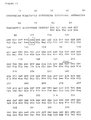
- pAMI1-37 Thirty seven clones were recovered (designated pAMI1-37) and grown in LB medium containing 100 mg/l ampicillin. Recombinant plasmid DNA was isolated and analyzed by restriction digests. Two groups of plasmid could be recognized, one (e.g. pAMI7) containing a 2.8 kb insert , the other (e.g. pAMI25) containing an 4.0 kb insert. An extensive restriction analysis showed that both types of inserts had a region of about 2.0 kb in common. Sequence determination of this region by the chemical degradation method (Maxam and Gilbert, 1980) revealed an open reading frame with a length of 1182 nucleotides which was identified as the coding region of GI. The nucleotide sequence of gi , together with the derived amino acid sequence are shown in Figure 15. In the following, the numbering of amino acids refer to Figure 15.
- Plasmid pLK94 (Botterman and Zabeau, 1987) was first modified to eliminate the PstI site in the -lactamase gene. This was done by isolating the 880 bp EcoRI/PstI fragment of pLK70 70p (Botterman and Zabeau, 1987) containing the N-terminal part of the -lactamase gene, and the 1700 base pair (bp) EcoRI/PstI fragment of pLK94 containing the C-terminal part of the -lactamase gene as well as the replication origin. Subsequent ligation of these fragments yielded pLK94p.
- pAMI7 was cleaved with PstI and a mixture of two purified fragments of about 1800 bp in length, one of which contains the GI gene, were ligated into the PstI site of pLK94p.
- the ligation mixture was used to transform E.coli strain AB 1886. Ampicillin resistant, GI+ transformants were obtained by growing on MMX.
- Plasmid DNA was isolated from a few selected transformants and characterized by restriction analysis. Plasmid pLK94p harboring the PstI fragment containing gi was designated as pLK94GI. The orientation of gi is such that the unique BamHI site is located about 470 bp upstream from the GTG initiation codon.
- pLK70-70p was cleaved with PstI, made blunt end by the Klenow fragment of DNA polymerase I (Klenow) and subsequently digested with XbaI.
- pLK94GI was linearized with BamHI and digested with exonuclease Bal31. Samples were taken at various times -the reaction was stopped with disodium ethylenediaminetetraacetate (EDTA)-, cleaved with XbaI and analyzed by gel electrophoresis to determine the average size of the resected BamHI-XbaI fragments. Fragments, ranging in size from 1350 to 1450 bp were eluted from the gel.
- EDTA disodium ethylenediaminetetraacetate
- the fragments were ligated in pLK70-70p.
- E.coli K514 Cold et al., 1965
- the plasmid DNA isolated from several transformants was characterized by restriction analysis. Twenty four clones containing plasmids with an intact gi were retained and tested for production of EcoAmi(DSM) GI. Cultures were grown overnight at 37°C whereafter total cellular extracts were fractionated by polyacrylamide gel electrophoresis (PAGE) on 12.5% sodium-dodecyl sulphate (SDS) (Laemmli, 1970).
- PAGE polyacrylamide gel electrophoresis
- SDS sodium-dodecyl sulphate
- the P R - gi transcriptional unit could be excised as a EcoRI-XbaI fragment, the sequence of which is given in Fig. 16. After elution from an agarose gel, this fragment was ligated in both pMa5-8 and pMc5-8 which were digested with EcoRI and XbaI, yielding pMa5-GI and pMc5-GI respectively. These vectors were found to direct equal and efficient synthesis of EcoAmi(DSM) GI while the expression level did not differ significantly from that obtained with pLK70GI.
- gi could be further increased by changing the GTG initiation codon into an ATG triplet. This was done by site directed mutagenesis as described in Example 1.D using the following oligonucleotide primer : 5′-GGACAGACATGGTTACC-3′
- Wild-type and mutant GI enzymes were produced in E. coli strain K514 grown in a medium composed of 1% tryptone, 1% NaCl, 0.5% yeast extract, and either ampicillin (100 mg/l) for pMa type vector or chloramphenicol (25 mg/l) for pMC type vector. Cells were grown overnight at 37°C and centrifuged.
- the EcoAmi(DSM) GI enzyme could be purified as follows.
- the cell pellet was resuspended in a minimal volume of 0.05 M Tris(hydroxymethyl)aminomethane (Tris/HCl), 0.1 mM CoCl2, 10 mM MgCl2, 0.2 M KCl, 5% glycerol, and 5 mM EDTA, pH 8.0, and lysozyme was added to a final concentration of 1.0 mg/ml.
- Tris/HCl Tris(hydroxymethyl)aminomethane
- CoCl2 10 mM MgCl2
- 0.2 M KCl 5% glycerol
- 5 mM EDTA pH 8.0
- lysozyme was added to a final concentration of 1.0 mg/ml.
- the cells were lysed using a French press, centrifuged (30 min at 23,000 g), and the supernatant diluted with an equal volume of 5% streptomycin sulphate. Incubation was maintained for 3 hours at 4°C and followed by centr
- the resulting supernatant was heated to 70°C (except for the mutants K253Q and K100R which were heated to only 50°C) for 30 min and centrifuged again.
- the soluble upper phase was made 80% with ammonium sulphate.
- the precipitate which contained most of the enzymatic activity was collected by centrifugation, and then dissolved in 0.02 M Tris/HCl, 5 mM EDTA, 0.85 M ammonium sulphate.
- the subsequent chromatographic steps included Phenyl-Superose, Sephacryl S-200 HR, and finally Mono-Q HR 10/10. Importantly, the addition of 5 mM EDTA to all buffers for chromatography was necessary in order to eliminate metal ions.
- the resulting enzyme was dialyzed 3-times against 200 volumes of 10 mM triethanolamine, pH 7.2, containing 10 mM EDTA (final pH is about 5.2), and again against 200 volumes of 5 mM ⁇ 2-(N-Morpholino)ethanesulfonic acid ⁇ (MES), pH 6.0, with 3 buffer changes.
- the metal content of the final enzyme preparation was determined by atomic absorption spectrometry on a Varian SpectrAA 30/40, and revealed that EcoAmi(DSM) GI was metal free; as an example, it could thus be shown that cobalt ions, which bind to the enzyme with very high affinity, accounted for only ⁇ 1 x 10 ⁇ 4 mol. per mol.
- EcoAmi(DSM) GI monomer when EDTA was omitted in the chromatographic buffers, the latter value increased to 0.5 mol. cobalt per mol. enzyme monomer).
- the purity of the EcoAmi(DSM) GI was assessed by SDS-PAGE and silver staining, and also by reversed phase high performance liquid chromatography (HPLC) on a Vydac C4 column.
- the enzymatic activity of glucose isomerase was assayed as described below (1 unit of enzymatic activity produces 1.0 micromole of product -D-xylulose or D-fructose- per minute; therefore, specific activity -spa- is expressed as units per mg of GI enzyme).
- GI activity could be assayed directly by measuring the increase in absorbance at 278 nm of xylulose produced at 35°C by isomerisation of xylose by glucose isomerases.
- This assay was performed in 50mM triethanolamine buffer, pH 7.5, containing 10mM MgSO4, and 0.1 M xylose.
- Glucose isomerase final concentration in the assay was about 0.01 mg/ml, and precisely determined, prior to dilution in the enzymatic assay mixture, by absorption spectroscopy using an extinction coefficient of 1.08 at 278 nm for a solution of enzyme of 1.0 mg/ml.
- D-Sorbitol Dehydrogenase Coupled Assay enzymatic determination of D-xylulose was performed at 35°C as previously described (Kersters-Hilderson et al., 1987) in 50mM triethanolamine, pH 7.5, 10mM MgSO4, and 0.1 M xylose, in the presence of ⁇ 2 x 10 ⁇ 8 M D-sorbitol dehydrogenase (L-iditol : NAD oxidoreductase, EC 1.1.1.14), and 0.15mM NADH, except that the incubation buffer also included 1mM ethylenebis(oxyethylenenitrilo)tetraacetic acid (EGTA). Glucose isomerase final concentration in this assay was about 2.5 x 10 ⁇ 3 mg/ml, and precisely determined as described above.
- GI activity can be assayed by measuring the amount of D-fructose produced during the isomerization reaction using the cysteine-carbazole method which is based on the reaction of ketosugars with carbazole in acids to yield a purple product (Dische and Borenfreund, 1951).
- the native-PAGE gel was transferred to the PhastSystem staining chamber and incubated for 15 min at 50°C in 20mM Tris/Hcl, pH 7.2, with 50mM xylose, 10mM MgCl2, 0.1mM CoCl2; after washing with demineralized water 0.5 min at 4°C, the gel was immersed for 3 min at 20°C in 0.1% triphenyl-tetrazoliumchloride freshly prepared in 1N NaOH; the reaction was stopped by incubating the gel in 2N HCl for 15 min at 20°C; final wash was in water (0.5 min at 4°C).
- Heat-inactivation kinetics experiments were performed on the metal-free glucose isomerase with the additions described in each specific case.
- the purified enzyme was equilibrated in the desired buffer and the solution was drawn up into a Hamilton gas-tight syringe with a Teflon needle, that had been previously inserted into a glass mantle connected to a circulating waterbath (Lauda, RM6) set at the indicated temperature.
- Previous experiments have shown that temperature equilibration of the enzyme solution from 25 to 85°C is achieved in less than one minute. At appropriate times, aliquots were withdrawn into Eppendorf tubes and the heat denaturation process was quenched by cooling the samples to 0°C.
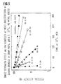
- Fig. 4 summarizes the data of Figures 1, 2, and 3, and shows that, in the temperature interval from 60-95°C, the Arrhenius plots are linear whether metal is present or not.
- Fig. 6 demonstrates that the kinetics of heat-inactivation of the enzyme in the absence of added metal was increased as a function of the ionic strength. This, together with the pH dependence data, strongly indicates that polar residues are involved in the thermal stability of EcoAmi(DSM) GI.
- GI is a tetramer made of four identical subunits (Rey et al., 1988).
- urea The influence of urea was assessed in an attempt to identify structural changes that might account for the loss of enzymatic activity as a result of heating i.e. subunit dissociation and/or unfolding.
- the oligomerization state of the enzyme was analyzed by size-exclusion high performance liquid chromatography (SEC-HPLC) on Superose-12 at room temperature, using an elution buffer consisting of 50 mM Tris/HCl, pH 8.0 at 25°C, and 150 mM NaCl, following prolonged incubation of the enzyme in 7M urea at room temperature.
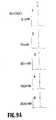
- Fig. 8 shows that prolonged incubation in urea is necessary to induce a dissociation of the native EcoAmi(DSM) GI-tetramer into dimers.
- Metal-free EcoAmi(DSM) GI was incubated in the absence of magnesium at 60°C without or with D-glucose (250mM) in 50mM MOPS, pH 7.7 at 60°C. At appropriate times, aliquots were withdrawn, cooled to 25°C and tested for residual enzymatic activity by the direct xylulose absorbance assay at 278 nm.
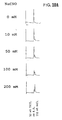
- Fig. 11, Panel A shows that the presence of glucose significantly increased the rate of heat inactivation of the enzyme at 60°C. This effect was not reversible as extensive buffer exchange against 50mM MES, pH 6.0, at 4°C, could not restore the catalytic efficiency of EcoAmi(DSM) GI (triangles in Fig. 11, Panel A). Moreover, analysis of the reaction products by SEC-HPLC clearly demonstrated that, as predicted, glycation was accompanied by tetramer to dimer dissociation in a time-dependent fashion (Fig. 11, Panels B and C), a finding that supports the contention that tetramer splitting occurs at high temperature. Dissociated dimers are trapped by covalent modification with glucose of reactive amino groups likely to reside in the interdimer contacts.
- Fig. 11, Panel D shows that heating also caused formation of a protein aggregate; this aggregation was greatly enhanced in the presence of glucose. It is not known, however, whether aggregate formation occurs independently of, or only subsequent to, GI dissociation into dimers. Very similar results could be reproduced in a different buffer system: 12.5mM potassium phosphate, pH 7.7 at 60°C.
- GI is a tetramer consisting of four identical subunits (A, B, C and D) (Rey et al., 1988) which can be viewed as an assembly of two dimers (AB and CD).
- A, B, C and D identical subunits
- AB and CD dimers
- a residue is said to participate in the subunit interface contacts if its accessible surface area (ASA) (Lee and Richards, 1971) calculated in the isolated subunit differs from that determined in the oligomer.
- Table 2 compiles the ASAs for the 20 subunit lysine residues both in the isolated monomer and in the GI tetramer. Eleven of these residues are seen to participate in subunit interfaces. Only Lys-100 and Lys-253 bury an extensive area (149A2 and 110A2, respectively) in the subunit interfaces, and become almost completely buried in the tetramer. In other words, both of these residues have low solvent accessibility in the tetramer.
- ASA accessible surface area
- Lys-100 and Lys-253 are involved in electrostatic interactions in the subunit interfaces. Lys-100 in the A-subunit (A_Lys-100) stabilizes, through hydrogen bonding the last turn of a small helix near position 373 in the B-subunit.
- Lys-253 in the A-subunit (A_Lys 253) on the other hand is involved in ionic interdimer interaction with Asp-190 of the C-subunit.
- model building techniques as described in Delhaise et al., 1984
- the environment of Lys-100 is not likely to accommodate a substitution to Arg, whereas the mutation of Lys-253 to Arg would be sterically possible because no bad physical contacts were apparent and ionic interactions with Asp-190 remained favorable.
- the substitution of Lys-253 with arginine would stabilize the electrostatic interaction across the dimer-dimer interface and thereby increase the stability of EcoAmi(DSM) GI towards thermal inactivation. Moreover, this substitution would also prevent chemical modification (by glucose or cyanate) at position 253.
- Lys-253 was mutated into glutamine to eliminate the ionic character of the lysine side-chain, this mutation being otherwise reasonably conservative.
- Site-directed mutagenesis was performed according to the gapped duplex DNA (gdDNA) method using the pMa5-8 and pMc5-8 like phasmid vectors (Stanssens et al. 1987). Since the mutagenesis strategy requires the use of unique restriction sites upstream and downstream of the region to be mutagenized, two additional cleavage sites were introduced in the GI coding sequence without altering the encoded amino acid sequence.
- a KpnI site was created by nucleotide base-exchange of G at position 177 into A using the following oligonucleotide primer: 5′-CGAAGGGTACCAGG-3′
- a XhoI site was created by substitution of a C for a G at position 825 using the following oligonucleotide primer: 5′ GCCGTTCTCGAGGAGGTCG-3′
- the conversion of the GTG into an ATG codon (see Example 1.A) and the creation of the KpnI site were accomplished in a single mutagenesis experiment in which the relevant enzymatically phosphorylated oligonucleotides were annealed to a gdDNA derived from single stranded pMc5-GI and the large BamHI-AatII fragment of pMa5-GI.
- the XhoI site was introduced in a separate experiment; the gdDNA used was constructed from single stranded pMc5-GI and the large SacI-SmaI fragment of pMa5-GI. The three mutations were assembled in a single gene by combining the appropriate fragments of the double mutant and the XhoI mutant. The resultant triple mutant was designated as pMa5-I.
- the complementary pMc5-I was constructed by insertion of the small EcoRI-XbaI fragment of pMa5-I, containing the P R - gi hybrid gene, between the EcoRI and XbaI sites of pMc5-8.
- pMa5-I and pMc5-I are the basic vectors used for the production of both the wild type and mutant GI's.
- the large SacI-XhoI fragment of pMc5-I and the following oligonucleotide primer were used: 5′-CCTGGTCGAACTGCGGGCCG-3′
- the mutant enzyme was well expressed; specific activity using xylose as a substrate was 96% that of wild-type EcoAmi(DSM) GI (Table 3).
- the large SacI-XhoI fragment of pMc5-I and the following oligonucleotide primer were used: 5′-CCTGGTCGAACCGCGGGCCG-3′
- thermostability of this mutant was tested in 50mM N-2-Hydroxyethylpiperazine-N′-2-ethanesulfonic acid (EPPS), pH 7.5, 5mM MgSO4, at temperatures ranging from 82-92°C.
- EPPS N-2-Hydroxyethylpiperazine-N′-2-ethanesulfonic acid
- Table 4 lists the half-lives in hours for K253R and wild-type enzymes; the results demonstrate that over this temperature range K253R is consistently more stable than wild-type enzyme.
- the present invention has thus been used successivefully to engineer mutations leading to a more stable GI.
- Lys-100 was mutated into arginine, in which case it was expected that, as mentioned earlier, a bad steric accommodation of the new residue would lead to a decrease of stability, although without affecting enzymatic activity.
- the large KpnI- AatII fragment of pMc5-I and the following oligonucleotide primer were used: 5′-CCGCCGTCCCGGAACACCGG-3′
- Example 2 Mutants of human CuZn superoxide dismutase with increased thermostability and increased resistance towards chemical modification.
- Superoxide dismutases (E.C.1.15.1.1) are enzymes that catalyze the conversion of superoxide radicals into hydrogen peroxide according to the following reaction:
- CuZnSODs are generally very stable enzymes which have been reported to exist as homodimers; each subunit has a molecular weight of approximately 16,000 daltons, contains one Cu(II) and one Zn(II), and associates with its partner by noncovalent interactions.
- the human CuZnSOD was similarly found to undergo glycation, i.e. nonenzymatic, covalent attachment of glucose to amino groups, with concomitant inactivation (Arai et al., 1987 a).
- the glycated sites were subsequently identified as Lys-122 and Lys-128 (Arai et al., 1987 b)
- the sod gene fused to the tac promoter was obtained from E.coli strain D1210 harboring plasmid pSOD11 as described in European patent application EP 138111 and deposited with the American Type Culture Collection (ATTC) under accession number 39679.
- the DNA fragment comprising the tac-sod hybrid gene was isolated as follows. Plasmid pSOD11 was partially digested with AccI, the restriction fragments were made blunt ended by treatment with Klenow and digested with SalI. The 1260 bp fragment, containing the tac-sod hybrid gene, was ligated to the large fragment of plasmid pMc5-8 which was digested with SmaI and SalI. The ligation mixture was used to transform E.coli WK6 (Zell and Fritz, 1987). Transformants were selected on LB agar plates containing 25 ⁇ g/ml chlorampenicol and isolated plasmid DNA was checked by restriction analysis.
- Plasmids which contained the tac-sod hybrid gene between the EcoRI and SalI sites of pMc5-8 was designated as pMc5-sod11.
- the 1260 bp EcoRI-XbaI of pMc5-sod11 was transferred into the EcoRI and XbaI sites of pMa5 8 yielding plasmid pMa5-sod11.
- tac-sod hybrid gene in the pMa/c vectors was checked by sequencing according to Maxam and Gilbert (1980). The sequence of this gene is given in Fig. 17.
- E.coli WK6 (pMa5-sod1) and WK6 (pMc5-sod1) direct the efficient synthesis of a soluble 16 kD protein as apparent from the analysis of total cellular extracts, prepared by sonication, on SDS-12.5% polyacrylamide gels (Laemmli, 1970). When compared to an uninduced control culture, the SDS-PAGE protein profiles of induced cultures contain an additional 16 kD band.
- the 16 kD protein was present in the soluble fraction in which it was estimated to amount to about 5-7 % of total protein content in E.coli WK6 (pMa5-sod1) and 10-15% in the E.coli sodAsodB ⁇ (pMa5-sod1) strain. Purification was performed as follows. Lysozyme (0.5 mg/ml, final) was added to the cell suspension, then made 0.1 mM EDTA. After incubation for 20 min on ice, the cells were lysed using a French Press, and the resulting homogenate was clarified by centrifugation.
- Streptomycin sulphate (0.5 %) was added to the supernatant, and after 3 hours at 4°C, the insoluble material was removed by centrifugation. The resulting supernatant was then made 0.05 mM ZnSO4 and 0.1 mM CuSO4, and incubated 2-4 hours at 25°C, before being heated to 70°C for 30 min. After elimination of the insoluble material by centrifugation, the clarified lysate was dialyzed twice against 50 volumes of 10 mM Tris/HCl, pH 8.0, at 4°C, and chromatographed onto Q-sepharose Fast Flow by means of a gradient from 40 to 200 mM CH3COONa in 20 mM Tris/HCl, pH 8.0.
- the SOD pool originating from the Q-sepharose, was made 1.4 M ammonium sulphate and chromatographed onto a Pharmacia Phenyl-Superose HR 10/10 column equilibrated in 20 mM Tris/HCl, 1.4 M (NH4)2SO4, pH 8.0. Under these conditions, the CuZnSOD was not bound to the hydrophobic resin, in contrast to an E.coli endogenous protein contaminant which eluted only when the ammonium sulphate concentration had been reduced to about 0.7 M.
- the enzyme was then concentrated on a Mono-Q HR 5/10 run in 20 mM Tris/HCl, pH 8.0, including 2 ⁇ M CuSO4 and ZnSO4, and eluted with a gradient from 0 to 160 mM NaOAc.
- the resulting enzyme was finally dialyzed against two buffer changes of 10 mM potassium phosphate, pH 7.8, and was shown to be free of contaminant by SDS-PAGE. It could be shown that no SOD activity from endogenous E.coli SODs remained in the samples.
- CuZnSOD could be assayed by the nitroblue tetrazolium (NBT) assay as modified from Winterbourn et al. (1975).
- NBT nitroblue tetrazolium
- the assay was performed in Microtiter-plates.
- the assay mixture contained 50 mM potassium phosphate, pH 7.8, 50 micromolar NBT (Janssen), 0.1 mM EDTA, 0.03% Triton-X 100, and various amounts of CuZnSOD.
- the reaction was started by addition of 2 mM riboflavin-5′-phosphate (Bio Rad) to the microtiter-plate set onto a fluorescent light-box.
- the plate was covered with aluminium foil, incubated for 12 minutes, and the absorbance at 540 nm was determined with a Titertek Multiskan MCC plate reader.
- the pyrogallol assay was essentially as described by Marklund and Marklund (1974) with minor modifications.
- the CuZnSOD inhibition of pyrogallol autooxidation was performed in an assay mixture containing 0.90 ml of 52.7 mM Tris/cacodylic acid, 1.1 mM diethylenetriamine pentaacetic acid, pH 8.20 (TCDA buffer), and 0.050 ml of various concentrations of CuZnSOD in 52.7 mM TCDA buffer containing 0.05 mg/ml catalase (from Aspergillus niger; Merck).
- the gel was soaked at 50°C first in 2.5 mM NBT for 20 min, and then in 36 mM potassium phosphate, pH 7.8, containing 28 mM TEMED (Bio-Rad), 28 micromolar riboflavin-5′-phosphate (Bio-Rad), and 0.1 mM EDTA, for an additional 30 min.
- the gel was then illuminated using a fluorescent light-box till enough contrast was obtained ( ⁇ 15 20 min), and subsequently fixed by soaking for 30 min in 10% ethanol, 5% acetic acid.
- the gel could be preserved by immersing it for 30 min in 10% acetic acid, 15% glycerol, before drying it.
- Protein concentration was determined using the Bradford assay with bovine serum albumin as a standard.
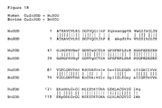
- the human CuZnSOD exhibits about 80% homology with the bovine ( Bos taurus ) enzyme (Fig. 18), the 3D-structure of which has been solved to high resolution (Brookhaven data base pdb file pdb2sod - Tainer et al., 1982). Accordingly, the structure of the human CuZnSOD can be derived using the bovine CuZnSOD as a template (Hallewell et al., 1989).
- Lys-122 and Lys-128 (corresponding to Lys-120 and Arg 126, respectively, in the bovine CuZnSOD), which are implicated in glycation (Arai et al., 1987 b), are fully solvent accessible, and due to charge conservation, their replacement by arginine should be well accommodated in the CuZnSOD structure. Therefore, Lys-122 and Lys-128 were mutated into arginine to prevent chemical modification and resulting inactivation as produced by glucose.
- the structure of the human CuZnSOD could be derived from that of the bovine CuZnSOD as follows:
- Model I One model (Model I) was obtained by substitution of all residues, differing from the human CuZnSOD and having one or more atoms within a radius of 10 Angstrom of Lys-9 in the 0-subunit, with their corresponding residues in the human CuZnSOD. No additions or deletions were required. The following substitutions had to be performed: O_Thr-17>Ile, O_Thr-34>Lys, O_Gln-53>Ala, O_Met-115>Leu, O_Lys-9>Lys (the arrow denotes the substitution event e.g. O_Thr-34>Lys means that Lys is substituted for Thr at position 34 in the O chain).
- Model II Another model (Model II) was obtained by substitution of all residues, differing from the human CuZnSOD and having one or more atoms within a radius of 10 Angstrom from either Lys-120 or Arg-126 in the O-subunit, with their corresponding residues in the human CuZnSOD. No additions or deletions were required. The following substitutions had to be performed: O_Asp-40>Leu, O_Pro-121>Ala, O_Arg-126>Lys.
- the stability of the K128R mutant will additionally be increased by the formation of one hydrogen bond (see Table 5) which is not present in the human CuZnSOD.
- the stability of the K122R mutant on the other hand will additionally be increased by the formation of two hydrogen bonds (see Table 5) which are not present in the human CuZnSOD.
- the double mutant SOD-K122R/K128R was produced by appropriate mutagenesis of either one of the single mutants.
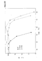
- the rates of inactivation of SOD-WT and SOD-K9R were determined at a fixed temperature. Thus, following incubation of SOD in 20 mM potassium phosphate, pH 7.8 at 85°C in both the absence and presence of metal, aliquots were withdrawn at appropriate time intervals and cooled to 0°C. Residual enzymatic activity, as measured using the pyrogallol assay at 25°C, is shown in Figure 19. The decay obeyed first-order kinetics.
- the inactivation rate constant at 85°C for SOD-WT in the absence (respectively, presence) of metal was 0.06 min ⁇ 1 (0.04 min ⁇ 1) which corresponds to a half-life of 12 min (17 min).
- the rate constant for heat-inactivation at 85°C for SOD-K9R was 0.01 min ⁇ 1 (0.007 min ⁇ 1), so that the half-life increased to 65 min (98 min).
- both wild-type and mutant enzymes (1 mg/ml; 7,000 units/mg) were incubated at 60°C in 0.2 M potassium phosphate, pH 7.35, in the absence or presence of 0.1 M ⁇ -D-glucose. At appropriate times, aliquots were withdrawn, immediately cooled to 0°C, and diluted 500-fold with a stock solution of 52.7 mM TCDA, containing 1 mg/ml catalase, and enzymatic activity was measured using the pyrogallol assay.
- the bovine CuZnSOD is engineered so as to include arginines at positions 9 and 120.
- the resulting enzymes are more stable than the wild-type bovine CuZnSOD.
- the present invention has thus been used successivefully to engineer mutations leading to more stable CuZnSODs.
- Example 3 Mutants of glyceraldehyde-3-phosphate dehydrogenase (GAPDH) from Bacillus subtilis with increased thermostability.
- GPDH glyceraldehyde-3-phosphate dehydrogenase
- Glyceraldehyde-3 phosphate dehydrogenase (GAPDH - E.C.1.2.1.12) is a homotetrameric enzyme which catalyzes the following reversible reaction in glycolysis,:
- thermostable GAPDH from Bacillus stearothermophilus ( Bst ) (Skarzynski et al., 1987) and from lobster (Moras et al., 1975; Biesecker et al., 1977) have been determined. These two GAPDH's differ considerably in their thermostability, Bst GAPDH being much more stable than lobster GAPDH. The difference in thermostability between these two enzymes has been suggested to originate from the presence of intersubunit bridges in the Bst GAPDH which are absent in the lobster enzyme (Biesecker et al., 1977; Skarzynski et al., 1987). However, comparisons with other thermostable (e.g.
- thermolabile GAPDH's e.g. pig, yeast
- GAPDH from Bsu turned out to be very unstable.
- the isolated recombinant plasmid, pBSgap-1 (Viaene et al., 1988) was obtained from P.Dhaese (Rijksuniversiteit Gent, Belgium).
- the plasmid consists of a partial Sau3A fragment containing the entire gap coding region, inserted in the BamHI site of pUC18 (Yannisch-Perron et al., , 1985).
- the ligation mixture was used to transform E.coli WK6 (Zell and Fritz, 1987) selecting on LB agar plates containing 25 ⁇ g/ml chloramphenicol. Plasmid DNA was isolated from a few selected transformants and characterized by restriction analysis. The plasmid with the desired structure, i.e. the tac-gap hybrid transcription unit inserted between the EcoRI and XbaI sites of pMc5-8, was identified and designated pMc5-gap1. The sequence of the tac-gap hybrid gene is shown in Fig. 20. For purposes of mutagenesis, a pMa type version of this vector was constructed by transferring the EcoRI-XbaI tac-gap expression module onto pMa5-8. The resultant plasmid was designated pMa5-gap1.
- E.coli WK6 For the induced synthesis of Bsu GAPDH, an overnight culture of E.coli WK6 , containing either pMa5-gap1 or pMc5-gap1, was diluted 100-fold in TB medium (1.2 % Bacto-tryptone; 2.4 % Bacto-yeast extract; 0.5 % glycerol; 28 mM KH2PO4; 72 mM K2HPO4) containing the appropriate antibiotic (resp. 100 ⁇ g/ml ampicillin or 25 ⁇ g/ml chloramphenicol).
- TB medium 1.2 % Bacto-tryptone; 2.4 % Bacto-yeast extract; 0.5 % glycerol; 28 mM KH2PO4; 72 mM K2HPO4
- IPTG isopropyl- ⁇ -D-thiogalactopyranoside; 0.1 mM final concentration.
- the cells were harvested by centrifugation after an induction period of about 16 hours.
- both clones i.e. E.coli WK6 (pMa5-gap1) and WK6 (pMc5-gap1) direct the efficient synthesis of a soluble 35 kD protein as apparent from the analysis of total cellular extracts on SDS-12.5% polyacrylamide gels (Laemmli, 1970).
- the protein profiles of induced cultures contain a prominent 35 kD band.
- the recombinant protein was present in the soluble fraction in which it was estimated to amount to at least 30 % of the total protein content. When purified, it was shown to behave identically with authentic GAPDH isolated from Bacillus subtilis BR151 on both native and SDS-PAGE.
- the recombinant Bsu GAPDH was purified from an induced culture of E.coli WK6 (pMa5-gap1) (see above) as follows. Cells were washed with PBS, frozen, thawed and resuspended (200 OD/ml) in 0.1 M triethanolamine pH 7.5 containing 2 mM dithiothreitol (DTT) and 5 mM EDTA. Lysozyme was added to a final concentration of 1 mg/ml. After 20 min on ice the cells were lyzed using a French press (2X). Cell debris was removed by centrifugation. Streptomycin sulphate was added to a final concentration of 0.5%.
- E.coli WK6 pMa5-gap1
- Protein concentration was determined with the Biorad method using bovine serum albumin as a standard.
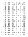
- the 3D-structure of Bsu GAPDH was derived from the known Bst GAPDH (Brookhaven data base pdb file pdb1gd1 - Skarzynski et al., 1987) by model building. Comparison of the primary structures of the Bsu and Bst GAPDH's (sequence of Bst GAPDH from Branlant et al., 1989) indicated that the sequences were 80% identical (Fig. 21). Therefore, the Bsu GAPDH 3D-structure could be derived using the structure of the Bst enzyme as a template.
- the 3D-structure of Bst GAPDH in the immediate neighbourhood of the Arg-281 residue in each subunit was changed by replacing all residues, having one or more atoms within a region of 10 Angstroms of these four Arg-281 residues, with the corresponding residues in the Bsu enzyme. No insertions or deletions are required.
- Arg-282 is also sterically accommodated in the subunit interface.
- the H-bond with O_Glu-201.o is not only much stronger than observed for the Lys, but a salt bridge with the carboxylate of Q_Glu-201 is also formed. Consequently, it can be expected that the electrostatic interaction between subunits will be improved, resulting in an increase of thermostability with respect to the Bsu GAPDH-G282K.
- the mutation of Gly-282 into lysine or arginine made use of a gapped DNA molecule - constructed from single stranded pMc5-gap1 (template strand) and the large StyI-ClaI fragment of pMa5-gap1 (gapped strand; see Fig. 20) - and the following oligonucleotides: 5′-GTTTCCGTTGTAGTCCTTCGAAACTAATGGC-3′ for introduction of a lysine, and 5′-CGTTGTAGTCGCGAGAAACTAATGG-3′ for the construction of the arginine mutant.
- the GAPDH activity of wild type and mutants was determined using a published procedure (Misset et al.,1986).
- the assay mixture contained 5.6 mM glycerate-3-phosphate, 1 mM ATP, 5 mM MgSO4, 1 mM EDTA, 1 mM DTT, 0.42 mM NADH, 25 ⁇ g/ml phosphoglycerate kinase (from yeast) and approximately 0.3 ⁇ g/ml GAPDH in 0.1 M triethanolamine buffer pH 7.5. All reagents were from Boehringer Mannheim. The initial rate was determined by monitoring the disappearance of NADH absorbance at 340 nm. One unit is defined as the amount of enzyme required to cause the disappearance of 1 ⁇ mol. of NADH per minute at 25°C.
- 0.05 ml protein samples were incubated during 15 min at different temperatures.
- the incubation mixture contained 40 units/ml GAPDH, 1 mM DTT, 1 mM EDTA and 1 mM NAD+ in 0.1 M triethanolamine buffer pH 7.8.
- the inactivation process was stopped by adding 0.45 ml of ice cold solution containing 1 mg/ml bovine serum albumin, 1 mM DTT, 1 mM NAD+ and 1 mM EDTA in 0.1 M triethanolamine pH 7.8.
- Residual activity was measured and shown in Figure 22; the temperature of 50% remaining activity was approximately 50°C for the wild-type (GAPDH-WT), 72°C for the GAPDH-G282K, and 75°C for the GAPDH-G282R mutant.
- the second experiment allowed to determine the rates of inactivation of these enzymes at a fixed temperature. Thus, following incubation at 74.5°C, samples were withdrawn at appropriate times. The inactivation process was stopped by ten-fold dilution in a NAD+ containing bovine serum albumin solution at 0°C and residual enzymatic activity was measured as shown in Figure 23. The decay obeyed first-order kinetics.
- the inactivation rate constant at 74.5°C for GAPDH-G282K was 0.15 min ⁇ 1 which corresponds with a half-life of 5 min.
- the rate constant for heat-inactivation at 74.5°C for GAPDH-G282R was 0.04 min ⁇ 1, so that the half life was increased to 18 min.
- the rate of heat-inactivation of GAPDH-WT could not be measured at 74.5°C, but it had a half-life of 19 min at 50°C.
- the present invention has thus been used successivefully to engineer mutations leading to more stable GAPDHs.
- Tables referred to hereinbefore are now presented. Table 1 Three and one letter codes for the twenty amino acids commonly found in proteins. Alanine Ala A Leucine Leu L Arginine Arg R Lysine Lys K Asparagine Asn N Methionine Met M Aspartic acid Asp D Phenylalanine Phe F Cysteine Cys C Proline Pro P Glutamic acid Glu E Serine Ser S Glutamine Gln Q Threonine Thr T Glycine Gly G Tryptophane Trp W Histidine His H Tyrosine Tyr Y Isoleucine Ile I Valine Val V Table 2 Accessible surface area (ASA) for each of the 20 lysine residues of the GI monomer.
- ASA Accessible surface area
- the buffer used was 50 mM EPPS, pH 7.5 at 84°C, 5 mM MgSO4.
- Table 5 Analysis of 3D-structure of human CuZnSOD as derived from the bovine structure (see text). NE: nonbonded energy.
- NE nonbonded energy: ASA T and ASA M : accessible surface area in respectively tetramer and monomer; ASA b : buried surface. Notation of the hydrogen bonds follow the UPAC Convention (In Proteins, 1976, (G.D.Fastman, ed.), CRC Critical Reviews in Biochemistry 1:59-90) P_Lys-282 P_Arg-282 ASA M (A) 22.2 34.7 ASA T (A) 132.6 148.8 ASA b (A) 110.4 114.1 NE(kcal/mol) -18.64 -31.3 H-bonds (distance in A) .hz1..0P407_Hoh.o (1.86) .he..O201_Glu.o (1.83) .hz2..0P429_Hoh.o (1.94) .hh11..0R408_Hoh.o (2.67) .hz2..O201_Glu.o (2.30) .hh12..0P534_Hoh
- Enzymes were produced by sodAsodB ⁇ E. coli (see text). [Glucose] 14 hours 18 hours 22 hours SOD-WT none 100 72 84 0.1 M 72 30 0 SOD-K122R/K128R none 100 100 98 0.1 M 100 57 30
- VILLAFRANCA J.E., HOWELL, E.E., OATLEY, S.J., XUONG, N. and KRAUT, J. (1987) Biochemistry 26 : 2182-2189.
Landscapes
- Life Sciences & Earth Sciences (AREA)
- Health & Medical Sciences (AREA)
- Chemical & Material Sciences (AREA)
- Engineering & Computer Science (AREA)
- Genetics & Genomics (AREA)
- Organic Chemistry (AREA)
- Wood Science & Technology (AREA)
- Zoology (AREA)
- Bioinformatics & Cheminformatics (AREA)
- General Engineering & Computer Science (AREA)
- Biomedical Technology (AREA)
- General Health & Medical Sciences (AREA)
- Molecular Biology (AREA)
- Biochemistry (AREA)
- Biotechnology (AREA)
- Microbiology (AREA)
- Medicinal Chemistry (AREA)
- General Chemical & Material Sciences (AREA)
- Chemical Kinetics & Catalysis (AREA)
- Proteomics, Peptides & Aminoacids (AREA)
- Physics & Mathematics (AREA)
- Biophysics (AREA)
- Plant Pathology (AREA)
- Enzymes And Modification Thereof (AREA)
- Peptides Or Proteins (AREA)
- Preparation Of Compounds By Using Micro-Organisms (AREA)
- Immobilizing And Processing Of Enzymes And Microorganisms (AREA)
- Micro-Organisms Or Cultivation Processes Thereof (AREA)
- Investigating Or Analysing Biological Materials (AREA)
- Measuring Or Testing Involving Enzymes Or Micro-Organisms (AREA)
Applications Claiming Priority (4)
| Application Number | Priority Date | Filing Date | Title |
|---|---|---|---|
| EP88201539 | 1988-07-15 | ||
| EP88201539 | 1988-07-15 | ||
| EP88402789 | 1988-11-04 | ||
| EP88402789 | 1988-11-04 |
Publications (2)
| Publication Number | Publication Date |
|---|---|
| EP0355039A2 true EP0355039A2 (de) | 1990-02-21 |
| EP0355039A3 EP0355039A3 (de) | 1990-10-31 |
Family
ID=26115226
Family Applications (1)
| Application Number | Title | Priority Date | Filing Date |
|---|---|---|---|
| EP19890201893 Withdrawn EP0355039A3 (de) | 1988-07-15 | 1989-07-17 | Verfahren und Mittel zur Kontrolle der Stabilität von Proteinen |
Country Status (12)
| Country | Link |
|---|---|
| EP (1) | EP0355039A3 (de) |
| JP (2) | JPH03501566A (de) |
| KR (1) | KR0140866B1 (de) |
| AT (1) | ATE214094T1 (de) |
| AU (2) | AU637766B2 (de) |
| BG (1) | BG60322B2 (de) |
| CA (1) | CA1339544C (de) |
| DE (1) | DE68929376T2 (de) |
| DK (1) | DK175633B1 (de) |
| FI (1) | FI102541B (de) |
| HU (1) | HU214783B (de) |
| WO (1) | WO1990000605A1 (de) |
Cited By (1)
| Publication number | Priority date | Publication date | Assignee | Title |
|---|---|---|---|---|
| WO2017102103A1 (en) | 2015-12-14 | 2017-06-22 | Luxembourg Institute Of Science And Technology (List) | Method for enzymatically modifying the tri-dimensional structure of a protein |
Families Citing this family (3)
| Publication number | Priority date | Publication date | Assignee | Title |
|---|---|---|---|---|
| DE69133491T2 (de) * | 1990-01-04 | 2006-07-27 | Genencor International, Inc., Palo Alto | Glucose-Isomerase mit geändertem pH-Optimum |
| EP0440273B1 (de) * | 1990-01-04 | 2003-04-09 | Genencor International Inc. | Eine Methode zur Herstellung von Glukose-Isomerasen mit geänderter Substratspezifität |
| JP4596827B2 (ja) * | 2004-06-22 | 2010-12-15 | サントリーホールディングス株式会社 | 安定化プロリントランスポーター |
Family Cites Families (1)
| Publication number | Priority date | Publication date | Assignee | Title |
|---|---|---|---|---|
| CA1289092C (en) * | 1987-01-15 | 1991-09-17 | Novartis Vaccines And Diagnostics, Inc. | Thermostable human cu/zn superoxide dismutase muteins |
-
1989
- 1989-07-17 AT AT89201892T patent/ATE214094T1/de active
- 1989-07-17 CA CA000605829A patent/CA1339544C/en not_active Expired - Lifetime
- 1989-07-17 DE DE68929376T patent/DE68929376T2/de not_active Expired - Lifetime
- 1989-07-17 HU HU894580A patent/HU214783B/hu not_active IP Right Cessation
- 1989-07-17 EP EP19890201893 patent/EP0355039A3/de not_active Withdrawn
- 1989-07-17 AU AU39792/89A patent/AU637766B2/en not_active Ceased
- 1989-07-17 AU AU40425/89A patent/AU638416B2/en not_active Ceased
- 1989-07-17 WO PCT/EP1989/000838 patent/WO1990000605A1/en not_active Ceased
- 1989-07-17 JP JP1508281A patent/JPH03501566A/ja active Pending
- 1989-07-17 JP JP1507756A patent/JPH03501565A/ja active Pending
-
1990
- 1990-03-14 DK DK199000662A patent/DK175633B1/da not_active IP Right Cessation
- 1990-03-14 FI FI901279A patent/FI102541B/fi active IP Right Grant
- 1990-03-15 BG BG091484A patent/BG60322B2/bg unknown
- 1990-03-15 KR KR90700559A patent/KR0140866B1/ko not_active Expired - Fee Related
Cited By (1)
| Publication number | Priority date | Publication date | Assignee | Title |
|---|---|---|---|---|
| WO2017102103A1 (en) | 2015-12-14 | 2017-06-22 | Luxembourg Institute Of Science And Technology (List) | Method for enzymatically modifying the tri-dimensional structure of a protein |
Also Published As
| Publication number | Publication date |
|---|---|
| JPH03501566A (ja) | 1991-04-11 |
| FI901279A0 (fi) | 1990-03-14 |
| CA1339544C (en) | 1997-11-18 |
| FI102541B1 (fi) | 1998-12-31 |
| KR900702021A (ko) | 1990-12-05 |
| FI102541B (fi) | 1998-12-31 |
| WO1990000605A1 (en) | 1990-01-25 |
| AU3979289A (en) | 1990-02-05 |
| EP0355039A3 (de) | 1990-10-31 |
| DK66290A (da) | 1990-03-14 |
| DK66290D0 (da) | 1990-03-14 |
| DE68929376D1 (de) | 2002-04-11 |
| AU4042589A (en) | 1990-02-05 |
| JPH03501565A (ja) | 1991-04-11 |
| AU637766B2 (en) | 1993-06-10 |
| HU214783B (hu) | 1998-05-28 |
| AU638416B2 (en) | 1993-07-01 |
| BG60322B2 (bg) | 1994-07-25 |
| HU894580D0 (en) | 1991-05-28 |
| DE68929376T2 (de) | 2002-10-17 |
| DK175633B1 (da) | 2004-12-27 |
| ATE214094T1 (de) | 2002-03-15 |
| KR0140866B1 (en) | 1998-06-15 |
Similar Documents
| Publication | Publication Date | Title |
|---|---|---|
| Monchois et al. | Characterization of Leuconostoc mesenteroides NRRL B-512F dextransucrase (DSRS) and identification of amino-acid residues playing a key role in enzyme activity | |
| Zenno et al. | Biochemical characterization of NfsA, the Escherichia coli major nitroreductase exhibiting a high amino acid sequence homology to Frp, a Vibrio harveyi flavin oxidoreductase | |
| Geoghegan et al. | Spontaneous α-N-6-Phosphogluconoylation of a “His Tag” inEscherichia coli: The Cause of Extra Mass of 258 or 178 Da in Fusion Proteins | |
| US5827712A (en) | Process for efficiently producing transglutaminase through DNA recombination | |
| Tanizawa et al. | Cloning and sequencing of phenylethylamine oxidase from Arthrobacter globiformis and implication of Tyr-382 as the precursor to its covalently bound quinone cofactor | |
| Lin et al. | Subunit interaction enhances enzyme activity and stability of sweet potato cytosolic Cu/Zn-superoxide dismutase purified by a His-tagged recombinant protein method | |
| Gilles et al. | Conservative replacement of methionine by norleucine in Escherichia coli adenylate kinase. | |
| Palmer et al. | Isolation of protein FA, a product of the mob locus required for molybdenum cofactor biosynthesis in Escherichia coli | |
| MINo et al. | Purification and characterization of serine acetyltransferase from Escherichia coli partially truncated at the C-terminal region | |
| EP0351029B1 (de) | Glucose-Isomerase-Enzyme und deren Verwendung | |
| US5290690A (en) | Methods and means for controlling the stability of proteins | |
| Hashimoto et al. | Molecular Cloning of Two Genes for β-d-Glucosidase inBacillussp. GL1 and Identification of One as a Gellan-Degrading Enzyme | |
| Doublet et al. | The glutamate racemase activity from Escherichia coli is regulated by peptidoglycan precursor UDP-N-acetylmuramoyl-L-alanine | |
| Klein et al. | Purification and characterization of invertase from a novel industrial yeast, Schwanniomyces occidentalis | |
| EP0355039A2 (de) | Verfahren und Mittel zur Kontrolle der Stabilität von Proteinen | |
| Wen et al. | The NAD (P) H-dependent glutamate dehydrogenase activities of Prevotella ruminicola B (1) 4 can be attributed to one enzyme (GdhA), and gdhA expression is regulated in response to the nitrogen source available for growth | |
| Hughes et al. | Production and characterization of a plant α‐hydroxynitrile lyase in Escherichia coli | |
| Fiorentino et al. | Decreasing the stability and changing the substrate specificity of the Bacillus stearothermophilus alcohol dehydrogenase by single amino acid replacements. | |
| McFARLAN et al. | Purification, characterization and revised amino acid sequence of a second thioredoxin from Corynebacterium nephridii | |
| Faridmoayer et al. | An improved purification procedure for soluble processing α-glucosidase I from Saccharomyces cerevisiae overexpressing CWH41 | |
| Hashimoto et al. | Polysaccharide Lyase: Molecular Cloning of Gellan Lyase Gene and Formation of the Lyase from a Huge Precursor Protein inBacillussp. GL1 | |
| Fusi et al. | Expression of a synthetic gene encoding P2 ribonuclease from the extreme thermoacidophilic archaebacterium Sulfolobus solfataricus in mesophylic hosts | |
| JP3493400B2 (ja) | 新規なアミノ末端保護基遊離酵素 | |
| Su et al. | Use of gene fusions of the structural gene sdaA to purify L‐serine deaminase 1 from Escherichia coli K‐12 | |
| Shim et al. | Improvement of the expression level of β-glucosidase from Agrobacterium sp. in Escherichia coli by rare codon optimization |
Legal Events
| Date | Code | Title | Description |
|---|---|---|---|
| PUAI | Public reference made under article 153(3) epc to a published international application that has entered the european phase |
Free format text: ORIGINAL CODE: 0009012 |
|
| AK | Designated contracting states |
Kind code of ref document: A2 Designated state(s): AT BE CH DE ES FR GB GR IT LI LU NL SE |
|
| 17P | Request for examination filed |
Effective date: 19900116 |
|
| PUAL | Search report despatched |
Free format text: ORIGINAL CODE: 0009013 |
|
| AK | Designated contracting states |
Kind code of ref document: A3 Designated state(s): AT BE CH DE ES FR GB GR IT LI LU NL SE |
|
| 17Q | First examination report despatched |
Effective date: 19940414 |
|
| STAA | Information on the status of an ep patent application or granted ep patent |
Free format text: STATUS: THE APPLICATION IS DEEMED TO BE WITHDRAWN |
|
| 18D | Application deemed to be withdrawn |
Effective date: 19960201 |