EP0386936B2 - Vorrichtung zur Steigerung von Ultraschall-Echos - Google Patents
Vorrichtung zur Steigerung von Ultraschall-Echos Download PDFInfo
- Publication number
- EP0386936B2 EP0386936B2 EP90302170A EP90302170A EP0386936B2 EP 0386936 B2 EP0386936 B2 EP 0386936B2 EP 90302170 A EP90302170 A EP 90302170A EP 90302170 A EP90302170 A EP 90302170A EP 0386936 B2 EP0386936 B2 EP 0386936B2
- Authority
- EP
- European Patent Office
- Prior art keywords
- particles
- patient
- indentations
- image
- composite material
- Prior art date
- Legal status (The legal status is an assumption and is not a legal conclusion. Google has not performed a legal analysis and makes no representation as to the accuracy of the status listed.)
- Expired - Lifetime
Links
Images



Classifications
-
- A—HUMAN NECESSITIES
- A61—MEDICAL OR VETERINARY SCIENCE; HYGIENE
- A61L—METHODS OR APPARATUS FOR STERILISING MATERIALS OR OBJECTS IN GENERAL; DISINFECTION, STERILISATION OR DEODORISATION OF AIR; CHEMICAL ASPECTS OF BANDAGES, DRESSINGS, ABSORBENT PADS OR SURGICAL ARTICLES; MATERIALS FOR BANDAGES, DRESSINGS, ABSORBENT PADS OR SURGICAL ARTICLES
- A61L31/00—Materials for other surgical articles, e.g. stents, stent-grafts, shunts, surgical drapes, guide wires, materials for adhesion prevention, occluding devices, surgical gloves, tissue fixation devices
- A61L31/14—Materials characterised by their function or physical properties, e.g. injectable or lubricating compositions, shape-memory materials, surface modified materials
-
- A—HUMAN NECESSITIES
- A01—AGRICULTURE; FORESTRY; ANIMAL HUSBANDRY; HUNTING; TRAPPING; FISHING
- A01K—ANIMAL HUSBANDRY; AVICULTURE; APICULTURE; PISCICULTURE; FISHING; REARING OR BREEDING ANIMALS, NOT OTHERWISE PROVIDED FOR; NEW BREEDS OF ANIMALS
- A01K85/00—Artificial bait for fishing
-
- A—HUMAN NECESSITIES
- A61—MEDICAL OR VETERINARY SCIENCE; HYGIENE
- A61B—DIAGNOSIS; SURGERY; IDENTIFICATION
- A61B8/00—Diagnosis using ultrasonic, sonic or infrasonic waves
- A61B8/08—Clinical applications
- A61B8/0833—Clinical applications involving detecting or locating foreign bodies or organic structures
-
- A—HUMAN NECESSITIES
- A61—MEDICAL OR VETERINARY SCIENCE; HYGIENE
- A61B—DIAGNOSIS; SURGERY; IDENTIFICATION
- A61B8/00—Diagnosis using ultrasonic, sonic or infrasonic waves
- A61B8/08—Clinical applications
- A61B8/0833—Clinical applications involving detecting or locating foreign bodies or organic structures
- A61B8/0841—Clinical applications involving detecting or locating foreign bodies or organic structures for locating instruments
-
- A—HUMAN NECESSITIES
- A61—MEDICAL OR VETERINARY SCIENCE; HYGIENE
- A61B—DIAGNOSIS; SURGERY; IDENTIFICATION
- A61B90/00—Instruments, implements or accessories specially adapted for surgery or diagnosis and not covered by any of the groups A61B1/00 - A61B50/00, e.g. for luxation treatment or for protecting wound edges
- A61B90/39—Markers, e.g. radio-opaque or breast lesions markers
-
- A—HUMAN NECESSITIES
- A61—MEDICAL OR VETERINARY SCIENCE; HYGIENE
- A61L—METHODS OR APPARATUS FOR STERILISING MATERIALS OR OBJECTS IN GENERAL; DISINFECTION, STERILISATION OR DEODORISATION OF AIR; CHEMICAL ASPECTS OF BANDAGES, DRESSINGS, ABSORBENT PADS OR SURGICAL ARTICLES; MATERIALS FOR BANDAGES, DRESSINGS, ABSORBENT PADS OR SURGICAL ARTICLES
- A61L31/00—Materials for other surgical articles, e.g. stents, stent-grafts, shunts, surgical drapes, guide wires, materials for adhesion prevention, occluding devices, surgical gloves, tissue fixation devices
- A61L31/14—Materials characterised by their function or physical properties, e.g. injectable or lubricating compositions, shape-memory materials, surface modified materials
- A61L31/18—Materials at least partially X-ray or laser opaque
-
- A—HUMAN NECESSITIES
- A61—MEDICAL OR VETERINARY SCIENCE; HYGIENE
- A61M—DEVICES FOR INTRODUCING MEDIA INTO, OR ONTO, THE BODY; DEVICES FOR TRANSDUCING BODY MEDIA OR FOR TAKING MEDIA FROM THE BODY; DEVICES FOR PRODUCING OR ENDING SLEEP OR STUPOR
- A61M25/00—Catheters; Hollow probes
- A61M25/0009—Making of catheters or other medical or surgical tubes
-
- A—HUMAN NECESSITIES
- A61—MEDICAL OR VETERINARY SCIENCE; HYGIENE
- A61B—DIAGNOSIS; SURGERY; IDENTIFICATION
- A61B90/00—Instruments, implements or accessories specially adapted for surgery or diagnosis and not covered by any of the groups A61B1/00 - A61B50/00, e.g. for luxation treatment or for protecting wound edges
- A61B90/39—Markers, e.g. radio-opaque or breast lesions markers
- A61B2090/3925—Markers, e.g. radio-opaque or breast lesions markers ultrasonic
Definitions
- This invention relates to echogenic medical devices to be positioned within a medium of a patient and with ultrasonic imaging equipment.
- Ultrasonic imaging in the medical field is widely used for a variety of applications.
- In addition to imaging physiological structures and tissue it is often desirable to produce an image of a medical device which has been inserted into a patient.
- Various approaches have been used to enhance ultrasonic imaging by modifying the reflective surface characteristics of these devices.
- the present invention is based upon the discovery that when discrete particulate type acoustic discontinuities which includes particles or indentations within the surface of the body of the device, or particles on the surface of the device, then the ultrasonic imaging of the device is significantly improved.
- the particles can be solid and have an acoustic impedance significantly different from that of the body of the device.
- the particulate type discontinuities can alternatively be shell-like in which case they can be filled with gas such as air.
- the particles or the particulate discontinuities which includes indentations, are either partially embedded in the surface of the body or formed entirely on the outer surface of the body say by adhesion.
- the outer parts of the shells may be removed to leave the discontinuities in the form of indentations in the outer surface of the body. These indentations produce a scattering component of the image in response to an input beam. The image is produced regardless of the angle of the incident beam.
- the body of the device in most of the above inventive instances is regarded as being a composite body, and in many practical uses, the outer surface of the body forms an interface with the biological tissue of a patient.
- These interfaces extend in a multiplicity of directions due to the spherical nature of each interface. These have the effect of scattering the reflected ultrasonic waves in a multiplicity of directions and that enables the device surface to be observed from those directions, thus making observation uncritical, this being particularly advantageous when an article being observed in non-linear, or flexible.
- the difference in the acoustic impedances of the member material and surrounding medium enhances the intensity of the scattered component of the image.
- the particles or indentations are formed from a substance as a plurality of spherical shaped particles or indentations that each have a predetermined spherical surface for establishing the interface.
- This substance is attached to, partially embedded in, or formed as indentations in the outside surface of the member material.
- glass particles are attached to the outside surface of the member with an adhesive material.
- the particles are partially embedded in the outside surface of the member material.
- the contoured substance such as the glass particles are affixed to the outside surface of a stainless steel member using, for example, another material such as silver solder.
- the substance has an acoustic impedance different from at least one of the impedances of the member material and surrounding tissue for enhancing the image produced in response to the sonic beam.
- the silver solder also presents another acoustic impedance to enhance an image.
- the particles In the embodiment which includes forming the interface by attaching a plurality of spherical particles to the surface of the elongated member, the particles have an acoustic impedance having at least the predetermined difference between at least one of the two impedances of the elongated member and biological tissue.
- a preferred diameter for the spherical indentations is in the range of between 1-50 microns.
- the composite material includes a formable matrix material with further discrete sound reflective particles made from a material different from and more sonically reflective than the matrix material and being embedded in the matrix material to enhance the echogenicity of the body member.
- the device can easily be made biologically inert and sterile for patient safety.
- the composite material of the embodiments still maintains the requisite flexibility for many medical applications, while providing echogenicity throughout the body of the device. In this way, the physician may observe a full image of the medical device in the patient.
- radiopaque material such as barium or tungsten to provide imaging with radiographic equipment.
- the reflective particles, and optionally the radiopaque material are mixed into the matrix material prior to forming the device by, for example, extrusion in the case of most catheters. Thus, no additional post extrusion fabrication steps are required to provide the desired echogenicity and a high level of quality control may be maintained.
- the echogenic body member is positioned in a sonic imaging beam; and an image is generated of the echogenic body member including the sound reflective particles from the sonic imaging beam.
- echogenic body member 123 is a part of an echogenic device which may be utilized in a large variety of medical devices.
- Member 123 is at least partially made up of a composite material which is echogenically imageable in the patient, such as by the use of ultrasonic imaging equipment.
- the composite material of the interface includes matrix material 125 with discrete sound reflective particles 127 partially embedded on the outer surface of the matrix material 125.
- matrix material 125 is a plastic, such as urethane, silicone, polyethylene, tetrafluorethylene.
- matrix 125 is a formable, pliable material which may be molded and/or extruded to a variety of shapes, depending upon a specific application.
- Particles 127 are preferably made of a hard material, such as small glass particles, of spherical shape forming glass microspheres. Glass microspheres with an outer diameter of about 5 microns is one acceptable size. Other sized particles may be utilized as, for example, ranging between 1 and 50 microns and beyond. Furthermore, a spherical surface may be provided on the outside and/or the inside of particles 127, as for example a particle with a hollow spherical space therein. Particles 127 are made up of a different material than matrix 125.
- the external and/or internal spherical shape provides for ultrasonic reflections at a variety of angles regardless of the direction from which the ultrasonic waves are emanating, and accordingly, are more likely to reflect at least a portion of the transmitted signal back to the ultrasonic receiver to generate an image.
- ultrasonic reflective particles 127 provide adequate reflection.
- the use of a composite, rather than a solution, provides adequate size for acoustic reflection from the discrete particles embedded in the matrix.
- a variety of materials may be utilized for the ultrasonic reflective particles, such as aluminum, hard plastic, sand, metal particles, and the like.
- liquids, gases, gels, microen-capsulants, and/or coacervates suspended in the matrix may alternatively be used either alone or in combination, so long as they form a composite with ultrasonically reflective particles in the matrix.
- glass balls or microspheres have been found to be very well suited.
- the matrix 125 comprise solder used to fuse parts together.
- the solder matrix with ultrasonically reflective particles associated with the outer surface thereof may be used to solder wires together in medical baskets (not shown) used to remove stones and other objects from medical patients.
- this technique may be used for other devices such as blood clot filters, guide wires and the like.
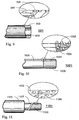
- FIG.9 Depicted in Fig.9 is a partial cross-sectional view of another illustrative embodiment of an echogenic medical device 901 that is insertable into a patient.
- Device 901 comprises an elongated tubular member 902 with a passageway 903, commonly known as a lumen, extending longitudinally therein.
- Member 902 is part of any well-known catheter, stent, needle, and the like for insertion into biological tissue or passageway of a patient.
- the elongated member has a characteristic or acoustic impedance, different from the characteristic or acoustic impedance of the surrounding medium.
- elongated member 902 comprises a plastic material. From the Kinsler and Wells references, soft plastic such as polythene is listed as having a characteristic impedance of 1.84 X 10 6 MKS rayl. A hard plastic such as Lucite in bulk form is listed as having a characteristic impedance of 3.2 X 10 6 MKS rayl.
- soft plastic such as polythene
- Lucite in bulk form is listed as having a characteristic impedance of 3.2 X 10 6 MKS rayl.
- Medical device 901 also includes an interface including outside surface 904 having a shape responsive to a sonic beam for producing one component such as a reflective component of the sonic image.
- the outside surface of the elongated member also includes a plurality of spherical indentations 905 therein. These spherical indentations scatter the ultrasonic beam to produce another component of the image.
- a dimension of 2.5 microns is one acceptable size for the radius of spherical indentations 905.
- the radius of the indentations may range, for example, between 0.5 and 25 microns and beyond. This radial dimension is related to the wavelength of the incoming ultrasonic beam in a predetermined manner such that the radius is much less than the wavelength of the beam.
- an ultrasonic beam emitted at 3.5 MHz has a wavelength of approximately 17,700 microns
- an ultrasonic beam emitted at 7.5 MHz has a wavelength of approximately 8,200 microns. Both of these frequencies are emitted from commercially available ultrasonic imaging equipment.
- the spherical indentations provide a curved surface from which the incident sonic beam may be scattered to produce the desired image regardless of the angle of incidence with respect to outer surface 904.
- the image produced by the interface including the outer surface and spherical indentations includes one or more components.
- Rayleigh scattering occurs.
- One known example of Rayleigh scattering is the interaction of ultrasound with blood cells.
- the amplitude of the scattered wave component is proportional to the square of the frequency of the sonic beam. Therefore, high frequency sonic beams are scattered the most strongly.
- dimensions of the reflecting surface must be greater than several wavelengths of the incident sonic beam.
- a refraction component is also produced when the incident beam propagates through the interface with a change in direction governed by well-known Snell's law.
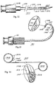
- the needle 1201 of Fig.12 has spherical indentations 1202 in outer surface 1203 of tubular stainless steel cannula 1209.
- the indentations are grouped together in three millimeter bands 1204-1205 spaced approximately two millimeters apart about the distal end 1207 of the needle.
- the catheter 1301 of Fig.13 also has spherical indentations such as 1302 in outer surface 1303 of flexible plastic material cannula 1304.
- spherical indentations such as 1302 in outer surface 1303 of flexible plastic material cannula 1304.
- three millimeter bands 1305-1309 of the indentations are grouped together and spaced approximately two millimeters apart about distal end 1310, an exploded view of band 1309 being shown.
- the spherical surface of particles 127 comprise glass, which has a characteristic or acoustic impedance ranging from 12.0 to 12.9 X 10 6 MKS rayls as indicated by the Kinsler reference.
- the acoustic impedance difference between the plastic matrix material 125 and the glass particles 127 is much greater than that of the plastic and tissue, thereby enhancing the scattered component of the image produced by the spherical surfaces.
- the surrounding medium includes the matrix material.
- the matrix material is considered the member material having a first acoustic impedance
- the glass particles are considered a substance having a predetermined contour for establishing the interface.
- the particles can be included within the member material and, according to the invention, are either partially embedded in, or attached to or indented in, the surface of the elongated member of the device.
- the glass particles have a third acoustic impedance different from the acoustic impedance of the matrix material and surrounding biological tissue when inserted therein.
- elongated tubular member 902 comprises a stainless steel material having an acoustic or characteristic impedance in the range of 39.0 to 51.5 X 10 6 MKS rayls.
- the outer surface of the elongated member includes a plurality of spherical indentations 905. Since the acoustic impedance difference between the stainless steel material and the surrounding tissue is even greater than that of glass or plastic and tissue, the intensity of the scattered component of the image produced from the spherical indentations is further increased.
- the elongated member of device 901 has an acoustic impedance different from the biological tissue or medium in which the member is to be inserted.
- the interface is produced in one of several different manners.
- the elongated member may be extruded from one material and the spherical indentations formed or embossed in the material as the elongated member is being, for example, extruded.
- the dye would include spherical projections having the desired predetermined radius to form the indentations in the extruded material.
- Device 1001 comprising a plurality of spherical particles 1002 consisting of, for example, glass may be attached to elongated tubular member 1003 using, for example, a well-known adhesive 1004.
- Member 1003 comprises suitable plastics material.
- particles 1002 may be attached to a stainless steel tubular member 1003 by silver solder. The acoustic impedance of the particles and solder may enhance the image.
- the stent 1401 has curled ends 1402 and 1403 for positioning the stent in a body passageway such as the ureter.
- the elongated plastic tubular member 1404 of the stent includes a plurality of ports 1405 for passing fluid therethrough .
- several bands 1406 of glass particles 1407 are attached to surface 1408 of the tubular member using a well-known medical grade adhesive 1409.
- the glass particles are partially embedded in the surface of the matrix material that forms the plastic tubular member.
- the bands are approximately three millimeters in width and positioned approximately two millimeters apart at ends 1402 and 1403.
- the bands of glass particles may also be spaced along the entire length of the tubular member.
- the glass particles form an interface that is imageable with sonic imaging equipment.
- a layer of plastic material 1410 coats the particles and surface 1408.
- Medical device 1101 comprises an elongated member such as cylindrical rod or stylet wire 1102 that is inserted into a passageway or lumen of catheter 1005 for inserting and guiding the catheter into a passageway or blood vessel of a patient.
- the outside surface 1103 of the rod includes a plurality of spherical indentations 1104 for producing an image.
- Elongated member includes a material such as stainless steel with an acoustic impedance for enhancing any image produced by the spherical indentations in surface 1103.
- the elongated rod may be inserted into the lumen or passageway of a smooth outer surface catheter and inserted into a vessel of the patient and guided through the vessel with the assistance of the image produced by the indentations of the rod.
- the image produced by the indentations assists the physician in guiding the catheter and elongated rod through the passageway of the patient.
- This methodology includes directing a sonic beam toward the passageway of the patient with the device inserted therein and receiving an image from the indentations of the rod.
- the material of the rod is selected to have an acoustic impedance different from that of the surrounding medium. It is envisioned that this surrounding medium may include body fluids from the patient or air which has an acoustic impedance of approximately 428 MKS rayls.
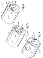
- Echogenic body member 223 forms a catheter with catheter wall 231 surrounding lumen 229.
- Lumen 229 has an inside diameter ID. In one embodiment, this internal diameter may be 0.1 cms (0.040 inches).
- the outside diameter OD of member 223 is 0.165 cms (0.065 inches).
- the outside diameter X of a typical microsphere is 5 ⁇ m. Reflective particles 227 can be totally embedded in matrix material 225, as well as being partially embedded on the outer surface of the catheter.
- Echogenic body member 323 (Fig. 3) is a two lumen catheter with lumens 329a and 329b being disposed in catheter wall 331.
- a multitude of ultrasonic reflective particles 327 can be totally embedded in matrix material 325, as well as being partially embedded in the outer surface of the catheter.
- Echogenic body member 423 (Fig. 4) is a triple lumen catheter having lumens 429a, 429b and 429c within catheter wall 431. Ultrasonic reflective particles 427 are shown in matrix 425.
- Catheter wall 731 (Fig. 7) supports echogenic body member 723.
- Member 723 is a composite as described above, with the matrix material being painted on adhesive with sound reflective particles ( 727 therein.
- Lumen 729 is in catheter wall 731.
- the ultrasonic reflective body is painted onto only a portion of the catheter as an annular stripe which is to be imaged.
- the composite material is made up of between about 5% and 30% of the sound reflective particles by volume.
- One embodiment comprises about 10% by volume of particles.
- Another has about 2 or 3% by volume particles.
- one percent or even a fraction of one percent, and up to 60% by volume of the ultrasonic reflective particles have been tested and determined to be acceptable for at least some applications. Nevertheless, as the percentage volume increases, the amount of matrix material cohesively bonding the particles together is reduced. Accordingly, there ultimately occur trade-offs in terms of flexibilty, durability and cohesiveness.
- Even ranges of less than 5% volume of particles may be utilized in specific applications.
- Certain medical instruments such as an echogenic ruler may utilize the composite material of the present invention only in selected localized positions on the medical device.
- the matrix material may be a glue or other compound which can be painted or otherwise applied to localized regions on the device where only that region of the device needs to be imaged echogenically (see e.g. Fig.7). It is noteworthy that in at least certain applications, such as catheters, where the sound reflective particles comprise about 30% of the volume of the composite material, no significant loss in tensile strength was detected.
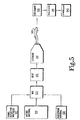
- Matrix material 525 may comprise plastic pellets which may be mixed with sound reflective particles 527 in the mixing step 533. Mixing may occur by gravity feed of the parts to be mixed into a screw or worm-gear type mechanism well known in extruder machines such as are used for catheter manufacture.
- radiopaque material 528 may also be mixed with the matrix material and the sound reflective particles.
- the radiopaque material may be one of numerous radiopaque materials already known, as for example, barium, bismuth or tungsten.
- the radiopacifier may be finely ground powder mixed during the mixing step 533. Before, during or after the mixing step 533 the mixture may be heated in the heating step 535.
- the heating maintains the matrix material in a molten or liquid state allowing it to be formed to the desired shape.
- the composite mixture is formed into an echogenic body member 523, including the sound reflective particles from 527 embedded in the matrix material.
- echogenic body 523 is a tubular catheter body having a longitudinal lumen as previously described. Other types of forming may be used, such as molding or other such shaping.
- the echogenic body member may be cut and/or shaped, as for example, cut into a specified length and/or cutting side drainage lumens, curling, tapering, or other such processes known in the plastics industry and in the catheter industry.
- the medical device is packaged during the packaging step 541, preferably hermedically sealed as is known to maintain the medical device in a surgically sterile condition.
- the medical device may be sterilized during the sterilizing step 543, using heat, chemicals or other known techniques.
- Fig.6 shows an echogenic medical device 221, such as a tubular catheter, inserted surgically into medical patient 645.
- Device 221 is sonically imaged using imaging device 647 with probe 651 to create image 649. This involves placing a device in the sonic imaging beam or field of the probe as illustrated and using ultrasonic imaging equipment, to produce image 649. and identifiable component of the image. Naturally occurring crystalline structures are also contemplated.
Landscapes
- Health & Medical Sciences (AREA)
- Life Sciences & Earth Sciences (AREA)
- Heart & Thoracic Surgery (AREA)
- Surgery (AREA)
- Animal Behavior & Ethology (AREA)
- General Health & Medical Sciences (AREA)
- Public Health (AREA)
- Veterinary Medicine (AREA)
- Engineering & Computer Science (AREA)
- Biomedical Technology (AREA)
- Molecular Biology (AREA)
- Physics & Mathematics (AREA)
- Pathology (AREA)
- Nuclear Medicine, Radiotherapy & Molecular Imaging (AREA)
- Biophysics (AREA)
- Medical Informatics (AREA)
- Vascular Medicine (AREA)
- Environmental Sciences (AREA)
- Radiology & Medical Imaging (AREA)
- Epidemiology (AREA)
- Pulmonology (AREA)
- Oral & Maxillofacial Surgery (AREA)
- Biodiversity & Conservation Biology (AREA)
- Animal Husbandry (AREA)
- Optics & Photonics (AREA)
- Marine Sciences & Fisheries (AREA)
- Anesthesiology (AREA)
- Hematology (AREA)
- Ultra Sonic Daignosis Equipment (AREA)
- Media Introduction/Drainage Providing Device (AREA)
- Measurement Of Velocity Or Position Using Acoustic Or Ultrasonic Waves (AREA)
- Materials For Medical Uses (AREA)
- Apparatus For Radiation Diagnosis (AREA)
Claims (9)
- Eine in einem Medium eines Patienten zu positionierende und mit Ultraschallgeräten verwendbare, ein Echo erzeugende medizinische Vorrichtung, die ein Körperglied (123) mit einem Bildverbesserungsmittel (127) zum Verbessern der Ultraschallabbildung der Vorrichtung umfaßt, wobei das Verbesserungsmittel mehrere diskrete akustische Stoßstellen (127) mit einer kugelförmigen Oberfläche zur Verbesserung der Ultraschallabbildung der Vorrichtung aufweist, dadurch gekennzeichnet, daß das besagte Verbesserungsmittel mit der Außenfläche (904) des Körpergliedes verbunden ist, wobei die besagten Stoßstellen die Form von auf der besagten Oberfläche befindlichen oder teilweise darin eingebetteten Teilchen (1002) oder von Einbuchtungen (905) in der Außenfläche aufweisen, wobei jedes Teilchen oder jede Einbuchtung die besagte kugel förmige Oberfläche aufweist.
- Vorrichtung nach Anspruch 1, wobei die Teilchen (127) eine sich von der des Körpers unterscheidende akustische Impedanz aufweisen.
- Vorrichtung nach Anspruch 2, wobei die Teilchen hohl sind oder fest sind.
- Vorrichtung nach Anspruch 1, wobei die Einbuchtungen eine Oberfläche aus sich von dem des Körpers unterscheidendem Material aufweisen.
- Vorrichtung nach Anspruch 1, 2, 3 oder 4, wobei die besagten Stoßstellen zufallsmäßig an der Oberfläche des Körpers angeordnet sind.
- Vorrichtung nach einem der vorhergehenden Ansprüche, wobei die Teilchenstoßstellen jeweils eine wesentlich geringere Abmessung als die in der Ultraschalleinrichtung zu benutzende Wellenlänge aufweisen.
- Vorrichtung nach Anspruch 4 oder einem der dem Anspruch 4 anhängenden Ansprüche, wobei das Körperglied eine sich vom besagten Medium unterscheidende akustische Impedanz aufweist.
- Vorrichtung nach einem der vorhergehenden Ansprüche, wobei der Wellenwiderstand des Körpers einen Unterschied zu dem des Mediums oder biologischen Gewebes aufweist, um Bildverbesserung zu erreichen, und wobei der Wellenwiderstand der Teilchen mindestens den besagten Unterschied zwischen den anderen Wellenwiderständen aufweist, wobei die Teilchen reflektierender als das Material des Körpergliedes sind.
- Vorrichtung nach einem der vorhergehenden Ansprüche, wobei die Stoßstellen Durchmesser im Bereich von 1 bis 50 Mikrometer aufweisen.
Applications Claiming Priority (5)
| Application Number | Priority Date | Filing Date | Title |
|---|---|---|---|
| US32096789A | 1989-03-09 | 1989-03-09 | |
| US320967 | 1989-03-09 | ||
| US383174 | 1989-07-20 | ||
| US07/383,174 US5081997A (en) | 1989-03-09 | 1989-07-20 | Echogenic devices, material and method |
| CA002022464A CA2022464C (en) | 1989-03-09 | 1990-08-01 | Echogenic devices, material and method |
Publications (3)
| Publication Number | Publication Date |
|---|---|
| EP0386936A1 EP0386936A1 (de) | 1990-09-12 |
| EP0386936B1 EP0386936B1 (de) | 1995-11-08 |
| EP0386936B2 true EP0386936B2 (de) | 2001-11-21 |
Family
ID=27168782
Family Applications (1)
| Application Number | Title | Priority Date | Filing Date |
|---|---|---|---|
| EP90302170A Expired - Lifetime EP0386936B2 (de) | 1989-03-09 | 1990-02-27 | Vorrichtung zur Steigerung von Ultraschall-Echos |
Country Status (8)
| Country | Link |
|---|---|
| US (1) | US5081997A (de) |
| EP (1) | EP0386936B2 (de) |
| JP (1) | JP2844238B2 (de) |
| AT (1) | ATE129873T1 (de) |
| AU (1) | AU635521B2 (de) |
| CA (1) | CA2022464C (de) |
| DE (1) | DE69023367T3 (de) |
| ES (1) | ES2078302T5 (de) |
Cited By (8)
| Publication number | Priority date | Publication date | Assignee | Title |
|---|---|---|---|---|
| US7235084B2 (en) | 2000-04-07 | 2007-06-26 | Image-Guided Neurologics, Inc. | Deep organ access device and method |
| US7497863B2 (en) | 2004-12-04 | 2009-03-03 | Medtronic, Inc. | Instrument guiding stage apparatus and method for using same |
| US7559935B2 (en) | 2003-02-20 | 2009-07-14 | Medtronic, Inc. | Target depth locators for trajectory guide for introducing an instrument |
| US7636596B2 (en) | 2002-12-20 | 2009-12-22 | Medtronic, Inc. | Organ access device and method |
| US7637915B2 (en) | 2000-08-17 | 2009-12-29 | Medtronic, Inc. | Trajectory guide with instrument immobilizer |
| US7658879B2 (en) | 2003-02-20 | 2010-02-09 | Medtronic, Inc. | Trajectory guide with angled or patterned guide lumens or height adjustment |
| US7704260B2 (en) | 2002-09-17 | 2010-04-27 | Medtronic, Inc. | Low profile instrument immobilizer |
| US7744606B2 (en) | 2004-12-04 | 2010-06-29 | Medtronic, Inc. | Multi-lumen instrument guide |
Families Citing this family (252)
| Publication number | Priority date | Publication date | Assignee | Title |
|---|---|---|---|---|
| US5201314A (en) * | 1989-03-09 | 1993-04-13 | Vance Products Incorporated | Echogenic devices, material and method |
| US5289831A (en) * | 1989-03-09 | 1994-03-01 | Vance Products Incorporated | Surface-treated stent, catheter, cannula, and the like |
| US5203337A (en) * | 1991-05-08 | 1993-04-20 | Brigham And Women's Hospital, Inc. | Coronary artery imaging system |
| JP3144849B2 (ja) * | 1991-09-30 | 2001-03-12 | 株式会社東芝 | 循環器用診断装置 |
| US5344640A (en) * | 1991-10-22 | 1994-09-06 | Mallinckrodt Medical, Inc. | Preparation of apatite particles for medical diagnostic imaging |
| ATE166582T1 (de) * | 1992-04-21 | 1998-06-15 | Vance Products Inc | Oberflächenbehandelte medizinische vorrichtungen |
| US5327891A (en) * | 1992-07-30 | 1994-07-12 | Rammler David H | Catheter track and catheter for diagnosis and treatment |
| DE4240722C2 (de) * | 1992-12-03 | 1996-08-29 | Siemens Ag | Gerät für die Behandlung von pathologischem Gewebe |
| US5383466A (en) * | 1993-05-14 | 1995-01-24 | Becton, Dickinson And Company | Instrument having enhanced ultrasound visibility |
| FR2716266A1 (fr) * | 1994-02-16 | 1995-08-18 | Bargoin Roger | Procédé de repérage d'un corps par exploration ondulatoire, du type échographie Doppler, et corps à éléments cristallins correspondant. |
| US5647374A (en) * | 1994-12-30 | 1997-07-15 | North American Scientific | Needle for imaging and sampling |
| US5766135A (en) * | 1995-03-08 | 1998-06-16 | Terwilliger; Richard A. | Echogenic needle tip |
| US5769795A (en) * | 1995-03-08 | 1998-06-23 | Terwilliger; Richard A. | Echogenic needle |
| US5611345A (en) * | 1995-04-24 | 1997-03-18 | Hibbeln; John F. | Medical instrument with improved ultrasonic visibility |
| US5782764A (en) * | 1995-11-07 | 1998-07-21 | Iti Medical Technologies, Inc. | Fiber composite invasive medical instruments and methods for use in interventional imaging procedures |
| GB9622711D0 (en) | 1996-10-31 | 1997-01-08 | British Tech Group | Instrument having enhanced ultrasound visibility |
| US5759154A (en) | 1996-12-23 | 1998-06-02 | C. R. Bard, Inc. | Print mask technique for echogenic enhancement of a medical device |
| US5993463A (en) | 1997-05-15 | 1999-11-30 | Regents Of The University Of Minnesota | Remote actuation of trajectory guide |
| US6752812B1 (en) | 1997-05-15 | 2004-06-22 | Regent Of The University Of Minnesota | Remote actuation of trajectory guide |
| US8668737B2 (en) | 1997-10-10 | 2014-03-11 | Senorx, Inc. | Tissue marking implant |
| US7637948B2 (en) * | 1997-10-10 | 2009-12-29 | Senorx, Inc. | Tissue marking implant |
| US6053870A (en) * | 1997-11-08 | 2000-04-25 | Angiodynamics, Inc. | Ultrasonic visible surgical needle |
| US20040199202A1 (en) * | 1997-11-12 | 2004-10-07 | Genesis Technologies Llc | Biological passageway occlusion removal |
| DE69839888D1 (de) * | 1997-11-12 | 2008-09-25 | Genesis Technologies Llc | Vorrichtung zum entfernen von okklusionen in biologischen durchgängen |
| US20040260333A1 (en) * | 1997-11-12 | 2004-12-23 | Dubrul William R. | Medical device and method |
| US9498604B2 (en) | 1997-11-12 | 2016-11-22 | Genesis Technologies Llc | Medical device and method |
| US20100030256A1 (en) * | 1997-11-12 | 2010-02-04 | Genesis Technologies Llc | Medical Devices and Methods |
| WO1999039649A1 (en) * | 1998-02-10 | 1999-08-12 | Dubrul William R | Occlusion, anchoring, tensioning and flow direction apparatus and methods for use |
| US6347241B2 (en) * | 1999-02-02 | 2002-02-12 | Senorx, Inc. | Ultrasonic and x-ray detectable biopsy site marker and apparatus for applying it |
| US5967988A (en) * | 1998-04-08 | 1999-10-19 | Medtronic, Inc. | Catheter having echogenicity enhancement |
| US6161034A (en) * | 1999-02-02 | 2000-12-12 | Senorx, Inc. | Methods and chemical preparations for time-limited marking of biopsy sites |
| US5921933A (en) * | 1998-08-17 | 1999-07-13 | Medtronic, Inc. | Medical devices with echogenic coatings |
| US6195577B1 (en) | 1998-10-08 | 2001-02-27 | Regents Of The University Of Minnesota | Method and apparatus for positioning a device in a body |
| US6340368B1 (en) | 1998-10-23 | 2002-01-22 | Medtronic Inc. | Implantable device with radiopaque ends |
| US6254601B1 (en) * | 1998-12-08 | 2001-07-03 | Hysterx, Inc. | Methods for occlusion of the uterine arteries |
| EP1554990A3 (de) | 1999-01-29 | 2008-08-06 | Medtronic, Inc. | Implantierbare medizinische Vorrichtung mit verbesserter Biokompatibilität und Biostabilität |
| US6725083B1 (en) | 1999-02-02 | 2004-04-20 | Senorx, Inc. | Tissue site markers for in VIVO imaging |
| US8361082B2 (en) * | 1999-02-02 | 2013-01-29 | Senorx, Inc. | Marker delivery device with releasable plug |
| US7651505B2 (en) | 2002-06-17 | 2010-01-26 | Senorx, Inc. | Plugged tip delivery for marker placement |
| US9820824B2 (en) | 1999-02-02 | 2017-11-21 | Senorx, Inc. | Deployment of polysaccharide markers for treating a site within a patent |
| US6862470B2 (en) | 1999-02-02 | 2005-03-01 | Senorx, Inc. | Cavity-filling biopsy site markers |
| US20080039819A1 (en) * | 2006-08-04 | 2008-02-14 | Senorx, Inc. | Marker formed of starch or other suitable polysaccharide |
| US20090030309A1 (en) * | 2007-07-26 | 2009-01-29 | Senorx, Inc. | Deployment of polysaccharide markers |
| US8498693B2 (en) * | 1999-02-02 | 2013-07-30 | Senorx, Inc. | Intracorporeal marker and marker delivery device |
| US7983734B2 (en) | 2003-05-23 | 2011-07-19 | Senorx, Inc. | Fibrous marker and intracorporeal delivery thereof |
| US6361557B1 (en) | 1999-02-05 | 2002-03-26 | Medtronic Ave, Inc. | Staplebutton radiopaque marker |
| WO2000051136A1 (en) * | 1999-02-25 | 2000-08-31 | Nycomed Amersham Plc | Medical tools and devices with improved ultrasound visibility |
| US6325136B1 (en) | 1999-04-15 | 2001-12-04 | Water Gremlin-Co. | Bismuth and bismuth alloy fishing sinker |
| US6689142B1 (en) * | 1999-04-26 | 2004-02-10 | Scimed Life Systems, Inc. | Apparatus and methods for guiding a needle |
| DE19921088C2 (de) * | 1999-04-30 | 2003-08-07 | Magforce Applic Gmbh | Stent zur Offenhaltung gangartiger Strukturen |
| JP3989729B2 (ja) | 1999-05-26 | 2007-10-10 | ジーイー・ヘルスケア・リミテッド | 滅菌放射活性シード |
| US6575991B1 (en) | 1999-06-17 | 2003-06-10 | Inrad, Inc. | Apparatus for the percutaneous marking of a lesion |
| US6527752B1 (en) | 1999-09-24 | 2003-03-04 | Cook Urological, Inc. | Embryo transfer catheter |
| US6358211B1 (en) | 1999-10-12 | 2002-03-19 | Scimed Life Systems, Inc. | Ultrasound lucent apparatus and methods of using |
| US6506156B1 (en) | 2000-01-19 | 2003-01-14 | Vascular Control Systems, Inc | Echogenic coating |
| US6577904B1 (en) | 2000-03-30 | 2003-06-10 | Cardiac Pacemakers, Inc. | Ultrasound echogenic cardiac lead |
| US20030120306A1 (en) * | 2000-04-21 | 2003-06-26 | Vascular Control System | Method and apparatus for the detection and occlusion of blood vessels |
| US7223279B2 (en) * | 2000-04-21 | 2007-05-29 | Vascular Control Systems, Inc. | Methods for minimally-invasive, non-permanent occlusion of a uterine artery |
| US6550482B1 (en) * | 2000-04-21 | 2003-04-22 | Vascular Control Systems, Inc. | Methods for non-permanent occlusion of a uterine artery |
| GB0011581D0 (en) | 2000-05-15 | 2000-07-05 | Nycomed Amersham Plc | Grooved brachytherapy |
| GB0011568D0 (en) | 2000-05-15 | 2000-06-28 | Nycomed Amersham Plc | Grooved medical devices |
| FR2810247B1 (fr) * | 2000-06-14 | 2008-07-25 | Prodimed | Dispositif echogene et/ou radio opaque pour le prelevement ou le transfert dans les organes genitaux |
| DE10050199A1 (de) * | 2000-10-11 | 2002-04-25 | Ethicon Gmbh | Flächiges Implantat mit im Ultraschall detektierbaren Elementen |
| US6638286B1 (en) | 2000-11-16 | 2003-10-28 | Vascular Control Systems, Inc. | Doppler directed suture ligation device and method |
| US6635065B2 (en) * | 2000-11-16 | 2003-10-21 | Vascular Control Systems, Inc. | Doppler directed suture ligation device and method |
| CA2659484C (en) * | 2000-11-20 | 2013-01-08 | Senorx, Inc. | Tissue site markers for in vivo imaging |
| US7014610B2 (en) * | 2001-02-09 | 2006-03-21 | Medtronic, Inc. | Echogenic devices and methods of making and using such devices |
| JP4227415B2 (ja) | 2001-03-28 | 2009-02-18 | ヴァスキュラー・コントロール・システムズ・インコーポレーテッド | 子宮動脈を検知及び結紮する方法及び装置 |
| US20030120286A1 (en) * | 2001-03-28 | 2003-06-26 | Vascular Control System | Luminal clip applicator with sensor |
| US7354444B2 (en) * | 2001-03-28 | 2008-04-08 | Vascular Control Systems, Inc. | Occlusion device with deployable paddles for detection and occlusion of blood vessels |
| US6921406B1 (en) | 2001-04-19 | 2005-07-26 | The Ohio State University | Stereotactic apparatus and methods |
| US6723052B2 (en) | 2001-06-07 | 2004-04-20 | Stanley L. Mills | Echogenic medical device |
| GB0120645D0 (en) * | 2001-08-24 | 2001-10-17 | Smiths Group Plc | Medico-surgical devices |
| GB0123596D0 (en) * | 2001-10-02 | 2001-11-21 | Smiths Group Plc | Medico-surgical devices |
| US7065394B2 (en) * | 2001-12-12 | 2006-06-20 | Medtronic, Inc | Guide catheter |
| US20040073158A1 (en) * | 2001-12-12 | 2004-04-15 | Medtronic, Inc. | Guide catheter |
| US7169155B2 (en) * | 2001-12-14 | 2007-01-30 | Scimed Life Systems, Inc. | Methods and apparatus for guiding a needle |
| US7527610B2 (en) * | 2002-03-29 | 2009-05-05 | Bioform Medical, Inc. | Connection indicator for a medical delivery/extraction system |
| US7303551B2 (en) | 2002-03-29 | 2007-12-04 | Bioform, Inc. | Medical delivery/extraction system |
| US7341576B2 (en) | 2002-03-29 | 2008-03-11 | Bioform Medical, Inc. | Medical delivery/extraction system |
| US7207996B2 (en) * | 2002-04-04 | 2007-04-24 | Vascular Control Systems, Inc. | Doppler directed suturing and compression device and method |
| US20040049164A1 (en) * | 2002-09-06 | 2004-03-11 | Bioform, Inc. | System including a tapered entry into an injection needle |
| US20060036158A1 (en) * | 2003-11-17 | 2006-02-16 | Inrad, Inc. | Self-contained, self-piercing, side-expelling marking apparatus |
| US20040097961A1 (en) * | 2002-11-19 | 2004-05-20 | Vascular Control System | Tenaculum for use with occlusion devices |
| US7172603B2 (en) * | 2002-11-19 | 2007-02-06 | Vascular Control Systems, Inc. | Deployable constrictor for uterine artery occlusion |
| US7404821B2 (en) * | 2003-01-30 | 2008-07-29 | Vascular Control Systems, Inc. | Treatment for post partum hemorrhage |
| US7651511B2 (en) * | 2003-02-05 | 2010-01-26 | Vascular Control Systems, Inc. | Vascular clamp for caesarian section |
| US7333844B2 (en) * | 2003-03-28 | 2008-02-19 | Vascular Control Systems, Inc. | Uterine tissue monitoring device and method |
| GB0307350D0 (en) | 2003-03-29 | 2003-05-07 | Smiths Group Plc | Catheters |
| US20040202694A1 (en) * | 2003-04-11 | 2004-10-14 | Vascular Control Systems, Inc. | Embolic occlusion of uterine arteries |
| AU2004240592A1 (en) * | 2003-05-15 | 2004-12-02 | Applied Medical Resources Corporation | Echogenic stent |
| USD496728S1 (en) | 2003-05-16 | 2004-09-28 | Kimberly-Clark Worldwide, Inc. | Cannula having band markings |
| US20050119562A1 (en) * | 2003-05-23 | 2005-06-02 | Senorx, Inc. | Fibrous marker formed of synthetic polymer strands |
| US7877133B2 (en) | 2003-05-23 | 2011-01-25 | Senorx, Inc. | Marker or filler forming fluid |
| US20040260269A1 (en) * | 2003-06-23 | 2004-12-23 | Yair Assaf | Method for treating false aneurysm and a needle for use in such a method |
| US7537788B2 (en) * | 2003-07-25 | 2009-05-26 | Rubicor Medical, Inc. | Post-biopsy cavity treatment implants and methods |
| US7744852B2 (en) | 2003-07-25 | 2010-06-29 | Rubicor Medical, Llc | Methods and systems for marking post biopsy cavity sites |
| US20050020899A1 (en) * | 2003-07-25 | 2005-01-27 | Rubicor Medical, Inc. | Post-biopsy cavity treatmetn implants and methods |
| USD669577S1 (en) | 2003-11-05 | 2012-10-23 | Kimberly-Clark Worldwide, Inc. | Cannula with band markings |
| US20050273002A1 (en) * | 2004-06-04 | 2005-12-08 | Goosen Ryan L | Multi-mode imaging marker |
| US7325546B2 (en) * | 2003-11-20 | 2008-02-05 | Vascular Control Systems, Inc. | Uterine artery occlusion device with cervical receptacle |
| US7686817B2 (en) * | 2003-11-25 | 2010-03-30 | Vascular Control Systems, Inc. | Occlusion device for asymmetrical uterine artery anatomy |
| US7637904B2 (en) * | 2003-12-19 | 2009-12-29 | Vance Products Incorporated | Catheter with snap on feature |
| EP1559379A1 (de) * | 2004-01-31 | 2005-08-03 | Labotect Verwaltungs GmbH | Vorrichtung zum Setzen von Markern in ein Gewebe |
| US7580756B2 (en) | 2004-02-13 | 2009-08-25 | Medtronic, Inc. | Methods and apparatus for securing a therapy delivery device within a burr hole |
| JP2005278939A (ja) * | 2004-03-30 | 2005-10-13 | Jms Co Ltd | スタイレット |
| JP2006061258A (ja) * | 2004-08-25 | 2006-03-09 | Jfe Steel Kk | 医療用セラミック被覆針を用いた医療処置方法およびセラミック被覆穿刺針を用いた医療用検体の採取方法 |
| WO2006044374A1 (en) * | 2004-10-14 | 2006-04-27 | Cook Incorporated | Echogenic medical device and method of forming echogenic surface |
| US7875036B2 (en) * | 2004-10-27 | 2011-01-25 | Vascular Control Systems, Inc. | Short term treatment for uterine disorder |
| US20060205073A1 (en) * | 2005-03-08 | 2006-09-14 | Vance Products Incorporated, D/B/A Cook Urological Incorporated | Aspirating oocytes from transplanted ovarian tissue |
| US10357328B2 (en) | 2005-04-20 | 2019-07-23 | Bard Peripheral Vascular, Inc. and Bard Shannon Limited | Marking device with retractable cannula |
| AU2006247571A1 (en) * | 2005-05-13 | 2006-11-23 | Cook Incorporated | Medical device delivery systems that facilitate medical device placement in the presence of ultrasonic waves |
| WO2007013130A1 (ja) * | 2005-07-25 | 2007-02-01 | Hakko Co., Ltd. | 超音波用穿刺針 |
| US20070049973A1 (en) * | 2005-08-29 | 2007-03-01 | Vascular Control Systems, Inc. | Method and device for treating adenomyosis and endometriosis |
| WO2007033206A2 (en) | 2005-09-13 | 2007-03-22 | Veran Medical Technologies, Inc. | Apparatus and method for image guided accuracy verification |
| US20070066881A1 (en) * | 2005-09-13 | 2007-03-22 | Edwards Jerome R | Apparatus and method for image guided accuracy verification |
| US8052658B2 (en) | 2005-10-07 | 2011-11-08 | Bard Peripheral Vascular, Inc. | Drug-eluting tissue marker |
| US7815659B2 (en) * | 2005-11-15 | 2010-10-19 | Ethicon Endo-Surgery, Inc. | Suture anchor applicator |
| US8430863B2 (en) * | 2005-12-02 | 2013-04-30 | Abbott Cardiovascular Systems Inc. | Visualization of a catheter viewed under ultrasound imaging |
| US8382674B2 (en) | 2005-12-02 | 2013-02-26 | Abbott Cardiovascular Systems Inc. | Visualization of a catheter viewed under ultrasound imaging |
| US20080230001A1 (en) * | 2006-02-23 | 2008-09-25 | Meadwestvaco Corporation | Method for treating a substrate |
| US8214022B2 (en) * | 2006-06-27 | 2012-07-03 | Cook Medical Technologies Llc | Hysterography and intrauterine venography catheter |
| US7799022B2 (en) * | 2006-07-06 | 2010-09-21 | Boston Scientific Scimed, Inc. | Ablation with echogenic insulative sheath |
| US20090171198A1 (en) * | 2006-08-04 | 2009-07-02 | Jones Michael L | Powdered marker |
| US7945307B2 (en) * | 2006-08-04 | 2011-05-17 | Senorx, Inc. | Marker delivery system with obturator |
| WO2008062451A2 (en) * | 2006-09-07 | 2008-05-29 | Gautam Allahbadia | Echogenic surface for surgical instrument |
| US11666377B2 (en) | 2006-09-29 | 2023-06-06 | Boston Scientific Medical Device Limited | Electrosurgical device |
| US12161390B2 (en) | 2006-09-29 | 2024-12-10 | Boston Scientific Medical Device Limited | Connector system for electrosurgical device |
| US8064987B2 (en) | 2006-10-23 | 2011-11-22 | C. R. Bard, Inc. | Breast marker |
| EP3542748B1 (de) | 2006-12-12 | 2023-08-16 | C. R. Bard, Inc. | Gewebemarker mit mehreren bildgebungsmodi |
| EP2101670B1 (de) | 2006-12-18 | 2013-07-31 | C.R.Bard, Inc. | Biopsiemarker mit in situ erzeugten bildgebungseigenschaften |
| US8864781B2 (en) * | 2007-02-28 | 2014-10-21 | Cook Medical Technologies Llc | Intestinal bypass using magnets |
| US20090105719A1 (en) * | 2007-10-19 | 2009-04-23 | Vance Products Incorporated, D/B/A Cook Urological Incorporated | Precision stent positioner |
| GB0722406D0 (en) | 2007-11-15 | 2007-12-27 | Smiths Group Plc | Medico-surgical assemblies and methods |
| US8311610B2 (en) * | 2008-01-31 | 2012-11-13 | C. R. Bard, Inc. | Biopsy tissue marker |
| DE102008025878A1 (de) | 2008-05-29 | 2009-12-10 | Pajunk Gmbh & Co. Kg Besitzverwaltung | Kanüle, insbesondere für die Regionalanästhesie |
| WO2010033406A2 (en) * | 2008-09-18 | 2010-03-25 | University Of Florida Research Foundation, Inc. | Needle having ultrasound opaque elements |
| US9327061B2 (en) * | 2008-09-23 | 2016-05-03 | Senorx, Inc. | Porous bioabsorbable implant |
| US20100160731A1 (en) * | 2008-12-22 | 2010-06-24 | Marc Giovannini | Ultrasound-visualizable endoscopic access system |
| CA2742765C (en) * | 2008-12-30 | 2016-04-12 | C.R. Bard Inc. | Marker delivery device for tissue marker placement |
| US8500688B2 (en) | 2009-04-16 | 2013-08-06 | Medtronic, Inc. | Retrograde coronary sinus perfusion cannula and methods of using same |
| WO2010151382A1 (en) * | 2009-06-26 | 2010-12-29 | Wilson-Cook Medical Inc. | Linear clamps for anastomosis |
| DE102009031769A1 (de) * | 2009-06-30 | 2011-01-05 | Aesculap Ag | Chirurgisches Instrument |
| US8522787B2 (en) * | 2009-07-29 | 2013-09-03 | Covidien Lp | Ultrasound-based tracheal tube placement device and method |
| WO2011056445A1 (en) | 2009-11-03 | 2011-05-12 | Wilson-Cook Medical Inc. | Planar clamps for anastomosis |
| US8480592B2 (en) | 2009-12-23 | 2013-07-09 | C. R. Bard, Inc. | Biopsy probe mechanism having multiple echogenic features |
| WO2011130388A1 (en) | 2010-04-14 | 2011-10-20 | Surti Vihar C | System for creating anastomoses |
| US10568628B2 (en) | 2017-05-23 | 2020-02-25 | Muffin Incorporated | Closing device for tissue openings |
| WO2011137224A1 (en) | 2010-04-29 | 2011-11-03 | Danny Azriel Sherwinter | Systems and methods for facilitating closure of bodily openings |
| US10335577B2 (en) | 2010-05-19 | 2019-07-02 | Nfinium Vascular Technologies, Llc | Augmented delivery catheter and method |
| US9561094B2 (en) | 2010-07-23 | 2017-02-07 | Nfinium Vascular Technologies, Llc | Devices and methods for treating venous diseases |
| US8903473B2 (en) * | 2010-09-15 | 2014-12-02 | Medtronic, Inc. | Radiopaque markers for implantable medical devices |
| US20120065500A1 (en) * | 2010-09-15 | 2012-03-15 | Medtronic, Inc. | Radiopaque embedded into desiccant for implantable medical device |
| US9254146B2 (en) | 2010-10-18 | 2016-02-09 | Avent, Inc. | Echogenic nerve block apparatus and system |
| WO2014141105A1 (en) | 2013-03-12 | 2014-09-18 | Baylis Medical Company Inc. | Medical device having a support structure |
| WO2012138626A1 (en) | 2011-04-05 | 2012-10-11 | Cook Medical Technologies Llc | Fallopian tube filter |
| CN103501844A (zh) | 2011-04-08 | 2014-01-08 | 库克医药技术有限责任公司 | 鞘可缩回的柔性注射针 |
| US9216258B2 (en) | 2011-04-12 | 2015-12-22 | Cook Medical Technologies, LLC | Apparatus for accurately controlling needle extension |
| US20140051989A1 (en) * | 2011-04-19 | 2014-02-20 | Drexel University | Devices adapted for ultrasound location in patients and method of use |
| EP2699312A1 (de) * | 2011-04-20 | 2014-02-26 | Cardiac Pacemakers, Inc. | Ultraschallüberwachung von implantierbaren medizinprodukten |
| WO2012148265A1 (en) | 2011-04-26 | 2012-11-01 | Encapson B.V. | Coating for improving the ultrasound visibility |
| WO2013065036A2 (en) * | 2011-11-01 | 2013-05-10 | Corassist Cardiovascular Ltd. | A system and method for assisting the positioning of medical instruments |
| US9173657B2 (en) | 2011-12-15 | 2015-11-03 | Ethicon Endo-Surgery, Inc. | Devices and methods for endoluminal plication |
| US9113867B2 (en) | 2011-12-15 | 2015-08-25 | Ethicon Endo-Surgery, Inc. | Devices and methods for endoluminal plication |
| DE102012100292A1 (de) | 2012-01-13 | 2013-07-18 | Rm Temena Gmbh | Medizinische Vorrichtung |
| US10940167B2 (en) | 2012-02-10 | 2021-03-09 | Cvdevices, Llc | Methods and uses of biological tissues for various stent and other medical applications |
| EP2816966B1 (de) | 2012-02-22 | 2023-10-25 | Veran Medical Technologies, Inc. | Lenkbarer chirurgischer katheter mit einer biopsievorrichtung an seinem distalen endteil |
| US9445837B2 (en) | 2012-03-16 | 2016-09-20 | Nfinium Vascular Technologies Llc | Surgical needle with enhanced ultrasound reflectivity |
| US8992547B2 (en) | 2012-03-21 | 2015-03-31 | Ethicon Endo-Surgery, Inc. | Methods and devices for creating tissue plications |
| US9821145B2 (en) | 2012-03-23 | 2017-11-21 | Pressure Products Medical Supplies Inc. | Transseptal puncture apparatus and method for using the same |
| GB2500628B (en) * | 2012-03-27 | 2016-08-10 | Cook Medical Technologies Llc | Medical balloon with particles therein |
| JP6416084B2 (ja) | 2012-05-31 | 2018-10-31 | ベイリス メディカル カンパニー インコーポレイテッドBaylis Medical Company Inc. | 医療機器 |
| US9066681B2 (en) | 2012-06-26 | 2015-06-30 | Covidien Lp | Methods and systems for enhancing ultrasonic visibility of energy-delivery devices within tissue |
| US9332959B2 (en) | 2012-06-26 | 2016-05-10 | Covidien Lp | Methods and systems for enhancing ultrasonic visibility of energy-delivery devices within tissue |
| US20140010043A1 (en) * | 2012-07-04 | 2014-01-09 | Stanley Jerome Pawlik | Portable Sonar Imaging System and Method |
| AU2013338772B2 (en) * | 2012-10-31 | 2016-09-29 | Encapson B.V. | Medical devices with coatings for enhanced echogenicity |
| KR102283240B1 (ko) | 2012-11-21 | 2021-07-30 | 씨. 알. 바드, 인크. | 코어 바늘 생검 디바이스 |
| US20140228937A1 (en) | 2013-02-11 | 2014-08-14 | Joshua Krieger | Expandable Support Frame and Medical Device |
| US20140257090A1 (en) * | 2013-03-06 | 2014-09-11 | Muffin Incorporated | Echolucent guidewire tip |
| US11937873B2 (en) | 2013-03-12 | 2024-03-26 | Boston Scientific Medical Device Limited | Electrosurgical device having a lumen |
| US11224724B2 (en) | 2013-03-12 | 2022-01-18 | Teleflex Medical Incorporated | Catheter insertion device |
| US9717886B2 (en) | 2013-03-12 | 2017-08-01 | Teleflex Medical Incorporated | Safety clip for a needle |
| US10357635B2 (en) | 2013-03-12 | 2019-07-23 | Teleflex Medical Incorporated | Catheter insertion device |
| US10292677B2 (en) | 2013-03-14 | 2019-05-21 | Volcano Corporation | Endoluminal filter having enhanced echogenic properties |
| WO2014152365A2 (en) | 2013-03-14 | 2014-09-25 | Volcano Corporation | Filters with echogenic characteristics |
| US10219887B2 (en) | 2013-03-14 | 2019-03-05 | Volcano Corporation | Filters with echogenic characteristics |
| JP6263254B2 (ja) * | 2013-03-14 | 2018-01-17 | マフィン・インコーポレイテッドMuffin Incorporated | ルーローの三角形を用いたエコー源性面 |
| EP2967602B1 (de) | 2013-03-15 | 2019-08-28 | Volcano Corporation | Distalschutzsysteme mit druck- und ultraschallmerkmalen |
| CA3220441A1 (en) | 2013-03-15 | 2015-09-17 | Boston Scientific Medical Device Limited | Electrosurgical device having a distal aperture |
| US10076307B2 (en) * | 2013-06-20 | 2018-09-18 | Avent, Inc. | Echogenic article with compound indentations |
| WO2015019132A1 (en) | 2013-08-07 | 2015-02-12 | Baylis Medical Company Inc. | Methods and devices for puncturing tissue |
| USD716450S1 (en) | 2013-09-24 | 2014-10-28 | C. R. Bard, Inc. | Tissue marker for intracorporeal site identification |
| USD716451S1 (en) | 2013-09-24 | 2014-10-28 | C. R. Bard, Inc. | Tissue marker for intracorporeal site identification |
| USD715942S1 (en) | 2013-09-24 | 2014-10-21 | C. R. Bard, Inc. | Tissue marker for intracorporeal site identification |
| USD715442S1 (en) | 2013-09-24 | 2014-10-14 | C. R. Bard, Inc. | Tissue marker for intracorporeal site identification |
| US9629981B2 (en) | 2013-12-13 | 2017-04-25 | Dolcera Information Technology Services Private Limited | Drainage catheter |
| US10661057B2 (en) | 2013-12-20 | 2020-05-26 | Baylis Medical Company Inc. | Steerable medical device handle |
| US20150305650A1 (en) | 2014-04-23 | 2015-10-29 | Mark Hunter | Apparatuses and methods for endobronchial navigation to and confirmation of the location of a target tissue and percutaneous interception of the target tissue |
| US20150305612A1 (en) | 2014-04-23 | 2015-10-29 | Mark Hunter | Apparatuses and methods for registering a real-time image feed from an imaging device to a steerable catheter |
| CN106470712B (zh) | 2014-04-30 | 2020-08-04 | 恩克普森有限公司 | 具有用于增强回声反射性的非均匀涂层的医疗装置 |
| EP3151904A4 (de) | 2014-06-04 | 2018-02-14 | Nfinium Vascular Technologies, LLC | Vaskuläre vorrichtung mit niedriger radialkraft und verfahren zur okklusion |
| EP3197368B1 (de) | 2014-09-24 | 2018-11-28 | Koninklijke Philips N.V. | Endoluminaler filter mit verbesserten echogenen eigenschaften |
| EP3267942B1 (de) * | 2015-03-12 | 2022-05-04 | G&G Biotechnology Ltd. | Implantatmaterial aus verbundstoff |
| AU2015399045B2 (en) | 2015-06-18 | 2020-06-25 | Avent, Inc. | Echogenic coil member for a catheter assembly |
| AU2015399046B2 (en) | 2015-06-18 | 2020-05-28 | Avent, Inc. | Echogenic catheter member |
| US11779306B2 (en) | 2015-07-21 | 2023-10-10 | Avent, Inc. | Ultrasonic catheter assembly |
| US11324548B2 (en) | 2015-08-21 | 2022-05-10 | Baylis Medical Company Inc. | Transvascular electrosurgical devices and systems and methods of using the same |
| JP6758612B2 (ja) * | 2015-09-01 | 2020-09-23 | 株式会社北里コーポレーション | 胚移植用具および胚移植用器具 |
| EP3346898B1 (de) | 2015-09-09 | 2021-04-28 | Baylis Medical Company Inc. | Nadel für den epikardialen zugang umfassend eine isolierung, eine freigelegte spitze und eine seitenöffnung |
| JP2019503777A (ja) | 2016-01-07 | 2019-02-14 | ベイリス メディカル カンパニー インコーポレイテッドBaylis Medical Company Inc. | ハイブリッド経中隔拡張器及びこれを使用する方法 |
| WO2017222523A1 (en) | 2016-06-23 | 2017-12-28 | Avent, Inc. | Echogenic coil member for a catheter assembly |
| US12558155B2 (en) | 2016-11-01 | 2026-02-24 | Boston Scientific Medical Device Limited | Methods and devices for puncturing tissue |
| US12207836B2 (en) | 2016-11-01 | 2025-01-28 | Boston Scientific Medical Device Limited | Methods and devices for puncturing tissue |
| AU2018251786B2 (en) | 2017-04-13 | 2020-08-27 | Teleflex Medical Llc | Catheter insertion device |
| CN107335099B (zh) * | 2017-06-21 | 2020-08-07 | 北京化工大学 | 一种超声显影材料及其制备方法与应用 |
| EP4230122A1 (de) | 2017-08-10 | 2023-08-23 | Boston Scientific Medical Device Limited | Wärmeaustausch- und temperaturmessvorrichtung und verwendungsverfahren |
| US11224725B2 (en) | 2017-12-05 | 2022-01-18 | Baylis Medical Company Inc. | Transseptal guide wire puncture system |
| US11872356B2 (en) * | 2018-01-05 | 2024-01-16 | Becton, Dickinson And Company | Echogenic catheter and catheter system |
| JP6850370B2 (ja) * | 2018-01-30 | 2021-03-31 | 富士フイルム株式会社 | 超音波プローブ、超音波プローブ用樹脂組成物、樹脂組成物の超音波プローブのシース材料としての使用、及び超音波プローブの製造方法 |
| IL276645B2 (en) | 2018-02-18 | 2025-01-01 | G & G Biotechnology Ltd | Implants with shell with improved adhesion |
| KR102849171B1 (ko) | 2018-05-08 | 2025-08-27 | 보스턴 사이언티픽 메디칼 디바이스 리미티드 | 디바이스용 결합 메커니즘들 |
| US20200129229A1 (en) * | 2018-07-02 | 2020-04-30 | Anil K Chagarlamudi | Venous ablation catheter and method |
| ES3013237T3 (en) * | 2018-11-29 | 2025-04-11 | Kitazato Corp | Embryo transplanting tool and embryo transplanting apparatus |
| US11504546B2 (en) | 2019-02-28 | 2022-11-22 | Cowles Ventures, Llc | Needle guidance device for brachytherapy and method of use |
| US11524176B2 (en) | 2019-03-14 | 2022-12-13 | Cowles Ventures, Llc | Locator for placement of fiducial support device method |
| BR112021021876A2 (pt) | 2019-04-29 | 2021-12-28 | Baylis Medical Co Inc | Sistemas, dispositivos e métodos de punção transeptal |
| US11766298B2 (en) | 2019-05-03 | 2023-09-26 | Neil Glossop | Systems, methods, and devices for registering and tracking organs during interventional procedures |
| WO2021046163A1 (en) * | 2019-09-04 | 2021-03-11 | Edwards Lifesciences Corporation | Medical instruments with echogenicity-enhancement characteristics |
| US11759190B2 (en) | 2019-10-18 | 2023-09-19 | Boston Scientific Medical Device Limited | Lock for medical devices, and related systems and methods |
| US11801087B2 (en) | 2019-11-13 | 2023-10-31 | Boston Scientific Medical Device Limited | Apparatus and methods for puncturing tissue |
| US11724070B2 (en) | 2019-12-19 | 2023-08-15 | Boston Scientific Medical Device Limited | Methods for determining a position of a first medical device with respect to a second medical device, and related systems and medical devices |
| CA3164840A1 (en) | 2020-01-29 | 2021-08-05 | John Paul Urbanski | Guidewire for reducing hoop stress |
| US11931098B2 (en) | 2020-02-19 | 2024-03-19 | Boston Scientific Medical Device Limited | System and method for carrying out a medical procedure |
| US12082792B2 (en) | 2020-02-25 | 2024-09-10 | Boston Scientific Medical Device Limited | Systems and methods for creating a puncture between aorta and the left atrium |
| US11986209B2 (en) | 2020-02-25 | 2024-05-21 | Boston Scientific Medical Device Limited | Methods and devices for creation of communication between aorta and left atrium |
| US11819243B2 (en) | 2020-03-19 | 2023-11-21 | Boston Scientific Medical Device Limited | Medical sheath and related systems and methods |
| CN115605152A (zh) | 2020-03-20 | 2023-01-13 | 波士顿科学医疗设备有限公司(Ie) | 撕裂系统和装置以及用于撕裂的方法 |
| US12011279B2 (en) | 2020-04-07 | 2024-06-18 | Boston Scientific Medical Device Limited | Electro-anatomic mapping system |
| US11826075B2 (en) | 2020-04-07 | 2023-11-28 | Boston Scientific Medical Device Limited | Elongated medical assembly |
| EP4149379A4 (de) | 2020-05-11 | 2024-06-19 | Boston Scientific Medical Device Limited | Elektrochirurgische vorrichtung mit automatischer abschaltung |
| US12420067B2 (en) | 2020-05-12 | 2025-09-23 | Boston Scientific Medical Device Limited | Guidewire assembly |
| EP4153062A1 (de) * | 2020-05-18 | 2023-03-29 | Baylor University | Ultraschallpositionierbares chirurgisches führungsdrahtsystem und verfahren |
| US11938285B2 (en) | 2020-06-17 | 2024-03-26 | Boston Scientific Medical Device Limited | Stop-movement device for elongated medical assembly |
| AU2021291158B2 (en) | 2020-06-17 | 2023-11-30 | Boston Scientific Medical Device Limited | Electroanatomical mapping system |
| US11937796B2 (en) | 2020-06-18 | 2024-03-26 | Boston Scientific Medical Device Limited | Tissue-spreader assembly |
| US12343042B2 (en) | 2020-07-16 | 2025-07-01 | Boston Scientific Medical Device Limited | Pericardial puncture device and method |
| US12042178B2 (en) | 2020-07-21 | 2024-07-23 | Boston Scientific Medical Device Limited | System of medical devices and method for pericardial puncture |
| US12005202B2 (en) | 2020-08-07 | 2024-06-11 | Boston Scientific Medical Device Limited | Catheter having tissue-engaging device |
| US12396785B2 (en) | 2020-08-12 | 2025-08-26 | Boston Scientific Medical Device Limited | System of medical devices and method for pericardial puncture |
| CA3128527A1 (en) | 2020-09-10 | 2022-03-10 | Baylis Medical Company Inc. | Elongated medical catheter including marker band |
| US11980412B2 (en) | 2020-09-15 | 2024-05-14 | Boston Scientific Medical Device Limited | Elongated medical sheath |
| CN113940699A (zh) * | 2021-10-11 | 2022-01-18 | 电子科技大学 | 一种超声探头自定位装置及其自定位方法 |
Citations (5)
| Publication number | Priority date | Publication date | Assignee | Title |
|---|---|---|---|---|
| JPS5438160A (en) † | 1977-09-01 | 1979-03-22 | Toshiba Corp | Ultrasonic refelector |
| JPS56113510U (de) † | 1980-01-31 | 1981-09-01 | ||
| JPS6115900U (ja) † | 1984-06-29 | 1986-01-29 | 古野電気株式会社 | 超音波送受波器 |
| US4582061A (en) † | 1981-11-18 | 1986-04-15 | Indianapolis Center For Advanced Research, Inc. | Needle with ultrasonically reflective displacement scale |
| US4657896A (en) † | 1983-05-30 | 1987-04-14 | Mitsui Toatsu Chemicals, Inc. | Method for the treatment of digestive ulcers |
Family Cites Families (15)
| Publication number | Priority date | Publication date | Assignee | Title |
|---|---|---|---|---|
| US3605750A (en) * | 1969-04-07 | 1971-09-20 | David S Sheridan | X-ray tip catheter |
| DE2425724C3 (de) * | 1974-05-28 | 1979-09-27 | Siemens Ag, 1000 Berlin Und 8000 Muenchen | Punktionskanüle |
| US4322164A (en) * | 1976-10-18 | 1982-03-30 | Oximetrix, Inc. | Sterilizable, disposable optical scattering reference medium and container assembly |
| US4111190A (en) * | 1976-11-11 | 1978-09-05 | Jane Plumridge | Medical applicator assembly for chain cystourethrographic procedure |
| US4277367A (en) * | 1978-10-23 | 1981-07-07 | Wisconsin Alumni Research Foundation | Phantom material and method |
| DE3037641A1 (de) * | 1980-10-04 | 1982-05-13 | Gesellschaft für Strahlen- und Umweltforschung mbH, 8042 Neuherberg | Ultraschallsender |
| US4401124A (en) * | 1981-08-13 | 1983-08-30 | Technicare Corporation | Reflection enhancement of a biopsy needle |
| US4805628A (en) * | 1982-12-06 | 1989-02-21 | Indianapolis Center For Advanced Research, Inc. | Ultrasound contrast media for medically implantable and insertable devices |
| US4718433A (en) * | 1983-01-27 | 1988-01-12 | Feinstein Steven B | Contrast agents for ultrasonic imaging |
| US4572203A (en) * | 1983-01-27 | 1986-02-25 | Feinstein Steven B | Contact agents for ultrasonic imaging |
| US4490139A (en) * | 1983-01-28 | 1984-12-25 | Eli Lilly And Company | Implant needle and method |
| US4567896A (en) * | 1984-01-20 | 1986-02-04 | Elscint, Inc. | Method and apparatus for calibrating a biopsy attachment for ultrasonic imaging apparatus |
| JP2556839B2 (ja) * | 1986-07-11 | 1996-11-27 | 株式会社島津製作所 | 超音波用カツプリング材 |
| JPS63181877U (de) * | 1987-05-11 | 1988-11-24 | ||
| US4869259A (en) * | 1988-05-17 | 1989-09-26 | Vance Products Incorporated | Echogenically enhanced surgical instrument and method for production thereof |
-
1989
- 1989-07-20 US US07/383,174 patent/US5081997A/en not_active Expired - Lifetime
-
1990
- 1990-02-15 AU AU49829/90A patent/AU635521B2/en not_active Expired
- 1990-02-27 AT AT90302170T patent/ATE129873T1/de not_active IP Right Cessation
- 1990-02-27 ES ES90302170T patent/ES2078302T5/es not_active Expired - Lifetime
- 1990-02-27 DE DE69023367T patent/DE69023367T3/de not_active Expired - Lifetime
- 1990-02-27 EP EP90302170A patent/EP0386936B2/de not_active Expired - Lifetime
- 1990-03-08 JP JP2055208A patent/JP2844238B2/ja not_active Expired - Lifetime
- 1990-08-01 CA CA002022464A patent/CA2022464C/en not_active Expired - Lifetime
Patent Citations (5)
| Publication number | Priority date | Publication date | Assignee | Title |
|---|---|---|---|---|
| JPS5438160A (en) † | 1977-09-01 | 1979-03-22 | Toshiba Corp | Ultrasonic refelector |
| JPS56113510U (de) † | 1980-01-31 | 1981-09-01 | ||
| US4582061A (en) † | 1981-11-18 | 1986-04-15 | Indianapolis Center For Advanced Research, Inc. | Needle with ultrasonically reflective displacement scale |
| US4657896A (en) † | 1983-05-30 | 1987-04-14 | Mitsui Toatsu Chemicals, Inc. | Method for the treatment of digestive ulcers |
| JPS6115900U (ja) † | 1984-06-29 | 1986-01-29 | 古野電気株式会社 | 超音波送受波器 |
Cited By (21)
| Publication number | Priority date | Publication date | Assignee | Title |
|---|---|---|---|---|
| US7833231B2 (en) | 2000-04-07 | 2010-11-16 | Medtronic, Inc. | Device for immobilizing a primary instrument and method therefor |
| US8911452B2 (en) | 2000-04-07 | 2014-12-16 | Medtronic, Inc. | Device for immobilizing a primary instrument and method therefor |
| US8845656B2 (en) | 2000-04-07 | 2014-09-30 | Medtronic, Inc. | Device for immobilizing a primary instrument and method therefor |
| US7235084B2 (en) | 2000-04-07 | 2007-06-26 | Image-Guided Neurologics, Inc. | Deep organ access device and method |
| US7857820B2 (en) | 2000-04-07 | 2010-12-28 | Medtronic, Inc. | Sheath assembly for an access device and method therefor |
| US7815651B2 (en) | 2000-04-07 | 2010-10-19 | Medtronic, Inc. | Device for immobilizing a primary instrument and method therefor |
| US7828809B2 (en) | 2000-04-07 | 2010-11-09 | Medtronic, Inc. | Device for immobilizing a primary instrument and method therefor |
| US7637915B2 (en) | 2000-08-17 | 2009-12-29 | Medtronic, Inc. | Trajectory guide with instrument immobilizer |
| US8192445B2 (en) | 2000-08-17 | 2012-06-05 | Medtronic, Inc. | Trajectory guide with instrument immobilizer |
| US7704260B2 (en) | 2002-09-17 | 2010-04-27 | Medtronic, Inc. | Low profile instrument immobilizer |
| US7636596B2 (en) | 2002-12-20 | 2009-12-22 | Medtronic, Inc. | Organ access device and method |
| US8116850B2 (en) | 2002-12-20 | 2012-02-14 | Medtronic, Inc. | Organ access device and method |
| US7658879B2 (en) | 2003-02-20 | 2010-02-09 | Medtronic, Inc. | Trajectory guide with angled or patterned guide lumens or height adjustment |
| US7896889B2 (en) | 2003-02-20 | 2011-03-01 | Medtronic, Inc. | Trajectory guide with angled or patterned lumens or height adjustment |
| US7981120B2 (en) | 2003-02-20 | 2011-07-19 | University Of South Florida | Trajectory guide with angled or patterned guide lumens or height adjustment |
| US7699854B2 (en) | 2003-02-20 | 2010-04-20 | Medtronic, Inc. | Trajectory guide with angled or patterned guide lumens or height adjustment |
| US7559935B2 (en) | 2003-02-20 | 2009-07-14 | Medtronic, Inc. | Target depth locators for trajectory guide for introducing an instrument |
| US7803163B2 (en) | 2004-12-04 | 2010-09-28 | Medtronic, Inc. | Multiple instrument retaining assembly and methods therefor |
| US7867242B2 (en) | 2004-12-04 | 2011-01-11 | Medtronic, Inc. | Instrument for guiding stage apparatus and method for using same |
| US7744606B2 (en) | 2004-12-04 | 2010-06-29 | Medtronic, Inc. | Multi-lumen instrument guide |
| US7497863B2 (en) | 2004-12-04 | 2009-03-03 | Medtronic, Inc. | Instrument guiding stage apparatus and method for using same |
Also Published As
| Publication number | Publication date |
|---|---|
| DE69023367T3 (de) | 2002-07-11 |
| ES2078302T3 (es) | 1995-12-16 |
| CA2022464C (en) | 1995-03-21 |
| JPH02268745A (ja) | 1990-11-02 |
| AU4982990A (en) | 1990-09-13 |
| JP2844238B2 (ja) | 1999-01-06 |
| ATE129873T1 (de) | 1995-11-15 |
| ES2078302T5 (es) | 2002-08-01 |
| AU635521B2 (en) | 1993-03-25 |
| EP0386936B1 (de) | 1995-11-08 |
| DE69023367D1 (de) | 1995-12-14 |
| DE69023367T2 (de) | 1996-04-04 |
| EP0386936A1 (de) | 1990-09-12 |
| US5081997A (en) | 1992-01-21 |
Similar Documents
| Publication | Publication Date | Title |
|---|---|---|
| EP0386936B1 (de) | Vorrichtung zur Steigerung von Ultraschall-Echos | |
| CA2087627C (en) | Echogenic devices, material and method | |
| US5289831A (en) | Surface-treated stent, catheter, cannula, and the like | |
| US12350099B2 (en) | Rotational intravascular ultrasound probe and method of manufacturing the same | |
| EP0935442B1 (de) | Instrument ausgerüstet zur verbesserten abbildung mittels ultraschall | |
| US5259837A (en) | Acoustically enhanced catheter | |
| Ziskin | Fundamental physics of ultrasound and its propagation in tissue. | |
| WO1991002489A1 (en) | Intravascular ultrasonic imaging probe | |
| CA2123088A1 (en) | Transport catheter and ultrasound probe for use with same | |
| CA2246322A1 (en) | Ultrasound transmissive pad | |
| US20030065266A1 (en) | Medico-surgical devices | |
| KR102568749B1 (ko) | 심부 조직층에서 의료 장치의 초음파 이미징을 위한 에코 발생 코팅의 용도 | |
| CA2094431C (en) | Surface-treated medical devices | |
| HK1013944B (en) | Echogenic devices, material and method | |
| US7909766B2 (en) | Systems and methods for improving the imaging resolution of an imaging transducer | |
| Winkler et al. | How accurate are volume flow measurements using Doppler ultrasound from commercial systems? |
Legal Events
| Date | Code | Title | Description |
|---|---|---|---|
| PUAI | Public reference made under article 153(3) epc to a published international application that has entered the european phase |
Free format text: ORIGINAL CODE: 0009012 |
|
| AK | Designated contracting states |
Kind code of ref document: A1 Designated state(s): AT BE CH DE DK ES FR GB GR IT LI LU NL SE |
|
| 17P | Request for examination filed |
Effective date: 19901218 |
|
| 17Q | First examination report despatched |
Effective date: 19930222 |
|
| GRAA | (expected) grant |
Free format text: ORIGINAL CODE: 0009210 |
|
| AK | Designated contracting states |
Kind code of ref document: B1 Designated state(s): AT BE CH DE DK ES FR GB GR IT LI LU NL SE |
|
| PG25 | Lapsed in a contracting state [announced via postgrant information from national office to epo] |
Ref country code: DK Effective date: 19951108 Ref country code: NL Free format text: LAPSE BECAUSE OF FAILURE TO SUBMIT A TRANSLATION OF THE DESCRIPTION OR TO PAY THE FEE WITHIN THE PRESCRIBED TIME-LIMIT Effective date: 19951108 Ref country code: LI Effective date: 19951108 Ref country code: GR Free format text: LAPSE BECAUSE OF FAILURE TO SUBMIT A TRANSLATION OF THE DESCRIPTION OR TO PAY THE FEE WITHIN THE PRESCRIBED TIME-LIMIT Effective date: 19951108 Ref country code: CH Effective date: 19951108 Ref country code: BE Effective date: 19951108 Ref country code: AT Effective date: 19951108 |
|
| REF | Corresponds to: |
Ref document number: 129873 Country of ref document: AT Date of ref document: 19951115 Kind code of ref document: T |
|
| ITF | It: translation for a ep patent filed | ||
| ET | Fr: translation filed | ||
| REF | Corresponds to: |
Ref document number: 69023367 Country of ref document: DE Date of ref document: 19951214 |
|
| REG | Reference to a national code |
Ref country code: ES Ref legal event code: FG2A Ref document number: 2078302 Country of ref document: ES Kind code of ref document: T3 |
|
| PG25 | Lapsed in a contracting state [announced via postgrant information from national office to epo] |
Ref country code: SE Effective date: 19960208 |
|
| PG25 | Lapsed in a contracting state [announced via postgrant information from national office to epo] |
Ref country code: LU Free format text: LAPSE BECAUSE OF NON-PAYMENT OF DUE FEES Effective date: 19960229 |
|
| NLV1 | Nl: lapsed or annulled due to failure to fulfill the requirements of art. 29p and 29m of the patents act | ||
| REG | Reference to a national code |
Ref country code: CH Ref legal event code: PL |
|
| PLBI | Opposition filed |
Free format text: ORIGINAL CODE: 0009260 |
|
| PLBQ | Unpublished change to opponent data |
Free format text: ORIGINAL CODE: EPIDOS OPPO |
|
| PLAB | Opposition data, opponent's data or that of the opponent's representative modified |
Free format text: ORIGINAL CODE: 0009299OPPO |
|
| PLBF | Reply of patent proprietor to notice(s) of opposition |
Free format text: ORIGINAL CODE: EPIDOS OBSO |
|
| 26 | Opposition filed |
Opponent name: DR. ROLAND SCHMIEDL Effective date: 19960807 |
|
| R26 | Opposition filed (corrected) |
Opponent name: DR. ROLAND SCHMIEDL Effective date: 19960807 |
|
| PLBF | Reply of patent proprietor to notice(s) of opposition |
Free format text: ORIGINAL CODE: EPIDOS OBSO |
|
| PLAW | Interlocutory decision in opposition |
Free format text: ORIGINAL CODE: EPIDOS IDOP |
|
| PLAW | Interlocutory decision in opposition |
Free format text: ORIGINAL CODE: EPIDOS IDOP |
|
| PUAH | Patent maintained in amended form |
Free format text: ORIGINAL CODE: 0009272 |
|
| STAA | Information on the status of an ep patent application or granted ep patent |
Free format text: STATUS: PATENT MAINTAINED AS AMENDED |
|
| 27A | Patent maintained in amended form |
Effective date: 20011121 |
|
| AK | Designated contracting states |
Kind code of ref document: B2 Designated state(s): AT BE CH DE DK ES FR GB GR IT LI LU NL SE |
|
| REG | Reference to a national code |
Ref country code: GB Ref legal event code: IF02 |
|
| ET3 | Fr: translation filed ** decision concerning opposition | ||
| REG | Reference to a national code |
Ref country code: ES Ref legal event code: DC2A Kind code of ref document: T5 Effective date: 20020221 |
|
| PGFP | Annual fee paid to national office [announced via postgrant information from national office to epo] |
Ref country code: ES Payment date: 20090218 Year of fee payment: 20 |
|
| PGFP | Annual fee paid to national office [announced via postgrant information from national office to epo] |
Ref country code: DE Payment date: 20090227 Year of fee payment: 20 |
|
| PGFP | Annual fee paid to national office [announced via postgrant information from national office to epo] |
Ref country code: GB Payment date: 20090106 Year of fee payment: 20 |
|
| PGFP | Annual fee paid to national office [announced via postgrant information from national office to epo] |
Ref country code: IT Payment date: 20090213 Year of fee payment: 20 |
|
| PGFP | Annual fee paid to national office [announced via postgrant information from national office to epo] |
Ref country code: FR Payment date: 20090206 Year of fee payment: 20 |
|
| REG | Reference to a national code |
Ref country code: GB Ref legal event code: PE20 Expiry date: 20100226 |
|
| REG | Reference to a national code |
Ref country code: ES Ref legal event code: FD2A Effective date: 20100301 |
|
| PG25 | Lapsed in a contracting state [announced via postgrant information from national office to epo] |
Ref country code: GB Free format text: LAPSE BECAUSE OF EXPIRATION OF PROTECTION Effective date: 20100226 |
|
| PG25 | Lapsed in a contracting state [announced via postgrant information from national office to epo] |
Ref country code: ES Free format text: LAPSE BECAUSE OF EXPIRATION OF PROTECTION Effective date: 20100301 |
|
| REG | Reference to a national code |
Ref country code: GB Ref legal event code: 732E Free format text: REGISTERED BETWEEN 20110804 AND 20110810 |
|
| PG25 | Lapsed in a contracting state [announced via postgrant information from national office to epo] |
Ref country code: DE Free format text: LAPSE BECAUSE OF EXPIRATION OF PROTECTION Effective date: 20100227 |