EP0410751B1 - Für M-CSF kodierendes Gen und dazu gehörende rekombinante Systeme - Google Patents
Für M-CSF kodierendes Gen und dazu gehörende rekombinante Systeme Download PDFInfo
- Publication number
- EP0410751B1 EP0410751B1 EP90308199A EP90308199A EP0410751B1 EP 0410751 B1 EP0410751 B1 EP 0410751B1 EP 90308199 A EP90308199 A EP 90308199A EP 90308199 A EP90308199 A EP 90308199A EP 0410751 B1 EP0410751 B1 EP 0410751B1
- Authority
- EP
- European Patent Office
- Prior art keywords
- csf
- dna
- polypeptide
- amino acid
- human
- Prior art date
- Legal status (The legal status is an assumption and is not a legal conclusion. Google has not performed a legal analysis and makes no representation as to the accuracy of the status listed.)
- Expired - Lifetime
Links
- 108010046938 Macrophage Colony-Stimulating Factor Proteins 0.000 title claims description 237
- 108090000623 proteins and genes Proteins 0.000 title claims description 117
- 102000007651 Macrophage Colony-Stimulating Factor Human genes 0.000 title description 209
- 210000004027 cell Anatomy 0.000 claims description 161
- 238000000034 method Methods 0.000 claims description 127
- 108020004414 DNA Proteins 0.000 claims description 119
- 239000013612 plasmid Substances 0.000 claims description 72
- 239000013598 vector Substances 0.000 claims description 69
- 108090000765 processed proteins & peptides Proteins 0.000 claims description 64
- 230000014509 gene expression Effects 0.000 claims description 58
- 229920001184 polypeptide Polymers 0.000 claims description 58
- 102000004196 processed proteins & peptides Human genes 0.000 claims description 58
- 230000000694 effects Effects 0.000 claims description 57
- 150000001413 amino acids Chemical class 0.000 claims description 55
- 239000013604 expression vector Substances 0.000 claims description 44
- 239000000178 monomer Substances 0.000 claims description 44
- 108020004999 messenger RNA Proteins 0.000 claims description 41
- 102000004169 proteins and genes Human genes 0.000 claims description 41
- 230000002485 urinary effect Effects 0.000 claims description 32
- 244000005700 microbiome Species 0.000 claims description 21
- 108091028043 Nucleic acid sequence Proteins 0.000 claims description 18
- YPHMISFOHDHNIV-FSZOTQKASA-N cycloheximide Chemical compound C1[C@@H](C)C[C@H](C)C(=O)[C@@H]1[C@H](O)CC1CC(=O)NC(=O)C1 YPHMISFOHDHNIV-FSZOTQKASA-N 0.000 claims description 18
- 210000004102 animal cell Anatomy 0.000 claims description 15
- 238000004519 manufacturing process Methods 0.000 claims description 15
- 238000005119 centrifugation Methods 0.000 claims description 12
- 230000008569 process Effects 0.000 claims description 11
- 230000001131 transforming effect Effects 0.000 claims description 11
- 230000000295 complement effect Effects 0.000 claims description 10
- PHEDXBVPIONUQT-RGYGYFBISA-N phorbol 13-acetate 12-myristate Chemical compound C([C@]1(O)C(=O)C(C)=C[C@H]1[C@@]1(O)[C@H](C)[C@H]2OC(=O)CCCCCCCCCCCCC)C(CO)=C[C@H]1[C@H]1[C@]2(OC(C)=O)C1(C)C PHEDXBVPIONUQT-RGYGYFBISA-N 0.000 claims description 10
- 230000001747 exhibiting effect Effects 0.000 claims description 9
- 241000701959 Escherichia virus Lambda Species 0.000 claims description 7
- 229930006000 Sucrose Natural products 0.000 claims description 6
- CZMRCDWAGMRECN-UGDNZRGBSA-N Sucrose Chemical compound O[C@H]1[C@H](O)[C@@H](CO)O[C@@]1(CO)O[C@@H]1[C@H](O)[C@@H](O)[C@H](O)[C@@H](CO)O1 CZMRCDWAGMRECN-UGDNZRGBSA-N 0.000 claims description 6
- 210000001185 bone marrow Anatomy 0.000 claims description 6
- 239000005720 sucrose Substances 0.000 claims description 6
- 108700028146 Genetic Enhancer Elements Proteins 0.000 claims description 5
- 239000000710 homodimer Substances 0.000 claims description 5
- 210000001616 monocyte Anatomy 0.000 claims description 5
- 241000124008 Mammalia Species 0.000 claims description 4
- 238000012258 culturing Methods 0.000 claims description 4
- 206010000890 Acute myelomonocytic leukaemia Diseases 0.000 claims description 2
- 208000036762 Acute promyelocytic leukaemia Diseases 0.000 claims description 2
- 208000033835 Myelomonocytic Acute Leukemia Diseases 0.000 claims description 2
- 208000033826 Promyelocytic Acute Leukemia Diseases 0.000 claims description 2
- 208000011912 acute myelomonocytic leukemia M4 Diseases 0.000 claims description 2
- 239000013599 cloning vector Substances 0.000 claims description 2
- 102100028123 Macrophage colony-stimulating factor 1 Human genes 0.000 claims 19
- 125000003275 alpha amino acid group Chemical group 0.000 claims 9
- QJCKNLPMTPXXEM-AUTRQRHGSA-N Glu-Glu-Val Chemical group CC(C)[C@@H](C(O)=O)NC(=O)[C@H](CCC(O)=O)NC(=O)[C@@H](N)CCC(O)=O QJCKNLPMTPXXEM-AUTRQRHGSA-N 0.000 claims 1
- 230000003362 replicative effect Effects 0.000 claims 1
- 239000012634 fragment Substances 0.000 description 94
- 241000588724 Escherichia coli Species 0.000 description 85
- 238000010804 cDNA synthesis Methods 0.000 description 68
- 108020004635 Complementary DNA Proteins 0.000 description 66
- 239000002299 complementary DNA Substances 0.000 description 66
- 239000002609 medium Substances 0.000 description 57
- FAPWRFPIFSIZLT-UHFFFAOYSA-M Sodium chloride Chemical compound [Na+].[Cl-] FAPWRFPIFSIZLT-UHFFFAOYSA-M 0.000 description 45
- 239000000243 solution Substances 0.000 description 38
- 210000003000 inclusion body Anatomy 0.000 description 32
- 235000001014 amino acid Nutrition 0.000 description 30
- 229940024606 amino acid Drugs 0.000 description 30
- 210000001175 cerebrospinal fluid Anatomy 0.000 description 30
- 239000000047 product Substances 0.000 description 30
- 238000000246 agarose gel electrophoresis Methods 0.000 description 27
- 238000010276 construction Methods 0.000 description 24
- 108091008146 restriction endonucleases Proteins 0.000 description 24
- 108091003079 Bovine Serum Albumin Proteins 0.000 description 22
- 239000012894 fetal calf serum Substances 0.000 description 22
- 239000011780 sodium chloride Substances 0.000 description 22
- 238000007792 addition Methods 0.000 description 20
- QKNYBSVHEMOAJP-UHFFFAOYSA-N 2-amino-2-(hydroxymethyl)propane-1,3-diol;hydron;chloride Chemical compound Cl.OCC(N)(CO)CO QKNYBSVHEMOAJP-UHFFFAOYSA-N 0.000 description 19
- 235000018102 proteins Nutrition 0.000 description 19
- KCXVZYZYPLLWCC-UHFFFAOYSA-N EDTA Chemical compound OC(=O)CN(CC(O)=O)CCN(CC(O)=O)CC(O)=O KCXVZYZYPLLWCC-UHFFFAOYSA-N 0.000 description 18
- LFQSCWFLJHTTHZ-UHFFFAOYSA-N Ethanol Chemical compound CCO LFQSCWFLJHTTHZ-UHFFFAOYSA-N 0.000 description 18
- AVKUERGKIZMTKX-NJBDSQKTSA-N ampicillin Chemical compound C1([C@@H](N)C(=O)N[C@H]2[C@H]3SC([C@@H](N3C2=O)C(O)=O)(C)C)=CC=CC=C1 AVKUERGKIZMTKX-NJBDSQKTSA-N 0.000 description 18
- 229960000723 ampicillin Drugs 0.000 description 17
- 238000011282 treatment Methods 0.000 description 17
- 238000011534 incubation Methods 0.000 description 16
- 239000006228 supernatant Substances 0.000 description 16
- WEVYAHXRMPXWCK-UHFFFAOYSA-N Acetonitrile Chemical compound CC#N WEVYAHXRMPXWCK-UHFFFAOYSA-N 0.000 description 15
- 238000000746 purification Methods 0.000 description 15
- 239000000523 sample Substances 0.000 description 15
- 108091034117 Oligonucleotide Proteins 0.000 description 14
- 210000002966 serum Anatomy 0.000 description 14
- 125000001429 N-terminal alpha-amino-acid group Chemical group 0.000 description 13
- 239000000872 buffer Substances 0.000 description 13
- 238000004458 analytical method Methods 0.000 description 12
- LOKCTEFSRHRXRJ-UHFFFAOYSA-I dipotassium trisodium dihydrogen phosphate hydrogen phosphate dichloride Chemical compound P(=O)(O)(O)[O-].[K+].P(=O)(O)([O-])[O-].[Na+].[Na+].[Cl-].[K+].[Cl-].[Na+] LOKCTEFSRHRXRJ-UHFFFAOYSA-I 0.000 description 12
- 238000001962 electrophoresis Methods 0.000 description 12
- 239000002953 phosphate buffered saline Substances 0.000 description 12
- 238000002360 preparation method Methods 0.000 description 12
- 230000002829 reductive effect Effects 0.000 description 12
- 238000002415 sodium dodecyl sulfate polyacrylamide gel electrophoresis Methods 0.000 description 12
- 229920001817 Agar Polymers 0.000 description 11
- 102000053602 DNA Human genes 0.000 description 11
- 239000008272 agar Substances 0.000 description 11
- 239000000203 mixture Substances 0.000 description 11
- XLYOFNOQVPJJNP-UHFFFAOYSA-N water Chemical compound O XLYOFNOQVPJJNP-UHFFFAOYSA-N 0.000 description 11
- 108010054576 Deoxyribonuclease EcoRI Proteins 0.000 description 10
- 238000010367 cloning Methods 0.000 description 10
- 239000000499 gel Substances 0.000 description 10
- 238000009396 hybridization Methods 0.000 description 10
- 239000008188 pellet Substances 0.000 description 10
- 239000002244 precipitate Substances 0.000 description 10
- 230000009466 transformation Effects 0.000 description 10
- 108091032973 (ribonucleotides)n+m Proteins 0.000 description 9
- PHEDXBVPIONUQT-UHFFFAOYSA-N Cocarcinogen A1 Natural products CCCCCCCCCCCCCC(=O)OC1C(C)C2(O)C3C=C(C)C(=O)C3(O)CC(CO)=CC2C2C1(OC(C)=O)C2(C)C PHEDXBVPIONUQT-UHFFFAOYSA-N 0.000 description 9
- FBOZXECLQNJBKD-ZDUSSCGKSA-N L-methotrexate Chemical compound C=1N=C2N=C(N)N=C(N)C2=NC=1CN(C)C1=CC=C(C(=O)N[C@@H](CCC(O)=O)C(O)=O)C=C1 FBOZXECLQNJBKD-ZDUSSCGKSA-N 0.000 description 9
- 240000004808 Saccharomyces cerevisiae Species 0.000 description 9
- 235000014680 Saccharomyces cerevisiae Nutrition 0.000 description 9
- 230000015572 biosynthetic process Effects 0.000 description 9
- 229960000485 methotrexate Drugs 0.000 description 9
- 229920001223 polyethylene glycol Polymers 0.000 description 9
- 239000000725 suspension Substances 0.000 description 9
- UXVMQQNJUSDDNG-UHFFFAOYSA-L Calcium chloride Chemical compound [Cl-].[Cl-].[Ca+2] UXVMQQNJUSDDNG-UHFFFAOYSA-L 0.000 description 8
- 108010017826 DNA Polymerase I Proteins 0.000 description 8
- 102000004594 DNA Polymerase I Human genes 0.000 description 8
- 239000003298 DNA probe Substances 0.000 description 8
- TWRXJAOTZQYOKJ-UHFFFAOYSA-L Magnesium chloride Chemical compound [Mg+2].[Cl-].[Cl-] TWRXJAOTZQYOKJ-UHFFFAOYSA-L 0.000 description 8
- 239000001110 calcium chloride Substances 0.000 description 8
- 229910001628 calcium chloride Inorganic materials 0.000 description 8
- 235000011148 calcium chloride Nutrition 0.000 description 8
- 238000004587 chromatography analysis Methods 0.000 description 8
- 238000007796 conventional method Methods 0.000 description 8
- 239000001963 growth medium Substances 0.000 description 8
- 239000003550 marker Substances 0.000 description 8
- 238000010369 molecular cloning Methods 0.000 description 8
- 239000002773 nucleotide Substances 0.000 description 8
- 125000003729 nucleotide group Chemical group 0.000 description 8
- 230000004044 response Effects 0.000 description 8
- 238000001890 transfection Methods 0.000 description 8
- -1 CSF Proteins 0.000 description 7
- 241000282693 Cercopithecidae Species 0.000 description 7
- 108010014303 DNA-directed DNA polymerase Proteins 0.000 description 7
- 102000016928 DNA-directed DNA polymerase Human genes 0.000 description 7
- 239000006142 Luria-Bertani Agar Substances 0.000 description 7
- 229920002684 Sepharose Polymers 0.000 description 7
- WSFSSNUMVMOOMR-UHFFFAOYSA-N formaldehyde Substances O=C WSFSSNUMVMOOMR-UHFFFAOYSA-N 0.000 description 7
- 238000013507 mapping Methods 0.000 description 7
- 238000011084 recovery Methods 0.000 description 7
- 238000012163 sequencing technique Methods 0.000 description 7
- 210000002700 urine Anatomy 0.000 description 7
- CSNNHWWHGAXBCP-UHFFFAOYSA-L Magnesium sulfate Chemical compound [Mg+2].[O-][S+2]([O-])([O-])[O-] CSNNHWWHGAXBCP-UHFFFAOYSA-L 0.000 description 6
- 241001465754 Metazoa Species 0.000 description 6
- 239000000020 Nitrocellulose Substances 0.000 description 6
- 101710141454 Nucleoprotein Proteins 0.000 description 6
- ISWSIDIOOBJBQZ-UHFFFAOYSA-N Phenol Chemical compound OC1=CC=CC=C1 ISWSIDIOOBJBQZ-UHFFFAOYSA-N 0.000 description 6
- 108010021757 Polynucleotide 5'-Hydroxyl-Kinase Proteins 0.000 description 6
- 102000008422 Polynucleotide 5'-hydroxyl-kinase Human genes 0.000 description 6
- DTQVDTLACAAQTR-UHFFFAOYSA-N Trifluoroacetic acid Chemical compound OC(=O)C(F)(F)F DTQVDTLACAAQTR-UHFFFAOYSA-N 0.000 description 6
- XSQUKJJJFZCRTK-UHFFFAOYSA-N Urea Chemical compound NC(N)=O XSQUKJJJFZCRTK-UHFFFAOYSA-N 0.000 description 6
- 210000002798 bone marrow cell Anatomy 0.000 description 6
- AIYUHDOJVYHVIT-UHFFFAOYSA-M caesium chloride Chemical compound [Cl-].[Cs+] AIYUHDOJVYHVIT-UHFFFAOYSA-M 0.000 description 6
- 239000001506 calcium phosphate Substances 0.000 description 6
- 229910000389 calcium phosphate Inorganic materials 0.000 description 6
- 235000011010 calcium phosphates Nutrition 0.000 description 6
- 238000006243 chemical reaction Methods 0.000 description 6
- 210000004978 chinese hamster ovary cell Anatomy 0.000 description 6
- 239000003636 conditioned culture medium Substances 0.000 description 6
- RWSXRVCMGQZWBV-WDSKDSINSA-N glutathione Chemical compound OC(=O)[C@@H](N)CCC(=O)N[C@@H](CS)C(=O)NCC(O)=O RWSXRVCMGQZWBV-WDSKDSINSA-N 0.000 description 6
- 238000010438 heat treatment Methods 0.000 description 6
- BPHPUYQFMNQIOC-NXRLNHOXSA-N isopropyl beta-D-thiogalactopyranoside Chemical compound CC(C)S[C@@H]1O[C@H](CO)[C@H](O)[C@H](O)[C@H]1O BPHPUYQFMNQIOC-NXRLNHOXSA-N 0.000 description 6
- 229920001220 nitrocellulos Polymers 0.000 description 6
- 230000006798 recombination Effects 0.000 description 6
- 238000005215 recombination Methods 0.000 description 6
- 238000004366 reverse phase liquid chromatography Methods 0.000 description 6
- ADWNFGORSPBALY-UHFFFAOYSA-M sodium;2-[dodecyl(methyl)amino]acetate Chemical class [Na+].CCCCCCCCCCCCN(C)CC([O-])=O ADWNFGORSPBALY-UHFFFAOYSA-M 0.000 description 6
- QORWJWZARLRLPR-UHFFFAOYSA-H tricalcium bis(phosphate) Chemical compound [Ca+2].[Ca+2].[Ca+2].[O-]P([O-])([O-])=O.[O-]P([O-])([O-])=O QORWJWZARLRLPR-UHFFFAOYSA-H 0.000 description 6
- OPIFSICVWOWJMJ-AEOCFKNESA-N 5-bromo-4-chloro-3-indolyl beta-D-galactoside Chemical compound O[C@@H]1[C@@H](O)[C@@H](O)[C@@H](CO)O[C@H]1OC1=CNC2=CC=C(Br)C(Cl)=C12 OPIFSICVWOWJMJ-AEOCFKNESA-N 0.000 description 5
- 108020004705 Codon Proteins 0.000 description 5
- 108010062580 Concanavalin A Proteins 0.000 description 5
- 102000012410 DNA Ligases Human genes 0.000 description 5
- 108010061982 DNA Ligases Proteins 0.000 description 5
- 102000004190 Enzymes Human genes 0.000 description 5
- 108090000790 Enzymes Proteins 0.000 description 5
- 241001131785 Escherichia coli HB101 Species 0.000 description 5
- 108090000371 Esterases Proteins 0.000 description 5
- GUBGYTABKSRVRQ-QKKXKWKRSA-N Lactose Natural products OC[C@H]1O[C@@H](O[C@H]2[C@H](O)[C@@H](O)C(O)O[C@@H]2CO)[C@H](O)[C@@H](O)[C@H]1O GUBGYTABKSRVRQ-QKKXKWKRSA-N 0.000 description 5
- 101000702488 Rattus norvegicus High affinity cationic amino acid transporter 1 Proteins 0.000 description 5
- 108091081024 Start codon Proteins 0.000 description 5
- 108020005038 Terminator Codon Proteins 0.000 description 5
- 238000003556 assay Methods 0.000 description 5
- 238000000376 autoradiography Methods 0.000 description 5
- 235000018417 cysteine Nutrition 0.000 description 5
- XUJNEKJLAYXESH-UHFFFAOYSA-N cysteine Natural products SCC(N)C(O)=O XUJNEKJLAYXESH-UHFFFAOYSA-N 0.000 description 5
- 239000000539 dimer Substances 0.000 description 5
- 238000012137 double-staining Methods 0.000 description 5
- 229940088598 enzyme Drugs 0.000 description 5
- 238000000605 extraction Methods 0.000 description 5
- 239000008101 lactose Substances 0.000 description 5
- 230000036961 partial effect Effects 0.000 description 5
- 238000000527 sonication Methods 0.000 description 5
- 238000010186 staining Methods 0.000 description 5
- 238000003786 synthesis reaction Methods 0.000 description 5
- 241000193830 Bacillus <bacterium> Species 0.000 description 4
- HEDRZPFGACZZDS-UHFFFAOYSA-N Chloroform Chemical compound ClC(Cl)Cl HEDRZPFGACZZDS-UHFFFAOYSA-N 0.000 description 4
- 102000007644 Colony-Stimulating Factors Human genes 0.000 description 4
- 108010071942 Colony-Stimulating Factors Proteins 0.000 description 4
- 101150074155 DHFR gene Proteins 0.000 description 4
- 229920002307 Dextran Polymers 0.000 description 4
- 108010017213 Granulocyte-Macrophage Colony-Stimulating Factor Proteins 0.000 description 4
- 102100039620 Granulocyte-macrophage colony-stimulating factor Human genes 0.000 description 4
- WZUVPPKBWHMQCE-UHFFFAOYSA-N Haematoxylin Chemical compound C12=CC(O)=C(O)C=C2CC2(O)C1C1=CC=C(O)C(O)=C1OC2 WZUVPPKBWHMQCE-UHFFFAOYSA-N 0.000 description 4
- QIVBCDIJIAJPQS-VIFPVBQESA-N L-tryptophane Chemical compound C1=CC=C2C(C[C@H](N)C(O)=O)=CNC2=C1 QIVBCDIJIAJPQS-VIFPVBQESA-N 0.000 description 4
- 102000016943 Muramidase Human genes 0.000 description 4
- 108010014251 Muramidase Proteins 0.000 description 4
- 108010062010 N-Acetylmuramoyl-L-alanine Amidase Proteins 0.000 description 4
- 239000012980 RPMI-1640 medium Substances 0.000 description 4
- QIVBCDIJIAJPQS-UHFFFAOYSA-N Tryptophan Natural products C1=CC=C2C(CC(N)C(O)=O)=CNC2=C1 QIVBCDIJIAJPQS-UHFFFAOYSA-N 0.000 description 4
- 239000011543 agarose gel Substances 0.000 description 4
- 238000003776 cleavage reaction Methods 0.000 description 4
- 230000002950 deficient Effects 0.000 description 4
- 238000012869 ethanol precipitation Methods 0.000 description 4
- 239000011521 glass Substances 0.000 description 4
- 210000003714 granulocyte Anatomy 0.000 description 4
- 238000002955 isolation Methods 0.000 description 4
- 239000004325 lysozyme Substances 0.000 description 4
- 229960000274 lysozyme Drugs 0.000 description 4
- 235000010335 lysozyme Nutrition 0.000 description 4
- 229910001629 magnesium chloride Inorganic materials 0.000 description 4
- 102000039446 nucleic acids Human genes 0.000 description 4
- 108020004707 nucleic acids Proteins 0.000 description 4
- 150000007523 nucleic acids Chemical class 0.000 description 4
- 239000012071 phase Substances 0.000 description 4
- 239000011541 reaction mixture Substances 0.000 description 4
- 238000010188 recombinant method Methods 0.000 description 4
- 230000010076 replication Effects 0.000 description 4
- FGDZQCVHDSGLHJ-UHFFFAOYSA-M rubidium chloride Chemical compound [Cl-].[Rb+] FGDZQCVHDSGLHJ-UHFFFAOYSA-M 0.000 description 4
- 230000007017 scission Effects 0.000 description 4
- 238000005063 solubilization Methods 0.000 description 4
- 230000007928 solubilization Effects 0.000 description 4
- 238000012360 testing method Methods 0.000 description 4
- 238000013518 transcription Methods 0.000 description 4
- 230000035897 transcription Effects 0.000 description 4
- 230000014616 translation Effects 0.000 description 4
- 229960004799 tryptophan Drugs 0.000 description 4
- 238000005406 washing Methods 0.000 description 4
- 229920000936 Agarose Polymers 0.000 description 3
- 108091026890 Coding region Proteins 0.000 description 3
- 230000006820 DNA synthesis Effects 0.000 description 3
- 108010053070 Glutathione Disulfide Proteins 0.000 description 3
- PEDCQBHIVMGVHV-UHFFFAOYSA-N Glycerine Chemical compound OCC(O)CO PEDCQBHIVMGVHV-UHFFFAOYSA-N 0.000 description 3
- DHMQDGOQFOQNFH-UHFFFAOYSA-N Glycine Chemical compound NCC(O)=O DHMQDGOQFOQNFH-UHFFFAOYSA-N 0.000 description 3
- 102000003886 Glycoproteins Human genes 0.000 description 3
- 108090000288 Glycoproteins Proteins 0.000 description 3
- 102100034343 Integrase Human genes 0.000 description 3
- JOCBASBOOFNAJA-UHFFFAOYSA-N N-tris(hydroxymethyl)methyl-2-aminoethanesulfonic acid Chemical compound OCC(CO)(CO)NCCS(O)(=O)=O JOCBASBOOFNAJA-UHFFFAOYSA-N 0.000 description 3
- 206010028980 Neoplasm Diseases 0.000 description 3
- 229910019142 PO4 Inorganic materials 0.000 description 3
- 108010076504 Protein Sorting Signals Proteins 0.000 description 3
- 108010092799 RNA-directed DNA polymerase Proteins 0.000 description 3
- 238000012300 Sequence Analysis Methods 0.000 description 3
- 108020004682 Single-Stranded DNA Proteins 0.000 description 3
- HEMHJVSKTPXQMS-UHFFFAOYSA-M Sodium hydroxide Chemical compound [OH-].[Na+] HEMHJVSKTPXQMS-UHFFFAOYSA-M 0.000 description 3
- DBMJMQXJHONAFJ-UHFFFAOYSA-M Sodium laurylsulphate Chemical compound [Na+].CCCCCCCCCCCCOS([O-])(=O)=O DBMJMQXJHONAFJ-UHFFFAOYSA-M 0.000 description 3
- 239000007994 TES buffer Substances 0.000 description 3
- KZSNJWFQEVHDMF-UHFFFAOYSA-N Valine Chemical compound CC(C)C(N)C(O)=O KZSNJWFQEVHDMF-UHFFFAOYSA-N 0.000 description 3
- 241000700605 Viruses Species 0.000 description 3
- JLCPHMBAVCMARE-UHFFFAOYSA-N [3-[[3-[[3-[[3-[[3-[[3-[[3-[[3-[[3-[[3-[[3-[[5-(2-amino-6-oxo-1H-purin-9-yl)-3-[[3-[[3-[[3-[[3-[[3-[[5-(2-amino-6-oxo-1H-purin-9-yl)-3-[[5-(2-amino-6-oxo-1H-purin-9-yl)-3-hydroxyoxolan-2-yl]methoxy-hydroxyphosphoryl]oxyoxolan-2-yl]methoxy-hydroxyphosphoryl]oxy-5-(5-methyl-2,4-dioxopyrimidin-1-yl)oxolan-2-yl]methoxy-hydroxyphosphoryl]oxy-5-(6-aminopurin-9-yl)oxolan-2-yl]methoxy-hydroxyphosphoryl]oxy-5-(6-aminopurin-9-yl)oxolan-2-yl]methoxy-hydroxyphosphoryl]oxy-5-(6-aminopurin-9-yl)oxolan-2-yl]methoxy-hydroxyphosphoryl]oxy-5-(6-aminopurin-9-yl)oxolan-2-yl]methoxy-hydroxyphosphoryl]oxyoxolan-2-yl]methoxy-hydroxyphosphoryl]oxy-5-(5-methyl-2,4-dioxopyrimidin-1-yl)oxolan-2-yl]methoxy-hydroxyphosphoryl]oxy-5-(4-amino-2-oxopyrimidin-1-yl)oxolan-2-yl]methoxy-hydroxyphosphoryl]oxy-5-(5-methyl-2,4-dioxopyrimidin-1-yl)oxolan-2-yl]methoxy-hydroxyphosphoryl]oxy-5-(5-methyl-2,4-dioxopyrimidin-1-yl)oxolan-2-yl]methoxy-hydroxyphosphoryl]oxy-5-(6-aminopurin-9-yl)oxolan-2-yl]methoxy-hydroxyphosphoryl]oxy-5-(6-aminopurin-9-yl)oxolan-2-yl]methoxy-hydroxyphosphoryl]oxy-5-(4-amino-2-oxopyrimidin-1-yl)oxolan-2-yl]methoxy-hydroxyphosphoryl]oxy-5-(4-amino-2-oxopyrimidin-1-yl)oxolan-2-yl]methoxy-hydroxyphosphoryl]oxy-5-(4-amino-2-oxopyrimidin-1-yl)oxolan-2-yl]methoxy-hydroxyphosphoryl]oxy-5-(6-aminopurin-9-yl)oxolan-2-yl]methoxy-hydroxyphosphoryl]oxy-5-(4-amino-2-oxopyrimidin-1-yl)oxolan-2-yl]methyl [5-(6-aminopurin-9-yl)-2-(hydroxymethyl)oxolan-3-yl] hydrogen phosphate Polymers Cc1cn(C2CC(OP(O)(=O)OCC3OC(CC3OP(O)(=O)OCC3OC(CC3O)n3cnc4c3nc(N)[nH]c4=O)n3cnc4c3nc(N)[nH]c4=O)C(COP(O)(=O)OC3CC(OC3COP(O)(=O)OC3CC(OC3COP(O)(=O)OC3CC(OC3COP(O)(=O)OC3CC(OC3COP(O)(=O)OC3CC(OC3COP(O)(=O)OC3CC(OC3COP(O)(=O)OC3CC(OC3COP(O)(=O)OC3CC(OC3COP(O)(=O)OC3CC(OC3COP(O)(=O)OC3CC(OC3COP(O)(=O)OC3CC(OC3COP(O)(=O)OC3CC(OC3COP(O)(=O)OC3CC(OC3COP(O)(=O)OC3CC(OC3COP(O)(=O)OC3CC(OC3COP(O)(=O)OC3CC(OC3COP(O)(=O)OC3CC(OC3CO)n3cnc4c(N)ncnc34)n3ccc(N)nc3=O)n3cnc4c(N)ncnc34)n3ccc(N)nc3=O)n3ccc(N)nc3=O)n3ccc(N)nc3=O)n3cnc4c(N)ncnc34)n3cnc4c(N)ncnc34)n3cc(C)c(=O)[nH]c3=O)n3cc(C)c(=O)[nH]c3=O)n3ccc(N)nc3=O)n3cc(C)c(=O)[nH]c3=O)n3cnc4c3nc(N)[nH]c4=O)n3cnc4c(N)ncnc34)n3cnc4c(N)ncnc34)n3cnc4c(N)ncnc34)n3cnc4c(N)ncnc34)O2)c(=O)[nH]c1=O JLCPHMBAVCMARE-UHFFFAOYSA-N 0.000 description 3
- 238000010521 absorption reaction Methods 0.000 description 3
- 238000001042 affinity chromatography Methods 0.000 description 3
- 239000003242 anti bacterial agent Substances 0.000 description 3
- 229940088710 antibiotic agent Drugs 0.000 description 3
- 239000012888 bovine serum Substances 0.000 description 3
- 229940041514 candida albicans extract Drugs 0.000 description 3
- 239000004202 carbamide Substances 0.000 description 3
- 230000008859 change Effects 0.000 description 3
- 229960005091 chloramphenicol Drugs 0.000 description 3
- WIIZWVCIJKGZOK-RKDXNWHRSA-N chloramphenicol Chemical compound ClC(Cl)C(=O)N[C@H](CO)[C@H](O)C1=CC=C([N+]([O-])=O)C=C1 WIIZWVCIJKGZOK-RKDXNWHRSA-N 0.000 description 3
- 210000000349 chromosome Anatomy 0.000 description 3
- 239000012228 culture supernatant Substances 0.000 description 3
- 210000000805 cytoplasm Anatomy 0.000 description 3
- RGWHQCVHVJXOKC-SHYZEUOFSA-N dCTP Chemical compound O=C1N=C(N)C=CN1[C@@H]1O[C@H](CO[P@](O)(=O)O[P@](O)(=O)OP(O)(O)=O)[C@@H](O)C1 RGWHQCVHVJXOKC-SHYZEUOFSA-N 0.000 description 3
- 238000012217 deletion Methods 0.000 description 3
- 230000037430 deletion Effects 0.000 description 3
- 230000004927 fusion Effects 0.000 description 3
- 238000001502 gel electrophoresis Methods 0.000 description 3
- 238000002523 gelfiltration Methods 0.000 description 3
- YPZRWBKMTBYPTK-BJDJZHNGSA-N glutathione disulfide Chemical compound OC(=O)[C@@H](N)CCC(=O)N[C@H](C(=O)NCC(O)=O)CSSC[C@@H](C(=O)NCC(O)=O)NC(=O)CC[C@H](N)C(O)=O YPZRWBKMTBYPTK-BJDJZHNGSA-N 0.000 description 3
- ZJYYHGLJYGJLLN-UHFFFAOYSA-N guanidinium thiocyanate Chemical compound SC#N.NC(N)=N ZJYYHGLJYGJLLN-UHFFFAOYSA-N 0.000 description 3
- 230000006698 induction Effects 0.000 description 3
- 238000003780 insertion Methods 0.000 description 3
- 230000037431 insertion Effects 0.000 description 3
- 208000032839 leukemia Diseases 0.000 description 3
- 201000002364 leukopenia Diseases 0.000 description 3
- 231100001022 leukopenia Toxicity 0.000 description 3
- 210000002540 macrophage Anatomy 0.000 description 3
- 229910052943 magnesium sulfate Inorganic materials 0.000 description 3
- 235000019341 magnesium sulphate Nutrition 0.000 description 3
- 239000000463 material Substances 0.000 description 3
- 235000013336 milk Nutrition 0.000 description 3
- 210000004080 milk Anatomy 0.000 description 3
- 239000003226 mitogen Substances 0.000 description 3
- 235000021317 phosphate Nutrition 0.000 description 3
- 230000000644 propagated effect Effects 0.000 description 3
- 238000012216 screening Methods 0.000 description 3
- 238000002741 site-directed mutagenesis Methods 0.000 description 3
- 239000001509 sodium citrate Substances 0.000 description 3
- 239000007858 starting material Substances 0.000 description 3
- 239000000126 substance Substances 0.000 description 3
- 230000002194 synthesizing effect Effects 0.000 description 3
- 238000012546 transfer Methods 0.000 description 3
- 238000013519 translation Methods 0.000 description 3
- 238000011144 upstream manufacturing Methods 0.000 description 3
- 239000012138 yeast extract Substances 0.000 description 3
- JWAZRIHNYRIHIV-UHFFFAOYSA-N 2-naphthol Chemical compound C1=CC=CC2=CC(O)=CC=C21 JWAZRIHNYRIHIV-UHFFFAOYSA-N 0.000 description 2
- NKDFYOWSKOHCCO-YPVLXUMRSA-N 20-hydroxyecdysone Chemical compound C1[C@@H](O)[C@@H](O)C[C@]2(C)[C@@H](CC[C@@]3([C@@H]([C@@](C)(O)[C@H](O)CCC(C)(O)C)CC[C@]33O)C)C3=CC(=O)[C@@H]21 NKDFYOWSKOHCCO-YPVLXUMRSA-N 0.000 description 2
- 241000186361 Actinobacteria <class> Species 0.000 description 2
- 108010024223 Adenine phosphoribosyltransferase Proteins 0.000 description 2
- 102100029457 Adenine phosphoribosyltransferase Human genes 0.000 description 2
- ZKHQWZAMYRWXGA-UHFFFAOYSA-N Adenosine triphosphate Natural products C1=NC=2C(N)=NC=NC=2N1C1OC(COP(O)(=O)OP(O)(=O)OP(O)(O)=O)C(O)C1O ZKHQWZAMYRWXGA-UHFFFAOYSA-N 0.000 description 2
- USFZMSVCRYTOJT-UHFFFAOYSA-N Ammonium acetate Chemical compound N.CC(O)=O USFZMSVCRYTOJT-UHFFFAOYSA-N 0.000 description 2
- 239000005695 Ammonium acetate Substances 0.000 description 2
- 208000032467 Aplastic anaemia Diseases 0.000 description 2
- 206010003210 Arteriosclerosis Diseases 0.000 description 2
- IJGRMHOSHXDMSA-UHFFFAOYSA-N Atomic nitrogen Chemical compound N#N IJGRMHOSHXDMSA-UHFFFAOYSA-N 0.000 description 2
- 241000701822 Bovine papillomavirus Species 0.000 description 2
- 241000186216 Corynebacterium Species 0.000 description 2
- 241000699802 Cricetulus griseus Species 0.000 description 2
- 108020003215 DNA Probes Proteins 0.000 description 2
- 108090000204 Dipeptidase 1 Proteins 0.000 description 2
- AOJJSUZBOXZQNB-TZSSRYMLSA-N Doxorubicin Chemical compound O([C@H]1C[C@@](O)(CC=2C(O)=C3C(=O)C=4C=CC=C(C=4C(=O)C3=C(O)C=21)OC)C(=O)CO)[C@H]1C[C@H](N)[C@H](O)[C@H](C)O1 AOJJSUZBOXZQNB-TZSSRYMLSA-N 0.000 description 2
- ULGZDMOVFRHVEP-RWJQBGPGSA-N Erythromycin Chemical compound O([C@@H]1[C@@H](C)C(=O)O[C@@H]([C@@]([C@H](O)[C@@H](C)C(=O)[C@H](C)C[C@@](C)(O)[C@H](O[C@H]2[C@@H]([C@H](C[C@@H](C)O2)N(C)C)O)[C@H]1C)(C)O)CC)[C@H]1C[C@@](C)(OC)[C@@H](O)[C@H](C)O1 ULGZDMOVFRHVEP-RWJQBGPGSA-N 0.000 description 2
- WHUUTDBJXJRKMK-UHFFFAOYSA-N Glutamic acid Natural products OC(=O)C(N)CCC(O)=O WHUUTDBJXJRKMK-UHFFFAOYSA-N 0.000 description 2
- 108010024636 Glutathione Proteins 0.000 description 2
- 108010017080 Granulocyte Colony-Stimulating Factor Proteins 0.000 description 2
- 102000004269 Granulocyte Colony-Stimulating Factor Human genes 0.000 description 2
- 241000238631 Hexapoda Species 0.000 description 2
- 208000035150 Hypercholesterolemia Diseases 0.000 description 2
- 206010021074 Hypoplastic anaemia Diseases 0.000 description 2
- 108010091358 Hypoxanthine Phosphoribosyltransferase Proteins 0.000 description 2
- 102100029098 Hypoxanthine-guanine phosphoribosyltransferase Human genes 0.000 description 2
- 108010050904 Interferons Proteins 0.000 description 2
- 102000014150 Interferons Human genes 0.000 description 2
- 108010002386 Interleukin-3 Proteins 0.000 description 2
- DCXYFEDJOCDNAF-REOHCLBHSA-N L-asparagine Chemical compound OC(=O)[C@@H](N)CC(N)=O DCXYFEDJOCDNAF-REOHCLBHSA-N 0.000 description 2
- AGPKZVBTJJNPAG-WHFBIAKZSA-N L-isoleucine Chemical compound CC[C@H](C)[C@H](N)C(O)=O AGPKZVBTJJNPAG-WHFBIAKZSA-N 0.000 description 2
- ROHFNLRQFUQHCH-YFKPBYRVSA-N L-leucine Chemical compound CC(C)C[C@H](N)C(O)=O ROHFNLRQFUQHCH-YFKPBYRVSA-N 0.000 description 2
- COLNVLDHVKWLRT-QMMMGPOBSA-N L-phenylalanine Chemical compound OC(=O)[C@@H](N)CC1=CC=CC=C1 COLNVLDHVKWLRT-QMMMGPOBSA-N 0.000 description 2
- OUYCCCASQSFEME-QMMMGPOBSA-N L-tyrosine Chemical compound OC(=O)[C@@H](N)CC1=CC=C(O)C=C1 OUYCCCASQSFEME-QMMMGPOBSA-N 0.000 description 2
- KDXKERNSBIXSRK-UHFFFAOYSA-N Lysine Natural products NCCCCC(N)C(O)=O KDXKERNSBIXSRK-UHFFFAOYSA-N 0.000 description 2
- 241000283973 Oryctolagus cuniculus Species 0.000 description 2
- 108010087702 Penicillinase Proteins 0.000 description 2
- NBIIXXVUZAFLBC-UHFFFAOYSA-N Phosphoric acid Chemical compound OP(O)(O)=O NBIIXXVUZAFLBC-UHFFFAOYSA-N 0.000 description 2
- 108010047620 Phytohemagglutinins Proteins 0.000 description 2
- 108010033737 Pokeweed Mitogens Proteins 0.000 description 2
- 108091034057 RNA (poly(A)) Proteins 0.000 description 2
- 108010083644 Ribonucleases Proteins 0.000 description 2
- 102000006382 Ribonucleases Human genes 0.000 description 2
- 239000004098 Tetracycline Substances 0.000 description 2
- 102000006601 Thymidine Kinase Human genes 0.000 description 2
- 108020004440 Thymidine kinase Proteins 0.000 description 2
- 241000209140 Triticum Species 0.000 description 2
- 235000021307 Triticum Nutrition 0.000 description 2
- 239000013504 Triton X-100 Substances 0.000 description 2
- 229920004890 Triton X-100 Polymers 0.000 description 2
- 241000269368 Xenopus laevis Species 0.000 description 2
- 239000002253 acid Substances 0.000 description 2
- 150000001412 amines Chemical class 0.000 description 2
- 229940043376 ammonium acetate Drugs 0.000 description 2
- 235000019257 ammonium acetate Nutrition 0.000 description 2
- BFNBIHQBYMNNAN-UHFFFAOYSA-N ammonium sulfate Chemical compound N.N.OS(O)(=O)=O BFNBIHQBYMNNAN-UHFFFAOYSA-N 0.000 description 2
- 229910052921 ammonium sulfate Inorganic materials 0.000 description 2
- 235000011130 ammonium sulphate Nutrition 0.000 description 2
- 150000001450 anions Chemical class 0.000 description 2
- 238000000137 annealing Methods 0.000 description 2
- 239000002246 antineoplastic agent Substances 0.000 description 2
- 208000011775 arteriosclerosis disease Diseases 0.000 description 2
- 230000001580 bacterial effect Effects 0.000 description 2
- 108010058966 bacteriophage T7 induced DNA polymerase Proteins 0.000 description 2
- 102000006635 beta-lactamase Human genes 0.000 description 2
- 230000004071 biological effect Effects 0.000 description 2
- 238000010805 cDNA synthesis kit Methods 0.000 description 2
- 230000015556 catabolic process Effects 0.000 description 2
- 238000004113 cell culture Methods 0.000 description 2
- 239000001913 cellulose Substances 0.000 description 2
- 229920002678 cellulose Polymers 0.000 description 2
- 239000003795 chemical substances by application Substances 0.000 description 2
- 229940047120 colony stimulating factors Drugs 0.000 description 2
- 238000012790 confirmation Methods 0.000 description 2
- 238000001816 cooling Methods 0.000 description 2
- SUYVUBYJARFZHO-RRKCRQDMSA-N dATP Chemical compound C1=NC=2C(N)=NC=NC=2N1[C@H]1C[C@H](O)[C@@H](COP(O)(=O)OP(O)(=O)OP(O)(O)=O)O1 SUYVUBYJARFZHO-RRKCRQDMSA-N 0.000 description 2
- SUYVUBYJARFZHO-UHFFFAOYSA-N dATP Natural products C1=NC=2C(N)=NC=NC=2N1C1CC(O)C(COP(O)(=O)OP(O)(=O)OP(O)(O)=O)O1 SUYVUBYJARFZHO-UHFFFAOYSA-N 0.000 description 2
- HAAZLUGHYHWQIW-KVQBGUIXSA-N dGTP Chemical compound C1=NC=2C(=O)NC(N)=NC=2N1[C@H]1C[C@H](O)[C@@H](COP(O)(=O)OP(O)(=O)OP(O)(O)=O)O1 HAAZLUGHYHWQIW-KVQBGUIXSA-N 0.000 description 2
- NHVNXKFIZYSCEB-XLPZGREQSA-N dTTP Chemical compound O=C1NC(=O)C(C)=CN1[C@@H]1O[C@H](COP(O)(=O)OP(O)(=O)OP(O)(O)=O)[C@@H](O)C1 NHVNXKFIZYSCEB-XLPZGREQSA-N 0.000 description 2
- 238000006731 degradation reaction Methods 0.000 description 2
- 238000000432 density-gradient centrifugation Methods 0.000 description 2
- BNIILDVGGAEEIG-UHFFFAOYSA-L disodium hydrogen phosphate Chemical compound [Na+].[Na+].OP([O-])([O-])=O BNIILDVGGAEEIG-UHFFFAOYSA-L 0.000 description 2
- 229910000397 disodium phosphate Inorganic materials 0.000 description 2
- 235000019800 disodium phosphate Nutrition 0.000 description 2
- 239000002158 endotoxin Substances 0.000 description 2
- 238000005516 engineering process Methods 0.000 description 2
- 229910052564 epsomite Inorganic materials 0.000 description 2
- 239000000284 extract Substances 0.000 description 2
- 238000009472 formulation Methods 0.000 description 2
- 238000005194 fractionation Methods 0.000 description 2
- 229960003180 glutathione Drugs 0.000 description 2
- 239000003102 growth factor Substances 0.000 description 2
- 238000004128 high performance liquid chromatography Methods 0.000 description 2
- 238000000338 in vitro Methods 0.000 description 2
- 239000000411 inducer Substances 0.000 description 2
- 230000001939 inductive effect Effects 0.000 description 2
- 208000015181 infectious disease Diseases 0.000 description 2
- 229940079322 interferon Drugs 0.000 description 2
- 238000004255 ion exchange chromatography Methods 0.000 description 2
- 229920006008 lipopolysaccharide Polymers 0.000 description 2
- 239000002502 liposome Substances 0.000 description 2
- 239000006166 lysate Substances 0.000 description 2
- 238000000520 microinjection Methods 0.000 description 2
- 239000002808 molecular sieve Substances 0.000 description 2
- 230000035772 mutation Effects 0.000 description 2
- 210000000287 oocyte Anatomy 0.000 description 2
- YPZRWBKMTBYPTK-UHFFFAOYSA-N oxidized gamma-L-glutamyl-L-cysteinylglycine Natural products OC(=O)C(N)CCC(=O)NC(C(=O)NCC(O)=O)CSSCC(C(=O)NCC(O)=O)NC(=O)CCC(N)C(O)=O YPZRWBKMTBYPTK-UHFFFAOYSA-N 0.000 description 2
- 238000004806 packaging method and process Methods 0.000 description 2
- 125000001997 phenyl group Chemical group [H]C1=C([H])C([H])=C(*)C([H])=C1[H] 0.000 description 2
- NBIIXXVUZAFLBC-UHFFFAOYSA-K phosphate Chemical compound [O-]P([O-])([O-])=O NBIIXXVUZAFLBC-UHFFFAOYSA-K 0.000 description 2
- 239000010452 phosphate Substances 0.000 description 2
- 239000008363 phosphate buffer Substances 0.000 description 2
- 230000001885 phytohemagglutinin Effects 0.000 description 2
- 239000013600 plasmid vector Substances 0.000 description 2
- SCVFZCLFOSHCOH-UHFFFAOYSA-M potassium acetate Chemical compound [K+].CC([O-])=O SCVFZCLFOSHCOH-UHFFFAOYSA-M 0.000 description 2
- 239000002243 precursor Substances 0.000 description 2
- 230000002062 proliferating effect Effects 0.000 description 2
- 230000006920 protein precipitation Effects 0.000 description 2
- 210000001938 protoplast Anatomy 0.000 description 2
- 229940070741 purified protein derivative of tuberculin Drugs 0.000 description 2
- 230000005855 radiation Effects 0.000 description 2
- 238000011160 research Methods 0.000 description 2
- 210000001995 reticulocyte Anatomy 0.000 description 2
- 230000002441 reversible effect Effects 0.000 description 2
- 108020004418 ribosomal RNA Proteins 0.000 description 2
- 229940102127 rubidium chloride Drugs 0.000 description 2
- 150000003839 salts Chemical class 0.000 description 2
- URGAHOPLAPQHLN-UHFFFAOYSA-N sodium aluminosilicate Chemical compound [Na+].[Al+3].[O-][Si]([O-])=O.[O-][Si]([O-])=O URGAHOPLAPQHLN-UHFFFAOYSA-N 0.000 description 2
- NLJMYIDDQXHKNR-UHFFFAOYSA-K sodium citrate Chemical compound O.O.[Na+].[Na+].[Na+].[O-]C(=O)CC(O)(CC([O-])=O)C([O-])=O NLJMYIDDQXHKNR-UHFFFAOYSA-K 0.000 description 2
- 230000003381 solubilizing effect Effects 0.000 description 2
- ATHGHQPFGPMSJY-UHFFFAOYSA-N spermidine Chemical compound NCCCCNCCCN ATHGHQPFGPMSJY-UHFFFAOYSA-N 0.000 description 2
- 239000000758 substrate Substances 0.000 description 2
- 229960002180 tetracycline Drugs 0.000 description 2
- 229930101283 tetracycline Natural products 0.000 description 2
- 235000019364 tetracycline Nutrition 0.000 description 2
- 150000003522 tetracyclines Chemical class 0.000 description 2
- 210000001519 tissue Anatomy 0.000 description 2
- 238000000844 transformation Methods 0.000 description 2
- 238000000108 ultra-filtration Methods 0.000 description 2
- 238000012795 verification Methods 0.000 description 2
- 235000013343 vitamin Nutrition 0.000 description 2
- 239000011782 vitamin Substances 0.000 description 2
- 229940088594 vitamin Drugs 0.000 description 2
- 229930003231 vitamin Natural products 0.000 description 2
- DGVVWUTYPXICAM-UHFFFAOYSA-N β‐Mercaptoethanol Chemical compound OCCS DGVVWUTYPXICAM-UHFFFAOYSA-N 0.000 description 2
- 125000003837 (C1-C20) alkyl group Chemical group 0.000 description 1
- WHTVZRBIWZFKQO-AWEZNQCLSA-N (S)-chloroquine Chemical compound ClC1=CC=C2C(N[C@@H](C)CCCN(CC)CC)=CC=NC2=C1 WHTVZRBIWZFKQO-AWEZNQCLSA-N 0.000 description 1
- OWEGMIWEEQEYGQ-UHFFFAOYSA-N 100676-05-9 Natural products OC1C(O)C(O)C(CO)OC1OCC1C(O)C(O)C(O)C(OC2C(OC(O)C(O)C2O)CO)O1 OWEGMIWEEQEYGQ-UHFFFAOYSA-N 0.000 description 1
- GUPXYSSGJWIURR-UHFFFAOYSA-N 3-octoxypropane-1,2-diol Chemical compound CCCCCCCCOCC(O)CO GUPXYSSGJWIURR-UHFFFAOYSA-N 0.000 description 1
- OSJPPGNTCRNQQC-UWTATZPHSA-N 3-phospho-D-glyceric acid Chemical compound OC(=O)[C@H](O)COP(O)(O)=O OSJPPGNTCRNQQC-UWTATZPHSA-N 0.000 description 1
- NXQJDVBMMRCKQG-UHFFFAOYSA-N 5-phenylimidazolidine-2,4-dione Chemical compound O=C1NC(=O)NC1C1=CC=CC=C1 NXQJDVBMMRCKQG-UHFFFAOYSA-N 0.000 description 1
- 102100027211 Albumin Human genes 0.000 description 1
- 108010088751 Albumins Proteins 0.000 description 1
- 101710187573 Alcohol dehydrogenase 2 Proteins 0.000 description 1
- 101710133776 Alcohol dehydrogenase class-3 Proteins 0.000 description 1
- 108020004774 Alkaline Phosphatase Proteins 0.000 description 1
- 102000002260 Alkaline Phosphatase Human genes 0.000 description 1
- GUBGYTABKSRVRQ-XLOQQCSPSA-N Alpha-Lactose Chemical compound O[C@@H]1[C@@H](O)[C@@H](O)[C@@H](CO)O[C@H]1O[C@@H]1[C@@H](CO)O[C@H](O)[C@H](O)[C@H]1O GUBGYTABKSRVRQ-XLOQQCSPSA-N 0.000 description 1
- 108010065511 Amylases Proteins 0.000 description 1
- 239000004475 Arginine Substances 0.000 description 1
- DCXYFEDJOCDNAF-UHFFFAOYSA-N Asparagine Natural products OC(=O)C(N)CC(N)=O DCXYFEDJOCDNAF-UHFFFAOYSA-N 0.000 description 1
- 241000972773 Aulopiformes Species 0.000 description 1
- 241000714235 Avian retrovirus Species 0.000 description 1
- 244000063299 Bacillus subtilis Species 0.000 description 1
- 235000014469 Bacillus subtilis Nutrition 0.000 description 1
- 241000894006 Bacteria Species 0.000 description 1
- 108091028026 C-DNA Proteins 0.000 description 1
- 125000001433 C-terminal amino-acid group Chemical group 0.000 description 1
- 101100085217 Caenorhabditis elegans ptp-4 gene Proteins 0.000 description 1
- 241000283707 Capra Species 0.000 description 1
- OKTJSMMVPCPJKN-UHFFFAOYSA-N Carbon Chemical compound [C] OKTJSMMVPCPJKN-UHFFFAOYSA-N 0.000 description 1
- 108010051152 Carboxylesterase Proteins 0.000 description 1
- 102000013392 Carboxylesterase Human genes 0.000 description 1
- 108010073254 Colicins Proteins 0.000 description 1
- 208000035473 Communicable disease Diseases 0.000 description 1
- 241000699800 Cricetinae Species 0.000 description 1
- 102000018832 Cytochromes Human genes 0.000 description 1
- 108010052832 Cytochromes Proteins 0.000 description 1
- GUBGYTABKSRVRQ-WFVLMXAXSA-N DEAE-cellulose Chemical compound OC1C(O)C(O)C(CO)O[C@H]1O[C@@H]1C(CO)OC(O)C(O)C1O GUBGYTABKSRVRQ-WFVLMXAXSA-N 0.000 description 1
- 229940122029 DNA synthesis inhibitor Drugs 0.000 description 1
- 101710088194 Dehydrogenase Proteins 0.000 description 1
- 102000007260 Deoxyribonuclease I Human genes 0.000 description 1
- 108010008532 Deoxyribonuclease I Proteins 0.000 description 1
- 206010059866 Drug resistance Diseases 0.000 description 1
- 239000006144 Dulbecco’s modified Eagle's medium Substances 0.000 description 1
- 241001524679 Escherichia virus M13 Species 0.000 description 1
- 241000282326 Felis catus Species 0.000 description 1
- GHASVSINZRGABV-UHFFFAOYSA-N Fluorouracil Chemical compound FC1=CNC(=O)NC1=O GHASVSINZRGABV-UHFFFAOYSA-N 0.000 description 1
- 241000287828 Gallus gallus Species 0.000 description 1
- 108010010803 Gelatin Proteins 0.000 description 1
- 108700007698 Genetic Terminator Regions Proteins 0.000 description 1
- WQZGKKKJIJFFOK-GASJEMHNSA-N Glucose Natural products OC[C@H]1OC(O)[C@H](O)[C@@H](O)[C@@H]1O WQZGKKKJIJFFOK-GASJEMHNSA-N 0.000 description 1
- 102100031132 Glucose-6-phosphate isomerase Human genes 0.000 description 1
- 108010070600 Glucose-6-phosphate isomerase Proteins 0.000 description 1
- 239000004471 Glycine Substances 0.000 description 1
- 102000005548 Hexokinase Human genes 0.000 description 1
- 108700040460 Hexokinases Proteins 0.000 description 1
- 101001002657 Homo sapiens Interleukin-2 Proteins 0.000 description 1
- 101000851176 Homo sapiens Pro-epidermal growth factor Proteins 0.000 description 1
- 101000611183 Homo sapiens Tumor necrosis factor Proteins 0.000 description 1
- 241000598171 Human adenovirus sp. Species 0.000 description 1
- 241000701024 Human betaherpesvirus 5 Species 0.000 description 1
- 108060003951 Immunoglobulin Proteins 0.000 description 1
- 108020005350 Initiator Codon Proteins 0.000 description 1
- 108090000723 Insulin-Like Growth Factor I Proteins 0.000 description 1
- 102000004218 Insulin-Like Growth Factor I Human genes 0.000 description 1
- 102000008070 Interferon-gamma Human genes 0.000 description 1
- 108010074328 Interferon-gamma Proteins 0.000 description 1
- 102000000589 Interleukin-1 Human genes 0.000 description 1
- 108010002352 Interleukin-1 Proteins 0.000 description 1
- 108010002350 Interleukin-2 Proteins 0.000 description 1
- 102000000588 Interleukin-2 Human genes 0.000 description 1
- ONIBWKKTOPOVIA-BYPYZUCNSA-N L-Proline Chemical compound OC(=O)[C@@H]1CCCN1 ONIBWKKTOPOVIA-BYPYZUCNSA-N 0.000 description 1
- QNAYBMKLOCPYGJ-REOHCLBHSA-N L-alanine Chemical compound C[C@H](N)C(O)=O QNAYBMKLOCPYGJ-REOHCLBHSA-N 0.000 description 1
- FFEARJCKVFRZRR-BYPYZUCNSA-N L-methionine Chemical compound CSCC[C@H](N)C(O)=O FFEARJCKVFRZRR-BYPYZUCNSA-N 0.000 description 1
- KZSNJWFQEVHDMF-BYPYZUCNSA-N L-valine Chemical compound CC(C)[C@H](N)C(O)=O KZSNJWFQEVHDMF-BYPYZUCNSA-N 0.000 description 1
- 229920001491 Lentinan Polymers 0.000 description 1
- ROHFNLRQFUQHCH-UHFFFAOYSA-N Leucine Natural products CC(C)CC(N)C(O)=O ROHFNLRQFUQHCH-UHFFFAOYSA-N 0.000 description 1
- HLFSDGLLUJUHTE-SNVBAGLBSA-N Levamisole Chemical compound C1([C@H]2CN3CCSC3=N2)=CC=CC=C1 HLFSDGLLUJUHTE-SNVBAGLBSA-N 0.000 description 1
- 102000003960 Ligases Human genes 0.000 description 1
- 108090000364 Ligases Proteins 0.000 description 1
- 108091036060 Linker DNA Proteins 0.000 description 1
- 108090001030 Lipoproteins Proteins 0.000 description 1
- 206010025323 Lymphomas Diseases 0.000 description 1
- 239000004472 Lysine Substances 0.000 description 1
- 239000007993 MOPS buffer Substances 0.000 description 1
- GUBGYTABKSRVRQ-PICCSMPSSA-N Maltose Natural products O[C@@H]1[C@@H](O)[C@H](O)[C@@H](CO)O[C@@H]1O[C@@H]1[C@@H](CO)OC(O)[C@H](O)[C@H]1O GUBGYTABKSRVRQ-PICCSMPSSA-N 0.000 description 1
- 229910021380 Manganese Chloride Inorganic materials 0.000 description 1
- GLFNIEUTAYBVOC-UHFFFAOYSA-L Manganese chloride Chemical compound Cl[Mn]Cl GLFNIEUTAYBVOC-UHFFFAOYSA-L 0.000 description 1
- 108090000157 Metallothionein Proteins 0.000 description 1
- 229930192392 Mitomycin Natural products 0.000 description 1
- 241001529936 Murinae Species 0.000 description 1
- 241000711408 Murine respirovirus Species 0.000 description 1
- 101100278853 Mus musculus Dhfr gene Proteins 0.000 description 1
- NWIBSHFKIJFRCO-WUDYKRTCSA-N Mytomycin Chemical compound C1N2C(C(C(C)=C(N)C3=O)=O)=C3[C@@H](COC(N)=O)[C@@]2(OC)[C@@H]2[C@H]1N2 NWIBSHFKIJFRCO-WUDYKRTCSA-N 0.000 description 1
- PYUSHNKNPOHWEZ-YFKPBYRVSA-N N-formyl-L-methionine Chemical compound CSCC[C@@H](C(O)=O)NC=O PYUSHNKNPOHWEZ-YFKPBYRVSA-N 0.000 description 1
- 108010049977 Peptide Elongation Factor Tu Proteins 0.000 description 1
- 102000002508 Peptide Elongation Factors Human genes 0.000 description 1
- 108010068204 Peptide Elongation Factors Proteins 0.000 description 1
- 102000001105 Phosphofructokinases Human genes 0.000 description 1
- 108010069341 Phosphofructokinases Proteins 0.000 description 1
- 108091000080 Phosphotransferase Proteins 0.000 description 1
- 206010035226 Plasma cell myeloma Diseases 0.000 description 1
- 239000002202 Polyethylene glycol Substances 0.000 description 1
- ONIBWKKTOPOVIA-UHFFFAOYSA-N Proline Natural products OC(=O)C1CCCN1 ONIBWKKTOPOVIA-UHFFFAOYSA-N 0.000 description 1
- 108010059712 Pronase Proteins 0.000 description 1
- XBDQKXXYIPTUBI-UHFFFAOYSA-N Propionic acid Substances CCC(O)=O XBDQKXXYIPTUBI-UHFFFAOYSA-N 0.000 description 1
- 241000589516 Pseudomonas Species 0.000 description 1
- 108010011939 Pyruvate Decarboxylase Proteins 0.000 description 1
- 108010055016 Rec A Recombinases Proteins 0.000 description 1
- 108020004511 Recombinant DNA Proteins 0.000 description 1
- 239000006146 Roswell Park Memorial Institute medium Substances 0.000 description 1
- 229910003798 SPO2 Inorganic materials 0.000 description 1
- 101100478210 Schizosaccharomyces pombe (strain 972 / ATCC 24843) spo2 gene Proteins 0.000 description 1
- 229920005654 Sephadex Polymers 0.000 description 1
- MTCFGRXMJLQNBG-UHFFFAOYSA-N Serine Natural products OCC(N)C(O)=O MTCFGRXMJLQNBG-UHFFFAOYSA-N 0.000 description 1
- VYPSYNLAJGMNEJ-UHFFFAOYSA-N Silicium dioxide Chemical compound O=[Si]=O VYPSYNLAJGMNEJ-UHFFFAOYSA-N 0.000 description 1
- 238000002105 Southern blotting Methods 0.000 description 1
- 241000194017 Streptococcus Species 0.000 description 1
- 210000001744 T-lymphocyte Anatomy 0.000 description 1
- 102000002933 Thioredoxin Human genes 0.000 description 1
- AYFVYJQAPQTCCC-UHFFFAOYSA-N Threonine Natural products CC(O)C(N)C(O)=O AYFVYJQAPQTCCC-UHFFFAOYSA-N 0.000 description 1
- 239000004473 Threonine Substances 0.000 description 1
- 102000004338 Transferrin Human genes 0.000 description 1
- 108090000901 Transferrin Proteins 0.000 description 1
- 108090000631 Trypsin Proteins 0.000 description 1
- 102000004142 Trypsin Human genes 0.000 description 1
- 239000006035 Tryptophane Substances 0.000 description 1
- 102100040247 Tumor necrosis factor Human genes 0.000 description 1
- 241000700618 Vaccinia virus Species 0.000 description 1
- MZVQCMJNVPIDEA-UHFFFAOYSA-N [CH2]CN(CC)CC Chemical group [CH2]CN(CC)CC MZVQCMJNVPIDEA-UHFFFAOYSA-N 0.000 description 1
- CDXSJGDDABYYJV-UHFFFAOYSA-N acetic acid;ethanol Chemical compound CCO.CC(O)=O CDXSJGDDABYYJV-UHFFFAOYSA-N 0.000 description 1
- CSCPPACGZOOCGX-UHFFFAOYSA-N acetone Substances CC(C)=O CSCPPACGZOOCGX-UHFFFAOYSA-N 0.000 description 1
- 238000010306 acid treatment Methods 0.000 description 1
- 239000013543 active substance Substances 0.000 description 1
- 239000000853 adhesive Substances 0.000 description 1
- 230000001070 adhesive effect Effects 0.000 description 1
- 229940009456 adriamycin Drugs 0.000 description 1
- 235000004279 alanine Nutrition 0.000 description 1
- 229910000147 aluminium phosphate Inorganic materials 0.000 description 1
- 230000003321 amplification Effects 0.000 description 1
- 230000001093 anti-cancer Effects 0.000 description 1
- 229940041181 antineoplastic drug Drugs 0.000 description 1
- 239000007900 aqueous suspension Substances 0.000 description 1
- ODKSFYDXXFIFQN-UHFFFAOYSA-N arginine Natural products OC(=O)C(N)CCCNC(N)=N ODKSFYDXXFIFQN-UHFFFAOYSA-N 0.000 description 1
- 235000009582 asparagine Nutrition 0.000 description 1
- 229960001230 asparagine Drugs 0.000 description 1
- 239000012298 atmosphere Substances 0.000 description 1
- 108010005774 beta-Galactosidase Proteins 0.000 description 1
- 229950011260 betanaphthol Drugs 0.000 description 1
- 230000003115 biocidal effect Effects 0.000 description 1
- 210000004369 blood Anatomy 0.000 description 1
- 239000008280 blood Substances 0.000 description 1
- 230000037396 body weight Effects 0.000 description 1
- 239000007853 buffer solution Substances 0.000 description 1
- 239000007975 buffered saline Substances 0.000 description 1
- 210000004899 c-terminal region Anatomy 0.000 description 1
- 229910052792 caesium Inorganic materials 0.000 description 1
- TVFDJXOCXUVLDH-UHFFFAOYSA-N caesium atom Chemical compound [Cs] TVFDJXOCXUVLDH-UHFFFAOYSA-N 0.000 description 1
- 244000309466 calf Species 0.000 description 1
- 229910052799 carbon Inorganic materials 0.000 description 1
- 125000003178 carboxy group Chemical group [H]OC(*)=O 0.000 description 1
- 125000002057 carboxymethyl group Chemical group [H]OC(=O)C([H])([H])[*] 0.000 description 1
- 239000000969 carrier Substances 0.000 description 1
- 150000001768 cations Chemical group 0.000 description 1
- 230000007910 cell fusion Effects 0.000 description 1
- 210000002421 cell wall Anatomy 0.000 description 1
- 239000003153 chemical reaction reagent Substances 0.000 description 1
- 239000003638 chemical reducing agent Substances 0.000 description 1
- 229940089960 chloroacetate Drugs 0.000 description 1
- 108010004215 chloroacetate esterase Proteins 0.000 description 1
- YTRQFSDWAXHJCC-UHFFFAOYSA-N chloroform;phenol Chemical compound ClC(Cl)Cl.OC1=CC=CC=C1 YTRQFSDWAXHJCC-UHFFFAOYSA-N 0.000 description 1
- 229960003677 chloroquine Drugs 0.000 description 1
- WHTVZRBIWZFKQO-UHFFFAOYSA-N chloroquine Natural products ClC1=CC=C2C(NC(C)CCCN(CC)CC)=CC=NC2=C1 WHTVZRBIWZFKQO-UHFFFAOYSA-N 0.000 description 1
- 238000012761 co-transfection Methods 0.000 description 1
- 239000012141 concentrate Substances 0.000 description 1
- 210000004748 cultured cell Anatomy 0.000 description 1
- 230000007812 deficiency Effects 0.000 description 1
- 230000006735 deficit Effects 0.000 description 1
- KXGVEGMKQFWNSR-LLQZFEROSA-N deoxycholic acid Chemical compound C([C@H]1CC2)[C@H](O)CC[C@]1(C)[C@@H]1[C@@H]2[C@@H]2CC[C@H]([C@@H](CCC(O)=O)C)[C@@]2(C)[C@@H](O)C1 KXGVEGMKQFWNSR-LLQZFEROSA-N 0.000 description 1
- 229960003964 deoxycholic acid Drugs 0.000 description 1
- KXGVEGMKQFWNSR-UHFFFAOYSA-N deoxycholic acid Natural products C1CC2CC(O)CCC2(C)C2C1C1CCC(C(CCC(O)=O)C)C1(C)C(O)C2 KXGVEGMKQFWNSR-UHFFFAOYSA-N 0.000 description 1
- 238000010586 diagram Methods 0.000 description 1
- 230000004069 differentiation Effects 0.000 description 1
- 230000029087 digestion Effects 0.000 description 1
- OZRNSSUDZOLUSN-LBPRGKRZSA-N dihydrofolic acid Chemical compound N=1C=2C(=O)NC(N)=NC=2NCC=1CNC1=CC=C(C(=O)N[C@@H](CCC(O)=O)C(O)=O)C=C1 OZRNSSUDZOLUSN-LBPRGKRZSA-N 0.000 description 1
- 238000010790 dilution Methods 0.000 description 1
- 239000012895 dilution Substances 0.000 description 1
- 238000003113 dilution method Methods 0.000 description 1
- 150000002009 diols Chemical class 0.000 description 1
- 239000006185 dispersion Substances 0.000 description 1
- 238000004090 dissolution Methods 0.000 description 1
- 239000012153 distilled water Substances 0.000 description 1
- VHJLVAABSRFDPM-QWWZWVQMSA-N dithiothreitol Chemical compound SC[C@@H](O)[C@H](O)CS VHJLVAABSRFDPM-QWWZWVQMSA-N 0.000 description 1
- 238000010828 elution Methods 0.000 description 1
- 239000000839 emulsion Substances 0.000 description 1
- 239000003623 enhancer Substances 0.000 description 1
- 229960003276 erythromycin Drugs 0.000 description 1
- ZMMJGEGLRURXTF-UHFFFAOYSA-N ethidium bromide Chemical compound [Br-].C12=CC(N)=CC=C2C2=CC=C(N)C=C2[N+](CC)=C1C1=CC=CC=C1 ZMMJGEGLRURXTF-UHFFFAOYSA-N 0.000 description 1
- 229960005542 ethidium bromide Drugs 0.000 description 1
- 210000003527 eukaryotic cell Anatomy 0.000 description 1
- 208000021045 exocrine pancreatic carcinoma Diseases 0.000 description 1
- 239000013613 expression plasmid Substances 0.000 description 1
- 238000000855 fermentation Methods 0.000 description 1
- 230000004151 fermentation Effects 0.000 description 1
- 229960002949 fluorouracil Drugs 0.000 description 1
- 230000002538 fungal effect Effects 0.000 description 1
- 229940044627 gamma-interferon Drugs 0.000 description 1
- 239000008273 gelatin Substances 0.000 description 1
- 229920000159 gelatin Polymers 0.000 description 1
- 235000019322 gelatine Nutrition 0.000 description 1
- 235000011852 gelatine desserts Nutrition 0.000 description 1
- 230000002068 genetic effect Effects 0.000 description 1
- 238000010353 genetic engineering Methods 0.000 description 1
- 239000008103 glucose Substances 0.000 description 1
- 235000013922 glutamic acid Nutrition 0.000 description 1
- 239000004220 glutamic acid Substances 0.000 description 1
- ZDXPYRJPNDTMRX-UHFFFAOYSA-N glutamine Natural products OC(=O)C(N)CCC(N)=O ZDXPYRJPNDTMRX-UHFFFAOYSA-N 0.000 description 1
- 102000006602 glyceraldehyde-3-phosphate dehydrogenase Human genes 0.000 description 1
- 108020004445 glyceraldehyde-3-phosphate dehydrogenase Proteins 0.000 description 1
- 230000002414 glycolytic effect Effects 0.000 description 1
- 230000013595 glycosylation Effects 0.000 description 1
- 238000006206 glycosylation reaction Methods 0.000 description 1
- PCHJSUWPFVWCPO-UHFFFAOYSA-N gold Chemical compound [Au] PCHJSUWPFVWCPO-UHFFFAOYSA-N 0.000 description 1
- 239000010931 gold Substances 0.000 description 1
- 229910052737 gold Inorganic materials 0.000 description 1
- 229960000789 guanidine hydrochloride Drugs 0.000 description 1
- PJJJBBJSCAKJQF-UHFFFAOYSA-N guanidinium chloride Chemical compound [Cl-].NC(N)=[NH2+] PJJJBBJSCAKJQF-UHFFFAOYSA-N 0.000 description 1
- 229910001385 heavy metal Inorganic materials 0.000 description 1
- 230000002949 hemolytic effect Effects 0.000 description 1
- HNDVDQJCIGZPNO-UHFFFAOYSA-N histidine Natural products OC(=O)C(N)CC1=CN=CN1 HNDVDQJCIGZPNO-UHFFFAOYSA-N 0.000 description 1
- 229940088597 hormone Drugs 0.000 description 1
- 239000005556 hormone Substances 0.000 description 1
- 102000055277 human IL2 Human genes 0.000 description 1
- 230000002209 hydrophobic effect Effects 0.000 description 1
- 125000002887 hydroxy group Chemical group [H]O* 0.000 description 1
- 230000014726 immortalization of host cell Effects 0.000 description 1
- 238000003018 immunoassay Methods 0.000 description 1
- 102000018358 immunoglobulin Human genes 0.000 description 1
- 230000002458 infectious effect Effects 0.000 description 1
- 230000002401 inhibitory effect Effects 0.000 description 1
- 150000002500 ions Chemical class 0.000 description 1
- AGPKZVBTJJNPAG-UHFFFAOYSA-N isoleucine Natural products CCC(C)C(N)C(O)=O AGPKZVBTJJNPAG-UHFFFAOYSA-N 0.000 description 1
- 229960000310 isoleucine Drugs 0.000 description 1
- 229960000318 kanamycin Drugs 0.000 description 1
- 229930027917 kanamycin Natural products 0.000 description 1
- SBUJHOSQTJFQJX-NOAMYHISSA-N kanamycin Chemical compound O[C@@H]1[C@@H](O)[C@H](O)[C@@H](CN)O[C@@H]1O[C@H]1[C@H](O)[C@@H](O[C@@H]2[C@@H]([C@@H](N)[C@H](O)[C@@H](CO)O2)O)[C@H](N)C[C@@H]1N SBUJHOSQTJFQJX-NOAMYHISSA-N 0.000 description 1
- 229930182823 kanamycin A Natural products 0.000 description 1
- 210000003734 kidney Anatomy 0.000 description 1
- 210000003292 kidney cell Anatomy 0.000 description 1
- 238000002372 labelling Methods 0.000 description 1
- 229940115286 lentinan Drugs 0.000 description 1
- 210000000265 leukocyte Anatomy 0.000 description 1
- 229960001614 levamisole Drugs 0.000 description 1
- 230000000670 limiting effect Effects 0.000 description 1
- 239000007791 liquid phase Substances 0.000 description 1
- 210000005229 liver cell Anatomy 0.000 description 1
- 210000004698 lymphocyte Anatomy 0.000 description 1
- 238000010841 mRNA extraction Methods 0.000 description 1
- 239000011565 manganese chloride Substances 0.000 description 1
- 235000002867 manganese chloride Nutrition 0.000 description 1
- 239000012528 membrane Substances 0.000 description 1
- 230000004060 metabolic process Effects 0.000 description 1
- 229930182817 methionine Natural products 0.000 description 1
- 230000011987 methylation Effects 0.000 description 1
- 238000007069 methylation reaction Methods 0.000 description 1
- 229960004857 mitomycin Drugs 0.000 description 1
- 238000002156 mixing Methods 0.000 description 1
- 238000002703 mutagenesis Methods 0.000 description 1
- 231100000350 mutagenesis Toxicity 0.000 description 1
- 231100000219 mutagenic Toxicity 0.000 description 1
- 230000003505 mutagenic effect Effects 0.000 description 1
- 201000000050 myeloid neoplasm Diseases 0.000 description 1
- GVUGOAYIVIDWIO-UFWWTJHBSA-N nepidermin Chemical compound C([C@@H](C(=O)N[C@@H]([C@@H](C)CC)C(=O)NCC(=O)N[C@@H](CCC(O)=O)C(=O)N[C@@H](CCCNC(N)=N)C(=O)N[C@@H](CS)C(=O)N[C@@H](CCC(N)=O)C(=O)N[C@@H](CC=1C=CC(O)=CC=1)C(=O)N[C@@H](CCCNC(N)=N)C(=O)N[C@@H](CC(O)=O)C(=O)N[C@@H](CC(C)C)C(=O)N[C@@H](CCCCN)C(=O)N[C@@H](CC=1C2=CC=CC=C2NC=1)C(=O)N[C@@H](CC=1C2=CC=CC=C2NC=1)C(=O)N[C@@H](CCC(O)=O)C(=O)N[C@@H](CC(C)C)C(=O)N[C@@H](CCCNC(N)=N)C(O)=O)NC(=O)CNC(=O)[C@@H](NC(=O)[C@@H](NC(=O)[C@H](CS)NC(=O)[C@H](CC(N)=O)NC(=O)[C@H](CS)NC(=O)[C@H](C)NC(=O)[C@H](CC=1C=CC(O)=CC=1)NC(=O)[C@H](CCCCN)NC(=O)[C@H](CC(O)=O)NC(=O)[C@H](CC(C)C)NC(=O)[C@H](C)NC(=O)[C@H](CCC(O)=O)NC(=O)[C@@H](NC(=O)[C@H](CC=1C=CC(O)=CC=1)NC(=O)[C@H](CCSC)NC(=O)[C@H](CS)NC(=O)[C@@H](NC(=O)CNC(=O)[C@H](CC(O)=O)NC(=O)[C@H](CC=1NC=NC=1)NC(=O)[C@H](CC(C)C)NC(=O)[C@H](CS)NC(=O)[C@H](CC=1C=CC(O)=CC=1)NC(=O)CNC(=O)[C@H](CC(O)=O)NC(=O)[C@H](CC=1NC=NC=1)NC(=O)[C@H](CO)NC(=O)[C@H](CC(C)C)NC(=O)[C@H]1N(CCC1)C(=O)[C@H](CS)NC(=O)[C@H](CCC(O)=O)NC(=O)[C@H](CO)NC(=O)[C@H](CC(O)=O)NC(=O)[C@H](CO)NC(=O)[C@@H](N)CC(N)=O)C(C)C)[C@@H](C)CC)C(C)C)C(C)C)C1=CC=C(O)C=C1 GVUGOAYIVIDWIO-UFWWTJHBSA-N 0.000 description 1
- 229910052757 nitrogen Inorganic materials 0.000 description 1
- 239000012457 nonaqueous media Substances 0.000 description 1
- 238000003199 nucleic acid amplification method Methods 0.000 description 1
- 235000015097 nutrients Nutrition 0.000 description 1
- 210000001672 ovary Anatomy 0.000 description 1
- 208000008443 pancreatic carcinoma Diseases 0.000 description 1
- 238000007911 parenteral administration Methods 0.000 description 1
- 229950009506 penicillinase Drugs 0.000 description 1
- 239000008194 pharmaceutical composition Substances 0.000 description 1
- 239000000825 pharmaceutical preparation Substances 0.000 description 1
- 229940127557 pharmaceutical product Drugs 0.000 description 1
- COLNVLDHVKWLRT-UHFFFAOYSA-N phenylalanine Natural products OC(=O)C(N)CC1=CC=CC=C1 COLNVLDHVKWLRT-UHFFFAOYSA-N 0.000 description 1
- 125000002467 phosphate group Chemical group [H]OP(=O)(O[H])O[*] 0.000 description 1
- 150000003013 phosphoric acid derivatives Chemical class 0.000 description 1
- 102000020233 phosphotransferase Human genes 0.000 description 1
- 239000002504 physiological saline solution Substances 0.000 description 1
- 229920003023 plastic Polymers 0.000 description 1
- 239000004033 plastic Substances 0.000 description 1
- 229920002401 polyacrylamide Polymers 0.000 description 1
- 238000002264 polyacrylamide gel electrophoresis Methods 0.000 description 1
- 102000040430 polynucleotide Human genes 0.000 description 1
- 108091033319 polynucleotide Proteins 0.000 description 1
- 239000002157 polynucleotide Substances 0.000 description 1
- 229920002714 polyornithine Polymers 0.000 description 1
- 108010055896 polyornithine Proteins 0.000 description 1
- 235000011056 potassium acetate Nutrition 0.000 description 1
- 238000001556 precipitation Methods 0.000 description 1
- 210000001236 prokaryotic cell Anatomy 0.000 description 1
- 230000035755 proliferation Effects 0.000 description 1
- 230000001902 propagating effect Effects 0.000 description 1
- 238000000734 protein sequencing Methods 0.000 description 1
- 238000001243 protein synthesis Methods 0.000 description 1
- 230000003161 proteinsynthetic effect Effects 0.000 description 1
- 238000001959 radiotherapy Methods 0.000 description 1
- 239000003161 ribonuclease inhibitor Substances 0.000 description 1
- 235000019515 salmon Nutrition 0.000 description 1
- 239000012266 salt solution Substances 0.000 description 1
- 238000005185 salting out Methods 0.000 description 1
- 239000012488 sample solution Substances 0.000 description 1
- 108700004121 sarkosyl Proteins 0.000 description 1
- 239000013606 secretion vector Substances 0.000 description 1
- 238000004062 sedimentation Methods 0.000 description 1
- 238000000926 separation method Methods 0.000 description 1
- 238000013207 serial dilution Methods 0.000 description 1
- 230000035939 shock Effects 0.000 description 1
- 239000013605 shuttle vector Substances 0.000 description 1
- 239000000741 silica gel Substances 0.000 description 1
- 229910002027 silica gel Inorganic materials 0.000 description 1
- 238000002791 soaking Methods 0.000 description 1
- KSAVQLQVUXSOCR-UHFFFAOYSA-M sodium lauroyl sarcosinate Chemical compound [Na+].CCCCCCCCCCCC(=O)N(C)CC([O-])=O KSAVQLQVUXSOCR-UHFFFAOYSA-M 0.000 description 1
- 239000001488 sodium phosphate Substances 0.000 description 1
- 229910000162 sodium phosphate Inorganic materials 0.000 description 1
- 235000011008 sodium phosphates Nutrition 0.000 description 1
- 239000007790 solid phase Substances 0.000 description 1
- 238000007711 solidification Methods 0.000 description 1
- 230000008023 solidification Effects 0.000 description 1
- 239000002904 solvent Substances 0.000 description 1
- 230000000392 somatic effect Effects 0.000 description 1
- 238000001179 sorption measurement Methods 0.000 description 1
- 229940063673 spermidine Drugs 0.000 description 1
- 239000012086 standard solution Substances 0.000 description 1
- 239000008174 sterile solution Substances 0.000 description 1
- 239000008223 sterile water Substances 0.000 description 1
- 230000004936 stimulating effect Effects 0.000 description 1
- 230000000638 stimulation Effects 0.000 description 1
- 238000000856 sucrose gradient centrifugation Methods 0.000 description 1
- 235000000346 sugar Nutrition 0.000 description 1
- 150000008163 sugars Chemical class 0.000 description 1
- 239000013589 supplement Substances 0.000 description 1
- 230000009469 supplementation Effects 0.000 description 1
- 239000004094 surface-active agent Substances 0.000 description 1
- 238000001308 synthesis method Methods 0.000 description 1
- 238000010257 thawing Methods 0.000 description 1
- 230000001225 therapeutic effect Effects 0.000 description 1
- 108060008226 thioredoxin Proteins 0.000 description 1
- 229940094937 thioredoxin Drugs 0.000 description 1
- 239000000724 thymus hormone Substances 0.000 description 1
- 231100000331 toxic Toxicity 0.000 description 1
- 230000002588 toxic effect Effects 0.000 description 1
- 239000012581 transferrin Substances 0.000 description 1
- 230000010474 transient expression Effects 0.000 description 1
- 230000014621 translational initiation Effects 0.000 description 1
- 238000002054 transplantation Methods 0.000 description 1
- HRXKRNGNAMMEHJ-UHFFFAOYSA-K trisodium citrate Chemical compound [Na+].[Na+].[Na+].[O-]C(=O)CC(O)(CC([O-])=O)C([O-])=O HRXKRNGNAMMEHJ-UHFFFAOYSA-K 0.000 description 1
- 229940038773 trisodium citrate Drugs 0.000 description 1
- RYFMWSXOAZQYPI-UHFFFAOYSA-K trisodium phosphate Chemical compound [Na+].[Na+].[Na+].[O-]P([O-])([O-])=O RYFMWSXOAZQYPI-UHFFFAOYSA-K 0.000 description 1
- 210000002993 trophoblast Anatomy 0.000 description 1
- 239000012588 trypsin Substances 0.000 description 1
- OUYCCCASQSFEME-UHFFFAOYSA-N tyrosine Natural products OC(=O)C(N)CC1=CC=C(O)C=C1 OUYCCCASQSFEME-UHFFFAOYSA-N 0.000 description 1
- 241000701161 unidentified adenovirus Species 0.000 description 1
- 241001430294 unidentified retrovirus Species 0.000 description 1
- 239000004474 valine Substances 0.000 description 1
- 108700026220 vif Genes Proteins 0.000 description 1
- 230000009385 viral infection Effects 0.000 description 1
Images













Classifications
-
- C—CHEMISTRY; METALLURGY
- C07—ORGANIC CHEMISTRY
- C07K—PEPTIDES
- C07K14/00—Peptides having more than 20 amino acids; Gastrins; Somatostatins; Melanotropins; Derivatives thereof
- C07K14/435—Peptides having more than 20 amino acids; Gastrins; Somatostatins; Melanotropins; Derivatives thereof from animals; from humans
- C07K14/52—Cytokines; Lymphokines; Interferons
- C07K14/53—Colony-stimulating factor [CSF]
Definitions
- the present invention relates to a gene which codes for a polypeptide exhibiting monocyte-macrophage colony stimulating factor (hereinafter referred to as M-CSF) activity, and a complementary gene thereto.
- M-CSF monocyte-macrophage colony stimulating factor
- the present invention also relates to a cloning and expression of said gene.
- the present invention relates to a vector for expression of M-CSF which comprises a DNA sequence encoding a human urinary M-CSF protein and a DNA sequence comprising a promoter operative in a host, preferably an animal cell and an enhancer sequence, and to a process for producing said human urinary M-CSF protein by using a transformant which carries said vector for expression of M-CSF.
- the present invention relates to a polypeptide exhibiting M-CSF activity, which has a partial amino acid sequence of the C-terminal side in naturally occurring human urinary M-CSF, and to the preparation thereof by the use of recombinant techniques. More particularly, the present invention relates to the efficient production of natural type M-CSF by the use of E . coli expression systems.
- M-CSF would be useful in treating leukopenia derived from administration of anticancer drugs and side effect of radio therapy, various infectious diseases, tumors, hypercholesterolemia, arteriosclerosis and the like, increasing leukocyte after the transplantation of bone marrow in the medical application as well as in diagnosing leukopenia, hypoplastic anemia and the like (Motoyoshi, K et al., Jap. J. Med., 21 , 187 (1982)).
- CSFs are materials which proliferate myeloleukocytic progenitors into granulocytes or macrophages via differentiation.
- CSF is also an essential factor for differentiating and proliferating myeloleukocytic progenitors to form colonies upon in vitro cultivation of bone marrow (Pluznik, D. H. & Sachs, H., J. Cell. Physiol., 66 , 319 (1965) and Bradley, T. R. & Metcalf, D., Aust. J. Exp. Biol. Med., 44 , 287 (1966)).
- CSFs are a group of glycoproteins which are produced by various animal cells and tissues and classified into each of four subclasses accoring to their in vitro functions and their forms of colonies that are differentiated and proliferated from myeloleukocytic progenitors in a semisolid culture medium: granulocyte colony stimulating factor (G-CSF), monocyte-macrophage colony stimulating factor (M-CSF or CSF-1), granulocyte-macrophage colony stimulating factor (GM-CSF) which produces granulocytes, macrophages or mixed colonies, and multi-CSF (IL-3) which effects in embryonal myeloleukocytic progenitors.
- G-CSF granulocyte colony stimulating factor
- M-CSF or CSF-1 monocyte-macrophage colony stimulating factor
- GM-CSF granulocyte-macrophage colony stimulating factor
- IL-3 multi-CSF
- Another method for producing a large amount of M-CSF is a gene manipulating technique.
- M-CSF which is produced by yeasts and monkey kindney cells transformed with the 1.6 Kb cDNA from MIA-PaCa-2 cells is active to murine bone marrow cells but human. It is even disclosed by Cerretti, et al (Mol. Immunol., 25 , 761 (1988)) that three M-CSFs expressed in the monkey kindney cell (those derived from cDNA which encodes the sequence each of 256, 438 and 554 amino acids) are inactive in proliferating human bone marrow cells.
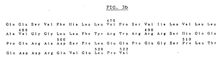
- a DNA which encodes exclusively a polypeptide comprising M-CSF activity and consisting of amino acids 1-522 of Fig. 3, and a DNA complementary thereto and of the same length.
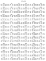
- the DNA encoding the said polypeptide comprises the base sequence having the formula shown in FIG. 2.
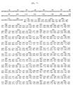
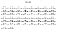
- the DNA encoding the said polypeptide comprises the base sequence having the formula shown in FIG. 1, or a part thereof.
- a process for preparing said gene which comprises; isolating mRNA from a selected human monocytic cell line; and preparing the gene coding for a polypeptide exhibiting M-CSF activity from said mRNA.
- a transformant which comprises said gene, and a biologically active plasmid (or vector) which carries said gene.
- a vector for expression of human urinary M-CSF which comprises a DNA sequence encoding one of the amino acid sequences of FIG. 10 and a DNA sequence comprising a promoter operative in an animal cell and an enhancer sequence.
- a process for producing human urinary M-CSF which comprises transforming said animal cell with said expression vector, and culturing the transformant to recover M-CSF from the culture.
- a polypeptide exhibiting M-CSF activity consisting of one of the amino acid sequences of FIG. 10, wherein X is Ser.
- a polypeptide exhibiting M-CSF activity which consists of one of the amino acid sequences of FIG. 20 or FIG. 21; a DNA sequence which codes for said polypeptides; a replicable cloning vector which comprises said DNA sequence and a replicon operative in a micro-organism, more particularly E. coli ; an expression system wherein said DNA sequence is operably ligated with a suitable control sequence; and a transformant micro-organism which is transformed with said expression system.
- a process for preparing human M-CSF which comprises; culturing a transformant micro-organism carrying a plasmid capable of expressing a human M-CSF monomer protein; recovering said monomer protein from the culture; and refolding said monomer protein into a biologically active human M-CSF homodimer protein.
- FIG. 1 depicts a base sequence of cDNA according to the present invention.
- FIG. 2 depicts a base sequence of translatable region in the cDNA fragment according to the present invention.
- FIG. 3 depicts an amino acid sequence of polypeptide which the cDNA fragment according to the present invention encodes.
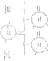
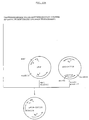
- FIG. 4 shows a schematic illustration for construction of plasmid pBSIICSF carrying the gene which encodes an amino acid sequence of polypeptide with M-CSF activity as described in Example 5.
- MCS Multicloning Site
- FIG. 5 shows a schematic illustration for reconstruction of pBSS and pBSL from pBSIICSF.
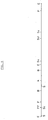
- FIG. 6 shows an outlined diagram of restriction map as obtained in Example 6.
- FIG. 7 shows schematically an outline of restriction sites used for the base sequence analysis together with directions and ranges of base fragments therefor as obtained in Example 6.
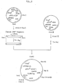
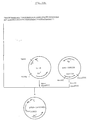
- FIG. 8 shows a schematic illustration for construction of expression vector pSVL-CSF carrying the gene which encodes an amino acid sequence of polypeptide with M-CSF activity as described in Example 7.
- the arrow indicates the direction of transcription of the promoter.
- FIG. 9 shows a schematic illustration for construction of expression vector pSV2*-hCSF1-mdhfr carrying the gene which encodes an amino acid sequence of polypeptide with M-CSF activity as described in Exmaple 8.
- the arrow indicates the direction of transcription of the promoter.
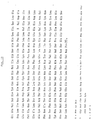
- FIG. 10 depicts an amino acid sequence of human urinary M-CSF.
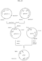
- FIG. 11 shows a schematic illustration for construction of plasmids pSVL-CSF214 and pSVL-CSF238, respectively.
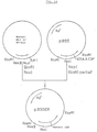
- FIG. 12 shows a schematic illustration for construction of plasmid pBSSER.
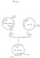
- FIG. 13 shows a schematic illustration for construction of plasmids pSVL-CSF214s and pSVL-CSF238s, respectively.
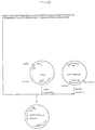
- FIG. 14 shows a schematic illustration for construction of plasmids pSV2d-CSF214 and pSV2d-CSF238, respectively.
- FIG. 15 shows a schematic illustration for construction of expression vector pPLN.
- FIG. 16 shows a schematic illustration for construction of pUC-CSF 214.
- FIG. 17a shows a schematic illustration for construction of expression vector pPLN-CSF214 for mutant M-CSF
- FIG. 17b shows a schematic illustration for construction of expression vector pPLN-CGF214 ⁇ 3 for mutant M-CSF.
- FIG. 18 shows a schematic illustration for construction of pUC-CSF238.
- FIG. 19a shows a schematic illustration for construction of expression vector pPLN-CSF238 for mutant M-CSF and
- FIG. 19b shows a shematic illustration for expression vector pPLN-CsF238 ⁇ 3 for mutant M-CSF.
- FIGS. 20 and 21 depict an amino acid sequence of M-CSF obtained from E . coli.
- the present invention relates to the novel gene which encodes a polypeptide having M-CSF activity and its preparation. More particularly, the present invention relates to the gene obtained by isolating messenger RNA (mRNA) from human monocytic cells which are stimulated with cell stimulators and preparing said gene from said mRNA by genetic manipulation techniques and its process.
- mRNA messenger RNA
- the production of novel proteins with M-CSF activity, the transformation of various host cells and the measures for assay of related genes, physiologically active products and the like can be given using said gene.
- the gene according to the present invention is preferably prepared by the following steps:
- the gene according to the present invention is prepared from mRNA which is isolated from human monocytic cells; however, when once the gene has thus been obtained and the sequence thereof has been known, it can be obtained from any of cells regardless of the initial source.
- the monocytic cell which is used in the present invention and as a source of mRNA may include a monocyte isolated from human blood by various known methods, a monocyte derived from a bone marrow progenitor by a suitable inducer, a human subacute lymphomatoid histocytic cell, a human acute promyelocytic leukemia cell, or an acute myelomonocytic leukemia cell.
- the leukemia cells are preferably established cell lines, and still preferably those which propagate excellently, for example, U937, HL-60, RC2a and the like.
- U937 which is a human monocytic cell line.
- the inducers which are used to differenciate said bone marrow progenitors into monocytes may include various colony stimulating factors such as GM-CSF and M-CSF as well as stimulated lymphocyte derived IL-3 and the like.
- immuno response modulators may include mitogens, bacterial somatic components, and physiologically active substances such as lipopoly saccharide (LPS), BCG (bacillus Calmette-Guérin), purified protein derivative of tuberculin (PPD), phorbol-12-myristate-13-acetate (PMA), concanavalin A (Con A), phytohemagglutinin (PHA), pokeweed mitogen (PWM), Corynebacterium parvum (CP), Corynebacterium granulosm, levamisole, lentinan, interferon, lymphokine, thymic hormone, hemolytic streptococcus, for example, picibanil (OK 432), etc.
- LPS lipopoly saccharide
- BCG Bacillus Calmette-Guèrin
- PMA purified protein derivative of tuberculin
- PMA phorbol-12-myristate-13-acetate
- Con A concanavalin A
- PWM phytohemagglu
- immuno response modulators may include cycloheximide and the like. These immuno response mudulators can be used alone or optionally in combination with other. More preferable immuno response modulators are mitogens such as phorbol-12-myristate-13-acetate (PMA), concanavalin A ( Con A) and the like, and/or cycloheximide, etc.
- the stimulation of human monocytic cell by these immuno response modulators can be carried out either prior to isolation of mRNA or in the course of propagating human monocytic cells, particularly human monocytic cell lines.
- the cultivation of human monocytic cell is carried out preferably under 0 - 200 ng/ml of PMA for 0 - 30 hours followed by preferably under 0 - 200 ⁇ g/ml of cycloheximide for 0 - 20 hours, prior to collection of mRNA.
- More preferable cultivation of human monocytic cell line U 937 prior to collection of mRNA may be carried out in base medium such as Dulbecco's modified Eagle's Medium, RPMI-1640, ASF 103, BME Hanks, BME Earle, Dico T Medium and the like, if necessary, supplemented with serum, for example, fetal calf serum, newborn bovine serum, human serum, calf serum and the like, growth factors, for example, human interleukin 2, ⁇ -interferon, somatomedin C, human EGF and the like, and various factors, for example, transferrin, interleukin I, CSF, albumin, GM-CSF and the like until the cell concentration reaches to from 1 x 10³ to 1 x 10⁷ cells/ml, preferably from 1 x 10⁵ to 1 x 10⁷ cells/ml followed by for 0.10 - 30 hours, preferably 0.25 - 20 hours in the presence of mitogens such as FMA at a final concentration of 0
- the cells thus obtained are recovered by centrifugation from the cell conditioned medium.
- the cells thus obtained may be further washed from 2 to 10 times by repeatedly suspending the cells in a buffered saline solution and the like follwed by centrifugation.
- the above cultivation conditions can be determined by either of the following methods after the cells are cultured under various conditions:
- U937 cell is used, cultured in RPMI-1640 (as a base medium) containing fetal calf serum (FCS) at a final concentration of 10% in a 10 l jar fermenter until the U937 cell reaches to 1 x 10⁶ cells/ml, then continued to culture for 6 hours after PMA is added to a final concentration of 50 ng/ml and subsequently for additional 4 hours after cycloheximide is added to a final concentration of 20 ⁇ g/ml and obtained by centrifugation from the broth.
- RPMI-1640 as a base medium
- FCS fetal calf serum
- the base medium which can be used in the present invention may be suitably selected from the group consisting of base media containing one or more components selected from the group consisting of sugars, amino acids, vitamins, hormones, proteins, antibiotics, growth factors, nucleic acids and its precursors, inorganic salts, heavy metals and the like and the base media supplemented with animal serum and the like.
- Such base medium includes commercially available RPMI-1640 Medium, MEM Medium, Dulbecco's modified MEM Medium and the like as mentioned above.
- the animal serum may include fetal calf serum, newborn bovine serum, bovine serum, horse serum, goat serum, porcine serum, rabbit serum, chicken serum, human serum and the like as mentioned above.
- Such serum can be used by suitable addition to the medium, preferably at a final concentration of up to 20 %.
- Suitable base media, sera and concentrations thereof, suitable densities of cell, cultivation periods, and immuno response modulators (stimulators) and concentrations thereof as well as suitable cultivation instruments and the like can be used by selecting from those conventionally employed depending on their purposes but not limited.
- the monocytic cell thus isolated is disrupted partially or completely during an increasing period in production of M-CSF active products to solubilize its cytoplasm and the extraction of total RNA is conducted by the fractionation of the cytoplasm.
- Such methods for disrupting and solubilizing cells include chemical or physical treatments such as methods using suitable surface active agents, methods using homogenizers, methods using freezing-thawing and the like.
- Such fractionation of each cytoplasm includes an extraction by phenol, chloroform and the like, a method using precipitants such as ammonium sulfate, a density gradient centrifugation using sucrose or CsCl, and the like.
- RNase inhibitor can be also carried out to prevent RNase.
- the monocytic cell is treated with guanidine-thiocyanate methods, for example, the method described in Chirgwin, J. M. et al., Biochemistry, 18 , 5294 (1979), subsequently with a CsCl density gradient centrifugation to obtain total RNA fractions which are subjected to affinity chromatography using an oligo (dT) cellulose column, a poly U-sepharose column and the like, to afford ploy A+-RNA fractions.
- guanidine-thiocyanate methods for example, the method described in Chirgwin, J. M. et al., Biochemistry, 18 , 5294 (1979)
- CsCl density gradient centrifugation to obtain total RNA fractions which are subjected to affinity chromatography using an oligo (dT) cellulose column, a poly U-sepharose column and the like, to afford ploy A+-RNA fractions.
- the mRNA thus obtained can be further fractionated by a sucrose gradient centrifugation, for example, according to the method described in Ishii, Y. et at., Immunol. Invest. 14 , 95 (1985), etc. for concentration.
- Identification of mRNA encoding a polypeptide having M-CSF activity in the resulting total mRNA is conducted by first fractionating the total mRNA through sucrose density gradient centrifugaiton, gel electrophoresis and the like, applying the resulting fraction to microinjection into oocytes of Xenopus laevis or cell free protein translation systems such as rabbit reticulocyte lysate, wheat germ system, and the like and either immunoassaying or determining the produced protein for its activity.
- such indentification can be achieved by chemically synthesizing DNA probes corresponding to a portion of the peptide sequence obtained from naturally occurring M-CSF, particularly human urinary M-CSF and selecting one from those which hybridize with plural fragments of said probes.
- the probes which are used in the selection of desired mRNA are desirably prepared selecting known sequences, sequences particularly related with activities and/or sequences not adjacent to each other.
- Such probes may be prepared by chemically solid or liquid-phase nucleotide synthesis methods, for example phosphite triester methods, phosphoamidite methods and the like.
- Labeling of the probe DNA thus synthesized is achieved using polynucleotide kinases, for example T4 polynucleotide kinase derived from T4 phage-infected E . coli and the like to transfer labeled phosphates, for example, 3P into the 5' terminus of the probe DNA.
- polynucleotide kinases for example T4 polynucleotide kinase derived from T4 phage-infected E . coli and the like to transfer labeled phosphates, for example, 3P into the 5' terminus of the probe DNA.
- Northern hybridization of mRNA with the labeled probe is conducted by first subjecting mRNA to electrophoresis, for example, the electrophoresis on agarose-formaldehyde gels, in the presence of denaturing agents (Maniatis, T., et al., Molecualr Cloning, p. 202 (1980), Cold Spring Harbor Laboratory), then transferring the mRNA from the gel to a nitrocellulose filter and finally hybridizing the filter with the labeled probe.
- electrophoresis for example, the electrophoresis on agarose-formaldehyde gels
- denaturing agents Maniatis, T., et al., Molecualr Cloning, p. 202 (1980), Cold Spring Harbor Laboratory
- the mRNA (which codes for a polypeptide having M-CSF activity) thus obtained according to the present invention is characterized by the following properties:
- double-stranded cDNA is symthesized.
- This synthesis can be carried out by any of various known methods.
- Preferable cDNA synthesis methods contain the following steps:
- Oligonucleotides corresponding to a portion of the amino acid sequence of protein having naturally occurring M-CSF activity, or oligo dT 12-18 is chemically synthesized and then by using the oligonucleotide as a primer and the mRNA according to the present invention as the template, complementary, single-stranded cDNA is prepared with reverse transcriptases, for example, AMV reverse transcriptase and the like, in the presence of dATP, dGTP, dCTP and dTTP.
- reverse transcriptases for example, AMV reverse transcriptase and the like
- double-stranded DNA is prepared with reverse transcriptases or DNA polymerases, for example, E . coli DNA polymerase I (Klenow fragment).
- a complementary single-standed cDNA-added vector is prepared and then a linker-added open-circular plasmid is anealed with linker DNA. Finally, a complete plasmid is produced with both RNase and DNA polymerase in the presence of dATP, dGTP, dCTP and dTTP.
- DNA synthesis consists of using oligo dT12 ⁇ 18 as the primer, the mRNA as the template and commercially available cDNA synthesizing kits, for example, Pharmacia cDNA Synthesis Kit, and first reacting the mixture in First-strand Reaction Mix followed by Second-strand Reaction Mix and subsequently with DNA polymerase, for example, E . coli DNA polymerase I (Klenow fragment) to produce double stranded DNA.
- DNA polymerase for example, E . coli DNA polymerase I (Klenow fragment)
- the double-stranded DNA thus obtained is used by ligating with suitable vectors in the next step.
- linker is desirably added to a terminus of the cDNA .
- linker can be conducted by using a suitable chemically synthetic DNA fragment.
- the linker-added cDNA can be treated with T4 phage DNA ligase together with vector DNA previously cleaved at a desired site with restriction enzyme in presence of ATP, etc. to obtain recombinant DNA.
- DNA for recombination can be obteined by adding either dC and dG chains or dA and dl chains to each of the double-stranded cDNA and the vector DNA previously cleaved at a desired site with restriction enzyme and then ligating their tails each other.
- the double-standed cDNA thus obtained can be used to construct cDNA libraries by inserting into suitable vectors such as EK plasmid vectors, for example, pBR327, pBR322, pDF41, Col E1, pMB9, pACYC1, pMIS4, pMIH17, pMIH18, pM10, pMF10, pMCR735 and the like, vectors for yeast, for example, YIp type, YEp type, YRp type, YCp type and the like, pLS102, pLS103, pUB110, pE194, pTP4, pTP5, pTA1302, pTA1321, pSCP, pSLP, pIJ, pMS, phage-derived vectors, for example, ⁇ gt, ⁇ c, ⁇ gt10, ⁇ gtWES and the like and subsequently transforming Escherichia coli ( E . coli ),
- E . coli for example, X1776, HB101, DH1, MM294, JM109, K12MC100 ⁇ lysogen, C600 strain, for example, Bacillus subtilis RM125, MT120 and the like, yeast, for example, Saccharomyces cerevisiae (e.g. SHY strain, AH22, AH22 pho80 and the like) and the like as a host.
- C600 strain for example, Bacillus subtilis RM125, MT120 and the like
- yeast for example, Saccharomyces cerevisiae (e.g. SHY strain, AH22, AH22 pho80 and the like) and the like as a host.
- the vectors used herein are preferably those comprising various replication origins adaptable to hosts, shine-Dalgarno sequences (SD sequences) and control sequences.
- Promoters which are utilized in these vectors may include the ⁇ -lactamase (e.g. penicillinase), the colicine E1 promoter, the lactose (lac) promoter systems, the tryptophan (trp) promorter systems, the elongation factor of protein synthesis EF-Tu gene (tuf B), the outer membrane lipoprotein gene (lpp), the rec A gene promoter, the tac promoter and the lambda phage-derived P L promoter systems.
- ⁇ -lactamase e.g. penicillinase
- lac lactose
- trp tryptophan
- tuf B the elongation factor of protein synthesis EF-Tu gene
- lpp outer membrane lipoprotein gene
- rec A gene promoter the tac promoter and the lambda phage-derived P L promoter systems.
- promoters for yeast may include the 3-phosphoglycerate kinase promoter system and those related to other glycolytic enzymes such as glyceraldehyde-3-phosphate dehydrogenase, hexokinase, pyruvate decarboxylase, phosphofructokinase, glucose-6-phosphate isomerase, alcohol dehydrogenase 2, cytochrome C, and acid phasphatase.
- glycolytic enzymes such as glyceraldehyde-3-phosphate dehydrogenase, hexokinase, pyruvate decarboxylase, phosphofructokinase, glucose-6-phosphate isomerase, alcohol dehydrogenase 2, cytochrome C, and acid phasphatase.
- Promoters for Bacillus may include the SPO2 promoters, the chloramphenicol resistant gene (Cm r ), the erythromycin resistant gene (Em r ) as well as the secretion vectors, for example, the amylase gene, the penicillinase gene and the like.
- particularly preferable DNA for transformation can be obtained by adding Eco RI linkers to both sides of the double-stranded cDNA as obtained above and then inserting the resulting DNA into Eco RI sites of ⁇ gt 10.
- the resultant recombinant thus obtained can be transfected into E . coli hosts such as C600Hfl after packaging and thereby recombinant phage libraries can be produced.
- cDNA libraries are obtained.
- the cDNA libraries thus obtained are screened by the colony or plaque hybridization tests using synthetic DNA probes (for example, Woo, S. L. C., Methods in Enzymology, 68 , 398 (1979); Szostak, J. et al., ibid, 68 , 419 (1979)).
- synthetic DNA probes for example, Woo, S. L. C., Methods in Enzymology, 68 , 398 (1979); Szostak, J. et al., ibid, 68 , 419 (1979)
- the desired products can be identified by the plus-minus method, the hybridization-translation assay and the like.
- Recombinant phage DNA is prepared from the colonies or plaques thus selected.
- the recombinant phage DNA is cut out with restriction enzyme to give DNA portions which comprise desired inserts, The DNA portions are inserted into suitable plasmid vectors and cloned. Such processes can be repeated plural times suitably depending on necessity.
- the cloned DNA fragment thus finally obtained is examined and the base sequence thereof is determined.
- First the cloned DNA fragment is digested with restriction enzyme and thereby each restriction map is prepared.
- the cloned DNA fragment is subcloned in M13 phage, preferably M13 mp18 and mp19 and subsequently the base sequence thereof is analyzed by the dideoxy sequencing, for example the method of Sanger et al. (Proc. Natl. Acad. Sci. USA ., 74 , 5463 (1977); J. Mol. Biol., 162, 729 (1982), etc.).
- Such sequencing may be also conducted according to Maxam-Gilbert method.
- cDNA which carries desired base sequence portions is selected by utilizing the restriction map obtained, the analyzed base sequence and the previously discovered base sequence corresponding to the partial amino acid sequence of protein which has M-CSF activity and thereby cloned DNA which comprises an base sequence encoding an amino acid sequence of polypeptide having M-CSF activity is determined.
- More particularly preferable methods for obtaining the cloned DNA which codes for the desired polypeptide having M-CSF activity comprise the following processes:
- cDNA libraries are screened by plaque hybridization test using the 3P labeled synthetic DNA probes as used for the selection of above-mentioned mRNA, for example, the method disclosed in Maniatis, et al., Molecular Cloning , p. 326 (1982), Cold Spring Harbor Laboratory and then recombinant phage DNA is constructed from each of selected plaques (for example, Perbal's method.
- the phage clone thus selected is cut out to give the desired DNA fragment, which is ligated with a suitable plasmid, for example, pBluescript II SK (Stratagene) and transfected into E . coli , for example, E . coli JM109.
- a suitable plasmid for example, pBluescript II SK (Stratagene) and transfected into E . coli , for example, E . coli JM109.
- the resulting recombinant is treated to afford plasmids which are treated according to the alkaline-SDS method, for example, described in Birnboim, H. C. & Doly, J., Nucleic Acids Res., 7 , 1513 (1979) and subjected to electrophoresis.
- the clone of interest is selected according to the indication that the above electrophoresis give the desired spot of base fragment. From these clone plasmids carrying the desired fragment are obtained, and suitably treated with restriction enzyme to afford fragments which are again ligated with plasmids, for example, suitable fragments of pBS+(Stratagene), and transfected into E. coli for example. E . coli JM109 to afford clones. Thus, the cloned DNA which carries the desired fragment is obtained in large quantities.
- the DNA fragment thus obtained in large quantities can be subcloned in M13 mp18 or mp19, anealed with primers followed by complementary chain synthesis using Sequenase TM (Unite States Biochemical Corporation) according to the dideoxy chain termination method (Sanger, et al., Proc. Nat. Acad. Sci. USA 74 , 5463 (1977); J. Mol. Biol., 162, 729 (1982)), and then subjected to gel electrophoresis and autoradiography. On the basis of the results thus obtained, the base sequence of gene which carries the DNA sequence encoding the polypeptide having M-CSF activity was determined.
- Figure 1 illustrates the base sequence of the gene according to the present invention.
- the DNA thus obtained according to the present invention can be chemically synthesized using known automated nucleotide synthesizers and the like by utilizing the information with regard to the resulting base sequence. Further the DNA according to the present invention can be prepared by enzymatically binding such partially synthetic fragments.
- the gene encoding the M-CSF active polypeptide according to the present is closely akin to naturally occuring one due to a derivative from the monocytic cell which is relatively close to natural cells, it can be expected that the peptide which is produced by using the same gene would have advantages in medical fields.
- the gene accoding to the present invention is relatively long as compared with those in the prior art. Consequently, the acquisition of gene demanded inventive skills.
- Particularly recombinant plasmids which will have excellent affinities to higher eukaryotic organisms can be obtained using the gene according to the present invention. More particularly the gene according to the present invention gives recombinant plasmids which are useful in transforming animal-derived cells efficiently.
- the base sequence of the gene according to the present invention is more suitable for expression of M-CSF active proteins in hosts. Therefore, when incorporated into hosts by recombinant techniques, it will effect in producing efficiently polypeptides of interest.
- the deletion of the gene will also be very low.
- the gene according to the present invention has a particular base sequence as well as is relatively long as compared with those in the prior art; therefore, when it is used in preparing transformants and expressed in the transformants, particularly under glycosylation conditions, excellent results can be obtained, which have never been achieved in the prior art.
- the suitable glycocylaiton can be expected, so that activites and stabilites of the procuded protein would increase.
- the present gene Since it is believed that the the present gene is close to the natural one, it would be expected that the transformation is efficiently conducted using the gene. Further, the expression of the recombinant gene can be controlled easily. It is still expected that the production of polypeptides according to the present invention is constant and excellently industrial due to the gene structure suitable for animal hosts.
- polypeptide product according to the present invention is very similar to natural one, it would be useful in medical applications and the like.
- the gene according to the present invention is advantageous in not only the characteristics of base sequence but also the stereochemical arrangement and the genetic orientation of control regions.
- the DNA fragment which carries the gene encoding the M-CSF active polypeptide is again inserted into suitable vector DNA and the products is used for transforming suitable hosts.
- recombinant vectors can be constructed by addition of suitable promoters and sequences for inducing a gene expression.
- prokaryotic and eukaryotic cells and the like can be used for hosts used for transformation according to the present invention.
- Examples of such cells are microorganisms such as E . coli , for example, K12, Bacillus, Actinomycetes, yeast and the like, insect cells, animal cells such as COS-7 cell CHO cell, mouse myeloma cell N51, VERO, Hela cell, monkey CV1, monkey AGMK, mouse L cell, FM3A, NIH3T3, C127, Friend leukemia human 293, monkey W162, hamster BHK, human 143, and the like.
- microorganisms such as E . coli , for example, K12, Bacillus, Actinomycetes, yeast and the like
- insect cells such as COS-7 cell CHO cell, mouse myeloma cell N51, VERO, Hela cell, monkey CV1, monkey AGMK, mouse L cell, FM3A, NIH3T3, C127, Friend leukemia human 293, monkey W162, hamster BHK, human 143, and the like.
- animal cells such as CO
- Vectors which can be used for transforming E . coli and the like may include pBR322, pKK223-3, lambda phage-derived vectors, virus-derived vectors such as SV40 and those known as shuttle vectors.
- Other vectors which are often used for transforming eukaryotic micro-organisms such as yeast include pAM82 and the like.
- the host cells and vectors which are used for constructing the above mentioned cDNA library can be used similarly for transforming the hosts.
- Vectors which can be used for transforming the above-mentioned animal cells may include those carrying early and late promoters of SV40, vectors, vaccinia virus-derived vectors, adenovirus-derived vectors, bovine papilloma virus (BPV)-derived vector, as well as those suitably inserted with mouse metallothionein gene promoter, retrovirus derived promoters (of animal) human cytomegalovirus derived immediate-early promoter, for example, avian retrovirus derived promoter, immunoglobulin promoters, or heat shock promoters.
- retrovirus derived promoters of animal human cytomegalovirus derived immediate-early promoter, for example, avian retrovirus derived promoter, immunoglobulin promoters, or heat shock promoters.
- Examples of the vector are pNE05, pL2BPV, pMSV gpt, pSV2-gpt, SV40 ori, pSV2 neo, pSV2 cat, pMDSG, BPV 69T DNA, pSV2 dhfr, pLTR dhfr26, pCVSVE, pMg4, ⁇ Eld/dl309, pZIP-neoSV, pMOV- and the like.
- Vectors which can be used for transforming the above-mentioned insect cells may include polyhedrosis virus-derived vectors.
- Selection markers used in the above-mentioned vectors may include thymidine kinase (TK) gene, hypoxanthine-guanine phosphoribosyl transferase (HPRT) gene, dihydrofolic acid dehydrogenase (DHFR) gene, adenine phosphoribosyltransferase (APRT) gene and the like.
- TK thymidine kinase
- HPRT hypoxanthine-guanine phosphoribosyl transferase
- DHFR dihydrofolic acid dehydrogenase
- APRT adenine phosphoribosyltransferase
- Transformation operons preferably utilizable in the present invention may include, for example, the operons for tryptophan, ⁇ -galactosydase, alkaline phosphatase, ⁇ -lactamase and the like.
- Transformation of host cells with expression vector thus obtained can be carried out by the following processes:
- the treatment of calcium chloride is used.
- E . coli is preferably treated in the presence of calcium chloride, magnesium chloride, rubidium chloride.
- transformations may include a direct introduction into protoplasts by polyethylene glycol or polyornithine, a liposome method, co-cultivation, a spheroplast method and the like.
- the transformations of mammal cell may include virus infection, DEAE-dextran method, calcium phosphate precipitation, microinjection, liposome fusion, protoplast fusion, cell fusion by Sendai virus, micronucleolus fusion, chromosome transinjection and the like.
- the plasmid which carries the gene code for the M-CSF active polypeptide is treated with DNA polymerase, for example, E. coli DNA polymerase I (Klenow fragment) to make blunt ends, then ligated with fragments of vector pSVL.
- the resulting product is transfected into E . coli , for example, HB101 to obtain recombinant clones, from which DNA is obtained.
- the DNA is examined for insertion direction by restriction enzyme mapping.
- two vectors wherein one has the same direction as the promoter and another has the reverse are selected.
- animal cells are transformed with the above vectors for expression according to the calucium phosphate method (for example, Okayama, et al., Mol. Cell. Biol. 7 , 2745 (1987)) and the like.
- the recombinant M-CSF active products according to the present invention are isolated and purified according to any of various conventional techniques.
- the recombinant products can be present either in or out of the transformants.
- the purification and/or isolation according to the present invention may include acid treatments, heat treatments, protein precipitation, electrophoresis, gel filtration, molecular sieve chromatography, ultrafiltration, reversed phase chromatography, high performance liquid chromatography, ion exchange chromatography, affinity chromatography, absorption chromatography and the like. These treatments can be used alone or in combination with each other depending on necessity.
- Carriers for separating in the gel filtration may include, for example, polyacrylamide gels, agarose gels, dextran gels and the like.
- the reversed phase chromatography may include those using materials wherein C1 ⁇ 20 alkyl, cyanopropyl, phenyl and the like are bound on silica gel bases.
- the ion exchange chromatography may include those using anion exchanger modified with dimethyl aminoethyl or diethylaminoethyl, and cation exchanger modified with carboxymethyl.
- the affinity chromatography may include those using Con-A sepharose, lectin-sepharose, antibody-bound sepharose and the like, preferably those using monoclonal antibody bound sepharose. Phenyl sepharose, octylsepharose and the like may be used for the absorption chromatography.
- the recombinant products which have M-CSF activity are easily obtained in large quantities and high purity by using the above mentioned isolation and purification alone or in combination with other.
- the product thus obtained can be administered according to a conventional method acceptable to proteinous preparations.
- the product may be administered intravenously, intramuscularly, intranasally, subcutaneously, or intradermally, even orally.
- formulations include sterile solutions or non-aqueous solutions, suspensions and emulsions. These may also included lyophilized formulations which will be dissolve in sterile water or physiological saline, or immediately prior to use, in sterile injectable medium.
- the products according to the present invention are useful in treating various infections, tumors, hypercholesterolemia, arteriosclerosis and leukopenia, preventing side effects derived from adminstration of anticancer and radiation, and the like.
- the products according to the present invention are also useful in diagnosing hypoplastic anemia and the like.
- the present therapeutic products may be used advantageously in combination with interleukine 2, TNF, interferon, adriamycin, mitomycin, 5-FU and the like.
- the pharmaceutical composition comprising the present products is useful in treating a serious tumor bearing patient who has lost resistance upon functional deficiency of bone marrow tissue by radiation and administration of anticancer agent, and also a serious infectious patient when the treatment of antibiotics is difficult.
- the dosage regimen When used for the above pharmaceutical purposes, the dosage regimen will be determined by taking the condition of the patient and the like into consideration.
- the present invention relates to the novel vector for expression of human urinary M-CSF, which comprises the DNA sequence encoding the amino acid sequence of FIG. 10 and the DNA sequence comprising the promoter operative in the animal cell and the enhancer sequence.
- the present invention also relates to the process for producing human urinary M-CSF which has the amino acid sequence of FIG. 10.
- the M-CSF genes which encode the amino acid sequence of FIG. 10 can be prepared by the use of human M-CSF cDNA, chemical synthesis of a portion of DNA, cleavage with restriction enzyme, and deletions, additions and ligations with modifying enzymes.
- plasmid pBS II CSF plasmid pBS II CSF.
- the expression vectors which carry cDNA coding for 522 amino acids (excluding the signal peptide) are digested with suitable restriction enzymes followd by separation and purification of DNA fragments.
- DNA is chemically synthesized in order to obtain 214 and 238 amino acids. Chemical DNA synthesis is conducted according to, for example, ⁇ -cyanoethyl amidite method and H-phosphonate method. The above vector and the synthetic DNA are ligated with T4 DNA ligase followed by transfection into E . coli to afford recombinants of interest.
- Partial change of amino acid is carried out according to the site directed mutagenesis (Kunkel, T. A. et al., Proc. Natl. Acad. Sci. USA. 82 , 488 (1985)). Verification of the gene sequence thus obtained is also conducted by the dideoxy chain termination method and Maxam-Gilbert method as mendioned above.
- the expression vector for animal cells preferably comprises a promoter at the up-stream of the gene, a splicing region at the down-stream thereof, a poly (A) signal and a poly (A) region.
- enhancer sequence on vectors can effect in increasing the productivity of gene product.
- the promoters used herein conventionally include these derived from viruses, for example, SV40 early and late promoters, human adenovirus main late promoters, human cytomegarovirus immediate early promoters and the like, but is not limited.
- the host animal cells used herein include, for example, monkey kidney COS cell (Gluzman, Y., Cell 23, 175 (1981) and chinese hamster ovary CHO dhfr cell (Urlaub, G, & Chasin, L. A., Proc. Natl. Acad. Sci. USA, 77 , 4216 (1980) and the like, and preferably CHO dhfr ⁇ cell.
- COS cell it is preferred that expression vectors carry the above mentioned expression unit on plasmids which have SV40 replication origins and exclude sequences inhibiting SV40 replication, so-called toxic sequences.
- vector pSV2-dhfr which carries mouse dhfr genes as a dominant marker (Sabramani, S. et al. Mol. Cell. Biol, 1 , 854 (1981)) is preferably used.
- the co-transfection wherein the CSF gene is not ligated with the dominant marker prior to transfection, and the method wherein the CSF gene is ligated and then transfected can be taken.
- the latter is preferably used.
- the expression according to the poly cistron method may achieve high translation efficiency (Kaufman, R. J. et al.; EMBO J. 6 , 187 (1987)).
- the transformation can be easily conducted according to conventional methods, for example, DEAE-dextran method (Arai, N. et al., Exp. Med. 5 , 1019 (1987)) and the calcium phosphate method (Okayama, H. et al.; Mol. Cell. Diol. 7 , 2724 (1987)).
- the resultant transformant is cultured according to a conventional method and the human urinary M-CSF is accumulated in the culture medium.
- the media used for COS cell according to the present invention include, for example, Dulbecco's modified MEM Medium (D-MEM) supplemented with fetal calf serum (FCS) and the like by depending on necessity.
- D-MEM Dulbecco's modified MEM Medium
- FCS fetal calf serum
- the cultivation is carried out at 35 - 37°C for 2 - 5 days under 0% - 10% CO2.
- nucleic acid-free MEM ⁇ (-) Medium supplemented with serum such as dialyzed fetal calf serum (FCS) by depending on necessity is preferably used.
- FCS dialyzed fetal calf serum
- the content of M-CSF in the conditioned medium obtained in the above-mentioned process is 1 - 10 mg per 1l.
- the purification of M-CSF is conducted as follows:
- the resulting conditioned medium is concentrated by ultrafiltration, adjusted to pH 4.0 - 5.0 by phosphoric acid and the like, and the precipitate removed.
- the concentrate is applied to an anion exchanger, for example, DEAE-cellulose to collect fractions which exhibit M-CSF activity.
- the active fraction is applied to hydrophobic chromatography, for example, phenyl-sepharose to collect active fractions.
- the fraction is further applied to molecular sieve chromatography, for example, a column packed with Superose 12, and fractionated.
- the fraction is desalted and concentrated.
- human urinary M-CSF is obtained in high yield and high purity.
- the present invention relates to the process for producing of human urinary M-CSF by using E . coli expression system.
- the present invention also relates to the polypeptide which has the amino acid sequence of FIG. 20, its related vector and transformant.
- the M-CSF genes which encode the amino acid sequence of FIG. 20 can be prepared by the method as mentioned above. They may also be chemically synthesized.
- plasmid pBS II CSF for example, plasmid pBS II CSF, pSVL-CSF214 and pSVL-CSF238.
- Plasmids pSVL-CSF214 and pSVL-CSF238 are used to construct expression vectors for E . coli .
- Each of the above plasmids carries the base sequences corresponding to a signal peptide containing 32 amino acids and a peptide containing either 214 amino acids or 238 amino acids.
- the plasmids are cleaved with suitable restriction enzymes to obtain fragments which are subcloned into vectors having multi-cloning sites, digested with suitable restriction enzymes which can cleave the site nearest to the N-terminus of human urinary M-CSF and the region excluding the signal peptide region.
- suitable restriction enzymes which can cleave the site nearest to the N-terminus of human urinary M-CSF and the region excluding the signal peptide region.
- the DNA fragment coding for M-CSF amino acid sequence is isolated.
- DNAs which correspond to fMet (translation initiation codon) and the deficient N-terminal amino acid sequence are chemically synthesized.
- Chemical DNA synthesis is conducted according to, for example, ⁇ -cyanoethyl amidite method and H-phosphonate method.
- the vectors capable of expressing in E . coli are constructed as follows:
- the expression vector is cleaved with suitable restriction enzymes (these enzymes should make its adhesive tails capable of ligating with both the recovered DNA and the synthetic DNA).
- the recovered DNA, the synthetic DNA and the cleaved expression vector are ligated with T4 DNA ligase and transfected into E . coli to afford the desired transformant.
- Partial change of amino acid is carried out according to the site directed mutagenesis (Kunkel, Proc. Natl. Acad. Sci. USA 82 , 488 (1985)). Further, the partial change of amino acid can be conducted by inserting partially synthetic DNA into a cleavage site of restriction enzyme. Verification of the gene sequence thus obtained is also conducted as mentioned above.
- the resultant transformant E . coli is cultured to afford a human urinary M-CSF monomer.
- the expression vectors for E . coli are those which comprise promoter ⁇ operator systems, SD (Shine-Dalgarno) sequences and protein synthetic initiation codons (for example, ATG) and a terminator downstream therefrom.
- the promoter ⁇ operator systems include lac promoter operator systems, trp promoter ⁇ operator systems, lambda phage P L promoter ⁇ operator systems, tac promoter-operator systems and the like but is not limited to.
- lactose repressers lac I
- lac promoter ⁇ operator systems and tac promoter ⁇ operator systems lac I
- cI repressers and their mutants lacI 857 repressers to lambda phage P L promoter ⁇ operator systems
- trp R trp promoter ⁇ operator systems.
- the utilizable combination of promoter ⁇ operator systems with repressers is not limited to. Therefore, known conventional systems can be used.
- the phenotypic expression of E . coli which is controlled by such repressers can be induced under various conditions.
- Such induction of expression can be conducted by when the lactose represser is used, lactose or IPTG (Isopropylthiogalactoside), when the cI represser is used, an DNA synthesis inhibitor, when the cI 857 represser is used, high temperature, 42°C and when the trp R is used, either Indolyl-3-acrylic acid, Indolyl-3-propionic acid or deficit of tryptophan.
- lactose represser lactose or IPTG (Isopropylthiogalactoside)
- IPTG Isopropylthiogalactoside
- an DNA synthesis inhibitor when the cI 857 represser is used, high temperature, 42°C and when the trp R is used, either Indolyl-3-acrylic acid, Indolyl-3-propionic acid or deficit of tryptophan.
- Such marker genes include antibiotic resistance genes with respect to ampicillin, tetracycline, chloramphenicol, kanamycin and the like, lac Z genes (the production of blue color by the degradation of X-gal on the basis of ⁇ -complementarity of lactose operon shows whether DNA of interest is inserted) but is not limited to.
- Transformation of E . coli with the resulting expression vector is conducted according to a conventional method.
- the transformation can be easily achieved by, for example, treatment of host E . coli with calcium chloride or treatment in the presence of calcium chloride, magnesium chloride or rubidium chloride followed by mixing with plasmids DNA (Maniatis, T., et al, Molecular Cloning, Cold Spring Harbor Laboratory (1989)).
- the transformant thus obtained can be cultured according to a conventional method.
- the media used for such cultivation are those for conventional cultivation of E . coli such as LB Medium, E Medium, M9 Medium, M63 Medium and depending on necessity, can be supplemented with various kinds of nitrogen sources, carbon sources, inorganic salts, vitamins and the like as conventionally known.
- Cultivation for E . coli is carried out at pH 5-9, preferably 7 or near, at 20 - 45°C, preferably 28 - 42°C.
- the cultured E . coli is collected and human urinary M-CSF protein is recovered from the cell.
- the efficient production of M-CSF is also achieved by expressing M-CSF according to 2 ⁇ cistron method.
- This method include gene expression systems comprising 2 continuous cistrons. This gene expression system results in producing M-CSF in the host E . coli in large quantities stably.
- Expression plasmids which carry a gene coding for a suitable polypeptide or a portion thereof as the first cistron and the M-CSF gene as the second cistron are constructed.
- the gene employed as the above first cistron can be expressed in hosts, it is not limited. However, since the polypeptide encoded by the gene employed as the first cistron is produced in the same system as the M-CSF of interest, the peptide easily separable from the M-CSF of interest is preferred.
- the gene which is directly controlled by the promoter systems used is directly controlled.
- P L promoter and N protein region which is controlled by the P L promoter are illustrated.
- the N protein region is cleaved with restriction enzyme so that the treated region remains only a portion encoding less than 100 amino acid at maximum, and modified by introduction of synthetic DNA after the unnecessary sequence is removed.
- the above synthetic DNA is prepared to contain the termination codon for the modified N protein, SD sequence for the expression of the second cistron and the initiation codon for the M-CSF.
- termination codons In order to prevent a read-through from the upstream, the introduction of termination codons into each of three reading frames is desired.
- the termination codon and initiation codon in the synthetic DNA any of naturally accurring codons can be employed.
- the expression vector which carries p L promoter and N protein region controlled by P L promoter is constructed, cleaved with restriction enzyme, isolated and purified after the unnecessary DNA sequence is discarded.
- the cloned vector which carries M-CSF gene is digested with suitable restriction enzyme and the M-CSF gene fragment is recovered.
- Both the recovered fragments and the synthetic DNA for the supplementation of the deficient DNA region thereof are ligated with T4 DNA ligase and the like to afford the desired expression vector.
- the resulting expression vector comprises a region of a part of N protein as the first cistron and a region of M-CSF as the second cistron as mentioned above.
- the novel expression of M-CSF is achieved by using the expression vector thus obtained.
- Host E. coli which carries cI 857 represser (for example, N4830-1, K12 delta H1) is transformed to afford the desired transformant in accordance with two cistron method.
- cI 857 represser for example, N4830-1, K12 delta H1
- the transformant thus obtained can be cultured according to a conventional method.
- the transformant is cultured in LB medium at 26 °C-37°C, more preferably 28°C - 32°C with shaking or stirring, then heated to from 37°C to 42°C of culture temperature at a suitable point of exponential phase and recultured for a constant period, for example, 2 to 24 hours at 37°C to 42°C. According to such cultivation it is observed that inclusion bodies of the produced M-CSF are formed in the cell of host E . coli . The cells are collected by centrifugation, and the resulting cells disrupted to obtain M-CSF as insoluble fractions.
- Disruption of micro-organisms can be conducted by sonication or according to the method of Nagai et al (Nagai, K. et al., Proc. Natl. Acad. Sci. USA, 82 , 7252 (1985)).
- the inclusion bodies of M-CSF thus obtained are solubilized in a buffer containing 2 to 8 M urea, 4 to 7 M guanidine hydrochloride or more than 0.1% (w/v) of sodium dodecyl sulfate by using a homogenizer and the like.
- the M-CSF monomer protein thus obtained is purified under solubilizing conditions according to various methods. Purification includes reversed phase chromatography, ion exhange chromatography, high performance liquid chromatography, absorption chromatography, gel filtration and the like.
- the M-CSF monomer can be purified by these techniques and refolded to obtain the M-CSF dimer. Refolding is achieved by either rapidly or slowly removing the above solubilizing agent. Formation of disulfide bond is achieved by either autooxidation or addition of a suitable oxidizing-reducing agent under refolding conditions.
- the reagents used for this treatment may include oxidized glutathione (GS-SG), reduced glutathione (GSH), DTT and the like.
- Refolding can also be conducted by enzymes such as thioredoxin.
- U937 cells (American Type Culture Collection (ATCC) CRL 1593) were propagated in 20 ml of RPMI-1640 containing 10% FCS at an atmosphere of 5% CO2 in air to achieve 1 x 10⁶ cells/ml (for about 72 hours).
- PMA was added to 0, 10, 50, 100 and 200 ng/ml followed by incubation for 0.25, 0.5, 1, 2, 4, 6, 12 and 20 hours, respectively.
- Cycloheximide was then added at a final concentration of 0, 20, 40, 60, 80 and 100 ⁇ g/ml followed by incubation for additional 0.25, 1, 2, 4, 6 and 12 hours.
- the medium was centrifuged at 800 x g for 5 min.
- the cells were harvested and suspended in 10 ml of PBS (5 mM phosphate buffer containing 0.15M NaCl, pH 7.2). The suspension was recentrifuged at 800 x g for 5 min and a supernatant discarded to recover cells.
- PBS 5 mM phosphate buffer containing 0.15M NaCl, pH 7.2
- the cells obtained in the above procedure (1) were suspended in 45 ⁇ l of 10 mM Tris-HCl (pH 7.0) containing 1 mM EDTA according to the method described by White and Baneroft (J. Biol. Chem., 257, 8569 (1982) followed by addition of 5 ⁇ l of 5% NP-40 solution and centrifugation at 15000 rpm after dissolution.
- To 50 ⁇ l of the supernatant obtained by centrifugation were added 30 ⁇ l of 20 x SSC (1 x SSC : 0.015M trisodium citrate containing 0.15 M NaCl) and 20 ⁇ l of 37% (w/v) formaldehyde followed by heat treatment at 60°C for 15 min.
- the resultant solution was diluted four times, blotted onto nitrocellulose filters and hybridized with a hybridizing solution containing 3P labeled DNA probes chemically synthesized according to the method in Example 3 at a concentration of 2 x 10⁶ cpm/ml, at 54°C for 20 hrs.
- the autoradiography shows that the culture condition at 50 ng/ml of PMA for 6 hrs. follwed by 20 ⁇ g/ml of cycloheximide for 4 hrs. is optimal because the supernatant obtained is active to the highest dilution.
- Example 1-(2) U937 cells were propagated in RPMI containing 10% FCS at an atomosphere of 5% CO2 in air to achieve a cell density of 1 x 10⁶ per ml (for about 72 hrs.).
- PMA was added at a final concentration of 50 ng/ml followed by incubation for 6 hrs.
- Cycloheximide was added at a final concentration of 20 ⁇ g/ml followed by incubation for additional 4 hrs.
- the cells thus obtained were collected by centrifugation at 800 x g (International Equipment Co.), suspended in 500 ml of PBS (5 mM phosphate buffer containing 0.15 M NaCl, pH 7.2) and washed by centrifuging at 1000 rpm for 5 min. and discarding a supernatant. After similarly the washing was repeated twice, the resultant cells (about 7.6 g) were stored at -80°C until use.
- RNA was extracted according to the methods such as the known guanidine-thiocyanate method (Chirgwin, J. M. et al.; Biochemistry, 18 , 5294 (1979)).
- guanidine-thiocyanate method As a starting material, 7.6 g of the cell was suspended in 60 ml of solution containing 6 M guanidine thiocyanate, 5 mM sodium citrate, 0.5% N-lauryl sarcosine sodium salt, and 0.1 M 2-mercaptoethanol, and homogenized.
- RNA fraction thus obtained was dissolved in 30 ml of 20 mM Tris-HCl (pH 7.5) containing 500 mM NaCl, 1 mM EDTA and 0.1% N-laurylsarcosine sodium salt, subjected to chromatography on an oligo dT12 ⁇ 18 cellulose column (1.0 x 2.0 cm, Pharmacia Type 7) previously equilibrated with the above-mentioned buffer, washed with 20 mM Tris-HCl (pH 7.5) containing 100 mM NaCl, 1 mM EDTA and 0.1% N-laurylsarcosine sodium salt followed by elution with 1 mM EDTA - 0.1% N-laurylsarcosine sodium salt solution to give a poly A+RNA fraction (about 290 ⁇ g).
- ribosomal RNAs 16S, 18S, 23S and 28S were used as a standard marker for determination of sedimentation constant.
- Example 2 (2) contains mRNA encoding M-CSF gene
- northern hybridization was carried out using synthetic 3P-labeled DNA probes.
- the base sequences (each corresponding to the second base sequence) complementary to the first one above-mentoned were chemically synthesized as the probe.
- Bioserch 8600 DNA synthesizer (Millipore) was used for the preparation of these sequences. Synthetic reaction was carried out for 2 to 3.5 hours.
- the resulting polynucleotides were purified by using a cartridge for purification of oligonucleotide (Applied Bio-Systems Japan, Japan).
- the complementary base sequence each thus chemically synthesized (2 nmoles/ml) was treated with 200 units/ml of polynucleotide kinase (T4 polynucleotide kinase derived from E .
- Two 3P labeled probes were prepared by transferring phosphate residues onto hydroxyl residues at the 5′ terminus of the base sequence from [ ⁇ -3P] ATP.
- Each specific radioactivity is 6 x 10⁶ cpm/pmol.
- the total poly A+ RNA (2 ⁇ g each) obtained in Example 2-(2) was applied to electrophoresis (60V, 6 hrs) using agarose containing MOPS (20 mM) and formaldehyde (2.2 M) (Maniatis, T., et al,; Molecular Cloning, p 202 (1980), Cold Spring Harbor Laboratory), transferred onto a nitrocellulose filter in 20 x SSC(1 X SSC; 15 mM sodium citrate and 0.15 M NaCl), fixed under heat treatment at 80°C for 5 hrs.
- prehybridizing solution 0.9 M NaCl, 0.09 M Tris-HCl (pH 7.5), 6 mM EDTA.2Na, 0.5% sodium N-lauroylsarcosinate and 0.4 mg/ml denatured salmon sperm carrier DNA
- the filter was hybridized in 3 ml of hybridizing solution containing 6 x 10⁶ cpm of 3P labeled synthetic DNA probe above-mentioned at 54°C for 20 to 40 hours.
- the hybridized filter was washed with 500 ml of 0.5% sodium N-laurylsarcosine solution twice at room temperature, twice at 54°C, and dried in air.
- cDNA preparation was carried out by using as a template the fractions 22 and 23 (24.7S to 27.1S) of mRNA obtained in Example 2-(2). Five ⁇ g of the mRNA fraction obtained in Example 2-(2) was dissolved in 20 ⁇ l of RNase-free water. The resulting solution was used as the template for the synthesis of cDNA.
- the oligo (dT)12 ⁇ 18 primed cDNA synthesis was carried out in First-strand Reaction Mix (cDNA Syntheris Kit, Pharmacia) at 37°C for 60 min. The resulting solution (32 ⁇ l) was reacted in Second-strand Reaction Mix (cDNA Synthesis Kit, Pharmacia) at 12°C for 60 min. followed by at 22°C for 60 min.
- the ethanol precipitation was conducted at -80°C for 30 min.
- the solution was centrifuged at 15,000 rpm for 30 min to recover the double stranded cDNA (about 1.5 ⁇ g) as a precipitate.
- the double stranded cDNA (1 ⁇ g) was ligated with Eco RI linker (5 ⁇ l) in Ligation Kit (Takara Shuzou): A solution 30 ⁇ l and B Solution 7.5 ⁇ l at 12°C overnight followed by the ethanol precipitation to obtain DNA.
- the DNA was loaded on 0.8% agarose gel.
- the electrophoresis was conducted at 100V for 2 hrs. using ⁇ phage Hind III fragments as a marker.
- a gel of 3 to 5 Kbp portions was cut out to obtain a double stranded cDNA.
- the resulting cDNA was ligated with Eco RI site of ⁇ gt 10 (1 ⁇ g) at 16°C overnight using the above-mentioned ligation kit, ethanol precipitated and dissolved in 5 ⁇ l of 10 mM Tris-HCl (pH7.5) containing 1 mM EDTA. Five ⁇ l of the solution was added to Freeze/Thaw extract, Gaigapack Gold (Stratagene) under ice cooling followed by the addition of 15 ⁇ l of Sonic extract and the incubation at 22°C for 2 hrs for packaging.
- the recombinant phage solution was added to 0.5 ml of SM medium containing 5.8 g of NaCl, 2.0 g of MgSO4, 50 ml of 1 M Tris-HCl (pH7.5) and 5 ml of 2% gelatin per one liter, centrifuged at 15,000 rpm for 30 seconds after the addition of 20 ⁇ l of chloroform and recollected to obtain a supernatant which was stored at 4°C.
- E . coli C600Hfl single colony was inoculated in 500 ml of TB Broth (5 g NaCl and 10 g of Bactotryptone per one liter) containing 10 mM MgSO4 and 0.2% maltose followed by the incubation at 30°C overnight.
- the microorganisms were harvested by centrifugation at 2,000 rpm for 10 min. and suspended in 10 mM MgSO4 to obtain 0.5 at O.D. 600 nm.
- NZY soft agar medium 5 g of NaCl, 2g of MgSO4 ⁇ 7H2O, 5 g of yeast extract, 10 g of NZ amine, and 7 g of Bactoagar per one liter
- the mixture was loaded on NZY agar plates (5 g NaCl, 2 g MgSO4 ⁇ 7H2O, 5 g of yeast extract, 10 g of NZ amine and 15 g of Bactoagar per one liter) made in 10 cm diameter dishes and incubated at 37°C overnight. The same treatments were repeated in total 10 dishes. The cDNA library was perpared. About 50,000 plaques were obtained.
- Plaque hybridization was carried out by using 3P labeled synthetic DNA probes for screening phage plaques containing cDNA encoding polypeptides having M-CSF activity from the above-mentioned cDNA libraries.
- Plaque hybridization was conducted according to the method as described in Maniatis et al., Molecular Cloning, p326 (1982), Cold Spring Harbor Laboratory.
- Nitrocellulose filters were laid on the surface of the plates containing plaques which were obtained in the above (2) to transfer the phages.
- the filters were laid on filter papers impregnated with 0.5 M NaOH-1.5 M NaCl and treated for 5 min.
- the filters were neutralized with 0.5 M Tris-HCl (pH7.5) containing 1.5 M NaCl for 5 min. and treated with 500 ml of 2 x SSC for 5 min. followed by at 80°C for 2 hrs, to fix the recombinant phage DNA on the nitrocellulose filters.
- the filters were prehybridized with 100 ml of prehybridization mix at 54°C overnight followed by 20 ml of prehybridization mix containing 4 x 10⁷ cpm of the probes at 54°C for 40 hours.
- Example 3 Washing of the filters and autoradiography were conducted according to the method in Example 3. Probes synthesized by the method in Example 3 were used as the 3P-labeled synthetic DNA probe. Ten plaques which reacted with both probes were obtained from about 50,000 plaques.
- Recombinant phage DNA was prepared from each plaque according to the method as described in Perbal et al., A Practical Guide To Molecular Cloning, p.171 (1985) John Wiley & Sons Inc., digested partially with Eco RI, and applied to electrophoresis on 0.8% agarose gels. The resulting gels were treated twice with 500 ml of 0.25 M HCl for 15 min. and washed twice with 500 ml of distilled water for 5 min.
- the gels were alkaline treated twice with 500 ml of 0.5 M NaOH-1 M NaCl for 15 min. and neutralized twice with 500 ml of 0.5 M Tris-HCl (pH7.5) containing 3 M NaCl for 15 min followed by the transfer to nitrocellulose filters in 20 x SSC.
- the filters were treated at 80°C for 2hrs to fix DNA. Sothern hybridization was conducted under the same condition as the above-mentioned plaque hybridizaiton.
- the recombinant phage ⁇ Csf1-I DNA (50 ⁇ g) obtained in Example 4 was cleaved with Eco R1 (2 units) at 37°C for 5 min., and applied to electrophoresis on 0.8% agarose gel (100V, 2 hrs) to give 2.5 ⁇ g of approximately 4.0 Kbp DNA fragment.
- This fragment was ligated with the Eco RI site of plasmid pBluescript II SK + (Stratagene) using the ligation kit (Takara Shuzou, Japan) at 16°C overnight.
- E . coli JM109 was cultured in 200 ml of LB medium (5 g of NaCl, 10 g of Bactotryptone, and 5 g of yeast extract per one liter) until the O.D. 600 nm reached to 0.5, and centrifuged at 4000 rpm for 10 min., to obtain the E . coli pellets which were suspended in 100 ml of 50 mM CaCl2, allowed to stand under ice cooling, recentrifuged at 4,000 rpm for 10 min., recollected and suspended in 20 ml of 0.1 M CaCl2 containing 14% glycerol. The suspension was divided into 1 ml each and stored at -80°C.
- the resulting competent E . coli JM 109 (0.2 ml) was transformed with the recombinant vector prepared in the above and seeded onto a LB agar medium containing 100 ⁇ g/ml of ampicillin in the presence of 5 mM IPTG and 40 ⁇ g/ml X-gal followed by the incubation at 37°C overnight.
- the transformants were obtained as a white colony.
- the five transformants were selected at random from the colonies obtained above, and incubated in 10 ml of LB medium at 37°C overnight
- the plasmids were prepared from the microorganisms by alkaline-SDS method (Birnboim, H. C. & Doly, J., Nucleic Acids Res., 7 , 1513 (1979). The microorganisms were collected by centrifugation at 15,000 rpm for 30 seconds and suspended in 600 ⁇ l of Lysozyme buffer (50 mM glucose, 25 mM Tris-HCl (pH8.0) and 10 mM EDTA).
- Lysozyme buffer 50 mM glucose, 25 mM Tris-HCl (pH8.0) and 10 mM EDTA.
- the resultant plasmid (1 ⁇ g) was partially digested with 0.01 unit of Eco RI at 37°C for 5 min. or cleaved with 10 unit each of Xba I and Hind III at 37°C for 120 min. The resulting mixture was applied to 0.8% agarose gel electrophoresis (100 V, 2 hrs) to afford the desired fragment (about 4.0 Kbp).
- the plasmid carrying the about 4.0 Kbp of fragment was designated herein as pBS II CSF (Fig. 4).
- the strain E . coli JM109 carrying this plasmid pBS II CSF was deposited with Fermentation Research Institute, Agency of Industrial Science and Technology, Ministry of International Trade and Industry, Japan (FRI) under the Budapest Treaty.
- Each four transformants were selected at random from the colonies thus obtained followed by the preparation of plasmids according to the method as described in the above (2).
- the resultant plasmid DNA (1 ⁇ g) was digested with 10 unit of Eco R1 at 37°C for 120 min. and applied to 0.8% agarose gel electrophoresis (100 V, 2 hrs) followed by the selection of clones containing the desired products (1.85 Kbp and 2.1 Kbp).
- the plasmid containing the 1.85 Kbp fragment was designated as pBSS, and the plasmid containing the 2.1 Kbp fragment, as pBSL ( Figure 5).
- the transformants containing pBS II CSF, pBSS and pBSL were cultured in 1 l of LB medium containing 100 ⁇ g/ml of ampicilline at 37°C overnight and centrifuged at 6000 rpm for 10 min. to obtain the cells, respectively.
- Plasmids pBS II CSF, pBSS and pBSL were obtained in a large amount from the cells (Matsubara, K. et al., J. Virol., 16 , 479 (1975); Kenichi Matsubara, Protein Nucleic Acid and Enzyme, Kakusan Jitsukenhou (Special Edition, II) p.9, Kyoritsu Publisher (1973)).
- the collected cells were first suspended in 30 ml of TES buffer (30 mM Tris-HCl (pH 8.0), 50 mM NaCl, 5 mM EDTA) and centrifuged at 6000 rpm for ten minutes.
- the re-collected cells were suspended in 30 ml of TES buffer containing 25% sucrose, followed by the addition of 5 ml of 250 mM EDTA and 10 ml of 5 mg/ml Lysozyme and the incubation at 37°C for 40 minutes. To the mixture was added 5 mg/ml pronase and incubated at 37°C for 30 minutes. To the solution were added 5 ml of 10% SDS and 6 ml of 5 M NaCl, and stored at 4°C overnight. The resultant solution was centrifuged at 50,000 rpm for 2 hours to recover a supernatant which was precipitated by the addition of 2.5 equivalent amounts of ethanol and centrifuged at 10,000 rpm for 5 minutes to recover pellets.
- the pellets were dissolved in 20 ml of TES buffer containing 0.4% N-laurylsarcosine sodium salt at 37°C. To the resulting solution (20 ml) was added 20 g of CsCl and then 1 ml of 10 mg/ml ethidium bromide, and divided into two 12 PA sealed tubes followed by centrifugation at 45,000 rpm for 20 hours to obtain plasmids. Each plasmid (2.0 mg, 2.4 mg and 2.2 mg) was obtained. These plasmids were digested with the restriction enzymes for insertion into M13 mp18 or M13 mp19 as described in the below (2) and subjected to agarose gel electrophoresis for restriction mapping (Figure 6).
- plasmids Five ⁇ g of plasmids was dissolved in 100 ⁇ l of buffer for each restriction enzyme, cleaved with 10 units of each restriction enzyme, and subjected to 0.7%, 1.0%, 1.2%, 1.5% or 1.8% agarose gel electrophoresis (100 V, 2 hurs). A portion of the desired products was cut out of the gels.
- the gel was dissolved in a NaI solution followed by adsorption of DNA on glass milks according to Geneclean TM (Bio 101).
- the glass milks were suspended in 1 ml of washing solution and washed by centrifugating at 15,000 rpm for a few seconds and discarding a supernatant. After the same washing was repeated twice, the glass milks were treated with water at 50°C for 2 min. to etute the DNA fragment.
- the fragment thus obtained was ligated with plasmids which were derived from M13 mp18 RF or M13 mp19 RF by cleavage with each restriction enzyme so to enable each fragment to insert into multi-cloning sites thereof in accordance with the previously mentioned method of ligation kits, followed by transfection into E .
- the cloned DNA base sequence was determined by annealing primers according to the dideoxy chain termination method as described by Sanger et al. (Proc. Natl. Acad. Sci. USA, 74 , 5463 (1977); J. Mol. Biol., 162 , 729 (1982)) after subcloning cDNA fragments into the M13 mp18 or mp19 according to the method of Messig, et al, (Gene, 19 , 269 (1982)) and by synthesizing complementary chains (labeled with [ ⁇ -35S] dCTP (1500 Ci/mmoles) by the use of Sequenase TM (Unites States Biochemical Corporation), followed by gel electrophoresis and autoradiography.
- Figure 7 shows the restriction sites for the base sequence analysis of DNA encoding M-CSF and the directions and ranges of the fragments used (as denoted herein by arrow).
- the open region of the Figure represents the coding sequence of M-CSF translation region.
- the base sequence encoding M-CSF is illustrated in Figure 1.
- the nucleotide No.186 to No.1847 is believed to represent a sequence coding for M-CSF precursor.
- nucleotide No.282 The region starting from nucleotide No.282 is believed to encode mature M-CSF in which the amino acid No.376 is Pro (CCG) while it is Leu (CTG) in those disclosed in Wong, et al., Science, 235, 1504 (1987).
- a TAG stop codon follows the codon for the amino acid No.522 (Val). Although there appears no difference of amino acid at the peptide coding region due to degeneracy of codon, the nucleotide No.1280 of the present M-CSF DNA is C and the No.1718 thereof C, respectively, while the nucleotide No.1280 of Wong's M-CSF DNA is A and the No.1718 thereof T, respectively. The region No.1 to 40 of the present M-CSF DNA is not present in Wong's M-CSF DNA.
- nucleotide No.2296 of the present M-CSF DNA is G, the No.3091 thereof T, and the No.3346 therof G, respectively while the nucleotide No.2296 of Wong's M-CSF DNA is C, the No.3091 thereof C and the No.3346 thereof T, respectively.
- New restriction sites appear in the M-CSF DNA according to the present invention due to such differences.
- Two micrograms of plasmid pBSS obtained in Example 5-(3) were digested with 10 units of Eco RI for 120 minutes and subjected to 0.8% agarose gel electrophoresis (100 V, 2 hours) to afford a fragment (approximately 1.85 Kbp, 200 ng) carrying M-CSF cDNA.
- the fragment (200 ng) was blunt-ended with T4 DNA polymerase at 37°C for 5 minutes by using Blunting Kit (Takara Shuzou, Japan) followed by phenol extraction and then ethanol precipitation to afford a blunt-ended fragment.
- Two micrograms of vector pSVL (Pharmacia) for expression in mammal cells were digested with 10 units of Sma I at 30°C for 120 minutes and ligated with the above M-CSF cDNA fragment according to the method as described in Example 5-(1).
- the ligated products were transfected into 0.3 ml of E . coli HB 101 competent cell, seeded on LB agar plates containing 100 ⁇ g/ml of ampicillin, and incubated at 37°C overnight to obtain clones.
- Plasmid DNA (approximately 20 ⁇ g) was purified from the clone by the previously mentioned alkaline-SDS method, and the inserted direction of M-CSF cDNA fragment was examined by restriction mapping.
- vector pSVL-CSF has a cDNA sequence in which the M-CSF cDNA fragment is inserted in the same direction as the promoter while vector pSVL-CSF (-) has sequence in which the fragment is inserted in the reverse ( Figure 8).
- COS-7 cells (ATCC CRL 1651) were transformed with 20 ⁇ g of DNA (pSVL-CSF, pSVL-CSF (-), or pSVL) by the calcium phosphate method (Okayama, H., et al., Mol. Cell. Biol, 7 , 2745 (1987)).
- the COS-7 cell lines (1x10⁶ cells) were seeded in 10 ml of D-MEM medium containing 10% FCS on 100mm plates and incubated at 37°C overnight under 5% CO2 in air.
- 20 ⁇ g of DNA was suspended in 1 ml of CaCl2-BBS buffer (0.125 M CaCl2, 25 mM BES (pH 6.95), 140 mM NaCl, 0.75 mM Na2HPO4) and allowed to stand at room temperature for 20 minutes to form a co-precipitate.
- the co-precipitate was added to COS-7 cells and cultured at 37°C for about 10 hours under 3% CO2 in air.
- the transformed cells were washed with D-MEM medium containing 4% FCS followed by addition of 5 ml of D-MEM medium containing 4% FCS and then incubation at 37°C for three days under 5% CO2 in air.
- the assay was conducted according to the method as disclosed by Pluznik, Sachs (J. Cell. Physiol., 66 , 319 (1965)); Bradly, Metcalf (Aust. J. Exp. Biol. Med., 44 , 287 (1966)); and Tsuneoka, Shikita (FEBS Letters, 77 , 243 (1977)).
- M-CSF standard solution 100 units
- the dishes were incubated at 37°C for 7 days in a 5% CO2 incubator. Seven days later groups with more than 50 cells were counted as a colony under an inverted microscope.
- One unit of CSF activity represent an amount of CSF by which one colony can be induced under the above culture condition.
- the cells are fixed by soaking the soft agar plate into a cold phosphate buffered formalin-acetone solution (pH 6.6) for 30 seconds.
- the cells are washed with water, dried in air and treated by non-specific esterase staining using 1-naphthylbutylate as a substrate and by chloroacetate esterase staining using 2- naphthol AS-D chloroacetate as a substrate. Colony figures are determined from the color of staining under the microscope.
- the plasmid pSVL-CSF (2 ⁇ g) obtained in Example 7-(1) was digested with 10 units of Xho I and 10 units of Sac I at 37°C for 120 minutes, and applied to 0.8% agarose gel electrophoresis (100 V, 2 hrs).
- the gel was cut out to afford an approximately 1.9 Kb M-CSF fragment.
- the resulting DNA fragment 300 ng was blunt-ended by the use of the previously mentioned Blunting Kit (Takara Shuzou, Japan).
- pSV*2-dhfr (designated pSV2-dhfr; Scahill, S. J., et al., Proc. Natl. Acad. Sci. USA 80 4654 (1983)) (2 ⁇ g) were digested with 10 units of Hind III at 37°C for 120 minutes and blunt-ended by the use of the Blunting Kit.
- the above M-CSF fragment was ligated with the vector, followed by transfection into E . coli HB101 competent cells and incubation to afford clones.
- the DNA was purified from the clones and examined for inserted direction of M-CSF cDNA fragment by restriction mapping. The result showed that vector pSV2*-hCSF1-mdhfr in which the M-CSF cDNA fragment was inserted in the same direction as the promoter was obtained ( Figure 9).
- CHO dhfr-deficient cells (Urlanb, G. & Chasin, L. A., Proc. Natl. Acad. Sci. USA, 77 , 4216 (1980)) were transfected with pSV2*-hCSF1-mdhfr in the same manner as described in Example 7-(2) according to the calcium phosphate method (Okayama, H., et al, supra ) and cultured according to the method of Kaufman, et al. (Kaufman, R. J. et al., Mol. Cell. Biol., 5 , 1750 (1985)).
- methotrexate (MTX) was added to the culture medium containing the transformants to a 0.02 ⁇ M concentration. The supernatant was recovered from the culture medium for the colonies after about two weeks.
- CHO cell lines capable of producing efficiently products with M-CSF activity may be obtained through cloning of the resulting colony by use of a limiting dilution method.
- the expression vector pSVL-CSF (5 ⁇ g) was digested with 10 units of BamH I (Takara Shuzou, Japan) at 37°C for 2 hours and subjected to 1% agarose gel electrophoresis to afford a 5.8 Kb fragment.
- Oligonucleotides corresponding to base sequences encoding two amino acid sequences covering amino acid Nos.213-214 and amino acid Nos.213-238 according to the numbering described in FIG. 10, respectively, were chemically synthesized by using Biosearch 8600 DNA Synthesizer (Millipore) as follows:
- oligonucleotide Purification of synthetic oligonucleotide was conducted by using a cartridge for purification of oligonucleotide (Applied Biosystems Japan, Japan) or according to the method of Khorana, et al. (Khorana, H. G. et al., Proc. Natl. Acad. Sci. USA, 81 , 2285 (1984)).
- Each purified oligonucleotide was treated with 10 units of T4 polynucleotide kinase (Takara Shuzou, Japan) at 37°C for one hour to phosphorylate the 5'-terminus followed by annealing.
- the oligonucleotide [213-238] was further subjected to 7.5% polyacrylamide gel electrophoresis.
- the band corresponding to 88 bp was cut out of the gel, eluted electrically and purified.
- Mutagenesis was carried out according to Mutan TM -K (site-directed mutagenesis system, Takara Shuzou, Japan).
- the plasmid pBSS (5,071 bp; 10 ⁇ g) was digested with 20 units of EcoR I and Sph I (both Takara Shuzou, Japan) to recover a 1,195 bp fragment by 1% agarose gel electrophoresis. The fragment was ligated into EcoR I-Sph I sites of M13 mp18 and transfected into E . coli BW313 [dut ⁇ , Ung ⁇ ].
- Single-stranded phage DNA was prepared from the transformant in accordance with a conventional method. This single-stranded DNA was annealed with mutagenic primer 5'5′-ATTACATTTGAGTCTGTCGACCAGGAACAG-3′ and treated with E. coli ligase and T4 DNA polymerase to make completely double-stranded DNA by synthesis of complementary chain, followed by transfection into E . coli BMH71-18 mut S.
- the resulting plaque was re-transfected into E . coli JM 109 using E . coli MV1184 as a indicator microorganism to prepare single and double stranded DNAs.
- the desired clone with the mutation was further obtained in accordance with the dideoxy chain termination method.
- the DNA from the clone with the mutation was digested with EcoR I and Nco I (Takara Shuzou, Japan) to afford a 839 bp fragment.
- Plasmid pBSS was digested completely with Nco I and partially with EcoR I to afford a 4,178 bp fragment. Both fragments were ligated and transfected into E . coli JM109 to give a transformant. The desired transformant pBSSER was selected through the restriction mapping of these transformants ( Figure 12).
- Plasmid pBSSER (10 ⁇ g) was digested with 20 units of Sma I (Takara Shuzou, Japan) and 20 units of BamH I to afford a 898 bp fragment. Then pSVL-CSF 214 and pSVL-CSF 238 were digested with Sma I and BamH I to afford a 4,927 bp fragment and a 4,999 bp fragment which were ligated with the 898 bp fragment followed by transfection into E. coli HB101, respectively. Through the restriction mapping of the resulting transformants, the desired transformants pSVL-CSF 214s and pSVL-CSF 238s were selected (Figure 13).
- COS 7 cells (ATCC CRL 1651) were transformed with the plasmids pSVL-CSF, pSVL-CSF 214, pSVL-CSF 238, pSVL-CSF 214s and pSVL-CSF 238s (from Example 9), respectively, according to the DEAE-dextran method (Arai, N. et al., supra ).
- COS 7 cells (1 x 10⁶cells) were plated on 10 cm petri dishes containing D-MEM supplemented with 10% FCS and incubated at 37°C overnight under 5% CO2 in air. Then the medium was removed and the cells were washed twice with D-MEM (FCS free). To the cells was added 4 ml of D-MEM containing 20 ⁇ g each of the above plasmids (containing 50 mM Tris-HCl (pH 7.4) and 400 ⁇ g/ml DEAE-dextran) followed by incubation at 37°C for 4 hours under 5% CO2 in air.
- the cells were washed twice with D-MEM followed by addition of 7 ml of D-MEM containing 2% FCS (containing 100 ⁇ M chloroquine) and incubation for 3 hours.
- the cells were washed twice with D-MEM followed by addition of 5 ml of D-MEM containing 4% FCS and then incubation for 3 days.
- the expression vector pSV2*-dhfr (2 ⁇ g) was digested with 4 units of Hind III (Takara Shuzou, Japan) at 37°C for 2 hours and blunt-ended with 2 units of T4 polymerase (Takara Shuzou, Japan) at 37°C for 30 minutes.
- the vectors pSVL-CSF 214 (10 ⁇ g) and pSVL-CSF 238 (10 ⁇ g) were digested with 20 units of Xho I (Takara Shuzou, Japan) to recover fragments carrying CSF gene (0.95 Kb and 1.03Kp) which were blunt-ended with 2 units of T4 polymerase at 37°C for 30 minutes, respectively.
- the above fragment obtained from the vector pSV2*-dhfr was ligated with the fragments carrying CSF gene, 0.95 Kb and 1.03 Kb followed by transfection into E . coli HB101 to afford transformants, respectively.
- Vectors pSV2d-CSF 214 and pSV2d-CSF 238 were obtained wherein the CSF gene was cloned in the same direction of transcription of the promoter ( Figure 14).
- CHO dhfr ⁇ cell lines were transformed with the plasmids pSV2d-CSF 214 and pSV2d-CSF 238 by the calcium phosphate method (Okayama, H. et al., supra) .
- CHO dhfr ⁇ cells (5 x 10⁵ cells) were plated on 10 cm petri dishes containing F-12 medium supplemented with 10% FCS and incubated at 37°C overnight under 5% CO2 in air.
- Calcium phosphate coprecipitates containing the plasmids pSV2d-CSF 214 and pSV2d-CSF 238 (25 ⁇ g each) (25 mM BES (pH 6.95), 140 mM NaCl, 0.75 mM Na2HPO4 and 125 mM CaCl2) were transfected into the CHO cells and cultured at 35°C overnight under 3% CO2 in air, respectively.
- the cells were washed twice with F-12 medium followed by addition of 10 ml of the same medium and then incubation at 37°C overnight under 5% CO2 in air.
- the cells were detached with Versene containing 0.25% trypsin (GIBCO), replated by 1 x 10⁵ cells on fresh 10 cm petri dishes containing F-12 medium supplemented with 10% FCS and incubated at 37°C overnight under 5% CO2 in air.
- the cells were washed twice with MEM ⁇ (-) medium containing 10% dialyzed FCS followed by addition of the same medium (10 ml) and then incubation at 37°C for approximately 2 weeks under 5% CO2 in air.
- the medium was exchanged 2 or 3 times per week.
- the four clones were cultured in MEM ⁇ (-) medium containing 0.02 ⁇ M MTX (containing 10% dialyzed FCS) to afford resistant clones which were incubated in the same medium containing 0.1 ⁇ M MTX.
- M-CSF activity in the conditioned media from the resistant clones are shown in Table 4 (psV2d-CSF 214) and Table 5 (pSV2d-CSF 238), respectively.
- the pKK223-3 (5 ⁇ g) was completely digested with 10 units each of EcoT14 I (Takara Shuzou,Japan) and AlwN I (NEB) at 37°C for 2 hours and subjected to 1.0% agarose gel electrophoresis (100V, 2 hrs.) to recover an about 3 Kbp fragment by GENECLEAN TM (BIO 101).
- the pAT153 (5 ⁇ g) was digested with the same restriction enzymes to recover an about 800 bp region carrying a replicator by the same electrophoresis and isolation.
- the E . coli was plated in a LB Agar medium containing ampicillin (100 ⁇ g/ml) and incubated at 37°C overnight. Twelve colonies were selected at random from the resulting clonies and incubated in a LB medium at 37°C overnight. From the resulting culture by using the previously mentioned alkaline-SDS method were prepared plasmids which were completely digested with 10 units of restriction enzyme Pvu II (Takara Shuzou) at 37°C for 2 hours, subjected to 1.0% agarose gel electrophoresis to select the desired clone giving an about 3.88 Kbp fragment (designated pAT223-3) ( Figure 15),
- regions carrying a P L promoter in heat inductive expression vector pKC30 (having a P L promoter) (CLONTECH Lab.) was exchanged with regions carrying a Tac promoter(derived from pK223-3) in pAT223-3.
- the pAT223-3 (5 ⁇ g) was completely digested with 10 units each of BamH I and Nru I (Takara Shuzou,Japan) at 37°C for 2 hrs. and subjected to 1.0% agarose gel electrophoresis (100 V, 2 hrs) to recover an about 3 Kbp fragment by using GENECLEAN TM .
- the pKC30 (5 ⁇ g) was completely digested with 10 units of Pvu I (Takara Shuzou,Japan) at 37°C for 2 hrs and blunt-ended with T4 DNA polymerase (Blunting Kit) at 37°C for 5 min. to recover fragments by phenol extraction and ethanol precipitation.
- the fragment was completely digested with 10 units of BamH I at 37°C for 2 hrs. and subjected to 1.0% agarose gel electrophoresis (100 V,2 hrs) to recover an about 1.3 Kbp fragment by using GENECLEAN TM . Both isolated fragments were ligated according to the method described in DNA Ligation Kit and transfected into E . coli N 99cI+ competent cells.
- the E . coli was plated in a LB agar medium containing ampicillin (100 ⁇ g/ml) at 37°C overnight.
- Plasmids were prepared from the culture by the alkaline-SDS method, digested with 10 units of Hpa I (Takara Shuzou,Japan) at 37°C for 2 hrs completely and subjected to 1.0% agarose gel electrophoresis to select the desired clone giving an about 4.3 Kbp fragment (designated pPLN) ( Figure 15).
- the M-CSF DNA region of the expression vector pSVL-CSF214 was inserted into a multi-cloning site of pUC19 (Nippon Gene, Japan).
- the pSVL-CSF 214 (5 ⁇ g) was completely digested with 10 units each of Xba I and Xho I (Takara Shuzou, Japan) at 37°C for 2 hours and subjected to 1.0% agarose gel electrophoresis (100 V, 2 hrs) to recover an about 950 bp fragmeut by using GENECLEAN TM .
- the pUC19 (5 ⁇ g) was completely digested with 10 units each of Xba I and Sal I (Takara Shuzou, Japan) at 37°C for 2 hrs and subjected to 1.0% agarose gel electrophoresis (100 V, 2 hrs.) to recover an about 2.7 Kbp fragment by using GENECLEAN TM .
- E . coli JM109 competent cells Both isolated fragments were ligated according to the method described in DNA Ligation kit and transfected into E . coli JM109 competent cells.
- the E . coli was plated in a LB agar medium containing ampicillin (100 ⁇ g/ml), X-gal (40 ⁇ g/ml) and IPTG (5 mM) at 37°C overnight.
- the transformants were obtained as a white colony. Twelve colonies were selected at random from the resultant colonies and cultured in a LB medium containing ampicillin (50 ⁇ g/ml) at 37°C overnight.
- Plasmids were prepared from the culture by using the alkaline-SDS method.
- the plasmids were reacted with Xba I and Xho I (10 units each) at 37°C for 2 hrs, respectively, and subjected to 1.0% agarose electrophoresis.
- the clone was selected which was not cleaved with Xho I, but cleaved only at one site with Xba I to give the desired about 3.6 Kbp fragment alone. It was designated pUC-CSF214 ( Figure 16)
- oligonucleotide Purification of synthetic oligonucleotide was conducted by using a cartridge for purification of oligonucleotide (Applied Biosystems Japan, Japan) or according to the method of Khorana, et al (Khorana, H.G.et al., Proc. Natl. Acad. Sci. USA, 81 , 2285 (1984)).
- oligonucleotide was phosphorylated at the 5' terminus with T4 polynucleotide kinase (Takara Shuzou, Japan). Subsequently the oligonucleotide (1) was annealed to the (2) and the oligonucleotide (3) was annealed to the (4). The two double-stranded DNA linkers were obtained (the former is designated as N-terminal linker and the latter N-terminal ⁇ 3 linker).
- the expression vector pPLN (5 ⁇ g) was completely digested with 10 units each of Hpa I and Hind III at 37°C for 2 hours and subjected to 1.0% agarose gel electrophoresis (100 V, 2 hrs.) to recover an about 3.5 Kbp fragment by using GENECLEAN TM .
- the pUC-CSF214 (5 ⁇ g) was completely digested with 10 units of Hind III at 37°C for 2 hours and subsequently with 10 units of Bst XI (NEB) at 55°C for 2 hours and subjected to 1.0% agarose gel electrophoresis (100 V, 2 hours) to recover an about 640bp fragment by using GENECLEAN TM .
- Both isolated fragments were ligated with the N-terminal linker or the N-terminal ⁇ 3 linker according to the method described in Ligation Kit and transfected into E . coli N4830-1 competent cells.
- the E . coli was plated in a LB agar medium containing ampicillin (100 ⁇ g/ml) at 32°C overnight. Twelve colonies were selected at random from the resulting colonies and incubated in a LB medium containing ampicillin (50 ⁇ g/ml) at 32°C overnight.
- Plasmids were prepared from the culture, digested with Nde I (BRL) or Bst XI, and subjected to 1.0% agarose gel electrophoresis to select three clones which gave an about 4.2 Kbp fragment.
- Plasmids were prepared from the clones by using the alkaline-SDS method.
- the resulting plasmids (5 ⁇ g each) were digested with 10 units each of Pst I and Bgl II (Takara Shuzou, Japan) at 37°C for 2 hours and subjected to 1.0% agarose gel electrophoresis (100 V, 2 hrs) to recover about 500 bp fragments.
- M13 mp18RF (Takara Shuzou, Japan) was digested with 10 units each of Pst I and BamH I at 37°C for 2 hours and subjected to 1.0% agarose gel electrophoresis (100 V, 2 hrs) to recover an about 7.2 Kbp fragment by using GENECLEAN TM .
- Both isolated fragments were ligated according to the method deseribed in DNA Ligation Kit and transfected into E . coli JM109 competent cells.
- the E . coli was plated in a B broth agar medium containing X-gal and IPTG and incubated at 37°C overnight by using E . coli JM109 as a indicator microorganism.
- Transformants were obtained as a white plaque. Four plaques each were selected at random from the resulting transformants to prepare single stranded recombinant phages according a conventional method.
- the M-CSF DNA region of the expression vector P SVL-CSF238 was inserted into a multi-cloning site of pUC19 in the same manner as in Example 12-(2).
- the P SVL-CSF238 (5 ⁇ g) was completely digested with 10 units each of Xba I and Xho I at 37°C for 2 hours and subjected to 1.0% agarose gel electrophoresis (100 V, 2 hrs.) to recover an about 1 Kbp fragment by using GENECLEAN TM .
- the pUC19 (5 ⁇ g) was completely digested with 10 units each of Xba I and Sal I at 37°C for 2 hrs and subjected to 1.0% agarose gel electrophoresis (100 V, 2 hrs.) to recover an about 2.7 Kbp fragment by using GENECLEAN TM .
- E . cole JM109 competent cells Both isolated fragments were ligated according to the method described in DNA Ligation Kit and transfected into E . cole JM109 competent cells.
- the E . coli was plated in a LB agar medium containing ampicillin (100 ⁇ g/ml), X-gal (40 ⁇ g/ml) and IPTG (5 mM) at 37°C overnight.
- the transformants were obtained as a white colony. Twelve colonies were selected at random from the resultant colonies and cultured in a LB medium containing ampicillin (50 ⁇ g/ml) at 37°C overnight.
- Plasmids were prepared from the culture by using the alkaline-SDS method.
- the plasmids were reacted with Xba I and Xho I (10 units each) at 37°C for 2 hrs, respectively, and subjected to 1.0% agarose electrophoresis.
- the clone was selected which was not cleaved with Xho I, but cleaved only at one site with Xba I to give the desired about 3.7 Kbp fragment alone.
- the expression vector pPLN (5 ⁇ g) was completely digested with 10 units each of Hpa I and Hind III at 37°C for 2 hours and subjected to 1.0% agarose gel electrophoresis (100 V, 2 hrs,) to recover an about 3.5 Kbp fragment by using GENECLEAN TM .
- the pUC-CSF238 (5 ⁇ g) was completely digested with 10 units of Hind III at 37°C for 2 hours and subsequently with 10 units of Bst XI at 55°C for 2 hours and subjected to 1.0% agarose gel electrophoresis (100 V, 2 hours) to recover an about 710 bp fragment by using GENECLEAN TM .
- Both isolated fragments were ligated with the N-terminal linker or the N-terminal ⁇ 3 linker according to the method described in Ligation Kit and transfected into E . coli N4830-1 competent cells.
- the E . coli was plated in a LB agar medium containing ampicillin (100 ⁇ g/ml) at 32°C overnight. Twelve colonies were selected at random from the resulting colonies and incubated in a LB medium containing ampicillin (50 ⁇ g/ml) at 32°C overnight.
- Plasmids were prepared from the culture, digested with Nde I (BRL) or Bst XI, and subjected to 1.0% agarose gel electrophoresis to select the desired clone which gave an about 4.3 Kbp fragment.
- the expression of 214 type M-CSF cDNA was conducted by using E . coli N4830-1 which was transformed with the expression vector pPLN-CSF214 constructed in Example 12(4).
- the E . coli transformed with pPLN-CSF214 is designated as PLN214.
- the above transformant was precultured in 50 ml of LB medium containing ampicillin (50 ⁇ g/ml) at 37°C overnight.
- the resulting preculture medium was added to 1 1 of LB medium containing ampicillin (50 ⁇ g/ml), cultured at 32°C until the O.D. 550 nm reached to 0.5, and heated in a water bath at 65°C until the meidum reached to 42°C. A portion of the medium was taken at this stage and observed under a phase contrast microscope.
- Inclusion bodies were observed in the culture medium while they were not present in untransformed E . coli N4830-1.
- the culture medium containing the inclusion body was centrifuged at 5,000 rpm for 10 minutes, collected and suspended in 100 ml of phosphate buffered saline (PBS). The suspension was centrifuged at 5,000 rpm for 10 min. and collected.
- PBS phosphate buffered saline
- the pellets of microorganism were disrupted by sonication, subjected to SDS-PAGE under reduced condition and monitored by a chromatoscanner (Shimazu, Japan).
- An amount of the produced 214 type M-CSF monomer was more than 10% of total protein amounts from the microorganisms.
- the inclusion bodies were recovered from the microorganisms collected in the above step (1) according to the method of Nagai, et al. (Nagai, K. etal, Proc. Natl, Acad. Sci. USA 82 , 7252 (1985)).
- the wet microorganism pellet (10 g) was suspended in 8 ml of 50 mM Tris-HCl buffer (containing 25% (W/W) sucrose and 1 mM EDTA; pH8.0). Lysozyme (20 mg per 10 g of wet cell pellets) was added to the suspension and allowed to stand for about 30 mimutes over ice. MgCl2 and MnCl2 were added to 10 mM and 1 mM at a final concentration, respectively. DNase I was also added to 10 ⁇ g/ml at a final concentration and allowed to stand at 4°C for about 30 minutes.
- the pellet was suspended in 50 mM Tris-HCl buffer (pH 8.0) containing 50 mM NaCl, 10 mM EDTA, and 0.5% Triton X-100 (10 times equivalent amounts of wet pellet weight), allowed to stand at 4°C for 5 minutes and centrifuged at 5,000 x g for 10 minutes to recover a precipitate. This treatment by Tris-HCl buffer containing Triton X-100 was repeated three times.
- the precipitate of inclusion body was obtained.
- the column was eluted with a linear gradient of 40% to 70% acetonitrile containing 0.1% trifluoroacetic acid to recover from 62% to 68% acetonitrile fractions.
- the eluate was dialyzed against PBS containing 0.05% PEG and centrifuged at 15,000 rpm for 10 minutes to afford a reformed inclusion body of 214 type M-CSF monomer as a precipitate.
- the purity of 214 type M-CSF monomer was more than 90%.
- the molecular weight of 214 type M-CSF monomer thus obtained is about 28,000 by SDS-PAGE analysis under reduced condition.
- the reformed inclusion body of 214 type M-CSF mononer obtained in the above step (3) was re-solubilized in a minimum amount of 50 mM Tris-HCl buffer (pH 8.0) containing 8 M urea, 1 mM EDTA, and 10 mM DTT, diluted with a reconstituting buffer (pH 8.5) containing 50 mM Tris-HCL, 5mM EDTA, 2 mM GSH (reduced glutathione), 1 mM GSSG (oxidized glutathione) until the O.D. 280 nm reached to 0.15, and stirred at 4°C for 3 days.
- a reconstituting buffer pH 8.5
- the result shows that the solution has more than 107 units/ml.
- the product was M-CSF because all the formed colonies were monocyte-macrophage.
- the molecular weight of the formed 214 type M-CSF dimer is about 60,000 by SDS-PAGE under non-reduced condition.
- N-terminal amino acid sequence was conducted by applying a portion of 214 type M-CSF monomer fraction to 477A Protein Sequencer (ABI) determining the resulting PTH (phenylhydantoin) amino acid after Edman degradation.
- ABSI Protein Sequencer
- N-terminal amino acid sequence of 214 type M-CSF produced by PLN 214 has the following:
- the expression of 214 ⁇ 3 type M-CSF c DNA was conducted by using E . coli N 4830-1 which was transformed with the expression vector pPLN-CSF 214 ⁇ 3 constructed in Example 12-(4).
- the E . coli transformed with pPLN-CSF 214 ⁇ 3 is designated as PLN 214 ⁇ 3.
- the above transformant PLN 214 ⁇ 3 was cultured, according to the method as described in Example 14-(1).
- the cells containing the inclusion body were collected according to the method as desecribed in Example 14-(1).
- the cells were disrupted by sonication, subjected to SDS-PAGE under reduced condition and assayed by the chromatoscanner.
- An amout of the produced 214 ⁇ 3 type M-CSF monomer was more than 10% of total protein amounts of the microorganisms similarly to that of 214 type M-CSF monomer.
- the inclusion body obtained in the above step (2) was solubilized according to the method as described in Example 14-(3).
- the solution was subjected to reversed phase chromatography to recover a from 62% to 68% acetonitrile fraction.
- the fraction was dialyzed against PBS containing 0.05% PEG to afford a reformed inclusion body of 214 ⁇ 3 type M-CSF monomer as a precipitate.
- the purity of 214 ⁇ 3 type M-CSF monomer was also more than 90%.
- the molecular weight of 214 ⁇ 3 type M-CSF monomer thus obtained is about 28,000 by SDS-PAGE analysis under reduced condition.
- the reconstruction of 214 ⁇ 3 type M-CSF monomer obtained in the above step (3) was conducted according to the method as described in Example 14-(3). After the reaction, the solution was dialyzed against PBS containing 0.05% PEG and directly determined for CSF activity.
- N-terminal amino acid sequence was conducted by using a portion of 214 ⁇ 3 type M-CSF monomer fraction obtained in the above step (3) according to the method as described in Example 14-(5)
- N-terminal amino acid sequence of 214A3 type M-CSF produced by PLN 214 ⁇ 3 has the following:
- the expression of 238 type M-CSF cDNA was conducted by using E . coli N 4820-1 which was transformed with the expression vector pPLN-CSF 238 constructed in Example 13-(2).
- the E. coli transformed with pPLN-CSF 238 is designated as PLN 238.
- the above transformant PLN 238 was cultured according to the method as described in Example 14-(1). After the heat treatment, a portion of the broth from the culture incubated at 42°C for 6 hours was observed under a phase contrast microscope.
- the cells containing the inclusion body were collected according to the method as described in Example 14-(1).
- the cells were disrupted by sonication, subjected to SDS-PAGE under reduced condition and assayed by the chromatoscanner.
- An amount of the produced 238 type M-CSF monomer was more than 10% of total protein amounts of the microorganisms similarly to that of 214 type M-CSF monomer.
- the inclusion body obtained in the above step (2) was solubilized according to the method as described in Example 14-(3). Similarly, the solution was subjected to reversed phase chromatography to recover a 62% - 68% acetonitrile fraction. The fraction was dialyzed against PBS containing 0.05% PEG to afford a reformed inclusion body of 238 type M-CSF monomer as a precipitate. At this stage, the purity of 238 type M-CSF monomer was also more than 90%. The molecular weight of 238 type M-CSF monomer is about 32,000 by SDS-PAGE analysis under reduced condition.
- N-terminal amino acid sequence was conducted by using a portion of 238 type M-CSF monomer fraction obtained in the above step (3) according to the method as described in Example 14-(5).
- N-terminal amino acid sequence of 238 type M-CSF produced by PLN 238 has the following:
- the E . coli transformed with pPLN-CSF 238 is designated as PLN 238 ⁇ 3.
- the above transformant PLN 238 ⁇ 3 was cultured according to the method as described in Example 14-(1). After the heat treatment, a portion of the broth from the culture incubated at 42°C for 6 hours was observed under a phase contrast microscope.
- the cells containing the inclusion body were collected according to the method as described in Example 14-(1).
- the cells were disrupted by sonication, subjected to SDS-PAGE under reduced condition and assayed by the chromatoscanner.
- An amount of the produced 238 ⁇ 3 type M-CSF monomer was more than 10% of total protein amounts of the microorganisms similarly to that of 214 type M-CSF monomer.
- the inclusion body obtained in the above step (2) was solubilized according to the method as described in Example 14-(3). Similarly, the solution was subjected to reversed phase chromatography to recover a 62% - 68% acetonitrile fraction. The fraction was dialyzed against PBS containing 0.05% PEG to afford a reformed inclusion body of 238 ⁇ 3 type M-CSF monomer as a precipitate. At this stage, the purity of 238 ⁇ 3 type M-CSF monomer was also more than 90%.
- the molecular weight of 238 ⁇ 3 type M-CSF monomer is about 32,000 by SDS-PAGE analysis under reduced condition.
- N-terminal amino acid sequence was conducted by using a portion of 238 ⁇ 3 type M-CSF monomer fraction obtained in the above step (3) according to the method as described in Example 14-(5)
- N-terminal amino acid sequence of 238 ⁇ 3 type M-CSF monomer produced by PLN 238 ⁇ 3 has the following:
Landscapes
- Chemical & Material Sciences (AREA)
- Health & Medical Sciences (AREA)
- Life Sciences & Earth Sciences (AREA)
- Organic Chemistry (AREA)
- General Health & Medical Sciences (AREA)
- Gastroenterology & Hepatology (AREA)
- Biochemistry (AREA)
- Biophysics (AREA)
- Zoology (AREA)
- Genetics & Genomics (AREA)
- Medicinal Chemistry (AREA)
- Molecular Biology (AREA)
- Proteomics, Peptides & Aminoacids (AREA)
- Toxicology (AREA)
- Preparation Of Compounds By Using Micro-Organisms (AREA)
- Micro-Organisms Or Cultivation Processes Thereof (AREA)
- Peptides Or Proteins (AREA)
Claims (41)
- DNS, die ausschließlich ein Polypeptid mit M-CSF-Aktivität und folgender Aminosäuresequenz codiert:
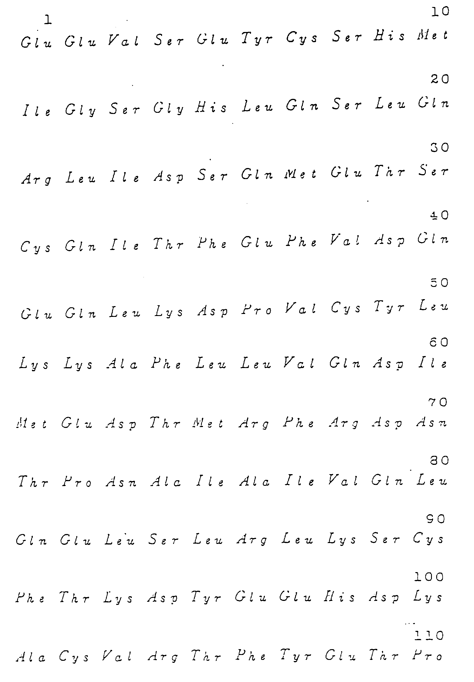
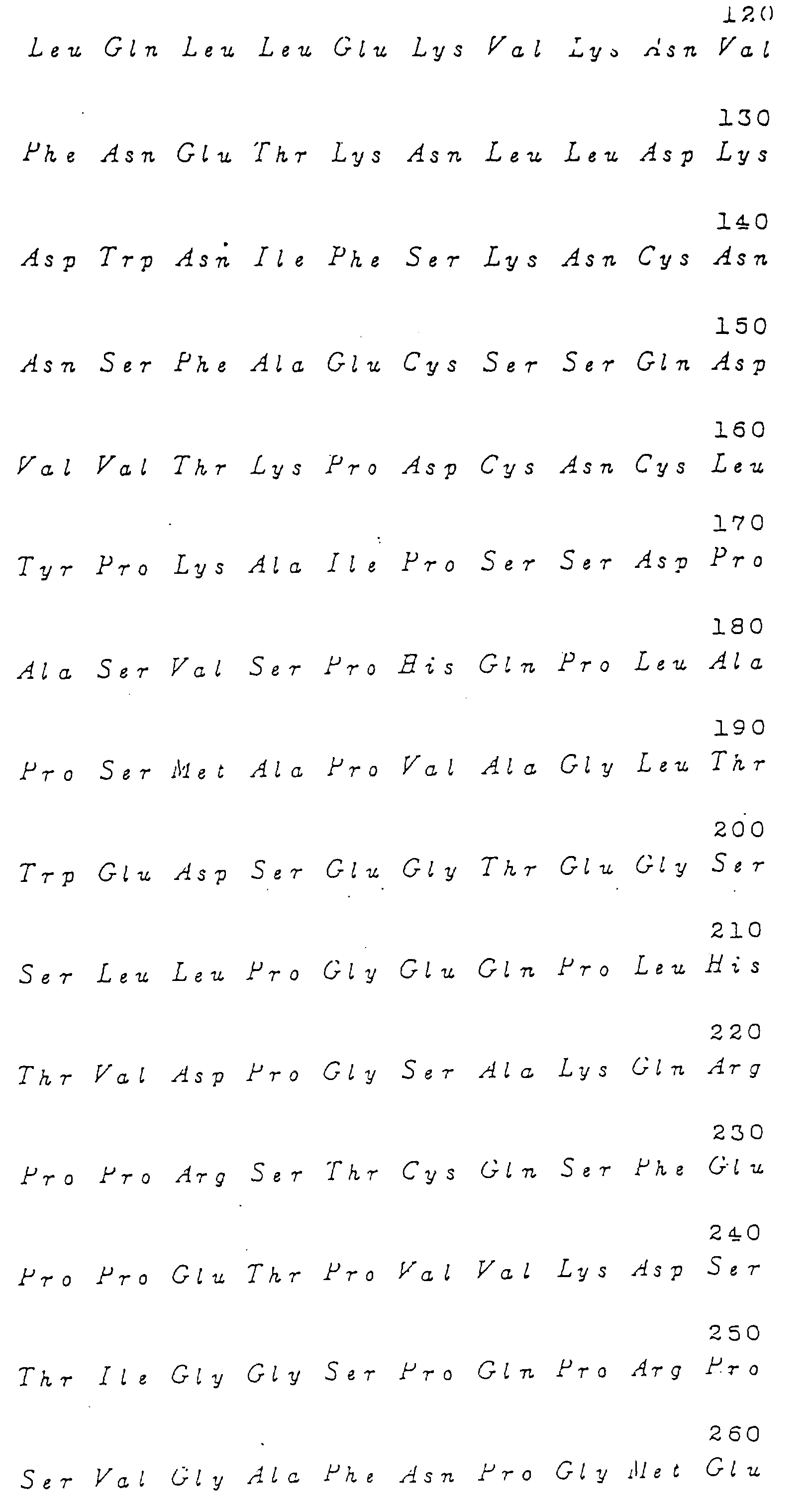
 sowie eine dazu komplementäre DNS derselben Länge.
- DNS gemäß Anspruch 1, wobei die das Polypeptid codierende DNS folgende Basensequenz aufweist:

- DNS gemäß Anspruch 1, wobei die das Polypeptid codierende DNS folgende Basensequenz oder einen Teil derselben aufweist:


- DNS gemäß einem der Ansprüche 1 - 3, die über eine mRNS hergestellt wird, die aus der M-CSF erzeugenden human Monocytenzellinie U937 (hinterlegt als ATCC CRL-1593) erhalten wird.
- DNS gemäß einem der Ansprüche 1 - 4, die über eine mRNS hergestellt wird, die mittels Zentrifugation in einem Saccharose-Dichtegradienten als 24,7 S- bis 27,0 S-Fraktion aus der M-CSF erzeugenden human Monocytenzellinie U937 (hinterlegt als ATCC CRL-1593) erhalten wird.
- Verfahren zur Herstellung der DNS gemäß den Ansprüchen 1 - 5, bei dem man eine mRNS aus einer human Monocytenzellinie isoliert, die aus einem von einer Knochenmarksvorläuferzelle abgeleitenen Monocyten, einem menschlichen subakuten lymphomatoiden Histiozyten sowie einer akuten myelomonocytischen Leukämiezelle ausgewählt ist, und die DNS aus der mRNS herstellt.
- Verfahren gemäß Anspruch 6, bei dem die M-CSF erzeugende human Monocytenzellinie U937 (hinterlegt als ATCC CRL-1953) in einem mit Phorbolmyristatacetat und Cycloheximid ergänzten Medium kultiviert wird.
- Transformant, der die DNS nach den Ansprüchen 1 - 5 enthält.
- DNS gemäß einem der Ansprüche 1 - 5, die mit einem nachweisbaren Marker verbunden ist.
- Biologisch aktives(r) Plasmid oder Vektor, das(der) eine DNS gemäß den Ansprüchen 1 - 5 enthält.
- Verfahren zur Herstellung eines Polypeptids mit M-CSF-Aktivität, bei dem man eine tierische Zelle mit einem von der DNS gemäß einem der Ansprüche 1 - 5 abgeleiteten Expressionsvektor transformiert.
- Verfahren gemäß Anspruch 11, bei dem die tierische Zelle aus CHO- und COSZellen ausgewählt wird.
- Verfahren gemäß Anspruch 11 oder 12, bei dem der Expressionsvektor von der folgenden Gruppe abgeleitet wird: dem Phagen λ CSF1-1 (gemäß der Definition in Beispiel 4(2)) und den Plasmiden pBS-II-CSF (hinterlegt als FERM BP-2526), pBSS (gemäß der Definition in Beispiel 5(3)), pSVL-CSF (gemäß der Definition in Beispiel 7(1)) und pSV2*-hCSF1-mdhfr (gemäß der Definition in Beispiel 8(1)).
- Verfahren nach den Ansprüchen 11 - 13, in denen das Polypeptid die kontinuierliche Aminosäuresequenz hat, die zwischen den Positionen 1 und 22 der Fig.3a und 3b liegt.
- Vektor für die Expression des im Harn ausgeschiedenen human M-CSF, der die für eine der Aminosäureseqzuenzen der Fig.10 codierende DNS, eine aus einem in einer tierischen Zelle wirkenden Promoter bestehende DNA und eine Enhancer-Sequenz aufweist.
- Verfahren zur Herstellung des im Harn ausgeschiedenen human M-CSF, bei dem man eine Säugerzelle mit dem Expressionsvektor gemäß Anspruch 15 transformiert und den Transformanten kultiviert, um den M-CSF aus der Kultur zu gewinnen.
- Polypeptid mit M-CSF-Aktivität, das aus einer der Aminosäuresequenzen der Fig.10 besteht, wobei X Ser bedeutet.
- Polypeptid gemäß Anspruch 17, bei dem n = 0 ist.
- Polypeptid gemäß Anspruch 17, bei dem n = 1 ist.
- Polypeptid mit M-CSF-Aktivität, das aus einer der Aminosäuresequenzen der Fig.20 besteht.
- Polypeptid gemäß Anspruch 20, bei dem n = 0 ist.
- Polypeptid gemäß Anspruch 20, bei dem n = 1 ist.
- Polypeptid gemäß einem der Ansprüche 20 - 22, bei dem Y die Bedeutung: -Gly-Ser-Ala-Lys-Gln-Arg-Pro-Pro-Arg-Ser-Thr-Cys-Gln-Aer-Phe-Glu-Pro-Pro-Glu-Thr-Pro-Val-Val-Lys hat.
- Polypeptid gemäß einem der Ansprüche 20 - 22, bei dem Y keine Aminosäure darstellt.
- Polypeptid gemäß einem der Ansprüche 20 - 24, bei dem X keine Aminosäure darstellt.
- Polypeptid gemäß einem der Ansprüche 20 - 24, bei dem X Glu-Glu-Val bedeutet.
- Polypeptid mit M-CSF-Aktivität, das aus einer der Aminosäuresequenzen der Fig.21 besteht.
- Polypeptid gemäß Anspruch 27, bei dem Y -Gly-Ser-Ala-Lys-Gln-Arg-Pro-Pro-Arg-Ser-Thr-Cys-Gln-Aer-Phe-Glu-Pro-Pro-Glu-Thr-Pro-Val-Val-Lys bedeutet.
- Polypeptid gemäß Anspruch 27, bei dem Y keine Aminosäure darstellt.
- Polypeptid gemäß einem der Ansprüche 27 - 29, bei dem X Met-Glu-Glu-Val bedeutet.
- Polypeptid gemäß einem der Ansprüche 27 - 29, bei dem X Met bedeutet.
- Polypeptid gemäß einem der Ansprüche 17 - 19 mit M-CSF-Aktivität und der Arninosäuresequenz der Fig.10 in Form eines Homodimeren.
- Polypeptid gemäß einem der Ansprüche 20 - 26 mit M-CSF-Aktivität und der Aminosäuresequenz der Fig.20 in Form eines Homodimeren.
- Polypeptid gemäß einem der Ansprüche 27 - 31 mit M-CSF-Aktivität und der Aminosäuresequenz der Fig.21 in Form eines Homodimeren.
- DNS-Sequenz, die ein Polypeptid gemäß einem der Ansprüche 17 - 31 codiert.
- Replizierbarer Clonierungsvektor, der die DNS-Sequenz gemäß Anspruch 35 sowie ein in einem Mikroorganismus funktionierendes Replikon enthält.
- Expressionssystem, in dem die DNS-Sequenz gemäß Anspruch 35 mit einer geeigneten Kontrollsequenz funktional verknüpft ist.
- Expressionssystem gemäß Anspruch 37, das in einem Vektor vorliegt, der sich in einer geigneten Wirtszelle replizieren kann.
- Expressionssystem gemäß Anspruch 35, bei dem die Expression des human M-CSF-Monomerproteins mittels zwei-Cistronen-Verfahren durchgeführt wird.
- Mikroorganismus als Transformant, der mit Hilfe eines Expressionssystems gemäß Anspruch 38 oder 39 transformiert worden ist.
- Verfahren zur Herstellung von human M-CSF, bei dem man einen ein Plasmid tragenden Transformanten, wobei das Plasmid ein human M-CSF-Monomerprotein exprimieren kann und der Transformant zur Erzeugung des Proteins die DNS gemäß Anspruch 35 aufweist, kultiviert, das Protein aus der Kultur gewinnt und das human M-CSF-Monomerprotein neu in ein biologisch aktives human M-CSF-Homodimerprotein faltet.
Applications Claiming Priority (4)
| Application Number | Priority Date | Filing Date | Title |
|---|---|---|---|
| JP192592/89 | 1989-07-27 | ||
| JP19259289 | 1989-07-27 | ||
| JP3635190 | 1990-02-19 | ||
| JP36351/90 | 1990-02-19 |
Publications (3)
| Publication Number | Publication Date |
|---|---|
| EP0410751A2 EP0410751A2 (de) | 1991-01-30 |
| EP0410751A3 EP0410751A3 (en) | 1991-10-23 |
| EP0410751B1 true EP0410751B1 (de) | 1996-04-10 |
Family
ID=26375395
Family Applications (1)
| Application Number | Title | Priority Date | Filing Date |
|---|---|---|---|
| EP90308199A Expired - Lifetime EP0410751B1 (de) | 1989-07-27 | 1990-07-26 | Für M-CSF kodierendes Gen und dazu gehörende rekombinante Systeme |
Country Status (3)
| Country | Link |
|---|---|
| EP (1) | EP0410751B1 (de) |
| JP (1) | JP2890326B2 (de) |
| DE (1) | DE69026440T2 (de) |
Families Citing this family (1)
| Publication number | Priority date | Publication date | Assignee | Title |
|---|---|---|---|---|
| US5650297A (en) * | 1986-09-17 | 1997-07-22 | Otsuka Pharmaceutical Co., Ltd. | DNA encoding human colony-stimulating factors |
Family Cites Families (8)
| Publication number | Priority date | Publication date | Assignee | Title |
|---|---|---|---|---|
| MX9203130A (es) * | 1986-05-06 | 1992-07-01 | Genetics Inst | Produccion de m-csf |
| JP2583770B2 (ja) * | 1986-09-17 | 1997-02-19 | 大塚製薬株式会社 | 遺伝子 |
| WO1988003173A2 (en) * | 1986-10-24 | 1988-05-05 | Cetus Corporation | New forms of colony stimulating factor-1 |
| AU606099B2 (en) * | 1986-12-07 | 1991-01-31 | Japan Science And Technology Corporation | Colony-stimulating factor and method for preparation thereof |
| JPH07100720B2 (ja) * | 1987-07-17 | 1995-11-01 | 森永乳業株式会社 | コロニー刺激因子の製造法 |
| IL86090A (en) * | 1987-04-16 | 1993-03-15 | Cetus Oncology Corp | Production of purified, biologically active, bacterially produced recombinant human csf-1 |
| DK54589A (da) * | 1988-02-08 | 1989-08-09 | Otsuka Pharma Co Ltd | Humane kolonistmulerende faktorer |
| JPH022391A (ja) * | 1988-02-08 | 1990-01-08 | Otsuka Pharmaceut Co Ltd | ヒトm―csf及びその製造法 |
-
1990
- 1990-07-26 EP EP90308199A patent/EP0410751B1/de not_active Expired - Lifetime
- 1990-07-26 DE DE1990626440 patent/DE69026440T2/de not_active Expired - Fee Related
- 1990-07-27 JP JP2197724A patent/JP2890326B2/ja not_active Expired - Lifetime
Non-Patent Citations (2)
| Title |
|---|
| CHEMICAL ABSTRACTS, vol. 108, no. 17, 25 April 1988, Columbus, OH (US); M. KAZUO, p. 152, no. 144143v * |
| THE EMBO JOURNAL, vol. 6, no. 9, September 1987, Eynsham, Oxford (GB); M.B. LADNER et al., pp. 2693-2698 * |
Also Published As
| Publication number | Publication date |
|---|---|
| DE69026440T2 (de) | 1996-10-17 |
| EP0410751A2 (de) | 1991-01-30 |
| DE69026440D1 (de) | 1996-05-15 |
| JPH03277284A (ja) | 1991-12-09 |
| EP0410751A3 (en) | 1991-10-23 |
| JP2890326B2 (ja) | 1999-05-10 |
Similar Documents
| Publication | Publication Date | Title |
|---|---|---|
| US5464939A (en) | Highly purified protein, production and use thereof | |
| EP0077670B1 (de) | Humanes Immuninterferon | |
| AU594045B2 (en) | Recombinant colony stimulating factor-1 | |
| US5096705A (en) | Human immune interferon | |
| EP0158198A1 (de) | DNA und ihre Verwendung | |
| JP2541761B2 (ja) | ミュレル管抑制物質様ポリペプチドおよびその製造方法 | |
| US5582824A (en) | Recombinant DES-CYS-TYR-CYS human immune interferon | |
| KR970002917B1 (ko) | 인터루킨-i 억제제 | |
| EP0456332B1 (de) | Gereinigtes Interleukin-1 und für Interleukin-1 kodierende DNS und deren Herstellung, solche DNS enthaltende Vektoren und deren Herstellung und Verwendung zum Transformieren von Wirten, die die Expression von Interleukin-1 zulassen | |
| US6171824B1 (en) | Hybrid cytokines | |
| CA2092542A1 (en) | Hemopoietic stem cell multiplier | |
| EP0410751B1 (de) | Für M-CSF kodierendes Gen und dazu gehörende rekombinante Systeme | |
| EP0328061A2 (de) | Menschlicher Koloniestimulierungsfaktor | |
| EP0194006B1 (de) | Analoge Interferon-Polypeptide, Verfahren zu deren Herstellung und diese enthaltende pharmazeutische Zusammensetzungen | |
| CA1302652C (en) | Method for purifying granulocyte-macrophage colony stimulating factor | |
| US5650297A (en) | DNA encoding human colony-stimulating factors | |
| JPH0335795A (ja) | ヒトインターロイキン2活性をもつポリペプチドの製造法 | |
| EP0200417B1 (de) | Produktion eines Proteins | |
| WO1989010962A1 (en) | Process for producing a human neutrophil chemotactic factor polypeptide | |
| US6465616B1 (en) | Interleukin-5 antagonist | |
| WO1997045448A1 (en) | An interleukin-5 antagonist | |
| AU723413B2 (en) | An interleukin-5 antagonist | |
| JP2839837B2 (ja) | 顆粒球コロニー刺激因子受容体のリガンド結合領域蛋白質をコードしているdna | |
| CA1339873C (en) | Recombinant colony stimulating factor-1 | |
| JPH0482900A (ja) | 新規m―csf及びその製造方法 |
Legal Events
| Date | Code | Title | Description |
|---|---|---|---|
| PUAI | Public reference made under article 153(3) epc to a published international application that has entered the european phase |
Free format text: ORIGINAL CODE: 0009012 |
|
| AK | Designated contracting states |
Kind code of ref document: A2 Designated state(s): DE FR GB |
|
| 17P | Request for examination filed |
Effective date: 19901221 |
|
| PUAL | Search report despatched |
Free format text: ORIGINAL CODE: 0009013 |
|
| AK | Designated contracting states |
Kind code of ref document: A3 Designated state(s): DE FR GB |
|
| 17Q | First examination report despatched |
Effective date: 19930707 |
|
| GRAH | Despatch of communication of intention to grant a patent |
Free format text: ORIGINAL CODE: EPIDOS IGRA |
|
| GRAA | (expected) grant |
Free format text: ORIGINAL CODE: 0009210 |
|
| AK | Designated contracting states |
Kind code of ref document: B1 Designated state(s): DE FR GB |
|
| REF | Corresponds to: |
Ref document number: 69026440 Country of ref document: DE Date of ref document: 19960515 |
|
| ET | Fr: translation filed | ||
| PLBE | No opposition filed within time limit |
Free format text: ORIGINAL CODE: 0009261 |
|
| STAA | Information on the status of an ep patent application or granted ep patent |
Free format text: STATUS: NO OPPOSITION FILED WITHIN TIME LIMIT |
|
| 26N | No opposition filed | ||
| PGFP | Annual fee paid to national office [announced via postgrant information from national office to epo] |
Ref country code: GB Payment date: 19980701 Year of fee payment: 9 |
|
| PGFP | Annual fee paid to national office [announced via postgrant information from national office to epo] |
Ref country code: FR Payment date: 19980730 Year of fee payment: 9 |
|
| PGFP | Annual fee paid to national office [announced via postgrant information from national office to epo] |
Ref country code: DE Payment date: 19980831 Year of fee payment: 9 |
|
| PG25 | Lapsed in a contracting state [announced via postgrant information from national office to epo] |
Ref country code: GB Free format text: LAPSE BECAUSE OF NON-PAYMENT OF DUE FEES Effective date: 19990726 |
|
| PG25 | Lapsed in a contracting state [announced via postgrant information from national office to epo] |
Ref country code: FR Free format text: THE PATENT HAS BEEN ANNULLED BY A DECISION OF A NATIONAL AUTHORITY Effective date: 19990731 |
|
| GBPC | Gb: european patent ceased through non-payment of renewal fee |
Effective date: 19990726 |
|
| PG25 | Lapsed in a contracting state [announced via postgrant information from national office to epo] |
Ref country code: DE Free format text: LAPSE BECAUSE OF NON-PAYMENT OF DUE FEES Effective date: 20000503 |
|
| REG | Reference to a national code |
Ref country code: FR Ref legal event code: ST |