EP0434399A1 - Instrument moniteur pour artères périphériques - Google Patents
Instrument moniteur pour artères périphériques Download PDFInfo
- Publication number
- EP0434399A1 EP0434399A1 EP90313929A EP90313929A EP0434399A1 EP 0434399 A1 EP0434399 A1 EP 0434399A1 EP 90313929 A EP90313929 A EP 90313929A EP 90313929 A EP90313929 A EP 90313929A EP 0434399 A1 EP0434399 A1 EP 0434399A1
- Authority
- EP
- European Patent Office
- Prior art keywords
- cuff
- arterial
- volume
- pressure
- deflating
- Prior art date
- Legal status (The legal status is an assumption and is not a legal conclusion. Google has not performed a legal analysis and makes no representation as to the accuracy of the status listed.)
- Granted
Links
Images




Classifications
-
- A—HUMAN NECESSITIES
- A61—MEDICAL OR VETERINARY SCIENCE; HYGIENE
- A61B—DIAGNOSIS; SURGERY; IDENTIFICATION
- A61B5/00—Measuring for diagnostic purposes; Identification of persons
- A61B5/02—Detecting, measuring or recording for evaluating the cardiovascular system, e.g. pulse, heart rate, blood pressure or blood flow
- A61B5/02007—Evaluating blood vessel condition, e.g. elasticity, compliance
-
- A—HUMAN NECESSITIES
- A61—MEDICAL OR VETERINARY SCIENCE; HYGIENE
- A61B—DIAGNOSIS; SURGERY; IDENTIFICATION
- A61B5/00—Measuring for diagnostic purposes; Identification of persons
- A61B5/02—Detecting, measuring or recording for evaluating the cardiovascular system, e.g. pulse, heart rate, blood pressure or blood flow
- A61B5/021—Measuring pressure in heart or blood vessels
- A61B5/022—Measuring pressure in heart or blood vessels by applying pressure to close blood vessels, e.g. against the skin; Ophthalmodynamometers
-
- A—HUMAN NECESSITIES
- A61—MEDICAL OR VETERINARY SCIENCE; HYGIENE
- A61B—DIAGNOSIS; SURGERY; IDENTIFICATION
- A61B5/00—Measuring for diagnostic purposes; Identification of persons
- A61B5/02—Detecting, measuring or recording for evaluating the cardiovascular system, e.g. pulse, heart rate, blood pressure or blood flow
- A61B5/021—Measuring pressure in heart or blood vessels
- A61B5/022—Measuring pressure in heart or blood vessels by applying pressure to close blood vessels, e.g. against the skin; Ophthalmodynamometers
- A61B5/02225—Measuring pressure in heart or blood vessels by applying pressure to close blood vessels, e.g. against the skin; Ophthalmodynamometers using the oscillometric method
Definitions
- This invention relates to monitoring instruments which provide information concerning peripheral vasculature and, in particular, to the use of such instruments to provide medical diagnostic information concerning arterial volume, cross-sectional area, and compliance.
- arteriosclerosis and hypertension are potentially debilitating and often life-threatening conditions which require early diagnosis and treatment. These conditions are characterized by changes in arterial blood flow volumes and rates and the response of arterial tissue to changes in blood pressure.
- a physiological phenomenon which plays a part in these arterial characteristics is referred to herein as arterial compliance, the ability of vasculature to respond to changes in these conditions.
- the arterial walls of the body include collagen, giving the walls the ability to expand and contract, and muscle tissue which in part controls this expansion and contraction.
- Vascular compliance includes the response of collagen and muscle in arterial walls to changing conditions.
- the condition of arteriosclerosis is characterized by the buildup of fatty substances along arterial walls.
- arteriosclerosis can occlude the artery, and can impede the ability of the arterial walls to respond to changing conditions of blood pressure.
- the fatty substances characteristic of arteriosclerosis are thus a further factor governing arterial compliance. It is thus desirable to be able to analytically understand arterial volume and compliance when diagnosing or treating the medical conditions of hypertension and arteriosclerosis.
- An understanding of a patient's arterial volume and compliance is also beneficial when administering anesthesia.
- the quantity of anesthetic administered to a patient should be just sufficient to eliminate a physiological response by the patient during surgery. If an insufficient amount of anesthetic has been administered, the cardiovascular system will respond reflexively when the patient is intubated prior to surgery. This response can be detected by monitoring arterial volume and compliance, and noting any reduction in these characteristics during intubation. A cardiovascular response can also be detected at the time of the first surgical incision, when an insufficient anesthetic will again be evidenced by a reduction in arterial compliance or volume. Thus, a surgical patient would benefit from the monitoring of arterial volume and compliance by the anesthesiologist and surgeon.
- Link extends this analysis to a technique in which the peak to peak amplitude of each cuff pulse and the patient's diastolic and systolic pressures are used to calculate a particular patient's own volumetric and compliance curves.
- the volumetric and pressure information is determined inferentially from arterial pressure pulse information.
- the volumetric and pressure curves are used by Link in the determination of systolic, diastolic, and mean arterial blood pressure.
- the Link technique utilizes a ramp-up method of measuring pressure pulses, wherein pulse data is taken during inflation of a blood pressure cuff.
- a display of information concerning arterial volume which is useful to the anesthesiologist, surgeon or diagnostician is a curve representing arterial volume (in cc) or area (in mm2) as a function of transmural pressure (in mm Hg).
- the volume or area may be represented by a value R, the effective arterial radius.
- the slope of the curve at any given point, dV/dP, represents arterial compliance, and a plot of dV/dP as a function of transmural pressure represents the arterial compliance curve.
- the patient's arterial volume and compliance is represented in this format and, in correspondence thereto, the value of R over time is calculated and displayed.
- the display of this data provides the anesthesiologist with information concerning the patient's arterial volume and compliance characteristics, and also provides information as to changes occurring in arterial volume over time. This will enable the anesthesiologist to detect any response of the cardiovascular system to intubation or incision during a surgical procedure, thereby facilitating the correct delivery of anesthetic to the patient.
- a display as described above may be further enhanced by providing the arterial compliance dV/dP at a given transmural pressure for a patient undergoing diagnosis or monitoring.
- the maximum value of dV/dP referred to as peak arterial compliance, can also be ascertained from this information.
- a further display of this information which would be of use to a clinician would be a representation of arterial capacity, R, in relation to the circumference of the limb at which the blood pressure cuff of the monitoring instrument is attached.
- a variation of this display format provides a display of the patient's arterial volume data prior to the initiation of any surgical intervention and, in correspondence therewith, a current display of arterial volume data as the surgical intervention proceeds. Comparison of the data informs the anesthesiologist of the cardiovascular system response to bodily stimuli during the procedure.
- a recent proposal relating to the determination of vascular compliance is known as the "Hartsafe Product Concept.”
- a pressure cuff is strapped to a patient's calf and inflated.
- a calibration step is initiated by injecting one ml of air into the cuff.
- the system measures the change in pressure resulting from this quantified injection and calculates a calibration factor based upon the change.
- Cuff inflation continues and volume pulse signals are recorded until a minimal volume pulse signal or a maximum pressure value of 225 mm Hg is attained.
- the system then commences a step deflate sequence. At individual pressure decrement steps of 10 mm Hg the volume pulse signal is recorded.
- the sequence continues until a minimal pressure level is attained, at which time data acquision is complete.
- the system then performs "signal conditioning" using the volume pulse and cuff pressure signals at each 10 mm Hg cuff pressure decrement, and the calibrate signal previously stored.
- the volume-pressure curve, peak compliance, and other parameters are obtained by this "signal conditioning."
- the "Hartsafe” approach seems to be a more direct measurement of arterial volume than the Link techniques, in which arterial volume is calculated premised upon its relationship to the arterial pressure pulses, because an actual measurement of system response to a known change in cuff volume is taken during the calibration step of "Hartsafe.” But the actual data which is computed for the volume-pressure curve appears to be similarly inferential, however, as the single calibration volume measurement is the only volumetric measure used in conjunction with the pulse signals to inferentially calculate the curve.
- a system for measuring arterial cross-section and compliance in which a pressure cuff on a peripheral part of the body is inflated to a pressure level which occludes arterial vessels. The cuff is then deflated, and pressure measurements taken in correspondence with decreasing pressure levels. Air which is expelled during deflation is removed from the cuff through means for determining the volume of air expelled.
- This means may comprise, for instance, an orifice or transfer volume of known characteristics, or a flow measurement device.
- the volume of air removed from the cuff is precisely known, or is calculated based upon an immediately obtained volumetric calibration.
- the oscillation pressure peaks and changes in cuff volume as a function of pressure are recorded over a range of cuff pressures. From this information the oscillation volume is calculated. From the knowledge of oscillation volume measurements over the range of cuff pressures and conventional determination of systolic and diastolic pressure levels, the patient's arterial volume and compliance curves are reconstructed. Thus, accurate and complete information concerning blood pressure and arterial volume, cross-section and compliance in relation to transmural pressure and/or time is provided to the physician for monitoring and diagnosis.
- FIGURE 1 a peripheral arterial monitoring instrument constructed in accordance with the principles of the present invention is shown in use on the leg of a patient.
- the instrument includes a conventional blood pressure cuff 10 having a length l which is wrapped about the thigh of the patient.
- the cuff 10 may be applied to any peripheral part of the body and is most conventionally applied to the upper arm, it is preferable to use the thigh in some procedures as that is where buildups of occlusive substances leading to arteriosclerosis and the like generally first manifest themselves. In other applications the upper arm or finger may be a preferred site for application of the cuff.
- the cuff 10 is connected by tubing 12 to a monitor and processor 14.
- the monitor and processor 14 includes a number of controls for actuating and adjusting the instrument in the performance of vascular measurements including blood pressure determination.
- the monitor and processor also includes a display 16 where the data taken during measurements of arterial volume is displayed, either in numerical or, preferably, in graphical form as shown in FIGURES 2a and 2b.
- the monitor and processor includes a controlled pneumatic system which controls inflation and deflation of the cuff 10 during which time measurements leading to the determination of the patient's arterial volume and compliance are taken.
- FIGURES 2a and 2b illustrate several preferred techniques for displaying the information obtained through these measurements.
- the upper portion of the display of FIGURE 2a is a graphical display of arterial volume (or area) versus transmural pressure.
- the height of the righthand portion of the curve 20 also represents the effective radius of the arterial vessels R when the vessels are filled with blood.
- the slope of the curve 20, dV/dP represents arterial compliance and the point at which dV/dP exhibits a maximum value is generally referred to as peak arterial compliance.
- the upper graph of FIGURE 2a provides the physician with information as to arterial volume, compliance and effective arterial radius in the limb where the cuff is affixed.
- the illustrative curve 22 of R versus time shown in the drawing is seen to be substantially flat, except at at time indicated by 23. This decrease in the R value may correlate for instance with the time at which some physical intervention such as intubation or incision is performed on the patient. If the patient is not fully anesthetized at that time, the cardiovascular system will react by contracting the arteries of the body, and the effective radius of arterial vessels will decline. Thus, the decline in curve 22 at point R would indicate to the anesthesiologist that the patient is not fully anesthetized, and further anesthetic may be required for patient comfort and safety.
- FIGURE 2b shows a further display of the arterial volume and compliance information which would be of assistance to an anesthesiologist.
- This curve of the patient's normal arterial volume is labelled as V/P init., the initial curve determined by the monitor and processor.
- V/P init. the initial curve determined by the monitor and processor.
- a current volume versus pressure curve is calculated periodically and displayed in correspondence with the initial curve.
- the current curve is labelled V/P curr. in FIGURE 2b.
- the display of FIGURE 2b provides the anesthesiologist with a continuous comparison of current arterial volume and compliance versus the patient's normal arterial volume and compliance prior to the administration of anesthetic.
- FIGURES 3a and 3b are cross-sectional illustrations of arteries showing the parameters measured by the monitor and processor 14.
- the R value is the radius of an artery 30 as shown in FIGURE 3a. Since the cuff encloses all arterial vessels in the portion of the limb about which it is wrapped, it will be understood that the R value is not the radius of a particular artery, but is in effect the sum of the radii of all of the arteries inside the cuff 10. Thus, the instrument provides an R value which is the effective radius taken over all arterial vessels inside the cuff.
- the artery 30 is defined by the arterial wall 32.
- the arterial wall is composed principally of two substances, collagen and smooth muscle tissue.
- Collagen provides the artery with flexibility, the ability to stretch and deform. This rubber-like characteristic is one contributor to arterial compliance, and is a passive characteristic of arteries.
- the muscle tissue is controlled by nerves to provide stretching and deformation of the artery under control of the body's nervous system. This stretching and deformation is an active characteristic of the artery which also is a factor in arterial compliance.
- FIGURE 3b Arterial volume and compliance are also affected in the case of arteriosclerosis or hardening of the arteries by the buildup of fatty substances on the inner walls of the arteries. This condition is shown in FIGURE 3b, where a buildup of substances is indicated at 34 lining the wall of the artery.
- the ability of the artery to expand or contract under the influence of arterial muscular contraction or blood pressure changes is adversely affected by this lining of fatty substances, which can retard such motion. Since the substances also occupy a portion of the inner volume of the artery, the effective radius of the vessel R' is decreased by the presence of these substances.
- FIGURE 4 illustrates an arrangement for taking measurements of arterial volume and compliance. Shown in FIGURE 4 are a limb of the body 40 in cross-section, about which a blood pressure cuff 10 is wrappped. The skin line of the limb is indicated at 41. The cross-sectional view of the limb shows the bone 42 at the center of the limb, and an artery 44 passing through the limb. The artery 44 is shown expanded during the pumping of blood, before the cuff is applied and inflated. After inflation of the cuff to a maximal pressure, the artery will be occluded, as shown at 44'.
- the cuff 10 is connected by pneumatic tubing to a pump 50.
- the pump 50 pumps up the cuff 10 at the start of the measurement cycle.
- the arrangement of FIGURE 4 is modified to perform the process of the "Hartsafe Product Concept" discussed above by the inclusion of a calibration chamber 54, which is connected to the pneumatic system.
- a calibration chamber 54 which is connected to the pneumatic system.
- the pump 50 is stopped and one ml of air is injected into the pneumatic system of the cuff. This may be accomplished by moving piston 56 in the chamber 54 to the right to displace one ml of air from the chamber. Given that all elements of the pneumatic system are substantially noncompliant, this one ml volume of air will compress the limb 40 by one ml.
- the effect of the piston displacement will be to displace one ml of blood from the vascular system within the confines of the cuff.
- the process of the "Hartsafe Product Concept" calculates its calibration factor at the outset of the measurement cycle.
- the pump then inflates the cuff to fully occlude the arterial vessels as shown at 44', and the deflate cycle commences.
- a deflation valve opens and closes to incrementally bleed air from the pneumatic system. Measurements taken by a pressure transducer P T at each pressure step are stored in correspondence with cuff pressure level and are subsequently used in a signal conditioning (processing) step at the end of the deflation cycle.
- FIGURE 4 illustrates a peripheral arterial volume and compliance measurement system of the present invention which obviates the need for such structural and operational complexity.
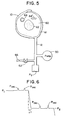
- the blood pressure cuff 10 is wrapped around the thigh 60 of the patient, shown in cross-section.
- the femur 62 is shown in the center of the thigh, and the skin line of the thigh is indicated at 61.
- the femural artery is illustrated at 64 in an unoccluded condition, and in an occluded condition at 64'.
- the cuff 10 is connected by pneumatic tubing 12 to a pump 50, a pressure transducer P T and a deflate valve 52.
- An orifice 66 of predetermined cross-sectional area is located in the deflate valve outlet.
- the pneumatic system of FIGURE 5 is operated in the conventional manner of a step-deflate automated blood pressure monitor such as the Critikon Dinamap TM 8100.
- the cuff 10 is inflated by the pump 50 to a pressure which is in excess of systolic pressure, sufficient to fully occlude the artery 64'.
- the cuff pressure is stepped down, and the cuff pressures and oscillation pulses are recorded from the pressure transducer.
- Two of the pressure steps during the deflate cycle are shown in FIGURE 6.
- the cuff pressures of the two steps are P 1 and P2, and the oscillation pulses are shown as P osc.
- the cuff pressure is stepped down in decrements of approximately 8 mm Hg.
- the flow of air from the pneumatic system is calculated from the equation where P is the pressure across the orifice, the number 760 is an adjustment factor for nominal barometric pressure, and ⁇ is an adiabatic constant.
- P is the pressure across the orifice
- the number 760 is an adjustment factor for nominal barometric pressure
- ⁇ is an adiabatic constant.
- the flow through a 1 cm2 orifice as a function of the pressure across the orifice during a typical deflate cycle is represented graphically in FIGURE 9.
- Other known methods for measuring the flow of a fluid may also be employed; for instance, if the orifice in a given embodiment does not conform to theoretical models, it may be approximated empirically.
- AV n the volume of air removed during each decrement
- a eq is the equivalent area of the orifice
- FLOW n is the flow rate between two pressure steps
- ⁇ t n is the time during which the deflate valve was open between the two pressure steps.
- the FLOW is known from the preceding equation, the equivalent area of the orifice is known, and the time during which the deflate valve is open is measured by a digital clock which runs during the time that the valve is open. Since the FLOW calculation is done for each deflation step based upon the known orifice and the then extant pressure, no recalibration or modification is necessary or required for the calculated values.
- a ratio can be formed of the ⁇ V n values and the respective cuff pressure differentials at which they were obtained.
- the value of P osc n used for each step decrement may be the amplitude of the oscillation pulses on the P 1 step, the P2 step, or an average of the two, due to the very small variation in oscillation pulse amplitude from one step to the next. Whichever approach is used, it is consistently applied for the full range of step values. Curves representing V osc and the oscillation pulses as a function of cuff pressure are illustrated in FIGURE 10.
- the arterial volume curve can now be computed in a two-step procedure.
- the first step is to compute a curve referred to herein as a reconstruction curve from knowledge of the V osc n values and the values of systolic and diastolic blood pressure determined by the Dinamap TM in the conventional manner.
- the arterial volume curve is then computed by coordinate system transformation, by which the reconstruction curve, referenced to cuff pressure, is converted to arterial transmural pressure with reference to systolic pressure.
- Recon n (P cuff ) V osc n (P cuff ) + Recon n (P cuff + S - D)
- S systolic pressure
- D diastolic pressure
- the difference of systolic minus diastolic pressure is referred to herein as pulse pressure.
- Recon n (P cuff + S - D) 0 when (p cuff + S - D) is greater than P cuff max' where P cuff max is the maximum cuff pressure used in a particular measurement. This follows from the knowledge that at maximum cuff pressure the arteries in the limb are completely occluded.
- a graphical plot of the points Recon n (P cuff ) as a function of cuff pressure is shown by the dashed curve Recon in FIGURE 11 in comparison with the V osc curve previously shown in FIGURE 10. It is seen that the plot of Recon converges with the V osc curve above and in the vicinity of systolic pressure.
- the arterial volume may be calculated as a function of transmural pressure by, in effect, transforming the Recon curve about the axis of systolic pressure.
- the arterial volume curve produced by this transformation is of the general shape of curve 20 of FIGURE 2a and the curves of FIGURE 2b.
- a point of reference for selection of R and dV/dP may be chosen in a number of ways.
- the monitor may compute mean arterial pressure in the conventional manner, and use the value of mean arterial pressure as the pressure for which R and dV/dP are chosen and displayed.
- the pressure at which dV/dP is at a maximum, peak arterial compliance can be used as the pressure reference for selecting R and dV/dP.
- the physician selects a transmural pressure value on the abscissa of the upper curve of FIGURE 2a as the pressure for R and dV/dP.
- the slope of the curve at the selected pressure point can be calculated to determine arterial compliance dV/dP, and the amplitude of the volume curve at the selected pressure provides the R value.
- the instrument is repeatedly actuated automatically and an R value is found each time.
- the R value is then displayed as a function of time as shown at the bottom of FIGURE 2a.
- the volume curve calculated at the beginning of a surgical procedure is stored and continuously displayed with the most recently calculated curve in the format shown in FIGURE 2b.
- Another display which can be obtained from this data which would be of use to a clinician is a plot of dV/dP versus time, showing historic changes in the patient's arterial compliance during a surgical procedure.
- R or arterial area or volume
- Limb size is obtained by measuring the circumference of the limb where the cuff is attached, and entering this information into the monitor and processor 14. The ratio of this circumference (or calculated limb radius or cross-sectional area) to R (or arterial area or volume) provides an indication of cardiovascular efficiency.
- a flowmeter which measures the flow of expelled air could be used to provide a direct measurement of flow volume at the output of the deflate valve 52.
- Another alternative embodiment is to use a transfer volume of known capacity as shown in FIGURES 7 and 8.
- the transfer volume comprises all of the volumetric space between an intermediate dump valve 52a and the deflate valve 52.
- the size of the vessel indicated at 58 is chosen to provide the desired volume of the entire transfer volume.
- the deflate valve 52 is closed after previous closure of the dump valve 52a.
- the air in the transfer volume between the two valves is now at atmospheric pressure.
- the dump valve 52a is then opened, and the transfer volume becomes pressurized to the cuff pressure, which declines to P tr by reason of the expansion of pressurized air into the transfer volume.
- the deflate valve 52 is then opened so that both valves are in the open condition. Air is expelled from the pneumatic system of the cuff and the pressure transducer is monitored until the pressure reaches the level P2, at which point the dump valve 52a is closed. The deflate valve 52 is then closed, stabilizing the transfer volume at atmospheric pressure in preparation for the next step decrement.
- the transfer volume technique is advantageously employed to enable use of a total pressure step P1-P2 which is conventional for a standard blood pressure monitor such as the Dinamap TM 8100, which uses pressure step decrements of approximately 8 mm Hg.
- a standard blood pressure monitor such as the Dinamap TM 8100, which uses pressure step decrements of approximately 8 mm Hg.
Landscapes
- Health & Medical Sciences (AREA)
- Life Sciences & Earth Sciences (AREA)
- Vascular Medicine (AREA)
- Cardiology (AREA)
- Biomedical Technology (AREA)
- Molecular Biology (AREA)
- Physiology (AREA)
- Biophysics (AREA)
- Pathology (AREA)
- Engineering & Computer Science (AREA)
- Veterinary Medicine (AREA)
- Heart & Thoracic Surgery (AREA)
- Medical Informatics (AREA)
- Physics & Mathematics (AREA)
- Surgery (AREA)
- Animal Behavior & Ethology (AREA)
- General Health & Medical Sciences (AREA)
- Public Health (AREA)
- Ophthalmology & Optometry (AREA)
- Measuring Pulse, Heart Rate, Blood Pressure Or Blood Flow (AREA)
- Measuring Volume Flow (AREA)
Applications Claiming Priority (2)
| Application Number | Priority Date | Filing Date | Title |
|---|---|---|---|
| US453519 | 1989-12-20 | ||
| US07/453,519 US5103833A (en) | 1989-12-20 | 1989-12-20 | Peripheral arterial monitoring instruments |
Publications (2)
| Publication Number | Publication Date |
|---|---|
| EP0434399A1 true EP0434399A1 (fr) | 1991-06-26 |
| EP0434399B1 EP0434399B1 (fr) | 1996-09-04 |
Family
ID=23800875
Family Applications (1)
| Application Number | Title | Priority Date | Filing Date |
|---|---|---|---|
| EP90313929A Expired - Lifetime EP0434399B1 (fr) | 1989-12-20 | 1990-12-19 | Instrument moniteur pour artères périphériques |
Country Status (10)
| Country | Link |
|---|---|
| US (4) | US5103833A (fr) |
| EP (1) | EP0434399B1 (fr) |
| JP (1) | JP3121845B2 (fr) |
| AU (1) | AU649003B2 (fr) |
| CA (1) | CA2032564A1 (fr) |
| DE (1) | DE69028380T2 (fr) |
| ES (1) | ES2090108T3 (fr) |
| FI (1) | FI906267A7 (fr) |
| NO (1) | NO905479L (fr) |
| ZA (1) | ZA9010238B (fr) |
Cited By (7)
| Publication number | Priority date | Publication date | Assignee | Title |
|---|---|---|---|---|
| FR2682279A1 (fr) * | 1991-10-11 | 1993-04-16 | Tissot Marie Francoise | Appareil pour mesurer le volume d'un membre. |
| EP0627191A1 (fr) * | 1993-05-03 | 1994-12-07 | Critikon, Inc. | Instrument de surveillance des artéries périphériques |
| EP0627190A1 (fr) * | 1993-05-17 | 1994-12-07 | Omron Corporation | Dispositif de mesure de la fonction vasculaire |
| WO1995007650A1 (fr) * | 1993-09-14 | 1995-03-23 | Fred Zacouto | Procede et dispositif pour l'etude, le diagnostic et le traitement du systeme cardiovasculaire, notamment arteriel |
| DE19505765A1 (de) * | 1994-02-18 | 1995-10-05 | Vladimir Dr Blazek | Vorrichtung zur Anpreßdruckmessung medizinischer Kompressionsstrümpfe und Verbände |
| US6126595A (en) * | 1995-05-12 | 2000-10-03 | Seiko Epson Corporation | Device for diagnosing physiological state and device for controlling the same |
| EP2814383A4 (fr) * | 2012-02-16 | 2015-09-23 | Welch Allyn Inc | Systèmes et procédés de surveillance d'un patient |
Families Citing this family (42)
| Publication number | Priority date | Publication date | Assignee | Title |
|---|---|---|---|---|
| US5103833A (en) * | 1989-12-20 | 1992-04-14 | Critikon, Inc. | Peripheral arterial monitoring instruments |
| US5607447A (en) * | 1993-09-28 | 1997-03-04 | Mcewen; James A. | Physiologic tourniquet |
| US5254087A (en) * | 1990-01-29 | 1993-10-19 | Ivra Systems, Inc. | Tourniquet apparatus for intravenous regional anesthesia |
| US5556415A (en) * | 1990-01-29 | 1996-09-17 | Mcewen; James A. | Physiologic tourniquet for intravenous regional anesthesia |
| US5584853A (en) * | 1990-01-29 | 1996-12-17 | Mcewen; James A. | Tourniquet cuff apparatus |
| JP3290176B2 (ja) * | 1991-06-12 | 2002-06-10 | フロリダ アトランティック ユニヴァーシティ リサーチ コーポレイション | ヒトのアテローム性動脈硬化症の検知 |
| DE69416475T2 (de) * | 1993-04-02 | 1999-07-22 | Nagano, Ken, Nagano, Nagano | Elektronisches instrument zur blutdruck-messung |
| US6371921B1 (en) | 1994-04-15 | 2002-04-16 | Masimo Corporation | System and method of determining whether to recalibrate a blood pressure monitor |
| US5766130A (en) * | 1994-08-16 | 1998-06-16 | Selmonosky; Carlos A. | Vascular testing method |
| US5518000A (en) * | 1994-09-30 | 1996-05-21 | Johnson & Johnson Medical, Inc. | Oscillometric blood pressure monitor employing deflation periods of alternating durations |
| US5606977A (en) * | 1995-01-04 | 1997-03-04 | Critikon, Inc. | Oscillometric blood pressure monitor which automatically determines when to take blood pressure measurements |
| US5680870A (en) * | 1995-01-04 | 1997-10-28 | Johnson & Johnson Medical, Inc. | Oscillometric blood pressure monitor which acquires blood pressure signals from composite arterial pulse signal |
| US5579776A (en) * | 1995-01-13 | 1996-12-03 | Johnson & Johnson Medical, Inc. | Oscillometric blood pressure monitor with enhanced cuff pressure control |
| US5577508A (en) * | 1995-01-13 | 1996-11-26 | Johnson & Johnson Medical, Inc. | Determination of oscillometric blood pressure by linear approximation |
| US5590662A (en) * | 1995-02-15 | 1997-01-07 | Johnson & Johnson Medical, Inc. | Detection of oscillometric blood pressure complexes using correlation |
| US5800359A (en) * | 1995-05-19 | 1998-09-01 | Johnson & Johnson Medical, Inc. | NIBP playback system |
| US5931853A (en) * | 1995-08-25 | 1999-08-03 | Mcewen; James A. | Physiologic tourniquet with safety circuit |
| US5855589A (en) * | 1995-08-25 | 1999-01-05 | Mcewen; James A. | Physiologic tourniquet for intravenous regional anesthesia |
| US6027452A (en) | 1996-06-26 | 2000-02-22 | Vital Insite, Inc. | Rapid non-invasive blood pressure measuring device |
| US5991654A (en) * | 1997-06-06 | 1999-11-23 | Kci New Technologies, Inc. | Apparatus and method for detecting deep vein thrombosis |
| GB9714550D0 (en) * | 1997-07-10 | 1997-09-17 | Lidco Ltd | Improved method and apparatus for the measurement of cardiac output |
| US6309359B1 (en) | 1998-06-01 | 2001-10-30 | Michael D. Whitt | Method and apparatus for noninvasive determination of peripheral arterial lumenal area |
| US6095983A (en) * | 1998-07-24 | 2000-08-01 | Welch Allyn, Inc. | Electro-pneumatic assembly for blood pressure cuff |
| US6152881A (en) * | 1999-03-29 | 2000-11-28 | Vasocor, Inc. | Calibrated measurement of blood vessels and endothelium after reactive hyperemia and method therefor |
| US6315735B1 (en) * | 1999-03-31 | 2001-11-13 | Pulsion Medical Systems Ag | Devices for in-vivo determination of the compliance function and the systemic blood flow of a living being |
| DE10016993A1 (de) | 2000-04-07 | 2001-10-11 | Basf Ag | Anchlorierte Kupferphthalocyaninpigmente |
| JP2003250772A (ja) * | 2002-03-01 | 2003-09-09 | Nippon Colin Co Ltd | 正常脈波伝播速度情報決定装置および脈波伝播速度情報測定装置 |
| US7296978B2 (en) * | 2003-03-06 | 2007-11-20 | General Electric Company | Compressed air system utilizing a motor slip parameter |
| US7124057B2 (en) * | 2003-08-19 | 2006-10-17 | Festo Corporation | Method and apparatus for diagnosing a cyclic system |
| JP4259251B2 (ja) * | 2003-09-25 | 2009-04-30 | オムロンヘルスケア株式会社 | 脈波計測装置 |
| US7031850B2 (en) * | 2004-04-16 | 2006-04-18 | Festo Ag & Co. Kg | Method and apparatus for diagnosing leakage in a fluid power system |
| RU2309668C1 (ru) * | 2006-02-20 | 2007-11-10 | Александр Сергеевич Парфенов | Способ неинвазивного определения функции эндотелия и устройство для его осуществления |
| US7981070B2 (en) * | 2006-05-04 | 2011-07-19 | Abatis Medical Technologies, Ltd. | Internal tourniquet for automatically controlling hemostasis within a joint capsule |
| US7405917B2 (en) * | 2006-06-16 | 2008-07-29 | Festo Ag & Co. | Method and apparatus for monitoring and determining the functional status of an electromagnetic valve |
| US20100094140A1 (en) * | 2007-09-20 | 2010-04-15 | Mindaugas Pranevicius | Noninvasive Method and Apparatus to Measure Body Pressure Using Extrinsic Perturbation |
| US8057400B2 (en) | 2009-05-12 | 2011-11-15 | Angiologix, Inc. | System and method of measuring changes in arterial volume of a limb segment |
| US8257289B2 (en) * | 2010-02-03 | 2012-09-04 | Tyco Healthcare Group Lp | Fitting of compression garment |
| US20120083712A1 (en) | 2010-09-30 | 2012-04-05 | Tyco Healthcare Group Lp | Monitoring Compliance Using Venous Refill Detection |
| WO2017062959A1 (fr) | 2015-10-09 | 2017-04-13 | Covidien Lp | Conformité de vêtement de compression |
| DE102015015784A1 (de) | 2015-12-08 | 2017-06-08 | WSH Engineering Services GbR (vertretungsberechtigter Gesellschafter: Hieronymi, Andreas, 61440 Oberursel) | Verfahren zur Erfassung von arteriellen Parametern eines Menschen und Vorrichtung zur Durchführung des Verfahrens |
| US10772571B2 (en) | 2016-11-15 | 2020-09-15 | Welch Allyn, Inc. | Method and systems for correcting for arterial compliance in a blood pressure assessment |
| US11089966B2 (en) * | 2018-05-07 | 2021-08-17 | The Chinese University Of Hong Kong | Devices and methods for non-invasive capillary blood pressure measurement |
Citations (6)
| Publication number | Priority date | Publication date | Assignee | Title |
|---|---|---|---|---|
| US4343314A (en) * | 1980-08-11 | 1982-08-10 | Bohumir Sramek | Non-invasive real time blood pressure measurement system |
| EP0060116A1 (fr) * | 1981-03-11 | 1982-09-15 | Warner, Sylvia | Dispositif de mesure de paramètres cardiologiques |
| EP0232234A2 (fr) * | 1986-01-23 | 1987-08-12 | Icor Ab | Dispositif d'évaluation de la profondeur de la narcose |
| EP0238086A2 (fr) * | 1986-03-21 | 1987-09-23 | CONTRAVES GmbH | Dispositif destiné à la détermination des propriétés élastiques des artères |
| EP0284095A1 (fr) * | 1987-03-27 | 1988-09-28 | Tno Biomedical Instrumentation | Procédé et appareil pour mesurer automatiquement par voie non-sanglante la pression sanguine |
| EP0333332A1 (fr) * | 1988-02-23 | 1989-09-20 | Colin Electronics Co., Ltd. | Méthode et appareil de surveillance de la pression sanguine |
Family Cites Families (32)
| Publication number | Priority date | Publication date | Assignee | Title |
|---|---|---|---|---|
| US3903872A (en) * | 1974-02-25 | 1975-09-09 | American Optical Corp | Apparatus and process for producing sphygmometric information |
| US4009709A (en) * | 1975-05-15 | 1977-03-01 | American Optical Corporation | Apparatus and process for determining systolic pressure |
| US4367751A (en) * | 1976-12-27 | 1983-01-11 | Warner-Lambert Company | Apparatus and process for producing artifact effect on sphygmometric information |
| US4174707A (en) * | 1976-12-27 | 1979-11-20 | American Optical Corporation | Apparatus and process for antifact rejection through cross correlation in sphygmometry |
| US4154238A (en) * | 1976-12-27 | 1979-05-15 | American Optical Corporation | Apparatus and process using second derivative of oscillometric waveform for producing sphygmometric information |
| US4349034A (en) * | 1978-04-10 | 1982-09-14 | Johnson & Johnson | Automatic mean blood pressure reading device |
| US4360029A (en) * | 1978-04-10 | 1982-11-23 | Johnson & Johnson | Automatic mean blood pressure reading device |
| NL8003548A (nl) * | 1980-06-19 | 1982-01-18 | Barry William Hyndman | Continue onbloedige bloeddrukmeter. |
| US4469099A (en) * | 1980-10-02 | 1984-09-04 | Western Clinical Engineering Ltd. | Pneumatic torniquet |
| DE3275900D1 (en) * | 1981-08-21 | 1987-05-07 | Nat Res Dev | Blood pressure measurement |
| NL8104879A (nl) * | 1981-10-28 | 1983-05-16 | Tno | Werkwijze en inrichting voor het regelen van de manchetdruk bij het meten van de vingerbloeddruk met een foto-electrische plethysmograaf. |
| US4718428A (en) * | 1984-02-17 | 1988-01-12 | Cortronic Corporation | Method for determining diastolic arterial blood pressure in a subject |
| US4669485A (en) * | 1984-02-17 | 1987-06-02 | Cortronic Corporation | Apparatus and method for continuous non-invasive cardiovascular monitoring |
| US4564020A (en) * | 1984-06-19 | 1986-01-14 | Norse Instruments, Inc. | Method and apparatus for obtaining an individual's systolic blood pressure |
| US4697596A (en) * | 1984-06-19 | 1987-10-06 | Baxter Travenol Laboratories, Inc. | Techniques for obtaining information associated with an individual's blood pressure including specifically a stat mode technique |
| US4651747A (en) * | 1984-06-20 | 1987-03-24 | Baxter Travenol Laboratories, Inc. | Waveform information obtaining techniques associated with an individual's blood pressure |
| US4587974A (en) * | 1984-11-13 | 1986-05-13 | Norse Instruments | Linear pressurizing and depressurizing device |
| US4699152A (en) * | 1984-12-21 | 1987-10-13 | Baxter Travenol Laboratories, Inc. | Techniques for obtaining information associated with an individual's blood pressure including specifically a stat mode technique |
| US4664126A (en) * | 1984-12-21 | 1987-05-12 | Baxter Travenol Laboratories, Inc. | Techniques for obtaining information associated with an individual's blood pressure including specifically a stat mode technique |
| US4699151A (en) * | 1984-12-21 | 1987-10-13 | Baxter Travenol Laboratories, Inc. | Techniques for obtaining information associated with an individual's blood pressure including specifically a stat mode technique |
| US4771792A (en) * | 1985-02-19 | 1988-09-20 | Seale Joseph B | Non-invasive determination of mechanical characteristics in the body |
| CA1278704C (fr) * | 1985-07-05 | 1991-01-08 | Maynard Ramsey, Iii | Moniteur oscillometrique de la pression sanguine utilisant des pas non-uniformes de decrementation de la pression |
| US4860760A (en) * | 1986-05-15 | 1989-08-29 | Omron Tateisi Electronics Co. | Electronic blood pressure meter incorporating compensation function for systolic and diastolic blood pressure determinations |
| US4712563A (en) * | 1986-05-28 | 1987-12-15 | Baxter Travenol Laboratories, Inc. | Method of and apparatus for determining the diastolic and systolic blood pressure of a patient |
| US4727884A (en) * | 1986-05-28 | 1988-03-01 | Link William T | Technique for obtaining the mean blood pressure constant for an individual's blood pressure |
| JPS63130043A (ja) * | 1986-11-19 | 1988-06-02 | テルモ株式会社 | 自動血圧計 |
| US4846189A (en) * | 1987-06-29 | 1989-07-11 | Shuxing Sun | Noncontactive arterial blood pressure monitor and measuring method |
| US5090417A (en) * | 1987-10-22 | 1992-02-25 | Mollan Raymond A B | Medical diagnostic apparatus |
| US4984577A (en) * | 1989-03-20 | 1991-01-15 | Hewlett-Packard Company | Oscillometric non-invasive method for measuring blood pressure and apparatus for automated oscillometric blood pressure measuring |
| US5089961A (en) * | 1989-07-24 | 1992-02-18 | Aci Medical Incorporated | Apparatus and method for vascular examination of a limb |
| US5103833A (en) * | 1989-12-20 | 1992-04-14 | Critikon, Inc. | Peripheral arterial monitoring instruments |
| US5101828A (en) * | 1991-04-11 | 1992-04-07 | Rutgers, The State University Of Nj | Methods and apparatus for nonivasive monitoring of dynamic cardiac performance |
-
1989
- 1989-12-20 US US07/453,519 patent/US5103833A/en not_active Expired - Lifetime
-
1990
- 1990-12-04 AU AU67751/90A patent/AU649003B2/en not_active Ceased
- 1990-12-18 CA CA002032564A patent/CA2032564A1/fr not_active Abandoned
- 1990-12-19 FI FI906267A patent/FI906267A7/fi not_active Application Discontinuation
- 1990-12-19 NO NO90905479A patent/NO905479L/no unknown
- 1990-12-19 DE DE69028380T patent/DE69028380T2/de not_active Expired - Fee Related
- 1990-12-19 ES ES90313929T patent/ES2090108T3/es not_active Expired - Lifetime
- 1990-12-19 ZA ZA9010238A patent/ZA9010238B/xx unknown
- 1990-12-19 EP EP90313929A patent/EP0434399B1/fr not_active Expired - Lifetime
- 1990-12-20 JP JP02411901A patent/JP3121845B2/ja not_active Expired - Fee Related
-
1992
- 1992-01-22 US US07/823,909 patent/US5218968A/en not_active Expired - Lifetime
-
1993
- 1993-03-25 US US08/036,797 patent/US5311872A/en not_active Expired - Lifetime
- 1993-12-13 US US08/166,438 patent/US5447163A/en not_active Expired - Fee Related
Patent Citations (6)
| Publication number | Priority date | Publication date | Assignee | Title |
|---|---|---|---|---|
| US4343314A (en) * | 1980-08-11 | 1982-08-10 | Bohumir Sramek | Non-invasive real time blood pressure measurement system |
| EP0060116A1 (fr) * | 1981-03-11 | 1982-09-15 | Warner, Sylvia | Dispositif de mesure de paramètres cardiologiques |
| EP0232234A2 (fr) * | 1986-01-23 | 1987-08-12 | Icor Ab | Dispositif d'évaluation de la profondeur de la narcose |
| EP0238086A2 (fr) * | 1986-03-21 | 1987-09-23 | CONTRAVES GmbH | Dispositif destiné à la détermination des propriétés élastiques des artères |
| EP0284095A1 (fr) * | 1987-03-27 | 1988-09-28 | Tno Biomedical Instrumentation | Procédé et appareil pour mesurer automatiquement par voie non-sanglante la pression sanguine |
| EP0333332A1 (fr) * | 1988-02-23 | 1989-09-20 | Colin Electronics Co., Ltd. | Méthode et appareil de surveillance de la pression sanguine |
Non-Patent Citations (2)
| Title |
|---|
| SOVIET INVENTIONS ILLUSTRA- TED, P,Q sections, week K 12, 4. May 1983 DERWENT PUBLICATIONS LTD., London, P 31 page 3 & SU-A-927 229 (STAKHANVSK). * |
| SOVIET PATENTS ABSTRACTS, P,Q sections, week 8919, 21. June 1989 DERWENT PUBLICATIONS, London, P 31 page 2 & SU-A-1436 985 (MAMEDOV). * |
Cited By (11)
| Publication number | Priority date | Publication date | Assignee | Title |
|---|---|---|---|---|
| FR2682279A1 (fr) * | 1991-10-11 | 1993-04-16 | Tissot Marie Francoise | Appareil pour mesurer le volume d'un membre. |
| EP0627191A1 (fr) * | 1993-05-03 | 1994-12-07 | Critikon, Inc. | Instrument de surveillance des artéries périphériques |
| EP0627190A1 (fr) * | 1993-05-17 | 1994-12-07 | Omron Corporation | Dispositif de mesure de la fonction vasculaire |
| WO1995007650A1 (fr) * | 1993-09-14 | 1995-03-23 | Fred Zacouto | Procede et dispositif pour l'etude, le diagnostic et le traitement du systeme cardiovasculaire, notamment arteriel |
| DE19505765A1 (de) * | 1994-02-18 | 1995-10-05 | Vladimir Dr Blazek | Vorrichtung zur Anpreßdruckmessung medizinischer Kompressionsstrümpfe und Verbände |
| US6126595A (en) * | 1995-05-12 | 2000-10-03 | Seiko Epson Corporation | Device for diagnosing physiological state and device for controlling the same |
| US6890304B1 (en) | 1995-05-12 | 2005-05-10 | Seiko Epson Corporation | Device for diagnosing physiological state and device for controlling the same |
| EP2814383A4 (fr) * | 2012-02-16 | 2015-09-23 | Welch Allyn Inc | Systèmes et procédés de surveillance d'un patient |
| US9241642B2 (en) | 2012-02-16 | 2016-01-26 | Welch Allyn, Inc. | Systems and methods for monitoring a patient |
| US11484274B2 (en) | 2012-02-16 | 2022-11-01 | Welch Allyn, Inc. | Systems and methods for monitoring a patient |
| US12471854B2 (en) | 2012-02-16 | 2025-11-18 | Welch Allyn, Inc. | Systems and methods for monitoring a patient |
Also Published As
| Publication number | Publication date |
|---|---|
| AU649003B2 (en) | 1994-05-12 |
| JP3121845B2 (ja) | 2001-01-09 |
| ZA9010238B (en) | 1992-08-26 |
| EP0434399B1 (fr) | 1996-09-04 |
| NO905479D0 (no) | 1990-12-19 |
| JPH04250134A (ja) | 1992-09-07 |
| US5218968A (en) | 1993-06-15 |
| DE69028380D1 (de) | 1996-10-10 |
| AU6775190A (en) | 1991-06-27 |
| NO905479L (no) | 1991-06-21 |
| CA2032564A1 (fr) | 1991-06-21 |
| US5447163A (en) | 1995-09-05 |
| FI906267A7 (fi) | 1991-06-21 |
| FI906267A0 (fi) | 1990-12-19 |
| US5103833A (en) | 1992-04-14 |
| ES2090108T3 (es) | 1996-10-16 |
| DE69028380T2 (de) | 1997-02-06 |
| US5311872A (en) | 1994-05-17 |
Similar Documents
| Publication | Publication Date | Title |
|---|---|---|
| EP0434399B1 (fr) | Instrument moniteur pour artères périphériques | |
| US5417220A (en) | Peripheral arterial monitoring instruments | |
| US4677984A (en) | Calibrated arterial pressure measurement device | |
| CA2130255C (fr) | Dispositif et methode pour determiner les caracteristiques physiologiques de la lumiere des structures corporelles | |
| US4669485A (en) | Apparatus and method for continuous non-invasive cardiovascular monitoring | |
| US4718428A (en) | Method for determining diastolic arterial blood pressure in a subject | |
| US4718427A (en) | Method for determining systolic arterial blood pressure in a subject | |
| EP1793733B1 (fr) | Procede et appareil permettant de mesurer de façon non invasive des parametres physiologiques, notamment le flux sanguin et la capacite veineuse | |
| EP0651970A1 (fr) | Méthode et appareil pour évaluer la performance cardiovasculaire | |
| US4718426A (en) | Method for determining diastolic arterial blood pressure in a subject | |
| US6440080B1 (en) | Automatic oscillometric apparatus and method for measuring blood pressure | |
| US7678059B2 (en) | Non-invasive blood pressure monitor with improved performance | |
| CN110840429B (zh) | 基于柯氏音的血压测量方法及血压测量和心血管系统评估系统 | |
| KR100804454B1 (ko) | 상지-하지 혈압 지수 측정 장치 | |
| JPS628735A (ja) | 不等圧力減少ステップを用いたオシロメトリック法血圧計 | |
| EP3705033A1 (fr) | Dispositif de mesure de la pression sanguine et procédé de commande | |
| CN211883777U (zh) | 基于柯氏音的血压测量和心血管系统评估系统 | |
| EP4384069A1 (fr) | Systèmes et procédés de mesure non invasive de forme d'onde de pression d'impulsion | |
| Foran et al. | Compression of the brachial artery in vivo | |
| KR20220035747A (ko) | 다중 맥파정보를 이용한 혈압측정 시스템 |
Legal Events
| Date | Code | Title | Description |
|---|---|---|---|
| PUAI | Public reference made under article 153(3) epc to a published international application that has entered the european phase |
Free format text: ORIGINAL CODE: 0009012 |
|
| AK | Designated contracting states |
Kind code of ref document: A1 Designated state(s): DE DK ES FR GB IT SE |
|
| 17P | Request for examination filed |
Effective date: 19911202 |
|
| 17Q | First examination report despatched |
Effective date: 19931227 |
|
| GRAH | Despatch of communication of intention to grant a patent |
Free format text: ORIGINAL CODE: EPIDOS IGRA |
|
| GRAH | Despatch of communication of intention to grant a patent |
Free format text: ORIGINAL CODE: EPIDOS IGRA |
|
| GRAA | (expected) grant |
Free format text: ORIGINAL CODE: 0009210 |
|
| AK | Designated contracting states |
Kind code of ref document: B1 Designated state(s): DE DK ES FR GB IT SE |
|
| PG25 | Lapsed in a contracting state [announced via postgrant information from national office to epo] |
Ref country code: DK Effective date: 19960904 |
|
| REF | Corresponds to: |
Ref document number: 69028380 Country of ref document: DE Date of ref document: 19961010 |
|
| REG | Reference to a national code |
Ref country code: ES Ref legal event code: FG2A Ref document number: 2090108 Country of ref document: ES Kind code of ref document: T3 |
|
| ET | Fr: translation filed | ||
| REG | Reference to a national code |
Ref country code: ES Ref legal event code: FG2A Ref document number: 2090108 Country of ref document: ES Kind code of ref document: T3 |
|
| ITF | It: translation for a ep patent filed | ||
| PLBE | No opposition filed within time limit |
Free format text: ORIGINAL CODE: 0009261 |
|
| STAA | Information on the status of an ep patent application or granted ep patent |
Free format text: STATUS: NO OPPOSITION FILED WITHIN TIME LIMIT |
|
| 26N | No opposition filed | ||
| PGFP | Annual fee paid to national office [announced via postgrant information from national office to epo] |
Ref country code: SE Payment date: 19971218 Year of fee payment: 8 |
|
| PG25 | Lapsed in a contracting state [announced via postgrant information from national office to epo] |
Ref country code: SE Free format text: LAPSE BECAUSE OF NON-PAYMENT OF DUE FEES Effective date: 19981220 |
|
| PGFP | Annual fee paid to national office [announced via postgrant information from national office to epo] |
Ref country code: ES Payment date: 19991221 Year of fee payment: 10 |
|
| PG25 | Lapsed in a contracting state [announced via postgrant information from national office to epo] |
Ref country code: ES Free format text: LAPSE BECAUSE OF NON-PAYMENT OF DUE FEES Effective date: 20011220 |
|
| REG | Reference to a national code |
Ref country code: GB Ref legal event code: IF02 |
|
| REG | Reference to a national code |
Ref country code: ES Ref legal event code: FD2A Effective date: 20020112 |
|
| PGFP | Annual fee paid to national office [announced via postgrant information from national office to epo] |
Ref country code: GB Payment date: 20041215 Year of fee payment: 15 |
|
| PGFP | Annual fee paid to national office [announced via postgrant information from national office to epo] |
Ref country code: FR Payment date: 20041217 Year of fee payment: 15 |
|
| PG25 | Lapsed in a contracting state [announced via postgrant information from national office to epo] |
Ref country code: IT Free format text: LAPSE BECAUSE OF NON-PAYMENT OF DUE FEES Effective date: 20051219 Ref country code: GB Free format text: LAPSE BECAUSE OF NON-PAYMENT OF DUE FEES Effective date: 20051219 |
|
| PGFP | Annual fee paid to national office [announced via postgrant information from national office to epo] |
Ref country code: DE Payment date: 20060630 Year of fee payment: 16 |
|
| GBPC | Gb: european patent ceased through non-payment of renewal fee |
Effective date: 20051219 |
|
| PG25 | Lapsed in a contracting state [announced via postgrant information from national office to epo] |
Ref country code: FR Free format text: LAPSE BECAUSE OF NON-PAYMENT OF DUE FEES Effective date: 20060831 |
|
| REG | Reference to a national code |
Ref country code: FR Ref legal event code: ST Effective date: 20060831 |
|
| PG25 | Lapsed in a contracting state [announced via postgrant information from national office to epo] |
Ref country code: DE Free format text: LAPSE BECAUSE OF NON-PAYMENT OF DUE FEES Effective date: 20070703 |