EP0627191A1 - Instrument de surveillance des artéries périphériques - Google Patents
Instrument de surveillance des artéries périphériques Download PDFInfo
- Publication number
- EP0627191A1 EP0627191A1 EP94303197A EP94303197A EP0627191A1 EP 0627191 A1 EP0627191 A1 EP 0627191A1 EP 94303197 A EP94303197 A EP 94303197A EP 94303197 A EP94303197 A EP 94303197A EP 0627191 A1 EP0627191 A1 EP 0627191A1
- Authority
- EP
- European Patent Office
- Prior art keywords
- cuff
- volume
- pressure
- arterial
- computing
- Prior art date
- Legal status (The legal status is an assumption and is not a legal conclusion. Google has not performed a legal analysis and makes no representation as to the accuracy of the status listed.)
- Withdrawn
Links
- 230000002093 peripheral effect Effects 0.000 title claims abstract description 16
- 238000012544 monitoring process Methods 0.000 title description 12
- 230000035488 systolic blood pressure Effects 0.000 claims abstract description 30
- 230000010355 oscillation Effects 0.000 claims abstract description 20
- 230000035485 pulse pressure Effects 0.000 claims abstract description 8
- 238000000034 method Methods 0.000 claims description 41
- 238000005259 measurement Methods 0.000 claims description 28
- 230000035487 diastolic blood pressure Effects 0.000 claims description 15
- 230000036772 blood pressure Effects 0.000 abstract description 24
- 238000009530 blood pressure measurement Methods 0.000 abstract description 5
- 210000001367 artery Anatomy 0.000 description 34
- 210000003414 extremity Anatomy 0.000 description 17
- 238000012546 transfer Methods 0.000 description 13
- 239000000126 substance Substances 0.000 description 10
- 230000008859 change Effects 0.000 description 9
- 230000004044 response Effects 0.000 description 8
- 230000003444 anaesthetic effect Effects 0.000 description 7
- 238000001356 surgical procedure Methods 0.000 description 7
- 210000000689 upper leg Anatomy 0.000 description 7
- 206010003210 Arteriosclerosis Diseases 0.000 description 6
- 230000004872 arterial blood pressure Effects 0.000 description 6
- 208000011775 arteriosclerosis disease Diseases 0.000 description 6
- 210000000748 cardiovascular system Anatomy 0.000 description 6
- 230000000694 effects Effects 0.000 description 6
- 230000002792 vascular Effects 0.000 description 5
- 102000008186 Collagen Human genes 0.000 description 4
- 108010035532 Collagen Proteins 0.000 description 4
- 239000008280 blood Substances 0.000 description 4
- 210000004369 blood Anatomy 0.000 description 4
- 238000004364 calculation method Methods 0.000 description 4
- 229920001436 collagen Polymers 0.000 description 4
- 230000007423 decrease Effects 0.000 description 4
- 238000005457 optimization Methods 0.000 description 4
- 230000008569 process Effects 0.000 description 4
- 230000003750 conditioning effect Effects 0.000 description 3
- 210000003205 muscle Anatomy 0.000 description 3
- 210000001519 tissue Anatomy 0.000 description 3
- 238000002627 tracheal intubation Methods 0.000 description 3
- 230000009466 transformation Effects 0.000 description 3
- 206010002091 Anaesthesia Diseases 0.000 description 2
- 206010020772 Hypertension Diseases 0.000 description 2
- 230000037005 anaesthesia Effects 0.000 description 2
- 238000013459 approach Methods 0.000 description 2
- 230000003247 decreasing effect Effects 0.000 description 2
- 238000003745 diagnosis Methods 0.000 description 2
- 230000002526 effect on cardiovascular system Effects 0.000 description 2
- 238000002347 injection Methods 0.000 description 2
- 239000007924 injection Substances 0.000 description 2
- 230000009467 reduction Effects 0.000 description 2
- 238000004088 simulation Methods 0.000 description 2
- 238000011477 surgical intervention Methods 0.000 description 2
- 210000005166 vasculature Anatomy 0.000 description 2
- 0 *C(C1)C=CCC1=C Chemical compound *C(C1)C=CCC1=C 0.000 description 1
- SPNVAVNUFUMWKJ-DRSKRZIISA-N C/C=C\C(\C=C)=C/C=N Chemical compound C/C=C\C(\C=C)=C/C=N SPNVAVNUFUMWKJ-DRSKRZIISA-N 0.000 description 1
- 208000002847 Surgical Wound Diseases 0.000 description 1
- 230000002411 adverse Effects 0.000 description 1
- 238000004458 analytical method Methods 0.000 description 1
- 230000008321 arterial blood flow Effects 0.000 description 1
- 230000009286 beneficial effect Effects 0.000 description 1
- 230000008901 benefit Effects 0.000 description 1
- 210000000988 bone and bone Anatomy 0.000 description 1
- 244000309466 calf Species 0.000 description 1
- 230000008602 contraction Effects 0.000 description 1
- 238000011161 development Methods 0.000 description 1
- 230000003205 diastolic effect Effects 0.000 description 1
- 230000000916 dilatatory effect Effects 0.000 description 1
- 238000006073 displacement reaction Methods 0.000 description 1
- 238000013399 early diagnosis Methods 0.000 description 1
- 210000001105 femoral artery Anatomy 0.000 description 1
- 239000012530 fluid Substances 0.000 description 1
- 238000007429 general method Methods 0.000 description 1
- 230000000977 initiatory effect Effects 0.000 description 1
- 230000003993 interaction Effects 0.000 description 1
- 210000002414 leg Anatomy 0.000 description 1
- 239000007788 liquid Substances 0.000 description 1
- 238000012986 modification Methods 0.000 description 1
- 230000004048 modification Effects 0.000 description 1
- 230000004118 muscle contraction Effects 0.000 description 1
- 210000005036 nerve Anatomy 0.000 description 1
- 210000000653 nervous system Anatomy 0.000 description 1
- 230000006461 physiological response Effects 0.000 description 1
- 238000012545 processing Methods 0.000 description 1
- 238000005086 pumping Methods 0.000 description 1
- 210000002460 smooth muscle Anatomy 0.000 description 1
- 239000000243 solution Substances 0.000 description 1
- 230000000087 stabilizing effect Effects 0.000 description 1
- 230000001131 transforming effect Effects 0.000 description 1
Images


Classifications
-
- A—HUMAN NECESSITIES
- A61—MEDICAL OR VETERINARY SCIENCE; HYGIENE
- A61B—DIAGNOSIS; SURGERY; IDENTIFICATION
- A61B5/00—Measuring for diagnostic purposes; Identification of persons
- A61B5/02—Detecting, measuring or recording for evaluating the cardiovascular system, e.g. pulse, heart rate, blood pressure or blood flow
- A61B5/02007—Evaluating blood vessel condition, e.g. elasticity, compliance
-
- A—HUMAN NECESSITIES
- A61—MEDICAL OR VETERINARY SCIENCE; HYGIENE
- A61B—DIAGNOSIS; SURGERY; IDENTIFICATION
- A61B5/00—Measuring for diagnostic purposes; Identification of persons
- A61B5/02—Detecting, measuring or recording for evaluating the cardiovascular system, e.g. pulse, heart rate, blood pressure or blood flow
- A61B5/021—Measuring pressure in heart or blood vessels
- A61B5/022—Measuring pressure in heart or blood vessels by applying pressure to close blood vessels, e.g. against the skin; Ophthalmodynamometers
- A61B5/02225—Measuring pressure in heart or blood vessels by applying pressure to close blood vessels, e.g. against the skin; Ophthalmodynamometers using the oscillometric method
Definitions
- This invention relates to monitoring instruments which provide information concerning peripheral vasculature and, in particular, to the use of such instruments to provide medical diagnostic information concerning arterial volume, cross-sectional area, and compliance, and especially, diastolic and systolic pressure.
- arteriosclerosis and hypertension are potentially debilitating and often life-threatening conditions which require early diagnosis and treatment. These conditions are characterized by changes in arterial blood flow volumes and rates and the response of arterial tissue to changes in blood pressure.
- a physiological phenomenon which plays a part in these arterial characteristics is referred to herein as arterial compliance, the ability of vasculature to respond to changes in these conditions.
- the arterial walls of the body include collagen, giving the walls the ability to expand and contract, and muscle tissue which in part controls this expansion and contraction.
- Vascular compliance includes the response of collagen and muscle in arterial walls to changing conditions.
- the condition of arteriosclerosis is characterized by the buildup of fatty substances along arterial walls.
- arteriosclerosis can occlude the artery, and can impede the ability of the arterial walls to respond to changing conditions of blood pressure.
- the fatty substances characteristic of arteriosclerosis are thus a further factor governing arterial compliance. It is thus desirable to be able to analytically understand arterial volume and compliance when diagnosing or treating the medical conditions of hypertension and arteriosclerosis.
- An understanding of a patient's arterial volume and compliance is also beneficial when administering anesthesia.
- the quantity of anesthetic administered to a patient should be just sufficient to eliminate a physiological response by the patient during surgery. If an insufficient amount of anesthetic has been administered, the cardiovascular system will respond reflexively when the patient is intubated prior to surgery. This response can be detected by monitoring arterial volume and compliance, and noting any reduction in these characteristics during intubation. A cardiovascular response can also be detected at the time of the first surgical incision, when an insufficient anesthetic will again be evidenced by a reduction in arterial compliance or volume. Thus, a surgical patient would benefit from the monitoring of arterial volume and compliance by the anesthesiologist and surgeon.
- Link extends this analysis to a technique in which the peak to peak amplitude of each cuff pulse and the patient's diastolic and systolic pressures are used to calculate a particular patient's own volumetric and compliance curves.
- the volumetric and pressure information is determined inferentially from arterial pressure pulse information.
- the volumetric and pressure curves are used by Link in the determination of systolic, diastolic, and mean arterial blood pressure.
- the Link technique utilizes a ramp-up method of measuring pressure pulses, wherein pulse data is taken during inflation of a blood pressure cuff.
- a display of information concerning arterial volume which is useful to the anesthesiologist, surgeon or diagnostician is a curve representing arterial volume (in cc) or area (in mm2) as a function of transmural pressure (in mm Hg).
- the volume or area may be represented by a value R, the effective arterial radius.
- the slope of the curve at any given point, dV/dP, represents arterial compliance, and a plot of dV/dP as a function of transmural pressure represents the arterial compliance curve.
- Serial No. 453,919 now U.S. Patent No. 5,103,833
- the patient's arterial volume and compliance is represented in this format and, in correspondence thereto, the value of R over time is calculated and displayed.
- the display of this data provides the anesthesiologist with information concerning the patient's arterial volume and compliance characteristics, and also provides information as to changes occurring in arterial volume over time. This will enable the anesthesiologist to detect any response of the cardiovascular system to intubation or incision during a surgical procedure, thereby facilitating the correct delivery of anesthetic to the patient.
- a display as described above may be further enhanced by providing the arterial compliance dV/dP at a given transmural pressure for a patient undergoing diagnosis or monitoring.
- the maximum value of dV/dP referred to as peak arterial compliance, can also be ascertained from this information.
- a further display of this information which would be of use to a clinician would be a representation of arterial capacity, R, in relation to the radius of the limb at which the blood pressure cuff of the monitoring instrument is attached.
- a variation of this display format provides a display of the patient's arterial volume data prior to the initiation of any surgical intervention and, in correspondence therewith, a current display of arterial volume data as the surgical intervention proceeds. Comparison of the data informs the anesthesiologist of the cardiovascular system response to bodily stimuli during the procedure.
- a recent proposal relating to the determination of vascular compliance is known as the "Hartsafe Product Concept.” This product concept is further described in Raines, Jaffrin and Rao, "A Noninvasive Pressure-Pulse Recorder: Development and Rationale", Medical Instrumentation, Vol. 4, Sept. - Oct. 1973, pp. 245-250.
- a pressure cuff is strapped to a patient's calf and inflated.
- a calibration step is initiated by injecting one ml of air into the cuff.
- the system measures the change in pressure resulting from this quantified injection and calculates a calibration factor based upon the change.
- volume pulse signals are recorded until a minimal volume pulse signal or a maximum pressure value of 225 mm Hg is attained.
- the system then commences a step deflate sequence. At individual pressure decrement steps of 10 mm Hg the volume pulse signal is recorded. The sequence continues until a minimal pressure level is attained, at which time data acquisition is complete.
- the system then performs "signal conditioning" using the volume pulse and cuff pressure signals at each 10 mm Hg cuff pressure decrement, and the calibrate signal previously stored.
- the volume-pressure curve, peak compliance, and other parameters are obtained by this "signal conditioning."
- the Raines/"Hartsafe” approach seems to be a more direct measurement of arterial volume than the Link techniques, in which arterial volume is calculated premised upon its relationship to the arterial pressure pulses, because an actual measurement of system response to a known change in cuff volume is taken during the calibration step of "Hartsafe.”
- the actual data which is computed for the volume-pressure curve appears to be similarly inferential, however, as the single calibration volume measurement is the only volumetric measure used in conjunction with the pulse signals to inferentially calculate the curve.
- U.S. Patent No. 5,103,833 a system for measuring arterial cross-section and compliance is provided in which a pressure cuff on a peripheral part of the body is inflated to a pressure level which occludes arterial vessels. The cuff is then deflated, and pressure measurements taken in correspondence with decreasing pressure levels. Air which is expelled during deflation is removed from the cuff through means for determining the volume of air expelled.
- This means may comprise, for instance, an orifice or transfer volume of known characteristics, or a flow measurement device.
- the volume of air removed from the cuff is precisely known, or is calculated based upon an immediately obtained volumetric calibration.
- the oscillation pressure peaks and changes in cuff volume as a function of pressure are recorded over a range of cuff pressures. From this information the oscillation volume is calculated. From the knowledge of oscillation volume measurements over the range of cuff pressures and conventional determination of systolic and diastolic pressure levels, the patient's arterial volume and compliance curves are reconstructed. Thus, accurate and complete information concerning blood pressure and arterial volume, cross-section and compliance in relation to transmural pressure and/or time is provided to the physician for monitoring and diagnosis.
- the methods and apparatus described by this invention disclose a means for performing measurements of vital signs which includes only: an inflatable cuff; inflating and deflating means connected to the cuff; means coupled to the inflating and deflating means which expels air from the cuff in decremental amounts, so that it may be quantified; and computational means which use these determined cuff pressures and decremental volumes to determine arterial volume, arterial capacity (compliance), volume versus pressure, and, importantly, diastolic and systolic blood pressures of the patient. Methods are also described for making all the above determinations. Importantly, it should be realized that the methods and apparatus herein described do not require a prior knowledge of the patient's systolic and diastolic pressures.
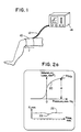
- FIGURE 1 a peripheral arterial monitoring instrument constructed in accordance with the principles of the present invention is shown in use on the leg of a patient.
- the instrument includes a conventional blood pressure cuff 10 having a length l which is wrapped about the thigh of the patient.
- the cuff 10 may be applied to any peripheral part of the body and is most conventionally applied to the upper arm, it is preferable to use the thigh in some procedures as that is where buildups of occlusive substances leading to arteriosclerosis and the like generally first manifest themselves. In other applications the upper arm or finger may be a preferred site for application of the cuff.
- the cuff 10 is connected by tubing 12 to a monitor and processor 14.
- the monitor and processor 14 includes a number of controls for actuating and adjusting the instrument in the performance of vascular measurements including blood pressure determination.
- the monitor and processor also includes a display 16 where the data taken during measurements of arterial volume is displayed, either in numerical or, preferably, in graphical form as shown in FIGURES 2a and 2b.
- the monitor and processor includes a controlled pneumatic system which controls inflation and deflation of the cuff 10 during which time measurements leading to the determination of the patient's arterial volume and compliance are taken.
- FIGURES 2a and 2b illustrate several preferred techniques for displaying the information obtained through these measurements.
- the upper portion of the display of FIGURE 2a is a graphical display of arterial volume (or arterial cross-sectional area, or arterial radius) versus transmural pressure.
- the arteries in the peripheral body part are infused with blood, the arteries expand and their volume increases as shown by the righthand portion of curve 20.
- the height of the righthand portion of the curve 20 also represents the effective radius of the arterial vessels R when the vessels are filled with blood.
- the slope of the curve 20, dV/dP represents arterial compliance and the point at which dV/dP exhibits a maximum value is generally referred to as peak arterial compliance.
- the upper graph of FIGURE 2a provides the physician with information as to arterial volume, compliance and effective arterial radius in the limb where the cuff is affixed.
- the illustrative curve 22 of R versus time shown in the drawing is seen to be substantially flat, except at time indicated by 23. This decrease in the R value may correlate for instance with the time at which some physical intervention such as intubation or incision is performed on the patient. If the patient is not fully anesthetized at that time, the cardiovascular system will react by contracting the arteries of the body, and the effective radius of arterial vessels will decline. Thus, the decline in curve 22 at point R would indicate to the anesthesiologist that the patient is not fully anesthetized, and further anesthetic may be required for patient comfort and safety.
- FIGURE 2b shows a further display of the arterial volume and compliance information which would be of assistance to an anesthesiologist.
- This curve of the patient's normal arterial volume is labelled as V(P) and the initial curve determined by the monitor and processor.
- V(P) the initial curve determined by the monitor and processor.
- a current volume versus pressure curve is calculated periodically and displayed in correspondence with the initial curve.
- the current curve is labelled V(P) curr. in FIGURE 2b.
- the display of FIGURE 2b provides the anesthesiologist with a continuous comparison of current arterial volume and compliance versus the patient's normal arterial volume and compliance prior to the administration of anesthetic.
- FIGURES 3a and 3b are cross-sectional illustrations of arteries showing the parameters measured by the monitor and processor 14.
- the R value is the radius of an artery 30 as shown in FIGURE 3a. Since the cuff encloses all arterial vessels in the portion of the limb about which it is wrapped, it will be understood that the R value is not the radius of a particular artery, but is in effect the sum of the radii of all of the arteries inside the cuff 10. Thus, the instrument provides an R value which is the effective radius taken over all arterial vessels inside the cuff.
- the artery 30 is defined by the arterial wall 32.
- the arterial wall is composed principally of two substances, collagen and smooth muscle tissue.
- Collagen provides the artery with flexibility, the ability to stretch and deform. This rubber-like characteristic is one contributor to arterial compliance, and is a passive characteristic of arteries.
- the muscle tissue is controlled by nerves to provide stretching and deformation of the artery under control of the body's nervous system. This stretching and deformation is an active characteristic of the artery which also is a factor in arterial compliance.
- FIGURE 3b Arterial volume and compliance are also affected in the case of arteriosclerosis or hardening of the arteries by the buildup of fatty substances on the inner walls of the arteries. This condition is shown in FIGURE 3b, where a buildup of substances is indicated at 34 lining the wall of the artery.
- the ability of the artery to expand or contract under the influence of arterial muscular contraction or blood pressure changes is adversely affected by this lining of fatty substances, which can retard such motion. Since the substances also occupy a portion of the inner volume of the artery, the effective radius of the vessel R' is decreased by the presence of these substances.
- FIGURE 4 illustrates an arrangement for taking measurements of arterial volume and compliance. Shown in FIGURE 4 are a limb of the body 40 in cross-section, about which a blood pressure cuff 10 is wrapped. The skin line of the limb is indicated at 41. The cross-sectional view of the limb shows the bone 42 at the center of the limb, and an artery 44 passing through the limb. The artery 44 is shown expanded during the pumping of blood, before the cuff is applied and inflated. After inflation of the cuff to a maximal pressure, the artery will be occluded, as shown at 44'.
- the cuff 10 is connected by pneumatic tubing to a pump 50.
- the pump 50 pumps up the cuff 10 at the start of the measurement cycle.
- the arrangement of FIGURE 4 is modified to perform the process of the "Hartsafe Product Concept" discussed above by the inclusion of a calibration chamber 54, which is connected to the pneumatic system.
- a calibration chamber 54 which is connected to the pneumatic system.
- the pump 50 is stopped and one ml of air is injected into the pneumatic system of the cuff. This may be accomplished by moving piston 56 in the chamber 54 to the right to displace one ml of air from the chamber. Given that all elements of the pneumatic system are substantially non-compliant, this one ml volume of air will compress the limb 40 by one ml.
- the effect of the piston displacement will be to displace one ml of blood from the vascular system within the confines of the cuff.
- the process of the Raines method or the "Hartsafe Product Concept" calculates its calibration factor at the outset of the measurement cycle.
- the pump then inflates the cuff to fully occlude the arterial vessels as shown at 44', and the deflate cycle commences.
- a deflation valve opens and closes to incrementally bleed air from the pneumatic system. Measurements taken by a pressure transducer P T at each pressure step are stored in correspondence with cuff pressure level and are subsequently used in a signal conditioning (processing) step at the end of the deflation cycle.
- FIGURE 4 illustrates a peripheral arterial volume and compliance measurement system of the present invention which obviates the need for such structural and operational complexity.
- the blood pressure cuff 10 is wrapped around the thigh 60 of the patient, shown in cross-section.
- the femur 62 is shown in the center of the thigh, and the skin line of the thigh is indicated at 61.
- the femoral artery is illustrated at 64 in an unoccluded condition, and in an occluded condition at 64'.
- the cuff 10 is connected by pneumatic tubing 12 to a pump 50, a pressure transducer P T , and a deflate valve 52.
- An orifice 66 of predetermined cross-sectional area is located in the deflate valve outlet.
- the pneumatic system of FIGURE 5 is operated in the conventional manner of a step-deflate automated blood pressure monitor such as the Critikon Dinamapç 8100.
- the cuff 10 is inflated by the pump 50 to a pressure which is in excess of systolic pressure, sufficient to fully occlude the artery 64'.
- the cuff pressure is stepped down, and the cuff pressures and oscillation pulses are recorded from the pressure transducer.
- Two of the pressure steps during the deflate cycle are shown in FIGURE 6.
- the cuff pressures of the two steps are P1 and P2, and the oscillation pulses are shown as P osc .
- the cuff pressure is stepped down in decrements of approximately 8 mm Hg.
- P the pressure across the orifice
- ⁇ an adjustment factor for nominal barometric pressure
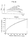
- the flow through a 1 cm2 orifice as a function of the pressure across the orifice during a typical deflate cycle is represented graphically in FIGURE 9.
- Other known methods for measuring the flow of a fluid may also be employed; for instance, if the orifice in a given embodiment does not conform to theoretical models, it may be approximated empirically.
- ⁇ V n A eq ⁇ FLOW n ⁇ ⁇ t n
- a eq is the equivalent area of the orifice
- FLOW n is the flow rate between two pressure steps
- ⁇ t n is the time during which the deflate valve was open between the two pressure steps.
- the FLOW is known from the preceding equation, the equivalent area of the orifice is known, and the time during which the deflate valve is open is measured by a digital clock which runs during the time that the valve is open. Since the FLOW calculation is done for each deflation step based upon the known orifice and the then extant pressure, no recalibration or modification is necessary or required for the calculated values.
- a ratio can be formed of the ⁇ V n values and the respective cuff pressure differentials at which they were obtained.
- the value of P osc n used for each step decrement may be the amplitude of the oscillation pulses on the P1 step, the P2 step, or an average of the two, due to the very small variation in oscillation pulse amplitude from one step to the next. Whichever approach is used, it is consistently applied for the full range of step values. Curves representing V osc and the oscillation pulses as a function of cuff pressure are illustrated in FIGURE 10.
- the arterial volume curve can now be computed in a two-step procedure.
- the first step is to compute a curve referred to herein as a reconstruction curve from knowledge of the V osc n values and the values of systolic and diastolic blood pressure determined by the DinamapTM in the conventional manner.
- the arterial volume curve is then computed by coordinate system transformation, by which the reconstruction curve, referenced to cuff pressure, is converted to arterial transmural pressure with reference to systolic pressure.
- a graphical plot of the points Recon n (P cuff ) as a function of cuff pressure is shown by the dashed curve Recon in FIGURE 11 in comparison with the V osc curve previously shown in FIGURE 10. It is seen that the plot of Recon converges with the V osc curve above and in the vicinity of systolic pressure.
- the arterial volume may be calculated as a function of transmural pressure by, in effect, transforming the Recon curve about the axis of systolic pressure.
- the arterial volume curve produced by this transformation is of the general shape of curve 20 of FIGURE 2a and the curves of FIGURE 2b.
- a point of reference for selection of R and dV/dP may be chosen in a number of ways.
- the monitor may compute mean arterial pressure in the conventional manner, and use the value of mean arterial pressure as the pressure for which R and dV/dP are chosen and displayed.
- the pressure at which dV/dP is at a maximum, peak arterial compliance can be used as the pressure reference for selecting R and dV/dP.
- the physician selects a transmural pressure value on the abscissa of the upper curve of FIGURE 2a as the pressure for R and dV/dP.
- the slope of the curve at the selected pressure point can be calculated to determine arterial compliance dV/dP, and the amplitude of the volume curve at the selected pressure provides the R value.
- the instrument is repeatedly actuated automatically and an R value is found each time.
- the R value is then displayed as a function of time as shown at the bottom of FIGURE 2a.
- the volume curve calculated at the beginning of a surgical procedure is stored and continuously displayed with the most recently calculated curve in the format shown in FIGURE 2b.
- R or arterial area or volume
- Limb size is obtained by measuring the circumference of the limb where the cuff is attached, and entering this information into the monitor and processor 14. The ratio of this R (or arterial area or volume) to circumference (or calculated limb radius or cross-sectional area) provides an indication of cardiovascular efficiency.
- a flowmeter which measures the flow of expelled air could be used to provide a direct measurement of flow volume at the output of the deflate valve 52.
- Another alternative embodiment is to use a transfer volume of known capacity as shown in FIGURES 7 and 8.
- the transfer volume comprises all of the volumetric space between an intermediate dump valve 52a and the deflate valve 52.
- the size of the vessel indicated at 58 is chosen to provide the desired volume of the entire transfer volume.
- the deflate valve 52 is closed after previous closure of the dump valve 52a.
- the air in the transfer volume between the two valves is now at atmospheric pressure.
- the dump valve 52a is then opened, and the transfer volume becomes pressurized to the cuff pressure, which declines to P tr by reason of the expansion of pressurized air into the transfer volume.
- V osc n [ ⁇ V tr /(P1-P tr ) ] n ⁇ P osc n
- the deflate valve 52 is then opened so that both valves are in the open condition. Air is expelled from the pneumatic system of the cuff and the pressure transducer is monitored until the pressure reaches the level P2, at which point the dump valve 52a is closed. The deflate valve 52 is then closed, stabilizing the transfer volume at atmospheric pressure in preparation for the next step decrement.
- the transfer volume technique is advantageously employed to enable use of a total pressure step P1-P2 which is conventional for a standard blood pressure monitor such as the DinamapTM 8100, which uses pressure step decrements of approximately 8 mm Hg.
- a standard blood pressure monitor such as the DinamapTM 8100, which uses pressure step decrements of approximately 8 mm Hg.
- Equation 1 This equation is an arctangent function with a linear term added for positive arterial pressures. Equation 1 now has four unknown constants; Ao, SLOPE, C, and E.
- Ao is the arterial volume (or area) at zero transmural pressure.
- SLOPE is the change in arterial volume with respect to the change in pressure across the wall. In other words, SLOPE estimates arterial compliance at high wall pressures.
- C is a parameter which describes the shape of the compliance curve, and the elasticity of the artery as it is being occluded.
- E is a parameter which adjusts the overall curve (up or down along the volume axis) to achieve zero volume at large negative pressures, thus achieving the condition of an occluded artery at high cuff pressures. The constraint that the volume cannot be negative is imposed.
- this equation is monotonically increasing, which is a known feature of the arterial pressure-volume curve.
- this curve uses parameters which have physiological significance. It is important to note that other general curve equations could be used, but the parameters would probably not have the direct physiological significance and, in some cases, a monitonically increasing constraint would need to be imposed.
- the starting point is the volume oscillations (Vosc), versus cuff pressures as described above and shown in example form in FIGURE 12.
- the general mathematical form for the pressure-volume curve is assumed and plotted for an arbitrary initial choice of parameters. Given these assumed choices out of a virtual infinity of possible choices, one can simulate mathematically what the volume oscillometric envelope would look like using the same cuff pressures that occurred during the actual determination sequence. The result of this simulation is plotted as Voscsim in FIGURE 12.
- VOSCSIM VARTEST(Ps-Pcs) - VARTEST(Pd-Pcd), where Pcs and Pcd are cuff pressures at systolic and diastolic pressures, respectfully.
- VOSCSIM is calculated for each of the cuff pressure points which occurred in the actual determination process and results in the VOSCSIM variable plotted in FIGURE 12.
- the correct choice of unknown parameters is the one which minimizes the error between the measured volume oscillometric waveform and the estimated oscillometric waveform. Individual point by point errors are calculated and then the sum of the errors squared is calculated.
- FIGURE 13 shows the results of this search optimization process.
- ⁇ V outcorr ⁇ V out ( from P c1 to P c2 )- ⁇ V art (from P c1 to P c2 )
- Systolic and pulse pressures are unknown at this point and they can be albragetically labeled as unknowns S and P.
- the problem now involves six or five unknown constants, and the inverse problem is a choice of constants which will best match the volume pressure envelope Vosc, as shown in FIGURE 12.
- Such estimated values A o , C, E and slope produce a guessed pressure-volume curve as shown as Voscsim in FIGURE 12.
- an assumed shape of the pressure curved affects both the dV/P estimate obtained during the calibration decrement, as well as the shape in which the simulated volume oscillometric curve takes.
- a best choice of parameters is still the set which minimizes the error between the volume of the curved obtained from this determination.
- What these determinations result in are actual measurements of all the parameters, including diastolic and systolic pressures, as compared to previous methods which only determine parameters on a scaled basis. In other words, there is no scaling of any of the parameters values, and actual numbers can be obtained. This will allow the user to obtain much more robust meanings for these readings and, therefore, allows for immediate and applicable in using these known methods.
Landscapes
- Health & Medical Sciences (AREA)
- Life Sciences & Earth Sciences (AREA)
- Vascular Medicine (AREA)
- Cardiology (AREA)
- Heart & Thoracic Surgery (AREA)
- Medical Informatics (AREA)
- Biophysics (AREA)
- Pathology (AREA)
- Engineering & Computer Science (AREA)
- Biomedical Technology (AREA)
- Physiology (AREA)
- Physics & Mathematics (AREA)
- Molecular Biology (AREA)
- Surgery (AREA)
- Animal Behavior & Ethology (AREA)
- General Health & Medical Sciences (AREA)
- Public Health (AREA)
- Veterinary Medicine (AREA)
- Ophthalmology & Optometry (AREA)
- Measuring Pulse, Heart Rate, Blood Pressure Or Blood Flow (AREA)
Applications Claiming Priority (2)
| Application Number | Priority Date | Filing Date | Title |
|---|---|---|---|
| US08/056,103 US5417220A (en) | 1989-12-20 | 1993-05-03 | Peripheral arterial monitoring instruments |
| US56103 | 1993-05-03 |
Publications (1)
| Publication Number | Publication Date |
|---|---|
| EP0627191A1 true EP0627191A1 (fr) | 1994-12-07 |
Family
ID=22002168
Family Applications (1)
| Application Number | Title | Priority Date | Filing Date |
|---|---|---|---|
| EP94303197A Withdrawn EP0627191A1 (fr) | 1993-05-03 | 1994-05-03 | Instrument de surveillance des artéries périphériques |
Country Status (5)
| Country | Link |
|---|---|
| US (2) | US5417220A (fr) |
| EP (1) | EP0627191A1 (fr) |
| AU (1) | AU672488B2 (fr) |
| CA (1) | CA2122687C (fr) |
| ZA (1) | ZA943014B (fr) |
Cited By (4)
| Publication number | Priority date | Publication date | Assignee | Title |
|---|---|---|---|---|
| US6126595A (en) * | 1995-05-12 | 2000-10-03 | Seiko Epson Corporation | Device for diagnosing physiological state and device for controlling the same |
| WO2001095798A3 (fr) * | 2000-06-12 | 2002-04-18 | Univ Rutgers | Procede et systeme de detection d'affections vasculaires au moyen du plethysmographe a brassard occlusif |
| EP2976007A4 (fr) * | 2013-03-15 | 2017-02-15 | Charles L. Davis | Méthode non invasive de mesure des paramètres cardiovasculaires, modélisation de la boucle vasculaire périphérique, analyse de l'état vasculaire et amélioration du diagnostic des maladies cardiovasculaires |
| WO2017216268A1 (fr) * | 2016-06-14 | 2017-12-21 | Koninklijke Philips N.V. | Dispositif et procédé d'évaluation non invasive de la compliance artérielle maximale |
Families Citing this family (20)
| Publication number | Priority date | Publication date | Assignee | Title |
|---|---|---|---|---|
| JP3580924B2 (ja) * | 1995-12-22 | 2004-10-27 | コーリンメディカルテクノロジー株式会社 | 動脈弾性度評価装置 |
| US5991654A (en) * | 1997-06-06 | 1999-11-23 | Kci New Technologies, Inc. | Apparatus and method for detecting deep vein thrombosis |
| US6309359B1 (en) * | 1998-06-01 | 2001-10-30 | Michael D. Whitt | Method and apparatus for noninvasive determination of peripheral arterial lumenal area |
| US6231532B1 (en) | 1998-10-05 | 2001-05-15 | Tyco International (Us) Inc. | Method to augment blood circulation in a limb |
| US6152881A (en) * | 1999-03-29 | 2000-11-28 | Vasocor, Inc. | Calibrated measurement of blood vessels and endothelium after reactive hyperemia and method therefor |
| FR2794961B1 (fr) | 1999-06-16 | 2001-09-21 | Global Link Finance | Procede de determination du decalage temporel entre les instants de passage d'une meme onde de pouls en deux points de mesure distincts d'un reseau arteriel d'un etre vivant et d'estimation de sa pression aortique |
| US7727157B2 (en) * | 2002-09-13 | 2010-06-01 | Sharrock Nigel E | Non-invasive measurement of suprasystolic signals |
| NZ524765A (en) * | 2000-07-19 | 2006-10-27 | Nigel E Sharrock | Non-invasive measurement of suprasystolic signals |
| WO2003034916A2 (fr) * | 2001-08-17 | 2003-05-01 | Russell Ted W | Procedes, appareil et articles manufactures pour mesure et surveillance non effractives du debit sanguin peripherique, de la perfusion, de la puissance du coeur, de la tension biophysique et de l'etat cardio-vasculaire |
| US7011631B2 (en) | 2003-01-21 | 2006-03-14 | Hemonix, Inc. | Noninvasive method of measuring blood density and hematocrit |
| US7689261B2 (en) * | 2003-11-26 | 2010-03-30 | General Electric Company | Cardiac display methods and apparatus |
| JP5363795B2 (ja) * | 2008-04-14 | 2013-12-11 | 国立大学法人広島大学 | 血管内皮機能評価装置及び血管内皮機能評価方法 |
| US9788733B2 (en) | 2009-06-02 | 2017-10-17 | Cordex Systems, Inc. | Method and device for detecting and assessing reactive hyperemia using segmental plethysmography |
| ES2757847T3 (es) | 2009-06-02 | 2020-04-30 | Cordex Systems Inc | Método para detectar y evaluar la hiperemia reactiva mediante pletismografía segmentaria |
| EP2319408A1 (fr) * | 2009-10-15 | 2011-05-11 | Finapres Medical Systems B.V. | Dispositif pour le contrôle de la pression dans un coussin à pression gonflable |
| US10945612B2 (en) | 2014-04-03 | 2021-03-16 | The Regents Of The University Of California | Assessing endothelial function using a blood pressure cuff |
| US20160120420A1 (en) * | 2014-10-30 | 2016-05-05 | David A. Liedl | Vascular measurement system |
| US9737217B2 (en) * | 2015-08-14 | 2017-08-22 | The Regents Of The University Of California | Assessing endothelial function and providing calibrated UFMD data using a blood pressure cuff |
| EP3432787A4 (fr) * | 2016-03-24 | 2019-01-30 | Board of Regents of the University of Nebraska | Dispositifs et procédés de détection et de mesure de vasomotricité sympathique |
| US10772571B2 (en) | 2016-11-15 | 2020-09-15 | Welch Allyn, Inc. | Method and systems for correcting for arterial compliance in a blood pressure assessment |
Citations (7)
| Publication number | Priority date | Publication date | Assignee | Title |
|---|---|---|---|---|
| CH515028A (de) * | 1970-03-09 | 1971-11-15 | Oripid Ag | Verfahren und Vorrichtung zur Bestimmung des Durchflussquerschnittes von Arterien |
| US3920004A (en) * | 1973-07-30 | 1975-11-18 | Ryu Nakayama | Device and method for noninvasive measurement of blood pressure, resistance inertance, compliance, impedance, blood flow rate, kinetic energy, flow velocity and pulse velocity of a segment in man |
| EP0188894A1 (fr) * | 1984-12-21 | 1986-07-30 | BAXTER INTERNATIONAL INC. (a Delaware corporation) | Technique utilisant une méthode statistique spécifique pour obtenir des informations associées à la pression sanguine d'un patient |
| US4699152A (en) * | 1984-12-21 | 1987-10-13 | Baxter Travenol Laboratories, Inc. | Techniques for obtaining information associated with an individual's blood pressure including specifically a stat mode technique |
| US4712563A (en) * | 1986-05-28 | 1987-12-15 | Baxter Travenol Laboratories, Inc. | Method of and apparatus for determining the diastolic and systolic blood pressure of a patient |
| EP0434399A1 (fr) * | 1989-12-20 | 1991-06-26 | Critikon, Inc. | Instrument moniteur pour artères périphériques |
| WO1992022239A1 (fr) * | 1991-06-12 | 1992-12-23 | Florida Atlantic University Research Corp. | Detection de l'atherosclerose chez l'homme |
Family Cites Families (28)
| Publication number | Priority date | Publication date | Assignee | Title |
|---|---|---|---|---|
| US3903872A (en) * | 1974-02-25 | 1975-09-09 | American Optical Corp | Apparatus and process for producing sphygmometric information |
| US4009709A (en) * | 1975-05-15 | 1977-03-01 | American Optical Corporation | Apparatus and process for determining systolic pressure |
| US4154238A (en) * | 1976-12-27 | 1979-05-15 | American Optical Corporation | Apparatus and process using second derivative of oscillometric waveform for producing sphygmometric information |
| US4174707A (en) * | 1976-12-27 | 1979-11-20 | American Optical Corporation | Apparatus and process for antifact rejection through cross correlation in sphygmometry |
| US4367751A (en) * | 1976-12-27 | 1983-01-11 | Warner-Lambert Company | Apparatus and process for producing artifact effect on sphygmometric information |
| US4349034A (en) * | 1978-04-10 | 1982-09-14 | Johnson & Johnson | Automatic mean blood pressure reading device |
| US4360029A (en) * | 1978-04-10 | 1982-11-23 | Johnson & Johnson | Automatic mean blood pressure reading device |
| NL8003548A (nl) * | 1980-06-19 | 1982-01-18 | Barry William Hyndman | Continue onbloedige bloeddrukmeter. |
| NL8104879A (nl) * | 1981-10-28 | 1983-05-16 | Tno | Werkwijze en inrichting voor het regelen van de manchetdruk bij het meten van de vingerbloeddruk met een foto-electrische plethysmograaf. |
| US4718428A (en) * | 1984-02-17 | 1988-01-12 | Cortronic Corporation | Method for determining diastolic arterial blood pressure in a subject |
| US4697596A (en) * | 1984-06-19 | 1987-10-06 | Baxter Travenol Laboratories, Inc. | Techniques for obtaining information associated with an individual's blood pressure including specifically a stat mode technique |
| US4564020A (en) * | 1984-06-19 | 1986-01-14 | Norse Instruments, Inc. | Method and apparatus for obtaining an individual's systolic blood pressure |
| US4651747A (en) * | 1984-06-20 | 1987-03-24 | Baxter Travenol Laboratories, Inc. | Waveform information obtaining techniques associated with an individual's blood pressure |
| US4587974A (en) * | 1984-11-13 | 1986-05-13 | Norse Instruments | Linear pressurizing and depressurizing device |
| US4664126A (en) * | 1984-12-21 | 1987-05-12 | Baxter Travenol Laboratories, Inc. | Techniques for obtaining information associated with an individual's blood pressure including specifically a stat mode technique |
| US4699151A (en) * | 1984-12-21 | 1987-10-13 | Baxter Travenol Laboratories, Inc. | Techniques for obtaining information associated with an individual's blood pressure including specifically a stat mode technique |
| US4771792A (en) * | 1985-02-19 | 1988-09-20 | Seale Joseph B | Non-invasive determination of mechanical characteristics in the body |
| US5054493A (en) * | 1986-01-31 | 1991-10-08 | Regents Of The University Of Minnesota | Method for diagnosing, monitoring and treating hypertension |
| US4727884A (en) * | 1986-05-28 | 1988-03-01 | Link William T | Technique for obtaining the mean blood pressure constant for an individual's blood pressure |
| CS272057B1 (en) * | 1987-03-27 | 1991-01-15 | Jan Doc Mudr Csc Penaz | Blood pressure automatic non-invasive meter |
| US4846189A (en) * | 1987-06-29 | 1989-07-11 | Shuxing Sun | Noncontactive arterial blood pressure monitor and measuring method |
| US5090417A (en) * | 1987-10-22 | 1992-02-25 | Mollan Raymond A B | Medical diagnostic apparatus |
| US4984577A (en) * | 1989-03-20 | 1991-01-15 | Hewlett-Packard Company | Oscillometric non-invasive method for measuring blood pressure and apparatus for automated oscillometric blood pressure measuring |
| JPH0638790B2 (ja) * | 1989-05-19 | 1994-05-25 | 松田 正義 | 動脈伸展性測定装置 |
| US5089961A (en) * | 1989-07-24 | 1992-02-18 | Aci Medical Incorporated | Apparatus and method for vascular examination of a limb |
| US5263485A (en) * | 1989-09-18 | 1993-11-23 | The Research Foundation Of State University Of New York | Combination esophageal catheter for the measurement of atrial pressure |
| US5211177A (en) * | 1990-12-28 | 1993-05-18 | Regents Of The University Of Minnesota | Vascular impedance measurement instrument |
| US5101828A (en) * | 1991-04-11 | 1992-04-07 | Rutgers, The State University Of Nj | Methods and apparatus for nonivasive monitoring of dynamic cardiac performance |
-
1993
- 1993-05-03 US US08/056,103 patent/US5417220A/en not_active Expired - Fee Related
-
1994
- 1994-05-02 ZA ZA943014A patent/ZA943014B/xx unknown
- 1994-05-02 CA CA002122687A patent/CA2122687C/fr not_active Expired - Fee Related
- 1994-05-03 EP EP94303197A patent/EP0627191A1/fr not_active Withdrawn
- 1994-05-03 AU AU61996/94A patent/AU672488B2/en not_active Ceased
- 1994-11-22 US US08/343,801 patent/US5724981A/en not_active Expired - Fee Related
Patent Citations (8)
| Publication number | Priority date | Publication date | Assignee | Title |
|---|---|---|---|---|
| CH515028A (de) * | 1970-03-09 | 1971-11-15 | Oripid Ag | Verfahren und Vorrichtung zur Bestimmung des Durchflussquerschnittes von Arterien |
| US3920004A (en) * | 1973-07-30 | 1975-11-18 | Ryu Nakayama | Device and method for noninvasive measurement of blood pressure, resistance inertance, compliance, impedance, blood flow rate, kinetic energy, flow velocity and pulse velocity of a segment in man |
| EP0188894A1 (fr) * | 1984-12-21 | 1986-07-30 | BAXTER INTERNATIONAL INC. (a Delaware corporation) | Technique utilisant une méthode statistique spécifique pour obtenir des informations associées à la pression sanguine d'un patient |
| US4699152A (en) * | 1984-12-21 | 1987-10-13 | Baxter Travenol Laboratories, Inc. | Techniques for obtaining information associated with an individual's blood pressure including specifically a stat mode technique |
| US4712563A (en) * | 1986-05-28 | 1987-12-15 | Baxter Travenol Laboratories, Inc. | Method of and apparatus for determining the diastolic and systolic blood pressure of a patient |
| EP0434399A1 (fr) * | 1989-12-20 | 1991-06-26 | Critikon, Inc. | Instrument moniteur pour artères périphériques |
| US5103833A (en) * | 1989-12-20 | 1992-04-14 | Critikon, Inc. | Peripheral arterial monitoring instruments |
| WO1992022239A1 (fr) * | 1991-06-12 | 1992-12-23 | Florida Atlantic University Research Corp. | Detection de l'atherosclerose chez l'homme |
Cited By (7)
| Publication number | Priority date | Publication date | Assignee | Title |
|---|---|---|---|---|
| US6126595A (en) * | 1995-05-12 | 2000-10-03 | Seiko Epson Corporation | Device for diagnosing physiological state and device for controlling the same |
| US6890304B1 (en) | 1995-05-12 | 2005-05-10 | Seiko Epson Corporation | Device for diagnosing physiological state and device for controlling the same |
| WO2001095798A3 (fr) * | 2000-06-12 | 2002-04-18 | Univ Rutgers | Procede et systeme de detection d'affections vasculaires au moyen du plethysmographe a brassard occlusif |
| US6626840B2 (en) | 2000-06-12 | 2003-09-30 | Rutgers, The State University Of New Jersey | Method and system for detecting vascular conditions using an occlusive arm cuff plethysmograph |
| EP2976007A4 (fr) * | 2013-03-15 | 2017-02-15 | Charles L. Davis | Méthode non invasive de mesure des paramètres cardiovasculaires, modélisation de la boucle vasculaire périphérique, analyse de l'état vasculaire et amélioration du diagnostic des maladies cardiovasculaires |
| WO2017216268A1 (fr) * | 2016-06-14 | 2017-12-21 | Koninklijke Philips N.V. | Dispositif et procédé d'évaluation non invasive de la compliance artérielle maximale |
| US11813044B2 (en) | 2016-06-14 | 2023-11-14 | Koninklijke Philips N.V. | Device and method for non-invasive assessment of maximum arterial compliance |
Also Published As
| Publication number | Publication date |
|---|---|
| AU6199694A (en) | 1994-11-10 |
| CA2122687A1 (fr) | 1994-11-04 |
| ZA943014B (en) | 1995-11-02 |
| US5417220A (en) | 1995-05-23 |
| AU672488B2 (en) | 1996-10-03 |
| CA2122687C (fr) | 2005-02-15 |
| US5724981A (en) | 1998-03-10 |
Similar Documents
| Publication | Publication Date | Title |
|---|---|---|
| EP0434399B1 (fr) | Instrument moniteur pour artères périphériques | |
| US5724981A (en) | Peripheral arterial monitoring instruments | |
| US4846189A (en) | Noncontactive arterial blood pressure monitor and measuring method | |
| CA2130255C (fr) | Dispositif et methode pour determiner les caracteristiques physiologiques de la lumiere des structures corporelles | |
| CA1259818A (fr) | Dispositif etalonne pour la mesure de la pression arterielle | |
| EP0152848B1 (fr) | Procédé pour déterminer la pression d'un fluide pulsatile s'écoulant au travers d'un tube flexible et appareil pour dèterminer la pression artérielle sanguine d'un sujet | |
| US6733461B2 (en) | Methods and apparatus for measuring arterial compliance, improving pressure calibration, and computing flow from pressure data | |
| US6338719B1 (en) | Method and system for detecting vascular conditions using an occlusive arm cuff plethysmograph | |
| JP4718553B2 (ja) | 血流および静脈容量の測定 | |
| US7678059B2 (en) | Non-invasive blood pressure monitor with improved performance | |
| US6440080B1 (en) | Automatic oscillometric apparatus and method for measuring blood pressure | |
| CN110840429B (zh) | 基于柯氏音的血压测量方法及血压测量和心血管系统评估系统 | |
| KR20020035438A (ko) | 상지-하지 혈압 지수 측정 장치 | |
| EP3705033A1 (fr) | Dispositif de mesure de la pression sanguine et procédé de commande | |
| EP3760109A1 (fr) | Unité de commande pour dériver une mesure de conformité artérielle | |
| US20230050058A1 (en) | Systems and methods for non-invasive pulse pressure waveform measurement | |
| CN211883777U (zh) | 基于柯氏音的血压测量和心血管系统评估系统 | |
| CN117715582A (zh) | 用于非侵入式脉搏压力波形测量的系统和方法 | |
| James | Simplified model for the design of an oscillometric blood pressure measuring system |
Legal Events
| Date | Code | Title | Description |
|---|---|---|---|
| PUAI | Public reference made under article 153(3) epc to a published international application that has entered the european phase |
Free format text: ORIGINAL CODE: 0009012 |
|
| AK | Designated contracting states |
Kind code of ref document: A1 Designated state(s): DE ES FR GB IT SE |
|
| 17P | Request for examination filed |
Effective date: 19950515 |
|
| 17Q | First examination report despatched |
Effective date: 19970612 |
|
| STAA | Information on the status of an ep patent application or granted ep patent |
Free format text: STATUS: THE APPLICATION IS DEEMED TO BE WITHDRAWN |
|
| 18D | Application deemed to be withdrawn |
Effective date: 19981020 |