EP0434909A2 - Protéines anticoagulantes et antihelmintiques, procédé pour les obtenir et leur utilisation - Google Patents
Protéines anticoagulantes et antihelmintiques, procédé pour les obtenir et leur utilisation Download PDFInfo
- Publication number
- EP0434909A2 EP0434909A2 EP90117879A EP90117879A EP0434909A2 EP 0434909 A2 EP0434909 A2 EP 0434909A2 EP 90117879 A EP90117879 A EP 90117879A EP 90117879 A EP90117879 A EP 90117879A EP 0434909 A2 EP0434909 A2 EP 0434909A2
- Authority
- EP
- European Patent Office
- Prior art keywords
- protein
- kda
- proteins
- sds
- cdna
- Prior art date
- Legal status (The legal status is an assumption and is not a legal conclusion. Google has not performed a legal analysis and makes no representation as to the accuracy of the status listed.)
- Withdrawn
Links
Images


Classifications
-
- C—CHEMISTRY; METALLURGY
- C12—BIOCHEMISTRY; BEER; SPIRITS; WINE; VINEGAR; MICROBIOLOGY; ENZYMOLOGY; MUTATION OR GENETIC ENGINEERING
- C12N—MICROORGANISMS OR ENZYMES; COMPOSITIONS THEREOF; PROPAGATING, PRESERVING, OR MAINTAINING MICROORGANISMS; MUTATION OR GENETIC ENGINEERING; CULTURE MEDIA
- C12N9/00—Enzymes; Proenzymes; Compositions thereof; Processes for preparing, activating, inhibiting, separating or purifying enzymes
- C12N9/14—Hydrolases (3)
- C12N9/48—Hydrolases (3) acting on peptide bonds (3.4)
- C12N9/50—Proteinases, e.g. Endopeptidases (3.4.21-3.4.25)
- C12N9/64—Proteinases, e.g. Endopeptidases (3.4.21-3.4.25) derived from animal tissue
- C12N9/6402—Proteinases, e.g. Endopeptidases (3.4.21-3.4.25) derived from animal tissue from non-mammals
- C12N9/6405—Proteinases, e.g. Endopeptidases (3.4.21-3.4.25) derived from animal tissue from non-mammals not being snakes
- C12N9/641—Cysteine endopeptidases (3.4.22)
-
- A—HUMAN NECESSITIES
- A61—MEDICAL OR VETERINARY SCIENCE; HYGIENE
- A61P—SPECIFIC THERAPEUTIC ACTIVITY OF CHEMICAL COMPOUNDS OR MEDICINAL PREPARATIONS
- A61P33/00—Antiparasitic agents
-
- C—CHEMISTRY; METALLURGY
- C07—ORGANIC CHEMISTRY
- C07K—PEPTIDES
- C07K14/00—Peptides having more than 20 amino acids; Gastrins; Somatostatins; Melanotropins; Derivatives thereof
- C07K14/435—Peptides having more than 20 amino acids; Gastrins; Somatostatins; Melanotropins; Derivatives thereof from animals; from humans
- C07K14/43504—Peptides having more than 20 amino acids; Gastrins; Somatostatins; Melanotropins; Derivatives thereof from animals; from humans from invertebrates
- C07K14/43536—Peptides having more than 20 amino acids; Gastrins; Somatostatins; Melanotropins; Derivatives thereof from animals; from humans from invertebrates from worms
- C07K14/4354—Peptides having more than 20 amino acids; Gastrins; Somatostatins; Melanotropins; Derivatives thereof from animals; from humans from invertebrates from worms from nematodes
-
- A—HUMAN NECESSITIES
- A61—MEDICAL OR VETERINARY SCIENCE; HYGIENE
- A61K—PREPARATIONS FOR MEDICAL, DENTAL OR TOILETRY PURPOSES
- A61K39/00—Medicinal preparations containing antigens or antibodies
Definitions
- the present invention relates to proteins and their use as vaccines against helminthes in mammals, particularly sheep.
- the invention also relates to methods for producing these proteins in substantially pure form.
- Haemonchus contortus is a blood feeding parasite commonly found in sheep, cattle, goats and wild ruminants of many species. These parasites feed on the blood of the host, and inject hemolytic proteins into the host's system, thereby resulting in anemia, emaciation, edema and intestinal disturbances. In cases of heavy infection, the host usually dies.
- Infection occurs when the third-stage juvenile, still wearing the remains of a second-stage cuticle, is eaten. Exsheathment takes place in the forestomach, at which point the parasite is referred to as an XL3 larvae (XL3s).
- XL3s XL3 larvae
- the worm molts Upon entering the abomasum, the worm molts within 48 hours, becoming an L4 larvae (L4s), or forth stage juvenile, which feeds on the blood of its host. In about three days, the worm molts for a final time, and egg production begins about 15 days later.
- the present inventors have found a method to purify adequate quantities of cuticular surface proteins, free of cellular proteins, identified a collagen peptide and a method of producing an anticoagulant protein extract, all of which are useful as vaccines against infection by helminthes in sheep.
- Collagen peptide Collagen peptide, cuticular proteins and an anticoagulant antiserum.
- the present invention provides methods of isolating cuticular, surface proteins, in relatively pure form, from helminthes. More specifically, the present invention provides methods of isolating surface proteins from two parasitic larval stages of Haemonchus contortus . Surface proteins purified by these procedures are immunogenic and induce antibodies that react with the native surface proteins on live worms.
- Live XL3s and L4s can be boiled briefly in a solution which specifically solubilizes surface proteins.
- SDS sodium dodecyl sulfate
- XL3 and L4 in a saline solution, more preferably 100 M NaCl.
- live XL3s or L4s may be suspended in a saline solution and then boiled briefly in order to solubilize the surface proteins.
- surface proteins obtained from XL3s by the NaCl procedure are quite pure, as would be expected from the SDS results, the surface proteins extracted from L4s are greatly enriched but are contaminated to a small degree by cuticle collagens and cellular proteins.
- Contamination can be detected by probing Western blots of L4 surface protein extracts with antisera prepared against a synthetic 18 amino acid long peptide derived from the sequence of a cuticle collagen gene.
- the NaCl extraction procedure does greatly enrich for L4 surface proteins, though, and it is possible to purify the proteins further by simple procedures such elution from SDS gels.
- live XL3s or L4s were suspended in 1 ml of 100mM NaCl, 10 mM tris-HCl pH 7.4, placed in boiling water for 2 minutes, removed from the water, and mixed by inversion for an additional 2 minutes, and then pelleted for 1 minute in a microfuge. The supernatant was drawn off, recentrifuged several times to remove all worms, frozen in a dry ice/ethanol bath and stored at -20°C. These samples were later thawed and concentrated at 4°C using a 2 ml centricon apparatus (Amicon).
- Amicon 2 ml centricon apparatus
- the protein extraction studies indicate that the XL3 has a single, major surface protein of 68-90 kDa, which probably covers the main portion of the XL3 surface. Other proteins of various molecular weights were also identified. The 180 kDa protein appears to be the next most abundant XL3 surface protein. Preliminary experiments indicate that a rabbit antiserum prepared against the 68-97 kDa protein electroeluted from SDS gels immunoprecipitates the 125I-labeled 180 kDa protein and reacts with the 180 kDa protein on Western blots of surface protein extracts.
- Another method includes surface-labeling of cuticle proteins with 125I and chloramine T.
- Two classes of proteins were identified by this method. The properties of these proteins appear to be similar in XL3s and L4s.
- One class of proteins comprises those that can be extracted from the cuticle with SDS.
- the other class comprises those proteins that require a disulfide reducing agent for efficient solubilization.
- the SDS-soluble surface proteins of XL3s and L4s include relatively few major species.
- the pattern of labeled proteins is distinct for each developmental stage, although some of the proteins have similar molecular weights. The proteins are nearly completely extracted from worms with SDS, in the absence of a disulfide reducing agent.
- the SDS-soluble surface proteins are not digestible by bacterial collagenase, indicating that they lack the repeating (Gly-X-Y)n structure characteristic of collagen, which is a major protein component of nematode cuticle.
- Several of the proteins are glycosylated, supporting the notion that they are extracellular, and probably located on the cuticular surface. Bone and Bottjer previously reported the binding of specific lectins to the surface of H. contortus adults and juveniles. In the above properties, the major SDS-soluble surface proteins of H. contortus resemble surface proteins described for other nematodes.
- SDS + BME extractable proteins that are labeled with 125I are digestible with collagenase, indicating that they are collagens. Certain of these proteins, however, were not digested by collagenase, and thus, are probably not collagens. The inventors predict that the labeling of these proteins is artifactual and that they derive from the internal portions of the cuticle and not from the cuticular surface. This prediction stems from the observations that antisera prepared against SDS-treated cuticles (which contain collagens) react weakly or not at all with live worms, suggesting that SDS has removed the major surface antigens.
- SDS-soluble XL3 surface proteins of 24 kDa and 36 kDa appear to be less tightly associated with the cuticle than other XL3 surface proteins because they are partially released from the cuticle by sonication, in the absence of detergents.
- the physiological role, if any, of this difference in affinity is unclear at present.
- Other researchers have reported that specific surface proteins of parasitic nematodes are shed into the media during in vitro incubation studies with live worms (9, 20). We have not determined if this is the case for any H. contortus surface proteins.
- Stage-specific surface antigens have been described for several parasitic nematodes (9, 10).
- the ability of parasitic nematodes to change surface proteins at molts during which the old cuticle is shed and a new cuticle is formed, may be a mechanism for evading the host's immune responses and may explain in part why primary infections with H. contortus proceed to the adult stage, whereas secondary infections in immune animals generally are halted at the XL3 or L4 stages.
- the amino acid compositions of XL3 and L4 protein extracts were determined and are given in Table I. Both sets of proteins are enriched for hydrophillic amino acids, and with the exception of alanine and glycine, are relatively poor in hydrophic amino acids.
- the major 68-97 kDa protein appears to be greatly enriched in glutamic acid and/or glutamine residues.
- 26 percent of the amino acids in the XL3 surface protein extract which should largely reflect the amino acid composition of the major 60-90 kd species, were either glutamic acid or glutamine.
- This protein is also enriched for aspartic acid and/or asparagine residues. Together, these four amino acids account for nearly 40% of the total amino acids detected in the XL3 surface protein extract. Since no comparable analyses have been reported for surface proteins of other nematodes, the inventors do not know how common or unusual this finding will prove to be.
- the L4 surface proteins which are a more heterogeneous mixture of proteins, are not as enriched for these amino acids.
- the difference in relative abundance of XL3 surface proteins detected by Coomassie blue staining and 125I-label may reflect the inefficient labeling of the 68-97 kDa protein by 125I and chloramine T, which labels predominantly tyrosine residues. Tyrosine is poorly represented in the XL3 surface protein extract and, hence, in the 68-97 kDa protein.
- the other SDS-soluble XL3 proteins identified as surface proteins by 125I-labeling studies i.e., the 24, 26, 30 and 36 kDa species probably are minor components of the XL3 surface.
- the IFA and immuneprecipitation experiments with the anti-XL3 surface protein serum provide further evidence that the antigens exposed on the surfaces of XL3s and L4s are immunologically different.
- the failure of the anti-XL3 surface protein serum to precipitate the major 27 and 29 kDa L4 surface proteins suggests that these L4 proteins are antigenically distinct from the major 68-97 kDa XL3 surface protein.
- the reactions of anti-L4 surface protein sera with live XL3s in IFA experiments and with 125I-labeled XL3 surface proteins in immunoprecipitation experiments probably is due to contamination of L4 surface proteins with small amounts of XL3 surface proteins.
- the L4s used for this study were obtained by cultivating XL3s for several days.
- the XL3 populations analyzed were obtained by exsheathing SL3s in vitro with CO2 and contained no L4s. Therefore, the inventors believe that the stage-specificity observed with the anti-XL3 surface protein serum is a more accurate reflection of the antigenic relatedness of XL3 and L4 surface proteins. However, the inventors cannot rule out the possibility that the anti-L4 surface protein sera recognize epitopes that are conserved between XL3 and L4 surface proteins and which are not recognized by the anti-XL3 surface protein serum. Monospecific antisera and monoclonal antibodies to individual XL3 and L4 surface proteins will better clarify the antigenic relationships between surface proteins and will allow precise localisation of these proteins on the XL3 and L4 surface.
- the inventors identified the anticoagulant activity in Haemonchus contortus as a fibrinogen degrading enzyme, fibrinogenase.
- the fibrinogenase has been characterized as having a native molecular weight of greater than 1,000,000 by means of gel filtration chromatography on an S12 sizing column, where it elutes at the void volume of the column. Standard proteins indicated that the molecular weight corresponding to the void volume is on the order of 1 million.
- Antibodies to the anticoagulant raised in rabbits were used to probe Western blots of extracts of Haemonchus contortus as well as the purified fractions. The antibodies were found to react specifically with the 35 and 55 kD bands. The antibody was further used to inhibit the activity of the enzyme. This was accomplished by incubating the antibody preparation with the anticoagulant prior to incubation with fibrinogen.
- the anti-coagulase activity from adult worms has been previously determined to be a specific fibrinogen cleavage activity.
- Fig. 1 the alpha and beta bands of bovine fibrinogen are degraded when incubated with increasing amounts of a partially purified enzyme preparation. Analysis of the enzyme preparation on SDS-PAGE and subsequent staining with Coomassie blue demonstrate two major bands of approximately 35 and 55 kDa.
- Figure 2 When the anti-coagulase is similarly electrophoresed and stained with the more sensitive silver staining method, a number of additional polypeptides are visualized (Figure 2). Some of the higher molecular weight minor bands are believed to represent collagen polypeptides. Antiserum towards the anticoagulant preparation raised in rabbits, as well as in sheep during protection experiments, react with collagens.
- the fibrinogenase activity is thiol dependent and the use of appropriate inhibitors and thiol label reagents indicates that the 35 kDa polypeptide contains an active thiol.
- Evidence that the fibrinogenase activity may be associated with other polypeptides or is a single polypeptide aggregate is provided from native molecular weight sizing columns showing the activity to elute at a molecular weight of at least one million. Attempts to disaggregate this complex and maintain activity were unsuccessful.
- the anticoagulant material is immunogenic in sheep, and can be used as a vaccine to protect sheep from Haemonchus contortus infections.
- H. contortus adults and SL3 (ensheathed third-stage), XL3 (exsheathed third-stage) and L4 (fourth-stage) larvae were obtained from Dr. R.J. Boisvenue of Eli Lilly and Company (Greenfield, IN).
- XL3s were obtained by exsheathing SL3s in vitro with 100% CO2 for 3 minutes and allowing the XL3s to crawl through a muslin filter ring suspended in physiological saline for a minimum of 8 h.
- XL3s were washed several times with EBSS/MES medium plus antimicrobial agents, amphotericin B, penicillin and streptomycin, and centrifuged gently at 700 rpm for 5 minutes between washings.
- the larval pellet was added to 300 ml of the medium plus antimicrobials in a 2.7l Corning plastic disposable roller bottle to provide a concentration of 12,000 XL3 per ml.
- the medium was gassed with 40% CO2/60% air for 5 minutes to achieve a final pH of 6.2 and the bottle was capped under sterile conditions.
- the culture bottle was placed on a rolling mill housed in an incubator set at 39°C with a velocity of 1 rpm/1.5 minutes for a minimum of 72 h. Following gentle centrifugation for 5 minutes, an L4 pellet was collected after aspirating the supernatant. In most cultures, more than 90% exsheathment of SL3s occurred and more than 85% of the XL3s developed to the L4 stage.
- XL3s that had passed through a muslin filter ring were separated from free cuticles released during the exsheathment process by suspending them in a small volume of saline, layering them over a 2 ml cushion of ice-cold 15% Ficoll (Sigma) and centrifuging them for 5 minutes at 300 g.
- the pellet, which contained live XL3s, was washed several times with saline, then subjected to a second 15% Ficoll step-gradient as described above.
- the XL3 pellet was washed several times with saline before being used in other procedures.
- sonication buffer 10 mM Tris-HCl pH 7.4, 1 mM EDTA, 1 mM phenylmethylsulfonyl fluoride
- particulate material collected by centrifugation in a clinical centrifuge.
- the supernatant (called sonication supernatant) was stored at -20.
- the pellet was boiled for 2 minutes in 0.5 ml of ST buffer (1% SDS, 0.125 M Tris-HCl pH 6.8) and shaken overnight at room temperature. After pelleting for 2 minutes in a microfuge, the supernatant (called SDS supernatant) was stored at -20.
- the pellet was boiled for 2 minutes in 0.5 ml of ST buffer, 5% 2-mercaptoethanol (BME) and shaken overnight at room temperature. After centrifugation for 2 minutes in a microfuge, the supernatant (called BME supernatant) was drawn off and stored at -20. Insoluble cuticle material was dissolved with Protosol (New England Nuclear) for radioactivity determinations. In some experiments the sonication step was omitted and the labeled worms were extracted directly with SDS.
- BME 2-mercaptoethanol
- Relative molecular weights were determined using 14C-labelled proteins purchased from Bethesda Research Laboratories as standards: myosin (200 kDa), phosphorylase B (97.4 kDa), bovine serum albumin (68 kDa), ovalbumin (43 kDa), alpha-chymotrypsinogen (25.7 kDa), -lactoglobulin (18 kDa), and lysozyme (14.3 kDa).
- Figure 1 shows typical results for the surface labeling experiments.
- XL3s six major proteins are consistently labeled with 125I and solubilized from worms or cuticles by boiling in SDS.
- the most heavily labeled species has a molecular weight of 68-97 kDa and forms a characteristic broad, H-shaped band.
- the five less heavily labeled proteins have respective molecular weights of 24a, 26, 30a, 36 (a fuzzy band) and 180 kDa.
- the 24a kDa protein sometimes appeared as a double band.
- the 24a and 36a kDa proteins are partially solubilized from worms during the sonication process, without the addition of detergents ( Figure 1).
- XL3 or L4 surface proteins were treated with bacterial collagenase to determine if any of them was a collagen.
- Labeled surface proteins were mixed with 50 micrograms of an unlabeled SDS + BME extract of adult worm cuticles (this extract contains cuticle collagens), precipitated for one hour on ice with 9 volumes of ice cold acetone, and pelleted in a microfuge.
- the pellet was air-dried, taken up in 40 microliters of collagenase digestion buffer (50 mM Tris-HCL pH 7.4, 10 mM CaCl2, 0.15M NaCl) and incubated overnight at 37 in the presence of 2 BTC units of Clostridial collagenase (Form III, Advanced Biofactures Corp.). An additional 2 units of collagenase was added the next morning and the digestions allowed to proceed for an additional 6 hrs. Samples were then diluted with SDS sample buffer plus 5% BME, boiled for 2 minutes and analyzed by SDS-PAGE and autoradiography. Gels were stained with Coomassie blue prior to autoradiography to monitor the extent of digestion of adult cuticle collagens and to detect any non-specific proteolysis.
- collagenase digestion buffer 50 mM Tris-HCL pH 7.4, 10 mM CaCl2, 0.15M NaCl
- Endoglycosidase F and N-glycanase were treated with Endoglycosidase F and N-glycanase. Both enzymes cleave N-linked sugar groups.
- Endoglycosidase F digestions 125I-labeled surface proteins in ST buffer were mixed with an equal volume of 200 mM sodium phosphate pH 8.6, 2% NP-40, 2% BME, 0.2% SDS, boiled for 5 minutes and cooled to room temperature. Endoglycosidase F or water was added and the mixtures incubated overnight at 37.
- 125I-labeled surface proteins in ST buffer were brought to a final concentration of 10% BME, boiled for 5 minutes and cooled to room temperature. Samples were mixed with an equal volume of 0.55 M sodium phosphate pH 8.6, 3% NP-40 and incubated overnight at 37 with N-glycanase. Reactions were terminated by diluting samples with SDS sample buffer and boiling for 2 minutes. Samples were analyzed by SDS-PAGE and autoradiography. Ovalbumin was sometimes added to the digestion reactions as a control glycoprotein. Deglycosylation of ovalbumin was monitored by Coomassie blue staining of the gels.
- the 27, 29, 36 and 78 kDa L4 surface proteins were digested by these glycosidases.
- a new, intense band of 25 kDa (a single band even in short exposures of the gels) is apparent after glycosidase-treatment and probably is the non-glycosylated precursor to the 27 and 29 kDa L4 proteins.
- the 78 kDa L4 surface protein changes mobility to 68 kDa after glycosidase treatment.
- the sensitivity of this protein to glycosidases indicates that it is distinct from the major XL3 surface protein of the corresponding molecular weight.
- a new, weak band of 33 kDa is present in the L4 extracts after glycosidase treatment and may be the precursor to the 36 kDa protein. We could not determine from these experiments if other minor L4 surface proteins were digested.
- Live XL3s and L4s were analyzed in immunofluorescence assays (IFAs) using rabbit antisera prepared against either native or SDS-treated cuticles.
- IFAs immunofluorescence assays
- a mixture of native XL3 and L4 cuticles was prepared from L4 worms and free XL3 cuticles that had been shed from XL3s as they molted into L4s.
- the L4 worms and free XL3 cuticles were sonicated together and the cuticle pieces washed extensively with sonication buffer as described above. Native adult cuticles were obtained by grinding frozen adults to fine particles with a mortar and pestle over liquid nitrogen. Worm material was then washed extensively with sonication buffer.
- Egg shells are the primary contaminant in adult cuticle preparations and were not removed.
- SDS-treated cuticles were prepared from XL3s and L4s that had been purified separately using Ficoll step gradients. After sonication and several washes with sonication buffer, the cuticle pieces were boiled for 2 min. in ST buffer and incubated overnight at room temperature with shaking. SDS-treated adult cuticles were prepared by treating native adult cuticle fragments with ST buffer in the same way. The next day, cuticle pieces were collected by centrifugation, boiled for 2 min. in fresh ST buffer and shaken at room temperature for several hours. Cuticle pieces were then washed 3 times with ST buffer and resuspended in physiological saline.
- Cuticle pieces were mixed with Freund's complete adjuvant and injected intramuscularly at several sites into New Zealand white rabbits. Rabbits were boosted at monthly intervals with additional cuticle antigen mixed with Freund's incomplete adjuvant. Rabbits were bled 10 to 14 days following each boost.
- Rabbit antisera prepared against native or SDS-treated adult cuticles was also tested for reactions with live XL3s or L4s in IFA experiments. No significant reactions with live worms were observed with these antisera. The appearance of XL3s and L4s incubated with these antisera was comparable to that depicted in Figures 4B and 4D. These antisera do react strongly to many proteins, including cuticle collagens, of XL3s and L4s on Western blots of whole worm extracts (data not shown).
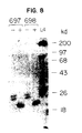
- Sera from two immune sheep (#697 and 698) and from a control, non-infected sheep (#695) were used in immunoprecipitation experiments to determine if they react with 125I-labeled surface proteins. These sera were obtained from Dr. R.J. Boisvenue of Eli Lilly and Company (Greenfield, IN). Sheep #697 and 698 had been orally infected with 2.5 ⁇ 104 H. contortus SL3 larvae, which developed into mature, egg-producing adults. About 40 days post-infection egg production abruptly ceased due to expulsion of the adult worms. At this time, the sheep were challenged with an additional 2.5 ⁇ 104 SL3 larvae.
- Live XL3s ( ca. 200 microliters packed volume) were suspended in 1 ml of 1% SDS, 0.125 M Tris-HCl pH 6.8, placed in boiling water for 2 min, removed from the water, mixed by inversion for an additional 2 min and pelleted for 1 min in a microfuge. The supernatant was drawn off, recentrifuged several times to remove all worms, frozen in a dry ice/ethanol bath and stored at -20C. Samples were later thawed and concentrated at 4°C using a 2 ml Centricon -10 apparatus (Amicon, 10 kDa molecular weight cut-off).
- Live worms ca. 200 microliters packed volume
- 10 mM Tris-HCl pH 7.4 placed in boiling water for 2 min, removed from the water, mixed by inversion for an additional 2 min and pelleted for 1 min in a microfuge.
- the supernatant was drawn off, recentrifuged several times to remove all worms, frozen in a dry ice/ethanol bath and stored at -20C. Samples were later thawed and concentrated at 4C using a 2 ml Centricon -10 apparatus (Amicon, 10 kDa molecular weight cut-off).
- the L4 NaCl extract pattern is similar both qualitatively and quantitatively to the pattern obtained by 125I-labeling.
- the predominant species have molecular weights of 27, 29, 78 and 200 kDa.
- the L4 NaCl extract has relatively less of a 36 kDa 125I-labeled species and more of a 42 kDa protein that is minor in 125I-labeled extracts. The amount of the latter protein was variable from extract to extract, but was never major. A protein of this molecular weight was also sometimes visible in NaCl and SDS extracts of XL3s, but again was only barely visible by 125I-labeling. The yield of L4 surface proteins was approximately the same as that obtained for XL3s.
- New Zealand white rabbits were immunized subcutaneously with 100 micrograms of surface proteins mixed with Freund's complete adjuvant. The rabbits were boosted one month later with 50 micrograms of protein mixed with Freund's incomplete adjuvant. Rabbits were bled 10 to 14 days later. Rabbits 9446 and 153 received NaCl-extracted XL3 and L4 surface proteins, respectively (see Section C). Rabbit 154 received NaCl-extracted L4 surface proteins that had been denatured and reduced by boiling for 2 min in 1% SDS, 5% BME prior to mixing with adjuvants.
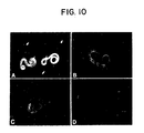
- Immunized rabbits produced antibodies capable of reacting with native proteins present on the surfaces of live worms as determined by indirect immunofluorescence experiments (Figure 10). Reaction of a mixture of live XL3s and L4s with the anti-XL3 surface protein serum (Rb-9446) labels only the XL3s. In contrast, the anti-L4 surface protein serum (Rb-153) reacts with both live L4s and XL3s. Labeling of XL3s with the latter serum may be due to the 5-10% contamination of L4s with XL3s and molted XL3 cuticles in the large worm populations used to prepare the L4 surface protein extracts.
- the L4 has a 78 kDa surface protein that migrates in the same region of the gel as the major 68-97 kDa XL3 surface protein, so it is not possible to detect contamination by one-dimensional gel electrophoresis.
- Another rabbit antiserum (Rb-154) which was prepared against L4 surface proteins that had been isolated using NaCl and later denatured and reduced by boiling in 1% SDS + 5% BME, also reacted strongly with the surfaces of live XL3s and L4s (data not shown).
- 125I-labeled XL3 and L4 surface proteins were boiled for 1 min, centrifuged in a microfuge for 10 min and 10- to 50-microliter aliquots mixed with 1 ml of 2% Triton-X- 100, 50 mM Tris- HCl pH 8.1, 150 mM NaCl, 0.1 mM EDTA and 25 microliters of hyperimmune or prebleed rabbit serum.
- a Protein-A Sepharose slurry 250 milligrams Protein-A Sepharose (Sigma) in 3.5 ml of 10 mM Tris-HCl pH 8.0
- the Sepharose beads containing bound antigens were pelleted by centrifugation in a microfuge.
- the beads were washed 3 x 1 ml with a solution of 2% Triton X-100, 50 mM Tris-HCl pH 8.1, 150 mM NaCl, 0.1 mM EDTA.
- Bound antigens were eluted by boiling the beads for 3 min in 100 microliters of 1% SDS, 0.125 M Tris-HCl pH 6.8, 5% BME and collecting the supernatant. Samples were analyzed by SDS-PAGE and autoradiography.
- the results of these experiments correlate with the IFA results, and are shown in Figure 11.
- the anti-XL3 surface protein serum precipitates all SDS-soluble 125I-labeled XL3 surface proteins. A weak reaction is observed with this serum against 68-97 kDa and 200 kDa proteins in the L4 SDS extract. These proteins could be of XL3 or L4 origin.
- the anti-XL3 surface protein serum does not precipitate the major 27 and 29 kDa SDS-soluble L4 surface proteins.
- both anti-L4 surface protein sera (Rb-153 and Rb-154) precipitate all SDS-soluble 125I- labeled L4 and XL3 surface proteins.
- the Rb-153 and Rb-154 sera also react weakly with the 40 to >200 kDa smear in the SDS + BME extracts of 125I-labeled XL3s and L4s (lanes labeled B in Figure 3). These proteins have been shown previously to be primarily cuticle collagens (Section I(B)(1)).
- the amino acid compositions of purified XL3 and L4 surface proteins were determined and are given in Table I.
- Surface proteins purified using the NaCl extraction procedure were analyzed in these experiments. Protein samples were brought to 1% SDS and precipitated by the addition of 9 volumes of ice-cold acetone. After washing twice with 90% acetone and air-drying, the samples were hydrolyzed in vacuo with 6N hydrochloric acid for 24 hr at 110°C and analyzed on a Beckman amino acid analyzer.
- Both sets of proteins are enriched for hydrophilic amino acids and, with the exception of alanine and glycine, are relatively poor in hydrophobic amino acids.
- the proteins are not enriched for glycine and proline residues, consistent with our previous finding that the proteins are not collagens (Section I(B)(1)).
- 26 percent of the amino acids in the XL3 surface protein extract which should largely reflect the amino acid composition of the major 68-97 kDa species, were either glutamic acid or glutamine.
- the L4 surface proteins contained lesser amounts of these amino acids.
- the present inventors attempted to obtain the amino-terminal sequence of the major 68-97 kDa XL3 protein after elution of the protein from SDS gels or by direct sequencing of the XL3 NaCl extract. In neither case could sequence information be obtained, suggesting that the amino-terminal amino acid of the protein may be modified.
- SL3s that had been frozen in liquid nitrogen were provided by Dr. R. Boisvenue of Eli Lilly and Company, Greenfield, Indiana.
- the frozen SL3s were ground to a fine powder with a mortar and pestle and placed in a liquid nitrogen bath.
- the worm powder was transferred to 30 ml Corex tubes and digested for 45 min at 65°C with Proteinase K (200 ug/ml) in a solution of 0.1 M Tris-HCl pH 8.5, 50 mM EDTA, 0.2 M NaCl, 1% SDS.
- the digestion mixture was alternately extracted several times with phenol and chloroform:isoamyl alcohol (24:1 ratio).
- DNA was precipitated from the aqueous phase by adjusting the solution to 0.3 M sodium acetate and adding 3 volumes of ethanol.
- the precipitated DNA was spooled out with a glass rod, air dried and resuspended in TE buffer (10 mM Tris-HCl pH 8.0, 1 mM EDTA).
- TE buffer 10 mM Tris-HCl pH 8.0, 1 mM EDTA.
- the DNA solution was treated with RNAse (50 ug/ml) for 60 min at 37, extracted sequentially several times with phenol and chloroform:isoamyl alcohol, brought to 0.3 M sodium acetate and the DNA precipitated by adding 3 volumes of ethanol. After air drying, the DNA was suspended in TE buffer.
- H. contortus DNA Eighty micrograms of H. contortus DNA was partially digested with Sau 3A so that the bulk of the DNA was in the 15-20 kb size range as determined by agarose gel electrophoresis.
- the digested DNA was heated to 65°C for 10 min, cooled to room temperature and gently layered over a 10-40% sucrose gradient in an SW41 tube (Maniatis et al. , 1982). After centrifugation for 25 hr at 30,000 rpm at 20°C, the gradient was dripped from the bottom with a 23 gauge needle and fractions of 150-200 l collected. Alternate fractions were electrophoresed on a 0.7% agarose gel and fractions containing 17-20 kb DNA fragments were pooled. The pooled DNA was dialyzed extensively versus TE buffer, precipitated with ethanol and resuspended in TE buffer.
- the size selected Haemonchus DNA was ligated overnight to the lambda phage vector EMBL-3 (Frischauf et al. , 1983) that had been digested with Eco RI and Bam HI.
- the ligation mix was packed in vitro using kits purchased from Boehringer Mannheim and plated on E. coli strains Q359.1 (which allows growth of only recombinant phage) and KRO (which allows growth of only non-recombinant EMBL-3 phage).
- These test platings revealed that a library of 160,000 recombinant phages containing H. contortus DNA inserts had been constructed.
- the entire library was amplified by plating on E. coli Q359.1 (10,000 phage per each of sixteen 15 cm agar plates).
- the amplified library consists of 6.6 x 10⁹ recombinant phages.
- RNA lysis buffer (4 M guanidine hydrochloride, 0.13 M sodium acetate, pH 5.2, 0.5% Sarkoxyl, 1M 2-mercaptoethanol).
- the mixture was layered over one-half volume of 5.7 m CsCl, 50 mM sodium acetate, pH 5.2 and centrifuged at 25,000 rpm for 18 hr at 18°C in an SW28 rotor.
- the RNA pellet was suspended in RNA lysis buffer and precipitated by the addition of 3 volumes of ethanol. After centrifugation, the RNA pellet was suspended in 0.3 M sodium acetate, pH 5.2, ethanol precipitated, dried and resuspended in water.
- the above procedures are slight modifications of those described by Chirgwin et al. (1979).
- Poly(A)+ mRNA was isolated by passage of the RNA through a column of oligo dT cellulose essentially as described by Efstratiadis and Kafatos (1976). The poly(A)+ mRNA was precipitated with ethanol, dried and resuspended in water. From 4.8 mg of total RNA the inventors isolated 191 ⁇ g of poly(A)+ mRNA.
- Double stranded cDNA was prepared from 2 ⁇ g of poly(A)+ mRNA using a kit purchased from Amersham. The methods used were essentially those outlined in the protocol manual that accompanies the kit. Briefly, first strand synthesis was performed using AMV reverse transcriptase and priming the RNA with poly(dT). A small amount of 32P-dATP was included in the reaction mixture to follow the reaction and identify cDNA in subsequent isolation steps. Second strand synthesis was performed using RNAse H and E. coli DNA Pol-1 and the cDNA ends made blunt with T4 DNA polymerase.
- the double stranded cDNA was methylated with Eco RI methylase, extracted with phenol and ether, and isolated by passage through a 1 ml column of Sephacryl S-300 using 20 mM Tris-HCl pH 8.0, 100 mM NaCl, 1 mM Na2 EDTA as the buffer. Phosphorylated Eco RI linkers, (8-mers purchased from New England Biolabs) were ligated overnight at 15°C to the cDNA using T4 DNA ligase. The cDNA was then digested with several hundred units of Eco RI and the cDNA separated from free linkers by passage through a 1 ml column of Sephacryl S-300. After ethanol precipitation and vacuum drying, the cDNA was suspended in water. Approximately 1.3 ⁇ g of double stranded cDNA was obtained.
- the cDNA (10-30 ng per ⁇ g of vector) was ligated to the expression vector ⁇ gt11 (Young and Davis, 1983) that had been digested with Eco RI and dephosphorylated (purchased from Stratagene, Inc.).
- the ligation mix was packaged in vitro (kits purchased from Stratagene,. Inc.) and plated on E. coli strain Y1088 (Young and Davis, 1983) in the presence or absence of IPTG and XGAL to determine the number of recombinant phages containing cDNA inserts.
- Test platings revealed that a library of 4 x 10⁶ recombinant phages had been constructed. This library was amplified by plating on E.
- coli Y1088 (100,000 phages per 15 cm agar plates). After the phages were eluted with lambda dil and collected, test platings revealed that the amplified library contained a total of 7.5 x 10¹² phages of which 97% contained cDNA inserts.
- H. contortus worms were recovered from young lambs infected with a pure drug-susceptible United States Department of Agriculture isolate BPL1. Approximately 35 days following experimental infection of worm-free lambs with an individual inoculum of 25,000 ⁇ 3% ensheathed infective third-stage larvae (SL3) by intraruminal injection, donor lambs were euthanized. Immediately upon necropsy the adult worms were individually collected from the abomasum, placed in warm phosphate buffered saline until sufficient number of worms were collected and then frozen in liquid nitrogen. Frozen worms were stored in liquid nitrogen or at -70°.
- H. contortus adults were homogenized (10 mM MOPS, 150mM NaCl, pH 7.0), 25% (w/v) glycerol, 1mM EDTA, 1mM dithiothreitol and phenylmethanesulfonyl fluoride was then added to a final concentration of 1mM. After low-speed centrifugation (10,000 rpm for 20 min. in a JA-20 rotor), proteins in the supernatant were size-fractionated on a Sepharose CL-4B column using a buffer of 20mM Bis-Tris-propane pH 7.0 (w/v) glycerol, 1mM EDTA, 1mM dithiothreitol.
- Fractions were assayed for fibrinogen-degrading activity using the assay described below. The void volume contained most of the enzymatic activity. These fractions were pooled and applied to an FPLC Mono Q column. Bound proteins were eluted with a gradient of 0.05 to 0.4M NaCl in 20mM Bis-Tris-propane pH 7.0, 10% (w/v) glycerol, 1mM EDTA, 1mM dithiothreitol and fractions tested for fibrinogen-degrading activity. Active fractions were eluted and were pooled. Protein concentrations were determined using a protein assay kit purchased from BioRad Laboratories (Richmond, CA).
- the fibrinogen degradation assay consisted of mixing 5-20 ⁇ l aliquots of the column fractions with an equal volume of a solution of 1% (w/v) bovine fribrinogen (Sigma) suspended in MBS, 1mM EDTA, 1mM dithiothreitol and incubating the samples for 1 hr at 37°C. Samples were then diluted into SDS sample buffer, boiled for 5 min and analyzed on 9% SDS-polyacrylamide gels (Laemmli, 1970). The gels were stained with Coomassie blue to identify fractions that degraded the fibrinogen.
- E64 The specificity of E64 for the active sites of thiol proteases was used to advantage in radiolabeling.
- [14C]-iodoacetic acid was used to label the thiols present in a partially purified preparation of the fibrinogenase. Because of enzymes, this reagent is highly selective towards thiol proteases.
- the active site thiol can be blocked before reaction with the labeled IAA. This allows confirmation that a reactive thiol is indeed at the active site of a protease.
- this technique was applied to partially purified preparation, several bands are labeled with IAA and are specifically protected by the E64.
- This labeling pattern includes the major band present in the purified fibrinogenase at about 35 kDa. Other bands are also labeled under these conditions, suggesting that they also correspond to thiol proteases. These could be related to the fibrinogenase, or they could conceivably be due to other activities which copurify with the fibrinogenase.
- the labeling of the 35 kDa band indicates that it contains the active site of the fibrinogenolytic enzyme.
- the fibrinogenase has been purified from the extracts of H. contortus by means of gel filtration chromatography and ion exchange. Extract was applied to a sepharose 4B column. Fractions were monitored for activity by fibrinogen degradation as observed on gels. The fractions with the highest activity were pooled and applied to a MonoQ column. The activity was elutd with a salt gradient. This two step purification yielded a preparation which contained two major bands on SDS electrophoresis, as well as a number of minor bands, some of which correspond to peptides identified as thiol proteases by IAA labeling. It has not been possible to further purify this preparation while retaining activity of the fibrinogenase.
- the fibrinogenase has been characterized as having a native MW of greater than 1,000,000 by means of gel filtration chromatography on an S12 sizing column, where it elutes at the void volume of the column.
- Standard proteins indicated that the MW corresponding to the void volume is on the order of 1 million.
- the activity also elutes in the void volume of the 4B column, which has an even higher cutoff.
- Antiodies have been raised in rabbits to the anticoagulant.
- the antibodies were used to probe Western blots of extracts of H. contortus as well as the purified fractions.
- the antibodies reacted specifically with the 35 and 55 kDa bands.
- the antibody was further used to inhibit the activity of the enzyme. This was accomplished by incubating the antibody preparation with the anticoagulant, prior to incubation with fibrinogen.
- a further test of the inhibitory powers of the antibody was to study clotting time of plasma from the hyperimmune rabbit and control plasma. This experiment showed a substantial decrease in the effectiveness of the anticoagulant in the hyperimmune plasma.
- Partially purified anticoagulant material from the Mono-Q column was emulsified with Freund's complete adjuvant and injected subcutaneously at several sites along the back of rabbit #9503.
- the rabbit was boosted with an additional protein emulsified with Freund's incomplete adjuvant.
- the rabbit was boosted at roughly one month intervals. Two weeks after each boost, the rabbit was bled and serum obtained after allowing the blood to clot overnight at 4°C.
- Partially-purified anticoagulant material obtained from the void volume of the Sepharose CL-4B sizing column was diluted into SDS sample buffer, electrophoresed on a 12% preparative SDS gel (0.75 mm thick) and stained for 15 min with 0.1% Coomassie blue in water to localize protein bands.
- Gel slices containing the 35 kDa and 55 kDa proteins were cut with a razor blade, diced, and the proteins eluted from the gel using an eluting apparatus purchased from Isco, Inc. The eluted proteins in running buffer were stored at -20°C.
- Rabbit antisera to the 55 kDa protein was prepared in the same way except that 50 ⁇ g of eluted 55 kDa protein was used per injection. These rabbit antisera are designated Rb-10284 and Rb-10286.
- cDNAs encoding the 35 kDa protease were isolated by screening the adult worm cDNA: ⁇ gt11 library with Rb-10285 antiserum.
- the adult cDNA: ⁇ gt11 expression library was plated on E. coli Y1090 (Young and Davis, 1983) at a density of 20,000 phages per 15 cm agar plate. The plates were incubated for 4 hr. at 42°C, then overlaid with nitrocellulose filters that had been wetted in 10 mM IPTG and air dried. The plates with filters were incubated overnight at 37°C.
- TBS Tris-HCl, pH 8, 150 mM NaCl
- the filters were incubated for 60 min in TBS + 2% BSA (bovine serum albumin), then for 2 hr with gentle rocking at room temperature with Rb-10285 serum diluted 1:200 in TBS + 2% BSA. These incubations were done in 15 cm petri dishes containing 20 ml of liquid. Two filters placed back to back were incubated per petri dish.
- the filters were then washed 3 x 15 min in TBS + .05% NP-40 and incubated for 60 min in 15 cm petri dishes containing horseradish peroxidase-conjugated goat anti-rabbit IgG antisera (Cappell Laboratories) diluted 1:500 in TBS + 2% BSA. After washing 3 x 15 min in TBS, the filters were placed in stain solution (200 ml TBS + 2.5 ml H2O2 + 40 ml of methanol containing 3 mg/ml 4 chloro-1-naphthol (Sigma- Aldrich Corp.)).
- the recombinant phage clones were used to affinity purify antibodies that react with their expressed antigen from the polyclonal rabbit serum ("antibody elution experiments"). The eluted antibodies were then used to probe Western blots of worm proteins to identify the target antigen corresponding to the cDNA in each phage clone.
- Phages were plated at a density of 1 ⁇ 104 per 15 cm diameter agar plate using E. coli Y1090 and incubated at 42°C for 4 h. Nitrocellulose filters that had been impregnated with 10 mM IPTG and air-dried were placed on top of the phages and the plates were incubated overnight at 37°C. After cooling, the filters were removed, cut into 70 x 100 mm strips, incubated in TBS + 2% BSA for 1 h, and incubated overnight with Rb-10285 serum diluted 1:200 in TBS + 2% BSA.
- DNA from phage 2B was prepared by the plate lysate procedure (Davis et al. , 1980) using E. coli Y1090 as the host (3 x 10⁶ phage mixed with 0.7 ml of an overnight Y1090 culture that had been centrifuged and resuspended in 1/2 volume of 10 mM MgSO4 per 15 cm agar plate). Phage particles were purified by banding in CsCl step and equilibrium gradients using established procedures (Davis et al. , 1980). Phage DNA was isolated by formamide extraction and ethanol precipitation (Davis et al. , 1980). Phage DNA was resuspended in TE buffer.
- phage 2B DNA with Eco RI Digestion of phage 2B DNA with Eco RI revealed that it contained a cDNA insert of about 180 bp.
- the nucleotide sequence of the cDNA was determined by the dideoxy nucleotide sequencing method (Sanger et al. , 1977; Biggen et al. , 1983) after subcloning into the Eco RI site of M13mp18.
- the nucleotide sequence of the cDNA revealed that it encoded only 12 amino acids fused to ⁇ -galactosidase; the remainder of the cDNA consisted of 3′ untranslated sequences and a poly(A) tail.
- the 3′ untranslated region contained a canonical poly(A) addition sequence AATAAA.
- cDNA 2B was labeled with 32P by nick-translation and used to screen the cDNA library by plaque hybridization in order to identify larger cDNAs.
- the first such screen yielded cDNA 3-1, which was about 870 bp in length.
- a 40 nucleotide long oligomer that corresponds to the sequence at the 5′ end of cDNA 3-1 was synthesized, end-labeled with 32P and used to screen the cDNA library for even larger cDNAs.
- Duplicate filters were screened with 32P-labeled cDNA 2B.
- the oligonucleotide which has the sequence 5′ - CACTTCAGGGTCGGGATCTTCTTTGACCATAAGATTTAGC - 3′ was synthesized on an Applied Biosystems DNA synthesizer.
- the labeled oligomer was hybridized to nitrocellulose filters at 32-52°C using 2X SSC/5X Denhardt's/0.5% sodium dodecyl sulfate. Filters were washed in 2X SSC/0.5% sodium dodecyl sulfate at 52°C.
- This screen yielded cDNAs F-1, 0-1 and T-1, all of which hybridized to the oligomer and to cDNA 2B.
- cDNA F-1 was the largest, ⁇ 1100 bp, and was chosen for further characterization.
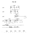
- cDNAs 2B, 3-1 and F-1 The relationship of cDNAs 2B, 3-1 and F-1 is shown in Figure 14, along with a composite restriction map of the cDNAs. Various regions of the cDNAs were sequenced and found to be identical in the regions in which the sequences overlapped. The one difference noted was that the 3′ untranslated region of cDNAs 3-1 and F-1 were shorter than that of cDNA 2B ( Figure 15). The composite nucleotide sequence and predicted amino acid sequence of the cDNAs is presented in Figure 15. This gene has been named AC-1.
- cDNA F-1 The largest cDNA, F-1, contained a single long open reading frame but was missing an initiator methionine codon at its 5′ end; therefore, the inventors presumed that the cDNA was not full-length.
- Northern blot hybridizations (see Section II(G)(1) indicated that cDNA F-1 hybridized to a 1.25 kb transcript in adult worm poly(A)+ mRNA preparations.
- contortus ⁇ EMBL-3 library (Section II(E)(3) confirmed this result and indicated that cDNA F-1 was missing the codon for only one amino acid, the initiator methionine. For completeness, the initiator methionine has been included in the sequence presented in Figure 15.
- the AC-1 protein comprises 342 amino acids and has a predicted molecular weight of 38.4 kDa.
- At the N-terminus of the protein is a stretch of about 15 hydrophobic amino acids that could function as a signal sequence for sequestration of the protein to the rough endoplasmic reticulum, as a prelude to extracellular secretion or localization to cellular organelles.
- Computer analysis predicts that the signal sequence would be cleaved between amino acids 18 and 19 (Ala-Asp). There are no other significant hydrophobic regions in the protein.
- the AC-1 protein contains 16 cysteine residues, two of which are present in the presumed signal sequence and would not be present in the mature protein.
- the protein also contains four potential N-linked glycosylation sequences (Asn-X-Ser/Thr, where X can be any amino acid), which are indicated in Figure 15.
- Treatment of purified anticoagulant proteins with Endoglycosidase F reduces the apparent molecular weight of the AC-1 protein to 33 kDa ( Figure 18), indicating that the protein is glycosylated in vivo .
- the enzyme was used according to the methods provided by the supplier (Genzyme).
- AC-1 The primary sequence of AC-1 was compared to sequences of other known thiol proteases. These analyses revealed that AC-1 shows significant homology with mammalian cathepsin B (human, rat and mouse) and to a lesser extent with other cathepsins and with the plant protease papain (Chan et al. , 1986; Cohen et al. , 1986; Wada et al. , 1987). AC-1 shares an overall 42% amino acid identity with human cathepsin B (Figure 17). A stretch of six identical amino acids that includes the active site cysteine of cathepsin B is present in AC-1 ( Figure 19). This sequence also is conserved in papain (Figure 19) and is present in the same relative locations in all three proteases.
- cysteine-114 is the active site cysteine of the AC-1 protease.
- AC-1 can be aligned for homology with mature cathepsin B by introducing only two single amino acid gaps in the proteins (Figure 17).
- Figure 17 When these minor alignments are introduced, all 14 cysteines in the mature cathepsin B protein align with cysteine residues in AC-1, suggesting the AC-1 and cathepsin B have similar tertiary structures.
- Histidine residue in AC-1 (residue #285) that is in an identical position as the histidine residue (#278) that forms part of the active site of cathepsin B.
- the amino acids immediately surrounding these histidine residues are not as conserved as those surrounding the active site cysteine residues (Figure 17).
- Cathepsin B is synthesized as a pre-proenzyme that contains an N-terminal signal sequence, followed by a stretch of 62 amino acids that must be cleaved from the proenzyme to generate the mature, active protease.
- the "pro" region also may be involved in localizing cathepsin B to lysosomes.
- the positions of the above amino acid cleavages in cathepsin B are marked in Figure 17. Nearly all of the amino acids that are identical between AC-1 and cathepsin B are located in the region of cathepsin B that constitutes the mature, active enzyme ( Figure 17). Little similarity, other than length, exists between the "pre” and "pro” sequences of cathepsin B and AC-1. When just the mature form of cathepsin B is compared to the corresponding region of AC-1, the amino acid similarity between the two proteases increases to 49%.
- Rb-9503 antiserum was used to screen the adult worm cDNA: ⁇ gt11 library using the procedures described in Section II(E)(1), with minor modification. This screen yielded cDNAs 2-1, 6-1 and 7-2. Nucleotide sequence analysis of the cDNAs revealed that they overlapped one another and encoded a protein that was related, but distinct from AC-1. The longest cDNA, 2-1, which was about 200 bp in length, was eluted from an agarose gel, labeled with 32P using random primers and used to screen the adult worm cDNA library by plaque hybridization. Several positive phages were identified and plaque-purified.
- V-22 and V-24 are incomplete cDNAs.
- Longer cDNAs can be isolated by screening the adult cDNA expression library with 32P-labeled oligonucleotides derived from the 5′ end of the cDNAs (or any other region of the cDNA sequence).
- the complete gene sequence also can be obtained by isolating the gene from a genomic DNA library, identifying the region of DNA missing from the cDNA and sequencing this region.
- the gene encoding AC-2 was isolated from the H. contortus ⁇ EMBL-3 phage library by screening the library with 32P-labeled cDNA 2B.
- the cDNA insert was isolated from 1.5% agarose gels using NA45 paper (Schleicher and Schuell).
- the cDNA insert was self-ligated overnight at 15°C with T4 ligase, ethanol precipitated and nick-translated with 32P.
- the labeled cDNA was used as a hybridization probe to screen the Haemonchus DNA:EMBL-3 library.
- the library was plated on E. coli LE392 at a density of 5 ⁇ 103 phage per 10 cm plate and screened by plaque hybridization (Benton and Davis, 1977).
- Hybridization conditions used were 50% formamide, 0.1 M sodium phosphate pH 7.0, 0.1% SDS, 10 ⁇ g/ml sheared salmon sperm DNA, 3 X SET (1 X SET is 50 mM Tris-HCl pH 8.0, 150 mM NaCl, 1 mM EDTA) at 37°C.
- a screen of 100,000 recombinant EMBL-3 phages yielded two positive phages designated ⁇ MB1 and ⁇ MB2. These phages were plaque purified by repeated hybridization to the labeled 2B cDNA probe. Phages were prepared from plate lysates using E. coli LE 392 as the host and banding in CsCl gradients.
- the inventors rescreened the ⁇ EMBL-3 library to identify phages containing Haemonchus DNA inserts that extended further upstream of the region present in ⁇ MB1 and 2 ("chromosome walking" technique).
- the ⁇ EMBL-3 library was screened in duplicate with 32P-labeled restriction fragments from the left end (the 3.9 kb Eco RI fragment) and the middle (the 3.5 kb Eco RI fragment) of ⁇ MB2 (see Figure 20).
- the restriction fragments were eluted out of agarose gels using NA45 paper, self-ligated overnight, ethanol precipitated, resuspended in water and labeled with 32P by nick- translation. Three phages that hybridized only with the 3.9 kb Eco RI fragment were identified and plaque-purified. Hybridization of the phage DNAs with the 40 nt oligomer revealed that each of them contained the missing exon(s). One of the phages, ⁇ MB3, was mapped with restriction enzymes and shown to overlap ⁇ MB1 and 2, as expected (Figure 20).
- cDNA F-1 does not contain an initiator methionine codon.
- the gene encodes a methionine three amino acids upstream of where cDNA F-1 terminates. Just upstream of this methionine codon is an in-frame TGA stop codon. Although it is possible that this methionine codon is the initiator methionine codon for the gene, the inventors do not believe so.
- TTTCAG/A is a consensus 3′ intron acceptor splice sequence; the slash indicates where splicing would occur. If this sequence functions as a splice acceptor sequence, then the above methionine codon is present in an intron and could not be present in the mature mRNA. The upstream region was searched for other potential initiator methionine codons. Approximately 80 bp upstream of the putative 3′ acceptor sequence is the sequence ATG/GTAA which fits the consensus intron 5′ splice donor sequence. Splicing would join this ATG methionine codon in-frame with exon 2 and the remainder of the gene.
- the nucleotide sequence of the gene including the small intron sequences is presented in Figure 21.
- the gene has 97% nucleotide identity with the AC-1 cDNA F-1, to which it is compared in Figure 21. Most of the nucleotide differences occur in the presumed 3′ untranslated region of the gene (cDNA) and in third-based codon wobble positions that do not change amino acids. Seven nucleotide changes result in different amino acids. Overall, the gene and the F-1 cDNA have 98% protein sequence identity. At this time the inventors do not know if the gene is distinct from the gene that encodes the AC-1 cDNAs or whether the nucleotide (protein) differences are due to polymorphisms in a single gene in the H.
- contortus worm populations used to construct the cDNA and genomic DNA libraries The inventors have isolated a partial cDNA (350 bp in length) that has an identical nucleotide sequence as the gene from the adult worm cDNA library, so the gene appears to be expressed (data not shown). Complicating this issue is the fact that the protease appears to be encoded by a multigene family (see below). Because of these uncertainties, the inventors have named the gene AC-2 to distinguish it from the AC-1 gene identified by cDNAs 2B, 3-1 and F-1 (Section II(E)(1)).
- the AC-2 gene contains 11 introns that range in size from 57 bp to over 5.2 kb.
- Approximately 40 bp upstream of the proposed initiator methionine is a sequence that is similar to the eukaryotic TATA promoter element. The inventors have no evidence as yet that this sequence functions as a promoter for the AC-2 gene.
- Downstream of the TGA stop codon is a canonical AATAAA poly(A) addition sequence.
- the active site of the AC-1 protease has been tentatively identified by homology with the active site sequences of Cathepsin B and papain.
- the AC-1 protease, Cathepsin B and papain have an identical six amino acid sequence that includes the active site cysteine of these other proteases. These six amino acids (Cys-Gly-Ser- Cys -Trp-Ala; the underlined Cys is the active site cysteine of papain and cathepsin B) also are conserved in the predicted AC-2 protein (marked in Fig. 21). Intron 7 interrupts this conserved domain between Gly and Ser, indicating that the conserved active site sequence has not evolved as a single exon unit.
- AC-1 has four potential N-linked glycosylation sites (Asn-X-Ser/Thr, where X can be any amino acid). All four potential glycosylation sites are conserved in AC-2 and are marked in Figure 21.
- AC proteases comprise a multi-gene family in H. contortus
- the labeled probe used for these hybridizations was the AC-1 cDNA 2B, which hybridizes to a single 1.0 kb Eco RI fragment in ⁇ MB1 (this fragment is marked in Figure 20). There is a 1.0 kb genomic DNA fragment that hybridizes to cDNA 2B and presumably corresponds to the 1.0 kb band in ⁇ MB1 ( Figure 22).
- the four other hybridizing bands detected in Eco RI digests of H. contortus DNA must derive from additional gene copies of the protease.
- the multiple hybridizing bands detected with the 2B cDNA probe is in contrast to the single hybridizing band detected with a tropomyosin gene probe (data not shown).
- Rabbit antisera were prepared against an AC-1:- galactosidase fusion protein in order to study expression of the protein in various developmental stages of H. contortus . Because of the high degree of amino acid sequence identity between the predicted AC-1 and AC-2 proteins, the inventors expect that antisera raised against the AC-1 protein will cross-react with the AC-2 protein; however, it has not proven possible yet to test this assumption.
- the gene fusion was constructed by subcloning a fragment of cDNA 3-1 into the ⁇ -galactosidase expression plasmid pSEV6. Plasmid pSEV6 was derived from plasmid pSEV4, which is identical to plasmid pLG2 (Guarente et al.
- pSEV4 DNA was digested to completion with Sph I and then partially digested with Aat II. T4 DNA polymerase was used to make the DNA ends blunt. After agarose gel electrophoresis, the 7.6 kb partial digestion product was electroeluted, ethanol precipitated, dried, resuspended in buffer and ligated overnight with T4 DNA ligase to seal the blunt ends. This step destroys the Sph I and Aat II restriction sites. The ligation mixture was used to transform E. coli AMA1004 (Casadaban et al.
- Plasmid DNA was isolated from blue colonies and one plasmid with the proper configuration was designated pSEV5.
- pSEV6 Plasmid DNA was isolated from blue colonies and one plasmid with the proper configuration was designated pSEV5.
- pSEV5 DNA was digested with Nco I and ligated overnight with complimentary DNA adapters of sequence 5′ CATGAGATCTGGTAC 3′ and 5′ CATGGTACCAGATCT 3′.
- plasmid DNA was isolated from several blue colonies and analyzed for the presence of unique Nco I, Kpn I and Bgl II sites in the proper orientation.
- One such plasmid was designated pSEV6.
- a map of pSEV6 is shown in Figure 24.
- pSEV6::AC-1 a pBR322 plasmid containing AC-1 cDNA 3-1 was digested with Eco RV and ligated to Eco RI linkers with the sequence 5′ CCGGAATTCCGG - 3′. After digestion with Eco RI, the ⁇ 800 bp fragment containing most of the AC-1 coding sequence was eluted from an agarose gel. The restriction fragment was subcloned into the unique Eco RI site of the ⁇ -galactosidase gene in plasmid pSEV6 ( Figure 24). cDNAs in the proper orientation with respect to the ⁇ -galactosidase gene were selected by antibody screening and by digestion with Xho I, which cleaves asymmetrically within the 3-1 cDNA.
- Bacteria containing the proper construct were grown in LB broth contain 50 ⁇ g ml ⁇ 1 ampicillin until the optical density at 600 nanometers was 0.3. Isopropyl--D-thiogalactopyranoside was then added to 1 mM and the culture shaken for an additional 2 h at 37°C. Bacteria were harvested by centrifugation and lysed by boiling briefly in SDS sample buffer (Laemmli and Favre, 1973) and vortexing.
- the AC-1: ⁇ -galactosidase fusion protein has a molecular weight of 140 kDa and contains the final 241 amino acids of AC-1.
- Antiserum from rabbit #10285 which was immunized with the natural 35 kDa protein isolated from adult worms and whose serum was used to isolate cDNA 2B from the ⁇ gt11:adult worm cDNA expression library, reacts on Western blots with the recombinant AC-1: ⁇ -galactosidase fusion protein (Figure 25), confirming that the modified cDNA was joined in the proper reading frame.
- the AC-1: ⁇ -galactosidase fusion protein was electroeluted from preparative SDS gels and used to immunize rabbits #8552 and #9190. Rabbits were given primary injections of 100 ⁇ g of total protein emulsified in Freund's complete adjuvant. Subsequent booster injections given at monthly intervals were with 100-200 ⁇ g of total protein mixed with Freund's incomplete adjuvant. All injections were given subcutaneously. Serum was obtained 10-14 d after each booster injection. Both immune rabbit antisera reacted with a 35 kDa protein in adult worm extracts (Figure 26).
- both immune sera react with an identical size polypeptide in "anticoagulant" extracts that have been purified from adult worms using a fibrinogen-degradation assay.
- Both immune antisera also reacted with a 37 kDa protein in adult worm extracts and in anticoagulant extracts.
- Rb-8552 serum reacted more intensely with this protein than did the Rb-9190 or Rb-10285 antisera.
- Endoglycosidase F digestion experiments suggest that the 37 kDa protein is a more heavily glycosylated form of the 35 kDa protein because the apparent molecular weights of both proteins are reduced to 33 kDa after Endoglycosidase F treatment.
- AC-1 encodes the 35 kDa protease present in anticoagulant extracts prepared from H. contortus adult worms and provide additional evidence that the 35 and 37 kDa proteins in these extracts are antigenically related and possibly different forms of the same protein.
- H. contortus is ingested by sheep as an SL3 (third-stage larva that retains the second larval-stage cuticle). In the rumen the SL3 sheds the second-stage cuticle and the resulting XL3 larva migrates to the abomasum. Within a few days, the XL3 molts into an L4 (fourth-stage larva). The L4 is the first stage that actively feeds by sucking blood. The L4 molts into a young adult after a few days. Very little growth occurs between the SL3 and young adult stages. Young adults are about 0.5 mm in length and grow to over 25 mm in length during the next few weeks. Nourishment for this growth is provided by metabolism of host blood components.
- the Rb-8552 and Rb-9190 antisera were used to probe Western blots (Towbin et al. , 1979) of protein extracts of SL3s, XL3s, L4s and mature adults.
- the XL3 and L4 worms analyzed in these experiments were obtained by culturing worms in vitro in a defined media and were not isolated from infected sheep.
- the 35 kDa protein (and the 37 kDa form) was detected only in extracts of adult worms.
- Identical results were obtained with Rb-10285 serum, which was prepared against the natural 35 kDa protein isolated from adult worms (Figure 27).
- Worm-free sheep approximately 12 months old were injected intramuscularly at several sites with purified anticoagulant material prepared as described in Section II(B) emulsified with adjuvant (vaccinated group) or with adjuvant only (control group).
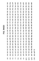
- the time course of injections, the amount of anticoagulant used per injection and the type of adjuvant used are presented in Table I.
- Ten days after the final boost the sheep were challenged with 2500 H. contortus ensheathed third stage larvae (SL3s) given via a single intraruminal injection. Fecal samples were collected over the next 2 months and the number of H. contortus eggs per several milligram samples determined. These numbers were converted to eggs per gram of feces (EPG) by multiplying by the appropriate conversion factor.
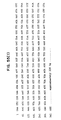
- Sheep vaccinated with the anticoagulant material exhibited both reduced group average EPGs and total worm counts at necropsy as shown in Table II.
- the group average EPGs of the vaccinated sheep were 77% lower than the group average of the control sheep at 42 days post-worm challenge.
- 3 of the 4 vaccinated sheep were negative for egg counts.
- the average total worm count at necropsy for the vaccinated group was 79% lower than that of the control group.
- This trial was a repeat of the first anticoagulant experiment and all parameters of the experiment were identical except that sheep were divided into 3 groups: Group 1 was treated identically to the vaccinated group of trial #1; Group 2 was treated identically to the control group of trial #1; Group 3 was treated identically as Group 1 except that one-fifth of the initial anticoagulant injection was given intravenously. The booster injections for Group 3 were given intramuscularly and in an identical manner to Group 1.
- sheep vaccinated with the anticoagulant material exhibited both reduced group average EPGs and total worm counts at necropsy as shown in Table III.
- All of the Group 1 sheep were negative for worm counts versus a group average of 325 for control Group 2.
- the total worm counts at necropsy for Group 1 sheep were 86% less than the total for the control sheep.
- Seven of the 8 sheep vaccinated with the anticoagulant material (Groups 1 and 3) had an average total worm count at necropsy of 42 versus an average of 269 for the control sheep.
- Only one of the vaccinated sheep in Group 3 (sheep #330) had a very high number of worms at necropsy and appears to be a non-responder animal.
- Each group contained 4 sheep and they were injected with protein according to the schedule outlined in Table I, except that Groups E and F sheep received injections of 50, 75, 100, 100, 100 ⁇ g of protein on successive weeks, rather than the larger amounts of anticoagulant proteins given.
- the primary injection used Freund's complete adjuvant, while booster injections used Freund's incomplete adjuvant. All injections were given intramuscularly.
- Table IV At necropsy, three out of the four sheep in each of the vaccinated groups receiving anticoagulant or 35 or 55 kDa proteins were negative for egg counts or produced only infertile eggs.
- three out of four sheep in the anticoagulant vaccinated or 35/55 kDa vaccinated groups had no normal appearing adult worms at necropsy. Some of the sheep had small, infertile adults which suggested an immunological reaction directed against the worms.
- the anticoagulant material is immunogenic in sheep and can be used as a vaccine to protect sheep from H. contortus infections as determined by two measurements of anthelmintic activity:reduction in worm egg counts and reduction of worm populations. Further, the inventors conclude that either the 35 kDa protein or the 55 kDa protein can be used individually as vaccines to protect sheep from H. contortus infections.
- the Haemonchus AC-1 protease has been expressed in E. coli in two forms.
- These constructs have been expressed using plasmids containing the T7 phage promoter or the Tac promoter to drive expression of the recombinant protein. All constructs use a translational coupler to enhance synthesis of the recombinant protein.
- cDNA F-1 was digested with Xba I.
- the cut DNA was ligated to synthetic linkers of sequence: MetAspGluAsnAlaAlaGlnGlyIlePro 5′ GATCCGATCTTGGAGGATGATTAAATGGACGAAAACGCTGCACAGGGTATCCCG - 3′ 3′ - GCTAGAACCTCCTACTAATTTACCTGCTTTTGCGACGTGTCCCATAGGGCGATC - 5′
- the ligation mixture was digested with EcoR I and the 1062 bp DNA fragment gel-purified. This DNA piece was ligated with T7 promoter plasmid pT5T DNA ( ) that had been digested with EcoR I and Bam HI. The ligated DNA was used to transform E. coli strain BL21/DE3 and colonies containing the plasmid selected on ampicillin plates. Plasmid DNAs from several colonies were purified using the rapid boiling method (Homes and Quigley, 1981) and digested with EcoR I and Bam HI to confirm that they contained the proper size DNA insert. Two plasmids with inserts of the correct size were identified and designated pT5T::X-1 and pT5T::X-8.
- cDNA F-1 was digested with EcoR V and ligated with synthetic linkers of sequence: Met 5′ GATCCGATCTTGGAGGATGATTAAATG - 3′ 3′- GCTAGAACCTCCTACTAATTTAC - 5′
- the ligated DNA was digested with EcoR I and the 857 bp band gel-purified. This DNA piece was ligated with plasmid pT5T DNA that had been digested with EcoR I and Bam HI. The ligated DNA was used to transform E. coli strain BL21/DE3 and colonies containing the plasmid selected on ampicillin plates. Plasmid DNAs from several colonies were purified and digested with EcoR I and Bam HI to make sure that they contained the proper size DNA insert. Two plasmids with correctly sized inserts were identified and designated pT5T::RV-2 and pT5T::RV-3.
- the X-8 construct was transferred to the TAC promoter plasmid pT3XI-2-tac10 as follows. Plasmid pT5T::X-8 DNA was digested with Sma I (the Sma I site occurs in the polylinker sequence downstream of the X-8 sequences), ligated with Sac II linkers of sequence: 5′ GCCGCGGC 3′, and digested with Bam HI and Sac II. The 1078 bp Bam HI: Sac II fragment was gel-purified and ligated with pT3XI-2-tac10 DNA that had been digested with Bam HI and Sac II and gel-purified. The ligation mixture was used to transform E.
- coli JM107 and colonies selected on tetracycline plates.
- Tac X-8 was verified by restriction mapping and by DNA sequencing of the 5′ end of the insert.
- a Coomassie blue stain of the gel is shown in Figure 30 and a Western blot of the proteins using antisera against the 35 kDa protein purified from adult Haemonchus (Rb-10285 sera; see section II(D)(2) is shown in Figure 30.
- the RV-2 construct produces a 30 kDa band that reacts with Rb-10285 sera and increases in abundance upon induction with IPTG.
- the X-8 construct produces proteins of molecular weights of 38 and 32 kDa that react with Rb-10285 serum.
- the 32 kDa protein is more abundant than the 38 kDa protein. Both proteins decrease in abundance upon induction with IPTG.
- the TAC X-8 construct in JM107 was grown to late log phase (OD660 1.0 to 2.0) in Luria broth (LB) with 12.5 ⁇ mg/ml of tetracyclin.
- the cells were harvested without induction.
- the pelleted cells were resuspended in 25 mM TRIS:Cl pH 8.0, 5 mM DDT and 15 mM NaCL at a proportion of 20 ml to 1 gm wet weight of cells.
- the suspension was put through a French Press at 18,000 psi three times. The cell lysate was then centrifuged for 30 minutes at 30,00 x g.
- the pellet was resuspended with a loose-fitting dounce in a solution of 6 M urea and 100 mM 2-mercaptoethanol at a volume of 10 ml to each original gm cells. Following resuspension 0.7 M borax pH 7.0 and 10% polyethyleneimine were added at 0.15 ml and 0.05 ml, respectively, to each 10 ml of resuspended volume. The resuspended solution was centrifuged for 30 min at 30,000 x g.
- the supernatant was then loaded onto an S-Sepharose column (Pharmacia) in a ratio of 10 ml per 1 ml of packed beads with a column height to diameter size of 11.0 cm x 1.0 cm.
- the column was equilibrated in buffer containing 25 mM TRIS:HCl pH 8.0, 5 mM DTT, 1 mM EDTA and 6 M urea. Following application of the sample the same buffer was used to wash the column. Proteins bound to the column were eluted with a linear gradient of NaCl up to 500 mM. The recombinant 35 kDa protein was eluted between 125 and 175 mM NaCl.
- rabbit serum specific towards the SDS-denatured 35 Kd polypeptide does not directly inhibit the fibrinogenase activity in the standard enzyme assay.
- this antiserum can be used to select out this activity, thus supporting the evidence that this polypeptide is at least in part, if not totally, responsible for the fibrinogenase activity. That the other major polypeptide, the 55 kd protein, may also be part of the fibrinogen cleavage activity could not be ruled out.
- Serum from control sheep and those immunized with the anticoagulant preparation showed partial inhibition of the fibrinogenase activity.
- IgG fractions isolated from these sera continued to demonstrate inhibition of the enzyme and therefore a direct link between protection and enzyme inhibition could not be made.
- the sera from immunized sheep do react with the 35 and 55 kDa polypeptides by Western blot analysis. It is possible that direct inhibition is not required for the effective protection to worm challenge, but that immune clearance or complement-mediated damage may be mechanisms for protection.
- the anti-coagulase activity from adult worms had been determined to be a specific fibrinogen cleavage activity.
- the alpha and beta bands of bovine fibrinogen are degraded when incubated with increasing amounts of the partially purified enzyme preparation.
- Analysis of the enzyme preparation of SDS-PAGE and subsequent staining with Coomassie blue demonstrate two major bands of approximately 35 and 55 kDa.
- the anti-coagulase is similarly electrophoresed and stained with the more sensitive silver staining method a number of additional polypeptides are visualized (Figure 33). Some of the higher molecular weight minor bands are believed to represent collagen polypeptides. Antiserum towards the anti-coagulant preparation raised in rabbits, as well as in sheep during protection experiments, react with collagens.
- the fibrinogenase activity is thiol dependent and the use of appropriate inhibitors and thiol labeling reagents indicate that the 35 kDa polypeptide contains an active thiol.
- Evidence that the fibrinogenase activity may be associated with other polypeptides or is a single polypeptide aggregate comes from native molecular weight sizing columns showing the activity to elute at a molecular weight of at least one million. Attempts to disaggregate the complex and maintain activity were unsuccessful.
- polyclonal rabbit sera were raised to these polypeptides.
- Preparative quantities of anti-coagulant were electrophoresed on SDS polyacrylamide gels and the proteins eluted from the gels.
- the purified and SDS denatured proteins were injected into rabbits and the serum tested for reactivity on Westerns.
- One serum obtained was specific for the 35 Kd, however the second serum was contaminated during the isolation and/or injection steps of the process and produced a serum reactive to both the 35 and 55 kDa polypeptides.
- the rabbit sera were tested for their ability to directly inhibit the fibrinogenase activity.
- the serum and enzyme were preincubated and then substrate was added and following incubation analyzed by gel electrophoresis. Both the preimmune and immune serum (anti-35 and anti-35/55) were inhibitory to the fibrinogenase.
- the IgG was fractionated from each of these serum samples. Control Western analysis indicated that the reactivity towards the polypeptides did indeed separate with the IgG fraction.
- the second part of this experiment was to confirm that the loss of activity was indeed due to the selection of the particular polypeptides in question. This was done by combining the Staph A cells from each sample and boiling off both the IgG and any polypeptides they had selected from the anticoagulase preparations. These samples were electro-phoresed on an SDS-PAGE and blotted to nitrocellulose. The Western was then incubated with antisera specific to the 35 and 55 kDa polypeptides. This Western is shown in Figure 36. Because the original sera was made in rabbit, the second antibody used for color visualization was goat anti-rabbit IgG linked to HRP.
- Immune and control sheep serum from anti-coagulant challenge experiment #1 were tested for their ability to inhibit the fibrinogenase. Similar to the rabbit serum experiments, serum from both control and test sheep inhibited the fibrinogenase. Therefore, IgG from the two control and four test sheep sera was isolated and tested for inhibition of the enzyme. The results, shown in Figure 37, indicate that both the control and the four test IgG fractions show partial inhibition of the enzyme. Close inspection suggests that the immune IgG fractions may inhibit slightly more than the test serum IgG. Unfortunately, the conclusion must be that the results are equivocal. It did not determine whether the inhibition is due to non-specific factors or specific immunoglobins towards the anti-coagulant. Additional steps towards eliminating the non-specific inhibition would be required. Also, other more sensitive assays could be used to measure inhibition.
- test sheep serum are reactive with the 35 and 55 kDa polypeptides. Therefore a similar experiment of immune selection described above by analogy should give a similar result.
- the H. contortus : ⁇ EMBL-3 library was screened by plaque-hybridization with 32P-labeled cDNA V-22 using procedures similar to those described in Section II.E.3.
- the hybridization and wash temperatures were 37°C.
- cDNA V-22 had been eluted from agarose gels and labeled with 32P using random primers.
- the phages have been named ⁇ MB4, ⁇ MB5, and ⁇ MB6.
- the regions of the phages that hybridize to the V-22 and F-1 cDNAs can be determined using procedures similar to those described in Section II for mapping the AC-2 gene in ⁇ MB1.
- DNA sequences encoding exons 1-4 can be identified using the F-1 exon 1-4 probe under low stringency hybridization conditions (30% formamide solutions-see Section II.P.3). If sequences corresponding to exons 1-4 are not present in ⁇ MB4-6, then "chromosome walking" techniques can be employed to rescreen the library to identify recombinant ⁇ EMBL-3 phages that contain H. contortus DNA sequences extending 5′ of the coding sequences present in ⁇ MB4-6. Procedures for doing so are fully presented in Section II.E.3, which describes the isolation and mapping of ⁇ EMBL-3 phages encoding exons 1-4 of the H. contortus AC-2 gene.
- Regions of the ⁇ EMBL-3 phages that hybridize to cDNAs V-22, F-1, or the F-1 exon 1-4 probe can be sequenced to identify DNA sequences corresponding to coding regions of the gene that are not present in cDNAs V-22 and V-24.
- Plasmid pBR325::F-1 which contains the ca. 1.0 kb Eco RI fragment of cDNA F-1 inserted into the Eco RI site of pBR325, was digested with Xba I and ligated with oligonucleotide adaptors of sequence: 5′ GATCCATGAAATACTTGGTGCTTGCACTTTGCACCT 3′- GTACTTTATGAACCACGAACGTGAAACGTGGA ATCTTTGTTCCCAAACCGGAGCAGACGAGAATGCTG TAGAAACAAGGGTTTGGCCTCGTCTGCTCTTACGAC CCCAAGGCATTCCT -3′ GGGTTCCGTAAGGAGATC -5′ .
- the ligation mixture was digested with Eco RI + Bam HI and the 1.1 kb band containing the F-1 oligonucleotide adapter DNA sequences was gel-purified.
- the eluted DNA band was ligated with plasmid pcDNA1 vector DNA (Invitrogen, San Diego, CA) that had been digested with Eco RI + Bam HI and gel-purified.
- the ligation mixture was used to transform E. coli strain MC1061/P3 (Invitrogen). Colonies containing plasmids were selected on ampicillin + tetracycline plates, by taking advantage of the SupF gene in the plasmid. Plasmids containing the correctly ligated H. contortus F-1 sequence were identified by restriction mapping and nucleotide sequencing. One such plasmid was named pcDNA::F1.
- a 280 bp DNA probe can be prepared by digesting pcDNA::F-1 DNA with Bam HI + Xho I.
- the DNA fragment can be purified from agarose gels, labeled with 32P and used in Southern blot hybridization experiments to locate DNA sequences encoding exons 1-4 of nematode cysteine protease genes.
- This DNA fragment contains sequences corresponding to exons 1-4 of the H. contortus AC-2 gene, plus sequences encoding 3 amino acids of exon 5.
- the inventors used this exon 1-4 probe to identify exon 1-4 coding sequences in ⁇ MB1, ⁇ MB3, ⁇ 002 and ⁇ 007.
- the exon 1-4 probe hybridized specifically to a 1.7 kb Eco RI fragment in ⁇ MB1 and a 3.5 kb fragment in ⁇ MB3 ( Figure 51) using hybridization solutions containing 30% or 40% formamide at 32C (see Section II.P.3. for hybridization solution recipes).
- the location of the hybridizing regions in ⁇ MB1 and ⁇ MB3 are shown in Figure 20.
- the fact that the probe hybridizes to ⁇ MB1 DNA indicates that there is a second cysteine protease gene encoded in part (exons 1-4) by H. contortus DNA sequences in ⁇ MB1.
- This second gene can be identified by using chromosome walking techniques (Section II.E.) to isolate new ⁇ EMBL-3 phages that extend 3′ of the coding sequences present in ⁇ MB1.
- cDNA F-1 can be used as a hybridization probe to localize exons 5-12 on these new ⁇ EMBL-3 phages.
- the H. contortus adult worm cDNA expression library was screened with rabbit and sheep antisera raised against the 55 kDa protein eluted from preparative SDS gels.
- the rabbit antisera used were Rb-10284 and Rb-10286, which are described in Section II.D.2.
- the sheep sera used were a pool of sera from sheep #641, 648, 673, and 842, which were part of sheep protection experiment # 3 (Section II.H.3.).
- the library was screened with antisera essentially as described in Section II.E.1.
- Rb-10284 serum was used at a dilution of 1:500. Screening of 4 ⁇ 105 recombinant phages with this antiserum yielded 17 positive phages which were plaque-purified and named 84-1 to 84-17.
- the cDNA expression library also was screened with Rb-10286 serum at a dilution of 1:500. This screen yielded cDNAs 86-5, 7 and 8.
- the cDNA library also was screened with the pool of sheep sera described above. Screening of 1.2 x 10⁶ phages yielded 5 positive phages.
- the phages were named SH-27a, -29a, -34a, -34b and -37a.
- DNAs were prepared from the phages as described in Section II.E.1.
- the sizes of the cDNA inserts were determined by agarose gel electrophoresis after digestion of the phage DNAs with Eco RI. Selected cDNA inserts were eluted from the gel, labeled with 32P by nick-translation and hybridized to nitrocellulose filters containing ordered arrays of the phages in order to determine which cDNAs were related.
- cDNAs 84-8, 86-7 and 86-8 also cross-hybridized and are products of the same or closely related genes.
- cDNA 84-4 appears to be unique.
- H. contortus proteins encoded by the cDNAs 84-1 to 84-17 were identified by antibody elution experiments, which were performed essentially as described in Section II.E.1.
- Rb-10284 serum was used at a dilution of 1:500.
- Antibodies selected by phages 84-1, -2, -3, -5, -7, -11, -12, -14 and -15 reacted with a 55 kDa protein in whole adult worm extracts and in purified anticoagulant extracts (Figure 46). The protein reactive with the antibodies selected by these phages sometimes appeared as a doublet at 55 kDa.
- Antibodies selected by phages 84-8, 86-7, and 86-8 reacted strongly with a 55 kDa protein in whole adult worm extracts, but weakly with purified anticoagulant extracts (Figure 46).
- these cDNAs encode a 55 kDa protein, called 55B, that is a minor component of anticoagulant extracts.
- the affinity-purified antibodies selected by these phage clones also reacted weakly with an ca. 100kDa protein in whole worm extracts ( Figure 46).
- the 100 kDa protein probably is an aggregate of the 55 kDa protein.
- This protein, called 55C also appears to be a minor component of purified anticoagulant extracts. Protein 55C is expressed by XL3s, L4s and adults ( Figure 47).
- Partial restriction enzyme maps and the cross-hybridization studies allowed the cDNAs that encode the 55A protein to be aligned. These studies showed that cDNAs 84-1 and 84-2 were the largest. The relationship of the two cDNAs is shown in Figure 48.
- nucleotide sequences of cDNAs 84-2 and 84-1 were determined using procedures described in Section II.E.1. The nucleotide sequences and predicted amino acid sequences are shown in Figure 49.
- Partial nucleotide sequences of cDNAs 84-4 and 84-8 were determined using similar procedures and are shown in Figures 54 and 55, respectively.
- cDNAs encoding the H. contortus 55A, B, and C proteins can be isolated by rescreening the adult worm cDNA library with 32P-labeled restriction fragments of the cDNAs or with 32P-labeled oligonucleotide probes corresponding to the sequence of the cDNAs. Examples of these procedures are outlined in Section II.E., which describes the isolation of larger cDNAs encoding the H. contortus 35 kDa AC-1 cysteine protease.
- cDNAs 84-2 (or 84-1) 84-4 and 84-8 can be achieved in E.coli using procedures similar to those described in Section II.
- the purified recombinant proteins can be administered to sheep to protect them from H. contortus infections.
- the 55A protein was expressed in E. coli in the following way: Plasmid pT5T DNA was digested with Bam HI and ligated to synthetic linkers of sequence: 5′ GAT CCG ATC TTG GAG GAT GAT TAA ATG G -3′ 3′ -GC TAG AAC CTC CTA CTA ATT TAC C TTAA -5′ The mixture was digested with Eco RI and the vector band purified after agarose gel electrophoresis. cDNA 84-2 was partially digested with Eco RI and the 1.7 kb cDNA band purified after agarose gel electrophoresis.
- the gel-purified 1.7 kb cDNA band and the Eco RI-digested vector band were ligated overnight and used to transform E. coli BL21/DE3. Tetracycline-resistant bacterial colonies were analyzed for those that contain the 55A cDNA in the proper orientation by antibody screening and by restriction enzyme digests of miniprep DNAs. E. coli cells containing the 55A cDNA in the proper orientation were induced to synthesize the 55A protein as described in Section II.I.3.
- the recombinant 55A protein can be purified from extracts of the expression host cell by standard techniques of protein chemistry until the recombinant protein is sufficiently pure to be used in pharmaceutical preparations.
- the procedures to be used for purification of the recombinant protein may include, but are not limited to, some or all of the following: ion exchange chromatography (e.g., Q-, S-, and DEAE-Sepharose ion exchange columns), gel permeation chromatography (e.g., Superose sizing columns), chromatofocusing (e.g., Mono-P columns), hydrophobic interaction chromatography (e.g., octyl- and phenyl-Sepharose HIC columns), affinity chromatography (e.g., zinc, copper, and mercury metal-affinity columns).
- ion exchange chromatography e.g., Q-, S-, and DEAE-Sepharose ion exchange columns
- gel permeation chromatography e.g
- the H. contortus :EMBL-3 library was screened by plaque-hybridization with 32P-labeled cDNA 84-2 using procedures similar to those described in Section II.E.3. The hybridization and wash temperatures were 37°C.
- Duplicate nitrocellulose filters were screened with the 750 bp and 900 bp Eco RI fragments of cDNA 84-2. Eleven phages out of ⁇ 90,000 phages screened hybridized with the labeled probes and were plaque-purified.
- the phages have been names ⁇ g MB7 to 17. Regions of the phages that hybridize to cDNAs 84-2 and 84-1 can be determined by restriction enzyme digest and Southern blot hybridization experiments.
- the 5′ to 3′ coding direction of the gene(s) can be determined using the known sequence of cDNA 84-2, e.g., the 750 bp Eco RI fragment encodes the 5′ half of the gene whereas the 900 bp Eco RI fragment encodes the 3′ half of the gene. Separate Southern blot experiments with these two labeled probes will allow the 5′ to 3′ direction of the gene to be determined.
- Known restriction enzyme sites within the cDNAs e.g., an Xho I site near the 3′ end of cDNA 84-2 ( Figure 48) also can be used to orient the 5′ ⁇ $ 3′ coding direction of the 55A gene(s). Any coding sequences missing from cDNAs 84-1 and 84-2 can be determined by sequencing portions of the EMBL-3 phages that lie 5′ of where the cDNAs terminate.
- H. contortus genomic DNA was digested with Eco RI, Sal I or Bam HI, size-fractionated on agarose gels and blotted onto a nitrocellulose filter as described in Section II.F.
- Duplicate filters were hybridized with the two Eco RI fragments of cDNA 84-2. Filters were hybridized and washed at 32°C using solutions described in Section II.F.
- Ensheathed third-stage larvae (SL3s) that had been frozen in liquid nitrogen and stored at -70C were obtained from Dr. R. Boisvenue (Eli Lilly and Company, Greenfield, IN).
- the worms were ground to a fine powder in a mortar and pestle over liquid nitrogen.
- the worms were then digested with Proteinase K at 65C as described in Section II(1). After alternating extractions with phenol and chloroform, the DNA was precipitated by adding one-tenth volume of 3M sodium acetate and 3 volumes of ethanol.
- This library was constructed essentially as described in Section II(1).
- One hundred micrograms of O. ostertagi DNA were partially digested with Sau 3A and size-fractionated on a 10-40% sucrose gradient. Fractions containing DNA fragments of 15-20 kb were identified by agarose gel electrophoresis, pooled, and dialyzed versus 3X SET then TE buffer. The dialyzed DNA was precipitated with ethanol and collected by centrifugation (25,000 rpm for 30 min at 4C using an SW28 rotor). The DNA was resuspended in TE buffer. Approximately 2 ⁇ g of size-fractionated DNA was obtained.
- the O. ostertagi :EMBL-3 library was screened by plaque hybridization (Benton and Davis, 1977) using a 32P-labeled nick-translated plasmid containing the 1014 bp Eco RI fragment of H. contortus cDNA F-1 as a hybridication probe.
- the Eco RI fragment of cDNA F-1 is inserted into the Eco RI site of pBR325.
- the library was plated at a density of 8X 10³ phages per 10 cm diameter plate using E. coli LE392 as the host.
- Nitrocellulose filters were prehybridized for 60 min in a solution of 40% formamide (Kodak, Spectrograde)/3X SET/0.1M sodium phosphate pH 7.4/0.1% sodium dodecyl sulfate/10 ⁇ g ml ⁇ 1 sheared salmon sperm DNA. The filters were then hybridized overnight at 32°C in fresh hybridization buffer. The next day the filters were washed 4X 30 min with several hundred ml of fresh hybridization buffer, then 1-2X with 2X SET buffer. After overnight exposure to X-ray film, positive phages were picked and rescreened using similar procedures until plaque-pure.
- nitrocellulose filters were prehybridized, hybridized and washed using solutions containing 30% formamide rather than 40% formamide. Lower formamide concentrations increased non-specific background hybridization. Screening of 10 plates yielded nine positive phages that were plaque-purified. These phages were named lambda 001-009. DNA from plaque-pure phages was obtained by the plate lysate technique (Davis et al. , 1980). Restriction enzyme maps of the phages were prepared using single and double restriction enzyme digests ( Figure 52).
- the restricted DNAs were size-fractionated on agarose gels, blotted onto nitrocellulose filters, the filters were baked at 80C, and hybridized to the 32P-labeled F-1 cDNA plasmid using the hybridization conditions described above. Restriction fragments that hybridized to the plasmid were eluted from preparative agarose gels using NA-45 paper (Schleicher and Schuell), subcloned into M13 phage vectors and sequenced using the dideoxy termination method.
- Regions of ⁇ 002, ⁇ 003, ⁇ 004 and ⁇ 007 that were sequenced are marked in Figures 52 and 53.
- the nucleotide sequences of these regions are presented in Figures 38 to 41.
- the O. ostertagi cysteine protease genes in these phages have identical intron/exon organizations as the H. contortus AC-2 gene ( Figure 42).
- the deduced amino acid sequences of the O. ostertagi cysteine protease genes are shown in Figures 38 to 42.
- Ostertagia cysteine protease amino acid sequences are compared to the corresponding regions of two Haemonchus cysteine protease sequences in Figure 42.
- Haemonchus and Ostertagia cysteine proteases share ⁇ 70% amino acid sequence identity on average within the regions compared.
- O. ostertagia cysteine protease genes on ⁇ 001-009 one need only sequence the remainder of the phage DNAs that hybridize to H. contortus cDNAs F-1 or the F-1 exon 1-4 probe (sections II(E)(3) and II(L)) and compare the nucleotide and amino acid sequence to those of cDNAs F-1 to V-24. If a portion of the gene is not present in ⁇ 001-009, then restriction fragments from these phages can be used to screen the EMBL-3 library for new phages that extend further upstream or downstream of the coding sequences in ⁇ 001-009.
- the library can be rescreened by this method until a phage is identified that hybridizes to the missing regions of cDNAs F-1 or the F-1 exon 1-4 probe. Procedures for doing this are detailed in Section II.E.3., which describes the isolation of ⁇ MB3.
- H. contortus cDNAs F-1 and V-24 or any of these O. ostertagia hybridizing regions can be used to isolate cDNAs for cysteine proteases.
- Poly A(+) mRNA can be isolated from O. ostertagi XL3s, L4s or preferably from adults and used to make cDNA libraries using techniques described in section II (2).
- the cDNA libraries can be screened using 32P- labeled F-1 or V-24 cDNAs or 32P-labeled restriction fragments of O. ostertagi cysteine protease genes (purified from agarose gels) using hybridization conditions described in sections II(F) and II(K)(3).
- the O. ostertagi cysteine protease cDNAs or "pseudo-cDNAs" can then be expressed in an expression vector, such as those described in Section II(I), to cause synthesis of recombinant protein.
- the recombinant protein can be administered to cattle to protect them from infection with O. ostertagi .
- cysteine protease genes can be used to isolate cysteine protease genes from any parasitic nematode.
- parasitic nematodes for which cysteine protease genes can be used as vaccines include, but are not limited to, other Haemonchus species, other Ostertagia species, Trichostrongylus species, Cooperia species, Ascaris species, Toxocara species, hookworms ( Ancylostoma and Necator species), filarial nematodes ( Dirofilaria , and Brugia species) and Trichinella species.
- O. ostertagi (and any other parasitic nematode) genes encoding homologues of the H. contortus 55A, 55B and 55C kDa genes can be isolated from cDNA or genomic DNA phage libraries using cDNAs 84-1 and 84-2 (55A), 84-8 (55B), or 84-4 (55C) as hybridization probes. Suggested hybridization conditions for isolation of these genes are those described in Section II.P.3., which details the isolation of O. ostertagi cysteine protease genes using an H. contortus cysteine protease DNA probe (cDNA F1). Suggested pre-hybridization, hybridization and wash solutions should contain 30% or 40% formamide. Other reduced hybridization stringency conditions known to those skilled in the art also could be used successfully.
- FIG. 56 An example of the isolation of a 55 kd homologue from O. ostertagi using the H. contortus 55A (84-2) cDNA is shown in Figure 56.
- the figure depicts a map of an EMBL-3 genomic clone selected from an O. ostertagi library using the 55A cDNA.
- the cDNA was cut into 5′ and 3′ regions by restriction with Eco RI.
- the smaller fragment, 750 bp represents the 5′ region of the clone; the longer fragment, 900 bp, represents the 3′ region.
- Each fragment was labeled and used as a separate hybridization probe.
- the 55A probes were hybridized to sets of double lifts of an O. ostertagi EMBL-3 library as described in Section II.P.3. using 40% formamide at 32°C. Twelve initial positives, 0055A-1 through -12, cross-hybridizing with both probes, were selected and plaque purified.
- the map of O. ostertagi 55A-11 genomic clone shown in Figure 56 represents one homologue of the 55 kd protein from this species selected in this manner.
- SL3s Haemonchus contortus ensheathed third stage larvae
- EPG eggs per gram of feces
- the EPGs and total worm counts in the vaccinated and control sheep are presented in Table 4.
- the vaccinated sheep exhibited reduced total worm counts at necropsy compared to the control, non-vaccinated sheep.
- the total worm counts at necropsy were 73% less in the vaccinated sheep when compared to the control sheep.
- the total worm counts between the two groups were significantly different (p >0.05) by statistical analysis.
- collagen peptide is immunogenic in sheep and can be used as a vaccine to protect sheep from Haemonchus contortus infections as determined by reduction of worm populations.
- Haemonchus contortus SL3s and adults were harvested from sheep as described previously.
- Haemonchus contortus XL3s were obtained by exsheathing SL3s in vitro with CO2 and allowing the XL3s to crawl through mulin filter rings. The XL3s were collected by centrifugation and either stored frozen or cultivated in vitro to the L4 stage.
- Ostertagia ostertagi SL3S were collected from the feces of experimentally infected calves using slight modifications of the procedures developed for Haemonchus contortus .
- Populations containing mixed developmental stages of C. elegans and Panagrellus redivivus were obtained from Susan Bektesh (Synergen, Inc.).
- Extracts of Haemonchus contortus larvae, P. redivivus and O. ostertagi were prepared by sonicating worms in 10mM Tris-HCl pH 7.4, 1mM ethylenediaminetetraacetate, 1mM phenylmethanesulfonyl fluoride, diluting the mixtures to final concentration of 1% SDS, 0.125 M Tris-HCl pH 6.8, 5% BME and boiling the samples for 2 mn.
- the mixed C. elegans population and larvae of H. bacteriophora and N. carpocapsae were boiled for 2 min in a solution of 1% SDS, 0.125 M Tris-HCl pH 6.8, 5% BME.
Landscapes
- Health & Medical Sciences (AREA)
- Chemical & Material Sciences (AREA)
- Life Sciences & Earth Sciences (AREA)
- Organic Chemistry (AREA)
- Engineering & Computer Science (AREA)
- Medicinal Chemistry (AREA)
- Genetics & Genomics (AREA)
- Zoology (AREA)
- Tropical Medicine & Parasitology (AREA)
- General Health & Medical Sciences (AREA)
- Molecular Biology (AREA)
- Biomedical Technology (AREA)
- Wood Science & Technology (AREA)
- Bioinformatics & Cheminformatics (AREA)
- Veterinary Medicine (AREA)
- Biochemistry (AREA)
- Biotechnology (AREA)
- Nuclear Medicine, Radiotherapy & Molecular Imaging (AREA)
- Toxicology (AREA)
- Gastroenterology & Hepatology (AREA)
- Biophysics (AREA)
- Proteomics, Peptides & Aminoacids (AREA)
- Microbiology (AREA)
- Chemical Kinetics & Catalysis (AREA)
- General Chemical & Material Sciences (AREA)
- General Engineering & Computer Science (AREA)
- Pharmacology & Pharmacy (AREA)
- Animal Behavior & Ethology (AREA)
- Public Health (AREA)
- Peptides Or Proteins (AREA)
- Preparation Of Compounds By Using Micro-Organisms (AREA)
- Enzymes And Modification Thereof (AREA)
- Medicines That Contain Protein Lipid Enzymes And Other Medicines (AREA)
Applications Claiming Priority (4)
| Application Number | Priority Date | Filing Date | Title |
|---|---|---|---|
| US40833989A | 1989-09-18 | 1989-09-18 | |
| US408339 | 1989-09-18 | ||
| US48718190A | 1990-03-01 | 1990-03-01 | |
| US487181 | 2000-01-19 |
Publications (2)
| Publication Number | Publication Date |
|---|---|
| EP0434909A2 true EP0434909A2 (fr) | 1991-07-03 |
| EP0434909A3 EP0434909A3 (en) | 1992-08-19 |
Family
ID=27020243
Family Applications (1)
| Application Number | Title | Priority Date | Filing Date |
|---|---|---|---|
| EP19900117879 Withdrawn EP0434909A3 (en) | 1989-09-18 | 1990-09-17 | Anticoagulant and antihelminthic proteins and methods for the production and use of same |
Country Status (6)
| Country | Link |
|---|---|
| EP (1) | EP0434909A3 (fr) |
| JP (1) | JPH04179494A (fr) |
| AU (1) | AU6256990A (fr) |
| CA (1) | CA2025577A1 (fr) |
| HU (1) | HUT56852A (fr) |
| IL (1) | IL95711A0 (fr) |
Cited By (7)
| Publication number | Priority date | Publication date | Assignee | Title |
|---|---|---|---|---|
| EP0635058A4 (fr) * | 1991-11-12 | 1994-12-07 | Univ Colorado State Res Found | Vaccin a base de protease anti-dirofilaria immitis. |
| WO1995012671A1 (fr) * | 1993-11-03 | 1995-05-11 | Biotechnology And Biological Sciences Research Council | Antigene de parasites helminthiques dote d'une activite du type aminopeptidase |
| WO1998001550A3 (fr) * | 1996-07-04 | 1998-05-28 | Univ Utrecht | Vaccins contre des nematodes gastro-intestinaux |
| US5795768A (en) * | 1991-02-12 | 1998-08-18 | Heska Corporation | Filariid nematode cysteine protease proteins, nucleic acid molecules and uses thereof |
| US6365392B1 (en) | 1991-02-12 | 2002-04-02 | Heska Corporation | Filariid nematode cysteine protease nucleic acid molecules and uses thereof |
| WO2001074373A3 (fr) * | 2000-04-04 | 2002-04-04 | Univ Georgia Res Found | Inhibiteurs d'hemoglobinase et procedes d'utilisation |
| WO2008032073A3 (fr) * | 2006-09-13 | 2008-09-18 | Moredun Res Inst | Nouvelle protéine de nématode et son utilisation dans la production d'agents anthelminthiques et de vaccins |
Families Citing this family (1)
| Publication number | Priority date | Publication date | Assignee | Title |
|---|---|---|---|---|
| CN111558034B (zh) * | 2020-04-16 | 2023-03-24 | 南京农业大学 | 一种捻转血矛线虫纳米材料亚单位疫苗及其应用 |
Family Cites Families (1)
| Publication number | Priority date | Publication date | Assignee | Title |
|---|---|---|---|---|
| GB8619293D0 (en) * | 1986-08-07 | 1986-09-17 | Munn E A | Anthelmintic agents |
-
1990
- 1990-09-17 EP EP19900117879 patent/EP0434909A3/en not_active Withdrawn
- 1990-09-17 AU AU62569/90A patent/AU6256990A/en not_active Abandoned
- 1990-09-17 IL IL95711A patent/IL95711A0/xx unknown
- 1990-09-18 HU HU905943A patent/HUT56852A/hu unknown
- 1990-09-18 JP JP2249911A patent/JPH04179494A/ja active Pending
- 1990-09-18 CA CA002025577A patent/CA2025577A1/fr not_active Abandoned
Cited By (9)
| Publication number | Priority date | Publication date | Assignee | Title |
|---|---|---|---|---|
| US5795768A (en) * | 1991-02-12 | 1998-08-18 | Heska Corporation | Filariid nematode cysteine protease proteins, nucleic acid molecules and uses thereof |
| US6365392B1 (en) | 1991-02-12 | 2002-04-02 | Heska Corporation | Filariid nematode cysteine protease nucleic acid molecules and uses thereof |
| US6419923B1 (en) | 1991-02-12 | 2002-07-16 | Heska Corporation | Filariid nematode cysteine protease proteins, and uses thereof |
| EP0635058A4 (fr) * | 1991-11-12 | 1994-12-07 | Univ Colorado State Res Found | Vaccin a base de protease anti-dirofilaria immitis. |
| WO1995012671A1 (fr) * | 1993-11-03 | 1995-05-11 | Biotechnology And Biological Sciences Research Council | Antigene de parasites helminthiques dote d'une activite du type aminopeptidase |
| US6413521B1 (en) * | 1993-11-03 | 2002-07-02 | The Barbraham Institute | Helminth parasite antigen with aminopeptidase-like activity |
| WO1998001550A3 (fr) * | 1996-07-04 | 1998-05-28 | Univ Utrecht | Vaccins contre des nematodes gastro-intestinaux |
| WO2001074373A3 (fr) * | 2000-04-04 | 2002-04-04 | Univ Georgia Res Found | Inhibiteurs d'hemoglobinase et procedes d'utilisation |
| WO2008032073A3 (fr) * | 2006-09-13 | 2008-09-18 | Moredun Res Inst | Nouvelle protéine de nématode et son utilisation dans la production d'agents anthelminthiques et de vaccins |
Also Published As
| Publication number | Publication date |
|---|---|
| IL95711A0 (en) | 1991-06-30 |
| HUT56852A (en) | 1991-10-28 |
| CA2025577A1 (fr) | 1991-03-19 |
| AU6256990A (en) | 1991-03-21 |
| HU905943D0 (en) | 1991-03-28 |
| JPH04179494A (ja) | 1992-06-26 |
| EP0434909A3 (en) | 1992-08-19 |
Similar Documents
| Publication | Publication Date | Title |
|---|---|---|
| RU2140986C1 (ru) | Молекула нуклеиновой кислоты (варианты), вектор экспрессии или клонирования, синтетический полипептид, способ получения синтетического полипептида, композиция вакцины для стимулирования иммунной реакции против гельминтных паразитов у человека или животных, способ стимуляции иммунной реакции против гельминтных паразитов у человека или животных, олигосахарид | |
| JP2837119B2 (ja) | コクシジウム症予防用抗原性蛋白質およびそれを含有するワクチン | |
| US4973551A (en) | Vector for the expression of fusion proteins and protein immunogens | |
| JP2000516083A (ja) | マラリア原虫msp―1のc末端断片を含む組換えタンパク質 | |
| JPS6169798A (ja) | コクシジウムのモノクロ−ナル抗体 | |
| CA1340301C (fr) | Vaccins contre les nematodes parasitaires chez les animaux | |
| Larson et al. | The identification and cloning of a murine major basic protein gene expressed in eosinophils | |
| EP0434909A2 (fr) | Protéines anticoagulantes et antihelmintiques, procédé pour les obtenir et leur utilisation | |
| EP0324648A2 (fr) | Vaccins recombinants contre eimeria tenella | |
| US6617312B1 (en) | Vasoactive amine binding molecules | |
| KR20000065265A (ko) | 말라리아 원충 msp-1의 c-말단 단편을 함유하는 재조합 단백질 | |
| EP0652769B1 (fr) | Vaccins contre les parasites metazoaires | |
| US6413521B1 (en) | Helminth parasite antigen with aminopeptidase-like activity | |
| US5198347A (en) | Dna encoding plasmodium vivax and plasmodium knowlesi duffy receptor | |
| AU701642B2 (en) | Vaccines against helminthic parasites | |
| US5824656A (en) | Recombinant and native group B eimeria tenella immunogens useful as coccidiosis vaccines | |
| JPH09502988A (ja) | 寄生虫に対する感染防御抗原 | |
| WO1995012679A1 (fr) | Vaccins anti-helminthiques | |
| US20030129204A1 (en) | Vaccines against helminthic parasites | |
| EP0326419A2 (fr) | Vaccins pour la protection des animaux contre l'hypodermose | |
| JPH08503601A (ja) | 避妊用ワクチン | |
| HK1019158B (en) | Vasoactive amine binding molecules |
Legal Events
| Date | Code | Title | Description |
|---|---|---|---|
| PUAI | Public reference made under article 153(3) epc to a published international application that has entered the european phase |
Free format text: ORIGINAL CODE: 0009012 |
|
| AK | Designated contracting states |
Kind code of ref document: A2 Designated state(s): AT BE CH DE DK ES FR GB GR IT LI LU NL SE |
|
| PUAL | Search report despatched |
Free format text: ORIGINAL CODE: 0009013 |
|
| AK | Designated contracting states |
Kind code of ref document: A3 Designated state(s): AT BE CH DE DK ES FR GB GR IT LI LU NL SE |
|
| 17P | Request for examination filed |
Effective date: 19930218 |
|
| STAA | Information on the status of an ep patent application or granted ep patent |
Free format text: STATUS: THE APPLICATION HAS BEEN WITHDRAWN |
|
| 17Q | First examination report despatched |
Effective date: 19931105 |
|
| 18W | Application withdrawn |
Withdrawal date: 19931105 |