EP0459266A2 - Schraubbare Platte für Bruchbehandlung der zahnförmigen Axisapophyse - Google Patents
Schraubbare Platte für Bruchbehandlung der zahnförmigen Axisapophyse Download PDFInfo
- Publication number
- EP0459266A2 EP0459266A2 EP91108144A EP91108144A EP0459266A2 EP 0459266 A2 EP0459266 A2 EP 0459266A2 EP 91108144 A EP91108144 A EP 91108144A EP 91108144 A EP91108144 A EP 91108144A EP 0459266 A2 EP0459266 A2 EP 0459266A2
- Authority
- EP
- European Patent Office
- Prior art keywords
- screw
- fragment
- axis
- plate
- tooth
- Prior art date
- Legal status (The legal status is an assumption and is not a legal conclusion. Google has not performed a legal analysis and makes no representation as to the accuracy of the status listed.)
- Granted
Links
Images
Classifications
-
- A—HUMAN NECESSITIES
- A61—MEDICAL OR VETERINARY SCIENCE; HYGIENE
- A61B—DIAGNOSIS; SURGERY; IDENTIFICATION
- A61B17/00—Surgical instruments, devices or methods
- A61B17/56—Surgical instruments or methods for treatment of bones or joints; Devices specially adapted therefor
- A61B17/58—Surgical instruments or methods for treatment of bones or joints; Devices specially adapted therefor for osteosynthesis, e.g. bone plates, screws or setting implements
- A61B17/68—Internal fixation devices, including fasteners and spinal fixators, even if a part thereof projects from the skin
- A61B17/70—Spinal positioners or stabilisers, e.g. stabilisers comprising fluid filler in an implant
-
- A—HUMAN NECESSITIES
- A61—MEDICAL OR VETERINARY SCIENCE; HYGIENE
- A61B—DIAGNOSIS; SURGERY; IDENTIFICATION
- A61B17/00—Surgical instruments, devices or methods
- A61B17/56—Surgical instruments or methods for treatment of bones or joints; Devices specially adapted therefor
- A61B17/58—Surgical instruments or methods for treatment of bones or joints; Devices specially adapted therefor for osteosynthesis, e.g. bone plates, screws or setting implements
- A61B17/68—Internal fixation devices, including fasteners and spinal fixators, even if a part thereof projects from the skin
- A61B17/70—Spinal positioners or stabilisers, e.g. stabilisers comprising fluid filler in an implant
- A61B17/7059—Cortical plates
Definitions
- the present invention relates to the treatment of fractures of the odontoid process of the axis, or second cervical vertebra C2, and in particular to the means of compression after reduction, of the bone fragments to be fused. It relates more particularly to a shaped plate intended to be fixed on the third cervical vertebra C3 and serving as a support for a screw screwed in the axis C2 to ensure interfragmentary compression in order to secure the bone fragments.
- the odontoid process or tooth of the axis C2 is the essential mechanical organ of the atlas joint, or first cervical vertebra C1 which supports the cranial box. It is around it that all the rotational movements of the head occur. Due to this functional specificity and its corresponding shape, this odontoid process is a zone of convergence of accidental forces affecting the cervical spine, therefore a privileged fracture zone, the fracture having a perfectly characteristic shape.
- the immobilization of the bone fragments to be secured by a conventional external device such as a neck brace, for example, can only be used in the event of a stable fracture, that is to say where the separated fragments do not tend to become move after reduction, and where there is no anterior or posterior displacement of the atlas or C1.
- the odontoid process is accessed by the cervical approach at the level of the anterior face of the neck, passing in front of the sterno-cleido-mastoid muscle.
- this technique recommended by J. BOEHLER, one proceeds to the interfragmentary compression of the tooth (odontoid process) by screwing operated under radioscopic visualization with image intensifier.
- One or more screws are used which may or may not perforate the cranial end (located on the side of the skull) of the tooth.
- the present invention which is similar to this operating mode, to which it will be returned in more detail, for better understanding, in the following description of a preferred embodiment, aims to overcome the drawbacks and inadequacies which have just been mentioned.
- a pilot hole by drill bit of the order of 2 mm in diameter for example, in the sagittal plane of symmetry, for the passage of a self-tapping screw with lanceolate end with a diameter of about 4 mm, and this, over a depth d '' about 4 to 5 mm.
- the screw having a head for example with a hexagon socket itself drills its way into the pilot hole under the impulse and control of a simple screwdriver and / or cardan, and this progression is controlled by the observation on image intensifier. It is then essential to obtain a stable interfragmentary compression, so that the grip in the cranial fragment of the tooth, or odontoid process, is stable and solid and that the end of the screw crosses the cranial summit of this tooth.
- a second condition for stability is that the screw used does not sweep the caudal fragment of the axis C2 which is of friable structure and that the screw head cannot penetrate into the body of the axis C2, which would remove, or at least would seriously obliterate everything useful compression effect between the fragments to be joined.
- a washer tends to avoid this undesirable penetration, but its diameter being necessarily limited for reasons of space, the result is far from always giving satisfaction.
- the interfragmentary compression does not have the desired stabilizing effect because this compression tends to shorten the height of the tooth, consequence intrinsic to any previous displacement.
- the invention being situated in the general framework of the operating mode which has just been quickly analyzed and criticized, essentially consists in removing the support washer from the screw head in order to replace it with a larger support surface eliminating any possibility of unwanted penetration of the screw head and / or washer into the friable caudal part of the odontoid process or tooth of the axis C2.
- a positive guide for the screw is also provided, integral with the bearing surface, to avoid any sweeping of the screw, in its threaded part or not, in the friable part of the axis C2, without possible radial deviation affecting the neighboring bone tissue.
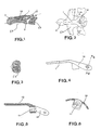
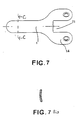
- the device according to the invention is essentially in the form of a plate 1 slightly curved in the transverse (as in FIG. 7a) and longitudinal directions, that is to say -to say cranial-caudal to marry at best by taking support there the shape of the anterior side of the caudal fragment 2 of the axis C2 whose odontoid process or tooth is fractured, the second fragment or cranial fragment of the tooth bearing the reference 3 .
- the plate 1 replaces the washer of the state of the art mentioned above. It is provided with a hole 11 with an axis oriented in the caudal-cranial direction of the tooth 2, 3, and of diameter corresponding to the passage of a screw 12 by serving as an axial bearing surface for the head 13 of this screw. This larger bearing surface is better opposed than before to the penetration of the head 13 of the screw 12 into the friable caudal fragment 2 of the axis C2.
- the screw 12 put in place as described with reference to the state of the art, crosses the caudal fragment 2 to come into positive engagement in the cortical tissue of the cranial fragment 3 of C2, preferably crossing the top of this fragment 3
- the screw 12 has, between its threaded end portion and its head, a smooth cylindrical portion with a diameter less than that of the thread so as not to interfere with the quality of the grip in the cranial fragment 3.
- the support plate 1 is extended on the caudal side beyond the passage of the screw 12 by a curved part 14 of mean plane inclined to that of the rest of the plate 1, preferably in the form of a fork. and coming to be secured on the anterior face of the third cervical vertebra C3 ( Figures 1 to 3), which increases the mechanical stability of the seat of the plate 1.
- the fixation on the third vertebra C3 can be done by screwing conventional, as illustrated for example in Figures 1 to 3.
- this drawback is eliminated by providing the support plate 1 no longer with a simple through hole, but with a guide barrel 11 of the screw 12 of sufficient length, for example at least equal to the diameter of the screw 12, preventing any deviation of the axis of the screw 12 in any radial plane.
- the screw 12 then has a smooth body with a diameter equal to that of the barrel 11 and a threaded diameter equal to that of the smooth body, whereas until now, the smooth body has been of diameter smaller than that of the thread, hence the scanning incidents already mentioned, because the screw head now rests on the plate 1 and not only a washer with a very small surface.
- the plate 1 being by nature non-deformable under the operating conditions and the angle between the mean planes of its parts based on the caudal fragment 2 of C2 and the third cervical vertebra C3 being fixed once and for all, any risk of scanning in whatever radial direction it is is therefore eliminated for screw 12.
- the invention eliminates the drawbacks of the known state of the operative technique for unstable odontoid process fractures, it can also advantageously be applied to non-displaced and stable fractures for injured people who would bear very badly an external consolidation device, which is particularly the case for multiple trauma patients, the elderly, or others injured in poor general condition.
Landscapes
- Health & Medical Sciences (AREA)
- Orthopedic Medicine & Surgery (AREA)
- Life Sciences & Earth Sciences (AREA)
- Neurology (AREA)
- Surgery (AREA)
- General Health & Medical Sciences (AREA)
- Veterinary Medicine (AREA)
- Biomedical Technology (AREA)
- Heart & Thoracic Surgery (AREA)
- Medical Informatics (AREA)
- Molecular Biology (AREA)
- Animal Behavior & Ethology (AREA)
- Nuclear Medicine, Radiotherapy & Molecular Imaging (AREA)
- Public Health (AREA)
- Engineering & Computer Science (AREA)
- Surgical Instruments (AREA)
- Crystals, And After-Treatments Of Crystals (AREA)
- Superconductors And Manufacturing Methods Therefor (AREA)
- Printing Plates And Materials Therefor (AREA)
- Finish Polishing, Edge Sharpening, And Grinding By Specific Grinding Devices (AREA)
- Polishing Bodies And Polishing Tools (AREA)
- Compositions Of Macromolecular Compounds (AREA)
- Anti-Oxidant Or Stabilizer Compositions (AREA)
- Saccharide Compounds (AREA)
Applications Claiming Priority (2)
| Application Number | Priority Date | Filing Date | Title |
|---|---|---|---|
| FR9006672 | 1990-05-23 | ||
| FR9006672A FR2662349B1 (fr) | 1990-05-23 | 1990-05-23 | Plaque vissable pour le traitement des fractures de l'apophyse odontouide de l'axis. |
Publications (3)
| Publication Number | Publication Date |
|---|---|
| EP0459266A2 true EP0459266A2 (de) | 1991-12-04 |
| EP0459266A3 EP0459266A3 (en) | 1991-12-27 |
| EP0459266B1 EP0459266B1 (de) | 1994-12-21 |
Family
ID=9397061
Family Applications (1)
| Application Number | Title | Priority Date | Filing Date |
|---|---|---|---|
| EP91108144A Expired - Lifetime EP0459266B1 (de) | 1990-05-23 | 1991-05-21 | Schraubbare Platte für Bruchbehandlung der zahnförmigen Axisapophyse |
Country Status (4)
| Country | Link |
|---|---|
| EP (1) | EP0459266B1 (de) |
| AT (1) | ATE115847T1 (de) |
| DE (1) | DE69106045D1 (de) |
| FR (1) | FR2662349B1 (de) |
Family Cites Families (3)
| Publication number | Priority date | Publication date | Assignee | Title |
|---|---|---|---|---|
| DE3114872C2 (de) * | 1981-04-13 | 1985-09-12 | Viktor KirilloviČ Krasnojarsk Gupalov | Implantat zur Korrektur von Wirbelsäulenverkrümmungen |
| AT378324B (de) * | 1982-09-13 | 1985-07-25 | Streli Elke | Zinkenplatte zur gegenseitigen lagefixierung der knochenteile bei knochenbruechen |
| GB8507671D0 (en) * | 1985-03-25 | 1985-05-01 | Howmedica Uk Ltd | Fracture fixation assembly |
-
1990
- 1990-05-23 FR FR9006672A patent/FR2662349B1/fr not_active Expired - Lifetime
-
1991
- 1991-05-21 AT AT91108144T patent/ATE115847T1/de not_active IP Right Cessation
- 1991-05-21 DE DE69106045T patent/DE69106045D1/de not_active Expired - Lifetime
- 1991-05-21 EP EP91108144A patent/EP0459266B1/de not_active Expired - Lifetime
Also Published As
| Publication number | Publication date |
|---|---|
| FR2662349A1 (fr) | 1991-11-29 |
| FR2662349B1 (fr) | 1992-09-18 |
| ATE115847T1 (de) | 1995-01-15 |
| EP0459266A3 (en) | 1991-12-27 |
| EP0459266B1 (de) | 1994-12-21 |
| DE69106045D1 (de) | 1995-02-02 |
Similar Documents
| Publication | Publication Date | Title |
|---|---|---|
| EP0767637B1 (de) | Wirbelgelenkfortsatz-prothese | |
| EP1113758B1 (de) | Vorrichtung zur posterioren wirbelsäulenosteosynthese | |
| EP0757545B1 (de) | Vorrichtung zur korrektion von verbiegungen der langen knochen | |
| EP2393441B1 (de) | Osteosynthese- und arthrodese-schraube | |
| EP1056407B1 (de) | Orthopädischer monolateraler aussenfixateur | |
| FR2781359A1 (fr) | Materiel d'osteosynthese rachidienne | |
| EP0317406A1 (de) | Künstlicher Dübel zur im Knochen liegenden Befestigung von Bandprothesen und Bandaufhängungen sowie Schlagaufnehmer mit Mitteln zur Befestigung solcher Dübel | |
| FR2655840A1 (fr) | Set chirurgical de ligamentoplastie et elements constitutifs. | |
| FR2785787A1 (fr) | Dispositif d'osteosynthese d'un segment rachidien par voie anterieure | |
| FR2651992A1 (fr) | Implant pour osteosynthese rachidienne dorso-lombaire anterieure destine a la correction de cyphoses. | |
| EP1489980A1 (de) | Wirbelsäulen-osteosynthesesystem | |
| FR2660856A1 (fr) | Prothese radio-cubitale inferieure. | |
| WO1997005830A1 (fr) | Cheville intrafocale | |
| EP0593321A1 (de) | Vorrichtung zur Osteosynthese von Rückenwirbeln | |
| EP0459266B1 (de) | Schraubbare Platte für Bruchbehandlung der zahnförmigen Axisapophyse | |
| FR2997837A1 (fr) | Dispositif d'osteosynthese pour le traitement des fractures du col de femur. | |
| WO1995026688A1 (fr) | Dispositif d'enclouage centromedullaire notamment pour fractures sous cervicales du femur | |
| FR2668921A1 (fr) | Plaque agrafe pour osteosynthese. | |
| FR2824468A1 (fr) | Fixateur externe pour l'aide a la reduction et a la fixation des factures epiphysaires d'un os long comme un radius | |
| EP0585455B1 (de) | Lineare orthopaedische vorrichtung fuer externe fixation | |
| FR2738737A1 (fr) | Piton d'ancrage utilisable en chirurgie | |
| FR2726759A1 (fr) | Cage pour fusion de vertebres lombaires par interposition de greffons osseux | |
| FR2728455A1 (fr) | Dispositif d'osteosynthese | |
| EP1336382B1 (de) | System zum Verbinden eines Stabes oder dergleichen mit einem Wirbelknochen | |
| FR2636226A1 (fr) | Dispositif de fixation au moyen d'une lame-broches adaptee aux fractures de l'extremite inferieure du radius |
Legal Events
| Date | Code | Title | Description |
|---|---|---|---|
| PUAI | Public reference made under article 153(3) epc to a published international application that has entered the european phase |
Free format text: ORIGINAL CODE: 0009012 |
|
| PUAL | Search report despatched |
Free format text: ORIGINAL CODE: 0009013 |
|
| AK | Designated contracting states |
Kind code of ref document: A2 Designated state(s): AT CH DE GB LI SE |
|
| AK | Designated contracting states |
Kind code of ref document: A3 Designated state(s): AT CH DE GB LI SE |
|
| 17P | Request for examination filed |
Effective date: 19920615 |
|
| 17Q | First examination report despatched |
Effective date: 19930506 |
|
| GRAA | (expected) grant |
Free format text: ORIGINAL CODE: 0009210 |
|
| AK | Designated contracting states |
Kind code of ref document: B1 Designated state(s): AT CH DE GB LI SE |
|
| PG25 | Lapsed in a contracting state [announced via postgrant information from national office to epo] |
Ref country code: GB Effective date: 19941221 Ref country code: AT Effective date: 19941221 |
|
| REF | Corresponds to: |
Ref document number: 115847 Country of ref document: AT Date of ref document: 19950115 Kind code of ref document: T |
|
| REF | Corresponds to: |
Ref document number: 69106045 Country of ref document: DE Date of ref document: 19950202 |
|
| PG25 | Lapsed in a contracting state [announced via postgrant information from national office to epo] |
Ref country code: SE Effective date: 19950321 |
|
| PG25 | Lapsed in a contracting state [announced via postgrant information from national office to epo] |
Ref country code: DE Effective date: 19950322 |
|
| RAP4 | Party data changed (patent owner data changed or rights of a patent transferred) |
Owner name: VICHARD, OLIVIER |
|
| GBV | Gb: ep patent (uk) treated as always having been void in accordance with gb section 77(7)/1977 [no translation filed] |
Effective date: 19941221 |
|
| PLBE | No opposition filed within time limit |
Free format text: ORIGINAL CODE: 0009261 |
|
| STAA | Information on the status of an ep patent application or granted ep patent |
Free format text: STATUS: NO OPPOSITION FILED WITHIN TIME LIMIT |
|
| 26N | No opposition filed | ||
| REG | Reference to a national code |
Ref country code: CH Ref legal event code: PL |
|
| REG | Reference to a national code |
Ref country code: CH Ref legal event code: AEN Free format text: LA POURSUITE DE LA PROCEDURE REQUISE LE 07.01.1997 A ETE ACCORDEE. LE BREVET EST REACTIVE. |
|
| PGFP | Annual fee paid to national office [announced via postgrant information from national office to epo] |
Ref country code: CH Payment date: 19990331 Year of fee payment: 9 |
|
| PG25 | Lapsed in a contracting state [announced via postgrant information from national office to epo] |
Ref country code: LI Free format text: LAPSE BECAUSE OF NON-PAYMENT OF DUE FEES Effective date: 20000531 Ref country code: CH Free format text: LAPSE BECAUSE OF NON-PAYMENT OF DUE FEES Effective date: 20000531 |
|
| REG | Reference to a national code |
Ref country code: CH Ref legal event code: PL |