EP0481000B1 - Rezeptoren für fibroblasten-wachstumsfaktoren - Google Patents
Rezeptoren für fibroblasten-wachstumsfaktoren Download PDFInfo
- Publication number
- EP0481000B1 EP0481000B1 EP90911235A EP90911235A EP0481000B1 EP 0481000 B1 EP0481000 B1 EP 0481000B1 EP 90911235 A EP90911235 A EP 90911235A EP 90911235 A EP90911235 A EP 90911235A EP 0481000 B1 EP0481000 B1 EP 0481000B1
- Authority
- EP
- European Patent Office
- Prior art keywords
- fgf
- receptor
- sequence
- growth factor
- fibroblast growth
- Prior art date
- Legal status (The legal status is an assumption and is not a legal conclusion. Google has not performed a legal analysis and makes no representation as to the accuracy of the status listed.)
- Expired - Lifetime
Links
Images


Classifications
-
- C—CHEMISTRY; METALLURGY
- C07—ORGANIC CHEMISTRY
- C07K—PEPTIDES
- C07K14/00—Peptides having more than 20 amino acids; Gastrins; Somatostatins; Melanotropins; Derivatives thereof
- C07K14/435—Peptides having more than 20 amino acids; Gastrins; Somatostatins; Melanotropins; Derivatives thereof from animals; from humans
- C07K14/705—Receptors; Cell surface antigens; Cell surface determinants
- C07K14/71—Receptors; Cell surface antigens; Cell surface determinants for growth factors; for growth regulators
-
- A—HUMAN NECESSITIES
- A61—MEDICAL OR VETERINARY SCIENCE; HYGIENE
- A61P—SPECIFIC THERAPEUTIC ACTIVITY OF CHEMICAL COMPOUNDS OR MEDICINAL PREPARATIONS
- A61P35/00—Antineoplastic agents
-
- A—HUMAN NECESSITIES
- A61—MEDICAL OR VETERINARY SCIENCE; HYGIENE
- A61P—SPECIFIC THERAPEUTIC ACTIVITY OF CHEMICAL COMPOUNDS OR MEDICINAL PREPARATIONS
- A61P43/00—Drugs for specific purposes, not provided for in groups A61P1/00-A61P41/00
-
- G—PHYSICS
- G01—MEASURING; TESTING
- G01N—INVESTIGATING OR ANALYSING MATERIALS BY DETERMINING THEIR CHEMICAL OR PHYSICAL PROPERTIES
- G01N33/00—Investigating or analysing materials by specific methods not covered by groups G01N1/00 - G01N31/00
- G01N33/48—Biological material, e.g. blood, urine; Haemocytometers
- G01N33/50—Chemical analysis of biological material, e.g. blood, urine; Testing involving biospecific ligand binding methods; Immunological testing
- G01N33/74—Chemical analysis of biological material, e.g. blood, urine; Testing involving biospecific ligand binding methods; Immunological testing involving hormones or other non-cytokine intercellular protein regulatory factors such as growth factors, including receptors to hormones and growth factors
-
- A—HUMAN NECESSITIES
- A61—MEDICAL OR VETERINARY SCIENCE; HYGIENE
- A61K—PREPARATIONS FOR MEDICAL, DENTAL OR TOILETRY PURPOSES
- A61K38/00—Medicinal preparations containing peptides
-
- G—PHYSICS
- G01—MEASURING; TESTING
- G01N—INVESTIGATING OR ANALYSING MATERIALS BY DETERMINING THEIR CHEMICAL OR PHYSICAL PROPERTIES
- G01N2333/00—Assays involving biological materials from specific organisms or of a specific nature
- G01N2333/435—Assays involving biological materials from specific organisms or of a specific nature from animals; from humans
- G01N2333/475—Assays involving growth factors
- G01N2333/50—Fibroblast growth factors [FGF]
-
- G—PHYSICS
- G01—MEASURING; TESTING
- G01N—INVESTIGATING OR ANALYSING MATERIALS BY DETERMINING THEIR CHEMICAL OR PHYSICAL PROPERTIES
- G01N2333/00—Assays involving biological materials from specific organisms or of a specific nature
- G01N2333/435—Assays involving biological materials from specific organisms or of a specific nature from animals; from humans
- G01N2333/705—Assays involving receptors, cell surface antigens or cell surface determinants
- G01N2333/71—Assays involving receptors, cell surface antigens or cell surface determinants for growth factors; for growth regulators
-
- G—PHYSICS
- G01—MEASURING; TESTING
- G01N—INVESTIGATING OR ANALYSING MATERIALS BY DETERMINING THEIR CHEMICAL OR PHYSICAL PROPERTIES
- G01N2500/00—Screening for compounds of potential therapeutic value
Definitions
- the present invention relates to receptors for grow factors, specifically to the fibroblast growth factor receptor (FGF-R). More particularly, it provides various purified fibroblast growth factor receptor proteins, nucleic acids encoding the receptor proteins, methods for the production of purified FGF-R proteins, proteins made by these methods, antibodies against these proteins, and diagnostic and therapeutic uses of these various reagents.
- FGF-R fibroblast growth factor receptor
- Polypeptide growth factors are mitogens that act on cells by specifically binding to receptors situated at the plasma membrane. These receptors usually have three major identifiable regions. The first is an extracellular region which contains the domain that binds the polypeptide growth factor (i.e. the ligand-binding domain). The second region is a transmembrane region and the third is an intracellular region. Many of these receptors contain a tyrosine kinase domain in the intracellular region.
- the fibroblast growth factor receptor (FGF-R) proteins bind to a family of related growth factor ligands, the fibroblast growth factor (FGF) family.
- FGF fibroblast growth factor
- This family of growth factors are characterized by amino acid sequence homology, heparin-binding avidity, the ability to promote angiogenesis and mitogenic activity toward cells of epithelial, mesenchymal and neural origin.
- the FGF family includes the following seven known FGFs:
- FGF receptor has been isolated from human umbilical vein-derived endothelial cells, and shown to have a mw of 130,000 (Neufield et al., J. of Cell. Physiol. 136 537-542). No sequence information was provided. Lee et al (Purification of the receptor for FGF and cloning of its CDNA) report the cloning without giving details thereof.
- the purified and cloned chicken bFGF and human bFGF receptors of this invention have amino acid sequence similarity with the bek and flg clones in the regions which have been isolated.
- both the bek and flg sequences reported were incomplete and there was no recognition of their function as FGF binding receptors.
- the prior reports failed to recognize many of the structural and functional features described in the present invention.
- FGFs may enhance tumor growth and invasiveness by stimulating blood vessel growth in the tumor or by inducing production of proteins such as plasminogen activator.
- identification of the components involved and understanding of the mechanisms and interactions involved remain woefully incomplete.
- FGF receptors and fragments Purified FGF receptors and fragments, and isolated DNA sequences encoding defined FGF receptors and defined fragments (e.g., the ligand-binding domain) will greatly accelerate the understanding of fibroblast growth factor functions. Antibodies against specific and defined regions of the FGF receptor also become available. These reagents will find both diagnostic and therapeutic uses in the aforementioned processes.
- the present invention fulfills these and other needs.
- the present invention provides purified fibroblast growth factor receptor proteins (FGF-R), nucleic acids encoding FGF-R proteins, methods for the production of purified FGF-R proteins, purified proteins made by these methods, antibodies against these proteins and fragments, and diagnostic and therapeutic uses of these reagents.
- FGF-R fibroblast growth factor receptor proteins
- nucleic acids encoding FGF-R proteins
- purified proteins made by these methods
- antibodies against these proteins and fragments antibodies against these proteins and fragments
- diagnostic and therapeutic uses of these reagents include soluble and secreted forms of the receptors exhibiting an unusual receptor structure.
- the present invention provides a purified soluble polypeptide comprising IgII and IgIII domains of any one of the amino acid sequences shown in Figure 7 or an amino acid sequence which comprises two domains at least 85% homologous to the IgII and IgIII domains respectively of any one of said sequences; which polypeptide lacks a transmembrane domain and is capable of inhibiting the binding between a fibroblast growth factor and a fibroblast growth factor receptor.
- Such a polypeptide according to the invention includes a polypeptide which comprises a first Ig domain which has (i) two cysteine residues, (ii) a tryptophan residue 11 or 12 amino acids on the carboxyl-terminal side of the one of the above-mentioned cysteine residues which is closest to the N-terminal end, and (iii) a sequence DXGXYXC on the amino terminal side of the other of said cysteine residues; a second Ig domain C-terminal to the first domain also comprising features (i), (ii) and (iii) as defined for the first Ig domain, except that the second said Ig domain may have a carboxyl-terminal sequence which is at least 85% homologous to the 79-amino acid sequence from residues 224 to 302 or 222 to 300 of human protein h4 or h5 respectively of Figure 7 in place of the said other cysteine residue and the sequence (iii).
- the invention also provides a purified polypeptide comprising the human sequence designated as h4 or h5 in Figure 7, and a purified polypeptide which is less than 85kD and comprises a fibroblast growth factor-binding domain of a fibroblast growth factor receptor; said receptor comprising an amino acid sequence as shown in Figure 7 or an allelic variant or mutant thereof, or a homologous sequence which is encoded by a nucleic acid which hybridises to DNA which encodes said sequence variant or mutant under stringent conditions including a temperature over 37°C and a salt concentration of less than 1 M.
- the invention provides a monoclonal antibody that binds to a polypeptide of the invention.
- the invention provides nucleic acids, particularly isolated nucleic acids which encodes a polypeptide according to the invention, such as an isolated nucleic acid which comprises or selectively hybridizes with all or part of a sequence in the IgII and IgIII domains shown in any one of Figures 3, 4 or 9 under stringent conditions including a temperature over 37°C and a salt concentration of less than 1 M, and which encodes a polypeptide which is capable of inhibiting the binding of a fibroblast growth factor with a fibroblast growth factor receptor.
- Nucleic acids may be expressed to provide polypeptides of the invention, for example by a method wherein the peptide is produced in a cell transformed with a nucleic acid comprising a sequence encoding said peptide operably linked to transcriptional and translational initiation regions, said method comprising expressing said nucleic acid and recovering the FGF-R peptide.
- Also provided is a method for producing an antibody against a fibroblast growth factor receptor fragment comprising a step of producing an antibody against a polypeptide epitope comprising at least six contiguous amino acids of a polypeptide according to the invention.
- the invention also provides a method of measuring a fibroblast growth factor or a fibroblast growth factor receptor in a target sample, said method comprising the steps of:
- the present invention has applications such as in a method for modifying in vivo a fibroblast growth factor receptor modulated activity comprising administering to a patient an amount of a fibroblast growth factor receptor blocking agent effective to inhibit fibroblast growth factor binding to said fibroblast growth factor receptor.
- the invention may be used to provide a method for inhibiting binding between a fibroblast growth factor and a fibroblast growth factor receptor in a solution.
- This method will contain a step of combining an FGF-R peptide, e.g., a peptide homologous in sequence to a sequence described in Figures 3, 4 or 7 to the solution or medium containing fibroblast growth factor receptor, usually native fibroblast growth factor receptor.
- FGF-R peptide e.g., a peptide homologous in sequence to a sequence described in Figures 3, 4 or 7
- Such methods will be useful in vitro , after employing labeled FGF-R peptide in assay procedures.
- compositions containing a soluble FGF-R polypeptide having between about five and two hundred contiguous amino acids from a human FGF-R extracellular domain are also described herein.
- One polypeptide contains at least about 80 amino acids from residues 1 to 287 of a human fibroblast growth factor receptor of Figure 7 or an IgII or IgIII domain, or both.
- a first aspect of the invention provides homogeneous FGF-R peptides.
- These homogeneous FGF-Rs include a chicken basic fibroblast growth factor receptor and various human fibroblast growth factor receptors. Homogeneous polypeptides either having FGF-R ligand-binding activity or comprising a portion of the ligand-binding domain of an FGF-R are described.
- the present invention provides homogeneous polypeptides corresponding to naturally occurring FGF-binding proteins having unexpected structural features.
- One class provides soluble proteins lacking a transmembrane segment, another class provides proteins possessing both a transmembrane segment and a tyrosine kinase domain. Both of these classes have an unexpected extracellular domain structure shorter than the corresponding chicken FGF-R. Experimental data indicating that a single receptor binds various FGF types is also described.
- a second aspect of the invention provides isolated DNA sequences These sequences encode polypeptides having FGF-R ligand-binding activity, including polypeptides which correspond to naturally occurring full-length fibroblast growth factor receptors.
- DNA sequences encoding a chicken bFGF-R or encoding various human FGF-Rs (hFGF-R) have been isolated.
- cloning and expression vehicles containing the FGF-R encoding sequences are also provided.
- a DNA sequence encoding the full-length FGF receptor or an FGF-R polypeptide fragment can be operably linked to control sequences and expressed in a culture of a compatible transformed, transfected or infected host cells.
- the invention also provides antibodies to defined domains of the receptor. Still further aspects of the invention include methods for evaluating compositions which are agonistic or antagonistic to ligand and receptor interactions, particularly those which promote or inhibit binding interactions.
- FGF-R fibroblast growth factor receptors
- the FGF family consists of polypeptide growth factors characterized by amino acid sequence homology, heparin-binding avidity, the ability to promote angiogenesis, and mitogenic activity toward cells of epithelial, mesenchymal, and neural origin.
- the FGF family includes acidic FGF, basic FGF, the int-2 gene product, the hst gene product (Kaposi sarcoma-FGF), FGF-5, the keratinocyte growth factor, and FGF-6.
- Members of the FGF family appear to have roles in development, tissue repair, maintenance of neurons, and the pathogenesis of disease. Aberrant expression of FGFs may cause cell transformation by an autocrine mechanism.
- FGFs may enhance tumor growth and invasiveness by stimulating blood vessel growth into the tumor or by inducing production of proteases such as plasminogen activator.
- ligand refers to the molecules, usually members of the fibroblast growth factor family, that bind the domains involved in the growth factor binding. Also, a ligand is a molecule which serves either as the natural ligand to which the receptor binds, or a functional analogue which may serve as an agonist or antagonist.
- a chicken bFGF receptor is characterized by various identifiable structural features.
- the chicken and human FGF-R structures are generalized to define a structural nomenclature applicable to other FGF-Rs.
- General descriptions of protein structure and its relationship to nucleic acid sequences are discussed in J.D. Watson et al., Molecular Biology of the Gene , 4th Ed., vols. 1 and 2, Benjamin/Cummings, Menlo Park, California, (1987); and B. Alberts et al., Molecular Biology of the Cell , 2d Ed., Garland, New York, (1989), each of which is incorporated herein by reference.
- Common structural features of known FGF-Rs are described, including various naturally occurring soluble human FGF binding proteins.
- a human fibroblast growth factor receptor is a protein either derived from a natural human FGF-R gene, or which shares significant structural characteristics peculiar to a naturally occurring human receptor for FGF.
- the isolated full-length chicken FGF-R mRNA contains a single hydrophobic segment similar to a membrane-spanning segment (designated the transmembrane segment).
- the segments of FGF-R amino-proximal to the transmembrane segment are designated the extracellular domain, while the segments carboxy-proximal to the transmembrane segment are designated the intracellular domain. From the amino-terminus, the extracellular domain has an NH 2 -terminal hydrophobic putative signal sequence, an immunoglobulin-like domain (designated IgI), and acidic segment, a second immunoglobulin-like domain (designated IgII), and a third immunoglobulin-like domain (designated IgIII).
- the most important functional property which defines the domain is the binding to the receptor ligands, e.g., members of the FGF family. As discussed below, this function is correlated with the combined presence of IgII and IgIII domains.
- the intracellular domain is characterized by the presence of a split tyrosine kinase structural domain and, in the chicken receptor, is about 424 residues long. Functionally, this domain is defined by its tyrosine kinase activity, typically modulated by ligand binding to the extracellular domain.
- a protein substantially lacks an intracellular domain when it lacks a prototypical intracellular domain, particularly lacking a tyrosine kinase domain.
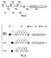
- the chicken receptor four unique human cDNA clones have been identified. These encode previously unknown FGF receptor variants which contain only two Ig-like domains. Two of the human clones encode membrane spanning receptors and two encode putative secreted forms. Both the forms exhibiting the 3 Ig-like or 2 Ig-like domain structures mediate biological responsiveness to acidic and basic FGF. Thus, the first Ig domain of the 3 Ig domain form may have a function other than binding of acidic and basic FGF.
- the multiple human receptor forms are identical in some regions but are highly divergent in other selected regions of the extracellular domain. Two of the human variant receptors, h4 and h5, are likely to encode a secreted form of the FGF receptor.
- a typical FGF-R nucleic acid sequence encodes a transitory NH 2 -terminal hydrophobic sequence, which is usually cleaved during the translocation process.
- the classical function of a signal sequence is to direct the nascent polypeptide chain to membrane bound ribosomes, thereby leading to membrane translocation.
- the signal sequence is typically removed in the translocation process, the signal sequence is absent in a mature polypeptide.
- the Ig-like domains are characterized by three main features: (i) the presence of two characteristic cysteine residues in each domain; (ii) the presence of a consensus tryptophan residue 11 to 12 amino acids on the COOH-terminal side of the first cysteine residue in each Ig-like domain; and (iii) the presence of the consensus sequence, DXGXYXC, on the NH 2 -terminal side of the second cysteine residue.
- the last feature is modified in the cases of the soluble receptor proteins, and substituted with an equivalently sized sequence.
- the amino-proximal Ig domain found in the chicken clone was designated IgI. As the chicken clone has three Ig domains, the domains have been numbered from the amino terminus. As indicated in Figure 6, the IgI domain includes the 45 amino acids flanked by a pair of cysteine residues. The chicken IgI domain has a high homology in sequence with the IgI domain found in the genomic sequence of the human FGF-R. However, the human forms appear to lack a domain corresponding to IgI.
- the next Ig domain is designated IgII, and in the chicken receptor includes 51 amino acids between the two cysteine residues (see Figs. 3 and 6). As described below, this domain, in combination with the IgIII domain is involved with ligand binding. The polypeptide sequence homology of this domain between the chicken and human receptors is quite high, as shown by the sequence alignments in Fig. 7. It will be noted that the human receptors lack an Ig I domain but have IgII and IgIII domains.
- cysteine residues used to delineate this domain are residues 176, 89, 87, 89, and 87 on the amino proximal side, and 228, 141, 139, 141, and 139 on the carboxy proximal side for the chicken, h2, h3, h4 and h5 receptors, respectively.
- the third Ig domain is designated IgIII and in the chicken receptor includes 63 amino acids between the two cysteine residues. See Figs. 3 and 6. Again, although the human receptors have only two domains, the domains correspond to IgII and IgIII. In both the chicken and human forms, the IgIII domain is that closest to the transmembrane segment.
- the cysteine residues for the chicken, h2, h3, h4 and h5 receptors, respectively, used to delineate this domain are residues 274, 187, 185, 187, and 185 on the amino proximal side and residues 339, 252, 250, 253, and 251 on the carboxy proximal side.
- the h4 and h5 soluble receptors have a substituted terminal segment designated IgIIIT.
- This segment is a substituted terminal segment replacing part of the membrane bound to IgIII, and is 79 amino acids long.
- This sequence corresponds to amino acids 224 and 222 of h4 and h5, respectively, while preserving many of the features found in the IgIII domain except of the DSGSYSC. It should be noted, however, the IgIIIT sequences are conserved between the soluble forms of the human FGF-R.
- the FGF receptors (shown for the basic FGF-R, but the same FGF-R binds both the acidic and basic FGFs) have a feature not found in other members of the immunoglobulin superfamily.
- There is a series of eight consecutive acidic residues (EDDDDEDD in the case of chicken, and EDDDDDDD in the case of human) followed by three serine residues and two additional acidic residues ( Figures 3 and 7).
- the 5 receptor species (e.g. the chicken, h2, h3, h4 and h5 forms) also exhibit variability at a specific location between the conserved acidic region and the conserved second Ig-like domain (IgII).
- the h2 and h4 receptor forms contain two amino acids (ArgMet) at positions 59 and 60, while the chicken receptor contains a single amino acid (Asn) at this position and the h3 and h5 receptor forms contain no corresponding amino acids at this position (see asterisks, Fig. 8).
- the juxtamembrane region is the region between the membrane spanning segment and the kinase domain. This region is normally conserved among receptor tyrosine kinases. For example, the juxtamembrane region is consistently 49 to 51 residues in length in the receptors for PDGF, CSF-1, epidermal growth factor (EGF), human epidermal growth factor-2 (HER2) and insulin.
- the FGF receptors with an intercellular domain have an unusually long juxtamembrane region of about 87 residues.
- the cytoplasmic regions of the amino acid sequences are about 424 and 425 residues long, respectively for the chicken and human forms. These also contain a tyrosine kinase sequence (about residues 482 to 759, 395 to 672, and 393 to 670, respectively for the chicken, h2, and h3 forms).
- a tyrosine kinase sequence about residues 482 to 759, 395 to 672, and 393 to 670, respectively for the chicken, h2, and h3 forms.
- the kinase region of the bFGF receptors shares the most sequence identity (about 51 to 53%) with the PDGF and CSF-1 receptors.
- the bFGF receptors contain the GXGXXG motif and the conserved lysine residue (about residue 512) that form part of the adenosine 5'-triphosphate (ATP) binding site of tyrosine kinases.
- the bFGF receptors also contain the two characteristic tyrosine kinase motifs, HRDLAARNVL and DFGLAR, and a tyrosine (about residues 651, 564 and 562) at the position analogous to the major phosphorylation site of pp60 v-src (about Tyr 416).
- the kinase coding sequence of the bFGF receptors are split by an insertion of 14 amino acids.
- the length of the insertion in the kinase region is shorter than that found in the receptors for PDGF and CSF-1 (104 and 70 amino acids, respectively) and is similar to the length of the inserted sequence in the receptors for insulin and insulin-like growth factor-I.
- the FGF-R appears to have three different biological functions. The first is the binding of ligands, usually the FGF proteins or their analogues. These ligands or analogues may also serve as either agonists or antagonists. The ligand binding site is apparently in the extracellular domain. The receptor transduces a signal in response to ligand binding, and the result is a ligand modulated activity. As the likely ligand is a FGF, the signal will ordinarily be FGF-modulated.
- a second biological activity relates to the tyrosine kinase enzymatic activity. This activity is typically activated in response to ligand binding. However, since the receptors are likely to function in a dimer state, the intrachain binding interactions may be considered another biological activity which may be mediated by blocking agents. this may serve as an additional means to modulate FGF-mediation of particular activities.
- FGFs The interactions of FGFs with their receptors cause changes in, on particular cell types, cell morphology and cell transformation, cell proliferation, cell differentiation, cell senescence, heparin sensitivity, and heparin effects.
- the in vivo effects of FGF include, in particular organisms, modulation of various activities, e.g., limb regeneration, lens regeneration, angiogenic effects on both normal and tumor cells, wound healing, adipocyte differentiation, and growth of various neural and myoblast cells. FGFs also exhibit potent angiogenic activities. It is thought that the angiogenic activity of FGFs is due in large part to the chemotactic and mitogenic effects of these factors on endothelial cells.
- compositions and cells comprising them can be used for diagnostic purposes and to study and treat diseases associated with FGF receptors.
- Cells expressing cloning vehicles containing defined sequences can be used to define specific sites of an FGF receptor necessary for effecting a particular activity.
- these cells may be useful to assess the ability of a selected receptor to bind different ligands (FGFs and analogues) thereby providing a powerful tool for evaluating the potential of drugs for promoting or inhibiting specific FGF-induced cellular responses.
- FGF-R sequence Cells transfected, injected, infected or electroporated with DNA or mRNA containing a full length natural FGF-R sequence will often express the native or wild type receptor and respond accordingly.
- Specific concentrations of a purified receptor or a receptor polypeptide fragment can be used to block the binding of the ligand (FGF) to native FGF receptors.
- FGF ligand
- antibodies to the receptor or fragment can have the same effect.
- Homogeneous and defined polypeptides and DNA sequences will find use in raising antibodies.
- antibodies against specific regions of the receptor e.g., the ligand-binding domain, will find use in diagnostic testing.
- the reagents FGF-R, FGF-R polypeptides and antibodies to specific regions of the receptor can be used to study regulation of FGF mediated activities.
- FGF agonists should stimulate blood vessel development, an effect particularly beneficial in wound healing and in the growth of collateral blood vessels in ischemic areas of the heart.
- FGF antagonists should find use in preventing aberrant angiogenesis as seen in diabetic retinopathy and rheumatoid arthritis or in controlling tumors by blocking proliferation of vascularization to a tumor.
- This invention includes fibroblast growth factor receptor polypeptides and proteins having FGF-R ligand-binding activity as defined in the accompanying claims.
- FGF receptors Various new human FGF receptors have been cloned and characterized, as described further below. Of particular note, various shorter forms (h2 and h3) and soluble versions (h4 and h5) of FGF receptors have been discovered.
- the soluble proteins e.g., forms lacking a transmembrane segment
- FGF binding capacity indicate that shorter forms will find therapeutic and/or diagnostic uses.
- the fibroblast growth factor receptor peptides of the present invention will exhibit at least about 85% homology with the naturally-occurring receptors in the IgII and IgIII regions, usually at least about 90% homology, and preferably at least about 95% homology.
- the ligand binding function is localized to the extracellular domain, and the soluble forms retain this particular function. Soluble fragments of FGF receptors should be useful in substituting for or interfering with the functions of the naturally soluble variants. Alternatively, the soluble forms may interfere with dimerization of FGF receptors, since the receptors may normally be in a dimer form. Receptor dimerization may be essential for proper physiological signal transduction.
- the human receptors possessing a transmembrane segment are unusual in having only the IgII and IgIII of the three Ig domains.
- the absence of the IgI domain indicates that certain functions may be absent in the human receptor, or, more likely, that the IgI domain is unnecessary in the human receptor. Data presented below shows that the IgI domain is not essential for ligand binding.
- substantially pure and homogenous describe a protein which has been separated from components which naturally accompany it.
- a monomeric protein is substantially pure when at least about 60 to 75% of a sample exhibits a single polypeptide backbone. Minor variants or chemical modifications typically share the same polypeptide sequence.
- a substantially pure protein will typically comprise over about 85 to 90% of a protein sample, more usually will comprise at least about 95%, and preferably will be over about 99% pure.
- purity is measured on a polyacrylamide gel, with homogeneity determined by staining. For certain purposes high resolution will be used and HPLC or a similar means for purification utilized. For most purposes, a simple chromatography column or polyacrylamide gel will be used to determine purity.
- a protein is substantially free of naturally-associated components when it is separated from the native contaminants which accompany it in its natural state.
- a protein which is chemically synthesized or synthesized in a cellular system different from the cell from which it naturally originates will be substantially free from its naturally-associated components.
- the term is used to describe receptors and nucleic acids which have been synthesized in heterologous mammalian cells or plant cells, E . coli and other prokaryotes.
- a polypeptide is substantially an entire membrane bound form of an FGF-R when it is substantially a full length peptide corresponding to, or highly homologous to a naturally occurring membrane bound form of an FGF-R.
- the present invention provides for substantially pure preparations.
- Various methods for their isolation from biological material may be devised, based in part upon the structural and functional descriptions contained herein.
- FGF receptor peptides including chicken and human FGF receptors may be purified using techniques of classical protein chemistry, see below. For example, a lectin affinity chromatography step may be used, followed by a highly specific ligand affinity chromatography procedure that utilizes an FGF conjugated to biotin through the cysteine residues of the FGF. Purified FGF-R receptors may also be obtained by a method such as FGF affinity chromatography using activated CH-Sepharose coupled to FGF through primary amino groups as described in Imamura, supra . This method, however, while resulting in a purified protein, may not provide a workable amount of purified protein (i.e. more than nanogram amounts).
- specific FGF receptors may also be purified using immunoaffinity chromatography.
- Antibodies prepared, as described below, may be immobilized to an inert substance to generate a highly specific affinity column. See Harlow and Lane, below.
- one purification procedure may be used which takes advantage of the fact that labeled biotin-bFGF binds with high affinity to receptors in cells containing high amounts of those receptors.
- 125 I-labeled biotin-bFGF will bind to bFGF receptors in Swiss 3T3 cells and can be cross-linked to the receptor protein.
- Various cell or tissue sources may be selected as starting materials usually selected due to an abundance of the desired receptor.
- Chicken embryos day 6, stage 29-30 are preferred because they contain relatively large amounts of the receptor protein as determined by high-affinity binding of human and bovine bFGF.
- Embryo extracts can first be fractionated on wheat germ agglutinin (WGA) Sepharose 4B and the partially purified bFGF receptors then bound to biotin-bFGF.
- the receptor-ligand complex may be adsorbed to an avidin-agarose due to the high affinity interaction between the biotin and avidin moieties.
- the avidin-agarose columns may be eluted with compounds which dissociate the FGF from its receptor such as suramin or SDS.
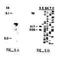
- the chicken protein which bound to avidin-agarose in an FGF-dependent manner migrated at the expected size (130 kDa) of the bFGF receptor. See Fig. 2B.
- the receptor may be digested with trypsin.
- Peptide fragments may be separated by reversed-phase high performance liquid chromatography (HPLC) and analyzed by gas-phase sequencing. Other sequencing methods known in the art may also be used.
- the FGF receptors or the specific external regions of the receptors may be used to affinity purify respective FGFs.
- the external region comprising the ligand-binding domain of the chicken bFGF-R shown in Figure 3 extends from about amino acid 22 to about amino acid 374.
- the ligand-binding domain of the human FGF-R shown in Figure 4 extends from about amino acid 22 to about amino acid 285.
- the ligand-binding domain varies with different FGF receptors and may be anywhere from 5% to 100% of the extracellular region.
- the minimal amount of protein sequence necessary for ligand bonding may be determined by excising various segments of the extracellular domain and assaying ligand binding to the remaining sequence.
- FGF receptor or FGF-R ligand-binding activity means having the ability to bind a fibroblast growth factor or other specific ligand.
- these ligands will be members of the FGF family. Therefore the external region has utility in establishing FGF agonists or antagonists.
- FGF-R like many other growth factor receptors, is found naturally in a multimeric protein complex, most likely in dimer form.
- other important regions of a receptor will be those, either extracellular or otherwise, which are involved in dimerization.
- the intracellular regions of the receptors may also be used as enzymes with tyrosine kinase activity.
- the bek gene has 84% amino acid sequence identity to the analogous region (tyrosine kinase region) of the chicken bFGF-R.
- the flg has 99% homology with various sequences of the human FGF receptor described in Figure 4.
- a signal or leader sequence directs a protein through the membrane of a cell.
- the signal sequences of the receptors may be used in conjunction with their respective receptors but may also be used with other proteins (e.g. amino acids about 1 through 21 of the N-terminal sequence comprise the leader or signal sequence of the chicken bFGF-R shown in Figure 3 and the human FGF-R shown in Figure 4).
- Chemical derivatizations and modifications of polypeptides such as acetylations, carboxylations and the like, may be performed, including glycosylation modifications and processing variants of a typical polypeptide. These processing steps specifically include enzymatic modifications, such as ubiquinization. See , e.g., Hershko and Ciechanover (1982), "Mechanisms of Intracellular Protein Breakdown," Ann. Rev. Bioch. , 51:335-364.
- Analogues include genetic variants, both natural and induced. Induced mutants may be derived from various techniques including both random mutagenesis of the encoding nucleic acids using irradiation or exposure to EMS, or may take the form of engineered changes by site-specific mutagenesis or other techniques of modern molecular biology. See , Sambrook, Fritsch and Maniatis (1989), Molecular Cloning: A Laboratory Manual (2d ed.), CSH Press.
- the present invention provides for biologically active fragments of the polypeptides, as defined in the accompanying claims.
- Immunogens may be produced which tandemly repeat polypeptide segments, thereby producing highly antigenic proteins. Alternatively, such polypeptides will serve as highly efficient competitors for Specific binding. Production of antibodies to fibroblast growth factor receptor polypeptides is described below.
- Fusion polypeptides between the receptors and other homologous or heterologous proteins may be made.
- Homologous polypeptides may be fusions between different growth factor receptors, resulting in, for instance, a hybrid protein exhibiting ligand specificity of one receptor and the intracellular domain of another, or a receptor which may have broadened or weakened specificity of binding.
- heterologous fusions may be constructed which would exhibit a combination of properties or activities of the derivative proteins.
- Typical examples are fusions of a reporter polypeptide, e.g., luciferase, with a domain of a receptor, e.g., a ligand-binding domain, so that the presence or location of a desired ligand may be easily determined.
- gene fusion partners include bacterial ⁇ -galactosidase, trpE Protein A, ⁇ -lactamase, alpha amylase, alcohol dehydrogenase and yeast alpha mating factor. See , e.g., Godowski et al. (1988), Science 241:812-816; and Experimental section below.
- Fusion proteins will typically be made by either recombinant nucleic acid methods or by synthetic polypeptide methods. Techniques for nucleic acid manipulation are described generally, for example, in Sambrook et al. (1989). Molecular Cloning: A Laboratory Manual (2d ed.), Vols. 1-3, Cold Spring Harbor Laboratory, Techniques for synthesis of polypeptides are described, for example, in Merrifield, J. Amer. Chem. Soc. 85:2149-2156 (1963).
- the recombinant nucleic acid sequences used to produce fusion proteins of the present invention may be derived from natural or synthetic sequences. Many natural gene sequences are obtainable from various cDNA or from genomic libraries using appropriate probes. See , GenBankTM, National Institutes of Health.
- Typical probes for fibroblast growth factor receptors may be selected from the sequences of Figures 3, 4, or 9 in accordance with standard procedures.
- Suitable synthetic DNA fragments may be prepared by the phosphoramidite method described by Beaucage and Carruthers, Tetra. Letts. 22:1859-1862 (1981).
- a double stranded fragment may then be obtained either by synthesizing the complementary strand and annealing the strand together under appropriate conditions or by adding the complementary strand using DNA polymerase with an appropriate primer sequence.
- the present invention provides nucleic acid sequences encoding various FGF receptor sequences described above.
- Figures 3, 4, and 7 respectively set forth the corresponding cDNA sequences encoding chicken and human FGF receptors.
- peptides Sequenced from purified protein are underlined, including the NH 2 -proximal sequences from amino acids 35-53 (ala---arg), 56-67 (leu---arg), and 139-158 (glu---lys).
- the transmembrane sequence is indicated by a dark bar, a unique acidic amino acid region is outlined, cysteine residues are circled, potential N-linked glycosylation sites are indicated by a dot and the dashed underlining indicates the putative hydrophobic signal sequence.
- the amino acid sequence includes an in-frame stop codon (about residue -12) followed by an initiator methionine.
- the structural sequence begins at about amino acid 22.
- the methionine of codon ATG starting at about nucleotide 529 is the first amino acid of the FGF-R gene.
- amino acid 22 of the receptor described in Figure 4 is an arginine residue (R) located two amino acids in from the left, two lines up from the bottom between "589” and "630" on page 1 of Figure 4.
- Nucleic acids according to the present invention will possess a sequence which is either derived from a natural human, chicken, or other FGF-R gene or one having substantial homology with a natural FGF-R gene or a portion thereof.
- Stringent hybridization conditions will typically include salt concentrations of less than about 1 M, more usually less than about 500 mM and preferably less than about 200 mM. Temperature conditions will typically be greater than 20°C, more usually greater than about 30°C and preferably in excess of about 37°C. As other factors may significantly affect the stringency of hybridization, including, among others, base composition and size of the complementary strands, presence of organic solvents and extent of base mismatching, the combination of parameters is more important than the absolute measure of any one.
- An isolated nucleic acid is one which has been substantially purified away from other sequences which normally accompany it, e.g., other cellular nucleic acid sequences.
- the term refers to a fragment of a genome which has been selectively cloned, isolated and purified to substantial homogeneity.
- Probes may be prepared based on the sequence of the FGF receptor cDNAs provided in Figures 3, 4, and 9.
- the probes will include an isolated nucleic acid attached to a label or reporter molecule and may be used to isolate other FGF receptor nucleic acid sequences by standard methods. See, e.g. J. Sambrook et al., Molecular Cloning: A Laboratory Manual , vols. 1-3, CSH Press, N.Y. (1989), Other similar nucleic acids may be selected for by using homologous nucleic acids. Alternatively, nucleic acids encoding these same or similar receptor polypeptides may be synthesized or selected by making use of the redundancy in the genetic code.
- codon substitutions may be introduced, e.g., silent changes thereby producing various restriction sites, or to optimize expression for a particular system. Mutations may be introduced to modify the properties of the receptors, perhaps to change the ligand binding affinities, the inter-chain affinities, or the polypeptide degradation or turnover rate.
- the DNA compositions of this invention may be derived from genomic DNA or cDNA, prepared by synthesis or may be a hybrid of the various combinations. Recombinant nucleic acids comprising sequences otherwise not naturally occurring are also provided by this invention.
- An isolated DNA sequence includes any sequence that has been obtained by primer or hybridization reactions or subjected to treatment with restriction enzymes or the like.
- Synthetic oligonucleotides can be formulated by the triester method according to Matteucci, et al., J. Am. Chem. Soc. , 103:3185 (1981) or by other methods such as commercial automated oligonucleotide synthesizers.
- Oligonucleotides can be labeled by excess polynucleotide kinase (e.g., about 10 units to 0.1 nmole substrate is used in connection with 50 mM Tris, pH 7.6, 5 mM dithiothreitol, 10 mM MgCl 2 , 1-2 mM ATP, 1.7 pmoles 32 P-ATP(2.9 mCi/mmole) 0.1 mM spermidine, 0.1 mM EDTA). Probes may also be prepared by nick translation, Klenow fill-in reaction, or other methods known in the art.
- excess polynucleotide kinase e.g., about 10 units to 0.1 nmole substrate is used in connection with 50 mM Tris, pH 7.6, 5 mM dithiothreitol, 10 mM MgCl 2 , 1-2 mM ATP, 1.7 pmoles 32 P-ATP(2.9 mCi/mmole)
- cDNA or genomic libraries of various types may be screened.
- the choice of cDNA libraries normally corresponds to a tissue source which is abundant in mRNA for the desired receptors. Phage libraries are normally preferred, but plasmid libraries may also be used.
- a keratinocyte cell genomic or cDNA library would be preferred to isolate and clone a keratinocyte growth factor receptor.
- Embryonic or placental libraries can be used for int-2, FGF-5 and hst receptors and an endothelial cell library is preferred for acidic FGF receptors. Clones of a library are spread onto plates, transferred to a substrate for screening, denatured and probed for the presence of desired sequences.
- each plate containing bacteriophage plaques is replicated onto duplicate nitrocellulose filter papers (Millipore-HATF).
- the phage DNA is denatured with a buffer such as 500 mM NaOH, 1.5 M NaCl for about 1 minute, and neutralized with, e.g., 0.5 M Tris-HCl, pH 7.5, 1.5 M NaCl (3 times for 10 minutes each).
- the filters are then washed. After drying, the filters are typically baked, e.g., for 2 hours at 80°C in a vacuum oven.
- Hybridization with an appropriate probe may be performed at 42°C for 16 hrs with 10 ml/filter of 1 x 10 6 cpm/ml of DNA hybridisation buffer containing labeled probe.
- the final concentration of formamide is varied according to the length of the probe and the degree of stringency desired. See, e.g., J.G. Wetmur ad Davidson, J. Mol. Biol. 31:349-370 (1968); and M. Kanehisa, Nuc. Acids Res. 12:203-213 (1984), for a discussion of hybridization conditions and sequence homology.
- oligonucleotide probe based on the amino acid sequence of the two tryptic peptides of the purified chicken bFGF-R was used to screen a chicken embryo (day 6) cDNA library under low stringency conditions. Sequences corresponding to TVALGSNVEFVCK and VYSDPQPHIQWLK, prepared using a commercial automated oligonucleotide synthesizer (Applied Biosystems) were used to obtain the chicken bFGF receptor clone described in Figure 3. This clone, or sequences derived from it, can be used to isolate bFGF-Rs in other species as well as other FGF-Rs in a target species.
- the probes described above which were used to isolate the chicken bFGF-R were also used to isolate a human bFGF receptor cDNA clone.
- any isolated DNA sequence which encodes an FGF-R complete structural sequence can be used as a probe.
- any DNA sequence that encodes an FGF-R hydrophobic signal sequence and its translational start site may be used.
- Preferred probes are cDNA clones of each isolated FGF receptor.
- the DNA sequences used in this invention will usually comprise at least about 5 codons (15 nucleotides), more usually at least about 7 codons, typically at least about 10 codons, preferably at least about 15 codons, more preferably at least about 25 codons and most preferably at least about 35 codons.
- One or more introns may also be present. This number of nucleotides is usually about the minimal length required for a Successful probe that would hybridize specifically with an FGF receptor. For example, epitopes characteristic of an FGF-R may be encoded in short peptides.
- the wild-type sequence will be employed, in some instances one or more mutations may be introduced, such as deletions, substitutions, insertions or inversions resulting in changes in the amino acid sequence to provide silent mutations, to modify a restriction site, or to provide specific mutations.
- the genomic sequence will usually not exceed about 200 kb, more usually not exceed about 100 kb, preferably not be greater than 0.5 kb.
- Portions of the DNA sequence having at least about 15 nucleotides, usually at least about 15 nucleotides, and fewer than about 6 kd, usually fewer than about 1.0 kb, from a DNA sequence encoding an FGF receptor are preferred as probes.
- the probes may also be used to determine whether mRNA encoding a specific FGF-R is present in a cell or different tissues.
- DNA constructs capable of introduction to and expression in an in vitro cell culture will be incorporated into DNA constructs capable of introduction to and expression in an in vitro cell culture.
- DNA constructs will be suitable for replication in a unicellular host, such as yeast or bacteria, but may also be intended for introduction to, with and without and integration within the genome, cultured mammalian or plant or other eukaryotic cell lines.
- DNA constructs prepared for introduction into bacteria or yeast will typically include a replication system recognized by the host, the intended DNA fragment encoding the desired receptor polypeptide, transcription and translational initiation regulatory sequences operably linked to the polypeptide encoding segment and transcriptional and translational termination regulatory sequences operably linked to the polypeptide encoding segment.
- the transcriptional regulatory sequences will typically include a heterologous enhancer or promoter which is recognized by the host.
- the selection of an appropriate promoter will depend upon the host, but promoters such as the trp, lac and phage promoters, tRNA promoters and glycolytic enzyme promoters are known. See , Sambrook et al. (1989).
- Conveniently available expression vectors which include the replication system and transcriptional and translational regulatory sequences together with the insertion site for the fibroblast growth factor receptor DNA sequence may be employed. Examples of workable combinations of cell lines and expression vectors are described in Sambrook et al. (1989); see also , Metzger et al. (1988), Nature 334:31-36.
- Expression vectors for these cells can include expression control sequences, such as an origin of replication, a promoter, an enhancer and necessary processing information sites, such as ribosome-binding sites, RNA splice sites, polyadenylation sites, and transcriptional terminator sequences.
- the enhancers or promoters will be those naturally associated with genes encoding the fibroblast growth factor receptors, although it will be understood that in many cases others will be equally or more appropriate.
- Other preferred expression control sequences are enhancers or promoters derived from viruses, such as SV40, Adenovirus, Bovine Papilloma Virus, and the like.
- preferred promoters are those found naturally in immunoglobulin-producing cells ( see , U.S. Patent No. 4,663,281, but SV40, polyoma virus, cytomegalovirus (human or murine) and the LTR from various retroviruses (such as murine leukemia virus, murine or Rous sarcoma virus and HIV) may be utilized, as well sa promoters endogenous to FGF-R genes. See, Enhancers and Eukaryotic Gene Expression , Cold Spring Harbor Press, N.Y., 1983,
- the vectors containing the DNA segments of interest can be transferred into the host cell by well-known methods, which vary depending on the type of cellular host. For example, calcium chloride transfection is commonly utilized for procaryotic cells, whereas calcium phosphate treatment may be used for other cellular hosts. See generally, Sambrook et al. (1989), Molecular Cloning: A Laboratory Manual (2d ed.), CSH Press (1989), The term "transformed cell” is meant to also include the progeny of a transformed cell.
- nucleic acid segments associated with the ligand-binding segment, the extracellular domain and the intracellular domain are particularly useful. These gene segments will be used as probes for screening for new genes exhibiting similar biological activities, though the controlling elements of these genes may also be of importance.
- DNA sequences may also be used to express polypeptides which exhibit or inhibit FGF receptor activity.
- a DNA sequence of from about 21 nucleotides (about 7 amino acids) to about 2.1 kb (about 700 amino acids) may be used to express a polypeptide having an FGF receptor specific activity, typically ligand-binding.
- receptor proteins may be prepared by expressing the whole receptor or parts of the receptor contained in the expression vehicles in compatible hosts such as E. coli , yeast, mammalian cells, insect cells or frog oocytes.
- the expression vehicles may be introduced into the cells using methods well known in the art such as calcium phosphate precipitation (discussed below), lipofection, electroporation or DEAE dextran.
- mammalian cell hosts will be immortalized
- FGF-R FGF receptor
- mammalian cells which lack or have low levels of an FGF receptor where the signal sequence directs the peptide into the cell membrane.
- Cells without significant FGF receptors include lymphocytes, myocytes, green monkey cos-7 cells and Chinese hamster ovary cells (CHO).
- Transformed or transfected, etc. cells encode a receptor that is functionally equivalent to a wild-type receptor and confers a FGF-sensitive mitogenic response on the cell. Such cells will enable one to analyze the binding properties of various native FGFs. Transfected cells may also be used to evaluate a composition or drug's effectiveness as an FGF antagonist or agonist.
- the level of receptor tyrosine kinase activity or the rate of nucleic acid synthesis can be determined by contacting transfected cells with drugs and comparing the effects of FGFs or their analogs on the drug-treated cells versus the controls.
- prokaryote cells used as hosts are strains of E. coli , other prokaryotes such as Bacillus subtilis or Pseudomonas may also be used.
- the DNA sequence of the invention including fragments or portions of the sequence encoding for an entire receptor, a portion of the receptor or a polypeptide having an FGF-R activity can be used to prepare an expression vehicle or construct for an FGF-R or polypeptide having an FGF-R activity.
- control sequence will be a eukaryotic promoter for expression in a mammalian cell.
- the receptor's own control sequences may also be used.
- a common procaryotic plasmid vector for transforming E. coli is pBR322 or its derivatives (e.g. the plasmid pkt279 (Clontech)) (Bolavar et al., Gene , 2:95 (1977)).
- the procaryotic vectors may also contain procaryotic promoters for transcription initiation, optionally with an operator.
- procaryotic promoters examples include the beta-lactamase (penicillinase) and lactose (lac) promoter (Cheng et al., Nature , 198:1056 (1977), the tryptophan promoter (trp) (Goeddell et al., Nucleic Acid Res. , 8 : 457 (1980)) the P L promoter and the N-gene ribosome binding site (Shimatake et al., Nature , 292:128 (1981).
- beta-lactamase penicillinase
- lactose lactose
- trp tryptophan promoter
- P L promoter and the N-gene ribosome binding site Shiatake et al., Nature , 292:128 (1981).
- Promoters used in conjunction with yeast can be promoters derived from the enolase gene (Holland et al., J. Biol. Chem. , 256:1385 (1981)) or the promoter for the synthesis of glycolytic enzymes such as 3-phosphoglycerate kinase (Hitzeman et al., J. Biol. Chem. , 255 (1980)).
- non-native mammalian promoters might include the early and late promoters from SV40 (Fiers et al., Nature , 273:113 (1978) or promoters derived from murine molony leukemia virus, mouse mammary tumor virus, avian sarcoma viruses, adenovirus II, bovine papilloma virus or polyoma.
- the construct may be joined to an amplifiable gene (e.g. DHFR) so that multiple copies of the FGF receptor gene may be made.
- Prokaryotes may be transformed by various methods, including using CaCl 2 (Cohen, S.N., Proc. Natl. Acad. Sci. USA , 69:2110 (1972)) or the RbCl method (Maniatis et al., Molecular Cloning: A Laboratory Manual , Cold Spring Harbor Press 1982)).
- Yeast may be transformed using a method described by Van Solingen et al., J. Bacter. , 130:946 (1977) and C.L. Hsiao et al., Proc. Natl. Acad. Sci. USA , 76:3829 (1979).
- mammalian cells may be transfected using a calcium phosphate precipitation method described by (Graham and van der Eb, Virology , 52:546 (1978)), or by lipofectin (BRL) or retroviral infection (E. Gilboa, Experimental Manipulation of Gene Expression , Chap. 9, Academic Press P. 175 (1983)).
- the actual expression vectors containing appropriate sequences may be prepared according to standard techniques involving ligation and restriction enzymes (See e.g., Maniatis supra .)
- Commercially available restriction enzymes for cleaving specific sites of DNA may be obtained from New England BioLabs, Waltham, Massachusetts.
- Clones are selected by using markers depending on the mode of the vector construction.
- the marker may be on the same or a different DNA molecule preferably the same DNA molecule.
- the receptor gene itself may be the best marker.
- the transformant may be selected by resistance to ampicillin, tetracycline or other antibiotics. Production of a particular product based on temperature sensitivity may also serve as an appropriate marker.
- Various methods may be used to harvest and purify the FGF-R receptor protein or peptide fragment.
- the peptide may be isolated from a lysate of the host.
- the peptide may be isolated from the cell supernatant if the peptide is secreted.
- the FGF-R peptide is then further purified as discussed above using HPLC, electrophoresis, affinity chromatography (preferably immuno-affinity or ligand affinity).
- PCR polymerase chain reaction
- the resulting amplified fragments are subcloned, and the resulting recombinant colonies are probed with 32 P-labeled full-length FGF-R cDNA using both high and low stringency conditions (see Examples 2 and 3). Clones which hybridize under low but not high stringency conditions represent FGF-R related mRNA transcripts. In addition this approach can be used to isolate variant FGF-R cDNA species which arise as a result of alternative splicing, see Frohman, M.A., et al ., Proc. Natl. Acad. Sci. USA , 85: 8998 (1988).
- Polyclonal and/or monoclonal antibodies to the various FGF receptors may also be prepared.
- the term antibody is used both to refer to a homogeneous molecular entity, or a mixture such as a serum product made up of a plurality of different molecular entities.
- Peptide fragments may be prepared synthetically in a peptide synthesizer and coupled to a carrier molecule (i.e. keyhole limpet hemocyanin) and injected into rabbits over several months. The rabbit sera is tested for immunoreactivity to the FGF receptor protein or fragment.
- Monoclonal antibodies may be made by injecting mice with FGF-R protein, FGF-R polypeptides or mouse cells expressing high levels of the cloned FGF receptor on its cell surface.
- Monoclonal antibodies will be screened by ELISA and tested for specific immunoreactivity with the FGF receptor protein or polypeptides thereof. See, E. Harlow and D. Lane, Antibodies: A Laboratory Manual , CSH Laboratories (1988), These antibodies will be useful in assays as well as pharmaceuticals.
- the protein may be used for various purposes.
- a typical use is the production of antibodies specific for binding to these receptors. These antibodies may be either polyclonal or monoclonal and may be produced by in vitro or in vivo techniques.
- an appropriate target immune system is selected, typically a mouse or rabbit.
- the substantially purified antigen is presented to the immune system in a fashion determined by methods appropriate for the animal and other parameters well known to immunologists. Typical sites for injection are in the footpads, intramuscularly, intraperitoneally, or intradermally. Of course, another species may be substituted for a mouse or rabbit.
- An immunological response is usually assayed with an immunoassay.
- immunoassays involve some purification of a source of antigen, for example, produced by the same cells and in the same fashion as the antigen was produced.
- the immunoassay may be a radioimmunoassay, an enzyme-linked assay (ELISA), a fluorescent assay, or any of many other choices, most of which are functionally equivalent but may exhibit advantages under specific conditions.
- Monoclonal antibodies with affinities of 10 8 M -1 preferably 10 9 to 10 10 , or stronger will typically be made by standard procedures as described, e.g., in Harlow and Lane, Antibodies: A Laboratory Manual , CSH Laboratory (1988); or Goding, Monoclonal Antibodies: Principles and Practice (2d ed) Academic Press, New York (1986), Briefly, appropriate animals will be selected and the desired immunization protocol followed. After the appropriate period of time, the spleens of such animals are excised and individual spleen cells fused, typically, to immortalized myeloma cells under appropriate selection conditions. Thereafter the cells are clonally separated and the supernatants of each clone are tested for their production of an appropriate antibody specific for the desired region of the antigen.
- Suitable labels include radionuclides, enzymes, substrates, cofactors, inhibitors, fluorescent agents, chemiluminescent agents, magnetic particles and the like. Patents, teaching the use of such labels include U.S. Patent Nos. 3,817,837; 3,850,752; 3,939,350; 3,996,345; 4,277,437; 4,275,149; and 4,366,241. Also, recombinant immunoglobulins may be produced, see Cabilly, U.S. Patent No. 4,816,567.
- the present invention provides a fibroblast growth factor-receptor (FGF-R) purification method as well as a method for synthesizing FGF receptors within cells. Also provided are the homogeneous receptors produced by these methods, the nucleic acid sequences encoding the receptors or portions of the receptors, as well as the expression vehicles containing these sequences, cells comprising the FGF-receptors and antibodies to the receptors. Of particular interest are the soluble forms of the receptors, which have binding sites which may compete with receptors to bind FGF.
- FGF-R fibroblast growth factor-receptor
- the FGF-R likely functions in a dimer state.
- the soluble forms of the receptor may interfere with the dimerization and may be effective in blocking signal transduction by a different mechanism from competitive affinity for the FGF ligands.
- the soluble, or intracellular or transmembrane fragments of the various receptor forms are expected to interfere with dimer formation and thus can serve to block at least some types of, or some fraction of signal transduction.
- This observation provides a method for modifying in vivo a fibroblast growth factor receptor modulated activity comprising administering to a patient an amount of a fibroblast growth factor receptor blocking agent effective to inhibit fibroblast growth factor binding to fibroblast growth factor receptors.
- a fibroblast growth factor receptor blocking agent effective to inhibit fibroblast growth factor binding to fibroblast growth factor receptors.
- the soluble FGF-R polypeptides may be effective in modifying the extent of FGF modulation of these processes. For this reason, the soluble forms of the receptors may find use as competitive binding sites for FGF.
- truncated FGF binding sites or binding sites which have been mutated, particularly those from the human forms described may be equally effective in this effect at a lesser cost, both in terms of economics and in terms of medical side-effects upon administration.
- the reagents provided herein will also find use in diagnosis of either FGF production or FGF-R production.
- Various medical conditions are indicated by an abnormal level of production of either of these proteins, including, e.g., Kaposi sarcoma, which produces Kaposi FGF, and diabetic retinopathy.
- Kaposi sarcoma which produces Kaposi FGF
- diabetic retinopathy e.g., diabetic retinopathy
- the FGF-R may serve to prevent such and result in suppression of tumor growth.
- the present invention may be an important addition to the arsenal of agents for fighting tumor growth.
- Viral infections may also be dependent upon binding to particular receptors for the invasion process.
- HSV Herpes simplex virus
- FGF-R proteins Infects by binding to FGF-R proteins.
- administration of therapeutically effective amounts of FGF-R soluble forms or fragments may serve as a prophylactic measure to minimize the risk of exposure to this, or other viruses, making use of this mechanism for cell entry.
- the mechanism of protection may depend upon competitive binding, disruption of dimer structure, a combination, or another.
- the quantities of reagents necessary for effective therapy will depend upon many different factors, including means of administration, target site, physiological state of the patient, and other medicants administered. Thus, one should titrate the dosage for treatment of particular conditions. Typically, dosages used in vitro may provide useful guidance in the amounts useful for in situ administration of these reagents. Animal testing of dosages for treatment of particular disorders will provide further predictive indication of human dosage. Various considerations are described in Gilman et al., Goodman and Gilman's: The Pharmacological Basis of Therapeutics , 7th Ed., MacMillan, New York (1985), Because of the high affinity binding between FGF and its receptors, low dosages of these reagents would be initially expected to be effective.
- dosage ranges would ordinarily be expected to be in amounts lower than mM concentrations, typically less than about 10 ⁇ M concentrations, usually less than about 100 nM concentrations, more usually less than about 1 nM, preferably less than about 10 pM (picomolar), more preferably less than about 100 fM (femtomolar), and most preferably less than about 1 fM, with an appropriate carrier.
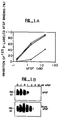
- 125 I-labeled bFGF was first competitively bound to Swiss 3T3 cells. As shown in Figure 1(A), 125 I-labeled bFGF (2 Ci/ ⁇ mol) was added to the confluent 3T3 cells (6 fmol of 125 I-labeled bFGF per 10 5 cells) in the presence of indicated concentrations of: unmodified bFGF (-X-); biotin-bFGF (solid square); the unbound fraction after biotin-bFGF was incubated with avidin-agarose, (open square) ; the unbound fraction after bFGF was incubated with avidin-agarose, (open triangle).
- Binding was performed for 30 min at 37°C in culture media (DME H21) containing 0.2% gelatin, and heparin (15 U/ml). The cells were washed three times with a buffer containing 20 mM HEPES (pH 7.4), 0.2% gelatin, and 150 mM NaCl. The radioactivity present was determined in a Beckman gamma counter. Maximal binding (0% inhibition) represents 5700 cpm of specific binding (nonspecific binding was 600 cpm). All determinations were made in triplicate. Recombinant human bFGF (Barr et al., J. Biol. Chem. , 263 : 16471 (1988)) was iodinated using IODOBEADS (Pierce).
- the bFGF was iodinated using 0.5- 1mCi; of 125 I per 1 ⁇ g FGF, 0.2M NaPi, pH 7.4, 2 IODOBEADS and incubated for 15 min. at room temperature, quenched with Na metabisulfite and excess KI. Iodinated bFGF was separated from unreacted free iodine by gel filtration on a PD 10 column equilibrated with 0.2M Na phosphate, pH 7.5, 0.2M NaCl, 0.2% gelatin.
- the bFGF was biotinylated using iodoacetyl-LC-biotin (Pierce) at a 4:1 molar excess of cysteine residues in 10 mM Tris-HCl (pH 8.0) for 5 hours at 4°C, according to the method of Yamamoto, et al., FEBS Lett. 176:75 (1984). Unreacted biotin was removed by gel filtration with PD 10 columns as described above (Pharmacia).
- modified bFGF was indistinguishable from unmodified bFGF in its ability to inhibit the binding of 125 I-labeled bFGF to high affinity bFGF receptors in Swiss 3T3 cells and in its ability to stimulate the phosphorylation of a 90 kD protein, known to be a substrate of bFGF-induced tyrosine kinase activity. See Fig. 1(A).
- the biotinylation reaction modified 90 to 95% of the bFGF molecules as measured by binding to avidin-conjugated agarose.
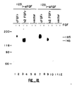
- cellular in situ bFGF receptors were cross-linked to labeled bFGF.
- 125 I-labeled biotin-bFGF or 125 I-labeled bFGF (0.1 pmol) was added to Swiss 3T3 cells (5 X 10 5 cells) in the presence or absence of unlabeled bFGF as indicated.
- the cells were washed and cross-linked with 0.15 mM disuccinimidyl suberate (DSS) (Pierce).
- DSS disuccinimidyl suberate
- the cells were then solubilized, subjected to SDS polyacrylamide gel electrophoresis (PAGE) and 125 I-labeled proteins were detected by autoradiography.
- Purified chicken bFGF receptor was prepared by homogenizing fresh day 6 chicken embryos (stage 29-30) with a Brinkmann polytron; (1500 embryos/batch); (1:1 v/v) in a final concentration of 0.25 M sucrose, 50 mM HEPES (pH 7.5), 2 mM EDTA, 50 mM NaF, 150 ⁇ M sodium orthovanadate, 30 mM sodium pyrophosphate, 1 mM phenylmethylsulfonyl fluoride (PMSF), aprotinin (20 to 30 kallikrein international units (KIU)/ml, leupeptin (10 ⁇ g/ml), and pepstatin (1 ⁇ g/ml).
- PMSF phenylmethylsulfonyl fluoride
- KIU kallikrein international units
- the homogenate was centrifuged at 17,700g for 45 minutes at 4°C.
- the pellet was resuspended in homogenization buffer (300 ml) and the resulting suspension was referred to as the membrane fraction (Mb).
- the membrane fraction was then incubated for 30 min at 4°C with an equal volume of 2X lysis buffer (1X lysis buffer consists of 10 mM Tris-HCl (pH 7.5)), 50 mM NaCl, 5 mM EDTA, 1% Triton X-100, 50 mM NaF, 150 ⁇ M sodium orthovanadate, 30 mM sodium pyrophosphate, 1 mM PMSF, aprotinin (20 to 30 KIU/ml), leupeptin (10 ⁇ g/ml) and pepstatin (1 ⁇ g/ml)), and then centrifuged at 31,000g for 30 min.
- 2X lysis buffer consists of 10 mM Tris-HCl (pH 7.5)), 50
- the supernatant was applied batchwise to a 150 ml WGA-Sepharose 4B column, washed with 300 ml of lysis buffer followed by 500 ml of column buffer which contained 20 mM HEPES (pH 7.5), 2 mM EDTA, 10% glycerol, 0.1% Triton X-100, 50 mM NaF, 150 ⁇ M sodium orthovanadate, 30 mM sodium pyrophosphate, 1 mM PMSF, aprotinin (20 to 30 KIU/ml), leupeptin (10 ⁇ g/ml) and pepstatin (1 ⁇ g/ml).
- the column was eluted with column buffer containing 0.5 M N-acetylglucosamine. Peak protein containing fractions were combined and stored at -70°C.
- chicken bFGF receptor was cross-linked by incubating 10 ⁇ l of the chicken embryo membrane fraction (Mb) or 100 ⁇ l of the eluate from the WGA-Sepharose 4B column with 125 I-labeled bFGF (0.1 pmol) in the presence (+) or absence (-) of a 200-fold excess of unlabeled bFGF for 30 min at 37°C (See Fig. 2(A)).
- DSS was added to a concentration of 0.15 mM, and the reaction mixture was incubated for 10 min on ice.
- ligand affinity purifications were performed (each using the material from 20,000 embryos).
- the eluate from the WGA-Sepharose 4B column was incubated with biotin-bFGF prepared as described above (10:1 molar excess of ligand to receptor) and heparin at a concentration of 15 U/ml (to reduce low affinity binding) for 30 min at 4°C.
- the mixture was then cycled twice through a 10 ml avidin-agarose column (bFGF-agarose).
- the eluted proteins were separated by acrylamide gel electrophoresis and stained with Coomassie Blue.
- the band corresponding to the bFGF receptor was cut out and the protein electroeluted according to the method of M.W. Hunkapiller, et al., Meth. In Enzymol. , 91: 227 (1983). This procedure resulted in the purification of 2 to 5 ng of pure FGF receptor per chicken embryo with an overall recovery of 5%.
- the purified protein was determined to be a purified bFGF receptor in that it bound to bFGF, was the expected molecular weight of the receptor, and contained tyrosine kinase sequences.
- a chicken embryo (day 6) cDNA library was constructed from size-selected poly A + mRNA. 200 ⁇ g of poly A + mRNA was size-fractionated on a 10%-30% sucrose gradient and fractions containing mRNA greater than or equal to 3.5 Kb were pooled. 5 ⁇ g of the sized mRNA was used to generate the cDNA according to the method of U. Gubler and B. Hoffman, Gene 25:263 (1983) using a cDNA synthesis kit from Pharmacia (cat.#27-9260-01).
- the synthesized cDNAs were Size-Selected for cDNAs greater than or equal to 2.0 kb, and the sized cDNAs were then cloned into the Eco RI site of the bacteriophage vector ZapII (Stratagene, cat.#236211).
- the resultant cDNA library contained 2.0 X 10 6 independent recombinants.
- the library was screened with a 32 p-labeled oligonucleotide probe that encoded the two contiguous peptides shown in Fig. 3 (TVALGSNVEFVCK and VYSDPQPHIQWLK).
- the oligonucleotides were prepared using a commercial automated oligonucleotide synthesizer. Two 43-45 base oligonucleotides containing a 12 base overlapping complementary sequence were annealed and labeled by Klenow fill-in with dNTP's (-dCTP), 32 P-dCTP, and DNA polymerase Klenow fragment yielding a 70 bp labeled probe.
- the transmembrane region and the hydrophobic signal sequence were identified by Kyte and Doolittle hydropathy analysis as described in Kyte and Doolittle, J. Mol. Biol. , 157:105 (1982).
- a single hybridizing band of approximately 3.5 Kb was identified by probing chicken embryo poly(A) + RNA (5 ⁇ g) with full-length chicken bFGF receptor cDNA under high stringency conditions (50% formamide), 5X Denhardt's solution and 5X SSC at 42°C. Filters were then washed with 0.2X SSC at 65°. The 3.5 kb single hybridizing band identified by the RNA blot analysis is shown in Fig. 5(A). Primer extension experiments with an oligonucleotide complementary to a sequence near the 5' end of the clone were performed.
- Chicken embryo poly(A) + RNA (5 ⁇ g) was denatured with 10 mM methylmercury, annealed to 32 P-labeled primer (5' CTGCACGTCATCGCGCA-3') and extended with murine Moloney leukemia virus reverse transcriptase.
- lane (S) represents 32 P-labeled DNA molecular size standards (1 kb);
- Lane (E) represents extended fragment (523 nucleotides);
- Lanes (G, A, T, and C) represent a 5% acrylamide sequencing gel. The data predicted that the mRNA of the receptor was 48 nucleotides longer than the isolated clone.
- the amino acid sequence of the longest open reading frame included an in-frame stop codon (amino acid residue -12) followed by an initiator methionine (residue 1) and the entire receptor coding sequence (Fig. 3).
- the cDNA encoded a protein with a deduced molecular mass of 91.7 kD that had features found in several known growth factor receptors. It contained a single-membrane spanning region, an NH 2 -terminal hydrophobic signal sequence, three extracellular immunoglobulin-like domains and an intracellular tyrosine kinase domain (Fig. 6). Eleven potential N-linked glycosylation sites were also found. N- and O-linked glycosylation of the chicken bFGF receptor may account for the disparity between the observed size of the bFGF receptor and the size predicted from the cDNA sequence.
- immunoglobulin-like domains in the putative extracellular region were identified on the basis of three criteria: (i) the presence of two characteristic cysteine residues in each domain; (ii) the presence of a consensus tryptophan residue 11 to 12 amino acids on the COOH-terminal side of the first cysteine residue in each immunoglobulin-like domain; and (iii) the presence of the consensus sequence, DXGXYXC, on the NH 2 -terminal side of the second cysteine residue in each immunoglobulin-like domain.
- the interleukin-1 (IL-1) receptor also has three immunoglobulin-like domains, and bFGF has 25-30% sequence identity to IL-1. Five immunoglobulin-like domains are present in the receptors for platelet-derived growth factor (PDGF) and colony-stimulating factor-1 (CSF-1).
- PDGF platelet-derived growth factor
- CSF-1 colony-stimulating factor-1
- the bFGF receptor has a feature not found in other members of the immunoglobulin superfamily.
- EDDDDEDD acidic residues
- Fig. 3 additional acidic residues
- the juxtamembrane region is the region between the membrane spanning segment and the kinase domain. This region is normally conserved among receptor tyrosine kinases. For example, the juxtamembrane region is consistently 49 to 51 residues in length in the receptors for PDGF, CSF-1, epidermal growth factor (EGF), human epidermal growth factor-2 (HER2) and insulin.
- the bFGF receptor has an unusually long juxtamembrane region of about 87 residues.
- the cytoplasmic region of the amino acid sequence is about 424 residues long and contains a tyrosine kinase sequence (about residues 482 to 759).
- the kinase region of the bFGF receptor shares the most sequence identity (about 51 to 53%) with the PDGF and CSF-1 receptors.
- the bFGF receptor contains the GXGXXG motif and the conserved lysine residue (about residue 512) that form part of the adenosine 5'-triphosphate (ATP) binding site of tyrosine kinases.
- the bFGF receptor also contains the two characteristic tyrosine kinase motifs, HRDLAARNVL and DFGLAR, and a tyrosine (about residue 651) at the position analogous to the major phosphorylation site of pp60 v-src (about Tyr 416).
- the kinase coding sequence of the bFGF receptor defined by homology to other tyrosine kinases, is split by an insertion of 14 amino acids.
- the length of the insertion in the kinase region is shorter than that found in the receptors for PDGF and CSF-1 (104 and 70 amino acids, respectively) and is similar to the length of the inserted sequence in the receptors for insulin and insulin-like growth factor-I.
- a human FGF receptor cDNA clone was isolated from a human endothelial cell cDNA library obtained from E. Sadler (R.D. Ye T-C Wun & J.E. Sadler, J. Biol. Chem. , 262: 3718-3725 (1987)) using the same oligonucleotide probe described in Example 2.
- the endothelial library was hybridized at high stringency with labeled probe 1 x 10 6 cpm/ml (50% formamide, 5X SSC, 5X Denhardts, 10mM NaPO 4 , pH 6.5, 100 ⁇ g/ml salmon sperm DNA at 42°C, (16-24 hrs) and washed at 65°C with 0.2X SSC, 0.1% SDS.
- Human endothelial cell or placental libraries are screened with full-length FGF-R probes or probes containing a portion of the sequence for FGF-R. Hybridization is performed at low stringency conditions and washed in increments of increasingly higher stringency. The low and high stringency conditions described in Examples 2 and 3 are followed. Between each increment, autoradiography is performed. Clones which are positive through to the most stringent conditions are most related to the bFGF receptors previously described in Examples 2 and 3. Clones which are positive at relaxed stringency but are no longer positive at high stringency conditions are more distantly related. All related but not identical (to Figure 4) clones are determined by restriction mapping and DNA sequencing. All related clones are selected, subcloned and expressed. The expressed FGF-related cDNAs are then tested for their ability to bind the various FGFs, i.e. acidic FGF.
- two probes are designed, one probe containing intracellular FGF-R sequence and the other extracellular FGF-R sequence.
- Triplicate filters are made.
- One filter is hybridized at high stringency (see Examples 2 and 3) with the intracellular FGF-R probe.
- Two filters are hybridized with the extracellular probe, one filter at high stringency and one at low stringency. Since acidic and basic FGFs have only 55% sequence identity, their receptors may also exhibit about 55% sequence identity in the ligand-binding domain.
- Clones which are positive at high stringency to the intracellular probe and positive only at low stringency to the extracellular probe are FGF-R related receptors. Thus clones are selected, restriction mapping performed, sequenced, and expressed. The expressed receptors are tested for their ability to bind to various FGFs, e.g., acidic FGF.
- full-length chicken FGF receptor cDNA containing 46 nucleotides of 5' nontranslated sequence and the entire 3' nontranslated sequence, and full-length human h2 cDNA containing 13 nucleotides of 5' nontranslated sequence and the entire 3' nontranslated sequence were individually subcloned into the BamHI/SalI sites of the mammalian expression vector pSV7d (P. Luciw, Chiron Corporation). This placed the receptor cDNA fragments in the proper orientation directly downstream from an SV40 promoter element.
- full-length chicken FGF receptor cDNA was subcloned into the BamHI/SalI sites of Bluescript Sk (Stratagene) and full-length human FGF receptor cDNAs (h2 and h3) were subcloned into the PstI/SalI sites of Bluescript KS. This placed the receptor sequences directly downstream from the T7 RNA polymerase promoter element. To enhance the possibility of efficient translation, ATG sequences upstream of the initiator methionine residue were removed prior to subcloning, leaving 46 and 13 nucleotides of intact 5' nontranslated sequence for the chicken and human constructs, respectively.
- Rat L6 skeletal muscle myoblasts (ATCC CRL 1458) were grown in DME H21 containing 10% fetal calf serum and transferred into Opti-MEM (GIBCO) just prior to transfection. Within 24 hours after plating, 1 X 10 6 cells were cotransfected with 20 ⁇ g of the appropriate expression construct (either cFGFR/pSV7d or h2FGFR/pSV7d) and 1 ⁇ g of a vector containing the neomycin resistance gene (pSV2neo). Cells were transfected using 50 ⁇ g of Lipofectin (Bethesda Research Laboratories) following the protocol provided by the manufacturer.
- Opti-MEM Opti-MEM
- Recombinant human aFGF and human bFGF were generously donated by Chiron Corporation and indicated.
- 5 x 10 6 cells were incubated for 30 minutes at 37°C with 0.1 pmoles of 125 I-aFGF or 125 I-bFGF in the presence or absence of a 200-fold excess of the corresponding unlabeled ligand.
- the cells were then washed once with ice cold DME H21 containing 20 mM HEPES pH 7.4, 0.2% gelatin, and twice with ice cold PBS.
- Disuccinimidyl suberate was added to a final concentration of 0.15 mM and incubations were allowed to proceed for 15 minutes at 4°C.
- the crosslinking agent was then removed and the cells were reSuspended in sample buffer containing 100 mM dithiothreitol, boiled for 5 minutes, and subjected to SDS PAGE followed by autoradiography.
- RNAs were transcribed from the linearized templates using T7 RNA polymerase in the presence of 500 ⁇ M rNTPs (200 ⁇ M rGTP) and 500 ⁇ M 5' GpppG 3 ' , (Pharmacia). Following incubation at 4°C for 2 hours, transcription reactions were treated with RNAse-free DNAse, phenol extracted, ethanol precipitated, dried and resuspended in water.
- Oocytes were injected into the vegetal pole with 50 nl of water or RNA solution (1 ⁇ g/ ⁇ l in water). Following injection, oocytes were incubated at 19°C for 48 hours before performing 45 Ca ++ efflux assays.
- oocytes Groups of 50 injected oocytes were added to single wells of a 24 well plate and washed four times with 0.5 ml of a Ca ++ -free MBSH solution containing no BSA. Oocytes were then incubated in 0.5 ml of the wash solution containing 45 CaCl 2 (100 ⁇ Ci/ml) for 3 hours at 19°C. Following incubation, oocytes were washed six times with 0.5 ml of MBSH (1 mg/ml BSA), then transferred to another 24 well plate (5 oocytes per well). All subsequent washes and incubations were performed using 0.5 ml of MBSH containing 1 mg/ml BSA.
- conditioned supernatants were removed from each well and replaced with fresh media.
- the conditioned media samples were counted individually in a Beckman scintillation counter. When background efflux stabilized, ligands were added to the specified concentrations and media collections were continued.
- Receptor levels in injected oocytes have not been measured because our anti-receptor polyclonal antisera nonspecifically recognizes an abundant oocyte protein of approximately the same molecular weight as the FGF receptor on western blots. Furthermore, the levels of exogenous receptors expressed in oocytes appears to be quite low.
- Clones h2, h3, and h4 were obtained from the endothelial cell library and clone h5 was obtained from the placenta library. Nucleotide sequence analyses revealed that all four clones contained identical 5' nontranslated sequences and had poly-A tracts at their 3' ends. However, only the poly-A tract at the 3' end of h2 was preceded upstream by a consensus poly adenylation signal sequence (AATAAA; 37), indicating that internal priming was responsible for the poly A tracts at the 3' ends of the other clones.
- AATAAA consensus poly adenylation signal sequence
- the h2, h3, h4, and h5 cDNAs contained 0.93 kb, 0.78 kb, 0.95 kb, and 0.2 kb of 3' nontranslated sequence, respectively.
- the 3' nontranslated sequences of h2 and h3 were identical and the 3' nontranslated sequences of h4 and h5 were also identical.
- the h2/h3 3' nontranslated sequences were entirely different from the 3' nontranslated sequences of h4/h5.
- Amplification reactions (42) were carried out using one primer corresponding to the human highly acidic region (approximately amino acids 44-52 in h2; S'GTTTCTTTCTCCTCTGAAGAGGAGT-3') and one degenerate primer corresponding to the IgI domain of the chicken FGF receptor (approximately amino acids 58-69; 5'- GA(TC)GACGTGCAG (A/T)(G/C)CATCAACTGGGTGCGTGATGG-3').
- Reaction mixtures contained 750 ng of human genomic DNA, 10 pmoles of each primer, 200 ⁇ M of each of the four dNTPs, and 1 unit of Taq polymerase (Perkin Elmer Cetus) in 50 ⁇ l of 10 mM Tris-HCl, pH 8.3, 50 mM KCl, 1.5 mM MgCl 2 , 100 ng/ml BSA. Reactions were carried out in an Ericomp twin block system. Thirty one cycles were performed, consisting of denaturation at 94°C for 50 seconds, annealing at 65°C for 1 minute, and extension at 72°C for 3 minutes.
- the chicken basic FGF receptor contains a single transmembrane domain, an extracellular region containing 3 Ig-like domains and a highly acidic domain, and an intracellular region containing a split tyrosine kinase domain.
- the chicken FGF receptor cDNA is highly homologous to a previously published partial cDNA (h flg ) which encodes a tyrosine kinase that, at the time of its description was of unknown function.
- the high degree of identity (95 percent) between the chicken bFGF receptor and human flg suggested that h flg was the human counterpart of the bFGF receptor.
- oligonucleotide probe based on the h flg cDNA sequence was used to screen a human umbilical vein endothelial cell cDNA library and a human placenta cDNA library. From the initial screenings of 250,000 plaques from each library, four positive clones were isolated from the endothelial cell library and three from the placenta library.
- the cDNA clones could be divided into two classes based on different patterns of restriction maps at their 3' ends. One of these classes derived from cDNA clones which were much shorter in length. Representatives of each class were present in the clones isolated from either library. Two clones (h2 and h3) representing the class of larger cDNA clones, and two clones (h4 and h5) representing the class of shorter cDNA clones were sequenced in their entirety. The deduced amino acid sequences of the four human receptor forms are shown in comparison to the chicken FGF receptor sequence in Figure 7. A schematic representation of the different receptor forms is shown in Figure 8.
- the predicted amino acid sequences of the h2 and h3 clones are virtually identical and differ only by three amino acids (amino acids 59, 60, and 103 in h2, Fig. 7). At the nucleotide level, h2 and h3 differ only at the positions encoding these three amino acid residues.
- the h2/h3 open reading frames include a hydrophobic signal sequence and the unusual acidic domain (8 consecutive acidic residues with accompanying residues) that was initially noted in the published sequence of the chicken FGF receptor cDNA.
- the extracellular domains of h2 and h3 are highly homologous to the chicken FGF receptor except that h2 and h3 lack the sequences of one Ig-like domain (labeled I in Fig. 8).
- the transmembrane region and cytoplasmic domains are highly homologous to the corresponding domains of the chicken FGF receptor.
- the coding sequences of the short cDNA clones, h4 and h5 differ only by two amino acids (positions 59 and 60 in h4; the nucleotide sequences of h4 and h5 differ only at the Positions encoding these two residues).
- the signal sequence, acidic region and one of the Ig-like domains (IgII) are essentially identical to the corresponding regions of h2 and h3.
- the distinctive feature of h4 and h5 is the Ig-like domain (IgIII) nearest the transmembrane domain. Approximately half of this domain is identical to the corresponding sequence of h2 and h3. However, the carboxyl terminal half of this Ig-like domain is unrelated to h2 and h3 sequences.
- h4 and h5 do not encode a hydrophobic membrane spanning region or a cytoplasmic domain.
- the sequences of all of the human cDNAs which have been isolated contain only 2 Ig-like domains.
- IgI the first Ig-like domain
- HFFs human foreskin fibroblasts
- this fragment contained coding sequences homologous (approximately 83 percent amino acid identity) to the IgI domain of the chicken FGF receptor.
- an intron sequence of approximately 1.0 kb separates these coding sequences from sequences encoding the highly acidic region of the receptor.
- the human FGF receptor gene clearly contains sequences encoding the IgI domain not found in the human cDNA clones.
- the presence of an intron between the IgI domain sequence and the acidic region sequence suggests that expression of 2 or 3 Ig domain forms may be regulated by alternative splicing.
- HFF cells To determine whether a 3 Ig domain form of the receptor is expressed in HFF cells, we performed PCR on cDNA generated from HFF mRNA. Using the primers described above, a single 0.24 kb fragment was amplified from HFF cDNA. This fragment contained sequences encoding the IgI domain and the acidic region, but no intron sequences. Thus, we conclude that HFF cells transcribe a 3 Ig domain form of the receptor. To determine whether HFF cells also express a 2 Ig domain form of the receptor, we utilized the acidic region primer and a second primer based on sequence from the 5' nontranslated region of the human FGF receptor.
- the 3 Ig domain receptor (initially isolated from a chicken cDNA library) was purified on the basis of its affinity for basic FGF, it was of interest to determine whether this receptor also binds acidic FGF.
- the 3 Ig domain chicken receptor was expressed in rat L6 myoblasts, a cell line which normally does not express FGF receptors.
- the 2 Ig domain human h2 receptor was also expressed in L6 cells.
- Figure 10 shows an affinity labeling experiment performed with transfected cells. Cells were incubated with either 125 I-aFGF or 125 I-bFGF and bound ligand was crosslinked in the presence of disuccinimidyl suberate (0.15 mM).
- a Three Ig Domain FGF Receptor and a Two Ig Domain FGF Receptor Mediate Biological Responses to Both Acidic and Basic FGF.
- Oocytes express endogenous receptors for carbachol, and oocytes injected with either FGF receptor RNA or water exhibited a positive response after carbachol stimulation.
- cFGF-R 3 Ig domain form
- h2 2 Ig domain form
- the ligand binding domains for acidic and basic FGF appear to lie in the receptor region encompassing the highly acidic domain and the IgII and IgIII domains.
- h2 receptor While the human h2 receptor clearly responds to both ligands, no response was seen in oocytes injected with RNA encoding the h3 receptor form. It is possible that the three amino acid differences between h2 and h3 cause these proteins to respond differently. Alternatively, the lack of a response in oocytes injected with the h3 RNA may be due to unusually low expression levels of the h3 protein. Unfortunately, we have not yet been able to determine receptor protein expression levels in oocytes.
- FGF-R forms having either 2 or 3 extracellular Ig-like domains will bind and respond to both acidic and basic FGF.
- Some forms of FGF receptor mRNA encode only the extracellular domain of the FGF receptor, a protein that is likely to be secreted from the cell.
- the h4 and h5 receptor forms lack transmembrane sequences and presumably represent secreted forms of the FGF receptor.
- Preliminary data indicates that cells transfected with the h4 cDNA secrete a 70 kd protein which is recognized by anti-FGF-R polyclonal antisera.
- the role of secreted forms of the FGF receptor is unclear.
- the secreted forms may act to regulate levels of extracellular FGFs, and thereby regulate availability of FGFs to cell surface FGF receptors.
- the secreted FGF receptors may serve to store and sequester FGFs at a particular location.
- Another possibility is that the secreted forms may bind to FGFs in an intracellular compartment and subsequently serve as a means for secreting the factor. This is an important consideration in view of the fact that aFGF and bFGF do not contain signal sequences and their mechanism of secretion is unknown.
- Affinity labeling experiments using either 125 I-aFGF or 125 I-bFGF identified a single 145 kd receptor protein on transfectant cells expressing the 3 Ig domain form of the FGF receptor, and a single 125 kd receptor protein on transfectant cells expressing the 2 domain form of the FGF receptor (see Fig. 11). It is possible that the presence of two receptor species may reflect coexpression of the 3 Ig domain and 2 Ig domain forms of the receptor. Our data clearly establish that a single FGF receptor species can bind both aFGF and bFGF with high affinity and mediate the biological effects of these factors. We have used acidic and basic FGF in these experiments because they are the best characterized members of the FGF family, and are readily available in recombinant form.
- a fragment containing all or part of the extracellular, ligand-binding domain of the FGF-R (i.e., containing amino acids 22-374 of Figure 3 or 22-285 of Figure 4) or analogs thereof are expressed in a host (e.g. mammalian cells or baculovirus infected insect cells) and purified as described in Example 1.
- a host e.g. mammalian cells or baculovirus infected insect cells
- fragments of the ligand-binding domain are made using a peptide synthesizer (Applied Biosystems) and purified by HPLC.
- Different concentrations of the FGF-R fragment or analogs thereof (FGF-Rexs) are tested for their ability to block the binding of 125 I-FGF to Swiss 3T3 cells.
- Competitive binding is performed as described in Figure 1A in Example 1 using FGF-Rexs instead of unlabeled ligand and competitive binding is determined.
- FGF-Rexs are also tested for their ability to inhibit FGF-induced mitogenesis as measured by 3 H-thymidine incorporation into cells and by counting cell numbers.
- FGF-Rexs which block binding of FGF to the cell-surface receptor may act as an antagonist and block 3 H-thymidine uptake and the increase in cell number induced by FGF.
- FGF-Rexs may also act as agonists, i.e. by dimerization with the cell surface receptor which may mimic a ligand-mediated receptor-recaptor interaction. In such an instance, FGF-Rexs may stimulate mitogenesis in the absence of ligand or may enhance the FGF mediated mitogenic response.
- FGF-Rexs are also tested for their ability to inhibit or activate FGF-induced tyrosine phosphorylation of the 90 substrate protein in Swiss 3T3 cells or autophosphorylation of the cell-associated FGF-R.
- FGF-Rexs which block FGF-induced tyrosine phosphorylation are antagonists.
- FGF-Rexs which activate autophosphorylation of the cell-associated FGF-R in the absence of FGF are agonists.
- FGF-Rexs are also tested for their anti-angiogenic activity. FGF-Rexs are tested first for their ability to inhibit the FGF-induced growth and the mobilization of endothelial cells into vessels in vitro. Angiogenesis is assayed in vitro using an aortic ring assay. Aortic rings are placed in a collagen matrix formed in the presence or absence of FGF and FGF-Rexs. Endothelial cells sprout and form vessels from the aortic ring within a few days in the presence of FGF. The addition of FGF-Rexs which are antagonists in the previous assays inhibit the FGF-induced growth of capillary sprouts. FGF-Rexs which are angiogenic even in the absence of FGF are agonists.
- FGF analogs, angiogenic factors, anti-angiogenic factors as well as antibodies to the extracellular portion of the FGF-R are tested for their ability to bind directly or compete for binding of native FGF for binding to purified or expressed FGF-R. In addition, they are tested for their ability to stimulate mitogenesis (agonists) or inhibit FGF-dependent mitogenesis (antagonists) as well as tyrosine phosphorylation in cells expressing the FGF-R. These studies are important in determining if the mode of action of each angiogenic and anti-angiogenic factor, etc., is receptor-mediated and in determining if there is receptor specificity (i.e. acidic versus basic FGF-R) for angiogenic and anti-angiogenic factors.
- receptor specificity i.e. acidic versus basic FGF-R
- FGF analogs are radiolabeled and binding is performed with labeled ligand, purified or expressed receptor in the appropriate physiologic buffer (i.e. culture media or phosphate buffered saline (PBS)) for 0.5 at 37°C or 2-24 hrs at 4°C.
- the complex is precipitated (5-10% polyethylene glycol, 1 mg/ml IgG) and separated by filtration through filters (i.e. Whitman GFA) and the associated radioactivity determined.
Landscapes
- Health & Medical Sciences (AREA)
- Life Sciences & Earth Sciences (AREA)
- Chemical & Material Sciences (AREA)
- Molecular Biology (AREA)
- Engineering & Computer Science (AREA)
- Immunology (AREA)
- General Health & Medical Sciences (AREA)
- Organic Chemistry (AREA)
- Medicinal Chemistry (AREA)
- Biomedical Technology (AREA)
- Hematology (AREA)
- Biochemistry (AREA)
- Urology & Nephrology (AREA)
- Cell Biology (AREA)
- Genetics & Genomics (AREA)
- General Chemical & Material Sciences (AREA)
- Endocrinology (AREA)
- Biophysics (AREA)
- Gastroenterology & Hepatology (AREA)
- Biotechnology (AREA)
- Zoology (AREA)
- Microbiology (AREA)
- Toxicology (AREA)
- Food Science & Technology (AREA)
- Physics & Mathematics (AREA)
- Analytical Chemistry (AREA)
- General Physics & Mathematics (AREA)
- Pathology (AREA)
- Chemical Kinetics & Catalysis (AREA)
- Proteomics, Peptides & Aminoacids (AREA)
- Nuclear Medicine, Radiotherapy & Molecular Imaging (AREA)
- Pharmacology & Pharmacy (AREA)
- Animal Behavior & Ethology (AREA)
- Public Health (AREA)
- Veterinary Medicine (AREA)
- Bioinformatics & Cheminformatics (AREA)
- Peptides Or Proteins (AREA)
- Compounds Of Unknown Constitution (AREA)
- Micro-Organisms Or Cultivation Processes Thereof (AREA)
- Polysaccharides And Polysaccharide Derivatives (AREA)
Claims (39)
- Verfahren zur Herstellung eines Fibroblasten-Wachstumsfaktor-Rezeptorpeptids, umfassend IgII- und IgIII-Domänen einer der in Fig. 7 dargestellten Aminosäuresequenzen oder eine Aminosäuresequenz, die zwei Domänen umfaßt, die zu zumindest 85 % homolog zur IgII- bzw. IgIII-Domäne einer der Sequenzen sind; wobei dem Polypeptid eine Transmembrandomäne fehlt und wobei das Polypeptid fähig ist, die Bindung zwischen einem Fibroblasten-Wachstumsfaktor und einem Fibroblasten-Wachstumsfaktor-Rezeptor zu hemmen, worin das Peptid in einer Zelle produziert wird, die mit einer Nukleinsäure transformiert ist, die eine für das Peptid kodierende Sequenz umfaßt, die mit Transkriptions- und Translationsinitiationsbereichen operabel verbunden ist, wobei das Verfahren das Exprimieren der Nukleinsäure und das Gewinnen des FGF-R-Peptids umfaßt.
- Verfahren nach Anspruch 1, worin das Peptid die IgII- und IgII-Domänen von Fibroblasten-Wachstumsfaktor-Rezeptor h3, wie in Fig. 7 dargestellt, oder ein in diesen Domänen zu 85 % dazu homologes Polypeptid umfaßt.
- Verfahren nach Anspruch 1 oder 2, worin das Peptid eine erste Ig-Domäne umfaßt, die (i) zwei Cysteinreste, (ii) einen Tryptophanrest, 11 oder 12 Aminosäuren an der carboxylterminalen Seite jenes der obengenannten Cysteinreste, der dem N-terminalen Ende am nächsten ist, von diesem entfernt, und (iii) eine Sequenz DXGXYXC an der aminoterminalen Seite des anderen der Cysteinreste umfaßt; eine zweite Ig-Domäne, C-terminal zur ersten Domäne, die ebenfalls die Merkmale (i), (ii) und (iii), wie für die erste Ig-Domäne definiert, aufweist, mit der Ausnahme, daß die zweite dieser Ig-Domänen anstelle des anderen Cysteinrests und der Sequenz (iii) eine carboxylterminale Sequenz aufweisen kann, die zu zumindest 85 % zur 79-Aminosäuren-Sequenz der Reste 224 bis 302 oder 222 bis 300 des menschlichen Proteins h4 bzw. h5 aus Fig. 7 homolog ist.
- Verfahren nach Anspruch 3, worin die erste Ig-Domäne die Reste 85 bis 141 einer menschlichen Sequenz, wie in Fig. 7 dargestellt, umfaßt.
- Verfahren nach einem der Ansprüche 1 bis 4, worin das Polypeptid weiters eine carboxylterminale Sequenz umfaßt, welche die 79-Aminosäuren-Sequenz der Reste 224 bis 302 oder 222 bis 300 der Sequenz einer menschlichen Sequenz h4 bzw. h5 ist, wie in Fig. 7 dargestellt.
- Verfahren nach Anspruch 1 zur Herstellung eines gereinigten Polypeptids, das die als h4 oder h5 bezeichnete menschliche Sequenz aus Fig. 7 umfaßt.
- Verfahren zur Herstellung eines Fibroblasten-Wachstumsfaktor-Rezeptorpeptids, das kleiner als 85 kD ist und eine Fibroblasten-Wachstumsfaktor-Bindungsdomäne eines Fibroblasten-Wachstumsfaktor-Rezeptors umfaßt, wobei der Rezeptor eine Aminosäuresequenz, wie in Fig. 7 dargestellt, oder eine Allel-Variante oder Mutante davon umfaßt, oder eine homologe Sequenz, für die eine Nukleinsäure kodiert, die sich unter rauhen Bedingungen, zu denen eine Temperatur von über 37°C und eine Salzkonzentration von weniger als 1 M zählen, an DNA hybridisiert, die für die Sequenzvariante oder -mutante kodiert, worin das Peptid in einer Zelle produziert wird, die mit einer Nukleinsäure transformiert ist, die eine für das Peptid kodierende Sequenz umfaßt, die mit Transkriptions und Translationsinitiationsbereichen operabel verbunden ist, wobei das Verfahren das Exprimieren der Nukleinsäure und das Gewinnen des FGF-R-Peptids umfaßt.
- Verfahren nach Anspruch 7, worin das Polypeptid löslich ist.
- Verfahren nach Anspruch 7, worin das Polypeptid weiters ein Segment aus zumindest 5 zusammenhängenden Aminosäuren umfaßt oder einschließt, das aus der Gruppe ausgewählt ist, die besteht aus:(a) einem Signalsegment;(b) einem sauren Segment, das eine Abfolge von 8 aufeinanderfolgenden sauren Resten aufweist;(c) bis zu drei Ig-Segmenten von Ig-Domänen, von denen jedes dadurch gekennzeichnet ist, daß es folgendes aufweist: (i) zwei Cysteinreste, (ii) einen Tryptophanrest, 11 oder 12 Aminosäuren an der carboxylterminalen Seite jenes der obengenannten Cysteinreste, der dem N-terminalen Ende am nächsten ist, von diesem entfernt, und (iii) eine Sequenz DXGXYXC an der aminoterminalen Seite des anderen der Cysteinreste; mit der Ausnahme, daß eine der Ig-Domänen anstelle des anderen Cysteinrests und der Sequenz (iii) eine carboxylterminale Sequenz aufweisen kann, die zu zumindest 85 % zur 79-Aminosäuren-Sequenz der Reste 224 bis 302 oder 222 bis 300 des menschlichen Proteins h4 bzw. h5 aus Fig. 7 homolog ist; oder(d) ein Transmembran-Segment.
- Verfahren nach einem der Ansprüche 7 bis 9, worin die Fibroblasten-Wachstumsfaktor-Bindungsdomäne zumindest 30 Aminosäuren jeder der beiden Ig-Domänen, wie in Anspruch 9 definiert, umfaßt, die dem Carboxylterminus am nächsten liegen, wie in Fig. 7 dargestellt.
- Verfahren nach Anspruch 7, worin der Fibroblasten-Wachstumsfaktor-Rezeptor ein Hühner-Fibroblasten-Wachstumsfaktor-Rezeptor ist.
- Verfahren nach einem der vorangegangenen Ansprüche, worin das Polypeptid in einem mehrkettigen Proteinkomplex vorliegt.
- Verfahren zur Herstellung eines Antikörpers gegen ein Fibroblasten-Wachstumsfaktor-Rezeptorfragment, wobei das Verfahren einen Schritt der Herstellung eines Antikörpers gegen ein Polypeptid-Epitop umfaßt, das zumindest 6 zusammenhängende Aminosäuren eines Polypeptids umfaßt, wie nach einem der Ansprüche 1 bis 12 definiert.
- Verfahren nach Anspruch 13, worin das Epitop aus der Gruppe von Proteinsegmenten ausgewählt ist, die aus folgendem besteht: einer Signalsequenz, einem Ig-Segment einer Ig-Domäne, die dadurch gekennzeichnet ist, daß sie folgendes aufweist: (i) zwei Cysteinreste, (ii) einen Tryptophanrest, 11 oder 12 Aminosäuren an der carboxylterminalen Seite jenes der obengenannten Cysteinreste, der dem N-terminalen Ende am nächsten ist, von diesem entfernt, und (iii) eine Sequenz DXGXYXC an der aminoterminalen Seite des anderen der Cysteinreste; einem sauren Segment, das eine Abfolge von 8 aufeinanderfolgenden sauren Resten aufweist, oder einem Segment einer modifizierten Ig-Domäne, die anstelle der Sequenz DXGXYXC eine carboxylterminale Sequenz aufweist, die zu zumindest 85 % zur 79-Aminosäuren-Sequenz der Reste 224 bis 302 oder 222 bis 300 des h4- bzw. h5-Proteins aus Fig. 7 homolog ist.
- Verfahren zum Messen eines Fibroblasten-Wachstumsfaktors oder eines Fibroblasten-Wachstumsfaktor-Rezeptors in einer Zielprobe, wobei das Verfahren folgende Schritte umfaßt:das Kombinieren der Zielprobe mit einem Polypeptid nach einem der Ansprüche 1 bis 12; unddas Bestimmen des Ausmaßes der Bindung zwischen dem Segment und der Probe.
- Verfahren zum Messen eines Fibroblasten-Wachstumsfaktors oder eines Fibroblasten-Wachstumsfaktor-Rezeptors, umfassend:das Kombinieren einer Zielprobe, die einen Fibroblasten-Wachstumsfaktor enthält, mit einem löslichen Polypeptid nach einem der Ansprüche 1 bis 12; unddas Bestimmen des Ausmaßes der Bindung zwischen dem Segment und dem Fibroblasten-Wachstumsfaktor in der Probe.
- Verfahren nach Anspruch 16, das weiters die Bewertung eines potentiellen Agonisten oder Antagonisten auf seine Fähigkeit zur Förderung oder Hemmung der Bindungswechselwirkung des Segments und des Wachstumsfaktors umfaßt.
Applications Claiming Priority (3)
| Application Number | Priority Date | Filing Date | Title |
|---|---|---|---|
| US37700389A | 1989-07-06 | 1989-07-06 | |
| US377003 | 1989-07-06 | ||
| PCT/US1990/003830 WO1991000916A2 (en) | 1989-07-06 | 1990-07-06 | Receptors for fibroblast growth factors |
Publications (3)
| Publication Number | Publication Date |
|---|---|
| EP0481000A1 EP0481000A1 (de) | 1992-04-22 |
| EP0481000A4 EP0481000A4 (en) | 1992-10-21 |
| EP0481000B1 true EP0481000B1 (de) | 1999-05-12 |
Family
ID=23487374
Family Applications (1)
| Application Number | Title | Priority Date | Filing Date |
|---|---|---|---|
| EP90911235A Expired - Lifetime EP0481000B1 (de) | 1989-07-06 | 1990-07-06 | Rezeptoren für fibroblasten-wachstumsfaktoren |
Country Status (15)
| Country | Link |
|---|---|
| US (4) | US6384191B1 (de) |
| EP (1) | EP0481000B1 (de) |
| JP (1) | JP3039802B2 (de) |
| KR (1) | KR100235266B1 (de) |
| AT (1) | ATE179862T1 (de) |
| AU (1) | AU638734B2 (de) |
| CA (1) | CA2063431C (de) |
| DE (1) | DE69033109T2 (de) |
| DK (1) | DK0481000T3 (de) |
| ES (1) | ES2133271T3 (de) |
| FI (1) | FI920027A0 (de) |
| HU (1) | HU215581B (de) |
| NO (1) | NO310032B1 (de) |
| SG (1) | SG72657A1 (de) |
| WO (1) | WO1991000916A2 (de) |
Families Citing this family (63)
| Publication number | Priority date | Publication date | Assignee | Title |
|---|---|---|---|---|
| GB9001466D0 (en) * | 1990-01-23 | 1990-03-21 | Erba Carlo Spa | Extracellular form of the human fibroblast growth factor receptor |
| CA2063431C (en) * | 1989-07-06 | 2002-10-29 | Lewis T. Williams | Receptors for fibroblast growth factors |
| JP2552942B2 (ja) * | 1990-06-01 | 1996-11-13 | 三井東圧化学株式会社 | ヒトbーFGF受容体遺伝子 |
| WO1992000999A1 (en) * | 1990-07-06 | 1992-01-23 | Rhone-Poulenc Rorer International (Holdings) Inc. | Fibroblast growth factor receptors |
| US6582959B2 (en) | 1991-03-29 | 2003-06-24 | Genentech, Inc. | Antibodies to vascular endothelial cell growth factor |
| IL100219A0 (en) * | 1991-12-02 | 1992-09-06 | Yeda Res & Dev | Variable region within fibroblast growth factor receptors that confers ligand specificity |
| ES2091018T3 (es) * | 1992-06-18 | 1996-10-16 | Whittier Inst Diabetes & Endoc | Procedimiento para la deteccion de enfermedades neoplasicas. |
| CZ291047B6 (cs) * | 1992-10-28 | 2002-12-11 | Genentech, Inc. | Farmaceutický prostředek obsahující antagonisty faktoru růstu vaskulárních endoteliálních buněk |
| US7060808B1 (en) * | 1995-06-07 | 2006-06-13 | Imclone Systems Incorporated | Humanized anti-EGF receptor monoclonal antibody |
| US6214795B1 (en) | 1996-11-12 | 2001-04-10 | Praecis Pharmaceuticals, Inc. | Peptide compounds useful for modulating FGF receptor activity |
| US20030224001A1 (en) * | 1998-03-19 | 2003-12-04 | Goldstein Neil I. | Antibody and antibody fragments for inhibiting the growth of tumors |
| AU3170499A (en) * | 1998-04-28 | 1999-11-16 | Eisai Co. Ltd. | Fibroblast growth factor mutein compositions and methods of use therefor |
| ZA200007412B (en) * | 1998-05-15 | 2002-03-12 | Imclone Systems Inc | Treatment of human tumors with radiation and inhibitors of growth factor receptor tyrosine kinases. |
| US20020192719A1 (en) * | 1998-09-30 | 2002-12-19 | Caliper Technologies Corp. | Homogeneous assay methods |
| US6656728B1 (en) | 1999-02-08 | 2003-12-02 | Chiron Corporation | Fibroblast growth factor receptor-immunoglobulin fusion |
| WO2000069459A1 (en) * | 1999-05-14 | 2000-11-23 | Imclone Systems Incorporated | Treatment of refractory human tumors with epidermal growth factor receptor antagonists |
| WO2002011677A2 (en) * | 2000-08-09 | 2002-02-14 | Imclone Systems Incorporated | Treatment of hyperproliferative diseases with epidermal growth factor receptor antagonists |
| AU2002236477A1 (en) | 2000-11-21 | 2002-06-03 | The Texas A & M University System | Fgf-affinity chromatography |
| US20080008704A1 (en) * | 2001-03-16 | 2008-01-10 | Mark Rubin | Methods of treating colorectal cancer with anti-epidermal growth factor antibodies |
| IL159177A0 (en) | 2001-06-20 | 2004-06-01 | Prochon Biotech Ltd | Antibodies that block receptor protein tyrosine kinase activation, methods of screening for and uses thereof |
| WO2003072126A2 (de) * | 2002-02-28 | 2003-09-04 | Switch Biotech Ag | Verwendung eines fibroblastenwachstumsfaktor-bindeproteins zur behandlung und diagnose von diabetischen wundheilungsstörungen |
| ATE276755T1 (de) * | 2002-02-28 | 2004-10-15 | Switch Biotech Ag | Verwendung eines fibroblastenwachstumsfaktor- bindeproteins zur behandlung und diagnose von diabetischen wundheilugnsstörungen |
| EP1579218A2 (de) | 2002-12-20 | 2005-09-28 | Enkam Pharmaceuticals A/S | Verfahren zur modulation der wechselwerkung von rezeptor und ligand |
| EP1622941A2 (de) * | 2003-03-20 | 2006-02-08 | ImClone Systems Incorporated | Methode zur produktion von anti-egfr antikörpern |
| WO2005066211A2 (en) * | 2003-12-19 | 2005-07-21 | Five Prime Therapeutics, Inc. | Fibroblast growth factor receptors 1, 2, 3, and 4 as targets for therapeutic intervention |
| RU2402569C2 (ru) * | 2004-03-19 | 2010-10-27 | Имклоун Элэлси | Человеческие антитела к рецептору эпидермального фактора роста |
| US20060234347A1 (en) * | 2005-04-13 | 2006-10-19 | Harding Thomas C | Targeting multiple angiogenic pathways for cancer therapy using soluble tyrosine kinase receptors |
| ATE450615T1 (de) * | 2005-07-22 | 2009-12-15 | Five Prime Therapeutics Inc | Zusammensetzungen und verfahren zur behandlung von krankheiten mit fgfr-fusionsproteinen |
| WO2007109733A2 (en) | 2006-03-21 | 2007-09-27 | The Johns Hopkins University | Diagnostic and prognostic markers and treatment strategies for multiple sclerosis |
| JP5537946B2 (ja) | 2006-11-22 | 2014-07-02 | ブリストル−マイヤーズ スクイブ カンパニー | Igf−irを含むチロシンキナーゼ受容体に対する改変タンパク質に基づく標的治療薬 |
| AR063975A1 (es) | 2006-11-28 | 2009-03-04 | Centelion | Fusiones fc con receptor para fgf soluble modificadas con actividad biologica mejorada |
| KR20100128291A (ko) | 2008-02-14 | 2010-12-07 | 브리스톨-마이어스 스큅 컴퍼니 | Egfr에 결합하는 조작된 단백질을 기초로 하는 표적화된 치료제 |
| AR071874A1 (es) | 2008-05-22 | 2010-07-21 | Bristol Myers Squibb Co | Proteinas de dominio de armazon basadas en fibronectina multivalentes |
| NZ590050A (en) | 2008-06-04 | 2012-08-31 | Amgen Inc | Fgf21 mutants and uses thereof |
| EP2313435A4 (de) * | 2008-07-01 | 2012-08-08 | Aveo Pharmaceuticals Inc | An den fibroblastenwachstumsfaktorrezeptor 3 (fgfr3) bindende proteine |
| CN102171343B (zh) * | 2008-08-04 | 2017-07-14 | 戊瑞治疗有限公司 | Fgfr细胞外结构域酸性区突变蛋白 |
| WO2010042747A2 (en) | 2008-10-10 | 2010-04-15 | Amgen Inc. | Fgf21 mutants and uses thereof |
| PL2842573T3 (pl) | 2008-11-07 | 2018-04-30 | Galaxy Biotech, Llc | Przeciwciała monoklonalne swoiste dla receptora 2 czynnika wzrostu fibroblastów |
| HRP20150965T1 (hr) * | 2009-03-25 | 2015-11-06 | Genentech, Inc. | Anti-fgr3 protutijela i metode koje ih koriste |
| JO3469B1 (ar) * | 2009-05-05 | 2020-07-05 | Amgen Inc | طافرات fgf21 واستخداماتها |
| EP2427207B1 (de) | 2009-05-05 | 2017-08-16 | Amgen, Inc | Fgf21-mutanten und ihre verwendungen |
| JP2012530493A (ja) * | 2009-06-17 | 2012-12-06 | アムジエン・インコーポレーテツド | キメラポリペプチドおよびその使用 |
| WO2011034940A1 (en) | 2009-09-15 | 2011-03-24 | Five Prime Therapeutics, Inc. | Hair growth methods using fgfr4 extracellular domains |
| PT2498799T (pt) | 2009-11-13 | 2016-11-04 | Five Prime Therapeutics Inc | Utilização de proteínas do domínio extracelular rfcf1 para tratar cancros caracterizados por mutações ativadoras dependentes de ligando em rfcf2 |
| US8372952B2 (en) * | 2009-12-02 | 2013-02-12 | Amgen Inc. | Binding proteins that bind to human FGFR1C, human β-klotho and both human FGFR1C and human β-klotho |
| UA109888C2 (uk) | 2009-12-07 | 2015-10-26 | ІЗОЛЬОВАНЕ АНТИТІЛО АБО ЙОГО ФРАГМЕНТ, ЩО ЗВ'ЯЗУЄТЬСЯ З β-КЛОТО, РЕЦЕПТОРАМИ FGF І ЇХНІМИ КОМПЛЕКСАМИ | |
| US8685931B2 (en) | 2009-12-17 | 2014-04-01 | Five Prime Therapeutics, Inc. | Hair growth methods using FGFR3 extracellular domains |
| WO2011130417A2 (en) | 2010-04-15 | 2011-10-20 | Amgen Inc. | HUMAN FGF RECEPTOR AND β-KLOTHO BINDING PROTEINS |
| US9226960B2 (en) | 2010-04-16 | 2016-01-05 | Andrew B. Bush | FGF modulation of in vivo antibody production and humoral immunity |
| US8435525B1 (en) | 2010-04-16 | 2013-05-07 | Andrew B. Bush | FGF modulation of in vivo antibody production and humoral immunity |
| RU2012153241A (ru) | 2010-05-11 | 2014-06-20 | Авео Фармасьютикалз, Инк. | Антитела к fgfr2 |
| US8481038B2 (en) | 2010-11-15 | 2013-07-09 | Five Prime Therapeutics, Inc. | Treatment of cancer with elevated dosages of soluble FGFR1 fusion proteins |
| EP2640377B1 (de) | 2010-11-15 | 2018-12-26 | Five Prime Therapeutics, Inc. | Kombinationstherapien mit extrazellulärer fgfr1-domäne |
| US8951972B2 (en) | 2010-12-09 | 2015-02-10 | Five Prime Therapeutics, Inc. | FGFR1 extracellular domain combination therapies for lung cancer |
| EP2780033B1 (de) | 2011-11-14 | 2019-05-08 | Five Prime Therapeutics, Inc. | Eine fgfr1 extrazelluläre domäne zur behandlung von krebs mit einer fgfr1 gen vervielfältigung |
| PE20160190A1 (es) | 2013-08-01 | 2016-04-28 | Five Prime Therapeutics Inc | Anticuerpos anti-fgfr2iiib afucosilados |
| US10373702B2 (en) * | 2014-03-27 | 2019-08-06 | Massachusetts Institute Of Technology | Water-soluble trans-membrane proteins and methods for the preparation and use thereof |
| AU2015320341B2 (en) | 2014-09-26 | 2021-05-27 | Rajendra Sahai Bhatnagar | Inhibitors of NF kappa-B activity for treatment of diseases and disorders |
| JP6952685B2 (ja) | 2015-06-05 | 2021-10-20 | アイバイオ・インコーポレイテッドIbio, Inc. | 線維症の治療において使用するためのエンドスタチン断片およびバリアント |
| JP7349787B2 (ja) | 2015-11-23 | 2023-09-25 | ファイヴ プライム セラピューティクス インク | 癌治療におけるfgfr2阻害剤単独または免疫刺激剤との組み合わせ |
| WO2018213304A1 (en) | 2017-05-16 | 2018-11-22 | Five Prime Therapeutics, Inc. | Anti-fgfr2 antibodies in combination with chemotherapy agents in cancer treatment |
| AR123306A1 (es) | 2020-08-21 | 2022-11-16 | Genzyme Corp | Anticuerpos fgfr3 y métodos de uso |
| KR20240049361A (ko) | 2021-08-25 | 2024-04-16 | 아이바이오, 인크. | 항-cd25 항체 |
Family Cites Families (13)
| Publication number | Priority date | Publication date | Assignee | Title |
|---|---|---|---|---|
| US4394443A (en) | 1980-12-18 | 1983-07-19 | Yale University | Method for cloning genes |
| US4785079A (en) * | 1984-11-09 | 1988-11-15 | The Salk Institute For Biological Studies | Isolation of fibroblast growth factor |
| US4668476A (en) * | 1984-03-23 | 1987-05-26 | Applied Biosystems, Inc. | Automated polypeptide synthesis apparatus |
| US4761371A (en) * | 1985-02-12 | 1988-08-02 | Genentech, Inc. | Insulin receptor |
| US4859609A (en) * | 1986-04-30 | 1989-08-22 | Genentech, Inc. | Novel receptors for efficient determination of ligands and their antagonists or agonists |
| US5057417A (en) * | 1987-06-12 | 1991-10-15 | Genentech, Inc. | Compositions and methods for the synthesis of growth hormone receptor and growth hormone binding protein |
| US5081228A (en) * | 1988-02-25 | 1992-01-14 | Immunex Corporation | Interleukin-1 receptors |
| US4968607A (en) * | 1987-11-25 | 1990-11-06 | Immunex Corporation | Interleukin-1 receptors |
| SE8804164A0 (sv) * | 1988-11-17 | 1990-05-18 | Per Prisell | Farmaceutisk beredning |
| US5225538A (en) * | 1989-02-23 | 1993-07-06 | Genentech, Inc. | Lymphocyte homing receptor/immunoglobulin fusion proteins |
| CA2063431C (en) | 1989-07-06 | 2002-10-29 | Lewis T. Williams | Receptors for fibroblast growth factors |
| US5229501A (en) * | 1991-01-11 | 1993-07-20 | Chiron Corporation | Expression and use of human fibroblast growth factor receptor |
| US6022741A (en) * | 1997-03-13 | 2000-02-08 | University Of North Carolina At Chapel Hill | Regulatory genetic DNA that regulates the Class II transactivator (CIITA) |
-
1990
- 1990-07-06 CA CA002063431A patent/CA2063431C/en not_active Expired - Lifetime
- 1990-07-06 EP EP90911235A patent/EP0481000B1/de not_active Expired - Lifetime
- 1990-07-06 AU AU60779/90A patent/AU638734B2/en not_active Expired
- 1990-07-06 JP JP02510738A patent/JP3039802B2/ja not_active Expired - Lifetime
- 1990-07-06 AT AT90911235T patent/ATE179862T1/de not_active IP Right Cessation
- 1990-07-06 WO PCT/US1990/003830 patent/WO1991000916A2/en not_active Ceased
- 1990-07-06 FI FI920027A patent/FI920027A0/fi unknown
- 1990-07-06 US US07/834,311 patent/US6384191B1/en not_active Expired - Lifetime
- 1990-07-06 ES ES90911235T patent/ES2133271T3/es not_active Expired - Lifetime
- 1990-07-06 HU HU906195A patent/HU215581B/hu unknown
- 1990-07-06 SG SG1996007270A patent/SG72657A1/en unknown
- 1990-07-06 DE DE69033109T patent/DE69033109T2/de not_active Expired - Lifetime
- 1990-07-06 DK DK90911235T patent/DK0481000T3/da active
-
1992
- 1992-01-03 NO NO920060A patent/NO310032B1/no not_active IP Right Cessation
- 1992-01-04 KR KR1019920700005A patent/KR100235266B1/ko not_active Expired - Lifetime
-
1995
- 1995-06-02 US US08/458,938 patent/US5707632A/en not_active Expired - Lifetime
- 1995-06-02 US US08/459,395 patent/US6350593B1/en not_active Expired - Lifetime
- 1995-06-02 US US08/459,082 patent/US6355440B1/en not_active Expired - Lifetime
Also Published As
| Publication number | Publication date |
|---|---|
| NO920060L (no) | 1992-03-02 |
| DK0481000T3 (da) | 1999-11-15 |
| DE69033109D1 (de) | 1999-06-17 |
| EP0481000A4 (en) | 1992-10-21 |
| ATE179862T1 (de) | 1999-05-15 |
| WO1991000916A2 (en) | 1991-01-24 |
| DE69033109T2 (de) | 1999-11-18 |
| JP3039802B2 (ja) | 2000-05-08 |
| HUT61052A (en) | 1992-11-30 |
| HU906195D0 (en) | 1992-06-29 |
| FI920027A7 (fi) | 1992-01-03 |
| AU638734B2 (en) | 1993-07-08 |
| US6384191B1 (en) | 2002-05-07 |
| ES2133271T3 (es) | 1999-09-16 |
| FI920027A0 (fi) | 1992-01-03 |
| SG72657A1 (en) | 2000-05-23 |
| NO920060D0 (no) | 1992-01-03 |
| KR100235266B1 (en) | 1999-12-15 |
| US5707632A (en) | 1998-01-13 |
| AU6077990A (en) | 1991-02-06 |
| US6355440B1 (en) | 2002-03-12 |
| EP0481000A1 (de) | 1992-04-22 |
| NO310032B1 (no) | 2001-05-07 |
| CA2063431A1 (en) | 1991-01-07 |
| HU215581B (hu) | 1999-01-28 |
| US6350593B1 (en) | 2002-02-26 |
| JPH04506604A (ja) | 1992-11-19 |
| CA2063431C (en) | 2002-10-29 |
| WO1991000916A3 (en) | 1991-10-17 |
Similar Documents
| Publication | Publication Date | Title |
|---|---|---|
| EP0481000B1 (de) | Rezeptoren für fibroblasten-wachstumsfaktoren | |
| US7071159B2 (en) | Inhibitor of vascular endothelial cell growth factor | |
| US5686572A (en) | Domains of extracellular region of human platelet derived growth factor receptor polypeptides | |
| US6255454B1 (en) | Expression and use of human fibroblast growth factor receptor | |
| US6599709B1 (en) | Method for screening for soluble ligands to a receptor-type tyrosine kinase | |
| US6835544B2 (en) | Methods of screening for compounds that bind to activin receptor | |
| EP0814827B1 (de) | Wachstumsfaktor-b, spezifisch für vaskuläre endothelzellen | |
| US5885794A (en) | Recombinant production of vertebrate activin receptor polypeptides and identification of receptor DNAs in the activin/TGF-β superfamily | |
| CA2086425C (en) | Fibroblast growth factor receptors | |
| WO1990008822A1 (en) | Erythropoietin receptor | |
| JPH025865A (ja) | ヒトの血小板由来の成長因子受容体 | |
| US20030017535A1 (en) | Human platelet-derived growth factor receptors | |
| US5863888A (en) | Human Bek Fibroblast growth factor receptor | |
| US6475782B1 (en) | Human platelet-derived growth factor receptor | |
| JPH10117791A (ja) | ヒトgタンパク質結合受容体hlyaz61 |
Legal Events
| Date | Code | Title | Description |
|---|---|---|---|
| PUAI | Public reference made under article 153(3) epc to a published international application that has entered the european phase |
Free format text: ORIGINAL CODE: 0009012 |
|
| 17P | Request for examination filed |
Effective date: 19920127 |
|
| AK | Designated contracting states |
Kind code of ref document: A1 Designated state(s): AT BE CH DE DK ES FR GB IT LI LU NL SE |
|
| A4 | Supplementary search report drawn up and despatched |
Effective date: 19920828 |
|
| AK | Designated contracting states |
Kind code of ref document: A4 Designated state(s): AT BE CH DE DK ES FR GB IT LI LU NL SE |
|
| RIN1 | Information on inventor provided before grant (corrected) |
Inventor name: WILLIAMS, LEWIS, T. Inventor name: LEE, PAULINE, E. Inventor name: JOHNSON, DANIEL, E. |
|
| 17Q | First examination report despatched |
Effective date: 19950512 |
|
| GRAG | Despatch of communication of intention to grant |
Free format text: ORIGINAL CODE: EPIDOS AGRA |
|
| GRAG | Despatch of communication of intention to grant |
Free format text: ORIGINAL CODE: EPIDOS AGRA |
|
| GRAG | Despatch of communication of intention to grant |
Free format text: ORIGINAL CODE: EPIDOS AGRA |
|
| GRAH | Despatch of communication of intention to grant a patent |
Free format text: ORIGINAL CODE: EPIDOS IGRA |
|
| GRAH | Despatch of communication of intention to grant a patent |
Free format text: ORIGINAL CODE: EPIDOS IGRA |
|
| GRAA | (expected) grant |
Free format text: ORIGINAL CODE: 0009210 |
|
| AK | Designated contracting states |
Kind code of ref document: B1 Designated state(s): AT BE CH DE DK ES FR GB IT LI LU NL SE |
|
| REF | Corresponds to: |
Ref document number: 179862 Country of ref document: AT Date of ref document: 19990515 Kind code of ref document: T |
|
| REG | Reference to a national code |
Ref country code: CH Ref legal event code: EP |
|
| REF | Corresponds to: |
Ref document number: 69033109 Country of ref document: DE Date of ref document: 19990617 |
|
| ITF | It: translation for a ep patent filed | ||
| ET | Fr: translation filed | ||
| REG | Reference to a national code |
Ref country code: ES Ref legal event code: FG2A Ref document number: 2133271 Country of ref document: ES Kind code of ref document: T3 |
|
| REG | Reference to a national code |
Ref country code: DK Ref legal event code: T3 |
|
| PLBE | No opposition filed within time limit |
Free format text: ORIGINAL CODE: 0009261 |
|
| STAA | Information on the status of an ep patent application or granted ep patent |
Free format text: STATUS: NO OPPOSITION FILED WITHIN TIME LIMIT |
|
| 26N | No opposition filed | ||
| REG | Reference to a national code |
Ref country code: GB Ref legal event code: IF02 |
|
| PGFP | Annual fee paid to national office [announced via postgrant information from national office to epo] |
Ref country code: ES Payment date: 20090727 Year of fee payment: 20 Ref country code: DK Payment date: 20090728 Year of fee payment: 20 Ref country code: FR Payment date: 20090717 Year of fee payment: 20 |
|
| PGFP | Annual fee paid to national office [announced via postgrant information from national office to epo] |
Ref country code: SE Payment date: 20090729 Year of fee payment: 20 Ref country code: NL Payment date: 20090724 Year of fee payment: 20 Ref country code: AT Payment date: 20090619 Year of fee payment: 20 Ref country code: CH Payment date: 20090728 Year of fee payment: 20 Ref country code: LU Payment date: 20090821 Year of fee payment: 20 Ref country code: GB Payment date: 20090727 Year of fee payment: 20 Ref country code: DE Payment date: 20090729 Year of fee payment: 20 |
|
| PGFP | Annual fee paid to national office [announced via postgrant information from national office to epo] |
Ref country code: BE Payment date: 20090818 Year of fee payment: 20 |
|
| PGFP | Annual fee paid to national office [announced via postgrant information from national office to epo] |
Ref country code: IT Payment date: 20090729 Year of fee payment: 20 |
|
| REG | Reference to a national code |
Ref country code: NL Ref legal event code: V4 Effective date: 20100706 |
|
| REG | Reference to a national code |
Ref country code: CH Ref legal event code: PL |
|
| REG | Reference to a national code |
Ref country code: DK Ref legal event code: EUP |
|
| REG | Reference to a national code |
Ref country code: GB Ref legal event code: PE20 Expiry date: 20100705 |
|
| BE20 | Be: patent expired |
Owner name: THE *REGENTS OF THE UNIVERSITY OF CALIFORNIA Effective date: 20100706 |
|
| REG | Reference to a national code |
Ref country code: ES Ref legal event code: FD2A Effective date: 20100707 |
|
| EUG | Se: european patent has lapsed | ||
| PG25 | Lapsed in a contracting state [announced via postgrant information from national office to epo] |
Ref country code: ES Free format text: LAPSE BECAUSE OF EXPIRATION OF PROTECTION Effective date: 20100707 Ref country code: NL Free format text: LAPSE BECAUSE OF EXPIRATION OF PROTECTION Effective date: 20100706 |
|
| PG25 | Lapsed in a contracting state [announced via postgrant information from national office to epo] |
Ref country code: GB Free format text: LAPSE BECAUSE OF EXPIRATION OF PROTECTION Effective date: 20100705 |
|
| PG25 | Lapsed in a contracting state [announced via postgrant information from national office to epo] |
Ref country code: DE Free format text: LAPSE BECAUSE OF EXPIRATION OF PROTECTION Effective date: 20100706 |