EP0570331B1 - Méthode et appareil pour obtenir des prothèses vasculaires - Google Patents
Méthode et appareil pour obtenir des prothèses vasculaires Download PDFInfo
- Publication number
- EP0570331B1 EP0570331B1 EP93810297A EP93810297A EP0570331B1 EP 0570331 B1 EP0570331 B1 EP 0570331B1 EP 93810297 A EP93810297 A EP 93810297A EP 93810297 A EP93810297 A EP 93810297A EP 0570331 B1 EP0570331 B1 EP 0570331B1
- Authority
- EP
- European Patent Office
- Prior art keywords
- tissue
- suspension
- cells
- particles
- vessel
- Prior art date
- Legal status (The legal status is an assumption and is not a legal conclusion. Google has not performed a legal analysis and makes no representation as to the accuracy of the status listed.)
- Expired - Lifetime
Links
- 238000000034 method Methods 0.000 title claims abstract description 75
- 230000002792 vascular Effects 0.000 title claims description 22
- 239000000725 suspension Substances 0.000 claims abstract description 70
- 239000002245 particle Substances 0.000 claims abstract description 50
- 230000001413 cellular effect Effects 0.000 claims abstract description 8
- 210000004027 cell Anatomy 0.000 claims description 97
- 210000001519 tissue Anatomy 0.000 claims description 88
- 230000008569 process Effects 0.000 claims description 26
- 238000001914 filtration Methods 0.000 claims description 25
- 230000029087 digestion Effects 0.000 claims description 16
- 238000000926 separation method Methods 0.000 claims description 16
- 239000008280 blood Substances 0.000 claims description 14
- 238000004519 manufacturing process Methods 0.000 claims description 14
- 230000006862 enzymatic digestion Effects 0.000 claims description 13
- 210000004369 blood Anatomy 0.000 claims description 11
- 239000010410 layer Substances 0.000 claims description 11
- 239000003814 drug Substances 0.000 claims description 6
- 230000005484 gravity Effects 0.000 claims description 6
- 210000002751 lymph Anatomy 0.000 claims description 6
- 239000012487 rinsing solution Substances 0.000 claims description 6
- 230000008929 regeneration Effects 0.000 claims description 5
- 238000011069 regeneration method Methods 0.000 claims description 5
- 238000003756 stirring Methods 0.000 claims description 4
- 210000004003 subcutaneous fat Anatomy 0.000 claims description 4
- 239000002344 surface layer Substances 0.000 claims description 4
- 210000003709 heart valve Anatomy 0.000 claims description 3
- 210000002747 omentum Anatomy 0.000 claims description 3
- 206010033675 panniculitis Diseases 0.000 claims description 3
- 239000000470 constituent Substances 0.000 claims description 2
- 230000006835 compression Effects 0.000 claims 1
- 238000007906 compression Methods 0.000 claims 1
- 230000002596 correlated effect Effects 0.000 claims 1
- 230000000977 initiatory effect Effects 0.000 claims 1
- 230000001172 regenerating effect Effects 0.000 claims 1
- 239000000463 material Substances 0.000 abstract description 31
- 239000011248 coating agent Substances 0.000 abstract description 15
- 238000000576 coating method Methods 0.000 abstract description 15
- 238000002513 implantation Methods 0.000 abstract description 12
- 239000012876 carrier material Substances 0.000 abstract description 11
- 238000005119 centrifugation Methods 0.000 abstract description 11
- 238000002955 isolation Methods 0.000 abstract description 4
- 238000012545 processing Methods 0.000 abstract description 4
- 210000002889 endothelial cell Anatomy 0.000 description 15
- 238000004062 sedimentation Methods 0.000 description 15
- 102000029816 Collagenase Human genes 0.000 description 11
- 108060005980 Collagenase Proteins 0.000 description 11
- 238000004140 cleaning Methods 0.000 description 10
- 229960002424 collagenase Drugs 0.000 description 10
- 239000012071 phase Substances 0.000 description 9
- 102000004190 Enzymes Human genes 0.000 description 8
- 108090000790 Enzymes Proteins 0.000 description 8
- 229940088598 enzyme Drugs 0.000 description 8
- 210000000577 adipose tissue Anatomy 0.000 description 7
- 210000000056 organ Anatomy 0.000 description 7
- 239000006285 cell suspension Substances 0.000 description 6
- 230000036512 infertility Effects 0.000 description 6
- 239000007788 liquid Substances 0.000 description 6
- 210000004925 microvascular endothelial cell Anatomy 0.000 description 6
- 230000008901 benefit Effects 0.000 description 5
- 230000001079 digestive effect Effects 0.000 description 5
- 230000000694 effects Effects 0.000 description 5
- 210000003038 endothelium Anatomy 0.000 description 5
- 239000000243 solution Substances 0.000 description 5
- 238000001356 surgical procedure Methods 0.000 description 5
- 238000002054 transplantation Methods 0.000 description 5
- 210000003462 vein Anatomy 0.000 description 5
- 210000001367 artery Anatomy 0.000 description 4
- 238000000338 in vitro Methods 0.000 description 4
- 238000010899 nucleation Methods 0.000 description 4
- 238000003860 storage Methods 0.000 description 4
- 239000008346 aqueous phase Substances 0.000 description 3
- 230000015556 catabolic process Effects 0.000 description 3
- 238000009826 distribution Methods 0.000 description 3
- 238000011833 dog model Methods 0.000 description 3
- 238000011068 loading method Methods 0.000 description 3
- 102000008186 Collagen Human genes 0.000 description 2
- 108010035532 Collagen Proteins 0.000 description 2
- 229920004934 Dacron® Polymers 0.000 description 2
- 206010020880 Hypertrophy Diseases 0.000 description 2
- 241001465754 Metazoa Species 0.000 description 2
- 206010073734 Microembolism Diseases 0.000 description 2
- 239000003146 anticoagulant agent Substances 0.000 description 2
- 229940127219 anticoagulant drug Drugs 0.000 description 2
- 230000000525 athrombogenic effect Effects 0.000 description 2
- 230000017531 blood circulation Effects 0.000 description 2
- 210000004204 blood vessel Anatomy 0.000 description 2
- 230000008859 change Effects 0.000 description 2
- 229920001436 collagen Polymers 0.000 description 2
- 210000002808 connective tissue Anatomy 0.000 description 2
- 238000010790 dilution Methods 0.000 description 2
- 239000012895 dilution Substances 0.000 description 2
- 238000001035 drying Methods 0.000 description 2
- 230000003511 endothelial effect Effects 0.000 description 2
- 230000007515 enzymatic degradation Effects 0.000 description 2
- 230000002255 enzymatic effect Effects 0.000 description 2
- 238000002474 experimental method Methods 0.000 description 2
- 210000002216 heart Anatomy 0.000 description 2
- 210000001365 lymphatic vessel Anatomy 0.000 description 2
- 239000011159 matrix material Substances 0.000 description 2
- 210000004088 microvessel Anatomy 0.000 description 2
- 239000000203 mixture Substances 0.000 description 2
- 230000002572 peristaltic effect Effects 0.000 description 2
- 239000011148 porous material Substances 0.000 description 2
- 238000002360 preparation method Methods 0.000 description 2
- 238000000746 purification Methods 0.000 description 2
- XLYOFNOQVPJJNP-UHFFFAOYSA-N water Substances O XLYOFNOQVPJJNP-UHFFFAOYSA-N 0.000 description 2
- 241000282472 Canis lupus familiaris Species 0.000 description 1
- 206010063560 Excessive granulation tissue Diseases 0.000 description 1
- 102000010834 Extracellular Matrix Proteins Human genes 0.000 description 1
- 108010037362 Extracellular Matrix Proteins Proteins 0.000 description 1
- 241000282412 Homo Species 0.000 description 1
- 238000012404 In vitro experiment Methods 0.000 description 1
- NIPNSKYNPDTRPC-UHFFFAOYSA-N N-[2-oxo-2-(2,4,6,7-tetrahydrotriazolo[4,5-c]pyridin-5-yl)ethyl]-2-[[3-(trifluoromethoxy)phenyl]methylamino]pyrimidine-5-carboxamide Chemical compound O=C(CNC(=O)C=1C=NC(=NC=1)NCC1=CC(=CC=C1)OC(F)(F)F)N1CC2=C(CC1)NN=N2 NIPNSKYNPDTRPC-UHFFFAOYSA-N 0.000 description 1
- 108010019160 Pancreatin Proteins 0.000 description 1
- 102000035195 Peptidases Human genes 0.000 description 1
- 108091005804 Peptidases Proteins 0.000 description 1
- 208000007536 Thrombosis Diseases 0.000 description 1
- 241000219793 Trifolium Species 0.000 description 1
- 102000004142 Trypsin Human genes 0.000 description 1
- 108090000631 Trypsin Proteins 0.000 description 1
- 230000009471 action Effects 0.000 description 1
- 210000001789 adipocyte Anatomy 0.000 description 1
- 210000004618 arterial endothelial cell Anatomy 0.000 description 1
- 238000000429 assembly Methods 0.000 description 1
- 230000000712 assembly Effects 0.000 description 1
- 230000015572 biosynthetic process Effects 0.000 description 1
- 210000000601 blood cell Anatomy 0.000 description 1
- 239000000872 buffer Substances 0.000 description 1
- 239000007853 buffer solution Substances 0.000 description 1
- 239000006227 byproduct Substances 0.000 description 1
- 210000001043 capillary endothelial cell Anatomy 0.000 description 1
- 210000000748 cardiovascular system Anatomy 0.000 description 1
- 210000001715 carotid artery Anatomy 0.000 description 1
- 238000004113 cell culture Methods 0.000 description 1
- 230000010261 cell growth Effects 0.000 description 1
- 238000006243 chemical reaction Methods 0.000 description 1
- 239000003153 chemical reaction reagent Substances 0.000 description 1
- 230000015271 coagulation Effects 0.000 description 1
- 238000005345 coagulation Methods 0.000 description 1
- 230000001427 coherent effect Effects 0.000 description 1
- 230000003750 conditioning effect Effects 0.000 description 1
- 210000004748 cultured cell Anatomy 0.000 description 1
- 238000005520 cutting process Methods 0.000 description 1
- 230000007423 decrease Effects 0.000 description 1
- 230000003247 decreasing effect Effects 0.000 description 1
- 230000032459 dedifferentiation Effects 0.000 description 1
- 230000008021 deposition Effects 0.000 description 1
- 108010007093 dispase Proteins 0.000 description 1
- 238000006073 displacement reaction Methods 0.000 description 1
- 230000009762 endothelial cell differentiation Effects 0.000 description 1
- 210000002744 extracellular matrix Anatomy 0.000 description 1
- 238000000605 extraction Methods 0.000 description 1
- 239000004744 fabric Substances 0.000 description 1
- 210000001126 granulation tissue Anatomy 0.000 description 1
- 210000004013 groin Anatomy 0.000 description 1
- 230000035876 healing Effects 0.000 description 1
- 239000007943 implant Substances 0.000 description 1
- 238000010348 incorporation Methods 0.000 description 1
- 208000015181 infectious disease Diseases 0.000 description 1
- 238000012432 intermediate storage Methods 0.000 description 1
- 238000011835 investigation Methods 0.000 description 1
- 150000002632 lipids Chemical class 0.000 description 1
- 239000007791 liquid phase Substances 0.000 description 1
- 230000007774 longterm Effects 0.000 description 1
- 230000001926 lymphatic effect Effects 0.000 description 1
- 238000003760 magnetic stirring Methods 0.000 description 1
- 230000002503 metabolic effect Effects 0.000 description 1
- 210000000496 pancreas Anatomy 0.000 description 1
- 229940055695 pancreatin Drugs 0.000 description 1
- 230000002093 peripheral effect Effects 0.000 description 1
- 230000035699 permeability Effects 0.000 description 1
- 239000002504 physiological saline solution Substances 0.000 description 1
- 229920001296 polysiloxane Polymers 0.000 description 1
- 239000000047 product Substances 0.000 description 1
- 229940024999 proteolytic enzymes for treatment of wounds and ulcers Drugs 0.000 description 1
- 238000005086 pumping Methods 0.000 description 1
- 238000011160 research Methods 0.000 description 1
- 239000013049 sediment Substances 0.000 description 1
- 238000007873 sieving Methods 0.000 description 1
- 239000002356 single layer Substances 0.000 description 1
- 238000005549 size reduction Methods 0.000 description 1
- 210000000952 spleen Anatomy 0.000 description 1
- 239000000126 substance Substances 0.000 description 1
- 229920002994 synthetic fiber Polymers 0.000 description 1
- 230000017423 tissue regeneration Effects 0.000 description 1
- 239000012588 trypsin Substances 0.000 description 1
- 238000009827 uniform distribution Methods 0.000 description 1
- 210000003556 vascular endothelial cell Anatomy 0.000 description 1
- 238000007631 vascular surgery Methods 0.000 description 1
- 230000000007 visual effect Effects 0.000 description 1
- 238000005406 washing Methods 0.000 description 1
- 238000004804 winding Methods 0.000 description 1
Images
Classifications
-
- A—HUMAN NECESSITIES
- A61—MEDICAL OR VETERINARY SCIENCE; HYGIENE
- A61F—FILTERS IMPLANTABLE INTO BLOOD VESSELS; PROSTHESES; DEVICES PROVIDING PATENCY TO, OR PREVENTING COLLAPSING OF, TUBULAR STRUCTURES OF THE BODY, e.g. STENTS; ORTHOPAEDIC, NURSING OR CONTRACEPTIVE DEVICES; FOMENTATION; TREATMENT OR PROTECTION OF EYES OR EARS; BANDAGES, DRESSINGS OR ABSORBENT PADS; FIRST-AID KITS
- A61F2/00—Filters implantable into blood vessels; Prostheses, i.e. artificial substitutes or replacements for parts of the body; Appliances for connecting them with the body; Devices providing patency to, or preventing collapsing of, tubular structures of the body, e.g. stents
- A61F2/02—Prostheses implantable into the body
- A61F2/04—Hollow or tubular parts of organs, e.g. bladders, tracheae, bronchi or bile ducts
- A61F2/06—Blood vessels
- A61F2/062—Apparatus for the production of blood vessels made from natural tissue or with layers of living cells
-
- A—HUMAN NECESSITIES
- A61—MEDICAL OR VETERINARY SCIENCE; HYGIENE
- A61F—FILTERS IMPLANTABLE INTO BLOOD VESSELS; PROSTHESES; DEVICES PROVIDING PATENCY TO, OR PREVENTING COLLAPSING OF, TUBULAR STRUCTURES OF THE BODY, e.g. STENTS; ORTHOPAEDIC, NURSING OR CONTRACEPTIVE DEVICES; FOMENTATION; TREATMENT OR PROTECTION OF EYES OR EARS; BANDAGES, DRESSINGS OR ABSORBENT PADS; FIRST-AID KITS
- A61F2/00—Filters implantable into blood vessels; Prostheses, i.e. artificial substitutes or replacements for parts of the body; Appliances for connecting them with the body; Devices providing patency to, or preventing collapsing of, tubular structures of the body, e.g. stents
- A61F2/02—Prostheses implantable into the body
- A61F2/04—Hollow or tubular parts of organs, e.g. bladders, tracheae, bronchi or bile ducts
- A61F2/06—Blood vessels
-
- A—HUMAN NECESSITIES
- A61—MEDICAL OR VETERINARY SCIENCE; HYGIENE
- A61F—FILTERS IMPLANTABLE INTO BLOOD VESSELS; PROSTHESES; DEVICES PROVIDING PATENCY TO, OR PREVENTING COLLAPSING OF, TUBULAR STRUCTURES OF THE BODY, e.g. STENTS; ORTHOPAEDIC, NURSING OR CONTRACEPTIVE DEVICES; FOMENTATION; TREATMENT OR PROTECTION OF EYES OR EARS; BANDAGES, DRESSINGS OR ABSORBENT PADS; FIRST-AID KITS
- A61F2/00—Filters implantable into blood vessels; Prostheses, i.e. artificial substitutes or replacements for parts of the body; Appliances for connecting them with the body; Devices providing patency to, or preventing collapsing of, tubular structures of the body, e.g. stents
- A61F2/02—Prostheses implantable into the body
- A61F2/24—Heart valves ; Vascular valves, e.g. venous valves; Heart implants, e.g. passive devices for improving the function of the native valve or the heart muscle; Transmyocardial revascularisation [TMR] devices; Valves implantable in the body
-
- A—HUMAN NECESSITIES
- A61—MEDICAL OR VETERINARY SCIENCE; HYGIENE
- A61L—METHODS OR APPARATUS FOR STERILISING MATERIALS OR OBJECTS IN GENERAL; DISINFECTION, STERILISATION OR DEODORISATION OF AIR; CHEMICAL ASPECTS OF BANDAGES, DRESSINGS, ABSORBENT PADS OR SURGICAL ARTICLES; MATERIALS FOR BANDAGES, DRESSINGS, ABSORBENT PADS OR SURGICAL ARTICLES
- A61L27/00—Materials for grafts or prostheses or for coating grafts or prostheses
- A61L27/28—Materials for coating prostheses
-
- A—HUMAN NECESSITIES
- A61—MEDICAL OR VETERINARY SCIENCE; HYGIENE
- A61L—METHODS OR APPARATUS FOR STERILISING MATERIALS OR OBJECTS IN GENERAL; DISINFECTION, STERILISATION OR DEODORISATION OF AIR; CHEMICAL ASPECTS OF BANDAGES, DRESSINGS, ABSORBENT PADS OR SURGICAL ARTICLES; MATERIALS FOR BANDAGES, DRESSINGS, ABSORBENT PADS OR SURGICAL ARTICLES
- A61L27/00—Materials for grafts or prostheses or for coating grafts or prostheses
- A61L27/50—Materials characterised by their function or physical properties, e.g. injectable or lubricating compositions, shape-memory materials, surface modified materials
- A61L27/507—Materials characterised by their function or physical properties, e.g. injectable or lubricating compositions, shape-memory materials, surface modified materials for artificial blood vessels
-
- A—HUMAN NECESSITIES
- A61—MEDICAL OR VETERINARY SCIENCE; HYGIENE
- A61L—METHODS OR APPARATUS FOR STERILISING MATERIALS OR OBJECTS IN GENERAL; DISINFECTION, STERILISATION OR DEODORISATION OF AIR; CHEMICAL ASPECTS OF BANDAGES, DRESSINGS, ABSORBENT PADS OR SURGICAL ARTICLES; MATERIALS FOR BANDAGES, DRESSINGS, ABSORBENT PADS OR SURGICAL ARTICLES
- A61L33/00—Antithrombogenic treatment of surgical articles, e.g. sutures, catheters, prostheses, or of articles for the manipulation or conditioning of blood; Materials for such treatment
- A61L33/18—Use of ingredients of undetermined constitution or reaction products thereof
-
- Y—GENERAL TAGGING OF NEW TECHNOLOGICAL DEVELOPMENTS; GENERAL TAGGING OF CROSS-SECTIONAL TECHNOLOGIES SPANNING OVER SEVERAL SECTIONS OF THE IPC; TECHNICAL SUBJECTS COVERED BY FORMER USPC CROSS-REFERENCE ART COLLECTIONS [XRACs] AND DIGESTS
- Y10—TECHNICAL SUBJECTS COVERED BY FORMER USPC
- Y10S—TECHNICAL SUBJECTS COVERED BY FORMER USPC CROSS-REFERENCE ART COLLECTIONS [XRACs] AND DIGESTS
- Y10S623/00—Prosthesis, i.e. artificial body members, parts thereof, or aids and accessories therefor
- Y10S623/92—Method or apparatus for preparing or treating prosthetic
- Y10S623/921—Blood vessel
Definitions
- the invention relates to a method and a device according to the preambles of the corresponding independent claims for the production of endoprostheses for at least partially direct contact with the blood or with the lymph, such as, for example, blood or lymphatic vessel prostheses, heart valves, artificial hearts and the like, one of the body's own cells containing suspension prepared and a preformed, artificial carrier material is treated with this suspension.
- a method is e.g. known from EP-A-0 393 788.
- the implantation of such endoprostheses can support the regeneration of parts of the cardiovascular system in humans and animals.
- Endoprostheses that are mostly or only partially in direct contact with blood or lymph have to meet very special requirements that are not required for endoprostheses that only have to grow into the tissue.
- Native blood or lymphatic vessels or other blood or lymphatic cavities are covered on their inner surfaces with a special cell layer, the endothelium.
- endoprostheses that have been used in the bloodstream have performed this task only moderately. It can be assumed that, despite intensive research, long-term use of synthetic materials in this regard can only be regarded as suboptimal and the blood (or lymph) is subject to additional stress that does not occur in native tissue, the endothelium.
- the cells change with such an increase, a so-called dedifferentiation taking place, which is associated with the conditions of the in vitro culture and in the course of which the cells generally fall back into a less specialized state for which there is no exact equivalent Body there. Therefore, the question is justified to what extent the after Culture period with many multiplication steps under artificial conditions cells, which originate from cells from the patient's body, still correspond in their functions to the body's own functions in prosthesis implantation.
- microvascular endothelial cells isolated from microvessels adapt and die Function of arterial endothelial cells can take over if they are sown in arterial prostheses in which, for example, the physiological flow velocity of the blood is significantly higher than in the microvascular part of the vascular network from which the cells have been isolated. It is therefore known that a functional, anticoagulant lining is formed in vascular prostheses that are seeded with microvascular endothelial cells, for example in dogs.
- the microvascular endothelial cells can be obtained, for example, from adipose tissue, for example by removing part of the omentum or a limited amount of subcutaneous adipose tissue.
- microvascular cells are isolated from the preferably subcutaneous fat tissue of a patient, which contains a lot of other cells and non-cellular tissue components in addition to the microvascular cells, and in a further process step these are applied to certain surface parts of a preformed artificial carrier. It has been shown that, after implantation of prostheses manufactured in this way, a tissue develops over the course of weeks to months on the parts of the surface that are permanently in contact with the blood, which is similar to the endothelium of native vessels and lies on a layer of tissue that is also the native one Artery wall is similar (Schmidt et al., J. Invest. Surg., 1: 35-44, 1988).
- neo-arteries These tissues, which form on the inside of vascular prostheses, for example, are therefore also referred to as neo-arteries. Like the native artery, the neo-artery is completely covered with athrombogenic endothelial cells, so that the blood in the prosthesis does not clot or develop its full and undisturbed functionality.
- the first process step consists of isolating the microvascular cells desired for storage by mechanical comminution of the tissue removed from the patient, subsequent enzymatic digestion of the comminuted tissue with a suitable enzyme and the separation of all undesirable components from the by the possible Digestion prepared suspension for isolation and concentration of the microvascular cells.
- the prosthesis For the coating of the prosthesis with the isolated cells, they are stored in a known manner either by filtering the suspension through a porous prosthesis or by centrifugation in a rapidly rotating prosthesis (publication EP-462051) in the appropriately designed surface of the prosthesis.
- the known method should therefore be improved such that an hour for the preparation of the suspension and the subsequent treatment of the prosthesis with the suspension are sufficient and that the prosthesis produced by the method according to the invention supports the regeneration of the endothelium to at least the same extent as prostheses which are manufactured by methods according to the prior art.
- This is the technical problem of preparing the transplant material in a minimal number of the simplest possible procedural steps with as few manipulations as possible, but still with the certainty that the transplanted cells will be able to optimally fulfill their task after the transplant.
- Lipid determinations have shown that only insignificantly higher amounts of fat remain per cm of prosthesis length if the centrifugation step is replaced by a rinse directly on the prosthesis.
- experimental investigations with the canine model implantation into the carotid artery
- cell suspensions that contain other cell fractions that do not belong to the native cell population at the implantation site do not cause any observable change in the healing process and in the formation of the Guide the neo-artery in the prosthesis.
- no hypertrophy of the tissue was measurable and the scanning electron microscopic examinations of the inner surface of the prosthesis showed a closed endothelial cell coating.
- cleaning of the cell suspension to obtain the cells desired for tissue regeneration can also be dispensed with, which allows the cell suspension obtained from the digestion step to be used directly for coating the prosthesis.
- a porous prosthesis itself can be used as a filter that retains cells and cell clusters, while the dissolved components, in particular also the enzymes for tissue disruption, for example the collagenase, are washed out, so that one for the Implantation sufficient cleaning of cells and cell groups is achieved. The cleaning step by centrifugation is therefore replaced by cleaning by filtration on the prosthesis.
- Tissue particles that contain cells that are as vital as possible and whose size is such that they can later be stored in the microporous structure of the support material without protruding too much into the lumen of the prosthesis and therefore possibly torn when the blood flow is released are necessary for coating the prosthesis to become, which can lead to micro-embolism.
- Tissue particles with a size that is as uniform as possible and a high content of vital cells are obtained according to the invention in that the process step of mechanical comminution and the process step of enzymatic digestion are precisely coordinated.
- collagenase has proven to be particularly suitable for digestion in in vitro experiments. Due to its specific effect, collagenase helps to remove individual cells, but also small cell groups, from the tissue without essentially damaging the vitality of the cells. If the collagenase had a hundred percent specificity for the collagen of the extracellular matrix, it could possibly break down the tissue without damaging the cells or their surface in the slightest. This is an ideal picture insofar as it has been shown that cells that are exposed to the action of proteolytic enzymes, for example collagenases, for a long time have damage to their surface receptors and that they can also lose their vitality entirely.
- the exposure time of the enzyme must be long enough to split the tissue into sufficiently small particles, the time required for this very much depending on the size of the available particles. However, it must not be too long, in order not to unnecessarily reduce the vitality of the released cells. Since for this reason there are no common optimal digestion parameters for particles of different sizes, appropriate mechanical comminution of the tissue must ensure that the tissue parts to be digested are as small as possible and, if possible, all are the same size.
- the enzymatic digestion is preceded by such a gentle, mechanical comminution that tissue parts are created which are all as large as possible.
- Crushing processes are gentle on the cells (atraumatic) if the tissue is cut and not crushed or broken. According to the invention, this is achieved by treating the tissue with a plurality of very sharp, coordinated moving blades.
- a narrow, precisely defined size distribution of the tissue particles to be digested allows a defined and optimized exposure time of the collagenase to be determined.
- this method for producing vascular prostheses is a tissue transplantation in which tissue particles containing vital cells are implanted on a carrier in such a way that the corresponding organ, in this case, for example, a vascular prosthesis or a heart valve, again conforms to the specified shape of the scaffold, it is conceivable that this principle of tissue transplantation can also be used for other organs, e.g. pancreas or spleen, if the process is appropriately adapted to the necessary cell types and tissues.
- Steps 1, 2, 4 and 5 are necessary for the human case, the remaining steps can be used or left out, depending on the type of prosthesis and the operation performed.
- the removed tissue for example, omentum, granulation tissue (tissue in sponge matrix according to Bishop et al., Vascular endothelial differentiation in sponge matrix allografts, Hum.Immunol., 28: 128-133, 1990) or a
- tissue in sponge matrix according to Bishop et al., Vascular endothelial differentiation in sponge matrix allografts, Hum.Immunol., 28: 128-133, 1990
- tissue rich in vessels is mechanically crushed very gently by treating it with a number of very sharp, coordinated, moving blades.
- the goal is to obtain the highest possible percentage of vital cells with a high absolute cell yield.
- comminution should take place in a relatively short time, for example 1-3 minutes / 100 g of tissue mass.
- the size of the tissue parts that are mechanically shredded is limited by the size of the shredder to be used; typical input sizes are pieces with dimensions of 3x3x2 cm, 5x5x2 cm or, expressed in weight, pieces from 20g to about 50g. These pieces should be cut into slices, chopsticks or cubes to 0.5mm-4mm, preferably 0.5mm-2mm. For tissue cubes the size of 3x3x3 mm to 4x5x5mm, this corresponds to a weight of approx. 0.01g to 0.1g.
- the mechanically comminuted tissue is then broken down enzymatically, for example with collagenase, in order to at least partially free the cells from the connective tissue and thereby further comminute the tissue particles.
- the collagen and elastic parts of the connective tissue are broken down so that the cells are released, but damaged as little as possible (minimal breakdown of components of the cell surface).
- the enzymatic breakdown should also take place as quickly (but gently) as possible, which is controlled via the temperature and the enzyme concentration. Moving the batch causes the phases to mix well, which further accelerates the breakdown.
- Collagenase can be used for the digestion step, for example, a recommended concentration is approx. 400 to 20,000 Mandl U / ml, typically 750-3,000 U / ml, the temperature at least 37 ° C but not above 40 ° C, preferably 37-38 ° C and the volume ratio of tissue and enzyme solution 1: 1 to 1: 2.
- the suspension can be separated for partial purification. This part of the process must also be able to be carried out quickly since, on the one hand, it is dealing with material whose metabolic activity is relatively high at processing temperatures, in contrast to the processing of cooled tissue materials, and on the other hand, a process that can be carried out quickly is sought for medical reasons.
- a partial separation of the suspension to remove excessively large particles or a part of the fat-containing components can be advantageous. This can either be carried out using the specific weight (letting settle and separating the lighter phase) and / or the size of the aggregates (filtration).
- the size separation is particularly important when using small-lumen prostheses to prevent the lumen from narrowing due to oversized transplanted particles. But also with large-lumen prostheses, a separation by size is advantageous, because for large applied particles there is a high risk that they will be detached from the surface and lead to problems as microthrombi in the final flow path.
- the separation by specific weight is advantageous in other applications. Both separation methods are briefly discussed below.
- the sedimentation can be accelerated, for example by a suitable choice of the depth of the sedimentation vessel, by dilution of the suspension after the end of the enzymatic digestion and / or by other suitable measures.
- the sedimentation for these volumes takes 1-15 minutes, preferably 3-10 minutes. After this time, there is a clear boundary between the aqueous and oily phases.
- Sedimentation and separation of the resulting layers is preferably carried out immediately after the enzymatic digestion, preferably in the same vessel and at 20-40 ° C, for adipose tissue preferably at 30-37 ° C.
- vascular prostheses For the coating of vascular prostheses for human medicine, it is necessary to separate excessively large particles.
- those particles whose diameter is greater than 0.4 mm and whose length is greater than 1 mm should be excluded.
- multi-stage sieves are recommended for larger quantities of material (from 50g tissue). Up to 300g of fabric, for example 3 steps, the diameter of the sieves being 4cm - 6cm and the distance between two sieves being 2cm to 6cm.
- the mesh size of the sieves is selected, for example, as follows: approx. 2 mm for the first, approx. 0.8 mm for the second and approx. 0.25 mm for the third sieve. This prevents the entire mass of the remaining fraction from loading a single sieve layer more and more, which results in increasing flow resistance and, above all, drastically changes the sieving effect is, so that with increasing loading the size of the passing particles decreases, whereby considerable losses of cell material occur. In the case of multi-stage sieves, on the other hand, the total quantity and the mesh size of the sieves used are distributed accordingly and the flow resistance and the sieve effect are influenced much less.
- the sieves are preferably arranged horizontally one above the other in order to be able to carry out the filtration by means of gravity, which enables a more uniform sieve loading and in turn a more defined filtration. Furthermore, fewer devices are required in such an arrangement, which in turn also complies with the strict sterility requirements.
- a pump transport has the advantage, however, that it can be used to carry out filtration against gravity, which enables air-free liquid transport in a simple manner, which is important for the subsequent treatment of the carrier material.
- Peristaltic pumps are used as pumps, preferably those with 2-roller heads. Surprisingly, it has been shown that this results in only insignificant losses of vital cells. In contrast, the use of such pumps enables a controlled, not simply gravity-controlled process in a closed system in a simple manner, which is not only clinically important with regard to sterility, but also for the time-controlled implementation of the process.
- the endothelial cells for the manufacture of the prostheses are mainly in the aqueous phase (separation by specific weight).
- the aqueous phase is preferably separated from the oily-greasy phase by removing the aqueous phase through the opening at or near the bottom of the vessel for sedimentation, so that the oily-greasy phase remains in the digestive and / or sedimentation vessel.
- Dissolved components of the suspension such as collagenase, which are used for the enzymatic degradation, and liquid components, such as oily fat from the tissue, especially if only a size separation has been carried out, are only later during / after the treatment of the Carrier material removed.
- the suspension In order to meet the high sterility requirements of the method, the suspension must be kept in a closed system that also contains the carrier during the entire process, preferably including application to the carrier material. That is made possible by, for example, hose connections between the digestive vessel and the coating module, with which the cell material is embedded in the surface of the carrier material (in the case of vascular prostheses, for example, on the inner surface).
- the liquid can be transported by means of gravity (different heights in the arrangement of the devices) or by means of peristaltic pumps, roller pumps or other displacement pumps, preferably those in which the suspension only comes into contact with disposable parts.
- a Watson pump with 2 rollers with a silicone tube of approx. 8mm diameter and without slippage was used for the liquid delivery to apply the cell material to a prosthesis material. It has been shown that the number of vital cells in the suspension is not significantly reduced even by pumping five times.
- the carrier material is porous
- the filtration technique is used, if the prosthesis material is not or too little permeable or if for some reason the filtration technique cannot be used, the rotation technique is used, in which the cell material is accelerated (centrifuged) onto the surface of the Prosthesis material is applied (publication EP-462051).
- the advantage of filtering the cell suspension through a porous material is that two steps are carried out in one operation, namely the incorporation of the tissue particles into the surface of the prosthesis and the washing out of dissolved substances such as collagenase and / or liquid phases of the suspension, such as oily fat. In this way, the cell material is cleaned in the same operation (second partial cleaning).
- the degree of porosity of the prosthesis material or the prosthesis is important, it is determined, for example, on the basis of water permeability (in this case measured using the Wesolewski method). Prostheses with a higher density (less than 100 ml water passage per cm 2 per minute) receive a different treatment when applying the cell material than prostheses with a lower density (over 100ml / cm 2 min). In any case, at least in the initial phase of application, both materials allow liquid to enter and pass through the material. If one then examines the cell deposition achieved on the surface of the prosthesis (here the inner tube wall), it can be seen that most of the cells are close to the inner surface, which is also desirable.
- water permeability in this case measured using the Wesolewski method.
- the multicellular aggregates are predominantly located in the top 5 ⁇ m, while individual cells occur in decreasing numbers down to a depth of around 100 ⁇ m. In the case of larger pores, these are filled or loaded with appropriately large aggregates.
- the rotation technique (publication EP-462051) is particularly suitable for very dense prostheses that have practically no porosity that can be used for the filtration method.
- the vascular prosthesis can also be rinsed through the lumen of the prosthesis (axial rinsing) after rinsing through the vascular wall (radial rinsing).
- axial rinsing rinsing
- radial rinsing pump with approximately physiological speed (blood speed at the implantation site), for example 200 ml to 400 ml of physiological solution per minute in the axial direction through the prosthesis.
- the cell vitality of the prostheses treated with cell material is maintained until use, they can be specially protected as the last step of the procedure until the wound is closed. This primarily protects the cell layer of the prepared prosthesis surface from drying out.
- the method for producing auto-grafts according to the invention discussed here is equally applicable to grafts, particularly in the field of human medicine but also in veterinary medicine.
- organ transplantation in which organs or parts of organs are transplanted (organ transplantation), it is cells and / or cell assemblies that are transplanted on a biocompatible carrier (cell transplantation) and that are necessary so that after the onset of the Prosthesis in the body to rebuild a corresponding organ, for example a neo-artery.
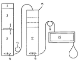
- a sketchy example of a closed device for carrying out the method according to the invention will now be discussed with the aid of the figure.
- Some of the individual device components are those that are available on the market.
- the interconnection of these devices in accordance with the new, compressed and, surprisingly, biologically very effective process described here corresponds to the process sequence according to the invention and thus represents a device for the production of prostheses according to claim 1.
- the device can be designed such that it is also used in an operating room can be made and the manufacture of prostheses according to the invention possible on site.
- the figure shows in a schematically simplified manner a completely lockable device in which the entire method can be carried out in a coherent, closed vessel and hose system. It can be easily arranged on a mobile frame and is therefore mobile for use in the laboratory or in the operating room, for example.
- the visual coherence naturally does not exclude the supply of tissue to be processed, reagents and solutions. But the unity should show how one can meet the very strict sterility requirements.
- a first unit comprises the devices for mechanical comminution, a drive 1, a conveyor section 3, which is aimed at a pair of knives / perforated plates 5, 7, and a digesting cell 9 with a stirrer 10 for support to disintegrate the tissue material to release the cells digestion and a first sedimentation.
- the devices can be switched so that the shredded tissue mass immediately falls from the perforated plate into the digestive vessel. After digestion is complete, sedimentation can also take place in this vessel in order to separate the oily phase. To this end, the vessel can be filled with buffer at the start of sedimentation to support the separation. Furthermore, a slight movement of 0.1-1 U / sec can be generated with the same target, for example with a magnetic stirrer.
- the suspension formed in the first unit is conveyed by a conveying means 13 into a second unit, which consists of a filter part 15 and a collecting vessel 17. During the conveyance into the vessel 17, a slight flow is also generated in the vessel 9.
- the filter section here is a multi-stage filter for the extraction of particles of a certain size, which are collected as a suspension in the collecting vessel. In this form, the sedimentation tendency of the suspension is only slight and is canceled in the vessel 17 by a second magnetic stirrer 18.
- Another funding 19 transfers the ready-to-use, homogeneous suspension into the third unit 25 for prosthesis coating.
- This unit preferably consists of a glove box in which the equipment required for coating is housed and the untreated prostheses are already ready for coating.
- the conditioning of the dry stored prosthesis with physiological solution takes place in this box 25, immediately before the cells are applied to the prostheses.
- the application / coating can be done by hand, but also by machine.
- the finished prostheses can, optionally with additional protection against Provide drying and adequately packaged, removed from the sterile interior of the coating box by means of a lock and safely transferred to the sterile operating table with the help of other special devices.
- it can be equipped for the series production of prostheses or for the ad hoc production of individual prostheses in the operating room.
- the filter 15 can also be connected without the vessel 17 between the vessel 9 and the coating unit, in such a way that the suspension is guided by the filter directly onto the prosthesis without intermediate storage.
Landscapes
- Health & Medical Sciences (AREA)
- Veterinary Medicine (AREA)
- Life Sciences & Earth Sciences (AREA)
- Animal Behavior & Ethology (AREA)
- General Health & Medical Sciences (AREA)
- Public Health (AREA)
- Oral & Maxillofacial Surgery (AREA)
- Transplantation (AREA)
- Cardiology (AREA)
- Engineering & Computer Science (AREA)
- Biomedical Technology (AREA)
- Vascular Medicine (AREA)
- Heart & Thoracic Surgery (AREA)
- Chemical & Material Sciences (AREA)
- Epidemiology (AREA)
- Medicinal Chemistry (AREA)
- Pulmonology (AREA)
- Gastroenterology & Hepatology (AREA)
- Dermatology (AREA)
- Surgery (AREA)
- Hematology (AREA)
- Chemical Kinetics & Catalysis (AREA)
- Materials For Medical Uses (AREA)
- Prostheses (AREA)
- Medicines Containing Plant Substances (AREA)
- Steroid Compounds (AREA)
- Micro-Organisms Or Cultivation Processes Thereof (AREA)
Claims (12)
- Procédé d'obtention d'endoprothèses utilisables en particulier en médecine humaine, qui présentent des surfaces de contact pour le contact direct avec le sang et/ou la lymphe et des cellules et groupes de cellules déposés sur ces surfaces de contact et initiant la régénération d'une couche de surface vivante, le procédé résidant dans le fait que des particules tissulaires sont produites par hachage à partir de tissu autogène qui présente, outre les cellules propres à régénérer la couche de surface, d'autres cellules et composants tissulaires non cellulaires, que ces particules sont mises en suspension et la suspension est filtrée à travers un support artificiel poreux préformé, caractérisé en ce qu'il comporte les étapes suivantes :a) des particules de tissu obtenues par hachage mécanique, d'une grosseur de 0,5 à 4 mm, sont encore réduites par digestion enzymatique dans une suspension de digestion et partiellement dégagées des composants non cellulaires du tissu, les particules de tissu prenant une dimension de moins de 1 mm ;b) la suspension digérée est débarrassée par filtration des particules dont la taille crée un risque pour le patient ;c) la suspension de particules tissulaires obtenue par filtration est filtrée à travers le support poreux sans étape de centrifugation préalable pour la décomposition de la suspension, les particules de tissu se déposant dans la région de la surface du support, etd) le support est rincé par l'injection sous pression d'une solution de rinçage à travers le support poreux, dans le même sens que la filtration a eu lieu dans l'étape c).
- Procédé selon la revendication 1, caractérisé en ce que l'on utilise comme tissu autogène l'épiploon ou du tissu adipeux sous-cutané.
- Procédé selon l'une quelconque des revendications 1 ou 2, caractérisé en ce que le hachage mécanique est réalisé par une pluralité de lames affûtées déplacées de façon corrélée.
- Procédé selon l'une ou l'ensemble des revendications 1 à 3, caractérisé en ce que la décantation de la suspension obtenue dans l'étape b) ou c) du procédé et l'utilisation de la couche la plus lourde permet de séparer les composants adipeux de la suspension.
- Procédé selon l'une ou l'ensemble des revendications 1 à 4, caractérisé en ce que la gravité ou une force centrifuge obtenue par rotation du support est exploitée dans l'étape d) du procédé.
- Procédé selon l'une ou l'ensemble des revendications 1 à 5, caractérisé en ce qu'en vue de l'élimination des particules tissulaires insuffisamment fixées, le support est traité après l'étape d) du procédé avec un courant de solution de rinçage orienté parallèlement à la surface traitée.
- Dispositif pour la mise en application du procédé selon la revendication 1, caractérisé en ce qu'il présente un récipient (9) doté de moyens d'agitation (10) pour la digestion enzymatique de particules de tissu, avec une entrée dans la région de laquelle est disposé un moyen (1, 3, 5, 7) pour le hachage mécanique de tissu en particules de tissu, et avec une sortie qui est disposée dans la partie inférieure du récipient (9) dans le sens de la gravité, en ce qu'il présente des moyens(25) pour la filtration d'une suspension de particules de tissu à travers un support artificiel poreux préformé et pour le rinçage du support dans le même sens, la sortie du récipient (9) et les moyens(25) pour la filtration étant reliés entre eux par une conduite par laquelle une suspension de particules de tissus peut être acheminée et dans lequel il est prévu entre le récipient (9) pour la digestion enzymatique et les moyens (25) pour la filtration une unité de filtrage (15) destinée à retenir des particules qui constitueraient un risque pour un patient du fait de leur taille, le dispositif constituant un système pouvant être fermé, sans moyens de centrifugation pour la séparation de la suspension digérée entre le récipient (9) de digestion enzymatique et les moyens (25) de filtration.
- Dispositif selon la revendication 7, caractérisé en ce que l'unité de filtrage (15) est disposée dans un autre récipient (17) pourvu d'un agitateur (18).
- Dispositif selon l'une quelconque des revendications 7 ou 8, caractérisé en ce que les moyens d'agitation (10, 18) sont des agitateurs magnétiques.
- Dispositif selon l'une ou l'ensemble des revendications 7 à 9, caractérisé en ce que les conduites entre le récipient (9) et les moyens (25) de filtration sont des tuyaux à usage unique et en ce que des pompes péristaltiques (13, 19) sont prévues pour le transport de la suspension.
- Utilisation du procédé et du dispositif selon l'une ou l'ensemble des revendications 1 à 10 pour l'obtention de prothèses vasculaires.
- Utilisation du procédé et du dispositif selon l'une ou l'ensemble des revendications 1 à 10 pour l'obtention de valvules mitrales.
Applications Claiming Priority (2)
| Application Number | Priority Date | Filing Date | Title |
|---|---|---|---|
| CH1498/92 | 1992-05-11 | ||
| CH149892 | 1992-05-11 |
Publications (2)
| Publication Number | Publication Date |
|---|---|
| EP0570331A1 EP0570331A1 (fr) | 1993-11-18 |
| EP0570331B1 true EP0570331B1 (fr) | 1997-09-10 |
Family
ID=4211862
Family Applications (1)
| Application Number | Title | Priority Date | Filing Date |
|---|---|---|---|
| EP93810297A Expired - Lifetime EP0570331B1 (fr) | 1992-05-11 | 1993-04-22 | Méthode et appareil pour obtenir des prothèses vasculaires |
Country Status (10)
| Country | Link |
|---|---|
| US (1) | US5634879A (fr) |
| EP (1) | EP0570331B1 (fr) |
| JP (1) | JPH0686809A (fr) |
| KR (1) | KR930023017A (fr) |
| AT (1) | ATE157856T1 (fr) |
| AU (1) | AU665813B2 (fr) |
| DE (1) | DE59307303D1 (fr) |
| DK (1) | DK0570331T3 (fr) |
| ES (1) | ES2107646T3 (fr) |
| GR (1) | GR3024888T3 (fr) |
Cited By (10)
| Publication number | Priority date | Publication date | Assignee | Title |
|---|---|---|---|---|
| US8691216B2 (en) | 2001-12-07 | 2014-04-08 | Cytori Therapeutics, Inc. | Methods of using regenerative cells to promote wound healing |
| US8771678B2 (en) | 2001-12-07 | 2014-07-08 | Cytori Therapeutics, Inc. | Methods of using adipose tissue-derived cells in augmenting autologous fat transfer |
| US8784801B2 (en) | 2008-08-19 | 2014-07-22 | Cytori Therapeutics, Inc. | Methods of using adipose tissue-derived cells in the treatment of the lymphatic system and malignant disease |
| US8883499B2 (en) | 2001-12-07 | 2014-11-11 | Cytori Therapeutics, Inc. | Systems and methods for isolating and using clinically safe adipose derived regenerative cells |
| US9133431B2 (en) | 2009-05-01 | 2015-09-15 | Bimini Technologies Llc | Systems, methods and compositions for optimizing tissue and cell enriched grafts |
| US9198937B2 (en) | 2001-12-07 | 2015-12-01 | Cytori Therapeutics, Inc. | Adipose-derived regenerative cells for treating liver injury |
| US9463203B2 (en) | 2001-12-07 | 2016-10-11 | Cytori Therapeutics, Inc. | Methods of using regenerative cells in the treatment of cartilage defects |
| US9480718B2 (en) | 2001-12-07 | 2016-11-01 | Cytori Therapeutics, Inc. | Methods of using adipose-derived regenerative cells in the treatment of peripheral vascular disease and related disorders |
| US9597395B2 (en) | 2001-12-07 | 2017-03-21 | Cytori Therapeutics, Inc. | Methods of using adipose tissue-derived cells in the treatment of cardiovascular conditions |
| US11395865B2 (en) | 2004-02-09 | 2022-07-26 | DePuy Synthes Products, Inc. | Scaffolds with viable tissue |
Families Citing this family (19)
| Publication number | Priority date | Publication date | Assignee | Title |
|---|---|---|---|---|
| WO1996001641A1 (fr) * | 1994-07-08 | 1996-01-25 | Sulzer Medizinaltechnik Ag | Procede de fabrication de materiaux pour greffe |
| FR2722974B1 (fr) * | 1994-07-29 | 1997-04-25 | Marie Therese Zabot | Procede de modification de la surface interne des protheses synthetiques utilisees en chirurgie vasculaire |
| US5976104A (en) * | 1994-08-19 | 1999-11-02 | Lifenet Research Foundation | Recirculation method for cleaning essentially intact bone grafts using pressure mediated flow of solutions and bone grafts produced thereby |
| US6206917B1 (en) * | 1997-05-02 | 2001-03-27 | St. Jude Medical, Inc. | Differential treatment of prosthetic devices |
| DE19834396C2 (de) * | 1998-07-30 | 2000-07-13 | Daimlerchrysler Aerospace Ag | Verfahren zur Oberflächenbeschichtung medizinischer Implantate |
| EP1392333A2 (fr) * | 2000-08-03 | 2004-03-03 | The University of Utah Research Foundation | Manipulation de l'identite arterioveineuse |
| US7514075B2 (en) | 2001-12-07 | 2009-04-07 | Cytori Therapeutics, Inc. | Systems and methods for separating and concentrating adipose derived stem cells from tissue |
| US7595043B2 (en) | 2001-12-07 | 2009-09-29 | Cytori Therapeutics, Inc. | Method for processing and using adipose-derived stem cells |
| US8404229B2 (en) | 2001-12-07 | 2013-03-26 | Cytori Therapeutics, Inc. | Methods of using adipose derived stem cells to treat acute tubular necrosis |
| US7744597B2 (en) | 2002-06-26 | 2010-06-29 | Lifenet Health | Device and process for producing fiber products and fiber products produced thereby |
| US7494811B2 (en) | 2003-05-01 | 2009-02-24 | Lifenet Health | In vitro growth of tissues suitable to the formation of bone and bone forming tissue formed thereby |
| US20050055085A1 (en) * | 2003-09-04 | 2005-03-10 | Rivron Nicolas C. | Implantable medical devices having recesses |
| US7759120B2 (en) * | 2005-03-02 | 2010-07-20 | Kps Bay Medical, Inc. | Seeding implantable medical devices with cells |
| US20060199265A1 (en) * | 2005-03-02 | 2006-09-07 | Wolf Michael F | Seeding implantable medical devices with cells |
| WO2008124494A1 (fr) * | 2007-04-03 | 2008-10-16 | The Cleveland Clinic Foundation | Enrichissement de cellules souches adultes dérivées du tissu sur la base de matériau matriciel extracellulaire retenu |
| US20100112696A1 (en) * | 2008-11-03 | 2010-05-06 | Baxter International Inc. | Apparatus And Methods For Processing Tissue To Release Cells |
| US8309343B2 (en) | 2008-12-01 | 2012-11-13 | Baxter International Inc. | Apparatus and method for processing biological material |
| GB0901207D0 (en) | 2009-01-26 | 2009-03-11 | Innospec Ltd | Chelating agents and methods relating thereto |
| IT1400069B1 (it) * | 2010-05-20 | 2013-05-17 | Tremolada | Dispositivo e metodo per la preparazione di tessuto, in particolare tessuto adiposo per trapianto ottenuto da materiale adiposo lobulare estratto tramite liposuzione |
Citations (3)
| Publication number | Priority date | Publication date | Assignee | Title |
|---|---|---|---|---|
| EP0393788A2 (fr) * | 1989-04-17 | 1990-10-24 | Koken Co. Ltd. | Prothèse vasculaire et procédé de production pour prothèse vasculaire |
| US5035708A (en) * | 1985-06-06 | 1991-07-30 | Thomas Jefferson University | Endothelial cell procurement and deposition kit |
| EP0462051A1 (fr) * | 1990-06-15 | 1991-12-18 | SULZER Medizinaltechnik AG | Procédé pour fabriquer des prothèses tubulaires poreuses en matière plastique, chargées de cellules vivantes |
Family Cites Families (4)
| Publication number | Priority date | Publication date | Assignee | Title |
|---|---|---|---|---|
| US5230693A (en) * | 1985-06-06 | 1993-07-27 | Thomas Jefferson University | Implantable prosthetic device for implantation into a human patient having a surface treated with microvascular endothelial cells |
| CH670759A5 (fr) * | 1986-06-02 | 1989-07-14 | Sulzer Ag | |
| EP0444270A1 (fr) * | 1990-02-26 | 1991-09-04 | Thomas Jefferson University | Dispositif pour la déposition des cellules endothéliales |
| NZ237832A (en) * | 1990-04-17 | 1994-05-26 | Curative Tech Inc | Coating a prosthetic surface with mammalian cells |
-
1993
- 1993-04-07 AU AU36771/93A patent/AU665813B2/en not_active Ceased
- 1993-04-22 DK DK93810297.7T patent/DK0570331T3/da not_active Application Discontinuation
- 1993-04-22 AT AT93810297T patent/ATE157856T1/de not_active IP Right Cessation
- 1993-04-22 DE DE59307303T patent/DE59307303D1/de not_active Expired - Fee Related
- 1993-04-22 EP EP93810297A patent/EP0570331B1/fr not_active Expired - Lifetime
- 1993-04-22 ES ES93810297T patent/ES2107646T3/es not_active Expired - Lifetime
- 1993-04-30 KR KR1019930007445A patent/KR930023017A/ko not_active Ceased
- 1993-05-04 US US08/056,746 patent/US5634879A/en not_active Expired - Lifetime
- 1993-05-11 JP JP5109491A patent/JPH0686809A/ja active Pending
-
1997
- 1997-09-30 GR GR970402535T patent/GR3024888T3/el unknown
Patent Citations (4)
| Publication number | Priority date | Publication date | Assignee | Title |
|---|---|---|---|---|
| US5035708A (en) * | 1985-06-06 | 1991-07-30 | Thomas Jefferson University | Endothelial cell procurement and deposition kit |
| EP0393788A2 (fr) * | 1989-04-17 | 1990-10-24 | Koken Co. Ltd. | Prothèse vasculaire et procédé de production pour prothèse vasculaire |
| US5171261A (en) * | 1989-04-17 | 1992-12-15 | Koken Co., Ltd. | Vascular prosthesis, manufacturing method of the same, and substrate for vascular prothesis |
| EP0462051A1 (fr) * | 1990-06-15 | 1991-12-18 | SULZER Medizinaltechnik AG | Procédé pour fabriquer des prothèses tubulaires poreuses en matière plastique, chargées de cellules vivantes |
Cited By (15)
| Publication number | Priority date | Publication date | Assignee | Title |
|---|---|---|---|---|
| US9463203B2 (en) | 2001-12-07 | 2016-10-11 | Cytori Therapeutics, Inc. | Methods of using regenerative cells in the treatment of cartilage defects |
| US9480718B2 (en) | 2001-12-07 | 2016-11-01 | Cytori Therapeutics, Inc. | Methods of using adipose-derived regenerative cells in the treatment of peripheral vascular disease and related disorders |
| US9597395B2 (en) | 2001-12-07 | 2017-03-21 | Cytori Therapeutics, Inc. | Methods of using adipose tissue-derived cells in the treatment of cardiovascular conditions |
| US8883499B2 (en) | 2001-12-07 | 2014-11-11 | Cytori Therapeutics, Inc. | Systems and methods for isolating and using clinically safe adipose derived regenerative cells |
| US9511094B2 (en) | 2001-12-07 | 2016-12-06 | Cytori Therapeutics, Inc. | Methods of using regenerative cells in the treatment of stroke and related diseases and disorders |
| US9198937B2 (en) | 2001-12-07 | 2015-12-01 | Cytori Therapeutics, Inc. | Adipose-derived regenerative cells for treating liver injury |
| US8771678B2 (en) | 2001-12-07 | 2014-07-08 | Cytori Therapeutics, Inc. | Methods of using adipose tissue-derived cells in augmenting autologous fat transfer |
| US9504716B2 (en) | 2001-12-07 | 2016-11-29 | Cytori Therapeutics, Inc. | Methods of using adipose derived regenerative cells to promote restoration of intevertebral disc |
| US8691216B2 (en) | 2001-12-07 | 2014-04-08 | Cytori Therapeutics, Inc. | Methods of using regenerative cells to promote wound healing |
| US9492483B2 (en) | 2001-12-07 | 2016-11-15 | Cytori Therapeutics, Inc. | Methods of using regenerative cells to treat a burn |
| US9511096B2 (en) | 2001-12-07 | 2016-12-06 | Cytori Therapeutics, Inc. | Methods of using regenerative cells to treat an ischemic wound |
| US11395865B2 (en) | 2004-02-09 | 2022-07-26 | DePuy Synthes Products, Inc. | Scaffolds with viable tissue |
| US9486484B2 (en) | 2008-08-19 | 2016-11-08 | Cytori Therapeutics, Inc. | Methods of using adipose tissue-derived cells in the treatment of the lymphatic system and malignant disease |
| US8784801B2 (en) | 2008-08-19 | 2014-07-22 | Cytori Therapeutics, Inc. | Methods of using adipose tissue-derived cells in the treatment of the lymphatic system and malignant disease |
| US9133431B2 (en) | 2009-05-01 | 2015-09-15 | Bimini Technologies Llc | Systems, methods and compositions for optimizing tissue and cell enriched grafts |
Also Published As
| Publication number | Publication date |
|---|---|
| ATE157856T1 (de) | 1997-09-15 |
| JPH0686809A (ja) | 1994-03-29 |
| US5634879A (en) | 1997-06-03 |
| AU3677193A (en) | 1993-11-18 |
| AU665813B2 (en) | 1996-01-18 |
| DE59307303D1 (de) | 1997-10-16 |
| DK0570331T3 (da) | 1997-12-15 |
| GR3024888T3 (en) | 1998-01-30 |
| ES2107646T3 (es) | 1997-12-01 |
| KR930023017A (ko) | 1993-12-18 |
| EP0570331A1 (fr) | 1993-11-18 |
Similar Documents
| Publication | Publication Date | Title |
|---|---|---|
| EP0570331B1 (fr) | Méthode et appareil pour obtenir des prothèses vasculaires | |
| DE68911178T2 (de) | Klinische Entwicklungen mit Amnionzellen. | |
| DE69333466T2 (de) | Herstellung von transplantatgeweben aus extrazellulärer matrix | |
| US5837235A (en) | Process for regenerating bone and cartilage | |
| DE3891033C2 (de) | Verwendung von mikrovaskularen Endothelzellen zur Reendothelisierung des von Endothelzellen im wesentlichen entkleideten Gefäßkanals eines Patienten | |
| DE69018246T2 (de) | Vorrichtung zur Sammlung und Verarbeitung von fetten Geweben zur Herstellung von Endothel-Zellprodukt. | |
| DE69626035T2 (de) | Ein biologisches material, das eine wirksame kultur von knochenmarkstammzellen, die teilweise oder vollständing in bindegewebszellen differenziert sind, enthält, und eine dreidimensionale, bioverträgliche und biologische abbaubare matrix, die aus einen hyaluronsäurederivat besteht | |
| KR101495281B1 (ko) | 피부 재생 또는 상처 치유를 위한 중간엽 줄기세포-하이드로겔-생분해성 또는 중간엽 줄기세포-하이드로겔-비분해성 지지체 조성물 | |
| DE60302820T2 (de) | Autologes knochenersatzmaterial | |
| JP5866203B2 (ja) | 組織産生のための方法および手段ならびに得られる組織 | |
| JP7105898B2 (ja) | 真皮再生シート用バイオインク組成物、これを用いたオーダーメード型真皮再生シートの製造方法、および該製造方法を用いて製造されたオーダーメード型真皮再生シート | |
| EP3319652B1 (fr) | Procédé pour produire une construction bioartificielle primairement acellulaire à base de fibrine et construction correspondante | |
| EP1286708A1 (fr) | Procede de production d'un transplant bio-artificiel | |
| KR20190036882A (ko) | 생체 이식 또는 삽입용 무세포 진피의 제조방법 | |
| DE69820254T2 (de) | Dermales hüllgewebe in der wundheilung | |
| EP1085919B1 (fr) | Epithelialisation ou endothelialisation autologue d'organes creux ou de vaisseaux | |
| EP1230939B2 (fr) | Matrice tissulaire bioartificielle vascularisée et tissu bioartificiel vascularisé | |
| EP1172120B1 (fr) | Prothèse indiviuelle de valvule véneuse | |
| WO2008092440A2 (fr) | Composition thérapeutique et utilisation d'une substance acellulaire | |
| RU2835053C1 (ru) | Способ стимуляции заживления обширных ожогов | |
| WO2005097154A2 (fr) | Melange de messagers favorisant la cicatrisation | |
| WO2013037349A1 (fr) | Procédé de fabrication d'un produit de synthèse tissulaire biologique et utilisation de cellules autologues obtenues de façon spécifique | |
| CN106110402A (zh) | 海藻酸钠水凝胶支架构建的组织工程皮肤及其制备方法 | |
| CN119701094A (zh) | 一种促血管化同种异体神经修复材料及其制备方法 | |
| DE102004043449B4 (de) | Verfahren zur Herstellung von Bandscheibenzelltransplantaten und deren Anwendung als Transplantationsmaterial |
Legal Events
| Date | Code | Title | Description |
|---|---|---|---|
| PUAI | Public reference made under article 153(3) epc to a published international application that has entered the european phase |
Free format text: ORIGINAL CODE: 0009012 |
|
| AK | Designated contracting states |
Kind code of ref document: A1 Designated state(s): AT BE CH DE DK ES FR GB GR IE IT LI NL PT SE |
|
| 17P | Request for examination filed |
Effective date: 19940425 |
|
| 17Q | First examination report despatched |
Effective date: 19951222 |
|
| GRAG | Despatch of communication of intention to grant |
Free format text: ORIGINAL CODE: EPIDOS AGRA |
|
| GRAH | Despatch of communication of intention to grant a patent |
Free format text: ORIGINAL CODE: EPIDOS IGRA |
|
| GRAH | Despatch of communication of intention to grant a patent |
Free format text: ORIGINAL CODE: EPIDOS IGRA |
|
| ITF | It: translation for a ep patent filed | ||
| GRAA | (expected) grant |
Free format text: ORIGINAL CODE: 0009210 |
|
| AK | Designated contracting states |
Kind code of ref document: B1 Designated state(s): AT BE CH DE DK ES FR GB GR IE IT LI NL PT SE |
|
| REF | Corresponds to: |
Ref document number: 157856 Country of ref document: AT Date of ref document: 19970915 Kind code of ref document: T |
|
| REG | Reference to a national code |
Ref country code: CH Ref legal event code: EP |
|
| REG | Reference to a national code |
Ref country code: CH Ref legal event code: NV Representative=s name: FREI PATENTANWALTSBUERO |
|
| GBT | Gb: translation of ep patent filed (gb section 77(6)(a)/1977) |
Effective date: 19970912 |
|
| REF | Corresponds to: |
Ref document number: 59307303 Country of ref document: DE Date of ref document: 19971016 |
|
| ET | Fr: translation filed | ||
| REG | Reference to a national code |
Ref country code: ES Ref legal event code: FG2A Ref document number: 2107646 Country of ref document: ES Kind code of ref document: T3 |
|
| REG | Reference to a national code |
Ref country code: DK Ref legal event code: T3 |
|
| REG | Reference to a national code |
Ref country code: GR Ref legal event code: FG4A Free format text: 3024888 |
|
| REG | Reference to a national code |
Ref country code: IE Ref legal event code: FG4D Free format text: 76347 |
|
| REG | Reference to a national code |
Ref country code: PT Ref legal event code: SC4A Free format text: AVAILABILITY OF NATIONAL TRANSLATION Effective date: 19971031 |
|
| PLBE | No opposition filed within time limit |
Free format text: ORIGINAL CODE: 0009261 |
|
| STAA | Information on the status of an ep patent application or granted ep patent |
Free format text: STATUS: NO OPPOSITION FILED WITHIN TIME LIMIT |
|
| 26N | No opposition filed | ||
| PGFP | Annual fee paid to national office [announced via postgrant information from national office to epo] |
Ref country code: PT Payment date: 20000417 Year of fee payment: 8 Ref country code: CH Payment date: 20000417 Year of fee payment: 8 |
|
| PGFP | Annual fee paid to national office [announced via postgrant information from national office to epo] |
Ref country code: DK Payment date: 20000418 Year of fee payment: 8 |
|
| PGFP | Annual fee paid to national office [announced via postgrant information from national office to epo] |
Ref country code: BE Payment date: 20000419 Year of fee payment: 8 |
|
| PGFP | Annual fee paid to national office [announced via postgrant information from national office to epo] |
Ref country code: AT Payment date: 20000421 Year of fee payment: 8 |
|
| PGFP | Annual fee paid to national office [announced via postgrant information from national office to epo] |
Ref country code: SE Payment date: 20000425 Year of fee payment: 8 Ref country code: IE Payment date: 20000425 Year of fee payment: 8 |
|
| PGFP | Annual fee paid to national office [announced via postgrant information from national office to epo] |
Ref country code: GR Payment date: 20000426 Year of fee payment: 8 |
|
| PGFP | Annual fee paid to national office [announced via postgrant information from national office to epo] |
Ref country code: NL Payment date: 20000428 Year of fee payment: 8 |
|
| PG25 | Lapsed in a contracting state [announced via postgrant information from national office to epo] |
Ref country code: DK Free format text: LAPSE BECAUSE OF NON-PAYMENT OF DUE FEES Effective date: 20010422 Ref country code: AT Free format text: LAPSE BECAUSE OF NON-PAYMENT OF DUE FEES Effective date: 20010422 |
|
| PG25 | Lapsed in a contracting state [announced via postgrant information from national office to epo] |
Ref country code: SE Free format text: LAPSE BECAUSE OF NON-PAYMENT OF DUE FEES Effective date: 20010423 Ref country code: IE Free format text: LAPSE BECAUSE OF NON-PAYMENT OF DUE FEES Effective date: 20010423 |
|
| PG25 | Lapsed in a contracting state [announced via postgrant information from national office to epo] |
Ref country code: GR Free format text: LAPSE BECAUSE OF NON-PAYMENT OF DUE FEES Effective date: 20010430 Ref country code: BE Free format text: LAPSE BECAUSE OF NON-PAYMENT OF DUE FEES Effective date: 20010430 |
|
| PG25 | Lapsed in a contracting state [announced via postgrant information from national office to epo] |
Ref country code: LI Free format text: LAPSE BECAUSE OF NON-PAYMENT OF DUE FEES Effective date: 20010521 Ref country code: CH Free format text: LAPSE BECAUSE OF NON-PAYMENT OF DUE FEES Effective date: 20010521 |
|
| BERE | Be: lapsed |
Owner name: SULZER MEDIZINALTECHNIK A.G. Effective date: 20010430 |
|
| PG25 | Lapsed in a contracting state [announced via postgrant information from national office to epo] |
Ref country code: PT Free format text: LAPSE BECAUSE OF NON-PAYMENT OF DUE FEES Effective date: 20011031 |
|
| PG25 | Lapsed in a contracting state [announced via postgrant information from national office to epo] |
Ref country code: NL Free format text: LAPSE BECAUSE OF NON-PAYMENT OF DUE FEES Effective date: 20011101 |
|
| EUG | Se: european patent has lapsed |
Ref document number: 93810297.7 |
|
| REG | Reference to a national code |
Ref country code: DK Ref legal event code: EBP |
|
| REG | Reference to a national code |
Ref country code: CH Ref legal event code: PL |
|
| REG | Reference to a national code |
Ref country code: GB Ref legal event code: IF02 |
|
| NLV4 | Nl: lapsed or anulled due to non-payment of the annual fee |
Effective date: 20011101 |
|
| REG | Reference to a national code |
Ref country code: IE Ref legal event code: MM4A |
|
| REG | Reference to a national code |
Ref country code: PT Ref legal event code: MM4A Free format text: LAPSE DUE TO NON-PAYMENT OF FEES Effective date: 20011031 |
|
| PGFP | Annual fee paid to national office [announced via postgrant information from national office to epo] |
Ref country code: GB Payment date: 20020422 Year of fee payment: 10 |
|
| PGFP | Annual fee paid to national office [announced via postgrant information from national office to epo] |
Ref country code: FR Payment date: 20020426 Year of fee payment: 10 |
|
| PGFP | Annual fee paid to national office [announced via postgrant information from national office to epo] |
Ref country code: ES Payment date: 20020429 Year of fee payment: 10 |
|
| PGFP | Annual fee paid to national office [announced via postgrant information from national office to epo] |
Ref country code: DE Payment date: 20020521 Year of fee payment: 10 |
|
| PG25 | Lapsed in a contracting state [announced via postgrant information from national office to epo] |
Ref country code: GB Free format text: LAPSE BECAUSE OF NON-PAYMENT OF DUE FEES Effective date: 20030422 |
|
| PG25 | Lapsed in a contracting state [announced via postgrant information from national office to epo] |
Ref country code: ES Free format text: LAPSE BECAUSE OF NON-PAYMENT OF DUE FEES Effective date: 20030423 |
|
| PG25 | Lapsed in a contracting state [announced via postgrant information from national office to epo] |
Ref country code: DE Free format text: LAPSE BECAUSE OF NON-PAYMENT OF DUE FEES Effective date: 20031101 |
|
| GBPC | Gb: european patent ceased through non-payment of renewal fee |
Effective date: 20030422 |
|
| PG25 | Lapsed in a contracting state [announced via postgrant information from national office to epo] |
Ref country code: FR Free format text: LAPSE BECAUSE OF NON-PAYMENT OF DUE FEES Effective date: 20031231 |
|
| REG | Reference to a national code |
Ref country code: FR Ref legal event code: ST |
|
| REG | Reference to a national code |
Ref country code: ES Ref legal event code: FD2A Effective date: 20030423 |
|
| PG25 | Lapsed in a contracting state [announced via postgrant information from national office to epo] |
Ref country code: IT Free format text: LAPSE BECAUSE OF NON-PAYMENT OF DUE FEES;WARNING: LAPSES OF ITALIAN PATENTS WITH EFFECTIVE DATE BEFORE 2007 MAY HAVE OCCURRED AT ANY TIME BEFORE 2007. THE CORRECT EFFECTIVE DATE MAY BE DIFFERENT FROM THE ONE RECORDED. Effective date: 20050422 |