EP0574512B1 - Änderung der zellularen immunität durch rezeptorchimäre - Google Patents
Änderung der zellularen immunität durch rezeptorchimäre Download PDFInfo
- Publication number
- EP0574512B1 EP0574512B1 EP92907958A EP92907958A EP0574512B1 EP 0574512 B1 EP0574512 B1 EP 0574512B1 EP 92907958 A EP92907958 A EP 92907958A EP 92907958 A EP92907958 A EP 92907958A EP 0574512 B1 EP0574512 B1 EP 0574512B1
- Authority
- EP
- European Patent Office
- Prior art keywords
- cell
- cells
- receptor
- seq
- chimeras
- Prior art date
- Legal status (The legal status is an assumption and is not a legal conclusion. Google has not performed a legal analysis and makes no representation as to the accuracy of the status listed.)
- Expired - Lifetime
Links
Images










Classifications
-
- C—CHEMISTRY; METALLURGY
- C12—BIOCHEMISTRY; BEER; SPIRITS; WINE; VINEGAR; MICROBIOLOGY; ENZYMOLOGY; MUTATION OR GENETIC ENGINEERING
- C12N—MICROORGANISMS OR ENZYMES; COMPOSITIONS THEREOF; PROPAGATING, PRESERVING, OR MAINTAINING MICROORGANISMS; MUTATION OR GENETIC ENGINEERING; CULTURE MEDIA
- C12N9/00—Enzymes; Proenzymes; Compositions thereof; Processes for preparing, activating, inhibiting, separating or purifying enzymes
- C12N9/10—Transferases (2.)
- C12N9/12—Transferases (2.) transferring phosphorus containing groups, e.g. kinases (2.7)
- C12N9/1205—Phosphotransferases with an alcohol group as acceptor (2.7.1), e.g. protein kinases
-
- A—HUMAN NECESSITIES
- A61—MEDICAL OR VETERINARY SCIENCE; HYGIENE
- A61K—PREPARATIONS FOR MEDICAL, DENTAL OR TOILETRY PURPOSES
- A61K35/00—Medicinal preparations containing materials or reaction products thereof with undetermined constitution
- A61K35/12—Materials from mammals; Compositions comprising non-specified tissues or cells; Compositions comprising non-embryonic stem cells; Genetically modified cells
- A61K35/13—Tumour cells, irrespective of tissue of origin
-
- A—HUMAN NECESSITIES
- A61—MEDICAL OR VETERINARY SCIENCE; HYGIENE
- A61K—PREPARATIONS FOR MEDICAL, DENTAL OR TOILETRY PURPOSES
- A61K35/00—Medicinal preparations containing materials or reaction products thereof with undetermined constitution
- A61K35/12—Materials from mammals; Compositions comprising non-specified tissues or cells; Compositions comprising non-embryonic stem cells; Genetically modified cells
- A61K35/48—Reproductive organs
- A61K35/54—Ovaries; Ova; Ovules; Embryos; Foetal cells; Germ cells
- A61K35/545—Embryonic stem cells; Pluripotent stem cells; Induced pluripotent stem cells; Uncharacterised stem cells
-
- A—HUMAN NECESSITIES
- A61—MEDICAL OR VETERINARY SCIENCE; HYGIENE
- A61K—PREPARATIONS FOR MEDICAL, DENTAL OR TOILETRY PURPOSES
- A61K40/00—Cellular immunotherapy
- A61K40/10—Cellular immunotherapy characterised by the cell type used
- A61K40/11—T-cells, e.g. tumour infiltrating lymphocytes [TIL] or regulatory T [Treg] cells; Lymphokine-activated killer [LAK] cells
-
- A—HUMAN NECESSITIES
- A61—MEDICAL OR VETERINARY SCIENCE; HYGIENE
- A61K—PREPARATIONS FOR MEDICAL, DENTAL OR TOILETRY PURPOSES
- A61K40/00—Cellular immunotherapy
- A61K40/40—Cellular immunotherapy characterised by antigens that are targeted or presented by cells of the immune system
- A61K40/46—Viral antigens
-
- A—HUMAN NECESSITIES
- A61—MEDICAL OR VETERINARY SCIENCE; HYGIENE
- A61P—SPECIFIC THERAPEUTIC ACTIVITY OF CHEMICAL COMPOUNDS OR MEDICINAL PREPARATIONS
- A61P35/00—Antineoplastic agents
-
- A—HUMAN NECESSITIES
- A61—MEDICAL OR VETERINARY SCIENCE; HYGIENE
- A61P—SPECIFIC THERAPEUTIC ACTIVITY OF CHEMICAL COMPOUNDS OR MEDICINAL PREPARATIONS
- A61P37/00—Drugs for immunological or allergic disorders
-
- C—CHEMISTRY; METALLURGY
- C07—ORGANIC CHEMISTRY
- C07K—PEPTIDES
- C07K14/00—Peptides having more than 20 amino acids; Gastrins; Somatostatins; Melanotropins; Derivatives thereof
- C07K14/435—Peptides having more than 20 amino acids; Gastrins; Somatostatins; Melanotropins; Derivatives thereof from animals; from humans
- C07K14/705—Receptors; Cell surface antigens; Cell surface determinants
-
- C—CHEMISTRY; METALLURGY
- C07—ORGANIC CHEMISTRY
- C07K—PEPTIDES
- C07K14/00—Peptides having more than 20 amino acids; Gastrins; Somatostatins; Melanotropins; Derivatives thereof
- C07K14/435—Peptides having more than 20 amino acids; Gastrins; Somatostatins; Melanotropins; Derivatives thereof from animals; from humans
- C07K14/705—Receptors; Cell surface antigens; Cell surface determinants
- C07K14/70503—Immunoglobulin superfamily
-
- C—CHEMISTRY; METALLURGY
- C07—ORGANIC CHEMISTRY
- C07K—PEPTIDES
- C07K14/00—Peptides having more than 20 amino acids; Gastrins; Somatostatins; Melanotropins; Derivatives thereof
- C07K14/435—Peptides having more than 20 amino acids; Gastrins; Somatostatins; Melanotropins; Derivatives thereof from animals; from humans
- C07K14/705—Receptors; Cell surface antigens; Cell surface determinants
- C07K14/70503—Immunoglobulin superfamily
- C07K14/7051—T-cell receptor (TcR)-CD3 complex
-
- C—CHEMISTRY; METALLURGY
- C07—ORGANIC CHEMISTRY
- C07K—PEPTIDES
- C07K14/00—Peptides having more than 20 amino acids; Gastrins; Somatostatins; Melanotropins; Derivatives thereof
- C07K14/435—Peptides having more than 20 amino acids; Gastrins; Somatostatins; Melanotropins; Derivatives thereof from animals; from humans
- C07K14/705—Receptors; Cell surface antigens; Cell surface determinants
- C07K14/70503—Immunoglobulin superfamily
- C07K14/70514—CD4
-
- C—CHEMISTRY; METALLURGY
- C07—ORGANIC CHEMISTRY
- C07K—PEPTIDES
- C07K14/00—Peptides having more than 20 amino acids; Gastrins; Somatostatins; Melanotropins; Derivatives thereof
- C07K14/435—Peptides having more than 20 amino acids; Gastrins; Somatostatins; Melanotropins; Derivatives thereof from animals; from humans
- C07K14/705—Receptors; Cell surface antigens; Cell surface determinants
- C07K14/70503—Immunoglobulin superfamily
- C07K14/70535—Fc-receptors, e.g. CD16, CD32, CD64 (CD2314/705F)
-
- C—CHEMISTRY; METALLURGY
- C07—ORGANIC CHEMISTRY
- C07K—PEPTIDES
- C07K19/00—Hybrid peptides, i.e. peptides covalently bound to nucleic acids, or non-covalently bound protein-protein complexes
-
- A—HUMAN NECESSITIES
- A61—MEDICAL OR VETERINARY SCIENCE; HYGIENE
- A61K—PREPARATIONS FOR MEDICAL, DENTAL OR TOILETRY PURPOSES
- A61K38/00—Medicinal preparations containing peptides
-
- C—CHEMISTRY; METALLURGY
- C07—ORGANIC CHEMISTRY
- C07K—PEPTIDES
- C07K2319/00—Fusion polypeptide
-
- C—CHEMISTRY; METALLURGY
- C07—ORGANIC CHEMISTRY
- C07K—PEPTIDES
- C07K2319/00—Fusion polypeptide
- C07K2319/01—Fusion polypeptide containing a localisation/targetting motif
- C07K2319/033—Fusion polypeptide containing a localisation/targetting motif containing a motif for targeting to the internal surface of the plasma membrane, e.g. containing a myristoylation motif
-
- C—CHEMISTRY; METALLURGY
- C07—ORGANIC CHEMISTRY
- C07K—PEPTIDES
- C07K2319/00—Fusion polypeptide
- C07K2319/32—Fusion polypeptide fusions with soluble part of a cell surface receptor, "decoy receptors"
Definitions
- the invention concerns functional T cell receptor, Fc receptor, or B cell receptor chimeras which are capable of redirecting immune system function. More particularly, it concerns the regulation of lymphocytes, macrophages, natural killer cells or granulocytes by the expression in said cells of chimeras which cause the cells to respond to targets recognized by the chimeras.
- the invention also concerns functional T cell receptor, Fc receptor, or B cell receptor chimeras which are capable of directing therapeutic cells to specifically recognize and destroy either cells infected with a specific infective agent, the infective agent itself, a tumor cell, or an autoimmune-generated cell.
- the invention relates to the production of T cell receptor or Fc receptor chimeras capable of directing cytotoxic T lymphocytes to specifically recognize and lyse cells expressing HIV envelope proteins.
- the invention therefore provides a therapy for diseases such as AIDS (Acquired Immunodeficiency Syndrome) which are caused by the HIV virus.
- T cell recognition of antigen through the T cell receptor is the basis of a range of immunological phenomena.
- the T cells direct what is called cell-mediated immunity. This involves the destruction by cells of the immune system of foreign tissues or infected cells.
- a T cell that recognizes and binds a unique antigen displayed on the surface of another cell becomes activated; it can then multiply, and if it is a cytotoxic cell, it can kill the bound cell.
- Autoimmune disease is characterized by production of either antibodies that react with host tissue or immune effector T cells that are autoreactive.
- autoantibodies may arise by a normal T- and B-cell response activated by foreign substances or organisms that contain antigens that cross react with similar compounds in body tissues.
- Examples of clinically relevant autoantibodies are antibodies against acetylcholine receptors in myasthenia gravis; and anti-DNA, anti-erythrocyte, and anti-platelet antibodies in systemic lupus erythematosus.
- HIV was shown to be the etiologic agent of AIDS. Since that time the definition of AIDS has been revised a number of times with regard to what criteria should be included in the diagnosis. However, despite the fluctuation in diagnostic parameters, the simple common denominator of AIDS is the infection with HIV and subsequent development of persistent constitutional symptoms and AIDS defining diseases such as a secondary infections, neoplasms, and neurologic disease. Harrison's Principles of Internal Medicine, 12th ed., McGraw Hill (1991).
- HIV is a human retrovirus of the lentivirus group.
- the four recognized human retroviruses belong to two distinct groups: the human T lymphotropic (or leukemia) retroviruses, HTLV-1 and HTLV-2, and the human immunodeficiency viruses, HIV-1 and HIV-2.
- the former are transforming viruses whereas the latter are cytopathic viruses.
- HIV-1 has been identified as the most common cause of AIDS throughout the world. sequence homology between HIV-2 and HIV-1 is about 40% with HIV-2 being more closely related to some members of a group of simian immunodeficiency viruses (SIV). See Curran, J. et al., Science , 329 :1357-1359 (1985); Weiss, R. et al., Nature , 324 :572-575 (1986).
- SIV simian immunodeficiency viruses
- HIV has the usual retroviral genes ( env , gag , and pol ) as well as six extra genes involved in the replication and other biologic activities of the virus.
- retroviral genes env , gag , and pol
- six extra genes involved in the replication and other biologic activities of the virus As stated previously, the common denominator of AIDS is a profound immunosuppression, predominantly of cell-mediated immunity. This immune suppression leads to a variety of opportunistic diseases, particularly certain infections and neoplasms.
- T4 lymphocytes The main cause of the immune defect in AIDS, has been identified as a quantitative and qualitative deficiency in the subset of thymus-derived (T) lymphocytes, the T4 population. This subset of cells is defined phenotypically by the presence of the CD4 surface molecule, which has been demonstrated to be the cellular receptor for HIV. Dalgleish et al., Nature , 312 :763 (1984). Although the T4 cell is the major cell type infected with HIV, essentially any human cell that expresses the CD4 molecule on its surface is capable of binding to and being infected with HIV.
- CD4 + T cells have been assigned the role of helper/inducer, indicating their function in providing an activating signal to B cells, or inducing T lymphocytes bearing the reciprocal CD8 marker to become cytotoxic/suppressor cells. Reinherz and Schlossman, Cell , 19 :821-827 (1980); Goldstein et al., Immunol. Rev. , 68 :5-42, (1982).
- HIV binds specifically and with high affinity, via a stretch of amino acids in the viral envelope (gp120), to a portion of the V1 region of the CD4 molecule located near its N-terminus.
- the virus fuses with the target cell membrane and is internalized. Once internalized it uses the enzyme reverse transcriptase to transcribe its genomic RNA to DNA, which is integrated into the cellular DNA where it exists for the life or the cell as a "provirus.”
- the provirus may remain latent or be activated to transcribe mRNA and genomic RNA, leading to protein synthesis, assembly, new virion formation, and budding of virus from the cell surface.
- the provirus may remain latent or be activated to transcribe mRNA and genomic RNA, leading to protein synthesis, assembly, new virion formation, and budding of virus from the cell surface.
- the host organism develops antibodies against viral proteins, including the major envelope glycoproteins gp120 and gp41.
- viral proteins including the major envelope glycoproteins gp120 and gp41.
- the disease progresses, resulting in a lethal immunosuppression characterized by multiple opportunistic infections, parasitemia, dementia and death.
- the failure of the host anti-viral antibodies to arrest the progression of the disease represents one of the most vexing and alarming aspects of the infection, and augurs poorly for vaccination efforts based upon conventional approaches.
- the immunodeficiency viruses Two factors may play a role in the efficacy of the humoral response to immunodeficiency viruses.
- the immunodeficiency viruses show a high mutation rate in response to host immune surveillance.
- the envelope glycoproteins themselves are heavily glycosylated molecules presenting few epitopes suitable for high affinity antibody binding. The poorly antigenic target which the viral envelope presents, allows the host little opportunity for restricting viral infection by specific antibody production.
- Gp120 mediates fusion events among CD4 + cells via a reaction similar to that by which the virus enters the uninfected cells, leading to the formation of short-lived multinucleated giant cells.
- Syncytium formation is dependent on a direct interaction of the gp120 envelope glycoprotein with the CD4 protein. Dalgleish et al., supra ; Klatzman, D. et al., Nature, 312 :763 (1984); McDougal, J.S. et al., Science , 231 :382 (1986); Sodroski, J.
- CD4-gp120 binding is responsible for viral infection of cells bearing the CD4 antigen includes the finding that a specific complex is formed between gp120 and CD4. McDougal et al., supra . Other investigators have shown that the cell lines, which were noninfective for HIV, were converted to infectable cell lines following transfection and expression of the human CD4 cDNA gene. Maddon et al., Cell , 46 :333-348 (1986).
- CD4 immunotoxin conjugates or fusion proteins show potent cytotoxicity for infected cells in vitro (Chaudhary et al., Nature , 335 :369-372 (1988); Till et al., Science , 242 :1166-1168 (1988)), the latency of the immunodeficiency syndrome makes it unlikely that any single-treatment therapy will be effective in eliminating viral burden, and the antigenicity of foreign fusion proteins is likely to limit their acceptability in treatments requiring repetitive dosing.
- T cell antigen receptor TCR
- TCR T cell antigen receptor
- ⁇ is the limiting polypeptide for surface expression of the complete complex (Sussman et al., Cell, 52 :85-95 (1988)) and is thought to mediate at least a fraction of the cellular activation programs triggered by receptor recognition of ligand (Weissman et al., EMBO J. , 8 :3651-3656 (1989); Frank et al., Science , 249 :174-177 (1990)).
- a 32kDa type I integral membrane homodimer, ⁇ (zeta) has a 9 residue extracellular domain with no sites for N-linked glycan addition, and a 112 residue (mouse) or 113 residue (human) intracellular domain (Weissman et al., Science , 238:1018-1020 (1988a); Weissman et al., Proc. Natl. Acad. Sci. USA , 85 :9709-9713 (1988b)).
- An isoform of ⁇ called ⁇ (eta) (Baniyash et al., J. Biol. Chem. , 263 :9874-9878 (1988); Orloff et al., J. Biol.
- the Fc receptor-associated ⁇ chain is expressed in cell surface complexes with additional polypeptides, some of which mediate ligand recognition, and others of which have undefined function.
- ⁇ gamma
- Fc ⁇ RI which consists of at least three distinct polypeptide chains (Blank et al., Nature , 337 :187-189 (1989); Ra et al., Nature , 241 :752-754 (1989)), and one of the low affinity receptors for IgG, represented in mice by Fc ⁇ RII ⁇ (RA et al., J.
- CD16 TM CD16 transmembrane
- Anderson et al. Proc. Natl. Acad. Sci. USA , 87 :2274-2278 (1990)
- CD16 TM CD16 transmembrane
- Anderson et al. Proc. Natl. Acad. Sci. USA , 87 :2274-2278 (1990)
- polypeptide of unidentified function Anderson et al., Proc. Natl. Acad. Sci, USA , 87 :2274-2278 (1990)
- ⁇ is expressed by a mouse T cell line, CTL, in which it forms homodimers as well as ⁇ - ⁇ and ⁇ - ⁇ heterodimers (Orloff et al., Nature , 347 :189-191 (1990)).
- the Fc receptors mediate phagocytosis of immune complexes, transcytosis, and antibody dependent cellular cytotoxicity (ADCC) (Ravetch and Kinet, Annu. Rev. Immunol . 9 :457-492 (1991); Unkeless et al., Annu. Rev. Immunol 6 :251-281 (1988); and Mellman, Curr. Opin. Immunol. 1:16-25 (1988)).
- FcR ⁇ IIIB1 murine low affinity Fc receptor isoforms
- Fcr ⁇ IIIA another low affinity receptor
- ADCC ADCC through its association with one or more members of a small family of 'trigger molecules'
- T cell receptor (TCR) ⁇ chain TCR ⁇ chain
- TCR ⁇ chain TCR ⁇ chain
- Fc receptor ⁇ chain Fc receptor ⁇ chain
- cytolysis following aggregation (Samelson et al., Cell 43 :223-231 (1985); Weissman et al., Science 239 :1018-1020 (1988); Jin et al., Proc. Natl. Acad. Sci.
- the present invention demonstrates the feasibility of creating chimeras between the intracellular domain of any of a variety of molecules which are capable of fulfilling the task of target recognition.
- chimeras consisting of an extracellular portion which is capable of specifically recognizing and binding an agent or an cell wherein the extracellular portion comprises an HIV envelope-binding portion of CD4, or a functional HIV envelope-binding derivative thereof, joined to an intracellular or transmembrane portion which is capable of signalling a therapeutic cell to destroy a receptor-bound agent or a receptor-bound cell, wherein the intra-cellular portion or the transmembrane portion is a signal-transducing portion of CD3 delta or T3 gamma, F c R ⁇ II, or a B cell receptor protein, or a functional derivative thereof allows the target recognition potential of an immune system cell to be specifically redirected to the antigen recognized by the extracellular antibody portion.
- immune system cells armed with the chimera would respond to the presence of the pathogen with the effector program appropriate to their lineage, e.g., helper T lymphocytes would respond by cytotoxic activity against the target, and B lymphocytes would be activated to synthesize antibody. Macrophages and granulocytes would carry out their effector programs, including cytokine release, phagocytosis, and reactive oxygen generation.
- the immune system response to the tumor would be beneficially elevated.
- the autoreactive cells could be selectively targeted for destruction.
- the invention is not limited in scope to antibody chimeras, and indeed, the use of specific nonantibody extracellular domains may have important advantages. For example with an extracellular portion that is the receptor for a virus, bacterium, or parasite, cells armed with the chimeras would specifically target cells expressing the viral, bacterial or parasitic determinants.
- the advantage of this approach over the use of antibodies is that the native receptor for pathogen may have uniquely high selectivity or affinity for the pathogen, allowing a greater degree of precision in the resulting immune response.
- chimeras Another use of the chimeras is the control of cell populations in vivo subsequent to other forms of genetic engineering. For example, the use of tumor infiltrating lymphocytes or natural killer cells to carry cytotoxic principles to the site of tumors has been proposed.
- the present invention provides a convenient means to regulate the numbers and activity of such lymphocytes and cells without removing them from the body of the patient for amplification in vitro.
- the intracellular domains of the chimeric receptors mediate the proliferative responses of the cells
- the coordination of the extracellular domains by a variety of aggregating stimuli specific for the extracellular domains e.g., an antibody specific for the extracellular domain
- the most convenient method for delivery of the chimeras to immune system cells is through some form of genetic therapy.
- reconstituting immune system cells with chimeric receptors by mixture of the cells with suitably solubilized purified chimeric protein would also result in the formation of an engineered cell population capable of responding to the targets recognized by the extracellular domain of the chimeras.
- Similar approaches have been used, for example, to introduce the intact HIV receptor, CD4, into erythrocytes for therapeutic purposes. In this case the engineered cell population would not be capable of self renewal.
- the present invention relates to functional simplified chimeras of CD3 delta or T3 gamma, F c R ⁇ II, or a B cell receptor protein, or a functional derivative thereof which are capable of redirecting immune system function. More particularly, it relates to the regulation of lymphocytes, macrophages, natural killer cells or granulocytes by the expression in said cells of chimeras which cause the cells to respond to targets recognized by the chimeras.
- the invention also relates to a method of directing cellular response to an infective agent, a tumor or cancerous cell, or an autoimmune generated cell.
- the method for directing the cellular response in a mammal comprises administering an effective amount of therapeutic cells to said mammal, said cells being capable of recognizing and destroying said infective agent, tumor, cancer cell or autoimmune generated cell.
- the method of directing cellular response to an infective agent comprises administering therapeutic cells capable of recognizing and destroying said agent, wherein the agent is a specific virus, bacteria, protozoa, or fungi. Even more specifically, the method is directed against agents such as HIV.
- the invention provides for a method of directing cellular response to an HIV infected cell.
- the method comprises administering to a patient an effective amount of cytotoxic T lymphocytes, said lymphocytes being capable of specifically recognizing and lysing cells infected with HIV.
- a method for directing cellular response to HIV infected cells comprising administering to a patient an effective amount of cytotoxic T lymphocytes which are capable of specifically recognizing and lysing cells infected with HIV.
- the chimeric receptor proteins which direct the cytotoxic T lymphocytes to recognize and lyse the HIV infected cell.
- Yet another embodiment of the invention comprises host cells transformed with a vector comprising the chimeric receptors.
- the present invention provides for an antibody against the chimeric receptors of the invention.
- cytotoxic T lymphocytes which specifically bind and lyse cells infected with HIV
- the present inventors therefore attempted, and herein receptor chimeras.
- These chimeric receptors are functionally active and possess the extraordinary ability of being able to specifically bind and lyse cells expressing gp120.
- the present invention thus provides a number of important advances in the therapy of AIDS.
- Standard reference works setting forth the general principles of recombinant DNA technology include Watson, J.D. et al., Molecular Biology of the Gene , Volumes I and II, the Benjamin/Cummings Publishing Company, Inc., publisher, Menlo Park, CA (1987); Darnell, J.E. et al., Molecular Cell Biology , Scientific American Books, Inc., Publisher, New York, N.Y. (1986); Lewin, B.M., Genes II , John Wiley & Sons, publishers, New York, N.Y.
- cloning is meant the use of in vitro recombination techniques to insert a particular gene or other DNA sequence into a vector molecule.
- in vitro recombination techniques to insert a particular gene or other DNA sequence into a vector molecule.
- it is necessary to employ methods for generating DNA fragments for joining the fragments to vector molecules, for introducing the composite DNA molecule into a host cell in which it can replicate, and for selecting the clone having the target gene from amongst the recipient host cells.
- cDNA is meant complementary or copy DNA produced from an RNA template by the action of RNA-dependent DNA polymerase (reverse transcriptase).
- a “cDNA clone” means a duplex DNA sequence complementary to an RNA molecule of interest, carried in a cloning vector.
- cDNA library is meant a collection of recombinant DNA molecules containing cDNA inserts which comprise DNA copies of mRNA being expressed by the cell at the time the cDNA library was made.
- a cDNA library may be prepared by methods known to those of skill, and described, for example, in Maniatis et al., Molecular Cloning: A Laboratory Manual , supra .
- RNA is first isolated from the cells of an organism from whose genome it is desired to clone a particular gene.
- Preferred for the purpose of the present invention are mammalian, and particularly human, lymphocytic cell lines.
- a presently preferred vector for this purpose is the vaccinia virus WR strain.
- vector is meant a DNA molecule, derived, e.g., from a plasmid, bacteriophage, or mammalian or insect virus, into which fragments of DNA may be inserted or cloned.
- a vector will contain one or more unique restriction sites and may be capable of autonomous replication in a defined host or vehicle organism such that the cloned sequence is reproducible.
- DNA expression vector is meant any autonomous element capable of directing the symthesis of a recombinant peptide.
- DNA expression vectors include bacterial plasmids and phages and mammalian and insect plasmids and viruses.
- substantially pure is meant a compound, e.g., a a protein, a polypeptide, or an antibody, that is substantially free of the components that naturally accompany it.
- a compound is substantially pure when at least 60%, more preferably at least 75%, and most preferably at least 90% of the total material in a sample is the compound of interest. Purity can be measured by any appropriate method, e.g., column chromatography, polyacrylamide gel electrophoresis, or HPLC analysis.
- substantially pure means a nucleic acid sequence, segment, or fragment that is free from the genes that flank it in its naturally-occurring state (e.g., free from the sequences that flank the nucleic acid in its native genomic position).
- functional derivative is meant the “fragments,” “variants,” “analogues,” or “chemical derivatives” of a molecule.
- a “fragment” of a molecule, such as any of the cDNA sequences of the present invention is meant to refer to any nucleotide subset of the molecule.
- a “variant” of such molecule is meant to refer to a naturally occurring molecule substantially similar to either the entire molecule, or a fragment thereof.
- an “analog” of a molecule is meant to refer to a non-natural molecule substantially similar to either the entire molecule or a fragment thereof.
- a molecule is said to be “substantially similar” to another molecule if the sequence of amino acids in both molecules is substantially the same. Substantially similar amino acid molecules will possess a similar biological activity. Thus, provided that two molecules possess a similar activity, they are considered variants as that term is used herein even if one of the molecules contains additional or fewer amino acid residues not found in the other, or if the sequence of amino acid residues is not identical.
- a molecule is said to be a "chemical derivative" of another molecule when it contains additional chemical moieties not normally a part of the molecule.
- Such moieties may improve the molecule's solubility, absorption, biological half life, etc.
- the moieties may alternatively decrease the toxicity of the molecule, eliminate or attenuate any undesirable side effect of the molecule, etc.
- Moieties capable of mediating such effects are disclosed, for example, in Remington's Pharmaceutical Sciences , 16th et., Mack Publishing Co., Easton, Penn. (1980).
- a “functional derivative" of a receptor chimera of the present invention is meant to include “fragments,” “variants,” or “analogues” of the gene, which may be “substantially similar” in nucleotide sequence, and which encode a molecule possessing similar activity to, for example, a T cell, B cell, or Fc receptor chimera.
- a T cell, B cells or Fc receptor chimera protein is also meant to include any functional derivative, fragments, variants, analogues, or chemical derivatives which may be substantially similar to the "wild-type" chimera and which possess similar activity (i.e., most preferably, 90%, more preferably, 70%, preferably 40%, or at least 10% of the wild-type receptor chimera's activity).
- the activity of a functional chimeric receptor derivative includes specific binding (with its extracellular portion) to a targeted agent or cell and resultant destruction (directed by its intracellular or transmembrane portion) of that agent or cell; such activity may be tested, e.g., using any of the assays described herein.
- a DNA sequence encoding the T cell, B cell, or Fc receptor chimera of the present invention, or its functional derivatives, may be recombined with vector DNA in accordance with conventional techniques, including blunt-ended or staggered-ended termini for ligation, restriction enzyme digestion to provide appropriate termini, filling in of cohesive ends as appropriate, alkaline phosphatase treatment to avoid undesirable joining, and ligation with appropriate ligases. Techniques for such manipulations are disclosed by Maniatis, T., et al., supra , and are well known in the art.
- a nucleic acid molecule such as DNA, is said to be "capable of expressing" a polypeptide if it contains nucleotide sequences which contain transcriptional and translational regulatory information and such sequences are “operably linked” to nucleotide sequences which encode the polypeptide.
- An operable linkage is a linkage in which the regulatory DNA sequences and the DNA sequence sought to be expressed are connected in such a way as to permit gene expression.
- regulatory regions needed for gene expression may vary from organism to organism, but shall in general include a promoter region which, in prokaryotes, contains both the promoter (which directs the initiation of RNA transcription) as well as the DNA sequences which, when transcribed into RNA, will signal the initiation of protein synthesis.
- promoter region which, in prokaryotes, contains both the promoter (which directs the initiation of RNA transcription) as well as the DNA sequences which, when transcribed into RNA, will signal the initiation of protein synthesis.
- Such regions will normally include those 5'-non-coding sequences involved with initiation of transcription and translation, such as the TATA box, capping sequence, CART sequence, and the like.
- the non-coding region 3' to the gene sequence coding for the protein may be obtained by the above-described methods.
- This region may be retained for its transcriptional termination regulatory sequences, such as termination and polyadenylation.
- the transcriptional termination signals may be provided. Where the transcriptional termination signals are not satisfactorily functional in the expression host cell, then a 3' region functional in the host cell may be substituted.
- Two DNA sequences are said to be operably linked if the nature of the linkage between the two DNA sequences does not (1) result in the introduction of a frame-shift mutation, (2) interfere with the ability of the promoter region sequence to direct the transcription of the receptor chimera gene sequence, or (3) interfere with the ability of the receptor chimera gene sequence to be transcribed by the promoter region sequence.
- a promoter region would be operably linked to a DNA sequence if the promoter were capable of effecting transcription of that DNA sequence.
- the present invention encompasses the expression of a T cell receptor, B cell receptor, or Fc receptor chimera protein (or a functional derivative thereof) in either prokaryotic or eukaryotic cells, although eukaryotic (and, particularly, human lymphocyte) expression is preferred.
- Antibodies according to the present invention may be prepared by any of a variety of methods. For example, cells expressing the receptor chimera protein, or a functional derivative thereof, can be administered to an animal in order to induce the production of sera containing polyclonal antibodies that are capable of binding the chimera.
- antibodies according to the present invention are monoclonal antibodies.
- Such monoclonal antibodies can be prepared using hybridoma technology (Kohler et al., Nature 256 :495 (1975); Kohler et al., Eur. J. Immunol. 6 :511 (1976); Kohler et al., Eur. J. Immunol. 6:292 (1976); Hammerling et al., In: Monoclonal Antibodies and T-Cell Hybridomas , Elsevier, N.Y., pp. 563-684 (1981)).
- T cell receptor B cell receptor
- Fc receptor chimera antigen chimera antigen
- the splenocytes of such animals are extracted and fused with a suitable myeloma cell line.
- a suitable myeloma cell line may be employed in accordance with the present invention.
- the resulting hybridoma cells are selectively maintained in HAT medium, and then cloned by limiting dilution as described by Wands, J.R., et al. ( Gastroenterology 80 :225-232 (1981).
- the hybridoma cells obtained through such a selection are then assayed to identify clones which secrete antibodies capable of binding the chimera.
- Antibodies according to the present invention also may be polyclonal, or, preferably, region specific polyclonal antibodies.
- Antibodies against the T cell receptor, B cell receptor, or Fc receptor chimera according to the present invention may be used to monitor the amount of chimeric receptor (or chimeric receptor-bearing cells) in a patient.
- Such antibodies are well suited for use in standard immunodiagnostic assay known in the art, including such immunometric or "sandwich” assays as the forward sandwich, reverse sandwich, and simultaneous sandwich assays.
- the antibodies may be used in any number of combinations as may be determined by those of skill without undue experimentation to effect immunoassays of acceptable specificity, sensitivity, and accuracy.
- detecting it is intended to include determining the presence or absence of a substance or quantifying the amount of a substance.
- the term thus refers to the use of the materials, compositions, and methods of the present invention for qualitative and quantitative determinations.
- an anti-idiotypic antibody is an antibody which recognizes unique determinants present on the antibody produced by the clone of interest.
- the anti-idiotypic antibody is prepared by immunizing an animal of the same strain used as the source of the monoclonal antibody with the monoclonal antibody of interest. The immunized animal will recognize and respond to the idiotypic determinants of the immunizing antibody by producing antibody to these idiotypic determinants (anti-idiotypic antibody).
- the hybrid cells may be cultivated both in vitro and in vivo .
- High in vivo production makes this the presently preferred method of culture. Briefly, cells from the individual hybrid strains are injected intraperitoneally into pristane-primed BALB/c mice to produce ascites fluid containing high concentrations of the desired monoclonal antibodies.
- Monoclonal antibodies of isotype IgM or IgG may be purified from cultured supernatants using column chromatography methods well known to those of skill in the art.
- Antibodies according to the present invention are particularly suited for use in immunoassays wherein they may be utilized in liquid phase or bound to a solid phase carrier.
- the antibodies in these immunoassays can be detectably labeled in various ways.
- labels and methods of labeling known in the art.
- Examples of the types of labels which can be used in the present invention include, but are not limited to, enzymes, radioisotopes, fluorescent compounds, chemiluminescent compounds, bioluminescent compounds and metal chelates.
- Those of ordinary skill in the art will know of other suitable labels for binding to antibodies, or will be able to ascertain the same by the use of routine experimentation.
- the binding of these labels to antibodies can be accomplished using standard techniques commonly known to those of ordinary skill in the art.
- antibodies according to the present invention can be detectably labeled is by linking the antibody to an enzyme.
- This enzyme when later exposed to its substrate, will react with the substrate in such a manner as to produce a chemical moiety which can be detected as, for example, by spectrophotometric or fluorometric means.
- enzymes which can be used to detectably label antibodies include malate dehydrogenase, staphylococcal nuclease, delta-V-steroid isomerase, yeast alcohol dehydrogenase, alpha-glycerophosphate dehydrogenase, triose phosphate isomerase, biotinavidin peroxidase, horseradish peroxidase, alkaline phosphatase, asparaginase, glucose oxidase, ⁇ -galactosidase, ribonuclease, urease, catalase, glucose-VI-phosphate dehydrogenase, glucoamylase and acetylcholine esterase.
- detectably labeled antibodies also can be detected by labeling the antibodies with a radioactive isotope which then can be determined by such means as the use of a gamma counter or a scintillation counter.
- Isotopes which are particularly useful for the purpose of the present invention are 3 H, 125 I, 32 P, 35 S, 14 C, 51 Cr, 36 Cl, 57 Co, 58 Co, 59 Fe and 75 Se.
- fluorescent labeling compounds fluorescein, isothiocyanate, rhodamine, phycoerythrin, phycocyanin, allophycocyanin, o-phthaldehyde and fluorescamine.
- the antibodies of the invention also can be detectably labeled using fluorescent emitting metals such as 152 Eu, or others of the lanthanide series. These metals can be attached to the antibody molecule using such metal chelating groups as diethylenteriaminepentaacetic acid (DTPA) or ethylenediaminetetraacetic acid (EDTA).
- DTPA diethylenteriaminepentaacetic acid
- EDTA ethylenediaminetetraacetic acid
- Antibodies also can be detectably labeled by coupling them to a chemiluminescent compound. The presence of the chemiluminescent-tagged antibody is then determined by detecting the presence of luminescence that arises during the course of the chemical reaction.
- chemiluminescent labeling compounds are luminal, isoluminol, theromatic acridinium ester, imidazole, acridinium salts, oxalate ester, and dioxetane.
- Bioluminescence is a type of chemiluminescence found in biological systems in which a catalytic protein increases the efficiency of the chemiluminescent reaction. The presence of a bioluminescent antibody is determined by detecting the presence of luminescence.
- Important bioluminescent compounds for purposes of labeling include luciferin, luciferase aequorin.
- kits may comprise a carrier means being compartmentalized to receive in close confinement therewith one or more container means such as vials, tubes and the like, each of said container means comprising the separate elements of the assay to be used.
- kits form are many, and include, for example, competitive and non-competitive assays.
- Typical examples of assays which can utilize the antibodies of the invention are radioimmunoassays (RIA), enzyme immunoassays (EIA), enzyme-linked immunosorbent assays (ELISA), and immunometric, or sandwich, immunoassays.
- RIA radioimmunoassays
- EIA enzyme immunoassays
- ELISA enzyme-linked immunosorbent assays
- immunometric or sandwich, immunoassays.
- immunometric assay or "sandwich immunoassay,” it is meant to include simultaneous sandwich, forward sandwich and reverse sandwich immunoassays. These terms are well understood by those skilled in the art. Those of skill will also appreciate that antibodies according to the present invention will be useful in other variations and forms of assays which are presently known or which may be developed in the future. These are intended to be included within the scope of the present invention.
- blocking agents In the preferred mode for performing the assays it is important that certain "blockers" be present in the incubation medium (usually added with the labeled soluble antibody).
- the “blockers” are added to assure that nonspecific proteins, protease, or human antibodies to mouse immunoglobulins present in the experimental sample do not cross-link or destroy the antibodies on the solid phase support, or the radiolabeled indicator antibody, to yield false positive or false negative results.
- the selection of "blockers” therefore adds substantially to the specificity of the assays described in the present invention.
- nonrelevant antibodies of the same class or subclass (isotype) as those used in the assays can be used as "blockers.”
- concentration of the "blockers” is important, in order to maintain the proper sensitivity yet inhibit any unwanted interference by mutually occurring cross reactive proteins in human serum.
- buffer system containing the "blockers” needs to be optimized.
- Preferred buffers are those based on weak organic acids, such as imidazole, HEPPS, MOPS, TES, ADA, ACES, HEPES, PIPES, TRIS, and the like, at physiological pH ranges. Somewhat less preferred buffers are inorganic buffers such as phosphate, borate or carbonate. Finally, known protease inhibitors should be added (normally at 0.01-10 ⁇ g/ml) to the buffer which contains the "blockers.”
- solid phase immunoadsorbents which have been employed and which can be used in the present invention.
- Well known immunoadsorbents include glass, polystyrene, polypropylene, dextran, nylon and other materials, in the form of tubes, beads, and microtiter plates formed from or coated with such materials, and the like.
- the immobilized antibodies can be either covalently or physically bound to the solid phase immunoadsorbent, by techniques such as covalent bonding via an amide or ester linkage, or by absorption.
- suitable solid phase immunoadsorbents and methods for immobilizing antibodies thereon or will be able to ascertain such, using no more than routine experimentation.
- labels such as radionuclides may be bound to antibodies according to the present invention either directly or by using an intermediary functional group.
- An intermediary group which is often used to bind radioisotopes which exist as metallic cations to antibodies is diethylenetriaminepentaacetic acid (DTPA).
- DTPA diethylenetriaminepentaacetic acid
- Typical examples of metallic cations which are bound in this manner are: 99m Tc, 123 I, 111 IN, 131 I, 97 Ru, 67 Cu, 67 Ga and 68 Ga.
- the antibodies of the invention can also be labeled with non-radioactive isotopes for purposes of diagnosis. Elements which are particularly useful in this manner are 157 Gd, 55 Mn, 162 Dy, 52 Cr and 56 Fe.
- the antigen of the invention may be isolated in substantially pure form employing antibodies according to the present invention.
- an embodiment of the present invention provides for substantially pure T cell receptor, B cell receptor, or Fc receptor chimera, said antigen characterized in that it is recognized by and binds to antibodies according to the present invention.
- the present invention provides a method of isolating or purifying the receptor chimeric antigen, by forming a complex of said antigen with one or more antibodies directed against the receptor chimera.
- the substantially pure T cell receptor, B cell receptor, or Fc receptor chimera antigens of the present invention may in turn be used to detect or measure antibody to the chimera in a sample, such as serum or urine.
- a sample such as serum or urine.
- one embodiment of the present invention comprises a method of detecting the presence or amount of antibody to receptor chimera antigen in a sample, comprising contacting a sample containing an antibody to the chimeric antigen with detectably labeled receptor chimera, and detecting said label.
- immunoreactive fractions and immunoreactive analogues of the chimera also may be used.
- immunoreactive fraction is intended any portion of the chimeric antigen which demonstrates an equivalent immune response to an antibody directed against the receptor chimera.
- immunoreactive analogue is intended a protein which differs from the receptor chimera protein by one or more animo acids, but which demonstrates an equivalent immunoresponse to an antibody of the invention.
- telomere binding polypeptide By “specifically recognizes and binds” is meant an antibody which recognizes and binds a chimeric receptor polypeptide but which does not substantially recognize and bind other molecules in a sample, e.g., in a biological sample, which includes the receptor polypeptide.
- autoimmune-generated cell cells producing antibodies that react with host tissue or immune effector T cells that are autoreactive; such cells include antibodies against acetylcholine receptors (leading, e.g., to myasthenia gravis) or anti-DNA, anti-erythrocyte, and anti-placelet autoantibodies (leading, e.g., to lupus erythematosus).
- therapeutic cell is meant a cell which has been transformed by a chimera of the invention so that it is capable of recognizing and destroying a specific infective agent, a cell infected by a specific agent, a tumor or cancerous cell, or an autoimmune-generated cell; preferably such therapeutic cells are cells of the hematopoietic system.
- extracellular is meant having at least a portion of the molecule exposed at the cell surface.
- intracellular is meant having at least a portion of the molecule exposed to the therapeutic cell's cytoplasm.
- transmembrane is meant having at least a portion of the molecule spanning the plasma membrane.
- An "extracellular portion”, an “intracellular portion” and a “transmembrane portion”, as used herein, may include flanking amino acid sequences which extend into adjoining cellular compartments.
- oligomerize is meant to complex with other proteins to form dimers, trimers, tetramers, or other higher order oligomers. Such oligomers may be homo-oligomers or hetero-oligomers.
- An "oligomerizing portion” is that region of a molecule which directs complex (i.e., oligomer) formation.
- cytolytic is meant to be capable of destroying a cell (e.g., a cell infected with a pathogen, a tumor or cancerous cell, or an autoimmune-generated) cell or to be capable of destroying an infective agent (e.g., a virus).
- a cell e.g., a cell infected with a pathogen, a tumor or cancerous cell, or an autoimmune-generated
- an infective agent e.g., a virus
- immunodeficiency virus is meant a retrovirus that, in wild-type form, is capable of infecting T4 cells of a primate host and possesses a viral morphogenesis and morphology characteristic of the lentivirus subfamily.
- the term includes, without limitation, all variants of HIV and SIV, including HIV-1, HIV-2, SIVmac, SIVagm, SIVmnd, SIVsmm, SIVman, SIVmand, and SIVcpz.
- MHC-independent is meant that the cellular cytolytic response does not require the presence of an MHC class II antigen on the surface of the targeted cell.
- a “functional cytolytic signal-transducing derivative” is meant a functional derivative (as defined above) which is capable of directing at least 10%, preferably 40%, more preferably 70%, or most preferably at least 90% of the biological activity of the wild type molecule.
- a “functional cytolytic signal-transducing derivative” may act by directly signaling the therapeutic cell to destroy a receptor-bound agent or cell (e.g., in the case of an intracellular chimeric receptor portion) or may act indirectly by promoting oligomerization with cytolytic signal transducing proteins of the therapeutic cell (e.g., in the case of a transmembrane domain). Such derivatives may be tested for efficacy, e.g., using the in vitro assays described herein.
- a “functional HIV envelope-binding derivative” is meant a functional derivative (as defined above) which is capable of binding any HIV envelope protein. Functional derivatives may be identified using, e.g., the in vitro assays described herein.
- the transformed cells of the present invention may be used for the therapy of a number of diseases.
- Current methods of administering such transformed cells involve adoptive immunotherapy or cell-transfer therapy. These methods allow the return of the transformed immune-system cells to the bloodstream. Rosenberg, S.A., Scientific American , 62 (May 1990); Rosenberg et al., The New England Journal of Medicine , 323(9):570 (1990).
- compositions of the invention may be administered to any animal which may experience the beneficial effects of the compounds of the invention.
- animals Foremost among such animals are humans, although the invention is not intended to be so limited.
- FIG. 1 Characterization of CD4 chimeras.
- Fig. 1A presents the amino acid sequence about the site of fusion between CD4 (residues 1-369) and the different receptor chains.
- the underlined sequence shows the position of the amino acids encoded within the BamHI site used for fusion construction.
- the beginning of the transmembrane domain is marked with a vertical bar.
- the ⁇ sequence is identical to the ⁇ sequence at the amino terminus, but diverges at the carboxyl terminus (Jin et al., Proc. Natl. Acad. Sci. USA , 87 :3319-3323 (1990)).
- Fig. 1A presents the amino acid sequence about the site of fusion between CD4 (residues 1-369) and the different receptor chains.
- the underlined sequence shows the position of the amino acids encoded within the BamHI site used for fusion construction.
- the beginning of the transmembrane domain is marked with a vertical bar.
- FIG. 1B presents flow cytometric analysis of surface expression of CD4, CD4: ⁇ , CD4: ⁇ and CD4: ⁇ in CV1 cells.
- Cells were infected with virus expressing CD4 chimeras or CD16 PI , incubated for 9 hours at 37°C, and stained with phycoerythrin-conjugated anti-CD4 MAb Leu3A.
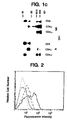
- Fig. 1C presents immunoprecipitation of labeled CD4: ⁇ , CD4: ⁇ , or native CD4 expressed in CV1 cells. Lanes were run with reducing (R) or without reducing (NR) agent. Molecular mass standards in kD are shown at left.
- FIG. 2 Surface expression of CD16 TM following coinfection of CD16TM alone (dense dots), or coinfected with virus expressing CD4:7 (dashes) or CD4: ⁇ (solid line). Sparse dots, cells infected with CD4: ⁇ alone, stained with 3G8 (Fleit et al., Proc. Natl. Acad. Sci. USA , 79 :3275-3279 (1982)) (anti-CD16 MAb).
- FIG. 3 Mutant CD4: ⁇ chimeric receptors lacking ⁇ Asp-15 do not support the coexpression of CD16 TM .
- Fig. 3A is an autoradiogram of immunoprecipitated mutant chimeras electrophoresed either with reduction (R) or without reduction (NR).
- Fig. 3B details surface expression of CD16 TM following coinfection by viruses expressing CD16 TM and the following ⁇ chimeras: CD4: ⁇ (thick line), CD4: ⁇ C11G (solid line); CD4: ⁇ (dashed line); CD4: ⁇ C11G/D15G (dense dots); no coinfection (CD16 TM alone, sparse dots).
- FIG. 4 Increased intracellular free calcium ion follows crosslinking of mutant ⁇ chimeras in a T cell line.
- Jurkat E6 cells Weiss et al., J. Immunol. , 133 :123-128 (1984)) were infected with recombinant vaccinia viruses and analyzed by flow cytometry. The results shown are for the gated CD4 + population, so that only cells expressing the relevant chimeric protein are analyzed.
- the mean ratio of violet to blue Indo-1 fluorescence reflects the intracellular free calcium concentration in the population as a whole and the percentage of responding cells reflects the fraction of cells which exceed a predetermined threshold ratio (set so that 10% of untreated cells are positive).
- FIGS. 4B show Jurkat cells expressing CD4: ⁇ (solid line) or CD16: ⁇ (dashed line) which were exposed to anti-CD4 MAb Leu3a (phycoerythrin conjugate), followed by crosslinking with goat antibody to mouse IgG.
- the dotted line shows the response of uninfected cells to anti-CD3 MAb OKT3.
- Figs. 4C and 4D show Jurkat cells expressing CD4: ⁇ D15G (solid line); CD4: ⁇ C11G/D15G (dashes); or CD4; ⁇ C11G (dots) which were treated and analyzed as in Figs. 4A and 4B.
- FIG. 5 CD4: ⁇ , CD4: ⁇ , and CD4: ⁇ receptors allow cytolytic T lymphocytes (CTL) to kill targets expressing HIV-1 gp120/41.
- Fig. 5A solid circles, CTL expressing CD4: ⁇ incubated with HeLa cells expressing gp120/41; open circles, CTL expressing CD4: ⁇ incubated with uninfected HeLa cells; solid squares, uninfected CTL incubated with HeLa cells expressing gp120/41; open squares, uninfected CTL incubated with uninfected HeLa cells.
- Fig. 5A solid circles, CTL expressing CD4: ⁇ incubated with HeLa cells expressing gp120/41; open circles, CTL expressing CD4: ⁇ incubated with uninfected HeLa cells; solid squares, uninfected CTL incubated with HeLa cells expressing gp120/41; open squares, uninfected CTL incubated with uninfected HeLa
- Fig. 5B solid circles, CTL expressing CD4: ⁇ incubated with HeLa cells expressing gp120/41; open circles, CTL expressing CD4: ⁇ incubated with HeLa cells expressing gp120/41; open squares, CTL expressing the C11G/D15G double mutant CD4: ⁇ chimera incubated with HeLa cells expressing gp120/41.
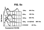
- Fig. 5C Flow cytometric analysis of CD4 expression by the CTL used in Fig. 5B.
- the percent of cells expressing CD4 chimera was determined by subtracting the scaled negative (uninfected) population by histogram superposition; for comparative purposes in this figure the uninfected cells were assigned an arbitrary threshold which gives roughly the same fraction positive for the other cell populations as would histogram subtraction.
- FIG. 6 Specificity of the CD4-directed cytolysis.
- Fig. 6A solid circles, CTL expressing CD4: ⁇ incubated with HeLa cells expressing CD16 PI ; open circles, CTL expressing CD4 incubated with HeLa cells expressing gp120; solid squares, CTL expressing CD16: ⁇ incubated with HeLa cells expressing gp120/41; open squares, CTL expressing CD16 PI incubated with HeLa cells expressing gp120/41.
- Fig. 6A solid circles, CTL expressing CD4: ⁇ incubated with HeLa cells expressing CD16 PI ; open circles, CTL expressing CD4 incubated with HeLa cells expressing gp120; open squares, CTL expressing CD16 PI incubated with HeLa cells expressing gp120/41.
- 6B solid circles, CTL expressing CD4: ⁇ incubated with Raji (MHC class II + ) cells; open circles, uninfected CTL cells incubated with RJ2.2.5 (MHC class II - Raji mutant) cells; solid squares, uninfected CTL incubated with Raji (MHC class II + ) cells; open squares, CTL expressing CD4: ⁇ incubated with RJ2.2.5 (MHC class II - ) cells.
- the ordinate scale is expanded.
- FIG. 7 Characterization of the CD16: ⁇ chimeric receptor.
- Fig. 7A is a schematic diagram of the CD16: ⁇ fusion protein. The extracellular portion of the phosphatidylinositol-linked form of monomeric CD16 was joined to dimeric ⁇ just external to the transmembrane domain. The protein sequence at the fusion junction is shown at the bottom.
- Fig. 7B shows a flow cytometric analysis of calcium mobilization following crosslinking of the CD16: ⁇ chimera in either a TCR positive or TCR positive or TCR negative cell line. The mean ratio of violet to blue fluorescence (a measure of relative calcium ion concentration) among cell populations treated with antibodies at time 0 is shown.
- Solid squares the response of Jurkat cells to anti-CD3 MAb OKT3; solid triangles, the response of CD16: ⁇ to anti-CD16 MAb 3G8 crosslinking in the REX33A TCR - mutant; open squares, the response to CD16: ⁇ crosslinking in the Jurkat TCR - mutant line JRT3.T3.5; open triangles, the response to CD16: ⁇ crosslinking in Jurkat cells; crosses, the response to nonchimeric CD16 in Jurkat cells; and dots, the response to nonchimeric CD16 in the REX33A TCR - cell line.
- FIG. 8 Deletion analysis of cytolytic potential.
- Fig. 8A shows the locations of the ⁇ deletion endpoints.
- mutations in ⁇ are represented by the original residue-location-mutant residue convention, so that D66*, for example, denotes replacement of Asp-66 by a termination codon.
- Fig. 8B shows cytolysis assay results of undeleted CD16: ⁇ and salient ⁇ deletions.
- Hybridoma cells expressing surface antibody to CD16 were loaded with 51 Cr and incubated with increasing numbers of human cytolytic lymphocytes (CTL) infected with vaccinia recombinants expressing CD16: ⁇ chimeras.
- CTL human cytolytic lymphocytes
- the percent of 51 Cr released is plotted as a function of the effector (CTL) to target (hybridoma) cell ratio (e/t).
- Solid circles cytolysis mediated by cells expressing CD16: ⁇ (mfi 18.7); solid squares, cytolysis mediated by cells expressing CD16: ⁇ Asp66* (mfi 940.2); open squares, cytolysis mediated by cells expressing CD16: ⁇ Glu60* (mfi 16.0); open circles, cytolysis mediated by cells expressing CD16: ⁇ Tyr51* (mfi 17.4); solid triangles, cytolysis mediated by cells expressing CD16: ⁇ Phe34* (mfi 17.8); and open triangles, cytolysis mediated by cells expressing nonchimeric CD16 (mfi 591).
- FIG. 9 Elimination of the potential for transmembrane interactions reveals a short ⁇ segment capable of mediating cytolysis.
- Fig. 9A is a schematic diagram of the monomeric bipartite and tripartite chimeras. At the top is the CD16: ⁇ construct truncated at residue 65 and lacking transmembrane Cys and Asp residues. Below are the CD16:CD5: ⁇ and CD16:CD7: ⁇ constructs and related controls. The peptide sequences of the intracellular domains are shown below.
- Fig. 9B shows the cytolytic activity of monomeric chimera deletion mutants.
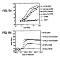
- Fig. 9C shows the cytolytic activity mediated by tripartite fusion proteins.
- Fig. 9D shows calcium mobilization by mutant and tripartite chimeras in the TCR negative Jurkat JRT3.T3.5 mutant cell line.
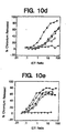
- FIG. 10 Contribution of individual amino acids to the activity of the 18 residue cytolytic signal-transducing motif.
- Figs. 10A and 10B show cytolytic activity and
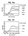
- Fig. 10C shows calcium ion mobilization mediated by chimeras bearing point mutations near the carboxyl terminal tyrosine (Y62).
- Figs. 10A and 10B represent data collected on cells expressing low and high amounts, respectively, of the CD16: ⁇ fusion proteins. Identical symbols are used for the calcuim mobilization and cytolysis assays, and are shown in one letter code at right.
- Figs. 10D and 10E show cytolytic activity and Fig. 10F shows calcium ion mobilization by chimeras bearing point mutations near the amino terminal tyrosine (Y51). Identical symbols are sued for the calcium mobilization and cytolysis assays and are shown at right.
- Solid circles, cells expressing CD16: ⁇ (mfi in D, 21.2; in E, 672); solid squares, cells expressing CD16:7: ⁇ (48-65) (mfi D, 31.3; E, 179); solid triangles, CD16:7: ⁇ (48-65)Asn48Ser (mfi D, 22.4; E, 209); open squares, CD16:7: ⁇ (48-65)Leu50Ser (mfi D, 25.0; E, 142); and open triangles, CD16:7: ⁇ (48-65)Tyr51Phe (mfi D, 32.3; E, 294).
- FIG. 11 Alignment of internal repeats of ⁇ and comparison of their ability to support cytolysis.
- Fig. 11A is a schematic diagram of chimeras formed by dividing the ⁇ intracellular domain into thirds and appending them to the transmembrane domain of a CD16:7 chimera. The sequences of the intracellular domains are shown below, with shared residues boxed, and related residues denoted by asterisks.
- Fig. 11B shows the cytolytic potency of the three ⁇ subdomains.
- Solid circles, cells expressing CD16: ⁇ (mfi 476); solid squares, CD16:7: ⁇ (33-65) (mfi 68); open squares, CD16:7: ⁇ (71-104) (mfi 114); and solid triangles, CD16:7: ⁇ (104-138) (mfi 104).
- FIG. 12 is a schematic diagram of the CD16:FcR ⁇ II chimeras.
- FIG. 13 Calcium mobilization following crosslinking of CD4:FcR ⁇ II and CD16:FcR ⁇ II chimeras.
- Fig. 13A shows the ratio of violet to blue fluorescence emitted by cells loaded with the calcium sensitive fluorophore Indo-1 shown as a function of time following crosslinking of the CD16 extracellular domain with antibodies.
- Fig. 13B shows a similar analysis of the increase in ratio of violet to blue fluorescence of cells bearing CD4:FcR ⁇ II chimeras, following crosslinking with antibodies.
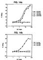
- FIG. 14 Cytolysis assay of CD4:FcR ⁇ II and CD16:FcR ⁇ II chimeras.
- Fig. 14A shows the percent of 51 Cr released from anti-CD16 hybridoma (target) cells when the cells are exposed to increasing numbers of cytotoxic T lymphocytes expressing CD16:FcR ⁇ II chimeras (effector cells).
- Fig. 14B shows a similar analysis of cytotoxicity mediated by CD4:FcR ⁇ II chimeras against target cells expressing HIV envelope glycoproteins.
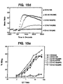
- FIG. 15 Identification of residues in the FcR ⁇ II A tail which are important for cytolysis.
- Fig. 15A is a schematic diagram of the deletion constructs.
- Figs. 15B and 15C shows calcium mobilization and cytolysis by carboxyl-terminal deletion variants of CD16:FcR ⁇ II A.
- Figs. 15D and 15E show calcium mobilization and cytolysis by tripartite chimeras bearing progressively less of the amino terminus of the intracellular tail of CD16:FcR ⁇ II A.
- FIG. 16 shows the amino acid sequence of the CD3 delta receptor protein; the boxed sequence represents a preferred cytolytic signal transducing portion.
- FIG. 17 shows the amino acid sequence of the T3 gamma receptor protein; the boxed sequence represents a preferred cytolytic signal transducing portion.
- FIG. 18 shows the amino acid sequence of the mb1 receptor protein; the boxed sequence represents a preferred cytolytic signal transducing portion.
- FIG. 19 shows the amino acid sequence of the B29 receptor protein; the boxed sequence represents a preferred cytolytic signal transducing portion.
- Human IgG1 heavy chain sequences were prepared by joining sequences in the C H 3 domain to a cDNA fragment derived from the 3' end of the transmembrane form of the antibody mRNA.
- the 3' end fragment was obtained by polymerase chain reaction using a tonsil cDNA library as substrate, and oligonucleotides having the sequences: corresponding to the 5' and 3' ends of the desired DNA fragments respectively.
- the 5' oligo is complementary to a site in the C H 1 domain of human IgG1, and the 3' oligo is complementary to a site just 5' of the sequences encoding the membrane spanning domain.
- the PCR product was digested with BstXI and BamHI and ligated between BstXI and BamHI sites of a semisynthetic IgG1 antibody gene bearing variable and constant regions. Following the insertion of the BstXI to BamHI fragment, the amplified portions of the construct were replaced up to the SmaI site in C H 3 by restriction fragment interchange, so that only the portion between the SmaI site and the 3' oligo was derived from the PCR reaction.
- the heavy chain gene ending in a BamHI site was joined to the BamHI site of the ⁇ chimera described below, so that the antibody sequences formed the extracellular portion.
- Flow cytometry of COS cells transfected with a plasmid encoding the chimera showed high level expression of antibody determinants when an expression plasmid encoding a light chain cDNA was cotransfected, and modest expression of antibody determinants when the light chain expression plasmid was absent.
- T cell receptor or Fc receptor protein Similar chimeras including human IgG1 fused to ⁇ or ⁇ (see below), or any signal-transducing portion of a T cell receptor or Fc receptor protein may be constructed generally as described above using standard techniques of molecular biology.
- a plasmid encoding a bicistronic mRNA was created from heavy and light chain coding sequences, and the 5' untranslated portion of the mRNA encoding the 78kD glucose regulated protein, otherwise known as grp78, or BiP.
- grp78 sequences were obtained by PCR of human genomic DNA using primers having the sequences: at the 5' and 3' ends respectively. Polymerase chain reactions with these oligos were performed in the presence of 10% dimethyl sulfoxide.
- the fragment obtained by PCR was digested with NotI and HincII and inserted between NotI and HpaI sites downstream from human IgG1 coding sequences. Sequences encoding a human IgG kappa light chain cDNA were then inserted downstream from the grp78 leader, using the HincII site and another site in the vector.
- the expression plasmid resulting from these manipulations consisted of the semisynthetic heavy chain gene, followed by the grp78 leader sequences, followed by the kappa light chain cDNA sequences, followed by polyadenylation signals derived from an SV40 DNA fragment. Transfection of COS cells with the expression plasmid gave markedly improved expression of heavy chain determinants, compared to transfection of plasmid encoding heavy chain determinants alone.
- the upstream heavy chain sequences can be replaced by any chimeric heavy chain/ receptor gene described herein.
- mutant ⁇ fusion proteins which lacked either the intramembranous Asp or intramembranous Cys residue or both were prepared.
- Flow cytometry showed that the intensity of cell surface expression by the different mutant chimeras was not appreciably different from the unmutated precursor (data not shown) and immunoprecipitation experiments showed that total expression by the chimeras was similar (Fig. 3).
- the mutant chimeras lacking the transmembrane cysteine residue were found not to form disulfide linked dimers (Fig. 3).
- the two mutant chimeras lacking Asp were incapable of supporting the surface expression of CD16TM, whereas the monomeric chimeras lacking Cys but bearing Asp allowed CD16 TM to be coexpressed, but at lower efficiency than the parental dimer (Fig. 3).
- Figure 4 shows the results of calcium flux experiments with cells infected with CD4: ⁇ and the Asp - and Cys - mutants of ⁇ . Crosslinking of the chimeras, reproducibly increased intracellular calcium. CD4: ⁇ and CD4: ⁇ similarly allowed accumulation intracellular calcium in infected cells (data not shown).
- Jurkat cells express low levels of CD4 on the cell surface, however, crosslinking of the native CD4 in the presence or absence of CD16: ⁇ (C.R. and B.S. unpublished) (Fig. 4 and data not shown) does not alter intracellular calcium levels.
- CD4 ⁇ , ⁇ , and ⁇ Chimeras Mediate Cytolysis of Targets Expressing HIV gp120/41
- a model target:effector system based on CD4 recognition of the HIV envelope gp120/gp41 complex was created.
- HeLa cells were infected with recombinant vaccinia viruses expressing gp120/gp41 (Chakrabarti et al., Nature , 320 :535-537 (1986); Earl et al., J. Virol. , 64 :2448-2451 (1990)) and labeled with 51 Cr.
- the labeled cells were incubated with cells from a human allospecific (CD8 + , CD4 - ) cytotoxic T lymphocyte line which had been infected with vaccinia recombinants expressing the CD4: ⁇ , CD4: ⁇ , or CD4: ⁇ chimeras, or the CD4: ⁇ Cys11Gly:Asp15Gly double mutant chimera.
- Fig. 5 shows that HeLa cells expressing gp120/41 were specifically lysed by cytotoxic T lymphocytes (CTL) expressing CD4 chimeras. Uninfected HeLa cells were not targeted by CTL armed with CD4: ⁇ chimeras, and HeLa cells expressing gp120/41 were not recognized by uninfected CTL.
- CTL cytotoxic T lymphocytes
- Fig. 5C shows a cytometric analysis of CD4 expression by the CTL used in the cytolysis experiment shown in Figs. 4A and 4B.
- the mean density of surface CD4: ⁇ greatly exceeded the mean density of CD4: ⁇ , the cytolytic efficiencies of cells expressing either form were similar.
- the CD4: ⁇ fusion was less active, as was the CD4: ⁇ fusion lacking the transmembrane Asp and Cys residues. However in both cases significant cytolysis was observed (Fig. 5).
- vaccinia infection might promote artefactual recognition by CTL
- similar cytolysis experiments were performed with target cells infected with vaccinia recombinants expressing the phosphatidylinositol linked form of CD16 (CD16 PI ) and labeled with 51 Cr, and with CTL infected with control recombinants expressing either CD16 PI or CD16: ⁇ .
- Fig. 6A shows that T cells expressing non-CD4 chimeras do not recognize native HeLa cells or HeLa cells expressing gp120/41, and similarly that T cells expressing CD4 chimeras do not recognize HeLa cells expressing other vaccinia-encoded surface proteins.
- CTLs expressing non-chimeric CD4 do not significantly lyse HeLa cells expressing gp120/41 (Fig. 6A).
- CD4 is thought to interact with a nonpolymorphic sequence expressed by MHC class II antigen (Gay et al., Nature, 328 :626-629 (1987); Sleckman et al., Nature , 328 :351-353 (1987)). Although a specific interaction between CD4 and class II antigen has never been documented with purified proteins, under certain conditions adhesion between cells expressing CD4 and cells expressing class II molecules can be demonstrated (Doyle et al., Nature, 330 :256-259 (1987); Clayton et al., J. Exp. Med. , 172 :1243-1253 (1990); Lamarre et al., Science , 245:743-746 (1989)).
- FIG. 6B shows that there is no specific cytolysis directed by CD4: ⁇ against the Raji B cell line, which expresses abundant class II antigen. Although a modest ( ⁇ 5%) cytolysis is observed, a class II-negative mutant of Raji, RJ2.2.5, (Accolla, R.S., J. Exp. Med. , 157 :1053-1058 (1983)) shows a similar susceptibility, as do Raji cells incubated with uninfected T cells.
- chimeras between CD4 and ⁇ can arm cytotoxic T lymphocytes (CTL) to kill target cells expressing HIV gp120
- CTL cytotoxic T lymphocytes
- an alternative to CD4 was sought in order to unambiguously compare the properties of zeta chimeras introduced into human T cell lines.
- Such lines can express CD4, making it difficult to specifically define the relationship between the type or degree of calcium mobilization and the cytotoxic potential of the different chimeras.
- chimeras were created between ⁇ and CD16 in which the extracellular domain of CD16 is attached to the transmembrane and intracellular sequences of ⁇ (Fig. 7A). The gene fusions were introduced into a vaccinia virus expression plasmid bearing the E.
- coli gpt gene as a selectable marker and inserted into the genome of the vaccinia WR strain by homologous recombination and selection for growth in mycophenolic acid (Falkner and Moss, J. Virol. 62 :1849 (1988); Boyle and Coupar, Gene 65 :123 (1988)).
- T cell lines were infected with the vaccinia recombinants and the relative cytoplasmic free calcium ion concentration was measured following crosslinking of the extracellular domains with antibodies. Both spectrofluorimetric (bulk population) and flow cytometric (single cell) measurements were performed, with cells loaded with the dye Indo-1 (Grynkiewicz et al., J. Biol. Chem. 260 :3440 (1985); Rabinovitch et al., J. Immunol. 137 :952 (1986)).
- Figure 7B shows an analysis of data collected from cells of the Jurkat human T cell leukemia line infected with vaccinia recombinants expressing CD16: ⁇ fusion protein.
- CTLs were infected with vaccinia recombinants expressing CD16 chimeras and used to specifically lyse hybridoma cells expressing membrane-bound anti-CD16 antibodies (see below).
- This assay is an extension of a hybridoma cytotoxicity assay originally developed to analyze effector mechanisms of cells bearing Fc receptors (Graziano and Fanger, J. Immunol. 138 :945, 1987; Graziano and Fanger, J. Immunol. 139 :35-36, 1987; Shen et al., Mol. Immunol. 26 :959, 1989; Fanger et al., Immunol. Today 10 : 92, 1989).
- CD16: ⁇ shows that expression of CD16: ⁇ in cytotoxic T lymphocytes allows the armed CTL to kill 3G8 (anti-CD16; Fleit et al., Proc. Natl. Acad. Sci. USA 79 :3275, 1982) hybridoma cells, whereas CTL expressing the phosphatidylinositol-linked form of CD16 are inactive. CTL armed with CD16: ⁇ also do not kill hybridoma cells expressing an irrelevant antibody (data not shown).
- a series of deletion mutants were prepared in which successively more of the ⁇ intracellular domain was removed from the carboxyl terminus (Fig. 8A). Most of the intracellular domain of zeta could be removed with little consequence for cytolytic potential; the full length chimera CD16: ⁇ was essentially equal in efficacy to the chimera deleted to residue 65, CD16: ⁇ Asp66* (Fig. 8B). A substantial decrease in cytotoxicity was observed on deletion to ⁇ residue 59 (chimera CD16: ⁇ Glu60*), and further deletion to residue 50 resulted in slightly less activity. However complete loss of activity was not observed even when the intracellular domain was reduced to a three residue transmembrane anchor (Fig. 8B).
- ⁇ is a disulfide linked dimer
- endogenous ⁇ was forming heterodimers with the chimeric ⁇ deletion, thereby reconstituting activity.
- ⁇ residues 11 and 15 were changed from Asp and Cys respectively to Gly (Cys11Gly/Asp15Gly), and immunoprecipitations were carried out as follows. Approximately 2 x 10 6 CV1 cells were infected for one hour in serum free DME medium with recombinant vaccinia at a multiplicity of infection (moi) of at least ten.
- moi multiplicity of infection
- the labeled cells were collected by centrifugation and lysed in 1% NP-40, 0.1% SDS, 0.15M NaCl, 0.05M Tris, pH 8.0, 5mM MgCl 2 , 5mM KCl, 0.2M iodoacetamide and 1mM PMSF.
- CD16 proteins were immunoprecipitated with antibody 3G8 (Fleit et al., supra , 1982; Medarex) and anti-mouse IgG agarose (Cappel, Durham, NC). Samples were electrophoresed through an 8% polyacrylamide/SDS gel under non-reducing conditions or through a 10% gel under reducing conditions. These immunoprecipitations confirmed that the CD16: ⁇ Cys11Gly/Aspl5Gly chimera did not associate in disulfide-linked dimer structures.
- the cytolytic activity of the mutant receptors was also tested.
- the mutated chimera deleted to residue 65 (CD16: ⁇ Cys11Gly/Asp15Gly/Asp66*) was, depending on the conditions of assay, two to eight fold less active in the cytolysis assay than the comparable unmutated chimera (CD16: ⁇ Asp66*), which was usually within a factor of two of, or indistinguishable in activity from, CD16: ⁇ (Fig. 9B).
- the reduction in activity of the mutant chimeras is comparable to the reduction seen with CD4 chimeras of similar structure (see above) and is most likely attributable to the lower efficiency of ⁇ monomers compared to dimers.
- Fig. 10 shows that while a number of relatively conservative substitutions (i.e., replacing acidic residues with their cognate amides, or tyrosine with phenylalanine) which spanned residues 59 to 63 yielded moderate compromise of cytolytic efficacy, in general the variants retained the ability to mobilize calcium.
- the intracellular domain of ⁇ was divided into three segments, spanning residues 33 to 65, 71 to 104, and 104 to 138. Each of these segments was attached to a CD16:CD7 chimera by means of a MluI site introduced just distal to the basic membrane anchoring sequences of the intracellular domain of CD7 (see below; Fig. 11A). Comparison of the cytolytic efficacy of the three elements showed they were essentially equipotent (Fig. 11B). Sequence comparison (Fig. 11A) shows that the second motif bears eleven residues between tyrosines, whereas the first and third motifs bear ten.
- chimeras bearing the 18 residue motif are approximately eight-fold less active than chimeras based on full length ⁇ , the reduced activity can be attributed to the loss of transmembrane interactions which normally allow wild-type ⁇ to form disulfide linked dimers. That is, ⁇ deletion constructs which have the same carboxyl terminus as the motif and lack transmembrane Cys and Asp residues typically show slightly less activity than chimeras bearing only the 18 residue motif.
- the cytolytic competency element on which we have focused has two tyrosines and no serines or threonines, restricting the possible contributions of phosphorylation to activity. Mutation of either tyrosine destroys activity, however, and although preliminary experiments do not point to a substantial tyrosine phosphorylation following crosslinking of chimeric surface antigens bearing the 18 reside motif, the possible participation of such phosphorylation at a low level cannot be excluded. In addition to the effects noted at the two tyrosine residues, a number of amino acid replacements at the amino and carboxyl terminus of the motif weaken activity under conditions of low receptor density.
- Sequences similar to the ⁇ active motif can be found in the cytoplasmic domains of several other transmembrane proteins, including the CD3 ⁇ and ⁇ molecules, the surface IgM associated proteins mb1 and B29, and the ⁇ and ⁇ chains of the high affinity IgE receptor, Fc ⁇ RI (Reth, Nature 338 :383, 1989). Although the function of these sequences is uncertain, if efficiently expressed, each may be capable of autonomous T cell activation, and such activity may explain the residual TCR responsiveness seen in a zeta-negative mutant cell line (Sussman et al., Cell 52 :85, 1988).
- ⁇ itself bears three such sequences, approximately equally spaced, and a rough trisection of the intracellular domain shows that each is capable of initiating a cytolytic response.
- ⁇ a splice isoform of ⁇ (Jin et al., supra , 1990; Clayton et al., Proc. Natl. Acad. Sci. USA 88 :5202, 1991), lacks the carboxyl half of the third motif. Because removal of the carboxyl half of the first motif abolishes activity, it appears likely that the majority of the biological effectiveness of ⁇ can be attributed to the first two motifs.
- ⁇ is equally as active as ⁇ in promoting antigen-mediated cytokine release (Bauer et al., Proc. Natl.
- ⁇ is not phosphorylated in response to receptor stimulation (Bauer et al., supra , 1991).
- the presence of all three motifs is required for phosphorylation, or the third motif represents a favored substrate for an unidentified tyrosine kinase.
- chimeric molecules were created in which the extracellular domain of the human CD4, CD5 or CD16 antigens were joined to the transmembrane and intracellular domains of the FcRII ⁇ A, B1, B2, and C subtypes (nomenclature of Ravetch and Kinet, Ann. Rev. Immunol. 9 :457, 1991).
- cDNA sequences corresponding to the transmembrane and cytoplasmic domains of the previously described FcRIIA, B1, and B2 isoforms were amplified from the preexisting clone PC23 or from a human tonsil cDNA library (constructed by standard techniques) using oligonucleotide primers: These primers contained cleavage sites for the enzymes BamHI and NotI, respectively, indented 6 residues from the 5' end. The NotI site was immediately followed by an antisense stop codon, either CTA or TTA. All primers contained 18 or more residues complementary to the 5' and 3' ends of the desired fragments.
- the cDNA fragment corresponding to the FcRII ⁇ C cytoplasmic domain, which differs from the IIA isoform in only one amino acid residue (L for P at residue 268) was generated by site directed mutagenesis by overlap PCR using primers of sequence:
- the PCR fragments were inserted into vaccinia virus expression vectors which contained the CD16 or CD4 extracellular domains respectively and subsequently inserted into wild type vaccinia by recombination at the thymidine kinase locus, using selection for cointegration of E coli gpt to facilitate identification of the desired recombinants.
- the identities of all isoforms (shown in Fig. 12) were confirmed by dideoxy sequencing.
- JRT3.T3.5 Approximately 10 7 JRT3.T3.5 cells were infected for one hour in serum free IMDM medium with recombinant vaccinia at a multiplicity of infection of at least ten. Twelve hours post-infection, the cells were harvested and surface labeled with 0.5mCi 125 I per 10 7 cells using the lactoperoxidase/glucose oxidase method (Clark and Einfeld, supra ). The labeled cells were collected by centrifugation and lysed 1% NP-40, 0.1mM MgCl 2 , 5mM KCl, 0.2M iodoacetamide and 1mM PMSF.
- cytotoxic T lymphocytes CTL
- vaccinia recombinants expressing CD16:FcR ⁇ II A, B1, B2 and C chimeras.
- the infected cells were then cocultured with 51 Cr-loaded hybridoma cells (i.e., 3G8 10-2 cells) which expressed cell surface antibody to CD16.
- 51 Cr-loaded hybridoma cells i.e., 3G8 10-2 cells
- CTLs bearing the CD16 chimera killed the hybridoma target cells (allowing release of free 51 Cr) if the CD16 extracellular domain of the chimera has been joined to an intracellular segment capable of activating the lymphocyte effector program; this cytolysis assay is described in detail below.
- Fig. 14A shows that CTL armed with CD16:FcR ⁇ IIA and C, but not FcR ⁇ II B1 or B2, are capable of lysing target cells expressing cell surface anti-CD16 antibody.
- cytolysis experiments were conducted in which the FcRII intracellular domains were attached to a CD4 extracellular domain.
- the target cells were HeLa cells expressing HIV envelope gp120/41 proteins (specifically, HeLa cells infected with the vaccinia vector vPE16 (available from the National Institute of Allergy and Infections Disease AIDS Depository, Bethesda, MD).
- target cells expressing HIV envelope were susceptible to lysis by T cells expressing the CD4:FcR ⁇ II A chimera, but not FcR ⁇ II B1 or B2 (Fig. 14B).
- the intracellular domains of FcR ⁇ II A and C share no appreciable sequence homology with any other protein, including the members of the extended FcR ⁇ /TCR ⁇ family.
- sequence elements responsible for induction of cytolysis 5' and 3' deletions of the intracellular domain coding sequences (described below and shown in Fig. 15A) were prepared and were evaluated for efficacy in calcium mobilization and cytolysis assays (as described herein).
- the transmembrane domain of FcR ⁇ II was replaced with the transmembrane domain of the unrelated CD7 antigen to eliminate the possible contribution of interactions mediated by the membrane-spanning domain.
- Figs. 15B and 15C show that removal of the 14 carboxyl-terminal residues, including tyrosine 298, resulted in a complete loss of cytolytic capacity and a substantial reduction in calcium mobilization potential. Further deletion to just before tyrosine 282 gave an identical phenotype (Figs. 15B and 15C). Deletion from the N-terminus of the intracellular domain to residue 268 had no substantial effect on either calcium profile or cytolytic potency, whereas deletion to residue 275 markedly impaired free calcium release but had little effect on cytolysis (Figs. 15D and 15E). Further deletion, to residue 282, gave FcR ⁇ II tails which lacked the ability to either mobilize calcium or trigger cytolysis (Figs. 15D and 15E). The 'active element' defined by these crude measures is relatively large (36 amino acids) and contains two tyrosines separated by 16 residues.
- T cell receptor proteins CD3 delta and T3 gamma
- B cell receptor proteins mb1 and B29.
- the amino acid sequences of these proteins is shown in Fig. 16 (CD3 delta; SEQ ID NO: 24), Fig. 17 (T3 gamma; SEQ ID NO: 25), Fig. 18 (mb1; SEQ ID NO: 26) and Fig. 19 (B29; SEQ ID NO: 27).
- the portions of the sequences sufficient for cytolytic signal transduction are shown in brackets. Chimeric receptors which include these protein domains are constructed and used in the therapeutic methods of the invention generally as described above.
- CV1 cells Approximately 5 x 10 6 CV1 cells were infected for one hour in serum free DME medium with recombinant vaccinia at a multiplicity of infection (moi) of at least ten (titer measured on CV1 cells). The cells were placed in fresh medium after infection and labelled metabolically with 200 ⁇ Ci/ml 35 S-methionine plus cysteine (Tran 35 S-label, ICN; Costo Mesa, CA) in methionine and cysteine free DMEM (Gibco; Grand Island, NY) for six hours.
- moi multiplicity of infection
- the labelled cells were detached with PBS containing 1mM EDTA, collected by centrifugation, and lysed in 1% NP-40, 0.1% SDS, 0.15 M NaCl, 0.05M Tris pH 8.0, 5mM EDTA, and 1mM PMSF. Nuclei were removed by centrifugation, and CD4 proteins immunoprecipitated with OKT4 antibody and anti-mouse IgG agarose (Cappel, Durham, NC). Samples were electrophoresed through 8% polyacrylamide/SDS gels under non-reducing (NR) and reducing (R) conditions. Gels containing 35 S-labelled samples were impregnated with En 3 Hance (New England Nuclear, Boston, MA) prior to autoradiography.
- NR non-reducing
- R reducing
- CD16 TM transmembrane form of CD16
- CV1 cells singly infected with CD16 TM
- expression in cells coinfected with viruses encoding CD16 TM and ⁇ or ⁇ chimeras After infection and incubation for six hours or more, cells were detached from plates with PBS, 1mM EDTA and the expression of CD16TM or the chimeras was measured by indirect immunofluorescence and flow cytometry.
- Jurkat subline E6 (Weiss et al., J. Immunol. , 133 :123-128 (1984)) cells were infected with recombinant vaccinia viruses for one hour in serum free IMDM at an moi of 10 and incubated for three to nine hours in IMDM, 10% FBS. Cells were collected by centrifugation and resuspended at 3 x 10 6 cells/ml in complete medium containing 1mM Indo-1 acetomethoxyester (Grynkiewicz et al., J. Biol. Chem. , 260 : 3340-3450 (1985)) (Molecular Probes) and incubated at 37°C for 45 minutes.
- 1mM Indo-1 acetomethoxyester (Grynkiewicz et al., J. Biol. Chem. , 260 : 3340-3450 (1985)) (Molecular Probes) and incubated at 37°C for 45 minutes.
- the Indo-1 loaded cells were pelleted and resuspended at 1 x 10 6 /ml in serum free IMDM and stored at room temperature in the dark. Cells were analyzed for free calcium ion by simultaneous measurement of the violet and blue fluorescence emission by flow cytometry (Rabinovitch et al., J. Immunol. , 137 :952-961 (1986)).
- PE-conjugated Leu-3A (anti-CD4) (Becton Dickinson, Lincoln Park, NJ) at 1 ⁇ g/ml was added to the cell suspension followed by 10 ⁇ g/ml of unconjugated goat anti-mouse IgG at time O or unconjugated 3G8 (anti-CD16) monoclonal antibody was added to the cell suspension at 1 ⁇ g/ml followed by 10 ⁇ g/ml of PE-conjugated Fab 2 ' goat anti-monse IgG at time 0. Histograms of the violet/blue emission ratio were collected from the PE positive (infected) cell population, which typically represented 40-80% of all cells.
- the T cell antigen receptor response in uninfected cells was triggered by antibody OKT3, without crosslinking.
- samples showing baseline drift toward lower intracellular calcium (without antibody) were excluded from the analysis.
- Histogram data were subsequently analyzed by conversion of the binary data to ASCII using Write Hand Man (Cooper City, FL) software, followed by analysis with a collection of FORTRAN programs.
- the violet/blue emission ratio prior to the addition of the second antibody reagents was used to establish the normalized initial ratio, set equal to unity, and the resting threshold ratio, set so that 10% of the resting population would exceed threshold.
- Human T cell line WH3, a CD8 + CD4 - HLA B44 restricted cytolytic line was maintained in IMDM, 10% human serum with 100 U/ml of IL-2 and was periodically stimulated either nonspecifically with irradiated (3000 rad) HLA-unmatched peripheral blood lymphocytes and 1 ⁇ g/ml of phytohemagglutinin, or specifically, with irradiated B44-bearing mononuclear cells. After one day of nonspecific stimulation, the PHA was diluted to 0.5 ⁇ g/ml by addition of fresh medium, and after three days the medium was changed. Cells were grown for at least 10 days following stimulation before use in cytotoxicity assays.
- the cells were infected with recombinant vaccinia at a multiplicity of infection of at least 10 for one hour in serum free medium, followed by incubation in complete medium for three hours. Cells were harvested by centrifugation and resuspended at a density of 1 x 10 7 cells/ml. 100 ⁇ l were added to each well of a U-bottom microtiter plate containing 100 ⁇ l/well of complete medium. Cells were diluted in two-fold serial steps. Two wells for each sample did not contain lymphocytes, to allow spontaneous chromium release and total chromium uptake to be measured.
- the target cells from HeLa subline S3, were infected in 6.0 or 10.0 cm plates at an approximate moi of 10 for one hour in serum free medium, followed by incubation in complete medium for three hours. They were then detached from the dishes with PBS, 1mM EDTA and counted. An aliquot of 10 6 target cells (HeLa, Raji, or RJ2.2.5 cells for the CD4 chimeric receptor experiments and 3G8 10-2 cells; Shen et al., Mol. Immunol .
- the cells in each well were resuspended by gentle pipetting, a sample removed to determine the total counts incorporated, and the microtiter plate spun at 750 x g for 1 minute. 100 ⁇ l aliquots of supernatant were removed and counted in a gamma ray scintillation counter. The percent killing was corrected for the fraction of infected target cells (usually 50-90%) measured by flow cytometry. For infected effector cells the effector:target ratio was corrected for the percent of cells infected (usually 20-50% for the CD4 chimeric receptor experiments and >70% for the CD16 chimeric receptor experiments).
- synthetic oligonucleotide primers extending from the BamHI site upstream of the ⁇ transmembrane domain, and converting native ⁇ residue 11 from Cys to Gly (C11G) or residue 15 from Asp to Gly (D15G) or both (C11G/D15G) were prepared and used in PCR reactions to generate mutated fragments which were reinserted into the wild type CD4: ⁇ constructs.
- ⁇ cDNA sequences were amplified by PCR using synthetic oligonucleotide primers designed to create a stop codon (UAG) after residues 50, 59, or 65.
- the primers contained the cleavage site for the enzyme NotI indented five or six residues from the 5' end, usually in a sequence of the form CGC GGG CGG CCG CTA (SEQ ID NO: 11), where the last three residues correspond to the stop anticodon.
- the NotI and stop anticodon sequences were followed by 18 or more residues complementary to the diesired 3' end of the fragment.
- the resulting chimeras were designated CD16: ⁇ Y51*, CD16: ⁇ E60* and CD16: ⁇ D66* respectively.
- the BamHI site upstream of the transmembrane domain and the NotI site were used to generate fragments that were reintroduced into the wild type CD16: ⁇ construct.
- Monomeric ⁇ chimeras were created by liberating the ⁇ transmembrane and membrane proximal intracellular sequences by BamHI and SacI digestion of the Asp - and Cys - CD4: ⁇ construct described above and inserting the fragment into the CD16: ⁇ E60* and CD16: ⁇ D66* construct respectively.
- the ⁇ cDNA sequence corresponding to the transmembrane domain and the 17 following residues of the cytoplasmic domain was replaced by corresponding transmembrane and cytoplasmic domain obtained from the CD5 and CD7 cDNA.
- the CD5 and CD7 fragments were generated by a PCR reaction using forward oligonucleotides including a BamHI restriction cleavage site and corresponding to the region just upstream of the transmembrane domain of CD5 and CD7 respectively and the following reverse oligonucleotides overlapping the CD5 and CD7 sequences respectively and the ⁇ sequence which contained the SacI restriction cleavage site.
- CD5 and CD7 PCR products were digested with BamHI and SacI and ligated to BamHI and SacI digested CD16: ⁇ E60* and replacing the ⁇ sequence from BamHI to SacI by the CD7 fragment.
- CD16:CD5 and CD16:CD7 CD5 and CD7 fragments were obtained by PCR using an oligonucleotide containing a NotI restriction cleavage site and encoding a stop codon (UAA) after the residue Gln416 and Ala193 of CD5 and CD7 respectively.
- the CD5 and CD7 PCR fragment were digested with BamHI and NotI and inserted in the CD16: ⁇ Asp66* construct.
- Synthetic oligonucleotide primers extending from the SacI site inside the ⁇ motif and converting native residue 48 from Asn to Ser (N48S), residue 50 from Leu to Ser (L50S) and residue 51 from Tyr to Phe (Y51F) were synthesized and used in a PCR reaction to generate fragments that were reintroduced into the wild type CD16:7: ⁇ (48-65) construct.
- Synthetic oligonucleotide primers extending from the NotI site 3' to the stop codon and converting native residue 60 from Glu to Gln (E60Q), residue 61 from Glu to Gln (E61Q), residue 62 from Tyr to Phe or Ser (Y62F or Y62S) and residue 63 from Asp to Asn (D63N) were synthesized and used in PCR to generate fragments that were subcloned into the wild type CD16: ⁇ D66* construct from the BamHI site to the NotI site.
- CD16:7: ⁇ (33-65), CD16:7: ⁇ (71-104), CD16:7: ⁇ (104-137) Chimera Constructions
- a CD7 transmembrane fragment bearing MluI and NotI sites at the junction between the transmembrane and intracellular domains was obtained by PCR using an oligonnucleotide with the following sequence: CGC GGG GCG GCC ACG CGT CCT CGC CAG CAC ACA (SEQ ID NO: 14).
- the resulting PCR fragment was digested with BamHI and NotI and reinserted into the CD16:7: ⁇ (48-65) construct.
- ⁇ fragments encoding residues 33 to 65, 71 to 104, and 104 to 137 were obtained by PCR reaction using pairs of primers containing MluI sites at the 5' end of the forward primers and stop codons followed by NotI sites at the 5' end of the reverse primers.
- the restriction sites were indented six residues from the 5' terminus of the primer to insure restriction enzyme cleavage.
- Carboxyl terminal FcRIIA deletion mutants were constructed by PCR in the same fashion as for the full length constructs, converting the sequences encoding tyrosine at positions 282 and 298 into stop codons (TAA).
- the N-terminal deletions were generated by amplifying fragments encoding successively less of the intracellular domain by PCR, using oligonucleotides which allowed the resulting fragments to be inserted between MluI and HotI restriction sites into a previously constructed expression plasmid encoding the CD16 extracellular domain fused to the CD7 transmembrane domain, the latter terminating in a MluI site ant the juncition between the transmembrane and the intracellular domain.
- ⁇ , ⁇ , or ⁇ chimeras suffices to initiate the cytolytic effector cell response in T cells.
- the known range of expression of ⁇ , ⁇ , and ⁇ which includes T lymphocytes, natural killer cells, basophilic granulocytes, macrophages and mast cells, suggests that conserved sequence motifs may interact with a sensory apparatus common to cells of hematopoietic origin and that an important component of host defense in the immune system may be mediated by receptor aggregation events.
- the potency of the cytolytic response and the absence of a response to target cells bearing MHC class II receptors demonstrates that chimeras based on ⁇ , ⁇ , or ⁇ form the basis for a genetic intervention for AIDS through adoptive immunotherapy.
- the broad distribution of endogenous ⁇ and ⁇ and evidence that Fc receptors associated with ⁇ mediate cytotoxicity in different cells types allows a variety of cells to be considered for this purpose. For example, neutrophilic granulocytes, which have a very short lifespan ( ⁇ 4h) in circulation and are intensely cytolytic, are attractive target cells for expression of the chimeras.
- T cells a population presently accessible to retroviral engineering
- IL-2 recombinant IL-2
- T cell populations can be expanded in culture with relative ease, and the expanded populations typically have a limited lifespan when reinfused (Rosenberg et al., N. Engl. J. Med. , 323 :570-578 (1990)).
- HIV recognition by cells expressing CD4 chimeras should also provide mitogenic stimuli, allowing the possibility that the armed cell population could respond dynamically to the viral burden.
- expression of the chimeras in helper lymphocytes might provide an HIV-mobilized source of cytokines which could counteract the collapse of the helper cell subset in AIDS.
- the ability to transmit signals to T lymphocytes through autonomous chimeras also provides the ability for the regulation of retrovirally engineered lymphocytes in vivo .
- Crosslinking stimuli mediated for example by specific IgM antibodies engineered to remove complement-binding domains, may allow such lymphocytes to increase in number in situ, while treatment with similar specific IgG antibodies (for example recognizing an amino acid variation engineered into the chimeric chain) could selectively deplete the engineered population.
- anti-CD4 IgM antibodies do not require additional crosslinking to mobilize calcium in Jurkat cells expressing CD4: ⁇ chimeras (data not shown).
- the ability to regulate cell populations without recourse to repeated extracorporeal amplification may substantially extend the range and efficacy of current uses proposed for genetically engineered T cells.
- cDNA or genomic sequences encoding an extracellular domain of the receptor can be endowed with a restriction site introduced at a location just preceding the transmembrane domain of choice.
- the extracellular domain fragment terminating in the restriction site can then be joined to ⁇ , ⁇ , or ⁇ sequences.
- Typical extracellular domains may be derived from receptors which recognize complement, carbohydrates, viral proteins, bacteria, protozoan or metazoan parasites, or proteins induced by them.
- ligands or receptors expressed by pathogens or tumor cells can be attached to ⁇ , ⁇ , or ⁇ sequences, to direct immune responses against cells bearing receptors recognizing those ligands.
Landscapes
- Health & Medical Sciences (AREA)
- Life Sciences & Earth Sciences (AREA)
- Chemical & Material Sciences (AREA)
- Organic Chemistry (AREA)
- General Health & Medical Sciences (AREA)
- Immunology (AREA)
- Medicinal Chemistry (AREA)
- Cell Biology (AREA)
- Zoology (AREA)
- Veterinary Medicine (AREA)
- Genetics & Genomics (AREA)
- Molecular Biology (AREA)
- Biochemistry (AREA)
- Public Health (AREA)
- Animal Behavior & Ethology (AREA)
- Epidemiology (AREA)
- Biophysics (AREA)
- Proteomics, Peptides & Aminoacids (AREA)
- Engineering & Computer Science (AREA)
- Toxicology (AREA)
- Gastroenterology & Hepatology (AREA)
- Developmental Biology & Embryology (AREA)
- Pharmacology & Pharmacy (AREA)
- Biomedical Technology (AREA)
- Biotechnology (AREA)
- Virology (AREA)
- Bioinformatics & Cheminformatics (AREA)
- Chemical Kinetics & Catalysis (AREA)
- General Chemical & Material Sciences (AREA)
- Nuclear Medicine, Radiotherapy & Molecular Imaging (AREA)
- Reproductive Health (AREA)
- Wood Science & Technology (AREA)
- General Engineering & Computer Science (AREA)
- Gynecology & Obstetrics (AREA)
- Microbiology (AREA)
- Peptides Or Proteins (AREA)
- Medicines Containing Antibodies Or Antigens For Use As Internal Diagnostic Agents (AREA)
- Micro-Organisms Or Cultivation Processes Thereof (AREA)
- Medicines That Contain Protein Lipid Enzymes And Other Medicines (AREA)
- Preparation Of Compounds By Using Micro-Organisms (AREA)
Claims (16)
- Verfahren zur Herstellung eines Arzneimittels für die therapeutische Anwendung bei einer Infektion, einer Tumorkrankheit oder einer Autoimmunkrankheit, dadurch gekennzeichnet, dass therapeutische Zellen eingesetzt werden, die eine zelluläre Antwort auf einen Infektionserreger, auf eine mit dem genannten Erreger infizierte Zelle, auf eine Tumor- oder Krebszelle oder auf eine durch eine Autoimmunreaktion gebildete Zelle in einem Säuger auslösen, wobei die genannten therapeutischen Zellen in der Lage sind, den genannten Erreger oder die genannte Zelle spezifisch zu erkennen und zu zerstören und wobei die genannten therapeutischen Zellen aus einer Gruppe ausgewählt werden, umfassend:wobei die genannten therapeutischen Zellen einen membranständigen, proteinhaltigen chimären Rezeptor exprimieren, umfassend (a) einen extrazellulären Anteil, der in der Lage ist, den genannten Erreger oder die genannte Zelle spezifisch zu erkennen und zu binden, wobei dieser extrazelluläre Anteil einen an die HIV-Hülle bindenden Anteil von CD4 oder ein an die HIV-Hülle bindendes funktionelles Derivat davon enthält; und (b) einen intrazellulären oder transmembranösen Anteil enthält, der in der Lage ist, der genannten therapeutischen Zelle zu signalisieren, einen rezeptorgebundenen Erreger oder eine rezeptorgebundene Zelle zu zerstören, wobei dieser intrazelluläre Anteil oder transmembranöse Anteil ein signaltransduzierender Anteil von CD3 Delta oder T3 Gamma, FcRγII oder ein B-Zellrezeptorprotein oder ein funktionelles Derivat davon ist.(a) T-Lymphozyten;(b) zytotoxische T-Lymphozyten;(c) neutrale Killerzellen;(d) Neutrophile;(e) Granulozyten;(f) Makrophagen;(g) Mastzellen;(h) HeLa-Zellen;
- Verfahren gemäß Anspruch 1, wobei die genannte zelluläre Antwort MHC-unabhängig ist.
- Verfahren gemäß Anspruch 2, wobei es sich bei dem genannten Anteil von FcRγII um FcRγIIA oder FcRγIIC handelt; oder wobei es sich bei dem genannten Anteil eines B-Zellrezeptorproteins um mb1 oder B29 handelt.
- Verfahren gemäß Anspruch 3, wobei der genannte chimäre Rezeptor die folgenden Aminosäuren umfasst:(a) Aminosäuren YMTLNPRAPTDDDKNIY;(b) Aminosäuren 132-171 aus Fig. 16 (Sequenz Nr. 24);(c) Aminosäuren 140-182 aus Fig. 17 (Sequenz Nr. 25);(d) Aminosäuren 162-220 aus Fig. 18 (Sequenz Nr. 26);(e) Aminosäuren 183-228 aus Fig. 19 (Sequenz Nr. 27).
- Verfahren gemäß Anspruch 1, wobei der genannte infektiöse Erreger ein Immundefizienzvirus ist.
- Verfahren gemäß Anspruch 5, wobei der genannte zytotoxische T-Lymphozyt ein zytotoxischer CD8+-T-Lymphozyt ist.
- Zelle, die einen proteinhaltigen membranständigen chimären Rezeptor exprimiert, wobei der genannte Rezeptor (a) einen extrazellulären Anteil umfasst, der in der Lage ist, einen infektiösen Erreger, eine mit diesem infektiösen Erreger infizierte Zelle, eine Tumoroder Krebszelle oder eine durch eine Autoimmunreaktion erzeugte Zelle spezifisch zu erkennen und zu binden, wobei dieser extrazelluläre Anteil einen an die HIV-Hülle bindenden Anteil von CD4 oder ein an die HIV-Hülle bindendes funktionelles Derivat davon umfasst; und (b) einen intrazellulären oder transmembranösen Anteil umfasst, der abgeleitet ist von CD3 Delta oder T3 Gamma, FcRγII oder einem B-Zellrezeptor und der in der Lage ist, der genannten Zelle zu signalisieren, einen rezeptorgebundenen Erreger oder eine rezeptorgebundene Zelle zu zerstören.
- Zelle gemäß Anspruch 7, wobei die genannte Bindung MHC-unabhängig ist.
- Zelle gemäß Anspruch 7, wobei der genannte chimäre Rezeptor aus Aminosäuren besteht:(a) Aminosäuren YMTLNPRAPTDDDKNIY;(b) Aminosäuren 132-171 aus Fig. 16 (Sequenz Nr. 24);(c) Aminosäuren 140-182 aus Fig. 17 (Sequenz Nr. 25);(d) Aminosäuren 162-220 aus Fig. 18 (Sequenz Nr. 26);(e) Aminosäuren 183-228 aus Fig. 19 (Sequenz Nr. 27).
- Zelle gemäß Ansprüchen 7 bis 9, wobei die genannten therapeutischen Zellen aus einer Gruppe ausgewählt werden, umfassend:(a) Granulozyten;(b) Makrophagen;(c) Mastzellen;(d) HeLa-Zellen.
- Zelle gemäß Anspruch 10, wobei der genannte zytotoxische T-Lymphozyt ein zytotoxischer CD8+-T-Lymphozyt ist
- Zelle gemäß Anspruch 7, wobei der genannte extrazelluläre Anteil ein Immunglobulinmolekül oder ein antigenbindendes Fragment davon ist.
- DNA, die für einen chimären Rezeptor der Zelle gemäß einem der Ansprüche 7 bis 12 codiert.
- Vektor, umfassend die DNA des chimären Rezeptors gemäß Anspruch 13.
- Antikörper, der im wesentlichen rein ist und der spezifisch einen chimären Rezeptor der Zelle gemäß einem der Ansprüche 7 bis 12 erkennt und bindet.
- Chimärer Rezeptor gemäß einem der Ansprüche 7 bis 12.
Applications Claiming Priority (3)
| Application Number | Priority Date | Filing Date | Title |
|---|---|---|---|
| US66596191A | 1991-03-07 | 1991-03-07 | |
| US665961 | 1991-03-07 | ||
| PCT/US1992/001785 WO1992015322A1 (en) | 1991-03-07 | 1992-03-06 | Redirection of cellular immunity by receptor chimeras |
Publications (3)
| Publication Number | Publication Date |
|---|---|
| EP0574512A1 EP0574512A1 (de) | 1993-12-22 |
| EP0574512A4 EP0574512A4 (en) | 1997-03-12 |
| EP0574512B1 true EP0574512B1 (de) | 2003-02-05 |
Family
ID=24672250
Family Applications (1)
| Application Number | Title | Priority Date | Filing Date |
|---|---|---|---|
| EP92907958A Expired - Lifetime EP0574512B1 (de) | 1991-03-07 | 1992-03-06 | Änderung der zellularen immunität durch rezeptorchimäre |
Country Status (23)
| Country | Link |
|---|---|
| EP (1) | EP0574512B1 (de) |
| JP (1) | JP3696232B2 (de) |
| KR (1) | KR100257780B1 (de) |
| AT (1) | ATE232107T1 (de) |
| AU (2) | AU662136B2 (de) |
| BR (1) | BR9205736A (de) |
| CA (1) | CA2104957C (de) |
| CZ (1) | CZ281881B6 (de) |
| DE (1) | DE69232921T2 (de) |
| DK (1) | DK0574512T3 (de) |
| ES (1) | ES2191006T3 (de) |
| FI (1) | FI117900B (de) |
| HU (1) | HU218732B (de) |
| IE (1) | IE920716A1 (de) |
| IL (1) | IL101147A (de) |
| MX (1) | MX9201002A (de) |
| NO (1) | NO317202B1 (de) |
| NZ (1) | NZ241855A (de) |
| PT (1) | PT100207B (de) |
| RU (1) | RU2161044C2 (de) |
| SK (1) | SK283643B6 (de) |
| UA (1) | UA41870C2 (de) |
| WO (1) | WO1992015322A1 (de) |
Cited By (6)
| Publication number | Priority date | Publication date | Assignee | Title |
|---|---|---|---|---|
| US8906682B2 (en) | 2010-12-09 | 2014-12-09 | The Trustees Of The University Of Pennsylvania | Methods for treatment of cancer |
| US11535662B2 (en) | 2017-01-26 | 2022-12-27 | Novartis Ag | CD28 compositions and methods for chimeric antigen receptor therapy |
| US12344651B2 (en) | 2019-11-26 | 2025-07-01 | Novartis Ag | CD19 and CD22 chimeric antigen receptors and uses thereof |
| US12559562B2 (en) | 2013-02-20 | 2026-02-24 | Novartis Ag | Treatment of cancer using humanized anti-EGFRvIII chimeric antigen receptor |
| US12577289B2 (en) | 2015-06-30 | 2026-03-17 | Arizona Board Of Regents On Behalf Of The University Of Arizona | Surrogate co-receptors for t cells and methods of use |
| US12594321B2 (en) | 2016-10-07 | 2026-04-07 | Novartis Ag | Bispecific chimeric antigen receptor that binds CD19 and CD20, encoding nucleic acid molecules thereof and methods of use thereof to treat cancer |
Families Citing this family (171)
| Publication number | Priority date | Publication date | Assignee | Title |
|---|---|---|---|---|
| US6319494B1 (en) | 1990-12-14 | 2001-11-20 | Cell Genesys, Inc. | Chimeric chains for receptor-associated signal transduction pathways |
| US6407221B1 (en) | 1990-12-14 | 2002-06-18 | Cell Genesys, Inc. | Chimeric chains for receptor-associated signal transduction pathways |
| ATE145428T1 (de) * | 1990-12-14 | 1996-12-15 | Cell Genesys Inc | Chimärische ketten zur transduktion von rezeptorverbundenen signalwegen |
| US5851828A (en) * | 1991-03-07 | 1998-12-22 | The General Hospital Corporation | Targeted cytolysis of HIV-infected cells by chimeric CD4 receptor-bearing cells |
| US6753162B1 (en) | 1991-03-07 | 2004-06-22 | The General Hospital Corporation | Targeted cytolysis of HIV-infected cells by chimeric CD4 receptor-bearing cells |
| US6004811A (en) | 1991-03-07 | 1999-12-21 | The Massachussetts General Hospital | Redirection of cellular immunity by protein tyrosine kinase chimeras |
| US5843728A (en) * | 1991-03-07 | 1998-12-01 | The General Hospital Corporation | Redirection of cellular immunity by receptor chimeras |
| US7049136B2 (en) | 1991-03-07 | 2006-05-23 | The General Hospital Corporation | Redirection of cellular immunity by receptor chimeras |
| US5912170A (en) * | 1991-03-07 | 1999-06-15 | The General Hospital Corporation | Redirection of cellular immunity by protein-tyrosine kinase chimeras |
| IL104570A0 (en) | 1992-03-18 | 1993-05-13 | Yeda Res & Dev | Chimeric genes and cells transformed therewith |
| US8211422B2 (en) | 1992-03-18 | 2012-07-03 | The United States Of America, As Represented By The Secretary, Department Of Health And Human Services | Chimeric receptor genes and cells transformed therewith |
| JP3588115B2 (ja) * | 1993-07-16 | 2004-11-10 | ザ ジェネラル ホスピタル コーポレーション | レセプターキメラによる細胞性免疫の回復 |
| WO1995026985A1 (en) * | 1994-04-05 | 1995-10-12 | Board Of Regents, The University Of Texas System | Modified receptors that continuously signal |
| KR100373813B1 (ko) | 1994-05-02 | 2003-10-08 | 베른트 그로너 | 이관능성단백질,그의제조및용도 |
| US7354587B1 (en) | 1994-07-06 | 2008-04-08 | Immunomedics, Inc. | Use of immunoconjugates to enhance the efficacy of multi-stage cascade boosting vaccines |
| US5798100A (en) * | 1994-07-06 | 1998-08-25 | Immunomedics, Inc. | Multi-stage cascade boosting vaccine |
| CA2195653C (en) * | 1994-08-02 | 2010-04-27 | Brian Seed | Cells bearing cd4 decoy receptors and related molecules and methods |
| US5837544A (en) * | 1995-02-02 | 1998-11-17 | Cell Genesys, Inc. | Method of inducing a cell to proliferate using a chimeric receptor comprising janus kinase |
| US6103521A (en) * | 1995-02-06 | 2000-08-15 | Cell Genesys, Inc. | Multispecific chimeric receptors |
| JP4170390B2 (ja) * | 1995-02-24 | 2008-10-22 | ザ ジェネラル ホスピタル コーポレーション | 受容体キメラによる細胞性免疫の再指示 |
| US6420140B1 (en) | 1996-10-11 | 2002-07-16 | Abgenix, Inc. | Production of multimeric protein by cell fusion method |
| US5916771A (en) | 1996-10-11 | 1999-06-29 | Abgenix, Inc. | Production of a multimeric protein by cell fusion method |
| WO1998017323A1 (en) * | 1996-10-23 | 1998-04-30 | The Trustees Of The University Of Pennsylvania | Improved vaccines |
| CA2311574A1 (en) | 1997-12-02 | 1999-06-10 | Medarex, Inc. | Cells expressing anti-fc receptor binding components |
| GB9809658D0 (en) * | 1998-05-06 | 1998-07-01 | Celltech Therapeutics Ltd | Biological products |
| RU2187813C2 (ru) * | 2000-09-11 | 2002-08-20 | Меркулова Екатерина Александровна | Способ диагностики патологии клеточного иммунитета при хронических вирусных инфекциях |
| CA2432301C (en) * | 2000-12-19 | 2013-06-04 | Research Development Foundation | Lentiviral vector-mediated gene transfer and uses thereof |
| US7723111B2 (en) | 2001-03-09 | 2010-05-25 | The United States Of America As Represented By The Department Of Health And Human Services | Activated dual specificity lymphocytes and their methods of use |
| US7045552B2 (en) * | 2002-09-27 | 2006-05-16 | Trimeris, Inc. | Pharmaceutical composition for improved administration of HIV gp41-derived peptides, and its use in therapy |
| US8551480B2 (en) | 2004-02-13 | 2013-10-08 | Immunomedics, Inc. | Compositions and methods of use of immunotoxins comprising ranpirnase (Rap) show potent cytotoxic activity |
| US8652484B2 (en) | 2004-02-13 | 2014-02-18 | Immunomedics, Inc. | Delivery system for cytotoxic drugs by bispecific antibody pretargeting |
| US8562988B2 (en) | 2005-10-19 | 2013-10-22 | Ibc Pharmaceuticals, Inc. | Strategies for improved cancer vaccines |
| US9481878B2 (en) | 2004-02-13 | 2016-11-01 | Immunomedics, Inc. | Compositions and methods of use of immunotoxins comprising ranpirnase (Rap) show potent cytotoxic activity |
| US8435539B2 (en) | 2004-02-13 | 2013-05-07 | Immunomedics, Inc. | Delivery system for cytotoxic drugs by bispecific antibody pretargeting |
| US7994298B2 (en) | 2004-09-24 | 2011-08-09 | Trustees Of Dartmouth College | Chimeric NK receptor and methods for treating cancer |
| IN2014DN06624A (de) | 2005-10-18 | 2015-07-10 | Univ Colorado | |
| US8986702B2 (en) | 2008-05-16 | 2015-03-24 | Taiga Biotechnologies, Inc. | Antibodies and processes for preparing the same |
| CN102186499B (zh) | 2008-08-20 | 2015-05-20 | Ibc医药公司 | 用于癌症治疗的对接和锁定(dnl)疫苗 |
| DK2966084T3 (en) | 2008-08-28 | 2018-08-06 | Taiga Biotechnologies Inc | MODULATORS OF MYC, PROCEDURES FOR USING SAME AND PROCEDURES FOR IDENTIFYING SUBSTANCES MODULATING MYC |
| US12492376B2 (en) | 2009-10-29 | 2025-12-09 | The Trustees Of Dartmouth College | T-cell receptor-deficient T cell compositions |
| US9273283B2 (en) | 2009-10-29 | 2016-03-01 | The Trustees Of Dartmouth College | Method of producing T cell receptor-deficient T cells expressing a chimeric receptor |
| WO2011059836A2 (en) | 2009-10-29 | 2011-05-19 | Trustees Of Dartmouth College | T cell receptor-deficient t cell compositions |
| UY33679A (es) | 2010-10-22 | 2012-03-30 | Esbatech | Anticuerpos estables y solubles |
| US9833476B2 (en) | 2011-08-31 | 2017-12-05 | The Trustees Of Dartmouth College | NKP30 receptor targeted therapeutics |
| ES2719495T5 (en) | 2012-05-07 | 2025-09-10 | Dartmouth College | Anti-b7-h6 antibody, fusion proteins, and methods of using the same |
| AU2013292330B2 (en) | 2012-07-20 | 2018-07-12 | Htyr Acquisition Llc | Enhanced reconstitution and autoreconstitution of the hematopoietic compartment |
| PL3824905T3 (pl) * | 2012-08-20 | 2025-05-05 | Fred Hutchinson Cancer Center | Sposób i kompozycje do immunoterapii komórkowej |
| EP4282419A1 (de) | 2012-12-20 | 2023-11-29 | Purdue Research Foundation | Chimären antigenrezeptor exprimierende t-zellen als antikrebsmittel |
| US9573988B2 (en) | 2013-02-20 | 2017-02-21 | Novartis Ag | Effective targeting of primary human leukemia using anti-CD123 chimeric antigen receptor engineered T cells |
| US10272115B2 (en) | 2013-03-11 | 2019-04-30 | Taiga Biotechnologies, Inc. | Production and use of red blood cells |
| EP2970426B1 (de) | 2013-03-15 | 2019-08-28 | Michael C. Milone | Anzielung zytotoxischer zellen mit chimären rezeptoren für eine adoptive immuntherapie |
| TWI654206B (zh) | 2013-03-16 | 2019-03-21 | 諾華公司 | 使用人類化抗-cd19嵌合抗原受體治療癌症 |
| US10801070B2 (en) | 2013-11-25 | 2020-10-13 | The Broad Institute, Inc. | Compositions and methods for diagnosing, evaluating and treating cancer |
| US11725237B2 (en) | 2013-12-05 | 2023-08-15 | The Broad Institute Inc. | Polymorphic gene typing and somatic change detection using sequencing data |
| CA2931684C (en) | 2013-12-19 | 2024-02-20 | Novartis Ag | Human mesothelin chimeric antigen receptors and uses thereof |
| NZ721908A (en) | 2013-12-20 | 2022-12-23 | Massachusetts Gen Hospital | Combination therapy with neoantigen vaccine |
| EP3087101B1 (de) | 2013-12-20 | 2024-06-05 | Novartis AG | Regulierbarer chimärer antigenrezeptor |
| WO2015112626A1 (en) | 2014-01-21 | 2015-07-30 | June Carl H | Enhanced antigen presenting ability of car t cells by co-introduction of costimulatory molecules |
| IL322264A (en) | 2014-04-07 | 2025-09-01 | Novartis Ag | Cancer treatment using chimeric antigen receptor (CAR) against CD19 |
| MY181834A (en) | 2014-07-21 | 2021-01-08 | Novartis Ag | Treatment of cancer using humanized anti-bcma chimeric antigen receptor |
| JP6736540B2 (ja) | 2014-07-21 | 2020-08-05 | ノバルティス アーゲー | Cll−1キメラ抗原受容体を使用した癌の処置 |
| US9777061B2 (en) | 2014-07-21 | 2017-10-03 | Novartis Ag | Treatment of cancer using a CD33 chimeric antigen receptor |
| US11542488B2 (en) | 2014-07-21 | 2023-01-03 | Novartis Ag | Sortase synthesized chimeric antigen receptors |
| SG11201700770PA (en) | 2014-08-19 | 2017-03-30 | Novartis Ag | Anti-cd123 chimeric antigen receptor (car) for use in cancer treatment |
| ES2891332T3 (es) | 2014-09-17 | 2022-01-27 | Novartis Ag | Direccionamiento a células citotóxicas con receptores quiméricos para la inmunoterapia adoptiva |
| AU2015330898B2 (en) | 2014-10-08 | 2022-03-10 | Novartis Ag | Biomarkers predictive of therapeutic responsiveness to chimeric antigen receptor therapy and uses thereof |
| JP7095992B2 (ja) * | 2014-12-08 | 2022-07-05 | 1グローブ バイオメディカル カンパニー, リミテッド | 可溶性ユニバーサルadcc増強合成融合遺伝子およびペプチド技術ならびにその使用 |
| US10975442B2 (en) | 2014-12-19 | 2021-04-13 | Massachusetts Institute Of Technology | Molecular biomarkers for cancer immunotherapy |
| EP4636401A2 (de) | 2014-12-19 | 2025-10-22 | The Broad Institute, Inc. | Verfahren zur profilierung des t-zell-rezeptor-repertoirs |
| CN107567461A (zh) | 2014-12-29 | 2018-01-09 | 诺华股份有限公司 | 制备嵌合抗原受体表达细胞的方法 |
| US11459390B2 (en) | 2015-01-16 | 2022-10-04 | Novartis Ag | Phosphoglycerate kinase 1 (PGK) promoters and methods of use for expressing chimeric antigen receptor |
| US11161907B2 (en) | 2015-02-02 | 2021-11-02 | Novartis Ag | Car-expressing cells against multiple tumor antigens and uses thereof |
| MX2017012939A (es) | 2015-04-08 | 2018-05-22 | Novartis Ag | Terapias cd20, terapias cd22 y terapias de combinacion con una celula que expresa un receptor quimerico de antigeno (car) de cd19. |
| AU2016249005B2 (en) | 2015-04-17 | 2022-06-16 | Novartis Ag | Methods for improving the efficacy and expansion of chimeric antigen receptor-expressing cells |
| EP3286211A1 (de) | 2015-04-23 | 2018-02-28 | Novartis AG | Behandlung von krebs mit chimärem antigenrezeptor- und proteinkinase-a-blocker |
| CA2986235A1 (en) | 2015-05-20 | 2016-11-24 | The Broad Institute, Inc. | Shared neoantigens |
| EP3436575A1 (de) | 2015-06-18 | 2019-02-06 | The Broad Institute Inc. | Neuartige crispr-enzyme und -systeme |
| US11059880B2 (en) | 2015-06-30 | 2021-07-13 | Arizona Board Of Regents On Behalf Of The University Of Arizona | Redirected cells with MHC chimeric receptors and methods of use in immunotherapy |
| WO2017015427A1 (en) | 2015-07-21 | 2017-01-26 | Novartis Ag | Methods for improving the efficacy and expansion of immune cells |
| US11667691B2 (en) | 2015-08-07 | 2023-06-06 | Novartis Ag | Treatment of cancer using chimeric CD3 receptor proteins |
| CN116334205A (zh) | 2015-09-03 | 2023-06-27 | 诺华股份有限公司 | 预测细胞因子释放综合征的生物标志物 |
| US12241053B2 (en) | 2015-10-09 | 2025-03-04 | The Brigham And Women's Hospital, Inc. | Modulation of novel immune checkpoint targets |
| WO2017075465A1 (en) | 2015-10-28 | 2017-05-04 | The Broad Institute Inc. | Compositions and methods for evaluating and modulating immune responses by detecting and targeting gata3 |
| EP3368689B1 (de) | 2015-10-28 | 2020-06-17 | The Broad Institute, Inc. | Zusammensetzungen zur modulierung von immunantworten unter verwendung von immunzellgensignaturen |
| WO2017075451A1 (en) | 2015-10-28 | 2017-05-04 | The Broad Institute Inc. | Compositions and methods for evaluating and modulating immune responses by detecting and targeting pou2af1 |
| CA3005878A1 (en) | 2015-11-19 | 2017-05-26 | The Brigham And Women's Hospital, Inc. | Lymphocyte antigen cd5-like (cd5l)-interleukin 12b (p40) heterodimers in immunity |
| CN108699557B (zh) | 2015-12-04 | 2025-08-15 | 诺华股份有限公司 | 用于免疫肿瘤学的组合物和方法 |
| PT3393504T (pt) | 2015-12-22 | 2025-12-30 | Novartis Ag | Recetor de antigénio quimérico (car) de mesotelina e anticorpo contra o inibidor de pd-l1 para utilização combinada em terapia anticancerígena |
| US11549099B2 (en) | 2016-03-23 | 2023-01-10 | Novartis Ag | Cell secreted minibodies and uses thereof |
| US12144850B2 (en) | 2016-04-08 | 2024-11-19 | Purdue Research Foundation | Methods and compositions for car T cell therapy |
| AU2017254477A1 (en) | 2016-04-18 | 2018-11-01 | Jennifer G. ABELIN | Improved HLA epitope prediction |
| WO2018035364A1 (en) | 2016-08-17 | 2018-02-22 | The Broad Institute Inc. | Product and methods useful for modulating and evaluating immune responses |
| US20190262399A1 (en) | 2016-09-07 | 2019-08-29 | The Broad Institute, Inc. | Compositions and methods for evaluating and modulating immune responses |
| WO2018064208A1 (en) | 2016-09-28 | 2018-04-05 | The Broad Institute, Inc. | Systematic screening and mapping of regulatory elements in non-coding genomic regions, methods, compositions, and applications thereof |
| WO2018067991A1 (en) | 2016-10-07 | 2018-04-12 | The Brigham And Women's Hospital, Inc. | Modulation of novel immune checkpoint targets |
| GB201618106D0 (en) | 2016-10-26 | 2016-12-07 | Lift Biosciences Ltd | Cancer-killing cells |
| WO2018089386A1 (en) | 2016-11-11 | 2018-05-17 | The Broad Institute, Inc. | Modulation of intestinal epithelial cell differentiation, maintenance and/or function through t cell action |
| CN113786476A (zh) | 2016-12-02 | 2021-12-14 | 泰加生物工艺学公司 | 纳米颗粒调配物 |
| US11549149B2 (en) | 2017-01-24 | 2023-01-10 | The Broad Institute, Inc. | Compositions and methods for detecting a mutant variant of a polynucleotide |
| CN116693695A (zh) | 2017-02-12 | 2023-09-05 | 百欧恩泰美国公司 | 基于hla的方法和组合物及其用途 |
| ES3010559T3 (en) | 2017-02-28 | 2025-04-03 | Endocyte Inc | Compositions and methods for car t cell therapy |
| KR20190130608A (ko) | 2017-03-22 | 2019-11-22 | 노파르티스 아게 | 면역종양학을 위한 조성물 및 방법 |
| US11963966B2 (en) | 2017-03-31 | 2024-04-23 | Dana-Farber Cancer Institute, Inc. | Compositions and methods for treating ovarian tumors |
| US11913075B2 (en) | 2017-04-01 | 2024-02-27 | The Broad Institute, Inc. | Methods and compositions for detecting and modulating an immunotherapy resistance gene signature in cancer |
| US20200071773A1 (en) | 2017-04-12 | 2020-03-05 | Massachusetts Eye And Ear Infirmary | Tumor signature for metastasis, compositions of matter methods of use thereof |
| WO2018191558A1 (en) | 2017-04-12 | 2018-10-18 | The Broad Institute, Inc. | Modulation of epithelial cell differentiation, maintenance and/or function through t cell action, and markers and methods of use thereof |
| EP3612629A1 (de) | 2017-04-18 | 2020-02-26 | The Broad Institute, Inc. | Zusammensetzungen zur erkennung von sekretion und verwendungsverfahren |
| WO2018209324A2 (en) | 2017-05-11 | 2018-11-15 | The Broad Institute, Inc. | Methods and compositions of use of cd8+ tumor infiltrating lymphocyte subtypes and gene signatures thereof |
| US12297436B2 (en) | 2017-05-18 | 2025-05-13 | The Broad Institute, Inc. | Systems, methods, and compositions for targeted nucleic acid editing |
| WO2018232195A1 (en) | 2017-06-14 | 2018-12-20 | The Broad Institute, Inc. | Compositions and methods targeting complement component 3 for inhibiting tumor growth |
| US12049643B2 (en) | 2017-07-14 | 2024-07-30 | The Broad Institute, Inc. | Methods and compositions for modulating cytotoxic lymphocyte activity |
| EP3490584B1 (de) | 2017-08-03 | 2021-12-08 | Taiga Biotechnologies, Inc. | Verfahren und zusammensetzungen zur behandlung von melanom |
| US10149898B2 (en) | 2017-08-03 | 2018-12-11 | Taiga Biotechnologies, Inc. | Methods and compositions for the treatment of melanoma |
| AU2018338318B2 (en) | 2017-09-21 | 2022-12-22 | Massachusetts Institute Of Technology | Systems, methods, and compositions for targeted nucleic acid editing |
| WO2019070755A1 (en) | 2017-10-02 | 2019-04-11 | The Broad Institute, Inc. | METHODS AND COMPOSITIONS FOR DETECTING AND MODULATING A GENETIC SIGNATURE OF IMMUNOTHERAPY RESISTANCE IN CANCER |
| KR20200086278A (ko) | 2017-10-18 | 2020-07-16 | 노파르티스 아게 | 선택적 단백질 분해를 위한 조성물 및 방법 |
| US11732257B2 (en) | 2017-10-23 | 2023-08-22 | Massachusetts Institute Of Technology | Single cell sequencing libraries of genomic transcript regions of interest in proximity to barcodes, and genotyping of said libraries |
| WO2019094955A1 (en) | 2017-11-13 | 2019-05-16 | The Broad Institute, Inc. | Methods and compositions for targeting developmental and oncogenic programs in h3k27m gliomas |
| EP3710039A4 (de) | 2017-11-13 | 2021-08-04 | The Broad Institute, Inc. | Verfahren und zusammensetzungen zur behandlung von krebs durch targeting des clec2d-klrb1-wegs |
| US11994512B2 (en) | 2018-01-04 | 2024-05-28 | Massachusetts Institute Of Technology | Single-cell genomic methods to generate ex vivo cell systems that recapitulate in vivo biology with improved fidelity |
| CN112218651A (zh) | 2018-01-08 | 2021-01-12 | 诺华公司 | 用于与嵌合抗原受体疗法组合的免疫增强rna |
| US11311576B2 (en) | 2018-01-22 | 2022-04-26 | Seattle Children's Hospital | Methods of use for CAR T cells |
| WO2019173324A1 (en) | 2018-03-06 | 2019-09-12 | The Trustees Of The University Of Pennsylvania | Prostate-specific membrane antigen cars and methods of use thereof |
| US11957695B2 (en) | 2018-04-26 | 2024-04-16 | The Broad Institute, Inc. | Methods and compositions targeting glucocorticoid signaling for modulating immune responses |
| US20210371932A1 (en) | 2018-06-01 | 2021-12-02 | Massachusetts Institute Of Technology | Methods and compositions for detecting and modulating microenvironment gene signatures from the csf of metastasis patients |
| WO2019241273A1 (en) | 2018-06-11 | 2019-12-19 | The Broad Institute, Inc. | Lineage tracing using mitochondrial genome mutations and single cell genomics |
| AU2019284911B2 (en) | 2018-06-13 | 2026-04-09 | Novartis Ag | BCMA chimeric antigen receptors and uses thereof |
| US12036240B2 (en) | 2018-06-14 | 2024-07-16 | The Broad Institute, Inc. | Compositions and methods targeting complement component 3 for inhibiting tumor growth |
| US20210324357A1 (en) | 2018-08-20 | 2021-10-21 | The Brigham And Women's Hospital, Inc. | Degradation domain modifications for spatio-temporal control of rna-guided nucleases |
| US12460244B2 (en) | 2018-08-20 | 2025-11-04 | The Broad Institute, Inc. | Inhibitors of RNA-guided nuclease activity and uses thereof |
| US12391685B2 (en) | 2018-08-20 | 2025-08-19 | The Broad Institute, Inc. | Inhibitors of RNA-guided nuclease target binding and uses thereof |
| WO2020072700A1 (en) | 2018-10-02 | 2020-04-09 | Dana-Farber Cancer Institute, Inc. | Hla single allele lines |
| WO2020081730A2 (en) | 2018-10-16 | 2020-04-23 | Massachusetts Institute Of Technology | Methods and compositions for modulating microenvironment |
| WO2020092455A2 (en) | 2018-10-29 | 2020-05-07 | The Broad Institute, Inc. | Car t cell transcriptional atlas |
| US20210388389A1 (en) | 2018-10-30 | 2021-12-16 | Yale University | Compositions and methods for rapid and modular generation of chimeric antigen receptor t cells |
| US20220062394A1 (en) | 2018-12-17 | 2022-03-03 | The Broad Institute, Inc. | Methods for identifying neoantigens |
| AU2019404547B2 (en) | 2018-12-21 | 2025-01-30 | Biontech Us Inc. | Method and systems for prediction of HLA class II-specific epitopes and characterization of CD4+ T cells |
| US11739156B2 (en) | 2019-01-06 | 2023-08-29 | The Broad Institute, Inc. Massachusetts Institute of Technology | Methods and compositions for overcoming immunosuppression |
| CA3123303A1 (en) | 2019-01-29 | 2020-08-06 | Juno Therapeutics, Inc. | Antibodies and chimeric antigen receptors specific for receptor tyrosine kinase like orphan receptor 1 (ror1) |
| WO2020186101A1 (en) | 2019-03-12 | 2020-09-17 | The Broad Institute, Inc. | Detection means, compositions and methods for modulating synovial sarcoma cells |
| EP3942023A1 (de) | 2019-03-18 | 2022-01-26 | The Broad Institute, Inc. | Zusammensetzungen und verfahren zur modulation metabolischer regulatoren der t-zell-pathogenität |
| CN113874033A (zh) | 2019-04-08 | 2021-12-31 | 泰加生物工艺学公司 | 用于冷冻保存免疫细胞的组合物和方法 |
| JP2022532608A (ja) | 2019-05-14 | 2022-07-15 | タイガ バイオテクノロジーズ,インク. | T細胞疲弊を処置するための組成物および方法 |
| US20220235340A1 (en) | 2019-05-20 | 2022-07-28 | The Broad Institute, Inc. | Novel crispr-cas systems and uses thereof |
| WO2020243371A1 (en) | 2019-05-28 | 2020-12-03 | Massachusetts Institute Of Technology | Methods and compositions for modulating immune responses |
| WO2021030627A1 (en) | 2019-08-13 | 2021-02-18 | The General Hospital Corporation | Methods for predicting outcomes of checkpoint inhibition and treatment thereof |
| US12421557B2 (en) | 2019-08-16 | 2025-09-23 | The Broad Institute, Inc. | Methods for predicting outcomes and treating colorectal cancer using a cell atlas |
| WO2021041922A1 (en) | 2019-08-30 | 2021-03-04 | The Broad Institute, Inc. | Crispr-associated mu transposase systems |
| US12297426B2 (en) | 2019-10-01 | 2025-05-13 | The Broad Institute, Inc. | DNA damage response signature guided rational design of CRISPR-based systems and therapies |
| US12394502B2 (en) | 2019-10-02 | 2025-08-19 | The General Hospital Corporation | Method for predicting HLA-binding peptides using protein structural features |
| US11981922B2 (en) | 2019-10-03 | 2024-05-14 | Dana-Farber Cancer Institute, Inc. | Methods and compositions for the modulation of cell interactions and signaling in the tumor microenvironment |
| US12195725B2 (en) | 2019-10-03 | 2025-01-14 | Dana-Farber Cancer Institute, Inc. | Compositions and methods for modulating and detecting tissue specific TH17 cell pathogenicity |
| US11793787B2 (en) | 2019-10-07 | 2023-10-24 | The Broad Institute, Inc. | Methods and compositions for enhancing anti-tumor immunity by targeting steroidogenesis |
| US11844800B2 (en) | 2019-10-30 | 2023-12-19 | Massachusetts Institute Of Technology | Methods and compositions for predicting and preventing relapse of acute lymphoblastic leukemia |
| US12195723B2 (en) | 2019-11-08 | 2025-01-14 | The Broad Institute, Inc. | Engineered antigen presenting cells and uses thereof |
| EP4065158A2 (de) | 2019-11-26 | 2022-10-05 | Novartis AG | Chimäre antigenrezeptoren bindende bcma und cd19 und verwendungen davon |
| GB201918341D0 (en) * | 2019-12-12 | 2020-01-29 | Lift Biosciences Ltd | Cells for treating infections |
| US12165747B2 (en) | 2020-01-23 | 2024-12-10 | The Broad Institute, Inc. | Molecular spatial mapping of metastatic tumor microenvironment |
| CN116583607A (zh) | 2020-08-13 | 2023-08-11 | 耶鲁大学 | 用于工程改造和选择具有期望表型的car t细胞的组合物和方法 |
| US20250228940A1 (en) | 2022-03-11 | 2025-07-17 | Yale University | Compositions and methods for efficient and stable genetic modification of eukaryotic cells |
| WO2023220644A1 (en) | 2022-05-10 | 2023-11-16 | Yale University | Compositions and methods of synthetic ctla-4 tails for reprogramming of car-t cells and enhancement of anti-tumor efficacy |
| WO2024077256A1 (en) | 2022-10-07 | 2024-04-11 | The General Hospital Corporation | Methods and compositions for high-throughput discovery ofpeptide-mhc targeting binding proteins |
| WO2024124044A1 (en) | 2022-12-07 | 2024-06-13 | The Brigham And Women’S Hospital, Inc. | Compositions and methods targeting sat1 for enhancing anti¬ tumor immunity during tumor progression |
| WO2024155821A1 (en) | 2023-01-18 | 2024-07-25 | Yale University | Chimeric antigen receptors (car) with intrinsically disordered regions and methods of use thereof |
| WO2024192141A1 (en) | 2023-03-13 | 2024-09-19 | Dana-Farber Cancer Institute, Inc. | Treatment of cancers having a drug-resistant mesenchymal cell state |
| WO2024226838A2 (en) | 2023-04-25 | 2024-10-31 | The Brigham And Women's Hospital, Inc. | Treatment of autoimmune diseases having a pathogenic t cell state |
| WO2024226829A2 (en) | 2023-04-26 | 2024-10-31 | Yale University | Enpp3-binding molecules, compositions formed therefrom, and methods of use thereof for the treatment of cancer |
| EP4713352A1 (de) | 2023-05-15 | 2026-03-25 | Yale University | Chimäre antigenrezeptorzusammensetzungen, daraus geformte car-mast-zellen und verfahren zur verwendung davon |
| WO2025059533A1 (en) | 2023-09-13 | 2025-03-20 | The Broad Institute, Inc. | Crispr enzymes and systems |
| WO2025097055A2 (en) | 2023-11-02 | 2025-05-08 | The Broad Institute, Inc. | Compositions and methods of use of t cells in immunotherapy |
| WO2025117544A1 (en) | 2023-11-29 | 2025-06-05 | The Broad Institute, Inc. | Engineered omega guide molecule and iscb compositions, systems, and methods of use thereof |
Family Cites Families (3)
| Publication number | Priority date | Publication date | Assignee | Title |
|---|---|---|---|---|
| IL162181A (en) * | 1988-12-28 | 2006-04-10 | Pdl Biopharma Inc | A method of producing humanized immunoglubulin, and polynucleotides encoding the same |
| EP0394827A1 (de) * | 1989-04-26 | 1990-10-31 | F. Hoffmann-La Roche Ag | Chimärische CD4-Immunoglobulin-Polypeptide |
| ATE145428T1 (de) * | 1990-12-14 | 1996-12-15 | Cell Genesys Inc | Chimärische ketten zur transduktion von rezeptorverbundenen signalwegen |
-
1992
- 1992-03-05 IL IL10114792A patent/IL101147A/en not_active IP Right Cessation
- 1992-03-05 NZ NZ241855A patent/NZ241855A/en not_active IP Right Cessation
- 1992-03-05 IE IE071692A patent/IE920716A1/en not_active IP Right Cessation
- 1992-03-06 AT AT92907958T patent/ATE232107T1/de active
- 1992-03-06 PT PT100207A patent/PT100207B/pt not_active IP Right Cessation
- 1992-03-06 DE DE69232921T patent/DE69232921T2/de not_active Expired - Lifetime
- 1992-03-06 RU RU93055035/13A patent/RU2161044C2/ru active
- 1992-03-06 BR BR9205736A patent/BR9205736A/pt not_active Application Discontinuation
- 1992-03-06 EP EP92907958A patent/EP0574512B1/de not_active Expired - Lifetime
- 1992-03-06 KR KR1019930702656A patent/KR100257780B1/ko not_active Expired - Lifetime
- 1992-03-06 MX MX9201002A patent/MX9201002A/es unknown
- 1992-03-06 SK SK956-93A patent/SK283643B6/sk not_active IP Right Cessation
- 1992-03-06 CA CA002104957A patent/CA2104957C/en not_active Expired - Lifetime
- 1992-03-06 AU AU15559/92A patent/AU662136B2/en not_active Expired
- 1992-03-06 CZ CS931840A patent/CZ281881B6/cs not_active IP Right Cessation
- 1992-03-06 ES ES92907958T patent/ES2191006T3/es not_active Expired - Lifetime
- 1992-03-06 UA UA93003975A patent/UA41870C2/uk unknown
- 1992-03-06 JP JP50757592A patent/JP3696232B2/ja not_active Expired - Lifetime
- 1992-03-06 WO PCT/US1992/001785 patent/WO1992015322A1/en not_active Ceased
- 1992-03-06 DK DK92907958T patent/DK0574512T3/da active
-
1993
- 1993-03-06 HU HU9302524A patent/HU218732B/hu unknown
- 1993-09-06 NO NO19933169A patent/NO317202B1/no not_active IP Right Cessation
- 1993-09-06 FI FI933882A patent/FI117900B/fi not_active IP Right Cessation
-
1995
- 1995-08-30 AU AU30328/95A patent/AU689289B2/en not_active Expired
Cited By (18)
| Publication number | Priority date | Publication date | Assignee | Title |
|---|---|---|---|---|
| US9464140B2 (en) | 2010-12-09 | 2016-10-11 | The Trustees Of The University Of Pennsylvania | Compositions and methods for treatment of cancer |
| US9499629B2 (en) | 2010-12-09 | 2016-11-22 | The Trustees Of The University Of Pennsylvania | Use of chimeric antigen receptor-modified T-cells to treat cancer |
| US8916381B1 (en) | 2010-12-09 | 2014-12-23 | The Trustees Of The University Of Pennyslvania | Methods for treatment of cancer |
| US8975071B1 (en) | 2010-12-09 | 2015-03-10 | The Trustees Of The University Of Pennsylvania | Compositions for treatment of cancer |
| US9102760B2 (en) | 2010-12-09 | 2015-08-11 | The Trustees Of The University Of Pennsylvania | Compositions for treatment of cancer |
| US9101584B2 (en) | 2010-12-09 | 2015-08-11 | The Trustees Of The University Of Pennsylvania | Methods for treatment of cancer |
| US9102761B2 (en) | 2010-12-09 | 2015-08-11 | The Trustees Of The University Of Pennsylvania | Compositions for treatment of cancer |
| US9328156B2 (en) | 2010-12-09 | 2016-05-03 | The Trustees Of The University Of Pennsylvania | Use of chimeric antigen receptor-modified T cells to treat cancer |
| US8911993B2 (en) | 2010-12-09 | 2014-12-16 | The Trustees Of The University Of Pennsylvania | Compositions for treatment of cancer |
| US8906682B2 (en) | 2010-12-09 | 2014-12-09 | The Trustees Of The University Of Pennsylvania | Methods for treatment of cancer |
| US9481728B2 (en) | 2010-12-09 | 2016-11-01 | The Trustees Of The University Of Pennsylvania | Compositions and methods for treatment of cancer |
| US9518123B2 (en) | 2010-12-09 | 2016-12-13 | The Trustees Of The University Of Pennsylvania | Compositions and methods for treatment of cancer |
| US9540445B2 (en) | 2010-12-09 | 2017-01-10 | The Trustees Of The University Of Pennsylvania | Compositions and methods for treatment of cancer |
| US12559562B2 (en) | 2013-02-20 | 2026-02-24 | Novartis Ag | Treatment of cancer using humanized anti-EGFRvIII chimeric antigen receptor |
| US12577289B2 (en) | 2015-06-30 | 2026-03-17 | Arizona Board Of Regents On Behalf Of The University Of Arizona | Surrogate co-receptors for t cells and methods of use |
| US12594321B2 (en) | 2016-10-07 | 2026-04-07 | Novartis Ag | Bispecific chimeric antigen receptor that binds CD19 and CD20, encoding nucleic acid molecules thereof and methods of use thereof to treat cancer |
| US11535662B2 (en) | 2017-01-26 | 2022-12-27 | Novartis Ag | CD28 compositions and methods for chimeric antigen receptor therapy |
| US12344651B2 (en) | 2019-11-26 | 2025-07-01 | Novartis Ag | CD19 and CD22 chimeric antigen receptors and uses thereof |
Also Published As
Similar Documents
| Publication | Publication Date | Title |
|---|---|---|
| US5843728A (en) | Redirection of cellular immunity by receptor chimeras | |
| EP0871495B1 (de) | Neuorientierung der zellulären immunität durch rezeptorchimären | |
| US7049136B2 (en) | Redirection of cellular immunity by receptor chimeras | |
| AU662136B2 (en) | Redirection of cellular immunity by receptor chimeras | |
| US5851828A (en) | Targeted cytolysis of HIV-infected cells by chimeric CD4 receptor-bearing cells | |
| AU703125C (en) | Redirection of cellular immunity by protein-tyrosine kinase chimeras | |
| IE83939B1 (en) | Redirection of cellular immunity by receptor chimeras | |
| EP0804552B1 (de) | Änderung der zellulären immunität durch rezeptorchimäre | |
| EP0781095B1 (de) | Zellen, die einen cd4-scheinrezeptor tragen und entsprechende moleküle und verfahren | |
| AU724652B2 (en) | Targeted cytolysis of HIV-infected cells by chimeric CD4 receptor-bearing cells | |
| HK1011935B (en) | Redirection of cellular immunity by receptor chimeras |
Legal Events
| Date | Code | Title | Description |
|---|---|---|---|
| PUAI | Public reference made under article 153(3) epc to a published international application that has entered the european phase |
Free format text: ORIGINAL CODE: 0009012 |
|
| 17P | Request for examination filed |
Effective date: 19930510 |
|
| AK | Designated contracting states |
Kind code of ref document: A1 Designated state(s): AT BE CH DE DK ES FR GB GR IT LI LU NL SE |
|
| A4 | Supplementary search report drawn up and despatched |
Effective date: 19970127 |
|
| AK | Designated contracting states |
Kind code of ref document: A4 Designated state(s): AT BE CH DE DK ES FR GB GR IT LI LU NL SE |
|
| 17Q | First examination report despatched |
Effective date: 19991027 |
|
| RIC1 | Information provided on ipc code assigned before grant |
Free format text: 7A 61K 38/16 A, 7C 07K 17/00 B, 7A 61P 35/00 B |
|
| GRAG | Despatch of communication of intention to grant |
Free format text: ORIGINAL CODE: EPIDOS AGRA |
|
| RIC1 | Information provided on ipc code assigned before grant |
Free format text: 7A 61K 38/16 A, 7C 07K 17/00 B, 7A 61P 35/00 B |
|
| GRAG | Despatch of communication of intention to grant |
Free format text: ORIGINAL CODE: EPIDOS AGRA |
|
| GRAH | Despatch of communication of intention to grant a patent |
Free format text: ORIGINAL CODE: EPIDOS IGRA |
|
| GRAH | Despatch of communication of intention to grant a patent |
Free format text: ORIGINAL CODE: EPIDOS IGRA |
|
| GRAA | (expected) grant |
Free format text: ORIGINAL CODE: 0009210 |
|
| AK | Designated contracting states |
Designated state(s): AT BE CH DE DK ES FR GB GR IT LI LU NL SE |
|
| REG | Reference to a national code |
Ref country code: GB Ref legal event code: FG4D |
|
| REG | Reference to a national code |
Ref country code: CH Ref legal event code: EP |
|
| REF | Corresponds to: |
Ref document number: 69232921 Country of ref document: DE Date of ref document: 20030313 Kind code of ref document: P |
|
| REG | Reference to a national code |
Ref country code: GR Ref legal event code: EP Ref document number: 20030401150 Country of ref document: GR |
|
| REG | Reference to a national code |
Ref country code: DK Ref legal event code: T3 |
|
| REG | Reference to a national code |
Ref country code: SE Ref legal event code: TRGR |
|
| REG | Reference to a national code |
Ref country code: ES Ref legal event code: FG2A Ref document number: 2191006 Country of ref document: ES Kind code of ref document: T3 |
|
| ET | Fr: translation filed | ||
| PLBE | No opposition filed within time limit |
Free format text: ORIGINAL CODE: 0009261 |
|
| 26N | No opposition filed |
Effective date: 20031106 |
|
| PGFP | Annual fee paid to national office [announced via postgrant information from national office to epo] |
Ref country code: DK Payment date: 20110310 Year of fee payment: 20 |
|
| PGFP | Annual fee paid to national office [announced via postgrant information from national office to epo] |
Ref country code: AT Payment date: 20110228 Year of fee payment: 20 Ref country code: CH Payment date: 20110314 Year of fee payment: 20 Ref country code: SE Payment date: 20110311 Year of fee payment: 20 Ref country code: NL Payment date: 20110317 Year of fee payment: 20 Ref country code: FR Payment date: 20110317 Year of fee payment: 20 Ref country code: IT Payment date: 20110315 Year of fee payment: 20 Ref country code: LU Payment date: 20110415 Year of fee payment: 20 |
|
| PGFP | Annual fee paid to national office [announced via postgrant information from national office to epo] |
Ref country code: GR Payment date: 20110217 Year of fee payment: 20 |
|
| PGFP | Annual fee paid to national office [announced via postgrant information from national office to epo] |
Ref country code: ES Payment date: 20110414 Year of fee payment: 20 Ref country code: GB Payment date: 20110302 Year of fee payment: 20 Ref country code: DE Payment date: 20110302 Year of fee payment: 20 Ref country code: BE Payment date: 20110311 Year of fee payment: 20 |
|
| REG | Reference to a national code |
Ref country code: DE Ref legal event code: R071 Ref document number: 69232921 Country of ref document: DE |
|
| REG | Reference to a national code |
Ref country code: DE Ref legal event code: R071 Ref document number: 69232921 Country of ref document: DE |
|
| REG | Reference to a national code |
Ref country code: NL Ref legal event code: V4 Effective date: 20120306 |
|
| REG | Reference to a national code |
Ref country code: CH Ref legal event code: PL |
|
| REG | Reference to a national code |
Ref country code: DK Ref legal event code: EUP |
|
| REG | Reference to a national code |
Ref country code: GB Ref legal event code: PE20 Expiry date: 20120305 |
|
| BE20 | Be: patent expired |
Owner name: THE *GENERAL HOSPITAL CORP. Effective date: 20120306 |
|
| PG25 | Lapsed in a contracting state [announced via postgrant information from national office to epo] |
Ref country code: DE Free format text: LAPSE BECAUSE OF EXPIRATION OF PROTECTION Effective date: 20120307 |
|
| REG | Reference to a national code |
Ref country code: GR Ref legal event code: MA Ref document number: 20030401150 Country of ref document: GR Effective date: 20120307 |
|
| PG25 | Lapsed in a contracting state [announced via postgrant information from national office to epo] |
Ref country code: GB Free format text: LAPSE BECAUSE OF EXPIRATION OF PROTECTION Effective date: 20120305 |
|
| REG | Reference to a national code |
Ref country code: SE Ref legal event code: EUG |
|
| REG | Reference to a national code |
Ref country code: AT Ref legal event code: MK07 Ref document number: 232107 Country of ref document: AT Kind code of ref document: T Effective date: 20120306 |
|
| REG | Reference to a national code |
Ref country code: ES Ref legal event code: FD2A Effective date: 20130725 |
|
| PG25 | Lapsed in a contracting state [announced via postgrant information from national office to epo] |
Ref country code: ES Free format text: LAPSE BECAUSE OF EXPIRATION OF PROTECTION Effective date: 20120307 |