EP0599337A2 - Verfahren und Sonde zum Nachweis von Nukleinsäuren - Google Patents
Verfahren und Sonde zum Nachweis von Nukleinsäuren Download PDFInfo
- Publication number
- EP0599337A2 EP0599337A2 EP93119103A EP93119103A EP0599337A2 EP 0599337 A2 EP0599337 A2 EP 0599337A2 EP 93119103 A EP93119103 A EP 93119103A EP 93119103 A EP93119103 A EP 93119103A EP 0599337 A2 EP0599337 A2 EP 0599337A2
- Authority
- EP
- European Patent Office
- Prior art keywords
- probe
- nucleic acid
- helical structure
- double helical
- interaction
- Prior art date
- Legal status (The legal status is an assumption and is not a legal conclusion. Google has not performed a legal analysis and makes no representation as to the accuracy of the status listed.)
- Granted
Links
- 239000000523 sample Substances 0.000 title claims abstract description 199
- 150000007523 nucleic acids Chemical class 0.000 title claims abstract description 115
- 108020004707 nucleic acids Proteins 0.000 title claims abstract description 113
- 102000039446 nucleic acids Human genes 0.000 title claims abstract description 113
- 238000000034 method Methods 0.000 title claims abstract description 82
- 238000001514 detection method Methods 0.000 title claims description 47
- 230000008859 change Effects 0.000 claims abstract description 103
- 239000003153 chemical reaction reagent Substances 0.000 claims abstract description 79
- 230000003993 interaction Effects 0.000 claims abstract description 71
- 239000012488 sample solution Substances 0.000 claims abstract description 17
- 108020004711 Nucleic Acid Probes Proteins 0.000 claims abstract description 6
- 239000002853 nucleic acid probe Substances 0.000 claims abstract description 6
- 238000012546 transfer Methods 0.000 claims description 64
- 239000000126 substance Substances 0.000 claims description 39
- 230000015572 biosynthetic process Effects 0.000 claims description 36
- 238000009396 hybridization Methods 0.000 claims description 33
- 239000000049 pigment Substances 0.000 claims description 22
- 230000000694 effects Effects 0.000 claims description 8
- 238000001228 spectrum Methods 0.000 claims description 6
- 230000003287 optical effect Effects 0.000 claims description 5
- 238000004435 EPR spectroscopy Methods 0.000 claims 8
- 239000002253 acid Substances 0.000 claims 1
- 108020004414 DNA Proteins 0.000 description 60
- 239000000243 solution Substances 0.000 description 43
- 238000006243 chemical reaction Methods 0.000 description 41
- DZBUGLKDJFMEHC-UHFFFAOYSA-N acridine Chemical compound C1=CC=CC2=CC3=CC=CC=C3N=C21 DZBUGLKDJFMEHC-UHFFFAOYSA-N 0.000 description 26
- 108091034117 Oligonucleotide Proteins 0.000 description 24
- AUNGANRZJHBGPY-SCRDCRAPSA-N Riboflavin Chemical compound OC[C@@H](O)[C@@H](O)[C@@H](O)CN1C=2C=C(C)C(C)=CC=2N=C2C1=NC(=O)NC2=O AUNGANRZJHBGPY-SCRDCRAPSA-N 0.000 description 19
- AUNGANRZJHBGPY-UHFFFAOYSA-N D-Lyxoflavin Natural products OCC(O)C(O)C(O)CN1C=2C=C(C)C(C)=CC=2N=C2C1=NC(=O)NC2=O AUNGANRZJHBGPY-UHFFFAOYSA-N 0.000 description 17
- 239000002151 riboflavin Substances 0.000 description 17
- 229960002477 riboflavin Drugs 0.000 description 17
- 235000019192 riboflavin Nutrition 0.000 description 17
- FCASKLHVRFDIJB-UHFFFAOYSA-N Riboflavine Natural products Cc1cc2N=C3C(NC(=O)NC3=O)N(CC(O)C(O)C(O)CO)c2cc1C FCASKLHVRFDIJB-UHFFFAOYSA-N 0.000 description 16
- GNBHRKFJIUUOQI-UHFFFAOYSA-N fluorescein Chemical compound O1C(=O)C2=CC=CC=C2C21C1=CC=C(O)C=C1OC1=CC(O)=CC=C21 GNBHRKFJIUUOQI-UHFFFAOYSA-N 0.000 description 13
- IAZDPXIOMUYVGZ-UHFFFAOYSA-N Dimethylsulphoxide Chemical compound CS(C)=O IAZDPXIOMUYVGZ-UHFFFAOYSA-N 0.000 description 12
- 102100031245 Disks large-associated protein 2 Human genes 0.000 description 11
- 101150020562 Dlgap2 gene Proteins 0.000 description 11
- 101100062430 Saccharomyces cerevisiae (strain ATCC 204508 / S288c) DAP2 gene Proteins 0.000 description 11
- 238000005259 measurement Methods 0.000 description 11
- 238000000926 separation method Methods 0.000 description 11
- 108090000623 proteins and genes Proteins 0.000 description 10
- OKKJLVBELUTLKV-UHFFFAOYSA-N Methanol Chemical compound OC OKKJLVBELUTLKV-UHFFFAOYSA-N 0.000 description 9
- 238000002835 absorbance Methods 0.000 description 9
- GDOPTJXRTPNYNR-UHFFFAOYSA-N methyl-cyclopentane Natural products CC1CCCC1 GDOPTJXRTPNYNR-UHFFFAOYSA-N 0.000 description 9
- 238000003786 synthesis reaction Methods 0.000 description 9
- HEDRZPFGACZZDS-UHFFFAOYSA-N Chloroform Chemical compound ClC(Cl)Cl HEDRZPFGACZZDS-UHFFFAOYSA-N 0.000 description 8
- 239000000203 mixture Substances 0.000 description 8
- 241000894006 Bacteria Species 0.000 description 7
- 108020005187 Oligonucleotide Probes Proteins 0.000 description 7
- 238000000862 absorption spectrum Methods 0.000 description 7
- 230000000295 complement effect Effects 0.000 description 7
- 239000002751 oligonucleotide probe Substances 0.000 description 7
- 238000010791 quenching Methods 0.000 description 7
- 230000000171 quenching effect Effects 0.000 description 7
- XLYOFNOQVPJJNP-UHFFFAOYSA-N water Substances O XLYOFNOQVPJJNP-UHFFFAOYSA-N 0.000 description 7
- RYHBNJHYFVUHQT-UHFFFAOYSA-N 1,4-Dioxane Chemical compound C1COCCO1 RYHBNJHYFVUHQT-UHFFFAOYSA-N 0.000 description 6
- FAPWRFPIFSIZLT-UHFFFAOYSA-M Sodium chloride Chemical compound [Na+].[Cl-] FAPWRFPIFSIZLT-UHFFFAOYSA-M 0.000 description 6
- 239000003795 chemical substances by application Substances 0.000 description 6
- 230000005284 excitation Effects 0.000 description 6
- 239000012634 fragment Substances 0.000 description 6
- 238000005406 washing Methods 0.000 description 6
- 102000053602 DNA Human genes 0.000 description 5
- 238000002474 experimental method Methods 0.000 description 5
- MHMNJMPURVTYEJ-UHFFFAOYSA-N fluorescein-5-isothiocyanate Chemical compound O1C(=O)C2=CC(N=C=S)=CC=C2C21C1=CC=C(O)C=C1OC1=CC(O)=CC=C21 MHMNJMPURVTYEJ-UHFFFAOYSA-N 0.000 description 5
- 229910052739 hydrogen Inorganic materials 0.000 description 5
- 239000001257 hydrogen Substances 0.000 description 5
- 238000002372 labelling Methods 0.000 description 5
- 239000008055 phosphate buffer solution Substances 0.000 description 5
- 238000001179 sorption measurement Methods 0.000 description 5
- 238000012360 testing method Methods 0.000 description 5
- -1 xanthene dyes Chemical class 0.000 description 5
- QGZKDVFQNNGYKY-UHFFFAOYSA-N Ammonia Chemical compound N QGZKDVFQNNGYKY-UHFFFAOYSA-N 0.000 description 4
- CDBYLPFSWZWCQE-UHFFFAOYSA-L Sodium Carbonate Chemical compound [Na+].[Na+].[O-]C([O-])=O CDBYLPFSWZWCQE-UHFFFAOYSA-L 0.000 description 4
- 238000010521 absorption reaction Methods 0.000 description 4
- 125000003277 amino group Chemical class 0.000 description 4
- MWPLVEDNUUSJAV-UHFFFAOYSA-N anthracene Chemical compound C1=CC=CC2=CC3=CC=CC=C3C=C21 MWPLVEDNUUSJAV-UHFFFAOYSA-N 0.000 description 4
- 238000011161 development Methods 0.000 description 4
- 230000035772 mutation Effects 0.000 description 4
- 230000033116 oxidation-reduction process Effects 0.000 description 4
- 230000002194 synthesizing effect Effects 0.000 description 4
- 125000003396 thiol group Chemical group [H]S* 0.000 description 4
- FPGSEBKFEJEOSA-UMMCILCDSA-N 8-Hydroxyguanosine Chemical compound C1=2NC(N)=NC(=O)C=2NC(=O)N1[C@@H]1O[C@H](CO)[C@@H](O)[C@H]1O FPGSEBKFEJEOSA-UMMCILCDSA-N 0.000 description 3
- 241000972773 Aulopiformes Species 0.000 description 3
- 241000282414 Homo sapiens Species 0.000 description 3
- 208000026350 Inborn Genetic disease Diseases 0.000 description 3
- 239000000020 Nitrocellulose Substances 0.000 description 3
- NBIIXXVUZAFLBC-UHFFFAOYSA-N Phosphoric acid Chemical compound OP(O)(O)=O NBIIXXVUZAFLBC-UHFFFAOYSA-N 0.000 description 3
- 239000000427 antigen Substances 0.000 description 3
- 102000036639 antigens Human genes 0.000 description 3
- 108091007433 antigens Proteins 0.000 description 3
- 239000000356 contaminant Substances 0.000 description 3
- 230000003247 decreasing effect Effects 0.000 description 3
- 239000000975 dye Substances 0.000 description 3
- 238000001362 electron spin resonance spectrum Methods 0.000 description 3
- 230000008030 elimination Effects 0.000 description 3
- 238000003379 elimination reaction Methods 0.000 description 3
- 238000002523 gelfiltration Methods 0.000 description 3
- 208000016361 genetic disease Diseases 0.000 description 3
- 238000004128 high performance liquid chromatography Methods 0.000 description 3
- 150000002500 ions Chemical class 0.000 description 3
- 229920001220 nitrocellulos Polymers 0.000 description 3
- 239000002773 nucleotide Substances 0.000 description 3
- 125000003729 nucleotide group Chemical group 0.000 description 3
- 239000012429 reaction media Substances 0.000 description 3
- 238000004366 reverse phase liquid chromatography Methods 0.000 description 3
- 235000019515 salmon Nutrition 0.000 description 3
- 150000003839 salts Chemical class 0.000 description 3
- 239000011780 sodium chloride Substances 0.000 description 3
- 208000024891 symptom Diseases 0.000 description 3
- 210000001550 testis Anatomy 0.000 description 3
- 102000002260 Alkaline Phosphatase Human genes 0.000 description 2
- 108020004774 Alkaline Phosphatase Proteins 0.000 description 2
- 230000006820 DNA synthesis Effects 0.000 description 2
- 238000007900 DNA-DNA hybridization Methods 0.000 description 2
- 238000002965 ELISA Methods 0.000 description 2
- 108090000790 Enzymes Proteins 0.000 description 2
- 102000004190 Enzymes Human genes 0.000 description 2
- VEXZGXHMUGYJMC-UHFFFAOYSA-N Hydrochloric acid Chemical compound Cl VEXZGXHMUGYJMC-UHFFFAOYSA-N 0.000 description 2
- 108020004682 Single-Stranded DNA Proteins 0.000 description 2
- 238000002105 Southern blotting Methods 0.000 description 2
- 230000004913 activation Effects 0.000 description 2
- 229910021529 ammonia Inorganic materials 0.000 description 2
- CUFNKYGDVFVPHO-UHFFFAOYSA-N azulene Chemical compound C1=CC=CC2=CC=CC2=C1 CUFNKYGDVFVPHO-UHFFFAOYSA-N 0.000 description 2
- 230000008901 benefit Effects 0.000 description 2
- 238000005415 bioluminescence Methods 0.000 description 2
- 230000029918 bioluminescence Effects 0.000 description 2
- 239000007853 buffer solution Substances 0.000 description 2
- 125000003178 carboxy group Chemical group [H]OC(*)=O 0.000 description 2
- 150000001875 compounds Chemical class 0.000 description 2
- 239000000470 constituent Substances 0.000 description 2
- 238000000354 decomposition reaction Methods 0.000 description 2
- 238000003745 diagnosis Methods 0.000 description 2
- 238000002845 discoloration Methods 0.000 description 2
- 238000004821 distillation Methods 0.000 description 2
- YQGOJNYOYNNSMM-UHFFFAOYSA-N eosin Chemical compound [Na+].OC(=O)C1=CC=CC=C1C1=C2C=C(Br)C(=O)C(Br)=C2OC2=C(Br)C(O)=C(Br)C=C21 YQGOJNYOYNNSMM-UHFFFAOYSA-N 0.000 description 2
- 239000000706 filtrate Substances 0.000 description 2
- 230000000977 initiatory effect Effects 0.000 description 2
- 238000004811 liquid chromatography Methods 0.000 description 2
- 238000002844 melting Methods 0.000 description 2
- 230000008018 melting Effects 0.000 description 2
- 238000012986 modification Methods 0.000 description 2
- 230000004048 modification Effects 0.000 description 2
- 239000002808 molecular sieve Substances 0.000 description 2
- 239000002777 nucleoside Substances 0.000 description 2
- 150000003833 nucleoside derivatives Chemical class 0.000 description 2
- 230000010287 polarization Effects 0.000 description 2
- XJMOSONTPMZWPB-UHFFFAOYSA-M propidium iodide Chemical compound [I-].[I-].C12=CC(N)=CC=C2C2=CC=C(N)C=C2[N+](CCC[N+](C)(CC)CC)=C1C1=CC=CC=C1 XJMOSONTPMZWPB-UHFFFAOYSA-M 0.000 description 2
- 102000004169 proteins and genes Human genes 0.000 description 2
- 239000012264 purified product Substances 0.000 description 2
- BBEAQIROQSPTKN-UHFFFAOYSA-N pyrene Chemical compound C1=CC=C2C=CC3=CC=CC4=CC=C1C2=C43 BBEAQIROQSPTKN-UHFFFAOYSA-N 0.000 description 2
- 238000006862 quantum yield reaction Methods 0.000 description 2
- 230000002285 radioactive effect Effects 0.000 description 2
- 230000027756 respiratory electron transport chain Effects 0.000 description 2
- 238000007894 restriction fragment length polymorphism technique Methods 0.000 description 2
- 230000035945 sensitivity Effects 0.000 description 2
- VYPSYNLAJGMNEJ-UHFFFAOYSA-N silicon dioxide Inorganic materials O=[Si]=O VYPSYNLAJGMNEJ-UHFFFAOYSA-N 0.000 description 2
- URGAHOPLAPQHLN-UHFFFAOYSA-N sodium aluminosilicate Chemical compound [Na+].[Al+3].[O-][Si]([O-])=O.[O-][Si]([O-])=O URGAHOPLAPQHLN-UHFFFAOYSA-N 0.000 description 2
- 229910000029 sodium carbonate Inorganic materials 0.000 description 2
- 239000002904 solvent Substances 0.000 description 2
- 230000007480 spreading Effects 0.000 description 2
- 238000003892 spreading Methods 0.000 description 2
- 238000003756 stirring Methods 0.000 description 2
- 125000003088 (fluoren-9-ylmethoxy)carbonyl group Chemical group 0.000 description 1
- VUZNLSBZRVZGIK-UHFFFAOYSA-N 2,2,6,6-Tetramethyl-1-piperidinol Chemical group CC1(C)CCCC(C)(C)N1O VUZNLSBZRVZGIK-UHFFFAOYSA-N 0.000 description 1
- VDVUCLWJZJHFAV-UHFFFAOYSA-N 2,2,6,6-tetramethylpiperidin-4-ol Chemical compound CC1(C)CC(O)CC(C)(C)N1 VDVUCLWJZJHFAV-UHFFFAOYSA-N 0.000 description 1
- YTMLLNPQZXFMLZ-UHFFFAOYSA-N 2-(4-aminophenyl)-1H-indol-6-amine dihydrochloride Chemical compound Cl.Cl.C1=CC(N)=CC=C1C1=CC2=CC=C(N)C=C2N1 YTMLLNPQZXFMLZ-UHFFFAOYSA-N 0.000 description 1
- FALRKNHUBBKYCC-UHFFFAOYSA-N 2-(chloromethyl)pyridine-3-carbonitrile Chemical compound ClCC1=NC=CC=C1C#N FALRKNHUBBKYCC-UHFFFAOYSA-N 0.000 description 1
- FWBHETKCLVMNFS-UHFFFAOYSA-N 4',6-Diamino-2-phenylindol Chemical compound C1=CC(C(=N)N)=CC=C1C1=CC2=CC=C(C(N)=N)C=C2N1 FWBHETKCLVMNFS-UHFFFAOYSA-N 0.000 description 1
- DCXJOVUZENRYSH-UHFFFAOYSA-N 4,4-dimethyloxazolidine-N-oxyl Chemical group CC1(C)COCN1[O] DCXJOVUZENRYSH-UHFFFAOYSA-N 0.000 description 1
- WSGDRFHJFJRSFY-UHFFFAOYSA-N 4-oxo-TEMPO Chemical compound CC1(C)CC(=O)CC(C)(C)N1[O] WSGDRFHJFJRSFY-UHFFFAOYSA-N 0.000 description 1
- XMVQMBLTFKAIOX-UHFFFAOYSA-N 6-azaniumylhexylazanium;dichloride Chemical compound [Cl-].[Cl-].[NH3+]CCCCCC[NH3+] XMVQMBLTFKAIOX-UHFFFAOYSA-N 0.000 description 1
- GSDSWSVVBLHKDQ-UHFFFAOYSA-N 9-fluoro-3-methyl-10-(4-methylpiperazin-1-yl)-7-oxo-2,3-dihydro-7H-[1,4]oxazino[2,3,4-ij]quinoline-6-carboxylic acid Chemical compound FC1=CC(C(C(C(O)=O)=C2)=O)=C3N2C(C)COC3=C1N1CCN(C)CC1 GSDSWSVVBLHKDQ-UHFFFAOYSA-N 0.000 description 1
- 239000003298 DNA probe Substances 0.000 description 1
- 241000196324 Embryophyta Species 0.000 description 1
- 241000588724 Escherichia coli Species 0.000 description 1
- 238000005727 Friedel-Crafts reaction Methods 0.000 description 1
- 241001465754 Metazoa Species 0.000 description 1
- 101710149004 Nuclease P1 Proteins 0.000 description 1
- 235000017284 Pometia pinnata Nutrition 0.000 description 1
- 240000007653 Pometia tomentosa Species 0.000 description 1
- WDVSHHCDHLJJJR-UHFFFAOYSA-N Proflavine Chemical compound C1=CC(N)=CC2=NC3=CC(N)=CC=C3C=C21 WDVSHHCDHLJJJR-UHFFFAOYSA-N 0.000 description 1
- JUJWROOIHBZHMG-UHFFFAOYSA-N Pyridine Chemical compound C1=CC=NC=C1 JUJWROOIHBZHMG-UHFFFAOYSA-N 0.000 description 1
- MZZINWWGSYUHGU-UHFFFAOYSA-J ToTo-1 Chemical compound [I-].[I-].[I-].[I-].C12=CC=CC=C2C(C=C2N(C3=CC=CC=C3S2)C)=CC=[N+]1CCC[N+](C)(C)CCC[N+](C)(C)CCC[N+](C1=CC=CC=C11)=CC=C1C=C1N(C)C2=CC=CC=C2S1 MZZINWWGSYUHGU-UHFFFAOYSA-J 0.000 description 1
- 241000700605 Viruses Species 0.000 description 1
- ULHRKLSNHXXJLO-UHFFFAOYSA-L Yo-Pro-1 Chemical compound [I-].[I-].C1=CC=C2C(C=C3N(C4=CC=CC=C4O3)C)=CC=[N+](CCC[N+](C)(C)C)C2=C1 ULHRKLSNHXXJLO-UHFFFAOYSA-L 0.000 description 1
- GRRMZXFOOGQMFA-UHFFFAOYSA-J YoYo-1 Chemical compound [I-].[I-].[I-].[I-].C12=CC=CC=C2C(C=C2N(C3=CC=CC=C3O2)C)=CC=[N+]1CCC[N+](C)(C)CCC[N+](C)(C)CCC[N+](C1=CC=CC=C11)=CC=C1C=C1N(C)C2=CC=CC=C2O1 GRRMZXFOOGQMFA-UHFFFAOYSA-J 0.000 description 1
- 230000005856 abnormality Effects 0.000 description 1
- 229910000147 aluminium phosphate Inorganic materials 0.000 description 1
- 238000004458 analytical method Methods 0.000 description 1
- 238000000137 annealing Methods 0.000 description 1
- 239000000981 basic dye Substances 0.000 description 1
- 238000007796 conventional method Methods 0.000 description 1
- 238000005520 cutting process Methods 0.000 description 1
- 125000001295 dansyl group Chemical group [H]C1=C([H])C(N(C([H])([H])[H])C([H])([H])[H])=C2C([H])=C([H])C([H])=C(C2=C1[H])S(*)(=O)=O 0.000 description 1
- 230000007812 deficiency Effects 0.000 description 1
- KZNICNPSHKQLFF-UHFFFAOYSA-N dihydromaleimide Natural products O=C1CCC(=O)N1 KZNICNPSHKQLFF-UHFFFAOYSA-N 0.000 description 1
- 230000008034 disappearance Effects 0.000 description 1
- 201000010099 disease Diseases 0.000 description 1
- 208000037265 diseases, disorders, signs and symptoms Diseases 0.000 description 1
- 238000007824 enzymatic assay Methods 0.000 description 1
- AINBZKYUNWUTRE-UHFFFAOYSA-N ethanol;propan-2-ol Chemical compound CCO.CC(C)O AINBZKYUNWUTRE-UHFFFAOYSA-N 0.000 description 1
- ZMMJGEGLRURXTF-UHFFFAOYSA-N ethidium bromide Chemical compound [Br-].C12=CC(N)=CC=C2C2=CC=C(N)C=C2[N+](CC)=C1C1=CC=CC=C1 ZMMJGEGLRURXTF-UHFFFAOYSA-N 0.000 description 1
- 229960005542 ethidium bromide Drugs 0.000 description 1
- 238000011156 evaluation Methods 0.000 description 1
- 238000001914 filtration Methods 0.000 description 1
- 229940072686 floxin Drugs 0.000 description 1
- GVEPBJHOBDJJJI-UHFFFAOYSA-N fluoranthrene Natural products C1=CC(C2=CC=CC=C22)=C3C2=CC=CC3=C1 GVEPBJHOBDJJJI-UHFFFAOYSA-N 0.000 description 1
- 238000000198 fluorescence anisotropy Methods 0.000 description 1
- 238000001502 gel electrophoresis Methods 0.000 description 1
- 230000002068 genetic effect Effects 0.000 description 1
- 239000011521 glass Substances 0.000 description 1
- 230000003100 immobilizing effect Effects 0.000 description 1
- 230000001900 immune effect Effects 0.000 description 1
- 238000011534 incubation Methods 0.000 description 1
- 230000001939 inductive effect Effects 0.000 description 1
- 238000003780 insertion Methods 0.000 description 1
- 230000037431 insertion Effects 0.000 description 1
- 238000011835 investigation Methods 0.000 description 1
- 239000007788 liquid Substances 0.000 description 1
- 230000028161 membrane depolarization Effects 0.000 description 1
- CXKWCBBOMKCUKX-UHFFFAOYSA-M methylene blue Chemical compound [Cl-].C1=CC(N(C)C)=CC2=[S+]C3=CC(N(C)C)=CC=C3N=C21 CXKWCBBOMKCUKX-UHFFFAOYSA-M 0.000 description 1
- 229960000907 methylthioninium chloride Drugs 0.000 description 1
- 238000004452 microanalysis Methods 0.000 description 1
- 239000011259 mixed solution Substances 0.000 description 1
- 239000003607 modifier Substances 0.000 description 1
- 230000000877 morphologic effect Effects 0.000 description 1
- 239000007800 oxidant agent Substances 0.000 description 1
- 150000004032 porphyrins Chemical class 0.000 description 1
- 238000002360 preparation method Methods 0.000 description 1
- 230000008569 process Effects 0.000 description 1
- 239000000047 product Substances 0.000 description 1
- 229960000286 proflavine Drugs 0.000 description 1
- WVIICGIFSIBFOG-UHFFFAOYSA-N pyrylium Chemical compound C1=CC=[O+]C=C1 WVIICGIFSIBFOG-UHFFFAOYSA-N 0.000 description 1
- 239000010453 quartz Substances 0.000 description 1
- 239000011541 reaction mixture Substances 0.000 description 1
- 238000011160 research Methods 0.000 description 1
- 108091008146 restriction endonucleases Proteins 0.000 description 1
- PYWVYCXTNDRMGF-UHFFFAOYSA-N rhodamine B Chemical compound [Cl-].C=12C=CC(=[N+](CC)CC)C=C2OC2=CC(N(CC)CC)=CC=C2C=1C1=CC=CC=C1C(O)=O PYWVYCXTNDRMGF-UHFFFAOYSA-N 0.000 description 1
- AZJPTIGZZTZIDR-UHFFFAOYSA-L rose bengal Chemical compound [K+].[K+].[O-]C(=O)C1=C(Cl)C(Cl)=C(Cl)C(Cl)=C1C1=C2C=C(I)C(=O)C(I)=C2OC2=C(I)C([O-])=C(I)C=C21 AZJPTIGZZTZIDR-UHFFFAOYSA-L 0.000 description 1
- STRXNPAVPKGJQR-UHFFFAOYSA-N rose bengal A Natural products O1C(=O)C(C(=CC=C2Cl)Cl)=C2C21C1=CC(I)=C(O)C(I)=C1OC1=C(I)C(O)=C(I)C=C21 STRXNPAVPKGJQR-UHFFFAOYSA-N 0.000 description 1
- OARRHUQTFTUEOS-UHFFFAOYSA-N safranin Chemical compound [Cl-].C=12C=C(N)C(C)=CC2=NC2=CC(C)=C(N)C=C2[N+]=1C1=CC=CC=C1 OARRHUQTFTUEOS-UHFFFAOYSA-N 0.000 description 1
- 239000000741 silica gel Substances 0.000 description 1
- 229910002027 silica gel Inorganic materials 0.000 description 1
- BEOOHQFXGBMRKU-UHFFFAOYSA-N sodium cyanoborohydride Chemical compound [Na+].[B-]C#N BEOOHQFXGBMRKU-UHFFFAOYSA-N 0.000 description 1
- 239000012064 sodium phosphate buffer Substances 0.000 description 1
- 241000894007 species Species 0.000 description 1
- 238000011895 specific detection Methods 0.000 description 1
- 230000006641 stabilisation Effects 0.000 description 1
- 238000011105 stabilization Methods 0.000 description 1
- 230000000087 stabilizing effect Effects 0.000 description 1
- 229940014800 succinic anhydride Drugs 0.000 description 1
- 229960002317 succinimide Drugs 0.000 description 1
- 238000010998 test method Methods 0.000 description 1
- 238000002560 therapeutic procedure Methods 0.000 description 1
- ACOJCCLIDPZYJC-UHFFFAOYSA-M thiazole orange Chemical class CC1=CC=C(S([O-])(=O)=O)C=C1.C1=CC=C2C(C=C3N(C4=CC=CC=C4S3)C)=CC=[N+](C)C2=C1 ACOJCCLIDPZYJC-UHFFFAOYSA-M 0.000 description 1
- ANRHNWWPFJCPAZ-UHFFFAOYSA-M thionine Chemical compound [Cl-].C1=CC(N)=CC2=[S+]C3=CC(N)=CC=C3N=C21 ANRHNWWPFJCPAZ-UHFFFAOYSA-M 0.000 description 1
- 238000013519 translation Methods 0.000 description 1
- 239000001018 xanthene dye Substances 0.000 description 1
Images


Classifications
-
- C—CHEMISTRY; METALLURGY
- C12—BIOCHEMISTRY; BEER; SPIRITS; WINE; VINEGAR; MICROBIOLOGY; ENZYMOLOGY; MUTATION OR GENETIC ENGINEERING
- C12Q—MEASURING OR TESTING PROCESSES INVOLVING ENZYMES, NUCLEIC ACIDS OR MICROORGANISMS; COMPOSITIONS OR TEST PAPERS THEREFOR; PROCESSES OF PREPARING SUCH COMPOSITIONS; CONDITION-RESPONSIVE CONTROL IN MICROBIOLOGICAL OR ENZYMOLOGICAL PROCESSES
- C12Q1/00—Measuring or testing processes involving enzymes, nucleic acids or microorganisms; Compositions therefor; Processes of preparing such compositions
- C12Q1/68—Measuring or testing processes involving enzymes, nucleic acids or microorganisms; Compositions therefor; Processes of preparing such compositions involving nucleic acids
- C12Q1/6813—Hybridisation assays
- C12Q1/6816—Hybridisation assays characterised by the detection means
- C12Q1/6818—Hybridisation assays characterised by the detection means involving interaction of two or more labels, e.g. resonant energy transfer
Definitions
- the present invention relates to a method for detecting and identifying a desired specific base sequence in a nucleic acid (DNA or RNA) of viruses, animals, plants, and human beings, or detecting mutation in a base sequence.
- the present invention also relates to a probe for the above method.
- RFLP restriction fragment length polymorphism
- the whole genes of a human being is cut by restriction enzyme, and the resulting DNA fragment is developed by agarose gel electrophoresis, fixed on a filter by a Southern blotting method, and hybridized with a probe comprising a DNA (or RNA) labeled by an isotope or the like. From the difference of the cut pattern of the DNA of the sample from that of a normal DNA, the gene is detected which causes the disease.
- the DNA diagnosis is useful not only for genes of human being but also for identification of infecting bacteria.
- DNA-DNA hybridization or DNA-RNA hybridization is tried particularly in detection and identification of microbism-causing bacteria.
- nucleic acid DNA or RNA
- a specified portion of nucleic acid of the bacteria which has a base sequence having homology to the tested nucleic acid sample is detected by hybridization, whereby the presence of the targeted bacteria in the sample is judged.
- the hybridization which is a basic technique for the above test, generally comprises the steps below.
- a labeled probe DNA and a targeted DNA form a hybrid between the respective complementary portions by hydrogen bonding.
- the probes employed in the hybridization reaction are changing with the times.
- a long DNA fragment was labeled with an radioactive isotope by nick translation.
- a synthesized oligonucleotide has come to be used in place of a long DNA, and the labeling substance has been changed from a dangerous radioactive isotope to a safe biotin-avidin type reagent, and further to a chemiluminescence type reagent.
- the reaction temperature and the ion strength should be selected to be optimum.
- the probe does not combine with the nucleic acid having a complementary sequence.
- the probe combines non-specifically with the nucleic acid.
- the conditions for the hybridization reaction and the washing should be decided strictly such that mismatch of one base pair level is eliminated.
- immobilization of a targeted nucleic acid on a support like nitrocellulose has the advantage of ease of the washing for elimination of non-specific bonding, etc. of the probe, but has disadvantages of complicacy of operation, difficulty in automation of the test, and long time required for operation. Therefore, this method is not suitable for treatment of a large number of samples.
- the detection of a targeted nucleic acid by hybridization reaction is troublesome because of a number of operations such as B/F separation (removal of excess probe), removal of non-specifically adsorbed matter and mismatching probes, and so forth, whether the targeted nucleic acid is immobilized or not.
- the optimum conditions of the respective operations vary depending on the probe length, and the respective base sequences, so that the operating conditions have to be set for each of the cases.
- the location of mismatched base on the probe affects significantly the stability of the hybrid, and in some cases the mismatched hybrid is not removable owing to the location of the mismatching base. Therefore the conditions for hybridization reaction have to be decided in consideration of the liability of mismatching of the base, which requires further troublesome operations.
- the detection methods by measurement of change of fluorescence without B/F separation also require complicated treatment for preventing non-specific adsorption and mismatching, or for removing non-specifically adsorbed matters or mismatched matters.
- a contaminant may impair the sensitivity of the measurement, and the probe concentration needs to be at the same level as the targeted nucleic acid. Therefore, these methods require use of a sufficient amount of a sample, and cannot be applied to microanalysis, disadvantageously.
- the present invention has been made to cancel the aforementioned disadvantages of prior art.
- An object of the present invention is to provide a method of detection of a targeted nucleic acid by utilizing hybridization by simpler steps without B/F separation at high measurement sensitivity, and to provide a probe therefor.
- Another object of the present invention is to provide a method of detection of a targeted nucleic acid which enables precise detection of only a desired hybrid even in the presence of a mismatched hybrid, and to provide a probe therefor.
- a method for detecting a nucleic acid hybrid comprising steps of adding a nucleic acid probe into a sample solution containing a targeted nucleic acid, and detecting a double helical structure of a hybrid formed between the probe and the targeted nucleic acid, the step for detecting the double helical structure comprising incorporating, into the sample solution, two or more reagents which are capable of causing a detectable change by interaction therebetween through the double helical structure, and measuring the change caused by the interaction of the reagents; and at least one of said two or more kinds of reagents being joined to the probe.
- a method for detecting a nucleic acid hybrid comprising steps of adding a nucleic acid probe into a sample solution containing a targeted nucleic acid, and detecting a double helical structure of a hybrid formed between the probe and the targeted nucleic acid, the step for detecting the double helical structure comprising incorporating, into the sample solution, a reagent which is capable of causing a detectable change by interaction with the double helical structure, measuring the change caused by the interaction of the reagent with the double helical structure.
- a probe for detection of a targeted nucleic acid having a sequence for hybridization with the targeted nucleic acid: the probe being linked to at least one of two or more reagents which are capable of causing detectable change by interaction between the reagents through a double helical structure formed by hybridization of the targeted nucleic acid and the probe.
- a probe for detection of a nucleic acid hybrid having a sequence for hybridization with the targeted nucleic acid: the probe being linked to a reagent which is capable of causing detectable change by interaction with a double helical structure formed by hybridization of the targeted nucleic acid with the probe.
- a method of detection of a targeted nucleic acid comprising adding a probe having at least two pigments linked thereto into a sample solution, detecting a double helical structure of a hybrid formed between the probe and the targeted nucleic acid in the sample solution by optical change given by charge-transfer between the pigments through the double helical structure.
- a probe for detection of a targeted nucleic acid having a sequence for hybridization with the targeted nucleic acid: the probe being linked to two or more pigments which are capable of causing detectable optical change by charge transfer therebetween through a double helical structure formed by hybridization of the targeted nucleic acid and the probe.
- Fig. 1A is a graph showing change of the signal intensity ratio with lapse of time in Example 1.
- Fig. 1B is a graph showing change of the ESR signal line width with lapse of time in Example 1.
- the symbol ⁇ denotes a data obtained from the reaction system containing a probe, a targeted DNA, and fluorescein with light irradiation;
- the symbol ⁇ denotes a data obtained from the reaction system containing a probe, a targeted DNA, and fluorescein without light irradiation;
- the symbol ⁇ denotes a data obtained from the reaction system containing a probe, and fluorescein with light irradiation, respectively.
- Fig. 2 is a graph showing change of the signal intensity ratio with lapse of time in Example 3.
- Fig. 3 is a graph showing dependence of the amount of the formed 8-hydroxyguanosine (ratio to G) on the amount of the probe used in Example 5.
- Fig. 4 is a graph showing dependence of the amount of the formed 8-hydroxyguanosine (color reaction by ELISA method) on the amount of the probe used in Example 6.
- Fig. 5 is a graph showing the interaction of the probe obtained in Example 7 with a hybrid of the targeted DNA.
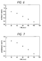
- Fig. 6 is a graph showing quenching of fluorescence of acridine obtained in Example 8.
- Fig. 7 is a graph showing decrease of absorbance ratio of acridine obtained in Example 8.
- the present invention provides a method of detection of a hybrid grounded on a novel principle different from that of prior art.
- the method of the present invention detects formation of a double helical structure resulting from hybrid formation without a B/F separation step. Further, the present invention enables detection of only a desired normal double helical structure with improved accuracy, even when non-specific adsorption or mismatching occurs, by selecting the conditions for detection only of a normal double helical structure without any additional treatment.
- the present invention is applicable to the cases where the nucleic acid forms a regular double helix such as the cases of DNA-DNA hybridization, and DNA-RNA hybridization.
- hybridization reaction has generally been conducted after immobilizing the nucleic acid (DNA or RNA).
- DNA or RNA nucleic acid
- hybridization in a solution when a nucleic acid forms a double strand of a certain length, the double strand is expected to form a double helical structure.
- the inventors of the present invention noticed the differences of a single-stranded nucleic acid from a double-stranded nucleic acid (hybrid) in the higher order structures and chemical properties, and established a detection system, whereby the present invention has been completed.
- the nucleic acid base portions forms a base pair by hydrogen bonding with the phosphoric acid portions and the sugar portions directing outside.
- the nucleic acid bases are stabilized by mutual stacking and take position at the center of the helix axis.
- the double helical structure includes an A form, a B form, a C form, a Z form, and variations thereof, which differ not only in the base sequence but in pitch length, symmetry of the helix, the width of the groove, the depth of the groove, etc. depending on the ion species, the salt concentration, etc. in annealing. Even with the same sequence, the double helical structure is said to vary depending on the conditions.
- DNA is said to be in a B form structure, in which the pitch is 33.8 ⁇ and the number of nucleic acid base is ten bases per pitch.
- the present invention relates to a method for detection of formation of a double helical structure by measuring a chemical change of an added reagent which causes a detectable change through a double helical structure of a hybrid.
- the useful reagents include two types:
- a probe having the reagent linked thereto is reacted with a sample solution to form a hybrid of the probe and a targeted nucleic acid, and the double helical structure of the hybrid is detected by the change of the constituents of the nucleic acid.
- the change caused by reaction of the reagent bonded to the probe with the double helical structure may be detected by direct measurement of the above change of the reagent if this change is measurable by itself, or otherwise by inducing other change which makes the change of the reagent detectable and by measuring the resulting detectable change.
- the change of the constituent of the nucleic acid is detected by decomposition into nucleic acid and nucleotide and analyzing directly by HPLC, or by use of an antigen to the changed nucleoside.
- the reagent which interacts with the double helical structure includes those which react with nucleic acid, etc. constituting the double helical structure to change by itself; those which causes a chemical or structural change in the double helix; and those which cause a detectable change by reacting with a third substance added in the reaction system in the presence of the double helical structure.
- Such interaction includes charge transfer between the double helical structure and the reagent may be used.
- the reagent is employed which functions as an electron donor or an electron acceptor.
- the interaction is detectable, for example, as a change caused by the hybrid formation in the chemical structure of the reagent, double helix, or a third substance capable of interaction with the above substance, as a change of electronic state, or as the change in signal given by the changed substance.
- the interaction grounded on the electron transfer includes the cases of "through-space” and “through-bond” similarly as in the (b) type reagent shown later.
- the examples are charge transfer through the stack of the nucleic acid base pairs and charge transfer caused by proximity effect between the electron donor and the electron acceptor resulting from the change of the structure to double helix.
- a mediator for the charge transfer may be used.
- the change of the interaction between the double helical structure and the reagent may be detected by measuring the change of the electron acceptor. This method is classified according to the detection means.
- the transferred charge is detected, by use of a spin-labeling agent, as spectrum change in ESR by spin elimination or the like method, or detected by emergence of a new absorption spectrum like a charge transfer absorption band or a change of absorption spectrum.
- a spin-labeling agent as spectrum change in ESR by spin elimination or the like method
- a new absorption spectrum like a charge transfer absorption band or a change of absorption spectrum.
- the change can be confirmed visually: this system is useful as a simplified system.
- a light-emitting system like fluorescence and phosphorescence is also useful for the reaction in which the fluorescence or the phosphorescence arises or disappears as the result of the interaction.
- An electron acceptor which changes chemically into another substance by the charge transfer may be used for the detection of the charge transfer, where the substance formed by the change may be detected by adding a third substance to the system to cause chemiluminescence.
- the third substance is an enzyme or an antigen, it may be detected by bioluminescence.
- the interaction may be detected by the change of an electron donor instead of the aforementioned change of an electron acceptor.
- the electron donor is a fluorescent substance
- the change like fluorescence quenching may be directly detected by decrease of quantum yield of the fluorescence as the consequence of charge transfer, or otherwise the resulting change may be made visible by an additional reaction.
- a third substance may be added which stimulates the reagent electron donor to generate electron provided that the third substance does not be activated to emit electron to cause charge transfer.
- the reagent is an electron acceptor to the double helical structure, it may be activated and thereby electron is drawn out from the electron acceptor, where the activation may be caused by light or another initiating agent like in the case of the electron donor.
- a mediator for mediating the charge transfer or a sensitizer may be present as a third substance as mentioned before. Such a substance may interact with the double helix to promote charge transfer to the electron donor or the electron acceptor which is not directly bonded to the double helix.
- the reagent of type (a) which combines with the probe may be any substance which has an oxidation-reduction potential' approximate to the oxidation-reduction potential of the nucleic acid.
- examples thereof include reagents capable of pulling out an electron such as riboflavin; oxidizing agents such as N,N'-dimethyl-2,7-diazapyrenium ion; basic dyes such as xanthene dyes, e.g., Rose Bengale, Floxin B, eosin, etc. and azine dyes, e.g., methylene blue, Safranine T, etc.
- the reagent may be any substance which causes a detectable change in the reagent itself or the nucleic acid.
- a hybrid is detected by measuring a change given by interaction between the two or more reagents caused by the structural change of nucleic acid from a single strand form to a double strand form.
- the interaction herein includes charge transfer.
- the "two or more reagents" comprises at least one combination of an electron donor and an electron acceptor, and occurrence of the interaction is detected, for example, as a change of the chemical structure, a change of the electronic state, or a change of a signal of the changed substance which is brought about by the hybrid formation.
- the relation between the electron donor and the electron acceptor depends on the energy states of the both substances.
- the electron donor or the electron acceptor is not necessarily used as generally defined.
- the electron donor and the electron acceptor are suitably selected.
- anthracene is classified as a typical electron donor according its oxidation-reduction potential, but simultaneously has characteristics of an electron acceptor.
- the interaction of the electron donor and the electron acceptor includes the cases of "through-space” and "through-bond".
- the through-space cases include interaction through the stack of nucleic acid base pairs and interaction caused by proximity effect between the electron donor and the electron acceptor resulting from the change of the structure to a double helix.
- the through-bond cases include transfer of electric charge through bases, phosphoric acid moieties, and sugar moieties constituting the nucleic acid.
- the type of the interaction is not limited provided that the interaction is caused by the double helix formation.
- the interaction through the stack of nucleic acid base pairs is a phenomenon that electrons released from an electron donor is transferred by electron cloud spreading over the nucleic acid base pairs through the adjacent nucleic acid pairs successively to an electron acceptor when the distance between the electron donor and the electron acceptor is so large that the interaction cannot be caused primarily, or conversely a phenomenon that an electron acceptor pulls out an electron from a nucleic acid base pair and electron is pulled out successively, finally from an electron donor.
- the nucleic acid base pairs function as a mediator in charge transfer.
- the interaction caused by proximity effect between the electron donor and the electron acceptor occurs when formation of double helical structure brings an electron donor and an electron acceptor close to each other to enable interaction.
- an electron donor and an electron acceptor are both bonded to a probe at such a distance that the interaction does not occur when the probe is in a single-stranded state and that the interaction occurs when the probe hybridizes with a targeted nucleic acid into a double helical structure.
- the formation of a double helical structure can be detected by detecting the occurrence of the interaction. If the charge transfer does not occur readily through the double helical structure between the electron donor and the electron acceptor, a mediator which mediates the charge transfer, or a sensitizer may naturally be added.
- the electron donor and the electron acceptor are required to be positioned at such sites that the donor and the acceptor are capable of reacting with the double helical structure to cause the interaction as described above in the present invention.
- the reagent is positioned so as to be capable of reacting with the double helical structure by insertion between a nucleic acid base pair like an intercalater, by embedding in a groove of the double helical structure, by placing close to the double helical structure, or by a like method.
- the reagent is required essentially in the present invention to be positioned to be specific to the double helical structure of a hybrid formed from a single-stranded probe and a targeted nucleic acid.
- the intercalater is generally a planar compound having spreading of electrons.
- the intercalater is placed and oriented in a direction parallel to the nucleic acid base pairs between the base pairs at the same distance as that between the base pairs. For example, when an intercalater is used as the electron donor and an electron acceptor is placed at the opposite side of the double helix, an electron released from the electron donor can be sent to the adjacent nucleic acid base pair, and further through the electron cloud of the nucleic acid base pairs toward the electron acceptor.
- an intercalater when used as the electron acceptor and an electron donor is placed at the opposite side of the double helix, an electron can be pulled out by an electron hole of the electron acceptor from an adjacent nucleic acid pair, and further electron is pulled out successively between the nucleic acid bases, and finally from the electron donor, thus causing charge transfer.
- the electron donor and the electron acceptor in charge transfer through stacked base pairs, is preferably an intercalater. More preferably both of them are respectively an intercalaters for improving the charge transfer efficiency.
- the intercalater is known to stabilize the double helical structure and to raise the melting temperature thereof. Therefore, use of intercalater as the electron donor or the electron acceptor is advantageous in stabilizing the hybrid of the probe and the targeted nucleic acid.
- the change of the interaction between two or more reagents resulting from double helix formation can be detected by change of the electron acceptor. This method is classified according to the detection means.
- the transferred charge is detected, by use of a spin-labeling agent, as spectrum change in ESR by spin elimination or the like method, or detected by emergence of a new absorption spectrum like a charge transfer absorption band or a change of absorption spectrum.
- a spin-labeling agent as spectrum change in ESR by spin elimination or the like method
- a new absorption spectrum like a charge transfer absorption band or a change of absorption spectrum.
- the change can be confirmed visually: this system is useful as a simplified system.
- a light-emitting system like fluorescence and phosphorescence is also useful for the reaction in which the fluorescence or the phosphorescence arises or disappears as the result of the interaction.
- An electron acceptor which changes chemically into another substance by the charge transfer may be used for the detection of the charge transfer, where the substance formed by the change may be detected by addition of a third substance to the system to cause chemiluminescence by the reaction of the two substances.
- the third substance is an enzyme or an antigen, it may be detected by bioluminescence.
- the interaction may be detected by the change of an electron donor instead of the aforementioned change of an electron acceptor.
- the electron donor is a fluorescent substance
- the change like fluorescence quenching may be directly detected by decrease of quantum yield of the fluorescence as the consequence of charge transfer, or the resulting change may be made visible by an additional reaction.
- a third substance may exist which stimulates the reagent electron donor to generate an electron except in the cases where the electron acceptor is activated by light or the like to release an electron and initiate charge transfer.
- the electron acceptor may be activated and thereby electron is pulled out from the electron acceptor, where the activation may be caused by light or another initiating agent like in the case of the electron donor.
- a mediator for the charge transfer, or a sensitizer may be present as a third substance as mentioned before. Such a substance may interact with the double helix to promote charge transfer to the electron donor or the electron acceptor which is not directly bonded to the double helix.
- the electron donor and the electron acceptor, used in the present invention may possibly interact each other irrespectively of the presence or absence of the double helical structure, which will increase background of the measurement to lower S/N ratio. Therefore, at least one of them is desirably linked to the probe preliminarily for precise detection.
- the linking of the electron donor or the electron acceptor to the probe is made through a linker such as -(CH2) n -, if necessary.
- the linking site is selected so as to achieve the interaction most efficiently.
- the most desirable is the case where both of the electron donor and the electron acceptor are linked to the probe.
- respective sites of the interacting reagents are known, so that the interaction can be controlled by selecting the sites on the probe, advantageously.
- the spatial interval between the electron donor and the electron acceptor on the probe is selected suitably depending on the kinds thereof. For example, for utilizing proximity effect, the interval is preferably in the range of from 20 to 120 ⁇ more preferably from 50 to 80 ⁇ in the formed hybrid, namely in the double helical structure.
- the interval is decided in consideration of change of the DNA structure such as unwinding caused by the respective dyes, and is preferably in the range of from 20 to 120 ⁇
- the preferred interval differs depending on whether the electron donor and the electron acceptor are both an intercalater or one or both of them are inserted to the primary groove or the secondary groove. Therefore, the interval is not limited to be in the aforementioned range. If the interval is too short, the charge transfer between the donor and the acceptor occurs directly without the interaction of the nucleic acid. Therefore, the interval is more preferably in the range of from 50 to 80 ⁇
- the donor and the acceptor are preferably linked separately at the respective ends of the probe from the ease of linking although it depends on the length of the probe.
- the length of the probe is selected suitably in an individual case so as to be capable of satisfactory hybridisation with a targeted nucleic acid and of forming a stable double helical structure.
- the length of the probe is decided in consideration of the interval of the linked donor and acceptor so that the interval is sufficient to prevent the interaction.
- the length is usually 8 bases or more, preferably 12 bases or more.
- the other donor or acceptor is present in the reaction system during formation of the double helical structure by hybridization of the probe and the targeted nucleic acid and the interaction is allowed to proceed after the double helix formation; and (b) the other donor or acceptor is added to the reaction system after the double helix formation by hybridisation to cause the interaction.
- the both of the donor and the acceptor are linked to the probe, the both are present in the reaction system during hybridisation, and the interaction is caused after the double helix formation.
- the specific examples of the reagents include spin-labeling agents such as 4,4-dimethyloxazolidine-N-oxyl (DOXXL) and its derivatives, 2,2,5,5-tetramethylpyrrolidine-N-oxyl (PROXL), and 2,2,6,6-tetramethylpiperidine-N-oxyl (TEMPO) and its derivatives; fluorescent intercalaters such as acridine, anthracene, pyrene, ethidium bromide, pyrylium, proflavine, porphyrin, thiazole orange dimer (TOTO), oxazole yellow (YOYO), 4',6-diamino-2-phenylindole dihydrochloride (DAPI), propidium iodide (PI), and the like and their derivatives; fluorescent pigments such as cyanine, azulene, three-nucleus pigment, dansyl, fluorescein, eosin,
- the base sequence was as below: 5'-GTTGTAAAACGACGGCCAGT-3'
- CDI carbonyl-N,N'-diimidazole
- the spin-labeled oligonucleotide was separated by aqueous concentrated ammonia, deprotected in a conventional manner, and purified by RPLC in a conventional manner.
- 0.2 ⁇ M of the oligonucleotide probe, prepared in the above process [1] having TEMPO introduced thereto, and 0.2 ⁇ M of M13mp18DNA (made by Takara Shuzo Co, Ltd.) were heated to 80°C in 1mM phosphate buffer solution/145mM NaCl/5mM KCl. It was gradually cooled to room temperature. Thereby, a probe-targeted DNA hybrid was prepared. To the reaction solution, fluorescein (made by Kodak Co.) was added to a final concentration of 10 ⁇ M. The resulting solution was subjected to ESR spectrum measurement as described below. Separately, the same operation was conducted without using M13mp18DNA to obtain a sample (probe alone).
- ESR ESR Frequency: 9.42 GHz Modulation: 100 kHz, 0.1 mT Field: 335 mT Time constant: 0.3 sec Power: 10 mW Sweep time: 8 min Receiver gain: 1.25 ⁇ 1000 Light irradiation Monochrometer: 490 nm Power supply: 88.5 V - 89 V/ 22A
- Fig. 1A shows the change of the signal intensity ratio with lapse of time
- Fig. 1B shows the change of the ESR signal line width with lapse of time.
- the probe alone the light irradiation caused neither the change of the intensity ratio nor the change of the line width.
- fluorescein/probe/M13mp18DNA without light irradiation also, no change was observed in the intensity ratio and the line width.
- Example 1 The same experiment was conducted as in Example 1 except that the base sequence of the probe was as shown below. 5'-GTTGTAAAAGGACGGCCAGT-3' This sequence in the probe is different from the one in Example 1 in that the tenth base from the 5'-terminal is G instead of C, which means that one base is different from the sequence in Example 1, whereby the probe is mismatched with the M13mp18DNA.
- a 20-mer oligonucleotide which had a base sequence partially complementary to a single stranded DNA, M13mp18DNA, as the targeted DNA in Example 1 was synthesized on 3'-amino-modifier CPG (1 ⁇ mol) made by Gren Research Co. as the support by means of an automatic DNA synthesizing apparatus 381A made by ABI Co. 5'-terminal dimethoxytrityl group was removed on the automatic synthesizing apparatus.
- the nucleotide (1 ⁇ mol) synthesized in the above step (1) together with the CGP support thereof was transferred into a gastight syringe. The subsequent reaction was allowed to proceed in this syringe.
- Onto the CPG support was added a solution of 50 mg of carbonyl-N,N'-diimidazole (CDI) in 1 ml of dioxane. It was left standing at room temperature for one hour. After washing with dioxane, 0.4 ml of 0.2M 4-aminohexylamino-TEMPO solution in DMSO used in Example 1 was added thereto. It was left standing at 55°C for 24 hours, and then washed successively with DMSO, dioxane, and methanol, and dried under a reduced pressure.
- CDI carbonyl-N,N'-diimidazole
- the spin-labeled oligonucleotide was separated by aqueous concentrated ammonia, deprotected in a conventional manner, and purified by RPLC in a conventional manner. Thus a spin-labeled oligonucleotide labeled at 5'-terminal with TEMPO was obtained.
- the spin-labeled oligonucleotide synthesized in the above step (2) has 3-amino-2-hydroxy group at the 3'-terminal sugar moiety. This amino group was bonded with FITC (fluorescein isothiocyanate, made by Sigma Co.) through the step below.
- FITC fluorescein isothiocyanate
- a solution of 0.2 ⁇ mol of the spin-labeled oligonucleotide synthesized in the above step (2) in 700 ⁇ l water was mixed with 100 ⁇ l of 1M sodium carbonate buffer solution (pH: 9).
- a solution of 2 mg of FITC in DMF was added, and reacted at 35°C for 24 hours.
- the reaction mixture was treated by a gel filtration column (NAP-25, made by Pharmacia Co.) to remove a large excess of FITC, and then purified by RPLC to obtain a probe having TEMPO bonded at the 5'-terminal and FITC at the 3'-terminal.
- the probe and the M13mp18DNA obtained in the above step (3) each in an amount of 0.2 ⁇ M were annealed in a conventional manner. Change of the intensity ratio and the line width of the ESR signal were measured in the same manner as in Example 1.
- the probe alone, and the non-irradiated probe-targeted DNA hybrid did not exhibit change of the intensity ratio, while the probe-targeted DNA hybrid, when irradiated by light of 490 nm which is excitation wavelength of fluorescein, exhibited decrease of the signal with time (see Fig. 2), thereby occurrence of charge transfer being confirmed.
- the spin disappearance degree was higher than that in Example 1 where fluorescein was not bonded to the probe. Thereby it was confirmed that electron transfer is more effectively conducted when an electron donor is linked to the one terminal and an electron acceptor is linked at the other terminal.
- Example 3 An oligonucleotide having the same base sequence as that of Example 2 which is different from M13mp18DNA at one base at the middle portion was synthesized , and TEMPO and fluorescein were bonded at the both terminals of the oligonucleotide in the same manner as in Example 3.
- a 20-mer oligonucleotide having the base sequence as that of Example 1 and having 3'-amino group bonded thereto was synthesized by means of an automatic DNA synthesizing apparatus.
- Riboflavine was oxidized to change its -CH2OH group to a -COOH group. This riboflavine was reacted with the above 3'-amino-oligonucleotide to obtain an oligonucleotide having riboflavine group bonded thereto at the one terminal.
- the riboflavine bonded oligonucleotide probe was hybridized with M13mp18 DNA.
- the reaction medium was a 10 mM phosphate buffer solution.
- the final concentration of the probe was adjusted to 3 ⁇ M, 6 ⁇ M, 10 ⁇ M, and 20 ⁇ M,
- the final concentration of the M13mp18DNA was adjusted to 10 ⁇ M.
- the solution of the hybrid of the riboflavine-bonded probe and the M13mp18DNA was irradiated with light of 470 nm, the excitation wavelength of riboflavine, for 5 minutes.
- a riboflavine-bonded 20-mer oligonucleotide was prepared in the same manner as in Example 5. A hybrid was formed between this oligonucleotide as a probe, and M13mp18DNA (as a target DNA).
- the reaction medium was a 10 mM phosphate buffer solution. The final concentration of the probe was adjusted to 3 ⁇ M, 6 ⁇ M, 10 ⁇ M, and 20 ⁇ M. The final concentration of the M13mp18DNA was adjusted to 10 ⁇ M.
- the solution of the hybrid of the riboflavine-bonded probe and the M13mp18DNA was irradiated with light of 470 nm, the excitation wavelength of riboflavine, for 5 minutes.
- a riboflavine-bonded 20-mer oligonucleotide was prepared in the same manner as in Example 5. A hybrid was formed between this oligonucleotide as a probe, and M13mp18DNA (as a target DNA).
- the reaction medium was a 10 mM phosphate buffer solution. The final concentration of the probe was adjusted to 3 ⁇ M, 6 ⁇ M, 10 ⁇ M, and 20 ⁇ M. The final concentration of the M13mp18DNA was adjusted to 10 ⁇ M.
- the solution of the hybrid of the riboflavine-bonded probe and the M13mp18DNA was irradiated with light of 470 nm, the excitation wavelength of riboflavine, for 5 minutes.
- the light absorbance of the riboflavine was measured at 450 nm for each of the samples.
- Fig. 5 shows the results.
- the solution containing only of riboflavine, the solution containing only of riboflavine-bonded probe, and the solution containing M13mp18DNA and riboflavine not bonded to a probe showed a constant optical density of 1.5.
- the solution containing the hybrid of the probe and M13mp18DNA showed decrease of the light absorbance, which shows that the riboflavine had changed into another substance as the result of charge transfer.
- DAP2+ was synthesized according to the method of Hunig (Ann. Chem. 1973, 339).
- the purified DAP2+ was reacted with succinic anhydride by Friedel-Craft reaction, thereby obtaining a purified product (I) having a carboxyl group.
- 0.5 g of the purified product (I) was placed in a 100 ml light-resistant reaction vessel, and dissolved in 30 ml of dried DMF. After the solution was cooled to -10°C, 0.4 g of N,N'-succinimidyl carbonate was added thereto. The reaction was allowed to proceed at that temperature for 5 hours. The reaction solution was poured into 150 ml of chloroform.
- an amino group was introduced to the 3'-terminal of the oligonucleotide by use of a Fmoc 3'-amino-modifier-CPG column (III) made by Miligene Co. in place of the ordinary amidide reagent CPG.
- a thiol group was introduced to the 5'-terminal thereof by use of a 5'-hexanol-thiol-linker (III') made by Miligene Co. in place of the ordinary amidide reagent.
- the oligonucleotide was cut out from the CPG support, deprotected, and purified by high speed liquid chromatography according to a predetermined protocol.
- the oligonucleotide has the base sequence as below: 5'-GTTGTAAAACGACGGCCAGT-3'
- 0.2 ⁇ M of the probe complex (VI) obtained in the step (4) above and 0.2 ⁇ M of M13mp18DNA were added to a mixed solution of 1mM phosphate buffer solution (pH: 7.0)/145mM NaCl/5mM KCl, and was heated to 80°C. The mixture was gradually cooled to room temperature to obtain a hybrid of the probe complex(VI) with M13mp18DNA. The absorption spectrum of acridine was shifted toward longer wavelength by about 20 nm as the result of the hybrid formation.
- a light irradiation apparatus equipped with a monochrometer was used.
- the probe and/or the hybrid were irradiated with light of 490 nm, the excitation wavelength of acridine, and the fluorescence at 533 nm was monitored.
- the probe itself showed slight quenching of fluorescence during long time of irradiation.
- the fluorescence intensity ratio of the hybrid of the probe complex (VI) with M13mp18DNA was measured relative to the fluorescence strength of the probe itself taken as 1 at each of the irradiation time. The results are shown in Fig. 6.
- the fluorescence of the hybrid decreased with the irradiation time. This means that the fluorescence of acridine was quenched as the result of charge transfer from acridine to DAP2+ through the aid of the double strand of the probe complex (VI)/M13mp18DNA hybrid. Thus the hybrid of the probe complex (VI)/M13mp18DNA was detected without B/F separation.
- the probe complex (VI) itself showed decrease of the absorbance owing to charge transfer between acridine and DAP2+.
- the absorbance ratio of the hybrid of the probe complex (VI) and M13mp18DNA was measured relative to the absorbance the probe itself taken as 1 at each of the irradiation time. The results are shown in Fig. 7.
- the absorbance of the hybrid of the probe complex (VI) with M13mp18DNA decreased significantly. This means that the charge transferred from acridine to DAP2+ through the stacked base pairs.
- a probe which has acridine at the 5'-terminal and DAP2+ at the 3'-terminal linked thereto was prepared in the same manner as in Example 8 except that the oligonucleotide of the base sequence shown below was used as the probe.
- the resulting probe was reacted with M13mp18DNA, and the fluorescence quenching and decrease of absorbance were measured in the same manner as in Example 8.
- 5'-GTTGTAAAAGGACGGCCAGT-3' This base sequence is different from the one in Example 8 in that the tenth base from the 5'-terminal is G instead of C, which means that one sequence is different from the sequence in Example 8 whereby the probe is mismatched with the M13mp18DNA.
- the method of detection of a targeted nucleic acid of the present invention has advantages that B/F separation is not necessary. Consequently various operations essential in conventional methods can be omitted such as removal of an excess prove, troublesome treatment for eliminating non-specific adsorption, investigation of operation conditions, and so forth.
- the present invention enables selective detection of a hybrid which has precise double helical structure by selecting reagents so that only the signal change of the precise hybrid may be detected, even in the case where mismatching occurs in a reaction system.
- a method for detecting a nucleic acid hybrid comprises steps of adding a nucleic acid probe into a sample solution containing a targeted nucleic acid, and detecting a double helical structure of a hybrid formed between the probe and the targeted nucleic acid, wherein the step for detecting the double helical structure comprises incorporating, into the sample solution, two or more kinds of reagents which are capable of causing a detectable change by interaction therebetween through the double helical structure and measuring the change caused by the interaction of the reagents; and at least one of the two or more kinds of reagents is joined to the probe.
Landscapes
- Chemical & Material Sciences (AREA)
- Organic Chemistry (AREA)
- Life Sciences & Earth Sciences (AREA)
- Zoology (AREA)
- Wood Science & Technology (AREA)
- Proteomics, Peptides & Aminoacids (AREA)
- Health & Medical Sciences (AREA)
- Engineering & Computer Science (AREA)
- Microbiology (AREA)
- Biochemistry (AREA)
- Physics & Mathematics (AREA)
- Molecular Biology (AREA)
- Biotechnology (AREA)
- Biophysics (AREA)
- Analytical Chemistry (AREA)
- Immunology (AREA)
- Bioinformatics & Cheminformatics (AREA)
- General Engineering & Computer Science (AREA)
- General Health & Medical Sciences (AREA)
- Genetics & Genomics (AREA)
- Measuring Or Testing Involving Enzymes Or Micro-Organisms (AREA)
- Investigating Or Analysing Biological Materials (AREA)
Applications Claiming Priority (4)
| Application Number | Priority Date | Filing Date | Title |
|---|---|---|---|
| JP318959/92 | 1992-11-27 | ||
| JP31895992A JP3706636B2 (ja) | 1992-11-27 | 1992-11-27 | 核酸ハイブリッド体の検出方法、プローブ、標的核酸の有無の確認方法、および2本鎖核酸ハイブリッド内のミスマッチの検出方法 |
| JP32050092A JP3618768B2 (ja) | 1992-11-30 | 1992-11-30 | 標的核酸の検出方法、プローブ、2本鎖核酸ハイブリッド内のミスマッチの検出方法及びプローブの製造方法 |
| JP320500/92 | 1992-11-30 |
Publications (3)
| Publication Number | Publication Date |
|---|---|
| EP0599337A2 true EP0599337A2 (de) | 1994-06-01 |
| EP0599337A3 EP0599337A3 (en) | 1995-08-30 |
| EP0599337B1 EP0599337B1 (de) | 2006-03-08 |
Family
ID=26569557
Family Applications (1)
| Application Number | Title | Priority Date | Filing Date |
|---|---|---|---|
| EP93119103A Expired - Lifetime EP0599337B1 (de) | 1992-11-27 | 1993-11-26 | Verfahren und Sonde zum Nachweis von Nukleinsäuren |
Country Status (4)
| Country | Link |
|---|---|
| US (1) | US5939256A (de) |
| EP (1) | EP0599337B1 (de) |
| AT (1) | ATE319853T1 (de) |
| DE (1) | DE69333991T2 (de) |
Cited By (31)
| Publication number | Priority date | Publication date | Assignee | Title |
|---|---|---|---|---|
| US5705348A (en) * | 1993-12-10 | 1998-01-06 | California Institute Of Technology | Nucleic acid mediated electron transfer |
| US5770369A (en) * | 1993-12-10 | 1998-06-23 | California Institute Of Technology | Nucleic acid mediated electron transfer |
| WO1999001578A1 (en) * | 1997-07-01 | 1999-01-14 | Lorne Park Research, Inc. | Assaying nucleotides in solution using a fluorescent intensity quenching effect |
| US5952172A (en) * | 1993-12-10 | 1999-09-14 | California Institute Of Technology | Nucleic acid mediated electron transfer |
| US6013459A (en) * | 1997-06-12 | 2000-01-11 | Clinical Micro Sensors, Inc. | Detection of analytes using reorganization energy |
| US6060242A (en) * | 1997-02-27 | 2000-05-09 | Lorne Park Research, Inc. | PNA diagnostic methods |
| US6071699A (en) * | 1996-06-07 | 2000-06-06 | California Institute Of Technology | Nucleic acid mediated electron transfer |
| US6090933A (en) * | 1996-11-05 | 2000-07-18 | Clinical Micro Sensors, Inc. | Methods of attaching conductive oligomers to electrodes |
| US6096273A (en) * | 1996-11-05 | 2000-08-01 | Clinical Micro Sensors | Electrodes linked via conductive oligomers to nucleic acids |
| US6156506A (en) * | 1994-05-26 | 2000-12-05 | Canon Kabushiki Kaisha | Method for detecting a target substance in a sample, utilizing pyrylium compound |
| US6194148B1 (en) | 1997-02-07 | 2001-02-27 | Olympus Optical Co., Ltd. | Method for detecting a hybridized nucleic acid molecule |
| US6232062B1 (en) | 1997-03-07 | 2001-05-15 | Clinical Micro Sensors, Inc. | AC methods for the detection of nucleic acids |
| US6251591B1 (en) | 1997-02-27 | 2001-06-26 | Lorne Park Research, Inc. | Quantitative method for detecting nucleotide concentration |
| US6255050B1 (en) | 1998-05-22 | 2001-07-03 | Lorne Park Research, Inc. | Dynamic hybridization system |
| US6264825B1 (en) | 1998-06-23 | 2001-07-24 | Clinical Micro Sensors, Inc. | Binding acceleration techniques for the detection of analytes |
| GB2365866A (en) * | 2000-05-12 | 2002-02-27 | Molecular Probes Inc | Quenching oligonucleotides |
| US6444423B1 (en) | 1996-06-07 | 2002-09-03 | Molecular Dynamics, Inc. | Nucleosides comprising polydentate ligands |
| US6541617B1 (en) | 1998-10-27 | 2003-04-01 | Clinical Micro Sensors, Inc. | Detection of target analytes using particles and electrodes |
| US6645733B1 (en) | 1999-06-25 | 2003-11-11 | Ingeneus Corporation | Fluorescent intensity method for assaying binding between proteins or peptides |
| US6686150B1 (en) | 1998-01-27 | 2004-02-03 | Clinical Micro Sensors, Inc. | Amplification of nucleic acids with electronic detection |
| US6740518B1 (en) | 1998-09-17 | 2004-05-25 | Clinical Micro Sensors, Inc. | Signal detection techniques for the detection of analytes |
| US6753143B2 (en) | 2000-05-01 | 2004-06-22 | Clinical Micro Sensors, Inc. | Target analyte detection using asymmetrical self-assembled monolayers |
| US6761816B1 (en) | 1998-06-23 | 2004-07-13 | Clinical Micro Systems, Inc. | Printed circuit boards with monolayers and capture ligands |
| US6833267B1 (en) | 1998-12-30 | 2004-12-21 | Clinical Micro Sensors, Inc. | Tissue collection devices containing biosensors |
| US6942771B1 (en) | 1999-04-21 | 2005-09-13 | Clinical Micro Sensors, Inc. | Microfluidic systems in the electrochemical detection of target analytes |
| US7045285B1 (en) | 1996-11-05 | 2006-05-16 | Clinical Micro Sensors, Inc. | Electronic transfer moieties attached to peptide nucleic acids |
| US7090804B2 (en) | 1998-01-27 | 2006-08-15 | Clinical Mirco Sensors, Inc. | Amplification of nucleic acids with electronic detection |
| US7160678B1 (en) | 1996-11-05 | 2007-01-09 | Clinical Micro Sensors, Inc. | Compositions for the electronic detection of analytes utilizing monolayers |
| US7172897B2 (en) | 2000-01-11 | 2007-02-06 | Clinical Micro Sensors, Inc. | Devices and methods for biochip multiplexing |
| US7312087B2 (en) | 2000-01-11 | 2007-12-25 | Clinical Micro Sensors, Inc. | Devices and methods for biochip multiplexing |
| US7935481B1 (en) | 1999-07-26 | 2011-05-03 | Osmetech Technology Inc. | Sequence determination of nucleic acids using electronic detection |
Families Citing this family (17)
| Publication number | Priority date | Publication date | Assignee | Title |
|---|---|---|---|---|
| US5620850A (en) | 1994-09-26 | 1997-04-15 | President And Fellows Of Harvard College | Molecular recognition at surfaces derivatized with self-assembled monolayers |
| US7087148B1 (en) | 1998-06-23 | 2006-08-08 | Clinical Micro Sensors, Inc. | Binding acceleration techniques for the detection of analytes |
| US6492111B1 (en) * | 1998-11-25 | 2002-12-10 | Isis Pharmaceuticals, Inc. | In situ binary synthesis of biologically effective molecules |
| US6960432B2 (en) | 1999-07-26 | 2005-11-01 | Canon Kabushiki Kaisha | Detection/quantification of targeted nucleotide chains, and detection/quantification of multi-stranded nucleotide chains by fluorescence |
| GB0205455D0 (en) | 2002-03-07 | 2002-04-24 | Molecular Sensing Plc | Nucleic acid probes, their synthesis and use |
| JP4458327B2 (ja) * | 2003-08-28 | 2010-04-28 | キヤノン株式会社 | 標的物質の定量方法および該方法に用いるプローブ担体 |
| JP4541731B2 (ja) * | 2004-03-12 | 2010-09-08 | キヤノン株式会社 | 核酸検出方法 |
| JP4183256B2 (ja) * | 2004-08-04 | 2008-11-19 | キヤノン株式会社 | 核酸増幅反応産物の鎖分離方法、核酸増幅反応産物の検出方法 |
| EP1815010A2 (de) * | 2004-09-24 | 2007-08-08 | Ingeneus Inc. | Genom-assay |
| JP2006094760A (ja) * | 2004-09-29 | 2006-04-13 | Canon Inc | 細胞のスクリーニング方法および口腔癌細胞検出方法 |
| US8535914B2 (en) * | 2005-01-21 | 2013-09-17 | Canon Kabushiki Kaisha | Probe, probe set and information acquisition method using the same |
| JP2007010413A (ja) * | 2005-06-29 | 2007-01-18 | Canon Inc | 核酸ハイブリッドの融点測定方法及びそのための装置 |
| US7642086B2 (en) * | 2005-08-09 | 2010-01-05 | Canon Kabushiki Kaisha | Labeled probe bound object, method for producing the same and method for using the same |
| US20090011413A1 (en) * | 2005-12-14 | 2009-01-08 | Canon Kabushiki Kaisha | Method for screening colon cancer cells and gene set used for examination of colon cancer |
| WO2007114516A1 (en) * | 2006-03-31 | 2007-10-11 | Canon Kabushiki Kaisha | Probe, probe set, probe-immobilized carrier, and genetic testing method |
| WO2007114517A1 (en) * | 2006-03-31 | 2007-10-11 | Canon Kabushiki Kaisha | Probe, probe set, probe-immobilized carrier, and genetic testing method |
| JP5729904B2 (ja) | 2009-06-02 | 2015-06-03 | キヤノン株式会社 | 細胞から蛋白質、dna、rnaを調製する方法 |
Family Cites Families (21)
| Publication number | Priority date | Publication date | Assignee | Title |
|---|---|---|---|---|
| US3786369A (en) * | 1972-12-27 | 1974-01-15 | Eastman Kodak Co | Laser dye composition |
| US4341894A (en) * | 1978-03-13 | 1982-07-27 | Eastman Kodak Company | Sensitizers for photoconductive compositions |
| US4840784A (en) * | 1982-12-22 | 1989-06-20 | Eastman Kodak Company | Use of pyrylium and thiapyrylium compounds as biological stains |
| US4555396A (en) * | 1982-12-22 | 1985-11-26 | Eastman Kodak Company | Use of pyrylium and thiapyrylium compounds as biological stains |
| US4820630A (en) * | 1984-11-23 | 1989-04-11 | Digene Diagnostics, Incorporated | Assay for nucleic acid sequences, particularly genetic lesions, using interactive labels |
| US5011769A (en) * | 1985-12-05 | 1991-04-30 | Meiogenics U.S. Limited Partnership | Methods for detecting nucleic acid sequences |
| CA1273552A (en) * | 1985-12-23 | 1990-09-04 | Michael J. Heller | Fluorescent stokes shift probes for polynucleotide hybridization assays |
| EP0232967B1 (de) * | 1986-01-10 | 1993-04-28 | Amoco Corporation | Kompetitiver homogener Test |
| JPS63238166A (ja) * | 1987-03-26 | 1988-10-04 | Mitsubishi Electric Corp | 有機電子素子材料 |
| JPH01153683A (ja) * | 1987-11-06 | 1989-06-15 | Eastman Kodak Co | アミノ置換チオピリリウム化合物 |
| AU622426B2 (en) * | 1987-12-11 | 1992-04-09 | Abbott Laboratories | Assay using template-dependent nucleic acid probe reorganization |
| JPH0275958A (ja) * | 1988-09-13 | 1990-03-15 | Kao Corp | Dna検出法 |
| US5324829A (en) * | 1988-12-16 | 1994-06-28 | Ortho Diagnostic Systems, Inc. | High specific activity nucleic acid probes having target recognition and signal generating moieties |
| US5229297A (en) * | 1989-02-03 | 1993-07-20 | Eastman Kodak Company | Containment cuvette for PCR and method of use |
| GB8902689D0 (en) * | 1989-02-07 | 1989-03-30 | Ici Plc | Assay method |
| CA2033692A1 (en) * | 1990-01-25 | 1991-07-26 | Wilhelm Bannwarth | Energy transfer systems |
| US5102784A (en) * | 1990-05-04 | 1992-04-07 | Oncor, Inc. | Restriction amplification assay |
| DE69128520T2 (de) * | 1990-10-31 | 1998-07-09 | Tosoh Corp | Verfahren zum Nachweis oder Quantifizierung von Zielnukleinsäuren |
| US5994056A (en) * | 1991-05-02 | 1999-11-30 | Roche Molecular Systems, Inc. | Homogeneous methods for nucleic acid amplification and detection |
| DK0567635T3 (da) * | 1991-11-15 | 2000-09-25 | Igen Int Inc | Hurtige assays for amplifikationsprodukter |
| US5591578A (en) * | 1993-12-10 | 1997-01-07 | California Institute Of Technology | Nucleic acid mediated electron transfer |
-
1993
- 1993-11-26 EP EP93119103A patent/EP0599337B1/de not_active Expired - Lifetime
- 1993-11-26 DE DE69333991T patent/DE69333991T2/de not_active Expired - Lifetime
- 1993-11-26 AT AT93119103T patent/ATE319853T1/de not_active IP Right Cessation
-
1996
- 1996-07-19 US US08/683,918 patent/US5939256A/en not_active Expired - Lifetime
Cited By (68)
| Publication number | Priority date | Publication date | Assignee | Title |
|---|---|---|---|---|
| US6268149B1 (en) | 1993-12-10 | 2001-07-31 | California Institute Of Technology | Nucleic acid mediated electron transfer |
| US6087100A (en) * | 1993-12-10 | 2000-07-11 | California Institute Of Technology | Nucleic acid mediated electron transfer |
| US5780234A (en) * | 1993-12-10 | 1998-07-14 | California Institute Of Technology | Nucleic acid mediated electron transfer |
| US7851146B2 (en) | 1993-12-10 | 2010-12-14 | California Institute Of Technology | Nucleic acid mediated electron transfer |
| US5952172A (en) * | 1993-12-10 | 1999-09-14 | California Institute Of Technology | Nucleic acid mediated electron transfer |
| US6258545B1 (en) | 1993-12-10 | 2001-07-10 | California Institute Of Technology | Nucleic acid mediated electron transfer |
| US5770369A (en) * | 1993-12-10 | 1998-06-23 | California Institute Of Technology | Nucleic acid mediated electron transfer |
| US6528266B2 (en) | 1993-12-10 | 2003-03-04 | California Institute Of Technology | Nucleic acid mediated electron transfer |
| US6200761B1 (en) | 1993-12-10 | 2001-03-13 | California Institute Of Technology | Nucleic acid mediated electron transfer |
| US6238870B1 (en) | 1993-12-10 | 2001-05-29 | California Institute Of Technology | Nucleic acid mediated electron transfer |
| US5705348A (en) * | 1993-12-10 | 1998-01-06 | California Institute Of Technology | Nucleic acid mediated electron transfer |
| US6277576B1 (en) | 1993-12-10 | 2001-08-21 | California Institute Of Technology | Nucleic acid mediated electron transfer |
| US6268150B1 (en) | 1993-12-10 | 2001-07-31 | California Institute Of Technology | Nucleic acid mediated electron transfer |
| US6177250B1 (en) | 1993-12-10 | 2001-01-23 | California Institute Of Technology | Nucleic acid mediated electron transfer |
| US6180352B1 (en) | 1993-12-10 | 2001-01-30 | California Insitute Of Technology | Nucleic acid mediated electron transfer |
| US6156506A (en) * | 1994-05-26 | 2000-12-05 | Canon Kabushiki Kaisha | Method for detecting a target substance in a sample, utilizing pyrylium compound |
| US7033760B2 (en) | 1995-06-07 | 2006-04-25 | California Institute Of Technology | Metallic solid supports modified with nucleic acids |
| US6291188B1 (en) | 1995-06-07 | 2001-09-18 | California Institute Of Technology | Metallic solid supports modified with nucleic acids |
| US6265155B1 (en) | 1995-06-07 | 2001-07-24 | California Institute Of Technology | Metallic solid supports modified with nucleic acids |
| US6071699A (en) * | 1996-06-07 | 2000-06-06 | California Institute Of Technology | Nucleic acid mediated electron transfer |
| US6444423B1 (en) | 1996-06-07 | 2002-09-03 | Molecular Dynamics, Inc. | Nucleosides comprising polydentate ligands |
| US6221583B1 (en) | 1996-11-05 | 2001-04-24 | Clinical Micro Sensors, Inc. | Methods of detecting nucleic acids using electrodes |
| US7384749B2 (en) | 1996-11-05 | 2008-06-10 | Clinical Micro Sensors, Inc. | Electrodes linked via conductive oligomers to nucleic acids |
| US7160678B1 (en) | 1996-11-05 | 2007-01-09 | Clinical Micro Sensors, Inc. | Compositions for the electronic detection of analytes utilizing monolayers |
| US7125668B2 (en) | 1996-11-05 | 2006-10-24 | Clinical Micro Sensors, Inc. | Electrodes linked via conductive oligomers to nucleic acids |
| US7056669B2 (en) | 1996-11-05 | 2006-06-06 | Clinical Micro Sensors, Inc. | AC methods for the detection of nucleic acids |
| US7045285B1 (en) | 1996-11-05 | 2006-05-16 | Clinical Micro Sensors, Inc. | Electronic transfer moieties attached to peptide nucleic acids |
| US6977151B2 (en) | 1996-11-05 | 2005-12-20 | Clinical Micro Sensors, Inc. | Electrodes linked via conductive oligomers to nucleic acids |
| US6096273A (en) * | 1996-11-05 | 2000-08-01 | Clinical Micro Sensors | Electrodes linked via conductive oligomers to nucleic acids |
| US6479240B1 (en) | 1996-11-05 | 2002-11-12 | Clinical Micro Sensors, Inc. | Electrodes linked via conductive oligomers to nucleic acids |
| US6090933A (en) * | 1996-11-05 | 2000-07-18 | Clinical Micro Sensors, Inc. | Methods of attaching conductive oligomers to electrodes |
| US6194148B1 (en) | 1997-02-07 | 2001-02-27 | Olympus Optical Co., Ltd. | Method for detecting a hybridized nucleic acid molecule |
| US6060242A (en) * | 1997-02-27 | 2000-05-09 | Lorne Park Research, Inc. | PNA diagnostic methods |
| US6251591B1 (en) | 1997-02-27 | 2001-06-26 | Lorne Park Research, Inc. | Quantitative method for detecting nucleotide concentration |
| US7381525B1 (en) | 1997-03-07 | 2008-06-03 | Clinical Micro Sensors, Inc. | AC/DC voltage apparatus for detection of nucleic acids |
| US6232062B1 (en) | 1997-03-07 | 2001-05-15 | Clinical Micro Sensors, Inc. | AC methods for the detection of nucleic acids |
| US7582419B2 (en) | 1997-06-12 | 2009-09-01 | Osmetech Technology Inc. | Detection of analytes using reorganization energy |
| US7566534B2 (en) | 1997-06-12 | 2009-07-28 | Osmetech Technology Inc. | Detection of analytes using reorganization energy |
| US7514228B2 (en) | 1997-06-12 | 2009-04-07 | Clinical Micro Sensors, Inc. | Detection of analytes using reorganization energy |
| US7579145B2 (en) | 1997-06-12 | 2009-08-25 | Osmetech Technology Inc. | Detection of analytes using reorganization energy |
| US6013170A (en) * | 1997-06-12 | 2000-01-11 | Clinical Micro Sensors, Inc. | Detection of analytes using reorganization energy |
| US6248229B1 (en) | 1997-06-12 | 2001-06-19 | Clinical Micro Sensors, Inc. | Detection of analytes using reorganization energy |
| US7018523B2 (en) | 1997-06-12 | 2006-03-28 | Clinical Micro Sensors, Inc. | Detection of analytes using reorganization energy |
| US6013459A (en) * | 1997-06-12 | 2000-01-11 | Clinical Micro Sensors, Inc. | Detection of analytes using reorganization energy |
| WO1999001578A1 (en) * | 1997-07-01 | 1999-01-14 | Lorne Park Research, Inc. | Assaying nucleotides in solution using a fluorescent intensity quenching effect |
| US7090804B2 (en) | 1998-01-27 | 2006-08-15 | Clinical Mirco Sensors, Inc. | Amplification of nucleic acids with electronic detection |
| US6686150B1 (en) | 1998-01-27 | 2004-02-03 | Clinical Micro Sensors, Inc. | Amplification of nucleic acids with electronic detection |
| US6255050B1 (en) | 1998-05-22 | 2001-07-03 | Lorne Park Research, Inc. | Dynamic hybridization system |
| US7655129B2 (en) | 1998-06-23 | 2010-02-02 | Osmetech Technology Inc. | Binding acceleration techniques for the detection of analytes |
| US6264825B1 (en) | 1998-06-23 | 2001-07-24 | Clinical Micro Sensors, Inc. | Binding acceleration techniques for the detection of analytes |
| US6290839B1 (en) | 1998-06-23 | 2001-09-18 | Clinical Micro Sensors, Inc. | Systems for electrophoretic transport and detection of analytes |
| US6761816B1 (en) | 1998-06-23 | 2004-07-13 | Clinical Micro Systems, Inc. | Printed circuit boards with monolayers and capture ligands |
| US9874542B2 (en) | 1998-09-17 | 2018-01-23 | Clinical Micro Sensors, Inc. | Signal detection techniques for the detection of analytes |
| US6740518B1 (en) | 1998-09-17 | 2004-05-25 | Clinical Micro Sensors, Inc. | Signal detection techniques for the detection of analytes |
| US6541617B1 (en) | 1998-10-27 | 2003-04-01 | Clinical Micro Sensors, Inc. | Detection of target analytes using particles and electrodes |
| US6833267B1 (en) | 1998-12-30 | 2004-12-21 | Clinical Micro Sensors, Inc. | Tissue collection devices containing biosensors |
| US6942771B1 (en) | 1999-04-21 | 2005-09-13 | Clinical Micro Sensors, Inc. | Microfluidic systems in the electrochemical detection of target analytes |
| US7534331B2 (en) | 1999-04-21 | 2009-05-19 | Osmetech Technology Inc. | Use of microfluidic systems in the electrochemical detection of target analytes |
| US8486247B2 (en) | 1999-04-21 | 2013-07-16 | Osmetch Technology, Inc. | Use of microfluidic systems in the electrochemical detection of target analytes |
| US9151746B2 (en) | 1999-04-21 | 2015-10-06 | Osmetech Technology, Inc. | Use of microfluidic systems in the electrochemical detection of target analytes |
| US9557295B2 (en) | 1999-04-21 | 2017-01-31 | Osmetech Technology, Inc. | Use of microfluidic systems in the electrochemical detection of target analytes |
| US6645733B1 (en) | 1999-06-25 | 2003-11-11 | Ingeneus Corporation | Fluorescent intensity method for assaying binding between proteins or peptides |
| US7935481B1 (en) | 1999-07-26 | 2011-05-03 | Osmetech Technology Inc. | Sequence determination of nucleic acids using electronic detection |
| US7312087B2 (en) | 2000-01-11 | 2007-12-25 | Clinical Micro Sensors, Inc. | Devices and methods for biochip multiplexing |
| US7172897B2 (en) | 2000-01-11 | 2007-02-06 | Clinical Micro Sensors, Inc. | Devices and methods for biochip multiplexing |
| US6753143B2 (en) | 2000-05-01 | 2004-06-22 | Clinical Micro Sensors, Inc. | Target analyte detection using asymmetrical self-assembled monolayers |
| GB2365866A (en) * | 2000-05-12 | 2002-02-27 | Molecular Probes Inc | Quenching oligonucleotides |
| GB2365866B (en) * | 2000-05-12 | 2002-07-31 | Molecular Probes Inc | Quenching oligonucleotides |
Also Published As
| Publication number | Publication date |
|---|---|
| EP0599337A3 (en) | 1995-08-30 |
| ATE319853T1 (de) | 2006-03-15 |
| EP0599337B1 (de) | 2006-03-08 |
| US5939256A (en) | 1999-08-17 |
| DE69333991D1 (de) | 2006-05-04 |
| DE69333991T2 (de) | 2006-12-14 |
Similar Documents
| Publication | Publication Date | Title |
|---|---|---|
| EP0599337B1 (de) | Verfahren und Sonde zum Nachweis von Nukleinsäuren | |
| EP0599338B1 (de) | Verfahren zum Nachweis einer Zielnukleinsäure | |
| US6451531B1 (en) | Dioxetane labeled probes and detection assays employing the same | |
| US4868103A (en) | Analyte detection by means of energy transfer | |
| KR960002561B1 (ko) | 균질보호분석법 | |
| Tan et al. | Molecular beacons: a novel DNA probe for nucleic acid and protein studies | |
| US4743535A (en) | Hybridization assay employing labeled probe and anti-hybrid | |
| US5827653A (en) | Nucleic acid detection with energy transfer | |
| JP2619866B2 (ja) | 競合的均質検定法 | |
| EP0684239B1 (de) | Verfahren zum Nachweis von einer Zielsubstanz in einer Probe mit Hilfe von Pyryliumverbindung | |
| US6297008B1 (en) | Process for detecting target nucleic acid, process for quantifying the same, and pyrylium compound for chemiluminescence analysis | |
| EP0144913A2 (de) | Hybridisierungstest mittels einer markierten Probe und Anti-Hybrid | |
| US20120040867A1 (en) | Microarrays of Binary Nucleic Acid Probes for Detecting Nucleic Acid Analytes | |
| EP1491629A1 (de) | Neues verfahren zur hochempfindlichen nukleinsäureanalyse | |
| CA2213240A1 (en) | Probes for nucleic acid analysis and method using these probes | |
| JP2007525218A (ja) | 生物学的に活性な化合物の検出 | |
| Schwechheimer et al. | Synthesis of Dye‐Modified Oligonucleotides via Copper (I)‐Catalyzed Alkyne Azide Cycloaddition Using On‐and Off‐Bead Approaches | |
| JP3618768B2 (ja) | 標的核酸の検出方法、プローブ、2本鎖核酸ハイブリッド内のミスマッチの検出方法及びプローブの製造方法 | |
| JP3706636B2 (ja) | 核酸ハイブリッド体の検出方法、プローブ、標的核酸の有無の確認方法、および2本鎖核酸ハイブリッド内のミスマッチの検出方法 | |
| US20100209909A1 (en) | Assays based on detection of photobleaching reaction products from dye catalytic complex | |
| AU780156B2 (en) | Hybridization self-recognition type probe | |
| JPH11286616A (ja) | 核酸染色剤、それを用いた二本鎖核酸の検出方法及び標的核酸の検出試薬 | |
| JP4114958B2 (ja) | 核酸ハイブリッド体の検出方法及びそれに用いるプローブ | |
| JPH10337199A (ja) | 核酸染色剤、それを用いた二本鎖核酸の検出方法及び標的核酸の検出試薬 | |
| JPH11290098A (ja) | 核酸染色剤、それを用いた二本鎖核酸の検出方法及び標的核酸の検出試薬 |
Legal Events
| Date | Code | Title | Description |
|---|---|---|---|
| PUAI | Public reference made under article 153(3) epc to a published international application that has entered the european phase |
Free format text: ORIGINAL CODE: 0009012 |
|
| AK | Designated contracting states |
Kind code of ref document: A2 Designated state(s): AT BE CH DE DK ES FR GB GR IT LI LU NL PT SE |
|
| PUAL | Search report despatched |
Free format text: ORIGINAL CODE: 0009013 |
|
| AK | Designated contracting states |
Kind code of ref document: A3 Designated state(s): AT BE CH DE DK ES FR GB GR IT LI LU NL PT SE |
|
| 17P | Request for examination filed |
Effective date: 19960115 |
|
| 17Q | First examination report despatched |
Effective date: 19990812 |
|
| GRAP | Despatch of communication of intention to grant a patent |
Free format text: ORIGINAL CODE: EPIDOSNIGR1 |
|
| GRAS | Grant fee paid |
Free format text: ORIGINAL CODE: EPIDOSNIGR3 |
|
| GRAA | (expected) grant |
Free format text: ORIGINAL CODE: 0009210 |
|
| AK | Designated contracting states |
Kind code of ref document: B1 Designated state(s): AT BE CH DE DK ES FR GB GR IT LI LU NL PT SE |
|
| PG25 | Lapsed in a contracting state [announced via postgrant information from national office to epo] |
Ref country code: IT Free format text: LAPSE BECAUSE OF FAILURE TO SUBMIT A TRANSLATION OF THE DESCRIPTION OR TO PAY THE FEE WITHIN THE PRE;WARNING: LAPSES OF ITALIAN PATENTS WITH EFFECTIVE DATE BEFORE 2007 MAY HAVE OCCURRED AT ANY TIME BEFORE 2007. THE CORRECT EFFECTIVE DATE MAY BE DIFFERENT FROM THE ONE RECORDED.SCRIBED TIME-LIMIT Effective date: 20060308 Ref country code: BE Free format text: LAPSE BECAUSE OF FAILURE TO SUBMIT A TRANSLATION OF THE DESCRIPTION OR TO PAY THE FEE WITHIN THE PRESCRIBED TIME-LIMIT Effective date: 20060308 Ref country code: AT Free format text: LAPSE BECAUSE OF FAILURE TO SUBMIT A TRANSLATION OF THE DESCRIPTION OR TO PAY THE FEE WITHIN THE PRESCRIBED TIME-LIMIT Effective date: 20060308 |
|
| REG | Reference to a national code |
Ref country code: GB Ref legal event code: FG4D |
|
| REG | Reference to a national code |
Ref country code: CH Ref legal event code: EP |
|
| REF | Corresponds to: |
Ref document number: 69333991 Country of ref document: DE Date of ref document: 20060504 Kind code of ref document: P |
|
| REG | Reference to a national code |
Ref country code: SE Ref legal event code: TRGR |
|
| PG25 | Lapsed in a contracting state [announced via postgrant information from national office to epo] |
Ref country code: DK Free format text: LAPSE BECAUSE OF FAILURE TO SUBMIT A TRANSLATION OF THE DESCRIPTION OR TO PAY THE FEE WITHIN THE PRESCRIBED TIME-LIMIT Effective date: 20060608 |
|
| PG25 | Lapsed in a contracting state [announced via postgrant information from national office to epo] |
Ref country code: ES Free format text: LAPSE BECAUSE OF FAILURE TO SUBMIT A TRANSLATION OF THE DESCRIPTION OR TO PAY THE FEE WITHIN THE PRESCRIBED TIME-LIMIT Effective date: 20060619 |
|
| REG | Reference to a national code |
Ref country code: CH Ref legal event code: NV Representative=s name: BOVARD AG PATENTANWAELTE |
|
| PG25 | Lapsed in a contracting state [announced via postgrant information from national office to epo] |
Ref country code: PT Free format text: LAPSE BECAUSE OF FAILURE TO SUBMIT A TRANSLATION OF THE DESCRIPTION OR TO PAY THE FEE WITHIN THE PRESCRIBED TIME-LIMIT Effective date: 20060808 |
|
| ET | Fr: translation filed | ||
| PLBE | No opposition filed within time limit |
Free format text: ORIGINAL CODE: 0009261 |
|
| STAA | Information on the status of an ep patent application or granted ep patent |
Free format text: STATUS: NO OPPOSITION FILED WITHIN TIME LIMIT |
|
| 26N | No opposition filed |
Effective date: 20061211 |
|
| PG25 | Lapsed in a contracting state [announced via postgrant information from national office to epo] |
Ref country code: GR Free format text: LAPSE BECAUSE OF FAILURE TO SUBMIT A TRANSLATION OF THE DESCRIPTION OR TO PAY THE FEE WITHIN THE PRESCRIBED TIME-LIMIT Effective date: 20060609 |
|
| PG25 | Lapsed in a contracting state [announced via postgrant information from national office to epo] |
Ref country code: LU Free format text: LAPSE BECAUSE OF NON-PAYMENT OF DUE FEES Effective date: 20061126 |
|
| PGFP | Annual fee paid to national office [announced via postgrant information from national office to epo] |
Ref country code: DE Payment date: 20101130 Year of fee payment: 18 |
|
| REG | Reference to a national code |
Ref country code: CH Ref legal event code: PFA Owner name: CANON KABUSHIKI KAISHA Free format text: CANON KABUSHIKI KAISHA#30-2, 3-CHOME, SHIMOMARUKO, OHTA-KU#TOKYO (JP) -TRANSFER TO- CANON KABUSHIKI KAISHA#30-2, 3-CHOME, SHIMOMARUKO, OHTA-KU#TOKYO (JP) |
|
| PGFP | Annual fee paid to national office [announced via postgrant information from national office to epo] |
Ref country code: GB Payment date: 20101126 Year of fee payment: 18 |
|
| PGFP | Annual fee paid to national office [announced via postgrant information from national office to epo] |
Ref country code: SE Payment date: 20111010 Year of fee payment: 19 Ref country code: CH Payment date: 20111109 Year of fee payment: 19 Ref country code: NL Payment date: 20111123 Year of fee payment: 19 Ref country code: FR Payment date: 20111212 Year of fee payment: 19 |
|
| REG | Reference to a national code |
Ref country code: NL Ref legal event code: V1 Effective date: 20130601 |
|
| REG | Reference to a national code |
Ref country code: CH Ref legal event code: PL |
|
| GBPC | Gb: european patent ceased through non-payment of renewal fee |
Effective date: 20121126 |
|
| PG25 | Lapsed in a contracting state [announced via postgrant information from national office to epo] |
Ref country code: LI Free format text: LAPSE BECAUSE OF NON-PAYMENT OF DUE FEES Effective date: 20121130 Ref country code: SE Free format text: LAPSE BECAUSE OF NON-PAYMENT OF DUE FEES Effective date: 20121127 Ref country code: CH Free format text: LAPSE BECAUSE OF NON-PAYMENT OF DUE FEES Effective date: 20121130 |
|
| REG | Reference to a national code |
Ref country code: FR Ref legal event code: ST Effective date: 20130731 |
|
| PG25 | Lapsed in a contracting state [announced via postgrant information from national office to epo] |
Ref country code: NL Free format text: LAPSE BECAUSE OF NON-PAYMENT OF DUE FEES Effective date: 20130601 |
|
| REG | Reference to a national code |
Ref country code: DE Ref legal event code: R119 Ref document number: 69333991 Country of ref document: DE Effective date: 20130601 |
|
| PG25 | Lapsed in a contracting state [announced via postgrant information from national office to epo] |
Ref country code: DE Free format text: LAPSE BECAUSE OF NON-PAYMENT OF DUE FEES Effective date: 20130601 |
|
| PG25 | Lapsed in a contracting state [announced via postgrant information from national office to epo] |
Ref country code: FR Free format text: LAPSE BECAUSE OF NON-PAYMENT OF DUE FEES Effective date: 20121130 Ref country code: GB Free format text: LAPSE BECAUSE OF NON-PAYMENT OF DUE FEES Effective date: 20121126 |