EP0752104B1 - Expansion differentielle de cellules souches foetales dans la circulation sanguine maternelle, pour l'analyse genetique prenatale - Google Patents
Expansion differentielle de cellules souches foetales dans la circulation sanguine maternelle, pour l'analyse genetique prenatale Download PDFInfo
- Publication number
- EP0752104B1 EP0752104B1 EP95915408A EP95915408A EP0752104B1 EP 0752104 B1 EP0752104 B1 EP 0752104B1 EP 95915408 A EP95915408 A EP 95915408A EP 95915408 A EP95915408 A EP 95915408A EP 0752104 B1 EP0752104 B1 EP 0752104B1
- Authority
- EP
- European Patent Office
- Prior art keywords
- fetal
- cells
- blood
- maternal
- cell
- Prior art date
- Legal status (The legal status is an assumption and is not a legal conclusion. Google has not performed a legal analysis and makes no representation as to the accuracy of the status listed.)
- Expired - Lifetime
Links
Images




Classifications
-
- C—CHEMISTRY; METALLURGY
- C12—BIOCHEMISTRY; BEER; SPIRITS; WINE; VINEGAR; MICROBIOLOGY; ENZYMOLOGY; MUTATION OR GENETIC ENGINEERING
- C12N—MICROORGANISMS OR ENZYMES; COMPOSITIONS THEREOF; PROPAGATING, PRESERVING, OR MAINTAINING MICROORGANISMS; MUTATION OR GENETIC ENGINEERING; CULTURE MEDIA
- C12N5/00—Undifferentiated human, animal or plant cells, e.g. cell lines; Tissues; Cultivation or maintenance thereof; Culture media therefor
- C12N5/06—Animal cells or tissues; Human cells or tissues
- C12N5/0602—Vertebrate cells
- C12N5/0634—Cells from the blood or the immune system
- C12N5/0641—Erythrocytes
-
- C—CHEMISTRY; METALLURGY
- C12—BIOCHEMISTRY; BEER; SPIRITS; WINE; VINEGAR; MICROBIOLOGY; ENZYMOLOGY; MUTATION OR GENETIC ENGINEERING
- C12N—MICROORGANISMS OR ENZYMES; COMPOSITIONS THEREOF; PROPAGATING, PRESERVING, OR MAINTAINING MICROORGANISMS; MUTATION OR GENETIC ENGINEERING; CULTURE MEDIA
- C12N5/00—Undifferentiated human, animal or plant cells, e.g. cell lines; Tissues; Cultivation or maintenance thereof; Culture media therefor
- C12N5/06—Animal cells or tissues; Human cells or tissues
- C12N5/0602—Vertebrate cells
- C12N5/0634—Cells from the blood or the immune system
- C12N5/0647—Haematopoietic stem cells; Uncommitted or multipotent progenitors
-
- G—PHYSICS
- G01—MEASURING; TESTING
- G01N—INVESTIGATING OR ANALYSING MATERIALS BY DETERMINING THEIR CHEMICAL OR PHYSICAL PROPERTIES
- G01N33/00—Investigating or analysing materials by specific methods not covered by groups G01N1/00 - G01N31/00
- G01N33/48—Biological material, e.g. blood, urine; Haemocytometers
- G01N33/50—Chemical analysis of biological material, e.g. blood, urine; Testing involving biospecific ligand binding methods; Immunological testing
- G01N33/53—Immunoassay; Biospecific binding assay; Materials therefor
- G01N33/569—Immunoassay; Biospecific binding assay; Materials therefor for microorganisms, e.g. protozoa, bacteria, viruses
- G01N33/56966—Animal cells
- G01N33/56972—White blood cells
-
- G—PHYSICS
- G01—MEASURING; TESTING
- G01N—INVESTIGATING OR ANALYSING MATERIALS BY DETERMINING THEIR CHEMICAL OR PHYSICAL PROPERTIES
- G01N33/00—Investigating or analysing materials by specific methods not covered by groups G01N1/00 - G01N31/00
- G01N33/48—Biological material, e.g. blood, urine; Haemocytometers
- G01N33/50—Chemical analysis of biological material, e.g. blood, urine; Testing involving biospecific ligand binding methods; Immunological testing
- G01N33/68—Chemical analysis of biological material, e.g. blood, urine; Testing involving biospecific ligand binding methods; Immunological testing involving proteins, peptides or amino acids
- G01N33/6863—Cytokines, i.e. immune system proteins modifying a biological response such as cell growth proliferation or differentiation, e.g. TNF, CNF, GM-CSF, lymphotoxin, MIF or their receptors
-
- G—PHYSICS
- G01—MEASURING; TESTING
- G01N—INVESTIGATING OR ANALYSING MATERIALS BY DETERMINING THEIR CHEMICAL OR PHYSICAL PROPERTIES
- G01N33/00—Investigating or analysing materials by specific methods not covered by groups G01N1/00 - G01N31/00
- G01N33/48—Biological material, e.g. blood, urine; Haemocytometers
- G01N33/50—Chemical analysis of biological material, e.g. blood, urine; Testing involving biospecific ligand binding methods; Immunological testing
- G01N33/68—Chemical analysis of biological material, e.g. blood, urine; Testing involving biospecific ligand binding methods; Immunological testing involving proteins, peptides or amino acids
- G01N33/6863—Cytokines, i.e. immune system proteins modifying a biological response such as cell growth proliferation or differentiation, e.g. TNF, CNF, GM-CSF, lymphotoxin, MIF or their receptors
- G01N33/6869—Interleukin
-
- G—PHYSICS
- G01—MEASURING; TESTING
- G01N—INVESTIGATING OR ANALYSING MATERIALS BY DETERMINING THEIR CHEMICAL OR PHYSICAL PROPERTIES
- G01N33/00—Investigating or analysing materials by specific methods not covered by groups G01N1/00 - G01N31/00
- G01N33/48—Biological material, e.g. blood, urine; Haemocytometers
- G01N33/50—Chemical analysis of biological material, e.g. blood, urine; Testing involving biospecific ligand binding methods; Immunological testing
- G01N33/80—Chemical analysis of biological material, e.g. blood, urine; Testing involving biospecific ligand binding methods; Immunological testing involving blood groups or blood types or red blood cells
-
- C—CHEMISTRY; METALLURGY
- C12—BIOCHEMISTRY; BEER; SPIRITS; WINE; VINEGAR; MICROBIOLOGY; ENZYMOLOGY; MUTATION OR GENETIC ENGINEERING
- C12N—MICROORGANISMS OR ENZYMES; COMPOSITIONS THEREOF; PROPAGATING, PRESERVING, OR MAINTAINING MICROORGANISMS; MUTATION OR GENETIC ENGINEERING; CULTURE MEDIA
- C12N2501/00—Active agents used in cell culture processes, e.g. differentation
- C12N2501/10—Growth factors
- C12N2501/125—Stem cell factor [SCF], c-kit ligand [KL]
-
- C—CHEMISTRY; METALLURGY
- C12—BIOCHEMISTRY; BEER; SPIRITS; WINE; VINEGAR; MICROBIOLOGY; ENZYMOLOGY; MUTATION OR GENETIC ENGINEERING
- C12N—MICROORGANISMS OR ENZYMES; COMPOSITIONS THEREOF; PROPAGATING, PRESERVING, OR MAINTAINING MICROORGANISMS; MUTATION OR GENETIC ENGINEERING; CULTURE MEDIA
- C12N2501/00—Active agents used in cell culture processes, e.g. differentation
- C12N2501/10—Growth factors
- C12N2501/14—Erythropoietin [EPO]
-
- C—CHEMISTRY; METALLURGY
- C12—BIOCHEMISTRY; BEER; SPIRITS; WINE; VINEGAR; MICROBIOLOGY; ENZYMOLOGY; MUTATION OR GENETIC ENGINEERING
- C12N—MICROORGANISMS OR ENZYMES; COMPOSITIONS THEREOF; PROPAGATING, PRESERVING, OR MAINTAINING MICROORGANISMS; MUTATION OR GENETIC ENGINEERING; CULTURE MEDIA
- C12N2501/00—Active agents used in cell culture processes, e.g. differentation
- C12N2501/20—Cytokines; Chemokines
- C12N2501/23—Interleukins [IL]
Definitions
- the present invention relates to noninvasive sampling of fetal cells for the purpose of prenatal diagnosis of genetic diseases.
- Candidate cells proposed by others include leukocytes, erythroid cells, and trophoblasts.
- Estimates of the amount of fetal blood which crosses into the maternal circulation in the first or second trimester include 2 ⁇ L per day or a total of 50 to 200 ⁇ L by the midtrimester (Parks et al. ).
- Other estimates suggest that about 1/50,000 cells are fetal (Schroder et al. ).
- a recent study determined that this ratio increased from 1/144,000 at 15 weeks gestation to 1/4,000 later in pregnancy (Hamada et al .).
- Fetal erythroblasts (and reticulocytes) in maternal blood were identified as early as 16 weeks gestation using fluorescence activated cell sorting (FACS) with monoclonal antibodies to the transferrin receptor (TFR) by Bianchi et al. (1990) Price et al. enhanced the enrichment with the addition of antibody to glycophorin A (GPA), and sorting according to cell size (forward angle light scatter) and granularity (side scatter). They concluded that there was an enrichment from 1 fetal nucleated red cell per 10 7 maternal cells to 1 per 10-20 cells, using in situ hybridization with markers for X and Y chromosomes. Gänshirt-Ahlert et al.
- MCS magnetic-activated cell sorting
- TFR and GPA did not determine the final proportion of fetal/maternal cells; they used either polymerase chain reaction (PCR) amplification of DNA for Southern blots or fluorescence in situ hybridization (FISH) to detect male cells or fetal aneuploidy.
- PCR polymerase chain reaction
- FISH fluorescence in situ hybridization
- trophoblasts which might be distinguished with the monoclonal antibody H315 (Covone et al., 1984, and Mueller et al .).
- H315-positive cells were found in nonpregnant women (Pool et al. ), and the consensus is that nontrophoblast cells may absorb this antigen (Covone et al. 1988).
- Bruch et al. reported that positive cells sorted with 3 monoclonal antibodies to trophoblasts had the morphology of leukocytes. Cacheux et al. depleted maternal lymphocytes with monoclonal antibodies and magnetic beads, and then used trophoblast antibodies and FACS; 4% of 1,000 recovered cells were Y-positive by FISH.
- the major methods for cell separation have included labelling with monoclonal antibodies, and separating with FACS, with Dynal magnetic beads, or micromagnetic beads (MACS). After separation, the resulting semipurified cells are then examined using PCR amplification or in situ hybridization of specific genes or chromosomes or by standard karyotyping of metaphases.
- WO-A-93 18136 discloses a process for in vitro support of mammalian hematopoietic cells derived from a patient peripheral blood whereby such cells are maintained in a medium containing at least one cytokine effective for supporting said cells. The treated cells are thereafter used in cell replacement therapy of said patient. The cells treated according to this document are not obtained from maternal blood.
- WO-A-93 08286 refers to a method for selectively expanding stem cells, whereby the stem cells, for example from bone marrow, are separated from other cells. The separated stem cells are incubated in a selected cytokine containing medium, such that the stem cells are selectively expanded. These cells are not obtained from maternal blood.
- IL-6 interleukin-6
- D3 does not describe fetal cells growth by incubating cells from maternal blood.
- the methods of the present invention use blood of pregnant females and fetal cell amplification to facilitate prenatal diagnosis without invading fetal space.
- This method is less invasive and simpler than amniocentesis or chorionic villus sampling because it involves only drawing blood from the mother to obtain a fetal cell sample.
- cells from the sample are a source of DNA enriched for fetal material. If the disease is one involving red cells, this method provides fetal red cells for analysis. If the disease involves white cells, different cytokines can be used to selectively produce fetal leukocytes.
- an abnormal chromosome pattern without a specific diagnostic risk (e.g. , advanced maternal age) is being sought, then the culture method provides dividing cells of fetal origin.
- DNA analyses are most readily done when molecular markers are available, as is known in current technology. There may be insufficient leakage of fetal stem cells into the maternal circulation. The fetus may have an intrinsic hematopoietic disease which results in decreased numbers of stem cells in the circulation. The mother may have a hematologic disease such as sickle cell disease whereby her own stem cells are expanded and the selective growth advantage of the fetal stem cells is obscured.
- a hematologic disease such as sickle cell disease whereby her own stem cells are expanded and the selective growth advantage of the fetal stem cells is obscured.
- FIG. 1 is a scheme of erythropoiesis; progenitors are defined by in vitro assays while precursors and progeny are morphologically identifiable in bone marrow and blood.
- FIG. 2A shows colonies derived from CFU-E.
- FIG. 2B shows a burst derived from one BFU-E.
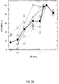
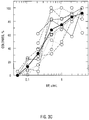
- FIG. 3A, FIG. 3B, and FIG. 3C show erythropoietin dose-response curves of blood BFU-E-derived colonies from fetuses, newborn infants, and adults. Data were normalized as the percentage of maximal growth. Each curve with open symbols represents a different donor, and each point is the mean of triplicate cultures. The curve with solid symbols represents the mean of all curves.
- FIG. 3A fetal.
- FIG. 3B newborn.
- FIG. 3C adult. (Weinberg et al. 1992).
- FIG. 4A, FIG. 4B, FIG. 4C and FIG. 4D show growth potential of blood erythroid progenitors from fetuses, newborn infants and adults.
- FIG. 4A and FIG. 4C are data from day 13;
- FIG. 4B and FIG. 4D are data from the day of peak growth.
- FIG. 4A and FIG. 4B data are expressed as colonies/100,000 cells plated.
- FIG. 4C and FIG. 4D data are expressed as BFU-E/mL of blood.
- Open symbols represent individual experiments; solid symbols represent the mean ⁇ 1 SD. (Weinberg et al. 1992).
- FIG. 5A, FIG. 5B, FIG. 5C, and FIG. 5D shows morphology of erythroid differentiation in liquid cultures. All slides were stained with benzidine Wright-Giemsa, original magnification X 1,000.
- FIG. 5A Day 0 mononuclear cells before culture.
- FIG. 5B Cells recovered from the first phase of culture after 7 days.
- FIG. 5C hemoglobinized erythroblasts appear after 7 days in the second phase with Ep.
- FIG. 5D Mature erythroblasts and erythrocytes recovered after 14 days in the second phase with Ep. From Weinberg et al. 1993.
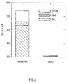
- FIG. 6 shows numbers of cells recovered after plating 100,00 blood mononuclear cells in liquid culture.
- the first 7 days were without cytokines, while the second 14 days were with Ep, SCF, IL-3, and IL-6.
- Solid bar hemoglobinized (polychromatophilic and orthochromatic) erythroblasts. Hatched bar, erythrocytes. Open bar, other nucleated cells (leukocytes and non-hemoglobinized erythroblasts). Note the more than 80-fold amplification of cell numbers in the newborn culture, compared with less than 2-fold in the adult experiment.
- the present invention concern a new approach to enrich for fetal hematopoietic progenitor cells.
- fetal progenitors differ from those of adults (Weinberg et al. 1992; Forestier et al .).
- Cultures of mononuclear cells obtained from maternal blood are used to improve the proportion of fetal to adult progeny by manipulation of culture conditions.
- the inventor's previous studies indicate that there may be advantageous differences between fetal, newborn, and adult erythroid progenitor cells.
- the presence of fetal stem cells in maternal blood is an important aspect of the present invention.
- the pluripotent stem cell when present in the circulation at all, is in such a low proportion that finding this fetal component in the adult is quite difficult.
- progenitor is used for cells which are committed to a specific lineage, and are not morphologically recognizable, but are identified by their progeny (FIG. 1).
- the earliest erythroid cell is called the BFU-E (burst forming unit-erythroid) (Axelrad).
- Blood or bone marrow mononuclear cells are isolated by recovering the light density fraction after centrifugation on Ficoll-hypaque, and cultured in methyl cellulose or plasma clot, with erythropoietin (Ep), plus fetal calf serum, or defined serum-free media which include burst promoting activities (BPA), such as interleukin-3 (IL-3) or granulocyte-macrophage colony stimulating factor (GM-CSF).
- BPA burst promoting activities
- IL-3 interleukin-3
- GM-CSF granulocyte-macrophage colony stimulating factor
- the CFU-E colony forming unit-erythroid
- FOG. 2 erythroblasts
- "Precursors" are more mature than progenitors, and are morphologically identifiable without culture. In the erythroid series these include proerythroblasts, basophilic, polychromatophilic, and orthochromatic erythroblasts. These then enucleate and thus no longer contain DNA. However, the reticulocytes do contain globin RNA, and may be used for the diagnosis of hemoglobinopathies.
- CD34 identifies the earliest stem and progenitor cells, but is lost as the progenitors mature. CD33 appears after the stem cell stage, but disappears by the level of the CFU-E. The c-kit tyrosine kinase receptor is present early, and also decreases after BFU-E.
- Ep is required for growth of fetal or adult BFU-E. Fetal BFU-E are more sensitive to Ep than are adult BFU-E, with the Ep 1/2 max (the Ep required for 50% of maximal growth) 0.3 compared to 0.4 U/mL (Table 2).
- Colonies derived from fetal BFU-E are larger than those from adult BFU-E, with more subcolonies and more total cells.
- Under standard culture conditions Ep 2U/mL, 14 days, there are on average 10-fold more BFU-E in fetal blood, expressed as either BFU-E per 10 5 mononuclear cells plated, or particularly per mL of blood (since the MNC/mL of blood is higher in fetal than adult blood) (Table 2 and FIG. 4). Since the fetal colonies are also larger, the overall relative amplification would be substantially more than 10-fold. The majority of the adult cultures took more than 14 days to achieve maximal growth, while the majority of the fetal cultures had reached their plateau by then.
- the present invention also concerns additional distinctions between BFU-E at various stages of ontogeny.
- a liquid culture system first described by Fibach et al. was modified (Weinberg et al. 1993).
- Mononuclear cells are cultured in phase 1 for about 7 days in the presence of stem cell factor (SCF), a recently described cytokine which acts at early as well as subsequent stages of erythropoiesis (McNiece et al .).
- SCF stem cell factor
- the resulting nucleated cells are then recultured in phase 2 with Ep alone or also with SCF, for an additional 7 to 14 days (a total of 14 to 21 days).
- FIG. 5 shows day 0 mononuclear cells, day 7 cells, and the final products on day 14 and 21 from an adult blood culture.
- the initial and day 7 cells are primarily lymphocytes and monocytes, while the end result contains nucleated and anucleate erythroid cells. Cultures from newborns are compared with those from adult blood. The maximum final amplification of newborn cells was 8 to 45-fold, of which 50-100% were erythroid (Table 3). TABLE 3 Liquid Cultures Phase 2 Additive Newborn Fold Amplify Newborn Erythroid % Adult Fold Amplify Adult Erythroid % Ep 3.1-5.6 50-100 1.6-3.2 20-60 Ep + SCF 7.5-45 50-100 2.6-5.3 60-90
- the plasma and medium were removed, and the interface layer containing the MNC was collected into 20 ml of alpha medium containing 2% fetal calf serum (FCS).
- FCS fetal calf serum
- the cells were washed twice with the same conditions, and resuspended in alpha medium with 2% FCS.
- the MNC were then cultured at 5 X 10 6 /ml for 7 days in alpha medium (with penicillin 0.1 u/ml and streptomycin 0.1 ⁇ g/l) with 10% FCS alone, or + SCF 100 ng/ml, or + SCF (AMGEN) 100 ng/ml + IL-3 (Immunex) 100 ng/ml + IL-6 (Genetics Institute) 100 u/ml.
- the room air incubator had high humidity and 5% CO 2 .
- the nonadherent cells were then recovered by aspirating the medium containing the cells after a gentle shake of the flask, and centrifugation as above.
- the cells were replated at 3X10 5 /ml in alpha medium with 30% FCS, 1% bovine serum albumin (BSA), 10 -4 M ⁇ -mercaptoethanol, penicillin 0.1 u/ml and streptomycin 0.1 ⁇ g/ml, as well as SCF 100 ng/ml, IL-3 100 ng/ml, and IL-6 100 u/ml.
- Incubations were in humidified incubators with 5% CO 2 , and either room air or 5% oxygen.
- the liquid culture assay is being further optimized.
- cytokines Ep, SCF, IL-3, IL-6
- the relative enrichment of newborn over adult cells is >40-fold; an even better growth should be obtained from fetal progenitors.
- the conditions were as described for Table 4.
- Stem and/or progenitor cells could be enriched using CD34, CD33, 17F11, DR, or CD71.
- the separation method could be panning, magnetic beads, MACS, or FACS. The enrichment could be done on day 0, or at some time during the initial phase of culture.
- Ep-dependent phase cells may be recovered and enriched for the erythroid lineage using CD71, CD36, blood group antigens, or GPA.
- Selective enrichment for cells of fetal origin can be accomplished with the appropriate paternal blood group antigens, or with antibodies to fetal erythrocyte features, such as fetal hemoglobin or i antigen, a membrane antigen specific to fetal red cells.
- the cultures can also be exploded using cytokine manipulations which favor fetal rather than adult growth.
- cytokine manipulations which favor fetal rather than adult growth.
- Such conditions include low concentration of Ep (Weinberg et al. 1992), addition of IL-6 (Gardner et al .), or a combination of factors including Ep, SCF, IL-3, and IL-6.
- Prenatal diagnosis using fetal cells in the maternal circulation are performed as follows:
- Blood (50 ml) is taken from a pregnant woman at 8-20 weeks gestation.
- MNC are isolated by centrifugation on Ficoll-hypaque, and cultured at 5 X 10 6 /ml for 7 days in alpha medium with 10% FCS, using SCF 100 ng/ml, IL-3 100 ng/ml, and IL-6 100 u/ml.
- the nonadherent cells are then recovered and replated at 3 X10 5 /ml in alpha medium with 30% FCS, 1% BSA, 10 -4 M ⁇ -mercaptoethanol, and penicillin and streptomycin, as well as SCF 100 ng/ml, IL-3 100 ng/ml, and IL-6 100 ⁇ /ml. All incubations are done in humidified incubators with 5% CO 2 , and either room air or 5% oxygen. After 21 days, the cells are recovered.
- Analysis (2) utilizes the relative amplification of fetal cells; PCR will be more reliable with fetal/adult ratios of abut 1/2000 compared to the about 1/50,000 prior to culture.
- cells that have characteristics of early erythroid cells may be enriched (e.g. , CD71).
- erythroid cells may be enriched, e.g. , using CD71, CD36, and/or GPA.
- the cells which result from these cultures are not purely fetal, but are relatively more so than the starting material and may be subjected to the same analytical techniques that have been used for unseparated material, mainly PCR, FISH, or karyotyping.
- globin mRNA could be recovered and used for RT-PCR for the diagnosis of hemoglobinopathies.
- the erythroid cells which are of fetal origin can be identified by staining with fluorescent antibodies to fetal hemoglobin.
- Kirsch-Volders et al., 1980, "Increase in the amount of fetal lymphocytes in maternal blood during pregnancy, J. Med. Genet., 17:267-72.
Landscapes
- Health & Medical Sciences (AREA)
- Life Sciences & Earth Sciences (AREA)
- Engineering & Computer Science (AREA)
- Biomedical Technology (AREA)
- Hematology (AREA)
- Immunology (AREA)
- Chemical & Material Sciences (AREA)
- Molecular Biology (AREA)
- Cell Biology (AREA)
- Biotechnology (AREA)
- Urology & Nephrology (AREA)
- Microbiology (AREA)
- General Health & Medical Sciences (AREA)
- Biochemistry (AREA)
- Zoology (AREA)
- Bioinformatics & Cheminformatics (AREA)
- General Physics & Mathematics (AREA)
- Organic Chemistry (AREA)
- Wood Science & Technology (AREA)
- Genetics & Genomics (AREA)
- Food Science & Technology (AREA)
- Medicinal Chemistry (AREA)
- Physics & Mathematics (AREA)
- Analytical Chemistry (AREA)
- Pathology (AREA)
- General Engineering & Computer Science (AREA)
- Proteomics, Peptides & Aminoacids (AREA)
- Developmental Biology & Embryology (AREA)
- Tropical Medicine & Parasitology (AREA)
- Virology (AREA)
- Measuring Or Testing Involving Enzymes Or Micro-Organisms (AREA)
- Micro-Organisms Or Cultivation Processes Thereof (AREA)
- Investigating Or Analysing Biological Materials (AREA)
- Medicines Containing Antibodies Or Antigens For Use As Internal Diagnostic Agents (AREA)
Abstract
Claims (8)
- Procédé d'obtention d'un échantillon de cellules enrichi en cellules foetales du sang maternel comprenant:l'obtention de cellules d'un échantillon de sang maternel;la stimulation sélective de la croissance de cellules foetales par
l'incubation desdites cellules dans un milieu comprenant au moins une cytokine dans une quantité ayant des effets stimulants plus grands sur les cellules foetales que sur les cellules maternelles, pour produire un échantillon de cellules enrichi en cellules foetales. - Procédé d'analyse génétique prénatale comprenant:l'obtention de cellules d'un échantillon de sang maternel;la stimulation sélective de la croissance de cellules foetales parl'incubation desdites cellules dans un milieu comprenant au moins une cytokine dans une quantité ayant des effets stimulants plus grands sur les cellules foetales que sur les cellules maternelles, pour produire un échantillon de cellules enrichi en cellules foetales; etl'analyse de cellules foetales pour le contenu génétique ou les anomalies génétiques.
- Procédé selon la revendication 1 ou 2, caractérisé en ce que la cytokine est au moins une parmi l'érythropoïétine, le facteur des cellules souches, l'interleukine-3 et l'interleukine-6.
- Procédé selon la revendication 1 ou 2, caractérisé en ce qu'au moins une cytokine est une combinaison de facteur des cellules souches, d'interleukine-3 et d'interleukine-6.
- Procédé selon la revendication 4, caractérisé en ce que les quantités de cytokine sont d'environ 100 ng/ml ou inférieures.
- Procédé selon la revendication 1 ou 2, caractérisé en ce que la concentration de cellules foetales est suivie en utilisant un anticorps spécifique d'un antigène des cellules foetales.
- Procédé selon la revendication 6, caractérisé en ce que l'antigène des cellules foetales est l'hémoglobine foetale ou l'antigène i foetal.
- Procédé selon la revendication 1 ou 2, caractérisé en ce que les cellules foetales sont des cellules-progéniteur hématopoïétiques foetales.
Applications Claiming Priority (3)
| Application Number | Priority Date | Filing Date | Title |
|---|---|---|---|
| US08/218,290 US5580724A (en) | 1994-03-25 | 1994-03-25 | Differential expansion of fetal stem cells in maternal circulation for use in prenatal genetic analysis |
| PCT/US1995/003659 WO1995026507A1 (fr) | 1994-03-25 | 1995-03-24 | Expansion differentielle de cellules souches foetales dans la circulation sanguine maternelle, pour l'analyse genetique prenatale |
| US218290 | 2002-08-14 |
Publications (2)
| Publication Number | Publication Date |
|---|---|
| EP0752104A1 EP0752104A1 (fr) | 1997-01-08 |
| EP0752104B1 true EP0752104B1 (fr) | 1997-12-29 |
Family
ID=22814517
Family Applications (1)
| Application Number | Title | Priority Date | Filing Date |
|---|---|---|---|
| EP95915408A Expired - Lifetime EP0752104B1 (fr) | 1994-03-25 | 1995-03-24 | Expansion differentielle de cellules souches foetales dans la circulation sanguine maternelle, pour l'analyse genetique prenatale |
Country Status (9)
| Country | Link |
|---|---|
| US (1) | US5580724A (fr) |
| EP (1) | EP0752104B1 (fr) |
| JP (1) | JP2996513B2 (fr) |
| AU (1) | AU684935B2 (fr) |
| CA (1) | CA2186366C (fr) |
| DE (1) | DE69501326T2 (fr) |
| ES (1) | ES2111401T3 (fr) |
| MX (1) | MXPA96004278A (fr) |
| WO (1) | WO1995026507A1 (fr) |
Families Citing this family (15)
| Publication number | Priority date | Publication date | Assignee | Title |
|---|---|---|---|---|
| US5731156A (en) * | 1996-10-21 | 1998-03-24 | Applied Imaging, Inc. | Use of anti-embryonic hemoglobin antibodies to identify fetal cells |
| US6617158B1 (en) | 1999-08-19 | 2003-09-09 | New England Medical Center | Method for stimulating the production of fetal hemoglobin producing erythroid cells |
| AU2002335610C1 (en) * | 2001-01-29 | 2008-09-25 | Hemogenix, Inc. | High-throughput stem cell assay of hematopoietic stem and progenitor cell proliferation |
| US7989178B2 (en) * | 2001-01-29 | 2011-08-02 | Hemogenix, Inc. | Colony assay miniaturization with enumeration output |
| US7354730B2 (en) * | 2002-01-29 | 2008-04-08 | Hemogenix, Inc. | High-throughput assay of hematopoietic stem and progenitor cell proliferation |
| US7666615B2 (en) * | 2001-01-29 | 2010-02-23 | Hemogenix, Inc. | High-throughput assay of hematopoietic stem and progenitor cell proliferation |
| FR2824144B1 (fr) * | 2001-04-30 | 2004-09-17 | Metagenex S A R L | Methode de diagnostic prenatal sur cellule foetale isolee du sang maternel |
| JP4330995B2 (ja) * | 2001-11-15 | 2009-09-16 | チルドレンズ メディカル センター コーポレーション | 絨毛膜絨毛、羊水、および胎盤からの胎児性幹細胞を単離、増殖、および分化させる方法、ならびにその治療的使用方法 |
| DK1449535T3 (da) * | 2003-02-18 | 2006-09-11 | Clinique La Prairie Res Sa | Præparater der omfatter fötalt hæmoglobin og bakterieendotoksin samt eventuelt fötale leverbestanddele |
| US8940535B2 (en) * | 2003-11-04 | 2015-01-27 | University Of Maryland, Baltimore | Stem cell culture medium and method of using said medium and the cells |
| JP2008506637A (ja) * | 2004-06-14 | 2008-03-06 | ザ・ボード・オブ・トラスティーズ・オブ・ザ・ユニバーシティ・オブ・イリノイ | 胎児起源の細胞に対する抗体 |
| PL2471907T3 (pl) | 2005-12-29 | 2019-07-31 | Celularity, Inc. | Populacje komórek macierzystych łożyska |
| AU2007311126A1 (en) * | 2006-10-16 | 2008-04-24 | Celula Inc. | Methods and compositions for differential expansion of fetal cells in maternal blood and their use |
| WO2012092485A1 (fr) | 2010-12-31 | 2012-07-05 | Anthrogenesis Corporation | Amélioration de l'efficacité de cellules souches placentaires sous l'effet de molécules d'arn modulateur |
| ES2707579T3 (es) | 2011-06-01 | 2019-04-04 | Celularity Inc | Tratamiento del dolor usando citoblastos placentarios |
Family Cites Families (5)
| Publication number | Priority date | Publication date | Assignee | Title |
|---|---|---|---|---|
| JPH05507404A (ja) * | 1990-04-23 | 1993-10-28 | セルプロ インコーポレイティド | 母体血液からの胎児細胞の富化のための方法 |
| EP0610359A1 (fr) * | 1991-10-23 | 1994-08-17 | Cellpro Incorporated | Procede pour enrichir des cellules souches f tales issues du sang maternel |
| CA2122141C (fr) * | 1991-10-23 | 2007-07-17 | Shelly Heimfeld | Methode de multiplication selective de cellules souches |
| WO1993018136A1 (fr) * | 1992-03-05 | 1993-09-16 | Cytomed, Inc. | Procede de maintien de cellules souche hematopoietiques |
| CA2154787A1 (fr) * | 1993-01-27 | 1994-08-04 | Peter W. Runstadler | Proliferation selective de cellules |
-
1994
- 1994-03-25 US US08/218,290 patent/US5580724A/en not_active Expired - Fee Related
-
1995
- 1995-03-24 ES ES95915408T patent/ES2111401T3/es not_active Expired - Lifetime
- 1995-03-24 MX MXPA96004278A patent/MXPA96004278A/es not_active IP Right Cessation
- 1995-03-24 JP JP7525227A patent/JP2996513B2/ja not_active Expired - Fee Related
- 1995-03-24 CA CA002186366A patent/CA2186366C/fr not_active Expired - Fee Related
- 1995-03-24 EP EP95915408A patent/EP0752104B1/fr not_active Expired - Lifetime
- 1995-03-24 DE DE69501326T patent/DE69501326T2/de not_active Expired - Fee Related
- 1995-03-24 WO PCT/US1995/003659 patent/WO1995026507A1/fr not_active Ceased
- 1995-03-24 AU AU22298/95A patent/AU684935B2/en not_active Ceased
Also Published As
| Publication number | Publication date |
|---|---|
| DE69501326T2 (de) | 1998-04-16 |
| CA2186366C (fr) | 2008-07-08 |
| JP2996513B2 (ja) | 2000-01-11 |
| AU2229895A (en) | 1995-10-17 |
| US5580724A (en) | 1996-12-03 |
| AU684935B2 (en) | 1998-01-08 |
| CA2186366A1 (fr) | 1995-10-05 |
| MXPA96004278A (es) | 2002-04-19 |
| ES2111401T3 (es) | 1998-03-01 |
| DE69501326D1 (de) | 1998-02-05 |
| WO1995026507A1 (fr) | 1995-10-05 |
| EP0752104A1 (fr) | 1997-01-08 |
| JPH09509328A (ja) | 1997-09-22 |
Similar Documents
| Publication | Publication Date | Title |
|---|---|---|
| EP0752104B1 (fr) | Expansion differentielle de cellules souches foetales dans la circulation sanguine maternelle, pour l'analyse genetique prenatale | |
| Lansdorp et al. | Ontogeny-related changes in proliferative potential of human hematopoietic cells. | |
| Goto et al. | Growth characteristics of leukemic and normal hematopoietic cells in Ph'+ chronic myelogenous leukemia and effects of intensive treatment | |
| JP3070865B2 (ja) | ヒトプロジェニター細胞のサブセット | |
| Sieff et al. | Dependence of highly enriched human bone marrow progenitors on hemopoietic growth factors and their response to recombinant erythropoietin. | |
| US7919316B2 (en) | Hematopoietic stem cell identification and isolation | |
| Little et al. | Frequency of fetal cells in sorted subpopulations of nucleated erythroid and CD34+ hematopoietic progenitor cells from maternal peripheral blood | |
| Jackson | Fetal cells and DNA in maternal blood | |
| Gupta et al. | Human CD34+ bone marrow cells regulate stromal production of interleukin-6 and granulocyte colony-stimulating factor and increase the colony-stimulating activity of stroma | |
| WO2001079851A1 (fr) | Diagnostique prenatal non invasif utilisant des cellules foetales cd45-negatives | |
| Yamaguchi et al. | Umbilical vein endothelial cells are an important source of c‐kit and stem cell factor which regulate the proliferation of haemopoietic progenitor cells | |
| AU2274595A (en) | Culture and isolation of fetal cells from maternal peripheral blood | |
| Bianchi | Fetomaternal cell traffic, pregnancy-associated progenitor cells, and autoimmune disease | |
| WO1994025873A1 (fr) | Procedes d'enrichissement de cellules souches f×tales provenant du sang maternel | |
| Bohmer et al. | Differential development of fetal and adult haemoglobin profiles in colony culture: isolation of fetal nucleated red cells by two-colour fluorescence labelling. | |
| Gluckman | Umbilical cord blood biology and transplantation | |
| US5879937A (en) | Cytokine-induced proliferation of amniotic t-cells | |
| WO1993008269A1 (fr) | Procede pour enrichir des cellules souches f×tales issues du sang maternel | |
| Gussin et al. | Culture of fetal cells from maternal blood for prenatal diagnosis | |
| Valerio et al. | Detection of fetal trisomy 18 by short-term culture of maternal peripheral blood | |
| Manotaya et al. | Evaluation of a culture system for enrichment of CD34+ hematopoietic progenitor cells present in maternal blood | |
| CN115896015B (zh) | 一种髓系来源抑制性细胞的体外培养方法 | |
| Slunga-Tallberg et al. | Nucleated erythrocytes in enriched and unenriched peripheral venous blood samples from pregnant and nonpregnant women | |
| Frenck et al. | Studies of the function and structure of in vitro propagated human granulocytes | |
| Kandarakov et al. | Magnetic‐Assisted Fractionation of Bone Marrow Cells into Subsets Differing in CD45 Expression Levels, Surface Phenotypes and Functional Properties |
Legal Events
| Date | Code | Title | Description |
|---|---|---|---|
| PUAI | Public reference made under article 153(3) epc to a published international application that has entered the european phase |
Free format text: ORIGINAL CODE: 0009012 |
|
| 17P | Request for examination filed |
Effective date: 19961022 |
|
| AK | Designated contracting states |
Kind code of ref document: A1 Designated state(s): DE ES FR GB IT |
|
| GRAG | Despatch of communication of intention to grant |
Free format text: ORIGINAL CODE: EPIDOS AGRA |
|
| 17Q | First examination report despatched |
Effective date: 19970304 |
|
| GRAG | Despatch of communication of intention to grant |
Free format text: ORIGINAL CODE: EPIDOS AGRA |
|
| GRAH | Despatch of communication of intention to grant a patent |
Free format text: ORIGINAL CODE: EPIDOS IGRA |
|
| GRAH | Despatch of communication of intention to grant a patent |
Free format text: ORIGINAL CODE: EPIDOS IGRA |
|
| GRAA | (expected) grant |
Free format text: ORIGINAL CODE: 0009210 |
|
| AK | Designated contracting states |
Kind code of ref document: B1 Designated state(s): DE ES FR GB IT |
|
| REF | Corresponds to: |
Ref document number: 69501326 Country of ref document: DE Date of ref document: 19980205 |
|
| REG | Reference to a national code |
Ref country code: ES Ref legal event code: FG2A Ref document number: 2111401 Country of ref document: ES Kind code of ref document: T3 |
|
| ET | Fr: translation filed | ||
| ITF | It: translation for a ep patent filed | ||
| PLBE | No opposition filed within time limit |
Free format text: ORIGINAL CODE: 0009261 |
|
| STAA | Information on the status of an ep patent application or granted ep patent |
Free format text: STATUS: NO OPPOSITION FILED WITHIN TIME LIMIT |
|
| 26N | No opposition filed | ||
| REG | Reference to a national code |
Ref country code: GB Ref legal event code: IF02 |
|
| PGFP | Annual fee paid to national office [announced via postgrant information from national office to epo] |
Ref country code: IT Payment date: 20060331 Year of fee payment: 12 |
|
| PGFP | Annual fee paid to national office [announced via postgrant information from national office to epo] |
Ref country code: GB Payment date: 20080326 Year of fee payment: 14 |
|
| PGFP | Annual fee paid to national office [announced via postgrant information from national office to epo] |
Ref country code: FR Payment date: 20080311 Year of fee payment: 14 Ref country code: ES Payment date: 20080418 Year of fee payment: 14 Ref country code: DE Payment date: 20080407 Year of fee payment: 14 |
|
| PG25 | Lapsed in a contracting state [announced via postgrant information from national office to epo] |
Ref country code: IT Free format text: LAPSE BECAUSE OF NON-PAYMENT OF DUE FEES Effective date: 20070324 |
|
| GBPC | Gb: european patent ceased through non-payment of renewal fee |
Effective date: 20090324 |
|
| REG | Reference to a national code |
Ref country code: FR Ref legal event code: ST Effective date: 20091130 |
|
| PG25 | Lapsed in a contracting state [announced via postgrant information from national office to epo] |
Ref country code: DE Free format text: LAPSE BECAUSE OF NON-PAYMENT OF DUE FEES Effective date: 20091001 |
|
| PG25 | Lapsed in a contracting state [announced via postgrant information from national office to epo] |
Ref country code: GB Free format text: LAPSE BECAUSE OF NON-PAYMENT OF DUE FEES Effective date: 20090324 Ref country code: FR Free format text: LAPSE BECAUSE OF NON-PAYMENT OF DUE FEES Effective date: 20091123 |
|
| REG | Reference to a national code |
Ref country code: ES Ref legal event code: FD2A Effective date: 20090325 |
|
| PG25 | Lapsed in a contracting state [announced via postgrant information from national office to epo] |
Ref country code: ES Free format text: LAPSE BECAUSE OF NON-PAYMENT OF DUE FEES Effective date: 20090325 |