EP0776969A2 - Verfahren zur Herstellung von Proteinen durch kontrollierte proteolytische spaltung von Pro-Proteinen - Google Patents
Verfahren zur Herstellung von Proteinen durch kontrollierte proteolytische spaltung von Pro-Proteinen Download PDFInfo
- Publication number
- EP0776969A2 EP0776969A2 EP96890172A EP96890172A EP0776969A2 EP 0776969 A2 EP0776969 A2 EP 0776969A2 EP 96890172 A EP96890172 A EP 96890172A EP 96890172 A EP96890172 A EP 96890172A EP 0776969 A2 EP0776969 A2 EP 0776969A2
- Authority
- EP
- European Patent Office
- Prior art keywords
- protein
- pro
- factor
- protease
- column
- Prior art date
- Legal status (The legal status is an assumption and is not a legal conclusion. Google has not performed a legal analysis and makes no representation as to the accuracy of the status listed.)
- Granted
Links
Images







Classifications
-
- C—CHEMISTRY; METALLURGY
- C12—BIOCHEMISTRY; BEER; SPIRITS; WINE; VINEGAR; MICROBIOLOGY; ENZYMOLOGY; MUTATION OR GENETIC ENGINEERING
- C12N—MICROORGANISMS OR ENZYMES; COMPOSITIONS THEREOF; PROPAGATING, PRESERVING, OR MAINTAINING MICROORGANISMS; MUTATION OR GENETIC ENGINEERING; CULTURE MEDIA
- C12N9/00—Enzymes; Proenzymes; Compositions thereof; Processes for preparing, activating, inhibiting, separating or purifying enzymes
- C12N9/14—Hydrolases (3)
- C12N9/48—Hydrolases (3) acting on peptide bonds (3.4)
- C12N9/50—Proteinases, e.g. Endopeptidases (3.4.21-3.4.25)
- C12N9/64—Proteinases, e.g. Endopeptidases (3.4.21-3.4.25) derived from animal tissue
- C12N9/6421—Proteinases, e.g. Endopeptidases (3.4.21-3.4.25) derived from animal tissue from mammals
- C12N9/6424—Serine endopeptidases (3.4.21)
- C12N9/6429—Thrombin (3.4.21.5)
-
- A—HUMAN NECESSITIES
- A61—MEDICAL OR VETERINARY SCIENCE; HYGIENE
- A61P—SPECIFIC THERAPEUTIC ACTIVITY OF CHEMICAL COMPOUNDS OR MEDICINAL PREPARATIONS
- A61P7/00—Drugs for disorders of the blood or the extracellular fluid
-
- A—HUMAN NECESSITIES
- A61—MEDICAL OR VETERINARY SCIENCE; HYGIENE
- A61P—SPECIFIC THERAPEUTIC ACTIVITY OF CHEMICAL COMPOUNDS OR MEDICINAL PREPARATIONS
- A61P7/00—Drugs for disorders of the blood or the extracellular fluid
- A61P7/02—Antithrombotic agents; Anticoagulants; Platelet aggregation inhibitors
-
- C—CHEMISTRY; METALLURGY
- C12—BIOCHEMISTRY; BEER; SPIRITS; WINE; VINEGAR; MICROBIOLOGY; ENZYMOLOGY; MUTATION OR GENETIC ENGINEERING
- C12N—MICROORGANISMS OR ENZYMES; COMPOSITIONS THEREOF; PROPAGATING, PRESERVING, OR MAINTAINING MICROORGANISMS; MUTATION OR GENETIC ENGINEERING; CULTURE MEDIA
- C12N9/00—Enzymes; Proenzymes; Compositions thereof; Processes for preparing, activating, inhibiting, separating or purifying enzymes
- C12N9/14—Hydrolases (3)
- C12N9/48—Hydrolases (3) acting on peptide bonds (3.4)
- C12N9/50—Proteinases, e.g. Endopeptidases (3.4.21-3.4.25)
-
- C—CHEMISTRY; METALLURGY
- C12—BIOCHEMISTRY; BEER; SPIRITS; WINE; VINEGAR; MICROBIOLOGY; ENZYMOLOGY; MUTATION OR GENETIC ENGINEERING
- C12N—MICROORGANISMS OR ENZYMES; COMPOSITIONS THEREOF; PROPAGATING, PRESERVING, OR MAINTAINING MICROORGANISMS; MUTATION OR GENETIC ENGINEERING; CULTURE MEDIA
- C12N9/00—Enzymes; Proenzymes; Compositions thereof; Processes for preparing, activating, inhibiting, separating or purifying enzymes
- C12N9/14—Hydrolases (3)
- C12N9/48—Hydrolases (3) acting on peptide bonds (3.4)
- C12N9/50—Proteinases, e.g. Endopeptidases (3.4.21-3.4.25)
- C12N9/64—Proteinases, e.g. Endopeptidases (3.4.21-3.4.25) derived from animal tissue
-
- C—CHEMISTRY; METALLURGY
- C12—BIOCHEMISTRY; BEER; SPIRITS; WINE; VINEGAR; MICROBIOLOGY; ENZYMOLOGY; MUTATION OR GENETIC ENGINEERING
- C12N—MICROORGANISMS OR ENZYMES; COMPOSITIONS THEREOF; PROPAGATING, PRESERVING, OR MAINTAINING MICROORGANISMS; MUTATION OR GENETIC ENGINEERING; CULTURE MEDIA
- C12N9/00—Enzymes; Proenzymes; Compositions thereof; Processes for preparing, activating, inhibiting, separating or purifying enzymes
- C12N9/14—Hydrolases (3)
- C12N9/48—Hydrolases (3) acting on peptide bonds (3.4)
- C12N9/50—Proteinases, e.g. Endopeptidases (3.4.21-3.4.25)
- C12N9/64—Proteinases, e.g. Endopeptidases (3.4.21-3.4.25) derived from animal tissue
- C12N9/6421—Proteinases, e.g. Endopeptidases (3.4.21-3.4.25) derived from animal tissue from mammals
- C12N9/6424—Serine endopeptidases (3.4.21)
- C12N9/6432—Coagulation factor Xa (3.4.21.6)
-
- C—CHEMISTRY; METALLURGY
- C12—BIOCHEMISTRY; BEER; SPIRITS; WINE; VINEGAR; MICROBIOLOGY; ENZYMOLOGY; MUTATION OR GENETIC ENGINEERING
- C12P—FERMENTATION OR ENZYME-USING PROCESSES TO SYNTHESISE A DESIRED CHEMICAL COMPOUND OR COMPOSITION OR TO SEPARATE OPTICAL ISOMERS FROM A RACEMIC MIXTURE
- C12P21/00—Preparation of peptides or proteins
- C12P21/06—Preparation of peptides or proteins produced by the hydrolysis of a peptide bond, e.g. hydrolysate products
-
- C—CHEMISTRY; METALLURGY
- C12—BIOCHEMISTRY; BEER; SPIRITS; WINE; VINEGAR; MICROBIOLOGY; ENZYMOLOGY; MUTATION OR GENETIC ENGINEERING
- C12Y—ENZYMES
- C12Y304/00—Hydrolases acting on peptide bonds, i.e. peptidases (3.4)
- C12Y304/21—Serine endopeptidases (3.4.21)
- C12Y304/21005—Thrombin (3.4.21.5)
-
- C—CHEMISTRY; METALLURGY
- C12—BIOCHEMISTRY; BEER; SPIRITS; WINE; VINEGAR; MICROBIOLOGY; ENZYMOLOGY; MUTATION OR GENETIC ENGINEERING
- C12Y—ENZYMES
- C12Y304/00—Hydrolases acting on peptide bonds, i.e. peptidases (3.4)
- C12Y304/21—Serine endopeptidases (3.4.21)
- C12Y304/21006—Coagulation factor Xa (3.4.21.6)
Definitions
- the invention relates to a method for producing and obtaining proteins by controlled cleavage of pro-proteins using a protease.
- prothrombin is converted to thrombin by the protease factor Xa in a prothrombinase complex reaction.
- Inactive factor X is e.g. converted into active factor Xa by the protease factor IXa.
- the extraction of the activated proteins is of great interest both for clinical use and for diagnostics.
- the activated proteins in pure form can then e.g. to control other proteolytic processes, such as blood clotting by thrombin in surgery, for therapy or diagnosis or to obtain specific antibodies.
- the active proteins can only be obtained in very limited quantities from living organisms, such as human blood. Therefore, the pro-proteins or pro-enzymes are mostly converted into the activated forms in vitro by the action of suitable proteases.
- suitable proteases Such a method is known from EP-0 378 798. According to this process, prothrombin from human plasma is adsorbed onto a solid support and the material is treated with Ca 2+ ions and, together with proteases present in the plasma, thereby causes the conversion of prothrombin to thrombin.
- the pro-protein to be activated is immobilized on a support and then converted into the active enzyme by a soluble protease. In order to be able to control this transformation, it is in presence detergent or chaotropic substance. However, further purification steps are then necessary to obtain the activated proteins, in particular in order to separate the protease again.
- prothrombin in soluble form is converted into thrombin by adding coagulation-active salts to a solution containing prothrombin.
- Thrombin is then further purified by ion exchange chromatography and / or affinity chromatography.
- thrombin is produced by treating a prothrombin-containing solution with immobilized trypsin for 150 minutes.
- immobilized trypsin for 150 minutes.
- the use of the protease in immobilized form makes it easier to separate the protease after activation.
- recombinant human protein C (rHPC) is activated by a 2-hour treatment with thrombin immobilized on glass beads. Activated rHPC is separated from immobilized thrombin by centrifugation.
- a major disadvantage of these processes is that, even if slowed down, further degradation to protein fragments still occurs and protein mixtures are formed which additionally contain large amounts of additives, such as detergents or glycerol. Complex processes for cleaning the activated enzymes are therefore necessary.
- Protein fragments and detergents or chaotropic substances were removed according to EP-A-0 565 511 by means of relatively complex chromatographic methods.
- the object of the present invention is therefore to provide a production process for proteins from pro-proteins which can be used to achieve a high yield of proteins and which makes it possible in a simple manner to obtain proteins with very high specific activity and purity .
- the method should not only be limited to certain starting solutions, but should also be applicable to a comprehensive spectrum of starting solutions.
- This object is achieved according to the invention by a process for the production and production of proteins by controlled proteolytic cleavage of pro-proteins using a protease, which is characterized in that a pro-protein-containing solution with a protease and a solid carrier which has a higher Has affinity for the protein as for the pro-protein or its functionally inactive degradation products, is brought into contact, the pro-protein being proteolytically cleaved to the protein and the protein being selectively separated off by adsorption on the solid support.
- An immobilized protease is preferably used. This has the advantage that it can be easily separated from the solution.
- the pro-protein-containing solution is treated with the protease for a period of contact which is sufficient to bring about a cleavage of the pro-protein to the protein, but in which essentially no further functionally inactive degradation products of the protein are formed.
- the duration of exposure of the protease to the pro-protein is an important factor for the quality of the protein to be produced, and that compliance with a given contact time between protease / pro-protein under given parameters is important for the production of a particular protein.
- the contact time must be selected so that the conversion from the pro-protein to the protein takes place, but that a further, unwanted degradation of the protein can no longer take place, but the action of the protease is prevented beforehand.
- Critical parameters for determining the optimal contact time are therefore the protease used, the ratio of pro-protein to protease and the reaction temperature.
- any person skilled in the art can easily determine the critical parameters and their optimization before the start of the reaction, provided these are not already known in the prior art.
- the contact duration specific for the production of a certain protein can be determined in each case by simply varying the contact duration in separate test batches.
- the products obtained are analyzed in each case and then that contact time is defined as the given contact time with which a maximum of the desired protein and a minimum of further breakdown products or uncleaved pro-protein can be obtained.
- This given contact time is of course different from system (pro-protein / protease) to system, but in many systems it is less than 24 hours. Time periods between 1 second and 24 hours, in particular between 1 second and 5 hours, particularly preferably between 1 second and 30 minutes, have proven to be preferred exposure times for the protease.
- the protease-formed protein is bound to a carrier which has a higher affinity for the protein than for the pro-protein or its functionally inactive degradation products. The protein is thus withdrawn from the further action of the protease in the reaction mixture, and further degradation of the protein can no longer take place.
- this can be done by connecting the outlet of the second, selective carrier to the first, protease-coupled column, the unbound pro-protein being able to be made accessible again to the action of the protease. These steps can be repeated until essentially all of the pro-protein has been cleaved to protein.
- the method according to the invention can of course be used by everyone Protease-mediated conversions of pro-proteins to proteins are used, it is preferably used for the production of enzymes from pro-enzymes.
- proteases are suitable for the proteolytic cleavage of pro-proteins, e.g. Chymotrypsin, dispase, endopeptidase Arg-C, endoproteinase Lys-C, endoproteinase Glu-C, endoproteinase Asp-N, factor Xa, kallikrein, papain, pepsin, plasmin, pronase, proteinase K, staphylocoagulase, serine proteases of the subtilisin family, such as Kexin or furin-like proteases, or subtilisin, thrombin, trypsin (especially human, bovine, porcines), trypsin-like protease from arthropods or microorganisms, such as e.g.
- pro-proteins e.g. Chymotrypsin, dispase, endopeptidase Arg-C, endoproteinase Lys-C, endoproteinas
- Streptomyces griseus trypsin or serine proteases from venomous snakes venom proteases
- venom proteases e.g. the prothrombin activators from Echis carinatus Venom and from Oxyuranus scutellatus Venom; the activators for factor X and factor V from Russell's Viper Venom, or the protein C activator Agkistrodon contortrix Venom.
- Trypsin, chymotrypsin, kallikrein, dispase, endoproteinases Glu-C, Lys-C or Asp-N, furin or factor Xa are preferably used as proteases.
- Recombinant proteases can of course also be used.
- the protease is preferably immobilized on a solid support, so that at no time can the protease react in soluble form with the pro-protein or the protein formed.
- Suitable natural or synthetic carriers for immobilizing the protease are in particular cellulose, Sepharose, dextrans, in particular agarose, acrylates or silicates.
- the immobilized protease (the protease gel) is packed in a glass column and the pro-protein-containing solution is filtered through the protease gel.
- the flow rate is chosen so that the given contact time between the immobilized protease and pro-protein is maintained.
- the protein activated in this way is, according to this embodiment, directly from the desorption supernatant (eluate) to a selective one Carrier adsorbed.
- inactivated pro-protein should not be bound by the specific carrier if possible and should pass through the column containing the selective carrier unhindered.
- this pro-protein-containing fraction is again brought into contact with the protease gel during the given contact period, whereupon further protein is formed, which in turn is adsorbed from the solution by the selective carrier.
- the pro-protein is completely converted to activated protein without the activated protein being further cleaved by the protease.
- Activated protein accumulates on the selective support and can then be eluted from the support.
- proteins which can be produced by the method according to the invention are functionally active blood coagulation enzymes which result from the inactive precursors of these enzymes by proteolytic degradation.
- thrombin from prothrombin (factor II) factor IX from pro-factor IX, factor IXa from factor IX, from Willebrand factor from Pro-von Willebrand factor, factor Xa from factor X, activated protein C from protein C, factor XIIIa from factor XIII, factor VIIa from factor VII, factor VIIIa from factor VIII, factor Va from factor V.
- the starting solutions are not restricted to any specially prepared solutions in the process according to the invention.
- preferred pro-protein-containing solutions contain the pro-protein in a purified form, since then a particularly pure protein preparation is made possible.
- Pro-protein-containing solutions which contain the pro-protein in a mixture with other proteins are, however, also suitable for the method according to the invention, because contaminating proteins are not a significant disruptive factor for the method according to the invention due to its high specificity.
- Pro-protein-containing solutions of biological origin in particular plasma or solutions derived from plasma or from plasma fractions, are therefore preferably used in the present method.
- biotechnologically produced pro-protein-containing solutions in particular culture supernatants, which have been generated by recombinant cell cultures are used as starting solutions.
- Suitable solid supports for adsorbing the activated proteins for the protein to be produced are particularly suitable for a matrix immobilized heparin, immobilized benzamidine, immobilized hirudin or fragments and derivatives derived from hirudin, various immobilized proteins or peptides, in particular specific immobilized antibodies.
- the protein is preferably selectively eluted from the affinity carriers, so that any other proteins which should also be bound to the carrier (specific or non-specific), albeit in small amounts, are separated.
- virus inactivation measures generally known from the prior art can be carried out to inactivate any infectious agents, in particular viruses which are pathogenic to humans.
- the present invention relates to the use of the method according to the invention for the production of proteins from pro-protein, preferably from activated enzymes from pro-enzymes, in particular the production of activated factor II, of activated factor V, of activated factor VII, of activated factor VIII, factor IX, activated factor IX, activated factor X, activated factor XIII, Willebrand factor and activated protein C.
- the protein-containing fraction obtained by the process according to the invention is - owing to its high purity and its high specific activity - excellently suitable for the production of a pharmaceutical preparation and - starting from the eluted protein - can be processed with the usual methods for such a pharmaceutical preparation .
- Another aspect of the invention therefore relates to pharmaceutical compositions which contain a high-purity protein, in particular factor IIa, factor Va, factor VIIa, factor VIIIa, factor IX, factor IXa, factor Xa, factor XIIIa, activated protein C and / or or von Willebrand factor, and optionally contains one or more physiologically acceptable carriers and / or other pharmaceutical additives.
- the choice of the selective solid carrier for the protein formed is a central point in the process according to the invention. Immobilized hirudin and polypeptides and fragments derived therefrom (in particular in the production of factor IIa) have proven to be particularly preferred carriers.
- the invention therefore also relates to hirudin and polypeptides and peptide fragments or derivatives derived therefrom, which are immobilized on a solid surface.
- Peptides derived from hirudin which originate from the C-terminal region of the protein and contain the thrombin binding site, have proven to be particularly suitable.
- the selected peptides are preferably coupled to the support via sulfur or amino groups.
- both purified human factor II from plasma Prothrombin, Stago Diagnostica
- purified recombinant factor II Falkner, FG, et al., Thromb. Haemost. (1992), 68, 119-124) become one Protein mixture containing factor II (Falkner, FG, et al., Thromb. Haemost. (1992), 68, 119-124) and purified factor X (Boehringer Mannheim) are used as the pro-protein-containing solution.
- Both immobilized trypsin and immobilized factor Xa are preferably used as proteases.
- the invention also relates to a protein complex containing a protein adsorbed on a solid support, produced by the process according to the invention, in particular a thrombin complex containing thrombin, which is adsorbed on a solid support, preferably on a solid support containing hirudin or fragments or derivatives derived from hirudin.
- Another aspect of the present invention relates to a device arrangement consisting of a container (I) containing a protease and a container (II) containing a solid support, wherein (I) and (II) are directly connected to which device arrangement for carrying out the invention Process, preferably in its one-step embodiment, is used.
- container (I) and (II) is a column, the end piece of (I) being directly coupled to the head piece of (II) and optionally the end piece of (II) being connected to the head piece of (I).
- FIIa factor IIa
- the activity determination of factor IIa was carried out photometrically in 50 mM Tris-HCl buffer, pH 8.0, 300 mM NaCl, 0.5% albumin, 7.5 mM EDTA, at 37 ° C.
- the FIIa-specific chromogenic substrate AcOH-H-D-CHG-Ala-Arg-pNA (available from Pentapharm) with a concentration of 0.2 mM was used as the substrate.
- Para-nitroaniline (pNA) released from the substrate by enzymatic hydrolysis was determined photometrically as a function of time at 405 nm.
- pNA Para-nitroaniline released from the substrate by enzymatic hydrolysis was determined photometrically as a function of time at 405 nm.
- an FIIa concentration standard from Immuno AG
- the activity of the sample was determined from the rate of hydrolysis of the substrate.
- factor Xa Factor Xa
- the activity of factor Xa was determined photometrically in 50 mM Tris-HCl buffer, pH 7.8, 0.5% albumin, at 37 ° C.
- the FXa-specific chromogenic substrate Bz-Ile-Glu (piperidyl) -Gly-Arg-pNA (available from Chromogenix) with a concentration of 0.3 mM was used as the substrate.
- PNA released from the substrate by enzymatic hydrolysis was determined photometrically as a function of time at 405 nm.
- the activity of the sample was determined from the rate of hydrolysis of the substrate using an FXa concentration standard (from Immuno AG).
- FII / rFII natural factor II and recombinant factor II
- FIIa / rFIIa recombinant factor II
- the protein determination for FX or FXa was carried out by measuring the absorption at 280 nm using an extinction coefficient (1%, 1 cm) of 12.4 (Human Protein Data, Ed. By A. Haeberli, VCH Weinheim, New York, 1992) .
- the protein concentrations of protein mixtures were determined using the Bradford method (M. Bradford, Anal. Biochem., 72 (1976), 248-254) using a commercially available system from Bio-Rad.
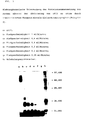
- the samples obtained were examined for their protein composition by denaturing electrophoresis (Laemmli, Nature, Vol. 227: 680-685, (1970)). The results are shown in FIG. 1, the molecular weight marker being shown in lane a, the 1-minute trypsin digestion in lane b and the 5-minute trypsin digestion in lane c.
- rFII was converted to rFIIa by digestion with trypsin. After only 1 minute exposure to rFII with an activity of 4 IU / ml rFIIa was formed therefrom with an activity of 500 IU / ml. With an exposure time of 5 minutes, however, the detectable activity of rFIIa was very low.
- the electrophoretic analysis shows that a trypsin exposure to rFII of only 1 minute results in a protein mixture with molecular weights (in Da) between 70,000 and 20,000, with a protein with a molecular weight between 31,000 and 36,000 dominating.
- thrombin Since thrombin is known to have a molecular mass of 33,000 in electrophoresis, it can be assumed that the protein with the molecular mass 31,000-36,000 is thrombin formed. However, residues of non-activated rFII (molecular weight 70000) are still present. In contrast, when incubating rFII with trypsin for 5 minutes, only low-molecular peptides were detected in electrophoresis. This indicates an almost complete digestion of the rFII by trypsin, but without the rFIIa accumulating.
- a glass column (1 cm in diameter) was filled with 0.1 ml of immobilized trypsin-agarose gel; this corresponds to a gel height of 1.25 mm.
- RFII was pumped through this trypsin agarose gel at different flow rates.
- rFII was dissolved at 0.5 mg / ml (3.5 IU / ml) in 20 mM Tris / HCl buffer, pH 8.0, 150 mM NaCl.
- the flow rate of the rFII solution through the gel was varied between 0.05 ml / minute and 1.0 ml / minute.
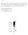
- the individual eluates were examined for their content of thrombin activity (see FIG. 2) and for their protein composition by means of electrophoresis (see FIG. 3).
- the flow rates and the dimensions of the trypsin agarose gel column result in average contact times of 6 seconds, 10 seconds, 15 seconds, 30 seconds, 60 Seconds and 120 seconds, with corresponding flow rates of 1.0 ml / minute, 0.6 ml / minute, 0.4 ml / minute, 0.2 ml / minute, 0.1 ml / minute and 0.05 ml / minute , between rFII and the immobilized Trypsin.
- ATU anti-thrombin units
- Pentapharm 4000 anti-thrombin units (ATU) hirudin (available from Pentapharm) were reduced and then according to the Manufacturer's instructions coupled to 1 ml of activated thiol-Sepharose (Pharmacia). Hirudin-thiol-Sepharose (HTS) was filled into a glass column (diameter 1 cm).
- the outlet of a glass column (diameter 1 cm) containing 0.1 ml of immobilized trypsin agarose gel (TAG) was connected to the inlet of the HTS column by a direct hose connection.
- the outlet of the HTS column could be connected to the inlet of the TAG column via a valve and a pump, creating a liquid circuit. Both columns were equilibrated with 20 mM Tris / -HCl buffer, pH 8.0.
- rFII was dissolved at 0.4 mg / ml (activity 3.5 IU / ml) in 4 ml of 20 mM Tris / HCl buffer, pH 8.0, and passed through the TAG at a flow rate of 0.8 ml / min. Pumped column. From there, the liquid flow was led directly to the HTS column without interruption and, after passing through the HTS column, pumped through the TAG column and HTS column a second time using a pump.
- the HTS column was then separated from the TAG column, rinsed with 50 mM citrate buffer, pH 6.5 (to remove unbound material) and then with 50 mM Na citrate buffer, pH 6.5, 500 mM NaCl , washed (0.5 M NaCl eluate).
- the HTS column was then eluted with 1.5 M KSCN in 50 mM citrate buffer, pH 6.5 (1.5 M KSCN eluate).
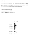
- FIG. 4 shows the electrophoretic examination of the activation described, the starting material (rFII) in lane a, the 1.5 M KSCN eluate (rFIIa) in lane b and a molecular weight marker being separated in lane c.
- thiol peptide 20 mg with the amino acid sequence NH2-Cys-Lys-Pro-Gln-Ser-His-Asn-Asp-Gly-Asp-Phe-Glu-Glu-Ile-Pro-Glu-Glu- Tyr-Leu-Gln-COOH were coupled to 1 ml of activated thiol-Sepharose (available from Pharmacia) in accordance with the manufacturer's instructions.
- Thiol-peptide-thiol-Sepharose (TPTS) was filled into a glass column (diameter 1 cm).
- a glass column (diameter 1 cm) containing 0.1 ml of immobilized trypsin agarose gel (TAG) was connected to the inlet of the TPTS column by a direct hose connection.
- the outlet of the TPTS column could be connected to the inlet of the TAG column via a valve and a pump, creating a liquid circuit. Both columns were equilibrated with 20 mM Tris / HCl buffer, pH 8.0.
- rFII was dissolved at 0.4 mg / ml (activity 3.5 IU / ml) in 4 ml of 20 mM Tris / HCl buffer, pH 8.0, and passed through the TAG at a flow rate of 0.8 ml / min. Pumped column. From there, the liquid flow was led directly to the TPTS column without interruption and after the passage of the TPTS column was pumped a second time through the TAG column and TPTS column.
- the TPTS column was then separated from the TAG column, rinsed with 50 mM citrate buffer, pH 6.5 (to remove unbound material) and then with 50 mM Na citrate buffer pH 6.5, 500 mM NaCl, washed (0.5 M NaCl eluate).
- the TPTS column was then eluted with 1.5 M KSCN in 50 mM citrate buffer, pH 6.5 (1.5 M KSCN eluate).
- the fractions obtained during activation were examined for thrombin activity (IU / ml), total activity (IU) and specific activity (thrombin activity / mg protein). The results of rFII activation obtained are listed in Table 3.
- Electrophoretic analysis of the activation of rFII to rFIIa by immobilized trypsin and recovery of the thrombin by Affinity chromatography on thiol-peptide-thiol-Sepharose is shown in FIG. 5, with the molecular weight marker in lane a, the starting material (rFII) in lane b and the 1.5 M KSCN eluate (rFIIa) in lane c.
- rFII was effectively converted to rFIIa by the described method.
- Formed rFIIa accumulated on the TPTS column and was obtained in electrophoretically pure form with a molecular weight of 33,000 and with very high specific activity by specific elution of the TPTS column.
- 1 I.U. rFII more than 150 IU pure rFIIa can be obtained.
- amino peptide amino peptide, AP
- amino acid sequence NH2-Lys-Pro-Gly-Pro-Gly-Ser-His-Ala-Asp-Gly-Asp-Phe-Glu-Glu-Ile-Pro-Glu- Glu-Tyr-Leu-COOH
- Pharmacia activated CH-Sepharose
- Amine-peptide-CH-Sepharose (APCHS) was filled into a glass column (1 cm in diameter).
- the outlet of a glass column (diameter 1 cm) containing 0.1 ml of immobilized trypsin agarose gel (TAG) was connected to the inlet of the APCHS column by a direct hose connection.
- the outlet of the APCHS column could be connected to the inlet of the TAG column via a valve and a pump, creating a liquid circuit. Both columns were equilibrated with 20 mM Tris / HCl buffer, pH 8.0.
- rFII was dissolved at 0.4 mg / ml (activity 3.5 IU / ml) in 4 ml of 20 mM Tris / HCl buffer, pH 8.0, and passed through the TAG at a flow rate of 0.8 ml / min. Pumped column. From there, the liquid flow was led directly to the APCHS column without interruption and, after passing through the APCHS column, again through the TAG column and APCHS column by means of a pump pumped.
- the APCHS column was then separated from the TAG column, rinsed with 50 mM citrate buffer, pH 6.5 (removing the unbound material) and then with 50 mM Na citrate buffer, pH 6.5, 500 mM NaCl, washed (0.5 M NaCl eluate).
- the APCHS column was then eluted with 1.5 M KSCN in 50 mM citrate buffer, pH 6.5 (1.5 M KSCN eluate).
- the fractions obtained during activation were examined for thrombin activity (IU / ml), total activity (IU) and specific activity (thrombin activity / mg protein).
- the results of rFII activation are summarized in Table 4.
- FIG. 6 shows the electrophoretic analysis of the activation of rFII to rFIIa by immobilized trypsin and recovery of the thrombin by affinity chromatography on amino-peptide-CH-Sepharose, with the molecular weight marker in lane a, the starting material (rFII) in lane b and in Lane c the 1.5 M KSCN eluate (rFIIa) have been separated.
- rFII was effectively converted to rFIIa by the method described in Example 5.
- Formed rFIIa accumulated on the APCHS column and was obtained in electrophoretically pure form with a molecular weight of 33,000 and with very high specific activity by specific elution of the APCHS column.
- more than 150 IU of pure rFIIa could be obtained from 1 IU of rFII.
- rFII was dissolved at 0.4 mg / ml (activity 3.5 IU / ml) in 4 ml of 20 mM Tris / HCl buffer, pH 8.0, and passed through the TAG at a flow rate of 0.8 ml / min. Pumped column. From there, the liquid flow was led directly to the BAS column without interruption and, after passing through the BAS column, was pumped a second time through the TAG column and BAS column. The BAS column was then separated from the TAG column, rinsed with 50 mM citrate buffer, pH 6.5 (to remove unbound material) and then with 50 mM Na citrate buffer, pH 6.5, 150 mM NaCl, washed (0.15 M NaCl eluate).
- FIG. 7 shows the electrophoretic analysis of the activation of rFII to rFIIa by immobilized trypsin and recovery of the thrombin by affinity chromatography on benzamidine-Sepharose, with the starting material (rFII) in lane a and the 0.1 M benzamidine eluate (rFIIa. In lane b) ) and the molecular weight markers have been applied in lane c.
- rFII was effectively converted to rFIIa by the method described in Example 6.
- Formed rFIIa accumulated on the BAS column and was obtained in electrophoretically pure form with a molecular weight of 33,000 and with very high specific activity by specific elution of the BAS column.
- 1 I.U. rFII more than 150 IU pure rFIIa can be obtained.
- rFII was dissolved at 0.4 mg / ml (activity 3.5 IU / ml) in 4 ml of 20 mM Tris / HCl buffer, pH 8.0, and passed through the TAG at a flow rate of 0.8 ml / min. Pumped column. From there, the liquid flow was led directly to the HS column without interruption and, after passing through the HS column, pumped through the TAG column and HS column a second time using a pump.
- the HS column was then separated from the TAG column, rinsed with 50 mM citrate buffer, pH 6.5 (to remove unbound material) and then with 50 mM Na citrate buffer, pH 6.5, 150 mM NaCl , washed (0.15 M NaCl eluate).
- the HS column was then eluted with 0.5 M NaCl in 50 mM citrate buffer, pH 6.5 (0.5 M NaCl eluate).
- the fractions obtained during activation were examined for thrombin activity (IU / ml), total activity (IU) and specific activity (thrombin activity / mg protein).
- Table 6 The results of the rFII activation from Example 7 are summarized in Table 6.
- FIG. 8 The electrophoretic analysis of the activation of rFII to rFIIa by immobilized trypsin and recovery of the thrombin by affinity chromatography on heparin-Sepharose is shown in FIG. 8, with the starting material (rFII) in lane a and the 0.5 M NaCl eluate in lane b (rFIIa) and in lane c the molecular weight markers have been applied.
- rFII was effectively converted to rFIIa by the method described in Example 7.
- Formed rFIIa accumulated on the HS column and was obtained in electrophoretically pure form with molecular weights of 33000 and 35000 and with very high specific activity by specific elution of the HS column.
- 1 I.U. rFII more than 150 IU pure rFIIa can be obtained.
- An amino peptide CH-Sepharose column (APCHS column) was prepared as described in Example 5.
- the outlet of a glass column (diameter 1 cm) containing 0.1 ml of immobilized trypsin agarose gel (TAG) was connected to the inlet of the APCHS column by a direct hose connection.
- the outlet of the APCHS column could be connected to the inlet of the TAG column via a valve and a pump, creating a liquid circuit. Both columns were equilibrated with 20 mM Tris / HCl buffer, pH 8.0.
- the APCHS column was then separated from the TAG column, rinsed with 50 mM citrate buffer, pH 6.5 (to remove unbound material) and then with 50 mM Na citrate buffer, pH 6.5, 500 mM NaCl , washed (0.5 M NaCl eluate).
- the APCHS column was then eluted with 1.5 M KSCN in 50 mM citrate buffer, pH 6.5 (1.5 M KSCN eluate).
- the fractions obtained during activation were based on thrombin activity (IU / ml), total activity (IE) and specific activity (thrombin activity / mg protein) were examined.
- the results of the rFII activation from Example 8 are summarized in Table 7.
- FIG. 9 shows the electrophoretic analysis of the activation of rFII to rFIIa in a protein mixture by immobilized trypsin and recovery of the thrombin by affinity chromatography on amino-peptide-CH-Sepharose, where in lane a the starting material (protein mixture), in lane b the 1.5 M KSCN eluate (rFIIa) and in lane c the molecular weight markers were applied.
- rFII was effectively converted to rFIIa in a protein mixture.
- Formed rFIIa accumulated on the APCHS column and was obtained in electrophoretically pure form with a molecular weight of 33,000 and with very high specific activity by specific elution of the APCHS column.
- 1 I.U. rFII more than 150 IU pure rFIIa can be obtained.
- hFII Human factor II
- the BAS column was then separated from the TAG column, rinsed with 50 mM citrate buffer, pH 6.5 (unbound material was removed in the process) and then with 50 mM Na citrate buffer, pH 6.5, 150 mM NaCl , washed (0.15 M NaCl eluate).
- the BAS column was then eluted with 0.1 M benzamidine in 50 mM citrate buffer, pH 6.5 (0.1 M benzamidine eluate).
- the fractions obtained during activation were examined for thrombin activity (IU / ml), total activity (IU) and specific activity (thrombin activity / mg protein).
- Table 8 The results of the FII activation from Example 9 are summarized in Table 8.
- FIG. 10 shows the electrophoretic analysis of the activation of hFII to FIIa by immobilized trypsin and recovery of the thrombin by affinity chromatography on benzamidine-Sepharose, with the molecular weight marker in lane a, the starting material (hFII) in lane b and 0.1 in lane c M benzamidine eluate (FIIa) has been applied.
- FIIa formed accumulated on the BAS column and was obtained in electrophoretically pure form with a molecular weight of 33,000 and with very high specific activity by specific elution of the BAS column. Using the described method, 1 I.U. FII more than 150 IU pure FIIa can be obtained.
- An amino peptide CH-Sepharose column (APCHS column) was prepared as described in Example 5. 30 mg of the protease factor Xa (Boehringer Mannheim) was coupled to 1 ml of CNBr-activated Sepharose (Pharmacia) according to the manufacturer's instructions and filled into a glass column (diameter 1 cm) (XaS column). The outlet of the XaS column was connected to the inlet of the APCHS column by a direct hose connection. The outlet of the APCHS column could be connected to the inlet of the XaS column via a valve and a pump, creating a liquid circuit. Both columns were equilibrated with 20 mM Tris / HCl buffer, pH 8.0.
- rFII was dissolved at 0.4 mg / ml (activity 3.5 IU / ml) in 4 ml of 20 mM Tris / HCl buffer, pH 8.0, and passed through the XaS at a flow rate of 0.1 ml / min. Pumped column. From there, the liquid flow was led directly to the APCHS column without interruption and after the passage of the APCHS column a second time by means of a pump through the XaS column and APCHS column pumped. This process was repeated three more times.
- the APCHS column was then separated from the XaS column, rinsed with 50 mM citrate buffer, pH 6.5 (removing unbound material) and then with 50 mM Na citrate buffer, pH 6.5, 500 mM NaCl , washed (0.5 M NaCl eluate).
- the APCHS column was then eluted with 1.5 M KSCN in 50 mM citrate buffer, pH 6.5 (1.5 M KSCN eluate).
- the fractions obtained during activation were examined for thrombin activity (IU / ml), total activity (IU) and specific activity (thrombin activity / mg protein).
- Table 9 The results of the rFII activation from Example 10 are summarized in Table 9.
- FIG. 11 shows the electrophoretic analysis of the activation of rFII to rFIIa by immobilized factor Xa and recovery of the thrombin by affinity chromatography on amino-peptide-CH-Sepharose, with the molecular weight marker in lane a, the starting material (rFII) in lane b and lane c the 1.5 M KSCN eluate (rFIIa) has been applied.
- rFII was also effectively converted to rFIIa by the protease factor Xa.
- Formed rFIIa accumulated on the APCHS column and was obtained in electrophoretically pure form with a molecular weight of 33,000 and with very high specific activity by specific elution of the APCHS column.
- more than 150 IU of pure rFIIa could be obtained from 1 IU rFII.
- the BAS column was then separated from the TAG column, rinsed with 50 mM citrate buffer, pH 6.5 (removal of unbound material) and then with 50 mM Na citrate buffer, pH 6.5, 150 mM NaCl, washed (0.15 M NaCl eluate).
- the BAS column was then eluted with 0.1 M benzamidine in 50 mM citrate buffer, pH 6.5 (0.1 M benzamidine eluate).
- the fractions obtained during activation were examined for activity of factor Xa (IU / ml), total activity (IU) and specific activity (activity / mg protein).
- the results of factor X activation from Example 11 are summarized in Table 10.
- FIG. 12 shows the electrophoretic analysis of the activation of FX to FXa by immobilized trypsin and recovery of the FXa by affinity chromatography on benzamidine-Sepharose, wherein in lane a the starting material (FX), in lane b the 0.1 M benzamidine eluate (FXa ) and the molecular weight markers have been applied in lane c.
Landscapes
- Health & Medical Sciences (AREA)
- Chemical & Material Sciences (AREA)
- Life Sciences & Earth Sciences (AREA)
- Organic Chemistry (AREA)
- Engineering & Computer Science (AREA)
- Bioinformatics & Cheminformatics (AREA)
- Wood Science & Technology (AREA)
- Zoology (AREA)
- Genetics & Genomics (AREA)
- General Health & Medical Sciences (AREA)
- Biochemistry (AREA)
- General Engineering & Computer Science (AREA)
- Biomedical Technology (AREA)
- Molecular Biology (AREA)
- Medicinal Chemistry (AREA)
- Biotechnology (AREA)
- Microbiology (AREA)
- Chemical Kinetics & Catalysis (AREA)
- General Chemical & Material Sciences (AREA)
- Pharmacology & Pharmacy (AREA)
- Hematology (AREA)
- Nuclear Medicine, Radiotherapy & Molecular Imaging (AREA)
- Diabetes (AREA)
- Animal Behavior & Ethology (AREA)
- Public Health (AREA)
- Veterinary Medicine (AREA)
- Peptides Or Proteins (AREA)
- Preparation Of Compounds By Using Micro-Organisms (AREA)
- Medicines That Contain Protein Lipid Enzymes And Other Medicines (AREA)
- Enzymes And Modification Thereof (AREA)
- Measuring Or Testing Involving Enzymes Or Micro-Organisms (AREA)
Abstract
Description
- Die Erfindung betrifft ein Verfahren zur Herstellung und Gewinnung von Proteinen durch kontrollierte Spaltung von Pro-Proteinen mittels einer Protease.
- Eine Vielzahl von Proteinen und Enzymen im lebenden Organismus wird durch die zelluläre Biosynthese als inaktive Vorstufen (Pro-Proteine (Vorläuferproteine) oder Pro-Enzyme) hergestellt. Diese inaktiven Vorstufen werden dann bei Bedarf in ihre aktiven Formen umgewandelt, z.B. durch limitierte Proteolyse. So wird im menschlichen Körper Prothrombin durch die Protease Faktor Xa in einer Prothrombinase-Komplex-Reaktion zu Thrombin umgewandelt. Inaktiver Faktor X wird z.B. durch die Protease Faktor IXa in aktiven Faktor Xa umgewandelt.
- Die Gewinnung der aktivierten Proteine ist sowohl für die klinische Anwendung als auch für die Diagnostik von großem Interesse. Die aktivierten Proteine in reiner Form können dann z.B. zur Steuerung anderer proteolytischer Prozesse, wie der Blutgerinnung durch Thrombin in der Chirurgie, zur Therapie oder Diagnose oder zur Gewinnung von spezifischen Antikörpern verwendet werden.
- Die aktiven Proteine können nur in sehr limitierter Menge aus lebenden Organismen, wie z.B. dem menschlichen Blut, gewonnen werden. Daher werden die Pro-Proteine oder Pro-Enzyme zumeist in vitro durch die Einwirkung geeigneter Proteasen in die aktivierten Formen umgewandelt. Ein derartiges Verfahren ist aus der EP-0 378 798 bekannt. Nach diesem Verfahren wird Prothrombin aus menschlichem Plasma an einen festen Träger adsorbiert und das Material mit Ca2+-Ionen behandelt und dadurch zusammen mit im Plasma vorhandenen Proteasen die Umwandlung von Prothrombin zu Thrombin bewirkt.
- Ein anderes Verfahren ist in der EP-A-0 565 511 beschrieben, wo nach einer bevorzugten Ausführungsform das zu aktivierende Pro-Protein an einem Träger immobilisiert wird und dann durch eine lösliche Protease in das aktive Enzym umgewandelt wird. Um diese Umwandlung kontrollieren zu können, wird sie in Anwesenheit eines Detergens oder einer chaotropen Substanz durchgeführt. Zur Gewinnung der aktivierten Proteine sind dann jedoch weitere Reinigungsschritte notwendig, insbesondere um die Protease wieder abzutrennen.
- Gemäß der EP-A-0 541 507 wird Prothrombin in löslicher Form durch Zugabe von gerinnungsaktiven Salzen zu einer Prothrombin-haltigen Lösung in Thrombin umgewandelt. Anschließend wird Thrombin durch Ionenaustauschchromatographie und/oder Affinitätschromatographie weiter gereinigt.
- Gemäß der EP-A-0 565 512 wird Thrombin durch eine 150-minütige Behandlung einer Prothrombin-haltigen Lösung mit immobilisiertem Trypsin hergestellt. Die Verwendung der Protease in immobilisierter Form ermöglicht dabei eine leichtere Abtrennung der Protease nach der Aktivierung.
- Gemäß der EP-A-0 416 890 wird rekombinantes humanes Protein C (rHPC) durch eine 2-stündige Behandlung mit an Glasperlen immobilisiertem Thrombin aktiviert. Die Trennung von aktiviertem rHPC von immobilisiertem Thrombin erfolgt durch Zentrifugation.
- Es hat sich aber bei der Verwendung von Proteasen zur Aktivierung von Pro-Enzymen oder Pro-Proteinen das Problem gezeigt, daß der Prozeß der Proteolyse nicht auf der Stufe der aktiven Enzyme endet. Vielmehr werden durch die Protease im Verlaufe der Reaktion immer weitere Peptidbindungen hydrolysiert, und die aktivierten Proteine werden weiter zu wiederum inaktiven niedermolekularen Peptiden gespalten (Kisiel und Hanahan, 1973, Biochim. Biophys. Acta 329, 221-232). Daher ist die Ausbeute in solchen Verfahren bekanntermaßen gering, und es ergibt sich z.B. bei der Aktivierung von Prothrombin zu Thrombin durch Trypsin nur eine 50%-ige Ausbeute (Kisiel und Hanahan, 1973, Biochim. Biophys. Acta 329, 221-232).
- Es wurde versucht, zur Verbesserung solcher Verfahren zur Aktivierungsreaktion Stabilisatoren, wie Albumin oder Glycin, zugegeben, um den weiteren Abbau der aktivierten Proteine zu verringern (Landaburu et al. 1961, Am. J. Physiol. 201, 298-302).
- Ein wesentlicher Nachteil dieser Verfahren besteht jedoch darin, daß, wenn auch verlangsamt, ein weiterer Abbau zu Proteinfragmenten trotzdem stattfindet und Proteinmischungen entstehen, die zusätzlich große Mengen an Zusätzen, wie Detergentien oder Glycerin, enthalten. Es sind daher aufwendige Verfahren zur Reinigung der aktivierten Enzyme notwendig.
- Proteinfragmente und Detergentien oder chaotrope Substanzen wurden gemäß der EP-A-0 565 511 mittels relativ aufwendigen chromatographischen Methoden entfernt.
- Die vorliegende Erfindung stellt sich daher zur Aufgabe, ein Herstellungsverfahren für Proteine aus Pro-Proteinen zur Verfügung zu stellen, mit dem eine hohe Ausbeute an Proteinen erzielt werden kann, und welches auf einfache Weise ermöglicht, Proteine mit sehr hoher spezifischer Aktivität und Reinheit zu gewinnen. Das Verfahren soll nicht nur auf bestimmte Ausgangslösungen beschränkt sein, sondern für ein umfassendes Spektrum an Ausgangslösungen anwendbar sein.
- Diese Aufgabe wird erfindungsgemäß gelöst durch ein Verfahren zur Herstellung und Gewinnung von Proteinen durch kontrollierte proteolytische Spaltung von Pro-Proteinen mittels einer Protease, welches dadurch gekennzeichnet ist, daß eine Pro-Protein-haltige Lösung mit einer Protease und einem festen Träger, der eine höhere Affinität zum Protein als zum Pro-Protein oder dessen funktionell inaktiven Abbauprodukten aufweist, in Kontakt gebracht wird, wobei das Pro-Protein zum Protein proteolytisch gespalten wird und das Protein durch Adsorption an den festen Träger selektiv abgetrennt wird.
- Bevorzugterweise wird eine immobilisierte Protease eingesetzt. Diese bietet den Vorteil, daß sie leicht aus der Lösung abgetrennt werden kann.
- Die Pro-Protein-haltige Lösung wird mit der Protease während einer Kontaktdauer behandelt, die ausreicht, um eine Spaltung des Pro-Proteins zum Protein herbeizuführen, bei welcher jedoch im wesentlichen noch keine weiteren funktionell inaktiven Abbauprodukte des Proteins gebildet werden.
- Es hat sich gezeigt, daß die Einwirkungsdauer der Protease auf das Pro-Protein ein wichtiger Faktor für die Qualität des herzustellenden Proteins ist, und die Einhaltung einer gegebenen Kontaktdauer zwischen Protease/Pro-Protein unter gegebenen Parametern für die Herstellung eines bestimmten Proteins wichtig ist. Die Kontaktdauer muß so gewählt sein, daß die Umwandlung vom Pro-Protein zum Protein erfolgt, daß jedoch ein weiterer, unerwünschter Abbau des Proteins nicht mehr erfolgen kann, sondern die Einwirkung der Protease vorher verhindert wird.
- Kritische Parameter für die Ermittlung der optimalen Kontaktdauer sind daher die verwendete Protease, das Verhältnis von Pro-Protein zu Protease und die Reaktionstemperatur. Entsprechend dem erfindungsgemäßen Verfahren kann jeder Fachmann die kritischen Parameter sowie deren Optimierung ohne weiteres vor Reaktionsbeginn ermitteln, falls diese nicht ohnehin schon im Stand der Technik bekannt sein sollten.
- Die für die Herstellung eines bestimmten Proteins spezifische Kontaktdauer kann jeweils durch einfaches Variieren der Kontaktdauer in getrennten Versuchsansätzen bestimmt werden. Die erhaltenen Produkte werden dabei jeweils analysiert und dann wird diejenige Kontaktdauer als gegebene Kontaktdauer definiert, mit der ein Maximum an erwünschtem Protein und ein Minimum an weiteren Abbauprodukten bzw. nicht gespaltenem Pro-Protein erhalten werden kann.
- Diese gegebene Kontaktdauer ist selbstverständlich von System (Pro-Protein/Protease) zu System verschieden, sie liegt aber in vielen Systemen unter 24 Stunden. Als bevorzugte Einwirkungszeiten für die Protease haben sich Zeiträume zwischen 1 Sekunde und 24 Stunden, insbesondere zwischen 1 Sekunde und 5 Stunden, besonders bevorzugt zwischen 1 Sekunde und 30 Minuten erwiesen.
- Als zusätzliche erfindungswesentliche Maßnahme wird, um die Kontaktdauer der Einwirkung der Protease möglichst exakt einzuhalten und den weiteren Abbau des Proteins zu verhindern, das durch die Protease gebildete Protein direkt nach der Proteasebehandlung an einen Träger gebunden, der eine höhere Affinität zum Protein als zum Pro-Protein oder dessen funktionell inaktiven Abbauprodukten aufweist. Damit wird das Protein dem weiteren Einwirken der Protease im Reaktionsgemisch entzogen, und ein weiterer Abbau des Proteins kann nicht mehr stattfinden.
- Dies kann beispielsweise ermöglicht werden, indem man den Affinitätsträger für das Protein in der Pro-Protein- bzw. Proteasehaltigen Lösung vorsieht oder, indem man zwei chromatographische Säulen hintereinanderschaltet, wobei sich in der ersten Säule die (immobilisierte) Protease und in der zweiten der feste Träger für das Protein befindet, und das Eluat der ersten Säule direkt auf den Kopf der zweiten Säule kommt. Gemäß einer bevorzugten Ausführungsform des erfindungsgemäßen Verfahrens kann - für den Fall, daß die Proteasebehandlung nicht quantitativ erfolgt ist - das in der Lösung verbliebene nicht-gespaltene Pro-Protein in einem Recycling-Prozeß erneut mit der (immobilisierten) Protease in Kontakt gebracht, gespalten und das entstandene Protein an den selektiven Träger gebunden werden. Beispielsweise kann dies durch Verbinden des Auslaufs des zweiten, selektiven Trägers mit der ersten, Protease-gekoppelten Säule erfolgen, wobei das nicht-gebundene Pro-Protein erneut der Einwirkung der Protease zugänglich gemacht werden kann. Diese Schritte können solange wiederholt werden, bis im wesentlichen das gesamte Pro-Protein zu Protein gespalten worden ist.
- Neben der bevorzugten kontinuierlichen Durchführung des (der) Proteasebehandlungsschritte(s) und des (der) Adsorptionsschritte(s) ist es natürlich auch möglich, das Verfahren diskontinuierlich durchzuführen, indem die (immobilisierte) Protease und der hochselektive Träger für das entstandene Protein sich in ein und demselben Behälter befinden, wobei man, wenn zwei verschiedene feste Träger zum Einsatz kommen, einen der beiden festen Träger selektiv entfernen kann (beispielsweise indem entweder Protease oder Protein an eine magnetische Oberfläche oder an ein entfernbares Insert gebunden wird).
- Das erfindungsgemäße Verfahren kann selbstverständlich für alle Protease-vermittelten Umwandlungen von Pro-Proteinen zu Proteinen eingesetzt werden, bevorzugt wird es zur Herstellung von Enzymen aus Pro-Enzymen verwendet.
- Zur proteolytischen Spaltung von Pro-Proteinen eignet sich eine Vielzahl von Proteasen, z.B. Chymotrypsin, Dispase, Endopeptidase Arg-C, Endoproteinase Lys-C, Endoproteinase Glu-C, Endoproteinase Asp-N, Faktor Xa, Kallikrein, Papain, Pepsin, Plasmin, Pronase, Proteinase K, Staphylocoagulase, Serin-Proteasen der Subtilisin-Familie, wie z.B. Kexin- oder Furin-artige Proteasen, oder Subtilisin, Thrombin, Trypsin (insbesondere humanes, bovines, porcines), trypsinartige Protease aus Arthropoden oder Mikroorganismen, wie z.B. Streptomyces griseus-Trypsin oder Serin-Proteasen aus Giftschlangen (Venom-Proteasen), wie z.B. die Prothrombin-Aktivatoren aus Echis carinatus Venom und aus Oxyuranus scutellatus Venom; die Aktivatoren für Faktor X und Faktor V aus Russell's Viper Venom, oder der Protein C-Aktivator Agkistrodon contortrix Venom. Bevorzugt eingesetzt werden als Protease Trypsin, Chymotrypsin, Kallikrein, Dispase, Endoproteinasen Glu-C, Lys-C oder Asp-N, Furin oder Faktor Xa. Es können selbstverständlich auch rekombinante Proteasen eingesetzt werden.
- Die Protease wird - wie erwähnt - bevorzugterweise an einem festen Träger immobilisiert, sodaß zu keinem Zeitpunkt die Protease in löslicher Form mit dem Pro-Protein oder dem gebildeten Protein reagieren kann. Zur Immobilisierung der Protease eignen sich als natürliche oder synthetische Träger insbesondere Cellulose, Sepharose, Dextrane, insbesondere Agarose, Acrylate oder Silikate.
- Gemäß einer routinemäßig verwendbaren bevorzugten Ausführungsform der Erfindung wird die immobilisierte Protease (das Protease-Gel) in eine Glassäule gepackt und die Pro-Protein-haltige Lösung durch das Protease-Gel filtriert. Die Fließgeschwindigkeit wird dabei so gewählt, daß die gegebene Kontaktdauer zwischen immobilisierter Protease und Pro-Protein eingehalten wird.
- Das derart aktivierte Protein wird gemäß dieser Ausführungsform direkt aus dem Desorptionsüberstand (Eluat) an einen selektiven Träger adsorbiert. Im Hinblick auf eine gezielte Gewinnung des gewünschten Proteins mit hoher Reinheit und hoher Ausbeute sollte nicht aktiviertes Pro-Protein durch den spezifischen Träger möglichst nicht gebunden werden und die Säule, welche den selektiven Träger beinhaltet, ungehindert durchlaufen. Zur weiteren Aktivierung wird diese Pro-Protein-haltige Fraktion wiederum während der gegebenen Kontaktdauer mit dem Protease-Gel in Kontakt gebracht, worauf weiteres Protein gebildet wird, welches dann wiederum durch den selektiven Träger aus der Lösung adsorbiert wird. Durch die kontinuierliche, kontrolierte Aktivierung an Protease-Gel und Elution sowie die sofortige Adsorption des aktivierten Proteins an den selektiven zweiten Träger wird das Pro-Protein vollständig zu aktiviertem Protein umgewandelt, ohne daß aktiviertes Protein weiter durch die Protease gespalten wird. Aktiviertes Protein akkumuliert am selektiven Träger und kann anschließend vom Träger eluiert werden.
- Besonders bevorzugte Proteine, die mit dem erfindungsgemäßen Verfahren hergestellt werden können, sind funktionell aktive Blutgerinnungsenzyme, welche aus den inaktiven Vorstufen dieser Enzyme durch proteolytischen Abbau hervorgehen. Beispielsweise können erfindungsgemäß Thrombin aus Prothrombin (Faktor II), Faktor IX aus pro-Faktor IX, Faktor IXa aus Faktor IX, von Willebrand-Faktor aus Pro-von Willebrand Faktor, Faktor Xa aus Faktor X, aktiviertes Protein C aus Protein C, Faktor XIIIa aus Faktor XIII, Faktor VIIa aus Faktor VII, Faktor VIIIa aus Faktor VIII, Faktor Va aus Faktor V, hergestellt werden.
- Die Ausgangslösungen (Pro-Protein-haltige Lösungen) sind beim erfindungsgemäßen Verfahren auf keine besonders aufbereiteten Lösungen beschränkt. Bevorzugte Pro-Protein-haltige Lösungen enthalten aber das Pro-Protein in gereinigter Form, da dann eine besonders reine Protein-Präparation ermöglicht wird.
- Pro-Protein-haltige Lösungen, welche das Pro-Protein in Mischung mit anderen Proteinen enthalten, sind jedoch ebenfalls für das erfindungsgemäße Verfahren geeignet, denn verunreinigende Proteine sind für das erfindungsgemäße Verfahren aufgrund seiner hohen Spezifität kein signifikanter Störfaktor.
- Bevorzugterweise werden daher Pro-Protein-haltige Lösungen biologischen Ursprungs, insbesondere Plasma oder von Plasma oder von Plasmafraktionen abgeleitete Lösungen, im vorliegenden Verfahren eingesetzt.
- Gemäß einer weiteren bevorzugten Ausführungsform des erfindungsgemäßen Verfahrens werden biotechnologisch hergestellte Pro-Protein-haltige Lösungen, insbesondere Kulturüberstände, welche von rekombinanten Zellkulturen erzeugt worden sind, als Ausgangslösungen eingesetzt.
- Als für das herzustellende Protein selektive feste Träger zur Adsorption der aktivierten Proteine eignen sich besonders an einer Matrix immobilisiertes Heparin, immobilisiertes Benzamidin, immobilisiertes Hirudin oder von Hirudin abgeleitete Fragmente und Derivate, verschiedene immobilisierte Proteine oder Peptide, insbesondere spezifische immobilisierte Antikörper.
- Von den Affinitätsträgern wird das Protein vorzugsweise selektiv eluiert, sodaß etwaige andere Proteine, die ebenfalls - wenn auch in geringen Mengen - an den Träger (spezifisch oder unspezifisch) gebunden werden sollten, abgetrennt werden.
- Im Rahmen des erfindungsgemäßen Verfahrens können zur Inaktivierung eventuell vorhandener infektiöser Agentien, insbesondere humanpathogener Viren, zusätzliche allgemein aus dem Stand der Technik bekannte Virusinaktivierungs-Maßnahmen durchgeführt werden.
- Gemäß einem anderen Aspekt betrifft die vorliegende Erfindung die Verwendung des erfindungsgemäßen Verfahrens zur Herstellung von Proteinen aus Pro-Protein, vorzugsweise von aktivierten Enzymen aus Pro-Enzymen, insbesondere die Herstellung von aktiviertem Faktor II, von aktiviertem Faktor V, von aktiviertem Faktor VII, von aktiviertem Faktor VIII, von Faktor IX, von aktiviertem Faktor IX, von aktiviertem Faktor X, von aktiviertem Faktor XIII, von von Willebrand-Faktor und von aktiviertem Protein C.
- Die durch das erfindungsgemäße Verfahren erhaltene Protein-haltige Fraktion eignet sich - aufgrund ihrer hohen Reinheit und ihrer hohen spezifischen Aktivität - in hervorragender Weise zur Herstellung einer pharmazeutischen Präparation und kann - ausgehend vom eluierten Protein - mit den üblichen Methoden zu einer solchen pharmazeutischen Präparation verarbeitet werden. Ein weiterer Aspekt der Erfindung betrifft daher pharmazeutische Zusammensetzungen, welche ein gemäß dem erfindungsgemäßen Verfahren hergestelltes hochreines Protein, insbesondere Faktor IIa, Faktor Va, Faktor VIIa, Faktor VIIIa, Faktor IX, Faktor IXa, Faktor Xa, Faktor XIIIa, aktiviertes Protein C und/oder von Willebrand-Faktor, sowie gegebenenfalls einen oder mehrere physiologisch akzeptable Träger und/oder andere pharmazeutische Zusatzstoffe enthält.
- Die Wahl des selektiven festen Trägers für das gebildete Protein ist ein zentraler Punkt im erfindungsgemäßen Verfahren. Als besonders bevorzugte Träger haben sich immobilisiertes Hirudin und davon abgeleitete Polypeptide und Fragmente (insbesondere bei der Herstellung von Faktor IIa) erwiesen.
- Daher betrifft die Erfindung auch Hirudin und davon abgeleitete Polypeptide und Peptid-Fragmente oder Derivate, welche an einer festen Oberfläche immobilisiert sind. Als besonders geeignet erwiesen sich dabei solche von Hirudin abgeleitete Peptide, die vom C-terminalen Bereich des Proteins stammen und die Thrombin-Bindungsstelle beinhalten. Erfindungsgemäß werden die ausgewählten Peptide bevorzugt über Schwefel- oder Aminogruppen an den Träger gekoppelt.
- Gemäß einer besonderen Ausführungsform der Erfindung werden sowohl gereinigter humaner Faktor II aus Plasma (Prothrombin, Stago Diagnostica), sowie gereinigter rekombinanter Faktor II (Falkner, F.G., et al.,Thromb. Haemost. (1992), 68, 119-124) eine Faktor II enthaltende Proteinmischung (Falkner, F.G., et al., Thromb. Haemost. (1992), 68, 119-124) und gereinigter Faktor X (Boehringer Mannheim) als Pro-Protein-haltige Lösung verwendet. Als Protease werden dabei bevorzugt sowohl immobilisiertes Trypsin, als auch immobilisierter Faktor Xa verwendet.
- Gemäß einem weiteren Aspekt betrifft die Erfindung auch einen Protein-Komplex enthaltend ein Protein, adsorbiert an einen festen Träger, hergestellt nach dem erfindungsgemäßen Verfahren, insbesondere einen Thrombinkomplex enthaltend Thrombin, welches an einen festen Träger adsorbiert ist, vorzugsweise an einen festen Träger enthaltend Hirudin oder von Hirudin abgeleitete Fragmente oder Derivate.
- Noch ein Aspekt der vorliegenden Erfindung betrifft eine Geräteanordnung, bestehend aus einem Behälter (I) enthaltend eine Protease, und einen Behälter (II) enthaltend einen festen Träger, wobei (I) und (II) direkt verbunden sind, welche Geräteanordnung zur Durchführung des erfindungsgemäßen Verfahrens, vorzugsweise in dessen einstufiger Ausführungsform, verwendet wird.
- Bevorzugterweise ist Behälter (I) und (II) eine Säule, wobei das Endstück von (I) mit dem Kopfstück von (II) direkt gekoppelt ist und gegebenenfalls das Endstück von (II) mit dem Kopfstück von (I) verbunden ist.
- Die Erfindung wird anhand der nachstehenden Beispiele und dazugehörigen Zeichnungsfiguren, auf die sie jedoch nicht beschränkt sein soll, noch weiter erläutert.
- Es zeigen:
- Fig. 1 die elektrophoretische Analyse der Proteolyseprodukte von rekombinantem Faktor II (rFII) durch Trypsin gemäß Beispiel 1;
- Fig. 2 die Abhängigkeit der Aktivität von entstandenem rFIIa in E/ml von der Fließgeschwindigkeit (ml/min);
- Fig. 3-Fig. 11 die elektrophoretischen Resultate der Aktivierung von FII zu FIIa gemäß den Beispielen 2 bis 10; und
- Fig. 12 die elektrophoretische Untersuchung der Aktivierung von FX zu FXa gemäß Beispiel 11.
- Die Aktivitätsbestimmung des Faktors IIa (FIIa) erfolgte photometrisch in 50 mM Tris-HCl-Puffer, pH 8,0, 300 mM NaCl, 0,5% Albumin, 7,5 mM EDTA, bei 37°C. Als Substrat wurde das FIIa spezifische chromogene Substrat AcOH-H-D-CHG-Ala-Arg-pNA (erhältlich von der Firma Pentapharm) mit einer Konzentration von 0,2 mM verwendet. Durch enzymatische Hydrolyse aus dem Substrat freigesetztes para-Nitroanilin (pNA) wurde photometrisch in Abhängigkeit von der Zeit bei 405 nm bestimmt. Unter Verwendung eines FIIa-Konzentrationsstandards (Firma Immuno AG) wurde aus der Geschwindigkeit der Hydrolyse des Substrates die Aktivität der Probe ermittelt.
- Die Aktivitätsbestimmung des Faktors Xa (FXa) erfolgte photometrisch in 50 mM Tris-HCl-Puffer, pH 7,8, 0,5% Albumin, bei 37°C. Als Substrat wurde das FXa-spezifische chromogene Substrat Bz-Ile-Glu(Piperidyl)-Gly-Arg-pNA (erhältlich von der Firma Chromogenix) mit einer Konzentration von 0,3 mM verwendet. Durch enzymatische Hydrolyse aus dem Substrat freigesetztes pNA wurde photometrisch in Abhängigkeit von der Zeit bei 405 nm bestimmt. Unter Verwendung eines FXa-Konzentrationsstandards (Firma Immuno AG) wurde aus der Geschwindigkeit der Hydrolyse des Substrates die Aktivität der Probe ermittelt.
- Die Proteinbestimmung für natürlichen Faktor II und rekombinanten Faktor II (FII/rFII) bzw. FIIa/rFIIa erfolgte durch Messung der Absorption bei 280 nm unter Verwendung der Extinktionskoeffizienten (1%, 1 cm) von 13,8 bzw. 17,9 (Human Protein Data, Ed. by A. Haeberli, VCH Weinheim, New York, 1992).
- Die Proteinbestimmung für FX bzw. FXa erfolgte unter Messung der Absorption bei 280 nm unter Verwendung eines Extinktionskoeffizienten (1%, 1 cm) von 12,4 (Human Protein Data, Ed. by A. Haeberli, VCH Weinheim, New York, 1992).
- Die Bestimmung der Proteinkonzentrationen von Proteingemischen erfolgte mittels Bradford-Methode (M. Bradford, Anal. Biochem., 72 (1976), 248-254) unter Verwendung eines handelsüblichen Systems der Firma Bio-Rad.
- Aktivierung von Prothrombin zu Thrombin durch immobilisiertes Trypsin in Abhängigkeit der Kontaktdauer zwischen Protease und Pro-Protein
- 4 ml rekombinantes Prothrombin (rekombinanter Faktor II, rFII) mit einer Konzentration von 0,5 mg/ml (Aktivität 4 I.E. FII/ml) in 20 mM Tris/HCl-Puffer, pH 8,0, 150 mM NaCl, wurde mit 0,1 ml immobilisiertem Trypsin-Agarosegel (Sigma, USA; 80 units Trypsin/ml Gel) versetzt und bei Raumtemperatur gerührt. Nach 1 Minute und nach 5 Minuten wurden Proben entnommen und auf Gehalt an rekombinantem Thrombin (rekombinanter Faktor IIa, rFIIa) untersucht (Daten siehe Tabelle 1).
TABELLE 1 Probe rFIIa-Aktivität (I.E./ml) Ausgangsprobe rFII, 0,4 mg/ml, 4 ml - Trypsinverdau 1 Minute rFIIa, 0,4 mg/ml, 4 ml 500 Trypsinverdau 5 Minuten rFIIa, 0,4 mg/ml, 4 ml 40 - Die erhaltenen Proben wurden durch denaturierende Elektrophorese (Laemmli, Nature, Bd. 227:680-685, (1970)) auf ihre Proteinzusammensetzung untersucht. Die Ergebnisse sind in Fig. 1 dargestellt, wobei in Bahn a der Molekulargewichtsmarker, in Bahn b der 1-minütige Trypsinverdau und in Bahn c der 5-minütige Trypsinverdau dargestellt ist.
- Aus diesen Ergebnissen geht hervor, daß durch den Verdau mit Trypsin rFII in rFIIa umgewandelt wurde. Nach nur 1 Minute Trypsineinwirkung auf rFII mit einer Aktivität von 4 I.E./ml wurde daraus rFIIa gebildet mit einer Aktivität von 500 I.E./ml. Bei einer Einwirkzeit von 5 Minuten jedoch war die nachweisbare Aktivität von rFIIa sehr gering. Die elektrophoretische Analyse zeigt, daß bei nur 1 Minute Trypsineinwirkung auf rFII ein Proteingemisch mit Molekulargewichten (in Da) zwischen 70000 und 20000 vorliegt, wobei ein Protein mit der Molekülmasse zwischen 31000 und 36000 dominiert. Da bekannterweise Thrombin in der Elektrophorese eine Molekularmasse von 33000 hat, kann angenommen werden, daß es sich bei dem Protein mit der Molekularmasse 31000 - 36000 um entstandenes Thrombin handelt. Jedoch sind auch noch Reste von nicht-aktiviertem rFII (Molekulargewicht 70000) vorhanden. Bei einer Inkubation von 5 Minuten von rFII mit Trypsin wurden dagegen nur nieder-molekulare Peptide in der Elektrophorese nachgewiesen. Das deutet auf einen nahezu vollständigen Verdau des rFII durch Trypsin hin, ohne daß jedoch rFIIa akkumulierte.
- Aktivierung von rekombinantem Prothrombin zu Thrombin durch immobilisiertes Trypsin
- Eine Glassäule (Durchmesser 1 cm) wurde mit 0,1 ml immobilisiertem Trypsin-Agarosegel gefüllt; dies entspricht einer Gelhöhe von 1,25 mm. Durch dieses Trypsin-Agarosegel wurde mit unterschiedlichen Fließgeschwindigkeiten rFII gepumpt. rFII war zu 0,5 mg/ml (3,5 I.E./ml) in 20 mM Tris/HCl-Puffer, pH 8,0, 150 mM NaCl, gelöst. Die Fließgeschwindigkeit der rFII-Lösung durch das Gel wurde zwischen 0,05 ml/Minute und 1,0 ml/Minute variiert. Die einzelnen Eluate wurden auf ihren Gehalt an Thrombin-Aktivität (siehe Fig. 2) und auf ihre Proteinzusammensetzung mittels Elektrophorese (siehe Fig. 3) untersucht. Aus den Fließgeschwindigkeiten und der Dimension der Trypsin-Agarosegel-Säule (Volumen 0,1 ml Gel; Durchmesser der Säule 1 cm; Schichtdicke 1,25 mm) ergeben sich durchschnittliche Kontaktzeiten von 6 Sekunden, 10 Sekunden, 15 Sekunden, 30 Sekunden, 60 Sekunden und 120 Sekunden, bei entsprechenden Fließgeschwindigkeiten von 1,0 ml/ Minute, 0,6 ml/Minute, 0,4 ml/Minute, 0,2 ml/Minute, 0,1 ml/Minute und 0,05 ml/Minute, zwischen rFII und dem immobilisiertem Trypsin.
- Die in Fig.3 abgebildeten Elektrophoresebahnen sind mit den folgenden Proben beladen worden:
- a: rFII;
- b: Fließgeschwindigkeit 1 ml/Minute;
- c: Fließgeschwindigkeit 0,6 ml/Minute;
- d: Fließgeschwindigkeit 0,4 ml/Minute;
- e: Fließgeschwindigkeit 0,2 ml/Minute;
- f: Fließgeschwindigkeit 0,1 ml/Minute;
- g: Fließgeschwindigkeit 0,05 ml/Minute;
- h: Molekulargewichtsmarker.
- Aus den Ergebnissen geht hervor, daß die Entstehung von rFIIa aus rFII durch Trypsinverdau sehr stark von der Fließgeschwindigkeit, d.h. von der Kontaktdauer zwischen rFII und immobilisiertem Trypsin abhängt. Die höchsten Aktivitäten an rFIIa wurden bei einer Fließgeschwindigkeit von 0,4 ml/Minute erzielt. Bei höheren Fließgeschwindigkeiten (0,6 ml/Minute und 1 ml/Minute) werden die Ausbeuten an rFIIa wieder geringer. Die Elektrophorese zeigt, daß bei diesen Fließgeschwindigkeiten nicht der gesamte rFII durch Trypsin proteolytisch in rFIIa umgewandelt wurde. Bei niedrigeren Fließgeschwindigkeiten (0,05 ml/Minute bis 0,2 ml/Minute) nimmt die Menge an rFIIa ebenfalls ab. Die elektrophoretische Untersuchung zeigt, daß durch die verlängerten Kontaktzeiten zwischen rFII und immobilisiertem Trypsin nur geringe Mengen an aktivem Thrombin akkumulieren, und mit abnehmender Fließgeschwindigkeit inaktive, nieder-molekulare Peptide entstehen.
- Aktivierung von rekombinantem Prothrombin zu Thrombin durch immobilisiertes Trypsin und Gewinnung des Thrombins durch Affinitätschromatographie an Hirudin-Thiol-Sepharose.
- 4000 Anti-Thrombin units (ATU) Hirudin (erhältlich von der Firma Pentapharm) wurden reduziert und anschließend entsprechend der Herstellervorschrift an 1 ml aktivierte Thiol-Sepharose (Pharmacia) gekoppelt. Hirudin-Thiol-Sepharose (HTS) wurde in eine Glassäule (Durchmesser 1 cm) gefüllt.
- Der Auslauf einer Glassäule (Durchmesser 1 cm) die 0,1 ml immobilisierte Trypsin-Agarosegel (TAG) enthielt, wurde durch eine direkte Schlauchverbindung mit dem Einlauf der HTS-Säule verbunden. Über ein Ventil und eine Pumpe konnte der Auslauf der HTS-Säule mit dem Einlauf der TAG-Säule verbunden werden, wodurch ein Flüssigkeitskreislauf entsteht. Beide Säulen wurden mit 20 mM Tris/-HCl-Puffer, pH 8,0, equilibriert.
- rFII wurde zu 0,4 mg/ml (Aktivität 3,5 I.E./ml) in 4 ml 20 mM Tris/HCl-Puffer, pH 8,0, gelöst und mit einer Fließgeschwindigkeit von 0,8 ml/min durch die TAG-Säule gepumpt. Von dort wurde der Flüssigkeitsstrom ohne Unterbrechung direkt auf die HTS-Säule geführt und nach der Passage der HTS-Säule ein zweites Mal mittels Pumpe wiederum durch die TAG-Säule und HTS-Säule gepumpt. Anschließend wurde die HTS-Säule von der TAG-Säule getrennt, mit 50 mM Citratpuffer, pH 6,5, gespült (um nicht-gebundenes Material zu entfernen) und dann mit 50 mM Na-Citratpuffer, pH 6,5, 500 mM NaCl, gewaschen (0,5 M NaCl-Eluat). Anschließend wurde die HTS-Säule mit 1,5 M KSCN in 50 mM Citratpuffer, pH 6,5, eluiert (1,5 M KSCN-Eluat).
- Die während der Aktivierung erhaltenen Fraktionen wurden auf Thrombinaktivität (I.E./ml), Gesamtaktivität (I.E.) und spezifische Aktivität (Thrombinaktivität/mg Protein) untersucht. In Tabelle 2 sind die Ergebnisse der rFII-Aktivierung aus Beispiel 3 zusammengestellt.
TABELLE 2 Probe rFIIa Aktivität rFIIa spezifische Aktivität (I.E./ml) (I.E. gesamt) (I.E./mg Protein) Ausgangmaterial 0 0 0 Nicht-gebundenes Material 0 0 0 0,5 M NaCl-Eluat 50 100 300 1,5 M KSCN-Eluat 390 2150 3400 - Fig.4 zeigt die elektrophoretische Untersuchung der beschriebenen Aktivierung, wobei in Bahn a das Ausgangsmaterial (rFII), in Bahn b das 1,5 M KSCN-Eluat (rFIIa)und in Bahn c ein Molekulargewichtsmarker aufgetrennt worden sind.
- Die Ergebnisse zeigen, daß durch das in Beispiel 3 beschriebene Verfahren rFII effektiv in rFIIa umgewandelt wurde. Gebildeter rFIIa akkumulierte an der HTS-Säule und wurde in elektrophoretisch reiner Form mit einem Molekulargewicht von 33000 und mit einer sehr hohen spezifischen Aktivität durch spezifische Elution der HTS-Säule erhalten. Durch das beschriebene Verfahren konnten aus 1 I.E. rFII mehr als 150 I.E. reiner rFIIa gewonnen werden. Dies entspricht einer im wesentlichen quantitativen Umwandlung.
- Aktivierung von rekombinantem Prothrombin zu Thrombin durch immobilisiertes Trypsin und Gewinnung des Thrombins durch Affinitätschromatographie an Thiol-Peptid-Thiol-Sepharose.
- 20 mg eines Peptides (Thiol-Peptid, TP) mit der Aminosäuresequenz NH2-Cys-Lys-Pro-Gln-Ser-His-Asn-Asp-Gly-Asp-Phe-Glu-Glu-Ile-Pro-Glu-Glu-Tyr-Leu-Gln-COOH wurden entsprechend der Herstellervorschrift an 1 ml aktivierte Thiol-Sepharose (erhältlich von der Firma Pharmacia) gekoppelt. Thiol-Peptid-Thiol-Sepharose (TPTS) wurde in eine Glassäule (Durchmesser 1 cm) gefüllt. Der Auslauf einer Glassäule (Durchmesser 1 cm) die 0,1 ml immobilisierte Trypsin-Agarosegel (TAG) enthielt, wurde durch eine direkte Schlauchverbindung mit dem Einlauf der TPTS-Säule verbunden. Über ein Ventil und eine Pumpe konnte der Auslauf der TPTS-Säule mit dem Einlauf der TAG-Säule verbunden werden, wodurch ein Flüssigkeitskreislauf entsteht. Beide Säulen wurden mit 20 mM Tris/HCl-Puffer, pH 8,0, equilibriert.
- rFII wurde zu 0,4 mg/ml (Aktivität 3,5 I.E./ml) in 4 ml 20 mM Tris/HCl-Puffer, pH 8,0, gelöst und mit einer Fließgeschwindigkeit von 0,8 ml/min durch die TAG-Säule gepumpt. Von dort wurde der Flüssigkeitsstrom ohne Unterbrechung direkt auf die TPTS-Säule geführt und nach der Passage der TPTS-Säule ein zweites Mal mittels Pumpe wiederum durch die TAG-Säule und TPTS-Säule gepumpt. Anschließend wurde die TPTS-Säule von der TAG-Säule getrennt, mit 50 mM Citratpuffer, pH 6,5, gespült (um nicht-gebundenes Material zu entfernen) und dann mit 50 mM Na-Citratpuffer pH 6,5, 500 mM NaCl, gewaschen (0,5 M NaCl-Eluat). Anschließend wurde die TPTS-Säule mit 1,5 M KSCN in 50 mM Citratpuffer, pH 6,5, eluiert (1,5 M KSCN-Eluat). Die während der Aktivierung erhaltenen Fraktionen wurden auf Thrombinaktivität (I.E./ml), Gesamtaktivität (I.E.) und spezifische Aktivität (Thrombin-Aktivität/mg Protein) untersucht. Die erhaltenen Ergebnisse der rFII-Aktivierung sind in Tabelle 3 aufgelistet.
TABELLE 3 Probe rFIIa Aktivität rFIIa spezifische Aktivität (I.E./ml) (I.E. gesamt) (I.E./mg Protein) Ausgangmaterial 0 0 0 Nicht-gebundenes Material 0 0 0 0,5 M NaCl-Eluat 45 100 300 1,5 M KSCN-Eluat 380 2250 3250 - Die elektrophoretische Analyse der Aktivierung von rFII zu rFIIa durch immobilisiertes Trypsin und Gewinnung des Thrombins durch Affinitätschromatographie an Thiol-Peptid-Thiol-Sepharose ist in Fig.5 abgebildet, wobei in Bahn a der Molekulargewichtsmarker, in Bahn b das Ausgangsmaterial (rFII) und in Bahn c das 1,5 M KSCN-Eluat (rFIIa) aufgetragen worden sind.
- Die Ergebnisse zeigen, daß durch das beschriebene Verfahren rFII effektiv in rFIIa umgewandelt wurde. Gebildeter rFIIa akkumulierte an der TPTS-Säule und wurde in elektrophoretisch reiner Form mit einem Molekulargewicht von 33000 und mit sehr hoher spezifischer Aktivität durch spezifische Elution der TPTS-Säule erhalten. Durch das beschriebene Verfahren konnten aus 1 I.E. rFII mehr als 150 I.E. reiner rFIIa gewonnen werden.
- Aktivierung von rekombinantem Prothrombin zu Thrombin durch immobilisiertes Trypsin und Gewinnung des Thrombins durch Affinitätschromatographie an Amino-Peptid-CH-Sepharose.
- 20 mg eines Peptides (Amino-Peptid, AP) mit der Aminosäuresequenz NH2-Lys-Pro-Gly-Pro-Gly-Ser-His-Ala-Asp-Gly-Asp-Phe-Glu-Glu-Ile-Pro-Glu-Glu-Tyr-Leu-COOH wurden entsprechend der Herstellervorschrift an 1 ml aktivierte CH-Sepharose (Pharmacia) gekoppelt. Amin-Peptid-CH-Sepharose (APCHS) wurde in eine Glassäule (Durchmesser 1 cm) gefüllt. Der Auslauf einer Glassäule (Durchmesser 1 cm) die 0,1 ml immobilisierte Trypsin-Agarosegel (TAG) enthielt, wurde durch eine direkte Schlauchverbindung mit dem Einlauf der APCHS-Säule verbunden. Über ein Ventil und eine Pumpe konnte der Auslauf der APCHS-Säule mit dem Einlauf der TAG-Säule verbunden werden, wodurch ein Flüssigkeitskreislauf entsteht. Beide Säulen wurden mit 20 mM Tris/HCl-Puffer, pH 8,0, equilibriert.
- rFII wurde zu 0,4 mg/ml (Aktivität 3,5 I.E./ml) in 4 ml 20 mM Tris/HCl-Puffer, pH 8,0, gelöst und mit einer Fließgeschwindigkeit von 0,8 ml/min durch die TAG-Säule gepumpt. Von dort wurde der Flüssigkeitsstrom ohne Unterbrechung direkt auf die APCHS-Säule geführt und nach der Passage der APCHS-Säule ein zweites Mal mittels Pumpe wiederum durch die TAG-Säule und APCHS-Säule gepumpt. Anschließend wurde die APCHS-Säule von der TAG-Säule getrennt, mit 50 mM Citratpuffer, pH 6,5, gespült (wobei das nicht-gebundene Material entfernt wurde) und dann mit 50 mM Na-Citratpuffer, pH 6,5, 500 mM NaCl, gewaschen (0,5 M NaCl-Eluat). Anschließend wurde die APCHS-Säule mit 1,5 M KSCN in 50 mM Citratpuffer, pH 6,5, eluiert (1,5 M KSCN-Eluat). Die während der Aktivierung erhaltenen Fraktionen wurden auf Thrombinaktivität (I.E./ml), Gesamtaktivität (I.E.) und spezifische Aktivität (Thrombinaktivität/mg Protein) untersucht. Die Ergebnisse der rFII-Aktivierung sind in Tabelle 4 zusammengestellt.
TABELLE 4 Probe rFIIa Aktivität rFIIa spezifische Aktivität (I.E./ml) (I.E. gesamt) (I.E./mg Protein) Ausgangmaterial 0 0 0 Nicht-gebundenes Material 0 0 0 0,5 M NaCl-Eluat 55 110 280 1,5 M KSCN-Eluat 320 2400 3500 - In Fig.6 ist die elektrophoretische Analyse der Aktivierung von rFII zu rFIIa durch immobilisiertes Trypsin und Gewinnung des Thrombins durch Affinitätschromatographie an Amino-Peptid-CH-Sepharose dargestellt, wobei in Bahn a der Molekulargewichtsmarker, in Bahn b das Ausgangsmaterial (rFII) und in Bahn c das 1,5 M KSCN-Eluat (rFIIa) aufgetrennt worden sind.
- Die Ergebnisse zeigen, daß durch das im Beispiel 5 beschriebene Verfahren rFII effektiv in rFIIa umgewandelt wurde. Gebildeter rFIIa akkumulierte an der APCHS-Säule und wurde in elektrophoretisch reiner Form mit einem Molekulargewicht von 33000 und mit sehr hoher spezifischer Aktivität durch spezifische Elution der APCHS-Säule erhalten. Durch das beschriebene Verfahren konnten aus 1 I.E. rFII mehr als 150 I.E. reiner rFIIa gewonnen werden.
- Aktivierung von rekombinantem Prothrombin zu Thrombin durch immobilisiertes Trypsin und Gewinnung des Thrombins durch Affinitätschromatographie an Benzamidin-Sepharose.
- 2 ml Benzamidin-Sepharose (BAS, Firma Pharmacia) wurde in eine Glassäule (Durchmesser 1 cm) gefüllt. Der Auslauf einer Glassäule (Durchmesser 1 cm), die 0,1 ml immobilisierte Trypsin-Agarosegel (TAG) enthielt, wurde durch eine direkte Schlauchverbindung mit dem Einlauf der BAS-Säule verbunden. Über ein Ventil und eine Pumpe konnte der Auslauf der BAS-Säule mit dem Einlauf der TAG-Säule verbunden werden, wodurch ein Flüssigkeitskreislauf entsteht. Beide Säulen wurden mit 20 mM Tris/HCl-Puffer, pH 8,0, equilibriert.
- rFII wurde zu 0,4 mg/ml (Aktivität 3,5 I.E./ml) in 4 ml 20 mM Tris/HCl-Puffer, pH 8,0, gelöst und mit einer Fließgeschwindigkeit von 0,8 ml/min durch die TAG-Säule gepumpt. Von dort wurde der Flüssigkeitsstrom ohne Unterbrechung direkt auf die BAS-Säule geführt und nach der Passage der BAS-Säule ein zweites Mal mittels Pumpe wiederum durch die TAG-Säule und BAS-Säule gepumpt. Anschließend wurde die BAS-Säule von der TAG-Säule getrennt, mit 50 mM Citratpuffer, pH 6,5 gespült (um nicht-gebundenes Material zu entfernen) und dann mit 50 mM Na-Citratpuffer, pH 6,5, 150 mM NaCl, gewaschen (0,15 M NaCl-Eluat). Anschließend wurde die BAS-Säule mit 0,1 M Benzamidin in 50 mM Citratpuffer, pH 6,5, eluiert (0,1 M Benzamidin-Eluat). Die während der Aktivierung erhaltenen Fraktionen wurden auf Thrombinaktivität (I.E./ml), Gesamtaktivität (I.E.) und spezifische Aktivität (Thrombinaktivität/mg Protein) untersucht. Die Ergebnisse der rFII-Aktivierung sind in Tabelle 5 aufgelistet.
TABELLE 5 Probe rFIIa Aktivität rFIIa spezifische Aktivität (I.E./ml) (I.E. gesamt) (I.E./mg Protein) Ausgangmaterial 0 0 0 Nicht-gebundenes Material 0 0 0 0,15 M NaCl-Eluat 0 0 0 0,1 M Benzamidin-Eluat 670 2100 360 - Fig. 7 zeigt die elektrophoretische Analyse der Aktivierung von rFII zu rFIIa durch immobilisiertes Trypsin und Gewinnung des Thrombins durch Affinitätschromatographie an Benzamidin-Sepharose, wobei in Bahn a das Ausgangsmaterial (rFII), in Bahn b das 0,1 M Benzamidin-Eluat (rFIIa) und in Bahn c der Molekulargewichtsmarker aufgetragen worden sind.
- Die Ergebnisse zeigen, daß durch das im Beispiel 6 beschriebene Verfahren rFII effektiv in rFIIa umgewandelt wurde. Gebildeter rFIIa akkumulierte an der BAS-Säule und wurde in elektrophoretisch reiner Form mit einem Molekulargewicht von 33000 und mit sehr hoher spezifischer Aktivität durch spezifische Elution der BAS-Säule erhalten. Durch das beschriebene Verfahren konnten aus 1 I.E. rFII mehr als 150 I.E. reiner rFIIa gewonnen werden.
- Aktivierung von rekombinantem Prothrombin zu Thrombin durch immobilisiertes Trypsin und Gewinnung des Thrombins durch Affinitätschromatographie an Heparin-Sepharose.
- 2 ml Heparin-Sepharose Fast Flow (HS, Pharmacia) wurde in eine Glassäule (Durchmesser 1 cm) gefüllt. Der Auslauf einer Glassäule (Durchmesser 1 cm), die 0,1 ml immobilisierte Trypsin-Agarosegel (TAG) enthielt, wurde durch eine direkte Schlauchverbindung mit dem Einlauf der HS-Säule verbunden. Über ein Ventil und eine Pumpe konnte der Auslauf der BAS-Säule mit dem Einlauf der TAG-Säule verbunden werden, wodurch ein Flüssigkeitskreislauf entsteht. Beide Säulen wurden mit 20 mM Tris/HCl-Puffer, pH 8,0, equilibriert. rFII wurde zu 0,4 mg/ml (Aktivität 3,5 I.E./ml) in 4 ml 20 mM Tris/HCl-Puffer, pH 8,0, gelöst und mit einer Fließgeschwindigkeit von 0,8 ml/min durch die TAG-Säule gepumpt. Von dort wurde der Flüssigkeitsstrom ohne Unterbrechung direkt auf die HS-Säule geführt und nach der Passage der HS-Säule ein zweites Mal mittels Pumpe wiederum durch die TAG-Säule und HS-Säule gepumpt. Anschließend wurde die HS-Säule von der TAG-Säule getrennt, mit 50 mM Citratpuffer, pH 6,5, gespült (um nicht-gebundenes Material zu entfernen) und dann mit 50 mM Na-Citratpuffer, pH 6,5, 150 mM NaCl, gewaschen (0,15 M NaCl-Eluat). Anschließend wurde die HS-Säule mit 0,5 M NaCl in 50 mM Citratpuffer, pH 6,5, eluiert (0,5 M NaCl-Eluat). Die während der Aktivierung erhaltenen Fraktionen wurden auf Thrombinaktivität (I.E./ml), Gesamtaktivität (I.E.) und spezifische Aktivität (Thrombinaktivität/mg Protein) untersucht. In Tabelle 6 sind die Ergebnisse der rFII-Aktivierung aus Beispiel 7 zusammengestellt.
TABELLE 6 Probe rFIIa Aktivität rFIIa spezifische Aktivität (I.E./ml) (I.E. gesamt) (I.E./mg Protein) Ausgangmaterial 0 0 0 Nicht-gebundenes Material 1 10 5 0,15 M NaCl-Eluat 0 0 0 0,5 M NaCl-Eluat 400 2110 2670 - Die elektrophoretische Analyse der Aktivierung von rFII zu rFIIa durch immobilisiertes Trypsin und Gewinnung des Thrombins durch Affinitätschromatographie an Heparin-Sepharose ist in Fig. 8 gezeigt, wobei in Bahn a das Ausgangsmaterial (rFII), in Bahn b das 0,5 M NaCl-Eluat (rFIIa) und in Bahn c der Molekulargewichtsmarker aufgetragen worden sind.
- Die Ergebnisse zeigen, daß durch das im Beispiel 7 beschriebene Verfahren rFII effektiv in rFIIa umgewandelt wurde. Gebildeter rFIIa akkumulierte an der HS-Säule und wurde in elektrophoretisch reiner Form mit Molekulargewichten von 33000 und 35000 und mit sehr hoher spezifischer Aktivität durch spezifische Elution der HS-Säule erhalten. Durch das beschriebene Verfahren konnten aus 1 I.E. rFII mehr als 150 I.E. reiner rFIIa gewonnen werden.
- Aktivierung von rekombinantem Prothrombin zu Thrombin in einer Proteinmischung durch immobilisiertes Trypsin und Gewinnung des Thrombins durch Affinitätschromatographie an Amino-Peptid-CH-Sepharose.
- Eine Amino-Peptid-CH-Sepharose-Säule (APCHS-Säule) wurde wie im Beispiel 5 beschrieben hergestellt. Der Auslauf einer Glassäule (Durchmesser 1 cm) die 0,1 ml immobilisierte Trypsin-Agarosegel (TAG) enthielt, wurde durch eine direkte Schlauchverbindung mit dem Einlauf der APCHS-Säule verbunden. Über ein Ventil und eine Pumpe konnte der Auslauf der APCHS-Säule mit dem Einlauf der TAG-Säule verbunden werden, wodurch ein Flüssigkeitskreislauf entsteht. Beide Säulen wurden mit 20 mM Tris/HCl-Puffer, pH 8,0, equilibriert.
- 4 ml eines Proteingemisches (Proteinkonzentration 1,7 mg/ml), welches rFII enthielt (Aktivität 3,5 I.E./ml), in 20 mM Tris/-HCl-Puffer, pH 8,0, wurde mit einer Fließgeschwindigkeit von 0,8 ml/min durch die TAG-Säule gepumpt. Von dort wurde der Flüssigkeitsstrom ohne Unterbrechung direkt auf die APCHS-Säule geführt und nach der Passage der APCHS-Säule ein zweites Mal mittels Pumpe wiederum durch die TAG-Säule und APCHS-Säule gepumpt. Anschließend wurde die APCHS-Säule von der TAG-Säule getrennt, mit 50 mM Citratpuffer, pH 6,5, gespült (zur Entfernung von nicht-gebundenem Material) und dann mit 50 mM Na-Citratpuffer, pH 6,5, 500 mM NaCl, gewaschen (0,5 M NaCl-Eluat). Anschließend wurde die APCHS-Säule mit 1,5 M KSCN in 50 mM Citratpuffer, pH 6,5, eluiert (1,5 M KSCN-Eluat). Die während der Aktivierung erhaltenen Fraktionen wurden auf Thrombinaktivität (I.E./ml), Gesamtaktivität (I.E.) und pezifische Aktivität (Thrombinaktivität/mg Protein) untersucht. In Tabelle 7 sind die Ergebnisse der rFII-Aktivierung aus Beispiel 8 zusammengestellt.
TABELLE 7 Probe rFIIa Aktivität rFIIa spezifische Aktivität (I.E./ml) (I.E. gesamt) (I.E./mg Protein) Ausgangmaterial 0 0 0 Nicht-gebundenes Material 0 0 0 0,5 M NaCl-Eluat 44 120 230 1,5 M KSCN-Eluat 305 2250 3300 - In Fig. 9 ist die elektrophoretische Analyse der Aktivierung von rFII zu rFIIa in einem Proteingemisch durch immobilisiertes Trypsin und Gewinnung des Thrombins durch Affinitätschromatographie an Amino-Peptid-CH-Sepharose gezeigt, wobei in Bahn a das Ausgangsmaterial (Proteingemisch), in Bahn b das 1,5 M KSCN-Eluat (rFIIa) und in Bahn c der Molekulargewichtsmarker aufgetragen worden sind.
- Die Ergebnisse zeigen, daß durch das im Beispiel 8 beschriebene Verfahren rFII in einem Proteingemisch effektiv in rFIIa umgewandelt wurde. Gebildeter rFIIa akkumulierte an der APCHS-Säule und wurde in elektrophoretisch reiner Form mit einem Molekulargewicht von 33000 und mit sehr hoher spezifischer Aktivität durch spezifische Elution der APCHS-Säule erhalten. Durch das beschriebene Verfahren konnten aus 1 I.E. rFII mehr als 150 I.E. reiner rFIIa gewonnen werden.
- Aktivierung von humanem Prothrombin zu Thrombin durch immobilisiertes Trypsin und Gewinnung des Thrombins durch Affinitätschromatographie an Benzamidin-Sepharose.
- 2 ml Benzamidin-Sepharose (BAS, Pharmacia) wurde in eine Glassäule (Durchmesser 1 cm) gefüllt. Der Auslauf einer Glassäule (Durchmesser 1 cm), die 0,1 ml immobilisierte Trypsin-Agarosegel (TAG) enthielt, wurde durch eine direkte Schlauchverbindung mit dem Einlauf der BAS-Säule verbunden. Über ein Ventil und eine Pumpe konnte der Auslauf der BAS-Säule mit dem Einlauf der TAG-Säule verbunden werden, wodurch ein Flüssigkeitskreislauf entsteht. Beide Säulen wurden mit 20 mM Tris/HCl-Puffer, pH 8,0, equilibriert.
- Humaner Faktor II (hFII) wurde zu 0,5 mg/ml (Aktivität 4 I.E./ml) in 4 ml 20 mM Tris/HCl-Puffer, pH 8,0, gelöst und mit einer Fließgeschwindigkeit von 0,8 ml/min durch die TAG-Säule gepumpt. Von dort wurde der Flüssigkeitsstrom ohne Unterbrechung direkt auf die BAS-Säule geführt und nach der Passage der BAS-Säule ein zweites Mal mittels Pumpe wiederum durch die TAG-Säule und BAS-Säule gepumpt. Anschließend wurde die BAS-Säule von der TAG-Säule getrennt, mit 50 mM Citratpuffer, pH 6,5, gespült (dabei wurde nicht-gebundenes Material entfernt) und dann mit 50 mM Na-Citratpuffer, pH 6,5, 150 mM NaCl, gewaschen (0,15 M NaCl-Eluat). Anschließend wurde die BAS-Säule mit 0,1 M Benzamidin in 50 mM Citratpuffer, pH 6,5, eluiert (0,1 M Benzamidin-Eluat). Die während der Aktivierung erhaltenen Fraktionen wurden auf Thrombinaktivität (I.E./ml), Gesamtaktivität (I.E.) und spezifische Aktivität (Thrombinaktivität/mg Protein) untersucht. Die Ergebnisse der FII-Aktivierung aus Beispiel 9 sind in Tabelle 8 zusammenfassend dargestellt.
TABELLE 8 Probe rFIIa Aktivität rFIIa spezifische Aktivität (I.E./ml) (I.E. gesamt) (I.E./mg Protein) Ausgangmaterial 0 0 0 Nicht-gebundenes Material 0,5 3 1,7 0,15 M NaCl-Eluat 0 0 0 0,1 M Benzamidin-Eluat 491 2450 3270 - Fig. 10 zeigt die elektrophoretische Analyse der Aktivierung von hFII zu FIIa durch immobilisiertes Trypsin und Gewinnung des Thrombins durch Affinitätschromatographie an Benzamidin-Sepharose, wobei in Bahn a der Molekulargewichtsmarker, in Bahn b das Ausgangsmaterial (hFII) und in Bahn c das 0,1 M Benzamidin-Eluat (FIIa) aufgetragen worden ist.
- Die Ergebnisse zeigen, daß durch das im Beispiel 9 beschriebene Verfahren hFII effektiv in FIIa umgewandelt wurde. Gebildeter FIIa akkumulierte an der BAS-Säule und wurde in elektrophoretisch reiner Form mit einem Molekulargewicht von 33000 und mit sehr hoher spezifischer Aktivität durch spezifische Elution der BAS-Säule erhalten. Durch das beschriebene Verfahren konnten aus 1 I.E. FII mehr als 150 I.E. reiner FIIa gewonnen werden.
- Aktivierung von rekombinantem Prothrombin zu Thrombin durch immobilisierten Faktor Xa und Gewinnung des Thrombins durch Affinitätschromatographie an Amino-Peptid-CH-Sepharose.
- Eine Amino-Peptid-CH-Sepharose-Säule (APCHS-Säule) wurde wie im Beispiel 5 beschrieben hergestellt. 30 mg der Protease Faktor Xa (Boehringer Mannheim) wurde entsprechend der Herstellervorschrift an 1 ml CNBr-aktivierte Sepharose (Pharmacia) gekoppelt und in eine Glassäule (Durchmesser 1 cm) gefüllt (XaS-Säule). Der Auslauf der XaS-Säule wurde durch eine direkte Schlauchverbindung mit dem Einlauf der APCHS-Säule verbunden. Über ein Ventil und eine Pumpe konnte der Auslauf der APCHS-Säule mit dem Einlauf der XaS-Säule verbunden werden, wodurch ein Flüssigkeitskreislauf entsteht. Beide Säulen wurden mit 20 mM Tris/HCl-Puffer, pH 8,0, equilibriert.
- rFII wurde zu 0,4 mg/ml (Aktivität 3,5 I.E./ml) in 4 ml 20 mM Tris/HCl-Puffer, pH 8,0, gelöst und mit einer Fließgeschwindigkeit von 0,1 ml/min durch die XaS-Säule gepumpt. Von dort wurde der Flüssigkeitsstrom ohne Unterbrechung direkt auf die APCHS-Säule geführt und nach der Passage der APCHS-Säule ein zweites Mal mittels Pumpe wiederum durch die XaS-Säule und APCHS-Säule gepumpt. Dieser Vorgang wurde noch drei Mal wiederholt. Anschließend wurde die APCHS-Säule von der XaS-Säule getrennt, mit 50 mM Citratpuffer, pH 6,5, gespült (wobei nicht-gebundenes Material entfernt wurde) und dann mit 50 mM Na-Citratpuffer, pH 6,5, 500 mM NaCl, gewaschen (0,5 M NaCl-Eluat). Anschließend wurde die APCHS-Säule mit 1,5 M KSCN in 50 mM Citratpuffer, pH 6,5, eluiert (1,5 M KSCN-Eluat). Die während der Aktivierung erhaltenen Fraktionen wurden auf Thrombinaktivität (I.E./ml), Gesamtaktivität (I.E.) und spezifische Aktivität (Thrombinaktivität/mg Protein) untersucht. In Tabelle 9 sind die Ergebnisse der rFII-Aktivierung aus Beispiel 10 zusammengefaßt.
TABELLE 9 Probe rFIIa Aktivität rFIIa spezifische Aktivität (I.E./ml) (I.E. gesamt) (I.E./mg Protein) Ausgangmaterial 0 0 0 Nicht-gebundenes Material 0 0 0 0,5 M NaCl-Eluat 35 80 80 0,1 M KSCN-Eluat 30 2230 3050 - Fig. 11 zeigt die elektrophoretische Analyse der Aktivierung von rFII zu rFIIa durch immobilisierten Faktor Xa und Gewinnung des Thrombins durch Affinitätschromatographie an Amino-Peptid-CH-Sepharose, wobei in Bahn a der Molekulargewichtsmarker, in Bahn b das Ausgangsmaterial (rFII) und in Bahn c das 1,5 M KSCN-Eluat (rFIIa) aufgetragen worden ist.
- Die Ergebnisse zeigen, daß durch das im Beispiel 10 beschriebene Verfahren rFII auch durch die Protease Faktor Xa effektiv in rFIIa umgewandelt wurde. Gebildeter rFIIa akkumulierte an der APCHS-Säule und wurde in elektrophoretisch reiner Form mit einem Molekulargewicht von 33000 und mit sehr hoher spezifischer Aktivität durch spezifische Elution der APCHS-Säule erhalten. Durch das beschriebene Verfahren konnten aus 1 I.E. rFII mehr als 150 I.E. reiner rFIIa gewonnen werden.
- Aktivierung von Faktor X zu Faktor Xa durch immobilisiertes Trypsin und Gewinnung des Faktors Xa durch Affinitätschromatographie an Benzamidin-Sepharose.
- 2 ml Benzamidin-Sepharose (BAS, Pharmacia) wurden in eine Glassäule (Durchmesser 1 cm) gefüllt. Der Auslauf einer Glassäule (Durchmesser 1 cm), die 0,1 ml immobilisiertes Trypsin-Agarosegel (TAG) enthielt, wurde durch eine direkte Schlauchverbindung mit dem Einlauf der BAS-Säule verbunden. Über ein Ventil und eine Pumpe konnte der Auslauf der BAS-Säule mit dem Einlauf der TAG-Säule verbunden werden, wodurch ein Flüssigkeitskreislauf entsteht. Beide Säulen wurden mit 20 mM Tris/HCl-Puffer, pH 8,0, equilibriert.
- 2 ml einer Lösung von Faktor X (FX, Firma Boehringer Mannheim) wurden zu 0,5 mg/ml in 20 mM Tris/HCl-Puffer, pH 8,0, gelöst und mit einer Fließgeschwindigkeit von 0,5 ml/min durch die TAG-Säule gepumpt. Von dort wurde der Flüssigkeitsstrom ohne Unterbrechung direkt auf die BAS-Säule geführt und nach der Passage der BAS-Säule ein zweites Mal mittels Pumpe wiederum durch die TAG-Säule und BAS-Säule gepumpt. Anschließend wurde die BAS-Säule von der TAG-Säule getrennt, mit 50 mM Citratpuffer, pH 6,5, gespült (Entfernen von nicht-gebundenem Material) und dann mit 50 mM Na-Citratpuffer, pH 6,5, 150 mM NaCl, gewaschen (0,15 M NaCl-Eluat). Anschließend wurde die BAS-Säule mit 0,1 M Benzamidin in 50 mM Citratpuffer, pH 6,5, eluiert (0,1 M Benzamidin-Eluat). Die während der Aktivierung erhaltenen Fraktionen wurden auf Aktivität des Faktors Xa (I.E./ml), Gesamtaktivität (I.E.) und spezifische Aktivität (Aktivität/mg Protein) untersucht. Die Ergebnisse der Faktor X-Aktivierung aus Beispiel 11 sind in Tabelle 10 zusammengefaßt.
TABELLE 10 Probe FXa Aktivität FXa spezifische Aktivität (I.E./ml) (I.E. gesamt) (I.E./mg Protein) Ausgangmaterial 0 0 0 Nicht-gebundenes Material 20 100 30 0,15 M NaCl-Eluat 0 0 0 0,1 M Benzamidin-Eluat 520 1560 2600 - Fig. 12 zeigt die elektrophoretische Analyse der Aktivierung von FX zu FXa durch immobilisiertes Trypsin und Gewinnung des FXa durch Affinitätschromatographie an Benzamidin-Sepharose, wobei in Bahn a das Ausgangsmaterial (FX), in Bahn b das 0,1 M Benzamidin-Eluat (FXa) und in Bahn c der Molekulargewichtsmarker aufgetragen worden sind.
- Die Ergebnisse zeigen, daß durch das im Beispiel 11 beschriebene Verfahren FX effektiv in FXa umgewandelt wurde. Gebildeter FXa akkumulierte an der BAS-Säule und wurde in elektrophoretisch reiner Form mit einem Molekulargewicht von 32000 und mit sehr hoher spezifischer Aktivität durch spezifische Elution der BAS-Säule erhalten.
Claims (38)
- Verfahren zur Herstellung und Gewinnung von Proteinen aus Pro-Proteinen in einem Verfahrensschritt, dadurch gekennzeichnet, daß eine Pro-Protein-haltige Lösung mit einer Protease und einem festen Träger, der eine höhere Affinität zum Protein als zum Pro-Protein oder dessen funktionell inaktiven Abbauprodukten aufweist, in Kontakt gebracht wird, wobei das Pro-Protein zum Protein proteolytisch gespalten wird und das Protein durch Adsorption an den festen Träger selektiv abgetrennt wird.
- Verfahren nach Anspruch 1, dadurch gekennzeichnet, daß die Protease immobilisiert ist.
- Verfahren nach Anspruch 1 oder 2, dadurch gekennzeichnet, daß die Pro-Protein-haltige Lösung mit der Protease während einer gegebenen Kontaktdauer behandelt wird, die ausreicht, um eine Spaltung des Pro-Proteins zum Protein herbeizuführen, bei welcher im wesentlichen keine weiteren funktionell inaktiven Abbauprodukte des Proteins gebildet werden.
- Verfahren nach Anspruch 1 bis 3, dadurch gekennzeichnet, daß nach dem Kontakt mit der Protease und Abtrennung des Proteins gegebenenfalls in Lösung verbliebenes, nicht-gespaltenes Pro-Protein erneut mit der Protease in Kontakt gebracht, das gebildete Protein an den festen Träger adsorbiert wird und diese Schritte gegebenenfalls solange wiederholt werden, bis im wesentlichen das gesamte Pro-Protein zu Protein gespalten worden ist.
- Verfahren nach Anspruch 1 oder 4, dadurch gekennzeichnet, daß der (die) Protasebehandlungsschritt(e) und der (die) Adsorptionsschritt(e) kontinuierlich durchgeführt werden.
- Verfahren nach einem der Ansprüche 1 bis 5, dadurch gekennzeichnet, daß das Pro-Protein ein Proenzym und das Protein ein Enzym ist.
- Verfahren nach einem der Ansprüche 1 bis 6, dadurch gekennzeichnet, daß die Kontaktdauer von Pro-Protein und Protease maximal 24 Stunden beträgt.
- Verfahren nach einem der Ansprüche 1 bis 7, dadurch gekennzeichnet, daß die Kontaktdauer von Pro-Protein und Protease zwischen 1 Sekunde und 24 Stunden, vorzugsweise zwischen 1 Sekunde und 5 Stunden, besonders bevorzugt zwischen 1 Sekunde und 30 Minuten, liegt.
- Verfahren nach einem der Ansprüche 1 bis 8, dadurch gekennzeichnet, daß die Protease aus Trypsin, Chymotrypsin, Faktor Xa, Venom-Protease, Thrombin, Plasmin oder einer Serin-Protease der Subtilisin-Familie, insbesondere Kexin- oder Furin-artige Proteasen, ausgewählt ist.
- Verfahren nach einem der Ansprüche 1 bis 9, dadurch gekennzeichnet, daß das Pro-Protein eine inaktive Vorstufe eines Blutgerinnungsfaktors, vorzugsweise Faktor II, Faktor V, Faktor VII, Faktor VIII, Pro-Faktor IX, Faktor IX, Faktor X, Faktor XIII, Protein C oder von Willebrand-Faktor, und das Protein das funktionell aktive Gerinnungsprotein ist.
- Verfahren nach einem der Ansprüche 1 bis 10, dadurch gekennzeichnet, daß als Pro-Protein-haltige Lösung eine Lösung eingesetzt wird, die das Pro-Protein in gereinigter Form enthält.
- Verfahren nach einem der Ansprüche 1 bis 11, dadurch gekennzeichnet, daß als Pro-Protein-haltige Lösung eine Lösung eingesetzt wird, in der das Protein in Mischung mit anderen Proteinen vorliegt.
- Verfahren nach einem der Ansprüche 1 bis 10 und 12, dadurch gekennzeichnet, daß als Pro-Protein-haltige Lösung eine Lösung eingesetzt wird, die biologischen Ursprungs ist.
- Verfahren nach einem der Ansprüche 1 bis 13, dadurch gekennzeichnet, daß als Pro-Protein-haltige Lösung Plasma, eine Plasmafraktion oder eine davon abgeleitete Lösung eingesetzt wird.
- Verfahren nach einem der Ansprüche 1 bis 12, dadurch gekennzeichnet, daß als Pro-Protein-haltige Lösung eine durch biotechnologische Verfahren hergestellte Lösung eingesetzt wird.
- Verfahren nach Anspruch 15, dadurch gekennzeichnet, daß als Pro-Protein-haltige Lösung ein von einer rekombinanten Zellkultur erzeugter Kulturüberstand eingesetzt wird.
- Verfahren nach einem der Ansprüche 1 bis 16, dadurch gekennzeichnet, daß der feste Träger ausgewählt ist aus immobilisiertem Hirudin oder von Hirudin abgeleiteten Polypeptiden oder Derivaten, immobilisiertem Heparin, immobilisiertem Benzamidin, und immobilisierten Proteinen oder Peptiden, insbesondere immobilisierten Antikörpern.
- Verfahren nach einem der Ansprüche 1 bis 17, dadurch gekennzeichnet, daß das am festen Träger adsorbierte Protein vom Träger selektiv eluiert wird.
- Verwendung eines Verfahrens nach einem der Ansprüche 1 bis 18 zur Herstellung von Proteinen aus Pro-Proteinen.
- Verwendung eines Verfahrens nach einem der Ansprüche 1 bis 18, zur Herstellung von aktivierten Enzymen aus Pro-Enzymen.
- Verwendung eines Verfahrens nach einem der Ansprüche 1 bis 18, zur Herstellung von aktiviertem Faktor II.
- Verwendung eines Verfahrens nach einem der Ansprüche 1 bis 18, zur Herstellung von aktiviertem Faktor V.
- Verwendung eines Verfahrens nach einem der Ansprüche 1 bis 18, zur Herstellung von aktiviertem Faktor VII.
- Verwendung eines Verfahrens nach einem der Ansprüche 1 bis 18, zur Herstellung von aktiviertem Faktor VIII.
- Verwendung eines Verfahrens nach einem der Ansprüche 1 bis 18, zur Herstellung von Faktor IX oder aktiviertem Faktor IX.
- Verwendung eines Verfahrens nach einem der Ansprüche 1 bis 18, zur Herstellung von aktiviertem Faktor X.
- Verwendung eines Verfahrens nach einem der Ansprüche 1 bis 18, zur Herstellung von aktiviertem Faktor XIII.
- Verwendung eines Verfahrens nach einem der Ansprüche 1 bis 18, zur Herstellung von aktiviertem Protein C.
- Verwendung eines Verfahrens nach einem der Ansprüche 1 bis 18, zur Herstellung von von Willebrand-Faktor.
- Pharmazeutische Zusammensetzung enthaltend ein gemäß einem Verfahren nach einem der Ansprüche 1 bis 18 hergestelltes Protein, insbesondere hochreinen aktivierten Faktor II, hochreinen aktivierten Faktor V, hochreinen aktivierten Faktor VII, hochreinen aktivierten Faktor VIII, hochreinen Faktor IX, hochreinen aktivierten Faktor IX, hochreinen aktivierten Faktor X, hochreinen aktivierten Faktor XIII, hochreines aktiviertes Protein C und/oder hochreinen von Willebrand-Faktor, sowie einen oder mehrere physiologisch akzeptable Träger.
- Hirudin oder von Hirudin abgeleitete Fragmente und Derivate, dadurch gekennzeichnet, daß es (sie) an einen festen Träger gekoppelt ist (sind).
- Verwendung von immobilisiertem Hirudin oder von immobilisierten Hirudin-Fragmenten oder Hirudin-Derivaten gemäß Anspruch 31 in einem Verfahren gemäß einem der Ansprüche 1 bis 18.
- Protein-Komplex enthaltend ein Protein, adsorbiert an einem festen Träger, hergestellt nach einem Verfahren nach Anspruch 1 bis 17.
- Thrombin-Komplex enthaltend Thrombin, absorbiert an einem festen Träger, hergestellt nach einem Verfahren nach Anspruch 1 bis 17.
- Thrombin-Komplex nach Anspruch 34, dadurch gekennzeichnet, daß der feste Träger Hirudin oder von Hirudin abgeleitete Fragmente oder Derivate davon enthält.
- Geräteanordnung, bestehend aus einem Behälter (I) enthaltend eine Protease, und einem Behälter (II) enthaltend einen festen Träger, wobei (I) und (II) direkt verbunden sind, zur Durchführung eines einstufigen Verfahrens nach einem der Ansprüche 1 bis 17.
- Geräteanordnung nach Anspruch 36, dadurch gekennzeichnet, daß Behälter (I) und Behälter (II) eine Säule ist, wobei das Endstück von (I) mit dem Kopfstück von (II) direkt gekoppelt ist und gegebenenfalls das Endstück von (II) mit dem Kopfstück von (I) verbunden ist.
- Verwendung einer Geräteanordnung nach einem der Ansprüche 36 oder 37 zur Durchführung eines Verfahrens nach Anspruch 1 bis 17.
Priority Applications (1)
| Application Number | Priority Date | Filing Date | Title |
|---|---|---|---|
| AT96890172T ATE277169T1 (de) | 1995-11-24 | 1996-11-19 | Verfahren zur herstellung von proteinen durch kontrollierte proteolytische spaltung von pro- proteinen |
Applications Claiming Priority (3)
| Application Number | Priority Date | Filing Date | Title |
|---|---|---|---|
| AT192795 | 1995-11-24 | ||
| AT1927/95 | 1995-11-24 | ||
| AT0192795A AT404597B (de) | 1995-11-24 | 1995-11-24 | Verfahren zur herstellung von proteinen |
Publications (3)
| Publication Number | Publication Date |
|---|---|
| EP0776969A2 true EP0776969A2 (de) | 1997-06-04 |
| EP0776969A3 EP0776969A3 (de) | 1999-06-02 |
| EP0776969B1 EP0776969B1 (de) | 2004-09-22 |
Family
ID=3524033
Family Applications (1)
| Application Number | Title | Priority Date | Filing Date |
|---|---|---|---|
| EP96890172A Expired - Lifetime EP0776969B1 (de) | 1995-11-24 | 1996-11-19 | Verfahren zur Herstellung von Proteinen durch kontrollierte proteolytische spaltung von Pro-Proteinen |
Country Status (7)
| Country | Link |
|---|---|
| US (1) | US6010844A (de) |
| EP (1) | EP0776969B1 (de) |
| JP (1) | JP4180671B2 (de) |
| AT (2) | AT404597B (de) |
| CA (1) | CA2190802C (de) |
| DE (1) | DE59611093D1 (de) |
| NO (1) | NO319266B1 (de) |
Cited By (1)
| Publication number | Priority date | Publication date | Assignee | Title |
|---|---|---|---|---|
| WO1999015197A1 (de) * | 1997-09-19 | 1999-04-01 | Immuno Aktiengesellschaft | Präparat zur behandlung von blutgerinnungsstörungen |
Families Citing this family (6)
| Publication number | Priority date | Publication date | Assignee | Title |
|---|---|---|---|---|
| US7560622B2 (en) * | 2000-10-06 | 2009-07-14 | Pioneer Hi-Bred International, Inc. | Methods and compositions relating to the generation of partially transgenic organisms |
| US6830917B2 (en) | 2001-12-10 | 2004-12-14 | Baxter Healthcare S.A. | Method of isolation and purification of trypsin from pronase protease and use thereof |
| US7235655B2 (en) * | 2002-03-22 | 2007-06-26 | Pharmacia & Upjohn Company | Processes to prepare eplerenone |
| ATE517914T1 (de) | 2004-03-08 | 2011-08-15 | Zymogenetics Inc | Dimere fusionsproteine und materialien und verfahren zu deren herstellung |
| JP2012050386A (ja) * | 2010-09-01 | 2012-03-15 | Kaneka Corp | 2種類以上の多孔質充填剤を充填するカラム |
| JP2012050387A (ja) * | 2010-09-01 | 2012-03-15 | Kaneka Corp | 2種類以上の分子を多孔質担体に固定化する方法 |
Citations (4)
| Publication number | Priority date | Publication date | Assignee | Title |
|---|---|---|---|---|
| EP0416890A1 (de) | 1989-09-05 | 1991-03-13 | Eli Lilly And Company | Verfahren zur Aktivierung von Protein C |
| EP0541507A2 (de) | 1991-11-04 | 1993-05-12 | IMMUNO Aktiengesellschaft | Thrombin sowie Verfahren zu seiner Herstellung |
| EP0565511A1 (de) | 1992-04-06 | 1993-10-13 | IMMUNO Aktiengesellschaft | Verfahren zur Spaltung von Proteinen |
| EP0565512A1 (de) | 1992-04-06 | 1993-10-13 | IMMUNO Aktiengesellschaft | Verfahren zur Aktivierung von Blutgerinnungsfaktoren |
Family Cites Families (4)
| Publication number | Priority date | Publication date | Assignee | Title |
|---|---|---|---|---|
| CA2000887A1 (en) * | 1988-11-01 | 1990-05-01 | Cecilia S.L. Ku | Thromboresistant materials and methods for making same |
| US5053453A (en) * | 1988-11-01 | 1991-10-01 | Baxter International Inc. | Thromboresistant materials and methods for making same |
| DE3843126C3 (de) * | 1988-12-22 | 1994-10-06 | Octapharma Ag | Verfahren zur Herstellung eines hochreinen Thrombinkonzentrates |
| US5296352A (en) * | 1990-10-02 | 1994-03-22 | Ciba-Geigy Corporation | Monoclonal antibodies directed against complexes formed by thrombin and hirudin |
-
1995
- 1995-11-24 AT AT0192795A patent/AT404597B/de not_active IP Right Cessation
-
1996
- 1996-11-19 DE DE59611093T patent/DE59611093D1/de not_active Expired - Lifetime
- 1996-11-19 AT AT96890172T patent/ATE277169T1/de active
- 1996-11-19 EP EP96890172A patent/EP0776969B1/de not_active Expired - Lifetime
- 1996-11-20 CA CA002190802A patent/CA2190802C/en not_active Expired - Fee Related
- 1996-11-20 US US08/752,892 patent/US6010844A/en not_active Expired - Lifetime
- 1996-11-22 NO NO19964965A patent/NO319266B1/no not_active IP Right Cessation
- 1996-11-25 JP JP33027896A patent/JP4180671B2/ja not_active Expired - Fee Related
Patent Citations (4)
| Publication number | Priority date | Publication date | Assignee | Title |
|---|---|---|---|---|
| EP0416890A1 (de) | 1989-09-05 | 1991-03-13 | Eli Lilly And Company | Verfahren zur Aktivierung von Protein C |
| EP0541507A2 (de) | 1991-11-04 | 1993-05-12 | IMMUNO Aktiengesellschaft | Thrombin sowie Verfahren zu seiner Herstellung |
| EP0565511A1 (de) | 1992-04-06 | 1993-10-13 | IMMUNO Aktiengesellschaft | Verfahren zur Spaltung von Proteinen |
| EP0565512A1 (de) | 1992-04-06 | 1993-10-13 | IMMUNO Aktiengesellschaft | Verfahren zur Aktivierung von Blutgerinnungsfaktoren |
Non-Patent Citations (6)
| Title |
|---|
| A. HAEBERLI: "Human Protein Data", 1992, VCH WEINHEIM, NEW YORK |
| F.G. FALKNER ET AL., THROMB. HAEMOST., vol. 68, 1992, pages 119 - 124 |
| KISIEL, HANAHAN, BIOCHIM. BIOPHYS. ACTA, vol. 329, 1973, pages 221 - 232 |
| LAEMMLI, NATURE, vol. 227, 1970, pages 680 - 685 |
| LANDABURU ET AL., AM. J. PHYSIOL., vol. 201, 1961, pages 298 - 302 |
| M. BRADFORD, ANAL. BIOCHEM., vol. 72, 1976, pages 248 - 254 |
Cited By (1)
| Publication number | Priority date | Publication date | Assignee | Title |
|---|---|---|---|---|
| WO1999015197A1 (de) * | 1997-09-19 | 1999-04-01 | Immuno Aktiengesellschaft | Präparat zur behandlung von blutgerinnungsstörungen |
Also Published As
| Publication number | Publication date |
|---|---|
| AT404597B (de) | 1998-12-28 |
| ATE277169T1 (de) | 2004-10-15 |
| NO964965D0 (no) | 1996-11-22 |
| JPH09183794A (ja) | 1997-07-15 |
| CA2190802A1 (en) | 1997-05-25 |
| JP4180671B2 (ja) | 2008-11-12 |
| CA2190802C (en) | 2008-02-12 |
| EP0776969A3 (de) | 1999-06-02 |
| DE59611093D1 (de) | 2004-10-28 |
| US6010844A (en) | 2000-01-04 |
| EP0776969B1 (de) | 2004-09-22 |
| NO964965L (no) | 1997-05-26 |
| ATA192795A (de) | 1998-05-15 |
| NO319266B1 (no) | 2005-07-11 |
Similar Documents
| Publication | Publication Date | Title |
|---|---|---|
| EP0966536B1 (de) | Faktor x-analoge mit modifizierter proteasespaltstelle | |
| EP1133555B1 (de) | Pharmazeutisches faktor vii-präparat | |
| EP1825865B1 (de) | Stabilisierte Zubereitungen von Serin-Endopeptidasen, deren Herstellung und Verwendung | |
| AT405517B (de) | Faktor x-deletionsmutanten und analoge davon | |
| EP0770625B1 (de) | Verfahren zur Reinigung von Faktor VII und aktiviertem Faktor VII | |
| EP0973544B1 (de) | Immuntolerante prothrombinkomplex-präparation | |
| DE2734427C3 (de) | Verfahren zur Gewinnung von thrombinartigen Enzymen aus Schlangengiften | |
| AT410216B (de) | Faktor x-analogon mit verbesserter aktivierbarkeit | |
| AT404597B (de) | Verfahren zur herstellung von proteinen | |
| EP0669342B1 (de) | Verfahren zur Isolierung und Reinigung Vitamin K-abhängiger Proteine | |
| EP0565512B1 (de) | Verfahren zur Aktivierung von Blutgerinnungsfaktoren | |
| EP0565511B1 (de) | Verfahren zur Spaltung von Proenzymen | |
| DE69520844T2 (de) | Verfahren zur Verhinderung der Zersetzung des Proteins C | |
| EP0519902A1 (de) | Verfahren zur Herstellung von aktiviertem Protein C | |
| DE69009668T2 (de) | Verfahren zur herstellung von aktiviertem protein c. | |
| US5945103A (en) | Process for producing thrombin | |
| EP0416890A1 (de) | Verfahren zur Aktivierung von Protein C | |
| Workman Jr et al. | On the preparation of bovine α-thrombin | |
| AU740727B2 (en) | Activated vitamin K-dependent blood factor and method for the production thereof | |
| EP0917567A1 (de) | Thrombinaktivierbarer plasminogenaktivator | |
| Ehrlich et al. | Protein C: Physiologie, Biochemie und Molekularbiologie | |
| DD234279A5 (de) | Verfahren zur herstellung einer serumunabhaengigen humanzellinie | |
| MXPA99008147A (en) | Activated vitamin k-dependent blood factor and method for the production thereof | |
| EP0256522A2 (de) | Verfahren zur Herstellung einer prothrombin-armen Präparation Vitamin K-abhängiger Gerinnungsfaktoren, sowie ein danach erhältliches Mittel | |
| DE1076060B (de) | Verfahren zur Gewinnung einer proteolytischen Enzymsubstanz |
Legal Events
| Date | Code | Title | Description |
|---|---|---|---|
| PUAI | Public reference made under article 153(3) epc to a published international application that has entered the european phase |
Free format text: ORIGINAL CODE: 0009012 |
|
| AK | Designated contracting states |
Kind code of ref document: A2 Designated state(s): AT BE CH DE DK ES FI FR GB IT LI NL SE |
|
| PUAL | Search report despatched |
Free format text: ORIGINAL CODE: 0009013 |
|
| AK | Designated contracting states |
Kind code of ref document: A3 Designated state(s): AT BE CH DE DK ES FI FR GB IT LI NL SE |
|
| RAP1 | Party data changed (applicant data changed or rights of an application transferred) |
Owner name: BAXTER AKTIENGESELLSCHAFT |
|
| 17P | Request for examination filed |
Effective date: 19990730 |
|
| RAP1 | Party data changed (applicant data changed or rights of an application transferred) |
Owner name: BAXTER AKTIENGESELLSCHAFT |
|
| 17Q | First examination report despatched |
Effective date: 20030528 |
|
| GRAP | Despatch of communication of intention to grant a patent |
Free format text: ORIGINAL CODE: EPIDOSNIGR1 |
|
| GRAS | Grant fee paid |
Free format text: ORIGINAL CODE: EPIDOSNIGR3 |
|
| GRAA | (expected) grant |
Free format text: ORIGINAL CODE: 0009210 |
|
| RIN1 | Information on inventor provided before grant (corrected) |
Inventor name: EIBL, JOHANN, DR. Inventor name: DORNER, FRIEDRICH, PROF. Inventor name: MITTERER, ARTUR, DR. Inventor name: FISCHER, BERNHARD, DOZ. |
|
| AK | Designated contracting states |
Kind code of ref document: B1 Designated state(s): AT BE CH DE DK ES FI FR GB IT LI NL SE |
|
| PG25 | Lapsed in a contracting state [announced via postgrant information from national office to epo] |
Ref country code: IT Free format text: LAPSE BECAUSE OF FAILURE TO SUBMIT A TRANSLATION OF THE DESCRIPTION OR TO PAY THE FEE WITHIN THE PRE;WARNING: LAPSES OF ITALIAN PATENTS WITH EFFECTIVE DATE BEFORE 2007 MAY HAVE OCCURRED AT ANY TIME BEFORE 2007. THE CORRECT EFFECTIVE DATE MAY BE DIFFERENT FROM THE ONE RECORDED.SCRIBED TIME-LIMIT Effective date: 20040922 Ref country code: FI Free format text: LAPSE BECAUSE OF FAILURE TO SUBMIT A TRANSLATION OF THE DESCRIPTION OR TO PAY THE FEE WITHIN THE PRESCRIBED TIME-LIMIT Effective date: 20040922 |
|
| REG | Reference to a national code |
Ref country code: GB Ref legal event code: FG4D Free format text: NOT ENGLISH |
|
| REG | Reference to a national code |
Ref country code: CH Ref legal event code: EP |
|
| REF | Corresponds to: |
Ref document number: 59611093 Country of ref document: DE Date of ref document: 20041028 Kind code of ref document: P |
|
| PG25 | Lapsed in a contracting state [announced via postgrant information from national office to epo] |
Ref country code: LI Free format text: LAPSE BECAUSE OF NON-PAYMENT OF DUE FEES Effective date: 20041130 Ref country code: CH Free format text: LAPSE BECAUSE OF NON-PAYMENT OF DUE FEES Effective date: 20041130 Ref country code: BE Free format text: LAPSE BECAUSE OF NON-PAYMENT OF DUE FEES Effective date: 20041130 |
|
| PG25 | Lapsed in a contracting state [announced via postgrant information from national office to epo] |
Ref country code: SE Free format text: LAPSE BECAUSE OF FAILURE TO SUBMIT A TRANSLATION OF THE DESCRIPTION OR TO PAY THE FEE WITHIN THE PRESCRIBED TIME-LIMIT Effective date: 20041222 Ref country code: DK Free format text: LAPSE BECAUSE OF FAILURE TO SUBMIT A TRANSLATION OF THE DESCRIPTION OR TO PAY THE FEE WITHIN THE PRESCRIBED TIME-LIMIT Effective date: 20041222 |
|
| PG25 | Lapsed in a contracting state [announced via postgrant information from national office to epo] |
Ref country code: ES Free format text: LAPSE BECAUSE OF FAILURE TO SUBMIT A TRANSLATION OF THE DESCRIPTION OR TO PAY THE FEE WITHIN THE PRESCRIBED TIME-LIMIT Effective date: 20050102 |
|
| GBT | Gb: translation of ep patent filed (gb section 77(6)(a)/1977) | ||
| BERE | Be: lapsed |
Owner name: BAXTER A.G. Effective date: 20041130 |
|
| REG | Reference to a national code |
Ref country code: CH Ref legal event code: PL |
|
| PLBE | No opposition filed within time limit |
Free format text: ORIGINAL CODE: 0009261 |
|
| STAA | Information on the status of an ep patent application or granted ep patent |
Free format text: STATUS: NO OPPOSITION FILED WITHIN TIME LIMIT |
|
| ET | Fr: translation filed | ||
| 26N | No opposition filed |
Effective date: 20050623 |
|
| BERE | Be: lapsed |
Owner name: *BAXTER A.G. Effective date: 20041130 |
|
| REG | Reference to a national code |
Ref country code: FR Ref legal event code: PLFP Year of fee payment: 20 |
|
| PGFP | Annual fee paid to national office [announced via postgrant information from national office to epo] |
Ref country code: GB Payment date: 20151127 Year of fee payment: 20 Ref country code: DE Payment date: 20151127 Year of fee payment: 20 |
|
| PGFP | Annual fee paid to national office [announced via postgrant information from national office to epo] |
Ref country code: AT Payment date: 20151103 Year of fee payment: 20 Ref country code: NL Payment date: 20151126 Year of fee payment: 20 Ref country code: FR Payment date: 20151117 Year of fee payment: 20 |
|
| REG | Reference to a national code |
Ref country code: DE Ref legal event code: R082 Ref document number: 59611093 Country of ref document: DE Representative=s name: HOFFMANN - EITLE PATENT- UND RECHTSANWAELTE PA, DE Ref country code: DE Ref legal event code: R081 Ref document number: 59611093 Country of ref document: DE Owner name: BAXALTA GMBH, GLATTPARK, CH Free format text: FORMER OWNER: BAXTER AG, WIEN, AT Ref country code: DE Ref legal event code: R081 Ref document number: 59611093 Country of ref document: DE Owner name: BAXALTA INCORPORATED, BANNOCKBURN, US Free format text: FORMER OWNER: BAXTER AG, WIEN, AT |
|
| REG | Reference to a national code |
Ref country code: NL Ref legal event code: PD Owner name: BAXTER EASTERN EUROPE VERTRIEBS GMBH; AT Free format text: DETAILS ASSIGNMENT: VERANDERING VAN EIGENAAR(S), VERANDERING VAN DE JURIDISCHE ENTITEIT; FORMER OWNER NAME: BAXTER AKTIENGESELLSCHAFT Effective date: 20160502 Ref country code: NL Ref legal event code: HC Owner name: BAXALTA INNOVATIONS GMBH; AT Free format text: DETAILS ASSIGNMENT: VERANDERING VAN EIGENAAR(S), VERANDERING VAN NAAM VAN DE EIGENAAR(S); FORMER OWNER NAME: BAXTER TRADING GMBH Effective date: 20160502 |
|
| REG | Reference to a national code |
Ref country code: NL Ref legal event code: PD Owner name: BAXALTA INCORPORATED; US Free format text: DETAILS ASSIGNMENT: VERANDERING VAN EIGENAAR(S), OVERDRACHT; FORMER OWNER NAME: BAXTER AKTIENGESELLSCHAFT Effective date: 20160524 |
|
| REG | Reference to a national code |
Ref country code: FR Ref legal event code: TQ Owner name: BAXALTA INNOVATIONS GMBH Effective date: 20160518 Ref country code: FR Ref legal event code: TQ Owner name: BAXALTA INCORPORATED, US Effective date: 20160518 Ref country code: FR Ref legal event code: CJ Effective date: 20160518 Ref country code: FR Ref legal event code: CD Owner name: BAXALTA INCORPORATED, US Effective date: 20160518 Ref country code: FR Ref legal event code: CD Owner name: BAXALTA INNOVATIONS GMBH Effective date: 20160518 |
|
| REG | Reference to a national code |
Ref country code: GB Ref legal event code: 732E Free format text: REGISTERED BETWEEN 20160707 AND 20160713 |
|
| REG | Reference to a national code |
Ref country code: AT Ref legal event code: PC Ref document number: 277169 Country of ref document: AT Kind code of ref document: T Owner name: BAXALTA GMBH, CH Effective date: 20160623 Ref country code: AT Ref legal event code: PC Ref document number: 277169 Country of ref document: AT Kind code of ref document: T Owner name: BAXALTA INCORPORATED, US Effective date: 20160623 |
|
| REG | Reference to a national code |
Ref country code: DE Ref legal event code: R071 Ref document number: 59611093 Country of ref document: DE |
|
| REG | Reference to a national code |
Ref country code: NL Ref legal event code: MK Effective date: 20161118 |
|
| REG | Reference to a national code |
Ref country code: GB Ref legal event code: PE20 Expiry date: 20161118 |
|
| REG | Reference to a national code |
Ref country code: AT Ref legal event code: MK07 Ref document number: 277169 Country of ref document: AT Kind code of ref document: T Effective date: 20161119 |
|
| PG25 | Lapsed in a contracting state [announced via postgrant information from national office to epo] |
Ref country code: GB Free format text: LAPSE BECAUSE OF EXPIRATION OF PROTECTION Effective date: 20161118 |