EP0870185B1 - Procede pour tester une suspension cellulaire - Google Patents
Procede pour tester une suspension cellulaire Download PDFInfo
- Publication number
- EP0870185B1 EP0870185B1 EP96944119A EP96944119A EP0870185B1 EP 0870185 B1 EP0870185 B1 EP 0870185B1 EP 96944119 A EP96944119 A EP 96944119A EP 96944119 A EP96944119 A EP 96944119A EP 0870185 B1 EP0870185 B1 EP 0870185B1
- Authority
- EP
- European Patent Office
- Prior art keywords
- osmolality
- cells
- diluent
- cell
- suspension
- Prior art date
- Legal status (The legal status is an assumption and is not a legal conclusion. Google has not performed a legal analysis and makes no representation as to the accuracy of the status listed.)
- Expired - Lifetime
Links
- 239000006285 cell suspension Substances 0.000 title claims abstract description 62
- 238000000034 method Methods 0.000 title claims abstract description 59
- 238000012360 testing method Methods 0.000 title abstract description 23
- 238000005259 measurement Methods 0.000 claims abstract description 58
- 239000000725 suspension Substances 0.000 claims abstract description 35
- 239000007788 liquid Substances 0.000 claims abstract description 17
- 238000007865 diluting Methods 0.000 claims abstract description 6
- 230000003204 osmotic effect Effects 0.000 claims abstract description 6
- 210000004027 cell Anatomy 0.000 claims description 172
- 239000003085 diluting agent Substances 0.000 claims description 65
- 230000008859 change Effects 0.000 claims description 12
- 210000000601 blood cell Anatomy 0.000 claims description 6
- 238000011088 calibration curve Methods 0.000 claims description 4
- 239000012530 fluid Substances 0.000 claims description 2
- 238000001727 in vivo Methods 0.000 abstract description 12
- 230000008961 swelling Effects 0.000 abstract description 2
- 230000001413 cellular effect Effects 0.000 abstract 1
- 239000002245 particle Substances 0.000 description 22
- 239000000872 buffer Substances 0.000 description 13
- 210000004369 blood Anatomy 0.000 description 9
- 239000008280 blood Substances 0.000 description 9
- 238000010790 dilution Methods 0.000 description 9
- 239000012895 dilution Substances 0.000 description 9
- 210000003743 erythrocyte Anatomy 0.000 description 6
- 238000004513 sizing Methods 0.000 description 6
- 239000000243 solution Substances 0.000 description 5
- 238000004458 analytical method Methods 0.000 description 3
- 239000007975 buffered saline Substances 0.000 description 3
- 230000000694 effects Effects 0.000 description 3
- KCXVZYZYPLLWCC-UHFFFAOYSA-N EDTA Chemical compound OC(=O)CN(CC(O)=O)CCN(CC(O)=O)CC(O)=O KCXVZYZYPLLWCC-UHFFFAOYSA-N 0.000 description 2
- 206010018910 Haemolysis Diseases 0.000 description 2
- 230000004075 alteration Effects 0.000 description 2
- 230000009172 bursting Effects 0.000 description 2
- 238000006243 chemical reaction Methods 0.000 description 2
- 238000012937 correction Methods 0.000 description 2
- 238000001514 detection method Methods 0.000 description 2
- LOKCTEFSRHRXRJ-UHFFFAOYSA-I dipotassium trisodium dihydrogen phosphate hydrogen phosphate dichloride Chemical compound P(=O)(O)(O)[O-].[K+].P(=O)(O)([O-])[O-].[Na+].[Na+].[Cl-].[K+].[Cl-].[Na+] LOKCTEFSRHRXRJ-UHFFFAOYSA-I 0.000 description 2
- 229960001484 edetic acid Drugs 0.000 description 2
- 230000005684 electric field Effects 0.000 description 2
- 239000003792 electrolyte Substances 0.000 description 2
- 230000008588 hemolysis Effects 0.000 description 2
- 238000011835 investigation Methods 0.000 description 2
- 230000003287 optical effect Effects 0.000 description 2
- 239000002953 phosphate buffered saline Substances 0.000 description 2
- 230000000704 physical effect Effects 0.000 description 2
- PUZPDOWCWNUUKD-UHFFFAOYSA-M sodium fluoride Chemical compound [F-].[Na+] PUZPDOWCWNUUKD-UHFFFAOYSA-M 0.000 description 2
- XLYOFNOQVPJJNP-UHFFFAOYSA-N water Substances O XLYOFNOQVPJJNP-UHFFFAOYSA-N 0.000 description 2
- QCDWFXQBSFUVSP-UHFFFAOYSA-N 2-phenoxyethanol Chemical compound OCCOC1=CC=CC=C1 QCDWFXQBSFUVSP-UHFFFAOYSA-N 0.000 description 1
- FAPWRFPIFSIZLT-UHFFFAOYSA-M Sodium chloride Chemical compound [Na+].[Cl-] FAPWRFPIFSIZLT-UHFFFAOYSA-M 0.000 description 1
- 230000002159 abnormal effect Effects 0.000 description 1
- 230000032683 aging Effects 0.000 description 1
- 239000000022 bacteriostatic agent Substances 0.000 description 1
- 238000004820 blood count Methods 0.000 description 1
- 239000006172 buffering agent Substances 0.000 description 1
- 238000004364 calculation method Methods 0.000 description 1
- 210000002421 cell wall Anatomy 0.000 description 1
- 238000005119 centrifugation Methods 0.000 description 1
- 239000003795 chemical substances by application Substances 0.000 description 1
- 230000000052 comparative effect Effects 0.000 description 1
- 239000007859 condensation product Substances 0.000 description 1
- 238000012790 confirmation Methods 0.000 description 1
- 238000013480 data collection Methods 0.000 description 1
- 239000008367 deionised water Substances 0.000 description 1
- 239000003599 detergent Substances 0.000 description 1
- 238000005516 engineering process Methods 0.000 description 1
- 230000007613 environmental effect Effects 0.000 description 1
- -1 ethylene oxide-polypropylene Chemical group 0.000 description 1
- 230000004907 flux Effects 0.000 description 1
- 238000007429 general method Methods 0.000 description 1
- 238000000338 in vitro Methods 0.000 description 1
- 239000000203 mixture Substances 0.000 description 1
- 230000007935 neutral effect Effects 0.000 description 1
- 239000006174 pH buffer Substances 0.000 description 1
- 229960005323 phenoxyethanol Drugs 0.000 description 1
- 239000003755 preservative agent Substances 0.000 description 1
- 230000002335 preservative effect Effects 0.000 description 1
- 230000008569 process Effects 0.000 description 1
- 239000000047 product Substances 0.000 description 1
- 230000004044 response Effects 0.000 description 1
- 238000000926 separation method Methods 0.000 description 1
- 239000000344 soap Substances 0.000 description 1
- 239000011775 sodium fluoride Substances 0.000 description 1
- 235000013024 sodium fluoride Nutrition 0.000 description 1
- 238000004611 spectroscopical analysis Methods 0.000 description 1
- 239000000126 substance Substances 0.000 description 1
- 239000004094 surface-active agent Substances 0.000 description 1
- 210000001048 venom Anatomy 0.000 description 1
- 239000002435 venom Substances 0.000 description 1
- 231100000611 venom Toxicity 0.000 description 1
- 239000002699 waste material Substances 0.000 description 1
Images

Classifications
-
- G—PHYSICS
- G01—MEASURING; TESTING
- G01N—INVESTIGATING OR ANALYSING MATERIALS BY DETERMINING THEIR CHEMICAL OR PHYSICAL PROPERTIES
- G01N13/00—Investigating surface or boundary effects, e.g. wetting power; Investigating diffusion effects; Analysing materials by determining surface, boundary, or diffusion effects
- G01N13/04—Investigating osmotic effects
-
- Y—GENERAL TAGGING OF NEW TECHNOLOGICAL DEVELOPMENTS; GENERAL TAGGING OF CROSS-SECTIONAL TECHNOLOGIES SPANNING OVER SEVERAL SECTIONS OF THE IPC; TECHNICAL SUBJECTS COVERED BY FORMER USPC CROSS-REFERENCE ART COLLECTIONS [XRACs] AND DIGESTS
- Y10—TECHNICAL SUBJECTS COVERED BY FORMER USPC
- Y10T—TECHNICAL SUBJECTS COVERED BY FORMER US CLASSIFICATION
- Y10T436/00—Chemistry: analytical and immunological testing
- Y10T436/10—Composition for standardization, calibration, simulation, stabilization, preparation or preservation; processes of use in preparation for chemical testing
-
- Y—GENERAL TAGGING OF NEW TECHNOLOGICAL DEVELOPMENTS; GENERAL TAGGING OF CROSS-SECTIONAL TECHNOLOGIES SPANNING OVER SEVERAL SECTIONS OF THE IPC; TECHNICAL SUBJECTS COVERED BY FORMER USPC CROSS-REFERENCE ART COLLECTIONS [XRACs] AND DIGESTS
- Y10—TECHNICAL SUBJECTS COVERED BY FORMER USPC
- Y10T—TECHNICAL SUBJECTS COVERED BY FORMER US CLASSIFICATION
- Y10T436/00—Chemistry: analytical and immunological testing
- Y10T436/10—Composition for standardization, calibration, simulation, stabilization, preparation or preservation; processes of use in preparation for chemical testing
- Y10T436/108331—Preservative, buffer, anticoagulant or diluent
-
- Y—GENERAL TAGGING OF NEW TECHNOLOGICAL DEVELOPMENTS; GENERAL TAGGING OF CROSS-SECTIONAL TECHNOLOGIES SPANNING OVER SEVERAL SECTIONS OF THE IPC; TECHNICAL SUBJECTS COVERED BY FORMER USPC CROSS-REFERENCE ART COLLECTIONS [XRACs] AND DIGESTS
- Y10—TECHNICAL SUBJECTS COVERED BY FORMER USPC
- Y10T—TECHNICAL SUBJECTS COVERED BY FORMER US CLASSIFICATION
- Y10T436/00—Chemistry: analytical and immunological testing
- Y10T436/25—Chemistry: analytical and immunological testing including sample preparation
-
- Y—GENERAL TAGGING OF NEW TECHNOLOGICAL DEVELOPMENTS; GENERAL TAGGING OF CROSS-SECTIONAL TECHNOLOGIES SPANNING OVER SEVERAL SECTIONS OF THE IPC; TECHNICAL SUBJECTS COVERED BY FORMER USPC CROSS-REFERENCE ART COLLECTIONS [XRACs] AND DIGESTS
- Y10—TECHNICAL SUBJECTS COVERED BY FORMER USPC
- Y10T—TECHNICAL SUBJECTS COVERED BY FORMER US CLASSIFICATION
- Y10T436/00—Chemistry: analytical and immunological testing
- Y10T436/25—Chemistry: analytical and immunological testing including sample preparation
- Y10T436/25625—Dilution
Definitions
- the present invention relates to a method in which a sample of cell suspension is analysed to determine a parameter of the cells relating to size, for instance volume, diameter or shape.
- a parameter of the cells relating to size, for instance volume, diameter or shape.
- the cells are tested after dilution and the size measurement is corrected to the environmental conditions, especially osmolality, of the sample as supplied.
- CBC complete blood count
- Electronic particle counters typically use a sensor which detects particles in a restricted flow, producing a measure of particle size and count for each particular type of particle.
- the sensor usually detects a change in an electrical field, an alteration in the light scatter from a laser, a change in the magnetic field density or magnetic flux, or changes in the optical, acoustic or other physical properties of the cells or cell suspension and/or suspending liquid. Whatever type of sensor is used, it produces a signal which is a product of a particle's size, shape, trajectory, number and other properties, some of which may be measured concomitantly.
- Electronic particle counters which use a direct or alternating current as a method to detect particles can be referred to as electronic particle sizing devices (hereinafter referred to as EPS), and produce a characteristic change in voltage or current, usually recorded as a voltage pulse as a particle passes through a restriction (aperture).
- EPS electronic particle sizing devices
- Electronic particle sizing relies on two electrodes suspended in a conducting solution which are isolated from each other except for a single conducting channel which is traversed by cells or other small particles in suspension. As a particle passes through the channel (aperture) the physical characteristics of the channel temporarily alters in proportion to the particle's size. By measuring the properties of these changes, the size and concentration of particles is determined. This is performed on red cells, white cells and platelets and any free cell suspension and may be combined with stains or other techniques to further differentiate the cells by any means (i.e. optically, NMR etc).
- EPS instruments do not use an isotonic diluent to produce a cell suspension, but instead use a hypertonic diluent typically phosphate buffered saline (PBS) fixed at 30 to 50 mOsm/Kg above the population's mean plasma osmolality. It is well known that there is a natural range of plasma osmolalities amongst individuals. It is also well known that for all permeable cells with non-rigid walls, cell size varies with the osmolality of the buffer in which it is suspended.
- PBS phosphate buffered saline
- EP-A-0,115,077 discloses a general purpose blood diluent solution which allows measurements of the blood cells to be made.
- the solution has neutral pH and consists of electrolytes, pH buffers, ethylene diamine tetraacetic acid, a preservative and the surfactant ethylene oxide-polypropylene condensation product.
- US 3,962,125 discloses a multipurpose blood diluent for use in the determination of various blood cell parameters which can be analysed using instrumentation of the Coulter type.
- the diluent is isotonic and osmotically balanced with respect to the solutions in blood cells and contains electrolytes, a bacteriostatic agent (2-phenoxyethanol), a buffering agent (ethylenediamine tetraacetic acid) and sodium fluoride.
- the method of the invention overcomes the above problems by testing cells at their in vivo osmolality, thereby minimizing the possibility of swelling over time and mimicking the cell's in vivo condition in respect of its osmotic environment.
- the most accurate in vitro measures of in vivo cell size is possible if cells are tested in a buffer of the same osmolality as their plasma, as this removes the need for "correction factors" to adjust for incorrect buffer osmolality. In existing technology these correction factors are always fixed because the individual's plasma osmolality is unknown or is not used in existing tests.
- haemolysis can be induced osmotically using water, or by detergents (eg soap), venoms or other chemical, thermal, mechanical or electrical agents.
- the critical osmolality at which haemolysis occurs can be determined by subjecting aliquots of a red blood cell suspension to a concentration gradient which may include stepped or continuous changes in concentration, for instance as described in DE-A-3103792.
- the osmolality of the plasma may vary from individual to individual and, occasionally, within an individual over time.
- the value which is obtained for the cell size when diluted in isotonic saline will be different to the in vivo cell size.
- the present invention seeks to correct such errors.
- a cell suspension is subjected to the following steps:
- the diluent used in step 1 may be constituted by plasma separated from the starting cell suspension.3 Separation may, for instance, be carried out manually or by other means. In this case it is unnecessary to determine the osmolality of the plasma to produce an accurate osmotically correct cell size. It may, however, be preferred in addition for a reference measurement of cell size to be conducted using, in place of the plasma, a diluent of known composition having an osmolality equal to that of the plasma, which requires a determination of that osmolality. Alternatively the reference measurement of size may consist of a series of measurements in varying osmolality diluents as described below in step 5. This allows a determination of the size measurement by interpolation. These reference tests allow confirmation to be obtained that the plasma used as diluent in step 1 is not having an effect on the values obtained in the measurement step 2. If the values are different further investigations have to be made.
- the osmolality of the starting cell suspension is determined and a diluent is then made up to that osmolality.
- the diluent may be made up using appropriate buffers of known concentrations in suitable amounts (eg using suitable amounts of hypotonic and hypertonic fluids).
- the osmolality is determined, for instance, by measuring the osmolality of the plasma of the starting cell suspension, usually after separating a portion of the cell suspension into cells and plasma.
- the osmolality can alternatively be calculated using standard blood chemistry analysis which measures the concentration of most of the molecules in the plasma which contribute to osmotic pressure or by using standard osmometers.
- the value obtained from the cell size measurement carried out in step 2 can be used to determine the osmolality of the plasma, in a preferred embodiment of this aspect of the invention.
- the method further includes the following steps:
- step 1 instead of or in addition to using neat plasma in step 1, plasma diluted by a known amount (for instance 1 volume (per volume) or more, usually up to 20 volumes, for instance 2-10 volumes) of a diluent having known osmolality is used as diluent for the cells.
- a known amount for instance 1 volume (per volume) or more, usually up to 20 volumes, for instance 2-10 volumes
- this step serves as a check on the cell size measurement determined in step 2 and hence on the value determined for the plasma osmolality calculated in step 6 above.
- the function determined in step 5 may be determined by several techniques. In one of these the cells of the cell suspension itself may be used to determine the function. In this method the following steps at least are carried out:
- the function may not be linear within the range of osmolalities between the hypertonic and hypotonic diluent, it is preferred for the cell size measurements to be carried out for the diluted suspensions at several different known osmolalities in the range.
- the range includes a hypotonic osmolality at which the cells have their maximum size before bursting.
- a calibrating cell suspension of a similar type to that of the starting cell suspension, but which has a well characterised shape change with changing osmolality is used to determine the function.
- the calibrating cell suspension is diluted using hypotonic and hypertonic diluents of various known concentrations and subjected to size measurements. This method is described further in our copending International patent applications, filed even date herewith, respectively claiming priority from British patent application Nos. 9526649.0, 9526684.7 and 9526720.9.
- the test can be carried out on several comparison cell suspensions of a similar type and the average of the population used as the function.
- cell size measurements may be carried out on the cells of the starting cell suspension when diluted to at least two known osmolalities.
- at least one of these is a hypotonic osmolality at which the cells are at their maximum size without bursting, since this optimises the determination of the relevant constants for the cells of the cell suspension.
- the method used in these embodiments of the invention for determining the osmolality of the plasma of the starting cell suspension used in step 1, may alternatively be used to determine the osmolality of any diluent of unknown osmolality.
- a diluent is used to dilute a portion of starting cell suspension which is made up of a known proportion of plasma and a known proportion of diluent of known osmolality
- the method can by used to determine-the osmolality of the plasma.
- smaller quantities of plasma need be used as diluent which reduces the size of the sample of starting cell suspension required to provide the plasma.
- the method can, in effect, be used as an osmometer for use in the determination of osmolality of a variety of aqueous liquids, provided that the cells' response to a buffer of known osmolality is also determined.
- a diluent of known preferably about isotonic, osmolality
- a calibration curve has been set up, using the calibrating cells, of cell size measurement against osmolality.
- the cell size measurement of those cells when suspended in a medium from the cell sample under test, allows a determination of the osmolality of the extracellular liquid. Accordingly it may be necessary to make only a single determination of cell size measurement at a single dilution of the O-negative cell, although it often improves the accuracy and may be just as convenient, when using automated apparatus capable of setting up the concentration gradient of changing osmolality, to determine the cell size measurement over a range of osmolalities down to the concentration at which the cells have their maximum size.
- the data collected in any of the above embodiments where cells are subjected to a range of diluent osmolalities may allow a profile of the average cell size measurement against dilution to be created, or for a frequency distribution of cell size measurement of individual cells or of a fixed number of cells. It is preferred for the data to be collected on a cell-by-cell basis and for the results to be reported as a frequency distribution of a population of individual cells, as described in our copending application No. 9526720.9.
- the profiles (frequency distribution) from the various series of tests can be compared with one another, in particular maxima of the profiles and the shapes of the curves, to allow an interpolation to determine the osmolality of the extracellular medium or of other diluent whose osmolality is to be determined.
- This technique can be used to determine the osmolality of the medium to such an accuracy that the value can be quoted to the nearest 0.1 or even 0.01 of a milliosmole. This is more accurate than existing conventional osmometers which can report values to the nearest 1 or 2 milliosmoles.
- a conventional osmometer may be used in conjunction with the present method to estimate the osmolality of the medium before the range of tonicities for the measurements for step 5 is arranged.
- the starting cell suspension is diluted preferably in a double dilution before being subjected to the respective cell size measurement step.
- each step dilutes by at least 10 volumes of diluent, usually at least 30 volumes, often at least 50 volumes, for instance up to 500 or 1000 volumes.
- the total dilution is usually in the range 4000-25,000. Suitable levels of dilution are twice 100 volumes (i.e. a total of 10,000).
- the cell size measurement which is made on the cell suspensions provides data about at least one size parameter of the cells.
- the measurement is usually determined using electronic particle sizing techniques, preferably determining the change in electric field as the diluted suspension passes through a restriction.
- the signal from the sensor is affected primarily by particles' size and shape, whilst the trajectory and, usually, number are kept approximately constant, using apparatus described in our copending application No. 9526652.4 filed even date herewith. From the data collected, information about the cells' size and shape under in vivo conditions can be determined using the methods of-calculation described in the above-mentioned copending applications.
- the present invention provides three methods of determining the true in vivo cell size. They are:
- sample volume 0.1 to 2 ⁇ l upwards A portion of a sample (sample volume 0.1 to 2 ⁇ l upwards) of blood is diluted twice by 100 volumes (per volume of the portion of sample) of buffered saline at 290 mOsm/kg, but ranging form 200 to 400 mOsm/kg. Successive aliquots of the diluted suspension are then subjected to successively increasing amounts of a hypotonic diluent, which in this example is deionised water to subject the cells to osmolalities in the range 400 mOsm/kg to 0 mOsm/kg (or equivalent resistance of 2k ⁇ to 18.3M ⁇ ).
- a hypotonic diluent which in this example is deionised water to subject the cells to osmolalities in the range 400 mOsm/kg to 0 mOsm/kg (or equivalent resistance of 2k ⁇ to 18.3M ⁇ ).
- the further diluted suspensions then pass through the measuring chamber of an electronic particle size spectroscopy apparatus (as illustrated in our copending application No. 9526652.4 filed even date herewith.
- the flow rate is about 5000 cells per second through the aperture (representing about 10% of the suspension and hence of the cells).
- the remaining 90% of the suspension is drained to waste.
- the electrical pulse for individual cells passing through the aperture is measured by an analogue peak detection circuit which measures the maximum voltage of each pulse detected. This allows the analogue pulse to be converted to digital data signals.
- the detector is serviced about ten times as often as a cell passes through the aperture, that is about 5 x 10 4 times per second. Each digital conversion of a cell's voltage is performed more quickly than a cell traverses the aperture. These data can be analysed and displayed in real time and/or stored for comparative analyses.
- the pulse may be continuously digitized using very high speed analogue to digital converters ( ⁇ 1 ⁇ S conversion time) eliminating the need to peak detection by comparing the digitized values as a cell traverses the aperture.
- a value for a cell size measurement parameter for each cell is determined.
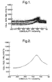
- the frequency distribution for the values is plotted against osmolality of the suspending liquid, which is calculated from the quantities of buffered saline and of water for the aliquot under observation (or it may be measured during the test), for all osmolality values down to the value below which the cells react to reach their maximum size and burst.
- the frequency distribution is shown in Figure 1.
- Figure 2 shows the mean cell size value against osmolality for the data from Figure 1.
- the maximum of the curve is the point at which the cells are at their maximum cell size (cm max ). Above this dilution, or below the osmolality O b , the cells burst. At isotonic osmolality the cells have cell size measurement cm i .
- the extracellular medium of the original cell suspension under test has an osmolality greater than the buffer osmolality i.e. is hypertonic O y , the true cell size measurement cm y of the cells in vivo will be lower than that determined at the plasma osmolality. Similarly if the extracellular medium of the original suspension is lower than average (O o ) the true in vivo cell size measurement (cm o ) will be higher than that determined at the plasma osmolality.
- a sample of O-negative red blood cells is subjected to the same general method as the cells under test in example 1 to generate the frequency distribution profile shown in Figure 3.
- a second portion of the blood sample used in example 1 is separated by centrifugation to remove cells from the extracellular liquid.
- the extracellular liquid is then diluted with 9 volumes of buffered saline as used in example 1 to dilute the first portion of the cell sample.
- This medium was then used to dilute a portion of the calibrating O-negative red blood cells and the cell size measurement of those cells determined as cm s .
- the value for the osmolality of the cells having that cell size measurement is determined as shown in Figure 4 to be O s . From this value the osmolality of the extracellular medium can be calculated. This value can then be used to determine the in vivo cell size measurement for the cells of the sample from the profile shown in Figures 3 and 4.
- the data is generally converted to digital where the data are collected on a cell-by-cell basis. It may be possible to measure the average cell size only, by using standard data collection techniques in which the voltages of a known number of cells are summated, although the accuracy of such techniques may not be good enough to obtain adequate results in the methods of the present invention.
Landscapes
- Physics & Mathematics (AREA)
- Health & Medical Sciences (AREA)
- Life Sciences & Earth Sciences (AREA)
- Chemical & Material Sciences (AREA)
- Analytical Chemistry (AREA)
- Biochemistry (AREA)
- General Health & Medical Sciences (AREA)
- General Physics & Mathematics (AREA)
- Immunology (AREA)
- Pathology (AREA)
- Investigating Or Analysing Biological Materials (AREA)
- Chemical Treatment Of Metals (AREA)
- Immobilizing And Processing Of Enzymes And Microorganisms (AREA)
- Sampling And Sample Adjustment (AREA)
Claims (18)
- Procédé dans lequel une suspension cellulaire est soumise aux étapes suivantes :1) une portion de la suspension cellulaire est diluée avec un diluant qui a la même osmolalité que le plasma liquide extracellulaire, et2) les cellules de la suspension diluée formée dans l'étape 1 sont soumises à une mesure de la taille des cellules, pendant qu'elles sont en suspension dans ladite suspension diluée.
- Procédé selon la revendication 1, dans lequel le diluant utilisé dans l'étape 1 peut être constitué d'un plasma séparé de la suspension cellulaire de départ.
- Procédé selon la revendication 1, dans lequel, en tant qu'étape préliminaire, l'osmolalité du plasma de la suspension cellulaire est déterminée, et un diluant est préparé pour avoir l'osmolalité telle que déterminée, et ensuite est utilisé dans l'étape 1 pour diluer un échantillon de la totalité de la suspension.
- Procédé selon la revendication 3, qui comprend en outre les étapes suivantes :3) une portion supplémentaire de la suspension cellulaire est diluée avec un diluant aqueux ayant une osmolalité connue,4) la taille des cellules se trouvant dans le diluant ayant l'osmolalité connue, est mesurée,5) la fonction définissant les variations de la taille des cellules en fonction de la variation de l'osmolalité, d'un état hypertonique à un état hypotonique, pour les cellules de la suspension cellulaire est déterminée, et6) la fonction déterminée dans l'étape 5, les tailles de cellules déterminées dans les étapes 2 et 4 et l'osmolalité du diluant utilisé dans l'étape 3, sont utilisées pour calculer l'osmolalité du plasma utilisé dans l'étape 1.
- Procédé selon la revendication 4, dans lequel ladite fonction de l'étape 5 est déterminée par mise en oeuvre des étapes suivantes :7) une portion de la suspension cellulaire de départ est diluée dans un diluant aqueux ayant une osmolalité hypertonique (par rapport au plasma de la suspension cellulaire de départ) connue, pour former une suspension diluée hypertonique,8) la taille des cellules de la suspension diluée hypertonique est mesurée,9) une portion supplémentaire de la suspension cellulaire est diluée dans un diluant aqueux ayant une osmolalité connue (hypotonique par rapport à l'osmolalité du plasma de la suspension cellulaire de départ) pour former une suspension diluée hypotonique,10) la taille des cellules de la suspension diluée hypotonique est mesurée, et11) les mesures de la taille des cellules, obtenues pour les suspensions cellulaires diluées hypertonique et hypotonique des étapes 8 et 10 et les valeurs de l'osmolalité du diluant hypertonique et du diluant hypotonique, sont utilisées pour calculer ladite fonction.
- Procédé selon la revendication 5, dans lequel les étapes 9 et 10 sont mises en oeuvre avec des diluants ayant des osmolalités hypotoniques connues différentes, et les étapes 7 et 8 sont mises en oeuvre avec des diluants ayant des osmolalités hypertoniques connues différentes.
- Procédé selon la revendication 5 ou 6, dans lequel la fonction et une courbe d'étalonnage de la taille en fonction de l'osmolalité.
- Procédé selon l'une quelconque des revendications 4 à 7, dans lequel les mesures de la taille des cellules sont effectuées par dilution d'au moins deux autres portions de suspension cellulaire avec des diluants ayant au moins deux osmolalités hypotoniques connues, et mesure de la taille des cellules de chacune des suspensions diluées, pendant qu'elles sont en suspension dans lesdites suspensions diluées, de préférence dans lequel lesdits diluants comprennent au moins un diluant qui a une osmolalité telle que les cellules voient leur taille augmenter jusqu'à leur taille maximale de cellule, ou une osmolalité plus faible.
- Procédé selon la revendication 8, dans lequel la taille maximale des cellules est ainsi déterminée.
- Procédé selon la revendication 8, dans lequel la taille maximale des cellules a été prédéterminée, et la courbe d'étalonnage de la taille des cellules en fonction de l'osmolalité, obtenue à partir desdites mesures de la taille des cellules en présence de diluants ayant une osmolalité connue, est utilisée pour déterminer l'osmolalité d'un diluant ayant une osmolalité inconnue, par dilution d'une autre portion de la suspension cellulaire avec ledit diluant ayant une osmolalité inconnue, ou avec un diluant constitué d'un mélange, contenant une quantité connue dudit diluant et une quantité connue d'un diluant ayant une osmolalité connue, et mesure de la taille des cellules de la suspension ainsi produite.
- Procédé selon la revendication 1, dans lequel il s'agit de déterminer l'osmolalité d'une suspension cellulaire comprenant des cellules et un plasma liquide extracellulaire, le procédé comprenanti) la dilution de la suspension cellulaire, par utilisation d'un diluant aqueux comprenant un volume connu de plasma séparé de la suspension cellulaire de départ, pour former une suspension diluée,ii) l'opération consistant à soumettre les cellules de la suspension diluée formée dans l'étape i) a une mesure de la taille des cellules, pour un certain nombre de concentrations différentes du diluant,iii) la dilution d'une autre portion de la suspension cellulaire, par utilisation d'un diluant de référence ayant une osmolalité connue, pour former une suspension diluée,iv) l'opération consistant à soumettre les cellules de la suspension diluée formée dans l'étape iii) a une mesure de la taille des cellules, pour un certain nombre de concentrations connues différentes du diluant de référence, etv) l'utilisation des mesures de la taille des cellules, obtenues dans les étapes ii) et iv) et de l'osmolalité connue du diluant de référence de l'étape iii) pour calculer l'osmolalité du plasma liquide extracellulaire de la suspension cellulaire, où, dans les deux étapes ii) et iv), la suspension cellulaire est diluée sur une plage de différentes concentrations, de façon que les cellules atteignent une taille maximale de cellule, et l'osmolalité du plasma est calculée à partir de la variation de concentration dans chacune des étapes ii) et iv), nécessaire pour arriver à la taille maximale des cellules et de l'osmolalité connue du diluant de référence.
- Procédé selon l'une quelconque des revendications 4 à 11, dans lequel des cellules sont soumises à un gradient osmotique, et les mesures de la taille des cellules sont effectuées à un certain nombre d'osmolalités différentes.
- Procédé selon l'un quelconque des revendications 4 à 11, dans lequel des cellules qui sont soumises à une mesure de la taille des cellules dans un diluant ayant une osmolalité connue sont d'abord séparées de tout fluide de mise en suspension, et lavées dans le diluant ayant une osmolalité connue.
- Procédé selon l'une quelconque des revendications précédentes dans lequel les cellules sont des cellules sanguines.
- Procédé pour déterminer l'osmolalité d'un liquide aqueux, dans lequel une quantité connue du liquide est utilisée, éventuellement on mélange avec une quantité connue d'un diluant de référence ayant une osmolalité connue, pour diluer une portion de cellules qui présentent une dépendance prédéterminée de la variation de taille en fonction d'une variation de l'osmolalité du milieu environnant, la suspension cellulaire diluée est soumise à une mesure de la taille des cellules, et la valeur obtenue par la mesure de la taille des cellules est utilisée pour calculer l'osmolalité du liquide aqueux.
- Procédé selon la revendication 15, dans lequel le liquide aqueux est un plasma.
- Procédé selon la revendication 15 ou 16, dans. lequel les cellules sont des cellules sanguines O-négatives.
- Procédé selon l'une quelconque des revendications 16 à 17, dans lequel plusieurs portions des cellules sont mises en suspension dans des portions de diluant à osmolalité hypotonique croissante, chacun comprenant une quantité connue dudit liquide aqueux et une quantité connue d'un diluant hypotonique ayant une osmolalité hypotonique connue, et une mesure de la taille des cellules est effectuée pour chaque suspension diluée, comprend de préférence une solution diluée ayant une osmolalité égale ou inférieure à celle qui amène les cellules à voir leur taille augmenter jusqu'à leur taille maximale.
Applications Claiming Priority (3)
| Application Number | Priority Date | Filing Date | Title |
|---|---|---|---|
| GB9526676 | 1995-12-29 | ||
| GBGB9526676.3A GB9526676D0 (en) | 1995-12-29 | 1995-12-29 | Method for testing a cell suspension |
| PCT/GB1996/003259 WO1997024599A1 (fr) | 1995-12-29 | 1996-12-27 | Procede pour tester une suspension cellulaire |
Publications (2)
| Publication Number | Publication Date |
|---|---|
| EP0870185A1 EP0870185A1 (fr) | 1998-10-14 |
| EP0870185B1 true EP0870185B1 (fr) | 2002-10-02 |
Family
ID=10786173
Family Applications (1)
| Application Number | Title | Priority Date | Filing Date |
|---|---|---|---|
| EP96944119A Expired - Lifetime EP0870185B1 (fr) | 1995-12-29 | 1996-12-27 | Procede pour tester une suspension cellulaire |
Country Status (9)
| Country | Link |
|---|---|
| US (1) | US6218190B1 (fr) |
| EP (1) | EP0870185B1 (fr) |
| JP (1) | JP4087446B2 (fr) |
| AT (1) | ATE225503T1 (fr) |
| AU (1) | AU724287B2 (fr) |
| CA (1) | CA2240475C (fr) |
| DE (1) | DE69624135T2 (fr) |
| GB (1) | GB9526676D0 (fr) |
| WO (1) | WO1997024599A1 (fr) |
Families Citing this family (2)
| Publication number | Priority date | Publication date | Assignee | Title |
|---|---|---|---|---|
| NZ506590A (en) * | 1998-12-29 | 2002-10-25 | Thomas Adam Shine | A method of analysing a sample of free cells |
| US12433869B2 (en) | 2019-04-22 | 2025-10-07 | Ian Basil Shine | Preventing and treating malaria |
Family Cites Families (14)
| Publication number | Priority date | Publication date | Assignee | Title |
|---|---|---|---|---|
| US3962125A (en) * | 1975-01-13 | 1976-06-08 | Coulter Diagnostics, Inc. | Multi-purpose diluent for use in blood analysis by electronic instrumentation of the coulter type |
| US4185964A (en) * | 1977-02-08 | 1980-01-29 | Central Laboratories of Associated Maryland Pathologists, Ltd. | Lysing reagent |
| US4213876A (en) * | 1978-08-22 | 1980-07-22 | Coulter Electronics, Inc. | Multi-purpose blood diluent for use in electronic blood analysis instrumentation |
| US4299726A (en) * | 1979-05-07 | 1981-11-10 | Coulter Electronics, Inc. | Process for preparing whole blood reference controls having long term stability, preconditioning diluent and media therefor |
| US4358436A (en) * | 1979-10-05 | 1982-11-09 | Ortho Diagnostics, Inc. | Blood typing tests and reagents |
| US4278936A (en) * | 1980-02-05 | 1981-07-14 | Coulter Electronics, Inc. | Biological cell parameter change test method and apparatus |
| US4346018A (en) * | 1980-06-16 | 1982-08-24 | Coulter Electronics, Inc. | Multi-purpose blood diluent and lysing agent for differential determination of lymphoid-myeloid population of leukocytes |
| US4322313A (en) * | 1980-10-02 | 1982-03-30 | J. T. Baker Chemicals B.V. | Stabilized multi-purpose blood diluent |
| US4535284A (en) * | 1981-07-10 | 1985-08-13 | Coulter Electronics, Inc. | High and low frequency analysis of osmotic stress of cells |
| US4506018A (en) * | 1982-12-30 | 1985-03-19 | Becton, Dickinson And Company | Blood diluent |
| US4968629A (en) * | 1986-08-20 | 1990-11-06 | Hematology Marketing Associates, Inc. | Blood diluent for automatic and semi-automatic determination of white cells and method of utilizing same |
| FR2633047B1 (fr) * | 1988-06-21 | 1990-08-24 | Univ Rene Descartes | Dispositif et appareil de detection et de mesure par echographie de particules en suspension dans un liquide |
| US5008202A (en) * | 1988-11-29 | 1991-04-16 | Sequoia Turner Corporation | Blood diluent for red blood cell analysis |
| US5227304A (en) * | 1991-01-16 | 1993-07-13 | Sequoia Turner Corporation | Method for counting whole blood diluent and detergent reagent system |
-
1995
- 1995-12-29 GB GBGB9526676.3A patent/GB9526676D0/en active Pending
-
1996
- 1996-12-27 EP EP96944119A patent/EP0870185B1/fr not_active Expired - Lifetime
- 1996-12-27 WO PCT/GB1996/003259 patent/WO1997024599A1/fr not_active Ceased
- 1996-12-27 AT AT96944119T patent/ATE225503T1/de not_active IP Right Cessation
- 1996-12-27 CA CA002240475A patent/CA2240475C/fr not_active Expired - Lifetime
- 1996-12-27 AU AU13849/97A patent/AU724287B2/en not_active Expired
- 1996-12-27 US US09/101,039 patent/US6218190B1/en not_active Expired - Lifetime
- 1996-12-27 DE DE69624135T patent/DE69624135T2/de not_active Expired - Lifetime
- 1996-12-27 JP JP52411897A patent/JP4087446B2/ja not_active Expired - Lifetime
Also Published As
| Publication number | Publication date |
|---|---|
| JP4087446B2 (ja) | 2008-05-21 |
| DE69624135T2 (de) | 2003-02-13 |
| WO1997024599A1 (fr) | 1997-07-10 |
| AU724287B2 (en) | 2000-09-14 |
| US6218190B1 (en) | 2001-04-17 |
| CA2240475A1 (fr) | 1997-07-10 |
| EP0870185A1 (fr) | 1998-10-14 |
| JP2000503116A (ja) | 2000-03-14 |
| AU1384997A (en) | 1997-07-28 |
| CA2240475C (fr) | 2004-11-23 |
| ATE225503T1 (de) | 2002-10-15 |
| GB9526676D0 (en) | 1996-02-28 |
| DE69624135D1 (de) | 2002-11-07 |
Similar Documents
| Publication | Publication Date | Title |
|---|---|---|
| US5197017A (en) | Potentiophotometric fibrinogen determination | |
| EP0874983B1 (fr) | Procede et dispositif d'evaluation de la teneur de globules rouges individuels en hemoglobine | |
| US4535284A (en) | High and low frequency analysis of osmotic stress of cells | |
| Lines | The electrical sensing zone method (the Coulter principle) | |
| JP3049371B2 (ja) | 試薬及び抵抗率の変化を測定する装置で血液中の好塩基球を自動的に計数するために該試薬を使用する方法 | |
| US3997838A (en) | Apparatus and method for measurement of total volume of particles in a liquid sample | |
| US4699887A (en) | Electrochemical determination of hematocrit | |
| US6218190B1 (en) | Method for testing a cell suspension | |
| US6030838A (en) | Hematological parameter | |
| US6699680B1 (en) | Method of determining a number of platelets | |
| US5939327A (en) | Measurement of bile pigments in serum or plasma | |
| EP1588164B1 (fr) | Amelioration de la qualité d'une analyse hématologique | |
| EP0801743A1 (fr) | Procede de mesure de la concentration de dioxyde de carbone dans le sang | |
| JP2005077148A (ja) | 血液検査方法及び装置 | |
| CN119757774B (zh) | 一种医学检验中血液细胞检验的质量控制方法 | |
| US4605628A (en) | Electrochemical determination of hematocrit | |
| Hamilton et al. | Evaluation of a semi‐automated reticulocyte counting method using the Coulter® STKS‐2A blood cell counter | |
| CA2012056A1 (fr) | Etalons optiques pour analyseurs automatiques de sang |
Legal Events
| Date | Code | Title | Description |
|---|---|---|---|
| PUAI | Public reference made under article 153(3) epc to a published international application that has entered the european phase |
Free format text: ORIGINAL CODE: 0009012 |
|
| 17P | Request for examination filed |
Effective date: 19980717 |
|
| AK | Designated contracting states |
Kind code of ref document: A1 Designated state(s): AT BE CH DE DK ES FI FR GB GR IE IT LI LU MC NL PT SE |
|
| 17Q | First examination report despatched |
Effective date: 20010426 |
|
| GRAG | Despatch of communication of intention to grant |
Free format text: ORIGINAL CODE: EPIDOS AGRA |
|
| GRAG | Despatch of communication of intention to grant |
Free format text: ORIGINAL CODE: EPIDOS AGRA |
|
| GRAG | Despatch of communication of intention to grant |
Free format text: ORIGINAL CODE: EPIDOS AGRA |
|
| GRAH | Despatch of communication of intention to grant a patent |
Free format text: ORIGINAL CODE: EPIDOS IGRA |
|
| GRAH | Despatch of communication of intention to grant a patent |
Free format text: ORIGINAL CODE: EPIDOS IGRA |
|
| GRAA | (expected) grant |
Free format text: ORIGINAL CODE: 0009210 |
|
| AK | Designated contracting states |
Kind code of ref document: B1 Designated state(s): AT BE CH DE DK ES FI FR GB GR IE IT LI LU MC NL PT SE |
|
| PG25 | Lapsed in a contracting state [announced via postgrant information from national office to epo] |
Ref country code: NL Free format text: LAPSE BECAUSE OF FAILURE TO SUBMIT A TRANSLATION OF THE DESCRIPTION OR TO PAY THE FEE WITHIN THE PRESCRIBED TIME-LIMIT Effective date: 20021002 Ref country code: LI Free format text: LAPSE BECAUSE OF FAILURE TO SUBMIT A TRANSLATION OF THE DESCRIPTION OR TO PAY THE FEE WITHIN THE PRESCRIBED TIME-LIMIT Effective date: 20021002 Ref country code: IT Free format text: LAPSE BECAUSE OF FAILURE TO SUBMIT A TRANSLATION OF THE DESCRIPTION OR TO PAY THE FEE WITHIN THE PRE;WARNING: LAPSES OF ITALIAN PATENTS WITH EFFECTIVE DATE BEFORE 2007 MAY HAVE OCCURRED AT ANY TIME BEFORE 2007. THE CORRECT EFFECTIVE DATE MAY BE DIFFERENT FROM THE ONE RECORDED.SCRIBED TIME-LIMIT Effective date: 20021002 Ref country code: GR Free format text: LAPSE BECAUSE OF FAILURE TO SUBMIT A TRANSLATION OF THE DESCRIPTION OR TO PAY THE FEE WITHIN THE PRESCRIBED TIME-LIMIT Effective date: 20021002 Ref country code: FI Free format text: LAPSE BECAUSE OF FAILURE TO SUBMIT A TRANSLATION OF THE DESCRIPTION OR TO PAY THE FEE WITHIN THE PRESCRIBED TIME-LIMIT Effective date: 20021002 Ref country code: CH Free format text: LAPSE BECAUSE OF FAILURE TO SUBMIT A TRANSLATION OF THE DESCRIPTION OR TO PAY THE FEE WITHIN THE PRESCRIBED TIME-LIMIT Effective date: 20021002 Ref country code: BE Free format text: LAPSE BECAUSE OF FAILURE TO SUBMIT A TRANSLATION OF THE DESCRIPTION OR TO PAY THE FEE WITHIN THE PRESCRIBED TIME-LIMIT Effective date: 20021002 Ref country code: AT Free format text: LAPSE BECAUSE OF FAILURE TO SUBMIT A TRANSLATION OF THE DESCRIPTION OR TO PAY THE FEE WITHIN THE PRESCRIBED TIME-LIMIT Effective date: 20021002 |
|
| REF | Corresponds to: |
Ref document number: 225503 Country of ref document: AT Date of ref document: 20021015 Kind code of ref document: T |
|
| REG | Reference to a national code |
Ref country code: GB Ref legal event code: FG4D |
|
| REG | Reference to a national code |
Ref country code: CH Ref legal event code: EP |
|
| REG | Reference to a national code |
Ref country code: IE Ref legal event code: FG4D |
|
| REF | Corresponds to: |
Ref document number: 69624135 Country of ref document: DE Date of ref document: 20021107 |
|
| PGFP | Annual fee paid to national office [announced via postgrant information from national office to epo] |
Ref country code: SE Payment date: 20021204 Year of fee payment: 7 |
|
| PGFP | Annual fee paid to national office [announced via postgrant information from national office to epo] |
Ref country code: AT Payment date: 20021211 Year of fee payment: 7 |
|
| PGFP | Annual fee paid to national office [announced via postgrant information from national office to epo] |
Ref country code: DK Payment date: 20021212 Year of fee payment: 7 |
|
| PGFP | Annual fee paid to national office [announced via postgrant information from national office to epo] |
Ref country code: GR Payment date: 20021220 Year of fee payment: 7 |
|
| PGFP | Annual fee paid to national office [announced via postgrant information from national office to epo] |
Ref country code: IE Payment date: 20021223 Year of fee payment: 7 |
|
| PG25 | Lapsed in a contracting state [announced via postgrant information from national office to epo] |
Ref country code: LU Free format text: LAPSE BECAUSE OF NON-PAYMENT OF DUE FEES Effective date: 20021227 |
|
| PGFP | Annual fee paid to national office [announced via postgrant information from national office to epo] |
Ref country code: NL Payment date: 20021227 Year of fee payment: 7 |
|
| PGFP | Annual fee paid to national office [announced via postgrant information from national office to epo] |
Ref country code: MC Payment date: 20021231 Year of fee payment: 7 |
|
| PG25 | Lapsed in a contracting state [announced via postgrant information from national office to epo] |
Ref country code: SE Free format text: LAPSE BECAUSE OF FAILURE TO SUBMIT A TRANSLATION OF THE DESCRIPTION OR TO PAY THE FEE WITHIN THE PRESCRIBED TIME-LIMIT Effective date: 20030102 Ref country code: PT Free format text: LAPSE BECAUSE OF FAILURE TO SUBMIT A TRANSLATION OF THE DESCRIPTION OR TO PAY THE FEE WITHIN THE PRESCRIBED TIME-LIMIT Effective date: 20030102 Ref country code: DK Free format text: LAPSE BECAUSE OF FAILURE TO SUBMIT A TRANSLATION OF THE DESCRIPTION OR TO PAY THE FEE WITHIN THE PRESCRIBED TIME-LIMIT Effective date: 20030102 |
|
| NLV1 | Nl: lapsed or annulled due to failure to fulfill the requirements of art. 29p and 29m of the patents act | ||
| ET | Fr: translation filed | ||
| PG25 | Lapsed in a contracting state [announced via postgrant information from national office to epo] |
Ref country code: ES Free format text: LAPSE BECAUSE OF FAILURE TO SUBMIT A TRANSLATION OF THE DESCRIPTION OR TO PAY THE FEE WITHIN THE PRESCRIBED TIME-LIMIT Effective date: 20030429 |
|
| REG | Reference to a national code |
Ref country code: CH Ref legal event code: PL |
|
| PLBE | No opposition filed within time limit |
Free format text: ORIGINAL CODE: 0009261 |
|
| STAA | Information on the status of an ep patent application or granted ep patent |
Free format text: STATUS: NO OPPOSITION FILED WITHIN TIME LIMIT |
|
| 26N | No opposition filed |
Effective date: 20030703 |
|
| PG25 | Lapsed in a contracting state [announced via postgrant information from national office to epo] |
Ref country code: IE Free format text: LAPSE BECAUSE OF NON-PAYMENT OF DUE FEES Effective date: 20031229 |
|
| PG25 | Lapsed in a contracting state [announced via postgrant information from national office to epo] |
Ref country code: MC Free format text: LAPSE BECAUSE OF NON-PAYMENT OF DUE FEES Effective date: 20031231 |
|
| REG | Reference to a national code |
Ref country code: IE Ref legal event code: MM4A |
|
| PGRI | Patent reinstated in contracting state [announced from national office to epo] |
Ref country code: DE Effective date: 20070427 |
|
| REG | Reference to a national code |
Ref country code: FR Ref legal event code: PLFP Year of fee payment: 20 |
|
| PGFP | Annual fee paid to national office [announced via postgrant information from national office to epo] |
Ref country code: GB Payment date: 20151223 Year of fee payment: 20 |
|
| PGFP | Annual fee paid to national office [announced via postgrant information from national office to epo] |
Ref country code: FR Payment date: 20151110 Year of fee payment: 20 |
|
| PGFP | Annual fee paid to national office [announced via postgrant information from national office to epo] |
Ref country code: DE Payment date: 20151222 Year of fee payment: 20 |
|
| REG | Reference to a national code |
Ref country code: DE Ref legal event code: R071 Ref document number: 69624135 Country of ref document: DE |
|
| REG | Reference to a national code |
Ref country code: GB Ref legal event code: PE20 Expiry date: 20161226 |
|
| PG25 | Lapsed in a contracting state [announced via postgrant information from national office to epo] |
Ref country code: GB Free format text: LAPSE BECAUSE OF EXPIRATION OF PROTECTION Effective date: 20161226 |