EP0871496B1 - Arteriovenöse und venöse transplantatbehandlungen: methoden und zusammensetzungen - Google Patents
Arteriovenöse und venöse transplantatbehandlungen: methoden und zusammensetzungen Download PDFInfo
- Publication number
- EP0871496B1 EP0871496B1 EP96912854A EP96912854A EP0871496B1 EP 0871496 B1 EP0871496 B1 EP 0871496B1 EP 96912854 A EP96912854 A EP 96912854A EP 96912854 A EP96912854 A EP 96912854A EP 0871496 B1 EP0871496 B1 EP 0871496B1
- Authority
- EP
- European Patent Office
- Prior art keywords
- oligonucleotide
- oligonucleotides
- vein
- myc
- antisense
- Prior art date
- Legal status (The legal status is an assumption and is not a legal conclusion. Google has not performed a legal analysis and makes no representation as to the accuracy of the status listed.)
- Expired - Lifetime
Links
- 238000011282 treatment Methods 0.000 title claims abstract description 87
- 238000000034 method Methods 0.000 title claims abstract description 41
- 239000000203 mixture Substances 0.000 title claims description 16
- 108091034117 Oligonucleotide Proteins 0.000 claims abstract description 296
- 101710135898 Myc proto-oncogene protein Proteins 0.000 claims abstract description 139
- 102100038895 Myc proto-oncogene protein Human genes 0.000 claims abstract description 139
- 101710150448 Transcriptional regulator Myc Proteins 0.000 claims abstract description 139
- 108010087776 Proto-Oncogene Proteins c-myb Proteins 0.000 claims abstract description 19
- 102000009096 Proto-Oncogene Proteins c-myb Human genes 0.000 claims abstract description 19
- 239000000074 antisense oligonucleotide Substances 0.000 claims description 145
- 238000012230 antisense oligonucleotides Methods 0.000 claims description 145
- 210000003462 vein Anatomy 0.000 claims description 124
- 210000001519 tissue Anatomy 0.000 claims description 75
- 238000001631 haemodialysis Methods 0.000 claims description 27
- 230000002792 vascular Effects 0.000 claims description 27
- 210000002808 connective tissue Anatomy 0.000 claims description 24
- 239000002773 nucleotide Substances 0.000 claims description 21
- 125000003729 nucleotide group Chemical group 0.000 claims description 21
- 239000000872 buffer Substances 0.000 claims description 11
- 206010016717 Fistula Diseases 0.000 claims description 10
- 230000003890 fistula Effects 0.000 claims description 10
- 210000003752 saphenous vein Anatomy 0.000 claims description 10
- 239000008194 pharmaceutical composition Substances 0.000 claims description 9
- 229920001343 polytetrafluoroethylene Polymers 0.000 claims description 9
- 239000004810 polytetrafluoroethylene Substances 0.000 claims description 9
- 101710163270 Nuclease Proteins 0.000 claims description 7
- 210000003191 femoral vein Anatomy 0.000 claims description 4
- 210000001715 carotid artery Anatomy 0.000 claims description 3
- 239000007943 implant Substances 0.000 claims description 3
- 241000283690 Bos taurus Species 0.000 claims description 2
- 230000003511 endothelial effect Effects 0.000 claims description 2
- 238000002360 preparation method Methods 0.000 claims description 2
- 241000385250 Epioblasma triquetra Species 0.000 claims 1
- 238000003780 insertion Methods 0.000 claims 1
- 230000037431 insertion Effects 0.000 claims 1
- JLCPHMBAVCMARE-UHFFFAOYSA-N [3-[[3-[[3-[[3-[[3-[[3-[[3-[[3-[[3-[[3-[[3-[[5-(2-amino-6-oxo-1H-purin-9-yl)-3-[[3-[[3-[[3-[[3-[[3-[[5-(2-amino-6-oxo-1H-purin-9-yl)-3-[[5-(2-amino-6-oxo-1H-purin-9-yl)-3-hydroxyoxolan-2-yl]methoxy-hydroxyphosphoryl]oxyoxolan-2-yl]methoxy-hydroxyphosphoryl]oxy-5-(5-methyl-2,4-dioxopyrimidin-1-yl)oxolan-2-yl]methoxy-hydroxyphosphoryl]oxy-5-(6-aminopurin-9-yl)oxolan-2-yl]methoxy-hydroxyphosphoryl]oxy-5-(6-aminopurin-9-yl)oxolan-2-yl]methoxy-hydroxyphosphoryl]oxy-5-(6-aminopurin-9-yl)oxolan-2-yl]methoxy-hydroxyphosphoryl]oxy-5-(6-aminopurin-9-yl)oxolan-2-yl]methoxy-hydroxyphosphoryl]oxyoxolan-2-yl]methoxy-hydroxyphosphoryl]oxy-5-(5-methyl-2,4-dioxopyrimidin-1-yl)oxolan-2-yl]methoxy-hydroxyphosphoryl]oxy-5-(4-amino-2-oxopyrimidin-1-yl)oxolan-2-yl]methoxy-hydroxyphosphoryl]oxy-5-(5-methyl-2,4-dioxopyrimidin-1-yl)oxolan-2-yl]methoxy-hydroxyphosphoryl]oxy-5-(5-methyl-2,4-dioxopyrimidin-1-yl)oxolan-2-yl]methoxy-hydroxyphosphoryl]oxy-5-(6-aminopurin-9-yl)oxolan-2-yl]methoxy-hydroxyphosphoryl]oxy-5-(6-aminopurin-9-yl)oxolan-2-yl]methoxy-hydroxyphosphoryl]oxy-5-(4-amino-2-oxopyrimidin-1-yl)oxolan-2-yl]methoxy-hydroxyphosphoryl]oxy-5-(4-amino-2-oxopyrimidin-1-yl)oxolan-2-yl]methoxy-hydroxyphosphoryl]oxy-5-(4-amino-2-oxopyrimidin-1-yl)oxolan-2-yl]methoxy-hydroxyphosphoryl]oxy-5-(6-aminopurin-9-yl)oxolan-2-yl]methoxy-hydroxyphosphoryl]oxy-5-(4-amino-2-oxopyrimidin-1-yl)oxolan-2-yl]methyl [5-(6-aminopurin-9-yl)-2-(hydroxymethyl)oxolan-3-yl] hydrogen phosphate Polymers Cc1cn(C2CC(OP(O)(=O)OCC3OC(CC3OP(O)(=O)OCC3OC(CC3O)n3cnc4c3nc(N)[nH]c4=O)n3cnc4c3nc(N)[nH]c4=O)C(COP(O)(=O)OC3CC(OC3COP(O)(=O)OC3CC(OC3COP(O)(=O)OC3CC(OC3COP(O)(=O)OC3CC(OC3COP(O)(=O)OC3CC(OC3COP(O)(=O)OC3CC(OC3COP(O)(=O)OC3CC(OC3COP(O)(=O)OC3CC(OC3COP(O)(=O)OC3CC(OC3COP(O)(=O)OC3CC(OC3COP(O)(=O)OC3CC(OC3COP(O)(=O)OC3CC(OC3COP(O)(=O)OC3CC(OC3COP(O)(=O)OC3CC(OC3COP(O)(=O)OC3CC(OC3COP(O)(=O)OC3CC(OC3COP(O)(=O)OC3CC(OC3CO)n3cnc4c(N)ncnc34)n3ccc(N)nc3=O)n3cnc4c(N)ncnc34)n3ccc(N)nc3=O)n3ccc(N)nc3=O)n3ccc(N)nc3=O)n3cnc4c(N)ncnc34)n3cnc4c(N)ncnc34)n3cc(C)c(=O)[nH]c3=O)n3cc(C)c(=O)[nH]c3=O)n3ccc(N)nc3=O)n3cc(C)c(=O)[nH]c3=O)n3cnc4c3nc(N)[nH]c4=O)n3cnc4c(N)ncnc34)n3cnc4c(N)ncnc34)n3cnc4c(N)ncnc34)n3cnc4c(N)ncnc34)O2)c(=O)[nH]c1=O JLCPHMBAVCMARE-UHFFFAOYSA-N 0.000 abstract description 155
- 230000015572 biosynthetic process Effects 0.000 abstract description 58
- 238000003786 synthesis reaction Methods 0.000 abstract description 48
- 102000008186 Collagen Human genes 0.000 abstract description 45
- 108010035532 Collagen Proteins 0.000 abstract description 45
- 229920001436 collagen Polymers 0.000 abstract description 45
- 102000010834 Extracellular Matrix Proteins Human genes 0.000 abstract description 43
- 108010037362 Extracellular Matrix Proteins Proteins 0.000 abstract description 43
- 150000001875 compounds Chemical class 0.000 abstract description 28
- 208000037265 diseases, disorders, signs and symptoms Diseases 0.000 abstract description 26
- 208000037803 restenosis Diseases 0.000 abstract description 14
- 102000052575 Proto-Oncogene Human genes 0.000 abstract description 12
- 108700020978 Proto-Oncogene Proteins 0.000 abstract description 12
- 208000035475 disorder Diseases 0.000 abstract description 12
- 230000002401 inhibitory effect Effects 0.000 abstract description 4
- 230000002784 sclerotic effect Effects 0.000 abstract description 2
- 230000000692 anti-sense effect Effects 0.000 description 111
- 210000004027 cell Anatomy 0.000 description 104
- 108020000948 Antisense Oligonucleotides Proteins 0.000 description 102
- 108010050808 Procollagen Proteins 0.000 description 95
- 210000000329 smooth muscle myocyte Anatomy 0.000 description 74
- 108090000623 proteins and genes Proteins 0.000 description 40
- 108010022452 Collagen Type I Proteins 0.000 description 38
- 102000012422 Collagen Type I Human genes 0.000 description 38
- 210000002950 fibroblast Anatomy 0.000 description 34
- 108020004999 messenger RNA Proteins 0.000 description 30
- 108050006400 Cyclin Proteins 0.000 description 29
- 102100036691 Proliferating cell nuclear antigen Human genes 0.000 description 29
- 102000043827 human Smooth muscle Human genes 0.000 description 28
- 108700038605 human Smooth muscle Proteins 0.000 description 28
- 238000002347 injection Methods 0.000 description 28
- 239000007924 injection Substances 0.000 description 28
- 102000004169 proteins and genes Human genes 0.000 description 27
- 238000010186 staining Methods 0.000 description 27
- 235000018102 proteins Nutrition 0.000 description 26
- 210000002744 extracellular matrix Anatomy 0.000 description 24
- 210000004351 coronary vessel Anatomy 0.000 description 23
- 230000000694 effects Effects 0.000 description 23
- 102000007469 Actins Human genes 0.000 description 22
- 108010085238 Actins Proteins 0.000 description 22
- 230000000322 hemodialysis Effects 0.000 description 21
- 230000003834 intracellular effect Effects 0.000 description 21
- 239000000499 gel Substances 0.000 description 20
- 238000001727 in vivo Methods 0.000 description 20
- 230000005764 inhibitory process Effects 0.000 description 20
- 238000009826 distribution Methods 0.000 description 19
- 210000001367 artery Anatomy 0.000 description 18
- 239000003814 drug Substances 0.000 description 18
- 208000014674 injury Diseases 0.000 description 18
- 210000001626 skin fibroblast Anatomy 0.000 description 18
- 108010038512 Platelet-Derived Growth Factor Proteins 0.000 description 17
- 102000010780 Platelet-Derived Growth Factor Human genes 0.000 description 17
- 230000004663 cell proliferation Effects 0.000 description 16
- 230000006378 damage Effects 0.000 description 16
- 229940079593 drug Drugs 0.000 description 16
- 238000002474 experimental method Methods 0.000 description 16
- 102000039446 nucleic acids Human genes 0.000 description 16
- 108020004707 nucleic acids Proteins 0.000 description 16
- 150000007523 nucleic acids Chemical class 0.000 description 16
- 230000009467 reduction Effects 0.000 description 16
- 230000014616 translation Effects 0.000 description 16
- 108091003079 Bovine Serum Albumin Proteins 0.000 description 15
- 208000034827 Neointima Diseases 0.000 description 15
- 208000027418 Wounds and injury Diseases 0.000 description 15
- 230000004872 arterial blood pressure Effects 0.000 description 15
- 239000012091 fetal bovine serum Substances 0.000 description 15
- GNBHRKFJIUUOQI-UHFFFAOYSA-N fluorescein Chemical compound O1C(=O)C2=CC=CC=C2C21C1=CC=C(O)C=C1OC1=CC(O)=CC=C21 GNBHRKFJIUUOQI-UHFFFAOYSA-N 0.000 description 15
- 239000002609 medium Substances 0.000 description 14
- -1 polytetrafluoroethylene Polymers 0.000 description 13
- RYYWUUFWQRZTIU-UHFFFAOYSA-K thiophosphate Chemical compound [O-]P([O-])([O-])=S RYYWUUFWQRZTIU-UHFFFAOYSA-K 0.000 description 13
- 238000001262 western blot Methods 0.000 description 13
- CIWBSHSKHKDKBQ-JLAZNSOCSA-N Ascorbic acid Chemical compound OC[C@H](O)[C@H]1OC(=O)C(O)=C1O CIWBSHSKHKDKBQ-JLAZNSOCSA-N 0.000 description 12
- OKKJLVBELUTLKV-UHFFFAOYSA-N Methanol Chemical compound OC OKKJLVBELUTLKV-UHFFFAOYSA-N 0.000 description 12
- 239000003636 conditioned culture medium Substances 0.000 description 12
- 230000007423 decrease Effects 0.000 description 12
- 238000011534 incubation Methods 0.000 description 12
- 238000004519 manufacturing process Methods 0.000 description 12
- 210000000056 organ Anatomy 0.000 description 12
- 102000040430 polynucleotide Human genes 0.000 description 12
- 108091033319 polynucleotide Proteins 0.000 description 12
- 239000002157 polynucleotide Substances 0.000 description 12
- 230000028327 secretion Effects 0.000 description 12
- 238000001356 surgical procedure Methods 0.000 description 12
- 108091032973 (ribonucleotides)n+m Proteins 0.000 description 11
- FAPWRFPIFSIZLT-UHFFFAOYSA-M Sodium chloride Chemical compound [Na+].[Cl-] FAPWRFPIFSIZLT-UHFFFAOYSA-M 0.000 description 11
- 230000027455 binding Effects 0.000 description 11
- LOKCTEFSRHRXRJ-UHFFFAOYSA-I dipotassium trisodium dihydrogen phosphate hydrogen phosphate dichloride Chemical compound P(=O)(O)(O)[O-].[K+].P(=O)(O)([O-])[O-].[Na+].[Na+].[Cl-].[K+].[Cl-].[Na+] LOKCTEFSRHRXRJ-UHFFFAOYSA-I 0.000 description 11
- 238000000338 in vitro Methods 0.000 description 11
- 239000002953 phosphate buffered saline Substances 0.000 description 11
- 210000004509 vascular smooth muscle cell Anatomy 0.000 description 11
- 108010067306 Fibronectins Proteins 0.000 description 10
- 102000016359 Fibronectins Human genes 0.000 description 10
- 201000010099 disease Diseases 0.000 description 10
- 230000004807 localization Effects 0.000 description 10
- QTBSBXVTEAMEQO-UHFFFAOYSA-N Acetic acid Chemical compound CC(O)=O QTBSBXVTEAMEQO-UHFFFAOYSA-N 0.000 description 9
- 239000006144 Dulbecco’s modified Eagle's medium Substances 0.000 description 9
- 239000012737 fresh medium Substances 0.000 description 9
- 230000003902 lesion Effects 0.000 description 9
- 239000000463 material Substances 0.000 description 9
- 238000001243 protein synthesis Methods 0.000 description 9
- 108020004414 DNA Proteins 0.000 description 8
- 102100036912 Desmin Human genes 0.000 description 8
- 108010044052 Desmin Proteins 0.000 description 8
- 102000057297 Pepsin A Human genes 0.000 description 8
- 108090000284 Pepsin A Proteins 0.000 description 8
- 238000002399 angioplasty Methods 0.000 description 8
- 210000004204 blood vessel Anatomy 0.000 description 8
- 210000005045 desmin Anatomy 0.000 description 8
- 230000001965 increasing effect Effects 0.000 description 8
- 238000002844 melting Methods 0.000 description 8
- 230000008018 melting Effects 0.000 description 8
- 239000002777 nucleoside Substances 0.000 description 8
- 229940111202 pepsin Drugs 0.000 description 8
- 230000036573 scar formation Effects 0.000 description 8
- 210000003491 skin Anatomy 0.000 description 8
- ONIBWKKTOPOVIA-FXLFCPKBSA-N (2S)-(214C)azolidine-2-carboxylic acid Chemical compound N1[14C@@H](CCC1)C(=O)O ONIBWKKTOPOVIA-FXLFCPKBSA-N 0.000 description 7
- 238000007792 addition Methods 0.000 description 7
- 238000004458 analytical method Methods 0.000 description 7
- 230000003247 decreasing effect Effects 0.000 description 7
- 230000002062 proliferating effect Effects 0.000 description 7
- CSCPPACGZOOCGX-UHFFFAOYSA-N Acetone Chemical compound CC(C)=O CSCPPACGZOOCGX-UHFFFAOYSA-N 0.000 description 6
- 108091028690 C-myc mRNA Proteins 0.000 description 6
- WSFSSNUMVMOOMR-UHFFFAOYSA-N Formaldehyde Chemical compound O=C WSFSSNUMVMOOMR-UHFFFAOYSA-N 0.000 description 6
- PEDCQBHIVMGVHV-UHFFFAOYSA-N Glycerine Chemical compound OCC(O)CO PEDCQBHIVMGVHV-UHFFFAOYSA-N 0.000 description 6
- WZUVPPKBWHMQCE-UHFFFAOYSA-N Haematoxylin Chemical compound C12=CC(O)=C(O)C=C2CC2(O)C1C1=CC=C(O)C(O)=C1OC2 WZUVPPKBWHMQCE-UHFFFAOYSA-N 0.000 description 6
- PMMYEEVYMWASQN-DMTCNVIQSA-N Hydroxyproline Chemical compound O[C@H]1CN[C@H](C(O)=O)C1 PMMYEEVYMWASQN-DMTCNVIQSA-N 0.000 description 6
- 102000035195 Peptidases Human genes 0.000 description 6
- 108091005804 Peptidases Proteins 0.000 description 6
- 108010087705 Proto-Oncogene Proteins c-myc Proteins 0.000 description 6
- 102000009092 Proto-Oncogene Proteins c-myc Human genes 0.000 description 6
- 241000282887 Suidae Species 0.000 description 6
- 241000282898 Sus scrofa Species 0.000 description 6
- 229960005070 ascorbic acid Drugs 0.000 description 6
- 235000010323 ascorbic acid Nutrition 0.000 description 6
- 239000011668 ascorbic acid Substances 0.000 description 6
- 238000003556 assay Methods 0.000 description 6
- 230000030570 cellular localization Effects 0.000 description 6
- 230000004087 circulation Effects 0.000 description 6
- 238000003776 cleavage reaction Methods 0.000 description 6
- 229940096422 collagen type i Drugs 0.000 description 6
- 238000009792 diffusion process Methods 0.000 description 6
- PMMYEEVYMWASQN-UHFFFAOYSA-N dl-hydroxyproline Natural products OC1C[NH2+]C(C([O-])=O)C1 PMMYEEVYMWASQN-UHFFFAOYSA-N 0.000 description 6
- YQGOJNYOYNNSMM-UHFFFAOYSA-N eosin Chemical compound [Na+].OC(=O)C1=CC=CC=C1C1=C2C=C(Br)C(=O)C(Br)=C2OC2=C(Br)C(O)=C(Br)C=C21 YQGOJNYOYNNSMM-UHFFFAOYSA-N 0.000 description 6
- 229960002591 hydroxyproline Drugs 0.000 description 6
- YBYRMVIVWMBXKQ-UHFFFAOYSA-N phenylmethanesulfonyl fluoride Chemical compound FS(=O)(=O)CC1=CC=CC=C1 YBYRMVIVWMBXKQ-UHFFFAOYSA-N 0.000 description 6
- 235000019833 protease Nutrition 0.000 description 6
- 239000000523 sample Substances 0.000 description 6
- 230000007017 scission Effects 0.000 description 6
- 239000011780 sodium chloride Substances 0.000 description 6
- 230000000638 stimulation Effects 0.000 description 6
- 230000008685 targeting Effects 0.000 description 6
- FFEARJCKVFRZRR-BYPYZUCNSA-N L-methionine Chemical compound CSCC[C@H](N)C(O)=O FFEARJCKVFRZRR-BYPYZUCNSA-N 0.000 description 5
- GLNADSQYFUSGOU-GPTZEZBUSA-J Trypan blue Chemical compound [Na+].[Na+].[Na+].[Na+].C1=C(S([O-])(=O)=O)C=C2C=C(S([O-])(=O)=O)C(/N=N/C3=CC=C(C=C3C)C=3C=C(C(=CC=3)\N=N\C=3C(=CC4=CC(=CC(N)=C4C=3O)S([O-])(=O)=O)S([O-])(=O)=O)C)=C(O)C2=C1N GLNADSQYFUSGOU-GPTZEZBUSA-J 0.000 description 5
- 239000013592 cell lysate Substances 0.000 description 5
- 238000005119 centrifugation Methods 0.000 description 5
- 230000008859 change Effects 0.000 description 5
- 238000002586 coronary angiography Methods 0.000 description 5
- 238000007887 coronary angioplasty Methods 0.000 description 5
- 230000029087 digestion Effects 0.000 description 5
- 239000001963 growth medium Substances 0.000 description 5
- 238000001802 infusion Methods 0.000 description 5
- 238000005259 measurement Methods 0.000 description 5
- 239000012528 membrane Substances 0.000 description 5
- 230000030648 nucleus localization Effects 0.000 description 5
- 238000011084 recovery Methods 0.000 description 5
- 230000002829 reductive effect Effects 0.000 description 5
- 230000004044 response Effects 0.000 description 5
- 238000002415 sodium dodecyl sulfate polyacrylamide gel electrophoresis Methods 0.000 description 5
- 239000000126 substance Substances 0.000 description 5
- 239000000758 substrate Substances 0.000 description 5
- FGMPLJWBKKVCDB-UHFFFAOYSA-N trans-L-hydroxy-proline Natural products ON1CCCC1C(O)=O FGMPLJWBKKVCDB-UHFFFAOYSA-N 0.000 description 5
- 210000005167 vascular cell Anatomy 0.000 description 5
- YBJHBAHKTGYVGT-ZKWXMUAHSA-N (+)-Biotin Chemical compound N1C(=O)N[C@@H]2[C@H](CCCCC(=O)O)SC[C@@H]21 YBJHBAHKTGYVGT-ZKWXMUAHSA-N 0.000 description 4
- DHMQDGOQFOQNFH-UHFFFAOYSA-N Glycine Chemical compound NCC(O)=O DHMQDGOQFOQNFH-UHFFFAOYSA-N 0.000 description 4
- 241000282412 Homo Species 0.000 description 4
- 102000008070 Interferon-gamma Human genes 0.000 description 4
- 108010074328 Interferon-gamma Proteins 0.000 description 4
- TWRXJAOTZQYOKJ-UHFFFAOYSA-L Magnesium chloride Chemical compound [Mg+2].[Cl-].[Cl-] TWRXJAOTZQYOKJ-UHFFFAOYSA-L 0.000 description 4
- 239000000020 Nitrocellulose Substances 0.000 description 4
- 102000004079 Prolyl Hydroxylases Human genes 0.000 description 4
- 108010043005 Prolyl Hydroxylases Proteins 0.000 description 4
- 206010060872 Transplant failure Diseases 0.000 description 4
- 230000035508 accumulation Effects 0.000 description 4
- 238000009825 accumulation Methods 0.000 description 4
- WZSUOQDIYKMPMT-UHFFFAOYSA-N argon krypton Chemical compound [Ar].[Kr] WZSUOQDIYKMPMT-UHFFFAOYSA-N 0.000 description 4
- HVYWMOMLDIMFJA-DPAQBDIFSA-N cholesterol Chemical group C1C=C2C[C@@H](O)CC[C@]2(C)[C@@H]2[C@@H]1[C@@H]1CC[C@H]([C@H](C)CCCC(C)C)[C@@]1(C)CC2 HVYWMOMLDIMFJA-DPAQBDIFSA-N 0.000 description 4
- 238000002224 dissection Methods 0.000 description 4
- 231100000673 dose–response relationship Toxicity 0.000 description 4
- 230000007717 exclusion Effects 0.000 description 4
- 239000000284 extract Substances 0.000 description 4
- 238000000799 fluorescence microscopy Methods 0.000 description 4
- 230000006870 function Effects 0.000 description 4
- 238000001114 immunoprecipitation Methods 0.000 description 4
- 229960003130 interferon gamma Drugs 0.000 description 4
- 230000004048 modification Effects 0.000 description 4
- 238000012986 modification Methods 0.000 description 4
- 230000008692 neointimal formation Effects 0.000 description 4
- 229920001220 nitrocellulos Polymers 0.000 description 4
- 125000003835 nucleoside group Chemical group 0.000 description 4
- 238000002135 phase contrast microscopy Methods 0.000 description 4
- 239000000902 placebo Substances 0.000 description 4
- 229940068196 placebo Drugs 0.000 description 4
- 229920000642 polymer Polymers 0.000 description 4
- 238000011160 research Methods 0.000 description 4
- 150000003839 salts Chemical class 0.000 description 4
- 239000012679 serum free medium Substances 0.000 description 4
- 230000000699 topical effect Effects 0.000 description 4
- 238000013519 translation Methods 0.000 description 4
- 230000014621 translational initiation Effects 0.000 description 4
- 230000008733 trauma Effects 0.000 description 4
- YNJBWRMUSHSURL-UHFFFAOYSA-N trichloroacetic acid Chemical compound OC(=O)C(Cl)(Cl)Cl YNJBWRMUSHSURL-UHFFFAOYSA-N 0.000 description 4
- 208000019553 vascular disease Diseases 0.000 description 4
- 201000001320 Atherosclerosis Diseases 0.000 description 3
- 102000053602 DNA Human genes 0.000 description 3
- 108010010803 Gelatin Proteins 0.000 description 3
- 208000002260 Keloid Diseases 0.000 description 3
- 206010023330 Keloid scar Diseases 0.000 description 3
- 241001465754 Metazoa Species 0.000 description 3
- 238000000636 Northern blotting Methods 0.000 description 3
- 208000031481 Pathologic Constriction Diseases 0.000 description 3
- 239000007983 Tris buffer Substances 0.000 description 3
- 230000001594 aberrant effect Effects 0.000 description 3
- 229940019748 antifibrinolytic proteinase inhibitors Drugs 0.000 description 3
- 230000008321 arterial blood flow Effects 0.000 description 3
- 239000011324 bead Substances 0.000 description 3
- 230000033228 biological regulation Effects 0.000 description 3
- 210000003855 cell nucleus Anatomy 0.000 description 3
- 230000003366 colagenolytic effect Effects 0.000 description 3
- 238000010276 construction Methods 0.000 description 3
- 238000000326 densiometry Methods 0.000 description 3
- 238000012377 drug delivery Methods 0.000 description 3
- 230000005284 excitation Effects 0.000 description 3
- 239000008273 gelatin Substances 0.000 description 3
- 229920000159 gelatin Polymers 0.000 description 3
- 235000019322 gelatine Nutrition 0.000 description 3
- 235000011852 gelatine desserts Nutrition 0.000 description 3
- 239000003102 growth factor Substances 0.000 description 3
- 238000011065 in-situ storage Methods 0.000 description 3
- 238000010348 incorporation Methods 0.000 description 3
- 210000001117 keloid Anatomy 0.000 description 3
- 230000014759 maintenance of location Effects 0.000 description 3
- 230000002503 metabolic effect Effects 0.000 description 3
- 238000013508 migration Methods 0.000 description 3
- 238000012544 monitoring process Methods 0.000 description 3
- 239000000178 monomer Substances 0.000 description 3
- 108700024542 myc Genes Proteins 0.000 description 3
- 210000000651 myofibroblast Anatomy 0.000 description 3
- 210000004940 nucleus Anatomy 0.000 description 3
- 239000008188 pellet Substances 0.000 description 3
- 239000000137 peptide hydrolase inhibitor Substances 0.000 description 3
- PTMHPRAIXMAOOB-UHFFFAOYSA-L phosphoramidate Chemical compound NP([O-])([O-])=O PTMHPRAIXMAOOB-UHFFFAOYSA-L 0.000 description 3
- 229920002401 polyacrylamide Polymers 0.000 description 3
- 230000008569 process Effects 0.000 description 3
- 230000035755 proliferation Effects 0.000 description 3
- 230000002441 reversible effect Effects 0.000 description 3
- 239000012723 sample buffer Substances 0.000 description 3
- 230000037390 scarring Effects 0.000 description 3
- 210000002460 smooth muscle Anatomy 0.000 description 3
- YZHUMGUJCQRKBT-UHFFFAOYSA-M sodium chlorate Chemical compound [Na+].[O-]Cl(=O)=O YZHUMGUJCQRKBT-UHFFFAOYSA-M 0.000 description 3
- 239000000243 solution Substances 0.000 description 3
- 230000036262 stenosis Effects 0.000 description 3
- 208000037804 stenosis Diseases 0.000 description 3
- 238000013268 sustained release Methods 0.000 description 3
- 239000012730 sustained-release form Substances 0.000 description 3
- 230000002194 synthesizing effect Effects 0.000 description 3
- 238000012546 transfer Methods 0.000 description 3
- 230000032258 transport Effects 0.000 description 3
- 238000002604 ultrasonography Methods 0.000 description 3
- QKNYBSVHEMOAJP-UHFFFAOYSA-N 2-amino-2-(hydroxymethyl)propane-1,3-diol;hydron;chloride Chemical compound Cl.OCC(N)(CO)CO QKNYBSVHEMOAJP-UHFFFAOYSA-N 0.000 description 2
- FWMNVWWHGCHHJJ-SKKKGAJSSA-N 4-amino-1-[(2r)-6-amino-2-[[(2r)-2-[[(2r)-2-[[(2r)-2-amino-3-phenylpropanoyl]amino]-3-phenylpropanoyl]amino]-4-methylpentanoyl]amino]hexanoyl]piperidine-4-carboxylic acid Chemical compound C([C@H](C(=O)N[C@H](CC(C)C)C(=O)N[C@H](CCCCN)C(=O)N1CCC(N)(CC1)C(O)=O)NC(=O)[C@H](N)CC=1C=CC=CC=1)C1=CC=CC=C1 FWMNVWWHGCHHJJ-SKKKGAJSSA-N 0.000 description 2
- KDCGOANMDULRCW-UHFFFAOYSA-N 7H-purine Chemical compound N1=CNC2=NC=NC2=C1 KDCGOANMDULRCW-UHFFFAOYSA-N 0.000 description 2
- HRPVXLWXLXDGHG-UHFFFAOYSA-N Acrylamide Chemical compound NC(=O)C=C HRPVXLWXLXDGHG-UHFFFAOYSA-N 0.000 description 2
- 206010003226 Arteriovenous fistula Diseases 0.000 description 2
- HEDRZPFGACZZDS-UHFFFAOYSA-N Chloroform Chemical compound ClC(Cl)Cl HEDRZPFGACZZDS-UHFFFAOYSA-N 0.000 description 2
- KRKNYBCHXYNGOX-UHFFFAOYSA-K Citrate Chemical compound [O-]C(=O)CC(O)(CC([O-])=O)C([O-])=O KRKNYBCHXYNGOX-UHFFFAOYSA-K 0.000 description 2
- 102000001187 Collagen Type III Human genes 0.000 description 2
- 108010069502 Collagen Type III Proteins 0.000 description 2
- KCXVZYZYPLLWCC-UHFFFAOYSA-N EDTA Chemical compound OC(=O)CN(CC(O)=O)CCN(CC(O)=O)CC(O)=O KCXVZYZYPLLWCC-UHFFFAOYSA-N 0.000 description 2
- 239000004471 Glycine Substances 0.000 description 2
- 101001030211 Homo sapiens Myc proto-oncogene protein Proteins 0.000 description 2
- 101000740205 Homo sapiens Sal-like protein 1 Proteins 0.000 description 2
- KFZMGEQAYNKOFK-UHFFFAOYSA-N Isopropanol Chemical compound CC(C)O KFZMGEQAYNKOFK-UHFFFAOYSA-N 0.000 description 2
- ONIBWKKTOPOVIA-BYPYZUCNSA-N L-Proline Chemical compound OC(=O)[C@@H]1CCCN1 ONIBWKKTOPOVIA-BYPYZUCNSA-N 0.000 description 2
- WHUUTDBJXJRKMK-VKHMYHEASA-N L-glutamic acid Chemical compound OC(=O)[C@@H](N)CCC(O)=O WHUUTDBJXJRKMK-VKHMYHEASA-N 0.000 description 2
- GDBQQVLCIARPGH-UHFFFAOYSA-N Leupeptin Natural products CC(C)CC(NC(C)=O)C(=O)NC(CC(C)C)C(=O)NC(C=O)CCCN=C(N)N GDBQQVLCIARPGH-UHFFFAOYSA-N 0.000 description 2
- 206010029113 Neovascularisation Diseases 0.000 description 2
- SNIOPGDIGTZGOP-UHFFFAOYSA-N Nitroglycerin Chemical compound [O-][N+](=O)OCC(O[N+]([O-])=O)CO[N+]([O-])=O SNIOPGDIGTZGOP-UHFFFAOYSA-N 0.000 description 2
- 239000000006 Nitroglycerin Substances 0.000 description 2
- 102000043276 Oncogene Human genes 0.000 description 2
- 108700020796 Oncogene Proteins 0.000 description 2
- ISWSIDIOOBJBQZ-UHFFFAOYSA-N Phenol Chemical compound OC1=CC=CC=C1 ISWSIDIOOBJBQZ-UHFFFAOYSA-N 0.000 description 2
- ONIBWKKTOPOVIA-UHFFFAOYSA-N Proline Natural products OC(=O)C1CCCN1 ONIBWKKTOPOVIA-UHFFFAOYSA-N 0.000 description 2
- 239000013614 RNA sample Substances 0.000 description 2
- 102100037204 Sal-like protein 1 Human genes 0.000 description 2
- 108091061750 Signal recognition particle RNA Proteins 0.000 description 2
- 108020004682 Single-Stranded DNA Proteins 0.000 description 2
- 241000191940 Staphylococcus Species 0.000 description 2
- 101710172711 Structural protein Proteins 0.000 description 2
- RYYWUUFWQRZTIU-UHFFFAOYSA-N Thiophosphoric acid Chemical class OP(O)(S)=O RYYWUUFWQRZTIU-UHFFFAOYSA-N 0.000 description 2
- 208000007536 Thrombosis Diseases 0.000 description 2
- IQFYYKKMVGJFEH-XLPZGREQSA-N Thymidine Chemical compound O=C1NC(=O)C(C)=CN1[C@@H]1O[C@H](CO)[C@@H](O)C1 IQFYYKKMVGJFEH-XLPZGREQSA-N 0.000 description 2
- FPKBIYKAYFGTKG-UHFFFAOYSA-N [5-(2-amino-6-oxo-1H-purin-9-yl)-2-[[[2-[[[2-[[[2-[[[2-[[[2-[[[2-[[[2-[[[2-[[[2-[[[2-[[[5-(2-amino-6-oxo-1H-purin-9-yl)-2-[[[5-(2-amino-6-oxo-1H-purin-9-yl)-2-[[[2-[[[2-[[[5-(2-amino-6-oxo-1H-purin-9-yl)-2-(hydroxymethyl)oxolan-3-yl]oxy-hydroxyphosphoryl]oxymethyl]-5-(4-amino-2-oxopyrimidin-1-yl)oxolan-3-yl]oxy-hydroxyphosphoryl]oxymethyl]-5-(6-aminopurin-9-yl)oxolan-3-yl]oxy-hydroxyphosphoryl]oxymethyl]oxolan-3-yl]oxy-hydroxyphosphoryl]oxymethyl]oxolan-3-yl]oxy-hydroxyphosphoryl]oxymethyl]-5-(4-amino-2-oxopyrimidin-1-yl)oxolan-3-yl]oxy-hydroxyphosphoryl]oxymethyl]-5-(6-aminopurin-9-yl)oxolan-3-yl]oxy-hydroxyphosphoryl]oxymethyl]-5-(6-aminopurin-9-yl)oxolan-3-yl]oxy-hydroxyphosphoryl]oxymethyl]-5-(6-aminopurin-9-yl)oxolan-3-yl]oxy-hydroxyphosphoryl]oxymethyl]-5-(4-amino-2-oxopyrimidin-1-yl)oxolan-3-yl]oxy-hydroxyphosphoryl]oxymethyl]-5-(4-amino-2-oxopyrimidin-1-yl)oxolan-3-yl]oxy-hydroxyphosphoryl]oxymethyl]-5-(6-aminopurin-9-yl)oxolan-3-yl]oxy-hydroxyphosphoryl]oxymethyl]-5-(5-methyl-2,4-dioxopyrimidin-1-yl)oxolan-3-yl]oxy-hydroxyphosphoryl]oxymethyl]-5-(5-methyl-2,4-dioxopyrimidin-1-yl)oxolan-3-yl]oxy-hydroxyphosphoryl]oxymethyl]-5-(5-methyl-2,4-dioxopyrimidin-1-yl)oxolan-3-yl]oxy-hydroxyphosphoryl]oxymethyl]oxolan-3-yl] [3-[[3-[[3-[[5-(2-amino-6-oxo-1H-purin-9-yl)-3-hydroxyoxolan-2-yl]methoxy-hydroxyphosphoryl]oxy-5-(5-methyl-2,4-dioxopyrimidin-1-yl)oxolan-2-yl]methoxy-hydroxyphosphoryl]oxy-5-(6-aminopurin-9-yl)oxolan-2-yl]methoxy-hydroxyphosphoryl]oxy-5-(6-aminopurin-9-yl)oxolan-2-yl]methyl hydrogen phosphate Chemical compound Cc1cn(C2CC(OP(O)(=O)OCC3OC(CC3O)n3cnc4c3nc(N)[nH]c4=O)C(COP(O)(=O)OC3CC(OC3COP(O)(=O)OC3CC(OC3COP(O)(=O)OC3CC(OC3COP(O)(=O)OC3CC(OC3COP(O)(=O)OC3CC(OC3COP(O)(=O)OC3CC(OC3COP(O)(=O)OC3CC(OC3COP(O)(=O)OC3CC(OC3COP(O)(=O)OC3CC(OC3COP(O)(=O)OC3CC(OC3COP(O)(=O)OC3CC(OC3COP(O)(=O)OC3CC(OC3COP(O)(=O)OC3CC(OC3COP(O)(=O)OC3CC(OC3COP(O)(=O)OC3CC(OC3COP(O)(=O)OC3CC(OC3COP(O)(=O)OC3CC(OC3COP(O)(=O)OC3CC(OC3CO)n3cnc4c3nc(N)[nH]c4=O)n3ccc(N)nc3=O)n3cnc4c(N)ncnc34)n3cnc4c3nc(N)[nH]c4=O)n3cnc4c3nc(N)[nH]c4=O)n3ccc(N)nc3=O)n3cnc4c(N)ncnc34)n3cnc4c(N)ncnc34)n3cnc4c(N)ncnc34)n3ccc(N)nc3=O)n3ccc(N)nc3=O)n3cnc4c(N)ncnc34)n3cc(C)c(=O)[nH]c3=O)n3cc(C)c(=O)[nH]c3=O)n3cc(C)c(=O)[nH]c3=O)n3cnc4c3nc(N)[nH]c4=O)n3cnc4c(N)ncnc34)n3cnc4c(N)ncnc34)O2)c(=O)[nH]c1=O FPKBIYKAYFGTKG-UHFFFAOYSA-N 0.000 description 2
- DZBUGLKDJFMEHC-UHFFFAOYSA-N acridine Chemical compound C1=CC=CC2=CC3=CC=CC=C3N=C21 DZBUGLKDJFMEHC-UHFFFAOYSA-N 0.000 description 2
- OIRDTQYFTABQOQ-KQYNXXCUSA-N adenosine Chemical compound C1=NC=2C(N)=NC=NC=2N1[C@@H]1O[C@H](CO)[C@@H](O)[C@H]1O OIRDTQYFTABQOQ-KQYNXXCUSA-N 0.000 description 2
- 229940059260 amidate Drugs 0.000 description 2
- 239000002246 antineoplastic agent Substances 0.000 description 2
- 229940041181 antineoplastic drug Drugs 0.000 description 2
- 238000013459 approach Methods 0.000 description 2
- 230000004888 barrier function Effects 0.000 description 2
- 230000008901 benefit Effects 0.000 description 2
- 229960002685 biotin Drugs 0.000 description 2
- 235000020958 biotin Nutrition 0.000 description 2
- 239000011616 biotin Substances 0.000 description 2
- 230000006652 catabolic pathway Effects 0.000 description 2
- 210000000170 cell membrane Anatomy 0.000 description 2
- 230000004700 cellular uptake Effects 0.000 description 2
- 239000003153 chemical reaction reagent Substances 0.000 description 2
- 210000000038 chest Anatomy 0.000 description 2
- 235000012000 cholesterol Nutrition 0.000 description 2
- 230000000295 complement effect Effects 0.000 description 2
- 239000002299 complementary DNA Substances 0.000 description 2
- 238000004624 confocal microscopy Methods 0.000 description 2
- 230000001351 cycling effect Effects 0.000 description 2
- 238000012303 cytoplasmic staining Methods 0.000 description 2
- 230000001419 dependent effect Effects 0.000 description 2
- 230000008021 deposition Effects 0.000 description 2
- PGUYAANYCROBRT-UHFFFAOYSA-N dihydroxy-selanyl-selanylidene-lambda5-phosphane Chemical compound OP(O)([SeH])=[Se] PGUYAANYCROBRT-UHFFFAOYSA-N 0.000 description 2
- 230000010339 dilation Effects 0.000 description 2
- NAGJZTKCGNOGPW-UHFFFAOYSA-K dioxido-sulfanylidene-sulfido-$l^{5}-phosphane Chemical compound [O-]P([O-])([S-])=S NAGJZTKCGNOGPW-UHFFFAOYSA-K 0.000 description 2
- 238000010494 dissociation reaction Methods 0.000 description 2
- 230000005593 dissociations Effects 0.000 description 2
- 230000003828 downregulation Effects 0.000 description 2
- 238000009510 drug design Methods 0.000 description 2
- 210000004177 elastic tissue Anatomy 0.000 description 2
- 238000001962 electrophoresis Methods 0.000 description 2
- NPUKDXXFDDZOKR-LLVKDONJSA-N etomidate Chemical compound CCOC(=O)C1=CN=CN1[C@H](C)C1=CC=CC=C1 NPUKDXXFDDZOKR-LLVKDONJSA-N 0.000 description 2
- 230000001747 exhibiting effect Effects 0.000 description 2
- 102000013373 fibrillar collagen Human genes 0.000 description 2
- 108060002894 fibrillar collagen Proteins 0.000 description 2
- 238000009472 formulation Methods 0.000 description 2
- 229960003711 glyceryl trinitrate Drugs 0.000 description 2
- ZJYYHGLJYGJLLN-UHFFFAOYSA-N guanidinium thiocyanate Chemical compound SC#N.NC(N)=N ZJYYHGLJYGJLLN-UHFFFAOYSA-N 0.000 description 2
- 210000002216 heart Anatomy 0.000 description 2
- 230000006698 induction Effects 0.000 description 2
- 239000003112 inhibitor Substances 0.000 description 2
- 230000003993 interaction Effects 0.000 description 2
- 230000007154 intracellular accumulation Effects 0.000 description 2
- 238000002372 labelling Methods 0.000 description 2
- GDBQQVLCIARPGH-ULQDDVLXSA-N leupeptin Chemical compound CC(C)C[C@H](NC(C)=O)C(=O)N[C@@H](CC(C)C)C(=O)N[C@H](C=O)CCCN=C(N)N GDBQQVLCIARPGH-ULQDDVLXSA-N 0.000 description 2
- 108010052968 leupeptin Proteins 0.000 description 2
- 230000000670 limiting effect Effects 0.000 description 2
- 229910001629 magnesium chloride Inorganic materials 0.000 description 2
- 239000003550 marker Substances 0.000 description 2
- 230000007246 mechanism Effects 0.000 description 2
- 230000010534 mechanism of action Effects 0.000 description 2
- YACKEPLHDIMKIO-UHFFFAOYSA-N methylphosphonic acid Chemical class CP(O)(O)=O YACKEPLHDIMKIO-UHFFFAOYSA-N 0.000 description 2
- 238000000386 microscopy Methods 0.000 description 2
- 230000005012 migration Effects 0.000 description 2
- 239000003068 molecular probe Substances 0.000 description 2
- 150000003833 nucleoside derivatives Chemical class 0.000 description 2
- 229950000964 pepstatin Drugs 0.000 description 2
- 108010091212 pepstatin Proteins 0.000 description 2
- FAXGPCHRFPCXOO-LXTPJMTPSA-N pepstatin A Chemical compound OC(=O)C[C@H](O)[C@H](CC(C)C)NC(=O)[C@H](C)NC(=O)C[C@H](O)[C@H](CC(C)C)NC(=O)[C@H](C(C)C)NC(=O)[C@H](C(C)C)NC(=O)CC(C)C FAXGPCHRFPCXOO-LXTPJMTPSA-N 0.000 description 2
- 239000008363 phosphate buffer Substances 0.000 description 2
- 150000004713 phosphodiesters Chemical class 0.000 description 2
- 150000008298 phosphoramidates Chemical class 0.000 description 2
- 230000004962 physiological condition Effects 0.000 description 2
- 229920001983 poloxamer Polymers 0.000 description 2
- 229920002338 polyhydroxyethylmethacrylate Polymers 0.000 description 2
- 230000004481 post-translational protein modification Effects 0.000 description 2
- 238000001556 precipitation Methods 0.000 description 2
- 230000037333 procollagen synthesis Effects 0.000 description 2
- 239000000047 product Substances 0.000 description 2
- 230000008458 response to injury Effects 0.000 description 2
- 231100000241 scar Toxicity 0.000 description 2
- JRPHGDYSKGJTKZ-UHFFFAOYSA-K selenophosphate Chemical compound [O-]P([O-])([O-])=[Se] JRPHGDYSKGJTKZ-UHFFFAOYSA-K 0.000 description 2
- 239000004017 serum-free culture medium Substances 0.000 description 2
- 238000002943 spectrophotometric absorbance Methods 0.000 description 2
- UCSJYZPVAKXKNQ-HZYVHMACSA-N streptomycin Chemical compound CN[C@H]1[C@H](O)[C@@H](O)[C@H](CO)O[C@H]1O[C@@H]1[C@](C=O)(O)[C@H](C)O[C@H]1O[C@@H]1[C@@H](NC(N)=N)[C@H](O)[C@@H](NC(N)=N)[C@H](O)[C@H]1O UCSJYZPVAKXKNQ-HZYVHMACSA-N 0.000 description 2
- 239000006228 supernatant Substances 0.000 description 2
- 229940124597 therapeutic agent Drugs 0.000 description 2
- 230000001225 therapeutic effect Effects 0.000 description 2
- 230000008719 thickening Effects 0.000 description 2
- 238000013518 transcription Methods 0.000 description 2
- 230000035897 transcription Effects 0.000 description 2
- LENZDBCJOHFCAS-UHFFFAOYSA-N tris Chemical compound OCC(N)(CO)CO LENZDBCJOHFCAS-UHFFFAOYSA-N 0.000 description 2
- 230000003966 vascular damage Effects 0.000 description 2
- 210000001835 viscera Anatomy 0.000 description 2
- 238000005406 washing Methods 0.000 description 2
- XLYOFNOQVPJJNP-UHFFFAOYSA-N water Substances O XLYOFNOQVPJJNP-UHFFFAOYSA-N 0.000 description 2
- GUEHESDOJBMSSE-UHFFFAOYSA-M (2-aminophenyl)mercury(1+);acetate Chemical compound CC(=O)O[Hg]C1=CC=CC=C1N GUEHESDOJBMSSE-UHFFFAOYSA-M 0.000 description 1
- XMQUEQJCYRFIQS-YFKPBYRVSA-N (2s)-2-amino-5-ethoxy-5-oxopentanoic acid Chemical compound CCOC(=O)CC[C@H](N)C(O)=O XMQUEQJCYRFIQS-YFKPBYRVSA-N 0.000 description 1
- 125000006273 (C1-C3) alkyl group Chemical group 0.000 description 1
- 125000004178 (C1-C4) alkyl group Chemical group 0.000 description 1
- YKBGVTZYEHREMT-KVQBGUIXSA-N 2'-deoxyguanosine Chemical compound C1=NC=2C(=O)NC(N)=NC=2N1[C@H]1C[C@H](O)[C@@H](CO)O1 YKBGVTZYEHREMT-KVQBGUIXSA-N 0.000 description 1
- FWBHETKCLVMNFS-UHFFFAOYSA-N 4',6-Diamino-2-phenylindol Chemical compound C1=CC(C(=N)N)=CC=C1C1=CC2=CC=C(C(N)=N)C=C2N1 FWBHETKCLVMNFS-UHFFFAOYSA-N 0.000 description 1
- CKTSBUTUHBMZGZ-ULQXZJNLSA-N 4-amino-1-[(2r,4s,5r)-4-hydroxy-5-(hydroxymethyl)oxolan-2-yl]-5-tritiopyrimidin-2-one Chemical compound O=C1N=C(N)C([3H])=CN1[C@@H]1O[C@H](CO)[C@@H](O)C1 CKTSBUTUHBMZGZ-ULQXZJNLSA-N 0.000 description 1
- HBYORZQNJPQOQI-UHFFFAOYSA-N 5-[[3-[(3,4-dichlorophenyl)methylsulfanyl]thiophene-2-carbonyl]sulfamoyl]-2-methoxybenzoic acid Chemical compound C1=C(C(O)=O)C(OC)=CC=C1S(=O)(=O)NC(=O)C1=C(SCC=2C=C(Cl)C(Cl)=CC=2)C=CS1 HBYORZQNJPQOQI-UHFFFAOYSA-N 0.000 description 1
- 208000002874 Acne Vulgaris Diseases 0.000 description 1
- 229920000936 Agarose Polymers 0.000 description 1
- QGZKDVFQNNGYKY-UHFFFAOYSA-N Ammonia Chemical compound N QGZKDVFQNNGYKY-UHFFFAOYSA-N 0.000 description 1
- QGZKDVFQNNGYKY-UHFFFAOYSA-O Ammonium Chemical compound [NH4+] QGZKDVFQNNGYKY-UHFFFAOYSA-O 0.000 description 1
- 206010002091 Anaesthesia Diseases 0.000 description 1
- 206010003162 Arterial injury Diseases 0.000 description 1
- 239000000592 Artificial Cell Substances 0.000 description 1
- BSYNRYMUTXBXSQ-UHFFFAOYSA-N Aspirin Chemical compound CC(=O)OC1=CC=CC=C1C(O)=O BSYNRYMUTXBXSQ-UHFFFAOYSA-N 0.000 description 1
- 241000972773 Aulopiformes Species 0.000 description 1
- DWRXFEITVBNRMK-UHFFFAOYSA-N Beta-D-1-Arabinofuranosylthymine Natural products O=C1NC(=O)C(C)=CN1C1C(O)C(O)C(CO)O1 DWRXFEITVBNRMK-UHFFFAOYSA-N 0.000 description 1
- COVZYZSDYWQREU-UHFFFAOYSA-N Busulfan Chemical compound CS(=O)(=O)OCCCCOS(C)(=O)=O COVZYZSDYWQREU-UHFFFAOYSA-N 0.000 description 1
- 101150012716 CDK1 gene Proteins 0.000 description 1
- 101100298998 Caenorhabditis elegans pbs-3 gene Proteins 0.000 description 1
- KXDHJXZQYSOELW-UHFFFAOYSA-M Carbamate Chemical compound NC([O-])=O KXDHJXZQYSOELW-UHFFFAOYSA-M 0.000 description 1
- BVKZGUZCCUSVTD-UHFFFAOYSA-L Carbonate Chemical compound [O-]C([O-])=O BVKZGUZCCUSVTD-UHFFFAOYSA-L 0.000 description 1
- 102000011632 Caseins Human genes 0.000 description 1
- 108010076119 Caseins Proteins 0.000 description 1
- 108090000317 Chymotrypsin Proteins 0.000 description 1
- 208000032544 Cicatrix Diseases 0.000 description 1
- 102000000503 Collagen Type II Human genes 0.000 description 1
- 108010041390 Collagen Type II Proteins 0.000 description 1
- 102100036213 Collagen alpha-2(I) chain Human genes 0.000 description 1
- 102000029816 Collagenase Human genes 0.000 description 1
- 108060005980 Collagenase Proteins 0.000 description 1
- 108020004635 Complementary DNA Proteins 0.000 description 1
- 206010048631 Coronary artery dissection Diseases 0.000 description 1
- 239000004971 Cross linker Substances 0.000 description 1
- HMFHBZSHGGEWLO-SOOFDHNKSA-N D-ribofuranose Chemical compound OC[C@H]1OC(O)[C@H](O)[C@@H]1O HMFHBZSHGGEWLO-SOOFDHNKSA-N 0.000 description 1
- 230000004543 DNA replication Effects 0.000 description 1
- 229920004934 Dacron® Polymers 0.000 description 1
- 239000004375 Dextrin Substances 0.000 description 1
- 229920001353 Dextrin Polymers 0.000 description 1
- 238000002965 ELISA Methods 0.000 description 1
- 102000016942 Elastin Human genes 0.000 description 1
- 108010014258 Elastin Proteins 0.000 description 1
- 101100059559 Emericella nidulans (strain FGSC A4 / ATCC 38163 / CBS 112.46 / NRRL 194 / M139) nimX gene Proteins 0.000 description 1
- 102000004190 Enzymes Human genes 0.000 description 1
- 108090000790 Enzymes Proteins 0.000 description 1
- 206010063560 Excessive granulation tissue Diseases 0.000 description 1
- 208000035874 Excoriation Diseases 0.000 description 1
- 108700024394 Exon Proteins 0.000 description 1
- 108050001049 Extracellular proteins Proteins 0.000 description 1
- 229920000544 Gore-Tex Polymers 0.000 description 1
- 108060003393 Granulin Proteins 0.000 description 1
- HTTJABKRGRZYRN-UHFFFAOYSA-N Heparin Chemical compound OC1C(NC(=O)C)C(O)OC(COS(O)(=O)=O)C1OC1C(OS(O)(=O)=O)C(O)C(OC2C(C(OS(O)(=O)=O)C(OC3C(C(O)C(O)C(O3)C(O)=O)OS(O)(=O)=O)C(CO)O2)NS(O)(=O)=O)C(C(O)=O)O1 HTTJABKRGRZYRN-UHFFFAOYSA-N 0.000 description 1
- 101000875067 Homo sapiens Collagen alpha-2(I) chain Proteins 0.000 description 1
- 108010001336 Horseradish Peroxidase Proteins 0.000 description 1
- 108091006905 Human Serum Albumin Proteins 0.000 description 1
- 102000008100 Human Serum Albumin Human genes 0.000 description 1
- UFHFLCQGNIYNRP-UHFFFAOYSA-N Hydrogen Chemical compound [H][H] UFHFLCQGNIYNRP-UHFFFAOYSA-N 0.000 description 1
- 101150017040 I gene Proteins 0.000 description 1
- DGAQECJNVWCQMB-PUAWFVPOSA-M Ilexoside XXIX Chemical compound C[C@@H]1CC[C@@]2(CC[C@@]3(C(=CC[C@H]4[C@]3(CC[C@@H]5[C@@]4(CC[C@@H](C5(C)C)OS(=O)(=O)[O-])C)C)[C@@H]2[C@]1(C)O)C)C(=O)O[C@H]6[C@@H]([C@H]([C@@H]([C@H](O6)CO)O)O)O.[Na+] DGAQECJNVWCQMB-PUAWFVPOSA-M 0.000 description 1
- 206010061218 Inflammation Diseases 0.000 description 1
- 108010002352 Interleukin-1 Proteins 0.000 description 1
- 102000000589 Interleukin-1 Human genes 0.000 description 1
- 208000032984 Intraoperative Complications Diseases 0.000 description 1
- YQEZLKZALYSWHR-UHFFFAOYSA-N Ketamine Chemical compound C=1C=CC=C(Cl)C=1C1(NC)CCCCC1=O YQEZLKZALYSWHR-UHFFFAOYSA-N 0.000 description 1
- FYYHWMGAXLPEAU-UHFFFAOYSA-N Magnesium Chemical compound [Mg] FYYHWMGAXLPEAU-UHFFFAOYSA-N 0.000 description 1
- 241000124008 Mammalia Species 0.000 description 1
- 102000002274 Matrix Metalloproteinases Human genes 0.000 description 1
- 108010000684 Matrix Metalloproteinases Proteins 0.000 description 1
- 102000005741 Metalloproteases Human genes 0.000 description 1
- 108010006035 Metalloproteases Proteins 0.000 description 1
- 108700026495 N-Myc Proto-Oncogene Proteins 0.000 description 1
- 102000055056 N-Myc Proto-Oncogene Human genes 0.000 description 1
- 206010067482 No adverse event Diseases 0.000 description 1
- 239000002033 PVDF binder Substances 0.000 description 1
- 229930182555 Penicillin Natural products 0.000 description 1
- JGSARLDLIJGVTE-MBNYWOFBSA-N Penicillin G Chemical compound N([C@H]1[C@H]2SC([C@@H](N2C1=O)C(O)=O)(C)C)C(=O)CC1=CC=CC=C1 JGSARLDLIJGVTE-MBNYWOFBSA-N 0.000 description 1
- 108091093037 Peptide nucleic acid Proteins 0.000 description 1
- 102100040990 Platelet-derived growth factor subunit B Human genes 0.000 description 1
- 101710103494 Platelet-derived growth factor subunit B Proteins 0.000 description 1
- 239000004952 Polyamide Substances 0.000 description 1
- 229920001213 Polysorbate 20 Polymers 0.000 description 1
- 102000016611 Proteoglycans Human genes 0.000 description 1
- 108010067787 Proteoglycans Proteins 0.000 description 1
- 102000017012 Proto-Oncogene Protein c-ets-1 Human genes 0.000 description 1
- 108010014651 Proto-Oncogene Protein c-ets-1 Proteins 0.000 description 1
- 102000017006 Proto-Oncogene Protein c-ets-2 Human genes 0.000 description 1
- 108010014644 Proto-Oncogene Protein c-ets-2 Proteins 0.000 description 1
- 108010071563 Proto-Oncogene Proteins c-fos Proteins 0.000 description 1
- 102000007568 Proto-Oncogene Proteins c-fos Human genes 0.000 description 1
- 108010001859 Proto-Oncogene Proteins c-rel Proteins 0.000 description 1
- 102000000850 Proto-Oncogene Proteins c-rel Human genes 0.000 description 1
- PYMYPHUHKUWMLA-LMVFSUKVSA-N Ribose Natural products OC[C@@H](O)[C@@H](O)[C@@H](O)C=O PYMYPHUHKUWMLA-LMVFSUKVSA-N 0.000 description 1
- 239000012722 SDS sample buffer Substances 0.000 description 1
- 108091081024 Start codon Proteins 0.000 description 1
- 108010005246 Tissue Inhibitor of Metalloproteinases Proteins 0.000 description 1
- 102000005876 Tissue Inhibitor of Metalloproteinases Human genes 0.000 description 1
- 108090000631 Trypsin Proteins 0.000 description 1
- 102000004142 Trypsin Human genes 0.000 description 1
- 108060008682 Tumor Necrosis Factor Proteins 0.000 description 1
- 102000000852 Tumor Necrosis Factor-alpha Human genes 0.000 description 1
- 208000006906 Vascular Ring Diseases 0.000 description 1
- 208000024248 Vascular System injury Diseases 0.000 description 1
- 208000012339 Vascular injury Diseases 0.000 description 1
- 206010048671 Venous stenosis Diseases 0.000 description 1
- 235000018936 Vitellaria paradoxa Nutrition 0.000 description 1
- 206010052428 Wound Diseases 0.000 description 1
- 101100273808 Xenopus laevis cdk1-b gene Proteins 0.000 description 1
- 230000002159 abnormal effect Effects 0.000 description 1
- 238000002835 absorbance Methods 0.000 description 1
- 238000010521 absorption reaction Methods 0.000 description 1
- 229960001138 acetylsalicylic acid Drugs 0.000 description 1
- 206010000496 acne Diseases 0.000 description 1
- 208000037919 acquired disease Diseases 0.000 description 1
- 230000009471 action Effects 0.000 description 1
- 230000004913 activation Effects 0.000 description 1
- 230000001154 acute effect Effects 0.000 description 1
- 210000000577 adipose tissue Anatomy 0.000 description 1
- 210000002945 adventitial reticular cell Anatomy 0.000 description 1
- HMFHBZSHGGEWLO-UHFFFAOYSA-N alpha-D-Furanose-Ribose Natural products OCC1OC(O)C(O)C1O HMFHBZSHGGEWLO-UHFFFAOYSA-N 0.000 description 1
- 230000004075 alteration Effects 0.000 description 1
- 230000037005 anaesthesia Effects 0.000 description 1
- 210000003484 anatomy Anatomy 0.000 description 1
- 150000001450 anions Chemical class 0.000 description 1
- 239000003242 anti bacterial agent Substances 0.000 description 1
- 229940088710 antibiotic agent Drugs 0.000 description 1
- 229930185229 antidesmin Natural products 0.000 description 1
- 239000003963 antioxidant agent Substances 0.000 description 1
- 235000006708 antioxidants Nutrition 0.000 description 1
- 230000036523 atherogenesis Effects 0.000 description 1
- 238000000211 autoradiogram Methods 0.000 description 1
- 238000000376 autoradiography Methods 0.000 description 1
- IQFYYKKMVGJFEH-UHFFFAOYSA-N beta-L-thymidine Natural products O=C1NC(=O)C(C)=CN1C1OC(CO)C(O)C1 IQFYYKKMVGJFEH-UHFFFAOYSA-N 0.000 description 1
- AGSPXMVUFBBBMO-UHFFFAOYSA-N beta-aminopropionitrile Chemical compound NCCC#N AGSPXMVUFBBBMO-UHFFFAOYSA-N 0.000 description 1
- 239000011230 binding agent Substances 0.000 description 1
- 230000008512 biological response Effects 0.000 description 1
- 230000000903 blocking effect Effects 0.000 description 1
- 210000000481 breast Anatomy 0.000 description 1
- UDSAIICHUKSCKT-UHFFFAOYSA-N bromophenol blue Chemical compound C1=C(Br)C(O)=C(Br)C=C1C1(C=2C=C(Br)C(O)=C(Br)C=2)C2=CC=CC=C2S(=O)(=O)O1 UDSAIICHUKSCKT-UHFFFAOYSA-N 0.000 description 1
- DQXBYHZEEUGOBF-UHFFFAOYSA-N but-3-enoic acid;ethene Chemical compound C=C.OC(=O)CC=C DQXBYHZEEUGOBF-UHFFFAOYSA-N 0.000 description 1
- 150000001720 carbohydrates Chemical class 0.000 description 1
- 235000014633 carbohydrates Nutrition 0.000 description 1
- 150000001735 carboxylic acids Chemical class 0.000 description 1
- 210000000269 carotid artery external Anatomy 0.000 description 1
- 150000001768 cations Chemical class 0.000 description 1
- 108700021031 cdc Genes Proteins 0.000 description 1
- 230000003915 cell function Effects 0.000 description 1
- 230000010261 cell growth Effects 0.000 description 1
- 230000012292 cell migration Effects 0.000 description 1
- 230000003833 cell viability Effects 0.000 description 1
- 230000001413 cellular effect Effects 0.000 description 1
- 230000019522 cellular metabolic process Effects 0.000 description 1
- 229920002678 cellulose Polymers 0.000 description 1
- 239000001913 cellulose Substances 0.000 description 1
- 239000002738 chelating agent Substances 0.000 description 1
- 238000006243 chemical reaction Methods 0.000 description 1
- 239000003795 chemical substances by application Substances 0.000 description 1
- 230000001684 chronic effect Effects 0.000 description 1
- 229960002376 chymotrypsin Drugs 0.000 description 1
- 208000019425 cirrhosis of liver Diseases 0.000 description 1
- 230000037319 collagen production Effects 0.000 description 1
- 230000001010 compromised effect Effects 0.000 description 1
- 239000012141 concentrate Substances 0.000 description 1
- 230000001143 conditioned effect Effects 0.000 description 1
- 208000018631 connective tissue disease Diseases 0.000 description 1
- 230000001276 controlling effect Effects 0.000 description 1
- 229920001577 copolymer Polymers 0.000 description 1
- 239000006071 cream Substances 0.000 description 1
- 238000004132 cross linking Methods 0.000 description 1
- OPTASPLRGRRNAP-UHFFFAOYSA-N cytosine Chemical class NC=1C=CNC(=O)N=1 OPTASPLRGRRNAP-UHFFFAOYSA-N 0.000 description 1
- 231100000433 cytotoxic Toxicity 0.000 description 1
- 230000001472 cytotoxic effect Effects 0.000 description 1
- RGWHQCVHVJXOKC-SHYZEUOFSA-J dCTP(4-) Chemical compound O=C1N=C(N)C=CN1[C@@H]1O[C@H](COP([O-])(=O)OP([O-])(=O)OP([O-])([O-])=O)[C@@H](O)C1 RGWHQCVHVJXOKC-SHYZEUOFSA-J 0.000 description 1
- 230000002939 deleterious effect Effects 0.000 description 1
- 238000002716 delivery method Methods 0.000 description 1
- 239000005549 deoxyribonucleoside Substances 0.000 description 1
- 238000013461 design Methods 0.000 description 1
- 235000019425 dextrin Nutrition 0.000 description 1
- UQLDLKMNUJERMK-UHFFFAOYSA-L di(octadecanoyloxy)lead Chemical compound [Pb+2].CCCCCCCCCCCCCCCCCC([O-])=O.CCCCCCCCCCCCCCCCCC([O-])=O UQLDLKMNUJERMK-UHFFFAOYSA-L 0.000 description 1
- 230000003292 diminished effect Effects 0.000 description 1
- 150000002016 disaccharides Chemical class 0.000 description 1
- NAGJZTKCGNOGPW-UHFFFAOYSA-N dithiophosphoric acid Chemical class OP(O)(S)=S NAGJZTKCGNOGPW-UHFFFAOYSA-N 0.000 description 1
- VHJLVAABSRFDPM-QWWZWVQMSA-N dithiothreitol Chemical compound SC[C@@H](O)[C@H](O)CS VHJLVAABSRFDPM-QWWZWVQMSA-N 0.000 description 1
- 238000001647 drug administration Methods 0.000 description 1
- 239000003937 drug carrier Substances 0.000 description 1
- 230000002526 effect on cardiovascular system Effects 0.000 description 1
- 229920002549 elastin Polymers 0.000 description 1
- 210000002889 endothelial cell Anatomy 0.000 description 1
- 238000005516 engineering process Methods 0.000 description 1
- 230000007515 enzymatic degradation Effects 0.000 description 1
- 229940088598 enzyme Drugs 0.000 description 1
- 239000005038 ethylene vinyl acetate Substances 0.000 description 1
- 210000001723 extracellular space Anatomy 0.000 description 1
- 235000013861 fat-free Nutrition 0.000 description 1
- 230000002349 favourable effect Effects 0.000 description 1
- 230000003328 fibroblastic effect Effects 0.000 description 1
- 230000003176 fibrotic effect Effects 0.000 description 1
- 210000000245 forearm Anatomy 0.000 description 1
- 230000002496 gastric effect Effects 0.000 description 1
- 238000001502 gel electrophoresis Methods 0.000 description 1
- 238000002695 general anesthesia Methods 0.000 description 1
- 229960002989 glutamic acid Drugs 0.000 description 1
- ZDXPYRJPNDTMRX-UHFFFAOYSA-N glutamine Natural products OC(=O)C(N)CCC(N)=O ZDXPYRJPNDTMRX-UHFFFAOYSA-N 0.000 description 1
- 230000013595 glycosylation Effects 0.000 description 1
- 238000006206 glycosylation reaction Methods 0.000 description 1
- 238000005469 granulation Methods 0.000 description 1
- 230000003179 granulation Effects 0.000 description 1
- 210000001126 granulation tissue Anatomy 0.000 description 1
- 230000012010 growth Effects 0.000 description 1
- 230000036541 health Effects 0.000 description 1
- 238000010438 heat treatment Methods 0.000 description 1
- 229960002897 heparin Drugs 0.000 description 1
- 229920000669 heparin Polymers 0.000 description 1
- 102000053563 human MYC Human genes 0.000 description 1
- 238000009396 hybridization Methods 0.000 description 1
- 239000001257 hydrogen Substances 0.000 description 1
- 229910052739 hydrogen Inorganic materials 0.000 description 1
- GPRLSGONYQIRFK-UHFFFAOYSA-N hydron Chemical compound [H+] GPRLSGONYQIRFK-UHFFFAOYSA-N 0.000 description 1
- 229920001477 hydrophilic polymer Polymers 0.000 description 1
- 125000002887 hydroxy group Chemical group [H]O* 0.000 description 1
- 238000012744 immunostaining Methods 0.000 description 1
- 230000001771 impaired effect Effects 0.000 description 1
- 230000001976 improved effect Effects 0.000 description 1
- 230000001939 inductive effect Effects 0.000 description 1
- 208000015181 infectious disease Diseases 0.000 description 1
- 230000004054 inflammatory process Effects 0.000 description 1
- 230000000977 initiatory effect Effects 0.000 description 1
- 230000010354 integration Effects 0.000 description 1
- 230000010039 intracellular degradation Effects 0.000 description 1
- 238000010255 intramuscular injection Methods 0.000 description 1
- 239000007927 intramuscular injection Substances 0.000 description 1
- PGLTVOMIXTUURA-UHFFFAOYSA-N iodoacetamide Chemical compound NC(=O)CI PGLTVOMIXTUURA-UHFFFAOYSA-N 0.000 description 1
- 230000002427 irreversible effect Effects 0.000 description 1
- 229960003299 ketamine Drugs 0.000 description 1
- 210000003734 kidney Anatomy 0.000 description 1
- 231100000636 lethal dose Toxicity 0.000 description 1
- 239000002502 liposome Substances 0.000 description 1
- 210000004185 liver Anatomy 0.000 description 1
- 239000012160 loading buffer Substances 0.000 description 1
- 230000033001 locomotion Effects 0.000 description 1
- 210000004072 lung Anatomy 0.000 description 1
- 239000006166 lysate Substances 0.000 description 1
- 239000011777 magnesium Substances 0.000 description 1
- 229910052749 magnesium Inorganic materials 0.000 description 1
- 210000001349 mammary artery Anatomy 0.000 description 1
- 238000001466 metabolic labeling Methods 0.000 description 1
- 230000004060 metabolic process Effects 0.000 description 1
- MYWUZJCMWCOHBA-VIFPVBQESA-N methamphetamine Chemical compound CN[C@@H](C)CC1=CC=CC=C1 MYWUZJCMWCOHBA-VIFPVBQESA-N 0.000 description 1
- 229930182817 methionine Natural products 0.000 description 1
- 239000004530 micro-emulsion Substances 0.000 description 1
- 239000003094 microcapsule Substances 0.000 description 1
- 235000013336 milk Nutrition 0.000 description 1
- 239000008267 milk Substances 0.000 description 1
- 210000004080 milk Anatomy 0.000 description 1
- 150000007522 mineralic acids Chemical class 0.000 description 1
- 150000002772 monosaccharides Chemical class 0.000 description 1
- XTGGILXPEMRCFM-UHFFFAOYSA-N morpholin-4-yl carbamate Chemical compound NC(=O)ON1CCOCC1 XTGGILXPEMRCFM-UHFFFAOYSA-N 0.000 description 1
- 230000000877 morphologic effect Effects 0.000 description 1
- 238000013425 morphometry Methods 0.000 description 1
- 108700021654 myb Genes Proteins 0.000 description 1
- 208000010125 myocardial infarction Diseases 0.000 description 1
- 210000004165 myocardium Anatomy 0.000 description 1
- 230000017095 negative regulation of cell growth Effects 0.000 description 1
- 230000024717 negative regulation of secretion Effects 0.000 description 1
- 230000007935 neutral effect Effects 0.000 description 1
- HYIMSNHJOBLJNT-UHFFFAOYSA-N nifedipine Chemical compound COC(=O)C1=C(C)NC(C)=C(C(=O)OC)C1C1=CC=CC=C1[N+]([O-])=O HYIMSNHJOBLJNT-UHFFFAOYSA-N 0.000 description 1
- 229960001597 nifedipine Drugs 0.000 description 1
- 239000002736 nonionic surfactant Substances 0.000 description 1
- 238000012758 nuclear staining Methods 0.000 description 1
- 230000003287 optical effect Effects 0.000 description 1
- 125000001181 organosilyl group Chemical group [SiH3]* 0.000 description 1
- 201000008482 osteoarthritis Diseases 0.000 description 1
- 229940094443 oxytocics prostaglandins Drugs 0.000 description 1
- 238000002559 palpation Methods 0.000 description 1
- 239000012188 paraffin wax Substances 0.000 description 1
- 230000003950 pathogenic mechanism Effects 0.000 description 1
- 230000001575 pathological effect Effects 0.000 description 1
- 230000000149 penetrating effect Effects 0.000 description 1
- 230000035515 penetration Effects 0.000 description 1
- 229940049954 penicillin Drugs 0.000 description 1
- 229960001412 pentobarbital Drugs 0.000 description 1
- WEXRUCMBJFQVBZ-UHFFFAOYSA-N pentobarbital Chemical compound CCCC(C)C1(CC)C(=O)NC(=O)NC1=O WEXRUCMBJFQVBZ-UHFFFAOYSA-N 0.000 description 1
- 230000002688 persistence Effects 0.000 description 1
- 239000000546 pharmaceutical excipient Substances 0.000 description 1
- 238000002205 phenol-chloroform extraction Methods 0.000 description 1
- 150000008300 phosphoramidites Chemical class 0.000 description 1
- 150000003017 phosphorus Chemical class 0.000 description 1
- 125000005642 phosphothioate group Chemical group 0.000 description 1
- 238000007747 plating Methods 0.000 description 1
- 229920000729 poly(L-lysine) polymer Polymers 0.000 description 1
- 229920001691 poly(ether urethane) Polymers 0.000 description 1
- 229920001200 poly(ethylene-vinyl acetate) Polymers 0.000 description 1
- 229920000747 poly(lactic acid) Polymers 0.000 description 1
- 229920002647 polyamide Polymers 0.000 description 1
- 239000005020 polyethylene terephthalate Substances 0.000 description 1
- 239000000256 polyoxyethylene sorbitan monolaurate Substances 0.000 description 1
- 235000010486 polyoxyethylene sorbitan monolaurate Nutrition 0.000 description 1
- 235000010482 polyoxyethylene sorbitan monooleate Nutrition 0.000 description 1
- 229920000053 polysorbate 80 Polymers 0.000 description 1
- 229920002981 polyvinylidene fluoride Polymers 0.000 description 1
- 230000001323 posttranslational effect Effects 0.000 description 1
- 230000003389 potentiating effect Effects 0.000 description 1
- 238000004321 preservation Methods 0.000 description 1
- 230000002265 prevention Effects 0.000 description 1
- 238000012545 processing Methods 0.000 description 1
- 230000000750 progressive effect Effects 0.000 description 1
- 150000003180 prostaglandins Chemical class 0.000 description 1
- 108060006633 protein kinase Proteins 0.000 description 1
- 230000009822 protein phosphorylation Effects 0.000 description 1
- 230000020978 protein processing Effects 0.000 description 1
- 208000005069 pulmonary fibrosis Diseases 0.000 description 1
- 150000003230 pyrimidines Chemical class 0.000 description 1
- 230000002285 radioactive effect Effects 0.000 description 1
- 238000002278 reconstructive surgery Methods 0.000 description 1
- 230000029219 regulation of pH Effects 0.000 description 1
- 230000022532 regulation of transcription, DNA-dependent Effects 0.000 description 1
- 230000001105 regulatory effect Effects 0.000 description 1
- 238000007634 remodeling Methods 0.000 description 1
- 230000000284 resting effect Effects 0.000 description 1
- 238000012552 review Methods 0.000 description 1
- 239000002342 ribonucleoside Substances 0.000 description 1
- 108020004418 ribosomal RNA Proteins 0.000 description 1
- 235000019515 salmon Nutrition 0.000 description 1
- 230000037387 scars Effects 0.000 description 1
- 238000012216 screening Methods 0.000 description 1
- 210000002966 serum Anatomy 0.000 description 1
- 208000017520 skin disease Diseases 0.000 description 1
- 206010040882 skin lesion Diseases 0.000 description 1
- 231100000444 skin lesion Toxicity 0.000 description 1
- 230000015590 smooth muscle cell migration Effects 0.000 description 1
- 239000011734 sodium Substances 0.000 description 1
- 229910052708 sodium Inorganic materials 0.000 description 1
- 239000012064 sodium phosphate buffer Substances 0.000 description 1
- 230000009870 specific binding Effects 0.000 description 1
- 230000006641 stabilisation Effects 0.000 description 1
- 238000011105 stabilization Methods 0.000 description 1
- 229960005322 streptomycin Drugs 0.000 description 1
- 238000007920 subcutaneous administration Methods 0.000 description 1
- 235000011044 succinic acid Nutrition 0.000 description 1
- 150000003444 succinic acids Chemical class 0.000 description 1
- IIACRCGMVDHOTQ-UHFFFAOYSA-N sulfamic acid group Chemical class S(N)(O)(=O)=O IIACRCGMVDHOTQ-UHFFFAOYSA-N 0.000 description 1
- 150000003460 sulfonic acids Chemical class 0.000 description 1
- 230000002459 sustained effect Effects 0.000 description 1
- 210000002437 synoviocyte Anatomy 0.000 description 1
- 230000009885 systemic effect Effects 0.000 description 1
- 238000012360 testing method Methods 0.000 description 1
- 238000002560 therapeutic procedure Methods 0.000 description 1
- 229940104230 thymidine Drugs 0.000 description 1
- 230000036962 time dependent Effects 0.000 description 1
- 208000037816 tissue injury Diseases 0.000 description 1
- GPRLSGONYQIRFK-MNYXATJNSA-N triton Chemical compound [3H+] GPRLSGONYQIRFK-MNYXATJNSA-N 0.000 description 1
- 239000012588 trypsin Substances 0.000 description 1
- 210000004026 tunica intima Anatomy 0.000 description 1
- 210000003954 umbilical cord Anatomy 0.000 description 1
- 210000003606 umbilical vein Anatomy 0.000 description 1
- 210000000689 upper leg Anatomy 0.000 description 1
- 230000003827 upregulation Effects 0.000 description 1
- 238000011144 upstream manufacturing Methods 0.000 description 1
- 208000014001 urinary system disease Diseases 0.000 description 1
- 210000001604 vasa vasorum Anatomy 0.000 description 1
- 230000035899 viability Effects 0.000 description 1
- 230000000007 visual effect Effects 0.000 description 1
- 230000003442 weekly effect Effects 0.000 description 1
- 230000029663 wound healing Effects 0.000 description 1
- 230000037314 wound repair Effects 0.000 description 1
- BPICBUSOMSTKRF-UHFFFAOYSA-N xylazine Chemical compound CC1=CC=CC(C)=C1NC1=NCCCS1 BPICBUSOMSTKRF-UHFFFAOYSA-N 0.000 description 1
- 229960001600 xylazine Drugs 0.000 description 1
- 238000007805 zymography Methods 0.000 description 1
- 235000021247 β-casein Nutrition 0.000 description 1
- DGVVWUTYPXICAM-UHFFFAOYSA-N β‐Mercaptoethanol Chemical compound OCCS DGVVWUTYPXICAM-UHFFFAOYSA-N 0.000 description 1
Images


Classifications
-
- C—CHEMISTRY; METALLURGY
- C12—BIOCHEMISTRY; BEER; SPIRITS; WINE; VINEGAR; MICROBIOLOGY; ENZYMOLOGY; MUTATION OR GENETIC ENGINEERING
- C12N—MICROORGANISMS OR ENZYMES; COMPOSITIONS THEREOF; PROPAGATING, PRESERVING, OR MAINTAINING MICROORGANISMS; MUTATION OR GENETIC ENGINEERING; CULTURE MEDIA
- C12N15/00—Mutation or genetic engineering; DNA or RNA concerning genetic engineering, vectors, e.g. plasmids, or their isolation, preparation or purification; Use of hosts therefor
- C12N15/09—Recombinant DNA-technology
- C12N15/11—DNA or RNA fragments; Modified forms thereof; Non-coding nucleic acids having a biological activity
- C12N15/113—Non-coding nucleic acids modulating the expression of genes, e.g. antisense oligonucleotides; Antisense DNA or RNA; Triplex- forming oligonucleotides; Catalytic nucleic acids, e.g. ribozymes; Nucleic acids used in co-suppression or gene silencing
- C12N15/1135—Non-coding nucleic acids modulating the expression of genes, e.g. antisense oligonucleotides; Antisense DNA or RNA; Triplex- forming oligonucleotides; Catalytic nucleic acids, e.g. ribozymes; Nucleic acids used in co-suppression or gene silencing against oncogenes or tumor suppressor genes
-
- A—HUMAN NECESSITIES
- A61—MEDICAL OR VETERINARY SCIENCE; HYGIENE
- A61K—PREPARATIONS FOR MEDICAL, DENTAL OR TOILETRY PURPOSES
- A61K48/00—Medicinal preparations containing genetic material which is inserted into cells of the living body to treat genetic diseases; Gene therapy
-
- A—HUMAN NECESSITIES
- A61—MEDICAL OR VETERINARY SCIENCE; HYGIENE
- A61P—SPECIFIC THERAPEUTIC ACTIVITY OF CHEMICAL COMPOUNDS OR MEDICINAL PREPARATIONS
- A61P11/00—Drugs for disorders of the respiratory system
-
- A—HUMAN NECESSITIES
- A61—MEDICAL OR VETERINARY SCIENCE; HYGIENE
- A61P—SPECIFIC THERAPEUTIC ACTIVITY OF CHEMICAL COMPOUNDS OR MEDICINAL PREPARATIONS
- A61P17/00—Drugs for dermatological disorders
-
- A—HUMAN NECESSITIES
- A61—MEDICAL OR VETERINARY SCIENCE; HYGIENE
- A61P—SPECIFIC THERAPEUTIC ACTIVITY OF CHEMICAL COMPOUNDS OR MEDICINAL PREPARATIONS
- A61P29/00—Non-central analgesic, antipyretic or antiinflammatory agents, e.g. antirheumatic agents; Non-steroidal antiinflammatory drugs [NSAID]
-
- A—HUMAN NECESSITIES
- A61—MEDICAL OR VETERINARY SCIENCE; HYGIENE
- A61P—SPECIFIC THERAPEUTIC ACTIVITY OF CHEMICAL COMPOUNDS OR MEDICINAL PREPARATIONS
- A61P35/00—Antineoplastic agents
-
- A—HUMAN NECESSITIES
- A61—MEDICAL OR VETERINARY SCIENCE; HYGIENE
- A61P—SPECIFIC THERAPEUTIC ACTIVITY OF CHEMICAL COMPOUNDS OR MEDICINAL PREPARATIONS
- A61P43/00—Drugs for specific purposes, not provided for in groups A61P1/00-A61P41/00
-
- A—HUMAN NECESSITIES
- A61—MEDICAL OR VETERINARY SCIENCE; HYGIENE
- A61P—SPECIFIC THERAPEUTIC ACTIVITY OF CHEMICAL COMPOUNDS OR MEDICINAL PREPARATIONS
- A61P9/00—Drugs for disorders of the cardiovascular system
-
- A—HUMAN NECESSITIES
- A61—MEDICAL OR VETERINARY SCIENCE; HYGIENE
- A61P—SPECIFIC THERAPEUTIC ACTIVITY OF CHEMICAL COMPOUNDS OR MEDICINAL PREPARATIONS
- A61P9/00—Drugs for disorders of the cardiovascular system
- A61P9/14—Vasoprotectives; Antihaemorrhoidals; Drugs for varicose therapy; Capillary stabilisers
-
- C—CHEMISTRY; METALLURGY
- C07—ORGANIC CHEMISTRY
- C07H—SUGARS; DERIVATIVES THEREOF; NUCLEOSIDES; NUCLEOTIDES; NUCLEIC ACIDS
- C07H21/00—Compounds containing two or more mononucleotide units having separate phosphate or polyphosphate groups linked by saccharide radicals of nucleoside groups, e.g. nucleic acids
-
- A—HUMAN NECESSITIES
- A61—MEDICAL OR VETERINARY SCIENCE; HYGIENE
- A61K—PREPARATIONS FOR MEDICAL, DENTAL OR TOILETRY PURPOSES
- A61K38/00—Medicinal preparations containing peptides
-
- C—CHEMISTRY; METALLURGY
- C12—BIOCHEMISTRY; BEER; SPIRITS; WINE; VINEGAR; MICROBIOLOGY; ENZYMOLOGY; MUTATION OR GENETIC ENGINEERING
- C12N—MICROORGANISMS OR ENZYMES; COMPOSITIONS THEREOF; PROPAGATING, PRESERVING, OR MAINTAINING MICROORGANISMS; MUTATION OR GENETIC ENGINEERING; CULTURE MEDIA
- C12N2310/00—Structure or type of the nucleic acid
- C12N2310/10—Type of nucleic acid
- C12N2310/11—Antisense
-
- C—CHEMISTRY; METALLURGY
- C12—BIOCHEMISTRY; BEER; SPIRITS; WINE; VINEGAR; MICROBIOLOGY; ENZYMOLOGY; MUTATION OR GENETIC ENGINEERING
- C12N—MICROORGANISMS OR ENZYMES; COMPOSITIONS THEREOF; PROPAGATING, PRESERVING, OR MAINTAINING MICROORGANISMS; MUTATION OR GENETIC ENGINEERING; CULTURE MEDIA
- C12N2310/00—Structure or type of the nucleic acid
- C12N2310/30—Chemical structure
- C12N2310/31—Chemical structure of the backbone
- C12N2310/315—Phosphorothioates
Definitions
- Arteriovenous fistulae are but one example of arteriovenous grafts that progressively become compromised over time.
- Other types of grafts i.e., venous grafts
- venous grafts that cause medical conditions requiring treatment include: aortocoronary bypass grafts, carotid grafts, iliofemoral grafts and fermoropopliteal grafts.
- aortocoronary bypass grafts carotid grafts
- iliofemoral grafts iliofemoral grafts
- fermoropopliteal grafts fermoropopliteal grafts.
- arteriovenous grafts of all types, or venous grafts placed in the arterial system is the decrease in the interior diameter of the vessel over time.
- venasothelial grafts e.g., arteriovenous grafts and venous grafts (e.g., aortocoronary bypasses) have provided information on the pathomechanism of these disorders.
- Thin-walled veins respond early to exposure to arterial blood flow and pressure. The vein starts to develop neointima. The response to high arterial pressure and flow ultimately leads to a significant narrowing of the lumen, causing diminished patency of the vein.
- vascular smooth muscle cells are primarily found in the media and neointima. Fibroblasts are found in the adventitia and perivascular tissue. Fibroblasts contribute to the granulation of the perivascular tissue during wound repair of the vein.
- both smooth muscle cells and fibroblasts increase matrix protein synthesis in the vessel wall and perivascular tissue. Consequently, the vein becomes narrowed and stiffer due to an increase of vessel wall thickness and an increase in scar formation in the perivascular tissue, which leads to irreversible failure of grafts, as in the case of arteriovenous fistulae or venous grafts.
- vascular disorders such as atherosclerosis and vascular restenosis
- vascular disorders are associated with local cell proliferation and migration, as well as the production of several classes of structural proteins and many growth factors, including platelet-derived growth factor (PDGF), basis fibroblastic growth factor, tumor necrosis factor ⁇ , interleukin-1, prostaglandins, and a variety of proto-oncogenes, e.g., Ross, Nature, 362: 801-809 (1993 ); and Morishita et al, Proc. Natl. Acad. Sci., 90:8474-8478 (1993 ).
- PDGF platelet-derived growth factor
- basis fibroblastic growth factor tumor necrosis factor ⁇
- interleukin-1 interleukin-1
- prostaglandins a variety of proto-oncogenes
- Such compounds may have additional non-antisense, sequence specific affects that rely on other mechanisms, such as but not limited to, aptameric affects.
- the specifically bound antisense compound then either renders the respective targets more susceptible to enzymatic degradation, blocks translation or processing, or otherwise blocks or inhibits the function of a target polynucleotide.
- an "antisense oligonucleotide specific for a nuclear proto-oncogene” is an oligonucleotide having a sequence (i) capable of forming a stable duplex with a portion of an mRNA transcript of a nuclear proto-oncogene or cell cycling gene, or (ii) capable of forming a stable triplex with a portion of a nuclear proto-oncogene or cell cycling gene.
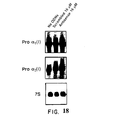
- the antisense oligonucleotides of the invention form stable duplexes with portions of an mRNA transcript of a nuclear proto-oncogene being c-myc or c-myb, more preferably c-myc.
- the invention includes the use, as above, for reducing scar formation in a human tissue and for inhibiting formation of fibrous connective tissue in a human.
- collagen refers to a class of molecules which are synthesized from procollagen. Collagen is synthesized from procollagen extracellularly, by the cleavage of N- and C- termini outside the cell. Antibodies to the triple helical region of collagen can recognize the triple helical region in procollagen as well. Antibodies used herein are either specific for collagen I and procollagen I, or collagen III and procollagen III. Collagen can also be synthesized in vitro from procollagen by pepsin digestion.
- graft refers to a vascular graft, such as a arteriovenous or venous graft.
- nucleoside as used herein includes the natural nucleosides, including 2'-deoxy and 2'-hydroxyl forms, e.g. as described in Kornberg and Baker, DNA Replication, 2nd Ed. (Freeman, San Francisco, 1992 ).
- "Analogs" in reference to nucleosides includes synthetic nucleosides having modified base moieties and/or modified sugar moieties, e.g. described generally by Scheit, Nucleotide Analogs (John Wiley, New York, 1980 ). Such analogs include synthetic nucleosides designed to enhance binding properties, e.g. duplex or triplex stability, specificity, or the like.
- oligonucleotide includes linear oligomers of natural or modified monomers or linkages, including deoxyribonucleosides, ribonucleosides, ⁇ - anomeric forms thereof, polyamide nucleic acids, and the like, capable of specifically binding to a target polynucleotide by way of a regular pattern of monomer-to-monomer interactions, such as Watson-Crick type of base pairing, Hoogsteen or reverse Hoogsteen types of base pairing, or the like.
- monomers are linked by phosphodiester bonds or analogs thereof to form oligonucleotides ranging in size from a few monomeric units, e.g. 3-4, to several hundreds of monomeric units.
- oligonucleotide is represented by a sequence of letters, such as "ATGCCTG,” it will be understood that the nucleotides are in 5'- > 3' order from left to right and that "A” denotes deoxyadenosine, “C” denotes deoxycytidine, “G” denotes deoxyguanosine, and “T” denotes thymidine, unless otherwise noted.
- Analogs of phosphodiester linkages include phosphorothioate, phosphorodithioate, phosphoroselenoate, phosphorodiselenoate, phosphoroanilothioate, phosphoranilidate, phosphoramidate, 3' ⁇ 5' phosphoramidate and the like, as more fully described below.
- “Stability” in reference to duplex or triplex formation roughly means how tightly an antisense oligonucleotide binds to its intended target sequence; more precisely, it means the free energy of formation of the duplex or triplex under physiological conditions. Melting temperature under a standard set of conditions, e.g. as described below, is a convenient measure of duplex and/or triplex stability.
- antisense oligonucleotides of the invention are selected that have melting temperatures of at least 50°C under the standard conditions set forth below; thus, under physiological conditions and the preferred concentrations duplex or triplex formation will be substantially favored over the state in which the antisense oligonucleotide and its target are dissociated.
- a stable duplex or triplex may in some embodiments include mismatches between base pairs and/or among base triplets in the case of triplexes.
- SEQ ID No: 1 which is a human sequence with a one base mismatch in the last nucleotide with respect to the comparable porcine sequence.
- antisense oligonucleotides of the invention form perfectly matched duplexes and/or triplexes with their target polynucleotides.
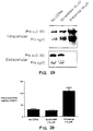
- synthesis refers to a process involved in the production of biological molecules, in particular extracellular matrix molecules and extracellular matrix proteins.
- the term “synthesis” includes processes both inside and outside a cell that result in biological molecules, for example, but not limited to, mRNA transcription, mRNA translation, post-translational protein modification, glycosylation, peptidase cleavage, extracellular peptidase cleavage, intra-cellular peptidase cleavage, protein transport, protein secretion and protein phosphorylation.
- the invention inhibits protein translation of extracellular matrix molecules, post-translational protein processing, protein secretion or intracellular peptidase cleavage.
- the invention relates to the use of certain antisense compounds to inhibit the inappropriate synthesis in a tissue of extracellular matrix proteins, particularly collagen, and more particularly type I and type III collagen.
- the inappropriate and/or excessive synthesis of extracellular matrix proteins can result in medical conditions that exhibit formation of, e.g.. unwanted fibrous connective tissue.
- medical conditions include sclerotic disorders, vascular restenosis, atherosclerosis, atherogenesis, keloid disease, liver cirrhosis, rheumatoid disorders of the joints, loss of arteriovenous and venous graft patency, post-surgical scarring, reconstructive surgery, and the like, generally found in human subjects.
- a range of tissue types can be the site of such inappropriate synthesis, including vascular smooth muscle cells, endothelial cells, skin and organ fibroblasts, e.g. in keloid disease or posttraumatic or postsurgical scar formation, and synovial cells and other joint components.
- One embodiment of the invention is particularly useful in preventing the failure or loss of patency of arteriovenous and venous grafts.
- Another embodiment of the invention is particularly useful in the treatment of restenosis, an acute vascular injury, typically following angioplasty in a human subject. Restenosis is associated with smooth muscle cell migration to the vascular intima, proliferation, and synthesis of extracellular matrix components, e.g. Holmes et al, Chapter 12 in Vlietstra et al, Editors, PTCA (Davis Company, Philadelphia, 1987 ).
- Target polynucleotides may be single stranded or double stranded DNA or RNA; however, single stranded DNA or RNA targets are preferred.
- the genomic nucleotide sequence and the mRNA transcripts of nuclear oncogenes of the invention are known in the art and include c- myc, c- myb, c-fos, N- myc, L- myc, p53, c-rel, c-ski, c-ets-1, c-ets-2, and the like, e.g.
- c-myc and c-myb sequences are described in the following references: c- myc proto-oncogene sequences are described in Marcu et al, Ann. Rev. Biochem. , 61:809-860 (1992 ); Watt et al, Nature, 303:725-728 (1983 ); Battey et al, Cell, 34:779-787 (1983 ); and Epstein et al, NTIS publication PB93-100576; the c-myb proto-oncogene is described in Gewirtz et al, U.S. patent 5,098,890 and Majello et al, Proc. Natl.
- proliferating cell nuclear antigen, cyclin (D 1 and B 1 ), cdc2 kinase, cdk kinase and E2F can be targetted (herein referred to as "cell-cycle genes").
- the nuclear proto-oncogenes targeted by antisense oligonucleotides of the invention are c- myc and/or c- myb .
- the target to which the c- myc antisense oligonucleotides of the invention are directed include allelic forms of the proto-oncogenes.
- sequences for antisense oligonucleotides are selected so that the G-C content is at least 60%.
- sequences of the oligonucleotides are selected from 12 to 15 contiguous bases of SEQ. ID No. 1, 5-7, or 8-11.
- Preferred mRNA targets include the 5' cap site, tRNA primer binding site, the initiation codon site, the mRNA donor splice site, the mRNA acceptor splice site, and the like, e.g. Goodchild et al, U.S. patent 4,806,463 .
- antisense oligonucleotides with consecutive sequences can be administered using oligonucleotides 12, 13, 14, 15, 16, 17, 18, 19, 20, 21, 22, 23, 24, 25, 26, 27, 28, 29, 30, 31, 32, 33, 34, or 35 bases in length and selected from a twenty five base region on either side of the ATG site (human sequence from Gazin et al, E.M.B.O. Journal 3:2:383-387 (1984 )):
- antisense oligonucleotides will include the ATG site. Twenty five base regions on either side of SEQ ID No:6 and 7 may be also be used to select oligonucleotides of the size discussed in this paragraph.
- Antisense oligonucleotides of the invention may comprise any polymeric compound capable of specifically binding to a target polynucleotide by way of a regular pattern of monomer-to-nucleoside interactions, such as Watson-Crick type of base pairing, Hoogsteen or reverse Hoogsteen types of base pairing, or the like.
- Antisense compounds of the invention may also contain pendent groups or moieties, either as part of or separate from the basic repeat unit of the polymer, to enhance specificity, nuclease resistance, delivery, or other property related to efficacy, e.g.
- duplex intercalators such as acridine, poly-L-lysine, "end capping" with one or more nuclease-resistant linkage groups such as phosphorothioate, and the like. Sequences of certain representative oligonucleotides useful in this invention are set forth in the Sequence Listing included herewith.
- Antisense compounds of the invention include the pharmaceutically acceptable salts thereof, including those of alkaline earths, e.g. sodium or magnesium, ammonium or NX 4 + , wherein X is C 1 -C 4 alkyl.
- Other pharmaceutically acceptable salts include organic carboxylic acids such as acetic, lactic, tartaric, malic, isethionic, lactobionic, and succinic acids; organic sulfonic acids such as methanesulfonic, ethanesulfonic, and benzenesulfonic; and inorganic acids such as hydrochloric, sulfuric, phosphoric, and sulfamic acids.
- Pharmaceutically acceptable salts of a compound having a hydroxyl group include the anion of such compound in combination with a suitable cation such as Na + , NH 4 + , or the like.
- nuclease resistance is conferred on the antisense compounds of the invention by providing nuclease-resistant internucleosidic linkages.
- nuclease-resistant internucleosidic linkages are known in the art, e.g. phosphorothioate: Zon and Geiser, Anti-Cancer Drug Design, 6: 539-568 (1991 ); Stec et al, U.S. patent 5,151,510 ; Hirschbein, U.S. patent 5,166,387 ; Bergot, U.S.
- Additional nuclease linkages include phosphoroselenoate, phosphorodiselenoate, phosphoroanilothioate, phosphoranilidate, alkylphosphotriester such as methyl- and ethylphosphotriester, carbonate such as carboxymethyl ester, carbamate, morpholino carbamate, 3'-thioformacetal, silyl such as dialkyl (C 1 -C 6 )- or diphenylsilyl, sulfamate ester, and the like.
- Such linkages and methods for introducing them into oligonucleotides are described in many references, e.g.
- phosphorus analogs of the phosphodiester linkage are employed in the compounds of the invention, such as phosphorothioate, phosphorodithioate, phosphoramidate, or methylphosphonate. More preferably, phosphorothioate is employed as the nuclease resistant linkage. It is understood that in addition to the preferred linkage groups, compounds of the invention may comprise additional modifications, e.g.
- antisense compounds of the invention are synthesized by conventional means on commercially available automated DNA synthesizers, e.g. an Applied Biosystems (Foster City, CA) model 380B, 392 or 394 DNA/RNA synthesizer.
- phosphoramidite chemistry is employed, e.g. as disclosed in the following references: Beaucage and Iyer, Tetrahedron, 48:2223-2311 (1992 ); Molko et al, U.S. patent 4,980,460 ; Koster et al, U.S. patent 4,725,677 ; Caruthers et al, U.S. patents 4,415,732 ; 4,458,066 ; and 4,973,679 ; and the like.
- third strand association via Hoogsteen type of binding is most stable along homopyrimidine-homopurine tracks in a double stranded target.
- base triplets form in T-A*T or C-G*C motifs (where "-" indicates Watson-Crick pairing and "*" indicates Hoogsteen type of binding); however, other motifs are also possible.
- Hoogsteen base pairing permits parallel and antiparallel orientations between the third strand (the Hoogsteen strand) and the purine-rich strand of the duplex to which the third strand binds, depending on conditions and the composition of the strands.
- nucleoside type e.g. whether ribose or deoxyribose nucleosides are employed
- base modifications e.g. methylated cytosine, and the like
- Roberts et al Proc. Natl. Acad. Sci., 88:9397-9401 (1991 ); Roberts et al, Science, 258:1463-1466 (1992 ); Distefano et al, Proc. Natl. Acad.
- oligonucleotide moieties is sufficiently large to ensure that specific binding will take place only at the desired target polynucleotide and not at other fortuitous sites, as explained in many references, e.g. Rosenberg et al, WO 93/01286 or Szostak et al, Meth. Enzymol. 68:419-429 (1979 ).
- antisense compounds of the invention have lengths in the range of about 12 to 60 nucleotides. More preferably, antisense compounds of the invention have lengths in the range of about 15 to 40 nucleotides; and most preferably, they have lengths in the range of about 18 to 30 nucleotides.
- the thermal stability of the antisense oligonucleotides of the invention are determined by way of melting, or strand dissociation, curves.
- the temperature of fifty percent strand dissociation is taken as the melting temperature, T m , which, in turn, provides a convenient measure of stability.
- T m measurements are typically carried out in a saline solution at neutral pH with target and antisense oligonucleotide concentrations at between about 1.0-2.0 ⁇ M. Typical conditions are as follows: 150 mM NaCl and 10 mM MgCl 2 in a 10 mM sodium phosphate buffer (pH 7.0) or in a 10 mM Tris-HCl buffer (pH 7.0); or like conditions.
- Data for melting curves are accumulated by heating a sample of the antisense oligonucleotide/target polynucleotide complex from room temperature to about 85-90°C. As the temperature of the sample increases, absorbance of 260 nm light is monitored at 1°C intervals, e.g. using a Cary (Australia) model 1E or a Hewlett-Packard (Palo Alto, CA) model HP 8459 UV/VIS spectrophotometer and model HP 89100A temperature controller, or like instruments. Such techniques provide a convenient means for measuring and comparing the binding strengths of antisense oligonucleotides of different lengths and compositions.
- Another aspect of this invention is a pharmaceutical composition useful in inhibiting the synthesis of extracellular matrix proteins, comprising a pharmaceutically-acceptable excipient and an antisense oligonucleotide specific for nuclear proto-oncogene that is present in an amount sufficient to inhibit the synthesis of extracellular matrix proteins when administered to a subject in need thereof.
- the antisense oligonucleotides of the invention are employed as one or more components of such a pharmaceutical composition.
- compositions of the invention depend on several factors, including the nature of the disease or condition being treated, the location of disease lesions, the mode of drug delivery and/or administration contemplated, the latter of which can include in vivo administration by way of a catheter into a target lesion or organ, topical application, intranasal administration, administration by implanted or transdermal sustained release systems, and the like.
- compositions of the invention include a pharmaceutical carrier that may contain a variety of components that provide a variety of functions, including regulation of drug concentration, regulation of solubility, chemical stabilization, regulation of viscosity, absorption enhancement, regulation of pH, and the like.
- a pharmaceutical carrier that may contain a variety of components that provide a variety of functions, including regulation of drug concentration, regulation of solubility, chemical stabilization, regulation of viscosity, absorption enhancement, regulation of pH, and the like.
- the pharmaceutical composition preferably includes a buffer such as a phosphate buffer, or other organic acid salt, preferably at a pH of between about 7 and 8.
- a nonionic surfactant such as Tween 80 in an amount of 0.04-0.05% (w/v), to increase solubility.
- antioxidants such as ascorbic acid
- hydrophilic polymers such as, monosaccharides, disaccharides, and other carbohydrates including cellulose or its derivatives, dextrins, chelating agents, such as EDTA, and like components well known to those in the pharmaceutical sciences, e.g. Remington's Pharmaceutical Science, latest edition (Mack Publishing Company, Easton, PA ).
- Sustained release systems suitable for use with the pharmaceutical compositions of the invention include semi-permeable polymer matrices in the form of films, microcapsules, or the like, comprising polylactides, copolymers of L-glutamic acid and gamma-ethyl-L-glutamate, poly(2-hydroxyethyl methacrylate), and like materials, e.g. Rosenberg et al, International application PCT/US92/05305 .
- Sustained release systems also include liposomally entrapped antisense compounds, e.g. as described in Liposome Technology, Vol. II, Incorporation of Drugs, Proteins and Genetic Material (CRC Press).
- an effective amount of c- myc antisense oligonucleotide for particular applications depends on several factors, including the chemical nature of the antisense oligonucleotide, the disorder being treated, the method of administration, and the like.
- an effective amount will provide a concentration of c- myc antisense oligonucleotide of between about 1 to 100 ⁇ M at the target polynucleotide; and more preferably, an effective amount will provide a concentration of antisense oligonucleotide of between about 1 to 10 ⁇ M at the target polynucleotide.
- antisense oligonucleotides specific for nuclear proto-oncogenes are preferably administered locally to the traumatized artery by way of a catheter.
- the antisense oligonucleotides delivered to restenosis lesion are specific for c- myc and/or c -myb ; more preferably, a combination of antisense oligonucleotides specific for both c- myc and c- myb is employed to reduce extracellular matrix protein synthesis associated with restenosis.
- restenosis occurs after a procedure such as percutaneous transluminal coronary angioplasty (PTCA) which causes a local disruption or injury to an arterial wall.
- PTCA percutaneous transluminal coronary angioplasty
- an occlusive lesion In response to such injury, an occlusive lesion frequently results from the following phenomena: inflammation of the of the artery at the injury site, thrombosis, the accumulation of smooth muscle cells by migration and proliferation, and smooth muscle cell and fibroblast synthesis of extracellular matrix.
- oligonucleotides are preferably delivered to the connective tissue surrounding the graft to prevent graft failure.
- failure of a graft occurs when the loss of patency reaches 50 percent or more.
- arteriovenous grafts such as, but not limited to, prosthetic fistulae (including PTFE (Gortex or Impra), poly-(ether-urethane), dacron, human umbilical cord, genetically engineered veins and immunosuppressed bovine or porcine vessel), arteriovenous shunts, native vein fistulae, (including radial artery-cephalic vein (Brescia-Cimino), ulnar or radial artery-basilic vein, brachial artery-cephalic or brachial vein and femoral artery-saphenous grafts) and venous grafts
- prosthetic fistulae including PTFE (Gortex or Impra), poly-(ether-urethane), dacron,
- oligonucleotides of the invention are generally injected in the perivascular tissue surrounding the site of intended application.
- other types of delivery systems can be used such as subcutaneous infusion pumps, gels and biodegradable matrices molded onto the graft and preferably mixed with human serum albumin at a 1:10 to 1:100 ratio of oligonucleotide to protein, gels (such as pluronic, Hydron, or ethylene-vinyl acetate gels), gels (as described by Edelman et al, Circulation Research 76:176-182 (1995 ) and Edelman et al., Proc.
- indwelling catheter systems such as Broviac catheters or completely indwelling catheter reservoirs systems such as those described by Poletti et al., J: Neurosurg. 55: 581-584 , impregnated PTFE tubing, other impregnated biocompatable materials or other drugs delivery compositions and systems described herein.
- a reservoir can be made to release the oligonucleotide in close proximity to the site, in order to extend the duration of administration of the oligonucleotide.
- the reservoir can simply be made by injecting oligonucleotide into perivascular tissue during or after surgery, but before closure of the wound, or by percutaneous injection after palpating the arteriovenous graft, as in the case of an arteriovenous fistula for example, or by guiding the injection with methods known in the art, e.g. sonograms. Injections can be repeated for extended treatments, e.g., in hemodialysis acess sites. It is not necessary nor desirable to remove the adventitia from the vessel, since the perivascular tissue can act as a depot or reservoir of oligonucleotide that has a relatively slow rate of diffusion.
- Oligonucleotide in a reservoir is also more slowly carried away by the circulation system than when administered by intraluminal catheter.
- the administered dose is in the range of .1 mg to 100 mg per vessel, preferably .25 mg to 50 mg and more preferably .5 mg to 10 mg per single administration.
- oligonucleotides In addition to the perivascular tissue administration of oligonucleotides to prevent graft failure, other anatomical structures such as the endothelial layer, media, and adventitia can be used as reservoir sites, particularly when the oligonucleotide is not applied from the lumen of the vessel.
- Such naturally occurring reservoir sites including the perivascular tissue, are particularly useful for treating arteriovenous grafts, and more particularly arteriovenous shunts, where the media or elastic lamina is intact and not dissected, despite surgical injury. It is particularly advantageous to use sites near the vein portion of an arteriovenous graft, and more particularly the vein portion of an arteriovenous fistulae or shunt.
- non-naturally occurring reservoirs can also supply a failing graft with oligonucleotide as shown with gray area in FIG. 1 , examples of which are discussed herein.
- non-naturally occurring reservoirs it is preferred that they are either biodegradable or refillable.
- Refillable reservoirs are preferably of two types 1) a refillable reservoir secured upstream of the venous portion of the arteriovenous graft and in, or along, the PTFE tubing or other biocompatable material used to form an arteriovenous graft and 2) a reservoir secured in the perivascular tissue surrounding the arteriovenous graft, preferably within 5 cm of venous portion of the arteriovenous graft and more preferably within 2 cm, and refillable with a needle by percutaneous injection.
- Such non-naturally occurring perivascular reservoirs can be implanted during surgery to construct the arteriovenous graft.
- Refillable perivascular reservoirs can be made of materials used in breast implants and are preferably used in conjunction with a semipermeable membrane that permits the diffusion of oligonucleotide into the perivascular tissue.
- veins are contacted ex vivo with an oligonucleotide complementary to nuclear proto-oncogene mRNA.
- veins are incubated for at least 10 minutes at temperatures between 20 and 37 °C in a sterile and physiologically acceptable buffer. Shorter times are permissible at particularly high concentrations. Longer incubation times between 30 and 90 minutes are also suitable and compatible with the time lag between vein removal and grafting experienced in such procedures.
- oligonucleotide concentration will range from 10 to 100 ⁇ M and more preferably from 25 to 100 ⁇ M.
- buffers can be used so long as they physiologically acceptable, such as 0.9 % saline (NaCl) or phosphate buffer saline, and other buffers and salts described herein.
- veins may be used with the invention including veins from non-humans, veins from genetically engineered mammals, especially pigs, and many human veins to from allografts and autografts alike (such as saphenous vein, cephalic vein, basilic vein, brachial vein, and femoral vein).
- antisense oligonucleotides are preferably administered in the vicinity of the lesion via catheter from inside the lumen, or through the adventitia (i.e. the most outer layer of the vessel wall) with materials aiding a slow release of antisense compound, e.g. a pluronic gel system as described by Simons et al, Nature, 359:67-70 (1992 ) or porous balloon or iontophoretic balloon as described by Fernandez-Ortiz, A., et al., Circulation, 89:1518-1522 (1994 ).
- Other slow release techniques for local delivery include coated stents with antisense compound, e.g.
- a dose delivered at the target lesion is in the range of from 1 ⁇ g to 10 mg for each or both of the antisense compounds employed in the pharmaceutical composition.
- the dose range is between 1 ⁇ g and 1 mg; and more preferably, the dose range is between 1 ⁇ g and 10 ⁇ g.
- the delivery time is in the range of about 30 seconds to 60 minutes, and more preferably, in the range of about 30 seconds to about 1-2 minutes, e.g. Zalewski et al, pages 79-87 in Goldberg, editor, Coronary Angioplasty (Davis, Philadelphia, 1988 ).
- the administered dose is in the range of .1 mg to 10 mg per artery, preferably .25 mg to 4 mg per artery and more preferably .5 mg to 2 mg per artery.
- Administered dose is the amount drug applied to the tissue, where as "dose delivered at the target lesion" is in the amount of drug in the tissue after delivery to the tissue.
- topical or intradermal application is preferred to inhibit extracellular matrix protein synthesis, particularly collagen synthesis, by skin fibroblasts.
- the administered dose (amount of drug applied to tissue) will generally range from .01 ⁇ g to 10 mg of drug per cm 2 , preferably from .2 ⁇ g to 7 mg of drug per cm 2 , and more preferably from 5 to 4 mg of drug per cm 2 .
- Administered dose for use with post-surgical dressing is preferably from .2 mg to 4 mg of drug per cm 2 .
- the administered dose for use with topical creams for skin abrasions, skin lesions, acne and post-surgical treatments is - preferably from .05 mg to 3 mg of drug per cm 2 .
- oligonucleotide For urologic disorders resulting in urethral, ureteral, or bladder scarring or narrowing, local administration via catheter or implants coated with antisense oligonucleotide is preferred.
- administration of antisense compounds can be systemic or local. Local administration includes direct injection of antisense oligonucleotides into the target organ, delivery via implanted gels or polymers, or slow release from coated stents or other prosthetic materials.
- vascular restenosis factors associated with susceptibility to the condition include male gender, dilation for stenosis of bypass grafts, Canadian Heart class 3 or 4 at baseline, and history of myocardial infarction, Holmes et al, Chapter 12 in Vlietstra et al, editors, PTCA (Davis Company, Philadelphia, 1987 ). Efficacy of treatment is assessed in the same manner as in any PTCA technique. Usually, results are estimated visually, but other techniques are available.
- Assessment of the hemodialysis access site failure includes color Doppler ultrasound, contrast angiograph and ultrasound (e.g ., Dousset et al, Radiology 181:89 (1991 ), Nonnast-Daniel et al, Lancet 339 (8786):143 (1992 ), and Davidson et al, Kidney International 40: 91-95 (1991 ), and physical findings (such as high-pitched Son over access site, or water hammer pulse).
- Assessment of the severity of a stenosis, or restenosis range from visual observation of the lesion to complex computer-based analysis of the dimensions of the artery in question, e.g.
- an in vivo model was first developed in pigs. This model permits monitoring of the patency of an arteriovenous graft, including the venous portion of the graft.
- perivascular tissue normal veins, unexposed to arterial pressure and ungrafted, showed no thickened perivascular tissue, a perivascular space with a small quantity of connective tissue and loose adipose tissue, and a thin and unbroken media, as shown in FIG. 2a (Verhoeff stain). Vein portions of grafts exhibited an increase in perivascular tissue that circumscribes the media as a collar with a large amount of granulation tissue. This was shown by Verhoeff staining, as shown in FIG. 2b . The same section also exhibits an intact and unbroken media lacking dissection. In some sections neointima was observed at 8 and 14 days after surgery.
- Veins were also stained for desmin, which is expressed by vascular smooth muscle cells, using anti-desmin antibodies.
- the vein portions of the venous grafts exhibited desmin staining of smooth muscle cell only in the media. Large numbers of smooth muscle cells in the media stained positive with an antibody for desmin, data not shown.
- Vein portions of grafts exhibited an increase in cell proliferation in the media and perivascular tissue.
- Large numbers of proliferating cells in the media and perivascular tissue were stained with antibodies specific for proliferating cell nuclear antigen (PCNA), a marker of cell proliferation, as shown in FIG. 5a .
- PCNA proliferating cell nuclear antigen
- FIG. 5c a similar section was stained with Verhoff stain, FIG. 5b , and Sirrius red stain, FIG. 5c .
- early neointima formation can be observed in FIG. 5b as a thin dark layer a few cells in from the lumen of the grafted vein. Normally the neointima is absent from the vein.
- PCNA staining was also quantitated: 30% of the cells in grafted veins stained positive for PCNA, while less than one percent of the cells in non-grafted veins stained positive for PCNA.
- vascular cells e.g. smooth muscle cells and fibroblasts
- collagen synthesis e.g. collagen synthesis
- Human vein portions of aortocoronary grafts also exhibited a large amount of collagen in the perivascular tissue and media. Large amounts of collagen stained positive with Sirrius red, a specific stain for fibrillar collagen (including type I and III), in the perivascular tissue and media, as shown in FIG. 6b .
- oligonucleotide of the invention in order to demonstrate the effectiveness of an oligonucleotide of the invention in reducing extracellular matrix protein production and vascular compromise after arteriovenous shunt or bypass procedures an organ culture model was first developed.
- This model permits monitoring of oligonucleotide uptake, stimulation of vascular cells by platelet derived growth factor B-chain (referred to herein as "PDGF”) and inhibition of PDGF stimulation of vascular cells by oligonucleotides of the invention.
- PDGF platelet derived growth factor B-chain
- porcine vascular rings were incubated at 37 °C in serum-free media (DMEM (Dulbecco's Modified Eagle's Medium) with antibiotics and 1% Neutrodoma) for four days with or without 100 ng per ml PDGF. unless otherwise indicated.
- PDGF normally stimulates vascular cells, e.g., smooth muscle cells and fibroblasts, to proliferate and is known to be released by platelets after tissue injury to promote wound healing.
- PDGF induced cell proliferation was measured by PCNA staining as discussed herein. Even at four days, the rings maintained their morphology.
- Oligonucleotides of the invention inhibited PDGF-induced stimulation of porcine blood vessel organ cultures.
- the addition of oligonucleotide (SEQ ID No:1) at 16 ⁇ M inhibited PDGF induced cell proliferation, as shown in FIG. 7c .
- oligonucleotides diffused across both the luminal surface of the veins and perivascular surface of the veins. Oligonucleotides, over time, localized in both cells and cell nuclei.
- perivascular tissue oligonucleotide delivery can be used ex vivo to deliver oligonucleotides to excised veins or arteries used for venous grafts and other types of vascular grafts.
- the perivascular tissue can be treated with oligonucleotides of the invention in vivo allowing for oligonucleotide transfer to the vessel wall, in order to prevent vessel wall and perivascular tissue thickening.
- oligonucleotides of the invention Patients requiring hemodialysis access sites for the first time or replacement of failed sites will be treated with the oligonucleotides of the invention. Either PTFE type fistulae or Brescia-Cimino fistulae will be treated.
- oligonucleotide used for administration is selected and synthesized as discussed herein and known in the art.
- sequences and methods discussed in this example and other examples should not be construed as limiting the scope of the invention but as specific embodiments of the invention.
- Further oligonucleotides need only be consistent with the structure proposed herein. It is not necessary for oligonucleotides to operate by a particular mechanism of action proposed herein, so long as the oligonucleotides test favorably in in vitro and in vivo models used herein.
- samples were electrophoresed through 7.5 % SDS-polyacrylamide gel and transferred to nitrocellulose filter.
- the sample-containing filters were incubated with biotin labeled primary antibody against human Collagen I and III (Southern Biotech. SC.) for 1 hour at room temperature.
- the filters were washed and then incubated with streptavidine labeled with horseradish peroxidase.
- the filters were exposed to X ray film. The experiments were repeated at least twice on separate occasions.
- c- myc antisense oligonucleotides (8 and 16 ⁇ M) reduced procollagen type I synthesis in a dose dependent manner.
- Cells treated with c- myc sense, 4 bp mismatch, or scrambled oligonucleotides showed similar levels of collagen type I protein as compared with control cells (no oligonucleotide added).
- EXAMPLE 8 REDUCTION OF NEOINTIMA FORMATION IN PORCINE MODEL OF CORONARY DENUDATION
- EXAMPLE 9 INHIBITION OF EXTRACELLULAR MATRIX PRODUCTION IN HUMAN SMOOTH MUSCLE CELLS BY C-MYC ANTISENSE OLIGONUCLEOTIDE TREATMENT
- c-myc antisense oligonucleotides corresponding to SEQ ID No: 1 were used in all examples, unless otherwise indicated.
- oligonucleotides sequences were incubated with or without oligonucleotides in methionine-free medium for 4 hours. Afterwards, [ 35 S]methionine (25 ⁇ Cl/ml, DuPont NEN, MA) was added for 16 hours and the cell number was counted in a Coulter counter. Extracellular fibronectin in the conditioned medium and c-myc protein in cell nuclear extracts were measured by immunoprecipitation following metabolic labeling.
- Post-confluent smooth muscle cells were incubated with serum-free medium with or without oligonucleotides. The aliquots of medium were collected 24 hours thereafter.
- Matrix metalloproteinase activities were determined after selective destruction of tissue inhibitor of metalloproteinase by dithiothritol and iodoacetamide.
- Latent collagenases produced by human smooth muscle cells were activated with aminophenylmercuric acetate, and then incubated with type 1 collagen or gelatin and cleavage products assessed by SDS-PAGE ( D.D. Dean, et al., J. Clin. Invest., 84:678-685 (1989 ) and P.E. Desrochers, et al., J. Biol.
- interferon- ⁇ a potent inhibitor of type I collagen synthesis, was inadvertently induced, interferon- ⁇ was assayed in the culture medium and human smooth muscle cells. Interferon- ⁇ was undetectable in the conditioned culture media and cell lysates after incubation with c-myc antisense (SEQ ID No:1) oligonucleotides using ELISA method.
- RNA samples (10 ⁇ g) were denatured and separated on 0.8% agarose/formaldehyde gels and blotted onto nitrocellulose membranes. The RNA was crosslinked to nitrocellulose membranes using a UV crosslinker (Stratagene, CA).
- the blots were hybridized at 42 °C for 24 hours with human procollagen ⁇ 1 (I) (ATCC) and human procollagen ⁇ 2 (I) (gift from H. Kuivaniemi) cDNA probes.
- the probe for 7S ribosomal RNA were used as control
- the probes were labeled with 32 P-CTP by nick translation to a specific activity of greater than 10 8 cpm/ ⁇ g DNA.
- the blots were sequentially washed twice in 2x, 1x, 0.5x and 0.1x SSC-0.1% SDS at 42 °C for 15 min. Each wash was repeated twice. Blots were exposed to Kodak-Omat film at -70°C with an intensifying screen for 6 hours to 3 days.
- procollagen ⁇ 1 (I) and procollagen ⁇ 2 (I) mRNA levels were analyzed in smooth muscle cells after their incubation with or without oligonucleotides using Northern blot analysis. As shown in FIG. 18 , antisense oligonucleotides did not affect procollagen ⁇ 1 (I) and procollagen ⁇ 2 (I) mRNA levels. Likewise, similar levels of procollagen ⁇ 1(III) mRNA were seen with or without c-myc antisense oligonucleotides (data not shown). 7S RNA remained unchanged in all three experimental conditions.
- oligonucleotide To measure intracellular procollagen after c-myc antisense oligonucleotide treatments, cells were incubated with or without oligonucleotides for 20 hours in 20% FBS-DMEM. Serum-free medium containing ascorbic acid (50 ⁇ g/ml), [ 14 C]proline (2.5 ⁇ CI/ml, DuPont NEN, MA) and oligonucleotides were added to the cells for the additional 4 hours. The culture medium was collected and proteinase inhibitors were added. Aliquots of media were centrifuged through Microcon-30 column in order to remove unincorporated radioisotope and to concentrate samples for electrophoresis.
- the corresponding cell layers were then washed 3 times with cold PBS, lysed in PAGE-SDS sample buffer and boiled for 5 minutes to inactivate proteinases.
- the aliquots of concentrated medium, cell lysates (from equal cell number) and procollagen I purified from human skin fibroblast cells (gift from W. Arnold) were electrophoresed on 6% polyacrylamide-SDS gels with 3% stacking regions. The gels were fixed for 30 minutes in 10% acetic acid, 20% methanol, dried and exposed to film at -70°C for 7 days.
- c-myc antisense treatment decreased in secreted [ 14 C] proline labeled procollagen and was similar to the Western blot DNA.
- c-myc antisense treatment significantly increased the intracellular concentration of procollagen I compared to untreated cells and cells treated with a scrambled oligonucleotide ( FIG. 19 ).
- the intracellular accumulation of procollagen may reflect the inhibition of post-translational modifications required for the assembly of procollagen a chains into a triple-helical conformation. Therefore, to rule out this possibility, the affects c-myc antisense treatment of prolyl 4-hydroxylase activity and hydroxyproline content were measured.
- Post-confluent smooth muscle cells were incubated with or without oligonucleotides in 20% FBS-DMEM for 24 hours as described above.
- Cell layers were washed with cold PBS 3 times and lysed in 0.2 M NaCI, 20 mM Tris-HCl, pH 7.4, 0.1 % Triton x-100, 0.1 M glycine and 10 ⁇ M DTT (the methods of K.J. Kivirikko, et al., Methods in Enzymology, 82:245-364 (1982 ) are incorporated by reference herein).
- the lysates were then sonicated for 30 seconds on ice and protein concentration was measured.
- the remaining cell lysates were centrifuged at 13,000 rpm for 20 minutes at 4°C.
- the aliquots of supernatant were assayed for prolyl 4-hydroxylase activity by measuring the formation of radioactive 4-hydroxyproline using a [ 14 C] proline-labeled procollagen 1 as substrate (the methods of K.J. Kivirikko, et al., Anal. Biochem., 19:249-255 (1967 ) are incorporated by reference herein).
- the aliquots of cell lysate were hydrolyzed, hydroxyproline was assayed by a specific chemical procedure.
- Prolyl 4-hydroxylase activity a pivotal enzyme controlling triple helix formation, was similar among experimental subgroups. It was 11.7 ⁇ 0.1 (dpm/ ⁇ g protein, mean ⁇ SD of 2 separate experiments) in control smooth muscle cells treated with no oligonucleotides. 12. ⁇ 2.3 in cells incubated with scrambled oligonucleotides (16 ⁇ M) and 10.5 ⁇ 2.0 after treatment with c-myc antisense oligonucleotides (16 ⁇ M).
- Hydroxyproline content was greater in c-myc antisense (SEQ ID No:1) cells than in untreated or scrambled oligonucleotide treated cells ( FIG. 20 ).
- the increase in hydroxyproline content reflects the increase in procollagen levels, which is a substrate for prolyl 4-hydroxylase.
- EXAMPLE 13 INHIBITION OF EXTRACELLULAR MATRIX PRODUCTION IN HUMAN FIBROBLASTS HUMAN SKIN FIBROBLASTS CELLS USING C-MYC ANTISENSE OLIGONUCLEOTIDES
- This example further demonstrates the effectiveness of c-myc antisense oligonucleotide treatment to inhibit the synthesis of extracellular martrix protein by human skin fibroblast cell, in particular procollagen I and III.
- Antisense treatment greatly reduced extracellular procollagen levels in stimulated human skin fibroblast cells (treated with 20% FBS). This example also further confirms the findings of Example 7.
- procollagen levels were determined, as discussed for human smooth muscle cells in Example 9 and 12.
- Human skin fibroblasts cells used in the human skin fibroblast experiments were confluent. Cells were obtained and cultured as in Example 7 and by methods known in the art.
- extracellular collagen I was measured using Western blots of human fibroblast cell protein isolated from human fibroblasts subjected to different treatments. Human fibroblast cells were exposed to either scrambled oligonucleotides (SEQ ID No:4; 16 ⁇ M), mismatched oligonucleotides (SEQ ID No:3; 16 ⁇ M), antisense oligonucleotides (SEQ ID No:1, 4.
- Oligonucleotide (SEQ ID No:1; 16 ⁇ M) also inhibited cell proliferation in freshly plated fibroblasts; compared to 20% FBS and sense controls, cell proliferation was inhibited approximately 25 and 50 percent after four days, respectively.
- fresh medium is medium containing 20% FBS and ascorbic acid 50 ⁇ g/ml and other components as indicated
- concentration of the oligonucleotides was 16 ⁇ M; SEQ ID Nos:1 and 3).
- the secretion of collagen III by human fibroblast cells was also inhibited by c-myc antisense oligonucleotides.
- Human fibroblast cells were treated with fresh medium containing either no oligonucleotides, 16 ⁇ M scrambled oligonucleotides or 16 ⁇ M c-myc antisense oligonucleotides for twenty four hours and the medium was assayed for the presence of extracellular collagen III at the end of the twenty four hours. No extracellular collagen III was observed in the antisense treated cells, while the control cells (no treatment and scrambled treated) showed the presence of extracellular collagen III (at least 10-100 times higher level of collagen III, as compared to the antisense treat cells).
- the amount of secreted procollagen I is reduced at least 10-20 times in the antisense treated cells compared to the control cells (no treatment, scrambled treated, lanes 1 and 2, respectively).
- the upper band in lanes 1-3 is approximately 116 Kd ("upper band"), which corresponds to procollagen ⁇ 1 (I, III); the lower band in lanes 1-3 (from left to right) is approximately 100 Kd, which corresponds to ⁇ 2 (I).
- lanes 4-6 show intracellular procollagen I is at least five times greater in antisense treated cells (lane 6) than the control cells (lanes 4 and 5).
- lane 7 shows the molecular weight markers.
- the tertiary structure of intracellular procollagen was not changed by c-myc antisense treatment.
- Human fibroblast cells were treated with fresh medium containing either no oligonucleotides, 16 ⁇ M scrambled oligonucleotides, or 16 ⁇ M c-myc antisense oligonucleotides for twenty four hours.
- the melting temperature of procollagen from each experimental condition was measured as discussed herein and known in the art.
- FIG. 23 shows that the melting profile for control cells (untreated and scrambled treated) and antisense treated are similar.
- c-myc antisense treatment does not lead to a detectable alteration of procollagen structure that could lead to increased intracellular degradation or decreased protein export from the cell.
- c-myc antisense oligonucleotide treatment does not inhibit the synthesis of extracellular martrix protein by human skin fibroblast cells, in particular procollagen I, by reducing the mRNA level of procollagen.
- Antisense treatment did not reduce mRNA procollagen levels in stimulated human skin fibroblast cells (treated with 20% FBS).
- the blots were prehybridized in 5x standard saline citrate, 2x Denhardt's solution, 0.1% SDS and 0.2mg/ml of denatured salmon sperm DNA for at least 4 hours at 65°C. Hybridization was performed overnight using the same buffer containing cDNAs radio labeled with 32 P dCTP by nick translation to a specific activity of approximately 108-109 cpm/ ⁇ g. Blots were washed at 65°C in 0.5x standard saline citrate and 0.1% SDS. The filters were exposed to Kodak X-Omat film at -70°C with intensifying screens for 1-5 days.
- the autoradiography of the resulting Northern blots was quantitated by scanning densitometry and integration of peak areas.
- the probes used for this study were as follows: a 1.8 Kb pro-alpha 1(1) (HF-677) cDNA corresponding to the COOH-terminal propeptide and the carboxy-terminal portion of the triple helical region of human pro-alpha 1(1) chain of type I procollagen ( Chu et al., Nucleic Acids Res. 10:5925-5934, 1982 ).
- the mRNA level of procollagen alpha I in human fibroblast cells was unaffected by c-myc antisense treatment.
- Human fibroblast cells were treated with fresh medium containing either no oligonucleotides, 16 ⁇ M scrambled oligonucleotides, or 16 ⁇ M c-myc antisense oligonucleotides for twenty four hours, mRNA was purified from each experimental condition and analyzed for the presence of procollagen alpha I mRNA, as discussed herein and known in the art.
- FIG. 24 shows that control cell procollagen I mRNA (untreated and scrambled treated: lanes 1 and 2, respectively) levels are similar to the antisense treated cell procollagen alpha I mRNA levels (lane 3).
- the mRNA results show that c-myc antisense treatment decreases the secretion of procollagen I and III, which are proteins that lead to the synthesis of collagen in the extracellular matrix.
- Human fibroblast cells were treated with fresh medium containing either no oligonucleotides, 16 ⁇ M scrambled oligonucleotides, or 16 ⁇ M c-myc antisense oligonucleotides for twenty four hours. Human fibroblast cells from each experimental condition were then exposed to fresh medium without any oligonucleotide additions and assayed for the presence of extracellular collagen I, as discussed herein.
- control cells untreated and scrambled pretreated
- antisense treated cells produced similar levels of extracellular collagen I twenty four hours after abatement of the treatment.
- c-myc antisense treatment did not produce any cytotoxic effects that resulted in the inability of the cell to return to pretreatment secretion levels of procollagen.
- This example further demonstrates the in vivo effectiveness of localized c-myc antisense oligonucleotide treatment to decrease the progression of extracellular matrix synthesis after tissue trauma.
- This example also further demonstrates the in vivo effectiveness of c-myc antisense treatment in decreasing the progression of internal organ scaring, as well as decreasing the progression of tissue scaring associated with the synthesis of extracellular matrix molecules, such as collagen.
- Example 8 To determine the effects of c-myc antisense treatment on the progression of extracellular matrix synthesis after tissue trauma, pig hearts were injured in vivo, treated with oligonucleotides as discussed in Example 8 (unless otherwise indicated), and sacrificed at three days, seven days, and twenty eight days after the delivery of oligonucleotides. The same c-myc antisense and sense oligonucleotides (SEQ ID No: and 2) and dosages (1 mg per artery) were used as in Example 8.
- Traumatized coronaries from each experimental group were examined as blind samples with light microscopy, magnification 20-62X. Tissue was stained with Verhoff-Van-Gieson stain, as known in the art, to highlight cells and extracellular matrix, and sirus red stain, as known in the art, to highlight collagen.
- the c-myc antisense treated vessels which displayed a smaller neointima area compared to control vessels, also displayed less red staining than the control vessels (sense treated).
- the antisense treated coronaries showed less extracellular fibrillar material compared to the sense treated coronaries; and the antisense treated coronaries showed less extracellular space between cells.
- Neointimal smooth muscle cell proliferation was also assessed using immunostaining for PCNA (Proliferating Cell Nuclear Antigen) following balloon denudation and transcatheter delivery of c-myc sense (S) or antisense (AS) oligonucleotides (1 mg) into porcine coronary arteries, see Table IV.
- Antisense treatment of coronaries inhibited the progression of smooth muscle cell proliferation caused by tissue trauma compared to sense treatment of coronaries.
- PCNA staining permitted the assessment of the number of proliferating cells in a given neointima cross-section.
- proliferative index PI
- I/M neointima/media
- ⁇ I/M the reduction in I/M ratio
- Coronaries with a reduction of the I/M ratio have an improved lumenal diameter.
- localized c-myc antisense treatment inhibits the progression of scar formation in a traumatized tissue, such as scar formation in coronaries after balloon angioplasty trauma caused by smooth muscle cell proliferation and extracellular matrix formation.
- This example demonstrates the in vivo effectiveness of c-myc antisense oligonucleotides in penetrating vascular and connective tissues using local delivery devices.
- these results demonstrate the ability of c-myc antisense treatment to localize in cell nuclei within 30 minutes and before the onset of c-myc induction (which occurs over three to four hours).
- the local distribution of c-myc antisense oligonucleotides after local administration to the vessel wall of coronary arteries decreases scar formation following balloon angioplasty.
- c-myc antisense oligonucleotides used in this Example were prepared as discussed herein.
- Phosphorothioate antisense oligonucleotides (SEQ ID No:1) directed against the translation initiation region of the human c - myc gene used in the distribution studies labeled with carboxyfluorosceins (480 nm excitation, 520 nm emission, Molecular Probes, Inc.) as previously described in Iverson, et al, Antisense Res. Dev. 2: 211-222 (1992 ),.
- carboxyfluorosceins 480 nm excitation, 520 nm emission, Molecular Probes, Inc.
- oligonucleotides were 35 S-labeled at each internucleotide link with the specific activity of 1.8 x 10 8 cpm/ ⁇ mol.
- the delivery device a Kaplan-Simpson Infusasleeve (of LocalMed, Palo Alto, CA) was advanced over a standard angioplasty balloon ("support" balloon) to the region of interest.
- This device consists of multilumen infusion region (18 mm) containing multiple sideholes (40 ⁇ m) which are juxtapositioned against the vessel wall by inflating a support balloon.
- the device used in studies infuses solutions using rigid, noncollapsable channels. This design minimizes drug loss to the bloodstream and permits separate control of drug infusion and placement of the drug delivery device in the vessel.
- oligonucleotides were administered. Intramural injections were carried out under 100 PSI of the external pressure. The total volume of the injectate was 5-6 ml, delivered by the external pump at 40 ml/min. The administered dose was 1 mg per artery. After injection was completed, devices were removed and final angiograms were recorded. To verify proper conditions of oligonucleotides delivery, serial quantitative coronary angiography was employed to measure the initial luminal diameter of selected coronary arteries, luminal diameter prior to oligonucleotides delivery (i.e., after denuding injury) and after oligonucleotides administration.
- the coronary arteries were excised with adjacent tissues. After the vessel lumen was flushed with PBS, the arteries were fixed in 10% buffered formalin for 6 hours and embedded in paraffin. The arteries were cut into serial 4- ⁇ m-thick sections (50 mm per vessel including 18 mm injected portion. The examination of the site of injection as well as proximal and distal portions of the vessel provided information regarding the longitudinal oligonucleotides distribution. In addition, noninjected coronary artery from the same animal served as the additional control. The sections were deparaffinized end covered with SlowFade-Light antifade reagent (Molecular Probes, Inc., Eugene, OR).
- SlowFade-Light antifade reagent Molecular Probes, Inc., Eugene, OR
- Each slide contained the vessel, periadventitial tissue and adjacent myocardium ( FIG. 17 ) which allowed to determine the depth of oligonucleotide penetration.
- the fluorescent-labeled antisense oligonucleotides were visualized under Nikon Optiphot microscope equipped for epifluorescence with XF-22 filter. Photographs were taken on Kodak Ektachrome P1600 films.
- oligonucleotides uptake by vascular smooth muscle cells were grown on chamber slides in medium containing 10% fetal bovine serum for 2 days and fluorescein-labeled oligonucleotides were added (8 ⁇ M) for 2, 30 minutes and 2 hours. Then, cells were washed 3 times with PBS and fixed in 50 % acetone and 50 % methanol.
- sections of the coronary arteries obtained as described above were analyzed by confocal microscopy at 0.4 - 0.5 ⁇ m optical thickness.
- Subintimal nontransmural pattern was common; it was either interspersed between transmural localization or at the edges of injected segments ( FIGS. 27C and 27D ).
- Midwall nontransmural pattern was the least common and it was found adjacent to transmural localization ( FIGS. 27E and 27F ).
- the vessel adventitia was devoid of antisense oligonucleotides in sections exhibiting nontransmural distribution.
- the above sustained level of oligonucleotides was achieved despite a rapid plasma clearance (t 1/2 15 ⁇ 2 min).
- the amount of detected oligonucleotides both at 30 min and 3 days corresponded to about 0.1 % of total administered dose.
- FIG. 30 transmural distribution of oligonucleotides ( FIG. 30A ) was associated with the intact internal elastic lamina and elastic tissues throughout the vessel wall ( FIG. 30B ). A mild decrease in cytoplasmic staining and nuclear pyknosis was occasionally noted at the site of oligonucleotides retention at 30 min ( FIG. 30C ). These changes were not found at 3 days after the delivery ( FIG. 30D ).
- This example further demonstrates the effectiveness of c-myc antisense oligonucleotide treatment in vivo by showing the rapid localization of c-myc antisense oligonucleotides in the nuclei of smooth muscle cells both in vitro (human) and in vivo (porcine). This rapid time course allows for inhibition of c-myc gene activation, previously established on the order of hours (3 to 4 hours).
Landscapes
- Health & Medical Sciences (AREA)
- Life Sciences & Earth Sciences (AREA)
- Chemical & Material Sciences (AREA)
- Engineering & Computer Science (AREA)
- Organic Chemistry (AREA)
- General Health & Medical Sciences (AREA)
- Genetics & Genomics (AREA)
- Bioinformatics & Cheminformatics (AREA)
- Molecular Biology (AREA)
- Public Health (AREA)
- Veterinary Medicine (AREA)
- Animal Behavior & Ethology (AREA)
- Pharmacology & Pharmacy (AREA)
- Medicinal Chemistry (AREA)
- Biotechnology (AREA)
- Chemical Kinetics & Catalysis (AREA)
- General Chemical & Material Sciences (AREA)
- Nuclear Medicine, Radiotherapy & Molecular Imaging (AREA)
- Biochemistry (AREA)
- Biomedical Technology (AREA)
- General Engineering & Computer Science (AREA)
- Zoology (AREA)
- Wood Science & Technology (AREA)
- Heart & Thoracic Surgery (AREA)
- Microbiology (AREA)
- Plant Pathology (AREA)
- Cardiology (AREA)
- Biophysics (AREA)
- Physics & Mathematics (AREA)
- Oncology (AREA)
- Pain & Pain Management (AREA)
- Dermatology (AREA)
- Rheumatology (AREA)
- Pulmonology (AREA)
- Epidemiology (AREA)
- Vascular Medicine (AREA)
- Pharmaceuticals Containing Other Organic And Inorganic Compounds (AREA)
- Medicines That Contain Protein Lipid Enzymes And Other Medicines (AREA)
- Medicines Containing Material From Animals Or Micro-Organisms (AREA)
- Acyclic And Carbocyclic Compounds In Medicinal Compositions (AREA)
Claims (24)
- Verwendung eines Antisense-Oligonucleotids, das für c-myb oder c-myc spezifisch ist, bei der Herstellung einer pharmazeutischen Zusammensetzung zur Behandlung eines Gefäßtransplantats.
- Verwendung eines Oligonucleotids nach Anspruch 1, worin das Gefäßtransplantat aus der aus künstlichen Fisteln, arteriovenösen Shunts, nativen Fisteln, Venentransplantaten, immunsupprimierten Allotransplantaten, immunsupprimierten Xenotransplantaten und Venenautotransplantaten bestehenden Gruppe ausgewählt ist.
- Verwendung eines Oligonucleotids nach Anspruch 1, worin das Gefäßtransplantat eine Hämodialyse-Zugangsstelle bereitstellt.
- Verwendung eines Oligonucleotids nach Anspruch 3, worin die Hämodialysestelle aus der aus Brescia-Cimino-Fisteln, Tabatiere-Fisteln, brachiocephalischen Fisteln, PTFE-Hämodialyse-Transplantaten, Rinder-Karotis-Transplantaten und autogenen Transplantaten bestehenden Gruppe ausgewählt ist.
- Verwendung eines Oligonucleotids nach einem der vorangegangenen Ansprüche, worin das Oligonucleotid für c-myc spezifisch ist.
- Verwendung eines Oligonucleotids nach einem der Ansprüche 1 bis 4, worin das Oligonucleotid für c-myb spezifisch ist.
- Verwendung eines Oligonucleotids nach einem der Ansprüche 1 bis 4, worin das Oligonucleotid aus der aus Seq.-ID Nr. 1, 5, 6 und 7 bestehenden Gruppe ausgewählt ist.
- Verwendung eines Oligonucleotids nach einem der vorangegangenen Ansprüche, worin das Oligonucleotid in einem Implantat oder Speicher zur Einführung in perivaskuläres Gewebe enthalten ist.
- Verwendung eines Oligonucleotids nach einem der vorangegangenen Ansprüche, worin das Oligonucleotid nucleasenresistent und 12 bis 60 Nucleotide lang ist.
- Verfahren, das den Schritt des Ex-vivo-Kontaktierens einer Vene zur Verwendung als Transplantat mit einem Antisense-Oligonucleotid, das für c-myc oder c-myb spezifisch ist, umfasst.
- Verfahren nach Anspruch 10, worin das Kontaktieren das Inkubieren der Vene mit dem Oligonucleotid für zumindest 10 Minuten in einem sterilen und physiologisch annehmbaren Puffer umfasst.
- Verfahren nach Anspruch 11, worin der physiologisch annehmbare Puffer 20 bis 37 °C aufweist.
- Verfahren nach einem der Ansprüche 10 bis 12, worin das Oligonucleotid eine Konzentration von 10 bis 200 µM aufweist.
- Verfahren nach einem der Ansprüche 10 bis 13, worin die Vene aus der aus Vena saphena, Vena cephalica, Vena basilica, Vena brachialis und Vena femoralis bestehenden Gruppe ausgewählt ist.
- Verfahren nach einem der Ansprüche 10 bis 14, worin das Oligonucleotid für c-myc spezifisch ist.
- Verfahren nach einem der Ansprüche 10 bis 14, worin das Oligonucleotid aus der aus Seq.-ID Nr. 1, 5, 6 und 7 bestehenden Gruppe ausgewählt ist.
- Zusammensetzung, umfassend ein isoliertes Gefäßtransplantat und ein Antisense-Oligonucleotid, das sich in zumindest einer Schicht des Gefäßtransplantats befindet, worin das Antisense-Oligonucleotid für c-myc oder c-myb spezifisch ist.
- Zusammensetzung nach Anspruch 17, worin das Transplantat eine künstliche Fistel, ein arteriovenöser Shunt, eine native Venenfistel, ein Venentransplantat, ein immunsupprimiertes Allotransplantat, ein immunsupprimiertes Xenotransplantat oder ein Venenautotransplantat ist.
- Zusammensetzung nach Anspruch 17, worin das Transplantat eine exzidierte humankompatible Vene ist.
- Zusammensetzung nach Anspruch 19, worin die exzidierte humankompatible Vene aus der aus Vena saphena, Vena cephalica, Vena basilica, Vena brachialis und Vena femoralis bestehenden Gruppe ausgewählt ist.
- Zusammensetzung nach einem der Ansprüche 17 bis 19, worin sich das Oligonucleotid in zumindest einer Schicht der Gefäßwand befindet, die aus der aus der Endothelschicht, der Media und der Adventitia bestehenden Gruppe ausgewählt ist.
- Zusammensetzung nach Anspruch 21, worin das Oligonucleotid eine Konzentration von zumindest 10 µM in der zumindest einen Schicht aufweist.
- Zusammensetzung nach einem der Ansprüche 17 bis 22, worin das Oligonucleotid für c-myc spezifisch ist.
- Zusammensetzung nach einem der Ansprüche 17 bis 22, worin das Oligonucleotid aus der aus Seq.-ID Nr. 1, 5, 6 und 7 bestehenden Gruppe ausgewählt ist.
Applications Claiming Priority (3)
| Application Number | Priority Date | Filing Date | Title |
|---|---|---|---|
| US424991 | 1995-04-19 | ||
| US08/424,991 US6323184B1 (en) | 1993-10-15 | 1995-04-19 | Arteriovenous and venous graft treatments: methods and compositions |
| PCT/US1996/005334 WO1996032966A1 (en) | 1995-04-19 | 1996-04-19 | Arteriovenous and venous graft treatments: methods and compositions |
Publications (3)
| Publication Number | Publication Date |
|---|---|
| EP0871496A4 EP0871496A4 (de) | 1998-10-21 |
| EP0871496A1 EP0871496A1 (de) | 1998-10-21 |
| EP0871496B1 true EP0871496B1 (de) | 2008-07-09 |
Family
ID=23684702
Family Applications (1)
| Application Number | Title | Priority Date | Filing Date |
|---|---|---|---|
| EP96912854A Expired - Lifetime EP0871496B1 (de) | 1995-04-19 | 1996-04-19 | Arteriovenöse und venöse transplantatbehandlungen: methoden und zusammensetzungen |
Country Status (15)
| Country | Link |
|---|---|
| US (1) | US6323184B1 (de) |
| EP (1) | EP0871496B1 (de) |
| JP (1) | JP3958362B2 (de) |
| KR (1) | KR19990007864A (de) |
| CN (1) | CN1177615C (de) |
| AT (1) | ATE400301T1 (de) |
| AU (1) | AU714658B2 (de) |
| BR (1) | BR9608454A (de) |
| CA (1) | CA2215631A1 (de) |
| CZ (1) | CZ295039B6 (de) |
| DE (1) | DE69637593D1 (de) |
| HU (1) | HU225244B1 (de) |
| NO (1) | NO974824L (de) |
| PL (1) | PL188628B1 (de) |
| WO (1) | WO1996032966A1 (de) |
Families Citing this family (31)
| Publication number | Priority date | Publication date | Assignee | Title |
|---|---|---|---|---|
| DE69840669D1 (de) | 1997-04-10 | 2009-04-30 | Stichting Katholieke Univ | Pca3, pca3-gene und verfahren zu ihrer verwendung |
| KR20010102992A (ko) * | 1999-01-29 | 2001-11-17 | 추후보정 | 표적 rna를 검출하는 비-침입적 방법 |
| US7214229B2 (en) | 1999-03-18 | 2007-05-08 | Fossa Medical, Inc. | Radially expanding stents |
| US6709465B2 (en) | 1999-03-18 | 2004-03-23 | Fossa Medical, Inc. | Radially expanding ureteral device |
| EP1222266B1 (de) | 1999-09-29 | 2006-03-29 | Diagnocure Inc. | Pca3 mrna in gutartigen und bösartigen prostatageweben |
| AU2001245734A1 (en) * | 2000-03-15 | 2001-09-24 | Orbus Medical Technologies Inc. | Coating that promotes endothelial cell adherence |
| AU2002249958B2 (en) | 2001-01-16 | 2007-11-08 | Vascular Therapies, Inc. | Implantable device containing resorbable matrix material and anti-proliferative drugs for preventing or treating failure of hemodialysis vascular access and other vascular grafts |
| US20070270579A1 (en) * | 2001-05-18 | 2007-11-22 | Sirna Therapeutics, Inc. | RNA interference mediated inhibition of gene expression using short interfering nucleic acid (siNA) |
| US20060241075A1 (en) * | 2001-05-18 | 2006-10-26 | Sirna Therapeutics, Inc. | RNA interference mediated inhibition of desmoglein gene expression using short interfering nucleic acid (siNA) |
| US20050282188A1 (en) * | 2001-05-18 | 2005-12-22 | Sirna Therapeutics, Inc. | RNA interference mediated inhibition of gene expression using short interfering nucleic acid (siNA) |
| US9994853B2 (en) | 2001-05-18 | 2018-06-12 | Sirna Therapeutics, Inc. | Chemically modified multifunctional short interfering nucleic acid molecules that mediate RNA interference |
| US20050159378A1 (en) * | 2001-05-18 | 2005-07-21 | Sirna Therapeutics, Inc. | RNA interference mediated inhibition of Myc and/or Myb gene expression using short interfering nucleic acid (siNA) |
| US20080161256A1 (en) * | 2001-05-18 | 2008-07-03 | Sirna Therapeutics, Inc. | RNA interference mediated inhibition of gene expression using short interfering nucleic acid (siNA) |
| US20070042983A1 (en) * | 2001-05-18 | 2007-02-22 | Sirna Therapeutics, Inc. | RNA interference mediated inhibition of gene expression using short interfering nucleic acid (siNA) |
| US7144381B2 (en) | 2001-06-20 | 2006-12-05 | The Regents Of The University Of California | Hemodialysis system and method |
| US9657294B2 (en) | 2002-02-20 | 2017-05-23 | Sirna Therapeutics, Inc. | RNA interference mediated inhibition of gene expression using chemically modified short interfering nucleic acid (siNA) |
| US7893248B2 (en) * | 2002-02-20 | 2011-02-22 | Sirna Therapeutics, Inc. | RNA interference mediated inhibition of Myc and/or Myb gene expression using short interfering nucleic acid (siNA) |
| EP1448590A4 (de) * | 2002-02-20 | 2004-12-15 | Sirna Therapeutics Inc | Durch rna-interferenz vermittelte hemmung der myc und myb-gene oder gene der entsprechenden synthesewege |
| US9181551B2 (en) | 2002-02-20 | 2015-11-10 | Sirna Therapeutics, Inc. | RNA interference mediated inhibition of gene expression using chemically modified short interfering nucleic acid (siNA) |
| EP1592809B1 (de) | 2003-02-07 | 2013-04-10 | Diagnocure Inc. | Verfahren zum nachweis von prostatakrebs in einer probe |
| AU2004249233A1 (en) | 2003-06-19 | 2004-12-29 | Vascular Therapies Llc | Medical devices and methods for regulating the tissue response to vascular closure devices |
| US10508277B2 (en) | 2004-05-24 | 2019-12-17 | Sirna Therapeutics, Inc. | Chemically modified multifunctional short interfering nucleic acid molecules that mediate RNA interference |
| CA2491067A1 (en) | 2004-12-24 | 2006-06-24 | Stichting Katholieke Universiteit | Mrna rations in urinary sediments and/or urine as a prognostic marker for prostate cancer |
| US20090176725A1 (en) * | 2005-08-17 | 2009-07-09 | Sirna Therapeutics Inc. | Chemically modified short interfering nucleic acid molecules that mediate rna interference |
| US9220837B2 (en) | 2007-03-19 | 2015-12-29 | Insuline Medical Ltd. | Method and device for drug delivery |
| US8622991B2 (en) | 2007-03-19 | 2014-01-07 | Insuline Medical Ltd. | Method and device for drug delivery |
| WO2009081262A1 (en) | 2007-12-18 | 2009-07-02 | Insuline Medical Ltd. | Drug delivery device with sensor for closed-loop operation |
| CN104069567A (zh) | 2007-03-19 | 2014-10-01 | 茵苏莱恩医药有限公司 | 药物输送设备 |
| WO2009105135A1 (en) | 2008-02-19 | 2009-08-27 | Celladon Corporation | Compositions for enhanced uptake of viral vectors in the myocardium |
| AU2009312474B2 (en) | 2008-11-07 | 2014-12-04 | Insuline Medical Ltd. | Device and method for drug delivery |
| WO2012058210A1 (en) | 2010-10-29 | 2012-05-03 | Merck Sharp & Dohme Corp. | RNA INTERFERENCE MEDIATED INHIBITION OF GENE EXPRESSION USING SHORT INTERFERING NUCLEIC ACIDS (siNA) |
Family Cites Families (15)
| Publication number | Priority date | Publication date | Assignee | Title |
|---|---|---|---|---|
| US5276019A (en) | 1987-03-25 | 1994-01-04 | The United States Of America As Represented By The Department Of Health And Human Services | Inhibitors for replication of retroviruses and for the expression of oncogene products |
| US5264423A (en) | 1987-03-25 | 1993-11-23 | The United States Of America As Represented By The Department Of Health And Human Services | Inhibitors for replication of retroviruses and for the expression of oncogene products |
| SE462364B (sv) | 1988-09-30 | 1990-06-18 | Goeran Hansson | Anvaendning av gamma-interferon foer beredning av ett preparat foer behandling av vaskulaer stenos |
| US5245022A (en) | 1990-08-03 | 1993-09-14 | Sterling Drug, Inc. | Exonuclease resistant terminally substituted oligonucleotides |
| US5122048A (en) | 1990-09-24 | 1992-06-16 | Exxon Chemical Patents Inc. | Charging apparatus for meltblown webs |
| EP0558697A1 (de) | 1991-06-28 | 1993-09-08 | Massachusetts Institute Of Technology | Lokale oligonukleotiden Behandlung |
| WO1993008845A1 (en) | 1991-11-08 | 1993-05-13 | Massachusetts Institute Of Technology | Localized oligonucleotide therapy |
| US5756476A (en) * | 1992-01-14 | 1998-05-26 | The United States Of America As Represented By The Department Of Health And Human Services | Inhibition of cell proliferation using antisense oligonucleotides |
| US5869462A (en) * | 1992-09-10 | 1999-02-09 | The Board Of Trustees Of The Leland Stanford Junior University | Inhibition of proliferation of vascular smooth muscle cell |
| US5821234A (en) | 1992-09-10 | 1998-10-13 | The Board Of Trustees Of The Leland Stanford Junior University | Inhibition of proliferation of vascular smooth muscle cell |
| AU5961094A (en) | 1992-12-31 | 1994-08-15 | Texas Biotechnology Corporation | Antisense molecules directed against a platelet derived growth factor receptor related gene |
| WO1994015645A1 (en) | 1992-12-31 | 1994-07-21 | Texas Biotechnology Corporation | Antisense molecules directed against genes of the raf oncogene family |
| DE69429087T2 (de) | 1993-01-07 | 2002-07-11 | Thomas Jefferson University, Philadelphia | Modulation der proliferation von glatten muskelzellen durch antisense inhibition von c-myc |
| JPH08511530A (ja) | 1993-06-11 | 1996-12-03 | ザ ボード オブ トラスティーズ オブ ザ リーランド スタンフォード ジュニア ユニバーシティ | 内因性酸化窒素生成又は活性の調節による血管変性疾患の治療 |
| AU8019694A (en) | 1993-10-15 | 1995-05-04 | Thomas Jefferson University | Inhibition of extracellular matrix synthesis by antisense compounds directed to nuclear proto-oncogenes |
-
1995
- 1995-04-19 US US08/424,991 patent/US6323184B1/en not_active Expired - Fee Related
-
1996
- 1996-04-19 JP JP53188996A patent/JP3958362B2/ja not_active Expired - Fee Related
- 1996-04-19 CN CNB961933615A patent/CN1177615C/zh not_active Expired - Fee Related
- 1996-04-19 BR BR9608454-5A patent/BR9608454A/pt not_active Application Discontinuation
- 1996-04-19 HU HU9800729A patent/HU225244B1/hu not_active IP Right Cessation
- 1996-04-19 CA CA002215631A patent/CA2215631A1/en not_active Abandoned
- 1996-04-19 KR KR1019970707387A patent/KR19990007864A/ko not_active Ceased
- 1996-04-19 CZ CZ19973302A patent/CZ295039B6/cs not_active IP Right Cessation
- 1996-04-19 WO PCT/US1996/005334 patent/WO1996032966A1/en not_active Ceased
- 1996-04-19 AT AT96912854T patent/ATE400301T1/de not_active IP Right Cessation
- 1996-04-19 EP EP96912854A patent/EP0871496B1/de not_active Expired - Lifetime
- 1996-04-19 AU AU55532/96A patent/AU714658B2/en not_active Ceased
- 1996-04-19 PL PL96323046A patent/PL188628B1/pl unknown
- 1996-04-19 DE DE69637593T patent/DE69637593D1/de not_active Expired - Fee Related
-
1997
- 1997-10-17 NO NO974824A patent/NO974824L/no not_active Application Discontinuation
Also Published As
| Publication number | Publication date |
|---|---|
| US6323184B1 (en) | 2001-11-27 |
| PL323046A1 (en) | 1998-03-02 |
| AU5553296A (en) | 1996-11-07 |
| WO1996032966A1 (en) | 1996-10-24 |
| DE69637593D1 (de) | 2008-08-21 |
| HUP9800729A3 (en) | 2000-07-28 |
| NO974824D0 (no) | 1997-10-17 |
| CN1177615C (zh) | 2004-12-01 |
| JP3958362B2 (ja) | 2007-08-15 |
| EP0871496A4 (de) | 1998-10-21 |
| JPH11503911A (ja) | 1999-04-06 |
| CZ330297A3 (cs) | 1998-03-18 |
| ATE400301T1 (de) | 2008-07-15 |
| KR19990007864A (ko) | 1999-01-25 |
| HU225244B1 (en) | 2006-08-28 |
| HUP9800729A2 (hu) | 1998-07-28 |
| CA2215631A1 (en) | 1996-10-24 |
| NO974824L (no) | 1997-12-16 |
| EP0871496A1 (de) | 1998-10-21 |
| BR9608454A (pt) | 2002-02-05 |
| PL188628B1 (pl) | 2005-03-31 |
| CN1181706A (zh) | 1998-05-13 |
| CZ295039B6 (cs) | 2005-05-18 |
| AU714658B2 (en) | 2000-01-06 |
Similar Documents
| Publication | Publication Date | Title |
|---|---|---|
| EP0871496B1 (de) | Arteriovenöse und venöse transplantatbehandlungen: methoden und zusammensetzungen | |
| US6133242A (en) | Inhibition of extracellular matrix synthesis by antisense compounds directed to nuclear proto-oncogenes | |
| US6159946A (en) | Antisense inhibition of c-myc to modulate the proliferation of smooth muscle cells | |
| US5585479A (en) | Antisense oligonucleotides directed against human ELAM-I RNA | |
| CA2359317C (en) | Method of treating restenosis by antisense targeting of c-myc | |
| US7094765B1 (en) | Antisense restenosis composition and method | |
| US5580969A (en) | Antisense oligonucleotides directed against human ICAM-I RNA | |
| EP0732940B1 (de) | Inhibierung der synthese von extrazellulärer matrix mittels "antisense"-zusammensetzungen, die gegen c-myc gerichtet sind | |
| JP2004510787A (ja) | 前炎症性遺伝子産物の転写調節 | |
| JPH06509329A (ja) | 細胞の部位特異的な点滴注入または細胞の部位特異的形質転換による疾患の治療およびそのためのキット | |
| JPH04504216A (ja) | 細胞の特定部位点滴注入または細胞の部位特異的形質転換による疾患の治療及び該治療のためのキット | |
| US20040220131A1 (en) | Method for treatment of cancerous angiogenic disorders | |
| US7767652B2 (en) | Medical devices and methods for reducing localized fibrosis | |
| US6339071B1 (en) | Antisense oligonucleotide modulating cyclin E gene expression and therapeutic uses thereof |
Legal Events
| Date | Code | Title | Description |
|---|---|---|---|
| PUAI | Public reference made under article 153(3) epc to a published international application that has entered the european phase |
Free format text: ORIGINAL CODE: 0009012 |
|
| 17P | Request for examination filed |
Effective date: 19971111 |
|
| A4 | Supplementary search report drawn up and despatched |
Effective date: 19980428 |
|
| AK | Designated contracting states |
Kind code of ref document: A4 Designated state(s): AT BE CH DE DK ES FI FR GB GR IE IT LI LU MC NL PT SE Kind code of ref document: A1 Designated state(s): AT BE CH DE DK ES FI FR GB GR IE IT LI LU MC NL PT SE |
|
| 17Q | First examination report despatched |
Effective date: 20020207 |
|
| GRAP | Despatch of communication of intention to grant a patent |
Free format text: ORIGINAL CODE: EPIDOSNIGR1 |
|
| GRAS | Grant fee paid |
Free format text: ORIGINAL CODE: EPIDOSNIGR3 |
|
| GRAA | (expected) grant |
Free format text: ORIGINAL CODE: 0009210 |
|
| AK | Designated contracting states |
Kind code of ref document: B1 Designated state(s): AT BE CH DE DK ES FI FR GB GR IE IT LI LU MC NL PT SE |
|
| REG | Reference to a national code |
Ref country code: GB Ref legal event code: FG4D |
|
| REG | Reference to a national code |
Ref country code: CH Ref legal event code: EP |
|
| REF | Corresponds to: |
Ref document number: 69637593 Country of ref document: DE Date of ref document: 20080821 Kind code of ref document: P |
|
| REG | Reference to a national code |
Ref country code: IE Ref legal event code: FG4D |
|
| NLV1 | Nl: lapsed or annulled due to failure to fulfill the requirements of art. 29p and 29m of the patents act | ||
| PG25 | Lapsed in a contracting state [announced via postgrant information from national office to epo] |
Ref country code: PT Free format text: LAPSE BECAUSE OF FAILURE TO SUBMIT A TRANSLATION OF THE DESCRIPTION OR TO PAY THE FEE WITHIN THE PRESCRIBED TIME-LIMIT Effective date: 20081209 Ref country code: NL Free format text: LAPSE BECAUSE OF FAILURE TO SUBMIT A TRANSLATION OF THE DESCRIPTION OR TO PAY THE FEE WITHIN THE PRESCRIBED TIME-LIMIT Effective date: 20080709 Ref country code: ES Free format text: LAPSE BECAUSE OF FAILURE TO SUBMIT A TRANSLATION OF THE DESCRIPTION OR TO PAY THE FEE WITHIN THE PRESCRIBED TIME-LIMIT Effective date: 20081020 |
|
| PG25 | Lapsed in a contracting state [announced via postgrant information from national office to epo] |
Ref country code: FI Free format text: LAPSE BECAUSE OF FAILURE TO SUBMIT A TRANSLATION OF THE DESCRIPTION OR TO PAY THE FEE WITHIN THE PRESCRIBED TIME-LIMIT Effective date: 20080709 Ref country code: AT Free format text: LAPSE BECAUSE OF FAILURE TO SUBMIT A TRANSLATION OF THE DESCRIPTION OR TO PAY THE FEE WITHIN THE PRESCRIBED TIME-LIMIT Effective date: 20080709 |
|
| PG25 | Lapsed in a contracting state [announced via postgrant information from national office to epo] |
Ref country code: BE Free format text: LAPSE BECAUSE OF FAILURE TO SUBMIT A TRANSLATION OF THE DESCRIPTION OR TO PAY THE FEE WITHIN THE PRESCRIBED TIME-LIMIT Effective date: 20080709 |
|
| PG25 | Lapsed in a contracting state [announced via postgrant information from national office to epo] |
Ref country code: DK Free format text: LAPSE BECAUSE OF FAILURE TO SUBMIT A TRANSLATION OF THE DESCRIPTION OR TO PAY THE FEE WITHIN THE PRESCRIBED TIME-LIMIT Effective date: 20080709 |
|
| PLBE | No opposition filed within time limit |
Free format text: ORIGINAL CODE: 0009261 |
|
| STAA | Information on the status of an ep patent application or granted ep patent |
Free format text: STATUS: NO OPPOSITION FILED WITHIN TIME LIMIT |
|
| 26N | No opposition filed |
Effective date: 20090414 |
|
| PG25 | Lapsed in a contracting state [announced via postgrant information from national office to epo] |
Ref country code: IT Free format text: LAPSE BECAUSE OF FAILURE TO SUBMIT A TRANSLATION OF THE DESCRIPTION OR TO PAY THE FEE WITHIN THE PRESCRIBED TIME-LIMIT Effective date: 20080709 |
|
| REG | Reference to a national code |
Ref country code: CH Ref legal event code: PL |
|
| GBPC | Gb: european patent ceased through non-payment of renewal fee |
Effective date: 20090419 |
|
| REG | Reference to a national code |
Ref country code: FR Ref legal event code: ST Effective date: 20091231 |
|
| PG25 | Lapsed in a contracting state [announced via postgrant information from national office to epo] |
Ref country code: SE Free format text: LAPSE BECAUSE OF FAILURE TO SUBMIT A TRANSLATION OF THE DESCRIPTION OR TO PAY THE FEE WITHIN THE PRESCRIBED TIME-LIMIT Effective date: 20081009 Ref country code: LI Free format text: LAPSE BECAUSE OF NON-PAYMENT OF DUE FEES Effective date: 20090430 Ref country code: DE Free format text: LAPSE BECAUSE OF NON-PAYMENT OF DUE FEES Effective date: 20091103 Ref country code: CH Free format text: LAPSE BECAUSE OF NON-PAYMENT OF DUE FEES Effective date: 20090430 |
|
| PG25 | Lapsed in a contracting state [announced via postgrant information from national office to epo] |
Ref country code: MC Free format text: LAPSE BECAUSE OF NON-PAYMENT OF DUE FEES Effective date: 20090430 Ref country code: IE Free format text: LAPSE BECAUSE OF NON-PAYMENT OF DUE FEES Effective date: 20090419 Ref country code: GB Free format text: LAPSE BECAUSE OF NON-PAYMENT OF DUE FEES Effective date: 20090419 Ref country code: FR Free format text: LAPSE BECAUSE OF NON-PAYMENT OF DUE FEES Effective date: 20091222 |
|
| PG25 | Lapsed in a contracting state [announced via postgrant information from national office to epo] |
Ref country code: GR Free format text: LAPSE BECAUSE OF FAILURE TO SUBMIT A TRANSLATION OF THE DESCRIPTION OR TO PAY THE FEE WITHIN THE PRESCRIBED TIME-LIMIT Effective date: 20081010 |
|
| PG25 | Lapsed in a contracting state [announced via postgrant information from national office to epo] |
Ref country code: LU Free format text: LAPSE BECAUSE OF NON-PAYMENT OF DUE FEES Effective date: 20090419 |