EP1012307B1 - Autokatalytisch aktivierbare zymogene vorstufen von proteasen und deren verwendung - Google Patents

Autokatalytisch aktivierbare zymogene vorstufen von proteasen und deren verwendung Download PDF

Info

Publication number
EP1012307B1
EP1012307B1 EP98945214A EP98945214A EP1012307B1 EP 1012307 B1 EP1012307 B1 EP 1012307B1 EP 98945214 A EP98945214 A EP 98945214A EP 98945214 A EP98945214 A EP 98945214A EP 1012307 B1 EP1012307 B1 EP 1012307B1
Authority
EP
European Patent Office
Prior art keywords
protease
zymogenic
trypsin
precursor
cleavage site
Prior art date
Legal status (The legal status is an assumption and is not a legal conclusion. Google has not performed a legal analysis and makes no representation as to the accuracy of the status listed.)
Expired - Lifetime
Application number
EP98945214A
Other languages
English (en)
French (fr)
Other versions
EP1012307A1 (de
Inventor
Erhard Kopetzki
Karl-Peter Hopfner
Wolfram Bode
Robert Huber
Current Assignee (The listed assignees may be inaccurate. Google has not performed a legal analysis and makes no representation or warranty as to the accuracy of the list.)
Roche Diagnostics GmbH
Original Assignee
Roche Diagnostics GmbH
Priority date (The priority date is an assumption and is not a legal conclusion. Google has not performed a legal analysis and makes no representation as to the accuracy of the date listed.)
Filing date
Publication date
Application filed by Roche Diagnostics GmbH filed Critical Roche Diagnostics GmbH
Priority to EP98945214A priority Critical patent/EP1012307B1/de
Priority to DK98945214T priority patent/DK1012307T3/da
Publication of EP1012307A1 publication Critical patent/EP1012307A1/de
Application granted granted Critical
Publication of EP1012307B1 publication Critical patent/EP1012307B1/de
Anticipated expiration legal-status Critical
Expired - Lifetime legal-status Critical Current

Links

Images

Classifications

    • C—CHEMISTRY; METALLURGY
    • C12—BIOCHEMISTRY; BEER; SPIRITS; WINE; VINEGAR; MICROBIOLOGY; ENZYMOLOGY; MUTATION OR GENETIC ENGINEERING
    • C12N—MICROORGANISMS OR ENZYMES; COMPOSITIONS THEREOF; PROPAGATING, PRESERVING, OR MAINTAINING MICROORGANISMS; MUTATION OR GENETIC ENGINEERING; CULTURE MEDIA
    • C12N9/00—Enzymes; Proenzymes; Compositions thereof; Processes for preparing, activating, inhibiting, separating or purifying enzymes
    • C12N9/14—Hydrolases (3)
    • C12N9/48—Hydrolases (3) acting on peptide bonds (3.4)
    • C12N9/50—Proteinases, e.g. Endopeptidases (3.4.21-3.4.25)
    • C12N9/64—Proteinases, e.g. Endopeptidases (3.4.21-3.4.25) derived from animal tissue
    • C—CHEMISTRY; METALLURGY
    • C12—BIOCHEMISTRY; BEER; SPIRITS; WINE; VINEGAR; MICROBIOLOGY; ENZYMOLOGY; MUTATION OR GENETIC ENGINEERING
    • C12N—MICROORGANISMS OR ENZYMES; COMPOSITIONS THEREOF; PROPAGATING, PRESERVING, OR MAINTAINING MICROORGANISMS; MUTATION OR GENETIC ENGINEERING; CULTURE MEDIA
    • C12N9/00—Enzymes; Proenzymes; Compositions thereof; Processes for preparing, activating, inhibiting, separating or purifying enzymes
    • C12N9/14—Hydrolases (3)
    • C12N9/48—Hydrolases (3) acting on peptide bonds (3.4)
    • C12N9/50—Proteinases, e.g. Endopeptidases (3.4.21-3.4.25)
    • C12N9/64—Proteinases, e.g. Endopeptidases (3.4.21-3.4.25) derived from animal tissue
    • C12N9/6421—Proteinases, e.g. Endopeptidases (3.4.21-3.4.25) derived from animal tissue from mammals
    • C12N9/6424—Serine endopeptidases (3.4.21)
    • C12N9/647—Blood coagulation factors not provided for in a preceding group or according to more than one of the proceeding groups
    • C—CHEMISTRY; METALLURGY
    • C12—BIOCHEMISTRY; BEER; SPIRITS; WINE; VINEGAR; MICROBIOLOGY; ENZYMOLOGY; MUTATION OR GENETIC ENGINEERING
    • C12N—MICROORGANISMS OR ENZYMES; COMPOSITIONS THEREOF; PROPAGATING, PRESERVING, OR MAINTAINING MICROORGANISMS; MUTATION OR GENETIC ENGINEERING; CULTURE MEDIA
    • C12N9/00—Enzymes; Proenzymes; Compositions thereof; Processes for preparing, activating, inhibiting, separating or purifying enzymes
    • C12N9/14—Hydrolases (3)
    • C12N9/48—Hydrolases (3) acting on peptide bonds (3.4)
    • C12N9/50—Proteinases, e.g. Endopeptidases (3.4.21-3.4.25)
    • C12N9/64—Proteinases, e.g. Endopeptidases (3.4.21-3.4.25) derived from animal tissue
    • C12N9/6421—Proteinases, e.g. Endopeptidases (3.4.21-3.4.25) derived from animal tissue from mammals
    • C12N9/6424—Serine endopeptidases (3.4.21)
    • C12N9/6427—Chymotrypsins (3.4.21.1; 3.4.21.2); Trypsin (3.4.21.4)
    • C—CHEMISTRY; METALLURGY
    • C12—BIOCHEMISTRY; BEER; SPIRITS; WINE; VINEGAR; MICROBIOLOGY; ENZYMOLOGY; MUTATION OR GENETIC ENGINEERING
    • C12N—MICROORGANISMS OR ENZYMES; COMPOSITIONS THEREOF; PROPAGATING, PRESERVING, OR MAINTAINING MICROORGANISMS; MUTATION OR GENETIC ENGINEERING; CULTURE MEDIA
    • C12N9/00—Enzymes; Proenzymes; Compositions thereof; Processes for preparing, activating, inhibiting, separating or purifying enzymes
    • C12N9/14—Hydrolases (3)
    • C12N9/48—Hydrolases (3) acting on peptide bonds (3.4)
    • C12N9/50—Proteinases, e.g. Endopeptidases (3.4.21-3.4.25)
    • C12N9/64—Proteinases, e.g. Endopeptidases (3.4.21-3.4.25) derived from animal tissue
    • C12N9/6421—Proteinases, e.g. Endopeptidases (3.4.21-3.4.25) derived from animal tissue from mammals
    • C12N9/6424—Serine endopeptidases (3.4.21)
    • C12N9/6432—Coagulation factor Xa (3.4.21.6)
    • C—CHEMISTRY; METALLURGY
    • C12—BIOCHEMISTRY; BEER; SPIRITS; WINE; VINEGAR; MICROBIOLOGY; ENZYMOLOGY; MUTATION OR GENETIC ENGINEERING
    • C12Y—ENZYMES
    • C12Y304/00—Hydrolases acting on peptide bonds, i.e. peptidases (3.4)
    • C12Y304/21—Serine endopeptidases (3.4.21)
    • C12Y304/21006—Coagulation factor Xa (3.4.21.6)

Definitions

  • enzymatic active trypsin is produced in the periplasm of E. coli after cleavage of the signal sequence and native trypsin protein folding to form 6 disulfide bridges.
  • the formation of active trypsin is toxic to the cell.
  • Active trypsin hydrolyzes the periplasmic E. coli proteins, causing the cells to lyse.
  • the protein folding of trypsin in particular, the correct native formation of the 6 disulfide bridges in the periplasm of E. coli, to be difficult.
  • the system was not suitable for isolation of larger amounts of trypsin (> 10 mg; Willet, W.S. et al., Biochem. 34 (1995) 2172-2180).
  • trypsin 50-100 mg
  • trypsinogen precursor in yeast under the control of a regulatable ADH / GAPDH Promotors and secreted by fusion with the yeast ⁇ -factor leader sequence. That secreted into the medium Trypsinogen zymogen was converted to trypsin in vitro by enterokinase. The yield was 10-15 mg / l (Hedstrom, L. et al., Science 255 (1992) 1249-1253).
  • EP-A 0 597 681 DNA sequences are described which are suitable for mature trypsin or trypsinogen encoding the preceding methionine.
  • WO 97/00316 a process for the preparation of trypsin or a derivative by a recombinant method in Aspergillus described.
  • a vector is used, which encodes trypsinogen or a derivative thereof which is N-terminal to a signal peptide is merged.
  • the object of the invention is a simple process for the recombinant production of proteases as well as new autocatalytically activatable proteases and their zymogens (inactive, zymogenic precursors) to provide.
  • Serine proteases within the meaning of the invention include both eukaryotic and microbial proteases to understand. Examples of these are given in the introduction to the description of this invention. This process is particularly advantageous for the recombinant production of trypsin, thrombin and Factor Xa ..
  • Another object of the invention is an autocatalytically cleavable (activatable) zymogenic Precursor of a serine protease, wherein in the zymogenic precursor a naturally occurring non-autocatalytic Cleavage site is replaced by an autocatalytic cleavage site (not autocatalytic can be activated).
  • the prokaryotic host cell according to the invention is any host cell in which proteins are understood as inclusion bodies.
  • prokaryotic host cells around E. coli cells.
  • Such methods are described, for example, in F.A.O. Marston, Biochem. J. 240 (1986) 1-12: L. Stryer, Biochemistry, W.H. Freeman and Company, San Francisco, 1975, 24-30; T. E. Creighton, Progress Biophys. Molec. Biol. 33 (1978) 231-291; C.H. Schein, Bio / Technology 8 (1990) 308-317; EP-B 0 114 506; A. Mitraki and J.
  • inclusion bodies is essentially insoluble, denatured and inactive protein which accumulates in the cytoplasm of the host cells is at least partially microscopically visible particles.
  • Another object of the invention is therefore a preparation of an inventive autocatalytically cleavable zymogenic precursor of a serine protease, characterized in that the Preparing protein inactive and denatured in the form of inclusion bodies in a prokaryotic Cell is present.
  • Another object of the invention is an aqueous solution consisting of a Denaturant in a concentration that is suitable to dissolve the inclusion bodies, and the dissolved inclusion bodies.
  • a zymogenic (inactive) precursor of a serine protease means a protein which even after renaturation of the inclusion bodies no or only a very small proteolytic
  • the recombinant zymogens differ Form of serine protease from the naturally occurring zymogenic form.
  • the recombinant protease is produced in the form of insoluble inclusion bodies. It is thus advantageous to add the amino acid sequence of the zymogenic form optimize that they lead as fully as possible to the formation of "inclusion bodies", from which the Protease expedient naturierbar with high yield.
  • the zymogenic form shows no proteolytic activity at all. It is even preferable that the zymogenic form shows a gringe proteolytic activity after the autocatalytic cleavage after To accelerate naturation.
  • the insertion of the cleavage site or the cleavage position takes place while maintaining the native Primary structure (protease domain).
  • this makes it possible, for example, active Trypsin with unchanged amino acid sequence in a simple manner in prokaryotes produce. In order to a protease is obtained in which no longer cleaves the N-terminal start codon (methionine) must become.
  • the active Serine protease immobilized can be achieved, for example, by binding to a polymeric, insoluble carriers, or by self-crosslinking (see, e.g., U.S. Patent 4,634,671 and U.S. Pat EP-A 0 367 302).
  • a preferred embodiment of the invention is an autocatalytically activatable zymogenic Precursor of factor X.
  • Factor X is a complex, multi-domain glycosylated protease. He belongs mechanistically to the family of serine proteases. FX will be in the liver synthesized as an inactive proenzyme (zymogen), secreted into the blood and when needed activated by specific proteolysis.
  • the factor X is in terms of the protein domain arrangement constructed analogously as the factor VII, IX and protein C.
  • the Amino acid sequences of these 4 proteases very homologous (amino acid sequence identity: approx. 40%). They are grouped into a protease subfamily, the factor IX family.
  • the enzymatically active proteases consist of 2 chains, which are designated heavy and light chain according to their molecular weight. The two chains are held together in the Factor IX protease family by an intermolecular disulfide bridge between the EGF2 domain and the protease domain.
  • the zymogen-enzyme transformation (activation) leads to conformational changes within the protease domain.
  • an essential salt bridge necessary for the protease activity can form between the ⁇ -NH 3 + group of the N-terminal amino acid of the protease domain and an Asp residue within the FXa protease domain.
  • the N-terminal region is very critical and must not be modified. Only then can the "active site" typical of serine proteases form with the catalytic triad of Ser, Asp and His (Blow, DM: Acc. Chem. Res. 9 (1976) 145-152; Polgar, L .: In: Mechanisms of protease action, Boca Raton, Fla., CRC Press, chapter 3 (1989)).
  • FX activation peptide processing already starts in the cell during secretion (first cleavage between the EGF2 domain and the activation peptide).
  • FX activation peptide processing already starts in the cell during secretion (first cleavage between the EGF2 domain and the activation peptide).
  • a second FIXa or FVIIa-catalyzed cleavage at the membrane in complex with the Cofactor FVIIIa or "tissue factor” is then activated FX to FXa (Mann, K.G. et al. Blood 76 (1990) 1-16).
  • trypsinogen I trypsinogen I
  • TRYI trypsinogen II
  • the trypsins like factor Xa, belong to the family of serine proteases. Activation of the trypsinogens by cleavage of the N-terminal activation peptide also leads here to a conformational change within the protease domain involving the free N-terminus (formation of an essential salt bridge between the ⁇ -NH 3 + group of the N-terminal amino acid of trypsin and the Asp194 residue within the protease domain), which can form the "active site" typical of serine proteases with the catalytic triad of Ser, Asp and His.
  • trypsinogen I gene encodes 247 amino acids including a signal sequence of 15 amino acids and an activation peptide 8 amino acids.
  • the mature trypsin I isoenzyme thus consists of 224 amino acid residues. It contains 10 cysteine residues that form 5 disulfide bridges (Emi, M. et al., Gene 41 (1986) 305-310).
  • the catalytic domain of trypsin is structurally analogous to FXa of 2 "barrel" constructed ⁇ -folders.
  • the human trypsin isoenzyme I has a sequence homology to the human trypsin isoenzyme II of 89%, to bovine trypsin a sequence homology of about 75% and to the catalytic Domain of the human factor Xa a sequence homology of about 43%.
  • the protein concentration of the proteases and protease variants was determined by determination of optical density (OD) at 280 nm using the amino acid sequence calculated molar extinction coefficient determined.
  • the vector for the expression of the autocatalytically activatable proteases is based on the Expression vector pSAM-CORE for core streptavidin.
  • the production and description of the plasmid pSAM-CORE is described in WO 93/09144 by Kopetzki, E. et al. described.
  • the core streptavidin gene was generated by the desired protease gene in the pSAM-CORE vector replaced.
  • the approximately 715 bp PCR product was labeled with the restriction endonucleases NcoI and HindIII digested and the approximately 700 bp NcoI / HindIII trypsinogen I fragment after purification by agarose gel electrophoresis into the approximately 2.55 kbp NcoI / HindIII-pSAM-CORE Vector fragment ligated.
  • the desired plasmid pTRYI was prepared by restriction mapping and the PCR-isolated TRYI cDNA sequence by DNA sequencing checked.
  • trypsin variant gene TRYI-GPK autocatalytically activatable trypsin variant, plasmid: pTRYI-GPK
  • the N-terminal native trypsinogen I prosegment AlaProPheAspAspAspAspLys was replaced by the activation peptide GlyProLys.
  • the desired TRYI-GPK trypsin variant gene was prepared by PCR technique.
  • the approximately 700 bp PCR product was coupled with the restriction endonucleases NcoI and HindIII digested and the ca. 685 bp NcoI / HindIII-TRYI-GPK fragment after purification by agarose gel electrophoresis into the approximately 2.55 kbp NcoI / HindIII-pSAM-CORE Vector fragment ligated.
  • the desired plasmid pTRYI-GPK was prepared by restriction mapping identified (additional MunI site) and amplified by PCR TRPI-GPK gene checked by DNA sequencing.
  • the human trypsinogen I cDNA from bp position 76-750 coding for trypsin I from amino acid position 24-247 (cDNA sequence and amino acid sequence numbering according to the publication of Emi, M. et al. (Gene 41 (1986) 305- 310) by PCR using primers N4 (SEQ ID NO: 4) and N2 (SEQ ID NO: 2, Example 1) and the plasmid pTRYI (Example 1) as a template DNA amplified.
  • N4 SEQ ID NO: 4
  • N2 SEQ ID NO: 2
  • Example 1 the plasmid pTRYI
  • an additional MunI site was introduced with the PCR primer N4 within the coding N-terminal region of the trypsin I structural gene, and partial "codonusage" (amino acid positions: 7, 8 and 10) adapted to the codons preferred in E. coli (" ATG environment with optimized codon expression ", characterized by lower case of the bases in the N4 primer), without changing the protein sequence.
  • the approximately 700 bp PCR product was coupled with the restriction endonucleases NcoI and HindIII digested and the ca. 685 bp NcoI / HindIII-TRYI-VGR fragment after purification by agarose gel electrophoresis into the approximately 2.55 kbp NcoI / HindIII-pSAM-CORE Vector fragment ligated.
  • the preparation and description of the plasmid pSAM-CORE is in WO 93/09144 by Kopetzki, E. et al. described.
  • the desired plasmid pTRYI-VGR was identified by restriction mapping (additional MunI interface) and the PCR-amplified TRPI-VGR gene checked by DNA sequencing.
  • trypsinogen I cDNA of bp position 353-750 encoding trypsinogen I of amino acid position 116-247 (cDNA sequence and amino acid sequence numbering according to the publication of Emi, M. et al. (Gene 41 (1986) 305- 310), by PCR using primers N5 (SEQ ID NO: 5) and N6 (SEQ ID NO: 6) and the plasmid pTRYI (Example 1) as a template DNA amplified.
  • the P132C and the P133A mutation were introduced by means of the PCR primer N5 and the Y233C mutation with the primer N6.
  • the mutations are characterized by lower case of the bases in the PCR primers.
  • the desired NLT to IDG amino acid sequence change was by means of a fusion PCR introduced.
  • the FX DNA of bp position 330-629 coding for the EGF2 domain and the activation peptide of amino acid position 111-209 was used the PCR primer N7 (SEQ ID NO: 7) and N8 (SEQ ID NO: 8) and the plasmid pFX-EGF2-AP-CD (preparation and description see: WO 97/03027, Example 3) as template DNA amplified.
  • PCR primer N8 a singular Van91I site was introduced at the 3 'end without altering the amino acid sequence.
  • the FX DNA from bp position 619-1362 coding for the C-terminal region of the modified FX activation peptide and the FX protease domain of amino acid position 207-454 (sequence and amino acid sequence numbering corresponding to Fig. 3, WO 97 / 03027) using PCR primers N9 (SEQ ID NO: 9) and N10 (SEQ ID NO: 10) and the plasmid pFX-EGF2-AP-CD as template DNA amplified.
  • PCR primer N9 a unique Van91I site and the desired NLT to IDG amino acid sequence change were introduced at the 5 'end.
  • an E. coli K12 strain (eg UT5600, Grodberg, J. and Dunn, JJ, J. Bacteriol 170 (1988) 1245-1253) was in each case labeled with one of the examples 2-5 expression plasmids described pTRYI-GPK, pTRYI-VGR, pTRYI-GPK-SS and pFX-EGF2-APau-CD (ampicillin resistance) and the lacI q repressor plasmid pUBS520 (kanamycin resistance, preparation and description see: Brinkmann, U. et al., Gene 85 (1989) 109-114).
  • the cells were harvested by centrifugation (Sorvall RC-5B centrifuge, GS3 rotor, 6000 rpm, 15 min) with 50 mmol / l Tris-HCl buffer, pH 7.2 washed and stored at -20 ° C until further processing.
  • the cell yield from a 1 1 shaking culture was 4-5 g (wet weight).
  • the insoluble cell components were sedimented (Eppendorf 5415 centrifuge, 14000 rpm, 5 min) and the supernatant with 1/5 Volume (vol) 5x SDS sample buffer (1x SDS sample buffer: 50 mM Tris-HCl, pH 6.8, 1% SDS, 1% mercaptoethanol, 10% glycerol, 0.001% bromophenol blue).
  • the insoluble Cell debris fraction (pellet) was loaded into 0.3 ml 1xSDS sample buffer containing 6-8 M urea resuspended, the samples incubated for 5 min at 95 ° C and centrifuged again. After that were the proteins were separated by SDS-polyacrylamide gel electrophoresis (PAGE) (Laemmli, U.K., Nature 227 (1970) 680-685) and stained with Coomassie Brilliant Blue R dye.
  • PAGE SDS-polyacrylamide gel electrophoresis
  • the protease variants synthesized in E. coli were homogeneous and were expressed exclusively in the insoluble cell debris fraction found ("inclusion bodies", IBs). The expression level was 10-50% based on the total E. coli protein.
  • the cell pellet of 3 L shake culture (about 15 g wet weight) was resuspended in 75 mL of 50 mM Tris-HCl, pH 7.2. The suspension was admixed with 0.25 mg / ml lysozyme and incubated at 0 ° C. for 30 min. After addition of 2 mmol / l MgCl 2 and 10 mg / ml DNase I (Boehringer Mannheim GmbH, cat.no. 104159), the cells were mechanically separated by means of high-pressure dispersion in a French® press from SLM Amico (Urbana, IL, USA). open minded. Subsequently, the DNA was digested at room temperature (RT) for 30 min.
  • RT room temperature
  • the purified IBs were in a concentration of 100 mg IB pellet (wet weight) / ml corresponding to 5-10 mg / ml protein in 6 mol / l guanidine-HCl, 100 mmol / l Tris-HCl, 20 mmol / l EDTA, 150 mmol / l GSSG and 15 mmol / l GSH, pH 8.0 while stirring at RT in 1-3 hrs. solved. Subsequently, the pH was adjusted to pH 5.0 and the insoluble constituents separated by centrifugation (Sorvall RC-5B centrifuge, SS34 rotor, 16000 rpm, 10 min). The supernatant was added to 100 vol. 4-6 mol / l guanidine-HCl pH 5.0 for 24 hrs Dialyzed at 4 ° C.
  • the renaturation of the protease variants solubilized in 6 mol / l guanidine-HCl and derivatized with GSSG / GSH was repeated at 4 ° C. by adding (in each case 5-fold) in each case 0.5 ml of IB solubilisate / derivative to 50 ml 50 mmol / l Tris-HCl, 0.5 mol / l arginine, 20 mmol / l CaCl 2 , 1 mmol / l EDTA and 0.5 mmol / l cysteine, pH 8.5 at intervals of 3-5 hrs. followed by incubation for 10-16 hrs. At 4 ° C causes. After completion of the renaturation reaction, insoluble constituents were separated by filtration with a filtration apparatus from Satorius (Göttingen, Germany) equipped with depth filter K 250 from Seitz (Bad Kreuznach, Germany).
  • the clear protease-containing supernatant was purified by cross-flow filtration in a minisette (Membrane type: Omega 3K) from the company Filtron (Karlstein, Germany) 10-15-fold concentrated and to remove guanidine-HCl and arginine against 100 vol. 20 mmol / l Tris-HCl and dialyzed 50 mmol / l NaCl, pH 8.2 for 12-24 hrs. at 20-25 ° C.
  • Precipitated protein was separated by centrifugation (Sorvall RC-5B centrifuge, SS34 rotor, 16000 rpm, 20 min) and the clear supernatant with a Nalgene® disposable filtration unit (pore diameter: 0.2 mm) from Nalge (Rochester, NY, U.S.A.).
  • the elution of the bound material was carried out through a gradient of 50-500 mmol / l NaCl in 20 mmol / l Tris-HCl, pH 8.2 (2 CV / h.).
  • the proteases were eluted at a NaCl concentration of 100-200 mmol / L.
  • the protease-containing fractions were identified by non-reducing and reducing SDS PAGE identified and the elution peak pooled.
  • the elution of the bound material was carried out by 10 mmol / l benzamidine in 20 mmol / l Tris-HCl and 200 mmol / NaCl, pH 8.2 (2 SV / hr).
  • the Protease-containing fractions were identified by non-reducing and reducing SDS PAGE and activity determination identified (see: Example 10).
  • the serine protease inhibitor benzamidine used for elution was treated by dialysis against 1 mmol / l HCl (trypsin) or 50 mmol / l sodium phosphate buffer, pH 6.5 (FXa).
  • the activity determination of trypsin was carried out in accordance with the instructions of Walsh, KA and Wilcox, PE (Methods Enzymol XIX (1970) 31-41) in a volume of 1 ml in 63 mmol / l sodium phosphate buffer, pH 7.6 and 0.23 mmol / l N-benzoyl-L-arginine ethyl ester hydrochloride (BAEE, Sigma-Aldrich Chemie GmbH, Deisenhofen, Germany, cat.-No. B-4500) at 25 ° C.
  • 1 Chromozym®TH Unit Trypsin is defined as the amount of enzyme that liberates 1 ⁇ mol pNitroaniline (pNA) per minute from the chromozyme substrate at 25 ° C.
  • the FXa activity determination was carried out with the chromogenic substrate Chromozym® X (0.1 mmol / l, N-methoxycarbonyl-D-Nle-Gly-Arg-pNA, Boehringer Mannheim GmbH, Mannheim, Cat. No. 789763) in a volume of 1 ml at 25 ° C in 50 mmol / l Tris-HCl, 150 mmol / l NaCl, 5 mmol / l CaCl 2 , 0.1% PEG 8000, pH 8.0 performed.
  • Chromozym® X 0.1 mmol / l, N-methoxycarbonyl-D-Nle-Gly-Arg-pNA, Boehringer Mannheim GmbH, Mannheim, Cat. No. 789763
  • FXa activity is defined as the amount of enzyme that liberates 1 ⁇ mol pNA per min from the chromozyme substrate at 25 ° C.
  • the activity and the kinetic constants were determined from the linear initial slope according to the Michaelis-Menten equation. The results are shown in Table 1. protease spec.
  • the temperature stability of the different trypsins was determined by differential scanning calorimetry (DSC).
  • DSC differential scanning calorimetry
  • Tm denaturation point
  • the trypsins in a defined solvent (1 mmol / l HCl), at a defined protein concentration (40 mg / ml) and a defined heating rate (1 ° C / min) was determined (see Table 2).
  • enzyme Tm (° C) DSC
  • Cattle trypsin 64 Pigs trypsin 74 rTRYI-GPK 70 rTRYI-VGR 70 rTRYI-GPK-SS 64
  • the trypsins were incubated at a concentration of 3.5 U / ml in 50 mol / l Tris-HCl, 150 mmol / l NaCl and 5 mmol / l CaCl 2 , pH 8.0, at 45 ° C and after 3, 6 and 29 hours, the residual trypsin activity with Chromozym®TH as substrate, as described in Example 10 b determined.
  • the results are shown in Table 3.

Landscapes

  • Health & Medical Sciences (AREA)
  • Chemical & Material Sciences (AREA)
  • Engineering & Computer Science (AREA)
  • Life Sciences & Earth Sciences (AREA)
  • Organic Chemistry (AREA)
  • Genetics & Genomics (AREA)
  • Zoology (AREA)
  • Bioinformatics & Cheminformatics (AREA)
  • Wood Science & Technology (AREA)
  • Biomedical Technology (AREA)
  • General Engineering & Computer Science (AREA)
  • Biochemistry (AREA)
  • General Health & Medical Sciences (AREA)
  • Microbiology (AREA)
  • Biotechnology (AREA)
  • Molecular Biology (AREA)
  • Medicinal Chemistry (AREA)
  • Hematology (AREA)
  • Enzymes And Modification Thereof (AREA)
  • Micro-Organisms Or Cultivation Processes Thereof (AREA)
  • Preparation Of Compounds By Using Micro-Organisms (AREA)

Description

Gegenstand der Erfindung sind zymogene Vorstufen von Serinproteasen, die aufgrund einer neu in das Molekül eingeführten Proteasespaltstelle autokatalytisch aktivierbar sind, und deren Verfahren zur Herstellung und Vewendung.
Spezifisch spaltende Proteasen spielen eine bedeutende Rolle in der Biotechnologie bei der rekombinanten Herstellung von Peptidhormonen (Übersicht Peptidhormone, siehe: Bristow, A.F.: In: Hider, R.C.; Barlow, D., eds. Polypeptides and Protein Drugs. Production, Characterization and Formulation. London, Ellis Horwood, pp. 54-69 (1991)) wie z.B. Insulin (Jonasson, P. et al., Eur. J. Biochem. 236 (1996) 656-661; WO 96/20724), den Insulin-ähnlichen Wachstumsfaktoren IGF-I und IGF-II (Nilsson, B. et al., Methods Enzymol. 185 (1991) 3-16; Forsber, G. et al., J. Prot. Chem. 11 (1992) 201-211), Relaxin, Parathyroidhormon (PTH) und Parathyroidhormon Peptidfragmenten (Forsberg, G. et.al., J. Prot. Chem. 10 (1991) 517-526; WO 97/18314), Calcitonin (EP-A 0 408 764), Glucagon (EP-A 0 189 998), Glucagon-ähnlichen Peptiden (GLP; WO 96/17942), natriuretischen Peptiden (WO 97/11186), "growth hormon releasing factor" (GRF; WO 96/17942) und Urogastron (Brewer, S.J. und Sassenfeld, H.M., Trends in Biotechnology 3 (1985) 119-122; EP-A 0 089 626) und z.B. N-terminal Initiatormethionin-freien Wachstumsfaktoren und Cytokinen (Übersicht Wachstumsfaktoren und Cytokine, siehe: Wang, E.A., TIBTECH 11 (1993) 379-383; Meyer-Ingold, W., TIBTECH 11 (1993) 387-392) wie z.B. IL-1-β, IL-2, GM-CSF (Miller, C.G. et al., Proc. Natl. Acad. Sci. USA 84 (1987) 2718-2722), G-CSF (EP-B 0 513 073). In der Medizin werden spezifisch spaltende Proteasen z.B. zur Behandlung der Hämophilie B (Faktor IX und Faktor VII), zur Clotauflösung, z.B. nach Herzinfarkt (tPA, "tissue type plasminogen activator" und Streptokinase) und Thrombin (US Patent 5,432,062) zur Förderung der Blutgerinnung bei der Wundbehandlung eingesetzt.
Hinsichtlich Substratspezifität unterscheidet man zwischen Proteasen, die selektiv N- oder C-terminal von einer Aminosäure, wie z.B. LysC Endoprotease nach Lysin, oder selektiv von 2 bestimmten Aminosäuren, wie z.B. Trypsin nach Lysin und Arginin, spalten. Daneben gibt es die sogenannten Restriktionsproteasen (Carter, P.: In: Ladisch, M.R.: Willson, R.C.; Painton, CC.; Builder, S.E., eds., Protein Purification: From Molecular Mechanisms to Large-Scale Processes. ACS Symposium Series No. 427, American Chemical Society, pp. 181-193 (1990)), die bevorzugt nach einem Erkennungsmotiv aus ≥ 2 Aminosäuren spalten. Dazu gehören z.B. die Prohormon-Konvertasen Furin, PC1/PC3, PC2, PC4, PC5 und PACE4 (Perona, J.J. und Craik, C.S., Protein Science 4 (1995) 337-360), die Blutgerinnungsproteasen FVIIa, FIXa, FXa, Thrombin, Protein C, Kallekrein, Plasmin, Plasminogen Aktivator und Urokinase (Furie, B. und Furie, B.C., Cell 53 (1988) 505- 518; Davie, E.W. et al., Biochem. 30 (1991) 10363-10370; WO 97/03027) und einige Proteasen viralen, wie z.B. die TEV Protease (Parks, T.D. et al., Anal. Biochem. 216 (1994) 413-417), und/oder Proteasen mikrobiellen Ursprungs, wie z.B. die Kexin (Kex2p) Protease aus Hefe (EP-A 0 467 839) und die IgA Protease aus Neisseria gonorrhoeae (Pohlner, J. et al., Nature 325 (1987) 458-462).
Zur rekombinanten Herstellung von Peptidhormonen und einigen therapeutisch genutzten Proteinen aus Fusionsproteinen werden spezifisch spaltende Proteasen, wie z.B. Trypsin, Carboxypeptidase B, Thrombin, Faktor Xa, Collagenase und Enterokinase, als Einsatzstoffe benötigt. Die Methodik der enzymatischen Spaltung von Fusionsproteinen ist Stand der Technik und die zur Spaltung von Fusionsproteinen kommerziell zur Verfügung stehenden Proteasen sind beschrieben (Flaschel, E. und Friehs, K., Biotech. Adv. 11 (1993) 31-78; Sassenfeld, H.M., Trends Biotechnol. 8 (1990) 88-93). In der Regel werden die dort aufgeführten Proteasen aus tierischen Rohstoffen, wie z.B. Pankreas, Leber und Blut, isoliert. Die aus tierischen Rohstoffen isolierten Einsatzstoffe sind jedoch nicht ideal, da sie mit infektiösen Agenzien, wie z.B. humanpathogenen Viren und Prionen, kontaminiert sein können. Trotz entsprechender Maßnahmen bei der Präparation von Proteinen / Enzymen aus tierischen und/oder menschlichen Rohstoffen (z.B. Blut) bleibt ein Restrisiko hinsichtlich der Infektion / Entstehung lebensbedrohlicher Krankheiten, wie z.B. AIDS, Hepatitis und spongiforme Enzephalopathien (Creutzfeld-Jakob-ähnlichen Krankheiten, Stichwort: BSE), bestehen. Da eine Vielzahl von Proteasen, zu denen unter anderen Trypsin, Thrombin und Faktor X zählen, in Form einer inaktiven (zymogenen) Vorstufe synthetisiert werden, wird selbst bei der rekombinanten Herstellung dieser Proteasen in der Regel noch eine zweite, aus einer tierischen Rohstoffquelle stammende Protease, zur enzymatischen Aktivierung der rekombinanten Protease benötigt. So wird z.B. zur Aktivierung von Trypsinogen Enterokinase (Hedstrom, L. et al., Science 255 (1992) 1249-1253), zur Aktivierung von Faktor X (Takeya, H. et al., J. Biol. Chem. 267 (1992) 14109-14117; WO 97/03027) und Thrombin (DiBella, E.E. et al., J. Biol. Chem. 270 (1995) 163-169) eine Protease aus Schlangengift eingesetzt.
Zudem spielen spezifisch spaltende Proteasen in der Medizin eine bedeutende Rolle. So werden z.B. FIX, FVII, tPA ("tissue type plasminogen activator"), Protein C und Thrombin und/oder davon abgeleitete Proteasevarianten bei der Behandlung von Gerinnungsstörungen eingesetzt bzw. befinden sich in der Erprobung. Außerdem ist Trypsin Bestandteil einiger Arzneimittel (Salben, Dragees und zu inhalierenden Aerosolen (Rote Liste, 1997; The United States Pharmacopeia, The National Formulary, USP23-NF18, 1995).
Von Graf, E. et al. (Biochem. 26 (1987) 2616-2623; Proc. Natl. Acad. Sci. USA 85 (1988) 4961-4965) wurde die Expression / Sekretion von Ratten Trypsinogen und Trypsinogenmutanten in E. coli beschrieben. Zur Sekretion der Trypsinogene ins Periplasma von E. coli wurde die native Trypsinogen Signalsequenz durch die Signalsequenz der bakteriellen alkalischen Phosphatase (phoA) ausgetauscht. Die sekretierten inaktiven Trypsinogene wurden aus dem Periplasma isoliert und durch enzymatische Spaltung mittels gereinigter Enterokinase aktiviert.
Von Vasquez, J.R. et al. (J. Cell. Biochem. 39 (1989) 265-276) wurde die Expression / Sekretion von anionischem Ratten Trypsin und Trypsinmutanten in E. coli beschrieben. Zur Expression / Sekretion der aktiven Trypsine ins Periplasma von E. coli wurde das native Trypsinogen prepro-Segment (Signalsequenz und Aktivierungspeptid) durch die Signalsequenz der bakteriellen alkalischen Phosphatase (phoA) ersetzt und der durch Phosphat regulierbare phoA Promotor verwendet. Aktives Trypsin wurde aus dem Periplasma isoliert. Die Ausbeute war jedoch sehr gering (ca. 1 mg/l).
Von Higaki, J.N. et al. (Biochem. 28 (1989) 9256-9263) wurde die Expression / Sekretion von Trypsin und Trypsinmutanten ins Periplasma von E. coli unter Verwendung des tac Promotors und der S. typhimurium hisJ Signalsequenz beschrieben. Die Ausbeute an aktivem Trypsin betrug ca. 0,3 mg/l. Durch Hochzelldichtefermentation konnte die Volumenausbeute von aktivem anionischem Rattentrypsin auf ca. 50 mg/l gesteigert werden (Yee, L. und Blanch, H.W., Biotechnol. Bioeng. 41 (1993) 781-790). Die Autoren weisen auf einige Probleme bei der Expression / Sekretion von aktivem Trypsin in E. coli hin. Enzymatisch aktives Trypsin entsteht im Periplasma von E. coli nach Abspaltung der Signalsequenz und nativer Trypsinproteinfaltung unter Ausbildung von 6 Disulfidbrücken. Die Bildung von aktivem Trypsin ist für die Zelle toxisch. Aktives Trypsin hydrolysiert die periplasmatischen E.coli Proteine, wodurch die Zellen lysieren. Zudem scheint die Proteinfaltung von Trypsin, im Speziellen die korrekte native Ausbildung der 6 Disulfidbrücken im Periplasma von E. coli, erschwert zu sein. Das System war nicht geeignet zur Isolierung größerer Mengen Trypsin (> 10 mg; Willet, W.S. et al., Biochem. 34 (1995) 2172-2180).
Zur Herstellung größerer Mengen Trypsin (50-100 mg) für röntgenkristallographische Untersuchungen wurde eine inaktive Trypsinogen Vorstufe in Hefe unter Kontrolle eines regulierbaren ADH/GAPDH Promotors und durch Fusion mit der Hefe α-Factor Leadersequenz sezerniert. Das ins Medium sezernierte Trypsinogen Zymogen wurde in vitro mittels Enterokinase in Trypsin überführt. Die Ausbeute betrug 10-15 mg/l (Hedstrom, L. et al., Science 255 (1992) 1249-1253).
In der EP-A 0 597 681 werden DNA Sequenzen beschrieben, die für reifes Trypsin bzw. Trypsinogen mit vorangehendem Methionrest kodieren.
In der WO 97/00316 wird ein Verfahren zur Herstellung von Trypsin oder eines Derivates durch ein rekombinantes Verfahren in Aspergillus beschrieben. Zur Transformation wird ein Vektor verwendet, welcher für Trypsinogen oder ein Derivat davon codiert, welches N-terminal an ein Signalpeptid fusioniert ist.
Aufgabe der Erfindung ist es, ein einfaches Verfahren zur rekombinanten Herstellung von Proteasen sowei neue autokatalytisch aktivierbare Proteasen sowie deren Zymogene (inaktive, zymogene Vorstufen) zur Verfügung zu stellen.
Gegenstand der Erfindung ist ein Verfahren zur rekombinanten Herstellung einer Serinprotease, gekennzeichnet durch:
  • a) Transformation einer prokaryontischen Wirtszelle mit einer rekombinanten Nukleinsäure, welche für eine zymogene Vorstufe der genannten Serinprotease codiert, wobei in der zymogenen Vorstufe eine natürlich vorkommende nicht-autokatalytische Spaltstelle durch eine autokatalytische Spaltstelle ersetzt ist, die von der aktiven Form der Protease erkannt wird und dabei die zymogene Vorstufe in die aktive Form spaltet,
  • b) Kultivierung der Wirtszelle und Expression der Nukleinsäure,
  • c) Isolierung der im Cytoplasma entstandenen und die zymogene Vorstufe enthaltenden inclusion bodies,
  • d) Naturierung der zymogenen Vorstufe und
  • e) autokatalytische Spaltung der zymogenen Vorstufe in die aktive Form der Serinprotease.
  • Es hat sich überraschenderweise gezeigt, dass mit dem erfindungsgemäßen Verfahren aktive Serinproteasen einfach und in hoher Ausbeute herstellbar sind.
    Unter Serinproteasen im Sinne der Erfindung sind sowohl eukaryonische als auch mikrobielle Proteasen zu verstehen. Beispiele hierfür sind in der Einleitung zur Beschreibung dieser Erfindung genannt. Besonders vorteilhaft ist dieses Verfahren zur rekombinanten Herstellung von Trypsin, Thrombin und Faktor Xa..
    Ein weiterer Gegenstand der Erfindung ist eine autokatalytisch spaltbare (aktivierbare) zymogene Vorstufe einer Serinprotease, wobei in der zymogenen Vorstufe eine natürlich vorkommende nicht-autokatalytische Spaltstelle durch eine autokatalytische Spaltstelle ersetzt ist (nicht autokatalytisch aktivierbar ist).
    Als prokaryontische Wirtszelle im Sinne der Erfindung ist jede Wirtszelle zu verstehen, in der Proteine als "inclusion bodies" entstehen können. Vorzugsweise handelt es sich bei prokaryontische Wirtszellen, um E.coli-Zellen. Solche Verfahren sind beispielsweise beschrieben in F.A.O. Marston, Biochem. J. 240 (1986) 1-12: L. Stryer, Biochemistry, W.H. Freeman and Company, San Francisco, 1975, 24-30; T.E. Creighton, Progress Biophys. Molec. Biol. 33 (1978) 231-291; C.H. Schein, Bio/Technology 8 (1990) 308-317; EP-B 0 114 506; A. Mitraki und J. King, Bio/Technology 7 (1989) 690-697; EP-B 0 219 874; EP-B 0 393 725 und EP-B 0 241 022. Danach ist unter "inclusion bodies" im wesentlichen unlösliches, denaturiertes und inaktives Protein zu verstehen, welches im Cytoplasma der Wirtszellen angesammelt wird und zumindest teilweise mikroskopisch sichtbare Partikel darstellt.
    Ein weiterer Gegenstand der Erfindung ist demzufolge eine Präparation einer erfindungsgemäßen autokatalytisch spaltbaren zymogenen Vorstufe einer Serinprotease, dadurch gekennzeichnet, dass das zu präparierende Protein inaktiv und denaturiert in Form von inclusion bodies in einer prokaryontischen Zelle vorliegt. Ein weiterer Gegenstand der Erfindung ist eine wässrige Lösung, die aus einem Denaturierungsmittel in einer Konzentration, die geeignet ist, die inclusion bodies aufzulösen, und den aufgelösten inclusion bodies besteht.
    Unter einer zymogenen (inaktiven) Vorstufe einer Serinprotease (Zymogen) ist ein Protein zu verstehen, welches auch nach Naturierung der inclusion bodies keine oder nur eine sehr geringe proteolytische
    Aktivität, im Gegensatz zur aktiven Protease in der korrekten Proteinstruktur, zeigt (mindestens um den Faktor 5, vorzugsweise den Faktor 10 geringere Aktivität als die aktive Form). Die zymogene Form unterscheidet sich von der aktiven Form der Protease im wesentlichen darin, dass sie zusätzliche Aminosäuren enthält, an welchen gespalten oder die abgespalten werden müssen, um die aktive Form der Protease zu erhalten. Diese Aminosäuren könne C-terminal, N-terminal und/oder innerhalb der Protease lokalisiert sein.
    Proteasen wie Faktor IX, Faktor X, Thrombin oder Plasminogenaktivator bestehen aus mehreren Domänen. Eine dieser Domänen ist die Proteasedomäne, also die Domäne, welche die Proteaseaktivität vermittelt. Eine Vorstufe der Protease ist im erfindungsgemäßen Sinne dann inaktiv, wenn am N-Terminus der Proteasedomäne zusätzliche Aminosäuren enthalten sind, die autokatalytisch abspaltbar sind.
    Unter Naturierung im Sinne der Erfindung ist ein Verfahren zu verstehen, bei welchem ein denaturiertes und im wesentlichen inatkives Protein in eine Konformation übergeführt wird, in der das Protein nach autokatalytischer Spaltung die gewünschte Aktivität zeigt. Üblicherweise handelt es sich bei dieser Konformation um die natürlicherweise vorhandene Konformation des Proteins. Zur Naturierung wird durch dem Fachmann geläufige Verfahren (siehe oben) das schwer lösliche denaturierte Protein in Denaturierungsmitteln, wie Guanidinhydrochlorid oder Harnstoff, gelöst, gegebenenfalls reduziert und durch Verringerung der Konzentration des Denaturierungsmittels und/oder gegebenenfalls durch Zugabe einer Denaturierungshilfe, wie Arginin, und/oder eines Redoxsystems, wie GSH/GSSG, in seine aktive Konformation übergeführt.
    Unter einer autokatalytischen Spaltung im Sinne der Erfindung ist eine Spaltung der zymogenen Vorstufe der Serinprotease in die aktive Form zu verstehen, welche ohne Zusatz von weiteren Enzymen erfolgt. Da die zymogenen Vorstufen der Proteasen üblicherweise noch geringe restliche proteolytische Aktivitäten zeigen, ist dies für den Start der autokatalytischen Proteolyse ausreichend. Vorzugsweise beträgt das Verhältnis der proteolytischen Aktivität der zymogenen Vorstufe und der aktiven Protease 1 : 5 oder weniger.
    Die rekombinante Herstellung von Proteasen ist schwierig zu bewerkstelligen, da diese Proteasen als aktives Protein die Proteine des Wirtsorganismus spalten und dadurch letal für den Wirtsorganismus sind. Wenn die Proteasen in inaktiver, zymogener Form rekombinant hergestellt werden, ist es erforderlich, das Protein nach der rekombinanten Herstellung in einem zusätzlichen Schritt in die aktive Form zu überführen. Zur Spaltung der zymogenen Form werden wiederum Proteasen, die üblicherweise aus natürlichen Quellen, wie tierischen Rohstoffen, gewonnen werden, verwendet. Damit ist ein solches Verfahren aufwendig und kostenintensiv.
    Das erfindungsgemäße Verfahren zeichnet sich dagegen dadurch aus, dass auf einen zusätzlichen Schritt zur Spaltung der zymogenen Form, durch Zusatz einer weiteren Protease, vollständig verzichtet werden kann. Außer der Kostenersparnis, hat dies auch den Vorteil, dass die so hergestellte rekombinante Serinprotease nicht durch weitere Proteasen (insbesondere durch Proteasen tierischer Herkunft) oder durch Proteine, die natürlicherweise als Verunreinigung der Proteasen bei der Isolierung aus natürlichen Quellen (z.B. Säugerzellenproteine) gefunden werden, verunreinigt ist.
    Die inaktive, zymogene Form der Serinprotease kann bei der rekombinanten Herstellung, wie jedes andere Protein, exprimiert, isoliert und aufgereinigt werden. Solche Verfahren sind dem Fachmann sowohl für eukaryontische als auch prokaryontische Zellen bekannt.
    Proteasen, die erfindungsgemäß hergestellt werden können, sind spezifisch spaltende Proteasen, wie insbesondere Serinproteasen (z.B. die eukaryontischen Proteasen, wie Faktor IX, Faktor X, Faktor VII, Protein C, Thrombin, Trypsin, Chymotrypsin oder Gewebeplasminogenaktivator).
    Erkennungssequenz(en) und Positionierung der in die Protease eingefügten autokatalytischen Spaltstelle oder Spaltstellen sind vom Typ der Protease und auch davon abhängig, wie die Aktivierung der zymogenen Form erfolgt. Die aktive Form von Faktor X entsteht beispielsweise dadurch, dass aus der inaktiven, zymogenen Form ein Fragment herausgeschnitten wird. Dies bedeutet, dass erfindungsgemäß am N- und C-Terminus dieses Fragment jeweils eine autokatalytische Spaltstelle vorhanden sein muss. Die Aktivierung von Trypsin verläuft in der Weise, dass am N- Terminus der zymogenen Form ein Aktivierungspeptid abgespalten wird. In diesem Fall ist es also erforderlich, das native Aktivierungspeptid so zu verändern, dass es autokatalytisch gespalten werden kann. Bei Plasminogenaktivatoren, wie Gewebsplasminogenaktitvator, erfolgt die Aktivierung nur durch Spaltung der Peptidbindung innerhalb des Moleküls. Erfindungsgemäß wird in einem solchen Falle also die natürlicherweise vorkommende Spaltstelle durch die autokatalytische Spaltstelle ersetzt.
    Bei der Endoproteinase LysC erfolgt die Aktivierung durch Abspaltung von jeweils einem N- und C-terminalen Fragment aus einer prepro-Form während der Sekretion. In diesem Fall sollte erfindungsgemäß das C-terminale Prosegment entfernt werden und zwischen das N-terminale Prosegment und die Proteasedomäne eine geeignete autokatalytische Spaltstelle eingefügt werden.
    In einer bevorzugten Ausführungsform der Erfindung unterscheidet sich die rekombinante zymogene Form der Serinprotease von der natürlicherweise vorkommenden zymogenen Form. Beispielsweise enthalten bakterielle Proteasen in der zymogenen Form (zelluläre Vorläuferform) Signalsequenzen und Prosequenzen, die dazu geeignet sind, die Ausschleusung der inaktiven Protease aus der Zelle zu ermöglichen. Beim erfindungsgemäßen Verfahren entsteht die rekombinante Protease in Form von unlöslichen "inclusion bodies". Es ist also vorteilhaft, die Aminosäuresequenz der zymogenen Form so zu optimieren, dass sie möglichst vollständig zur Bildung von "inclusion bodies" führt, aus denen die Protease zweckmäßig mit hoher Ausbeute naturierbar ist. Da die "inclusion bodies" aufgrund der denaturierten Form des Proteins keine enzymatische Aktivität zeigen, ist es nicht unbedingt erforderlich, dass die zymogene Form überhaupt keine proteolytische Aktivität zeigt. Es ist sogar bevorzugt, dass die zymogene Form eine gringe proteolytische Aktivität zeigt, um die autokatalytische Spaltung nach Naturierung zu beschleunigen.
    Vorzugsweise erfolgt die Einfügung der Spaltstelle oder der Spaltstellung unter Beibehaltung der nativen Primärstruktur (Proteasedomäne). Erfindungsgemäß ist es dadurch beispielsweise möglich, aktives Trypsin mit unveränderter Aminosäuresequenz auf einfache Weise in Prokaryonten herzustellen. Damit wird eine Protease erhalten, bei der nicht mehr das N-terminale Startcodon (Methionin) abgespalten werden muss.
    In einer weiteren bevorzugten Ausführungsform wird die erfindungsgemäß hergestellte aktive Serinprotease immobilisiert. Eine solche Immobilisierung kann beispielsweise durch Bindung an einen polymeren, unlöslichen Träger erfolgen oder durch Selbstvemetzung (vgl. z.B. US-Patent 4,634,671 und EP-A 0 367 302).
    Die zymogene Form kann im wesentlichen (bis auf die autokatalytische Spaltstelle) der natürlichen zymogenen Form der Protease entsprechen. Im Falle von Faktor X enthält die natürlicherweise vorkommende zymogene Form bereits eine autokatalytische Spaltstelle. In diesem Fall ist die zweite Spaltstelle der zymogenen Form durch eine weitere autokatalytische Spaltstelle ersetzt. In einer bevorzugten Ausführungsform ist die Sequenz des abzuspaltenden Fragments oder der abzuspaltenden Fragmente weiter modifiziert. Geeignete autokatalytische Spaltstellen sind für bevorzugte Enzyme in nachfolgender Tabelle angegeben:
    Restriktionsendoprotease / Spaltstelle
    • Enterokinase   (Asp)4Lys↓
    • Faktor Xa (Rind)   IleGluGlyArg↓
    • Faktor Xa (Mensch)   IleAspGlyArg↓
    • Thrombin   ArgGlyProArg↓
    • TEV Protease   GluAsnLeuTyrPheGln↓Gly/Ser
    • IgA Protease   YyyPro↓XxxPro, Yyy = Pro, Ala, Gly, Thr; Xxx = Thr, Ser, Ala
    • Kex2p Protease   2 benachbarte basische Aminosäuren (Lys oder Arg)
    • LysC Endoprotease   Lys↓
    • Trypsin   Lys↓ oder Arg↓
    • Clostripain   Arg↓
    • S. aureus V8   Glu↓
    Eine bevorzugte Ausführungsform der Erfindung ist eine autokatalytisch aktivierbare zymogene Vorstufe von Faktor X. Faktor X ist eine komplexe aus mehreren Domänen bestehende glycosylierte Protease. Er gehört mechanistisch zur Familie der Serinproteasen. FX wird in der Leber als inaktives Proenzym (Zymogen) synthetisiert, ins Blut sezerniert und bei Bedarf durch spezifische Proteolyse aktiviert. Der Faktor X ist hinsichtlich der Proteindomänenanordnung analog wie der Faktor VII, IX und Protein C aufgebaut. Zudem sind die Aminosäuresequenzen dieser 4 Proteasen sehr homolog (Aminosäuresequenz-Identität: ca. 40%). Sie werden zu einer Proteaseunterfamilie, der Faktor IX-Familie, zusammengefaßt.
    Ebenfalls erfindungsgemäß bevorzugt werden die Proteasen der Faktor IX-Familie (Faktor VII, IX, X und Protein C) Diese Proteasen bestehen gemäß Furie, B. und Furie, B.C. (Cell 53 (1988) 505- 518) aus
    • einem Propeptid,
    • einer GLA-Domäne,
    • einer "aromatic amino acid stack" Domäne,
    • zwei EGF-Domänen (EGF1 und EGF2),
    • einer Zymogen Aktivierungsdomäne (Aktivierungspeptid, AP) und
    • einer katalytischen Proteasedomäne (CD).
    Zudem werden die Blutplasmaproteasen während der Sekretion posttranslational modifiziert:
    • 11 - 12 Disulfidbrücken
    • N- und/oder O-Glycosylierung (GLA-Domäne und Aktivierungspeptid)
    • Bharadwaj, D. et al., J. Biol. Chem. 270 (1995) 6537-6542
    • Medved, L.V. et al., J. Biol. Chem. 270 (1995) 13652-13659
    • Abspaltung des Propeptides
    • γ-Carboxylierung von Glu-Resten (GLA-Domäne)
    • β-Hydroxylierung eines Asp-Restes (EGF-Domänen)
    Nach Aktivierung der Zymogene (zymogene Form des Proteins) durch spezifische Spaltung von ein oder zwei Peptidbindungen (Abspaltung eines Aktivierungspeptids) bestehen die enzymatisch aktiven Proteasen aus 2 Ketten, die entspechend ihrem Molekulargewicht mit schwerer und leichter Kette bezeichnet werden. Die beiden Ketten werden in der Faktor IX-Proteasefamilie durch eine intermolekulare Disulfidbrücke zwischen der EGF2-Domäne und der Proteasedomäne zusammengehalten. Die Zymogen-Enzym Transformation (Aktivierung) führt zu Konformationsänderungen innerhalb der Proteasedomäne. Dadurch kann sich eine für die Proteaseaktivität notwendige essentielle Salzbrücke zwischen der α-NH3 +-Gruppe der N-terminalen Aminosäure der Proteasedomäne und einem Asp-Rest innerhalb der FXa-Proteasedomäne ausbilden. Für diese Untergruppe von Serinproteasen ist der N-terminale Bereich sehr kritisch und darf nicht modifiziert werden. Nur dann kann sich die für Serinproteasen typische "active site" mit der katalytischen Triade aus Ser, Asp und His ausbilden (Blow, D.M.: Acc. Chem. Res. 9 (1976) 145-152; Polgar, L.: In: Mechanisms of protease action. Boca Raton, Florida, CRC Press, chapter 3 (1989)).
    Die FX Aktivierungspeptid-Prozessierung beginnt bereits in der Zelle während der Sekretion (erste Spaltung zwischen der EGF2-Domäne und dem Aktivierungspeptid). Durch eine zweite FIXa- oder FVIIa-katalysierte Spaltung an der Membran im Komplex mit dem Cofaktor FVIIIa bzw. "tissue factor" wird dann FX zu FXa aktiviert (Mann, K. G. et al., Blood 76 (1990) 1-16).
    Die katalytische Domäne von FXa besteht aus 254 Aminosäuren, ist nicht glycosyliert und bildet 4 Disulfidbrücken aus. Sie ist strukturell aus 2 "tonnenförmigen" β-Faltblättem aufgebaut, den sogenannten Halbseiten.
    Die Herstellung von verkürzten posttranslational nicht modifizierten Blutplasmaproteasevarianten der Faktor IX-Familie (Faktor VII, IX, X und Protein C) bestehend aus EGF2-Domäne, Aktivierungspeptid (AP) und katalytischer Domäne (CD) durch Expression der entsprechenden Gene in E. coli und anschließende Naturierung und Aktivierung der inaktiven Proteaseproteine in vitro ist in der WO 97/03027 umfassend beschrieben.
    Ebenfalls erfindungsgemäß bevorzugt sind autokatalytisch spaltbare zymogene Vorstufen von Trypsinproteasen. Die Trypsinproteasen werden in den exokrinen Acinuszellen des Pankreas als inaktive Proenzyme (Zymogene) den sogenannten Trypsinogenen gebildet. Aus dem menschlichen Pankreassekret wurden 4 unterschiedlicheTrypsinogene (Trypsinogen I, II, III und IV) isoliert, enzymatisch charakterisiert und die Aminosäuresequenzen ermittelt. Die 2 am stärksten exprimierten Trypsinogen Gene TRYI (Trypsinogen I) und TRYII (Trypsinogen II) sind bekannt. Sie wurden durch Klonierung der entsprechenden cDNAs (Emi, M. et al., Gene 41 (1986) 305-310) isoliert. Die humanen Trypsinogen Gene TRYI und TRYII kodieren gemäß einem sezernierten Protein für ein übliches Signalpeptid aus 15 Aminosäureresten. Danach folgt ein für die Trypsinogen Gene charakteristisches Prosegment, das im Falle der humanen Trypsinogene I und II aus dem N-terminalen Aktivierungspeptid AlaProPheAspAspAspAspLys besteht (Guy, O. et al., Biochem. 17 (1978) 1669-1675). Dieses Prosegment wird von der aus den Dünndarm-Mucosazellen in den Dünndarm sezernierten Glycoprotease Enterokinase erkannt und in Gegenwart von Calcium abgespalten, wodurch die inaktiven Trypsinogene in die aktive Form, die Trypsine überführt werden. Die Aktivierung der Trypsinogene verläuft teilweise autokatalytisch. Die Spaltung durch Enterokinase ist jedoch über 1000 mal schneller.
    Die Trypsine gehören wie der Faktor Xa zur Familie der Serinproteasen. Die Aktivierung der Trypsinogene durch Abspaltung des N-terminalen Aktivierungspeptids führt auch hier zu einer Konformationsänderung innerhalb der Proteasedomäne unter Beteiligung des freien N-Terminus (Ausbildung einer essentiellen Salzbrücke zwischen der α-NH3 +-Gruppe der N-terminalen Aminosäure von Trypsin und dem Asp194-Rest innerhalb der Proteasedomäne), wodurch sich die für Serinproteasen typische "active site" mit der katalytischen Triade aus Ser, Asp und His ausbilden kann.
    Das humane am stärksten exprimierte Trypsinogen I Gen (TRYI) kodiert für 247 Aminosäuren inklusive einer Signalsequenz aus 15 Aminosäuren und einem Aktivierungpeptid aus 8 Aminosäuren. Das reife Trypsin I Isoenzym besteht somit aus 224 Aminosäureresten. Es enthält 10 Cysteinreste, die 5 Disulfidbrücken ausbilden (Emi, M. et al., Gene 41 (1986) 305-310). Die katalytische Domäne von Trypsin ist analog wie FXa strukturell aus 2 "tonnenförmigen" β-Faltblättern aufgebaut.
    Das humane Trypsinisoenzym I besitzt zu dem humanen Trypsinisoenzym II eine Sequenzhomologie von 89%, zu Rindertrypsin eine Sequenzhomologie von ca. 75% und zu der katalytischen Domäne des humanen Faktors Xa eine Sequenzhomologie von ca. 43%.
    Die folgenden Beispiele, Publikationen, das Sequenzprotokoll und die Figuren erläutern die Erfindung, deren Schutzumfang sich aus den Patentansprüchen ergibt, weiter. Die beschriebenen Verfahren sind als Beispiele zu verstehen, die auch noch nach Modifikationen den Gegenstand der Erfindung beschreiben.
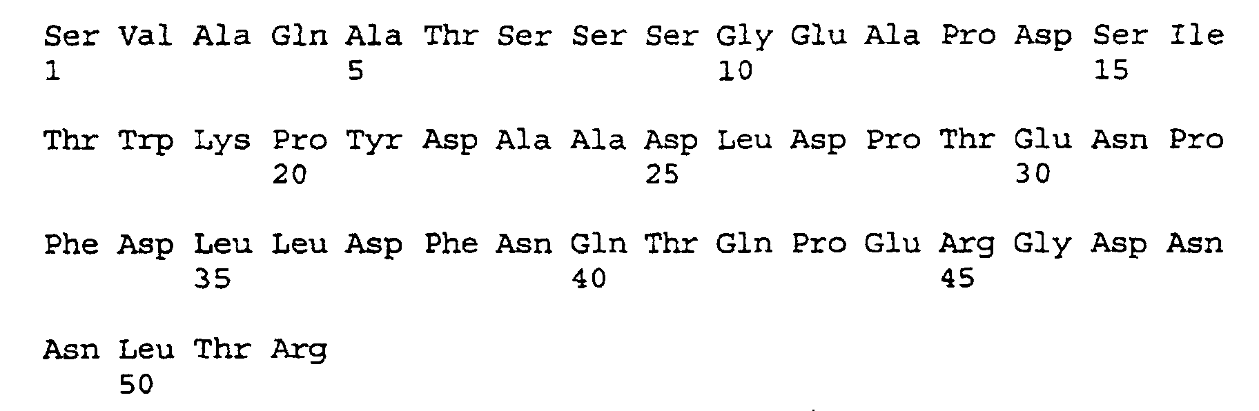
    Im Sequenzprotokoll bedeuten:
    SEQ ID NO:1 - SEQ ID NO:10
    Primer N1 - N10.
    SEQ ID NO: 11
    das native FX Aktivierungspeptid.
    SEQ ID NO:12
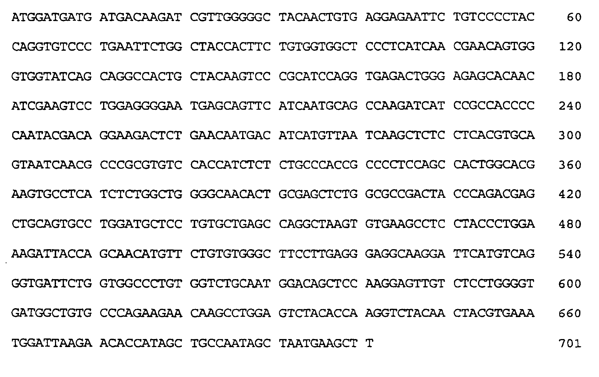
    die Nukleotidsequenz des klonierten TRYI-Variantengens.
    Fig. 1
    zeigt die Nukleotid- und abgeleitete Aminosäuresequenz des klonierten TRYI Trypsin-Variantengens mit verkürztem Prosegment. Die modifizierten Aktivierungspeptide der autokatalytisch aktivierbaren Trypsinvarianten rTRYI-GPK und rTRYI-VGR sind eingezeichnet. Die in der TRYI-GPK-SS Variante zusätzlich eingeführten Mutationen (P132C, P133A und Y233C; Aminosäuresequenz-Nummerierung entsprechend der Publikation von Emi, M. et al. (Gene 41 (1986) 305-310) sind hervorgehoben.
    Beispiele Methoden Rekombinante DNA-Technik
    Zur Manipulation von DNA wurden Standardmethoden benutzt, wie sie bei Sambrook, J. et al. (1989) In: Molecular cloning: A laboratory manual. Cold Spring Harbor Laboratory Press, Cold Spring Harbor, New York, beschrieben sind. Die verwendeten molekularbiologischen Reagenzien wurden nach den Angaben des Herstellers eingesetzt.
    Proteinbestimmung
    Die Proteinkonzentration der Proteasen und Proteasevarianten wurde durch Bestimmung der optischen Dichte (OD) bei 280 nm unter Verwendung des anhand der Aminosäuresequenz errechneten molaren Extinktionskoeffizienten ermittelt.
    Expressionsvektor
    Der Vektor zur Expression der autokatalytisch aktivierbaren Proteasen basiert auf dem Expressionsvektor pSAM-CORE für core-Streptavidin. Die Herstellung und Beschreibung des Plasmids pSAM-CORE ist in der WO 93/09144 von Kopetzki, E. et al. beschrieben. Das core-Streptavidin Gen wurde durch das gewünschte Proteasegen im pSAM-CORE Vektor ersetzt.
    Beispiel 1 Klonierung des humanen Trypsinogen I Gens (Plasmid: pTRYI)
    Die Trypsinogen I cDNA von Bp-Position 61-750, kodierend für Trypsinogen I von Aminosäureposition 19-247 (cDNA Sequenz- und Aminosäuresequenz-Nummerierung entsprechend der Publikation von Emi, M. et al. (Gene 41 (1986) 305-310) wurde in einer "Polymerase Chain Reaktion" (PCR) gemäß der Methode von Mullis, K.B. und Faloona, F.A. (Methods Enzymol. 155 (1987) 355-350) unter Verwendung der PCR Primer N1 (SEQ ID NO: 1) und N2 (SEQ ID NO: 2)
    Figure 00130001
    Figure 00140001
    und einer kommerziell erhältlichen humanen Leber cDNA Genbank (Vektor: Lambda ZAP® II) der Firma Stratagene (La Jolla, CA, U.S.A.) als Template DNA amplifiziert. Mittels der PCR Primer wurde am 5'-Ende der kodierenden Region eine singuläre NcoI Schnittstelle und ein ATG-Startkodon und am 3'-Ende der kodierenden Region eine singuläre HindIII Schnittstelle eingeführt (Fig. 1).
    Das ca. 715 Bp lange PCR Produkt wurde mit den Restriktionsendonukleasen NcoI und HindIII verdaut und das ca. 700 Bp lange NcoI/HindIII-Trypsinogen I Fragment nach Reinigung durch Agarosegelelektrophorese in das ca. 2,55 kBp lange NcoI/HindIII-pSAM-CORE Vektorfragment ligiert. Das gewünschte Plasmid pTRYI wurde durch Restriktionskartierung identifiziert und die durch PCR isolierte TRYI cDNA Sequenz durch DNA Sequenzierung überprüft.
    Beispiel 2 Konstruktion des Trypsinvariantengens TRYI-GPK (autokatalytisch aktivierbare Trypsinvariante; Plasmid: pTRYI-GPK)
    Das N-terminale native Trypsinogen I Prosegment AlaProPheAspAspAspAspLys (Guy, O. et al., Biochem. 17 (1978) 1669-1675) wurde durch das Aktivierungspeptid GlyProLys ersetzt. Das gewünschte TRYI-GPK Trypsinvariantengen wurde mittels PCR-Technik hergestellt.
    Dazu wurde die humane Trypsinogen I cDNA von Bp-Position 73-750, kodierend für Lys-Trypsin I von Aminosäureposition 23-247 (cDNA Sequenz- und Aminosäuresequenz-Numerierung entsprechend der Publikation von Emi, M. et al. (Gene 41 (1986) 305-310) mittels PCR unter Verwendung der Primer N3 (SEQ ID NO: 3) und N2 (SEQ ID NO: 2, Beispiel 1)
    Figure 00140002
    und dem Plasmid pTRYI (Beispiel 1) als Template DNA amplifiziert. Mittels des 5'-überhängenden Endes des PCR Primers N3 wurde eine DNA Sequenz kodierend für ein ATG-Startkodon und das Aktivierungspeptid GlyProLys mit einer singulären NcoI Schnittstelle am 5'-Ende eingeführt. Zudem wurde mit dem PCR Primer N3 innerhalb der kodierenden N-terminalen Region des Trypsin I Strukturgens eine zusätzliche MunI Schnittstelle eingeführt und teilweise die "Codonusage" (Aminosäurepositionen: 7, 8 und 10) an die in E. coli bevorzugt benutzten Kodons angepaßt ("ATG-Umgebung mit optimierter Codonusage", gekennzeichnet durch Kleinschreibung der Basen im N3 Primer), ohne die Proteinsequenz zu verändern.
    Das ca. 700 Bp lange PCR Produkt wurde mit den Restriktionsendonukleasen NcoI und HindIII verdaut und das ca. 685 Bp lange NcoI/HindIII-TRYI-GPK Fragment nach Reinigung durch Agarosegelelektrophorese in das ca. 2,55 kBp lange NcoI/HindIII-pSAM-CORE Vektorfragment ligiert. Das gewünschte Plasmid pTRYI-GPK wurde durch Restriktionskartierung identifiziert (zusätzliche MunI Schnittstelle) und das durch PCR amplifzierte TRPI-GPK Gen durch DNA Sequenzierung überprüft.
    Beispiel 3 Konstruktion des Trypsinvariantengens TRYI-VGR (autokatalytisch aktivierbare Trypsinvariante; Plasmid: pTRYI-VGR)
    Das N-terminale native Trypsinogen I Prosegment AlaProPheAspAspAspAspLys wurde durch das Aktivierungspeptid ValGlyArg ersetzt. Das TRYI-VGR Trypsinvariantengen wurde analog wie das TRYI-GPK Trypsinvariantengen mittels PCR-Technik hergestellt.
    Dazu wurde die humaneTrypsinogen I cDNA von Bp-Position 76-750, kodierend für Trypsin I von Aminosäureposition 24-247 (cDNA Sequenz- und Aminosäuresequenz-Nummerierung entsprechend der Publikation von Emi, M. et al. (Gene 41 (1986) 305-310) mittels PCR unter Verwendung der Primer N4 (SEQ ID NO: 4) und N2 (SEQ ID NO: 2, Beispiel 1)
    Figure 00150001
    und dem Plasmid pTRYI (Beispiel 1) als Template DNA amplifiziert. Mittels des 5'-überhängenden Endes des PCR Primers N4 wurde eine DNA Sequenz kodierend für ein ATG-Startkodon und das Aktivierungspeptid ValGlyArg mit einer singulären NcoI-Schnittstelle am 5'-Ende eingeführt. Zudem wurde mit dem PCR Primer N4 innerhalb der kodierenden N-terminalen Region des Trypsin I Strukturgens eine zusätzliche MunI Schnittstelle eingeführt und teilweise die "Codonusage" (Aminosäurepositionen: 7, 8 und 10) an die in E. coli bevorzugt benutzten Kodons angepaßt ("ATG-Umgebung mit optimierter Codonusage", gekennzeichnet durch Kleinschreibung der Basen im N4 Primer), ohne die Proteinsequenz zu verändern.
    Das ca. 700 Bp lange PCR Produkt wurde mit den Restriktionsendonukleasen NcoI und HindIII verdaut und das ca. 685 Bp lange NcoI/HindIII-TRYI-VGR Fragment nach Reinigung durch Agarosegelelektrophorese in das ca. 2,55 kBp lange NcoI/HindIII-pSAM-CORE Vektorfragment ligiert. Die Herstellung und Beschreibung des Plasmids pSAM-CORE ist in der WO 93/09144 von Kopetzki, E. et al. beschrieben. Das gewünschte Plasmid pTRYI-VGR wurde durch Restriktionskartierung identifiziert (zusätzliche MunI Schnittstelle) und das durch PCR amplifizierte TRPI-VGR Gen durch DNA Sequenzierung überprüft.
    Beispiel 4 Konstruktion des Trypsinvariantengens TRYI-GPK-SS (autokatalytisch aktivierbare Trypsinvariante mit einer zusätzlichen Disulfidbrücke; Plasmid: pTRYI-GPK-SS)
    Durch Einführung einer zusätzlichen Disulfidbrücke wurde das humane Trypsin I Isoenzym verändert. Dazu wurde die Aminosäure Pro an Position 132 und die Aminosäure Tyr an Position 233 zu Cys mutiert (Aminosäuresequenz-Nummerierung entsprechend der Publikation von Emi, M. et al. (Gene 41 (1986) 305-310)). Durch diese 2 Punktmutationen (P132C und Y233C) kann sich eine zusätzliche 6-te Disulfidbrücke zwischen den erfindungsgemäß eingeführten Cysteinresten an Position 132 und 233 ausbilden. Zudem wurde die Aminosäure Pro an der Position 133 zu Ala mutiert (P133A).
    Dazu wurde die Trypsinogen I cDNA von Bp-Position 353-750, kodierend für Trypsinogen I von Aminosäureposition 116-247 (cDNA Sequenz- und Aminosäuresequenz-Nummerierung entsprechend der Publikation von Emi, M. et al. (Gene 41 (1986) 305-310), mittels PCR unter Verwendung der Primer N5 (SEQ ID NO: 5) und N6 (SEQ ID NO: 6)
    Figure 00160001
    Figure 00160002
    und dem Plasmid pTRYI (Beispiel 1 ) als Template DNA amplifiziert. Mittels des PCR Primers N5 wurde die P132C und die P133A Mutation und mit dem Primer N6 die Y233C Mutation eingeführt. Die Mutationen sind durch Kleinschreibung der Basen in den PCR Primern gekennzeichnet.
    Das ca. 420 Bp lange PCR Produkt wurde mit den Restriktionsendonukleasen BsgI und HindIII verdaut und das ca. 405 Bp lange BsgI/HindIII-TRYI Fragment nach Reinigung durch Agarosegelelektrophorese in das ca. 2,85 kBp lange BsgI/HindIII-pTRPI-GPK Vektorfragment ligiert (Beispiel 2). Das gewünschte Plasmid pTRYI-GPK-SS wurde durch Restriktionskartierung identifiziert (fehlende DraIII Schnittstelle) und die durch PCR amplifizierte TRYI DNA Sequenz durch DNA Sequenzierung überprüft.
    Beispiel 5 Konstruktion des FX Proteasevariantengens FX-EGF2-APau-CD (autokatalytisch aktivierbare FX-EGF2-AP-CD Variante; Plasmid: pFX-EGF2-APau-CD)
    Die Klonierung des FX Gens und die Konstruktion des Plasmids pFX-EGF2-AP-CD ist in der WO 97/03027, Beispiel 3) ausführlich beschrieben. Die FX-EGF2-AP-CD Expressionseinheit auf dem Plasmid pFX-EGF2-AP-CD kodiert für eine N-terminal verkürzte FX Proteasevariante aus EGF2-Domäne, Aktivierungspeptid und katalytischer Proteasedomäne.
    Im DNA Segment kodierend für das native FX Aktivierungspeptid (AP) (SEQ-ID-NO:11)
    Figure 00170001
    wurden die C-terminalen drei Aminosäuren Asn, Leu, Thr durch Ile, Asp, Gly ersetzt, wobei das modifizierte FX-Aktivierungspeptid APau entstand.
    Die gewünschte NLT zu IDG Aminosäuresequenzänderung wurde mittels einer Fusions-PCR eingeführt.
    Dazu wurde in einer ersten PCR die FX DNA von Bp-Position 330-629, kodierend für die EGF2-Domäne und das Aktivierungspeptid von Aminosäureposition 111-209 (Sequenz- und Aminosäuresequenz-Nummerierung entsprechend Fig. 3, WO 97/03027) unter Verwendung der PCR Primer N7 (SEQ ID NO: 7) und N8 (SEQ ID NO: 8)
    Figure 00180001
    Figure 00180002
    und des Plasmids pFX-EGF2-AP-CD (Herstellung und Beschreibung siehe: WO 97/03027, Beispiel 3) als Template DNA amplifiziert. Mittels des PCR Primers N8 wurde am 3'-Ende eine singuläre Van91I Schnittstelle eingeführt, ohne die Aminosäuresequenz zu verändern.
    In einer zweiten PCR wurde die FX DNA von Bp-Position 619-1362 kodierend für die C-terminale Region des modifizierten FX Aktivierungspeptids und der FX Proteasedomäne von Aminosäureposition 207-454 (Sequenz- und Aminosäuresequenz-Nummerierung entsprechend Fig. 3, WO 97/03027) unter Verwendung der PCR Primer N9 (SEQ ID NO: 9) und N10 (SEQ ID NO: 10)
    Figure 00180003
    Figure 00180004
    und des Plasmids pFX-EGF2-AP-CD als Template DNA amplifiziert. Mittels des PCR Primers N9 wurde am 5'-Ende eine singuläre Van91I Schnittstelle und die gewünschte NLT zu IDG Aminosäuresequenzveränderung eingeführt.
    Das ca. 300 Bp lange EGF2-AP DNA Fragment der 1-ten PCR wurde mit den Restriktionsendonukleasen PstI und Van91I und das ca. 750 Bp lange FX DNA Fragment der 2-ten PCR wurde mit den Restriktionsendonukleasen Van91I und HindIII verdaut. Nach Reinigung durch Agarosegelelektrophorese wurde das ca. 285 Bp lange PstI/Van91I-EGF2-AP DNA Fragment mit dem 740 Bp langen Van91I/HindIII-FX DNA Fragment und dem ca. 2,56 kBp PstI/HindIII pFX-EGF2-AP-CD Vektorfragment in einer Dreifragmentligation ligiert. Das gewünschte Plasmid pFX-EGF2-APau-CD wurde durch Restriktionskartierung identifiziert (zusätzliche Van91I Schnittstelle) und das FX-EGF2-APau-CD Gen durch DNA Sequenzierung überprüft.
    Beispiel 6 a) Expression der Proteasegene in E. coli
    Zur Expression der Trypsin- und FX Variantengene wurde ein E. coli K12 Stamm (z.B. UT5600; Grodberg, J. und Dunn, J.J., J. Bacteriol. 170 (1988) 1245-1253) jeweils mit einem der in den Beispielen 2-5 beschriebenen Expressionsplasmiden pTRYI-GPK, pTRYI-VGR, pTRYI-GPK-SS und pFX-EGF2-APau-CD (Ampicillin-Resistenz) und dem lacIq-Repressorplasmid pUBS520 (Kanamycin-Resistenz, Herstellung und Beschreibung siehe: Brinkmann, U. et al., Gene 85 (1989) 109-114) transformiert.
    Die mit den Expressionsplasmiden transformierten UT5600/pUBS520/Zellen wurden in Schüttelkultur in DYT-Medium (1% (w/v) Hefeextrakt, 1% (w/v) Bacto Tryptone, Difco, und 0,5% NaCl) mit 50-100 mg/l Ampicillin und 50 mg/l Kanamycin bei 37°C bis zu einer optischen Dichte bei 550 nm (OD550) von 0,6-0,9 angezogen und anschließend mit IPTG (1-5 mmol/l Endkonzentration) induziert. Nach einer Induktionsphase von 4 - 8 Stunden (Std.) bei 37°C wurden die Zellen durch Zentrifugation (Sorvall RC-5B Zentrifuge, GS3 Rotor, 6000 UPM, 15 min) geerntet, mit 50 mmol/l Tris-HCl Puffer, pH 7,2 gewaschen und bis zur Weiterverarbeitung bei -20°C gelagert. Die Zellausbeute aus einer 1 1 Schüttelkultur betrug 4-5 g (Naßgewicht).
    b) Expressionsanalyse
    Die Expression der mit den Expressionsplasmiden pTRYI-GPK, pTRYI-VGR, pTRYI-GPK-SS und pFX-EGF2-APau-CD transformierten UT5600/pUBS520/Zellen wurde analysiert. Dazu wurden Zellpellets aus jeweils 1 ml abzentrifugiertem Anzuchtmedium in 0,25 ml 10 mmol/l Tris-HCl, pH 7,2 resuspendiert und die Zellen durch Ultraschallbehandlung (2 Pulse a 30 s mit 50% Intensität) mit einem Sonifier® Cell Disruptor B15 der Firma Branson (Heusenstamm, Deutschland) aufgeschlossen. Die unlöslichen Zellbestandteile wurden sedimentiert (Eppendorf 5415 Zentrifuge, 14000 UPM, 5 min) und der Überstand mit 1/5 Volumen (Vol) 5xSDS-Probenpuffer (1xSDS-Probenpuffer: 50 mmol/l Tris-HCl, pH 6,8, 1% SDS, 1% Mercaptoethanol, 10% Glycerin, 0,001% Bromphenolblau) versetzt. Die unlösliche Zelltrümmerfraktion (Pellet) wurde in 0,3 ml 1xSDS-Probenpuffer mit 6-8 M Harnstoff resuspendiert, die Proben 5 min bei 95°C inkubiert und erneut zentrifugiert. Danach wurden die Proteine durch SDS-Polyacrylamid Gelelektrophorese (PAGE) aufgetrennt (Laemmli, U.K., Nature 227 (1970) 680-685) und mit Coomassie Brilliant Blue R Farbstoff angefärbt.
    Die in E. coli synthetisierten Proteasevarianten waren homogen und wurden ausschließlich in der unlöslichen Zelltrümmerfraktion gefunden ("inclusion bodies", IBs). Die Expressionshöhe betrug 10-50% bezogen auf das E. coli Gesamtprotein.
    Beispiel 7 Zell-Lyse, Solubilisierung und Naturierung der Proteasegene a) Zell-Lyse und Präparation der "inclusion bodies" (IBs)
    Das Zellpellet aus 3 1 Schüttelkultur (ca. 15 g Naßgewicht) wurde in 75 ml 50 mmol/l Tris-HCl, pH 7,2, resuspendiert. Die Suspension wurde mit 0,25 mg/ml Lysozym versetzt und 30 min bei 0°C inkubiert. Nach Zugabe von 2 mmol/l MgCl2 und 10 mg/ml DNase I (Boehringer Mannheim GmbH, Kat.-Nr. 104159) wurden die Zellen mechanisch mittels Hochdruckdispersion in einer French® Press der Firma SLM Amico (Urbana, IL, U.S.A.) aufgeschlossen. Anschließend wurde die DNA 30 min bei Raumtemperatur (RT) verdaut. Der Ansatz wurde mit 37,5 ml 50 mmol/l Tris-HCl pH 7,2, 60 mmol/l EDTA, 1,5 mol/l NaCl, 6% Triton X-100 versetzt, weitere 30 min bei RT inkubiert und in einer Sorvall RC-5B Zentrifuge (GSA Rotor, 12000 UPM, 15 min) zentrifugiert. Der Überstand wurde verworfen, das Pellet mit 100 ml 50 mmol/l Tris-HCl, pH 7,2, 20 mmol/l EDTA versetzt, 30 min unter Rühren bei 4°C inkubiert und erneut sedimentiert. Der letzte Waschschritt wurde wiederholt. Die gereinigten IBs (1,5-2,0 g Naßgewicht, 25-30% Trockenmasse, 100-150 mg Protease) wurden bis zur Weiterverarbeitung bei -20°C gelagert.
    b) Solubilisierung und Derivatisierung der IBs
    Die gereinigten IBs wurden in einer Konzentration von 100 mg IB-Pellet (Naßgewicht)/ml entsprechend 5-10 mg/ml Protein in 6 mol/l Guanidin-HCl, 100 mmol/l Tris-HCl, 20 mmol/l EDTA, 150 mmol/l GSSG und 15 mmol/l GSH, pH 8,0 unter Rühren bei RT in 1-3 Std. gelöst. Anschließend wurde der pH auf pH 5,0 eingestellt und die unlöslichen Bestandteile durch Zentrifugation (Sorvall RC-5B Zentrifuge, SS34 Rotor, 16000 UPM, 10 min) abgetrennt. Der Überstand wurde gegen 100 Vol. 4-6 mol/l Guanidin-HCl pH 5,0 für 24 Std. bei 4°C dialysiert.
    c) Naturierung
    Die Naturierung der in 6 mol/l Guanidin-HCl solubilisierten und mit GSSG/GSH derivatisierten Proteasevarianten wurde bei 4°C durch wiederholte (z.B. 5-fache) Zugabe von jeweils 0,5 ml IB-Solubilisat/Derivat zu 50 ml 50 mmol/l Tris-HCl, 0,5 mol/l Arginin, 20 mmol/l CaCl2, 1 mmol/l EDTA und 0,5 mmol/l Cystein, pH 8,5 im Zeitabstand von 3-5 Std. und anschließende Inkubation für 10-16 Std. bei 4°C bewirkt. Nach Abschluß der Naturierungsreaktion wurden unlösliche Bestandteile durch Filtration mit einem Filtrationsgerät der Firma Satorius (Göttingen, Deutschland) bestückt mit Tiefenfilter K 250 der Firma Seitz (Bad Kreuznach, Deutschland) abgetrennt.
    d) Konzentrierung und Dialyse der Naturierungsansätze
    Der klare proteasehaltige Überstand wurde durch Cross-Flow-Filtration in einer Minisette (Membran Typ: Omega 3K) der Firma Filtron (Karlstein, Deutschland) 10-15-fach konzentriert und zur Entfernung von Guanidin-HCl und Arginin gegen 100 Vol. 20 mmol/l Tris-HCl und 50 mmol/l NaCl, pH 8,2 für 12-24 Std. bei 20-25°C dialysiert. Präzipitiertes Protein wurde durch Zentrifugation abgetrennt (Sorvall RC-5B Zentrifuge, SS34 Rotor, 16000 UPM, 20 min) und der klare Überstand mit einer Nalgene® Einmalfiltrationseinheit (Porendurchmesser: 0,2 mm) der Firma Nalge (Rochester, NY, U.S.A.) filtriert.
    Beispiel 8 Reinigung der naturierten aktiven Trypsin- und FXa Proteasevarianten
    Die Aktivierung der Trypsin- und rFX-Proteasevarianten rTRYI-GPK, rTRYI-VGR, rTRYI-GPK-SS und rFX-EGF2-APau-CD durch Autokatalyse erfolgt bereits bei der Naturierung und der anschließenden Konzentrierung und Dialyse des Naturierungsansatzes.
    Die aktiven Trypsin- und rFXa Proteasevarianten rTRYI-GPK, rTRYI-VGR, rTRYI-GPK-SS und rFX-EGF2-APau-CD aus den Naturierungsansätzen können bei Bedarf mit chromatographischen Methoden, die dem Fachmann bekannt sind, weiter gereinigt werden.
    Reinigung der Proteasevarianten durch Ionenaustauschchromatographie an Q-Sepharose ff
    Der konzentrierte und gegen 20 mmol/l Tris-HCl und 50 mmol/l NaCl, pH 8,2 dialysierte Naturierungsansatz wurde auf eine mit dem gleichen Puffer äquilibrierte Q-Sepharose-ff-Säule (1,5 x 11 cm, V = 20 ml; Beladungskapazität: 10 mg Protein / ml Gel) der Firma Pharmacia Biotech (Freiburg, Deutschland) aufgetragen (2 Säulenvolumen/Stunde, 2 SV/Std.) und so lange mit dem Äquilibrierungspuffer gewaschen, bis die Absorption des Eluats bei 280 nm den Leerwert des Puffers erreichte. Die Elution des gebundenen Materials erfolgte durch einen Gradienten von 50-500 mmol/l NaCl in 20 mmol/l Tris-HCl, pH 8,2 (2 SV/Std.). Die Proteasen wurden bei einer NaCl Konzentration von 100-200 mmol/l eluiert. Die proteasehaltigen Fraktionen wurden durch nicht-reduzierende und reduzierende SDS PAGE identifiziert und der Elutionspeak vereinigt.
    Beispiel 9 Reinigung der aktiven Proteasevarianten durch Affinitätschromatographie an Benzamidin-Sepharose-CL-6B
    Der konzentrierte und gegen 20 mmol/l Tris-HCl und 50 mmol/l NaCl, pH 8,2 dialysierte Naturierungsansatz wurde auf eine mit dem gleichen Puffer äquilibrierte Benzamidin-Sepharose-CL-6B-Säule (1,0 x 10 cm, V = 8 ml; Beladungskapazität: 2-3 mg Protein / ml Gel) der Firma Pharmacia Biotech (Freiburg, Deutschland) aufgetragen (2 SV/Std.) und so lange mit dem Äquilibrierungspuffer gewaschen, bis die Absorption des Eluats bei 280 nm den Leerwert des Puffers erreichte. Die Elution des gebundenen Materials erfolgte durch 10 mmol/l Benzamidin in 20 mmol/l Tris-HCl und 200 mmol/ NaCl, pH 8,2 (2 SV/Std.). Die proteasehaltigen Fraktionen wurden durch nicht-reduzierende und reduzierende SDS PAGE und Aktivitätsbestimmung identifiziert (siehe: Beispiel 10).
    Der zur Elution verwendete Serinprotease-Inhibitor Benzamidin wurde durch Dialyse gegen 1 mmol/l HCl (Trypsin) bzw. 50 mmol/l Natriumphosphat-Puffer, pH 6,5 (FXa) entfernt.
    Beispiel 10 Charakterisierung der gereinigten Proteasevarianten a) SDS-PAGE
    Sowohl die Oligomeren- und Aggregatbildung durch intermolekulare Disulfidbrückenbildung als auch die Homogenität und Reinheit der naturierten autokatalytisch aktivierten und gereinigten Trypsin- und rFXa Proteasevarianten wurde durch nicht-reduzierende (minus Mercaptoethanol) und reduzierende (plus Mercaptoethanol) SDS PAGE (Laemmli, U.K., Nature 227 (1970) 680-685) untersucht.
    b) Aktivitätsbestimmung, Bestimmung der kinetischen Konstanten Kcat und Km Bestimmung der Trypsin Aktivität mit N-Benzoyl-L-argininethylester (BAEE)
    Die Aktivitätsbestimmung von Trypsin wurde in Anlehnung an die Vorschrift von Walsh, K.A. und Wilcox, P.E. (Methods Enzymol. XIX (1970) 31-41) in einem Volumen von 1 ml in 63 mmol/l Natriumphosphat-Puffer, pH 7,6 und 0,23 mmol/l N-Benzoyl-L-argininethylester Hydrochlorid (BAEE; Sigma-Aldrich Chemie GmbH, Deisenhofen, Deutschland, Kat.-Nr. B-4500) bei 25°C durchgeführt. Die Reaktion wurde durch Zugabe von 50 µl Trypsin-haltiger Probe (gelöst in 1 mmol/l HCl, 300-600 U/ml) gestartet und die Absorptionsänderung/min (ΔA/min) bei 253 nm bestimmt. 1 BAEE Unit Trypsin ist definiert als die Menge Enzym, die unter den angegebenen Testbedingungen eine Absorptionsänderung (Δ A/min253) von 0,0032 bewirkt. Die spezifische Aktivität von gereinigtem humanem Trypsin beträgt unter diesen Testbedingungen ca. 12000 U/mg Protein. Die Aktivität und die kinetischen Konstanten wurden aus der linearen Anfangssteigung gemäß der Michaelis-Menten-Gleichung bestimmt.
    Bestimmung der Trypsin Aktivität mit Chromozym®TH als Substrat
    Die Aktivitätsbestimmung von Trypsin wurde in einem Volumen von 1 ml in 50 mmol/l Tris-HCl, 150 mmol/l NaCl, 5 mmol/l CaCl2, 0,1% PEG 8000, pH 8,0 und 0,1 mmol/l des chromogenen Substrats Chromozym®TH (Tosyl-Gly-Pro-Arg-4-pNA, Kat.-Nr. 838268, Boehringer Mannheim GmbH, Mannheim, Deutschland) bei 25°C durchgeführt. Die Reaktion wurde durch Zugabe von 1-10 µl Trypsin-haltiger Probe (0,4-4 U/ml, gelöst in 1 mmol/l HCl) gestartet und die Absorptionsänderung/min (ΔA/min) bei 405 nm (ε405 = 9,75 [mmol-1 x 1 x cm-1]) bestimmt. 1 Chromozym®TH Unit Trypsin ist definiert als die Menge Enzym, die 1 µmol pNitroanilin (pNA) pro min bei 25°C aus dem chromozymen Substrat freisetzt.
    Bestimmung der Faktor Xa Aktivität
    Die FXa Aktivitätsbestimmung wurde mit dem chromogenen Substrat Chromozym® X (0,1 mmol/l, N-Methoxycarbonyl-D-Nle-Gly-Arg-pNA, Boehringer Mannheim GmbH, Mannheim, Kat.-Nr. 789763) in einem Volumen von 1 ml bei 25°C in 50 mmol/l Tris-HCl, 150 mmol/l NaCl, 5 mmol/l CaCl2, 0,1% PEG 8000, pH 8,0, durchgeführt. Die Reaktion wurde durch Zugabe von 1-10 µl rFXa-haltiger Probe (0,4-4 U/ml) gestartet und die Absorptionsänderung/min (ΔA/min) bei 405 nm (ε405 = 9,75 [mmol-1 x 1 x cm-1]) gemessen. 1 Unit (U) FXa Aktivität ist definiert als die Menge Enzym, die 1 µmol pNA pro min bei 25°C aus dem chromozymen Substrat freisetzt. Die Aktivität und die kinetischen Konstanten wurden aus der linearen Anfangssteigung gemäß der Michaelis-Menten-Gleichung bestimmt.
    Die Ergebnisse zeigt Tabelle 1.
    Protease spez. Aktivität
    [U/mg]
    kcat [1/s] Km [µM] kcat/Km
    [1/µM/s]
    Rinder Trypsin 230 112±2 14±1 8,0
    Schweine Trypsin 222 97±3 8,5±0,9 11,4
    rTRYI-GPK 209 95±1 13±1 7,3
    rTRYI-VGR 210 95±1 13±1 7,3
    rTRYI-GPK-SS 213 99±2 19±1 5,2
    rEGF2-AP-CD 206 --- --- ---
    rEGF2-APau-CD 211 --- --- ---
    c) Ermittlung der Trypsin Temperaturstabilität (Tm-Werte)
    Die Temperaturstabilität der unterschiedlichen Trypsine wurde durch "differential scanning calorimetry" (DSC) ermittelt. Dazu wurde der Denaturierungspunkt (Tm) der Trypsine in einem definierten Lösungsmittel (1 mmol/l HCl), bei definierter Proteinkonzentration (40 mg/ml) und definierter Aufheizrate (1°C/min) bestimmt (siehe Tabelle 2).
    Enzym Tm ( °C ] (DSC)
    Rinder Trypsin 64
    Schweine Trypsin 74
    rTRYI-GPK 70
    rTRYI-VGR 70
    rTRYI-GPK-SS 64
    d) Bestimmung der Trypsin Restaktivität nach Temperaturbelastung
    Zur Ermittlung der Temperaturstabilität wurden die Trypsine in einer Konzentration von 3,5 U/ml in 50 mol/l Tris-HCl, 150 mmol/l NaCl und 5 mmol/l CaCl2, pH 8,0, bei 45°C inkubiert und nach 3, 6 und 29 Std. die Trypsin Restaktivität mit Chromozym®TH als Substrat, wie in Beispiel 10 b beschrieben, bestimmt. Die Ergebnisse zeigt Tabelle 3.
    Enzym Trypsin Restaktivität [ % ]
    3 [ Std. ] 6 [ Std. ] 29 [ Std. ]
    Rinder Trypsin 80 69 22
    Schweine Trypsin 96 98 71
    rTRYI-GPK 96 97 96
    rTRYI-VGR 97 96 97
    rTRYI-GPK-SS 90 87 84
    Referenzliste
  • Bharadwaj, D. et al., J. Biol. Chem. 270 (1995) 6537-6542
  • Blow, D.M.: Acc. Chem. Res. 9 (1976) 145-152
  • Brewer, S.J. und Sassenfeld, H.M., Trends in Biotechnology 3 (1985) 119-122
  • Brinkmann, U. et al., Gene 85 (1989) 109-114
  • Bristow, A.F.: The current status of therapeutic peptides and proteins. In: Hider, R.C.; Barlow, D., eds., Polypeptides and Protein Drugs. Production, Characterization and Formulation. London, Ellis Horwood, pp. 54-69 (1991)
  • Carter, P.: Site-specific proteolysis of fusion proteins. In: Ladisch, M.R.; Willson, R.C.; Painton, C.C.; Builder, S.E. eds., Protein Purification: From Molecular Mechanisms to Large-Scale Processes. ACS Symposium Series No. 427, American Chemical Society, pp. 181-193 (1990)
  • Creighton, T.E., Progress Biophys. Molec. Biol. 33 (1978) 231 - 291
  • Davie, E.W. et al., Biochem. 30 (1991) 10363-10370
  • DiBella, E.E. et al., J. Biol. Chem. 270 (1995) 163-169
  • Emi, M. et al., Gene 41 (1986) 305-310
  • EP-A 0 597 681
  • EP-A 0 089 626
  • EP-A 0 189 998
  • EP-A 0 367 302
  • EP-B 0 114 506
  • EP-B 0 219 874
  • EP-B 0241 022
  • EP-B 0 393 725
  • EP-A 0 408 764
  • EP-A 0 467 839
  • EP-B 0 513 073
  • Flaschel, E. und Friehs, K., Biotech. Adv. 11 (1993) 31-78
  • Forsberg, G. et al., J. Prot. Chem. 10 (1991) 517-526
  • Forsberg, G. et al., J. Prot. Chem. 11 (1992) 201-211
  • Furie, B. und Furie, B.C., Cell 53 (1988) 505- 518
  • Graf, E. et al. (Biochem. 26 (1987) 2616-2623
  • Graf, E. et al. , Proc. Natl. Acad. Sci. USA 85 (1988) 4961-4965
  • Grodberg, J. und Dunn, J.J., J. Bacteriol. 170 (1988) 1245-1253
  • Guy, O. et al., Biochem. 17 (1978) 1669-1675
  • Hedstrom, L. et al., Science 255 (1992) 1249-1253
  • Higaki, J.N. et al., Biochem. 28 (1989) 9256-9263
  • Jonasson, P. et al., Eur. J. Biochem. 236 (1996) 656-661
  • Laemmli, U.K., Nature 227 (1970) 680-685
  • Mann, K. G. et al., Blood 76 (1990) 1-16
  • Marston, F.A.O., Biochem. J. 240 (1986) 1-12
  • Medved, L.V. et al., J. Biol. Chem. 270 (1995) 13652-13659
  • Meyer-Ingold, W.,TIBTECH 11(1993) 387-392
  • Miller, C.G. et al., Proc. Natl. Acad. Sci. USA 84 (1987) 2718-2722
  • Mitraki, A. und King, J., Bio/Technology 7 (1989) 690 - 697
  • Mullis, K.B. und Faloona, F.A., Methods Enzymol. 155 (1987) 355-350
  • Nilsson, B. et al., Methods Enzymol. 185 (1991) 3-16
  • Parks, T.D. et al., Anal. Biochem. 216 (1994) 413-417
  • Perona, J.J. und Craik, C.S., Protein Science 4 (1995) 337-360
  • Pohlner, J. et al., Nature 325 (1987) 458-462
  • Polgar, L.: Structure and function of serine proteases. In: Mechanisms of protein action. Boca Raton, Florida, CRC Press, chapter 3 (1989)
  • Rote Liste, 1997
  • Sambrook, J.; Fritsch, E.F.; Maniatis,T.: Molecular cloning: A laboratory manual. Cold Spring Harbor Press, Cold Spring Harbor, New York, (1989)
  • Sassenfeld, H.M., Trends Biotechnol. 8 (1990) 88-93
  • Schein, C.H., Bio/Technology 8 (1990) 308 - 317
  • Stryer, L., Biochemistry, Freeman and Company, W.H., San Francisco, 1975, Seiten 24 - 30
  • Takeya, H. et al., J. Biol. Chem. 267 (1992) 14109-14117
  • The United States Pharmacopeia, The National Formulary, USP23-NF 18, 1995
  • US Patent 4,634,671
  • US Patent 5,432,062
  • Vasquez, J.R. et al. (J. Cell. Biochem. 39 (1989) 265-276
  • Walsh, K.A. und Wilcox, P.E., Methods Enzymol. XIX (1970) 31-41
  • Wang, E.A., TIBTECH 11 (1993) 379-383
  • Willett, W.S. et al., Biochem. 34 (1995) 2172-2180
  • WO 93/09144
  • WO 96/17942
  • WO 96/20724
  • WO 97/03027
  • WO 97/11186
  • WO 97/18314
  • WO 97/00316
  • Yee, L. und Blanch, H.W., Biotechnol. Bioeng. 41 (1993) 781-790
  • SEQUENZPROTOKOLL
  • (1) ALLGEMEINE ANGABEN:
  • (i) ANMELDER:
  • (A) NAME: BOEHRINGER MANNHEIM GMBH
  • (B) STRASSE: Sandhofer Str. 116
  • (C) ORT: Mannheim
  • (E) LAND: Germany
  • (F) POSTLEITZAHL: D-68305
  • (G) TELEFON: 08856/60-3446
  • (H) TELEFAX: 08856/60-3451
  • (ii) BEZEICHNUNG DER ERFINDUNG: Autokatalytisch aktivierbare zymogene Vorstufen von Proteasen und deren Verwendung
  • (iii) ANZAHL DER SEQUENZEN: 12
  • (iv) COMPUTER-LESBARE FASSUNG:
  • (A) DATENTRÄGER: Floppy disk
  • (B) COMPUTER: IBM PC compatible
  • (C) BETRIEBSSYSTEM: PC-DOS/MS-DOS
  • (D) SOFTWARE: PatentIn Release #1.0, Version #1.30B (EPA)
  • (2) ANGABEN ZU SEQ ID NO: 1:
  • (i) SEQUENZKENNZEICHEN:
  • (A) LÄNGE: 35 Basenpaare
  • (B) ART: Nucleotid
  • (C) STRANGFORM: Einzelstrang
  • (D) TOPOLOGIE: linear
  • (ii) ART DES MOLEKÜLS: other nucleic acid
  • (A) BESCHREIBUNG: /desc = "Primer "N1""
  • (xi) SEQUENZBESCHREIBUNG: SEQ ID NO: 1:
    Figure 00290001
  • (2) ANGABEN ZU SEQ ID NO: 2:
  • (i) SEQUENZKENNZEICHEN:
  • (A) LÄNGE: 39 Basenpaare
  • (B) ART: Nucleotid
  • (C) STRANGFORM: Einzelstrang
  • (D) TOPOLOGIE: linear
  • (ii) ART DES MOLEKÜLS: other nucleic acid
  • (A) BESCHREIBUNG: /desc = "Primer "N2""
  • (xi) SEQUENZBESCHREIBUNG: SEQ ID NO: 2:
    Figure 00300001
  • (2) ANGABEN ZU SEQ ID NO: 3:
  • (i) SEQUENZKENNZEICHEN:
  • (A) LÄNGE: 58 Basenpaare
  • (B) ART: Nucleotid
  • (C) STRANGFORM: Einzelstrang
  • (D) TOPOLOGIE: linear
  • (ii) ART DES MOLEKÜLS: other nucleic acid
  • (A) BESCHREIBUNG: /desc = "Primer "N3""
  • (xi) SEQUENZBESCHREIBUNG: SEQ ID NO: 3:
    Figure 00300002
  • (2) ANGABEN ZU SEQ ID NO: 4:
  • (i) SEQUENZKENNZEICHEN:
  • (A) LÄNGE: 58 Basenpaare
  • (B) ART: Nucleotid
  • (C) STRANGFORM: Einzelstrang
  • (D) TOPOLOGIE: linear
  • (ii) ART DES MOLEKÜLS: other nucleic acid
  • (A) BESCHREIBUNG: /desc = "Primer "N4""
  • (xi) SEQUENZBESCHREIBUNG: SEQ ID NO: 4:
    Figure 00300003
  • (2) ANGABEN ZU SEQ ID NO: 5:
  • (i) SEQUENZKENNZEICHEN:
  • (A) LÄNGE: 77 Basenpaare
  • (B) ART: Nucleotid
  • (C) STRANGFORM: Einzelstrang
  • (D) TOPOLOGIE: linear
  • (ii) ART DES MOLEKÜLS: other nucleic acid
  • (A) BESCHREIBUNG: /desc = "Primer "N5""
  • (xi) SEQUENZBESCHREIBUNG: SEQ ID NO: 5:
    Figure 00310001
  • (2) ANGABEN ZU SEQ ID NO: 6:
  • (i) SEQUENZKENNZEICHEN:
  • (A) LÄNGE: 80 Basenpaare
  • (B) ART: Nucleotid
  • (C) STRANGFORM: Einzelstrang
  • (D) TOPOLOGIE: linear
  • (ii) ART DES MOLEKÜLS: other nucleic acid
  • (A) BESCHREIBUNG: /desc = "Primer "N6""
  • (xi) SEQUENZBESCHREIBUNG: SEQ ID NO: 6:
    Figure 00310002
  • (2) ANGABEN ZU SEQ ID NO: 7:
  • (i) SEQUENZKENNZEICHEN:
  • (A) LÄNGE: 29 Basenpaare
  • (B) ART: Nucleotid
  • (C) STRANGFORM: Einzelstrang
  • (D) TOPOLOGIE: linear
  • (ii) ART DES MOLEKÜLS: other nucleic acid
  • (A) BESCHREIBUNG: /desc = "Primer "N7""
  • (xi) SEQUENZBESCHREIBUNG: SEQ ID NO: 7:
    Figure 00310003
  • (2) ANGABEN ZU SEQ ID NO: 8:
  • (i) SEQUENZKENNZEICHEN:
  • (A) LÄNGE: 38 Basenpaare
  • (B) ART: Nucleotid
  • (C) STRANGFORM: Einzelstrang
  • (D) TOPOLOGIE: linear
  • (ii) ART DES MOLEKÜLS: other nucleic acid
  • (A) BESCHREIBUNG: /desc = "Primer "N8""
  • (xi) SEQUENZBESCHREIBUNG: SEQ ID NO: 8:
    Figure 00320001
  • (2) ANGABEN ZU SEQ ID NO: 9:
  • (i) SEQUENZKENNZEICHEN:
  • (A) LÄNGE: 60 Basenpaare
  • (B) ART: Nucleotid
  • (C) STRANGFORM: Einzelstrang
  • (D) TOPOLOGIE: linear
  • (ii) ART DES MOLEKÜLS: other nucleic acid
  • (A) BESCHREIBUNG: /desc = "Primer "N9""
  • (xi) SEQUENZBESCHREIBUNG: SEQ ID NO: 9:
    Figure 00320002
  • (2) ANGABEN ZU SEQ ID NO: 10:
  • (i) SEQUENZKENNZEICHEN:
  • (A) LÄNGE: 41 Basenpaare
  • (B) ART: Nucleotid
  • (C) STRANGFORM: Einzelstrang
  • (D) TOPOLOGIE: linear
  • (ii) ART DES MOLEKÜLS: other nucleic acid
  • (A) BESCHREIBUNG: /desc = "Primer "N10""
  • (xi) SEQUENZBESCHREIBUNG: SEQ ID NO: 10:
    Figure 00320003
  • (2) ANGABEN ZU SEQ ID NO: 11:
  • (i) SEQUENZKENNZEICHEN:
  • (A) LÄNGE: 52 Aminosäuren
  • (B) ART: Aminosäure
  • (C) STRANGFORM: Einzelstrang
  • (D) TOPOLOGIE: linear
  • (ii) ART DES MOLEKÜLS: Peptid
  • (xi) SEQUENZBESCHREIBUNG: SEQ ID NO: 11:
    Figure 00330001
  • (2) ANGABEN ZU SEQ ID NO: 12:
  • (i) SEQUENZKENNZEICHEN:
  • (A) LÄNGE: 701 Basenpaare
  • (B) ART: Nucleotid
  • (C) STRANGFORM: Einzelstrang
  • (D) TOPOLOGIE: linear
  • (ii) ART DES MOLEKÜLS: cDNA
  • (xi) SEQUENZBESCHREIBUNG: SEQ ID NO: 12:
    Figure 00330002
  • Claims (7)

    1. Verfahren zur rekombinanten Herstellung einer Serinprotease, gekennzeichnet durch:
      a) Transformation einer prokaryontischen Wirtszelle mit einer rekombinanten Nukleinsäure, welche für eine zymogene Vorstufe der genannten Serinprotease codiert, wobei in der zymogenen Vorstufe eine natürlich vorkommende nicht-autokatalytische Spaltstelle durch eine autokatalytische Spaltstelle ersetzt ist, die von der aktiven Form der Protease erkannt wird und dabei die zymogene Vorstufe in die aktive Form spaltet,
      b) Kultivierung der Wirtszelle und Expression der Nukleinsäure,
      c) Isolierung der im Cytoplasma entstandenen und die zymogene Vorstufe enthaltenden inclusion bodies,
      d) Naturierung der zymogenen Vorstufe und
      e) autokatalytische Spaltung der zymogenen Vorstufe in die aktive Form der Serinprotease.
    2. Verfahren nach Anspruch 1, dadurch gekennzeichnet, dass die Serinprotease Trypsin, Thrombin, Faktor Xa oder Lysyl-Endoproteinase ist.
    3. Autokatalytisch spaltbare zymogene Vorstufe einer Serinprotease, wobei in der zymogenen Vorstufe eine natürlich vorkommende nicht-autokatalytische Spaltstelle durch eine autokatalytische Spaltstelle ersetzt ist.
    4. Verfahren zur rekombinanten Herstellung von Trypsin, dadurch gekennzeichnet, dass eine prokaryontische oder eukaryontische Wirtszelle mit einer Nukleinsäure transformiert wird, welche für ein Trypsin der Aminosäuresequenz SEQ ID NO:12 codiert, die Nukleinsäure exprimiert und das dabei entstandene Protein isoliert wird.
    5. Verfahren nach Anspruch 1, dadurch gekennzeichnet, dass das Verhältnis der proteolytischen Aktivität der zymogenen Vorstufe und der aktiven Serinprotease 1 : 5 oder weniger beträgt.
    6. Präparation einer autokatalytisch spaltbaren zymogenen Vorstufe einer Serinprotease, wobei in der zymogenen Vorstufe eine natürlich vorkommende nicht-autokatalytische Spaltstelle durch eine autokatalytische Spaltstelle ersetzt ist, dadurch gekennzeichnet, dass das Protein inaktiv und denaturiert in der Form von inclusion bodies in einer prokaryontischen Zelle vorliegt.
    7. Präparation von inclusion bodies, welche eine autokatalytisch spaltbare zymogene . Vorstufe einer Serinprotease enthalten, wobei in der zymogenen Vorstufe eine natürlich vorkommende nicht-autokatalytische Spaltstelle durch eine autokatalytische Spaltstelle ersetzt ist.
    EP98945214A 1997-08-22 1998-08-12 Autokatalytisch aktivierbare zymogene vorstufen von proteasen und deren verwendung Expired - Lifetime EP1012307B1 (de)

    Priority Applications (2)

    Application Number Priority Date Filing Date Title
    EP98945214A EP1012307B1 (de) 1997-08-22 1998-08-12 Autokatalytisch aktivierbare zymogene vorstufen von proteasen und deren verwendung
    DK98945214T DK1012307T3 (da) 1997-08-22 1998-08-12 Autokatalytisk aktiverbare zymogene proteaseforstadier og deres anvendelse

    Applications Claiming Priority (6)

    Application Number Priority Date Filing Date Title
    EP97114513 1997-08-22
    EP97114513 1997-08-22
    EP97117816 1997-10-15
    EP97117816 1997-10-15
    PCT/EP1998/005094 WO1999010503A1 (de) 1997-08-22 1998-08-12 Autokatalytisch aktivierbare zymogene vorstufen von proteasen und deren verwendung
    EP98945214A EP1012307B1 (de) 1997-08-22 1998-08-12 Autokatalytisch aktivierbare zymogene vorstufen von proteasen und deren verwendung

    Publications (2)

    Publication Number Publication Date
    EP1012307A1 EP1012307A1 (de) 2000-06-28
    EP1012307B1 true EP1012307B1 (de) 2005-05-04

    Family

    ID=26145739

    Family Applications (1)

    Application Number Title Priority Date Filing Date
    EP98945214A Expired - Lifetime EP1012307B1 (de) 1997-08-22 1998-08-12 Autokatalytisch aktivierbare zymogene vorstufen von proteasen und deren verwendung

    Country Status (9)

    Country Link
    EP (1) EP1012307B1 (de)
    JP (1) JP3947653B2 (de)
    AT (1) ATE294869T1 (de)
    AU (1) AU9260598A (de)
    CA (1) CA2301698A1 (de)
    DE (1) DE59812768D1 (de)
    DK (1) DK1012307T3 (de)
    IL (1) IL134653A0 (de)
    WO (1) WO1999010503A1 (de)

    Families Citing this family (13)

    * Cited by examiner, † Cited by third party
    Publication number Priority date Publication date Assignee Title
    US6159722A (en) 1997-12-03 2000-12-12 Boehringer Mannheim Gmbh Chimeric serine proteases
    GB9911689D0 (en) * 1999-05-19 1999-07-21 Medical Res Council Refolding method
    HK1047127B (en) * 1999-08-09 2015-08-21 Sandoz Ag Production of proteins by autoproteolytic cleavage
    CZ302366B6 (cs) * 1999-08-09 2011-04-13 Sandoz Ag Zpusob rekombinantní produkce heterologního polypeptidu pomocí peptidu s autoproteolytickou aktivitou
    AU7053500A (en) * 1999-09-15 2001-04-17 Eli Lilly And Company Chymotrypsin-free trypsin
    AU2001238721A1 (en) * 2000-03-03 2001-09-17 Zymogenetics Inc. Human serine protease
    WO2002061064A2 (de) 2001-02-01 2002-08-08 Roche Diagnostics Gmbh Verfahren zur herstellung von rekombinantem trypsin
    ATE439432T1 (de) 2002-08-30 2009-08-15 Novozymes Inc Verfahren zur herstellung von säugetier-trypsinen
    US20040132156A1 (en) * 2002-10-04 2004-07-08 Schering Aktiengesellschaft Modified hepsin molecules having a substitute activation sequence and uses thereof
    US7176013B2 (en) 2003-06-11 2007-02-13 Schering Aktiengesellschaft Modified corin molecules having substitute activation sequences and uses thereof
    JP2015092852A (ja) * 2013-11-12 2015-05-18 倉敷紡績株式会社 生体組織分散用組成物
    JPWO2018174231A1 (ja) * 2017-03-24 2020-01-23 株式会社カネカ 酵母におけるプロテアーゼの製造方法
    KR20250110226A (ko) 2022-11-23 2025-07-18 에프. 호프만-라 로슈 아게 재조합 단백질 발현을 증가시키는 방법

    Family Cites Families (5)

    * Cited by examiner, † Cited by third party
    Publication number Priority date Publication date Assignee Title
    CA1340740C (en) * 1987-12-08 1999-09-14 Eileen R. Mulvihill Co-expression in eukaryotic cells
    DE4140699A1 (de) * 1991-01-11 1992-07-16 Boehringer Mannheim Gmbh Rekombinante iga-protease
    IL107536A0 (en) * 1992-11-13 1994-02-27 Lilly Co Eli Expression vectors for bovine trypsin and trypsinogen and host cells transformed therewith
    EP0797671B1 (de) * 1994-12-09 2004-03-03 Novo Nordisk A/S Verfahren zur herstellung extrazellulärer proteine in bakterien
    JPH11507207A (ja) * 1995-06-16 1999-06-29 ノボ ノルディスク アクティーゼルスカブ トリプシン(トリプシノーゲン)の製造法

    Also Published As

    Publication number Publication date
    WO1999010503A1 (de) 1999-03-04
    DE59812768D1 (de) 2005-06-09
    JP3947653B2 (ja) 2007-07-25
    EP1012307A1 (de) 2000-06-28
    AU9260598A (en) 1999-03-16
    IL134653A0 (en) 2001-04-30
    JP2001514003A (ja) 2001-09-11
    ATE294869T1 (de) 2005-05-15
    DK1012307T3 (da) 2005-09-05
    CA2301698A1 (en) 1999-03-04

    Similar Documents

    Publication Publication Date Title
    US7666629B2 (en) Method for producing recombinant trypsin
    Cowley et al. Expression, purification and characterization of recombinant human proinsulin
    AT404838B (de) Herstellung von proteinen aus pro-proteinen durch fusionsproteine abgeleitet von furin oder furinanalogen
    WO1998038317A1 (de) Faktor x-analoge mit modifizierter proteasespaltstelle
    JP3169131B2 (ja) 外在的なペプチド配列を用いた処理による生物学的に活性なポリペプチドの製造方法およびそのポリペプチド
    EP1012307A1 (de) Autokatalytisch aktivierbare zymogene vorstufen von proteasen und deren verwendung
    WO1998038318A1 (de) Faktor x-deletionsmutanten und analoge davon
    AU708674B2 (en) Chimeric serine protiases
    EP0917566B1 (de) Rekombinante blutgerinnungsproteasen
    AT410216B (de) Faktor x-analogon mit verbesserter aktivierbarkeit
    DE3786317T2 (de) Proteinanaloge des gewebs-plasminogenaktivators.
    JP4472870B2 (ja) 分子内シャペロン様配列を含有するキメラタンパク質およびインスリン製造へのその適用
    JP2004525631A (ja) 組換えプロテイナーゼk
    DE19943177C2 (de) Verfahren zur Herstellung von aktiven Serienproteasen und inaktiven Varianten
    US20030207402A1 (en) Autocatalytically activatable zymogenic precursors of proteases and their use
    DK2561071T3 (en) PROCEDURE FOR MANUFACTURING RECOMBINANT THROMBIN
    WO1999010483A2 (de) Autokatalytisch aktivierbare vorstufen von proteasen und deren verwendung
    EP0927764B1 (de) Chimäre Serinproteasen
    WO2026030472A1 (en) Compositions and methods relating to recombinant chymotrypsin
    EP3221449B1 (de) Verfahren zur rückfaltung von rekombinantem chymotrypsin
    EP0750667B1 (de) Verwendung von rekombinantem inhibitor aus erythrina caffra zur reinigung von serinproteasen
    WO1998005762A1 (de) Thrombinaktivierbarer plasminogenaktivator
    JP2003531567A (ja) チモーゲン活性化系
    MXPA99007817A (en) Factor x deletion mutants and analogues thereof

    Legal Events

    Date Code Title Description
    PUAI Public reference made under article 153(3) epc to a published international application that has entered the european phase

    Free format text: ORIGINAL CODE: 0009012

    17P Request for examination filed

    Effective date: 20000322

    AK Designated contracting states

    Kind code of ref document: A1

    Designated state(s): AT BE CH DE DK FR GB IT LI NL SE

    17Q First examination report despatched

    Effective date: 20030902

    GRAP Despatch of communication of intention to grant a patent

    Free format text: ORIGINAL CODE: EPIDOSNIGR1

    GRAS Grant fee paid

    Free format text: ORIGINAL CODE: EPIDOSNIGR3

    GRAA (expected) grant

    Free format text: ORIGINAL CODE: 0009210

    AK Designated contracting states

    Kind code of ref document: B1

    Designated state(s): AT BE CH DE DK FR GB IT LI NL SE

    PG25 Lapsed in a contracting state [announced via postgrant information from national office to epo]

    Ref country code: NL

    Free format text: LAPSE BECAUSE OF FAILURE TO SUBMIT A TRANSLATION OF THE DESCRIPTION OR TO PAY THE FEE WITHIN THE PRESCRIBED TIME-LIMIT

    Effective date: 20050504

    Ref country code: IT

    Free format text: LAPSE BECAUSE OF FAILURE TO SUBMIT A TRANSLATION OF THE DESCRIPTION OR TO PAY THE FEE WITHIN THE PRE;WARNING: LAPSES OF ITALIAN PATENTS WITH EFFECTIVE DATE BEFORE 2007 MAY HAVE OCCURRED AT ANY TIME BEFORE 2007. THE CORRECT EFFECTIVE DATE MAY BE DIFFERENT FROM THE ONE RECORDED.SCRIBED TIME-LIMIT

    Effective date: 20050504

    REG Reference to a national code

    Ref country code: GB

    Ref legal event code: FG4D

    Free format text: NOT ENGLISH

    REG Reference to a national code

    Ref country code: CH

    Ref legal event code: EP

    REF Corresponds to:

    Ref document number: 59812768

    Country of ref document: DE

    Date of ref document: 20050609

    Kind code of ref document: P

    REG Reference to a national code

    Ref country code: CH

    Ref legal event code: NV

    Representative=s name: F. HOFFMANN-LA ROCHE AG

    PG25 Lapsed in a contracting state [announced via postgrant information from national office to epo]

    Ref country code: SE

    Free format text: LAPSE BECAUSE OF FAILURE TO SUBMIT A TRANSLATION OF THE DESCRIPTION OR TO PAY THE FEE WITHIN THE PRESCRIBED TIME-LIMIT

    Effective date: 20050804

    REG Reference to a national code

    Ref country code: DK

    Ref legal event code: T3

    GBT Gb: translation of ep patent filed (gb section 77(6)(a)/1977)

    Effective date: 20050824

    NLV1 Nl: lapsed or annulled due to failure to fulfill the requirements of art. 29p and 29m of the patents act
    PLBE No opposition filed within time limit

    Free format text: ORIGINAL CODE: 0009261

    STAA Information on the status of an ep patent application or granted ep patent

    Free format text: STATUS: NO OPPOSITION FILED WITHIN TIME LIMIT

    ET Fr: translation filed
    26N No opposition filed

    Effective date: 20060207

    REG Reference to a national code

    Ref country code: FR

    Ref legal event code: PLFP

    Year of fee payment: 19

    PGFP Annual fee paid to national office [announced via postgrant information from national office to epo]

    Ref country code: CH

    Payment date: 20160725

    Year of fee payment: 19

    Ref country code: GB

    Payment date: 20160725

    Year of fee payment: 19

    Ref country code: DE

    Payment date: 20160831

    Year of fee payment: 19

    Ref country code: DK

    Payment date: 20160725

    Year of fee payment: 19

    PGFP Annual fee paid to national office [announced via postgrant information from national office to epo]

    Ref country code: FR

    Payment date: 20160719

    Year of fee payment: 19

    Ref country code: AT

    Payment date: 20160725

    Year of fee payment: 19

    PGFP Annual fee paid to national office [announced via postgrant information from national office to epo]

    Ref country code: BE

    Payment date: 20160822

    Year of fee payment: 19

    REG Reference to a national code

    Ref country code: DE

    Ref legal event code: R119

    Ref document number: 59812768

    Country of ref document: DE

    REG Reference to a national code

    Ref country code: DK

    Ref legal event code: EBP

    Effective date: 20170831

    REG Reference to a national code

    Ref country code: CH

    Ref legal event code: PL

    REG Reference to a national code

    Ref country code: AT

    Ref legal event code: MM01

    Ref document number: 294869

    Country of ref document: AT

    Kind code of ref document: T

    Effective date: 20170812

    GBPC Gb: european patent ceased through non-payment of renewal fee

    Effective date: 20170812

    PG25 Lapsed in a contracting state [announced via postgrant information from national office to epo]

    Ref country code: LI

    Free format text: LAPSE BECAUSE OF NON-PAYMENT OF DUE FEES

    Effective date: 20170831

    Ref country code: CH

    Free format text: LAPSE BECAUSE OF NON-PAYMENT OF DUE FEES

    Effective date: 20170831

    REG Reference to a national code

    Ref country code: FR

    Ref legal event code: ST

    Effective date: 20180430

    PG25 Lapsed in a contracting state [announced via postgrant information from national office to epo]

    Ref country code: AT

    Free format text: LAPSE BECAUSE OF NON-PAYMENT OF DUE FEES

    Effective date: 20170812

    REG Reference to a national code

    Ref country code: BE

    Ref legal event code: MM

    Effective date: 20170831

    PG25 Lapsed in a contracting state [announced via postgrant information from national office to epo]

    Ref country code: DE

    Free format text: LAPSE BECAUSE OF NON-PAYMENT OF DUE FEES

    Effective date: 20180301

    Ref country code: DK

    Free format text: LAPSE BECAUSE OF NON-PAYMENT OF DUE FEES

    Effective date: 20170831

    Ref country code: GB

    Free format text: LAPSE BECAUSE OF NON-PAYMENT OF DUE FEES

    Effective date: 20170812

    PG25 Lapsed in a contracting state [announced via postgrant information from national office to epo]

    Ref country code: BE

    Free format text: LAPSE BECAUSE OF NON-PAYMENT OF DUE FEES

    Effective date: 20170831

    Ref country code: FR

    Free format text: LAPSE BECAUSE OF NON-PAYMENT OF DUE FEES

    Effective date: 20170831