EP1013668A1 - Monoklonaler antikörper gegen humane monozyten - Google Patents
Monoklonaler antikörper gegen humane monozyten Download PDFInfo
- Publication number
- EP1013668A1 EP1013668A1 EP98941774A EP98941774A EP1013668A1 EP 1013668 A1 EP1013668 A1 EP 1013668A1 EP 98941774 A EP98941774 A EP 98941774A EP 98941774 A EP98941774 A EP 98941774A EP 1013668 A1 EP1013668 A1 EP 1013668A1
- Authority
- EP
- European Patent Office
- Prior art keywords
- cells
- human
- antigen
- mab
- monoclonal antibody
- Prior art date
- Legal status (The legal status is an assumption and is not a legal conclusion. Google has not performed a legal analysis and makes no representation as to the accuracy of the status listed.)
- Withdrawn
Links
Images
Classifications
-
- C—CHEMISTRY; METALLURGY
- C07—ORGANIC CHEMISTRY
- C07K—PEPTIDES
- C07K16/00—Immunoglobulins [IG], e.g. monoclonal or polyclonal antibodies
- C07K16/18—Immunoglobulins [IG], e.g. monoclonal or polyclonal antibodies against material from animals or humans
- C07K16/28—Immunoglobulins [IG], e.g. monoclonal or polyclonal antibodies against material from animals or humans against receptors, cell surface antigens or cell surface determinants
Definitions
- the present invention relates to a monoclonal antibody against human mononuclear leukocytes with the ability to inhibit transendothelial migration of the leukocytes in cocultures with vascular endothelial cell monolayers, said monoclonal antibody being useful for the study and treatment of inflammatory response; to a hybridoma producing this antibody.
- Inflammatory reaction one of the most important responses in the body defense, not only diminishes tissue damage caused by bacterial or viral infections but also promotes repair of such injured tissue.
- the inflammation often induces progressive disorders, such as autoimmune diseases or other incurable diseases of unknown cause.
- autoimmune diseases therefore, it is essential to elucidate the mechanisms and pathological profiles of such inflammatory response.
- Extravasation of peripheral blood leukocytes which is observed in the earliest stage of inflammation is an important target in the therapeutic strategy for chronic inflammatory diseases.
- the process of leukocyte extravasation consists of three serial steps. Briefly, the initial step is leukocyte attachment to and rolling along the luminal surface of endothelium that is mediated by selectin. Subsequently, the leukocyte integrins are activated during this contact with the endothelial surface, by which tight adhesion of leukocytes to the endothelial cell surface occurs. The final step is the process of transendothelial migration after their tight adhesion.
- the purpose of the present invention is to provide a monoclonal antibody directed to a molecule that participates in the transmigration process (diapedesis) of human mononuclear leukocytes after their adhesion to vascular endothelial cells.
- the present inventor has developed a monoclonal antibody capable of inhibiting transmigration of human mononuclear leukocytes after their adhesion to vascular endothelial cells using the following methods: 1) a method of immunization with human mononuclear cells bound to human endothelial cells and migrated through human endothelial cell monolayers cultured on collagen gels into the gels below; 2) a method of screening for antibodies that inhibit transmigration of human mononuclear cells across endothelial cell monolayers cultured on collagen gels, but not their endothelial adhesion.
- the present invention provides a monoclonal antibody against human mononuclear leukocytes which is capable of inhibiting transmigration of human mononuclear leukocytes after their adhesion to vascular endothelial cells.
- the present invention provides a hybridoma producing the monoclonal antibody.
- the hybridoma that produces the monoclonal antibody of the present invention can be obtained by a cell fusion method that a myeloma cell line from mammals is fused with antibody-producing cells from mammals immunized by human mononuclear leukocytes as an antigen.
- mice As an example of mammals, mice, rabbits, rats, goats, or chickens can be used in this prevention, with mice being preferred.
- human mononuclear leukocytes are generally employed: (a) human mononuclear cells that adhered to human vascular endothelial cells; (b) human mononuclear cells that migrated through human endothelial cell monolayers cultured on collagen gels into the gels below; it is particularly preferable if (a) is used as an antigen for sensitization, and (b) is used as an antigen for the final immunization.
- the human mononuclear cells can be obtained from peripheral blood, synovial fluids, pleural fluids, etc., with peripheral blood mononuclear cells being preferable.
- Immunization may be carried out according to a standard technique. For example, mononuclear cell suspension in routinely-used buffer or saline or a mixture of the suspension and Freund's adjuvant is administered to animals intraperitoneally, subcutaneously, or through any suitable routes for the first immunization. Thereafter, a similar procedure may be repeated if necessary.
- the number of mononuclear cells served as an antigen depends on administration routes and animals used, it is about 1 x 10' cells per mouse for each administration intraperitoneally or intravenously.
- Antibody-producing cells are obtained from spleen 2-4 days after the final immunization, but lymph nodes and peripheral blood may be used instead of spleen.
- myeloma cell lines can be used, such as P3/NS1/1-Ag4-1 (NS1), SP2/0-Ag14 (SP2), P3/x63-Ag8 (x63), P3/x63-Ag8.U1 (P3U1), F0, MCP-11, x63.653, S194 in mice, and 210.RCY3.Ag1.2.3 (Y3) in rats.
- fused cells are cultured for several days to weeks in HAT medium.
- the hybridomas obtained are screened for an antibody of interest.
- the screening is performed by measuring the transmigration of mononuclear cells after their adhesion to endothelial cells; hybridomas producing antibodies that inhibit the post-adhesive transmigration without interfering the adhesion are selected.
- Cloning of the selected hybridomas may be performed with a limiting dilution or Sorterlone method, with the former being preferable.
- the cloned hybridoma can be subcultured in ordinary medium and easily stored for a long time by being frozen in liquid nitrogen.
- the monoclonal antibody of the present invention is obtained by a standard method of isolation from culture supernatants of the hybridoma or malignant ascites of animals injected with the hybridoma.
- the former is carried out to have highly-purified antibodies and the latter to have a large amount of antibodies.
- the monoclonal antibody obtained can be further purified to the immunoglobulin fraction by a standard method with ammonium sulfate fractionation, gel filtration including DEAE cellulose column chromatography, etc.
- human peripheral blood mononuclear leukocytes Prior to immunization, human peripheral blood mononuclear leukocytes were cocultured with human umbilical vein endothelial cells (HUVEC), and subsequently leukocytes except non-adherent leukocytes were collected to be used as an antigen.
- mononuclear cells were used that migrated through HUVEC monolayers cultured on collagen gels 5 hr after addition to the HUVEC cultures. Briefly, HUVEC were cultured in medium containing endothelial growth supplements on Type-1 collagen gels (2 ml per dish) with a final collagen concentration of 0.8 mg/ml in 60 mm-plastic dishes.
- HUVEC mononuclear cells were added to and cocultured with the HUVEC. Following an appropriate interval, HUVEC and non-migrated cells were removed from the collagen gels with 0.4% EDTA treatment, and mononuclear cells (b) migrated into the gels were released from the gels by treatment with collagenase.
- Mononuclear cells suspended in phosphate buffer saline (PBS) were intraperitoneally injected into mice (1 x 10 7 cells per mouse). This injection was repeated 4 times or more every 2 to 4 weeks. For the final immunization, approximately 1 x 10 7 cells of migrated mononuclear cells were intravenously injected into the mice. Spleen was removed from the mice 2-4 days after the final immunization, and 1 x 10 8 spleen cells including antibody-producing cells were prepared for cell fusion with 1-2 x 10 7 myeloma cells that were maintained in RPMI-1640 medium containing 10% fetal calf serum (FCS).
- FCS fetal calf serum
- NS-1 Twenty million P3/NS1/1-Ag4-1 (NS-1) cells suspended in Iscove medium were mixed with the prepared spleen cells (1 x 10 8 ) followed by centrifugation. After suction of the supernatant, 1 ml of polyethylene glycol 4000 warmed at 37°C was slowly dropped to the cell pellet. Subsequently, 9 ml of Iscove medium was added to the mixture, Following centrifugation, the supernatant containing polyethylene glycol was aspirated, and the spleen cells were resuspended in HAT medium plus interleukin 6 at 1 x 10 6 /ml. One hundred microliter of the cell suspension was added to each well in 96-well flat-bottom plates and cultured at 37°C until sufficient proliferation was achieved.
- HUVEC monolayers were cultured on collagen gels (50 ⁇ l/well) in 96-multiwell microtiter plates according to the method described in Section (1) of Example 1. Fifty microliters of antibody supernatant and 50 ⁇ l of suspended mononuclear cells were added to each well and cultured.
- the number of adherent mononuclear cells to HUVEC in the presence of hybridoma supernatant was compared to that (control) in the absence of hybridoma supernatant under a phase contrast microscope.
- the wells showing no significant difference in the number of adherent cells were selected.
- endothelial cells of the selected wells were removed from the collagen gel surface with 0.4% EDTA, mononuclear cells migrated into the gels were counted under a phase contrast microscope.
- hybridomas with the ability to suppress 50% or more of transmigration were considered to secrete the antibody of interest and selected for cloning.
- hybridoma with a high suppressive ability of transmigration was chosen by recloning at least 3 times, which was considered to be the hybridoma producing the antibody of interest.
- hybrid cells were intraperitoneally injected into mice which are the same strain as the hybridoma. Malignant ascites were developed and further purified to IgG by a Protein A-coupled column.
- the anti-4C8 monoclonal antibody (mAb) obtained was the IgG3 subclass.
- the mAb at 1 ⁇ g/ml inhibited 80-90% of transmigration of T lymphocytes or mononuclear cells including T lymphocytes after their adhesion to HUVEC monolayers.
- the anti-4C8 mAb of the present invention was characterized, with the results that are: (1) the mAb reacts with human lymphocytes, NK cells, monocytes, granulocytes including eosinophils, and adherent cells such as vascular endothelial cells; (2) the mAb changes assembly of cytoskeletal proteins such as actin polymerization; (3) the mAb has chemokine-like or chemoattractant-like activity for human lymphocytes; (4) the mAb induces attachment of human lymphocytes to the substrate; (5) the mAb increases the cell surface expression of cell activation-related antigens such as CD69; (6) the mAb reacts with granulocytes including eosinophils and and stimulates cell activation; (7) the 4C8 expression on mononuclear cells and granulocytes is decreased in patients with rheumatoid arthritis taking anti-rheumatic drugs; (8) the mAb reacts with malignant tumor cells.
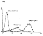
- the anti-4C8 mAb reacts with human lymphocytes, NK cells, monocytes, granulocytes including eosinophils, and vascular endothelial cells.
- Anti-4C8 mAb was conjugated with fluorescein by using a fluorescein labeling kit and an appropriate amount of the labeled mAb was added to 0.1 ml of peripheral whole blood with anticoagulants and incubated for 30 min on ice. After washing 3 times with cold PBS containing 0.1% bovine serum albumin (BSA), the blood cells were hemolyzed by a hemolytic reagent and fixed with paraformaldehyde. Vascular endothelial cells were removed from plastic plates with 0.4% EDTA solution, and 3 x 10 5 cells were stained with fluorescein-labeled anti-4C8 mAb as described above. The stained cells were analyzed for the fluorescence intensity on a flow cytometer with gating on the granulocytes, monocytes or lymphocytes.
- BSA bovine serum albumin
- the 4C8 antigen is expressed on 90% or more of lymphocytes and monocytes from healthy subjects, especially, intensely on lymphocytes and to a lesser extent on monocytes.
- NK cells which comprise about 10% of lymphocytes, apparently express the 4C8 antigen.
- granulocytes were almost negative for the antigen.
- the 4C8 expression on granulocytes was clearly detected in a part of patients with rheumatoid arthritis or other chronic inflammatory deseases.
- the positive expression on eosinophils was observed in patients with atopic diseases. While adherent cells, such as vascular endothelial cells, did not express the 4C8 antigen in the resting and non-proliferative state, the antigen was detected in a subpopulation of proliferating endothelial cells.
- the anti-4C8 mAb changes assembly of cytoskeletal proteins such as actin polymerization.
- the phalloidin-bound cells were detected in about 20% of the total lymphocytes gated by flow cytometry when stimulated with control IgG3. However, the percentage was increased up to 65% in a dose-dependent manner when stimulated with anti-4C8 mAb. Thus, stimulation of the 4C8 antigen certainly induced actin polymerization in lymphocytes.
- the anti-4C8 mAb stimulates migration of human lymphocytes, like chemokines.
- lymphocytes To determine whether migration of lymphocytes is induced by stimulation of the 4C8 antigen on the surface of lymphocytes, anti-4C8 mAb was used as a ligand. Lymphocytes (3 x 10 5 cells/well) suspended in M199 medium without serum were added directly onto collagen gels (50 ⁇ l/well) with impregnated anti-4C8 mAb at varying concentrations in 96-multiwell plates. After a 3 hr incubation, lymphocytes adherent to the surface of the gels were removed by treatment with 0.4% EDTA, and migrated cells in the gels were counted per a field under a phase contrast microscope.
- the anti-4C8 mAb stimulates adhesion of human lymphocytes to the substrate.
- Anti-4C8 mAb-coated plates as described in section 2) were further treated for 2 hr with 3% BSA solution.
- 51 Cr-labeled human lymphocytes (3 x 10 5 cells/well) were added to the plates.
- non-adherent cells were removed followed by cell lysis of adherent cells with 1N-NaOH.
- the radioactivity of the cells was measured by a gamma counter.
- Anti-4C8 mAb increased cell adhesion to the plastic plates in a dose-dependent manner ranging from 1 ⁇ g/ml to 100 ⁇ g/ml. Approximately 60% of the added cells adhered to the plates coated with 100 ⁇ g/ml of ant-4C8 mAb, whereas the adherence was only 5% or less in the range of 1 to 100 ⁇ g/ml of control IgG3. It was evident that stimulation via the 4C8 antigen markedly increased lymphocyte adhesion to the plastic plates.
- the anti-4C8 mAb induces expression of cell activation-related molecules such as CD69.
- the anti-4C8 mAb reacts with granulocytes including eosinophils, resulting in cell activation.
- the 4C8 antigen is often expressed on granulocytes from patients with rheumatoid arthritis (RA), but not healthy subjects. Whether stimulation of the 4C8 antigen activates granulocytes from such RA patients was examined. Peripheral blood was mixed with 1% dextran sulfate in the ratio of 1 to 0.75. The mixture was allowed to stand for 40-50 min, and then a granulocyte-enriched fraction was overlaid on a Ficoll-Conray solution, followed by centrifugation for 30 min. After aspiration of the supernatant, the cell pellet was washed three times with PBS at 4° C. Finally granulocytes obtained were suspended in RPMI-1640 medium.
- the granulocyte suspension was added to the plates coated with anti-4C8 mAb as described in section 2). Thirty minutes later, these cells were stained with fluorescein-conjugated anti-CD11b mAb which binds to activated granulocytes, and analyzed on a flow cytometer.
- results The mean fluorescence intensity of the CD11b expression on granulocytes was significantly augmented as the concentration of coated anti-4C8 mAb increased. Thus, stimulation of the 4C8 antigen activates granulocytes.
- Mononuclear cells and granulocytes were stained with fluorescein-conjugated anti-4C8 mAb. After that, lymphocytes, monocytes, and granulocytes were examined for the mean fluorescence intensity of the 4C8 expression on a flow cytometry.
- results The 4C8 expression on mononuclear cells was low in RA patients who responded well to anti-rheumatic drugs, compared with RA patients who showed high disease activity or had never been treated. Particularly, regarding the 4C8 expression on monocytes, the fluorescence intensity level in the former patients was about 25% of that in the latter patients. The 4C8 antigen expressed on granulocytes in some of patients with active RA became almost undetectable by successful therapy with anti-rheumatic drugs. In addition to the study for the pharmacological action of anti-rheumatic drugs, assessment of the 4C8 expression on monocytes or other cells from RA patients could be clinically applicable to a means for judging or predicting the effect of such drugs.
- the anti-4C8 mAb reacts with malignant tumor cell lines.
- the mechanism of the transendothelial migration of leukocytes observed typically in inflammatory response or the mechanism of metastasis of malignant cells remains to be elucidated.
- the present invention provides a means for investigation of these mechanisms at cellular, protein, and molecular levels.
- the molecule recognized by anti-4C8 mAb of the present invention is related to various pathological conditions and diseases, the mAb can he used for clinical diagnosis and therapy.
Landscapes
- Health & Medical Sciences (AREA)
- Chemical & Material Sciences (AREA)
- Immunology (AREA)
- Organic Chemistry (AREA)
- General Health & Medical Sciences (AREA)
- Biophysics (AREA)
- Biochemistry (AREA)
- Genetics & Genomics (AREA)
- Medicinal Chemistry (AREA)
- Molecular Biology (AREA)
- Proteomics, Peptides & Aminoacids (AREA)
- Life Sciences & Earth Sciences (AREA)
- Preparation Of Compounds By Using Micro-Organisms (AREA)
- Micro-Organisms Or Cultivation Processes Thereof (AREA)
- Peptides Or Proteins (AREA)
Applications Claiming Priority (3)
| Application Number | Priority Date | Filing Date | Title |
|---|---|---|---|
| JP24520397 | 1997-09-10 | ||
| JP24520397 | 1997-09-10 | ||
| PCT/JP1998/004035 WO1999012972A1 (fr) | 1997-09-10 | 1998-09-09 | Anticorps monoclonal contre des monocytes humains |
Publications (1)
| Publication Number | Publication Date |
|---|---|
| EP1013668A1 true EP1013668A1 (de) | 2000-06-28 |
Family
ID=17130167
Family Applications (1)
| Application Number | Title | Priority Date | Filing Date |
|---|---|---|---|
| EP98941774A Withdrawn EP1013668A1 (de) | 1997-09-10 | 1998-09-09 | Monoklonaler antikörper gegen humane monozyten |
Country Status (5)
| Country | Link |
|---|---|
| US (1) | US6515111B1 (de) |
| EP (1) | EP1013668A1 (de) |
| JP (1) | JP3593032B2 (de) |
| CA (1) | CA2303431A1 (de) |
| WO (1) | WO1999012972A1 (de) |
Cited By (10)
| Publication number | Priority date | Publication date | Assignee | Title |
|---|---|---|---|---|
| US8003385B2 (en) | 2005-12-21 | 2011-08-23 | Sanofi Pasteur Vax Design Corp. | In vitro germinal centers |
| US8030070B2 (en) | 2004-04-28 | 2011-10-04 | Sanofi Pasteur Vaxdesign Corp. | Artificial lymphoid tissue equivalent |
| US8062889B2 (en) | 2004-04-28 | 2011-11-22 | Sanofi Pasteur Vaxdesign Corp. | Methods of evaluating a test agent in a diseased cell model |
| US8071373B2 (en) | 2004-04-28 | 2011-12-06 | Sanofi Pasteur Vaxdesign Corp. | Co-culture lymphoid tissue equivalent (LTE) for an artificial immune system (AIS) |
| US8080416B2 (en) | 2004-04-28 | 2011-12-20 | Sanofi Pasteur Vaxdesign Corp. | Method for determining the immunogenicity of an antigen |
| US8119403B2 (en) | 2004-04-28 | 2012-02-21 | Sanofi Pasteur Vaxdesign Corp. | Disease model incorporation into an artificial immune system (AIS) |
| US8288159B2 (en) | 2004-04-28 | 2012-10-16 | Sanofi Pasteur Vaxdesign Corp. et al. | Artificial immune system: methods for making and use |
| US8298824B2 (en) | 2004-04-28 | 2012-10-30 | Sanofi Pasteur Vaxdesign Corporation | Methods of evaluating a test agent in a diseased cell model |
| US8298823B2 (en) | 2004-04-28 | 2012-10-30 | Sanofi Pasteur Vaxdesign Corporation | Methods for antibody production |
| US8647837B2 (en) | 2007-07-16 | 2014-02-11 | Sanofi Pasteur Vaxdesign Corp. | Artificial tissue constructs comprising alveolar cells and methods for using the same |
Families Citing this family (4)
| Publication number | Priority date | Publication date | Assignee | Title |
|---|---|---|---|---|
| JP2003102471A (ja) * | 2001-10-01 | 2003-04-08 | Kirin Brewery Co Ltd | 調節性t細胞の分化誘導・増殖促進方法 |
| EP1618891A1 (de) * | 2003-03-31 | 2006-01-25 | Kirin Beer Kabushiki Kaisha | Verfahren zur induktion von differenzierungs- und proliferierungs-regelnden t-zellen durch anti-cd52-antikörper und medizinische zusammensetzung dafür |
| EP1723251A4 (de) * | 2004-03-05 | 2008-04-23 | Novartis Vaccines & Diagnostic | In-vitro-testsystem zur prognose der verträglichkeit eines patienten gegenüber therapeutika |
| WO2005104755A2 (en) * | 2004-04-28 | 2005-11-10 | Vaxdesign Corporation | Artificial immune system: methods for making and use |
-
1998
- 1998-09-09 EP EP98941774A patent/EP1013668A1/de not_active Withdrawn
- 1998-09-09 CA CA002303431A patent/CA2303431A1/en not_active Abandoned
- 1998-09-09 US US09/508,071 patent/US6515111B1/en not_active Expired - Fee Related
- 1998-09-09 WO PCT/JP1998/004035 patent/WO1999012972A1/ja not_active Ceased
- 1998-09-09 JP JP2000510777A patent/JP3593032B2/ja not_active Expired - Fee Related
Non-Patent Citations (1)
| Title |
|---|
| See references of WO9912972A1 * |
Cited By (18)
| Publication number | Priority date | Publication date | Assignee | Title |
|---|---|---|---|---|
| US8288159B2 (en) | 2004-04-28 | 2012-10-16 | Sanofi Pasteur Vaxdesign Corp. et al. | Artificial immune system: methods for making and use |
| US8298823B2 (en) | 2004-04-28 | 2012-10-30 | Sanofi Pasteur Vaxdesign Corporation | Methods for antibody production |
| US8030070B2 (en) | 2004-04-28 | 2011-10-04 | Sanofi Pasteur Vaxdesign Corp. | Artificial lymphoid tissue equivalent |
| US8062889B2 (en) | 2004-04-28 | 2011-11-22 | Sanofi Pasteur Vaxdesign Corp. | Methods of evaluating a test agent in a diseased cell model |
| US8071373B2 (en) | 2004-04-28 | 2011-12-06 | Sanofi Pasteur Vaxdesign Corp. | Co-culture lymphoid tissue equivalent (LTE) for an artificial immune system (AIS) |
| US8080416B2 (en) | 2004-04-28 | 2011-12-20 | Sanofi Pasteur Vaxdesign Corp. | Method for determining the immunogenicity of an antigen |
| US8119403B2 (en) | 2004-04-28 | 2012-02-21 | Sanofi Pasteur Vaxdesign Corp. | Disease model incorporation into an artificial immune system (AIS) |
| US9625444B2 (en) | 2004-04-28 | 2017-04-18 | Sanofi Pasteur Vaxdesign Corporation | Artificial immune system: methods of use |
| US8962319B2 (en) | 2004-04-28 | 2015-02-24 | Sanofi Pasteur Vaxdesign Corp. | Methods for testing an immune response using cultures of T cells, B cells, dendritic cells and follicular dendritic cells |
| US8722402B2 (en) | 2004-04-28 | 2014-05-13 | Sanofi Pasteur Vaxdesign Corporation | Artificial immune system: methods of use |
| US8298824B2 (en) | 2004-04-28 | 2012-10-30 | Sanofi Pasteur Vaxdesign Corporation | Methods of evaluating a test agent in a diseased cell model |
| US8697371B2 (en) | 2004-04-28 | 2014-04-15 | Sanofi Pasteur Vaxdesign Corp. | Methods for testing an immune response using cultures of T cells, B cells and dendritic cells |
| US8669105B2 (en) | 2005-12-21 | 2014-03-11 | Sanofi Pasteur Vaxdesign Corp. | Methods for assaying responses to vaccines |
| US8003385B2 (en) | 2005-12-21 | 2011-08-23 | Sanofi Pasteur Vax Design Corp. | In vitro germinal centers |
| US8003387B2 (en) | 2005-12-21 | 2011-08-23 | Sanofi Pasteur Vaxdesign Corp. | In vitro germinal centers |
| US8247226B2 (en) | 2005-12-21 | 2012-08-21 | Sanofi Pasteur Vaxdesign Corp. | Methods of evaluating an immune response to an antigen |
| US8647837B2 (en) | 2007-07-16 | 2014-02-11 | Sanofi Pasteur Vaxdesign Corp. | Artificial tissue constructs comprising alveolar cells and methods for using the same |
| US9566250B2 (en) | 2007-07-16 | 2017-02-14 | Sanofi Pasteur Vaxdesign Corporation | Artificial tissue constructs comprising alveolar cells and methods for using the same |
Also Published As
| Publication number | Publication date |
|---|---|
| US6515111B1 (en) | 2003-02-04 |
| WO1999012972A1 (fr) | 1999-03-18 |
| CA2303431A1 (en) | 1999-03-18 |
| JP3593032B2 (ja) | 2004-11-24 |
Similar Documents
| Publication | Publication Date | Title |
|---|---|---|
| US5019648A (en) | Monoclonal antibody specific for the adhesion function domain of a phagocyte cell surface protein | |
| ABDOU et al. | The monoclonal nature of lymphocytes in multiple myeloma: effects of therapy | |
| Abrahams et al. | Induction of tumor necrosis factor α production by adhered human monocytes: a key role for Fcγ receptor type IIIA in rheumatoid arthritis | |
| Brett et al. | Repopulation of blood lymphocyte sub‐populations in rheumatoid arthritis patients treated with the depleting humanized monoclonal antibody, CAMPATH‐1H | |
| US4550086A (en) | Monoclonal antibodies that recognize human T cells | |
| US6515111B1 (en) | Monoclonal antibody which inhibits transendothelial migration of human mononuclear leukocytes | |
| JP2703764B2 (ja) | 補体成分C5aに対するモノクローナル抗体 | |
| JPH0223156B2 (de) | ||
| US5219997A (en) | Monoclonal antibody which inhibits the adhesion functions of the β integrin, CR3 | |
| JPWO1999012972A1 (ja) | ヒト単核球に対するモノクローナル抗体 | |
| EP0673389A1 (de) | Antikörper gegen den gm-csf rezeptor und dessen verwendung | |
| JPS63276494A (ja) | Lfa−3の精製法 | |
| EP0726915B1 (de) | Ligand fähig zur bindung an der adenosinedeaminase-bindungsstelle von cd26 | |
| Bank et al. | Functional role of VLA-1 (CD49A) in adhesion, cation-dependent spreading, and activation of cultured human T lymphocytes | |
| EP0160486B1 (de) | Monoklonale Antikörper | |
| Fukunaga et al. | Increased density of ecto 5'nucleotidase antigen on leukemic T cells from patients with cutaneous T-cell lymphoma and adult T-cell leukemia/lymphoma | |
| Li et al. | Targeting leukocyte recruitment in the treatment of psoriasis | |
| Elenström-Magnusson et al. | IL-4-induced B cell migration involves transient interactions between β1 integrins and extracellular matrix components | |
| Springer et al. | Functional and structural interrelationships among the Mac-1, LFA-1 family of leukocyte adhesion glycoproteins, and their deficiency in a novel heritable disease | |
| Satoh et al. | Cytolysis of oligodendrocytes is mediated by killer (K) cells but not by natural killer (NK) cells | |
| AU684806B2 (en) | Human cell adhesion protein AAMP-1 and uses thereof | |
| Issekutz et al. | Role of interleukin‐1 and tumour necrosis factor in leukocyte recruitment to acute dermal inflammation | |
| HUMAN | Robert F. Todd, III and Stuart F. Schlossman | |
| HRP940830A2 (en) | Hybrid cell line for producing monoclonal antibody to a human t cell antigen, antibody, and methods |
Legal Events
| Date | Code | Title | Description |
|---|---|---|---|
| PUAI | Public reference made under article 153(3) epc to a published international application that has entered the european phase |
Free format text: ORIGINAL CODE: 0009012 |
|
| 17P | Request for examination filed |
Effective date: 20000303 |
|
| AK | Designated contracting states |
Kind code of ref document: A1 Designated state(s): AT BE CH CY DE DK ES FI FR GB GR IE IT LI LU MC NL PT SE |
|
| RAP1 | Party data changed (applicant data changed or rights of an application transferred) |
Owner name: TOYAMA CHEMICAL CO., LTD. |
|
| RIN1 | Information on inventor provided before grant (corrected) |
Inventor name: MASUYAMA, JUNICHI |
|
| RAP1 | Party data changed (applicant data changed or rights of an application transferred) |
Owner name: MASUYAMA, JUNICHI Owner name: TOYAMA CHEMICAL CO., LTD. |
|
| RAP1 | Party data changed (applicant data changed or rights of an application transferred) |
Owner name: MASUYAMA, JUNICHI |
|
| STAA | Information on the status of an ep patent application or granted ep patent |
Free format text: STATUS: THE APPLICATION IS DEEMED TO BE WITHDRAWN |
|
| 18D | Application deemed to be withdrawn |
Effective date: 20040401 |