EP1040185B1 - Cellules precurseurs neurales, leur procede de production et leur utilisation pour la therapie d'anomalies neurales - Google Patents
Cellules precurseurs neurales, leur procede de production et leur utilisation pour la therapie d'anomalies neurales Download PDFInfo
- Publication number
- EP1040185B1 EP1040185B1 EP98966817A EP98966817A EP1040185B1 EP 1040185 B1 EP1040185 B1 EP 1040185B1 EP 98966817 A EP98966817 A EP 98966817A EP 98966817 A EP98966817 A EP 98966817A EP 1040185 B1 EP1040185 B1 EP 1040185B1
- Authority
- EP
- European Patent Office
- Prior art keywords
- cells
- neural
- precursor cells
- growth factor
- cell
- Prior art date
- Legal status (The legal status is an assumption and is not a legal conclusion. Google has not performed a legal analysis and makes no representation as to the accuracy of the status listed.)
- Expired - Lifetime
Links
- 230000001537 neural effect Effects 0.000 title claims abstract description 141
- 238000000034 method Methods 0.000 title claims abstract description 60
- 239000002243 precursor Substances 0.000 title claims description 86
- 230000007547 defect Effects 0.000 title claims description 13
- 238000002560 therapeutic procedure Methods 0.000 title claims description 8
- 238000004519 manufacturing process Methods 0.000 title description 12
- 210000004027 cell Anatomy 0.000 claims abstract description 424
- 239000003102 growth factor Substances 0.000 claims abstract description 88
- 230000002518 glial effect Effects 0.000 claims abstract description 54
- 239000012679 serum free medium Substances 0.000 claims abstract description 54
- 230000004069 differentiation Effects 0.000 claims abstract description 38
- 210000002242 embryoid body Anatomy 0.000 claims abstract description 25
- 210000003061 neural cell Anatomy 0.000 claims abstract description 21
- 210000001671 embryonic stem cell Anatomy 0.000 claims abstract description 13
- 239000002609 medium Substances 0.000 claims description 104
- 210000002569 neuron Anatomy 0.000 claims description 74
- 238000002054 transplantation Methods 0.000 claims description 69
- 108090000379 Fibroblast growth factor 2 Proteins 0.000 claims description 60
- 210000000653 nervous system Anatomy 0.000 claims description 31
- 230000035755 proliferation Effects 0.000 claims description 31
- 210000004498 neuroglial cell Anatomy 0.000 claims description 25
- 238000000338 in vitro Methods 0.000 claims description 24
- 241000282414 Homo sapiens Species 0.000 claims description 23
- 230000003140 astrocytic effect Effects 0.000 claims description 21
- 239000007924 injection Substances 0.000 claims description 21
- 238000002347 injection Methods 0.000 claims description 21
- 108010005939 Ciliary Neurotrophic Factor Proteins 0.000 claims description 17
- 102100031614 Ciliary neurotrophic factor Human genes 0.000 claims description 17
- 210000002257 embryonic structure Anatomy 0.000 claims description 17
- 239000000203 mixture Substances 0.000 claims description 14
- 108090000623 proteins and genes Proteins 0.000 claims description 13
- 238000002955 isolation Methods 0.000 claims description 11
- 230000001404 mediated effect Effects 0.000 claims description 10
- 238000002360 preparation method Methods 0.000 claims description 8
- 230000006378 damage Effects 0.000 claims description 7
- 230000006698 induction Effects 0.000 claims description 7
- 238000012546 transfer Methods 0.000 claims description 7
- 241000699800 Cricetinae Species 0.000 claims description 6
- 210000004602 germ cell Anatomy 0.000 claims description 6
- 241000288906 Primates Species 0.000 claims description 5
- 229920001184 polypeptide Polymers 0.000 claims description 5
- 102000004196 processed proteins & peptides Human genes 0.000 claims description 5
- 108090000765 processed proteins & peptides Proteins 0.000 claims description 5
- 231100001221 nontumorigenic Toxicity 0.000 claims description 4
- 238000000926 separation method Methods 0.000 claims description 3
- 102100024785 Fibroblast growth factor 2 Human genes 0.000 claims 9
- 239000008194 pharmaceutical composition Substances 0.000 claims 7
- 239000002356 single layer Substances 0.000 claims 3
- 210000005155 neural progenitor cell Anatomy 0.000 abstract description 80
- 210000000130 stem cell Anatomy 0.000 abstract description 40
- 230000002062 proliferating effect Effects 0.000 abstract description 17
- 230000008569 process Effects 0.000 abstract description 9
- 238000012258 culturing Methods 0.000 abstract description 6
- 210000002308 embryonic cell Anatomy 0.000 abstract 1
- 102000003974 Fibroblast growth factor 2 Human genes 0.000 description 51
- 238000004113 cell culture Methods 0.000 description 46
- 241001465754 Metazoa Species 0.000 description 34
- 102100033237 Pro-epidermal growth factor Human genes 0.000 description 33
- 210000004556 brain Anatomy 0.000 description 33
- 239000000427 antigen Substances 0.000 description 31
- 102000036639 antigens Human genes 0.000 description 31
- 108091007433 antigens Proteins 0.000 description 31
- 241000700159 Rattus Species 0.000 description 21
- 238000010790 dilution Methods 0.000 description 19
- 239000012895 dilution Substances 0.000 description 19
- 239000006285 cell suspension Substances 0.000 description 18
- 102000053171 Glial Fibrillary Acidic Human genes 0.000 description 17
- 101710193519 Glial fibrillary acidic protein Proteins 0.000 description 17
- 210000005046 glial fibrillary acidic protein Anatomy 0.000 description 17
- 210000004248 oligodendroglia Anatomy 0.000 description 17
- 239000006144 Dulbecco’s modified Eagle's medium Substances 0.000 description 16
- 210000001130 astrocyte Anatomy 0.000 description 16
- 238000010166 immunofluorescence Methods 0.000 description 16
- 108010055896 polyornithine Proteins 0.000 description 16
- 229920002714 polyornithine Polymers 0.000 description 16
- 239000000243 solution Substances 0.000 description 15
- 210000001519 tissue Anatomy 0.000 description 15
- 238000005516 engineering process Methods 0.000 description 13
- 102000016359 Fibronectins Human genes 0.000 description 12
- 108010067306 Fibronectins Proteins 0.000 description 12
- 102000006386 Myelin Proteins Human genes 0.000 description 11
- 108010083674 Myelin Proteins Proteins 0.000 description 11
- IRJCBFDCFXCWGO-SCSAIBSYSA-N (2r)-2-azaniumyl-2-(3-oxo-1,2-oxazol-5-yl)acetate Chemical compound [O-]C(=O)[C@H]([NH3+])C1=CC(=O)NO1 IRJCBFDCFXCWGO-SCSAIBSYSA-N 0.000 description 10
- IRJCBFDCFXCWGO-UHFFFAOYSA-N Ibotenic acid Natural products OC(=O)C(N)C1=CC(=O)NO1 IRJCBFDCFXCWGO-UHFFFAOYSA-N 0.000 description 10
- 102000004058 Leukemia inhibitory factor Human genes 0.000 description 10
- 108090000581 Leukemia inhibitory factor Proteins 0.000 description 10
- NOESYZHRGYRDHS-UHFFFAOYSA-N insulin Chemical compound N1C(=O)C(NC(=O)C(CCC(N)=O)NC(=O)C(CCC(O)=O)NC(=O)C(C(C)C)NC(=O)C(NC(=O)CN)C(C)CC)CSSCC(C(NC(CO)C(=O)NC(CC(C)C)C(=O)NC(CC=2C=CC(O)=CC=2)C(=O)NC(CCC(N)=O)C(=O)NC(CC(C)C)C(=O)NC(CCC(O)=O)C(=O)NC(CC(N)=O)C(=O)NC(CC=2C=CC(O)=CC=2)C(=O)NC(CSSCC(NC(=O)C(C(C)C)NC(=O)C(CC(C)C)NC(=O)C(CC=2C=CC(O)=CC=2)NC(=O)C(CC(C)C)NC(=O)C(C)NC(=O)C(CCC(O)=O)NC(=O)C(C(C)C)NC(=O)C(CC(C)C)NC(=O)C(CC=2NC=NC=2)NC(=O)C(CO)NC(=O)CNC2=O)C(=O)NCC(=O)NC(CCC(O)=O)C(=O)NC(CCCNC(N)=N)C(=O)NCC(=O)NC(CC=3C=CC=CC=3)C(=O)NC(CC=3C=CC=CC=3)C(=O)NC(CC=3C=CC(O)=CC=3)C(=O)NC(C(C)O)C(=O)N3C(CCC3)C(=O)NC(CCCCN)C(=O)NC(C)C(O)=O)C(=O)NC(CC(N)=O)C(O)=O)=O)NC(=O)C(C(C)CC)NC(=O)C(CO)NC(=O)C(C(C)O)NC(=O)C1CSSCC2NC(=O)C(CC(C)C)NC(=O)C(NC(=O)C(CCC(N)=O)NC(=O)C(CC(N)=O)NC(=O)C(NC(=O)C(N)CC=1C=CC=CC=1)C(C)C)CC1=CN=CN1 NOESYZHRGYRDHS-UHFFFAOYSA-N 0.000 description 10
- 210000005012 myelin Anatomy 0.000 description 10
- 210000001577 neostriatum Anatomy 0.000 description 10
- UCSJYZPVAKXKNQ-HZYVHMACSA-N streptomycin Chemical compound CN[C@H]1[C@H](O)[C@@H](O)[C@H](CO)O[C@H]1O[C@@H]1[C@](C=O)(O)[C@H](C)O[C@H]1O[C@@H]1[C@@H](NC(N)=N)[C@H](O)[C@@H](NC(N)=N)[C@H](O)[C@H]1O UCSJYZPVAKXKNQ-HZYVHMACSA-N 0.000 description 10
- 230000002950 deficient Effects 0.000 description 9
- IJGRMHOSHXDMSA-UHFFFAOYSA-N Atomic nitrogen Chemical compound N#N IJGRMHOSHXDMSA-UHFFFAOYSA-N 0.000 description 8
- 239000012981 Hank's balanced salt solution Substances 0.000 description 8
- 108700020796 Oncogene Proteins 0.000 description 8
- RJKFOVLPORLFTN-LEKSSAKUSA-N Progesterone Chemical compound C1CC2=CC(=O)CC[C@]2(C)[C@@H]2[C@@H]1[C@@H]1CC[C@H](C(=O)C)[C@@]1(C)CC2 RJKFOVLPORLFTN-LEKSSAKUSA-N 0.000 description 8
- 239000003550 marker Substances 0.000 description 8
- 210000005055 nestin Anatomy 0.000 description 8
- KIDHWZJUCRJVML-UHFFFAOYSA-N putrescine Chemical compound NCCCCN KIDHWZJUCRJVML-UHFFFAOYSA-N 0.000 description 8
- 241001269524 Dura Species 0.000 description 7
- 102000004142 Trypsin Human genes 0.000 description 7
- 108090000631 Trypsin Proteins 0.000 description 7
- SHGAZHPCJJPHSC-YCNIQYBTSA-N all-trans-retinoic acid Chemical compound OC(=O)\C=C(/C)\C=C\C=C(/C)\C=C\C1=C(C)CCCC1(C)C SHGAZHPCJJPHSC-YCNIQYBTSA-N 0.000 description 7
- 210000005013 brain tissue Anatomy 0.000 description 7
- 238000002474 experimental method Methods 0.000 description 7
- 229960003692 gamma aminobutyric acid Drugs 0.000 description 7
- BTCSSZJGUNDROE-UHFFFAOYSA-N gamma-aminobutyric acid Chemical compound NCCCC(O)=O BTCSSZJGUNDROE-UHFFFAOYSA-N 0.000 description 7
- 238000002513 implantation Methods 0.000 description 7
- 230000003902 lesion Effects 0.000 description 7
- 239000002858 neurotransmitter agent Substances 0.000 description 7
- 229930002330 retinoic acid Natural products 0.000 description 7
- 238000011282 treatment Methods 0.000 description 7
- 229960001727 tretinoin Drugs 0.000 description 7
- 239000012588 trypsin Substances 0.000 description 7
- 102000004219 Brain-derived neurotrophic factor Human genes 0.000 description 6
- 108090000715 Brain-derived neurotrophic factor Proteins 0.000 description 6
- 102000016911 Deoxyribonucleases Human genes 0.000 description 6
- 108010053770 Deoxyribonucleases Proteins 0.000 description 6
- 102000008730 Nestin Human genes 0.000 description 6
- 108010088225 Nestin Proteins 0.000 description 6
- 210000003050 axon Anatomy 0.000 description 6
- 208000037265 diseases, disorders, signs and symptoms Diseases 0.000 description 6
- 239000003814 drug Substances 0.000 description 6
- 230000006872 improvement Effects 0.000 description 6
- 210000001161 mammalian embryo Anatomy 0.000 description 6
- 201000006417 multiple sclerosis Diseases 0.000 description 6
- 230000023105 myelination Effects 0.000 description 6
- 210000000287 oocyte Anatomy 0.000 description 6
- QZAYGJVTTNCVMB-UHFFFAOYSA-N serotonin Chemical compound C1=C(O)C=C2C(CCN)=CNC2=C1 QZAYGJVTTNCVMB-UHFFFAOYSA-N 0.000 description 6
- 241000283690 Bos taurus Species 0.000 description 5
- 108020004414 DNA Proteins 0.000 description 5
- 102000004877 Insulin Human genes 0.000 description 5
- 108090001061 Insulin Proteins 0.000 description 5
- 206010028980 Neoplasm Diseases 0.000 description 5
- 229930182555 Penicillin Natural products 0.000 description 5
- JGSARLDLIJGVTE-MBNYWOFBSA-N Penicillin G Chemical compound N([C@H]1[C@H]2SC([C@@H](N2C1=O)C(O)=O)(C)C)C(=O)CC1=CC=CC=C1 JGSARLDLIJGVTE-MBNYWOFBSA-N 0.000 description 5
- 108091000117 Tyrosine 3-Monooxygenase Proteins 0.000 description 5
- 102000048218 Tyrosine 3-monooxygenases Human genes 0.000 description 5
- VIEXQFHKRAHTQS-UHFFFAOYSA-N chloroselanyl selenohypochlorite Chemical compound Cl[Se][Se]Cl VIEXQFHKRAHTQS-UHFFFAOYSA-N 0.000 description 5
- 239000012595 freezing medium Substances 0.000 description 5
- 230000006870 function Effects 0.000 description 5
- 239000011521 glass Substances 0.000 description 5
- 238000010348 incorporation Methods 0.000 description 5
- 229940125396 insulin Drugs 0.000 description 5
- 239000007788 liquid Substances 0.000 description 5
- 229940049954 penicillin Drugs 0.000 description 5
- 230000000644 propagated effect Effects 0.000 description 5
- 210000002966 serum Anatomy 0.000 description 5
- 229960005322 streptomycin Drugs 0.000 description 5
- 239000000725 suspension Substances 0.000 description 5
- 108020003215 DNA Probes Proteins 0.000 description 4
- 239000003298 DNA probe Substances 0.000 description 4
- 208000016192 Demyelinating disease Diseases 0.000 description 4
- KCXVZYZYPLLWCC-UHFFFAOYSA-N EDTA Chemical compound OC(=O)CN(CC(O)=O)CCN(CC(O)=O)CC(O)=O KCXVZYZYPLLWCC-UHFFFAOYSA-N 0.000 description 4
- 208000023105 Huntington disease Diseases 0.000 description 4
- WHUUTDBJXJRKMK-VKHMYHEASA-N L-glutamic acid Chemical compound OC(=O)[C@@H](N)CCC(O)=O WHUUTDBJXJRKMK-VKHMYHEASA-N 0.000 description 4
- 102100021118 Microtubule-associated protein 2 Human genes 0.000 description 4
- 102000004230 Neurotrophin 3 Human genes 0.000 description 4
- 108090000742 Neurotrophin 3 Proteins 0.000 description 4
- 229930040373 Paraformaldehyde Natural products 0.000 description 4
- 208000018737 Parkinson disease Diseases 0.000 description 4
- 102000004590 Peripherins Human genes 0.000 description 4
- 108010003081 Peripherins Proteins 0.000 description 4
- 239000005700 Putrescine Substances 0.000 description 4
- 108020004487 Satellite DNA Proteins 0.000 description 4
- 239000002253 acid Substances 0.000 description 4
- VREFGVBLTWBCJP-UHFFFAOYSA-N alprazolam Chemical compound C12=CC(Cl)=CC=C2N2C(C)=NN=C2CN=C1C1=CC=CC=C1 VREFGVBLTWBCJP-UHFFFAOYSA-N 0.000 description 4
- 238000004458 analytical method Methods 0.000 description 4
- 210000004958 brain cell Anatomy 0.000 description 4
- 229940077737 brain-derived neurotrophic factor Drugs 0.000 description 4
- 230000004663 cell proliferation Effects 0.000 description 4
- 210000003169 central nervous system Anatomy 0.000 description 4
- 238000005119 centrifugation Methods 0.000 description 4
- 210000004289 cerebral ventricle Anatomy 0.000 description 4
- 208000035475 disorder Diseases 0.000 description 4
- 210000002950 fibroblast Anatomy 0.000 description 4
- 238000007667 floating Methods 0.000 description 4
- 229920000159 gelatin Polymers 0.000 description 4
- 235000019322 gelatine Nutrition 0.000 description 4
- 229930195712 glutamate Natural products 0.000 description 4
- 238000007914 intraventricular administration Methods 0.000 description 4
- 210000003140 lateral ventricle Anatomy 0.000 description 4
- 230000005012 migration Effects 0.000 description 4
- 238000013508 migration Methods 0.000 description 4
- 230000004031 neuronal differentiation Effects 0.000 description 4
- 229940032018 neurotrophin 3 Drugs 0.000 description 4
- 229910052757 nitrogen Inorganic materials 0.000 description 4
- 229920002866 paraformaldehyde Polymers 0.000 description 4
- 210000005047 peripherin Anatomy 0.000 description 4
- 239000000186 progesterone Substances 0.000 description 4
- 229960003387 progesterone Drugs 0.000 description 4
- KWTSXDURSIMDCE-QMMMGPOBSA-N (S)-amphetamine Chemical compound C[C@H](N)CC1=CC=CC=C1 KWTSXDURSIMDCE-QMMMGPOBSA-N 0.000 description 3
- KISWVXRQTGLFGD-UHFFFAOYSA-N 2-[[2-[[6-amino-2-[[2-[[2-[[5-amino-2-[[2-[[1-[2-[[6-amino-2-[(2,5-diamino-5-oxopentanoyl)amino]hexanoyl]amino]-5-(diaminomethylideneamino)pentanoyl]pyrrolidine-2-carbonyl]amino]-3-hydroxypropanoyl]amino]-5-oxopentanoyl]amino]-5-(diaminomethylideneamino)p Chemical compound C1CCN(C(=O)C(CCCN=C(N)N)NC(=O)C(CCCCN)NC(=O)C(N)CCC(N)=O)C1C(=O)NC(CO)C(=O)NC(CCC(N)=O)C(=O)NC(CCCN=C(N)N)C(=O)NC(CO)C(=O)NC(CCCCN)C(=O)NC(C(=O)NC(CC(C)C)C(O)=O)CC1=CC=C(O)C=C1 KISWVXRQTGLFGD-UHFFFAOYSA-N 0.000 description 3
- 241000557626 Corvus corax Species 0.000 description 3
- 108010010803 Gelatin Proteins 0.000 description 3
- 101000979001 Homo sapiens Methionine aminopeptidase 2 Proteins 0.000 description 3
- 101000969087 Homo sapiens Microtubule-associated protein 2 Proteins 0.000 description 3
- 108010020004 Microtubule-Associated Proteins Proteins 0.000 description 3
- 102000009664 Microtubule-Associated Proteins Human genes 0.000 description 3
- 101710138657 Neurotoxin Proteins 0.000 description 3
- 241000282898 Sus scrofa Species 0.000 description 3
- 102000001435 Synapsin Human genes 0.000 description 3
- 108050009621 Synapsin Proteins 0.000 description 3
- 102000004338 Transferrin Human genes 0.000 description 3
- 108090000901 Transferrin Proteins 0.000 description 3
- 229940025084 amphetamine Drugs 0.000 description 3
- 230000008901 benefit Effects 0.000 description 3
- 230000015572 biosynthetic process Effects 0.000 description 3
- 210000002459 blastocyst Anatomy 0.000 description 3
- 230000030833 cell death Effects 0.000 description 3
- 230000024245 cell differentiation Effects 0.000 description 3
- 210000000877 corpus callosum Anatomy 0.000 description 3
- 229940079593 drug Drugs 0.000 description 3
- 239000008273 gelatin Substances 0.000 description 3
- 235000011852 gelatine desserts Nutrition 0.000 description 3
- 210000003016 hypothalamus Anatomy 0.000 description 3
- 238000007901 in situ hybridization Methods 0.000 description 3
- 230000035800 maturation Effects 0.000 description 3
- 239000002581 neurotoxin Substances 0.000 description 3
- 231100000618 neurotoxin Toxicity 0.000 description 3
- 108010017843 platelet-derived growth factor A Proteins 0.000 description 3
- 238000011084 recovery Methods 0.000 description 3
- 238000004062 sedimentation Methods 0.000 description 3
- 229940076279 serotonin Drugs 0.000 description 3
- 241000894007 species Species 0.000 description 3
- 230000004083 survival effect Effects 0.000 description 3
- 238000004114 suspension culture Methods 0.000 description 3
- 239000012581 transferrin Substances 0.000 description 3
- 206010000117 Abnormal behaviour Diseases 0.000 description 2
- 241000251468 Actinopterygii Species 0.000 description 2
- 206010003694 Atrophy Diseases 0.000 description 2
- OYPRJOBELJOOCE-UHFFFAOYSA-N Calcium Chemical compound [Ca] OYPRJOBELJOOCE-UHFFFAOYSA-N 0.000 description 2
- 102100023460 Choline O-acetyltransferase Human genes 0.000 description 2
- 108010058699 Choline O-acetyltransferase Proteins 0.000 description 2
- 208000031404 Chromosome Aberrations Diseases 0.000 description 2
- PMATZTZNYRCHOR-CGLBZJNRSA-N Cyclosporin A Chemical compound CC[C@@H]1NC(=O)[C@H]([C@H](O)[C@H](C)C\C=C\C)N(C)C(=O)[C@H](C(C)C)N(C)C(=O)[C@H](CC(C)C)N(C)C(=O)[C@H](CC(C)C)N(C)C(=O)[C@@H](C)NC(=O)[C@H](C)NC(=O)[C@H](CC(C)C)N(C)C(=O)[C@H](C(C)C)NC(=O)[C@H](CC(C)C)N(C)C(=O)CN(C)C1=O PMATZTZNYRCHOR-CGLBZJNRSA-N 0.000 description 2
- 108010036949 Cyclosporine Proteins 0.000 description 2
- 241000196324 Embryophyta Species 0.000 description 2
- 102000004190 Enzymes Human genes 0.000 description 2
- 108090000790 Enzymes Proteins 0.000 description 2
- WQZGKKKJIJFFOK-GASJEMHNSA-N Glucose Natural products OC[C@H]1OC(O)[C@H](O)[C@@H](O)[C@@H]1O WQZGKKKJIJFFOK-GASJEMHNSA-N 0.000 description 2
- NYHBQMYGNKIUIF-UUOKFMHZSA-N Guanosine Chemical compound C1=NC=2C(=O)NC(N)=NC=2N1[C@@H]1O[C@H](CO)[C@@H](O)[C@H]1O NYHBQMYGNKIUIF-UUOKFMHZSA-N 0.000 description 2
- 241000282412 Homo Species 0.000 description 2
- 241000124008 Mammalia Species 0.000 description 2
- 241000699666 Mus <mouse, genus> Species 0.000 description 2
- 102000047918 Myelin Basic Human genes 0.000 description 2
- 101710107068 Myelin basic protein Proteins 0.000 description 2
- 208000012902 Nervous system disease Diseases 0.000 description 2
- 208000025966 Neurological disease Diseases 0.000 description 2
- 241001494479 Pecora Species 0.000 description 2
- 102000016202 Proteolipids Human genes 0.000 description 2
- 108010010974 Proteolipids Proteins 0.000 description 2
- IQFYYKKMVGJFEH-XLPZGREQSA-N Thymidine Chemical compound O=C1NC(=O)C(C)=CN1[C@@H]1O[C@H](CO)[C@@H](O)C1 IQFYYKKMVGJFEH-XLPZGREQSA-N 0.000 description 2
- AUYYCJSJGJYCDS-LBPRGKRZSA-N Thyrolar Chemical class IC1=CC(C[C@H](N)C(O)=O)=CC(I)=C1OC1=CC=C(O)C(I)=C1 AUYYCJSJGJYCDS-LBPRGKRZSA-N 0.000 description 2
- DRTQHJPVMGBUCF-XVFCMESISA-N Uridine Chemical compound O[C@@H]1[C@H](O)[C@@H](CO)O[C@H]1N1C(=O)NC(=O)C=C1 DRTQHJPVMGBUCF-XVFCMESISA-N 0.000 description 2
- 230000035508 accumulation Effects 0.000 description 2
- 238000009825 accumulation Methods 0.000 description 2
- 150000007513 acids Chemical class 0.000 description 2
- OIRDTQYFTABQOQ-KQYNXXCUSA-N adenosine Chemical compound C1=NC=2C(N)=NC=NC=2N1[C@@H]1O[C@H](CO)[C@@H](O)[C@H]1O OIRDTQYFTABQOQ-KQYNXXCUSA-N 0.000 description 2
- 230000001464 adherent effect Effects 0.000 description 2
- 238000010171 animal model Methods 0.000 description 2
- 108010054176 apotransferrin Proteins 0.000 description 2
- 238000013459 approach Methods 0.000 description 2
- 238000003556 assay Methods 0.000 description 2
- 230000000712 assembly Effects 0.000 description 2
- 238000000429 assembly Methods 0.000 description 2
- 230000037444 atrophy Effects 0.000 description 2
- 230000001580 bacterial effect Effects 0.000 description 2
- 238000009227 behaviour therapy Methods 0.000 description 2
- 230000037396 body weight Effects 0.000 description 2
- 210000000133 brain stem Anatomy 0.000 description 2
- 102000014823 calbindin Human genes 0.000 description 2
- 108060001061 calbindin Proteins 0.000 description 2
- 229910052791 calcium Inorganic materials 0.000 description 2
- 239000011575 calcium Substances 0.000 description 2
- 230000006727 cell loss Effects 0.000 description 2
- 210000001638 cerebellum Anatomy 0.000 description 2
- 230000008859 change Effects 0.000 description 2
- 238000006243 chemical reaction Methods 0.000 description 2
- 231100000005 chromosome aberration Toxicity 0.000 description 2
- 239000011248 coating agent Substances 0.000 description 2
- 238000000576 coating method Methods 0.000 description 2
- 238000011161 development Methods 0.000 description 2
- 230000018109 developmental process Effects 0.000 description 2
- 201000010099 disease Diseases 0.000 description 2
- 230000008030 elimination Effects 0.000 description 2
- 238000003379 elimination reaction Methods 0.000 description 2
- 230000001605 fetal effect Effects 0.000 description 2
- 239000000835 fiber Substances 0.000 description 2
- 239000012737 fresh medium Substances 0.000 description 2
- 230000005714 functional activity Effects 0.000 description 2
- 230000003371 gabaergic effect Effects 0.000 description 2
- 230000002068 genetic effect Effects 0.000 description 2
- 239000008103 glucose Substances 0.000 description 2
- 230000012010 growth Effects 0.000 description 2
- 238000009396 hybridization Methods 0.000 description 2
- 230000001506 immunosuppresive effect Effects 0.000 description 2
- 238000001727 in vivo Methods 0.000 description 2
- 239000007928 intraperitoneal injection Substances 0.000 description 2
- 230000000302 ischemic effect Effects 0.000 description 2
- 238000002372 labelling Methods 0.000 description 2
- 239000000463 material Substances 0.000 description 2
- 210000000944 nerve tissue Anatomy 0.000 description 2
- 238000010606 normalization Methods 0.000 description 2
- 210000004940 nucleus Anatomy 0.000 description 2
- 239000008188 pellet Substances 0.000 description 2
- 230000010412 perfusion Effects 0.000 description 2
- 238000007747 plating Methods 0.000 description 2
- 239000000047 product Substances 0.000 description 2
- 102000004169 proteins and genes Human genes 0.000 description 2
- 238000000746 purification Methods 0.000 description 2
- GJAWHXHKYYXBSV-UHFFFAOYSA-N quinolinic acid Chemical compound OC(=O)C1=CC=CN=C1C(O)=O GJAWHXHKYYXBSV-UHFFFAOYSA-N 0.000 description 2
- 239000012266 salt solution Substances 0.000 description 2
- 210000000278 spinal cord Anatomy 0.000 description 2
- 238000012453 sprague-dawley rat model Methods 0.000 description 2
- 239000007858 starting material Substances 0.000 description 2
- 239000000126 substance Substances 0.000 description 2
- 239000006228 supernatant Substances 0.000 description 2
- 229930101283 tetracycline Natural products 0.000 description 2
- 229960002180 tetracycline Drugs 0.000 description 2
- 210000001103 thalamus Anatomy 0.000 description 2
- 239000005495 thyroid hormone Substances 0.000 description 2
- 229940036555 thyroid hormone Drugs 0.000 description 2
- DGVVWUTYPXICAM-UHFFFAOYSA-N β‐Mercaptoethanol Chemical compound OCCS DGVVWUTYPXICAM-UHFFFAOYSA-N 0.000 description 2
- UHDGCWIWMRVCDJ-UHFFFAOYSA-N 1-beta-D-Xylofuranosyl-NH-Cytosine Natural products O=C1N=C(N)C=CN1C1C(O)C(O)C(CO)O1 UHDGCWIWMRVCDJ-UHFFFAOYSA-N 0.000 description 1
- 108010022794 2',3'-Cyclic-Nucleotide Phosphodiesterases Proteins 0.000 description 1
- 102100040458 2',3'-cyclic-nucleotide 3'-phosphodiesterase Human genes 0.000 description 1
- JKMHFZQWWAIEOD-UHFFFAOYSA-N 2-[4-(2-hydroxyethyl)piperazin-1-yl]ethanesulfonic acid Chemical compound OCC[NH+]1CCN(CCS([O-])(=O)=O)CC1 JKMHFZQWWAIEOD-UHFFFAOYSA-N 0.000 description 1
- 201000011452 Adrenoleukodystrophy Diseases 0.000 description 1
- 208000024827 Alzheimer disease Diseases 0.000 description 1
- 241000271566 Aves Species 0.000 description 1
- 241000894006 Bacteria Species 0.000 description 1
- 108010081589 Becaplermin Proteins 0.000 description 1
- DWRXFEITVBNRMK-UHFFFAOYSA-N Beta-D-1-Arabinofuranosylthymine Natural products O=C1NC(=O)C(C)=CN1C1C(O)C(O)C(CO)O1 DWRXFEITVBNRMK-UHFFFAOYSA-N 0.000 description 1
- 108091003079 Bovine Serum Albumin Proteins 0.000 description 1
- 239000002126 C01EB10 - Adenosine Substances 0.000 description 1
- FGUUSXIOTUKUDN-IBGZPJMESA-N C1(=CC=CC=C1)N1C2=C(NC([C@H](C1)NC=1OC(=NN=1)C1=CC=CC=C1)=O)C=CC=C2 Chemical compound C1(=CC=CC=C1)N1C2=C(NC([C@H](C1)NC=1OC(=NN=1)C1=CC=CC=C1)=O)C=CC=C2 FGUUSXIOTUKUDN-IBGZPJMESA-N 0.000 description 1
- 241000050051 Chelone glabra Species 0.000 description 1
- MIKUYHXYGGJMLM-GIMIYPNGSA-N Crotonoside Natural products C1=NC2=C(N)NC(=O)N=C2N1[C@H]1O[C@@H](CO)[C@H](O)[C@@H]1O MIKUYHXYGGJMLM-GIMIYPNGSA-N 0.000 description 1
- 229930105110 Cyclosporin A Natural products 0.000 description 1
- UHDGCWIWMRVCDJ-PSQAKQOGSA-N Cytidine Natural products O=C1N=C(N)C=CN1[C@@H]1[C@@H](O)[C@@H](O)[C@H](CO)O1 UHDGCWIWMRVCDJ-PSQAKQOGSA-N 0.000 description 1
- NYHBQMYGNKIUIF-UHFFFAOYSA-N D-guanosine Natural products C1=2NC(N)=NC(=O)C=2N=CN1C1OC(CO)C(O)C1O NYHBQMYGNKIUIF-UHFFFAOYSA-N 0.000 description 1
- 206010012305 Demyelination Diseases 0.000 description 1
- 101800003838 Epidermal growth factor Proteins 0.000 description 1
- 101710145505 Fiber protein Proteins 0.000 description 1
- 239000001828 Gelatine Substances 0.000 description 1
- 239000007995 HEPES buffer Substances 0.000 description 1
- 108090000031 Hedgehog Proteins Proteins 0.000 description 1
- 206010021143 Hypoxia Diseases 0.000 description 1
- 206010062016 Immunosuppression Diseases 0.000 description 1
- 206010061216 Infarction Diseases 0.000 description 1
- YQEZLKZALYSWHR-UHFFFAOYSA-N Ketamine Chemical compound C=1C=CC=C(Cl)C=1C1(NC)CCCCC1=O YQEZLKZALYSWHR-UHFFFAOYSA-N 0.000 description 1
- ZDXPYRJPNDTMRX-VKHMYHEASA-N L-glutamine Chemical compound OC(=O)[C@@H](N)CCC(N)=O ZDXPYRJPNDTMRX-VKHMYHEASA-N 0.000 description 1
- 229930182816 L-glutamine Natural products 0.000 description 1
- 102000009030 Member 1 Subfamily D ATP Binding Cassette Transporter Human genes 0.000 description 1
- 108010049137 Member 1 Subfamily D ATP Binding Cassette Transporter Proteins 0.000 description 1
- 108090000192 Methionyl aminopeptidases Proteins 0.000 description 1
- 241001529936 Murinae Species 0.000 description 1
- 206010028289 Muscle atrophy Diseases 0.000 description 1
- 108010025020 Nerve Growth Factor Proteins 0.000 description 1
- 102000007072 Nerve Growth Factors Human genes 0.000 description 1
- 102000043276 Oncogene Human genes 0.000 description 1
- 208000001388 Opportunistic Infections Diseases 0.000 description 1
- 208000017493 Pelizaeus-Merzbacher disease Diseases 0.000 description 1
- 108010038512 Platelet-Derived Growth Factor Proteins 0.000 description 1
- 102000010780 Platelet-Derived Growth Factor Human genes 0.000 description 1
- 241000282887 Suidae Species 0.000 description 1
- 239000004098 Tetracycline Substances 0.000 description 1
- 240000007591 Tilia tomentosa Species 0.000 description 1
- 208000030886 Traumatic Brain injury Diseases 0.000 description 1
- 102000004243 Tubulin Human genes 0.000 description 1
- 108090000704 Tubulin Proteins 0.000 description 1
- 208000024967 X-linked recessive disease Diseases 0.000 description 1
- 210000001015 abdomen Anatomy 0.000 description 1
- 210000003815 abdominal wall Anatomy 0.000 description 1
- 230000002159 abnormal effect Effects 0.000 description 1
- 238000002835 absorbance Methods 0.000 description 1
- 230000009471 action Effects 0.000 description 1
- 229960005305 adenosine Drugs 0.000 description 1
- 230000032683 aging Effects 0.000 description 1
- 150000001413 amino acids Chemical class 0.000 description 1
- 230000003698 anagen phase Effects 0.000 description 1
- IQFYYKKMVGJFEH-UHFFFAOYSA-N beta-L-thymidine Natural products O=C1NC(=O)C(C)=CN1C1OC(CO)C(O)C1 IQFYYKKMVGJFEH-UHFFFAOYSA-N 0.000 description 1
- DRTQHJPVMGBUCF-PSQAKQOGSA-N beta-L-uridine Natural products O[C@H]1[C@@H](O)[C@H](CO)O[C@@H]1N1C(=O)NC(=O)C=C1 DRTQHJPVMGBUCF-PSQAKQOGSA-N 0.000 description 1
- 230000008499 blood brain barrier function Effects 0.000 description 1
- 210000001218 blood-brain barrier Anatomy 0.000 description 1
- 230000036760 body temperature Effects 0.000 description 1
- 230000004641 brain development Effects 0.000 description 1
- 230000003925 brain function Effects 0.000 description 1
- 239000000872 buffer Substances 0.000 description 1
- 239000007853 buffer solution Substances 0.000 description 1
- 210000000845 cartilage Anatomy 0.000 description 1
- 239000006143 cell culture medium Substances 0.000 description 1
- 239000002771 cell marker Substances 0.000 description 1
- 210000003855 cell nucleus Anatomy 0.000 description 1
- 238000002659 cell therapy Methods 0.000 description 1
- 230000001413 cellular effect Effects 0.000 description 1
- 230000005754 cellular signaling Effects 0.000 description 1
- 210000003710 cerebral cortex Anatomy 0.000 description 1
- XMEVHPAGJVLHIG-FMZCEJRJSA-N chembl454950 Chemical compound [Cl-].C1=CC=C2[C@](O)(C)[C@H]3C[C@H]4[C@H]([NH+](C)C)C(O)=C(C(N)=O)C(=O)[C@@]4(O)C(O)=C3C(=O)C2=C1O XMEVHPAGJVLHIG-FMZCEJRJSA-N 0.000 description 1
- 235000019365 chlortetracycline Nutrition 0.000 description 1
- 229960001231 choline Drugs 0.000 description 1
- OEYIOHPDSNJKLS-UHFFFAOYSA-N choline Chemical compound C[N+](C)(C)CCO OEYIOHPDSNJKLS-UHFFFAOYSA-N 0.000 description 1
- 229960001265 ciclosporin Drugs 0.000 description 1
- 239000000356 contaminant Substances 0.000 description 1
- 230000008094 contradictory effect Effects 0.000 description 1
- 238000007796 conventional method Methods 0.000 description 1
- 239000012228 culture supernatant Substances 0.000 description 1
- 125000004122 cyclic group Chemical group 0.000 description 1
- UHDGCWIWMRVCDJ-ZAKLUEHWSA-N cytidine Chemical compound O=C1N=C(N)C=CN1[C@H]1[C@H](O)[C@@H](O)[C@H](CO)O1 UHDGCWIWMRVCDJ-ZAKLUEHWSA-N 0.000 description 1
- 230000003247 decreasing effect Effects 0.000 description 1
- 230000007812 deficiency Effects 0.000 description 1
- 230000003412 degenerative effect Effects 0.000 description 1
- 230000001419 dependent effect Effects 0.000 description 1
- 238000001514 detection method Methods 0.000 description 1
- 239000000539 dimer Substances 0.000 description 1
- 230000003291 dopaminomimetic effect Effects 0.000 description 1
- 239000003651 drinking water Substances 0.000 description 1
- 235000020188 drinking water Nutrition 0.000 description 1
- 230000000694 effects Effects 0.000 description 1
- 229940116977 epidermal growth factor Drugs 0.000 description 1
- 239000012894 fetal calf serum Substances 0.000 description 1
- 239000012530 fluid Substances 0.000 description 1
- 238000001943 fluorescence-activated cell sorting Methods 0.000 description 1
- 239000012634 fragment Substances 0.000 description 1
- 230000000762 glandular Effects 0.000 description 1
- 229940049906 glutamate Drugs 0.000 description 1
- 210000004884 grey matter Anatomy 0.000 description 1
- 229940029575 guanosine Drugs 0.000 description 1
- 230000002008 hemorrhagic effect Effects 0.000 description 1
- 210000001320 hippocampus Anatomy 0.000 description 1
- 238000010562 histological examination Methods 0.000 description 1
- 230000006801 homologous recombination Effects 0.000 description 1
- 238000002744 homologous recombination Methods 0.000 description 1
- 229940088597 hormone Drugs 0.000 description 1
- 239000005556 hormone Substances 0.000 description 1
- 230000001146 hypoxic effect Effects 0.000 description 1
- 238000010185 immunofluorescence analysis Methods 0.000 description 1
- 238000013115 immunohistochemical detection Methods 0.000 description 1
- 230000002055 immunohistochemical effect Effects 0.000 description 1
- 238000003364 immunohistochemistry Methods 0.000 description 1
- 230000001771 impaired effect Effects 0.000 description 1
- 230000007574 infarction Effects 0.000 description 1
- 208000015181 infectious disease Diseases 0.000 description 1
- 230000002458 infectious effect Effects 0.000 description 1
- 210000003552 inferior colliculi Anatomy 0.000 description 1
- 230000030214 innervation Effects 0.000 description 1
- 230000010354 integration Effects 0.000 description 1
- 210000003963 intermediate filament Anatomy 0.000 description 1
- 229960003299 ketamine Drugs 0.000 description 1
- 230000000670 limiting effect Effects 0.000 description 1
- 238000011068 loading method Methods 0.000 description 1
- 238000011866 long-term treatment Methods 0.000 description 1
- 238000012423 maintenance Methods 0.000 description 1
- 210000004962 mammalian cell Anatomy 0.000 description 1
- 238000005259 measurement Methods 0.000 description 1
- 208000030159 metabolic disease Diseases 0.000 description 1
- 230000002503 metabolic effect Effects 0.000 description 1
- 238000001000 micrograph Methods 0.000 description 1
- 239000008267 milk Substances 0.000 description 1
- 210000004080 milk Anatomy 0.000 description 1
- 235000013336 milk Nutrition 0.000 description 1
- 210000002161 motor neuron Anatomy 0.000 description 1
- 230000020763 muscle atrophy Effects 0.000 description 1
- 201000000585 muscular atrophy Diseases 0.000 description 1
- IXRNQIKIVWWFBH-UHFFFAOYSA-N n-(1-phenylethenyl)acetamide Chemical compound CC(=O)NC(=C)C1=CC=CC=C1 IXRNQIKIVWWFBH-UHFFFAOYSA-N 0.000 description 1
- 230000009826 neoplastic cell growth Effects 0.000 description 1
- 230000001613 neoplastic effect Effects 0.000 description 1
- 230000004770 neurodegeneration Effects 0.000 description 1
- 230000001272 neurogenic effect Effects 0.000 description 1
- 230000007971 neurological deficit Effects 0.000 description 1
- 238000010449 nuclear transplantation Methods 0.000 description 1
- 239000002777 nucleoside Substances 0.000 description 1
- 125000003835 nucleoside group Chemical group 0.000 description 1
- 210000000956 olfactory bulb Anatomy 0.000 description 1
- 210000001328 optic nerve Anatomy 0.000 description 1
- 210000005112 optic tract Anatomy 0.000 description 1
- 230000004421 optic tracts Effects 0.000 description 1
- 210000000056 organ Anatomy 0.000 description 1
- 230000008506 pathogenesis Effects 0.000 description 1
- 210000002856 peripheral neuron Anatomy 0.000 description 1
- 108010000685 platelet-derived growth factor AB Proteins 0.000 description 1
- 230000010287 polarization Effects 0.000 description 1
- 230000001681 protective effect Effects 0.000 description 1
- 230000007420 reactivation Effects 0.000 description 1
- 230000009467 reduction Effects 0.000 description 1
- 230000002829 reductive effect Effects 0.000 description 1
- 230000001172 regenerating effect Effects 0.000 description 1
- 230000008439 repair process Effects 0.000 description 1
- 238000011160 research Methods 0.000 description 1
- 230000000717 retained effect Effects 0.000 description 1
- 239000000523 sample Substances 0.000 description 1
- 229920006395 saturated elastomer Polymers 0.000 description 1
- 238000012216 screening Methods 0.000 description 1
- 239000013049 sediment Substances 0.000 description 1
- 230000008054 signal transmission Effects 0.000 description 1
- 238000009331 sowing Methods 0.000 description 1
- 208000020431 spinal cord injury Diseases 0.000 description 1
- 238000010186 staining Methods 0.000 description 1
- 238000010561 standard procedure Methods 0.000 description 1
- 238000001356 surgical procedure Methods 0.000 description 1
- 208000024891 symptom Diseases 0.000 description 1
- 230000000946 synaptic effect Effects 0.000 description 1
- 208000001608 teratocarcinoma Diseases 0.000 description 1
- 238000012360 testing method Methods 0.000 description 1
- 235000019364 tetracycline Nutrition 0.000 description 1
- 150000003522 tetracyclines Chemical class 0.000 description 1
- 238000010257 thawing Methods 0.000 description 1
- 229940104230 thymidine Drugs 0.000 description 1
- 230000025366 tissue development Effects 0.000 description 1
- 210000003371 toe Anatomy 0.000 description 1
- 231100000331 toxic Toxicity 0.000 description 1
- 230000002588 toxic effect Effects 0.000 description 1
- 230000000472 traumatic effect Effects 0.000 description 1
- 238000011269 treatment regimen Methods 0.000 description 1
- 230000001960 triggered effect Effects 0.000 description 1
- 238000001665 trituration Methods 0.000 description 1
- 230000005740 tumor formation Effects 0.000 description 1
- DRTQHJPVMGBUCF-UHFFFAOYSA-N uracil arabinoside Natural products OC1C(O)C(CO)OC1N1C(=O)NC(=O)C=C1 DRTQHJPVMGBUCF-UHFFFAOYSA-N 0.000 description 1
- 229940045145 uridine Drugs 0.000 description 1
- VBEQCZHXXJYVRD-GACYYNSASA-N uroanthelone Chemical compound C([C@@H](C(=O)N[C@H](C(=O)N[C@@H](CS)C(=O)N[C@@H](CC(N)=O)C(=O)N[C@@H](CS)C(=O)N[C@H](C(=O)N[C@@H]([C@@H](C)CC)C(=O)NCC(=O)N[C@@H](CC=1C=CC(O)=CC=1)C(=O)N[C@@H](CO)C(=O)NCC(=O)N[C@@H](CC(O)=O)C(=O)N[C@@H](CCCNC(N)=N)C(=O)N[C@@H](CS)C(=O)N[C@@H](CCC(N)=O)C(=O)N[C@@H]([C@@H](C)O)C(=O)N[C@@H](CCCNC(N)=N)C(=O)N[C@@H](CC(O)=O)C(=O)N[C@@H](CC(C)C)C(=O)N[C@@H](CCCNC(N)=N)C(=O)N[C@@H](CC=1C2=CC=CC=C2NC=1)C(=O)N[C@@H](CC=1C2=CC=CC=C2NC=1)C(=O)N[C@@H](CCC(O)=O)C(=O)N[C@@H](CC(C)C)C(=O)N[C@@H](CCCNC(N)=N)C(O)=O)C(C)C)[C@@H](C)O)NC(=O)[C@H](CO)NC(=O)[C@H](CC(O)=O)NC(=O)[C@H](CC(C)C)NC(=O)[C@H](CO)NC(=O)[C@H](CCC(O)=O)NC(=O)[C@@H](NC(=O)[C@H](CC=1NC=NC=1)NC(=O)[C@H](CCSC)NC(=O)[C@H](CS)NC(=O)[C@@H](NC(=O)CNC(=O)CNC(=O)[C@H](CC(N)=O)NC(=O)[C@H](CC(C)C)NC(=O)[C@H](CS)NC(=O)[C@H](CC=1C=CC(O)=CC=1)NC(=O)CNC(=O)[C@H](CC(O)=O)NC(=O)[C@H](CC=1C=CC(O)=CC=1)NC(=O)[C@H](CO)NC(=O)[C@H](CO)NC(=O)[C@H]1N(CCC1)C(=O)[C@H](CS)NC(=O)CNC(=O)[C@H]1N(CCC1)C(=O)[C@H](CC=1C=CC(O)=CC=1)NC(=O)[C@H](CO)NC(=O)[C@@H](N)CC(N)=O)C(C)C)[C@@H](C)CC)C1=CC=C(O)C=C1 VBEQCZHXXJYVRD-GACYYNSASA-N 0.000 description 1
- 210000004291 uterus Anatomy 0.000 description 1
- 230000002861 ventricular Effects 0.000 description 1
- 239000008207 working material Substances 0.000 description 1
- 238000002689 xenotransplantation Methods 0.000 description 1
- BPICBUSOMSTKRF-UHFFFAOYSA-N xylazine Chemical compound CC1=CC=CC(C)=C1NC1=NCCCS1 BPICBUSOMSTKRF-UHFFFAOYSA-N 0.000 description 1
Images


Classifications
-
- C—CHEMISTRY; METALLURGY
- C12—BIOCHEMISTRY; BEER; SPIRITS; WINE; VINEGAR; MICROBIOLOGY; ENZYMOLOGY; MUTATION OR GENETIC ENGINEERING
- C12N—MICROORGANISMS OR ENZYMES; COMPOSITIONS THEREOF; PROPAGATING, PRESERVING, OR MAINTAINING MICROORGANISMS; MUTATION OR GENETIC ENGINEERING; CULTURE MEDIA
- C12N5/00—Undifferentiated human, animal or plant cells, e.g. cell lines; Tissues; Cultivation or maintenance thereof; Culture media therefor
- C12N5/06—Animal cells or tissues; Human cells or tissues
- C12N5/0602—Vertebrate cells
- C12N5/0618—Cells of the nervous system
- C12N5/0623—Stem cells
-
- A—HUMAN NECESSITIES
- A61—MEDICAL OR VETERINARY SCIENCE; HYGIENE
- A61P—SPECIFIC THERAPEUTIC ACTIVITY OF CHEMICAL COMPOUNDS OR MEDICINAL PREPARATIONS
- A61P25/00—Drugs for disorders of the nervous system
-
- A—HUMAN NECESSITIES
- A61—MEDICAL OR VETERINARY SCIENCE; HYGIENE
- A61K—PREPARATIONS FOR MEDICAL, DENTAL OR TOILETRY PURPOSES
- A61K35/00—Medicinal preparations containing materials or reaction products thereof with undetermined constitution
- A61K35/12—Materials from mammals; Compositions comprising non-specified tissues or cells; Compositions comprising non-embryonic stem cells; Genetically modified cells
-
- A—HUMAN NECESSITIES
- A61—MEDICAL OR VETERINARY SCIENCE; HYGIENE
- A61K—PREPARATIONS FOR MEDICAL, DENTAL OR TOILETRY PURPOSES
- A61K38/00—Medicinal preparations containing peptides
-
- A—HUMAN NECESSITIES
- A61—MEDICAL OR VETERINARY SCIENCE; HYGIENE
- A61K—PREPARATIONS FOR MEDICAL, DENTAL OR TOILETRY PURPOSES
- A61K48/00—Medicinal preparations containing genetic material which is inserted into cells of the living body to treat genetic diseases; Gene therapy
-
- C—CHEMISTRY; METALLURGY
- C12—BIOCHEMISTRY; BEER; SPIRITS; WINE; VINEGAR; MICROBIOLOGY; ENZYMOLOGY; MUTATION OR GENETIC ENGINEERING
- C12N—MICROORGANISMS OR ENZYMES; COMPOSITIONS THEREOF; PROPAGATING, PRESERVING, OR MAINTAINING MICROORGANISMS; MUTATION OR GENETIC ENGINEERING; CULTURE MEDIA
- C12N2500/00—Specific components of cell culture medium
- C12N2500/05—Inorganic components
- C12N2500/10—Metals; Metal chelators
- C12N2500/20—Transition metals
- C12N2500/24—Iron; Fe chelators; Transferrin
- C12N2500/25—Insulin-transferrin; Insulin-transferrin-selenium
-
- C—CHEMISTRY; METALLURGY
- C12—BIOCHEMISTRY; BEER; SPIRITS; WINE; VINEGAR; MICROBIOLOGY; ENZYMOLOGY; MUTATION OR GENETIC ENGINEERING
- C12N—MICROORGANISMS OR ENZYMES; COMPOSITIONS THEREOF; PROPAGATING, PRESERVING, OR MAINTAINING MICROORGANISMS; MUTATION OR GENETIC ENGINEERING; CULTURE MEDIA
- C12N2501/00—Active agents used in cell culture processes, e.g. differentation
- C12N2501/10—Growth factors
- C12N2501/11—Epidermal growth factor [EGF]
-
- C—CHEMISTRY; METALLURGY
- C12—BIOCHEMISTRY; BEER; SPIRITS; WINE; VINEGAR; MICROBIOLOGY; ENZYMOLOGY; MUTATION OR GENETIC ENGINEERING
- C12N—MICROORGANISMS OR ENZYMES; COMPOSITIONS THEREOF; PROPAGATING, PRESERVING, OR MAINTAINING MICROORGANISMS; MUTATION OR GENETIC ENGINEERING; CULTURE MEDIA
- C12N2501/00—Active agents used in cell culture processes, e.g. differentation
- C12N2501/10—Growth factors
- C12N2501/115—Basic fibroblast growth factor (bFGF, FGF-2)
-
- C—CHEMISTRY; METALLURGY
- C12—BIOCHEMISTRY; BEER; SPIRITS; WINE; VINEGAR; MICROBIOLOGY; ENZYMOLOGY; MUTATION OR GENETIC ENGINEERING
- C12N—MICROORGANISMS OR ENZYMES; COMPOSITIONS THEREOF; PROPAGATING, PRESERVING, OR MAINTAINING MICROORGANISMS; MUTATION OR GENETIC ENGINEERING; CULTURE MEDIA
- C12N2501/00—Active agents used in cell culture processes, e.g. differentation
- C12N2501/10—Growth factors
- C12N2501/13—Nerve growth factor [NGF]; Brain-derived neurotrophic factor [BDNF]; Cilliary neurotrophic factor [CNTF]; Glial-derived neurotrophic factor [GDNF]; Neurotrophins [NT]; Neuregulins
-
- C—CHEMISTRY; METALLURGY
- C12—BIOCHEMISTRY; BEER; SPIRITS; WINE; VINEGAR; MICROBIOLOGY; ENZYMOLOGY; MUTATION OR GENETIC ENGINEERING
- C12N—MICROORGANISMS OR ENZYMES; COMPOSITIONS THEREOF; PROPAGATING, PRESERVING, OR MAINTAINING MICROORGANISMS; MUTATION OR GENETIC ENGINEERING; CULTURE MEDIA
- C12N2501/00—Active agents used in cell culture processes, e.g. differentation
- C12N2501/10—Growth factors
- C12N2501/135—Platelet-derived growth factor [PDGF]
-
- C—CHEMISTRY; METALLURGY
- C12—BIOCHEMISTRY; BEER; SPIRITS; WINE; VINEGAR; MICROBIOLOGY; ENZYMOLOGY; MUTATION OR GENETIC ENGINEERING
- C12N—MICROORGANISMS OR ENZYMES; COMPOSITIONS THEREOF; PROPAGATING, PRESERVING, OR MAINTAINING MICROORGANISMS; MUTATION OR GENETIC ENGINEERING; CULTURE MEDIA
- C12N2501/00—Active agents used in cell culture processes, e.g. differentation
- C12N2501/30—Hormones
- C12N2501/38—Hormones with nuclear receptors
- C12N2501/39—Steroid hormones
- C12N2501/392—Sexual steroids
-
- C—CHEMISTRY; METALLURGY
- C12—BIOCHEMISTRY; BEER; SPIRITS; WINE; VINEGAR; MICROBIOLOGY; ENZYMOLOGY; MUTATION OR GENETIC ENGINEERING
- C12N—MICROORGANISMS OR ENZYMES; COMPOSITIONS THEREOF; PROPAGATING, PRESERVING, OR MAINTAINING MICROORGANISMS; MUTATION OR GENETIC ENGINEERING; CULTURE MEDIA
- C12N2506/00—Differentiation of animal cells from one lineage to another; Differentiation of pluripotent cells
- C12N2506/02—Differentiation of animal cells from one lineage to another; Differentiation of pluripotent cells from embryonic cells
Definitions
- the invention relates to isolated and purified neural precursor cells from embryonic stem cells (ES cells), methods for their production in unlimited quantities, the use of neural progenitor cells for the therapy of neural defects, especially in mammals, preferably humans, and for obtaining polypeptides.
- ES cells embryonic stem cells
- the mature mammalian nervous system has an extremely low regenerative capacity. This is mainly due to the fact that the precursor cells necessary for the structure of the nervous system are present with only a few exceptions during brain development. However, such immature progenitor cells are required to restore defects in the mature nervous system by transplantation. As a result, cells derived from the embryonic brain are used as the donor tissue for neural transplants. For example, currently brain tissue from up to seven human embryos must be pooled to obtain enough material for the transplantation of a Parkinson's disease patient. This not only raises enormous ethical issues. It can be assumed that even if the ethical problem is accepted, such a procedure can not be implemented to the extent that is necessary for the Maintenance of a widely accessible cell therapy treatment strategy would be required.
- Newer and more controllable variants of this method are to use so-called temperature-sensitive mutants.
- the cells can be propagated at a "permissive" temperature in vitro .
- the non-permissive temperature is chosen according to the body temperature of the recipient, the gene product is no longer stable after transplantation and growth arrest occurs (Renfranz et al., Cell 66: 713-729, 1991).
- the oncogene remains in the transplanted cells. A potential threshold activity or later reactivation can not be completely ruled out in this process.
- Another method for in vitro propagation of progenitor cells prior to transplantation is to treat the cells taken from the brain with growth factors (Cattaneo & McKay, Nature 347: 762-765, 1990, Reynolds & Weiss, Science 255: 1707-1710, Richards et al., Proc Natl Acad Sci., USA 89: 8591-8595, 1992; Ray et al., Proc Natl Acad Sci., USA 90: 3602-3606, 1993, Klipatrick & Bartlett, Neuron 10: 255-265, 1993; Ray & Gage, J.
- growth factors Cattaneo & McKay, Nature 347: 762-765, 1990, Reynolds & Weiss, Science 255: 1707-1710, Richards et al., Proc Natl Acad Sci., USA 89: 8591-8595, 1992; Ray et al., Proc Natl Acad Sci., USA 90: 3602-3606, 1993, Kli
- Oncogene-mediated cell proliferation strategies involve high risks from the oncogene introduced and any chromosomal aberrations.
- the most serious disadvantage of both strategies, however, is the fact that brain tissue is required as starting material, which is taken mainly from embryonic donor brains.
- Embryonic stem cells offer a completely new perspective for the production of donor cells for transplantation purposes. Such cells were first described in the mouse in 1981 (Martin, Proc Natl Acad Sci., USA 78: 7634-7638, 1981; Evans & Kaufman, Nature 292: 154-156, 1981). These are cells which are obtained, for example, in the mouse from the so-called inner cell mass of approximately 3.5-day-old embryos. These cells are totipotent and can differentiate into all cell and tissue types.
- ES cells injected into another embryo are able to participate in the formation of all tissues, including germ line populations, and to produce so-called chimeric animals (Bradley et al., Nature 309: 255-256, 1984).
- the unique feature of ES cells is that they can maintain and multiply in their totipotent state in the presence of leukemia inhibitory factor (LIF) over many passages (Smith et al., Nature 336: 688-690, 1988).
- LIF leukemia inhibitory factor
- This is often used today to genetically modify the cells in vitro to then produce genetically modified animals after injection of these cells into a blastocyst (Robertson et al., Nature 323: 445-448, 1986).
- ES cells have been isolated from the most diverse species, for example from rats (Iannaconne et al., Dev. Biol. 163: 288-292, 1994), hamsters (Doetschman et al., Dev. Biol. 127: 224- 227, 1988), birds (Pain et al., Development 122: 2339-2348, 1996), fish (Sun et al., Mol. Mar. Biol.
- ES cells can also be generated by transplantation of cell nuclei derived from embryonic and mature mammalian cells into enucleated oocytes (Campbell et al., Nature 380: 64-66, 1996, Wilmut et al., Nature 385: 810-813, 1997).
- Retinoic acid-mediated induction of neural differentiation has two major disadvantages. On the one hand, neural differentiation takes place only in part of the cells. A satisfactory purification of these cells has not been possible. On the other hand retinoic acid exerts a strong differentiating influence on the ES cells. The differentiated in the presence of retinoic acid neurons and glial cells have for the most part already exceeded the precursor cell stage and are often in a postmitotic state. A use for transplantation and further enrichment of these cells are thus limited.
- progenitor cells can be further propagated as cell turf with the aid of basic fibroblast growth factor (bFGF) and differentiate into neurones and astrocytes after removal of bFGF (Okabe et al., Mech. Dev. 59: 89-102, 1996).
- bFGF basic fibroblast growth factor
- the ability to expand under bFGF administration is limited; After only a few passages, there is an increasing astrocytic differentiation of the cells (Okabe, from: Current Protocols in Neuroscience, John Wiley, New York, 1997). After such a limited growth phase, numerous undifferentiated embryonic and non-neurally differentiated cells are still present within the cultures.
- progenitor cells are derived from mammalian embryonic brain tissue, with material from up to seven, for example, for the transplantation of a Parkinsonian patient human embryos is required.
- Such a strategy is extremely problematic ethically and in no way able to ensure the long-term treatment of a larger number of Parkinson's patients.
- Efforts to increase the brain cells harvested before transplantation in vitro have so far not led to any significant improvements.
- Oncogene-mediated immortalization attempts are due to the Introduction of an oncogene into the cells to be transplanted is very risky. The extent of potential growth factor-mediated proliferation of precursor cells is not yet significant.
- there is skepticism about the incorporation capacity of such cells after transplantation (Svendsen et al., Exp. Neurol 137: 376-388, 1996).
- ES cells represent an interesting alternative donor source for neural grafts. This is because these cells proliferate for long periods of time in an undifferentiated, totipotent state to large numbers of cells (Slack, from: From Egg to Embryo, Cambridge University Press, Cambridge , 1991). They retain the ability to differentiate into all tissues, including brain tissue. However, it has not been possible to selectively produce neural precursor cells from ES cells. Attempts to differentiate ES cells into neural cells using retinoic acid (Bain et al., Dev. Biol. 168: 342-357, 1995; Strübing et al., Mech. Dev. 53: 275-287, 1995; Fraichard et al., J.
- Peripherin is typically produced by peripheral neurons and neurons in the brain stem and spinal cord.
- the invention is therefore based on the object, isolated, purified non-tumorigenic progenitor cells with neuronal or glial properties of embryonic stem cells (ES cells), in particular purified neurons and glial cells, and provide methods for producing the precursor cells in virtually unlimited amount.
- ES cells embryonic stem cells
- purified progenitor cells succeeds a transplantation without tumor formation, in the nervous system and a functional activity in vivo, such as a remyelination or nerve cell replacement with normalization of neuronal cell loss due to abnormal behavior, and an improvement in the therapy of neural defects.
- astrocyt used here means a glial cell of the nervous system, about whose function little is known. The extensions of these cells form part of the so-called blood-brain barrier, a separation system between the bloodstream and the brain's own fluids.
- autologous means that the cells are from the same individual.
- bFGF used here means "Basic Fibroblast Growth Factor”. bFGF is a growth factor that stimulates neural progenitor cells to proliferate.
- CNTF used here means "Ciliary Neurotrophic Factor".
- DMEM / F12 as used herein means Dulbecco's Modified Eagle Medium / Nutritient Mix F12 (1: 1).
- DMEM / F12 is a commercially available medium used for serum-free cell culture and is available, for example, from Life Technolo- gies, No. 11320.
- EGF as used herein means "epidermal growth factor”.
- EGF is a growth factor that includes neural Progenitor cells for division (proliferation) stimulates.
- embryoid bodies used herein means supernatant floating cell aggregates formed by ES cells cultured in bacterial culture dishes. These cell aggregates show similarity to early embryos and form a variety of different tissues and are therefore referred to as embryoid bodies.
- ES cells means embryonic stem cells. For example, ES cells can be obtained from very early embryos of mammals in the blastocyst stage.
- ES cells also means ES cell-like cells obtained from embryonic germ cells.
- feeder cells means a population of cells that supports the growth of a cell population to be cultured. For example, ES cells are often grown on an embryonic fibroblast feeder cell lawn.
- ITSFn and N3FL used herein are media modified from the DMEM / F12 medium (Okabe et al., Mech. Dev. 59: 89-102, 1996).
- LIF used here means "leukemia inhibitory factor”. LIF is a factor that prevents the differentiation of ES cells.
- neural progenitor cells as used herein means immature cells which have the ability to form mature cells of the nervous system, eg, neurons and glia (astrocytes and oligodendrocytes).
- neural spheroids used herein means cell aggregates obtained by proliferation of neural progenitor cells in serum-free growth factor-containing medium in uncoated cell culture dishes.
- oligodendrocyte used here means a cell of the nervous system which is subordinate to the so-called glial cells. The most important known function of these cells is the electrical isolation of nerve cell processes (axons). The axons are enveloped by a shell (myelin) formed by the oligodendrocytes.
- PDGF as used herein means "platelet-derived growth factor”.
- PDGF is a growth factor that, in combination with other growth factors, stimulates proliferation of neural progenitor cells.
- PDGF occurs in the form of dimers AA, AB and BB and is referred to as PDGF-AA, PDGF-AB or PDGF-BB .
- the term used herein means toti, pluri, multi- and bipotent cells Progenitor cells that can differentiate into all (totipotent), many different (pluripotent or multipotent) or two (bipotent) mature cell types. In neurobiology, bipotent cells are often used as a synonym for progenitor cells that can still differentiate into astrocytes and oligodendrocytes.
- the neural progenitor cells according to the invention can be used as working materials (medicaments) in medicine. Preference is given to the use of the purified neural progenitor cells for the production of a working medium (medicament) for the therapy of neural defects.
- the use of the purified neural progenitor cells for reconstitution of neuronal cells or for remyelination of demyelinated nerve cells, in particular demyelinated areas of the nervous system, by means of cell transplantation into the nervous system is particularly preferred.
- neuronal cells which have functionally impaired or failed in their function in the context of traumatic, ischemic, degenerative, genetic (plant-related), hypoxic, metabolic, infectious, neoplastic or toxic disorders of the nervous system.
- Preferred disorders of the nervous system to which this method may be applied include traumatic brain and spinal cord injury, ischemic and hemorrhagic infarcts of portions of the nervous system, Parkinson's disease, Huntington's disease, Alzheimer's disease, atrophy of the cerebellum and the plant Brainstem, motoneuron and spinal forms of muscle atrophy.
- the reconstitution of neuronal cells which are disturbed or precipitated in their function in the context of the physiological aging process.
- remyelination of demyelinated brain areas Preferred disorders of the nervous system to which the transplantation of the glial progenitor cells derived according to the method can be used as a remyelinating measure include, among others, multiple sclerosis (MS), adrenoleukodystrophy and Pelizaeus-Merzbacher's disease.
- neural progenitor cells for cell-mediated gene transfer into the nervous system.
- Preferred disorders of the nervous system to which the transplantation of the glial precursor cells obtained according to the method are applicable in the form of a cell-mediated gene transfer include, among others, metabolic diseases based on innate enzyme defects as well as neoplasias within the nervous system.
- the precursor cells according to the invention can also be used for the in vitro production of clinically and commercially useful factors such as polypeptides.
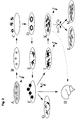
- FIG 1 is a schematic (flow chart) of the recovery of neural progenitor cells from ES cells.
- A shows ES cells (open circles) on a feeder cell lawn of embryonic fibroblasts (small rectangles). Following proliferation, the ES cells may be further processed or deep-frozen in liquid nitrogen.
- B shows ES cells in gelatin-coated feeder cell-free cell culture dishes.
- C shows embryoid bodies, in where an initial differentiation takes place, inter alia, in neural cells (black circles).
- D shows plated embryoid bodies in a cell culture dish containing ITSFn medium. This serum-free medium favors the survival of neural cells (black circles).
- E shows proliferated neural progenitor cells that continue to proliferate in a growth factor-containing serum-free medium (so-called N3FL medium) in the presence of bFGF.
- F shows proliferated neural precursor cells that proliferate further in another growth-fold containing serum-free medium (so-called N3EFL medium) in the presence of bFGF and EGF.
- G shows proliferated neural progenitor cells which proliferate further in a further growth factor-containing serum-free medium (so-called N2FP medium) in the presence of bFGF and PDGF. After 2 passages in N2FP medium, the cells can be used for remyelinating grafts or frozen for later use in serum free frozen medium.
- FIG. 2 is a schematic representation (a flow chart) of the recovery of neural spheroids and the cell populations derived therefrom from ES cells.
- a to E are identical to Figure 1.
- F shows N3FL cultures in uncoated cell culture dishes established neural spheroids, which proliferate in a further growth factor-containing serum-free medium (so-called N2EF medium) in the presence of bFGF and EGF and can also be cryopreserved.
- G shows the in vitro differentiation of such spheroids by growth factor deprivation, resulting in mature neurons, astrocytes and oligodendrocytes.
- H shows the transplantation of such spheroids into the nervous system.
- I shows the formation of glial progenitor cell populations in the form of so-called touch-down cultures.
- the spheroids obtained in F are grown in the presence of growth factors until they begin to attach to uncoated cell culture dishes and glial precursor cells from the spheroids migrate to the cell culture dish.
- K shows how, after removal of the adhered spheroids, the precursor cells which have migrated onto the cell culture dish are further proliferated in the presence of growth factors.
- the cells produced in I and K can be cryopreserved.
- L and M show how by adding CNTF or T3 and consecutive growth factor deprivation an astrocytic (L) or oligodendroglial (M) differentiation of glial precursor cells derived from ES cells can be induced.
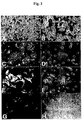
- Figure 3 shows cell cultures of neural progenitor cells after in vitro proliferation and differentiation of ES cells.
- a and B show phase-contrast micrographs of neural progenitor cells derived from J1-ES cells during proliferation in N2FP.
- C and D show oligodendroglial differentiation of N2FP cultures 4 days after withdrawal of the growth factors bFGF and PDGF. Numerous cells express the oligodendroglial marker antigen O4.
- C is an immunofluorescent image
- D is the corresponding phase contrast.
- E and F show astrocytic differentiation of N2FP cultures. Four days after growth factor withdrawal, numerous GFAP-positive astrocytes can be detected.
- E is an immunofluorescent image
- F is the corresponding phase contrast absorbance.
- G and H show neuronal differentiation in N2FP cultures. In addition to oligodendroglial and astrocytic cells, neurons can be detected after growth factor withdrawal. These have long processes and show a clear expression of the neuronal marker antigen ⁇ -III-tubulin.
- G is an immunofluorescent image
- H is the corresponding phase contrast.
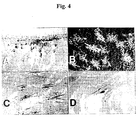
- FIG. 4 shows vibratome sections of a recipient brain after transplantation of glial precursor cells obtained from ES cells.
- the glial precursor cells differentiate into fetal rat brain in oligo- and astroglia after transplantation.
- the oligodendrocytes produced from ES cells myelinate the recipient brain.
- (A) shows astrocytes derived from the transplanted cells that migrate into the brain parenchyma after intraventricular transplantation into a fetal rat brain.
- N2FP cultures were injected as a single cell suspension into the ventricular system of 17-day-old rat embryos.
- the recipient animals were analyzed three weeks after birth. For the detection of the donor cells, an antibody against the mouse-specific glial antigen M2 was used.
- FIG. 1 At the top of the picture is the III. Recognize the ventricle.
- B shows incorporation of ES cell-derived glia into the cerebral cortex of a newborn rat. The cells had been implanted into embryonic day 16 in the recipient's ventricular system. The presentation was carried out by an immunofluorescence study with an antibody against the mouse-specific antigen M6. The cells have a characteristic astrocytic morphology.
- C shows incorporation of ES cell-derived oligodendrocytes into the lower inferior colliculus of a 22-day-old myeloid deficient rat, which had received an intraventricular injection of an N2FP culture on embryonic day 17.
- the donor cells have been identified by DNA in situ hybridization with a DNA probe molecule against mouse satellite DNA (black stained nuclei). The incorporated cells have begun to myelinate the recipient brain. Since myelin-deficient rats do not form immunoreactive PLP, myelination could be detected immunohistochemically with an antibody against PLP (black processes).
- (D) shows a close-up of an ES cell-derived myelinating oligodendrocyte after incorporation into the hypothalamus of a 22 day old recipient animal. One clearly recognizes the hybridized core and the PLP-positive extensions in a typical parallel arrangement.
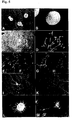
- Figure 5 shows cultures of neural spheroids derived from ES cells and cell populations derived from them.
- A shows 5-day-old neural spheroids derived from the ES cell line J1, such as those used for transplantation into the nervous system.
- B shows two-week-old J1-ES cell derived neural spheroids stained with an antibody to the intermediate filament nestin typically expressed in neural progenitor cells. At this time, the spheroids have reached a macroscopically visible size (immunofluorescence recording).
- C and D show 5-day-old neural spheroids derived from the ES cell line CJ7 seeded in polyornithine and fibronectin-coated cell culture dishes and cultured for a further 5 days without growth factors.
- the spheroids disintegrate and differentiate into neural cells.
- Shown in D is a double immunofluorescence study with antibodies to the neuronal antigen ⁇ -III-tubulin (brighter signal) and the intermediate filament nestin typical of neural progenitor cells (darker signal, arrows).
- the comparison with the corresponding phase-contrast image (C) reveals that each of the cells shown expresses at least one of these neural markers.
- E and F show a 5 day old neural spheroid culture differentiated from polyornithine and fibronectin in the absence of growth factors for 7 days, obtained from the ES cell line J1, stained with antibodies to the neuronal marker microtubule-associated protein 2 (MAP2, E) and the neurotransmitter GABA (F) (immunofluorescence images). It can be seen that many neurons express the neurotransmitter GABA. Cell populations which are rich in GABAergic cells can be used for the Transplantation of M. Huntington's patients can be used. G and H show cells of the same culture stained with antibodies to the neuronal marker microtubule-associated protein 2 (MAP2; G) and the neurotransmitter glutamate (immunofluorescence images).
- MAP2 neuronal marker microtubule-associated protein 2
- G neurotransmitter GABA
- BDNF Neurotrophin Brain-Derived Neurotrophic Factor
- K shows a 5-day-old neural spheroid culture stained for polyornithine and fibronectin in the absence of growth factors, obtained from the ES cell line J1, stained with an antibody to tyrosine hydroxylase. Tyrosine hydroxylase-positive neurons are used, for example, for the transplantation of Parkinson's patients. This immunofluorescence image shows that many of the ESC derived neurons produce this enzyme.
- L shows a 11-day-old neurogenic spheroid differentiated from polyornithine and fibronectin in the absence of growth factors for one week, obtained from the ES cell line J1, stained with an antibody against the astrocytic antigen Glial Fibrillary Acidic Protein (GFAP).
- GFAP Glial Fibrillary Acidic Protein
- Figure 6 shows glial precursor cells derived from neural spheroids.
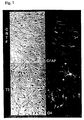
- A shows a typical example of a nascent so-called touch-down culture. Neural spheroids derived from the ES cell line J1 adhere to uncoated cell culture dishes, and many glial cells migrate out of the spheroids. The adhered spheroids can be easily solved by shaking the cell culture dish, with a pure glial precursor cell lawn remains.
- B shows an immunofluorescence study of a glial precursor cell population thus obtained from the ES cell line J1 with an antibody against the neural antigen A2B5. It can be seen that the glial precursor cells obtained express this neural antigen.
- Figure 7 shows the control of glial differentiation of glial precursor cells derived from ES cells by adding single factors.
- a so-called touch-down culture obtained from the ES cell line J1 which had been cultured in the presence of EGF and FGF, was additionally supplemented with the factors CNTF (10 ng / ml) or T3 (3 ng / ml).
- CNTF 10 ng / ml
- T3 3 ng / ml
- the figure which shows immunofluorescence and corresponding phase-contrast images, shows that the addition of CNTF or T3 to the precursor cells obtained from ES cells influences their differentiation.
- CNTF most of the cells express astrocytic morphology and express the astrocytic marker GFAP.
- T3 In the presence of T3, however, there are numerous cells with star-shaped, oligodendroglial morphology and expression of the oligodendroglial O4 O4.
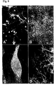
- Figure 8 shows the neuronal differentiation of neural spheroids derived from the ES cell line J1 in vitro (A and B) and after transplantation into the nervous system of a ibotenic acid-treated rat (an animal model for Huntington's Disease, C and D).
- 5-day-old neural spheroids were implanted into the striatum of an adult, ibotenic acid-treated rat.
- Part of the spheroids was parallel differentiated in vitro by growth factor withdrawal for 5 days and then stained with an antibody against the neurotransmitter GABA (A, immunofluorescence uptake, B, corresponding phase contrast uptake).
- C and D show a general view of a graft stained with an antibody against the mouse-specific neural antigen M6 (immunofluorescence).
- M6 mouse-specific neural antigen
- embryonic stem cells eg of the mouse
- ES cell lines for example the mouse embryonic stem cell lines J1 (Li et al., Cell 69: 915-926, 1992), R1 (Nagy et al., Proc. Natl.
- ES cells can also be obtained from embryos, for example from 3 to 4 day old mouse blastocysts (Hogan et al., Manipulating the Mouse Embryo, Cold Spring Harbor Press, New York, 1994). Furthermore, ES cells can also be used from other species. Typical examples of isolated ES cells include rat (Lannaconne et al., Dev. Biol. 163: 288-292, 1994), hamster (Doetschman et al., Dev. Biol.
- Feeder cell-dependent ES cells may then be used e.g. according to Okabe et al. seeded in gelatin-coated dishes and then aggregated in uncoated Petri dishes in the absence of LIF to embryoid bodies (Okabe et al., Mech. Dev. 59: 89-102, 1996). Three to four-day-old embryoid bodies can then be plated on cell culture dishes and cultured for 4-8 days in a serum-free medium (ITSFn medium) (Okabe et al., Mech. Dev. 59: 89-102, 1996). During this time, massive cell death occurs among non-neural cells.
- ITSFn medium serum-free medium
- the cells can be triturated into a single cell suspension (repeated aspiration and draining of liquid using a narrow-mouthed pipette) and transferred to N3FL medium. It is a serum-free medium to which bFGF is added (Okabe et al., Mech. Dev. 59: 89-102, 1996). bFGF has a proliferative effect on neural progenitor cells.
- N3FL medium After 4-8 days in N3FL medium, the actual delivery of glial and neuronal precursor cell populations begins in the form of a cell lawn. In contrast to neuronal cells, glial cells and especially their immediate precursors are characterized by a strong ability to proliferate.
- the method according to the invention utilizes this property.
- the N3FL cultures described above are sequentially transferred to various serum-free, growth factor-containing media. During this phase, there is a strong accumulation of bipotent oligo-astrocytic progenitor cells with simultaneous differentiation and elimination of primitive embryonic and non-neurally differentiated cells.
- the cells cultivated in N3FL medium are mechanically detached from the culture dish in a conventional manner, triturated in a single cell suspension and transferred into serum-free medium containing, for example, the growth factors bFGF and EGF (N3EFL medium).
- the cells are propagated to subconfluence as cell turf.
- the resulting cells can be used for transplant purposes after about two passages in this medium. For further purification, however, an additional step can be carried out.
- the cells cultured in N3EFL medium may also be redissolved in a conventional manner from the culture dish, triturated in a single cell suspension and replated in a serum-free medium containing a second growth factor combination, e.g. bFGF and PDGF.
- a second growth factor combination e.g. bFGF and PDGF.
- the purified neural progenitor cells thus obtained may be subjected to further passages in this medium. After about two passages, the population consists of purified neural progenitor cells, which can be used directly or preferably after isolation for eg remyelinating grafts. Microscopically, a homogeneous population of bipolar to star-shaped cells can be seen, which fill the culture dish in the form of a uniformly distributed cell lawn. At this, but also earlier, the neural progenitor cell population can be frozen in serum-free freezing medium and thawed at a later time in a conventional manner without losing its precursor cell properties. If the growth factors are not added for several days, ie withdrawn, there is a beginning differentiation in vitro .
- oligodendroglial (eg O4) and astrocytic (eg GFAP) antigens can be detected by immunohistochemistry.
- O4-positive cells can account for as much as 30-70% and GFAP-positive cells as 20-40% of the cell population.
- GFAP-positive cells can account for as much as 30-70% and GFAP-positive cells as 20-40% of the cell population.
- cells expressing neuronal antigens are found under these conditions.
- the cells cultivated in N3FL medium are mechanically detached from the culture dish in the usual way, triturated in a single cell suspension and cultured in uncoated cell culture dishes in serum-free medium, which is e.g. contains the growth factors bFGF and EGF.
- serum-free medium which is e.g. contains the growth factors bFGF and EGF.
- spheroidal cell aggregates are formed, which consist predominantly of nestin-positive neural progenitor cells.
- the neural spheroids can be propagated free-floating in the cell culture supernatant. Differentiated neural and non-neural cells, on the other hand, show a strong tendency to adhere to the surface of the cell culture dishes.
- small spheroids Once small spheroids have formed, they are removed from the culture and transferred to new uncoated cell culture dishes. Such spheroids can be used after a few (5-7) days for transplantation purposes.
- the neural progenitor cells contained in the spheroids differentiate into mature neural cells, which innervate the recipient brain. Undifferentiated small spheroids can be frozen in serum-free freezing medium in liquid nitrogen and later thawed and differentiated.
- Another object of the invention are differentiated neural cells in the form of spheroids, which are suitable for transplantation. They can be prepared by in vitro induction of differentiation of precursor cells according to the invention having neuronal or glial properties. For this purpose, the growth factors are removed and the spheroids are seeded with the undifferentiated neural progenitor cells in, for example, polyornithine and fibronectin-coated cell culture dishes. The spheroids then attach rapidly to the surface of the cell culture dish, forming neurons, astrocytes and oligodendrocytes in addition to nestin-positive neural progenitor cells.
- the adherent neurons can be further matured and then express a variety of neuronal markers, such as MAP2, ⁇ -III tubulin, synapsin, choline acetyltransferase, tyrosine hydroxylase, GABA, glutamate, serotonin, peripherin and calbindin.
- neurotrophins such as Brain-Derived Neurotrophic Factor (BDNF) or Neurotrophin-3 (NT-3) to the cell culture medium.
- BDNF Brain-Derived Neurotrophic Factor
- NT-3 Neurotrophin-3
- the spheroids may be up to several weeks in serum-free medium containing e.g. containing the growth factors bFGF and EGF, are kept in suspension culture.
- the spheroids may increase in size so far that they are easily recognizable macroscopically.
- increasing cell differentiation occurs.
- Such differentiation of ES cells into free-flowing spheroids makes it physically possible to transplant neurons derived from ES cells also in a more differentiated state, e.g. in the form of such spheroids. This is not possible after differentiation of neurons adhering to a culture dish, since the numerous appendages adhering to the surface are usually damaged during detachment and the neurons are thereby destroyed.
- the spheroids are maintained in growth-factor-containing serum-free medium in suspension culture until they begin to adhere easily to the uncoated surface of the cell culture dish.
- growth factors such as bFGF and EGF (alone and in combination).
- bFGF and EGF alone and in combination.
- cells of glial morphology migrate from the spheroids to the cell culture plates (so-called touch-down cells).
- touch-down cells The genesis of this cell population is unclear. It is however to be assumed that in the Over the course of increasing cell differentiation within the spheroids, glial progenitor cells are formed, which are characterized by a stronger adhesion and migration behavior.
- Such touch-down cell-producing spheroids can be used as generators for glial precursor cells.
- the spheroids are cultured for a short time ( ⁇ 1 day) in uncoated cell culture dishes. Once glial cells have settled, the spheroids are mechanically released from the shell and transferred to a new shell. What remains behind in such cases is a ring of glial progenitor cells, which can be further propagated in the presence of growth factors, for example, bFGF and EGF, alone or in combination.
- Such “touch-down cells” show expression of the neural antigen A2B5 (Eisenbarth et al., Proc Natl Acad Sci., USA 76: 4913-4917, 1979) and differentiate into growth factor deprivation in astrocytes and oligodendrocytes. Immunohistochemically, marker antigens for oligodendrocytes (e.g., O4) and astrocytes (e.g., GFAP) can then be detected. Undifferentiated “touch-down cells” can be frozen in serum-free freezing medium in liquid nitrogen without losing their proliferation and differentiation properties. Such glial progenitor cells can also be used for transplantation into the nervous system
- the differentiation of the glial precursor cells obtained from the ES cells can be influenced by the addition of individual factors. Addition of CNTF (ciliary neurotrophic factor) shortly before and during growth factor withdrawal leads to predominantly astrocytic differentiation. Addition of the thyroid hormone T3 during this phase leads to an increased differentiation in oligodendrocytes. The addition of serum-containing medium during or after growth factor treatment results in a large increase in the number of astrocytic toes in these cultures.
- CNTF ciliary neurotrophic factor
- the frozen and thawed neural progenitor cells can also be used for grafts.
- Previously frozen neural progenitor cell populations are subjected to the appropriate passages after the usual thawing to obtain the homogeneous population of bipolar to star-shaped cells.
- the method according to the invention can furthermore be combined with known cell separation and sorting methods.
- neural subpopulations may be separated at certain times in the method of the invention by immunofluorescent cell sorting (FACS), immunopanning or similar methods.
- FACS immunofluorescent cell sorting
- Detailed sorting and subclassification may allow in vitro generation of matched (and possibly genetically modified) neural replacement cells. Since both ES cells and the neural progenitor cells of the invention obtained therefrom can be frozen and thawed without loss of their properties, it is possible to construct corresponding cell banks, for example, of autologous cell banks.
- the neural progenitor cells obtainable by the method according to the invention can be produced in an amount and purity as required, for example, for the transplantation and repair of defects in the nervous system.
- the neural progenitor cells of the invention obtainable by the method according to the invention contain, for example, only small to no amounts of, for example, primitive embryonic and non-neural cells.
- the purity of the neural precursor cells of the present invention is well above the purity of about 85% as measured by the method of Okabe et al. (Mech. Dev. 59: 89-102, 1996). With the method according to the invention neural precursor cells can be obtained with a purity of up to 100%.
- the neural progenitor cells according to the invention can be obtained from ES cells, for example from the mouse, rat, hamster, bird, fish, pig, cattle, primate or human.
- the ES cells may be present as an ES cell line or recovered from embryos.
- the ES cells can be obtained by proliferation of oocytes.
- the oocytes may be gutted and injected with a nucleus of eg differentiated tissue so that autologous oocytes are used to obtain ES cells.
- ES cells or ES cell-like cells can also be obtained from embryonic germ cells .
- the ES cells can also be genetically modified in the usual way. For example, homologous recombination can replace a defective gene with a "normal" gene become. Furthermore, defective genes can be deleted in the usual way. These methods have been widely used in the mouse, for example, and are state of the art.
- this can be used, for example, to colonize large sections of the nervous system with genetically modified progenitor cells which substitute locally missing substances or secrete polypeptides with protective or other action.
- the genetic modifiability of the cells can also be used to eliminate the expression of rejection-causing surface antigens by elimination of the corresponding genes, thus allowing a broad clinical use of ES cell-derived progenitor cells without immunosuppression.
- the neural progenitor cells of the invention can be used as medical work tools for the therapy of neural defects.
- a typical example of using the neural progenitor cells of the present invention is the reconstitution of functionally deficient or lost neurons by transplanted neural progenitor cells.
- the spheroids of the invention are implanted into brain cultures after 4-7 days in suspension culture, for example, which have a loss of neurons.
- mature neurons can be observed 6 weeks after surgery, which innervate the recipient brain.
- the axons emanating from the transplanted neurons and sprouting into the recipient's brain tissue may be identified with antibodies to donor-specific neural antigens. This reconstitution of neuronal cells is functional. This can be demonstrated, for example, by behavioral tests on ibotenic acid-treated rats before and after transplantation.
- the rat ibotenic acid lesion model and the functional capacity of neural grafts via an analysis of the rotational behavior of the animals are state of the art (Björklund et al., From: Functional Neural Transplantation, pages 157-195, Raven Press, New York, 1994).
- the neural spheroids according to the invention are implanted in the brains of ibotenic acid-treated rats, a marked improvement in the abnormal rotational behavior of the recipients caused by the ibotic acid lesion occurs postoperatively. Transplantation of, for example, neural cells into the human nervous system is already clinically useful today (Olanow et al., TINS 19: 102-109, 1996).
- the glial progenitor cells of this invention can also be used as medical devices for the therapy of neural defects.
- a typical example of using the glial progenitor cells of the invention is the remyelination of demyelinated brain sections by transplanted neural progenitor cells. Since demyelinating diseases often involve large sections of the central nervous system (CNS), the glial precursor cells according to the invention, which are characterized by pronounced migration behavior, can be transplanted into the CNS. A localized injection may be sufficient to infiltrate large sections of the CNS with these cells and to achieve large-scale remyelination.
- a typical example of a potentially treatable disease is multiple sclerosis (MS).
- the transplantation of the neural progenitor cells according to the invention can bring about a remyelination of such defects.
- the pronounced migration behavior of the neural progenitor cells according to the invention can be exploited in such a way that numerous demyelinated areas can be reached and repaired from one or a few implantation sites.
- cells of the invention still under growth factor treatment derived from monolayers (cell lawns) or neural spheroids, e.g. detached from the culture dishes trypsin-free and triturated to a single cell suspension.
- growth factor treatment derived from monolayers (cell lawns) or neural spheroids
- oligodendrocytes and astrocytes derived from the implanted cells can be detected approximately three weeks after implantation.
- the myelinization of the nerve cell processes of the recipient can be demonstrated by an identification of these processes starting from the donor cells, wherein proteins characteristic of myelin can be detected immunohistochemically in e.g. myelin basic protein (MBP) and proteolipid protein (PLP).
- MBP myelin basic protein
- PGP proteolipid protein
- neural progenitor cells of the invention may also be used for the in vitro production of clinically and commercially useful factors such as polypeptides.
- J1 ES cells (Li et al., Cell 69: 915-926, 1992) were first visualized on a lawn of mitotically inactivated embryonic fibroblasts in DMEM medium (Life Technologies # 11965) with 20% fetal calf serum (FBS, Life Technologies No. 10119) in the presence of 1000 U / ml LIF (Life Technologies No. 13275) by standard methods (Hogan et al., Manipulating the Mouse Embryo, Cold Spring Harbor Press, New York, 1994). In addition to FBS and LIF, this medium contained standard concentrations (Okabe et al., Mech. Dev.
- the culture was rinsed once with 0.04% EDTA in PBS buffer and the cells were rinsed by adding a solution of 0.05% trypsin (Life Technologies # 35400) and 0.04% EDTA replaced in PBS.
- the cells were then triturated several times through a Pasteur pipette to a single cell suspension and the trypsin was neutralized by adding serum-containing medium. In this case, an equivalent amount of the previously used medium was added to the trypsin solution. After centrifugation (5 min, 300 ⁇ g, RT), the cells in the previously used medium were plated out on 6 cm cell culture dishes coated with 0.2% gelatin (Sigma No. G-2500) (approximately 6 ⁇ 10 6 cells per 6 cm Shell) and in the presence of LIF (1000 U / ml).
- the cells were added by adding a solution of 0.05% trypsin and 0.04% EDTA in PBS (about 1.5 ml per 6 cm dish) of the Gelatine detached.
- the trypsin was neutralized by addition of DMEM medium with 10% FBS (about 6.5 ml per 6 cm dish, this medium corresponds to the medium described above, but contains only 10% FBS and no LIF).
- This suspension was then distributed to 8 uncoated bacterial culture Petri dishes (Nunc no. 240045) and further cultured in a total volume of about 4 ml DMEM / 10% FBS per 6 cm dish.
- the embryoid bodies formed were collected by simple (1 x g) sedimentation for 5 min in a 50 ml cell culture tube. The supernatant was discarded and the embryoid bodies were distributed on 10 cm cell culture dishes so that after lowering the embryoid bodies about 50% of the surface of the cell culture dish were covered (embryoid bodies from about 4-6 bacteria culture dishes were usually sufficient for the loading of a 10 cm Cell culture dish). The embryoid bodies were cultured overnight in DMEM / 10% FBS (10 ml per 10 cm dish).
- ITSFn medium consists of DMEM / F12. 5 ⁇ g / ml insulin (Intergen No. 540101), 50 ⁇ g / ml apo-transferrin (Intergen No. 445275), 30 nM selenium chloride (Sigma No. S-5261), 5 ⁇ g / ml fibronectin (Life Technologies No.
- the cells cultured in ITSFn were detached from the culture dish by means of a solution of 0.05% trypsin and 0.04% EDTA in PBS and the trypsin neutralized by the addition of an equivalent amount of serum-containing medium (DMEM / 10% FBS). After sedimentation (5 min, 300 xg, RT), the cells were resuspended in calcium and magnesium-free Hanks Buffered Salt Solution (CMF-HBSS, Life Technologies No. 14180) with 0.1% DNase (Worthington No. 2139) ( about 3 ml on a pellet obtained from a 10 cm dish) and triturated with the aid of Pasteur pipettes to form a single-cell suspension.
- CMF-HBSS calcium and magnesium-free Hanks Buffered Salt Solution
- the cells were then centrifuged (300xg, 5 min, RT) and seeded at a density of about 30,000 cells / cm 2 on polyornithine coated cell culture dishes in the following medium: DMEM / F12 (1: 1, Life Technologies # 11320 ), 25 ⁇ g / ml insulin, 50 ⁇ g / ml human apo-transferrin, 20 nM progesterone (Sigma No. P-8783), 100 ⁇ M putrescine (Sigma No.
- the cells were mechanically detached from the culture dish under trypsin-free conditions.
- the culture was rinsed three times with CMF-HBSS, the last rinse being added to 0.1% DNase.
- the cells were then scraped from the plate with a cell lifter (Costar # 3008) and triturated to a single cell suspension using the flame-polished Pasteur pipettes in the mode described above. After centrifugation (300 ⁇ g, 5 min, RT), the yield of a 10 cm cell culture dish was plated in approximately 5 previously polyornithine-coated cell culture dishes in N3EFL medium.
- DMEM / F12 (1: 1), 25 ⁇ g / ml insulin, 100 ⁇ g / ml transferrin, 20 nM progesterone, 100 ⁇ M putrescine, 30 nM selenium chloride, 1 ⁇ g / ml laminin, penicillin / streptomycin ( 100 IU / ml / 100 ⁇ g / ml), 10 ng / ml human recombinant bFGF and 20 ng / ml human recombinant EGF (R & D Systems # 236-EG), bFGF and EGF were added daily to a final concentration of 10 ng / ml for bFGF and 20 ng / ml added for EGF. The medium was replaced every other day with laminin not added after the second medium change.
- the cells were again mechanically detached from the culture dish under trypsin-free conditions with a cell lifter and triturated into a single cell suspension using flame-polished Pasteur pipettes. After centrifugation (300 xg, 5 min, RT), the yield of a 10 cm cell culture dish was plated in approximately 5 cell culture dishes previously coated with polyornithine in N2FP medium. This was composed as follows: DMEM / F12 (1: 1), 25 ⁇ g / ml insulin, 100 ⁇ g / ml transferrin.
- the cells were passaged trypsin-free in the mode described above 1: 5. A trituration of the cells in DNase-containing buffer solution was here usually unnecessary.
- the population consisted of neural progenitor cells as could be used for remyelinating grafts. Alternatively, at this stage the cells could be trypsin-free detached from the culture dish and deep-frozen in serum-free freezing medium (Sigma # C-6295) for later use.
- the purified neural progenitor cells in the medium can be isolated and placed in an injection medium, e.g. in calcium and magnesium-free Hanks Buffered Salt Solution (CMF-HBSS, Life Technologies # 14180).
- CMF-HBSS calcium and magnesium-free Hanks Buffered Salt Solution
- Example 3 Obtaining neural spheroids and neural progenitor cells established therefrom
- Example 1.6 After 4-5 days in the N3FI medium mentioned in Example 1.6, the cells were mechanically detached from the culture dish under trypsin-free conditions. This was the culture rinsed three times with CMF-HBSS, adding 0.1% DNase to the last rinse. The cells were then scraped from the plate with a cell lifter (Costar # 3008) and triturated to a single cell suspension using the flame-polished Pasteur pipettes in the mode described above.
- the cells were seeded in a density of 1200-12,000 cells / cm 2 in uncoated cell culture dishes in the following medium: DMEM / F12 (1: 1), 25 ⁇ g / ml insulin, 100 ⁇ g / ml transferrin, 20 nM progesterone, 100 ⁇ M putrescine, 30 nM selenium chloride, penicillin / streptomycin (100 IU / ml / 100 ⁇ g / ml), 20 ng / ml human recombinant bFGF and 20 ng / ml human recombinant EGF (R & D Systems No 236-EG), bFGF and EGF were added daily to a final concentration of 20 ng / ml and the medium (so-called N2EF medium) was renewed every two days.
- DMEM / F12 1: 1
- 25 ⁇ g / ml insulin 100 ⁇ g / ml transferrin
- the spent medium was withdrawn and centrifuged at 150 xg for 3 min at RT to sediment any floating in the medium spheroids.
- the pellet was resuspended in fresh medium and seeded again. After the second medium change, the cells were seeded in fresh uncoated cell culture dishes.
- Example 3.1 Five-day-old neural spheroids obtained according to Example 3.1 were centrifuged at 150 ⁇ g for 3 min at RT and seeded in the medium mentioned in Example 3.1, but without bFGF and EGF, in polyulturithin and fibronectin-coated cell culture dishes.
- the plates were first coated with polyornithine in the manner described in Example 1.6 and rinsed three times with PBS. The dishes were then charged with a solution of 1 ⁇ g fibronectin / ml PBS and incubated at RT for 2-12 hours. The fibronectin solution was withdrawn and seeded the cell suspension in the thus coated dish.
- neural spheroids had attached themselves to the surface of the cell culture dish.
- the medium was withdrawn, the dish washed three times with fresh medium or CMF-HBSS, and filled with new medium, which was subsequently replaced every two days.
- Such differentiated neural spheroid cultures were fixed 5 days after plating and subjected to immunofluorescence analysis.
- the expression of neural antigens distributed as follows (mean number of colored cells ⁇ SEM): nestin-positive neural progenitor cells 66 ⁇ 3% (antibody from M.
- GABA neurotransmitters of Sigma, # A-2052, 1: 700 dilution
- glutamate Sigma antibody, # G-6642, 1: 700 dilution
- the proportion of GABA-positive neurons was up to 60% in some preparations.
- tyrosine hydroxylase Eugene antibody, # TE101, 1: 200 dilution
- choline acetyltransferase was additionally immunoreacted in the spheroid neurons (Antibody of Chemicon, No. MAB305, dilution 1: 250), serotonin (antibody of Eugene, No. NT102, dilution 1: 200), Synapsin (antibody of Dr. MB Kennedy, Pasadena, CA, USA, dilution 1: 1,000 ), Peripherin (Chemicon antibody, No.
- Nerve cell differentiation and survival were achieved by addition of the neurotrophins Brain-Derived Neurotrophic Factor (BDNF; 20 ng / ml, Pepro Tech Inc. No. 450-02) and / or Neurotrophin 3 (NT-3, Pepro Tech Inc. No 450-03).
- BDNF Brain-Derived Neurotrophic Factor
- NT-3 Neurotrophin 3
- spheroid cultures differentiated over a period of 10 days and more, the oligodendrocytes were further matured, in which cyclic nucleotide 3'-phosphodiesterase (CNPase, antibody from Sigma, No. C-5922 , Dilution 1: 200).
- CNPase cyclic nucleotide 3'-phosphodiesterase
- the spheroids obtained according to Example 3.1 were cultured in the growth factor-containing medium mentioned under Example 3.1 until they began to spontaneously adhere to the uncoated surface of the cell culture dishes and cells from the spheroids had migrated to the surface of the cell culture dish. This was usually the case after a period of 10-14 days. As soon as a "ring" of emigrated cells became visible, those of the cell culture dish became very loose adhering spheroids by shaking the shell detached and removed by means of a pipette from the culture. The spheroids thus removed could be transferred into further uncoated cell culture dishes and further cultured in the same medium, again producing "rings" of growing glial progenitor cells.
- glial progenitor cells were further proliferated in the growth factor-containing medium and after reaching about 80% confluency trypsin-free in the mode described above 1: 5 passaged.
- glial precursor cells showed in immunofluorescence studies a clear expression of the neural antigen A2B5 (antibody from Boehringer, No. 1300 016, dilution 1: 200). They could be cryopreserved in serum-free freezing medium (Sigma # C-6295) in liquid nitrogen and further proliferated, transplanted or differentiated at a later time.
- O4-positive cells with oligodendroglial morphology ⁇ 1% GFAP-positive cells ⁇ 1% Second O4-positive cells with oligodendroglial morphology: 8.2 ⁇ 2.6% GFAP-positive cells 28 ⁇ 6% Third O4-positive cells with oligodendroglial morphology: 2.6 ⁇ 1.8% GFAP-positive cells 78 ⁇ 2.5% 4th O4-positive cells with oligodendroglial morphology: 17 ⁇ 3.6% GFAP-positive cells 39 ⁇ 5.7%
- Example 4 Transplantation of oligodendroglial / astrocytic precursor cells
- the remyelinating potency of these cells was checked by the following transplantation experiment: Subconfluent cultures from Example 2.3, which had received bFGF and PDGF up to the day before transplantation, were detached from the culture dish trypsin-free (with a cell lifter) at 300 ⁇ g for 5 min sedimented and resuspended in CMF-HBSS with 0.1% DNase. The cells were then triturated with flame polished pasteur pipettes to a single cell suspension, counted in a counting chamber and sedimented again.
- transplant recipients used were embryonic myelindeficient Sprague-Dawley rats (Ian Duncan, Department of Medical Sciences, School of Veterinary Medicine, University of Wisconsin-Madison, 2015 Linden Drive West, Madison, Wisconsin 53706, USA).
- the myelin-deficient recipient animals have the advantage that myelin formed by the donor cells is easily detectable.
- the xenotransplantation itself has the advantage that the transplanted cells can be detected easily and with high reliability with the aid of species-specific DNA samples (Brüstle et al., Neuron 15: 1275-1285, 1995).
- the technique of intrauterine transplantation into the embryonic brain is known in the art (Brüstle et al., Neuron 15: 1275-1285, 1995; Brüstle et al., From Current Protocols in Neuroscience, John Wiley, New York, 1997).
- the glass capillary was removed and the abdominal wall was surgically closed after injection of several or all embryos. Such animals are born spontaneously. Because it is an X-linked recessive disease, on average 50% of male offspring are myelin-deficient. These animals begin to shake severely in the third week of life and usually die in the fourth week of life.
- the animals were anesthetized in the usual way in the third to fourth week of life and fixed in a conventional manner by transcardiac perfusion with a 4% paraformaldehyde solution (Sigma No. P-6148) in PBS.
- the brains were prepared in the usual way and postfixed overnight in the fixation solution at 4 ° C. Subsequently, 50 ⁇ m thick sections were made on a vibratome.
- the donor cells were detected by DNA in situ hybridization with a DNA probe motif for mouse satellite DNA (Brüstle et al., Neuron 15: 1275-1285, 1995). Myelin formed by the donor cells was detected by immunohistochemical detection of myelin proteins in hybridized cell extensions.
- Hybridized cells were found, inter alia, in the cortex, hippocampus, septum, striatum, olfactory bulb, thalamus, hypothalamus, tectum, cerebellum, corpus callosum, in the anterior commissure and in the optic tract and within the optic nerve. After transplantation, no space-occupying accumulations of donor cells could be detected in the ventricular system of the recipient animals. Out of 35 operated embryos, 11 male offspring showed symptoms of myeloid deficiency. Of these, 8 had been successfully transplanted intraventricularly. In 6 of these 8 animals a myelination originating from the donor cells could be detected. For this purpose, a double-labeling of hybridized cells with the PLP or MBP antibody was used.
- astrocytes derived from the ES cells could also be detected. These were immunohistochemically labeled with antibodies to the mouse-specific antigens M2 (Zhou et al., J. Comp. Neurol. 292: 320-330, 1990) and M6 (Lund et al., Neurosci. Lett. 61: 221-226, 1985) or by immunohistochemical double-labeling of cells labeled with the mouse-specific DNA probe with an antibody against glial glial fiber protein (GFAP, ICN No. 69-110).
- M2 Zahou et al., J. Comp. Neurol. 292: 320-330, 1990
- M6 Long et al., Neurosci. Lett. 61: 221-226, 1985
- GFAP glial glial fiber protein
- 5-day-old neural spheroids were obtained from the ES cell line J1 in the striatum of adult Sprague-Dawley rats according to Example 3.1, in which previously by stereotactic injection of the neurotoxin ibotenic acid, a one-sided neuronal degeneration in the striatum had been induced.
- the striatal ibotenic acid lesion is prior art and the lesion protocol used published (Brüstle et al., From: Current Protocols in Neuroscience, Unit 3.10, John Wiley, New York, 1997).
- the medium and the spheroids floating therein were removed from the cell culture dish and transferred to a 50 ml plastic tube. After addition of DNase to a total concentration of 0.1%, the spheroids were sedimented at 150 xg for 5 min at RT and then in CMF-HBSS containing 2 g / L glucose (Sigma No. G-7021) in a 0.5 ml plastic reaction tube resuspended. The resuspended spheroids were stored on ice until transplantation (up to 4 hours). The recipient animals were anesthetized in a conventional manner and the spheroids were introduced into the striatum via a stereotactic injection.
- a glass capillary with an opening of about 0.25 - 0.75 mm was used.
- the transplantation protocol used is prior art and has been extensively published (Brüstle et al., From: Current Protocols in Neuroscience, Unit 3.10, John Wiley, New York, 1997). A total of 6 animals were transplanted.
- the spheroids accumulated at the bottom of the 0.5 ml tube were drawn into the glass capillary and injected into the striatum using a stereotactic frame (Stoelting No. 51600).
- the following stereotactic data were used:
- Place 1 Anterior Holder: - 2.3 mm
- Anteroposterior alignment + 0.2 mm (relative to bregma)
- Mediolateral orientation 3.0 mm (relative to sagittal seam) injection depth 5.5 mm (relative to the dura)
- Place 2 Anterior Holder: - 2.3 mm
- Anteroposterior alignment + 0.2 mm (relative to bregma)
- Mediolateral orientation 3.0 mm (relative to sagittal seam) injection depth 4.0 mm (relative to the dura)
- Place 1 Anterior Holder: - 2.3 mm Anteroposterior alignment: + 0.2 mm (relative to bregma) Mediolateral orientation: 3.0 mm (relative to sagittal seam) injection depth 6.5 mm (relative to the dura)
- Place 2 Anterior Holder: - 2.3 mm Anteroposterior alignment: + 0.2 mm (relative to bregma) Mediolateral orientation: 3.0 mm (relative to sagittal seam) injection depth 5.3 mm (relative to the dura)
- Place 3 Anterior Holder: - 2.3 mm Anteroposterior alignment: + 0.2 mm (relative to bregma) Mediolateral orientation: 3.0 mm (relative to sagittal seam) injection depth 4.0 mm (relative to the dura)
- Place 4 Anterior Holder: - 2.3 mm Anteroposterior alignment: + 1.5 mm (relative
- the animals were immunosuppressed.
- the recipient animals received daily, starting one day before transplantation, an intraperitoneal injection of 20 mg cyclosporin A per kg body weight (Sandimmun, Fa. Sandoz).
- tetracycline was added to the drinking water during the immunosuppressive treatment (Achromycin, Lederle, final concentration 100 mg / L).
- the animals were intraperitoneally injected with a dose of 5 mg D-amphetamine sulfate (Sigma No. A-3278) per kg body weight. After a 5 minute exposure time, the number of triggered rotations per minute was measured over 90 minutes and averaged.
- the measurement of amphetamine-induced rotational behavior as a parameter for the function of striatal grafts is state of the art and published (Brüstle et al., From: Current Protocols in Neuroscience, Unit 3.10, John Wiley, New York. 1997). The data show that 3 of the 4 animals examined showed a marked reduction in rotational behavior.
- the animals were anesthetized 5-9 weeks after transplantation and fixed in a conventional manner by transcardiac perfusion with a 4% paraformaldehyde solution in PBS.
- the brains were prepared in the usual way and postfixed overnight in the fixation solution at 4 ° C. Subsequently, 50 ⁇ m sections were made on a vibratome.
- the donor cells were detected by DNA in situ hybridization with a probe for mouse satellite DNA (Hörz & Altenburger, Nucl. Acids Res. 9: 683-696, 1981; Brüstle et al., Neuron 15: 1275-1285, 1995).
- hybridized cells were found in the implantation area. In addition, part of the hybridized cells had migrated from the implantation area into neighboring brain sections, especially into the corpus callosum. The animal 200 had a very large lateral ventricle and an intraventricular localized graft. Apparently, the previous ibotic acid lesion produced severe atrophy of the striatum with consecutive ventricular dilatation, and the subsequently implanted cells landed in the lateral ventricle rather than in the striatum. The incorrect position in the lateral ventricle also explains the lack of functional improvement of this recipient animal in the rotational tests.
- the transplanted cells exhibited marked expression of the mouse-specific neural antigen M6.
- This antigen is particularly expressed on axons (Lund et al., Neurosci. Lett. 61: 221-226, 1985; antibodies from C. Lagenaur, Department of Neurobiology, University of Pittsburgh School of Medicine, 818A Scaife Hall, Pittsburgh, Pennsylvania 15261 , USA, 1:10 dilution).
- numerous M6-positive axons could be seen, which emanated from the graft and were sprouted into the surrounding brain tissue. This indicates that the nerve cells resulting from the transplanted spheroids innervate the recipient brain.
Landscapes
- Health & Medical Sciences (AREA)
- Engineering & Computer Science (AREA)
- Biomedical Technology (AREA)
- Life Sciences & Earth Sciences (AREA)
- Bioinformatics & Cheminformatics (AREA)
- Organic Chemistry (AREA)
- Chemical & Material Sciences (AREA)
- Wood Science & Technology (AREA)
- Zoology (AREA)
- Biotechnology (AREA)
- Genetics & Genomics (AREA)
- Neurosurgery (AREA)
- Neurology (AREA)
- General Health & Medical Sciences (AREA)
- General Engineering & Computer Science (AREA)
- Biochemistry (AREA)
- Microbiology (AREA)
- Cell Biology (AREA)
- Developmental Biology & Embryology (AREA)
- General Chemical & Material Sciences (AREA)
- Chemical Kinetics & Catalysis (AREA)
- Public Health (AREA)
- Animal Behavior & Ethology (AREA)
- Pharmacology & Pharmacy (AREA)
- Nuclear Medicine, Radiotherapy & Molecular Imaging (AREA)
- Medicinal Chemistry (AREA)
- Veterinary Medicine (AREA)
- Micro-Organisms Or Cultivation Processes Thereof (AREA)
- Medicines Containing Material From Animals Or Micro-Organisms (AREA)
- Preparation Of Compounds By Using Micro-Organisms (AREA)
- Medicines That Contain Protein Lipid Enzymes And Other Medicines (AREA)
- Pharmaceuticals Containing Other Organic And Inorganic Compounds (AREA)
- Peptides Or Proteins (AREA)
Claims (32)
- Composition cellulaire non-tumorigène, qui est issue de cellules souches embryonnaires de mammifères, contenanta) au moins 85 % de cellules précurseurs nerveuses isolées ayant une aptitude à évoluer en des cellules neuronales ou gliales etb) au plus 15 % de cellules embryonnaires et non-nerveuses primitives, pouvant être obtenues par les étapes suivantes:(a) prolifération de cellules ES,(b) culture des cellules ES de l'étape (a) pour donner des cellules précurseurs nerveuses,(c) prolifération des cellules précurseurs nerveuses dans un milieu exempt de sérum, contenant un facteur de croissance,(d) prolifération des cellules précurseurs nerveuses de l'étape (c) dans un autre milieu exempt de sérum, contenant un facteur de croissance, et isolement des cellules précurseurs nerveuses purifiées, et(e) prolifération des cellules précurseurs nerveuses de l'étape (d) dans un autre milieu exempt de sérum, contenant un facteur de croissance, et isolement des cellules précurseurs purifiées ayant l'aptitude à évoluer en des cellules neuronales ou gliales,
tandis que le milieu exempt de sérum, contenant un facteur de croissance, dans l'étape (d) comporte du bFGF et du EGF, et tandis que le milieu exempt de sérum, contenant un facteur de croissance, dans l'étape (e) comporte du bFGF et du PDGF, avec pour condition que le procédé ne comporte pas la destruction d'embryons humains. - Composition cellulaire selon la revendication 1, dans laquelle les cellules ES dans l'étape (a) sont mises à proliférer en des agrégats de cellules, en particulier en des corps embryoïdes.
- Composition cellulaire non-tumorigène, qui provient de cellules souches embryonnaires de mammifères, contenanta) au moins 85 % de cellules précurseurs nerveuses isolées ayant une aptitude à évoluer en des cellules neuronales ou gliales etb) au plus 15 % de cellules embryonnaires et non-nerveuses primitives, pouvant être obtenues par les étapes suivantes:(a') prolifération de cellules ES,(b') culture des cellules ES de l'étape (a') pour donner des cellules précurseurs nerveuses,(c') prolifération des cellules précurseurs nerveuses dans un milieu exempt de sérum, contenant un facteur de croissance,(d') prolifération des cellules précurseurs nerveuses de l'étape (c') dans un autre milieu exempt de sérum, contenant un facteur de croissance pour former des sphéroïdes nerveux, et isolement des sphéroïdes nerveux, et(e') prolifération des sphéroïdes nerveux de l'étape (d') dans un milieu exempt de sérum, contenant un facteur de croissance, jusqu'à formation d'un amas de cellules consistant en des cellules précurseurs gliales et isolement des cellules précurseurs purifiées ayant l'aptitude à évoluer en des cellules gliales,
tandis que le milieu exempt de sérum, contenant un facteur de croissance, dans l'étape (d'), (e') comporte du bFGF et/ou du EGF, avec pour condition que le procédé ne comporte pas la destruction d'embryons humains. - Composition cellulaire selon la revendication 3, dans laquelle les cellules ES dans l'étape (a') sont mises à proliférer en des agrégats de cellules, en particulier en des corps embryoïdes.
- Composition cellulaire selon l'une des revendications 1 à 4 sous la forme d'un amas de cellules.
- Composition cellulaire selon l'une des revendications 1 à 4 sous la forme de sphéroïdes.
- Composition cellulaire selon l'une des revendications 1 à 4, comportant des cellules ayant l'aptitude à évoluer en des cellules neuronales, astrocytaires et/ ou oligodendrogliales.
- Composition cellulaire selon l'une des revendications 1 à 4, dans laquelle les cellules souches embryonnaires ont été obtenues à partir de cellules germinales embryonnaires.
- Composition cellulaire selon l'une des revendications 1 à 4, dans laquelle les cellules ont été isolées à partir du groupe comprenant les souris, rats, hamsters, moutons, porcs, primates ou humains.
- Composition cellulaire selon l'une des revendications 1 à 7, dans laquelle les cellules précurseurs ont été modifiées par génie génétique.
- Composition cellulaire selon l'une des revendications 1 à 10, sous forme congelée.
- Bibliothèque de cellules, comprenant des cellules autologues et non-autologues selon l'une des revendications 1 à 11.
- Sphéroïdes, comprenant des cellules nerveuses différentiées des cellules précurseurs selon l'une des revendications 1 à 4.
- Sphéroïdes selon la revendication 13, obtenus par induction à la différenciation des cellules précurseurs selon l'une des revendications 1 à 5 in vitro.
- Procédé pour la préparation de cellules précurseurs nerveuses purifiées, ayant l'aptitude à évoluer en des cellules neuronales ou gliales, comportant les étapes suivantes:(a) prolifération de cellules ES,(b) culture des cellules ES de l'étape (a) pour donner des cellules précurseurs nerveuses,(c) prolifération des cellules précurseurs nerveuses dans un milieu exempt de sérum, contenant un facteur de croissance,(d) prolifération des cellules précurseurs nerveuses de l'étape (c) dans un autre milieu exempt de sérum, contenant un facteur de croissance, et isolement des cellules précurseurs nerveuses purifiées, et(e) prolifération des cellules précurseurs nerveuses de l'étape (d) dans un autre milieu exempt de sérum, contenant un facteur de croissance, et isolement des cellules précurseurs purifiées ayant l'aptitude à évoluer en des cellules neuronales ou gliales,
tandis que le milieu exempt de sérum, contenant un facteur de croissance, dans l'étape (d) comporte du bFGF et du EGF, et tandis que le milieu exempt de sérum, contenant du facteur de croissance, dans l'étape (e) comporte du bFGF et du PDGF, avec pour condition que le procédé ne comporte pas la destruction d'embryons humains. - Procédé selon la revendication 15, dans lequel on fait proliférer les cellules ES dans l'étape (a) pour former des agrégats de cellules, en particulier des corps embryoïdes.
- Procédé selon la revendication 15 ou 16, dans lequel le milieu exempt de sérum, contenant un facteur de croissance dans l'étape (c) comporte du bFGF.
- Procédé pour la préparation de cellules précurseurs nerveuses purifiées ayant une aptitude à évoluer en cellules neuronales ou gliales, comportant les étapes suivantes:(a') prolifération de cellules ES,(b') culture des cellules ES de l'étape (a') pour donner des cellules précurseurs nerveuses,(c') prolifération des cellules précurseurs nerveuses dans un milieu exempt de sérum, contenant un facteur de croissance,(d') prolifération des cellules précurseurs nerveuses de l'étape (c') dans un autre milieu exempt de sérum, contenant un facteur de croissance pour former des sphéroïdes ayant un potentiel de différenciation neuronale et gliale, et isolement des sphéroïdes, et(e') prolifération des sphéroïdes nerveux de l'étape (d') dans un milieu exempt de sérum, contenant un facteur de croissance, jusqu'à formation d'un amas de cellules consistant en des cellules précurseurs gliales et isolement des cellules précurseurs purifiées ayant l'aptitude à évoluer en des cellules gliales,tandis que le milieu exempt de sérum, contenant un facteur de croissance, dans l'étape (d'), (e') comporte du bFGF et/ou du EGF, avec pour condition que le procédé ne comporte pas la destruction d'embryons humains.
- Procédé selon la revendication 18, dans lequel on fait proliférer les cellules ES dans l'étape (a') pour former des agrégats de cellules, en particulier des corps embryoïdes.
- Procédé selon la revendication 18 ou 19, comportant en outre l'étape suivante:(f') pilotage de la différenciation des cellules précurseurs gliales de l'étape (e') dans une direction astrocytaire ou dans une direction oligodendrogliale et isolement des cellules précurseurs ayant l'aptitude à évoluer en des cellules astrocytaires ou oligodendrogliales, tandis que le milieu exempt de sérum, contenant un facteur de croissance dans l'étape (d'), (e') comporte du bFGF et/ ou du EGF.
- Procédé selon l'une des revendications 18 à 20, dans lequel le milieu exempt de sérum, comportant un facteur de croissance dans l'étape (c') comporte du bFGF.
- Procédé selon l'une des revendications 20 à 21, dans lequel le milieu dans l'étape (f') comporte soit du CNTF, soit T3.
- Procédé selon l'une des revendications 15 à 22, dans lequel le procédé est combiné avec un procédé de séparation et de triage de cellules.
- Procédé selon l'une des revendications 15 à 23, dans lequel on incorpore les cellules précurseurs purifiées dans un milieu d'injection.
- Utilisation des cellules précurseurs selon l'une des revendications 1 à 12 pour la préparation d'une composition pharmaceutique pour transplantation dans le système nerveux.
- Utilisation des sphéroïdes selon la revendication 13 ou 14 pour la préparation d'une composition pharmaceutique pour transplantation dans le système nerveux.
- Utilisation des cellules précurseurs selon l'une des revendications 1 à 12 pour la préparation d'une composition pharmaceutique pour thérapie d'anomalies nerveuses.
- Utilisation des cellules précurseurs selon l'une des revendications 1 à 12 pour la préparation d'une composition pharmaceutique pour reconstitution de cellules neuronales.
- Utilisation des cellules précurseurs selon l'une des revendications 1 à 12 pour la préparation d'une composition pharmaceutique pour remyélinisation de zones démyélinisées du système nerveux.
- Utilisation des cellules précurseurs selon l'une des revendications 1 à 12 pour la préparation d'une composition pharmaceutique pour transfert de gènes à médiation cellulaire dans le système nerveux.
- Utilisation des cellules précurseurs selon l'une des revendications 1 à 12 pour l'obtention de polypeptides in vitro.
- Composition médicamenteuse, contenant des cellules précurseurs selon l'une des revendications 1 à 12 pour la thérapie d'anomalies nerveuses.
Priority Applications (1)
| Application Number | Priority Date | Filing Date | Title |
|---|---|---|---|
| EP05017964A EP1637592A1 (fr) | 1997-12-19 | 1998-12-18 | Cellules précurseurs neurales, leur procédé de production et leur utilisation pour la thérapie d'anomalies neurales |
Applications Claiming Priority (3)
| Application Number | Priority Date | Filing Date | Title |
|---|---|---|---|
| DE19756864 | 1997-12-19 | ||
| DE19756864.5A DE19756864C5 (de) | 1997-12-19 | 1997-12-19 | Neurale Vorläuferzellen, Verfahren zu ihrer Herstellung und ihre Verwendung zur Therapie von neuralen Defekten |
| PCT/DE1998/003817 WO1999032606A2 (fr) | 1997-12-19 | 1998-12-18 | Cellules precurseurs neurales, leur procede de production et leur utilisation pour la therapie d'anomalies neurales |
Related Child Applications (2)
| Application Number | Title | Priority Date | Filing Date |
|---|---|---|---|
| EP05017964A Division EP1637592A1 (fr) | 1997-12-19 | 1998-12-18 | Cellules précurseurs neurales, leur procédé de production et leur utilisation pour la thérapie d'anomalies neurales |
| EP05017964A Division-Into EP1637592A1 (fr) | 1997-12-19 | 1998-12-18 | Cellules précurseurs neurales, leur procédé de production et leur utilisation pour la thérapie d'anomalies neurales |
Publications (4)
| Publication Number | Publication Date |
|---|---|
| EP1040185A2 EP1040185A2 (fr) | 2000-10-04 |
| EP1040185B1 true EP1040185B1 (fr) | 2006-02-22 |
| EP1040185B9 EP1040185B9 (fr) | 2006-06-28 |
| EP1040185B2 EP1040185B2 (fr) | 2018-02-28 |
Family
ID=7852738
Family Applications (2)
| Application Number | Title | Priority Date | Filing Date |
|---|---|---|---|
| EP05017964A Withdrawn EP1637592A1 (fr) | 1997-12-19 | 1998-12-18 | Cellules précurseurs neurales, leur procédé de production et leur utilisation pour la thérapie d'anomalies neurales |
| EP98966817.3A Expired - Lifetime EP1040185B2 (fr) | 1997-12-19 | 1998-12-18 | Cellules precurseurs neurales, leur procede de production et leur utilisation pour la therapie d'anomalies neurales |
Family Applications Before (1)
| Application Number | Title | Priority Date | Filing Date |
|---|---|---|---|
| EP05017964A Withdrawn EP1637592A1 (fr) | 1997-12-19 | 1998-12-18 | Cellules précurseurs neurales, leur procédé de production et leur utilisation pour la thérapie d'anomalies neurales |
Country Status (11)
| Country | Link |
|---|---|
| US (1) | US7968337B2 (fr) |
| EP (2) | EP1637592A1 (fr) |
| JP (1) | JP4090692B2 (fr) |
| AT (1) | ATE318301T1 (fr) |
| AU (1) | AU755028B2 (fr) |
| CA (1) | CA2315538C (fr) |
| DE (2) | DE19756864C5 (fr) |
| DK (1) | DK1040185T3 (fr) |
| ES (1) | ES2259220T3 (fr) |
| IL (2) | IL136833A0 (fr) |
| WO (1) | WO1999032606A2 (fr) |
Families Citing this family (39)
| Publication number | Priority date | Publication date | Assignee | Title |
|---|---|---|---|---|
| DE19756864C5 (de) | 1997-12-19 | 2014-07-10 | Oliver Brüstle | Neurale Vorläuferzellen, Verfahren zu ihrer Herstellung und ihre Verwendung zur Therapie von neuralen Defekten |
| IL129966A (en) * | 1999-05-14 | 2009-12-24 | Technion Res & Dev Foundation | ISOLATED HUMAN EMBRYOID BODIES (hEB) DERIVED FROM HUMAN EMBRYONIC STEM CELLS |
| AU2005202789B2 (en) * | 1999-05-14 | 2007-04-05 | Technion Research And Development Foundation Ltd. | Differentiated human embryoid cells and a method for producing them |
| CA2377906A1 (fr) * | 1999-07-16 | 2001-01-25 | Prescient Neuropharma Inc. | Methodes de production et de preparation de cellules pour une therapie cellulaire |
| CA2397292A1 (fr) * | 2000-01-14 | 2001-07-19 | Bresagen Limited | Production de cellules |
| US6602711B1 (en) * | 2000-02-21 | 2003-08-05 | Wisconsin Alumni Research Foundation | Method of making embryoid bodies from primate embryonic stem cells |
| US7439064B2 (en) | 2000-03-09 | 2008-10-21 | Wicell Research Institute, Inc. | Cultivation of human embryonic stem cells in the absence of feeder cells or without conditioned medium |
| US7005252B1 (en) * | 2000-03-09 | 2006-02-28 | Wisconsin Alumni Research Foundation | Serum free cultivation of primate embryonic stem cells |
| US7011828B2 (en) * | 2000-03-14 | 2006-03-14 | Es Cell International Pte. Ltd. | Implanting neural progenitor cells derived for human embryonic stem cells |
| AU2005200148B2 (en) * | 2000-03-14 | 2007-12-20 | Es Cell International Pte Ltd | Embryonic stem cells and neural progenitor cells derived therefrom |
| WO2001068815A1 (fr) * | 2000-03-14 | 2001-09-20 | Es Cell International Pte Ltd | Cellules embryonnaires et cellules souches neuronales derivees de celles-ci |
| WO2001083715A2 (fr) * | 2000-05-01 | 2001-11-08 | THE GOVERNMENT OF THE UNITED STATES OF AMERICA as represented by the Secretary, | Derivation de neurones dopaminergiques du mesencephale de cellules souches embryonnaires |
| US7250294B2 (en) * | 2000-05-17 | 2007-07-31 | Geron Corporation | Screening small molecule drugs using neural cells differentiated from human embryonic stem cells |
| WO2001088104A2 (fr) | 2000-05-17 | 2001-11-22 | Geron Corporation | Populations de progeniteurs neuronaux |
| JP2004504834A (ja) | 2000-08-01 | 2004-02-19 | イスム リサーチ ディベロップメント カンパニー | 胚性細胞の定方向分化 |
| WO2002026941A2 (fr) * | 2000-09-29 | 2002-04-04 | Kooy Derek V D | Cellules nerveuses embryonnaires primitives et procede de differenciation de cellules embryonnaires par rapport a des cellules nerveuses |
| CA2364095C (fr) * | 2001-07-20 | 2011-07-12 | Neurostasis, Inc | Production de cellules gliales radiales |
| US8153424B2 (en) | 2001-10-03 | 2012-04-10 | Wisconsin Alumni Research Foundation | Method of in vitro differentiation of neural stem cells, motor neurons and dopamine neurons from primate embryonic stem cells |
| US7588937B2 (en) | 2001-10-03 | 2009-09-15 | Wisconsin Alumni Research Foundation | Method of in vitro differentiation of neural stem cells, motor neurons and dopamine neurons from primate embryonic stem cells |
| WO2003038075A1 (fr) * | 2001-10-30 | 2003-05-08 | Renomedix Institute Inc. | Procede permettant d'induire une differenciation de cellules souches embroyonnaires mesodermiques, de cellules es ou de cellules immortalisees dans des cellules du systeme nerveux |
| US20060281179A1 (en) * | 2001-11-15 | 2006-12-14 | Yoshiki Sasai | Inducer for differentiation of embryo stem cells into ectodermal cells method of obtaining the same and use thereof |
| AU2003202988A1 (en) | 2002-01-14 | 2003-07-30 | The Board Of Trustees Of The University Of Illinois | Use of modified pyrimidine compounds to promote stem cell migration and proliferation |
| WO2004015077A2 (fr) * | 2002-08-08 | 2004-02-19 | University Of Georgia Research Foundation, Inc. | Compositions et methodes de differentiation neuronale de cellules souches embryonnaires humaines |
| ATE492636T1 (de) | 2002-10-22 | 2011-01-15 | Eisai R&D Man Co Ltd | Spezifisch in postmitotischen dopamin- produzierenden neuronalen vorläuferzellen exprimiertes gen |
| BRPI0408733A (pt) * | 2003-03-12 | 2006-03-07 | Reliance Life Sciences Pvt Ltd | derivação de neurÈnios dopaminérgicos terminalmente diferenciados de células tronco embriÈnicas humanas |
| US8617887B2 (en) * | 2003-06-12 | 2013-12-31 | Yeda Research And Development Co. Ltd | Method of generating oligodendrocytes from neurosphere cells |
| NZ551832A (en) * | 2004-06-09 | 2010-01-29 | Univ Edinburgh | Neural stem cells |
| WO2005123902A1 (fr) * | 2004-06-18 | 2005-12-29 | Riken | Méthode pour induire la différenciation de cellules souches embryonnaires en nerf à l'aide d'une culture en suspension exempte de sérum |
| KR100838013B1 (ko) * | 2006-06-07 | 2008-06-12 | 제일약품주식회사 | 인간배아줄기세포로부터 단계적 선별법을 통해 고수율로신경전구세포, 신경세포 및 기능성 도파민신경세포를생성하는 방법 |
| EP3187581A1 (fr) | 2008-12-23 | 2017-07-05 | BOCO Silicon Valley, Inc. | Populations cibles de cellules précurseurs d'oligodendrocytes et leurs procédés de fabrication et d'utilisation |
| JP5783504B2 (ja) * | 2009-03-30 | 2015-09-24 | 国立研究開発法人国立環境研究所 | 胎生プログラミングに対する影響を評価するための方法 |
| MX364742B (es) * | 2011-01-12 | 2019-05-07 | Tsuneo Kido | Metodo de cultivo para obtener y mantener una poblacion pura o enriquecida de celulas no diferenciadas neurales y/o celulas progenitoras neurales de mamifero que con propensas a diferenciarse en celulas del linaje oligodendrocitico in vitro. |
| US20170296595A1 (en) | 2014-09-18 | 2017-10-19 | North Carolina State University | Mammalian Lung Spheroids and Lung Spheroid Cells and Uses Thereof |
| DE102014018663A1 (de) | 2014-12-13 | 2015-03-05 | Heinz Kiefer | Nachweis von vermehrungsfähigen Zellen zur Überprüfung von Arzneimittel-Wirkstoffangaben bei Zulassungsverfahren |
| EP3237605B1 (fr) * | 2014-12-24 | 2021-02-24 | Neuorphan Pty Ltd. | Améliorations de procédés de culture d'oligodendrocytes et de méthodes de traitement de troubles neurodégénératifs à l'aide d'hormones thyroïdiennes ou analogues |
| AU2017290805B2 (en) | 2016-07-01 | 2023-11-16 | Research Development Foundation | Elimination of proliferating cells from stem cell-derived grafts |
| CN115873796B (zh) * | 2021-09-29 | 2024-05-14 | 中国科学院动物研究所 | 一种神经细胞组合和其应用 |
| CN113913383A (zh) * | 2021-10-08 | 2022-01-11 | 山东万海生物科技有限公司 | 一种胚胎神经干细胞体外培养基及培养方法 |
| CN113846060B (zh) * | 2021-10-29 | 2024-04-19 | 上海市同济医院 | 一种纳米材料在促进胚胎干细胞向神经前体细胞分化中的应用 |
Family Cites Families (6)
| Publication number | Priority date | Publication date | Assignee | Title |
|---|---|---|---|---|
| US5980885A (en) * | 1991-07-08 | 1999-11-09 | Neurospheres Holdings Ltd. | Growth factor-induced proliferation of neural precursor cells in vivo |
| US6432711B1 (en) * | 1993-11-03 | 2002-08-13 | Diacrin, Inc. | Embryonic stem cells capable of differentiating into desired cell lines |
| US5753505A (en) * | 1995-07-06 | 1998-05-19 | Emory University | Neuronal progenitor cells and uses thereof |
| IL124005A (en) * | 1995-11-01 | 2004-06-20 | Genentech Inc | Normal neural epithelial precursor cells |
| DE19756864C5 (de) | 1997-12-19 | 2014-07-10 | Oliver Brüstle | Neurale Vorläuferzellen, Verfahren zu ihrer Herstellung und ihre Verwendung zur Therapie von neuralen Defekten |
| US7629169B2 (en) * | 2004-12-01 | 2009-12-08 | Stem Cell Therapeutics Corp. | Methods for the production of platelet-derived growth factor-responsive neural precursor cells |
-
1997
- 1997-12-19 DE DE19756864.5A patent/DE19756864C5/de not_active Expired - Lifetime
-
1998
- 1998-12-18 JP JP2000525525A patent/JP4090692B2/ja not_active Expired - Fee Related
- 1998-12-18 CA CA2315538A patent/CA2315538C/fr not_active Expired - Lifetime
- 1998-12-18 DE DE59813404T patent/DE59813404D1/de not_active Expired - Lifetime
- 1998-12-18 AU AU25106/99A patent/AU755028B2/en not_active Ceased
- 1998-12-18 EP EP05017964A patent/EP1637592A1/fr not_active Withdrawn
- 1998-12-18 EP EP98966817.3A patent/EP1040185B2/fr not_active Expired - Lifetime
- 1998-12-18 AT AT98966817T patent/ATE318301T1/de active
- 1998-12-18 ES ES98966817T patent/ES2259220T3/es not_active Expired - Lifetime
- 1998-12-18 DK DK98966817T patent/DK1040185T3/da active
- 1998-12-18 IL IL13683398A patent/IL136833A0/xx active IP Right Grant
- 1998-12-18 WO PCT/DE1998/003817 patent/WO1999032606A2/fr not_active Ceased
-
2000
- 2000-06-15 IL IL136833A patent/IL136833A/en not_active IP Right Cessation
-
2008
- 2008-03-14 US US12/048,840 patent/US7968337B2/en not_active Expired - Fee Related
Also Published As
| Publication number | Publication date |
|---|---|
| EP1040185B9 (fr) | 2006-06-28 |
| AU2510699A (en) | 1999-07-12 |
| DE19756864C1 (de) | 1999-04-29 |
| ES2259220T3 (es) | 2006-09-16 |
| DE19756864C5 (de) | 2014-07-10 |
| CA2315538A1 (fr) | 1999-07-01 |
| DE59813404D1 (de) | 2006-04-27 |
| CA2315538C (fr) | 2012-07-24 |
| JP2001526884A (ja) | 2001-12-25 |
| US20080213888A1 (en) | 2008-09-04 |
| DK1040185T3 (da) | 2006-07-03 |
| EP1040185A2 (fr) | 2000-10-04 |
| EP1637592A1 (fr) | 2006-03-22 |
| ATE318301T1 (de) | 2006-03-15 |
| JP4090692B2 (ja) | 2008-05-28 |
| IL136833A0 (en) | 2001-06-14 |
| WO1999032606A3 (fr) | 1999-08-26 |
| AU755028B2 (en) | 2002-11-28 |
| IL136833A (en) | 2007-06-03 |
| EP1040185B2 (fr) | 2018-02-28 |
| US7968337B2 (en) | 2011-06-28 |
| WO1999032606A2 (fr) | 1999-07-01 |
Similar Documents
| Publication | Publication Date | Title |
|---|---|---|
| EP1040185B1 (fr) | Cellules precurseurs neurales, leur procede de production et leur utilisation pour la therapie d'anomalies neurales | |
| DE69433661T2 (de) | Genetische modifikation von neuralen stammzellen | |
| DE69233061T2 (de) | Auf wachstumsfaktoren reagierende neurale vorläuferzellen, die in vitro vermehrt werden können. | |
| Hartley et al. | Functional synapses are formed between human NTera2 (NT2N, hNT) neurons grown on astrocytes | |
| DE69332759T2 (de) | Biologische faktoren und neuronale stammzellen | |
| DE69926639T2 (de) | Knochenmarkszellen als quelle von neuronen zur wiederherstellung von gehirn und knochenmark | |
| US9795637B1 (en) | Human cord blood as a source of neural tissue repair of the brain and spinal cord | |
| US7651853B2 (en) | Cultures of GFAP+ nestin+ cells that differentiate to neurons | |
| US7632680B2 (en) | Compositions and methods for isolation, propagation, and differentiation of human stem cells and uses thereof | |
| DE60129943T2 (de) | Differenzierung von knochenmarkzellen in neuronale zellen und deren verwendungen | |
| JP2009077725A (ja) | 細胞を精製する方法 | |
| US20020098584A1 (en) | Postmortem stem cells | |
| DE60035191T2 (de) | Materialien und methoden zur entwicklung von dopaminergen neuronen | |
| KR20060037346A (ko) | 휘돌기교세포 전구세포 및 이를 획득하고 배양하는 방법 | |
| DE69924728T2 (de) | Nicht-embryonale ependymale neuronale stammzellen und methoden zu ihrer isolierung | |
| DE60123937T2 (de) | Behandlung von neuro-degenerativen gastrointestinalkrankheiten durch impantation von neuronalen stammzellen und/oder deren nachkommen in gastrointestinalorgane | |
| Schumm et al. | Enhanced viability and neuronal differentiation of neural progenitors by chromaffin cell co-culture | |
| Schumm et al. | Improved neural progenitor cell survival when cografted with chromaffin cells in the rat striatum | |
| DE69634363T2 (de) | Vorläufer von normalen neuralen epithelzellen | |
| MXPA00005880A (en) | Neural precursor cells, method for the production and use thereof in neural defect therapy |
Legal Events
| Date | Code | Title | Description |
|---|---|---|---|
| PUAI | Public reference made under article 153(3) epc to a published international application that has entered the european phase |
Free format text: ORIGINAL CODE: 0009012 |
|
| 17P | Request for examination filed |
Effective date: 20000629 |
|
| AK | Designated contracting states |
Kind code of ref document: A2 Designated state(s): AT BE CH CY DE DK ES FI FR GB GR IE IT LI LU MC NL PT SE |
|
| 17Q | First examination report despatched |
Effective date: 20001120 |
|
| RAP3 | Party data changed (applicant data changed or rights of an application transferred) |
Owner name: BRUESTLE, OLIVER |
|
| RIN1 | Information on inventor provided before grant (corrected) |
Inventor name: BRUESTLE, OLIVER |
|
| GRAP | Despatch of communication of intention to grant a patent |
Free format text: ORIGINAL CODE: EPIDOSNIGR1 |
|
| GRAS | Grant fee paid |
Free format text: ORIGINAL CODE: EPIDOSNIGR3 |
|
| RAP1 | Party data changed (applicant data changed or rights of an application transferred) |
Owner name: BRUESTLE, OLIVER, PROF. DR. |
|
| RIN1 | Information on inventor provided before grant (corrected) |
Inventor name: BRUESTLE, OLIVER, PROF. DR. |
|
| GRAA | (expected) grant |
Free format text: ORIGINAL CODE: 0009210 |
|
| AK | Designated contracting states |
Kind code of ref document: B1 Designated state(s): AT BE CH CY DE DK ES FI FR GB GR IE IT LI LU MC NL PT SE |
|
| REG | Reference to a national code |
Ref country code: GB Ref legal event code: FG4D Free format text: NOT ENGLISH |
|
| REG | Reference to a national code |
Ref country code: CH Ref legal event code: EP |
|
| REG | Reference to a national code |
Ref country code: IE Ref legal event code: FG4D Free format text: LANGUAGE OF EP DOCUMENT: GERMAN |
|
| REF | Corresponds to: |
Ref document number: 59813404 Country of ref document: DE Date of ref document: 20060427 Kind code of ref document: P |
|
| REG | Reference to a national code |
Ref country code: SE Ref legal event code: TRGR |
|
| GBT | Gb: translation of ep patent filed (gb section 77(6)(a)/1977) |
Effective date: 20060530 |
|
| REG | Reference to a national code |
Ref country code: CH Ref legal event code: NV Representative=s name: MATTHIAS W. SCHAEFER |
|
| REG | Reference to a national code |
Ref country code: DK Ref legal event code: T3 |
|
| PG25 | Lapsed in a contracting state [announced via postgrant information from national office to epo] |
Ref country code: PT Free format text: LAPSE BECAUSE OF FAILURE TO SUBMIT A TRANSLATION OF THE DESCRIPTION OR TO PAY THE FEE WITHIN THE PRESCRIBED TIME-LIMIT Effective date: 20060724 |
|
| REG | Reference to a national code |
Ref country code: ES Ref legal event code: FG2A Ref document number: 2259220 Country of ref document: ES Kind code of ref document: T3 |
|
| ET | Fr: translation filed | ||
| PLBI | Opposition filed |
Free format text: ORIGINAL CODE: 0009260 |
|
| PLAX | Notice of opposition and request to file observation + time limit sent |
Free format text: ORIGINAL CODE: EPIDOSNOBS2 |
|
| PG25 | Lapsed in a contracting state [announced via postgrant information from national office to epo] |
Ref country code: MC Free format text: LAPSE BECAUSE OF NON-PAYMENT OF DUE FEES Effective date: 20061231 |
|
| 26 | Opposition filed |
Opponent name: GERON CORPORATION Effective date: 20061121 |
|
| PLAN | Information deleted related to communication of a notice of opposition and request to file observations + time limit |
Free format text: ORIGINAL CODE: EPIDOSDOBS2 |
|
| NLR1 | Nl: opposition has been filed with the epo |
Opponent name: GERON CORPORATION |
|
| PLAX | Notice of opposition and request to file observation + time limit sent |
Free format text: ORIGINAL CODE: EPIDOSNOBS2 |
|
| PLBB | Reply of patent proprietor to notice(s) of opposition received |
Free format text: ORIGINAL CODE: EPIDOSNOBS3 |
|
| PG25 | Lapsed in a contracting state [announced via postgrant information from national office to epo] |
Ref country code: GR Free format text: LAPSE BECAUSE OF FAILURE TO SUBMIT A TRANSLATION OF THE DESCRIPTION OR TO PAY THE FEE WITHIN THE PRESCRIBED TIME-LIMIT Effective date: 20060523 |
|
| PG25 | Lapsed in a contracting state [announced via postgrant information from national office to epo] |
Ref country code: CY Free format text: LAPSE BECAUSE OF FAILURE TO SUBMIT A TRANSLATION OF THE DESCRIPTION OR TO PAY THE FEE WITHIN THE PRESCRIBED TIME-LIMIT Effective date: 20060222 |
|
| PLAB | Opposition data, opponent's data or that of the opponent's representative modified |
Free format text: ORIGINAL CODE: 0009299OPPO |
|
| REG | Reference to a national code |
Ref country code: CH Ref legal event code: PCAR Free format text: PATENTANWALT MATTHIAS W. SCHAEFER;AM SCHANZENGRABEN 27;8002 ZUERICH (CH) |
|
| TPAC | Observations filed by third parties |
Free format text: ORIGINAL CODE: EPIDOSNTIPA |
|
| TPAC | Observations filed by third parties |
Free format text: ORIGINAL CODE: EPIDOSNTIPA |
|
| PGFP | Annual fee paid to national office [announced via postgrant information from national office to epo] |
Ref country code: FI Payment date: 20121214 Year of fee payment: 15 Ref country code: LU Payment date: 20121218 Year of fee payment: 15 Ref country code: DK Payment date: 20121219 Year of fee payment: 15 Ref country code: IE Payment date: 20121213 Year of fee payment: 15 |
|
| PGFP | Annual fee paid to national office [announced via postgrant information from national office to epo] |
Ref country code: IT Payment date: 20121221 Year of fee payment: 15 Ref country code: ES Payment date: 20121217 Year of fee payment: 15 |
|
| PGFP | Annual fee paid to national office [announced via postgrant information from national office to epo] |
Ref country code: AT Payment date: 20121214 Year of fee payment: 15 Ref country code: NL Payment date: 20121217 Year of fee payment: 15 |
|
| TPAC | Observations filed by third parties |
Free format text: ORIGINAL CODE: EPIDOSNTIPA |
|
| TPAC | Observations filed by third parties |
Free format text: ORIGINAL CODE: EPIDOSNTIPA |
|
| TPAC | Observations filed by third parties |
Free format text: ORIGINAL CODE: EPIDOSNTIPA |
|
| PGFP | Annual fee paid to national office [announced via postgrant information from national office to epo] |
Ref country code: BE Payment date: 20121217 Year of fee payment: 15 |
|
| RDAE | Information deleted related to despatch of communication that patent is revoked |
Free format text: ORIGINAL CODE: EPIDOSDREV1 |
|
| RDAF | Communication despatched that patent is revoked |
Free format text: ORIGINAL CODE: EPIDOSNREV1 |
|
| RDAF | Communication despatched that patent is revoked |
Free format text: ORIGINAL CODE: EPIDOSNREV1 |
|
| APBM | Appeal reference recorded |
Free format text: ORIGINAL CODE: EPIDOSNREFNO |
|
| APBP | Date of receipt of notice of appeal recorded |
Free format text: ORIGINAL CODE: EPIDOSNNOA2O |
|
| APAH | Appeal reference modified |
Free format text: ORIGINAL CODE: EPIDOSCREFNO |
|
| APBQ | Date of receipt of statement of grounds of appeal recorded |
Free format text: ORIGINAL CODE: EPIDOSNNOA3O |
|
| PLBP | Opposition withdrawn |
Free format text: ORIGINAL CODE: 0009264 |
|
| BERE | Be: lapsed |
Owner name: *BRUSTLE OLIVER Effective date: 20131231 |
|
| REG | Reference to a national code |
Ref country code: DK Ref legal event code: EBP Effective date: 20131231 |
|
| REG | Reference to a national code |
Ref country code: NL Ref legal event code: V1 Effective date: 20140701 |
|
| REG | Reference to a national code |
Ref country code: AT Ref legal event code: MM01 Ref document number: 318301 Country of ref document: AT Kind code of ref document: T Effective date: 20131218 |
|
| PG25 | Lapsed in a contracting state [announced via postgrant information from national office to epo] |
Ref country code: LU Free format text: LAPSE BECAUSE OF NON-PAYMENT OF DUE FEES Effective date: 20131218 Ref country code: FI Free format text: LAPSE BECAUSE OF NON-PAYMENT OF DUE FEES Effective date: 20131218 |
|
| REG | Reference to a national code |
Ref country code: IE Ref legal event code: MM4A |
|
| PG25 | Lapsed in a contracting state [announced via postgrant information from national office to epo] |
Ref country code: IE Free format text: LAPSE BECAUSE OF NON-PAYMENT OF DUE FEES Effective date: 20131218 Ref country code: BE Free format text: LAPSE BECAUSE OF NON-PAYMENT OF DUE FEES Effective date: 20131231 Ref country code: NL Free format text: LAPSE BECAUSE OF NON-PAYMENT OF DUE FEES Effective date: 20140701 |
|
| PG25 | Lapsed in a contracting state [announced via postgrant information from national office to epo] |
Ref country code: AT Free format text: LAPSE BECAUSE OF NON-PAYMENT OF DUE FEES Effective date: 20131218 |
|
| PG25 | Lapsed in a contracting state [announced via postgrant information from national office to epo] |
Ref country code: DK Free format text: LAPSE BECAUSE OF NON-PAYMENT OF DUE FEES Effective date: 20131231 |
|
| PGFP | Annual fee paid to national office [announced via postgrant information from national office to epo] |
Ref country code: SE Payment date: 20141216 Year of fee payment: 17 Ref country code: DE Payment date: 20141205 Year of fee payment: 17 Ref country code: CH Payment date: 20141216 Year of fee payment: 17 Ref country code: GB Payment date: 20141216 Year of fee payment: 17 |
|
| PGFP | Annual fee paid to national office [announced via postgrant information from national office to epo] |
Ref country code: FR Payment date: 20141212 Year of fee payment: 17 |
|
| APBU | Appeal procedure closed |
Free format text: ORIGINAL CODE: EPIDOSNNOA9O |
|
| REG | Reference to a national code |
Ref country code: ES Ref legal event code: FD2A Effective date: 20150331 |
|
| PG25 | Lapsed in a contracting state [announced via postgrant information from national office to epo] |
Ref country code: ES Free format text: LAPSE BECAUSE OF NON-PAYMENT OF DUE FEES Effective date: 20131219 |
|
| PLAY | Examination report in opposition despatched + time limit |
Free format text: ORIGINAL CODE: EPIDOSNORE2 |
|
| PG25 | Lapsed in a contracting state [announced via postgrant information from national office to epo] |
Ref country code: IT Free format text: LAPSE BECAUSE OF NON-PAYMENT OF DUE FEES Effective date: 20131231 |
|
| PG25 | Lapsed in a contracting state [announced via postgrant information from national office to epo] |
Ref country code: IT Free format text: LAPSE BECAUSE OF NON-PAYMENT OF DUE FEES Effective date: 20131218 |
|
| REG | Reference to a national code |
Ref country code: DE Ref legal event code: R119 Ref document number: 59813404 Country of ref document: DE |
|
| REG | Reference to a national code |
Ref country code: CH Ref legal event code: PL |
|
| REG | Reference to a national code |
Ref country code: SE Ref legal event code: EUG |
|
| GBPC | Gb: european patent ceased through non-payment of renewal fee |
Effective date: 20151218 |
|
| PG25 | Lapsed in a contracting state [announced via postgrant information from national office to epo] |
Ref country code: SE Free format text: LAPSE BECAUSE OF NON-PAYMENT OF DUE FEES Effective date: 20151219 |
|
| REG | Reference to a national code |
Ref country code: FR Ref legal event code: ST Effective date: 20160831 |
|
| PG25 | Lapsed in a contracting state [announced via postgrant information from national office to epo] |
Ref country code: LI Free format text: LAPSE BECAUSE OF NON-PAYMENT OF DUE FEES Effective date: 20151231 Ref country code: DE Free format text: LAPSE BECAUSE OF NON-PAYMENT OF DUE FEES Effective date: 20160701 Ref country code: GB Free format text: LAPSE BECAUSE OF NON-PAYMENT OF DUE FEES Effective date: 20151218 Ref country code: CH Free format text: LAPSE BECAUSE OF NON-PAYMENT OF DUE FEES Effective date: 20151231 |
|
| PG25 | Lapsed in a contracting state [announced via postgrant information from national office to epo] |
Ref country code: FR Free format text: LAPSE BECAUSE OF NON-PAYMENT OF DUE FEES Effective date: 20151231 |
|
| RIC2 | Information provided on ipc code assigned after grant |
Ipc: C12N 5/0797 20100101AFI20161124BHEP Ipc: A61K 38/00 20060101ALI20161124BHEP Ipc: A61K 35/12 20060101ALI20161124BHEP |
|
| RIC2 | Information provided on ipc code assigned after grant |
Ipc: A61K 48/00 20060101AFI20161129BHEP Ipc: C12N 5/0797 20100101ALI20161129BHEP Ipc: A61K 35/12 20060101ALI20161129BHEP Ipc: A61K 38/00 20060101ALI20161129BHEP |
|
| PLAB | Opposition data, opponent's data or that of the opponent's representative modified |
Free format text: ORIGINAL CODE: 0009299OPPO |
|
| PUAH | Patent maintained in amended form |
Free format text: ORIGINAL CODE: 0009272 |
|
| STAA | Information on the status of an ep patent application or granted ep patent |
Free format text: STATUS: PATENT MAINTAINED AS AMENDED |
|
| 27A | Patent maintained in amended form |
Effective date: 20180228 |
|
| AK | Designated contracting states |
Kind code of ref document: B2 Designated state(s): AT BE CH CY DE DK ES FI FR GB GR IE IT LI LU MC NL PT SE |
|
| REG | Reference to a national code |
Ref country code: DE Ref legal event code: R102 Ref document number: 59813404 Country of ref document: DE |
|
| RDAF | Communication despatched that patent is revoked |
Free format text: ORIGINAL CODE: EPIDOSNREV1 |
|
| RDAE | Information deleted related to despatch of communication that patent is revoked |
Free format text: ORIGINAL CODE: EPIDOSDREV1 |