EP1046038B1 - Massenfingerabdrucken und seiner verwendung zur identifizierung von protein-affinitätsliganden - Google Patents
Massenfingerabdrucken und seiner verwendung zur identifizierung von protein-affinitätsliganden Download PDFInfo
- Publication number
- EP1046038B1 EP1046038B1 EP99900568A EP99900568A EP1046038B1 EP 1046038 B1 EP1046038 B1 EP 1046038B1 EP 99900568 A EP99900568 A EP 99900568A EP 99900568 A EP99900568 A EP 99900568A EP 1046038 B1 EP1046038 B1 EP 1046038B1
- Authority
- EP
- European Patent Office
- Prior art keywords
- protein
- proteins
- peptide mass
- antibodies
- affinity ligands
- Prior art date
- Legal status (The legal status is an assumption and is not a legal conclusion. Google has not performed a legal analysis and makes no representation as to the accuracy of the status listed.)
- Expired - Lifetime
Links
Images




Classifications
-
- G—PHYSICS
- G01—MEASURING; TESTING
- G01N—INVESTIGATING OR ANALYSING MATERIALS BY DETERMINING THEIR CHEMICAL OR PHYSICAL PROPERTIES
- G01N33/00—Investigating or analysing materials by specific methods not covered by groups G01N1/00 - G01N31/00
- G01N33/48—Biological material, e.g. blood, urine; Haemocytometers
- G01N33/50—Chemical analysis of biological material, e.g. blood, urine; Testing involving biospecific ligand binding methods; Immunological testing
- G01N33/68—Chemical analysis of biological material, e.g. blood, urine; Testing involving biospecific ligand binding methods; Immunological testing involving proteins, peptides or amino acids
- G01N33/6803—General methods of protein analysis not limited to specific proteins or families of proteins
- G01N33/6848—Methods of protein analysis involving mass spectrometry
-
- C—CHEMISTRY; METALLURGY
- C07—ORGANIC CHEMISTRY
- C07K—PEPTIDES
- C07K16/00—Immunoglobulins [IG], e.g. monoclonal or polyclonal antibodies
-
- G—PHYSICS
- G01—MEASURING; TESTING
- G01N—INVESTIGATING OR ANALYSING MATERIALS BY DETERMINING THEIR CHEMICAL OR PHYSICAL PROPERTIES
- G01N33/00—Investigating or analysing materials by specific methods not covered by groups G01N1/00 - G01N31/00
- G01N33/48—Biological material, e.g. blood, urine; Haemocytometers
- G01N33/50—Chemical analysis of biological material, e.g. blood, urine; Testing involving biospecific ligand binding methods; Immunological testing
- G01N33/68—Chemical analysis of biological material, e.g. blood, urine; Testing involving biospecific ligand binding methods; Immunological testing involving proteins, peptides or amino acids
- G01N33/6803—General methods of protein analysis not limited to specific proteins or families of proteins
- G01N33/6848—Methods of protein analysis involving mass spectrometry
- G01N33/6851—Methods of protein analysis involving laser desorption ionisation mass spectrometry
-
- G—PHYSICS
- G01—MEASURING; TESTING
- G01N—INVESTIGATING OR ANALYSING MATERIALS BY DETERMINING THEIR CHEMICAL OR PHYSICAL PROPERTIES
- G01N33/00—Investigating or analysing materials by specific methods not covered by groups G01N1/00 - G01N31/00
- G01N33/48—Biological material, e.g. blood, urine; Haemocytometers
- G01N33/50—Chemical analysis of biological material, e.g. blood, urine; Testing involving biospecific ligand binding methods; Immunological testing
- G01N33/68—Chemical analysis of biological material, e.g. blood, urine; Testing involving biospecific ligand binding methods; Immunological testing involving proteins, peptides or amino acids
- G01N33/6854—Immunoglobulins
-
- G—PHYSICS
- G01—MEASURING; TESTING
- G01N—INVESTIGATING OR ANALYSING MATERIALS BY DETERMINING THEIR CHEMICAL OR PHYSICAL PROPERTIES
- G01N2550/00—Electrophoretic profiling, e.g. for proteome analysis
-
- H—ELECTRICITY
- H01—ELECTRIC ELEMENTS
- H01J—ELECTRIC DISCHARGE TUBES OR DISCHARGE LAMPS
- H01J49/00—Particle spectrometers or separator tubes
Definitions
- the present invention relates to the field of proteome analysis (proteomics) and to methods for the generation of tools for proteomics and the use of these tools in for example protein expression.
- Proteomics therefore encompasses a broad range of experimental approaches.
- Such tools are molecules or ligands that can specifically bind to the individual proteins of interest with appropriate affinity for the desired application - 'protein affinity ligands'.
- Such tools have many uses. For example they might be used to measure protein expression, purify the protein for functional analysis, influence the activity of the proteins or be used as diagnostic reagents.
- the invention described herein provides a high throughput method to produce and identify such 'protein affinity ligands' either to proteins of interest or to proteins that may previously have been unidentified. It is an aim of the present invention to provide a high throughput method to produce 'catalogued' libraries of 'protein affinity ligands'.
- These 'protein affinity ligands' have many potential applications and will have applicability in drug target identification and validation, drug discovery and development and as diagnostic tools. They will also provide important tools for the development of separation independent approaches to the measurement of protein expression and post-translational modification.
- protein affinity ligand' used herein refers to a molecule (large or small) that binds to a protein be it the polypeptide backbone (or part thereof) or any modified part of the protein, for example, a phosphorylated, fatty acylated, ADP-ribosylated or glycosylated moiety or indeed any known or hitherto unidentified modification either natural or introduced by experimental intervention.
- Such protein affinity ligands may be best exemplified by antibodies, polyclonal, monoclonal or phage-displayed, but include peptide or peptoid moieties (including peptide-nucleic acid molecules), oligonucleotides, modified oligonucleotides or indeed any molecule or chemical that binds to a protein with appropriate specificity and affinity. Chemicals might include those selected from combinatorial libraries. Hence in one embodiment, the invention is directed towards antibodies as the 'protein affinity ligands', although other affinity ligands may be produced and identified by the invention.
- Described herein is a method, termed "inverse screening",of identifying and isolating 'protein affinity ligands' to individual proteins. Also described herein is a method of using mixtures of proteins to generate libraries of well characterised 'protein affinity ligands'. It is a feature of the invention that 'protein affinity ligands' to individual proteins may be generated without requiring access to the individual protein. The 'protein affinity ligands' so generated and identified by the present method and their protein targets (if previously unknown) are also therefore the subject of the present invention.
- a method of selecting and / or identifying one or more protein affinity ligand's that bind to one or more proteins of interest comprising the steps of:
- the other protein characteristics might include, for example, amino acid composition analysis or other sensitive and specific techniques.
- the other mass spectrometry based characteristics might include, for example, the use of sequence tag data.
- the mixture of proteins referred to above may be simple or complex; they may be obtained from any organism, any type of tissue, cell or combinations thereof that may have been subjected to any treatment.
- the protein mixture may also be obtained from fractionation of an organism, tissue or cell such as secreted, membrane, cytoplasmic, organellar or nuclear proteins or fractions of proteins bearing modifications for example carbohydrate containing or phosphorylated proteins.
- the protein mixture may also be obtained by in vitro expression of cDNAs.
- the proteins may be native or may have been modified in any way, for example, by biotinylation or by addition of fluorescent or other chemical moieties.
- the 'protein affinity ligands' may specifically recognise more than one protein for example by way of binding to proteins that interact with each other. Under these circumstances the mass spectrometry data may be used to reveal such multiple proteins and the 'protein affinity ligands' may have additional value and/or use by virtue of their ability to recognise (i) multiple proteins of, for example, a protein family, or (ii) proteins that are bound to each other through protein:protein interactions.
- the one or more proteins of interest are resolved by 2D electrophoresis.
- step (B)(i) are cloned prior to step (C).
- the antibodies are immobilised on a solid phase support.
- the support is of nitrocellulose or PVDF. Such supports were found to be particularly effective at binding proteins in the methods of the invention.
- the remaining binding sites on the solid support, after the antibodies are immobilised thereon, are blocked to prevent non-specific protein binding.
- Traditional agents proved ineffective for the methodology of the invention.
- oligosaccharides and polyvinylpyrrolidines proved particularly effective.
- a volatile reagent i.e one which is readily removed by for example evaporation, is used as the eluting agent.
- Formic acid was found to be particularly favoured since it could be used with a wide range of antibodies and was sufficiently volatile to be easily removed. Its use had the advantage that subsequent manipulation steps to remove the eluting agent were avoided.
- step (b) may be omitted where the protein(s) of interest are previously known and DNA sequence data for the corresponding gene(s) exits as under these circumstances step (e) could be undertaken by comparing the data obtained in step (d) with that obtained from theoretical peptide mass fingerprinting of the corresponding DNA sequence.
- steps (a) and (b) may be omitted where the protein mixture derives from an organism for which significant DNA sequence data is available as under these circumstances step (e) could be undertaken by comparing the data obtained in step (d) with that obtained from theoretical peptide mass fingerprinting of the potential open reading frames encoded in the DNA sequence data.
- steps (a) and (b) may be omitted where the protein mixture derives from an organism for which significant DNA sequence data is available as under these circumstances step (e) could be undertaken by comparing the data obtained in step (d) with that obtained from theoretical peptide mass fingerprinting of the potential open reading frames encoded in the DNA sequence data.
- the protein mixture used in step (d) may or may not be the protein mixture used in the prior steps.
- the process represents a novel linkage between the resolution and identification by a protein characterisation, for example, mass spectrometry of individual proteins present in protein mixtures with the screening of affinity ligands by such means.
- the most significant advantage of the process is the provision of a method to generate and screen affinity ligands to proteins without requiring pure protein. To date, no other screening process which circumvents the need for pure protein has been described.
- the present invention provides a method to generate large numbers of 'protein affinity ligands' for which the target proteins are identified without requiring access to the pure proteins.
- the present invention includes provision for undertaking those steps in which proteins are selected by a 'protein affinity ligand' and subsequently analysed by, for example, mass spectrometry (i.e. step (d)) directly on the mass spectrometry targets or alternatively after some additional processing of'protein affinity ligands' or the selected proteins prior to mass spectrometry.
- the present invention also includes provision for isolation and manipulation of'protein affinity ligands', for example affinity isolation of antibodies from tissue culture supernatants, prior to use for inverse screening
- an antigen or mixture of antigens is administered to mice, rats or other animal and the immune response to the antigen(s) is monitored by ELISA, dot blotting or, less frequently, Western blotting.
- monoclonal antibodies may be produced by taking the spleen cells from the animal and fusing them with cells of a myeloma cell line to generate hybridomas - immortalised cells which produce antibodies.
- the hybridoma cells are cloned, for example, by limiting dilution and the clones then screened by a range of different screening approaches to determine which cells are producing the desired antibodies.
- the screening process must be robust, rapid and reliable because it is undertaken in parallel with the initial culture of the hybridoma cells.
- antibody capture assays In general, the vast majority of screens are currently undertaken by antibody capture. In this approach the pure antigen bound to a solid surface is used to capture the antibodies and these are then detected with appropriate labelled anti-immunoglobulin antibodies.
- This process screens for antibodies to an individual protein antigen and requires significant quantities of purified protein. Antigen capture screens are rarely used unless the protein antigen is available in large quantities - this is because they require labeling of the antigen (often with radioisotopes i.e. 125 I).
- immunisation may require only small amounts of an individual protein antigen (often less than 1 ⁇ g is appropriate) the screening process requires relatively large quantities of pure protein.
- 2-DE is used to resolve thousands of proteins in sub- ⁇ g quantities existing methods for antibody production and isolation are unsuitable.
- hybridomas or other 'protein affinity ligands' are cloned at an early stage and each antibody screened. By archiving the cloned hybridomas or other'protein affinity ligands' it is possible to re-screen them under different conditions and against different protein mixtures.
- the process described below provides a novel approach to the widespread production of antibodies or other 'protein affinity ligands' to proteins individually or in mixtures.
- the novel linkage between resolution of protein mixtures and the screening of affinity ligands by, for example, mass spectrometry provides a powerful approach to the generation of characterised 'protein affinity ligands'.
- a protein mixture is separated by, for example, two dimensional electrophoresis (2-DE) or by any other means to provide individual 'target proteins'.
- the proteins of interest are subjected to peptide mass fingerprinting or other mass spectrometry based method - this gives a characteristic peptide pattern and other data such as sequence tag that are unique to each of the proteins and it is this data that is exploited to select antibodies which are capable of binding the 'target proteins'.
- Antibodies secreted from individual hybridoma clones are allowed to interact with a protein mixture containing the protein or proteins for which antibodies are desired
- the appropriate antibodies will bind the target protein(s) of interest and the protein bound by each antibody may then be eluted and subjected to peptide mass fingerprinting or other mass spectrometry based analysis.
- the specificity and sensitivity of the mass spectrometry will allow the identification of the target protein(s) of interest and so reveal which hybridoma cells produce antibodies to the target protein(s).
- Fig. 1 is a flow diagram of the method.
- mammals for example, rats or mice are immunised (Step C) with individual proteins recovered from, for example, 2-DE gels.
- An aliquot of the protein(s) may be subjected to peptide mass fingerprinting (Step B).
- the resulting hybridomas are cloned, for example, by limiting dilution of the cells.
- the antibodies produced by individual clones or mixtures of clones are screened by mass spectrometry.
- monoclonal antibodies are recovered from the tissue culture media and immobilised to a solid phase support which may for example be a 96 well plate.
- a protein mixture is added to the immobilised monoclonal antibodies and the proteins which have bound to individual monoclonal antibodies eluted and subjected to peptide mass fingerprinting (Step D) or other mass spectrometry based characterisation.
- the process is streamlined by application of automated multi-well plate handling technology and automated high-throughput mass spectrometry for example by use of automated sample target loading so facilitating the screening of large numbers of individual cell clones.
- Fig. 2 is a flow diagram of the method.
- mice/rats are immunised (Step C) with a mixture of proteins and the immune response may be screened by ELISA using the protein mixture.
- the antiserum may be used for Western blotting for example, against 1-DE or 2-DE resolved proteins of the original protein mixture (Step A) or any other protein mixture.
- the spleen cells are fused with a myeloma cell line to generate hybridomas and the antibodies produced by the total pool of hybridoma cells may be re-screened by, for example, Western blotting against the 1-DE or 2-DE resolved proteins.
- the hybridoma mixture is then cloned for example by limiting dilution and antibodies immobilised, a complex mixture of proteins added, the proteins which have bound eluted (Step D) and screened by mass spectrometry. As with example 1 the data can be compared (Step E) and the clones of interest selected (Step F).
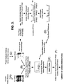
- Fig. 3 is a flow diagram of the method.
- the proteins are Western blotted (Step C) using an antibody phage library with detection of bound phage. Individual protein spots of interest and associated antibody displaying phage are excised from the Western blot, the phage eluted and used to re-infect E. coli. Individual colonies of E. coli may be propagated (in 96 well plate format), phage antibody secretion induced with IPTG and individual antibodies (PhAbs) recovered. The PhAbs are then immobilised and a protein mixture added. Proteins which bind are then eluted (Step D) and characterised by peptide mass fingerprinting. As with Example 1 the data can be compared (Step E) and PhAbs which bind the protein(s) of interest will be selected (Step F).
- the inverse screening method was used to screen a commercially available polyclonal antibody to bovine serum albumin (BSA) against a mixture of proteins (containing BSA).
- BSA bovine serum albumin
- Polyclonal anti-BSA antibody (5-80 ⁇ g; Sigma B-3759) in phosphate-buffered saline (PBS) was immobilised in the wells of 96 well filtration plates that incorporated a high protein binding hydrophobic PVDF membrane (Millipore MultiScreen® ImmobilonTM-P filtration plates) by incubating the pre-wetted membranes with the antibody containing solution for 2h at room temperature.
- PBS phosphate-buffered saline
- the membranes were pre-wetted according to manufacturers instructions; thus to each well was added 50 ⁇ l of 50% ethanol and the membranes incubated for 1 min at room temperature and after vacuum aspirating the wells each well was washed twice with 200 ⁇ l of de-ionised water or PBS. After antibody binding the wells were washed extensively with PBS (6 x 0.35ml). It is clear that different conditions for the immobilisation of antibody may be used and many have been examined in detail. These other methods include use of different buffers, different incubation times and temperatures, different methods for removal of remaining antibody solutions (vacuum aspiration through membrane or direct removal) all of which are known to those of skill in the art. The method described here proved optimal for the antibody used in this example.
- the extent to which antibody binding had been effected was examined by several methods. Most routinely it was examined by determining the protein concentration (Bradford assay) of the antibody solution after it had been incubated with the wells. Under the conditions described greater than 95% of the antibody was immobilised when antibody solutions containing up to 40 ⁇ g of antibody were added to each well. The maximum protein binding recorded (using solutions containing up to 80 ⁇ g of antibody) was 44 ⁇ g which is in close agreement with the manufacturer's data.
- a further method also found to be effective involved combining the ficoll block described above with dry blocking as recommended for the manufacturer for treatment of ImmobilonTM-P membranes when used in standard Western blotting applications.
- the wells were dried by carefully removing all remaining solution and incubating in a desiccated environment at 37°C for 2h.
- the wells were then treated with 0.5% ficoll in PBS for 2h at room temperature, washed, treated with elution reagent and washed again as described above.
- the elution reagents and conditions must be compatible with the subsequent protein characterisation method which in this case was peptide mass fingerprinting by MALDI mass spectrometry.
- a large number of elution conditions were investigated with attention focussed on those that would minimise subsequent manipulation steps and be compatible with MALDI mass spectrometry.
- immobilised antibody was incubated with a protein mixture (0.5-1.0mg; soya milk proteins) containing the protein antigen (BSA, 0.015-1.0mg) for 2h at room temperature.
- the wells were washed with PBS (6 x 0.35ml) and bound protein eluted by incubation with 20-100 ⁇ l of 0.5% formic acid for 10 minutes at room temperature.
- the elution reagent was then removed by evaporating the samples to dryness under vacuum in a rotary evaporator.
- Eluted proteins were subjected to peptide mass fingerprinting by standard protocols (see Courchesne, P.L. and Patterson, S.D. (1999) 2-D Proteome Analysis Protocols [Link Ed.], Methods in Molecular Biology 112, 487-511. Humana Press). Thus, protein samples were subjected to enzymatic digestion (37°C, 18 hours) in 25mM ammonium bicarbonate, pH 7.8 containing 50ng of trypsin. Protein-doped crystals were prepared using ⁇ -cyano-4-hydroxycinnamic acid (HCCA) as a matrix. A saturated HCCA matrix solution (10mg/ml) was prepared in 70% MeCN/0.1 % TFA and any undissolved matrix removed by centrifugation.
- HCCA ⁇ -cyano-4-hydroxycinnamic acid
- the inverse screening method was used to screen a commercially available monoclonal antibody to bovine serum albumin (BSA) against a mixture of proteins (containing BSA).
- BSA bovine serum albumin
- Monoclonal anti-BSA antibody (5-80 ⁇ g; Chemicon) in phosphate-buffered saline (PBS) was immobilised in the wells of 96 well filtration plates that incorporated a high protein binding hydrophobic PVDF membrane (Millipore MultiScreen® ImmobilonTM-P filtration plates) by incubating the pre-wetted membranes with the antibody containing solution for 2h at room temperature.
- the membranes were pre-wetted according to manufacturers instructions; thus to each well was added 50 ⁇ l of 50% ethanol and the membranes incubated for I min at room temperature and after vacuum aspirating the wells each well was washed twice with 200 ⁇ l of PBS. After antibody binding the wells were washed extensively with PBS (6 x 0.35ml). It is clear that different conditions for the immobilisation of antibody may be used and many have been examined in detail. These other methods include use of different buffers, different ... incubation times and temperatures, different methods for removal of remaining antibody solutions (vacuum aspiration through membrane or direct removal) all of which are known to those of skill in the art. The method described here proved optimal for the antibody used in this example. Other methods described where appropriate were optimal for the other examples cited.
- the extent to which antibody binding had been effective was examined by several methods. Most routinely it was examined by determining the protein concentration (Bradford assay) of the antibody solution after it had been incubated with the wells. Under the conditions described greater than 95% of the antibody was immobilised when antibody solutions containing up to 40 ⁇ g of antibody were added to each well.
- the wells were dried by carefully removing all remaining solution and incubating in a desiccated environment at 37°C for 2h.
- the wells were then treated with 0.5% ficoll in PBS for 2h at room temperature, washed, treated with elution reagent and washed again as described above.
- the immobilised antibody was then incubated with a protein mixture (0.5-1.0mg; soya milk proteins) containing the protein antigen (BSA, 0.015-1.0mg) for 2h at room temperature.
- a protein mixture 0.5-1.0mg; soya milk proteins
- BSA protein antigen
- the wells were washed with PBS (6 x 0.35ml) and bound protein eluted by incubation with 20-100 ⁇ l elution reagent (0.5% formic acid) for 10 minutes at room temperature.
- the elution reagent was then removed and evaporated to dryness under vacuum in a rotary evaporator. Eluted proteins were subjected to peptide mass fingerprinting by standard protocols (see Courchesne, P.L. and Patterson, S.D.
- mice were immunised with the protein by standard methods (see Antibodies, a Laboratory Manual, E. Harlow and D. Lane (Eds.)).
- BSA bovine serum albumin
- a pre-bleed was taken from each mouse which was then injected subcutaneously at 2 sites with a 1:1 mixture of protein and Freund's incomplete adjuvant (100 ⁇ l total).
- Twenty one days later tail bleeds were taken and each mouse injected subcutaneously at 2 sites with a further 100 ⁇ l of a 1:1 mixture of protein and PBS.
- a further twenty one days later tail bleeds were again taken and each mouse injected subcutaneously at 2 sites with a further 100 ⁇ l of a 1:1 mixture of protein and PBS.
- mice After a further 3 days some mice were splenectomised and a terminal bleed obtained.
- the splenocytes were extracted from the spleens of suitable mice, fused with myeloma cell line Sp2/0-Ag14 and cultured under selection pressure until the culture supernatants were ready for screening.
- hybridomas Prior to screening by the inverse screening process hybridomas were screened by a conventional antibody capture ELISA (see Antibodies, a Laboratory Manual, E. Harlow and D. Lane (Eds.)).
- BSA was bound to the wells of 96 well plates by incubation with 0.5-10 ⁇ g protein/well by standard procedures.
- mice were immunised with the protein by standard methods (see Antibodies, a Laboratory Manual, E. Harlow and D. Lane (Eds.)).
- a pre-bleed was taken from each mouse which was then injected subcutaneously at 2 sites with a 1:1 mixture of protein and Freund's complete adjuvant (100 ⁇ l total).
- Twenty one days later tail bleeds were taken and each mouse injected subcutaneously at 2 sites with a further 100 ⁇ l of a 1:1 mixture of protein and PBS.
- a further twenty one days later tail bleeds were again taken and each mouse injected subcutaneously at 2 sites with a further 100 ⁇ l of a 1:1 mixture of protein and PBS.
- Antibody binding to yeast was detected by incubation with anti-mouse Ig conjugated to horse radish peroxidase (Sigma) and the immunoblots developed by enhanced chemiluminescence (ECL, Amersham). The individual animals showed different patterns of antibody reactivity and were producing antibodies to a significant number of proteins (note 1-DE Western blotting will necessary underestimate the potential antibody repertoire of the animals). After a further 3 days some mice were splenectomised and a terminal bleed obtained. The splenocytes were extracted from the spleens and fused with myeloma cell line Sp2/0-Ag14 and cultured under selection pressure until the culture supernatants were ready for screening.
- hybridomas Prior to screening by the inverse screening process hybridomas were screened by a conventional antibody capture ELISA (see Antibodies, a Laboratory Manual, E. Harlow and D. Lane (Eds.)). Thus, yeast proteins were bound to the wells of 96 well plates by incubation with 0.5-10 ⁇ g protein/well by standard procedures. Wells were blocked by incubation with 20% soya milk in PBS for 2 hours at room temperature and then washed with PBS before 100 ⁇ l of hybridoma culture supernatant was applied to each well for 2 hours at room temperature.
Landscapes
- Life Sciences & Earth Sciences (AREA)
- Health & Medical Sciences (AREA)
- Engineering & Computer Science (AREA)
- Molecular Biology (AREA)
- Chemical & Material Sciences (AREA)
- Immunology (AREA)
- Physics & Mathematics (AREA)
- Biomedical Technology (AREA)
- Urology & Nephrology (AREA)
- Hematology (AREA)
- Bioinformatics & Cheminformatics (AREA)
- Bioinformatics & Computational Biology (AREA)
- Biochemistry (AREA)
- Medicinal Chemistry (AREA)
- Proteomics, Peptides & Aminoacids (AREA)
- General Health & Medical Sciences (AREA)
- Biophysics (AREA)
- Food Science & Technology (AREA)
- Cell Biology (AREA)
- Analytical Chemistry (AREA)
- Biotechnology (AREA)
- Spectroscopy & Molecular Physics (AREA)
- General Physics & Mathematics (AREA)
- Pathology (AREA)
- Microbiology (AREA)
- Organic Chemistry (AREA)
- Genetics & Genomics (AREA)
- Optics & Photonics (AREA)
- Peptides Or Proteins (AREA)
- Other Investigation Or Analysis Of Materials By Electrical Means (AREA)
- Investigating Or Analysing Biological Materials (AREA)
- Preparation Of Compounds By Using Micro-Organisms (AREA)
- Paper (AREA)
- Measuring Or Testing Involving Enzymes Or Micro-Organisms (AREA)
- Investigating Or Analysing Materials By The Use Of Chemical Reactions (AREA)
Claims (14)
- Verfahren zur Auswahl und/oder Identifizierung von einem oder mehr Protein-Affinitätsliganden, die an ein oder mehr Protein(e) von Interesse binden, umfassend die Schritte von:(A) Erhalt eines echten oder theoretischen Peptid-Massenfingerprints oder einer anderen auf der Massenspektrometrie basierenden Charakterisierung oder einer anderen Proteincharakterisierung des einen Proteins oder von mehr Proteinen durch entweder:i. Aussetzen des einen Proteins oder von mehr Proteinen dem Peptid-Massenfingerprinting oder einer anderen auf der Massenspektroskopie basierenden Charakterisierung oder einer anderen Proteincharakterisierung; oderii. Vorhersagen des Peptid-Massenfingerprints oder einer anderen auf der Massenspektrometrie basierenden Charakterisierung oder einer anderen Proteincharakterisierung aus bekannten Daten;(B) Verwendung des einen Proteins oder von mehr Proteinen entweder individuell oder als ein Gemisch zum:i. Generieren von einem oder mehr Antikörper(n) dazu durch Immunisierung; und/oderii. Auswählen, unter Verwendung von einer einzelnen oder mehrfachen Bindungsrunde(n), von einem oder mehr Protein-Affinitätsliganden dazu;(C) Screening des in Schritt (B)(i) generierten einen Antikörpers oder von mehr Antikörpern und/oder des in Schritt (B)(ii) ausgewählten einen oder von mehr Protein-Affinitätsliganden durch:(i) Zufügen des einen Proteins oder von mehr Proteinen, individuell oder als ein Gemisch aus Proteinen zum in Schritt (B)(i) generierten einen oder von mehr Antikörper(n), oder dem in Schritt (B)(i) ausgewählten einen oder von mehr Protein-Affinitätsliganden, wobei jeder Antikörper oder Protein-Affinitätsligand individuell verwendet wird, und(ii) nach Entfernung jeglicher Proteine, die nicht gebunden wurden, Eluieren des mindestens einen Proteins, das gebunden wurde;(D) Aussetzen des mindestens einen eluierten Proteins dem Peptid-Massenfingerprinting und/oder einer anderen auf der Massenspektrometrie basierenden Charakterisierung und/oder einer anderen Proteincharakterisierung; und(E) durch Vergleich der in Schritten (A) und (D) erhaltenen Peptid-Massenfingerprints oder einer anderen auf der Massenspektroskopie basierenden Charakterisierung oder einer anderen Proteincharakterisierung, wobei der mindestens eine Protein-Affinitätsligand, der an ein oder mehr Protein(e) von Interesse bindet, ausgewählt und/oder identifiziert wird.
- Verfahren nach Anspruch 1, worin das eine oder mehr Protein(e) von Interesse durch 2D-Elektrophorese aufgetrennt wird/werden.
- Verfahren nach Anspruch 1 oder 2, worin zwischen Schritten (B) und (C) die in Schritt (B)(i) erhaltenen Antikörper kloniert werden.
- Verfahren nach einem der vorangehenden Ansprüche, worin das eine oder mehr Protein(e) von Interesse in einem Gemisch von Proteinen anwesend ist/sind.
- Verfahren nach einem der vorangehenden Ansprüche, worin das Verfahren ein Shotgun-Verfahren zur Auswahl und Identifizierung von Protein-Affinitätsliganden an einer Vielzahl von Proteinen darstellt.
- Verfahren nach einem der vorangehenden Ansprüche, worin die andere auf der Massenspektrometrie basierende Charakterisierung die Erfassung von "Sequence-Tag"-Daten einschließt.
- Verfahren nach einem der vorangehenden Ansprüche, worin die in Schritt (B)(i) generierten Antikörper auf einem Träger, umfassend Nitrocellulose oder PVDF, immobilisiert sind.
- Verfahren nach Anspruch 7, worin der Träger, auf dem die Antikörper immobilisiert sind, mit Oligosaccharid- oder Polyvinylpyrrolidon-Lösung zum Blockieren jeglicher verbleibender Bindungsstellen behandelt werden.
- Verfahren nach Anspruch 8, worin das Oligosaccharid Ficoll darstellt.
- Verfahren nach einem der Ansprüche 7 bis 9, worin das Elutionsmittel ein flüchtiges Reagens darstellt.
- Verfahren nach Anspruch 10, worin das flüchtige Reagens Ameisensäure darstellt.
- Verfahren nach einem oder mehr der vorangehenden Ansprüche, worin der eine oder mehr Antikörper monoklonal ist/sind.
- Verfahren zur Generierung einer Antikörperbibliothek, umfassend die Schritte von:(a) Auftrennung eines komplexen Proteingemischs und Aussetzen des aufgetrennten Proteins/der aufgetrennten Proteine dem Peptid-Massenfingerprinting zum Erhalt eines Profils von der Peptidmasse; oder(b) Erhalt eines theoretischen Profils von der Peptidmasse für ein Protein, das gesucht wird;(c) Verwendung des oder eines anderen komplexen Proteingemischs zum Generieren von einem oder mehr monoklonalen Antikörper(n) dazu;(d) Zufügen des oder des anderen komplexen Proteingemischs an den in Schritt (c) generierten einen oder von mehr monoklonalen Antikörper(n) zur Auswahl der Proteine, die den einen oder mehr monoklonale(n) Antikörper binden und Aussetzen des/der ausgewählten Proteins/Proteine dem Peptid-Massenfingerprinting zum Erhalt eines Profils von der Peptidmasse;(e) Vergleich der in Schritten (a oder b) und (d) erhaltenen Profile von der Peptidmasse; und(f) Identifizierung der monoklonalen Antikörper von potenziellem Interesse für eine monoklonale Antikörperbibliothek.
- Verfahren zur Auswahl gewünschter Glieder einer Affinitätsligandenbibliothek, umfassend die Schritte von:(a) Auftrennung eines komplexen Proteingemischs und Aussetzen des/der aufgetrennten Proteins/Proteine dem Peptid-Massenfingerprinting zum Erhalt eines Profils von der Peptidmasse; oder(b) Erhalt eines theoretischen Profils von der Peptidmasse für ein Protein, das gesucht wird;(c) Nutzung von einem oder mehr der aufgetrennten Proteine zur Auswahl von einem oder mehr Affinitätsliganden aus einer Bibliothek;(d) Zufügen des oder eines anderen komplexen Proteingemischs zum in Schritt (c) generierten einen oder von mehr Affinitätsliganden zur Auswahl der Proteine, die den einen oder mehr Affinitätsliganden binden und Aussetzen des/der ausgewählten Proteins/Proteine dem Peptid-Massenfingerprinting zum Erhalt eines Profils von der Peptidmasse;(e) Vergleich der in Schritten (a oder b) und (d) erhaltenen Profile von der Peptidmasse; und(f) Auswahl eines oder von mehr Affinitätsliganden von Interesse.
Applications Claiming Priority (3)
| Application Number | Priority Date | Filing Date | Title |
|---|---|---|---|
| GBGB9800378.3A GB9800378D0 (en) | 1998-01-08 | 1998-01-08 | Proteome analysis |
| GB9800378 | 1998-01-08 | ||
| PCT/GB1999/000071 WO1999035502A1 (en) | 1998-01-08 | 1999-01-08 | Use of mass fingerprinting for identification of protein affinity ligands |
Publications (2)
| Publication Number | Publication Date |
|---|---|
| EP1046038A1 EP1046038A1 (de) | 2000-10-25 |
| EP1046038B1 true EP1046038B1 (de) | 2005-10-12 |
Family
ID=10825008
Family Applications (1)
| Application Number | Title | Priority Date | Filing Date |
|---|---|---|---|
| EP99900568A Expired - Lifetime EP1046038B1 (de) | 1998-01-08 | 1999-01-08 | Massenfingerabdrucken und seiner verwendung zur identifizierung von protein-affinitätsliganden |
Country Status (7)
| Country | Link |
|---|---|
| EP (1) | EP1046038B1 (de) |
| JP (1) | JP2002501176A (de) |
| AT (1) | ATE306671T1 (de) |
| DE (1) | DE69927668T2 (de) |
| ES (1) | ES2251171T3 (de) |
| GB (1) | GB9800378D0 (de) |
| WO (1) | WO1999035502A1 (de) |
Cited By (1)
| Publication number | Priority date | Publication date | Assignee | Title |
|---|---|---|---|---|
| CN106778032A (zh) * | 2016-12-14 | 2017-05-31 | 南京邮电大学 | 药物设计中配体分子海量特征筛选方法 |
Families Citing this family (7)
| Publication number | Priority date | Publication date | Assignee | Title |
|---|---|---|---|---|
| DE19943743C2 (de) * | 1999-09-03 | 2002-02-07 | Jerini Biotools Gmbh | Verfahren zur Identifizierung von Bindungspartnern mit positions-spezifischen Arrays |
| IL156446A0 (en) | 2000-12-14 | 2004-01-04 | Paul Stroobant | Differential phage capture proteomics |
| JP2002333430A (ja) * | 2001-05-09 | 2002-11-22 | Japan Science & Technology Corp | プロテオーム解析方法 |
| WO2003049602A2 (en) * | 2001-12-11 | 2003-06-19 | University Of Medicine And Dentistry Of New Jersey | Diagnostic methods for protein profiling |
| WO2004001377A2 (en) * | 2002-06-20 | 2003-12-31 | Paul Stroobant | Improved methods for performing differential capture proteomics |
| JP2007309689A (ja) * | 2006-05-16 | 2007-11-29 | Hitachi High-Technologies Corp | タンパク質結合部位同定技術 |
| US20220034902A1 (en) * | 2018-12-11 | 2022-02-03 | Merck Sharp & Dohme Corp. | Method for identifying high affinity monoclonal antibody heavy and light chain pairs from high throughput screens of b-cell and hybridoma libraries |
Family Cites Families (9)
| Publication number | Priority date | Publication date | Assignee | Title |
|---|---|---|---|---|
| US6124107A (en) * | 1988-06-10 | 2000-09-26 | Merck & Co., Inc. | Assay for marker of human polymorphonuclear leukocyte elastase activity |
| US5244816A (en) * | 1989-10-11 | 1993-09-14 | Akzo N.V. | Method for purifying chelator conjugated compounds |
| JPH07101998A (ja) * | 1993-08-09 | 1995-04-18 | Sumitomo Electric Ind Ltd | Paf受容体に特異的に反応するモノクローナル抗体、その製造方法、及び該抗体を産生するハイブリドーマ |
| US5538897A (en) * | 1994-03-14 | 1996-07-23 | University Of Washington | Use of mass spectrometry fragmentation patterns of peptides to identify amino acid sequences in databases |
| US5599668A (en) * | 1994-09-22 | 1997-02-04 | Abbott Laboratories | Light scattering optical waveguide method for detecting specific binding events |
| GB9502112D0 (en) * | 1995-02-03 | 1995-03-22 | British Biocell Int | Assay device and method |
| GB9507366D0 (en) * | 1995-04-08 | 1995-05-31 | Zeneca Ltd | Assay |
| US5834318A (en) * | 1995-05-10 | 1998-11-10 | Bayer Corporation | Screening of combinatorial peptide libraries for selection of peptide ligand useful in affinity purification of target proteins |
| NZ335258A (en) * | 1996-10-04 | 2002-06-28 | Peter Mose Larsen | Differences in 2D electrophoresis protein patterns between hypertensive and healthy animals |
-
1998
- 1998-01-08 GB GBGB9800378.3A patent/GB9800378D0/en not_active Ceased
-
1999
- 1999-01-08 JP JP2000527833A patent/JP2002501176A/ja active Pending
- 1999-01-08 DE DE69927668T patent/DE69927668T2/de not_active Expired - Fee Related
- 1999-01-08 ES ES99900568T patent/ES2251171T3/es not_active Expired - Lifetime
- 1999-01-08 WO PCT/GB1999/000071 patent/WO1999035502A1/en not_active Ceased
- 1999-01-08 AT AT99900568T patent/ATE306671T1/de not_active IP Right Cessation
- 1999-01-08 EP EP99900568A patent/EP1046038B1/de not_active Expired - Lifetime
Cited By (2)
| Publication number | Priority date | Publication date | Assignee | Title |
|---|---|---|---|---|
| CN106778032A (zh) * | 2016-12-14 | 2017-05-31 | 南京邮电大学 | 药物设计中配体分子海量特征筛选方法 |
| CN106778032B (zh) * | 2016-12-14 | 2019-06-04 | 南京邮电大学 | 药物设计中配体分子海量特征筛选方法 |
Also Published As
| Publication number | Publication date |
|---|---|
| ES2251171T3 (es) | 2006-04-16 |
| GB9800378D0 (en) | 1998-03-04 |
| DE69927668D1 (de) | 2006-02-23 |
| ATE306671T1 (de) | 2005-10-15 |
| JP2002501176A (ja) | 2002-01-15 |
| WO1999035502A1 (en) | 1999-07-15 |
| DE69927668T2 (de) | 2006-06-29 |
| EP1046038A1 (de) | 2000-10-25 |
Similar Documents
| Publication | Publication Date | Title |
|---|---|---|
| KR101107765B1 (ko) | 난소암의 검출을 위한 생물 마커의 용도 | |
| EP2044443B1 (de) | Peptidantikörperdepletion und anwendung zur probenvorbereitung für massenspektrometrie | |
| JP2003532055A (ja) | 前立腺癌マーカー | |
| Rowley et al. | Applications of protein mass spectrometry in cell biology | |
| US20200400674A1 (en) | A method of detecting magea4 | |
| AU2006336091A1 (en) | Method and markers for the diagnosis of renal diseases | |
| EP1046038A1 (de) | Massenfingerabdrucken und seiner verwendung zur identifizierung von protein-affinitätsliganden | |
| WO2003057014A2 (en) | Biomarkers for detecting ovarian cancer | |
| WO2005019831A2 (en) | Methods for reducing complexity of a sample using small epitope antibodies | |
| US20100068818A1 (en) | Algorithms for multivariant models to combine a panel of biomarkers for assessing the risk of developing ovarian cancer | |
| US20030228639A1 (en) | Prostate cancer markers | |
| WO2010094300A1 (en) | A method for determining in silico- a set of selected target epitopes | |
| US20060205010A1 (en) | Methods of host cell protein analysis | |
| JP6358630B2 (ja) | 免疫測定方法 | |
| US20060177870A1 (en) | Immunoassays | |
| WO2004030521A2 (en) | Use of biomarkers for detecting acute renal transplant rejection | |
| WO2005071421A1 (en) | Specific detection of troponin and modified forms of troponin | |
| Krone | Mass spectrometric methods for application in immunoassay and biomolecular characterization | |
| Stern et al. | The ProteinChip2 Biomarker System from Ciphergen Biosystems: a novel proteomics platform for rapid biomarker discovery and validation | |
| Prinz et al. | Document Title: Establishment of a Fast and Accurate Proteomic Method for Body Fluid/Cell Type Identification |
Legal Events
| Date | Code | Title | Description |
|---|---|---|---|
| PUAI | Public reference made under article 153(3) epc to a published international application that has entered the european phase |
Free format text: ORIGINAL CODE: 0009012 |
|
| 17P | Request for examination filed |
Effective date: 20000713 |
|
| AK | Designated contracting states |
Kind code of ref document: A1 Designated state(s): AT BE CH DE DK ES FR GB IE IT LI NL SE |
|
| 17Q | First examination report despatched |
Effective date: 20031007 |
|
| GRAP | Despatch of communication of intention to grant a patent |
Free format text: ORIGINAL CODE: EPIDOSNIGR1 |
|
| RAP1 | Party data changed (applicant data changed or rights of an application transferred) |
Owner name: UNIVERSITY COLLEGE DUBLIN |
|
| GRAS | Grant fee paid |
Free format text: ORIGINAL CODE: EPIDOSNIGR3 |
|
| GRAA | (expected) grant |
Free format text: ORIGINAL CODE: 0009210 |
|
| AK | Designated contracting states |
Kind code of ref document: B1 Designated state(s): AT BE CH DE DK ES FR GB IE IT LI NL SE |
|
| PG25 | Lapsed in a contracting state [announced via postgrant information from national office to epo] |
Ref country code: NL Free format text: LAPSE BECAUSE OF FAILURE TO SUBMIT A TRANSLATION OF THE DESCRIPTION OR TO PAY THE FEE WITHIN THE PRESCRIBED TIME-LIMIT Effective date: 20051012 Ref country code: AT Free format text: LAPSE BECAUSE OF FAILURE TO SUBMIT A TRANSLATION OF THE DESCRIPTION OR TO PAY THE FEE WITHIN THE PRESCRIBED TIME-LIMIT Effective date: 20051012 |
|
| REG | Reference to a national code |
Ref country code: GB Ref legal event code: FG4D |
|
| REG | Reference to a national code |
Ref country code: CH Ref legal event code: EP |
|
| REG | Reference to a national code |
Ref country code: IE Ref legal event code: FG4D |
|
| REG | Reference to a national code |
Ref country code: SE Ref legal event code: TRGR |
|
| REG | Reference to a national code |
Ref country code: CH Ref legal event code: NV Representative=s name: ISLER & PEDRAZZINI AG |
|
| PGFP | Annual fee paid to national office [announced via postgrant information from national office to epo] |
Ref country code: SE Payment date: 20060105 Year of fee payment: 8 Ref country code: DE Payment date: 20060105 Year of fee payment: 8 |
|
| PGFP | Annual fee paid to national office [announced via postgrant information from national office to epo] |
Ref country code: FR Payment date: 20060110 Year of fee payment: 8 |
|
| PG25 | Lapsed in a contracting state [announced via postgrant information from national office to epo] |
Ref country code: DK Free format text: LAPSE BECAUSE OF FAILURE TO SUBMIT A TRANSLATION OF THE DESCRIPTION OR TO PAY THE FEE WITHIN THE PRESCRIBED TIME-LIMIT Effective date: 20060112 |
|
| PGFP | Annual fee paid to national office [announced via postgrant information from national office to epo] |
Ref country code: IE Payment date: 20060112 Year of fee payment: 8 |
|
| PGFP | Annual fee paid to national office [announced via postgrant information from national office to epo] |
Ref country code: CH Payment date: 20060113 Year of fee payment: 8 |
|
| PGFP | Annual fee paid to national office [announced via postgrant information from national office to epo] |
Ref country code: IT Payment date: 20060131 Year of fee payment: 8 |
|
| PGFP | Annual fee paid to national office [announced via postgrant information from national office to epo] |
Ref country code: ES Payment date: 20060215 Year of fee payment: 8 |
|
| REF | Corresponds to: |
Ref document number: 69927668 Country of ref document: DE Date of ref document: 20060223 Kind code of ref document: P |
|
| PGFP | Annual fee paid to national office [announced via postgrant information from national office to epo] |
Ref country code: BE Payment date: 20060307 Year of fee payment: 8 |
|
| NLV1 | Nl: lapsed or annulled due to failure to fulfill the requirements of art. 29p and 29m of the patents act | ||
| REG | Reference to a national code |
Ref country code: ES Ref legal event code: FG2A Ref document number: 2251171 Country of ref document: ES Kind code of ref document: T3 |
|
| ET | Fr: translation filed | ||
| PLBE | No opposition filed within time limit |
Free format text: ORIGINAL CODE: 0009261 |
|
| STAA | Information on the status of an ep patent application or granted ep patent |
Free format text: STATUS: NO OPPOSITION FILED WITHIN TIME LIMIT |
|
| 26N | No opposition filed |
Effective date: 20060713 |
|
| PG25 | Lapsed in a contracting state [announced via postgrant information from national office to epo] |
Ref country code: IE Free format text: LAPSE BECAUSE OF NON-PAYMENT OF DUE FEES Effective date: 20070108 |
|
| PG25 | Lapsed in a contracting state [announced via postgrant information from national office to epo] |
Ref country code: SE Free format text: LAPSE BECAUSE OF NON-PAYMENT OF DUE FEES Effective date: 20070109 |
|
| PG25 | Lapsed in a contracting state [announced via postgrant information from national office to epo] |
Ref country code: LI Free format text: LAPSE BECAUSE OF NON-PAYMENT OF DUE FEES Effective date: 20070131 Ref country code: CH Free format text: LAPSE BECAUSE OF NON-PAYMENT OF DUE FEES Effective date: 20070131 |
|
| PG25 | Lapsed in a contracting state [announced via postgrant information from national office to epo] |
Ref country code: DE Free format text: LAPSE BECAUSE OF NON-PAYMENT OF DUE FEES Effective date: 20070801 |
|
| REG | Reference to a national code |
Ref country code: CH Ref legal event code: PL |
|
| EUG | Se: european patent has lapsed | ||
| GBPC | Gb: european patent ceased through non-payment of renewal fee |
Effective date: 20070108 |
|
| REG | Reference to a national code |
Ref country code: IE Ref legal event code: MM4A |
|
| REG | Reference to a national code |
Ref country code: FR Ref legal event code: ST Effective date: 20070930 |
|
| PG25 | Lapsed in a contracting state [announced via postgrant information from national office to epo] |
Ref country code: GB Free format text: LAPSE BECAUSE OF NON-PAYMENT OF DUE FEES Effective date: 20070108 |
|
| BERE | Be: lapsed |
Owner name: *UNIVERSITY COLLEGE DUBLIN Effective date: 20070131 |
|
| PG25 | Lapsed in a contracting state [announced via postgrant information from national office to epo] |
Ref country code: BE Free format text: LAPSE BECAUSE OF NON-PAYMENT OF DUE FEES Effective date: 20070131 |
|
| REG | Reference to a national code |
Ref country code: ES Ref legal event code: FD2A Effective date: 20070109 |
|
| PG25 | Lapsed in a contracting state [announced via postgrant information from national office to epo] |
Ref country code: FR Free format text: LAPSE BECAUSE OF NON-PAYMENT OF DUE FEES Effective date: 20070131 |
|
| PG25 | Lapsed in a contracting state [announced via postgrant information from national office to epo] |
Ref country code: ES Free format text: LAPSE BECAUSE OF NON-PAYMENT OF DUE FEES Effective date: 20070109 |
|
| PGFP | Annual fee paid to national office [announced via postgrant information from national office to epo] |
Ref country code: GB Payment date: 20060104 Year of fee payment: 8 |
|
| PG25 | Lapsed in a contracting state [announced via postgrant information from national office to epo] |
Ref country code: IT Free format text: LAPSE BECAUSE OF NON-PAYMENT OF DUE FEES Effective date: 20070108 |