EP1118660B1 - Ein plasmaprotein-fragment erzeugendes enzym mit krebsmetastase-hemmender wirkung und durch dieses enzym fragmentiertes plasmaprotein-fragment - Google Patents
Ein plasmaprotein-fragment erzeugendes enzym mit krebsmetastase-hemmender wirkung und durch dieses enzym fragmentiertes plasmaprotein-fragment Download PDFInfo
- Publication number
- EP1118660B1 EP1118660B1 EP99970118A EP99970118A EP1118660B1 EP 1118660 B1 EP1118660 B1 EP 1118660B1 EP 99970118 A EP99970118 A EP 99970118A EP 99970118 A EP99970118 A EP 99970118A EP 1118660 B1 EP1118660 B1 EP 1118660B1
- Authority
- EP
- European Patent Office
- Prior art keywords
- enzyme
- plasminogen
- cancer
- pace4
- growth
- Prior art date
- Legal status (The legal status is an assumption and is not a legal conclusion. Google has not performed a legal analysis and makes no representation as to the accuracy of the status listed.)
- Expired - Lifetime
Links
Images


Classifications
-
- C—CHEMISTRY; METALLURGY
- C12—BIOCHEMISTRY; BEER; SPIRITS; WINE; VINEGAR; MICROBIOLOGY; ENZYMOLOGY; MUTATION OR GENETIC ENGINEERING
- C12N—MICROORGANISMS OR ENZYMES; COMPOSITIONS THEREOF; PROPAGATING, PRESERVING, OR MAINTAINING MICROORGANISMS; MUTATION OR GENETIC ENGINEERING; CULTURE MEDIA
- C12N9/00—Enzymes; Proenzymes; Compositions thereof; Processes for preparing, activating, inhibiting, separating or purifying enzymes
- C12N9/14—Hydrolases (3)
- C12N9/48—Hydrolases (3) acting on peptide bonds (3.4)
- C12N9/50—Proteinases, e.g. Endopeptidases (3.4.21-3.4.25)
- C12N9/64—Proteinases, e.g. Endopeptidases (3.4.21-3.4.25) derived from animal tissue
- C12N9/6421—Proteinases, e.g. Endopeptidases (3.4.21-3.4.25) derived from animal tissue from mammals
- C12N9/6424—Serine endopeptidases (3.4.21)
- C12N9/6454—Dibasic site splicing serine proteases, e.g. kexin (3.4.21.61); furin (3.4.21.75) and other proprotein convertases
-
- A—HUMAN NECESSITIES
- A61—MEDICAL OR VETERINARY SCIENCE; HYGIENE
- A61P—SPECIFIC THERAPEUTIC ACTIVITY OF CHEMICAL COMPOUNDS OR MEDICINAL PREPARATIONS
- A61P27/00—Drugs for disorders of the senses
-
- A—HUMAN NECESSITIES
- A61—MEDICAL OR VETERINARY SCIENCE; HYGIENE
- A61P—SPECIFIC THERAPEUTIC ACTIVITY OF CHEMICAL COMPOUNDS OR MEDICINAL PREPARATIONS
- A61P27/00—Drugs for disorders of the senses
- A61P27/02—Ophthalmic agents
-
- A—HUMAN NECESSITIES
- A61—MEDICAL OR VETERINARY SCIENCE; HYGIENE
- A61P—SPECIFIC THERAPEUTIC ACTIVITY OF CHEMICAL COMPOUNDS OR MEDICINAL PREPARATIONS
- A61P35/00—Antineoplastic agents
-
- A—HUMAN NECESSITIES
- A61—MEDICAL OR VETERINARY SCIENCE; HYGIENE
- A61P—SPECIFIC THERAPEUTIC ACTIVITY OF CHEMICAL COMPOUNDS OR MEDICINAL PREPARATIONS
- A61P35/00—Antineoplastic agents
- A61P35/04—Antineoplastic agents specific for metastasis
-
- A—HUMAN NECESSITIES
- A61—MEDICAL OR VETERINARY SCIENCE; HYGIENE
- A61P—SPECIFIC THERAPEUTIC ACTIVITY OF CHEMICAL COMPOUNDS OR MEDICINAL PREPARATIONS
- A61P43/00—Drugs for specific purposes, not provided for in groups A61P1/00-A61P41/00
-
- C—CHEMISTRY; METALLURGY
- C12—BIOCHEMISTRY; BEER; SPIRITS; WINE; VINEGAR; MICROBIOLOGY; ENZYMOLOGY; MUTATION OR GENETIC ENGINEERING
- C12N—MICROORGANISMS OR ENZYMES; COMPOSITIONS THEREOF; PROPAGATING, PRESERVING, OR MAINTAINING MICROORGANISMS; MUTATION OR GENETIC ENGINEERING; CULTURE MEDIA
- C12N9/00—Enzymes; Proenzymes; Compositions thereof; Processes for preparing, activating, inhibiting, separating or purifying enzymes
- C12N9/14—Hydrolases (3)
- C12N9/48—Hydrolases (3) acting on peptide bonds (3.4)
- C12N9/50—Proteinases, e.g. Endopeptidases (3.4.21-3.4.25)
- C12N9/64—Proteinases, e.g. Endopeptidases (3.4.21-3.4.25) derived from animal tissue
- C12N9/6421—Proteinases, e.g. Endopeptidases (3.4.21-3.4.25) derived from animal tissue from mammals
- C12N9/6424—Serine endopeptidases (3.4.21)
- C12N9/6435—Plasmin (3.4.21.7), i.e. fibrinolysin
-
- C—CHEMISTRY; METALLURGY
- C12—BIOCHEMISTRY; BEER; SPIRITS; WINE; VINEGAR; MICROBIOLOGY; ENZYMOLOGY; MUTATION OR GENETIC ENGINEERING
- C12N—MICROORGANISMS OR ENZYMES; COMPOSITIONS THEREOF; PROPAGATING, PRESERVING, OR MAINTAINING MICROORGANISMS; MUTATION OR GENETIC ENGINEERING; CULTURE MEDIA
- C12N9/00—Enzymes; Proenzymes; Compositions thereof; Processes for preparing, activating, inhibiting, separating or purifying enzymes
- C12N9/14—Hydrolases (3)
- C12N9/48—Hydrolases (3) acting on peptide bonds (3.4)
- C12N9/50—Proteinases, e.g. Endopeptidases (3.4.21-3.4.25)
- C12N9/64—Proteinases, e.g. Endopeptidases (3.4.21-3.4.25) derived from animal tissue
- C12N9/6421—Proteinases, e.g. Endopeptidases (3.4.21-3.4.25) derived from animal tissue from mammals
- C12N9/6478—Aspartic endopeptidases (3.4.23)
-
- C—CHEMISTRY; METALLURGY
- C12—BIOCHEMISTRY; BEER; SPIRITS; WINE; VINEGAR; MICROBIOLOGY; ENZYMOLOGY; MUTATION OR GENETIC ENGINEERING
- C12Y—ENZYMES
- C12Y304/00—Hydrolases acting on peptide bonds, i.e. peptidases (3.4)
- C12Y304/21—Serine endopeptidases (3.4.21)
- C12Y304/21007—Plasmin (3.4.21.7), i.e. fibrinolysin
-
- A—HUMAN NECESSITIES
- A61—MEDICAL OR VETERINARY SCIENCE; HYGIENE
- A61K—PREPARATIONS FOR MEDICAL, DENTAL OR TOILETRY PURPOSES
- A61K35/00—Medicinal preparations containing materials or reaction products thereof with undetermined constitution
Definitions
- the present disclosure relates to the use of an enzyme that degrades plasma proteins such as plasminogen and fibronectin molecular species into fragments having an inhibitory activity to metastasis and growth of cancer, for producing such plasma protein fragments.
- Metastasis and growth of cancer refers to release of cancer cells from the primary cancerous focus through the vascular system into other sites of the living body where the cancer cells grow. Under the circumstances where cancer cells are spread through the vascular system so that they can grow in a wide range within the living body, it is almost impossible to perfectly remove such cancer cells even with the surgical technique highly progressed nowadays. Thus, in such a case, chemotherapy such as an anti-cancer agent has been used for prophylaxis and treatment of cancer. Even this measure, however, is confronted with the problem of drug resistance to the chemical substance, accompanied by increase in a dose for attaining efficacy of that chemical substance and as a consequence by increase in detrimental side effects. There have been developed a great deal techniques in order to overcome these problems. On the other hand, another approach that is actively pursued by researchers nowadays is to investigate intervention to cancer cells invasion into the vascular system, i.e. intervention to cancer invasion and inhibition of vascularization.
- Cancer cells invasion into the vascular system has two aspects: one is related to cancer cells that move from the primary cancerous focus into the vascular system and the other to a drawing-in of the vascular system into the primary cancerous focus.
- the former is called as cancer cells invasion and the latter as a vascularization.
- Cancer cells invasion is phenomena in which cancer cells that acquired an ability of metastasis and growth are released from the primary focus (tumor), degrade a barrier between cancer cells and the blood vessel called stroma, and invade into the vascular system ( Mignatti P. et al., J. Cell Biol., vol.108, p.671-682 (1989 )).
- vascularization is phenomena in which novel vessels are formed from the existing vessels. Cancer cells move towards the blood flow through these newly developed vessels. Vascularization is not observed in healthy adult individuals except for sex cycle in females and wound healing but occurs under pathological conditions such as cancer, diabetic retinosis, rheumatism, psoriasis ( Folkman J., Adv. Cancer Res., vol. 43, p.175-203 (1985 ); Zetter B. R., Chest, vol. 93 (Suppl.), p.159S-166S (1998 ); Patz A., Invest. Ophthalmol. Visual Sci., vol. 19, p.1133-1138 (1980 ); Amore D. et al., Ann. Rev. Physiol., vol. 49, p.453-464 (1987 )). '
- Cancer cells draw in newly developed vessels from the existing vessels in order to provide them with oxygen or nutrients necessary for their growth. These newly developed vessels facilitate that cancer cells move into the vascular system.
- Vascularization is a consequence of catabolism of endothelial cells arranged in line within the luminal cavity that are stimulated by cancer cells.
- steps must occur for development of new vessels after the endothelial cells reached the cancer cells, such as digestion of the basal membrane, release and propagation of the vascular endothelial cells, and formation of luminal cavity.
- to intervene to cancer cells invading into the vascular system to inhibit metastasis and growth of cancer is in other words is intervene to any of processes composed of the above multiple steps. Which of these processes should be intervened is disputable but degradation and digestion of the stroma, a common process between cancer invasion and vascularization, is the most highlighted.
- TIMP tissue Inhibitor of Metallo Protease
- a matrix-degrading enzyme is usually present extracellularly in the form of an enzyme precursor ( Chen, W. T., Curr. Opin. Cell Biol., vol. 4, p.802-809 (1992 )).
- an enzyme precursor Chen, W. T., Curr. Opin. Cell Biol., vol. 4, p.802-809 (1992 )
- a serine protease that activates a matrix-degrading enzyme includes plasmin, urokinase-type plasminogen activator.
- u-PA tissue-type plasminogen activator
- t-PA tissue-type plasminogen activator
- trypsin trypsin
- plasmin an enzyme associated with blood fibrinolysis
- cancer cells have an ability to produce u-PA
- vascular endothelial cells to produce t-PA wherein plasmin is a consequence of activation of its precursor plasminogen through cleavage by the action of a plasminogen activator such as u-PA and t-PA. It is reported that both u-PA and t-PA are expressed at a high level under cancerous conditions.
- plasmin In addition to activation of a matrix-degrading enzyme as mentioned above, plasmin also has an ability to activate TGF- ⁇ , a factor involved in vascularization, and an ability to degrade the extracellular matrix ( Werb Z. et al., N. Engl. J. Med., vol. 296, p.1017 (1977 ); Brunner G. et al., J. Cell Biol., vol. 6, p.1275-1283 (1991 )). Moreover, it is considered that plasmin has an ability to destroy protective mechanism of hosts that besieges cancer with fibrin membrane to thereby suppress cancer dispersion.
- Angiostatin is a vascularization-inhibitory substance comprising an inner fragment of plasminogen and has a potent inhibitory activity to vascularization and growth of metastasized cancerous focus at an extremely low amount. It also possesses, remarkably, an ability to induce recession of cancer (primary focus) in a dose dependent manner without any side effects or drug resistance ( O'Reilly M. S. et al., Nat.
- Angiostatin has been correlated with inhibition to vascularization and cancer as demonstrated by O'Reilly et al. in sophisticated experiments. However, most of mechanism of inhibitory action of angiostatin and its production still remains to be elucidated.
- the present inventors have earnestly investigated in order to solve the above problems and as a result have found a novel enzyme activity in a culture of PC-3, prostate cancer cell line, said enzyme activity being able to fragment plasminogen only under low pH condition. It was found that plasminogen fragments produced by said enzyme comprises plasminogen Kringles 1 to 4 and that said enzyme inactivates plasmin by cleaving it in the vicinity of the active center. This activity is expressed in most of cancer cells and hence suggested to be specific to cancer.
- the enzyme purified from supernatant of PC-3 culture was reacted with plasminogen to produce plasminogen fragments that were then administered to a mouse model in which Lewis lung cancer was transplanted.
- various plasma components were cleaved with this enzyme and the obtained fragments were screened for their inhibitory activity to vascularization.
- fibronectin and vitronectin both components involved in cell adhesion, and human hepatocyte growth factor (HGF) were also fragmented by this enzyme and that plasma protein fragments produced by fragmentation had a potent inhibitory activity to vascularization.
- HGF human hepatocyte growth factor
- the present invention relates the use of an aspartic enzyme as defined in the claims.
- a preferential measure for investigating an enzyme that produces an angiostatin-like molecule and inactivates plasmin is to find out an enzyme that produces angiostatin.
- angiostatin has been found in plasma and urine of a mouse transplanted with Lewis lung cancer subspecies 3LL-LM and hence an enzyme that produces angiotensin should exist in said mouse.
- O'Reilly et al. made a model using 3LL-LM cells with perception of rapid growth of distally metastasized cancerous focus that is rarely observed after excision of malignant tumor and found in plasma and urine of the model a potent inhibitory factor to vascularization, leading to discovery of angiostatin.
- cancer a factor derived from a primary focus (cancer) is circulating throughout the blood stream to thereby inhibit growth of a distally metastasized cancerous focus.
- angiostatin it is estimated that since cancer cells do not produce plasminogen, an enzyme derived from cancer cells cleaves plasminogen occurring in blood to thereby produce angiostatin.
- PC-3 cells human prostate cancer cells
- PACE Proliferative Angiostatin Converting Enzyme
- PC-3 cells are such human type cells that have property that excision of primary cancerous focus can conversely promote growth of distally metastasized cancerous focus ( Soff G. A. et al., J. Clin. Invest., vol.
- Dong et al. focused on macrophage invasion observed under cancerous environments and revealed that an enzyme derived from macrophage (matrix metallo elastase) was an enzyme that produced angiostatin ( Dong Z. et al., Cell, vol. 88, p.801-810 (1997 )).
- Brain et al. screened Matrix metallo proteinase (MMP) to thereby reveal that MMP-7 (also referred to as "Matrilysin”) and MMP-9 (also referred to as "Gelatinase B”) exhibited the activity to produce angiostatin ( Brain C. et al., J. Biol. Chem., vol. 272, p.28823-28825 (1997 )).
- the present inventors have constructed a screening system of our own with which we tried to isolate and purify the desired enzyme. As a result, of thorough investigation, the present inventors have successfully found, in addition to an enzyme that fragments plasminogen at neutral pH, that an enzyme activity that can specifically cleave a restricted site of plasminogen under lower pH condition was present in PC-3 culture supernatant. This enzyme activity has not been reported previously and is utterly different from PACE according to Gately et al. The cleavage pattern of this enzyme proved, as shown in Figure 1 , that it specifically cleaved a restricted site of plasminogen unlike PACE that nonspecifically cleaved plasminogen.
- the present inventors have paid attention to lower pH at the cancerous state since previously and reported possibility that some plasminogen fragments produced by cleavage through elastase activity might accumulate at the site with lower pH ( The 56th General Meeting of Japan Association of Oncology, excerpt, p.426, 1997 ).
- the present inventors have already found that an enzyme that functions at that lower pH was related to cancerous conditions by investigating enzyme activities with various types of cancer cells and normal cells and perceived that said enzyme was produced in a large amount only under cancerous conditions through preliminary investigation.
- a purpose of the present inventors' screening is to prove the hypothesis that "an enzyme that cleaves plasmin to thereby produce angiostatin-like molecules that can do away with the plasmin activity should have been provided in the living body as a protective mechanism to cancer development". It is highly possible that it is this enzyme functioning only under lower pH conditions what is referred to in said hypothesis. Thus, the present inventors have initiated investigation for isolation and purification of said enzyme.
- the most characteristic property of the enzyme of the present invention is its ability to specifically cleave plasma proteins such as plasminogen and fibronectin at a restricted site.
- the present inventors have firstly initiated investigation and isolation of the enzyme in order to elucidate relationship between said enzyme and cancer cells.
- Fig. 2 schematically illustrates the screening system for the plasminogen-fragmenting enzyme.
- This method is a kind of modified sandwich ELISA wherein a specific antibody to Plasminogen Lysine Binding Site I (hereinafter referred to as "LBS I") is used as an immobilized antibody whereas a specific antibody to Mini Plasminogen (hereinafter referred to as "mPlg.”) is used as a labeled antibody.
- LBS I Plasminogen Lysine Binding Site I
- mPlg. a specific antibody to Mini Plasminogen
- plasminogen When an objective enzyme is contacted with a predetermined amount of plasminogen, plasminogen is cleaved to thereby lose a region that is recognized by the labeled antibody and as a consequence color development in ELISA declines.
- the enzyme activity is estimated by detecting this decline of color development.
- Preparation of the desired enzyme was achieved, using PC-3 culture supernatant as starting material, by a series of chromatographic procedures including a cation exchanger, a heparin chromatography, an anion exchanger, gel filtration, and hydroxyapatite.
- the first procedure with a cation exchanger was carried out primarily for the purpose of removing contaminating single chain urokinase-type plasminogen activator (scu-PA) whereas heparin chromatography was used for removal of serine protease.
- Scu-PA a precursor of plasminogen activator, was activated by the presence of plasmin into an active form of u-PA, which also cleaves plasminogen at a neutral pH range.
- Fig. 3 shows a chromatogram of typical hydrophobic chromatography wherein the open circle indicates absorbance, the closed circle indicates an enzyme activity, and the wave line indicates an ionic strength.
- Fig. 3 clearly indicates two distinct enzyme activities with different hydrophobicity in PC-3 culture supernatant.
- the enzyme activity that specifically cleaved plasminogen to produce plasminogen fragments comprising Kringles 1 to 4 was detected in the peak with higher hydrophobicity (at right side of chromatogram).
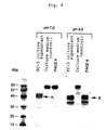
- Fig. 4 shows results of SDS-PAGE analysis of the enzyme of the present invention wherein a band with the activity was found at the position corresponding to 45 kDa of a molecular weight. Since it was revealed that the enzyme of the present invention was an aspartic enzyme as described below, it was purified in the end by affinity chromatography using pepstatin as a ligand.
- the enzyme of the present invention may be prepared, in addition to the procedure as described above starting from PC-3 culture supernatant, by constructing cells that produce said enzyme with the genetic recombinant technique and recovering said enzyme produced by said cells. That is, the enzyme of the present invention may also be prepared by an approach wherein a gene encoding said enzyme is introduced with the aid of an appropriate vector into an appropriate host including prokaryotic, eukaryotic, mammal or insect cells, from which the enzyme is recovered.
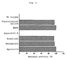
- FIG. 5 A mode of the inhibitory activity of the enzyme of the present invention is summarized in Fig. 5 wherein a concentration of each inhibitor was: aprotinin (0.3 ⁇ M), benzamidine (10 mM), elastinal (100 ⁇ M), pepstatin A (1 ⁇ M), and EDTA (10 mM).
- aprotinin 0.3 ⁇ M
- benzamidine 10 mM
- elastinal 100 ⁇ M
- pepstatin A (1 ⁇ M)
- EDTA mM
- the arrow A ( ⁇ A) indicates a band corresponding to angiostatin from PC-3 according to Gately et al. whereas the arrow B ( ⁇ B) indicates a band corresponding to a fragment comprising Kringles 1 to 4 produced by PACE4.
- Fig. 7 shows sites where the enzyme of the present invention cleaves plasminogen, which is cleaved into A (plasminogen Kringles 1 to 4), B (Mini Plasminogen) and C (Mini Plasminogen fragments). The N-terminal amino acid residue of each fragment was analyzed to be F-74 for A, P-452 for B and A-700 for C, respectively.
- the enzyme of the present invention cleaves plasminogen at between the 451st, from the N-terminus, Leu and the 452nd Pro (hereinafter referred to as "451L-P"), and between the 73rd Leu and the 74th Phe (hereinafter referred to as "73L-F") to leave a fragment comprising Kringles 1 to 4 of plasminogen.
- Plasminogen circulates within the living body mostly in the form not cleaved and has the N-terminal Glu (intact plasminogen). However, a part of plasminogen, several percent, is cleaved at the N-terminal and has the N-terminal 78 Lys, called Lys plasminogen.
- the enzyme of the present invention in addition to intact plasminogen, fragments Lys plasminogen as well and thus plasminogen fragments produced by the enzyme of the present invention include one with the N-terminal amino acid being 78 Lys.
- the enzyme of the present invention also cleaves plasmin in the vicinity of its active center (between the 699th Phe and 700th Ala) to let plasmin be inactivated.
- This cleaving property of the enzyme is quite significant in view of efficient protection against cancer development. This is because angiostatin that inhibits growth of cancer is produced by fragmentation of plasminogen while the simultaneously produced active center of plasmin (plasmin serine protease domain) possibly further promotes cancer development.
- vascularization and cancer invasion accelerated by the plasmin activity is further accelerated by removal of the Kringle domains.
- ⁇ 2-PI alpha 2 plasmin inhibitor
- the enzyme of the present invention is distinct from the previously reported enzymes that produce angiostatin in that it not only produces fragments comprising Kringles 1 to 4 of plasminogen but also inactivates the remaining plasmin activity. Supposing whatever protective role in the living body the enzyme of the present invention plays, it is probably a top-rated candidate enzyme.
- Fig. 8 shows a relationship between plasminogen cleavage by the enzyme of the present invention with passage of time and the remaining plasmin activity: (A) SDS-PAGE when plasminogen is fragmented with PACE4; (B) the plasmin activity measured for the samples. Fig. 8 clearly indicates that the plasmin activity disappears as mini plasminogen is more cleaved.
- the enzyme of the present invention can function in favor of the living body and hence it is expected that a substrate protein to which the enzyme of the present invention acts is not limited to plasminogen.
- the present inventors in order to verify this possibility, also performed fragmentation of other plasma proteins as described below and could successfully prepare fragments with the activity equivalent to or even exceeding ones from plasminogen using the enzyme of the present invention.
- the purified enzyme sample was electrophoresed on SDS-PAGE, it was transferred to GVDF membrane by blotting.
- the blotted membrane was dyed with Amido Black, bands corresponding to 45 kDa were excised and the N-terminal amino acid sequence was read with an amino acid sequence analyzer. As a result, the band corresponding to 45 kDa had a sequence LVRIPLHKFT.
- the determined amino acid sequence was compared with the existing amino acid data bank and was found to have homology with a precursor of human cathepsin D.
- the enzyme purified by immunoblot was reacted with an anti-human cathepsin D antibody, said enzyme responded to this antibody.
- the enzyme of the present invention has a high homology with human cathepsin D.
- Cathepsin D is produced as cathepsin D precursor having a whole amino acid sequence which is then cleaved by other enzymes in lysosome into active cathepsin D.
- a malignant tumor typically human breast cancer
- the enzyme of the present invention is the cathepsin D precursor, it is just a novel and interesting finding that the cathepsin D precursor in the form of a precursor does cleave plasminogen.
- cDNAs were synthesized from mRNAs prepared from PC-3 cells that were used as starting material for preparing PACE4.
- Sense and antisense primers for amplifying a whole translation region of cathepsin D were synthesized on the basis of cDNA of cathepsin D.
- the sense and antisense primers were used for amplification with AmpliTaq (PERKIN ELMER) and the amplified cDNA fragment was cloned into a plasmid vector with TA cloning kit (Invitrogen). The obtained gene fragment was sequenced to confirm that it was identical to the known nucleotide sequence of cathepsin D.
- PACE4 is identified as the cathepsin D precursor, however, some queries arise why an inactive form precursor can cleave, say, plasminogen, or why cathepsin, that is an intracellular enzyme, is excreted out of cells.
- an inactive form precursor say, plasminogen
- cathepsin that is an intracellular enzyme
- the enzyme of the present invention is genuinely identical to the cathepsin D precursor, a whole amino acid sequence as well as a structure of the enzyme remain to be elucidated.
- both cathepsin D precursor and active cathepsin D have the enzymatic activity of the present enzyme and are encompassed by PACE4.
- derivatives of cathepsin D with deletion, substitution or chemical modifications are included within a scope of the enzyme of the present invention insofar as they are used for the purpose of preparing plasma protein fragments having an inhibitory activity to metastasis and growth of cancer.
- Plasminogen and PACE4 are mixed at a predetermined ratio and reacted at pH 4.0. After dialysis with a buffer at a neutral pH range, the reaction mixture is contacted with lysine Sepharose so that fractions containing a fragment comprising Kringles 1 to 4 of plasminogen are absorbed to the resin and separated based on binding capacity to lysine. The obtained fragments are lyophilized after dialysis with an ammonium carbonate buffer. Fibronectin may similarly be cleaved and a fragment mixture is subjected to heparin Sepharose separation to prepare a desired fragment.
- the protein fragments prepared as described above were dissolved in a physiological saline and used as a sample for animal tests.
- a system for estimating an inhibitory effect to growth of metastasized cancerous focus cancer capable of metastasis and growth was transplanted to mice where distally metastasized cancerous focus was observed.
- Lung cancer cells (LL/2; Dainippon Seiyaku K.K.) were subcutaneously transplanted to mice at the back. The animals were bred until a primary cancerous focus grows up to a predetermined size and the primary focus was then removed surgically.
- the sample was administered to mice intraperitoneally at 1 mg/mouse/day for 10 days.
- mice were administered with a physiological saline alone as used for dissolving the lyophilized sample (test for inhibitory activity to growth of metastasized cancerous focus).
- mice were bred for additional predetermined period of time and then administered with the sample at 1 mg/mouse/day for 10 days.
- mice were dissected.
- mice As a system for estimating inhibitory effects to growth of metastasized cancerous focus of PACE4 per se, immunodeficient (Scid) mice were employed. Lung cancer cells (3LL; provided from Cell Resource Center for Biomedical Research, Institute of Development, Aging and Cancer, Tohoku University) were subcutaneously transplanted to Scid mice at the back. The animals were bred until a primary cancerous focus grows up to a predetermined size. So that severity of disease conditions is taken into consideration, mice were divided into two groups based on the size of a primary cancerous focus; one with a primary cancerous focus weighing not more than 1200 mg and the other with a primary cancerous focus weighing more than 1200 mg. The primary cancerous focus in each group was removed surgically.
- Each group was then further divided into three groups: a group receiving a high or low concentration of PACE4, and a control group.
- the sample was administered to mice intraperitoneally at 40 ⁇ g/mouse/day or 8 ⁇ g/mouse/day for 10 days.
- As a control the same amount of physiological saline, as used for dissolving the lyophilized sample, was administered to mice.
- mice were dissected and the lung was weighed and compared with the control group (Mann Whetney U test).
- the enzyme of the present invention exhibited inhibitory activity to growth of metastasized cancerous focus in a concentration dependent manner in the group of a primary cancerous focus weighing more than 1200 mg (B) ( Fig. 9 ).
- cathepsin D-like enzyme of the present invention functions within the living body implies that there exists an environment with low pH, at least pH 5.0. Such an environment with pH of as low as 5.0 is not physiologically observed. At present it has not yet been elucidated how this enzyme functions physiologically but there is also a report that a cancerous environment provides this low pH environment. Briozzo et al. reported that cathepsin D, which is an aspartic enzyme classified into the same class as the enzyme of the present invention, was secreted out of cancer cells and acted at distal sites where it degraded the extracellular matrix ( Briozzo et al., Cancer Res., vol. 48, p.3688-3692 (1988 )).
- Intracellular proteases such as cathepsin D are primarily localized in membranous organelle such as endosome and lysosome. Both of these membranous organelle are extremely shifted to acidic side due to distribution of H-ATPase and play a role in degradation and reuse of intracellularly or extracellularly existing heterogeneous substances. It is also conjectured that a certain condition is loaded to cancerous cells by some reasons to release such membranous organelle out of the cells, which in turn digest plasminogen or the extracellular matrix neighboring said membranous organelle.
- the present inventors focused on fibronectin, a plasma component that is produced in great deal under cancerous conditions and is deeply involved in cell adhesion, and cleaved this with the enzyme of the present invention.
- Fibronectin was degraded restrictedly with this enzyme and its degraded products were found to have a potent inhibitory ability to BCE (Bovine capillary endothelial) vascular growth not found in intact fibronectin.
- BCE Bovine capillary endothelial
- the degraded products produced by the enzyme from fibronectin were separated with heparin Sepharose (Pharmacia). A fraction of a heparin-binding activity was administered to mice of the model system as described above at 1 mg/kg/day to prove that this fragment had an inhibitory activity to growth of metastasized cancerous focus.
- the present inventors also cleaved several kinds of fractions obtained from alcoholic fractionation of plasma proteins with the enzyme of the present invention.
- Alcohol was removed from alcoholic fractions by dialysis, followed by dialysis with citric phosphate buffer (pH 4.0).
- the resulting fractions were mixed with the enzyme at a ratio of the protein to the enzyme, 100:1, and the mixture was reacted at 38°C overnight.
- the crude solutions of the protein may form a lot of precipitates at 4°C and hence were centrifuged at 6000 rpm (manufactured by Tommy) prior to the enzymatic reaction and their supernatant was used.
- reaction solutions were dialyzed against a 50 mM Tris/50 mM NaCl (pH 7.2) buffer overnight, filtered with a 0.45 ⁇ m filter (Milex HA: manufactured by Millipore) and treated with 5 ml of heparin Sepharose (Hi-trap Heparin: manufactured by Millipore) to give protein mixtures.
- Milex HA manufactured by Millipore
- Hi-trap Heparin manufactured by Millipore
- PACE4 activity in plasma from patients suffering from cancer was measured as described for measurement of PACE4 activity in PC-3 culture supernatant.
- Test samples were subjected to measurement of PACE4 wherein enzymatic activities released out of various cancer cells into culture supernatant were measured by detecting the presence of the plasminogen-degrading activity as described above.
- Fig. 10 it was revealed that, in normal cells, PACE4 was not released out of cells but a great deal of PACE4 was released out of some cancerous cells.
- PLG plasminogen
- NKLF smooth muscle cells
- HUVEC vascular endothelial cells
- PC-3 prostate cancer cells
- HepG2 hepatic cancer cells
- COLON colon cancer cells
- MCF7 breast cancer cells: LL/2: mouse lung cancer cells. It was found that PACE4 activity was not detected in normal cells but a great deal of PACE4 was produced in prostate cancer, colon cancer and lung cancer cells. It was also revealed that, in hepatic cancer cells, a certain aspartic enzyme distinct from PACE4 was present. In breast cancer cells, no PACE4 activity was detected.
- the enzyme prepared as described above or the protein fragments with anti-cancer activity that was a fragmented product with said enzyme, in order to maintain their activity at maximum, is preferably used freshly, or if stored, they are stored at 4°C and preferably used within 7 days after storage. Alternatively, they may be stored by lyophilization or in a liquid state together with an appropriate stabilizing agent such as human albumin, gelatin, salts, sugars or amino acids, or even it is possible to store them by freezing. For the purpose of inactivating contaminant infectious viruses, they may preferably be treated under suitable conditions, such as by heating at 65°C for 96 hours, in the lyophilized state from the viewpoint of safety.
- PACE4 as an active ingredient or the protein fragments with anti-cancer activity produced by said enzyme may be formulated into a medicament for inhibiting metastasis and growth of cancer in combination with the conventional appropriate excipients using the conventional procedures.
- An effective dose of the medicament for inhibiting metastasis and growth of cancer comprising as an active ingredient PACE4 or the plasma protein fragments with anti-cancer activity produced by PACE4 may vary depending on, for instance, age, symptoms, or severity of patients and will be determined by physician's discretion. It is envisaged, however, that, for instance, around 30 to 150 mg per adult may be administered at once or divided in two portions. Most preferably, it is administered by bolus (single and large amount) or by intravenous drip. Optionally, it may be used in combination with other anti-cancer agents, which may be present, in a preferable embodiment, in a medicament for inhibiting metastasis and growth of cancer.
- the plasminogen fragments derived from blood as used in Examples herein were confirmed for their safety by a toxicity test with a single intravenous administration in mice, a general pharmacological test in which effects on the respiratory and circulatory organs were examined in Beagle dog, and a virus inactivation test.
- the medicament for inhibiting metastasis and growth of cancer comprising as an active ingredient the plasma protein-fragmenting enzyme or the plasma protein fragments produced by said enzyme of the present invention may suitably be used for clinically treating solid cancers, typically lung cancer and colon cancer.
- the enzyme of the present invention since starting material for preparing angiostatin, plasminogen, is present in a large amount within the living body, it is possible to efficiently produce angiostatin from plasminogen using a small amount of the enzyme of the present invention; 2. the enzyme of the present invention not only produces angiostatin but also inactivates plasmin, the activity of which promotes cancer development, to thereby enable more effective exertion of the angiostatin activity; 3. since the enzyme of the present invention does not function within the normal living body where a neutral pH range is maintained except for the external secretion system (stomach) but functions only at cancerous environment with a lower pH range, at which PACE4 exerts its activity, it is efficient and safe; 4.
- the enzyme of the present invention cleaves not only plasminogen but also other proteins such as fibronectin and tetranectin to produce fragments having an inhibitory activity to vascularization; and 5. therefore, via synergistic effects, the enzyme of the present invention can inhibit metastasis and growth of cancer, which is dependent on vascularization, more efficiently.
- PC-3 Human prostate cancer cells
- FCS fetal calf serum
- serum free medium RPMI-1640 free of FCS
- the enzymatic activity of the stock solution was estimated, in accordance with Gately et al., by reacting the enzyme stock solution with plasminogen, separating the reaction solution by SDS-PAGE, and analyzing with immunoblot using anti-LBSI antibody to determine a degree to which plasminogen was degraded.
- Example 3 (Effects of pH on Fragmentation of Plasminogen by Culture Supernatant)
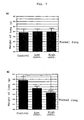
- Example 2 The culture supernatant prepared in Example 1 (100 ⁇ l), a solution of plasminogen (1 mg/ml, 100 ⁇ l) and buffer solutions at various pHs were mixed together at a ratio, 1:1:2. The mixture was incubated at 37°C and the enzymatic activity was determined as described in Example 2. The results are shown in Fig. 1 wherein distinct fragmentation patterns of plasminogen were apparent between pH ranges of more and less than 5.0.
- the arrow ( ⁇ ) indicates a bond corresponding to the plasminogen fragment from PACE reported by Gately et al.
- the reaction solution at pH 4.0 was subjected to 12.5% SDS-PAGE and the proteins were then transferred to Immovilon membrane (manufactured by Millipore) in a conventional manner.
- Immovilon membrane manufactured by Millipore
- the results are shown in Fig. 11 .
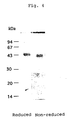
- Fig. 11 three bands were observed in electrophoresis under non-reducing condition.
- a reaction solution wherein plasminogen was fragmented under conditions described in Example 3 was subjected to 12.5% SDS-PAGE electrophoresis and the proteins were transferred to Immovilon membrane (manufactured by Millipore) by blotting. Then, dying with Amido Black and decoloration with purified water were performed and the band at around 40, 43 kDa and the band at around 35 kDa were excised. With an amino acid N-terminus analyzer (manufactured by Bio Applied), the N-terminal amino acid residue was determined for each band to identify 79Leu for the band of 40, 43 kDa and 480Pro for the band of 35 kDa. Based on their molecular weight, these 40 and 43 kDa fragments prepared by cleavage at pH 4.0 were estimated to comprise Kringles 1 to 4 and herein referred to as "PAN4" (PACE derived Angiostatin pH 4.0).
- PAN4 PACE derived Angiostatin pH 4.0
- Plasminogen was fragmented as described in Example 3 except that culture supernatants of human fibroblast cells, vascular endothelial cells from human umbilical vein, vascular endothelial cells from bovine aorta, human hepatic cancer cells (HepG2) and mouse lung cancer cells (LL/2) were used in place of PC-3 cells of Example 1. Analogous enzymes that fragmented plasminogen were found in supernatants from cancer cells other than PC-3.
- Fig. 2 schematically illustrates procedures for screening PACE4 wherein anti-Mini Plasminogen antibody is used as an immobilized antibody whereas antibody directed to Kringles 1 to 3 of plasminogen is used as a labeled antibody.
- a predetermined amount of plasminogen and samples are mixed together and the mixture is added to this system. If plasminogen is cleaved at between Kringle 4 and Mini Plasminogen, a color development will become declined in proportion to a degree of the cleavage. An enzyme level is determined based on a rate of this decline in color development.
- phosphate/citrate buffer pH 3.0
- sample culture supernatant or intermediate material obtained during purification
- 5.0 ⁇ l plasminogen or Lys plasminogen at a final concentration 20 ⁇ g/ml and the mixture was reacted at 37°C for 1 hour.
- the reaction solution was diluted with a phosphate buffer containing 20 U/ml aprotinin and 1% BSA and served as samples for ELISA.
- Anti-human LBSI antibody was dissolved in a dilution solution for antibody (phosphate buffer) at 20 ⁇ g/ml. Each 100 ⁇ l/well of the solution was added to a 96-well microtiter plate (IMMUNO MODULE MAXISORP F8, manufactured by Nunc) and the plate was incubated at 4°C overnight. The antibody solution was sucked from each well with a microtiter plate washer (manufactured by DIATECH, ULTRA WASH II). The plate was washed with PBS, added with 1% albumin 300 ⁇ l (Albumin Fraction V, Bovine, manufactured by Seikagaku Kogyo K.K.) and left to stand at 4°C overnight.
- albumin 300 ⁇ l Albumin Fraction V, Bovine, manufactured by Seikagaku Kogyo K.K.
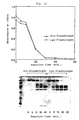
- FIG. 12 A serial dilution of a culture supernatant of PC-3 cells was measured by the system for enzyme determination according to the present invention. The results are shown in Fig. 12 wherein the open circle in the graph (upper) indicates those from plasminogen as a substrate whereas the closed circle from Lys plasminogen.
- the plasminogen-fragmenting enzyme in PC-3 culture supernatant cleaved both substrates with passage of time.
- Lys plasminogen was cleaved more efficiently at earlier stage than Glu plasminogen but such a difference in a rate of cleavage between Lys and Glu plasminogens was diminished from 10 minutes onward.
- Fig. 12 also shows in lower part how plasminogen is cleaved with passage of time with electrophoretogram.
- the enzyme of the present invention was purified by a combination of various chromatographs including hydrophobic chromatography, anion exchange chromatography, gel filtration and hydroxyapatite.
- the enzyme stock solution (5 L) prepared as described in Example 1 was diluted twice with 50 mM Tris buffer (pH 7.4).
- the solution was passed through CM-Sepharose 6B ( ⁇ 5 ⁇ 150 mm, manufactured by Pharmacia) equilibrated with 50 mM Tris/50 mM NaCl buffer (pH 7.4) and then through heparin-Sepharose 4B ( ⁇ 2.5 ⁇ 100 mm, manufactured by Pharmacia) equilibrated with the same buffer to prepare a crude solution of PACE4.
- To the crude solution was added 1 M ammonium sulfate and the mixture was left to stand overnight. The mixture was centrifuged at 6,000 rpm at 4°C and the supernatant was recovered.
- the supernatant from centrifugation was filtered through filter paper with 25 ⁇ m of pore size (AP-25: manufactured by Amicon).
- the filtrate was treated with Phenyl-Sepharose Hiperformance ( ⁇ 2.5 ⁇ 100 mm, manufactured by Pharmacia) equilibrated with 50 mM phosphate buffer (pH 7.2) containing 1 M ammonium sulfate. After washing with the same buffer, gradient elution was performed with 50 mM phosphate buffer (pH 7.2) followed by elution with 50 mM phosphate buffer (pH 7.2) containing 40% ethylene glycol.
- Active fractions which were eluted with 50 mM phosphate buffer (pH 7.2) and with the same buffer containing 40% ethylene glycol, were recovered and dialyzed against a large excess of 10 mM Tris buffer (pH 7.4). After dialysis, the solution was treated with Q-Sepharose Hiperformance ( ⁇ 1.5 ⁇ 100 mm, manufactured by Pharmacia) equilibrated with 50 mM Tris buffer (pH 7.4) and gradient elution was performed with the same buffer containing 1 M NaCl.
- Active fractions were recovered, concentrated with ultrafiltration membrane (YM-10: manufactured by Amicon) and then gel filtration was performed by passing through Sephacryl S-200 ( ⁇ 2.5 ⁇ 100 cm, manufactured by Pharmacia) equilibrated with 50 mM Tris/100 mM NaCl buffer (pH 7.4). Peaks eluted at a molecular weight of around 50,000 to 60,000 were collected and subjected to chromatography with Q-Sepharose Hiperformance ( ⁇ 1.5 ⁇ 100 mm, manufactured by Pharmacia) under the above conditions to prepare active fractions.
- YM-10 manufactured by Amicon
- the obtained active fractions were finally dialyzed against 20 mM phosphate buffer (pH 7.4), passed through hydroxyapatite ( ⁇ 2.5 ⁇ 100 mm, manufactured by BioRad) equilibrated with the same buffer and gradient elution was performed with 200 mM phosphate buffer (pH 7.4).
- the purified protein was detected as a band corresponding to a molecular weight of 42 to 45 kDa (under reduced condition).
- Fig. 4 shows SDS-PAGE after purification wherein PACE4 was identified as a protein with a molecular weight 42 to 45 kDa (under reduced condition in which sample was treated with mercaptoethanol).
- the enzyme of the present invention prepared from PC-3 culture supernatant, after being transferred to GVDF membrane (Immovilon, manufactured by Millipore) by blotting, was sequenced with an amino acid sequencer (Applied Biosystem Model 477A protein sequencer). This revealed that the said enzyme had the N-terminal amino acid sequence LVRIPLHKFT, which was identical to that of Human Cathepsin D precursor as a result of homology search.
- Plasminogen fragments produced by the enzyme of the present invention were prepared by incubating Lys plasminogen and the enzyme of the present invention and separating the reaction solution by lysine affinity chromatography. Lys plasminogen and the enzyme of the present invention were mixed together at a ratio 100:1 in a buffer of 50 mM Tris/0.15 M NaCl (pH 8.0) and reacted at 37°C for 3 hours. The reaction solution was passed through Lysine Sepharose 4B ( ⁇ 10 ⁇ 15 mm) equilibrated with the same buffer and, after washing, elution was performed with the same buffer containing 0.1 M epsilon amino caproic acid. When plasminogen was used for preparing fragments, the reaction was at 37°C overnight followed by the same procedures. The obtained fraction was dialyzed against 0.1 M ammonium carbonate, lyophilized and stored at -80°C till use.
- the plasminogen fragments comprising Kringles 1 to 4 prepared by the enzyme of the present invention were electrophoresed on 12.5% SDS polyacrylamide and dyed with Coomassie Blue. The results are shown in Fig. 13 . Two bands corresponding to a molecular weight of 55 kDa and 63 kDa were observed with the plasminogen fragments treated with 2-mercaptoethanol (under reduced condition). Also, two bands corresponding to a molecular weight of 40 kDa and 43 kDa, molecular species after reduction, were observed with the plasminogen fragments not treated with 2-mercaptoethanol (under non-reduced condition).
- the N-terminal amino acid residue was determined for the obtained plasminogen fragments. It was 77Lys in both cases where Lys plasminogen or plasminogen was used as a substrate.
- Lewis lung cancer cells LL/2, were purchased from Dainippon Seiyaku K.K. whereas Lewis lung cancer cells, 3LL, were given from Cell Resource Center for Biomedical Research, Institute of Development, Aging and Cancer, Tohoku University. These cells were cultured for maintenance on RPMI-1640/High Glucose medium supplemented with 10% FBS (manufactured by Dainippon Seiyaku K.K.) at 37°C and 5% CO 2 .
- FBS manufactured by Dainippon Seiyaku K.K.
- mice of six weeks old were purchased from Kyushu Dobutsu K.K. whereas immunodeficient (Scid) mice were purchased from Charles River. These animals were adapted and bred in a sterile house for a week, with 4 to 5 animals being accommodated per cage, until experiment.
- mice of 6 weeks old were transplanted subcutaneously 10 6 Lewis lung cancer cells (LL/2) at the back.
- the animals were bred for 14 to 17 days and cancer (primary cancerous focus) formed at the back was surgically removed. Based on size of the primary cancerous focus formed, the animals were divided into two groups: one with the primary cancerous focus of not more than 1200 mg (200 mg to 1200 mg) and the other of more than 1200 mg (1201 mg to 2500 mg).
- Each of these two groups was further divided into three groups: ones that received a high or low dose of the enzyme of the present invention (PACE4) and control.
- PACE4 high or low dose of the enzyme of the present invention
- Fig. 9 wherein (A) Group with the primary cancerous focus of not more than 1200 mg; (B) Group with the primary cancerous focus of more than 1200 mg; and the dot line indicates a weight of normal lung. As shown in Fig. 9 , no significant difference was observed between PACE4 administration and control in case of the group with the primary cancerous focus of not more than 1200 mg.
- mice of 6 weeks old were transplanted subcutaneously 10 6 Lewis lung cancer cells (LL/2) at the back.
- the animals were bred for 14 to 17 days and cancer (primary cancerous focus) formed at the back was surgically removed when it weighed 300 to 1200 mg.
- the affected part was disinfected and sutured.
- Mice were bred for additional 14 days and thereafter intraperitoneally administered with 25 ⁇ g/animal of the plasminogen fragments produced by cleavage with PACE4 for 10 days. On Day 11, mice were dissected and the lung was removed and weighed.
- Example 13 Test for Inhibitory Activity to Growth of Vascular Endothelial Cells
- a test for inhibitory activity to growth of vascular endothelial cells was performed as described by Gately et al. using Human Umbilical vein endothelial (HUV) cells (HUVEC; Sanko Jun-yaku K.K.).
- HUVEC cells were plated on a 24-well plate (Nunclone, manufactured by Nunc) at 12,500 cells/well and cultured with a culture medium attached to the kit overnight. The cells were added with 1, 10, 50, or 100 ⁇ g/ml of the plasminogen fragments or a physiological saline as a control and cultured at 5% CO 2 , 37°C, for 72 hours. The cells were peeled off the well with trypsin/EDTA solution and counted with Coulter counter (manufactured by Coulter).
- BAE cells bovine aorta endothelial (BAE) cells purchased from Sanko Jun-yaku K.K.
- BAE cells were cultured for maintenance on a 24-well plate (manufactured by Iwaki Glass K.K.) coated with gelatin in a DMEM basal medium containing 10% fetal calf serum (FCS), 3 ng/ml basic FGF (bFGF), 1% glutamine, 1% penicillin and 1% streptomycin at 5% CO 2 .
- FCS fetal calf serum
- bFGF basic FGF
- glutamine fetal calf serum
- streptomycin streptomycin
- the culture medium was replaced with a culture medium in which the plasminogen fragments were diluted to 100 ⁇ g/ml in the basal medium that contained 5% FCS but was deprived of bFGF and the plasminogen fragments were reacted with the cells at 37°C for 20 minutes.
- a physiological saline was used as a negative control whereas angiostatin (Angiostatin, manufactured by Technoclone) was used as a positive control.
- the basal medium containing 5% FCS and 2 ng/ml bFGF was added and the cells were cultured at 37°C for additional 72 hours. The cells were peeled off the well with trypsin/EDTA solution and counted with Coulter counter (manufactured by Coulter).
- the plasminogen fragments (0 to 20 ⁇ g/ml) prepared by PACE4 were reacted with the vascular endothelial cells to prove that said fragments had no inhibitory activity to growth of the vascular endothelial cells.
- angiostatin purchased from Technoclone exhibited the inhibitory activity.
- the N-terminal analysis of PACE4 revealed that the N-terminal amino acid sequence of PACE4 had a high homology with the amino acid sequence of cathepsin D, lysosomal proteinase. Cathepsin D per se had the similar activity to PACE4. Thus, it was estimated that cathepsin D was responsible for the activity of PACE4. Based on these facts, cDNA of PACE4 was isolated as described below.
- RNAs were extracted from about 1.6 ⁇ 10 7 cells of human prostate cancer cells (PC-3) in a conventional manner using ISOGEN solution (manufactured by Wako Jun-yaku K.K.). From the obtained whole RNAs, 27 ⁇ g mRNAs were purified with oligo(dT) column. The purified mRNAs together with a 10-fold amount of 3 M sodium acetate and a 2.5-fold amount of cold ethanol were stored at -80°C till use.
- cDNA of PACE4 was synthesized from 1 ⁇ g of the purified mRNA using oligo(dT) as a primer in accordance with the manufacturer's instructions. Then, sense and antisense primers for amplifying a whole translation region of PACE4 gene were synthesized based on the nucleotide sequence of cDNA of cathepsin D precursor laid open public on data base and a desired gene was amplified with AmpliTaq (manufactured by PERKIN ELMER).
- the amplified cDNA fragment was cloned into a plasmid vector in accordance with the manufacture's instructions.
- the cDNA was sequenced by the dideoxy terminating method using dideoxy nucleotides labeled with fluorescence.
- the determined nucleotide sequence and an amino acid sequence deduced therefrom were compared with the known sequences of human cathepsin D precursor to confirm an extremely high homology between them.
- PACE4 is scarcely released out of normal cells but a great deal is released out of certain cancer cells. This implies possibility that PACE4 might be a key marker for examining cancerous state.
- PACE4 has an extremely high homology with human cathepsin D precursor as described above and thus anti-PACE4 antibody also strongly responds to human cathepsin D precursor and its active form human cathepsin D. Therefore, results obtained by using anti-human cathepsin may also be applied to PACE4 and can be important information for investigating correlation between cancer and PACE4. This correlation has been reported primarily for breast cancer.
- Example 6 It is generally recognized that a high antigen level of human cathepsin D is observed in breast cancer and elevation of human cathepsin D level in turn makes breast cancer be malignant.
- the results in Example 6 indicate that the PACE4 activity that fragments plasminogen is scarcely detected for breast cancer cells (MFC-7) while Lewis lung cancer and prostate cancer cells, which inhibit growth of distally metastasized cancerous focus, release a great deal of the PACE4 activity out of cells. Accordingly, in case of breast cancer, it is inferred that there is disagreement between the antigenic level and the PACE4 activity.
- the PACE4 activity is determined by measuring with ELISA an amount of plasminogen fragments produced after fragmentation by PACE4. This determination is about 100- to 1000-fold more sensitive than the determination of cathepsin D wherein pigment level in hemoglobin fragments produced after cleavage is measured by absorbance or based on an amount of protein. Thus, a trace amount of cathepsin D present in blood cannot be determined by the conventional procedure.
- the present inventors applied the screening method for PACE4 as described in Example 7 to determination of the PACE4 activity in plasma from patients suffering from cancer and measured the PACE4 activity.
- PACE4 activity was measured as described in Example 7 for plasma from patients suffering from either breast cancer, hepatic cancer or lung cancer and for plasma from healthy individuals.
- Plasma 100 ⁇ l from patients suffering from cancer, a physiological saline 100 ⁇ l containing 1% EDTA and a pH-modifying solution 100 ⁇ l were added to a microcentrifuge tube and the mixture was incubated at 37°C overnight.
- the reaction solution was centrifuged at 15,000 rpm for 3 minutes. A supernatant was collected and used as a sample for determination.
- the sample was subjected to ELISA as described in Example 7 to measure the PACE4 activity.
- plasma 100 ⁇ l from healthy individual, PACE4/100 ⁇ l (0 to 50 Unit) and a pH-modifying solution 100 ⁇ l were added to a microcentrifuge tube and the mixture was incubated at 37°C overnight, followed by the procedures as described above.
- the obtained measurements were used to prepare a calibration curve. In this determination, endogenous plasminogen present in plasma was used as a substrate. Thus, endogenous plasminogen level was measured with ELISA for determination of plasminogen and made uniform.
- Fibronectin was purified as described by Rouslahti et al. (Method Enzymol. vol. 82, p.803-831 ) using a resin with gelatin being a ligand. Fresh lyophilized plasma 500 ml was subjected to freezing-thawing. Resulting cryoprecipitate was dissolved in PBS 200 ml and the solution was left to stand at 4°C overnight. The solution was then centrifuged at 10,000 rpm to give transparent precipitate. The precipitate was dissolved in PBS at room temperature and the solution was passed through gelatin Sepharose 4B. After thorough washing, elution was performed with PBS containing 4M urea. The obtained fibronectin was subjected to gel filtration with Sephacryl HR500 (manufactured by Pharmacia), confirmed for its purity by SDS-PAGE and stored at -80°C till use.
- Sephacryl HR500 manufactured by Pharmacia
- Vitronectin and human hepatocyte growth factor (HGF) were purchased from Becton Dickinson and CalBiochem, respectively.
- the above proteins were dissolved in 0.1M phosphate/citrate buffer and pH was adjusted to 4.0. To this solution was added the enzyme of the present invention at a ratio of PACE4 and the protein, 200:1 and the mixture was reacted at 37°C overnight. 0.5M phosphate buffer (pH 7.0) was added to the mixture to quench the reaction. The mixture was dialyzed against PBS and subjected to sterile filtration and the obtained filtrate was used as a sample.
- Fig. 14 shows electrophoretogram of the reaction mixture of PACE4 and fibronectin under indicated conditions wherein lane 1 indicates untreated fibronectin; lane 2, a reaction solution of fibronectin reacted at pH 4.0 for 12 hours; and lane 3, a reaction solution of fibronectin and PACE4 reacted at pH 4.0 for 12 hours.
- Fig. 15 shows results obtained by reacting the fragmented products of fibronectin by PACE4 with the vascular endothelial cells. As shown in Fig. 15 , the fibronectin fragments produced by PACE4 fragmentation significantly inhibited growth of the vascular endothelial cells while fibronectin per se not fragmented by PACE4 exhibited no such inhibitory activity.
Landscapes
- Health & Medical Sciences (AREA)
- Chemical & Material Sciences (AREA)
- Life Sciences & Earth Sciences (AREA)
- Engineering & Computer Science (AREA)
- Organic Chemistry (AREA)
- Bioinformatics & Cheminformatics (AREA)
- Zoology (AREA)
- Wood Science & Technology (AREA)
- Genetics & Genomics (AREA)
- General Health & Medical Sciences (AREA)
- Biomedical Technology (AREA)
- Medicinal Chemistry (AREA)
- Biochemistry (AREA)
- General Engineering & Computer Science (AREA)
- Microbiology (AREA)
- Biotechnology (AREA)
- Molecular Biology (AREA)
- Animal Behavior & Ethology (AREA)
- Public Health (AREA)
- Chemical Kinetics & Catalysis (AREA)
- Pharmacology & Pharmacy (AREA)
- General Chemical & Material Sciences (AREA)
- Nuclear Medicine, Radiotherapy & Molecular Imaging (AREA)
- Veterinary Medicine (AREA)
- Ophthalmology & Optometry (AREA)
- Oncology (AREA)
- Medicines That Contain Protein Lipid Enzymes And Other Medicines (AREA)
- Enzymes And Modification Thereof (AREA)
- Medicines Containing Material From Animals Or Micro-Organisms (AREA)
- Peptides Or Proteins (AREA)
- Preparation Of Compounds By Using Micro-Organisms (AREA)
Claims (2)
- Verwendung eines Asparaginenzyms in vitro, welches eine hohe Homologie zu einem Cathepsin D-Vorläufer hat, für die Produktion von Plasmaproteinfragmenten, die eine inhibitorische Aktivität auf Krebsmetastase und -wachstum haben, wobei das Enzym dadurch gekennzeichnet ist, dass:(a) es ein Molekulargewicht von etwa 45 kDa hat, wie mit SDS-Elektrophorese unter nicht-reduzierender Bedingung gemessen;(b) es die N-terminale Aminosäuresequenz LVRIPLHKFT hat; und(c) es Plasmaproteine in einem sauren pH-Bereich von nicht mehr als pH 5.0 abbaut, um Plasmaproteinfragmente zu produzieren, die eine inhibitorische Aktivität auf Krebsmetastase und -wachstum haben;und wobei die Plasmaproteine, die fragmentiert werden sollen, ausgewählt sind aus der Gruppe bestehend aus Plasminogen, Vitronectin und menschlichem Hepatocytenwachstumsfaktor (HGF).
- Verwendung nach Anspruch 1, wobei das Enzym Plasminogen an 73L-74F und/oder 451L-452P spaltet, um Fragmente zu produzieren, die Kringles 1 bis 4 von Plasminogen umfassen.
Applications Claiming Priority (3)
| Application Number | Priority Date | Filing Date | Title |
|---|---|---|---|
| JP29609598 | 1998-10-02 | ||
| JP10296095A JP2000106882A (ja) | 1998-10-02 | 1998-10-02 | 癌転移増殖抑制作用を有する血漿蛋白断片産生酵素および当該酵素により断片化された血漿蛋白断片 |
| PCT/JP1999/005322 WO2000020570A1 (en) | 1998-10-02 | 1999-09-29 | Enzyme producing plasma protein fragment having effect of inhibiting cancer metastasis and plasma protein fragment fragmented by the enzyme |
Publications (3)
| Publication Number | Publication Date |
|---|---|
| EP1118660A1 EP1118660A1 (de) | 2001-07-25 |
| EP1118660A4 EP1118660A4 (de) | 2003-04-23 |
| EP1118660B1 true EP1118660B1 (de) | 2011-04-27 |
Family
ID=17829071
Family Applications (1)
| Application Number | Title | Priority Date | Filing Date |
|---|---|---|---|
| EP99970118A Expired - Lifetime EP1118660B1 (de) | 1998-10-02 | 1999-09-29 | Ein plasmaprotein-fragment erzeugendes enzym mit krebsmetastase-hemmender wirkung und durch dieses enzym fragmentiertes plasmaprotein-fragment |
Country Status (6)
| Country | Link |
|---|---|
| US (1) | US20070117180A1 (de) |
| EP (1) | EP1118660B1 (de) |
| JP (1) | JP2000106882A (de) |
| AT (1) | ATE507289T1 (de) |
| DE (1) | DE69943393D1 (de) |
| WO (1) | WO2000020570A1 (de) |
Families Citing this family (20)
| Publication number | Priority date | Publication date | Assignee | Title |
|---|---|---|---|---|
| ES2229236T3 (es) | 1994-01-11 | 2005-04-16 | Dyax Corporation | Inhibidores de la plasmina humana derivados de los dominios de kunitz. |
| EP1835926A4 (de) * | 2004-11-22 | 2009-04-01 | Dyax Corp | Plasmin-hemmende therapien |
| JP5532216B2 (ja) * | 2009-12-28 | 2014-06-25 | 国立大学法人埼玉大学 | 乳癌の検出方法 |
| EP2625264B1 (de) | 2010-10-08 | 2022-12-07 | Terumo BCT, Inc. | Verfahren und systeme für züchtung und ernte von zellen in einem hohlfaser-bioreaktorsystem mit steuerungsbedingungen |
| JP6633522B2 (ja) | 2013-11-16 | 2020-01-22 | テルモ ビーシーティー、インコーポレーテッド | バイオリアクターにおける細胞増殖 |
| WO2015148704A1 (en) | 2014-03-25 | 2015-10-01 | Terumo Bct, Inc. | Passive replacement of media |
| US20160090569A1 (en) | 2014-09-26 | 2016-03-31 | Terumo Bct, Inc. | Scheduled Feed |
| WO2017004592A1 (en) | 2015-07-02 | 2017-01-05 | Terumo Bct, Inc. | Cell growth with mechanical stimuli |
| ES2900302T3 (es) | 2016-04-28 | 2022-03-16 | Alkahest Inc | Fracciones de plasma como terapia para el crecimiento y la progresión del tumor |
| JP7034949B2 (ja) | 2016-05-25 | 2022-03-14 | テルモ ビーシーティー、インコーポレーテッド | 細胞の増殖 |
| US11685883B2 (en) | 2016-06-07 | 2023-06-27 | Terumo Bct, Inc. | Methods and systems for coating a cell growth surface |
| US11104874B2 (en) | 2016-06-07 | 2021-08-31 | Terumo Bct, Inc. | Coating a bioreactor |
| US11624046B2 (en) | 2017-03-31 | 2023-04-11 | Terumo Bct, Inc. | Cell expansion |
| US12234441B2 (en) | 2017-03-31 | 2025-02-25 | Terumo Bct, Inc. | Cell expansion |
| CN117247899A (zh) | 2017-03-31 | 2023-12-19 | 泰尔茂比司特公司 | 细胞扩增 |
| AU2020388572A1 (en) * | 2019-11-20 | 2022-05-26 | Alkahest, Inc. | Blood plasma fractions for use in liver regeneration |
| US11484551B2 (en) | 2019-11-20 | 2022-11-01 | Alkahest, Inc. | Method of treating liver failure with plasma fraction IV-4 |
| GB2619893A (en) | 2021-03-23 | 2023-12-20 | Terumo Bct Inc | Cell capture and expansion |
| US12152699B2 (en) | 2022-02-28 | 2024-11-26 | Terumo Bct, Inc. | Multiple-tube pinch valve assembly |
| USD1099116S1 (en) | 2022-09-01 | 2025-10-21 | Terumo Bct, Inc. | Display screen or portion thereof with a graphical user interface for displaying cell culture process steps and measurements of an associated bioreactor device |
Family Cites Families (5)
| Publication number | Priority date | Publication date | Assignee | Title |
|---|---|---|---|---|
| US5198423A (en) * | 1989-05-26 | 1993-03-30 | Takara Shuzo Co., Ltd. | Functional polypeptide containing a cell binding domain and a heparin binding domain of fibronectin |
| US6566098B1 (en) * | 1990-09-14 | 2003-05-20 | The United States Of America As Represented By The Department Of Health And Human Services | DNA encoding truncated hepatocyte growth factor variants |
| US5288489A (en) * | 1991-08-28 | 1994-02-22 | Orion Therapeutic Systems, Inc. | Fibrinolysis and fibrinogenolysis treatment |
| US5800814A (en) * | 1994-04-22 | 1998-09-01 | Oklahoma Medical Research Foundation | Method for inhibition of breast tumor growth |
| EP0837074A3 (de) * | 1996-09-19 | 1998-09-16 | Hisamitsu Pharmaceutical Co., Inc. | Biologisch aktives Fibronektin-Fragment als Krebsmetastasehemmstoff |
-
1998
- 1998-10-02 JP JP10296095A patent/JP2000106882A/ja active Pending
-
1999
- 1999-09-29 DE DE69943393T patent/DE69943393D1/de not_active Expired - Lifetime
- 1999-09-29 AT AT99970118T patent/ATE507289T1/de not_active IP Right Cessation
- 1999-09-29 EP EP99970118A patent/EP1118660B1/de not_active Expired - Lifetime
- 1999-09-29 WO PCT/JP1999/005322 patent/WO2000020570A1/ja not_active Ceased
-
2006
- 2006-10-04 US US11/542,180 patent/US20070117180A1/en not_active Abandoned
Also Published As
| Publication number | Publication date |
|---|---|
| WO2000020570A1 (en) | 2000-04-13 |
| DE69943393D1 (de) | 2011-06-09 |
| ATE507289T1 (de) | 2011-05-15 |
| EP1118660A4 (de) | 2003-04-23 |
| US20070117180A1 (en) | 2007-05-24 |
| JP2000106882A (ja) | 2000-04-18 |
| EP1118660A1 (de) | 2001-07-25 |
Similar Documents
| Publication | Publication Date | Title |
|---|---|---|
| EP1118660B1 (de) | Ein plasmaprotein-fragment erzeugendes enzym mit krebsmetastase-hemmender wirkung und durch dieses enzym fragmentiertes plasmaprotein-fragment | |
| ES2370155T3 (es) | Angiostatina y método para la inhibición de la angiogénesis. | |
| EP0824546B1 (de) | Angiostatinfragmente und verfahren für deren verwendung | |
| US5801146A (en) | Compound and method for inhibiting angiogenesis | |
| KR20010020458A (ko) | 안지오스타틴 단편 및 사용방법 | |
| JP2006213724A (ja) | アンジオスタチンおよび血管形成の抑制におけるその使用 | |
| CZ286016B6 (cs) | Derivát lidské bílkoviny C, způsob jeho přípravy a farmaceutický prostředek, který ho obsahuje | |
| JP2002510209A (ja) | インビボで血管形成を調節することができるプラスミノーゲンのクリングルドメイン1−5 | |
| EP0914830B1 (de) | Hemmstoff für die gefässneubildung, der den gewebefaktor-inhibitor (tfpi) enthält | |
| EP2970434B1 (de) | Potenter kunitz-inhibitor der fibrinolyse mit dualer reaktivität | |
| KR20010052371A (ko) | 세린 프로테아제를 이용한 내피 세포 증식 억제 및안기오게네시스 조절을 위한 조성물 및 방법 | |
| JP2002538803A (ja) | 新規の抗脈管形成ペプチド | |
| WO2000049871A1 (en) | An anti-angiogenic kringle protein and its mutants | |
| JP4452791B2 (ja) | カテプシンeの腫瘍マーカーとしての用途およびカテプシンeならびにカテプシンdの腫瘍血管新生阻害療法のターゲットとしての用途 | |
| JP2002535372A (ja) | プラスミノーゲンクリングル4領域フラグメントおよび利用法 | |
| JP4666767B2 (ja) | プラスミノーゲンの脱グリコシル化クリングル1〜5領域フラグメント及びこれらの使用方法 | |
| JP2005528879A (ja) | 抗脈管形成剤および抗腫瘍剤としてのヒスチジンプロリンリッチ糖タンパク質(hprg) | |
| US20020031518A1 (en) | Plasminogen fragment having activity to inhibit tumor metastasis and growth and process for preparing same technical field | |
| US7157556B1 (en) | Deglycosylated kringle 1-3 region fragments of plasminogen and methods of use | |
| JP3806471B2 (ja) | 腫瘍転移増殖抑制効果を有するプラスミノーゲン断片および該断片の調製方法 | |
| US7119069B2 (en) | Human kininogen D3 domain polypeptide as an anti-angiogenic and anti-tumor agent | |
| MXPA99011041A (en) | Angiostatin fragments and method of use | |
| KR19990008029A (ko) | 안기오스타틴 단편과 안기오스타틴 응집체 및 이의 이용방법 | |
| HK1002457B (en) | Angiostatin fragments and methods of use | |
| KR19990088583A (ko) | 신혈관형성억제용단백질의정제방법 |
Legal Events
| Date | Code | Title | Description |
|---|---|---|---|
| PUAI | Public reference made under article 153(3) epc to a published international application that has entered the european phase |
Free format text: ORIGINAL CODE: 0009012 |
|
| 17P | Request for examination filed |
Effective date: 20010403 |
|
| AK | Designated contracting states |
Kind code of ref document: A1 Designated state(s): AT BE CH CY DE DK ES FI FR GB GR IE IT LI LU MC NL PT SE |
|
| AX | Request for extension of the european patent |
Free format text: AL;LT;LV;MK;RO;SI |
|
| RIC1 | Information provided on ipc code assigned before grant |
Free format text: 7C 12N 15/00 A, 7C 12N 9/50 B, 7C 07K 14/78 B, 7C 07K 14/745 B, 7C 07K 1/22 B, 7A 61K 38/48 B, 7C 12N 9/64 B, 7C 12N 9/68 B |
|
| A4 | Supplementary search report drawn up and despatched |
Effective date: 20030307 |
|
| 17Q | First examination report despatched |
Effective date: 20050824 |
|
| 17Q | First examination report despatched |
Effective date: 20050824 |
|
| GRAP | Despatch of communication of intention to grant a patent |
Free format text: ORIGINAL CODE: EPIDOSNIGR1 |
|
| GRAC | Information related to communication of intention to grant a patent modified |
Free format text: ORIGINAL CODE: EPIDOSCIGR1 |
|
| GRAS | Grant fee paid |
Free format text: ORIGINAL CODE: EPIDOSNIGR3 |
|
| GRAA | (expected) grant |
Free format text: ORIGINAL CODE: 0009210 |
|
| RIN1 | Information on inventor provided before grant (corrected) |
Inventor name: MIYAMOTO, SEIJI Inventor name: NOZAKI, CHIKATERU Inventor name: MAEDA, HIROAKI Inventor name: TAKEMOTO, SUMIYO Inventor name: KAMINAKA, KAZUYOSHI Inventor name: MORIKAWA, WATARU |
|
| AK | Designated contracting states |
Kind code of ref document: B1 Designated state(s): AT BE CH CY DE DK ES FI FR GB GR IE IT LI LU MC NL PT SE |
|
| REG | Reference to a national code |
Ref country code: GB Ref legal event code: FG4D |
|
| REG | Reference to a national code |
Ref country code: CH Ref legal event code: EP |
|
| REG | Reference to a national code |
Ref country code: IE Ref legal event code: FG4D |
|
| REF | Corresponds to: |
Ref document number: 69943393 Country of ref document: DE Date of ref document: 20110609 Kind code of ref document: P |
|
| REG | Reference to a national code |
Ref country code: DE Ref legal event code: R096 Ref document number: 69943393 Country of ref document: DE Effective date: 20110609 |
|
| REG | Reference to a national code |
Ref country code: NL Ref legal event code: VDEP Effective date: 20110427 |
|
| PG25 | Lapsed in a contracting state [announced via postgrant information from national office to epo] |
Ref country code: PT Free format text: LAPSE BECAUSE OF FAILURE TO SUBMIT A TRANSLATION OF THE DESCRIPTION OR TO PAY THE FEE WITHIN THE PRESCRIBED TIME-LIMIT Effective date: 20110829 Ref country code: SE Free format text: LAPSE BECAUSE OF FAILURE TO SUBMIT A TRANSLATION OF THE DESCRIPTION OR TO PAY THE FEE WITHIN THE PRESCRIBED TIME-LIMIT Effective date: 20110427 |
|
| PG25 | Lapsed in a contracting state [announced via postgrant information from national office to epo] |
Ref country code: ES Free format text: LAPSE BECAUSE OF FAILURE TO SUBMIT A TRANSLATION OF THE DESCRIPTION OR TO PAY THE FEE WITHIN THE PRESCRIBED TIME-LIMIT Effective date: 20110807 Ref country code: AT Free format text: LAPSE BECAUSE OF FAILURE TO SUBMIT A TRANSLATION OF THE DESCRIPTION OR TO PAY THE FEE WITHIN THE PRESCRIBED TIME-LIMIT Effective date: 20110427 Ref country code: GR Free format text: LAPSE BECAUSE OF FAILURE TO SUBMIT A TRANSLATION OF THE DESCRIPTION OR TO PAY THE FEE WITHIN THE PRESCRIBED TIME-LIMIT Effective date: 20110728 Ref country code: CY Free format text: LAPSE BECAUSE OF FAILURE TO SUBMIT A TRANSLATION OF THE DESCRIPTION OR TO PAY THE FEE WITHIN THE PRESCRIBED TIME-LIMIT Effective date: 20110427 Ref country code: BE Free format text: LAPSE BECAUSE OF FAILURE TO SUBMIT A TRANSLATION OF THE DESCRIPTION OR TO PAY THE FEE WITHIN THE PRESCRIBED TIME-LIMIT Effective date: 20110427 Ref country code: FI Free format text: LAPSE BECAUSE OF FAILURE TO SUBMIT A TRANSLATION OF THE DESCRIPTION OR TO PAY THE FEE WITHIN THE PRESCRIBED TIME-LIMIT Effective date: 20110427 |
|
| PG25 | Lapsed in a contracting state [announced via postgrant information from national office to epo] |
Ref country code: NL Free format text: LAPSE BECAUSE OF FAILURE TO SUBMIT A TRANSLATION OF THE DESCRIPTION OR TO PAY THE FEE WITHIN THE PRESCRIBED TIME-LIMIT Effective date: 20110427 |
|
| PG25 | Lapsed in a contracting state [announced via postgrant information from national office to epo] |
Ref country code: DK Free format text: LAPSE BECAUSE OF FAILURE TO SUBMIT A TRANSLATION OF THE DESCRIPTION OR TO PAY THE FEE WITHIN THE PRESCRIBED TIME-LIMIT Effective date: 20110427 |
|
| PLBE | No opposition filed within time limit |
Free format text: ORIGINAL CODE: 0009261 |
|
| STAA | Information on the status of an ep patent application or granted ep patent |
Free format text: STATUS: NO OPPOSITION FILED WITHIN TIME LIMIT |
|
| 26N | No opposition filed |
Effective date: 20120130 |
|
| PG25 | Lapsed in a contracting state [announced via postgrant information from national office to epo] |
Ref country code: MC Free format text: LAPSE BECAUSE OF NON-PAYMENT OF DUE FEES Effective date: 20110930 |
|
| REG | Reference to a national code |
Ref country code: CH Ref legal event code: PL |
|
| REG | Reference to a national code |
Ref country code: DE Ref legal event code: R097 Ref document number: 69943393 Country of ref document: DE Effective date: 20120130 |
|
| PG25 | Lapsed in a contracting state [announced via postgrant information from national office to epo] |
Ref country code: IT Free format text: LAPSE BECAUSE OF FAILURE TO SUBMIT A TRANSLATION OF THE DESCRIPTION OR TO PAY THE FEE WITHIN THE PRESCRIBED TIME-LIMIT Effective date: 20110427 |
|
| REG | Reference to a national code |
Ref country code: IE Ref legal event code: MM4A |
|
| PG25 | Lapsed in a contracting state [announced via postgrant information from national office to epo] |
Ref country code: IE Free format text: LAPSE BECAUSE OF NON-PAYMENT OF DUE FEES Effective date: 20110929 Ref country code: LI Free format text: LAPSE BECAUSE OF NON-PAYMENT OF DUE FEES Effective date: 20110930 Ref country code: CH Free format text: LAPSE BECAUSE OF NON-PAYMENT OF DUE FEES Effective date: 20110930 |
|
| PG25 | Lapsed in a contracting state [announced via postgrant information from national office to epo] |
Ref country code: LU Free format text: LAPSE BECAUSE OF NON-PAYMENT OF DUE FEES Effective date: 20110929 |
|
| PGFP | Annual fee paid to national office [announced via postgrant information from national office to epo] |
Ref country code: FR Payment date: 20130911 Year of fee payment: 15 Ref country code: GB Payment date: 20130920 Year of fee payment: 15 |
|
| PGFP | Annual fee paid to national office [announced via postgrant information from national office to epo] |
Ref country code: DE Payment date: 20131031 Year of fee payment: 15 |
|
| REG | Reference to a national code |
Ref country code: DE Ref legal event code: R119 Ref document number: 69943393 Country of ref document: DE |
|
| GBPC | Gb: european patent ceased through non-payment of renewal fee |
Effective date: 20140929 |
|
| REG | Reference to a national code |
Ref country code: DE Ref legal event code: R119 Ref document number: 69943393 Country of ref document: DE Effective date: 20150401 |
|
| REG | Reference to a national code |
Ref country code: FR Ref legal event code: ST Effective date: 20150529 |
|
| PG25 | Lapsed in a contracting state [announced via postgrant information from national office to epo] |
Ref country code: GB Free format text: LAPSE BECAUSE OF NON-PAYMENT OF DUE FEES Effective date: 20140929 Ref country code: DE Free format text: LAPSE BECAUSE OF NON-PAYMENT OF DUE FEES Effective date: 20150401 |
|
| PG25 | Lapsed in a contracting state [announced via postgrant information from national office to epo] |
Ref country code: FR Free format text: LAPSE BECAUSE OF NON-PAYMENT OF DUE FEES Effective date: 20140930 |