EP1318401A2 - Verfahren zur Früherkennung der Serokonversion eines gegen Hepatitis C-Virus gerichteten Antikörpers - Google Patents
Verfahren zur Früherkennung der Serokonversion eines gegen Hepatitis C-Virus gerichteten Antikörpers Download PDFInfo
- Publication number
- EP1318401A2 EP1318401A2 EP03001660A EP03001660A EP1318401A2 EP 1318401 A2 EP1318401 A2 EP 1318401A2 EP 03001660 A EP03001660 A EP 03001660A EP 03001660 A EP03001660 A EP 03001660A EP 1318401 A2 EP1318401 A2 EP 1318401A2
- Authority
- EP
- European Patent Office
- Prior art keywords
- polypeptide
- antigen
- antibody
- solid phase
- hcv
- Prior art date
- Legal status (The legal status is an assumption and is not a legal conclusion. Google has not performed a legal analysis and makes no representation as to the accuracy of the status listed.)
- Granted
Links
Classifications
-
- C—CHEMISTRY; METALLURGY
- C07—ORGANIC CHEMISTRY
- C07K—PEPTIDES
- C07K14/00—Peptides having more than 20 amino acids; Gastrins; Somatostatins; Melanotropins; Derivatives thereof
- C07K14/005—Peptides having more than 20 amino acids; Gastrins; Somatostatins; Melanotropins; Derivatives thereof from viruses
-
- A—HUMAN NECESSITIES
- A61—MEDICAL OR VETERINARY SCIENCE; HYGIENE
- A61P—SPECIFIC THERAPEUTIC ACTIVITY OF CHEMICAL COMPOUNDS OR MEDICINAL PREPARATIONS
- A61P1/00—Drugs for disorders of the alimentary tract or the digestive system
- A61P1/16—Drugs for disorders of the alimentary tract or the digestive system for liver or gallbladder disorders, e.g. hepatoprotective agents, cholagogues, litholytics
-
- A—HUMAN NECESSITIES
- A61—MEDICAL OR VETERINARY SCIENCE; HYGIENE
- A61P—SPECIFIC THERAPEUTIC ACTIVITY OF CHEMICAL COMPOUNDS OR MEDICINAL PREPARATIONS
- A61P31/00—Antiinfectives, i.e. antibiotics, antiseptics, chemotherapeutics
- A61P31/12—Antivirals
-
- A—HUMAN NECESSITIES
- A61—MEDICAL OR VETERINARY SCIENCE; HYGIENE
- A61P—SPECIFIC THERAPEUTIC ACTIVITY OF CHEMICAL COMPOUNDS OR MEDICINAL PREPARATIONS
- A61P31/00—Antiinfectives, i.e. antibiotics, antiseptics, chemotherapeutics
- A61P31/12—Antivirals
- A61P31/14—Antivirals for RNA viruses
-
- G—PHYSICS
- G01—MEASURING; TESTING
- G01N—INVESTIGATING OR ANALYSING MATERIALS BY DETERMINING THEIR CHEMICAL OR PHYSICAL PROPERTIES
- G01N33/00—Investigating or analysing materials by specific methods not covered by groups G01N1/00 - G01N31/00
- G01N33/48—Biological material, e.g. blood, urine; Haemocytometers
- G01N33/50—Chemical analysis of biological material, e.g. blood, urine; Testing involving biospecific ligand binding methods; Immunological testing
- G01N33/53—Immunoassay; Biospecific binding assay; Materials therefor
- G01N33/569—Immunoassay; Biospecific binding assay; Materials therefor for microorganisms, e.g. protozoa, bacteria, viruses
- G01N33/56983—Viruses
-
- G—PHYSICS
- G01—MEASURING; TESTING
- G01N—INVESTIGATING OR ANALYSING MATERIALS BY DETERMINING THEIR CHEMICAL OR PHYSICAL PROPERTIES
- G01N33/00—Investigating or analysing materials by specific methods not covered by groups G01N1/00 - G01N31/00
- G01N33/48—Biological material, e.g. blood, urine; Haemocytometers
- G01N33/50—Chemical analysis of biological material, e.g. blood, urine; Testing involving biospecific ligand binding methods; Immunological testing
- G01N33/53—Immunoassay; Biospecific binding assay; Materials therefor
- G01N33/576—Immunoassay; Biospecific binding assay; Materials therefor for hepatitis
- G01N33/5767—Immunoassay; Biospecific binding assay; Materials therefor for hepatitis non-A, non-B hepatitis
-
- A—HUMAN NECESSITIES
- A61—MEDICAL OR VETERINARY SCIENCE; HYGIENE
- A61K—PREPARATIONS FOR MEDICAL, DENTAL OR TOILETRY PURPOSES
- A61K38/00—Medicinal preparations containing peptides
-
- C—CHEMISTRY; METALLURGY
- C12—BIOCHEMISTRY; BEER; SPIRITS; WINE; VINEGAR; MICROBIOLOGY; ENZYMOLOGY; MUTATION OR GENETIC ENGINEERING
- C12N—MICROORGANISMS OR ENZYMES; COMPOSITIONS THEREOF; PROPAGATING, PRESERVING, OR MAINTAINING MICROORGANISMS; MUTATION OR GENETIC ENGINEERING; CULTURE MEDIA
- C12N2770/00—MICROORGANISMS OR ENZYMES; COMPOSITIONS THEREOF; PROPAGATING, PRESERVING, OR MAINTAINING MICROORGANISMS; MUTATION OR GENETIC ENGINEERING; CULTURE MEDIA ssRNA viruses positive-sense
- C12N2770/00011—Details
- C12N2770/24011—Flaviviridae
- C12N2770/24211—Hepacivirus, e.g. hepatitis C virus, hepatitis G virus
- C12N2770/24222—New viral proteins or individual genes, new structural or functional aspects of known viral proteins or genes
-
- Y—GENERAL TAGGING OF NEW TECHNOLOGICAL DEVELOPMENTS; GENERAL TAGGING OF CROSS-SECTIONAL TECHNOLOGIES SPANNING OVER SEVERAL SECTIONS OF THE IPC; TECHNICAL SUBJECTS COVERED BY FORMER USPC CROSS-REFERENCE ART COLLECTIONS [XRACs] AND DIGESTS
- Y10—TECHNICAL SUBJECTS COVERED BY FORMER USPC
- Y10S—TECHNICAL SUBJECTS COVERED BY FORMER USPC CROSS-REFERENCE ART COLLECTIONS [XRACs] AND DIGESTS
- Y10S436/00—Chemistry: analytical and immunological testing
- Y10S436/82—Hepatitis associated antigens and antibodies
Definitions
- the invention relates to a method for improved early detection of Seroconversion of an anti-hepatitis C virus (HCV) Antibody in a sample liquid.
- HCV hepatitis C virus
- HCV hepatitis C virus
- HCV hepatitis C virus
- Structural proteins are made from this genome (Core and envelope proteins) and non-structural proteins.
- the non-structural protein 3 (NS3) region of HCV contains one protease and one Helicase. Protease activity is in the amino terminal third of the NS3 region localized.
- EP-A-0 318 216 there is a partial Nucleotide sequence of HCV disclosed. There will be the use of Nucleic acid fragments or polypeptide sections from HCV for diagnostic evidence and therapeutic treatment claimed.
- EP-A-0 450 931 discloses the complete nucleotide and Amino acid sequence from HCV. Furthermore, a combination of synthetic HCV antigens described, comprising a first HCV antigen from the C domain and at least one further HCV antigen one of the non-structural domains NS3, NS4 or NS5 and the envelope domain S.
- a preferred antigen from the NS3 domain is one with C33c designated antigen, which amino acids 1192 to 1457 of the in Figure 1 of EP-A-0 450 931 comprises HCV genome shown.
- HCV proteins are called fusion proteins with ⁇ -galactosidase in E. coli expressed.
- a protein from the NS3 region showed the Amino acids 1295 to 1541 of the HCV genome contained the highest Sensitivity.
- a disadvantage of such fusion proteins is that Cross-reactions with the fused protein portion can occur which reduce the specificity of the detection reaction.
- One of the objects underlying the present invention was therefore in the provision of a polypeptide from the HCV genome for a process for improved early detection of seroconversion against hepatitis C virus-directed antibody, which has these disadvantages of the prior art Technology are at least partially eliminated and in particular a higher specificity and sensitivity compared to known antigens possesses and is expressible and stable in high yield.
- polypeptide that consists of sequence regions Contains hepatitis C virus, especially from the NS3 region.
- the subject of the present application is therefore a method for improved early detection of seroconversion of a hepatitis C virus directed antibody in a sample liquid, taking the Sample liquid with at least one polypeptide, the sequence regions from hepatitis C virus, in particular from the NS3 region of hepatitis C virus, contains, incubated and the antibody via a binding with the Detects polypeptide, where a polypeptide from a range that contains at least one cysteine residue, and (a) the determination performing the antibody under reducing conditions, (b) one or several cysteine residues covalently modified and / and (c) one or several cysteine residues replaced by other amino acids.
- the method according to the invention is based on the immunological Determination of an antibody directed against hepatitis C virus in a Sample liquid, wherein the sample liquid with at least one Polypeptide, the sequence regions from the hepatitis C virus, in particular contains from the NS3 region of hepatitis C virus, incubated and the Detects antibody by binding to the polypeptide, thereby characterized in that a polypeptide from a range that contains at least one cysteine residue, and (a) the determination performing the antibody under reducing conditions, (b) one or several cysteine residues covalently modified and / and (c) one or several cysteine residues replaced by other amino acids.
- the NS3 region contains a particularly large cluster of cysteine residues.
- the use of NS3 antigens has been shown to be physiological Buffer conditions, e.g. in immunological test procedures, as extreme problematic because under these conditions the free sulfhydryl groups the cysteine residues oxidize easily. This leads to both intra- and to intermolecular violations of the antigen, causing its immunological reactivity is significantly reduced.
- polypeptide is a polypeptide made up of amino acids 1207 ⁇ 10 to 1488 ⁇ 10 of a hepatitis C virus and less than 20, preferably less than 15 foreign amino acids.
- the polypeptide contains amino acids 1207 ⁇ 5 to 1488 ⁇ 5, more preferably 1207 ⁇ 2 to 1488 ⁇ 2 and most preferred 1207 to 1488 of a hepatitis C virus, the numbering of the Amino acid residues refer to Figure 1 of EP-A-0 450 931.
- the polypeptide used in the method according to the invention can consist of any HCV isolate, for example from an HCV isolate with a nucleotide sequence as described in EP-A-0 450 931.
- the polypeptide preferably originates from the HCV isolate from which the clone NS3 described in WO 92/11370 comes from the German Collection of Microorganisms and Cell Cultures GmbH (DSM), Mascheroder Weg 1b, 38124 Braunschweig, under the number DSM 6847 was deposited. This is particularly preferred in the invention Polypeptide used by recombinant expression of the method Vector pUC-D26 available.
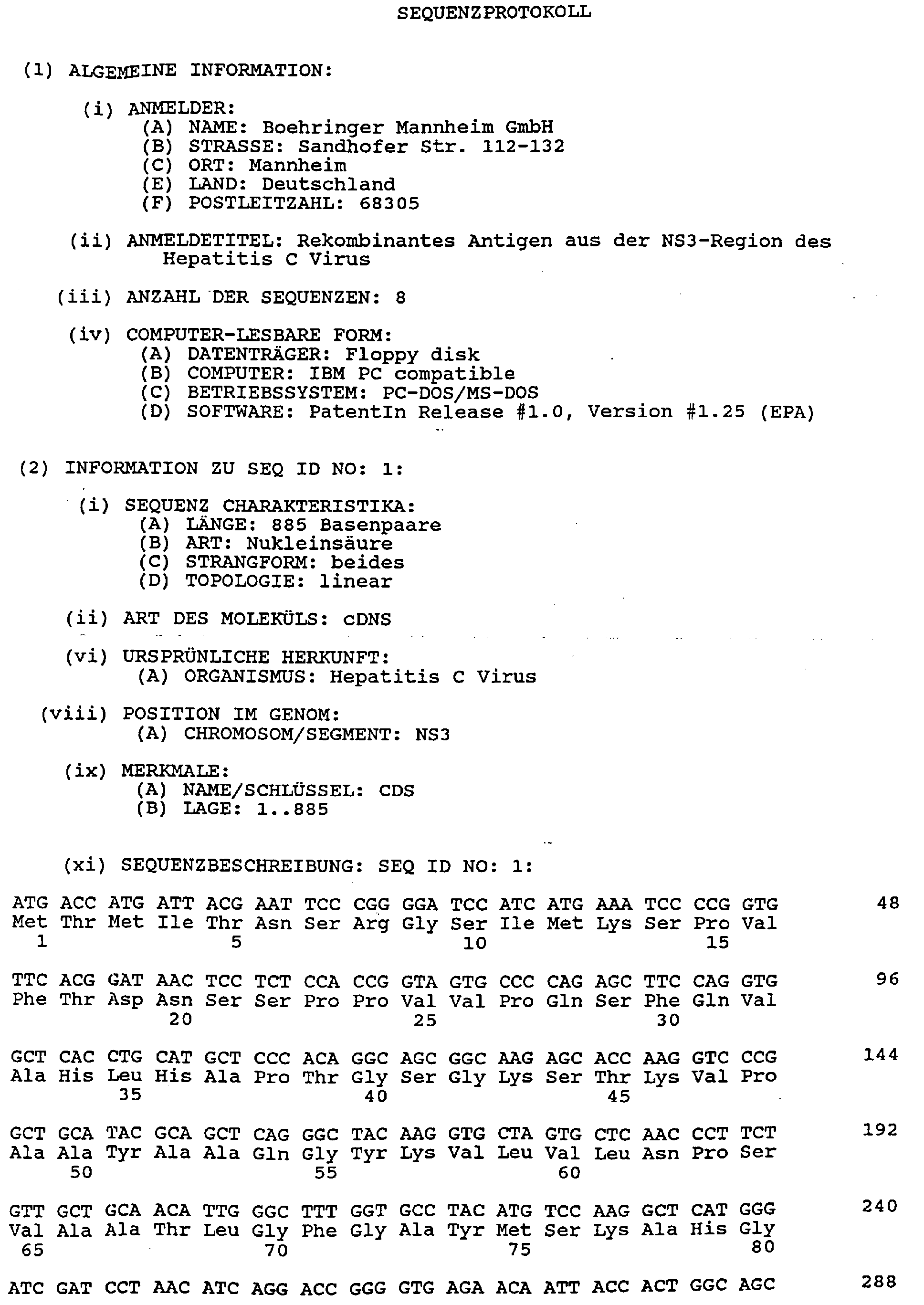
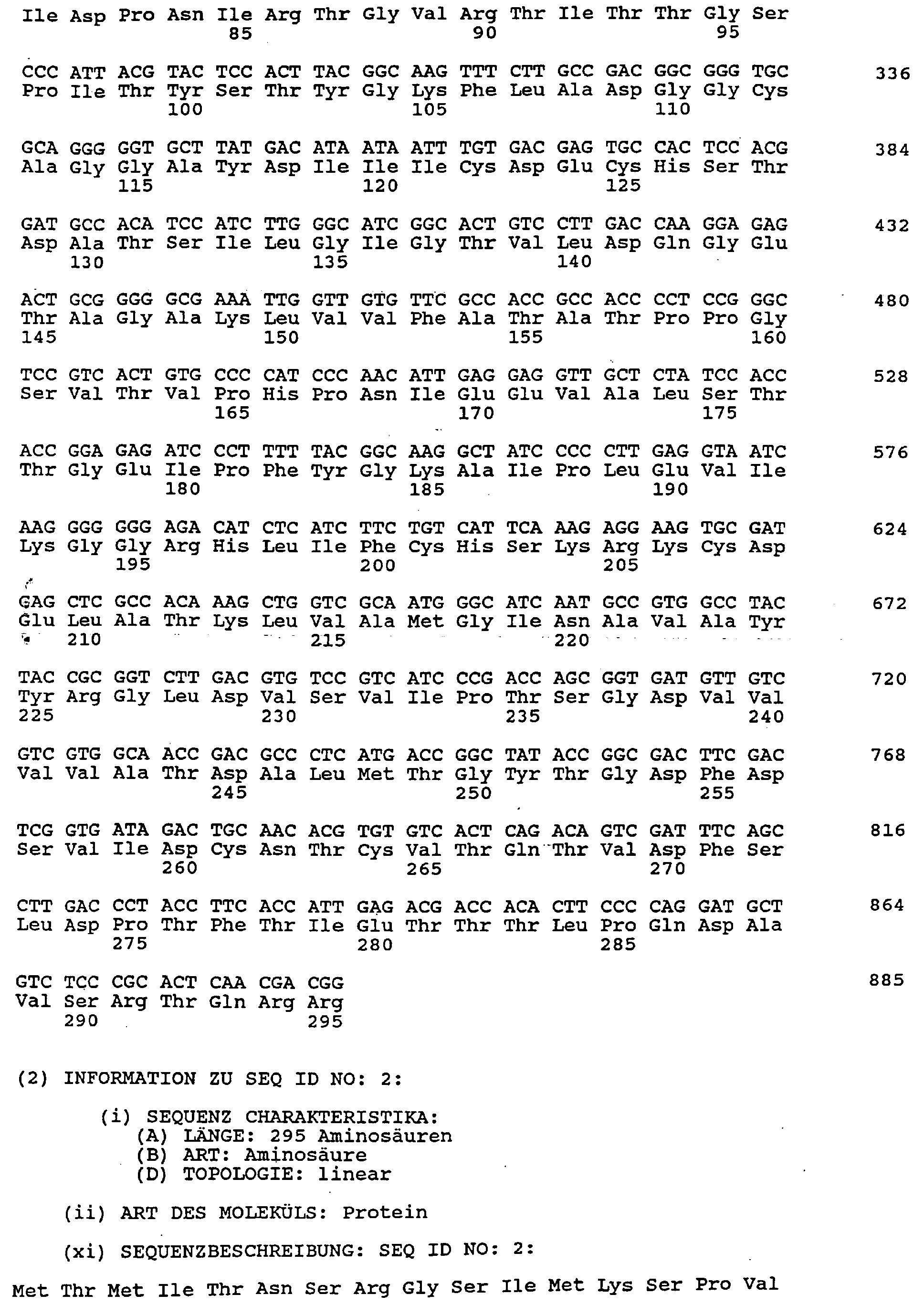
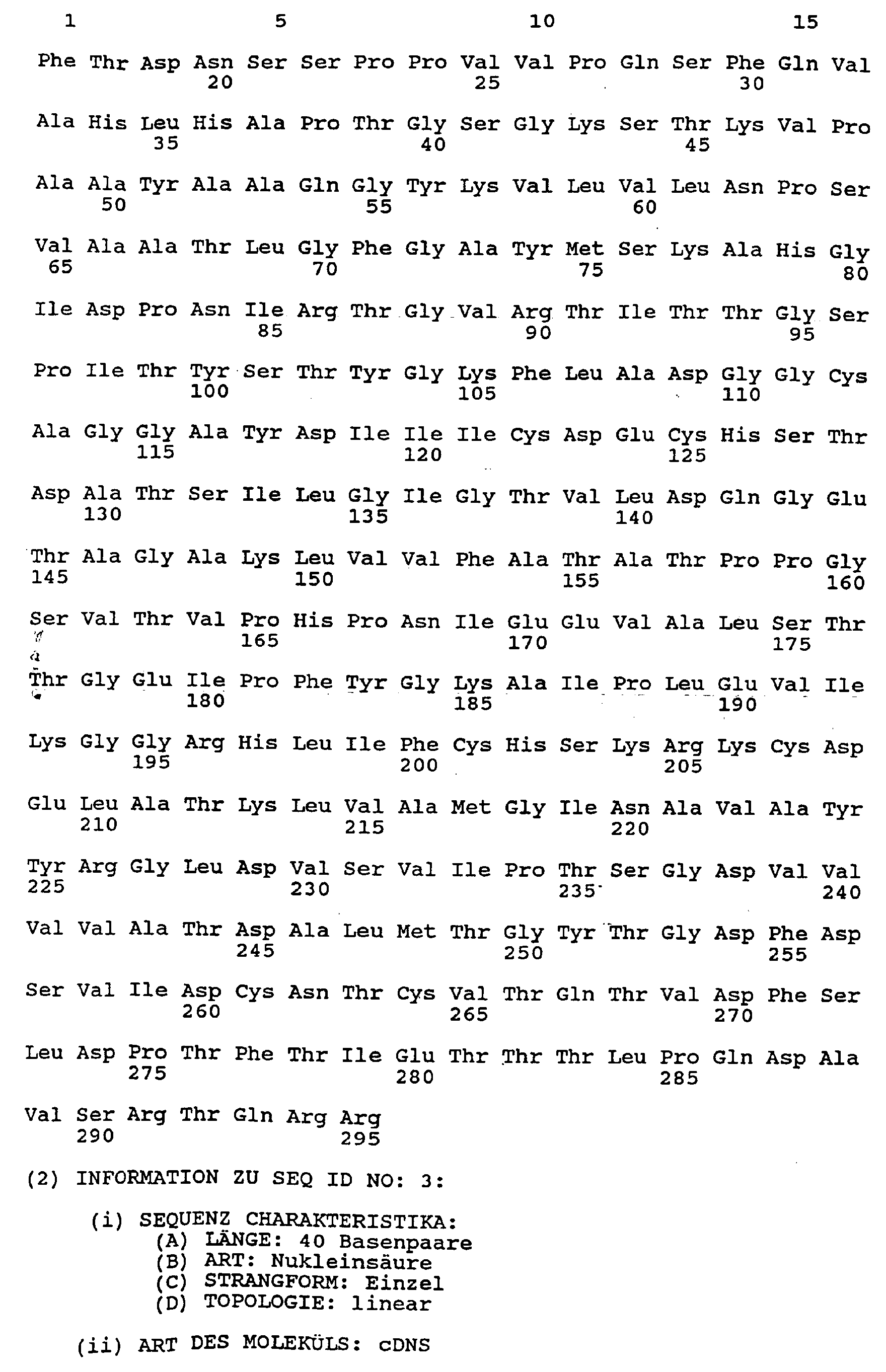
- SEQ ID NO. 1 is the amino acid sequence of one in the Polypeptide preferably used method according to the invention shown.
- Amino acids 1-13 are foreign amino acids.
- Amino acids 14-295 come from HCV.
- the invention particularly preferably contains Polypeptide the amino acids 14-295 of the in SEQ ID NO. 1 and 2 amino acid sequence shown or at least 90% of it homologous amino acid sequence.
- Polypeptide may be a polypeptide as defined above which contains at least one Marking group carries. All known come as a marking group Marker groups in question that have been demonstrated in a test system can be, i.e. directly or indirectly detectable Labeling groups. It is under a directly detectable Marker group to understand such a group that a directly generates a detectable signal, e.g. a radioactive group, a Enzyme group, a luminescent group, a metal complex, etc. On the other hand, the marking group can also be an indirectly detectable one Be a group, e.g.
- a biotin or hapten group which by reaction with a suitable binding partner (streptavidin, avidin or anti-hapten antibody), which in turn carries a signal generating group, is detectable.
- the labeling group can be based on the antigen be coupled in a known manner, for example via a bifunctional spacer. Such methods for coupling Labeling groups on peptide antigens are known to a person skilled in the art Known in the field of immunology and need not be described here become.
- the polypeptide used in the method according to the invention contains preferably 1 to 12 and particularly preferably 3 to 7 Labeling groups.
- Polypeptide be a polypeptide as defined above in which one or several of the sulfhydryl groups from cysteine residues in covalent modified form.
- suitable covalent Modification groups are maleimidodioxaoctylamine (MADOO), N-methyl-maleimide (NMM), iodoacetic acid and iodoacetamide.
- the covalent coupling is particularly preferably carried out directly or indirectly detectable labeling groups on the sulfhydryl groups of Polypeptide.
- SH-reactive bifunctional linkers for coupling Sulfhydryl groups are maleimidopropylamine (MP), maleimidoethylamine (MEA) and maleimidodioxaoctylamine (MADOO).
- cysteine residues pass through other natural or artificial amino acids are replaced.
- cysteine residues are replaced by structurally analogous ⁇ -amino acids, e.g. serine, or ⁇ -aminobutyric acid replaced. With cysteine substitutions one maintain particularly high stability. This was especially true for NS3 + DTT are shown.
- Polypeptide has surprising compared to known polypeptides Advantages on. Compared to the antigen described in EP-A-0 450 931 C33, which contains amino acid residues 1192 to 1457 of the HCV sequence contains, this polypeptide shows a significantly higher specificity, which can be found in a statistically significantly lower number of false positives Results with negative sera shows. Compared to that in WO 92/11370 described antigen NS3, which covers the range of amino acids 1007 contains up to 1534 of the HCV sequence, this polypeptide clearly shows a higher stability under test conditions.
- this polypeptide Compared to a polypeptide that which contains amino acids 1227 to 1528 from the NS3 region of HCV this polypeptide has the advantage of better expression efficiency and one higher sensitivity. Because of these advantages, that's in the Polypeptide used according to the invention preferably all previously known HCV antigens from the NS3 region are clearly superior.
- the polypeptide used in the method according to the invention is through encodes a nucleic acid.
- a preferred example of such Nucleic acid is the foreign DNA insertion in the vector pUC-D26.
- SEQ ID NO. 1 is also the nucleotide sequence for an im Process according to the invention preferably used polypeptide coding nucleic acid shown for the polypeptide of SEQ ID NO. 1 and 2 coded. Nucleotides 40-885 code for that from HCV originating region of the polypeptide.
- the for one in the invention Process preferably used nucleic acid encoding polypeptide preferably contains (a) nucleotides 40-885 of the one shown in SEQ ID NO. 1 nucleotide sequence shown or (b) one of the sequence from (a) in the frame nucleotide sequence corresponding to the degeneration of the genetic code.
- nucleic acid coding for the polypeptide used is present in a vector, wherein the vector has at least one copy of such a nucleic acid contains.
- the vector is preferably a prokaryotic vector, i.e. on vector suitable for propagation in a prokaryotic host cell. Examples of such vectors are described in Sambrook et al. (Molecular cloning. A Laboratory Manual, 2nd Ed., Cold Spring Harbor Laboratory Press (1989)), especially in chapters 1 to 4 and 17.
- the vector is particularly preferably a circular plasmid.
- Nucleic acid on the vector is preferably under control a promoter sequence which expresses the expression in the invention Process preferred polypeptide allowed.
- a favorite An example of such a vector is pUC-D26.
- a nucleic acid or vector described above can be in a cell, the cell with at least one copy such a nucleic acid or vector is transformed.
- the cell is preferably a prokaryotic cell, particularly preferred a gram negative prokaryotic cell and most preferably one E. coli cell.
- the polypeptide used in the method according to the invention becomes preferably as an antigen in an immunological test procedure used.
- the polypeptide can also be used as a helicase protein and due to its excellent antigenic effect Production of a vaccine against HCV infection can be used.
- the method according to the invention is used for immunological determination of an antibody directed against hepatitis C virus in one Sample liquid and is characterized in that the Sample liquid with at least one polypeptide according to the invention incubated and the antibody via a binding with the polypeptide prove.
- This immunological method of determination can in itself according to any known test format, e.g. in a homogeneous Immunoassay with a single reaction phase or in a heterogeneous Immunoassay with more than one reaction phase.
- a heterogeneous test format used, in which the presence of the Antibody is detected in the presence of a reactive solid phase.
- this test format is the so-called double antigen bridge test concept.
- the sample liquid is incubated with at least two polypeptides P 1 and P 2 which can be used in the method according to the invention, the polypeptide P 1 (a) being bound to a solid phase or (b) being in a form capable of binding to a solid phase, and the polypeptide P 2 carries a labeling group.
- the antibody in the sample liquid is detected by determining the label in the solid phase and / or in the liquid phase, preferably in the solid phase, via an immobilized complex, that is to say bound to the solid phase.
- the test procedure preferably involves mixing the sample liquid with a purified labeled antigen P 2 and with the purified solid phase antigen P 1 in order to obtain a labeled immobilized complex of labeled antigen, antibody and solid phase bound antigen.
- the bridge test formats both improve the sensitivity, ie additional immunoglobulin classes such as IgM are recognized, and the specificity, ie non-specific reactivities with anti-IgG conjugate are reduced.
- the labeled antigen P 2 carries a directly or indirectly detectable labeling group as described above.
- the solid phase-side antigen P 1 can be bound directly to the solid phase, for example via a bifunctional spacer.
- P 1 is preferably a conjugate in the liquid phase composed of a polypeptide which can be used in the process according to the invention and a reaction partner of a specific binding system.
- the other reaction partner of the specific binding system is bound to the solid phase.
- specific binding systems are biotin / avidin, biotin / strepatavidin, biotin / antibiotin, hapten / antihapten, carbohydrate / lectin and antibodies or antibody fragments and antibodies against this antibody or against the antibody fragment.
- the antigen P 1 is preferably in the form of a biotin conjugate.
- the polypeptide antigen is tested in such a double antigen bridge test preferably used in soluble form to increase blank values and an unfavorable signal / noise ratio due to aggregations of the To avoid antigen.
- the antigen is either used in suitable Expression systems already expressed soluble, or after expression in soluble form renatured in vitro in a known manner.
- the immunological test under mild reducing conditions (add of mild reducing reagents, preferably of Sulfydryl reagents, preferably DTT (dithiothreitol) or DTE (Dithioerythritol) in the concentration range from 1 mmol / l to 25 mmol / l) be carried out and / or preferably a covalent to sulfhydryl groups modified antigen and / or preferably an antigen with at least partially substituted cysteine residues can be used.
- mild reducing reagents preferably of Sulfydryl reagents, preferably DTT (dithiothreitol) or DTE (Dithioerythritol) in the concentration range from 1 mmol / l to 25 mmol / l
- DTT dithiothreitol
- DTE Dithioerythritol
- the polypeptide which can be used in the method according to the invention can also be used in other test formats.
- An example of this is an indirect immunoassay for the detection of specific immunoglobulin by binding to a wall-side specific antigen and indirect detection via a conjugate with a second antibody.
- the sample liquid is incubated with a polypeptide P 1 , which (a) is bound to a solid phase or (b) is in a form capable of binding to a solid phase, and with another antibody directed against P 1 , the carries a marker group.
- the antibody to be determined is detected indirectly by determining the label in the solid phase and / or in the liquid phase, preferably in the solid phase.
- the signal generated on the solid phase by an immobilized complex of labeled antibody and solid-phase-bound antigen is indirectly proportional to the concentration of antibodies to be determined in the sample liquid.
- the polypeptide used in the method according to the invention can be present in a reagent for the immunological determination of an antibody directed against hepatitis C virus, the reagent containing at least one polypeptide which can be used in the method according to the invention.
- the reagent preferably contains at least two polypeptides P 1 and P 2 , the polypeptide P 1 (a) being bound to a solid phase or (b) being in a form capable of binding to a solid phase and that Polypeptide P 2 carries a labeling group.
- the binding of the polypeptide P 1 to the solid phase can take place either by direct binding or via a specific binding pair, preferably straptavidin / avidin and biotin.
- the polypeptide P 1 is particularly preferably in biotinylated form.
- the reagent When used in an indirect immunoassay, the reagent preferably contains a polypeptide P 1 which (a) is bound to a solid phase or (b) is in a form capable of binding to a solid phase, and an antibody directed against P 1 which bears a labeling group , Antibodies directed against the polypeptide used are produced in a known manner by immunizing experimental animals with the corresponding antigen and obtaining polyclonal antisera from the experimental animal. Alternatively, a monoclonal antibody against the antigen can be produced by the Koehler and Milstein method or a further development of this method. Instead of a complete antibody, antibody fragments or antibody derivatives can also be used.
- polypeptides used - is for example the production of Vaccines.
- the polypeptides are preferably purified Mold and then brought in the form of injectable liquids, which are either solutions or suspensions of the polypeptides can act.
- the polypeptides can also be enclosed in liposomes his.
- Other components of the vaccines are e.g. Water, salt solutions, Glucose or glycerin.
- the vaccines contain little Amounts of auxiliary substances, such as emulsifiers, buffer substances, if necessary, adjuvants that increase the immune response.
- the vaccines are usually parenterally by injection, preferably subcutaneously or applied intramuscularly.
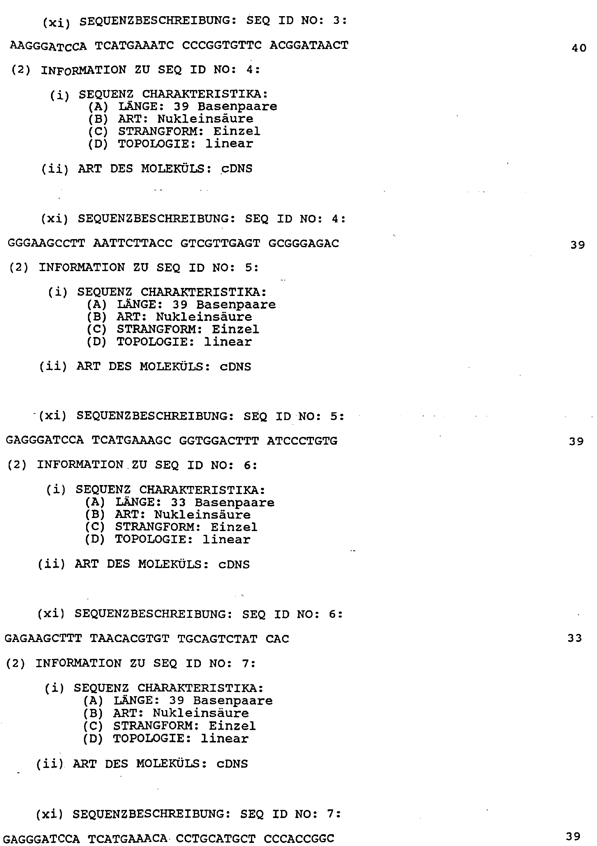
- PCR Using PCR, starting from clone NS3 (DSM 6847) Use of the primers (1) and (2), whose nucleotide sequence in SEQ ID NO. 3 and SEQ ID NO. 4, a DNA fragment is amplified. At the 5 'end This DNA fragment contains sequences for cloning (BamHI, BspHI, EcoRI restriction sites) and an ATG codon and an AAA (Lys) codon to increase expression. At the 3 'end there are restriction interfaces for HindIII and EcoRI as well as a Stop Codon (TTA). The area homologous to HCV begins in the primers (1) and (2) each at nucleotide No. 19.
- the DNA fragment obtained in this way was placed in a BamHI and HindIII cut pUC8 vector used.
- the resulting Plasmid was named pUC-D26.
- the Induction and antigen production takes place by adding 2 mmol / l des Inductor isopropylthio- ⁇ -D-glactoside (IPTG) and further shaking for 3 up to 4 h. The bacteria were then pelleted by centrifugation and processed.
- IPTG Inductor isopropylthio- ⁇ -D-glactoside
- the bacterial pellets from two 1 I cultures were in 50 ml 50 mmol / l Tris-HCl, pH 8.5, 0.2 mg / ml lysozyme and 2 mmol / l dithioerythritol (DTE) resuspended. Then EDTA (final concentration: 15 mmol / l), Phenylmethylsulfonyl fluoride (final concentration: 1 mmol / l) and 4 mg DNase added. The suspension was magnetic for a few minutes mixed and incubated for 45 min at 37 ° C in a water bath.
- Triton-X100 was then added (final concentration: 1 %) and stirring for 30 minutes in a magnetic stirrer. After freezing at -20 ° C overnight and thawing the cells at 37 for at least 1 hour ° C stirred and optionally sonified. Incubation at 37 ° C and / or the ultrasound treatment should continue until the Viscosity of the suspension with the lysed cells clearly decreased Has.
- the mixture was then centrifuged at 35000 g and 4 ° C. for 20 minutes.
- the resulting pellet was dissolved in 30 ml of 50 mmol / l Tris-HCl, pH 8.5, 2 mmol / l DTE, 150 mmol / l EDTA and 1.5% OGP (octyl- ⁇ -dglucoapyranoside, Biomol) resuspended.
- This suspension was at Room temperature intensely on the magnetic stirrer for at least 3 hours stirred and then at 35000 g and 4 ° C for 20 minutes centrifuged.
- the pellet was dissolved in 100 ml of 8 mol / l urea, 20 mmol / l Tris-HCl, pH 8.5, 2 mmol / l DTE dissolved and stirred.
- the now released antigen can be up to Further processing can be frozen at -20 ° C.
- the protein was purified by the following Chromatography steps described at room temperature were carried out.
- the storage of the antigen between the Chromatography steps were carried out at -20 ° C.
- the first chromatography step was carried out on a Q-Sepharose Fast Flow Column (Pharmacia) with a 20 mmol / l Tris-HCl, pH 8.5, 8 mol / l urea, 2 mmol / l DTE buffer. Elution was carried out using a NaCl gradient (0 to 0.7 mol / l). The run and fractions were by SDS-PAGE tested. The polypeptide used in the method according to the invention was included in the first main peak. The positive fractions were combined and overnight against a 10-fold volume of 4 mol / l Dialysis of urea, 2 mmol / l DTE, 20 mmol / l Tris-HCl, pH 7.3.
- the same column was used for the second chromatography step used.
- the column buffer was the one after the first Chromatography step used dialysis buffer.
- the elution was carried out with a NaCI gradient (0 to 0.5 mol / l) and then with 1 mol / l NaCI, NaOH pH 13.4 mol / l urea.
- the size of the antigen in the SDS polyacrylamide gel is approximately 41 kDa on.
- the yield is approximately 10 mg of antigen per liter of culture medium.
- the expression of the NS-3 antigen was carried out using the deposited clones NS-3 (DSM 6847).
- the cloning and expression of the Antigens C33 and D27 were carried out according to that in Example 1 described method.
- the coding regions of amino acids 1192 to 1457 for C33 and amino acids 1227 to 1528 for D27 by means of PCR starting from the plasmid pUC-N3 from the clone NS-3 Standard methods amplified.
- the primers (3) and (4) used the nucleotide sequences in SEQ ID NO. 5 or SEQ ID NO. 6 are specified.
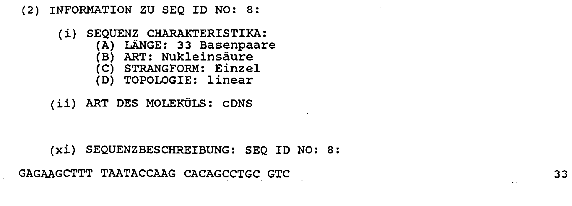
- Primers (5) and (6), whose Nucleotide sequences in SEQ ID NO. 7 or SEQ ID NO. 8 are specified.
- the area homologous to HCV begins in primers (3) and (5) Nucleotide No. 19 and in primers (4) and (6) with nucleotide No. 13.
- the amplified DNA fragments were after treatment with the Restriction enzymes BamHI and HindIII and subsequent purification by agarose gel electrophoresis in the with BamHI and HindIII cut vector pUC8 inserted.
- the two of the pUC vectors expressed antigens show a range of 13 at the N-terminal end non-HCV-encoded foreign amino acids (Met-Thr-Met-lle-Thr-Asn-Ser-Arg-Gly-Ser-lle-Met-Lys).
- the plasmids were then transformed into E. coli JM109. at Clones which receive a DNA fragment coding for the C33 antigen, an antigen with a molecular weight of approx. 34 kDa (SDS-PAGE) expressed. For clones with a DNA fragment that is responsible for the antigen D27 encoded, a band with 48 kDa is found.
- the share of total protein in C33 is approx. 10% and is approximately exact as large as the D26 polypeptide. For D27 one becomes significant weaker expression found in the range of 5% or less.
- Lysates from D26, D27 and C33 expressing clones were found separated side by side in the SDS gel and transferred to nitrocellulose. After overnight incubation with various HCV positive sera in 1: 100 dilution, bound antibodies were detected by means of an anti-human IgG antibody-peroxidase conjugate and color reaction with 3,3'-diaminobenzidine tetrachloride (DAB) / hydrogen peroxide.
- DAB 3,3'-diaminobenzidine tetrachloride
- the stability of the antigens NS3 and D26 was compared with the indirect test concept. A clearly poor stability of the NS3 antigen was found compared to the antigen D26 which is preferably used in the method according to the invention. The stability of the antigens was examined after 72 h of incubation at 37 ° C. construct Sample No. Signal Recovery NS3 1 ⁇ 20% 2 ⁇ 20% D26 1 99% 2 103%
- the time of the seroconversion was based on the reactivity of serum samples taken at different times with HCV-NS3 helicase antigen under physiological conditions with or without 20 mmol / l DTT tested.
- the test concept was a double antigen bridge test for class-independent detection of all immunoglobulins, in which a with an electrochemical marking group (ruthenium metal complex) provided and an antigen (biotinylated Antigen) was used.
Landscapes
- Health & Medical Sciences (AREA)
- Life Sciences & Earth Sciences (AREA)
- Chemical & Material Sciences (AREA)
- Engineering & Computer Science (AREA)
- Immunology (AREA)
- Molecular Biology (AREA)
- General Health & Medical Sciences (AREA)
- Virology (AREA)
- Medicinal Chemistry (AREA)
- Urology & Nephrology (AREA)
- Hematology (AREA)
- Biomedical Technology (AREA)
- Organic Chemistry (AREA)
- Biochemistry (AREA)
- Analytical Chemistry (AREA)
- Microbiology (AREA)
- General Physics & Mathematics (AREA)
- Pathology (AREA)
- Physics & Mathematics (AREA)
- Food Science & Technology (AREA)
- Biotechnology (AREA)
- Cell Biology (AREA)
- Communicable Diseases (AREA)
- Chemical Kinetics & Catalysis (AREA)
- Pharmacology & Pharmacy (AREA)
- Gastroenterology & Hepatology (AREA)
- General Chemical & Material Sciences (AREA)
- Animal Behavior & Ethology (AREA)
- Public Health (AREA)
- Nuclear Medicine, Radiotherapy & Molecular Imaging (AREA)
- Veterinary Medicine (AREA)
- Oncology (AREA)
- Tropical Medicine & Parasitology (AREA)
- Proteomics, Peptides & Aminoacids (AREA)
- Genetics & Genomics (AREA)
- Biophysics (AREA)
- Bioinformatics & Cheminformatics (AREA)
- Peptides Or Proteins (AREA)
- Preparation Of Compounds By Using Micro-Organisms (AREA)
- Micro-Organisms Or Cultivation Processes Thereof (AREA)
Abstract
Description
- SEQ ID NO. 1:
- die Nukleotidsequenz eines in dem erfindungsgemäßen Verfahren bevorzugt verwendeten Polypeptids,
- SEQ ID NO. 2:
- die Aminosäuresequenz einer für ein in dem erfindungsgemäßen Verfahren bevorzugt verwendetes Polypeptid kodierenden Nukleinsäure,
- SEQ ID NO. 3:
- die Nukleotidsequenz von Primer (1),
- SEQ ID NO. 4:
- die Nukleotidsequenz von Primer (2),
- SEQ ID NO. 5:
- die Nukleotidsequenz von Primer (3),
- SEQ ID NO. 6:
- die Nukleotidsequenz von Primer (4),
- SEQ ID NO. 7:
- die Nukleotidsequenz von Primer (5) und
- SEQ ID NO. 8:
- die Nukleotidsequenz von Primer (6).
| Konstrukt | falsch positive Bewertung | richtig negative Bewertung |
| C33 | 8 | 952 |
| D26 | 2 | 958 |
| Konstrukt | richtig positive Bewertung | falsch negative Bewertung |
| NS3 | 20 | 0 |
| C33 | 20 | 0 |
| D26 | 20 | 0 |
| D27 | 19 | 1 |
| Konstrukt | Proben Nr. | Signalwiederfindung |
| NS3 | 1 | < 20 % |
| 2 | < 20 % | |
| D26 | 1 | 99 % |
| 2 | 103 % |
| Tag der Blutentnahme | Tage nach Beginn der Blutentnahme | Reaktivität des NS3-Antigens ohne DTT (Signal/Cut off) | Reaktivität des NS3-Antigens mit DTT (Signal/Cut off) |
| 28.07.1988 | 0 | 0,1 | 0,1 |
| 01.08.1988 | 4 | 0,1 | 0,1 |
| 08.08.1988 | 11 | 0,1 | 0,1 |
| 11.08.1988 | 14 | 0,1 | 0,1 |
| 15.08.1988 | 18 | 0,1 | 0,1 |
| 25.08.1988 | 28 | 0,1 | 0,1 |
| 29.08.1988 | 32 | 0,1 | 1,6 |
| 14.09.1988 | 48 | 0,1 | 6,5 |
| 05.10.1988 | 69 | 1,3 | 4,8 |
| 19.10.1988 | 83 | 2,1 | 6,8 |
Claims (5)
- Verfahren zur verbesserten Früherkennung der Serokonversion eines gegen Hepatitis C-Virus gerichteten Antikörpers in einer Probenflüssigkeit, wobei man die Probenflüssigkeit mit mindestens einem Polypeptid, das Sequenzbereiche aus Hepatitis C-Virus, insbesondere aus der NS3-Region von Hepatitis C-Virus enthält, inkubiert und den Antikörper über eine Bindung mit dem Polypeptid nachweist,
dadurch gekennzeichnet, dass man ein Polypeptid aus einem Bereich, der mindestens einen Cysteinrest enthält, verwendet und (a) die Bestimmung des Antikörpers unter reduzierenden Bedingungen durchführt, (b) einen oder mehrere Cysteinreste kovalent modifiziert oder/und (c) einen oder mehrere Cysteinreste durch andere Aminosäuren ersetzt. - Verfahren nach Anspruch 1
dadurch gekennzeichnet, dass es nach einem homogenen Immunassay mit einer einzigen Reaktionsphase oder nach einem heterogenen Immunassay mit mehr als einer Reaktionsphase erfolgt. - Verfahren nach Anspruch 1 oder 2,
dadurch gekennzeichnet, dass man die Probenflüssigkeit mit zwei Polypeptiden P1 und P2 inkubiert,
wobei das Polypeptid P1 (a) an eine Festphase gebunden ist oder (b) in einer an eine Festphase bindefähigen Form vorliegt und das Polypeptid P2 eine Markierungsgruppe trägt,
und den Antikörper durch Bestimmung der Markierung in der Festphase oder/und in der flüssigen Phase nachweist. - Verfahren nach Anspruch 1 oder 2,
dadurch gekennzeichnet, dass man die Probenflüssigkeit mit einem Polypeptiden P1, das (a) an eine Festphase gebunden ist oder (b) in einer an eine Festphase bindefähigen Form vorliegt und mit einem weiteren, gegen P1 gerichteten Antikörper, der eine Markierungsgruppe trägt, inkubiert und den zu bestimmenden Antikörper durch Bestimmung der Markierung in der Festphase oder/und in der flüssigen Phase nachweist. - Verfahren nach einem der Ansprüche 1 bis 4,
dadurch gekennzeichnet, dass die Probenflüssigkeit menschliches Serum ist.
Priority Applications (2)
| Application Number | Priority Date | Filing Date | Title |
|---|---|---|---|
| EP08016723A EP2048505A3 (de) | 1994-08-12 | 1995-08-11 | Verfahren zur Früherkennung der Serokonversion eines gegen Hepatitis C-Virus gerichteten Antikörpers |
| EP10183478A EP2275818A3 (de) | 1994-08-12 | 1995-08-11 | Verfahren zur Früherkennung der Serokonversion eines gegen Hepatitis C-Virus gerichteten Antikörpers |
Applications Claiming Priority (3)
| Application Number | Priority Date | Filing Date | Title |
|---|---|---|---|
| DE4428705 | 1994-08-12 | ||
| DE4428705A DE4428705A1 (de) | 1994-08-12 | 1994-08-12 | Rekombinantes Antigen aus der NS3-Region des Hepatitis C Virus |
| EP95112700A EP0696640A3 (de) | 1994-08-12 | 1995-08-11 | Rekombinantes Antigen aus der NS3-Region des Hepatitis C Virus |
Related Parent Applications (2)
| Application Number | Title | Priority Date | Filing Date |
|---|---|---|---|
| EP95112700A Division EP0696640A3 (de) | 1994-08-12 | 1995-08-11 | Rekombinantes Antigen aus der NS3-Region des Hepatitis C Virus |
| EP95112700.0 Division | 1995-08-11 |
Related Child Applications (1)
| Application Number | Title | Priority Date | Filing Date |
|---|---|---|---|
| EP08016723A Division EP2048505A3 (de) | 1994-08-12 | 1995-08-11 | Verfahren zur Früherkennung der Serokonversion eines gegen Hepatitis C-Virus gerichteten Antikörpers |
Publications (4)
| Publication Number | Publication Date |
|---|---|
| EP1318401A2 true EP1318401A2 (de) | 2003-06-11 |
| EP1318401A3 EP1318401A3 (de) | 2006-08-16 |
| EP1318401B1 EP1318401B1 (de) | 2008-10-08 |
| EP1318401B9 EP1318401B9 (de) | 2009-05-13 |
Family
ID=6525606
Family Applications (4)
| Application Number | Title | Priority Date | Filing Date |
|---|---|---|---|
| EP03001660A Revoked EP1318401B9 (de) | 1994-08-12 | 1995-08-11 | Verfahren zur Früherkennung der Serokonversion eines gegen Hepatitis C-Virus gerichteten Antikörpers |
| EP95112700A Withdrawn EP0696640A3 (de) | 1994-08-12 | 1995-08-11 | Rekombinantes Antigen aus der NS3-Region des Hepatitis C Virus |
| EP10183478A Withdrawn EP2275818A3 (de) | 1994-08-12 | 1995-08-11 | Verfahren zur Früherkennung der Serokonversion eines gegen Hepatitis C-Virus gerichteten Antikörpers |
| EP08016723A Withdrawn EP2048505A3 (de) | 1994-08-12 | 1995-08-11 | Verfahren zur Früherkennung der Serokonversion eines gegen Hepatitis C-Virus gerichteten Antikörpers |
Family Applications After (3)
| Application Number | Title | Priority Date | Filing Date |
|---|---|---|---|
| EP95112700A Withdrawn EP0696640A3 (de) | 1994-08-12 | 1995-08-11 | Rekombinantes Antigen aus der NS3-Region des Hepatitis C Virus |
| EP10183478A Withdrawn EP2275818A3 (de) | 1994-08-12 | 1995-08-11 | Verfahren zur Früherkennung der Serokonversion eines gegen Hepatitis C-Virus gerichteten Antikörpers |
| EP08016723A Withdrawn EP2048505A3 (de) | 1994-08-12 | 1995-08-11 | Verfahren zur Früherkennung der Serokonversion eines gegen Hepatitis C-Virus gerichteten Antikörpers |
Country Status (6)
| Country | Link |
|---|---|
| US (6) | US6096319A (de) |
| EP (4) | EP1318401B9 (de) |
| JP (3) | JPH08127592A (de) |
| AT (1) | ATE410692T1 (de) |
| DE (2) | DE4428705A1 (de) |
| ES (1) | ES2315434T3 (de) |
Families Citing this family (21)
| Publication number | Priority date | Publication date | Assignee | Title |
|---|---|---|---|---|
| DE4428705A1 (de) * | 1994-08-12 | 1996-02-15 | Boehringer Mannheim Gmbh | Rekombinantes Antigen aus der NS3-Region des Hepatitis C Virus |
| DE19637718A1 (de) | 1996-04-01 | 1997-10-02 | Boehringer Mannheim Gmbh | Rekombinante inaktive Core-Streptavidin Mutanten |
| AU751362B2 (en) * | 1998-04-17 | 2002-08-15 | Innogenetics N.V. | Improved immunodiagnostic assays using reducing agents |
| US6986892B1 (en) | 1999-11-24 | 2006-01-17 | Chiron Corporation | Immunogenic Hepatitis C virus non-structural polypeptides |
| DE60036881T2 (de) * | 1999-11-24 | 2008-07-10 | Novartis Vaccines and Diagnostics, Inc., Emeryville | Neues nichtstrukturelles hcv-polypeptid |
| BRPI0111731C1 (pt) * | 2000-06-15 | 2021-07-27 | Chiron Corp | suporte sólido de imunoensaio, métodos de detecção de infecção pelo vírus da hepatite c (hcv) em uma amostra biológica, e de produção de um suporte sólido de imunoensaio, e, kit de teste de imunodiagnóstico |
| EP1195381A1 (de) * | 2000-09-28 | 2002-04-10 | Immusystems GmbH | CD4+ T-Lymphozyten spezifische Hepatitis C Virus-Epitope |
| ATE522603T1 (de) * | 2001-06-22 | 2011-09-15 | Hoffmann La Roche | Löslicher komplex, der retrovirale oberflächen- glykoproteine und fkpa oder slyd enthält |
| ES2312806T3 (es) * | 2002-09-09 | 2009-03-01 | Novartis Vaccines And Diagnostics, Inc. | Ensayo de vhc. |
| US20050014136A1 (en) * | 2003-05-28 | 2005-01-20 | Innogenetics N.V. | Modified HCV NS5 |
| ES2349649T3 (es) * | 2004-07-30 | 2011-01-07 | Advanced Life Science Institute, Inc. | Complejo sonda. |
| US7658930B2 (en) * | 2005-02-02 | 2010-02-09 | Peng Cui | Kit for detecting the antibody of HCV and its preparing method |
| US20080220448A1 (en) * | 2006-09-01 | 2008-09-11 | Blincko Stuart J | Antigenic protein conjugates and process for preparing same |
| EP2527474A1 (de) * | 2006-10-20 | 2012-11-28 | Innogenetics N.V. | Verfahren zur Analyse von Sequenzvariationen innerhalb des HCV-NS3/4A-Genombereichs |
| FR2984328B1 (fr) | 2011-12-20 | 2016-12-30 | Bio-Rad Innovations | Procede de detection d'une infection par le virus de l'hepatite c |
| WO2014143342A1 (en) * | 2013-03-14 | 2014-09-18 | Abbott Laboratories | Hcv ns3 recombinant antigens and mutants thereof for improved antibody detection |
| CN105378099B (zh) | 2013-03-14 | 2021-05-11 | 雅培制药有限公司 | Hcv核心脂质结合结构域单克隆抗体 |
| MX362075B (es) | 2013-03-14 | 2019-01-07 | Abbott Lab | Ensayo de combinación de antígeno-anticuerpo del virus de la hepatitis c (vhc) y métodos y composiciones para usarlo. |
| WO2016069762A2 (en) * | 2014-10-29 | 2016-05-06 | Abbott Laboratories | Subject anti-hcv antibody detection assays employing ns3 capture peptides |
| CN113454455B (zh) | 2018-12-28 | 2024-04-30 | 雅培制药有限公司 | 微粒上的单分子的直接检测 |
| CN110456073B (zh) * | 2019-08-21 | 2023-09-22 | 广东菲鹏生物有限公司 | 双抗原夹心检测抗体的方法及试剂盒 |
Family Cites Families (21)
| Publication number | Priority date | Publication date | Assignee | Title |
|---|---|---|---|---|
| USRE32696E (en) * | 1975-09-04 | 1988-06-14 | Akzona Incorporated | Enzymatic immunological method for determination of antigens and antibodies |
| US4649039A (en) * | 1984-07-03 | 1987-03-10 | E. I. Du Pont De Nemours And Company | Radiolabeling of methionine-containing proteins and peptides |
| US5523215A (en) * | 1985-03-28 | 1996-06-04 | Chiron Corporation | Enhanced purification and expression of insoluble recombinant proteins |
| DE3705686C2 (de) | 1987-02-23 | 1995-11-30 | Boehringer Mannheim Gmbh | Verfahren zur Bestimmung von Antikörpern |
| WO1989004669A1 (en) * | 1987-11-18 | 1989-06-01 | Chiron Corporation | Nanbv diagnostics and vaccines |
| EP0341439B1 (de) * | 1988-05-11 | 1993-08-04 | Abbott Laboratories | Verfahren zur Erhöhung der Spezifizität in kompetitiven Immuntests |
| HUT54896A (en) * | 1989-03-17 | 1991-04-29 | Chiron Corp | Process for producing aquous diagnosticum and vaccine of nanbv |
| US5582968A (en) * | 1990-02-16 | 1996-12-10 | United Biomedical, Inc. | Branched hybrid and cluster peptides effective in diagnosing and detecting non-A, non-B hepatitis |
| KR100206150B1 (ko) * | 1990-04-04 | 1999-07-01 | 로버트 피. 블랙버언 | 항-에이치씨브이 항체에 대한 면역분석용 씨형 간염 바이러스(에이치디브이)항원의 조합체 |
| AU7675491A (en) * | 1990-04-04 | 1991-10-30 | Chiron Corporation | Hepatitis c virus protease |
| DE4041304A1 (de) | 1990-12-21 | 1992-06-25 | Mikrogen Molekularbiol Entw | Von strukturproteinen des hepatitis c-virus abgeleitete polypeptide, testkits, die diese polypeptide enthalten und impfstoffe gegen infektionen von hepatitis c-viren |
| JP2921118B2 (ja) * | 1991-04-23 | 1999-07-19 | 東レ株式会社 | 3環性トリアゾロ誘導体、その製造方法及びその医薬用途 |
| US5683998A (en) * | 1991-04-23 | 1997-11-04 | Toray Industries, Inc. | Tricyclic triazolo derivatives, processes for producing the same and the uses of the same |
| EP0608258A1 (de) * | 1991-10-16 | 1994-08-03 | Unilever Plc | Wässrige, enzymatische reinigungsmittelzusammensetzung |
| EP0573624A4 (en) | 1991-11-07 | 1997-09-17 | Baxter Diagnostics Inc | Epitope mapping ot the c33c region of hcv |
| UA39944C2 (uk) * | 1992-07-07 | 2001-07-16 | Чірон Корпорейшн | Спосіб визначення ранньої сероконверсії у ссавця-хазяїна до вірусу гепатиту с і набір для використання в способі |
| JP3225468B2 (ja) * | 1992-08-28 | 2001-11-05 | ダイナボット株式会社 | 抗体測定用試薬 |
| SK153594A3 (en) * | 1993-04-14 | 1995-07-11 | Int Murex Tech Corp | Determination method of antibodies to two or bigger number of different patogens in tested sample |
| US5670310A (en) * | 1994-07-29 | 1997-09-23 | The United States Of America As Represented By The Department Of Health And Human Services | Methods and compositions for differential diagnosis of acute and chronic hepatitis c virus infection |
| DE4428705A1 (de) * | 1994-08-12 | 1996-02-15 | Boehringer Mannheim Gmbh | Rekombinantes Antigen aus der NS3-Region des Hepatitis C Virus |
| CA2303165C (en) * | 1997-09-22 | 2006-07-11 | Chiron Corporation | Buffers for stabilizing antigens |
-
1994
- 1994-08-12 DE DE4428705A patent/DE4428705A1/de not_active Withdrawn
-
1995
- 1995-08-07 US US08/511,759 patent/US6096319A/en not_active Expired - Lifetime
- 1995-08-11 EP EP03001660A patent/EP1318401B9/de not_active Revoked
- 1995-08-11 ES ES03001660T patent/ES2315434T3/es not_active Expired - Lifetime
- 1995-08-11 EP EP95112700A patent/EP0696640A3/de not_active Withdrawn
- 1995-08-11 EP EP10183478A patent/EP2275818A3/de not_active Withdrawn
- 1995-08-11 EP EP08016723A patent/EP2048505A3/de not_active Withdrawn
- 1995-08-11 AT AT03001660T patent/ATE410692T1/de active
- 1995-08-11 DE DE59511098T patent/DE59511098D1/de not_active Expired - Lifetime
- 1995-08-14 JP JP7207086A patent/JPH08127592A/ja active Pending
-
1997
- 1997-07-15 US US08/892,704 patent/US6306579B1/en not_active Expired - Fee Related
-
2000
- 2000-06-12 US US09/592,197 patent/US6270960B1/en not_active Expired - Lifetime
-
2001
- 2001-04-25 JP JP2001127276A patent/JP3475183B2/ja not_active Expired - Fee Related
- 2001-06-29 US US09/896,032 patent/US7122302B2/en not_active Expired - Fee Related
-
2003
- 2003-05-14 JP JP2003136377A patent/JP3632027B2/ja not_active Expired - Fee Related
-
2004
- 2004-03-17 US US10/802,891 patent/US20050095584A1/en not_active Abandoned
-
2007
- 2007-10-11 US US11/974,030 patent/US20090098528A1/en not_active Abandoned
Also Published As
| Publication number | Publication date |
|---|---|
| US20050095584A1 (en) | 2005-05-05 |
| US6270960B1 (en) | 2001-08-07 |
| US20030129582A1 (en) | 2003-07-10 |
| EP1318401A3 (de) | 2006-08-16 |
| JP3475183B2 (ja) | 2003-12-08 |
| EP2275818A2 (de) | 2011-01-19 |
| US20090098528A1 (en) | 2009-04-16 |
| US6096319A (en) | 2000-08-01 |
| EP1318401B9 (de) | 2009-05-13 |
| EP0696640A2 (de) | 1996-02-14 |
| ES2315434T3 (es) | 2009-04-01 |
| EP0696640A3 (de) | 1997-12-29 |
| DE59511098D1 (de) | 2008-11-20 |
| JPH08127592A (ja) | 1996-05-21 |
| US7122302B2 (en) | 2006-10-17 |
| JP3632027B2 (ja) | 2005-03-23 |
| EP2048505A3 (de) | 2009-04-22 |
| EP1318401B1 (de) | 2008-10-08 |
| DE4428705A1 (de) | 1996-02-15 |
| ATE410692T1 (de) | 2008-10-15 |
| US6306579B1 (en) | 2001-10-23 |
| EP2275818A3 (de) | 2011-06-29 |
| JP2002027990A (ja) | 2002-01-29 |
| EP2048505A2 (de) | 2009-04-15 |
| JP2004004075A (ja) | 2004-01-08 |
Similar Documents
| Publication | Publication Date | Title |
|---|---|---|
| EP1318401B1 (de) | Verfahren zur Früherkennung der Serokonversion eines gegen Hepatitis C-Virus gerichteten Antikörpers | |
| DE68929529T2 (de) | Rekombinante HIV-2 Antigene, abgeleitet aus synthetischen DNA | |
| DE69131488T2 (de) | Kombinationen der Hepatitis-C-Virus (HCV) Antigene zur Verwendung in Immuntests für Anti-HCV-Antikörper | |
| DE69734776T2 (de) | Fusionsprotein mit multiplen Epitopen | |
| DE69331845T2 (de) | Immunoassays für anti-hcv-antikörper unter verwendung von antigenen mit konformationsepitopen | |
| DE69230917T2 (de) | Verfahren zur detektion von hepatitis c | |
| EP0918865B1 (de) | Immunologisch aktive proteine von borrelia burgdorferi, dafür kodierende nukleinsäuren sowie deren verwendung in testkits und als impfstoffe | |
| DE69630008T2 (de) | Treponema palladium Fusions-Antigen, Nachweis für Anti-treponema palladium Antikörpern mit diesen Fusions-Antigenen | |
| EP0582243B2 (de) | HCV Peptidantigene und Verfahren zur Bestimmung von HCV | |
| EP0252531A1 (de) | Strukturelles Phosphoprotein (pp 150) des menschlichen Cytomegalovirus, seine Herstellung und Verwendung | |
| DE69934810T2 (de) | Neues hepatitis c viruspeptid und seine verwendung | |
| DE19728991A1 (de) | Rezeptorbindungsassay, für den Rezeptorbindungsassay geeigneter rekombinanter Fusionsrezeptor, Vektor zu dessen Herstellung sowie Reagenziensatz für die Durchführung des Rezeptorbindungsassays | |
| EP0702083A2 (de) | Polypeptide und Fusionsproteine bestehend aus dem UL 57 Leserahmen bzw. dem C-terminalen Bereich des Tegumentproteins pp150 aus HCMV, entsprechende Oligonucleotide und Nachweis reagentien | |
| EP3784686B9 (de) | Rubella virus spike konstrukt | |
| EP0754755B1 (de) | Rekombinante autologe Fusionsproteine des Epstein-Barr-Virus, diese enthaltende Testkits und Verfahren zum Nachweis von Epstein-Barr-Virus-spezifischen Antikörpern | |
| DE69331796T2 (de) | Kernantigenprotein des Hepatitis C-Virus, sowie diagnostisches Verfahren und Satz von Reagenzen | |
| DE19756214C1 (de) | Polypeptide mit Aminosäuresequenzen aus dem N-terminalen Bereich von gp116 und deren Verwendung bei der Diagnostik, Prophylaxe und Therapie | |
| DE19919121B4 (de) | Multimeres CMV-Antigen | |
| DE69231932T2 (de) | Hepatitis c-nachweistest unter benutzung rekombinanter antigene der c-100-region | |
| EP1069136B1 (de) | Verfahren zur rekombinanten Herstellung von Ribonukleoproteinen | |
| EP0891427B1 (de) | Expression von hgv-antigenen und deren verwendung | |
| DE69720470T2 (de) | Western blot test zur identifizierung von hcmv spezifische antiköpern | |
| DE19645729C1 (de) | Rezeptorbindungsassay, für den Rezeptorbindungsassay geeigneter rekombinanter Fusionsrezeptor, Vektor zu dessen Herstellung sowie Reagenziensatz für die Durchführung des Rezeptorbindungsassays |
Legal Events
| Date | Code | Title | Description |
|---|---|---|---|
| PUAI | Public reference made under article 153(3) epc to a published international application that has entered the european phase |
Free format text: ORIGINAL CODE: 0009012 |
|
| 17P | Request for examination filed |
Effective date: 20030124 |
|
| AC | Divisional application: reference to earlier application |
Ref document number: 0696640 Country of ref document: EP Kind code of ref document: P |
|
| AK | Designated contracting states |
Designated state(s): AT BE CH DE ES FR GB IT LI NL SE |
|
| PUAL | Search report despatched |
Free format text: ORIGINAL CODE: 0009013 |
|
| AK | Designated contracting states |
Kind code of ref document: A3 Designated state(s): AT BE CH DE ES FR GB IT LI NL SE |
|
| AKX | Designation fees paid |
Designated state(s): AT BE CH DE ES FR GB IT LI NL SE |
|
| 17Q | First examination report despatched |
Effective date: 20070417 |
|
| GRAP | Despatch of communication of intention to grant a patent |
Free format text: ORIGINAL CODE: EPIDOSNIGR1 |
|
| GRAS | Grant fee paid |
Free format text: ORIGINAL CODE: EPIDOSNIGR3 |
|
| GRAA | (expected) grant |
Free format text: ORIGINAL CODE: 0009210 |
|
| AC | Divisional application: reference to earlier application |
Ref document number: 0696640 Country of ref document: EP Kind code of ref document: P |
|
| AK | Designated contracting states |
Kind code of ref document: B1 Designated state(s): AT BE CH DE ES FR GB IT LI NL SE |
|
| REG | Reference to a national code |
Ref country code: GB Ref legal event code: FG4D Free format text: NOT ENGLISH |
|
| REG | Reference to a national code |
Ref country code: CH Ref legal event code: NV Representative=s name: BOHEST AG Ref country code: CH Ref legal event code: EP |
|
| REF | Corresponds to: |
Ref document number: 59511098 Country of ref document: DE Date of ref document: 20081120 Kind code of ref document: P |
|
| REG | Reference to a national code |
Ref country code: SE Ref legal event code: TRGR |
|
| RIN2 | Information on inventor provided after grant (corrected) |
Inventor name: MOTZ, MANFRED, DR. Inventor name: SEIDEL, CHRISTOPH, DR. Inventor name: WIENHUES, URSULA-HENRIKE, DR. Inventor name: SOUTSCHEK, ERWIN, DR. Inventor name: UPMEIER, BARBARA, DR. Inventor name: WIEDMANN, MICHAEL, DR. Inventor name: SCHMITT, URBAN |
|
| REG | Reference to a national code |
Ref country code: ES Ref legal event code: FG2A Ref document number: 2315434 Country of ref document: ES Kind code of ref document: T3 |
|
| PLBI | Opposition filed |
Free format text: ORIGINAL CODE: 0009260 |
|
| 26 | Opposition filed |
Opponent name: CABINET BECKER ET ASSOCIES Effective date: 20090629 |
|
| PLBI | Opposition filed |
Free format text: ORIGINAL CODE: 0009260 |
|
| PLAX | Notice of opposition and request to file observation + time limit sent |
Free format text: ORIGINAL CODE: EPIDOSNOBS2 |
|
| 26 | Opposition filed |
Opponent name: CABINET BECKER ET ASSOCIES Effective date: 20090629 Opponent name: INNOGENETICS N.V. Effective date: 20090708 Opponent name: BIO MERIEUX Effective date: 20090706 |
|
| NLR1 | Nl: opposition has been filed with the epo |
Opponent name: CABINET BECKER ET ASSOCIES |
|
| NLR1 | Nl: opposition has been filed with the epo |
Opponent name: CABINET BECKER ET ASSOCIES Opponent name: BIO MERIEUX Opponent name: INNOGENETICS N.V. |
|
| PLAF | Information modified related to communication of a notice of opposition and request to file observations + time limit |
Free format text: ORIGINAL CODE: EPIDOSCOBS2 |
|
| PLBB | Reply of patent proprietor to notice(s) of opposition received |
Free format text: ORIGINAL CODE: EPIDOSNOBS3 |
|
| PLAB | Opposition data, opponent's data or that of the opponent's representative modified |
Free format text: ORIGINAL CODE: 0009299OPPO |
|
| R26 | Opposition filed (corrected) |
Opponent name: CABINET BECKER ET ASSOCIES Effective date: 20090629 Opponent name: INNOGENETICS N.V. Effective date: 20090708 Opponent name: BIO MERIEUX Effective date: 20090706 |
|
| RDAF | Communication despatched that patent is revoked |
Free format text: ORIGINAL CODE: EPIDOSNREV1 |
|
| PLAB | Opposition data, opponent's data or that of the opponent's representative modified |
Free format text: ORIGINAL CODE: 0009299OPPO |
|
| R26 | Opposition filed (corrected) |
Opponent name: BIO MERIEUX Effective date: 20090706 Opponent name: INNOGENETICS N.V. Effective date: 20090708 Opponent name: CABINET BECKER ET ASSOCIES Effective date: 20090629 |
|
| APAH | Appeal reference modified |
Free format text: ORIGINAL CODE: EPIDOSCREFNO |
|
| APAW | Appeal reference deleted |
Free format text: ORIGINAL CODE: EPIDOSDREFNO |
|
| APAY | Date of receipt of notice of appeal deleted |
Free format text: ORIGINAL CODE: EPIDOSDNOA2O |
|
| APBM | Appeal reference recorded |
Free format text: ORIGINAL CODE: EPIDOSNREFNO |
|
| APBP | Date of receipt of notice of appeal recorded |
Free format text: ORIGINAL CODE: EPIDOSNNOA2O |
|
| APBQ | Date of receipt of statement of grounds of appeal recorded |
Free format text: ORIGINAL CODE: EPIDOSNNOA3O |
|
| PGFP | Annual fee paid to national office [announced via postgrant information from national office to epo] |
Ref country code: CH Payment date: 20110729 Year of fee payment: 17 |
|
| PGFP | Annual fee paid to national office [announced via postgrant information from national office to epo] |
Ref country code: SE Payment date: 20120809 Year of fee payment: 18 Ref country code: GB Payment date: 20120726 Year of fee payment: 18 |
|
| PGFP | Annual fee paid to national office [announced via postgrant information from national office to epo] |
Ref country code: IT Payment date: 20120809 Year of fee payment: 18 Ref country code: FR Payment date: 20120809 Year of fee payment: 18 Ref country code: DE Payment date: 20120831 Year of fee payment: 18 Ref country code: ES Payment date: 20120809 Year of fee payment: 18 |
|
| APAH | Appeal reference modified |
Free format text: ORIGINAL CODE: EPIDOSCREFNO |
|
| PGFP | Annual fee paid to national office [announced via postgrant information from national office to epo] |
Ref country code: NL Payment date: 20120813 Year of fee payment: 18 Ref country code: BE Payment date: 20120904 Year of fee payment: 18 |
|
| PGFP | Annual fee paid to national office [announced via postgrant information from national office to epo] |
Ref country code: AT Payment date: 20120726 Year of fee payment: 18 |
|
| BERE | Be: lapsed |
Owner name: ROCHE DIAGNOSTICS G.M.B.H. Effective date: 20130831 |
|
| REG | Reference to a national code |
Ref country code: NL Ref legal event code: V1 Effective date: 20140301 |
|
| REG | Reference to a national code |
Ref country code: CH Ref legal event code: PL |
|
| REG | Reference to a national code |
Ref country code: SE Ref legal event code: EUG |
|
| REG | Reference to a national code |
Ref country code: AT Ref legal event code: MM01 Ref document number: 410692 Country of ref document: AT Kind code of ref document: T Effective date: 20130811 |
|
| GBPC | Gb: european patent ceased through non-payment of renewal fee |
Effective date: 20130811 |
|
| PG25 | Lapsed in a contracting state [announced via postgrant information from national office to epo] |
Ref country code: CH Free format text: LAPSE BECAUSE OF NON-PAYMENT OF DUE FEES Effective date: 20130831 Ref country code: LI Free format text: LAPSE BECAUSE OF NON-PAYMENT OF DUE FEES Effective date: 20130831 Ref country code: NL Free format text: LAPSE BECAUSE OF NON-PAYMENT OF DUE FEES Effective date: 20140301 Ref country code: DE Free format text: LAPSE BECAUSE OF NON-PAYMENT OF DUE FEES Effective date: 20140301 Ref country code: SE Free format text: LAPSE BECAUSE OF NON-PAYMENT OF DUE FEES Effective date: 20130812 |
|
| REG | Reference to a national code |
Ref country code: FR Ref legal event code: ST Effective date: 20140430 |
|
| PG25 | Lapsed in a contracting state [announced via postgrant information from national office to epo] |
Ref country code: IT Free format text: LAPSE BECAUSE OF NON-PAYMENT OF DUE FEES Effective date: 20130811 Ref country code: BE Free format text: LAPSE BECAUSE OF NON-PAYMENT OF DUE FEES Effective date: 20130831 Ref country code: AT Free format text: LAPSE BECAUSE OF NON-PAYMENT OF DUE FEES Effective date: 20130811 |
|
| REG | Reference to a national code |
Ref country code: DE Ref legal event code: R119 Ref document number: 59511098 Country of ref document: DE Effective date: 20140301 |
|
| PG25 | Lapsed in a contracting state [announced via postgrant information from national office to epo] |
Ref country code: GB Free format text: LAPSE BECAUSE OF NON-PAYMENT OF DUE FEES Effective date: 20130811 |
|
| PG25 | Lapsed in a contracting state [announced via postgrant information from national office to epo] |
Ref country code: FR Free format text: LAPSE BECAUSE OF NON-PAYMENT OF DUE FEES Effective date: 20130902 |
|
| REG | Reference to a national code |
Ref country code: ES Ref legal event code: FD2A Effective date: 20140905 |
|
| APBY | Invitation to file observations in appeal sent |
Free format text: ORIGINAL CODE: EPIDOSNOBA2O |
|
| PG25 | Lapsed in a contracting state [announced via postgrant information from national office to epo] |
Ref country code: ES Free format text: LAPSE BECAUSE OF NON-PAYMENT OF DUE FEES Effective date: 20130812 |
|
| APBU | Appeal procedure closed |
Free format text: ORIGINAL CODE: EPIDOSNNOA9O |
|
| RDAG | Patent revoked |
Free format text: ORIGINAL CODE: 0009271 |
|
| STAA | Information on the status of an ep patent application or granted ep patent |
Free format text: STATUS: PATENT REVOKED |
|
| 27W | Patent revoked |
Effective date: 20141217 |
|
| REG | Reference to a national code |
Ref country code: SE Ref legal event code: ECNC |