EP1403369A1 - Procede d'evaluation de la fonction de phagocytose et utilisation associee - Google Patents
Procede d'evaluation de la fonction de phagocytose et utilisation associee Download PDFInfo
- Publication number
- EP1403369A1 EP1403369A1 EP02778910A EP02778910A EP1403369A1 EP 1403369 A1 EP1403369 A1 EP 1403369A1 EP 02778910 A EP02778910 A EP 02778910A EP 02778910 A EP02778910 A EP 02778910A EP 1403369 A1 EP1403369 A1 EP 1403369A1
- Authority
- EP
- European Patent Office
- Prior art keywords
- phagocyte
- dna
- digested
- microorganism
- cells
- Prior art date
- Legal status (The legal status is an assumption and is not a legal conclusion. Google has not performed a legal analysis and makes no representation as to the accuracy of the status listed.)
- Withdrawn
Links
Images







Classifications
-
- G—PHYSICS
- G01—MEASURING; TESTING
- G01N—INVESTIGATING OR ANALYSING MATERIALS BY DETERMINING THEIR CHEMICAL OR PHYSICAL PROPERTIES
- G01N33/00—Investigating or analysing materials by specific methods not covered by groups G01N1/00 - G01N31/00
- G01N33/48—Biological material, e.g. blood, urine; Haemocytometers
- G01N33/50—Chemical analysis of biological material, e.g. blood, urine; Testing involving biospecific ligand binding methods; Immunological testing
- G01N33/5005—Chemical analysis of biological material, e.g. blood, urine; Testing involving biospecific ligand binding methods; Immunological testing involving human or animal cells
- G01N33/5008—Chemical analysis of biological material, e.g. blood, urine; Testing involving biospecific ligand binding methods; Immunological testing involving human or animal cells for testing or evaluating the effect of chemical or biological compounds, e.g. drugs, cosmetics
- G01N33/5044—Chemical analysis of biological material, e.g. blood, urine; Testing involving biospecific ligand binding methods; Immunological testing involving human or animal cells for testing or evaluating the effect of chemical or biological compounds, e.g. drugs, cosmetics involving specific cell types
- G01N33/5047—Cells of the immune system
-
- C—CHEMISTRY; METALLURGY
- C12—BIOCHEMISTRY; BEER; SPIRITS; WINE; VINEGAR; MICROBIOLOGY; ENZYMOLOGY; MUTATION OR GENETIC ENGINEERING
- C12Q—MEASURING OR TESTING PROCESSES INVOLVING ENZYMES, NUCLEIC ACIDS OR MICROORGANISMS; COMPOSITIONS OR TEST PAPERS THEREFOR; PROCESSES OF PREPARING SUCH COMPOSITIONS; CONDITION-RESPONSIVE CONTROL IN MICROBIOLOGICAL OR ENZYMOLOGICAL PROCESSES
- C12Q1/00—Measuring or testing processes involving enzymes, nucleic acids or microorganisms; Compositions therefor; Processes of preparing such compositions
- C12Q1/68—Measuring or testing processes involving enzymes, nucleic acids or microorganisms; Compositions therefor; Processes of preparing such compositions involving nucleic acids
- C12Q1/6876—Nucleic acid products used in the analysis of nucleic acids, e.g. primers or probes
- C12Q1/6888—Nucleic acid products used in the analysis of nucleic acids, e.g. primers or probes for detection or identification of organisms
- C12Q1/689—Nucleic acid products used in the analysis of nucleic acids, e.g. primers or probes for detection or identification of organisms for bacteria
-
- G—PHYSICS
- G01—MEASURING; TESTING
- G01N—INVESTIGATING OR ANALYSING MATERIALS BY DETERMINING THEIR CHEMICAL OR PHYSICAL PROPERTIES
- G01N33/00—Investigating or analysing materials by specific methods not covered by groups G01N1/00 - G01N31/00
- G01N33/48—Biological material, e.g. blood, urine; Haemocytometers
- G01N33/50—Chemical analysis of biological material, e.g. blood, urine; Testing involving biospecific ligand binding methods; Immunological testing
- G01N33/5005—Chemical analysis of biological material, e.g. blood, urine; Testing involving biospecific ligand binding methods; Immunological testing involving human or animal cells
-
- C—CHEMISTRY; METALLURGY
- C12—BIOCHEMISTRY; BEER; SPIRITS; WINE; VINEGAR; MICROBIOLOGY; ENZYMOLOGY; MUTATION OR GENETIC ENGINEERING
- C12Q—MEASURING OR TESTING PROCESSES INVOLVING ENZYMES, NUCLEIC ACIDS OR MICROORGANISMS; COMPOSITIONS OR TEST PAPERS THEREFOR; PROCESSES OF PREPARING SUCH COMPOSITIONS; CONDITION-RESPONSIVE CONTROL IN MICROBIOLOGICAL OR ENZYMOLOGICAL PROCESSES
- C12Q1/00—Measuring or testing processes involving enzymes, nucleic acids or microorganisms; Compositions therefor; Processes of preparing such compositions
- C12Q1/68—Measuring or testing processes involving enzymes, nucleic acids or microorganisms; Compositions therefor; Processes of preparing such compositions involving nucleic acids
- C12Q1/6813—Hybridisation assays
- C12Q1/6841—In situ hybridisation
Definitions
- the present invention relates to a process for evaluating a phagocytotic function, and more particularly, provides an experimental model of an infection which is useful in diagnosis of an infectious state by a foreign microorganism, such as for example, sepsis and the like by bacteria, fungi or the like, or in development of therapeutic drugs for an infectious disease.
- the present invention also relates to a process for detecting, identifying and evaluating phagocytotic ability and/or germicidal capacity by a phagocyte; a process for the determination of an effect of a modulator of a phagocytotic function; a process for screening a modulator of a phagocytotic function; and a kit for putting any of such processes into practice.
- Infectious diseases and sepsis are often caused due to the underlying disease which had been presented previously, through infection by an attenuated microorganism. Although such a state may frequently occur in a clinical scene, ideal animal models which cover all of the clinical symptoms have not yet established. Factors for such a current state involve complicated infectious conditions exhibited by a bacterial infection owing to the difference in the underlying disease, large gaps of sensitivity and the like of the animal spices toward the bacterial strain, and thus, systems of the infection model have been individually established depending on the purpose of the research.
- Examples of the system of the infection model which are well known at present include: (i) a process in which formation of intraabdominal abscess is allowed through the infection of any of various microorganisms into a peritoneal cavity of a mouse or a rat to chase the pathological state of sepsis (biphasic infection theory), (ii) particularly, in instances to study dynamics of the infection under a lowered immune state, a process in which infection of an attenuated microorganism such as Pseudomonas aeruginosa or the like is rendered through decreasing leukocytes by previously administering cyclophosphamide, and a process in which adhesion of Pseudomonas aeruginosa is allowed through making burn injury in a wide area of the skin with an electric trowel in order to facilitate the infection, (iii) in instances to examine dynamics of a living humoral factor such as cytokine released by a macrophage, neutrophil and the like, a process
- Requirements for preferred animal model include: (I) possible migration of bacteria from a primary focus of the infection into blood (direct administration of bacteria within blood causes bacterial shock in many cases, which can not be controlled as a pathological state), (II) capability of securing sufficient amount of the blood when comparison and examination is executed with time by both test processes, and of performing collection of blood without causing contamination and secondary infection at the site of blood collection, (III) capability of securing phagocytes in an identical amount to that in human because less phagocytes such as neutrophils may be present depending on the animal spices and age of weeks, (IV) no great influence on each individual and detection sensitivity by alteration of an immune system by the stress and shock upon blood drawing through frequent collection of blood, (v) possible securement of number of experiments to some extent such that individual difference is avoided, and the like.
- Biphasic infection theory that is the most general process as a system to produce an animal model is a historically conventional system, which starts in 1931 by Meleney et al, and investigated and established by Hite (1949), Mergehagen (1958) and McDonald (1963) et al.
- This model has been established on the basis of a theoretical ground of an infection route in which a secondary infection focus is formed from the bacterium of a primary focus via blood irrespective of whether the bacterium is an anaerobe or aerobe.
- this model has been generally used as an infection model of sepsis.
- this system was established as a system for use in analysis of pathologic states of bacterial infectious disease, no importance is attached to the amount of bacteria which migrate from the abdominal cavity into the blood. Therefore, because the amount of bacteria which was intraperitoneally administered is not reflected to the amount of bacteria in the blood due to the influence of the individual difference of each rat, it is difficult to consider the difference resulting from the administered amount of bacteria in an in vivo test.
- Bacterial Translocation methods also involve problems.
- a factor for impossibility of easy comparison of the detection sensitivity of attenuated infectious bacteria such as Escherichia coli, Enterobactor cloacae, Klebsiella pneumoniae, Enteroccocus faecalis, Staphylococcus epidermidis and the like may involve that these bacterial strains are indigenous bacteria which are enteric and mucosal.
- analysis of a pathogenic state of sepsis is intended rather than the dynamics of the administered bacteria, therefore, invasion of a bacterial strain other than the administered bacteria is not considered.
- An object of the present invention is to provide an experimental model which permits the evaluation of a phagocytotic function of phagocytes in vitro , taking into account of such a current status. Moreover, another object of the present invention is to provide a process which permits the evaluation of an immune function and efficiency of differentiation to phagocytes through the use of such an experimental model. Furthermore, still another object of the present invention is to provide a process for screening various kinds of drugs such as immune function stimulators, anticancer agents, leukocyte differentiation factors, antibiotics and the like; a process for clinical laboratory test in which dosage regimen of various agents are examined; and the like by using such an experimental model.
- drugs such as immune function stimulators, anticancer agents, leukocyte differentiation factors, antibiotics and the like
- phagocytes post phagocytosis of a foreign microorganism are prepared by isolating the cells; and adjusting thus resulting phagocytes post phagocytosis of a foreign microorganism to give about 1 ⁇ 10 4 cells/ ⁇ l to about 5 ⁇ 10 4 cells/ ⁇ l.
- phagocyte post phagocytosis of a foreign microorganism means a cell attached to a foreign microorganism or a cell including a foreign microorganism involving not alone cells after completing phagocytosis of a foreign microorganism already, but cells during phagocytosis and cells which are ready to the initiate phagocytosis after the adhesion of a foreign microorganism on the cell surface.
- the phagocyte referred to herein is not particularly limited as long as it is a cell capable of incorporating a foreign substance as well as a foreign microorganism within the cell of its own, and examples thereof include macrophage, monocyte, neutrophil, eosinophil and the like.
- a phagocyte system such as U937 cells, HL60 cells or the like may be also used.
- phagocytes derived from a living body are also included in body fluids such as e.g., blood, tissue fluids, lymph fluid, cerebrospinal fluid, pyo, mucus, snot, sputum, urine, ascites and the like, or dialysis drainage, or otherwise lavage obtained after washing nasal cavity, bronchus, skin, any of various organs, bone or the like, phagocytes can be also prepared from these sources.
- phagocytes can be prepared also from a tissue such as skin, lung, kidney, mucosa or the like.
- Macrophage which is one of phagocytes transforms into a variety of morphology such as monocyte, pulmonary alveolus macrophage, peritoneal cavity macrophage, fixed macrophage, free macrophage, Hansemann macrophage, inflammatory macrophage, hepatic Kupffer cell, cerebral microglia cell and the like, therefore, any tissue including these may be used also as a source of the phagocyte in addition to blood.
- cells are detached by using an enzyme such as trypsin after collecting the tissue to isolate phagocytes which are present in the tissue.
- a phagocyte (leukocyte) fraction from a body fluid or the like
- any known method can be used. For example, about 5 ml of heparinized venous blood (10 ml when number of leukocytes is small) is collected, followed by mixing of this blood with a reagent for separating blood (225 mg of sodium chloride, 1.5 g of dextran (MW: 200,000-300,000), adjusted to give the total volume of 25 ml with sterile purified water) at a ratio of 4:1 and leaving to stand still at about 10°C to about 40°C for about 15 minutes to about 120 minutes, preferably at 37°C for about 30 minutes. Accordingly, a leukocyte fraction (upper layer) can be obtained.
- a reagent for separating blood 225 mg of sodium chloride, 1.5 g of dextran (MW: 200,000-300,000), adjusted to give the total volume of 25 ml with sterile purified water
- a leukocyte fraction (upper layer) can be obtained.
- Leukocytes can be obtained by centrifugation of the resultant leukocyte fraction at 0°C to about 20°C for about 3 minutes to about 60 minutes at about 100 to about 500 ⁇ g, preferably, at 4°C for 10 minutes at about 140 to about 180 ⁇ g. When erythrocytes are contaminated upon this operation, it is preferred that a hemolysis operation is conducted.
- sterile purified water 1 ml of sterile purified water may be added to a pellet of leukocytes and suspended, and immediately thereafter an excess amount of PBS (18.24 g of sodium chloride, 6.012 g of sodium monohydrogen phosphate 12 hydrate, 1.123 g of sodium dihydrogen phosphate dihydrate, adjusted to give the total volume of 120 ml with sterile purified water (PBS stock solution; hereinafter referred to simply as " PBSstock solution ) diluted to 20 fold with sterile purified water; hereinafter referred to simply as " PBS) may be added thereto to result in isotonization, followed by centrifugation once again at 4° C for 10 minutes at about 140 to about 180 ⁇ g.
- PBS stock solution hereinafter referred to simply as " PBSstock solution diluted to 20 fold with sterile purified water
- the foreign microorganism which may result in an infectious disease is not particularly limited as long as it is a microorganism which is subjected to phagocytosis by a phagocyte, and eaxmples thereof include bacteria, fungi, viruses, protozoan, parasites and the like.
- bacteria include e.g., staphylococci, Pseudomonas aeruginosa, enterococci, coli bacteria, Streptococci, pneumococci, tubercle bacilli, helicobacter pylori, listeria, yersinia, brucella and the like.
- fungi examples include e.g., candida, aspergillus, actinomyces, coccidioldes, blastomyces and the like.
- viruses examples include e.g., influenza virus, polio virus, herpes virus, hepatitis virus, AIDS virus and the like.
- protozoan examples include e.g., amebic dysentery, Trichomonas vaginalis, malaria, toxoplasma and the like.
- parasite examples include trypanosome and the like.
- examples of causative microorganism of sepsis or bacteremia include e.g., staphylococci ( Staphylococcus aureus, Staphylococcus epidermidis ), enterococci ( Enterococcus faecalis, Enterococcus faecium, Streptococcus pneumoniae, Streptococcus pyogenes, Streptociccus agalactiae ) which are Gram positive bacteria; coli bacteria ( Escherichia coli ), enterobacter ( Enterobacter cloacae ), Escherichia coli analogous enteric bacteria ( Klebsiella oxytoca, Serratia marcesens, Proteus vulgaris, Citrobacter freundii ) such as klebsiella ( Klebsiella pneumoniae ) which are Gram negative bacteria; pseudomonas (Pseudomonas aeruginosa ) which are an aerobic bacteria
- Acinetobacter calcoaceticus, Aeromonas hydrophilia, Flavobacterium meningosepticum, Bacillus cereus or the like may serve as the cause.
- Gram negative bacteria, or one or more microorganism selected from the group consisting of Staphylococcus aureus, Staphylococcus epidermidis, Enterococcus faecalis, Pseudomonas aeruginosa, Escherichia coli and Candida albicans, and mixtures thereof are suitably used.
- Heparinized healthy human blood is collected, and the aforementioned reagent for separating hemocyte is added thereto at a ratio of approximately 4:1, followed by leaving to stand still at about 20°C to about 40°C, preferably at 37°C for about 30 minutes to yield a leukocyte fraction.
- leukocyte fraction is suspended in PBS.
- the supernatant in the flask for culture which had been charged with the foreign microorganism is gently discarded, and the leukocyte fraction diluted in PBS is added to the flask followed by leaving to stand still at room temperature for about 10 minutes.
- leukocytes adhered on the bottom of the flask are recovered in a centrifuge tube using PBS containing 0.02% EDTA, and are collected by e.g., centrifugation at 4°C for 10 minutes at about 140 to about 180 ⁇ g.
- leukocytes may be collected by: allowing hemolysis through gently suspending the precipitates of leukocytes in sterile purified water, isotonization through adding PBS, followed by centrifugation once again at 4°C for 10 minutes at about 140 to about 180 g.
- the collected leukocytes are suspended in PBS, and cell number is counted with a counting chamber to adjust to give about 1 ⁇ 10 4 cells/pl to about 5 ⁇ 10 4 cells/ ⁇ l.
- Process for fixing leukocytes may involve for example, carrying out Carnoy fixation.
- the supporting carrier described above is preferably any of those made from an insoluble material, and for example, glass, metal, synthetic resins (polystyrene, polyethylene, polypropylene, polyvinyl chloride, polyester, polyacrylic ester, nylon, polyacetal, fluorine resin and the like), polysaccharides (cellulose, agarose and the like) are preferred.
- synthetic resins polystyrene, polyethylene, polypropylene, polyvinyl chloride, polyester, polyacrylic ester, nylon, polyacetal, fluorine resin and the like
- polysaccharides cellulose, agarose and the like
- the insoluble supporting carrier may be in any of various forms such as, for example, plate-like, tray-like, spherical, fibrous, cylindrical, discal, vessel-like, cell-like, tubular and the like.
- preferable supporting carrier for use in one embodiment of the present invention is a slide glass.
- the slide glass include e.g., a slide glass (item number: MS311BL) manufactured by JAPAN AR BROWN CO., LTD.
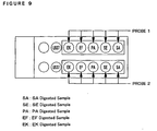
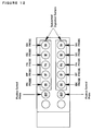
- This slide glass (item number: MS311BL) is provided with 14 circular wells having the diameter of 5 mm addition, upon practical use, it is preferred that an APS coated slide glass is used which is a slide glass with 3-aminopropyltriethoxysilane (APS, SIGMA) coated thereon for the purpose of improving adhesiveness of cells. Alternatively, a slide glass with poly-L-lysine or gelatin coated thereon may be also used.
- a slide glass (item number: MS311BL) is first fixed on a slide holder, and thereafter is washed by immersing in a diluted neutral detergent for 30 minutes, and the detergent is sufficiently removed with running water. Next, the slide glass is washed with purified water and dried sufficiently at high temperature (100°C or greater) followed by leaving to stand to cool at room temperature. Then, the slide glass is immersed in acetone containing 2% APS for 1 minute, and immediately thereafter washed briefly with acetone and sterile purified water sequentially followed by air drying.
- the APS coated slide glass can be produced by drying at about 20°C to about 60°C, preferably at 42°C.
- leukocytes When the leukocytes are supported on the APS coated slide glass, it is preferred that leukocytes are smeared on each well such that they are spread over to give a single layer and air dried. It is preferred that phagocytes for use in fixing are prepared such that the density (x cells/ml) is about 5 ⁇ 10 6 cells/ml ⁇ x cells/ml ⁇ about 1 ⁇ 10 8 cells/ml; and preferably about 1 ⁇ 10 7 cells/ml x cells/ml about 5 ⁇ 10 7 cells/ml.
- cell number of the leukocytes fixed on the APS coated slide glass per 1 well is preferably adjusted to be about 2.5 ⁇ 10 4 cells/well ⁇ y cells/well ⁇ about 5 ⁇ 10 5 cells/well, and preferably about 5 ⁇ 10 4 cells/well y cells/well about 2.5 ⁇ 10 5 cells/well.
- a small amount of PBS is added to a leukocyte pellet obtained by centrifugation of the leukocyte fraction at 4°C for about 10 minutes at about 140 ⁇ g to about 180 ⁇ g followed by suspension, and the cell number of the leukocytes is counted using a counting chamber.
- Preparation can be perfected by smearing 5 ⁇ l of the leukocyte suspension, which was prepared with PBS such that cell number becomes about 5 ⁇ 10 4 cells/well to about 2.5 ⁇ 10 5 cells/well, on each well of the APS coated slide glass to allow the leukocytes spread to form a single layer followed by complete air drying.
- a process may be employed in which immersion is conducted in PBS for about 3 to about 30 minutes, followed by immersing in a solution of an enzyme pretreatment reagent (prepared by mixing 1.25 g of saponin, 1.25 ml of t-octylphenoxypolyethoxyethanol (specific gravity: 1.068 to 1.075 (20/4°C), pH (5 w/v%) 5.5-7.5) and 25 ml of a PBS stock solution, and adjusting to give the total volume of 50 ml with sterile purified water) diluted to about 2 to about 50 fold in sterile purified water, and allowing infiltration on a shaker for about 3 to about 30 minutes.
- an enzyme pretreatment reagent prepared by mixing 1.25 g of saponin, 1.25 ml of t-octylphenoxypolyethoxyethanol (specific gravity: 1.068 to 1.075 (20/4°C), pH (5 w/v%) 5.5-7.5
- an enzyme pretreatment reagent prepared by mixing 1.
- an enzyme reagent solution is prepared by adding 1 ml of an enzyme reagent dissolving solution (prepared by about 100 fold dilution of dimethylsulfoxide (DMSO) which contains phenylmethylsulfonylfluoride (PMSF) in PBS) to an enzyme reagent (N-acetylmyramidase, lysozyme and/or lysostafin) per 1 slide, and thereafter, 1 ml of this enzyme solution is dropped on a site of the leukocyte smear, and left to stand still for about 10 to about 60 minutes in a humid box at about 20°C to about 60°C, preferably at about 37° C to about 42°C.
- an enzyme reagent dissolving solution prepared by about 100 fold dilution of dimethylsulfoxide (DMSO) which contains phenylmethylsulfonylfluoride (PMSF) in PBS
- an enzyme reagent N-acetylmyramidase, lyso
- DMSO has the potential of lowering the activity of lysozyme and lysostafin at the concentration of 5% or greater, it is preferably used at the concentration of less than 5%.
- PMSF as a substance for retaining the morphology of the phagocytes
- other known protease inhibitor e.g., tosyl lysine chloromethyl ketone (TLCK) and a mixture thereof may be also used.
- TLCK tosyl lysine chloromethyl ketone
- a solvent such as DMSO may be changed ad libitum.
- optimal titer of lysostafin may be set to 1 unit/ml to about 1,000 unit/ml, and preferably about 10 unit/ml to about 100 unit/ml.
- lysis did not occur when the titer of N-acetylmuramidase is about 10 unit/ml or less, while the titer of lysozyme was fixed to be about 10,000 unit/ml. In respect of lysozyme, when the titer of N-acetylmuramidase was fixed to be 100 unit/ml, lysis did not occur with the titer of lysozyme of 1,000 unit/ml or less.
- optimal titer of N-acetylmuramidase may be set to be about 10 unit/ml to about 10,000 unit/ml, and preferably about 100 unit/ml to about 1,000 unit/ml, whilst the optimal titer of lysozyme may be set to be about 1,000 unit/ml to about 1,000,000 unit/ml, and preferably about 10,000 unit/ml to about 100,000 unit/ml.
- the range of titer may be about 50 unit/ml to about 500 unit/ml, preferably about 100 unit/ml to about 500 unit/ml of zymolase.
- PMSF protease inhibitor
- the enzyme to be used may be optionally selected.
- two or more enzymes may be used in combination for the purpose of achieving lysis more effectively. According to the present invention, it was revealed that by using a mixture of three kinds, lysozyme, lysostafin and N-acetylmuramidase, lytic activity was elevated in comparison with the case where a single enzyme was used.
- Temperature of the enzymatic treatment may be preferably about 4°C to about 60°C for Staphylococcus aureus ; higher than about 25° C, preferably about 37° C or higher for Staphylococcus epidermidis ; and higher than about 25° C and less than about 60° C, preferably about 37° C to about 42° C for Enterococcus faecalis . Accordingly, it is most preferred that optimal temperature for the enzymatic treatment is set to be about 37° C to about 42° C. Additionally, critical temperature is expected to be about 26° C to about 59° C in the common range for those three kinds of the bacteria.
- time period of the enzymatic treatment may be 20 minutes or longer for any of digested samples of Staphylococcus aureus, Staphylococcus epidermidis and Enterococcus faecalis (inadequate in 0 minute and 10 minutes), and because no bacterial body was found within the leukocytes, the time period is preferably at least about 15 minutes or longer, preferably about 20 minutes or longer, and in addition, optimal time period of the enzymatic treatment shall be about 30 minutes to about 60 minutes. Moreover, the time period of the enzymatic treatment may be about 15 minutes to about 120 minutes.
- N-acetylmuramidase is an enzyme which lowers the absorbance at 600 nm when thermally treated dry powder of Enterococcus faecalis by a heat treatment and N-acetylmuramidase are subjected to a reaction in a 5 mmol/l Tris-HCl buffer (pH 6.0) containing 2 mmol/l magnesium chloride at 37° C for 5 minutes.
- Lysozyme is an enzyme which lowers the absorbance at 600 nm when Micrococcus luteus and lysozyme were subjected to a reaction in PBS at 37°C for 5 minutes. Furthermore, when the enzymatic activity to lower the absorbance at 540 nm of Micrococcus luteus by 0.001 at 35°C, pH 6.2 in 1 minute is determined as 1 unit, it is preferred that one having the enzymatic activity of 50,000 unit/mg or greater is used.
- Lysostafin is an enzyme which lowers the absorbance at 600 nm when Staphylococcus epidermidis and lysostafin are subjected to a reaction in PBS at 37° C for 5 minutes. Furthermore, when the enzymatic activity to lower the absorbance at 620 nm of Staphylococcus aureus from 0.240 to 0.125 at 37°C, pH 7.5 in 10 minutes is determined as 1 unit, it is preferred that one having the enzymatic activity of 500 unit/mg or greater is used.
- Zymolase (trade name: Zymolyase, Seikagaku Corporation) is an enzyme prepared from a culture liquid of Arthrobacter lutesul, having a potent lytic activity against cell walls of yeast living cells.
- Essential enzyme involving in lysis of cell walls included in zymolase is -1,3-glucan lanimaripentaohydrolase, which acts on a glucose polymer having -1,3-bonds to produce lanimaripentaose as a major product.
- Zymolyase-100T which is purified by ammonium sulfate fractionation, and further purified by affinity chromatography (Kitamura, K. et al.; J. Ferment.
- Technol., 60, 257, 1982 has the activity of 100,000 unit/g.
- the activity of this enzyme is known to be altered depending on the type of yeast to be a substrate, culture condition and growing stage (Kitamura, K. et al.; J. Gen. Appl. Microbiol., 20, 323, 1974, Kitamura, K. et al.; Agric. Biol. Chem., 45, 1761, 1981, Kitamura, K. et al.; Agric. Biol. Chem., 46, 553, 1982).
- Zymolyase-100T includes about 1.0 ⁇ 10 7 unit/g of -1,3-glucanase, about 1.7 ⁇ 10 4 unit/g of protease, and about 6.0 ⁇ 10 4 unit/g mannase, however, DNase and RNase are not found therein (Kitamura, K. et al.; J. Gen. Appl. Microbiol., 18, 57, 1972).
- the optimal pH of Zymolyase is about 5.5 to about 8.5, and preferably about 6.5 to about 7.5, whilst the optimal temperature is about 25°C to about 55° C, and preferably about 35° C to about 45°C.
- lysis spectrum (genus name) against yeast (cells in logarithmic growth phase) includes Ashbya, Candida, Debaryomyces, Eremothecium, Endomyces, Hansenula, Hanseniaspora, Kloekera, Kluyveromyces, Lipomyces, Helschkowia, Pichia, Pullularia, Torulopsis, Saccharomyces, Saccharomycopsis, Saccharomycodes, Schwanniomyces and the like.
- examples of those in genus candida include Candida albicans, Candida tropicalis, Candida parasilosis, Candida galacta, Candida guilliermondii, Candida krusei, Cryptococcus neoformans and the like.
- an SH compound e.g., cysteine, 2-mercaptoethanol, dithiothreitol and the like can be used.
- Fungi belonging to these genera may be also used in the present invention.
- this enzyme an enzymatic activity required for decreasing about 30% of A 800 of a reaction liquid (enzyme: 1 ml of a 0.05 to 0.1 mg/ml solution, substrate: 3 ml of a beer yeast suspension (2 mg dry weight/ml), buffer: 5 ml of M/15 phosphate buffer (pH 7.5), adjusted to give the total volume of 10 ml with 1 ml of sterile purified water) using the beer yeast suspension as a substrate at about 25°C within two hours is determined as 1 unit.
- Zymolyase-100T has the activity of 100,000 unit/g.
- the concentration of PMSF (added in order to protect the leukocytes from protease so that the morphology thereof is retained) which is used as a solvent for the enzyme reagent is in the range of 10 ⁇ mol/l to 10 mmol/l, and preferably 0.1 mmol/l to 1 mmol/l, because effects were observed at the concentration of 10 ⁇ mol/l or greater, while deterioration of morphology of leukocytes was completely suppressed at the concentration of 0.1 mmol/l or greater.
- the concentration of DMSO is less than 5%, preferably 2% or less, and further approximately the concentration of 1%.
- the enzyme reagent dissolving solution is preferably prepared by 100 to 1,000 fold dilution of dimethylsulfoxide (DMSO) which contains 0.1 mol/l phenylmethylsulfonylfluoride (PMSF) in PBS.
- DMSO dimethylsulfoxide
- PMSF phenylmethylsulfonylfluoride
- the step of acetylation of cell membrane proteins may be inserted. Specifically, it can be carried out through immersing the slide glass in an acetylation reagent, which was prepared by adding acetic anhydride to an acetylating reagent (7.46 g of triethanolamine, an appropriate amount of hydrochloric acid, adjusted to give the total volume of 50 ml with an appropriate amount of sterile purified water) and diluting about 2 fold to about 50 fold, preferably about 10 fold in sterile purified water to give the final concentration of acetic anhydride of 0.1 to 3.0%, preferably 0.8%, followed by shaking for 5 to 30 minutes on a shaker. Thereafter, the slide glass is sequentially immersed in 75%, 85%, and 98% ethanol for 2 to 5 minutes respectively, and completely air dried.
- an acetylation reagent which was prepared by adding acetic anhydride to an acetylating reagent (7.46 g of triethanolamine, an appropriate amount of hydroch
- the step of forming a single stranded DNA by an alkali treatment of the DNA of the causative microorganism of an infectious disease can be also inserted. Specifically, it can be carried out through immersing the slide glass in PBS which contains about 10 mmol/l to about 300 mmol/l, preferably about 70 mmol/l sodium hydroxide (prepared by adding sodium hydroxide in the PBS stock solution, diluting to 20 fold with sterile purified water to give the final concentration of sodium hydroxide of 70 mmol/l) for about 2 to about 5 minutes. Thereafter, the slide glass is sequentially immersed in 75%, 85%, and 98% ethanol for 2 to 5 minutes respectively, and completely air dried.
- PBS which contains about 10 mmol/l to about 300 mmol/l, preferably about 70 mmol/l sodium hydroxide (prepared by adding sodium hydroxide in the PBS stock solution, diluting to 20 fold with sterile purified water to give the final concentration of sodium hydroxide of 70
- a liquid containing the DNA probe for detection prepared in a probe dilution solution is coated on the smeared site, and is left to stand still in a humid box at about 25°C to about 50°C, preferably at about 37°C to about 42°C for about 1 to about 3 hours, preferably for about 2 hours.
- the probe dilution solution includes 600 ⁇ l of salmon sperm DNA, 50 ⁇ l of 100 ⁇ Denhardt' s solution 500 pl of hybridization stock solution, 2,250 ⁇ l of formamide, 1,000 ⁇ l of 50% dextran sulfate.
- the probe solution preferably includes 15 ng of each DNA probe for detection, which may be adjusted to give the total volume of 50 ⁇ l with the probe dilution solution.
- Concentration of the probe for SA, SE, PA, EF and EK may be about 0.6 ng/ ⁇ l to about 1.8 ng/ ⁇ l, preferably about 0.6 ng/ ⁇ l to about 1.2 ng/ ⁇ l. Further, the result of inadequate was brought at 0.06 ng/ ⁇ l, and the result of adequate was brought at 0.6 ng/ ⁇ l, therefore, it is preferred that the concentration is set to be at least 0.1 ng/ ⁇ l or greater. Moreover, because the result of inadequate was brought at 2.4 ng/ ⁇ l, and the result of adequate was brought at 1.8 ng/ ⁇ l, it is preferred that the concentration is set to be 2.2 ng/ ⁇ l or less. In addition, the optimal concentrations of positive control and negative control may be 0.4 to 2.0 ng/ ⁇ l and 0.6 to 2.0 ng/ ⁇ l respectively, and preferably 0.6 to 1.0 ng/ ⁇ l in common.
- time period of the hybridization is at least 30 minutes or longer, preferably 60 minutes or longer, and more preferably 90 minutes or longer. More preferred optimal time period of the hybridization may be set to be about 120 minutes to about 900 minutes.
- SDS sodium dodecyl sulfate
- concentration of SDS is 1% or less, more preferably about 0.1% to about 0.5%, still more preferably about 0.25%.
- SDS may be added to a solution used upon the hybridization, or may be in the probe dilution solution or in the probe solution, which was mixed beforehand.
- one or more DNA probe having the chain length of about 350 to about 600 base length, preferably about 350 to about 550 base length is employed as the DNA probe for detection, because the probe is efficiently introduced into phagocytes, and firm contact with the gene of the incorporated foreign microorganism is permitted. It is not intended that base length (number of base pairs) of the subject probe must necessarily fall within the aforementioned range of the base length, but that it is allowable as long as the base length in the aforementioned range is included in the distribution of the base length of the probe.
- These probes may be used alone, or several kinds of probes (more than one) may be also used. More than one probes may be multiple kinds of probes which can hybridize to one bacterial strain. Alternatively, the kind of the probe may be multiple owing to the presence of multiple types of the bacterial strains although a single probe may be employed for a single bacterial strain. Thus, there is no particulate limitation as far as the kind of the probe is one or more.
- These probes preferably comprise a DNA fragment having a sequence which does not any how hybridize with the phagocyte itself, and additionally, they should not cross hybridize with a gene derived from any other strain of microorganism. For example, when a subtraction method is used, a specific probe can be produced in a short period of time.
- These probes may be prepared and labelled according to a common nick translation process using a non-radioisotopic labelling substance such as fluorescein isothiocyanate (FITC), biotin, digoxigenin (digoxigenin (DIG)-11-dUTP) or the like.
- FITC fluorescein isothiocyanate
- DIG digoxigenin
- Chain length of the probe can be controlled such that most efficient labelling is enabled, by changing the ratio of amount of DNase I and DNA polymerase I added in the nick translation reaction.
- a probe for efficiently labelling 2 ⁇ g of the DNA probe (SA-24), and for regulating the chain length of a probe to enable efficient in situ hybridization with the DNA of a foreign microorganism (base length of about 350 to about 600), when 2 ⁇ l of 10 U/ ⁇ l DNA polymerase I is included in the reaction liquid of total volume of 100 ⁇ l, 6 ⁇ l of DNase I may be included which was prepared such that about 10 to about 350 mU, preferably about 25 to about 200 mU, more preferably about 50 to about 150 mU is present in total volume of 100 pl.
- volume of each enzyme and total volume of the reaction liquid and the like in this instance may optionally vary as long as the proportion according to the aforementioned essential condition for an optimal reaction is kept constant.
- DNase I when 20U of DNA polymerase I is included in total volume of 100 ⁇ l, DNase I may be prepared in an amount of about 10 to about 350 mU, preferably about 25 to about 200 mU, and more preferably about 50 to about 150 mU.
- nick translation may be conducted using about 0.5/1,000 to about 17.5/1,000, preferably about 1.25/1,000 to about 10/1,000, and more preferably about 2.5/1,000 to about 7.5/1,000 unit of DNase I.
- DNA polymerase I is present in an amount of about 10 U
- DNase I is present in an amount of about 5 to about 175 mU, preferably about 12.5 to about 100 mU, and more preferably about 25 to about 75 mU.
- the amount of DNA as well as optimal conditions for the reaction of DNA polymerase I and DNase I can be determined with reference to the optimal conditions for the reaction as described above, and chain length of the probe (base length of about 350 to about 600) can be regulated to result in efficient labelling and efficient in situ hybridization with a foreign microorganism DNA.
- the stringent condition for carrying out in situ hybridization may be for example, a condition which comprises incubating in the presence of about 30% to about 60%, preferably about 50% of formamide, at about 30 to about 50°C, preferably at about 38 to about 42°C followed by washing.
- an operation of blocking may be performed. Specifically, 1 ml of a blocking reagent (2 ml of normal rabbit serum, 0.5 ml of the PBS stock solution, adjusted to give the total amount of 10 ml with sterile purified water) is dropped on the smear site per one slide glass in a humid box, and left to stand still for 15 to 60 minutes. Thereafter, the blocking reagent is removed.
- a blocking reagent 2 ml of normal rabbit serum, 0.5 ml of the PBS stock solution, adjusted to give the total amount of 10 ml with sterile purified water
- color reaction For detecting a signal which results from the hybridization with a gene derived from the microorganism (genomic DNA or RNA), color reaction may be conducted in which any conventional method for an antigen-antibody reaction is utilized. In other words, after enough washes of the sample following completing the hybridization, an operation for blocking is conducted. Thereafter, a treatment is executed using a conjugate of an anti-FITC antibody, anti-digoxigenin antibody or the like, e.g., an alkaline phosphatase conjugate, and then, color developmet of the signal is allowed by a color development system of the conjugate to determine the states of hybridization.
- an anti-FITC antibody, anti-digoxigenin antibody or the like e.g., an alkaline phosphatase conjugate
- an anti-digoxigenin-alkaline phosphatase conjugate is used, and the detection may be conducted through utilizing a substrate which is generally used for alkaline phosphatase (nitroblue tetrazolium, 5-bromo-4-chloro-3-indolyl phosphate and the like).
- a substrate which is generally used for alkaline phosphatase (nitroblue tetrazolium, 5-bromo-4-chloro-3-indolyl phosphate and the like).
- the smear preparation washed after the color reaction is subjected to counter staining with naphthol black Fast Green (20 mg/50ml, manufactured by Wako Chemical Co. ) or the like to observe intracellular signals with a light microscope.
- a labelled antibody solution is prepared by diluting a labelled antibody (1.05 unit of alkaline phosphatase labelled anti-digoxigenin antibody solution, adjusted with 12.6 ⁇ l of buffer A (746 mg of triethanolamine, 17.5 mg of sodium chloride, 20.3 mg of magnesium chloride hexahydrate, 1.36 mg of zinc chloride, 1,000 mg of bovine serum albumine, an appropriate amount of hydrochloric acid, adjusted to give the total volume of 100 ml with sterile purified water) to give the total volume of 14 ⁇ l) in a labelled antibody diluent (8.48 mg of Tris-(hydroxymethyl)-aminomethane, 6.14 mg of sodium chloride, an appropriate amount of hydrochloric acid, adjusted to give the total volume of 0.7 ml with sterile purified water) to 10 to 200 fold, preferably 50 fold, and
- a labelled antibody washing solution (1 ml of polysorbate 20, 50 ml of the PBS stock solution, adjusted to give the total volume of 100 ml with sterile purified water) diluted to 2 to 50 fold, preferably 10 fold, and is allowed for infiltration on a shaker for about 5 to about 30 minutes as it is.
- a coloring pretreatment liquid obtained by mixing a coloring pretreatment liquid 1 (6.06 g of Tris-(hydroxymethyl)-aminomethane, 2.92 g of sodium chloride, an appropriate amount of hydrochloric acid, adjusted to give the total volume of 50 ml with sterile purified water) and a coloring pretreatment liquid 2 (5.08 g of magnesium chloride hexahydrate, adjusted to give the total volume of 50 ml with sterile purified water) in an equivalent volume and diluting to approximately 5 fold in sterile purified water, and then shaken for 5 to 30 minutes on a shaker as it is.
- a coloring pretreatment liquid 1 (6.06 g of Tris-(hydroxymethyl)-aminomethane, 2.92 g of sodium chloride, an appropriate amount of hydrochloric acid, adjusted to give the total volume of 50 ml with sterile purified water)
- a coloring pretreatment liquid 2 (5.08 g of magnesium chloride hexahydrate, adjusted to give the total volume of 50
- a coloring reagent nitrogenblue tetrazolium (NBT)/ 5-bromo-4-chloro-3-indolylphosphate (BCIP)
- NBT nitrogen tetrazolium
- BCIP 5-bromo-4-chloro-3-indolylphosphate
- a coloring reagent washing solution (606 mg of Tris-(hydroxymethyl)-aminomethane, 186 mg of ethylenediamine tetraacetate disodium dihydrate, an appropriate amount of hydrochloric acid, adjusted to give the total volume of 50 ml with an appropriate amount of sterile purified water) diluted to about 2 to about 50 fold, preferably about 10 fold for about 2 to about 10 minutes, and is air dried.
- a counter staining solution 50 mg of fast green FCF (edible dye, green color No.
- the coloring reagent described above may be one prepared separately.
- the alkaline phosphatase labelled anti-digoxigenin antibody solution which may be preferably used is one which results in color development in a site of DNA blotting when 1 ng of a digoxigenin labelled DNA is blotted on a membrane for blotting, subjected to blotting, treated with the alkaline phosphatase labelled anti-digoxigenin antibody solution diluted to 10,000 fold, and allowed to react with a coloring substrate (NBT/BCIP), but one which does not result in color development even though similar operation is conducted with a DNA without digoxigenin labelling.
- the anti-digoxigenin antibody is preferably derived from sheep. In detail, it may be purified from serum of an immunized sheep by an ion exchanging chromatography and an antibody column chromatography.
- the coloring reagent (NBT/BCIP solution, pH 9.0 to 10.0) preferably contains 3.3 mg of nitroblue tetrazolium (NBT), 1.65 mg of 5-bromo-4-chloro-3-indolylphosphate (BCIP), 99 ⁇ g of N,N-dimethylformamide, 121 mg of Tris-(hydroxymethyl)-aminomethane, an appropriate amount of hydrochloric acid, 58.4 mg of sodium chloride, 101.6 mg of magnesium chloride hexahydrate, and is adjusted to give the total amount of 10 ml with an appropriate amount of sterile purified water.
- the coloring reagent which may be preferably used is one which exhibits a dark purple signal on the blotted site when a protein labeled with alkaline phosphatase is blotted on a membrane for blotting followed by a treatment of the membrane with the coloring reagent at room temperature under light shielding.
- an edible dye e.g., yellow No. 4 (tartrazine) can be used for the purpose of further clarification of the contrast between the signal and the cell.
- the grounds therefor may be difficulties in the counter staining on behalf of the similar color among the purple color developed by the substrate and the blue color developed by naphthol black.
- the process which may be employed for labelling digoxigenin can be a nick translation method.
- a PCR method, a random primer labelling method, an in vitro transcription labelling method, a terminal transferase labelling method or the like can be employed.
- Determination may be carried out by microscopic examination with a light microscope ( ⁇ 1,000), and observation of at least one color development of bluish purple color may be determined as positive in cells within a single well stained with the counter staining solution as described above.
- the working cell bank (SA-24) is smeared by streaking with a platinum loop, a disposable plastic loop or the like on an L-broth solid medium containing 50 ⁇ g/ml ampicillin prepared in a sterile petri dish (microorganism picking).
- the culture liquid in the regular culture is centrifuged at 4° C for 10 minutes at 4,000 ⁇ g to collect the microorganism.
- the culture supernatant is removed, and thereto is added 20 ml of STE (10 mmol/l Tris-hydrochloric acid (pH 8.0), 1 mmol/l disodium ethylenediamine tetraacetate (EDTA), 0.1 mmol/l sodium chloride) to resuspend the cell bodies.
- STE Tris-hydrochloric acid
- EDTA disodium ethylenediamine tetraacetate
- a solution-1 50 mmol/l glucose, 25 mmol/l Tris-hydrochloric acid (pH 8.0), 10 mmol/l EDTA
- 10 mg/ml lysozyme 10 mg/ml lysozyme
- 10 ml of a solution-2 0.2 mmol/l sodium hydroxide, 1% sodium dodecyl sulfate (SDS)
- SDS sodium dodecyl sulfate
- 7.5 ml of an ice cold solution-3 (3 mol/l potassium acetate (pH 4.8)) mixed by inversion and left to stand on ice for 10 minutes.
- plasmid DNA containing SA-24 800 ⁇ l of 10 mg/ml ethidium bromide and 8.6 g of cesium chloride are added to the resulting plasmid DNA followed by mixing by inversion to dissolve the plasmid.
- the solution is placed in a centrifuge tube, which is then capped or sealed. After centrifugation at 20°C for 5 hours at 500,000 ⁇ g with a vertical rotor, a band of the plasmid DNA is fractionated using a glass syringe or an injection needle under the irradiation of an ultraviolet ray light.
- TE-saturated 1-butanol To the fractionated plasmid DNA solution is added an equivalent amount of TE-saturated 1-butanol followed by mixing by inversion and centrifugation at 15,000 ⁇ g for 5 minutes by a high speed microcentrifuge to remove the supernatant. This operation is repeated to eliminate ethidium bromide in the plasmid DNA solution. Next, thereto is added TE to give the volume of 1.5 ml followed by desalting on a demineralization column (NAP-10). To the desalted plasmid DNA solution is added 30 ⁇ l of a 3 mol/l sodium acetate solution followed by mixing, and 3 fold amount of 99.5% ethanol is added thereto followed by mixing by inversion and leaving to stand at -20°C for 30 minutes or longer.
- centrifugation is conducted with a high speed refrigerated micro centrifuge at 4°C for 20 minutes at 15,000 ⁇ g to remove the supernatant. Thereafter, cold 70% ethanol is added thereto to suspend therein, and once again, centrifugation is conducted with a high speed refrigerated micro centrifuge at 4°C for 20 minutes at 15,000 ⁇ g to remove the supernatant.
- resulting precipitate of the plasmid DNA is evaporated to dryness under a reduced pressure.
- TE in an amount of 100 ⁇ l is added to the plasmid DNA to dissolve completely, and the concentration is measured on the basis of the absorbance at 260 nm (Purification of plasmid DNA containing SA-24). Then, size check of the plasmid DNA containing SA-24 is carried out by a treatment with arestriction enzyme and agarose electrophoresis.
- SA-24 is extracted from the agarose gel and purified, and the concentration is measured with an absorbance meter. A part of the purified SA-24 is electrophoresed on a 0.8% agarose gel to verify that a single band is found.
- 2 ⁇ g of the purified SA-24 is used, and may be subjected to digoxigenin labelling in a reaction liquid having the composition described in Table 1 below. Table 1: Composition of the reaction liquid for labeling Amount included ( ⁇ L) DNA probe X 10 ⁇ L.B.
- X represents the volume which may be added such that preferred concentration of the probe as described above is provided depending upon the concentration of the probe stock solution, and the amount Y of purified water is determined following this volume to adjust the final volume.
- reaction liquid 100 ⁇ l of TE is added to the reaction liquid to terminate the reaction.
- the reaction terminated solution is poured into a spin column, and centrifuged at 4°C for 10 minutes at 380 x g to remove free nucleotides.
- concentration of the eluate is measured with an absorbance meter, and then adjusted to give 10 ng/ ⁇ l with TE.
- 0.5 ⁇ l of the labelled SA-24 is dropped onto a membrane, and air dried.
- the membrane is immersed in a blocking reagent, and blocked at room temperature for 30 minutes.
- an alkaline phosphatase labelled anti-digoxigenin antibody solution diluted to 5,000 fold in 0.1 mol/l Tris-hydrochloric acid (pH 7.5) and 0.15 mol/l sodium chloride, is immersed the membrane at room temperature for 30 minutes.
- the membrane is immersed in 0.1 mol/l Tris-hydrochloric acid (pH 7.5), 0.15 mol/l sodium chloride, and washed twice by shaking at room temperature for 10 minutes.
- Tris-hydrochloric acid pH 9.5
- 0.15 mol/l sodium chloride and 50 mmol/l magnesium chloride is immersed the membrane at room temperature for 10 minutes.
- the membrane is immersed in the coloring reagent at room temperature under light shielding for 10 minutes.
- the membrane is immersed in TE to terminate the color development. Verification of the labelling is executed by the observation of bluish purple coloring at the potion under the spotting.
- a small amount of sterilized glass wool is packed in a 1 ml disposable syringe.
- dot blot hybridization may be carried out according to the following procedure.
- each 100 ng of various types of bacterial genomic DNA as prepared is spotted to a nylon membrane (Pall Biodyne (R) type B, manufactured by Nihon Pall Ltd. ) on a filter paper (manufactured by Whatman, 3MM) saturated with a solution containing 0.5 mol/l sodium hydroxide and 1.5mol/l sodium chloride according to a conventional process, and the air dried membrane is left to stand for 10 minutes.
- the membrane is allowed to stand still on the filter paper described above which is saturated with a solution containing 0.5 mol/l Tris-hydrochloric acid (pH 7.5) and 1.5 mol/l sodium chloride for 10 minutes to neutralize the denaturated DNA.
- the membrane is left to stand still on the filter paper as described above which is saturated with a 2 ⁇ SSC (Standard Saline Citrate) solution for 5 minutes followed by rinsing. Thereafter, the membrane is air dried, immersed in a 2 ⁇ SSC solution and allowed for infiltration for 5 minutes.
- the membrane is immersed in a prehybridization solution within a plastic bag, and affinitized at 42°C for 60 minutes.
- the membrane is immersed in 15 ml of a hybridization solution containing 400 mg of the probe in the plastic bag, and the reaction is allowed at 42°C overnight.
- the membrane is immersed in a solution containing 2 ⁇ SSC and 0.1% SDS (sodium dodecyl sulfate), and washed for 5 minutes (repeated twice). Thereafter, the membrane is immersed in a solution containing 0.1 ⁇ SSC and 0.1% SDS, and washed at 60°C, for 10 minutes (repeated three times). The membrane is then immersed in a 2 ⁇ SSC solution, and washed for 5 minutes. The membrane is immersed in a solution containing 3% bovine serum albumines, 1% blocking buffer (manufactured by Boeringer), 0.1 mol/l Tris-hydrochloric acid (pH 7.5) and 0.15 mol/l sodium chloride, and is gently shaken for 30 minutes.
- SDS sodium dodecyl sulfate
- the membrane is immersed in a solution of alkaline phosphatase labelled anti-digoxigenin antibody (manufactured by Boeringer) diluted to 5,000 fold in a solution containing 0.1 mol/l Tris-hydrochloric acid (pH 7.5) and 0.15 mol/l sodium chloride, and is gently shaken for 30 minutes.
- the membrane is immersed in a solution containing 0.1 mol/l Tris-hydrochloric acid (pH 7.5) and 0.15 mol/l sodium chloride, and is shaken for 15 minutes (twice).
- the membrane is immersed in a solution containing 0.1 mol/l Tris-hydrochloric acid (pH 9.5), 0.1 mol/l sodium chloride and 5 mmol/l magnesium chloride, and is shaken for 5 minutes.
- the membrane is immersed in an NBT-BCIP solution (manufactured by GIBCO BRL), and the color development reaction is allowed under light shielding.
- NBT-BCIP solution manufactured by GIBCO BRL
- TE 10 mmol/l Tris-hydrochloric acid (pH 8.0)
- 1 mmol/l EDTA is immersed the membrane to terminate the color development reaction, and is air dried.
- the prehybridization solution and the hybridization solution are as shown in Table 2 below.
- the surfactant which may be used in the step of in situ hybridization is any of known surfactants.
- Surfactants are generally classified in anion surfactants, nonionic surfactants, cation surfactants and ampholytic surfactants.
- Anion surfactants are also referred to as anionic surfactants, which yield an organic anion upon ionization in water.
- anion surfactants include RCOONa, RSO 3 Na, RSO 4 Na and the like.
- An aqueous solution of the surfactant containing a weakly acidic group such as RCOONa is liable to be hydrolyzed and is weak alkaline.
- an aqueous solution of a surfactant having a strongly acidic group such as RSO 3 Na, RSO 4 Na or the like is resistant to hydrolysis, which shall be neutral. Because it is anionic, it may lose surface activity in the presence of a large quantity of cationic substance, and may be inactivated in a strongly acidic circumstance.
- Nonionic surfactants refer to those having a hydrophilic group which is nonionic.
- An ethylene oxide group (-CH 2 CH 2 O-) is often used as the hydrophilic group.
- hydrophilicity is increased.
- lipophilicity is increased. Therefore, it is characterized in that surfactants with variously altered hydrophilicity and lipophilicity can be obtained. Because a nonionic surfactant does not ionize in water and is hardly affected by inorganic salts, less action is exerted also on a living body.
- the detergent action thereof is potent with comparatively less foaming, therefore, it is widely used not alone as a detergent, but in pharmaceuticals, cosmetics, foods and the like.
- Water soluble nonionic surfactant becomes insoluble in water at a certain temperature as the temperature rises, and then the aqueous solution starts to be turbid. Such turbidity results from the cleavage of hydrogen bonds between the hydrophilic groups and water.
- Cation surfactants are also referred to as cationic surfactants, which yield an organic cation upon ionization in water. Although cation surfactants do not have potent detergent action in general, they strongly bind to anionic substances such as bacteria, leading to a great bactericidal action. Moreover, they also have an anti-static ability for fibers and plastics.
- dodecyltrimethyl chloride [C 12 H 25 (CH 3 ) 3 N]Cl as a typical exemplary cation surfactant is water soluble
- didodecyldimethylammonium chloride [(C 12 H 25 ) 2 (CH 3 ) 2 N]Cl is insoluble in water, which forms a vesicle in the form of a bimolecular film in water, and is soluble in benzene.
- Ampholytic surfactants are surfactants having both an anionic group and a cationic group in the molecule. Ionization state thereof in water is similar to those of amino acids, and thus many of ampholytic surfactants are amino acid derivatives. Therefore, they have an isoelectric point similarly to amino acids, which act as an anion surfactant in an alkaline region from the isoelectric point, whilst as a cation surfactant in an acidic region. Water solubility becomes the lowest at the isoelectric point, and the surface tension is also reduced. Ampholytic surfactants are used for a bactericide, an antistatic agent or the like.
- anion surfactants are classified into the carboxylic acid type, sulfonic acid type, sulfate ester type and phosphate ester type, whilst nonionic surfactants are classified into the ester type, ether type, ester ether type and alkanolamide type.
- Cation surfactants are classified into alkylamine salt type and quaternary ammonium salt type, whilst ampholytic surfactants are classified into carboxy betaine type, 2-alkylimidazoline derivative type and glycine type.
- the anion surfactants of carboxylic acid type are further classified into fatty acid monocarboxylate salts, N-acylsarcosine salts and N-acylglutamate salts.
- Representative examples thereof respectively include: sodium laurate and medicated soap as the fatty acid monocarboxylate salts; sodium N-lauroylsarcosine as the N-acylsarcosine salt; and disodium N-lauroylglutamate as the N-acylglutamate.
- the sulfonic acid type is further classified into dialkyl sulfosuccinate salts, alkane sulfonate salts, alpha-olefin sulfonate salts, straight chain alkyl benzenesulfonate salts, alkyl (branched chain) benzenesulfonate salts, alkyl naphthalenesulfonate salts, naphthalenesulfonate salts-formaldehyde condensates and N-methyl-N-acyltaurine salts.
- Representative examples include: sodium dioctyl sulfosuccinate as the dialkyl sulfosuccinate salt; sodium dodecane sulfonate as the alkane sulfonate; sodium straight chain dodecyl benzenesulfonate as the straight chain alkyl benzenesulfonate salt; sodium dodecyl benzenesulfonate as the alkyl (branched chain) benzenesulfonate salt; sodium butyl naphthalenesulfonate as the alkyl naphthalenesulfonate salt; and sodium N-methyl-N-stearoyltaurine as the N-methyl N-acyltaurine salt.
- the sulfate ester type is further classified into alkyl sulfate salts, polyoxyethylene alkyl ether sulfate salts and oil-and-fat sulfate ester salts.
- Representative examples include sodium dodecyl sulfate, sodium lauryl sulfate and sodium cetyl sulfate as the alkyl sulfate salt; and polyoxyethylene lauryl ether sulfate triethanolamine as the polyoxyethylene alkyl ether sulfate salt.
- the phosphate ester type is further classified into alkyl phosphate salts, polyoxyethylene alkyl ether phosphate salts and polyoxyethylene alkylphenyl ether phosphate salts.
- Representative examples include disodium monolauryl phosphate as the alkyl phosphate salt; and sodium polyoxyethylene lauryl ether phosphate and polyoxyethylene oleyl ether phosphate (8MOL) as the polyoxyethylene alkyl ether phosphate salt.
- Ester type of the nonionic surfactants is further classified into fatty acid glycerin, fatty acid sorbitan and fatty acid sucrose ester.
- Representative examples respectively include: glycerin monostearate as the fatty acid glycerin; sorbitan monostearate, sorbitan trioleate, sorbitan sesquioleate, sorbitan monolaurate, polysorbate 20 (polyoxyethylene sorbitan fatty acid ester), polysorbate 60 and polysorbate 80 as the fatty acid sorbitan; and stearic acid sucrose ester as the fatty acid sucrose ester.
- the ether type is further classified into polyoxyethylene alkyl ether, polyoxyethylene alkyl phenyl ether and polyoxyethylene polyoxypropylene glycol.
- Representative examples include: polyoxyethylene lauryl ether, polyoxyethylene stearyl ether and polyoxyethylene cetyl ether as the polyoxyethylene alkyl ether; and polyoxyethylene nonyl phenyl ether and polyoxyethylene octyl phenyl ether as the polyoxyethylene alkyl phenyl ether.
- the ester ether type is further classified into fatty acid polyethylene glycol and fatty acid polyoxyethylene sorbitan.
- Representative examples thereof respectively include oleic acid polethylene glycol as the fatty acid polyethylene glycol; and polyoxyethylene sorbitan palmitate and polyoxyethylene sorbitan monolaurate as the fatty acid polyoxyethylene sorbitan.
- the alkanolamide type involves only fatty acid alkanolamide alone.
- Representative example is lauric diethanolamide.
- the alkyl amine salt type of the cation surfactant includes monoalkyl amine salts, dialkyl amine salt and trialkyl amine salts. Representative examples thereof include monostearyl amine hydrochloride. Moreover, the quaternary ammonium salt type is further classified into alkyltrimethyl ammonium chloride (or bromide or iodide), dialkyldimethyl ammonium chloride (or bromide or iodide), and alkyl benzalkonium chloride.
- Representative examples respectively include: stearyltrimethyl ammonium chloride as the alkyltrimethyl ammonium chloride (or bromide or iodide); distearyldimethyl ammonium chloride as the dialkyldimethyl ammonium chloride (or bromide or iodide); and lauryl benzalkonium chloride as the alkyl benzalkonium chloride.
- the carboxy betaine type of the ampholytic surfactant is only alkyl betaine alone.
- Representative example is lauryl betaine.
- the 2-alkyl imidazoline derivative type is only 2-alkyl-N-carboxymethyl-N-hydroxyethyl imidazolinium betaine alone.
- Representative example includes 2-undecyl-N-carboxymethyl-N-hydroxyethyl imidazolinium betaine.
- the glycine type may be alkyl (or dialkyl) diethylene triaminoacetic acid, and the representative example includes dioctyl diethylene triaminoacetic acid.
- the surfactant may be admixed in the probe solution or probe dilution solution, alternatively, a solution containing the surfactant which was separately prepared from the probe solution may be added prior to, concurrently with or later than coating of the probe solution on the smear site.
- a solution containing the surfactant which was separately prepared from the probe solution may be added prior to, concurrently with or later than coating of the probe solution on the smear site.
- Such a process may be altered ad libitum by the person skilled in this art.
- a positive control probe when required, it can be produced as follows.
- U937 cells are first cultured in a 5% carbon dioxide gas incubator at 37° C using an RPMI1640 medium (25 ml) in a cell culture flask (175 cm 2 ).
- the U937 culture solution is placed in a 50 ml centrifuge tube, and centrifuged at 4° C for 10 minutes at 220 ⁇ g to recover the U937 cells.
- the cells are suspended and washed in 10 ml of PBS, and again centrifuged at 4° C for 10 minutes at 180 ⁇ g to recover the cells.
- the cells are suspended in 1 ml of a TE solution containing 200 ⁇ g/ml proteinase K and containing 1% SDS, followed by leaving to stand at 37°C for 30 minutes. Phenol extraction is repeated three to four times to execute deproteinization. Genome deposited through the ethanol precipitation is recovered, dissolved in 500 ⁇ l of sterile purified water containing 2.5 pg of ribonuclease, and left to stand at 42°C for 30 minutes.
- a positive control probe can be produced by measuring the concentration with an absorbance meter, and subjecting to digoxigenin labelling.
- the positive control probe which may preferably used is one which permits to ascertain the hybrid formation when the positive control probe is subjected to dot hybridization on a membrane with 100 ng of U937 genome spotted thereon.
- a negative control probe it can be produced by any known method.
- digested sample Specific process for producing a phagocyte post phagocytosis of the present invention (hereinafter, referred to as digested sample ) is illustrated below.
- U937 cell human monocyte established cell: ATCC CRL-1593.2
- Staphylococcus aureus ATCC 126000
- Staphylococcus epidermidis ATCC 14990
- Pseudomonas aeruginosa ATCC 10145
- Enterococcus faecalis ATCC 19433
- Escherichia coli ATCC 11775
- heparinized healthy human blood brain heart infusion (BHI) (manufactured by DIFCO)
- RPMI 1640 RPMI medium 1640 (manufactured by GIBCO)) containing Fetal Bovine Serum (final concentration: 10%, manufactured by GIBCO) and Antibiotic-Antimycitic (final concentration: 1%, manufactured by GIBCO) and the like.
- Instruments for use which are required include a carbon dioxide gas incubator (manufactured by Tabai Espec Corporation: BNA-121D type), a low speed refrigerated centrifuge (manufactured by Beckman: CS-6KR type), a counting chamber (manufactured by Elmer: bright line type), a shaking incubator (manufactured by TIETECH Co., Ltd.
- BR-300L type BR-300L type
- an absorbance meter manufactured by Beckman: DU68 type
- an incubator manufactured by Yamato Scientific Co., Ltd.: IC-62 type
- an incident-light inverted microscope manufactured by Nikon: DIAPHOT type
- a fluorescence microscope manufactured by Nikon: OPTIPHOT type
- a CCD camera manufactured by Hamamatsu Photonics KK.: C5810-01 type
- U937 cells (human monocyte established cell: ATCC CRL-1593.2) are cultured in an RPMI 1640 medium within a cell culture flask (e.g., 175 cm 2 ) in a 5% carbon dioxide gas incubator at about 20 to about 40°C, preferably at about 37° C.
- the cell culture flask is preferably one including a material consisting of a component which is liable to adhere to cells.
- the U937 cell culture liquid is placed in a centrifuge tube, and centrifuged at 0° C to about 10° C, preferably at about 4°C for about 10 minutes at about 150 to about 350 ⁇ g, preferably about 220 ⁇ g to recover the U937 cells.
- recovered U937 cells are suspended in PBS, and the cell number is counted with a counting chamber.
- the cell number may be adjusted to about 1 ⁇ 10 4 cells/ ⁇ l to 2 ⁇ 10 4 cells/ ⁇ l.
- Staphylococcus aureus, Staphylococcus epidermidis, Pseudomonas aeruginosa, Enterococcus faecalis and Escherichia coli are inoculated in a BHI culture liquid (supra), and cultured at about 20 to about 40° C, preferably at about 37° C for 6 hours or longer.
- the cultured bacterial liquid is centrifuged at 0 to about 10°C, preferably at 4° C, for example, at 2,000 ⁇ g for about 10 minutes to collect the bacteria.
- pellet of the bacteria is suspended using PBS, and centrifuged once again at 4° C for 10 minutes at 2,000 ⁇ g to collect the bacteria. After suspending thus collected bacteria in PBS, they may be diluted in PBS to produce a bacterial liquid prepared such that it has the turbidity (O.D.
- Heparinized healthy human blood is collected, and thereto is added the aforementioned reagent for separating hemocyte at a ratio of approximately 4 : 1, and left to stand still at about 20 to about 40°C, preferably at about 37°C for 30 minutes to yield the leukocyte fraction.
- leukocyte fraction is suspended in PBS.
- the supernatant in the culture flask is gently discarded, and the leukocyte fraction diluted in PBS is added to the flask followed by leaving to stand still at room temperature for about 10 minutes.
- the supernatant in the culture flask is discarded, and the leukocytes attached to the bottom of the flask are recovered in a centrifuge tube with PBS containing 0.02% EDTA, and centrifuged e.g., at 4°C for 10 minutes at about 140 to about 180 ⁇ g to collect the leukocytes.
- PBS containing 0.02% EDTA
- centrifuged e.g., at 4°C for 10 minutes at about 140 to about 180 ⁇ g to collect the leukocytes.
- precipitates of the leukocytes are gently suspended in sterile purified water to allow hemolysis, subjected to isotonization through adding PBS, followed by centrifugation once again at 4° C for 10 minutes at about 140 to about 180 ⁇ g to collect the leukocytes.
- the collected leukocytes are suspended in PBS, and cell number is counted with a counting chamber to adjust to give about 1 ⁇ 10 4 cells/ ⁇ l to about 5 ⁇ 10 4 cells/ ⁇ l.
- These digested samples are referred to as SA digested sample, SE digested sample, PA digested sample, EF digested sample and EK digested sample.
- the prepared U937 cells and each bacterial digested sample produced as described above is smeared on each well of an APS coated slide glass followed by air drying.
- cell number of each bacterial digested sample smeared and fixed on the slide glass is about 5.0 ⁇ 10 4 to about 2.5 ⁇ 10 5 cells/well, while cell number of U937 cells is about 5.0 ⁇ 10 4 to about 1.0 ⁇ 10 5 cells/well.
- the sample is immersed in Carnoy' s fixative(supra) for 20 minutes and thereafter immersed in 75% ethanol for 5 minutes. After washing Carnoy's fixative and air drying, the sample may be stored at 4°C until use in the test.
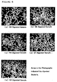
- Fig. 6 illustrates the state of the phagocytes prepared and observed by microscopic examination. Specific operation process involving Carnoy fixation, treatment for promoting permeability of the leukocyte cell membranes, lytic treatment, acetylation of the cell membrane protein, alkaline treatment of DNA of the bacterial body, in situ hybridization, blocking, reaction with labelled antibody, detection, and determination, which may be employed is as described herein.
- the present invention also includes a kit for evaluating a phagocytotic function which comprises fixing phagocytes post phagocytosis of a foreign microorganism, executing a treatment for promoting permeability of the cell membranes of the phagocytes, executing a treatment for exposing the DNA of the foreign microorganism existing in the phagocytes, carrying out in situ hybridization using a DNA probe for detection capable of hybridizing with the DNA under a stringent condition in the presence of a surfactant; and evaluating the phagocytotic function by the resulting signal, the kit having (1) the foreign microorganism, (2) at least one or more enzyme selected from the group consisting of lysostafin, lysozyme, N-acetylmuramidase and zymolase used in the exposing step of the DNA, and (3) one or more DNA probe for detection.
- This kit includes, reagent for separating blood, enzyme pretreatment reagent, enzyme reagent, acetylation reagent, probe solution, blocking reagent, labelled antibody, labelled antibody diluent, coloring pretreatment liquid-1, coloring pretreatment liquid-2, coloring reagent, counter staining solution, PBS stock solution, hybridization stock solution, labelled antibody washing solution, coloring reagent washing solution, APS coated slide glass, probe dilution solution, buffer A and the like as demonstrated in the following Examples.
- various reagents used in the present invention may be included for example, chloroform, ethanol, acetic anhydride, DMSO, PMSF, formamide, acetic acid, hydrochloric acid, sodium hydroxide and the like.
- instrument and machine such as low speed centrifuge, incubator, counting chamber, shaker, humid box, incubator, light microscope, variable pipette, blood collection tube, tip, pipette, staining bottle, measuring cylinder, glass syringe, 0.2 ⁇ m syringe top filter may be included.
- the present invention provides a process for monitoring the gene of a foreign microorganism phagocytized by a phagocyte included in a clinical specimen which contains a phagocyte derived from a living body.
- the present invention provides a process for identifying the gene of a microorganism which becomes a candidate of the causative microorganism which a causative microorganism of sepsis or a causative microorganism of bacteremia is specified on the basis of the results identified.
- the clinical specimen which may be used herein is a clinical specimen which contains a phagocyte derived from a living body, and examples thereof include body fluids such as blood, tissue fluid, lymph fluid, cerebrospinal fluid, pyo, mucus, snot, sputum and the like. Additionally, in compliance with the disease states such as diabetes, renal disorder, hepatic disorder or the like, phagocytes derived from the living body may be included in urine, ascites, dialysis drainage and the like as well as in lavage obtained after washing nasal cavity, bronchial tube, skin, various organs, bone or the like, therefore, these may be used as the clinical specimen according to the present invention.
- tissues such as skin, lung, kidney, mucosa and the like may be used as the clinical specimen.
- a macrophage which is one of the phagocytes varies to several forms such as monocyte, pulmonary alveolus macrophage, peritoneal cavity macrophage, fixed macrophage, free macrophage, Hansemann macrophage, inflammatory macrophage, liver Kupffer cell, brain microglia cell, not only blood but also tissues including these cells can be used as the clinical specimen of the present invention.
- a causative microorganism of nephritis can be detected and identified through obtaining the renal tissue from a patient suspected as suffering from nephritis by kidney biopsy, obtaining phagocytes which are present in the tissue by detaching the cells using an enzyme such as trypsin or the like, and using thus resulting phagocytes.
- a performance test which is characterized in that a phagocyte post phagocytosis of a foreign microorganism is used, and examples of the test include sensitivity tests, specificity tests, reproducibility tests and the like of a kit for evaluating a phagocytotic function.
- a phagocyte post phagocytosis of a foreign microorganism can be used as a positive control.
- a digested sample is used in the performance test for Staphylococcus aureus, particularly in a sensitivity test, it may be defined that a signal can be detected when the test is performed according to the in situ hybridization process described herein using a digested sample of Staphylococcus aureus.
- a signal can be detected for Staphylococcus aureus alone, when the test is performed according to the in situ hybridization process described herein using various bacterial digested samples.
- cell number smeared and fixed on the slide glass of each bacterial digested sample is preferably about 5.0 ⁇ 10 4 to about 2.5 ⁇ 10 5 cells/well, whilst cell number of U937 cells is preferably about 5.0 ⁇ 10 4 to about 1.0 ⁇ 10 5 cells/well.
- phagocytosis rate (%) [(Positive cell number /Measured cell number) ⁇ 100]
- evaluation may be made by not only the signal obtained by carrying out in situ hybridization, but also for example, calculation through employing the phagocytosis rate as described above.
- the process for evaluating a phagocytotic function can be performed on the basis of the morphologic observation by the in situ hybridization process and staining.
- Such an evaluation process can be also utilized in a process for evaluating an immune function of a living body, a process for evaluating differentiation efficiency into a phagocyte, a process for evaluating a modulator against a phagocytotic function, a process for screening, a process for the clinical test to examine a dosage regimen of an agent.
- Suitable immune function may be a phagocytotic ability for a living microorganism by a leukocyte, in particular, a phagocytotic ability for a living microorganism by a leukocyte of a patient after the radiation exposure or the administration of an anticancer agent.
- a certain agent is administered intending to promote or antagonize a function of a phagocyte such as potentiation of a declined immune system accompanied by the administration of a chemotherapeutic agent in a cancer therapy, suppression of a rejection symptom upon organ transplantation, and the like, this process can ascertain whether or not the agent effectively acts in vivo actually. Therefore, a useful guideline can be provided for the selection of a drug or a dosage.
- the process of the present invention has an effect to contribute to a basic study and a clinical study in regard to the interaction between a microorganism in the field of bacteremia and a phagocyte, and may be also employed in the determination of effectiveness of a modulator of a phagocytotic function or in the screening of a novel substance having a modulatory action against a phagocytotic function. Also in this process, the aforementioned process for evaluating a phagocytotic function is utilized, therefore, substantial effects by a modulator such as an agonist or an antagonist toward a subject can be assessed with higher reliability than any conventional process.
- a clinical testing process which is characterized in: obtaining phagocytes from a subject prior to and following the administration of an agent to the subject; evaluating a function of the phagocyte by the process as described above; and examining a dosage regimen of the agent judging from the effect of the agent determined on the basis of the evaluation result.
- the modulator is not limited as long as it is a substance which directly or indirectly participates in a phagocyte, for example, a substance which promotes or suppresses the differentiation of a phagocyte, a substance which promotes or suppresses a phagocytotic function, or the like. Examples thereof include G-CSG, anticancer agents, antibiotics, immune function activators, leukocyte differentiation factors and the like.
- An APS coated slide glass was used which is a slide glass (manufactured by JAPAN AR BROWN CO., LTD., item number: MS311BL) with 3-aminopropyltriethoxysilane (APS, SIGMA) coated thereon.
- a slide glass (item number: MS311BL) was first fixed on a slide holder, and thereafter was washed by immersing in a diluted neutral detergent for 30 minutes, and the detergent is sufficiently removed with running water. Next, the slide glass was washed with purified water and sufficiently dried at high temperature (100°C or greater) followed by leaving to stand to cool at room temperature.
- this slide glass was immersed in acetone containing 2% APS for 1 minute, and immediately thereafter washed briefly with acetone and sterile purified water sequentially followed by air drying.
- the APS coated slide glass was produced by drying at 42°C.
- Leukocyte cell number of the leukocyte fraction is measured using a counting chamber after adding a small amount of PBS to the leukocytes pellet obtained by centrifugation at 4°C for 10 minutes at 160 ⁇ g followed by suspending therein.
- Leukocytes were supported on the APS coated slide glass by smearing 5 ⁇ l of the leukocyte suspension, which was prepared to yield the cell number of 1 x 10 5 cells/well with PBS, on each well of the APS coated slide glass such that the leukocytes are spread over to give a single layer, and completely air drying.
- the slide glass was immersed in PBS for 10 minutes, and thereafter, in a solution of an enzyme pretreatment reagent (prepared by mixing 1.25 g of saponin, 1.25 ml of t-octylphenoxypolyethoxyethanol (specific gravity: 1.068 to 1.075 (20/4°C), pH (5 w/v%) 5.5 - 7.5) and 25 ml of the PBS stock solution, and adjusting to give the total volume of 50 ml with sterile purified water) diluted to 10 fold in sterile purified water, and allowing infiltration on a shaker for 10 minutes.
- an enzyme pretreatment reagent prepared by mixing 1.25 g of saponin, 1.25 ml of t-octylphenoxypolyethoxyethanol (specific gravity: 1.068 to 1.075 (20/4°C), pH (5 w/v%) 5.5 - 7.5) and 25 ml of the PBS stock solution, and adjusting to give the total volume of 50 ml with ster
- an enzyme reagent solution was prepared by adding 1 ml of an enzyme reagent dissolving solution (prepared by 100 fold dilution of dimethylsulfoxide (DMSO) which contains 0.1 mol/l phenylmethylsulfonylfluoride (PMSF) in PBS) to an enzyme reagent (N-acetylmyramidase 1,000 units/ml, lysozyme 100,000 units/ml and/or lysostafin 100 units/ml) per 1 slide glass, and thereafter, 1 ml of this enzyme reagent solution was dropped on a site of the leukocyte smear, and left to stand still for 30 minutes in a humid box at 37°C to 42°C.
- DMSO dimethylsulfoxide
- PMSF phenylmethylsulfonylfluoride
- Acetylation was carried out through immersing the slide glass in an acetylation reagent, which was prepared by adding acetic anhydride to an acetylating reagent (7.46 g of triethanolamine, an appropriate amount of hydrochloric acid, adjusted to give the total volume of 50 ml with an appropriate amount of sterile purified water) and diluting 10 fold in sterile purified water to give the final concentration of acetic anhydride of 0.8%, followed by shaking for 10 minutes on a shaker. Thereafter, the slide glass was sequentially immersed in 75%, 85%, and 98% ethanol for 3 minutes respectively, and completely air dried.
- an acetylation reagent which was prepared by adding acetic anhydride to an acetylating reagent (7.46 g of triethanolamine, an appropriate amount of hydrochloric acid, adjusted to give the total volume of 50 ml with an appropriate amount of sterile purified water) and diluting 10 fold in sterile purified water to
- An alkaline treatment was carried out through immersing the slide glass in PBS which contains 70 mmol/l sodium hydroxide (prepared by adding sodium hydroxide in the PBS stock solution, diluting to 20 fold with sterile purified water to give the final concentration of sodium hydroxide of 70 mmol/l) for 3 minutes. Thereafter, the slide glass was sequentially immersed in 75%, 85%, and 98% ethanol for 3 minutes respectively, and completely air dried.
- a solution containing 15 ng of a digoxigenin labelled DNA probe prepared with a probe dilution solution including 0.25% SDS, 600 ⁇ l of salmon sperm DNA, 50 ⁇ l of 100 ⁇ Denhardt s solution, 500 ⁇ l of a hybridization stock solution, 2250 ⁇ l of formamide, 1000 ⁇ l of 50% dextran sulfate) is coated on the smeared site, and the slide was left to stand still in a humid box at 37°C to 42° C for 2 hours.
- a probe solution without including SDS was determined as a control.
- the digoxigenin labelled DNA probe was produced by a nick translation method.
- Digoxigenin labelled DNA probe utilized was each probe of SA-24 (SEQ ID NO: 1), SA-36 (SEQ ID NO: 2) and SA-77 (SEQ ID NO: 3), and SE-22 (SEQ ID NO: 4), SE-3 (SEQ ID NO: 5) and SE-32 (SEQ ID NO: 6) (see, Japanese Patent No. 2798499), as a probe for Staphylococcus aureus and Staphylococcus epidermidis. Further, as a probe for Pseudomonas aeruginosa , the probe of P2-2 (SEQ ID NO: 7) (see, Japanese Patent No.
- each probe was produced by a nick translation method.
- a blocking solution (2 ml of normal rabbit serum, 0.5 ml of the PBS stock solution, adjusted to give the total volume of 10 ml with sterile purified water) was dropped on the smear site per one slide glass in a humid box, and left to stand still for 30 minutes. Thereafter, the blocking reagent was removed.
- a labelled antibody solution was prepared by diluting a labelled antibody (1.05 unit of alkaline phosphatase labelled anti-digoxigenin antibody solution, adjusted with 12.6 ⁇ l of buffer A (746 mg of triethanolamine, 17.5 mg of sodium chloride, 20.3mg of magnesium chloride hexahydrate, 1.36 mg of zinc chloride, 1000 mg of bovine serum albumine, an appropriate amount of hydrochloric acid, adjusted to give the total volume of 100 ml with sterile purified water) to give the total volume of 14 ⁇ l) in a labelled antibody diluent (8.48 mg of Tris-(hydroxymethyl)-aminomethane, 6.14 mg of sodium chloride, an appropriate amount of hydrochloric acid, adjusted to give the total volume of 0.7 ml with sterile purified water) to 50 fold, and each 10 ⁇ l of this labelled antibody solution was dropped on the smear site, followed by leaving to stand still for 30 minutes.
- buffer A 746 mg of triethanolamine, 1
- a labelled antibody washing solution (1 ml of polysorbate 20, 50 ml of the PBS stock solution, adjusted to give the total volume of 100 ml with sterile purified water) diluted to 10 fold, and was allowed for infiltration on a shaker for 10 minutes as it was.
- a coloring pretreatment liquid obtained by mixing a coloring pretreatment liquid 1 (6.06 g of Tris-(hydroxymethyl)-aminomethane, 2.92 g of sodium chloride, an appropriate amount of hydrochloric acid, adjusted to give the total volume of 50 ml with sterile purified water) and a coloring pretreatment liquid 2 (5.08 g of magnesium chloride hexahydrate, adjusted to give the total volume of 50 ml with sterile purified water) in an equivalent volume and diluting to 5 fold with sterile purified water, and then shaken for 10 minutes on a shaker as it was.
- a coloring pretreatment liquid 1 (6.06 g of Tris-(hydroxymethyl)-aminomethane, 2.92 g of sodium chloride, an appropriate amount of hydrochloric acid, adjusted to give the total volume of 50 ml with sterile purified water)
- a coloring pretreatment liquid 2 (5.08 g of magnesium chloride hexahydrate, adjusted to give the total volume of 50 ml with
- NBT nitroblue tetrazolium
- BCIP -5-bromo-4-chloro-3-indolylphosphate
- a coloring reagent washing solution (606 mg of Tris-(hydroxymethyl)-aminomethane, 186 mg of ethylenediamine tetraacetate disodium dihydrate, an appropriate amount of hydrochloric acid, adjusted to give the total volume of 50 ml with an appropriate amount of sterile purified water) diluted to 10 fold for 5 minutes, and was air dried.
- a solution of a counter staining solution 50 mg of fast green FCF (edible dye, green color No. 3) , adjusted to give the total volume of 50 ml with an appropriate amount of sterile purified water) diluted to 10 fold and then in 1% acetic acid solution. Thereafter, the excess counter staining solution was washed away by immersing again in a solution of the coloring reagent washing solution described above diluted to 10 fold followed by complete air drying.
- Determination was conducted by microscopic examination with a light microscope ( ⁇ 1,000), and observation of at least one color development of bluish purple color was determined as positive in cells within a single well stained with the counter staining solution.
- bacteria were detected in 5 specimens among 12 specimens by the process according to the present invention. Details of the 5 specimens were specimen A-SA ( Staphylococcus aureus ), specimens F and G-SE ( Staphylococcus epidermidis ), specimens J-SE and EF (Enterococcus faecalis ), specimens L-SA and CA ( Candida albicans ).
- Fig. 1 illustrates the effects of addition of SDS to the probe dilution solution. It is clear that detection sensitivity of the signal can be markedly elevated by adding 0.25% SDS, as shown in Fig. 1. Also with respect to other specimens, detection of a favorable signal was similarly enabled by adding SDS.
- the probe used in this Example is a probe produced by nick translation using the base sequences of SA-24 (SEQ ID NO: 1), SA-36 (SEQ ID NO: 2) and SA-77 (SEQ ID NO: 3) in combination.
- Optimal cell number of leukocytes to be smeared on the well of an APS coated slide glass was examined. Heparinized healthy human blood in an amount of 10 ml was collected, and leukocytes were obtained according to the procedure described in Example 1. Next, thus resulting leukocytes were suspended in an appropriate amount of PBS, and the cell number of the leukocytes per 1 ml was measured using a counting chamber.
- the density of the phagocytes to be immobilized is about 5 ⁇ 10 6 cells/ml ⁇ x cells/ml ⁇ about 1 ⁇ 10 8 cells/ml, and in particular, about 1 ⁇ 10 7 cells/ml x cells/ml about 5 ⁇ 10 7 cells/ml.
- cell number of the leukocytes fixed on the APS coated slide glass per 1 well may be prepared to be about 2.5 ⁇ 10 4 cells/well ⁇ y cells/well (diameter of 5 mm) ⁇ about 5 ⁇ 10 5 cells/well, and preferably, about 5 ⁇ 10 4 cells/well y cells/well (diameter of 5 mm) about 2.5 ⁇ 10 5 cells/well.
- Experimental results for the samples (a) to (f) are shown in Figs. 2 (a) to (f), respectively.
- Staphylococcus aureus ATCC 12600
- Staphylococcus epidermidis ATCC 14990
- Pseudomonas aeruginosa ATCC 10145
- Enterococcus faecalis ATCC 19433
- Escherichia coli ATCC 11775
- Staphylococcus aureus and Staphylococcus epidermidis lysostafin was used as the lytic enzyme (Bur. J. Biochem., 38, 293-300, 1973).
- Enterococcus faecalis N-acetylmuramidase (Archs.
- Lysis was evaluated by decrease in turbidity of the bacterial liquid at the absorbance of 600 nm using a microplate reader. Consequently, Staphylococcus aureus and Staphylococcus epidermidis were lysed by lysostafin. In respect of Pseudomonas aeruginosa and Escherichia coli , no enzymatic treatment was required because lysis was conducted with PBS containing 70 mmol/l sodium hydroxide. Furthermore, in connection with Enterococcus faecalis, it was proven that more excellent lytic activity could be achieved when lysozyme was used in combination than use of N-acetylmuramidase alone.
- each enzyme for the pretreatment which is used in lysis of the foreign microorganism according to the present invention is effective not only for the aforementioned bacterial strain, but also for other bacterial strain that includes other genus staphylococcus, genus streptococcus, genus bacillus, genus micrococcus and the like.
- each of such an enzyme can be used alone, but is more effective when used as a mixture.
- the results are illustrated in Fig. 3, specifically, in regard to: (a) Staphylococcus aureus and Staphylococcus epidermidis , (b) Pseudomonas aeruginosa and Escherichia coli , and (c) Enterococcus faecalis .
- DMSO which is a solubilizer of PMSF added for the purpose of retaining the morphology of the leukocytes
- Enterococcus faecalis was inoculated in 50 ml of the aforementioned BHI liquid medium, and cultured at 37°C for 8 hours or longer. This culture liquid was centrifuged at 4° C for 10 minutes at 2,000 ⁇ g to collect the bacteria, followed by subjecting to a heat treatment in an autoclave (120°C, for 10 minutes) after suspending in PBS.
- the cultured bacterial liquid was centrifuged at 4° C for 10 minutes at 2,000 ⁇ g to collect the bacteria. After discarding the supernatant, and suspending and washing the bacterial pellet with 5 ml of PBS, centrifugation was conducted once again at 4°C for 10 minutes at 2,000 ⁇ g to collect the bacteria.
- collected bacteria were suspended in PBS containing 0 to 10% DMSO to give the samples for lysozyme.
- Staphylococcus epidermidis was cultured and collected similarly to the instance of lysozyme, and suspended in PBS containing 0 to 10% DMSO to give the samples for lysostafin.
- Enzymatic activity was evaluated on the basis of the decrease in turbidity of the sample at the absorbance of 600 nm using a microplate reader.
- Each enzyme titer in this test was (a) N-acetylmuramidase: 300 unit/ml, (b) lysozyme: 10,000 unit/ml, (c) lysostafin: 50 unit/ml, and the influences of DMSO on the enzymatic activity were examined.
- DMSO hardly influenced on the N-acetylmuramidase activity.
- DMSO 5% or more.
- concentration of DMSO for dissolving PMSF may be at least less than 5%, preferably 2% or less, more preferably approximately 1%.
- PMSF protease included in the enzyme reagent deteriorates the morphology of leukocytes
- PIERCE PMSF
- PMSF was dissolved in 100 ⁇ l of DMSO (manufactured by Wako Pure Chemical Industries, Ltd), and diluted to 10 ml with PBS such that the final concentration of PMSF becomes none (0 mmol/l) to 1 mmol/l.
- proteinase K manufactured by Boeringer Mannheim
- Heparinized healthy human blood in an amount of 5 ml was collected, and leukocytes were obtained according to the process described in Example 1.
- the leukocytes were suspended in an appropriate amount of PBS, and the cell number was measured using a counting chamber. Cell number was adjusted to about 5 ⁇ 10 4 cells/well to about 2.5 ⁇ 10 5 cells/well, and 5 ⁇ l therefrom was smeared on the well of the APS coated slide glass. After air drying, fixation was executed according to the method of Carnoy fixation described in Example 2. Using this sample, tests were performed according to the process described in Examples 3 to 11.
- Optimal titer of zymolase for exposing DNA was examined through lysis of Candida albicans.
- Candida albicans was inoculated in YPD medium, and cultured over day and night at 30°C. Then, two types of the solutions were prepared: a solution of Candida albicans as a substrate suspended in PBS (substrate 1); and a solution prepared by Carnoy s fixation, immersing in 70% ethanol, air drying and suspension in PBS (substrate 2).
- the reaction was allowed at 37° C for 2 hours, and the OD 800 was measured. Furthermore, the concentration of zymolase (Zymolyase-100T) for use was 0 mg/ml, 0.01 mg/ml, 0. 025 mg/ml, 0.05 mg/ml, 0.1 mg/ml, 0.25 mg/ml, 0.5 mg/ml, 1 mg/ml, 2.5 mg/ml and 5 mg/ml.
- each OD 800 value when the substrate 1 was used was 0.533, 0.521, 0.553, 0.554, 0.548, 0.417, 0.394, 0.288, 0.163 and 0.113
- each OD 800 value when the substrate 2 was used was 0.445, 0.411, 0.359, 0.282, 0.232, 0.146, 0.115, 0.096, 0.08 and 0.057. It was proven that effectiveness was brought when both of the substrate 1 and substrate 2 were in the range of 0.5 mg/ml to 5 mg/ml, and particularly 1 mg/ml to 5 mg/ml. That is, the amount of zymolase to be used is preferably 50 unit/ml to 500 unit/ml, particularly 100 unit/ml to 500 unit/ml.
- U937 cells (monocyte established cell line: ATCC CRL-1593.2) were cultured in an RPMI 1640 medium (25 ml) within a cell culture flask (175 cm 2 ) in a 5% carbon dioxide gas incubator at 37°C. Next, the U937 cell culture liquid was placed in a 50 ml centrifuge tube, and centrifuged at 4°C for 10 minutes at 220 ⁇ g to recover the U937 cells. Then, thus recovered U937 cells were suspended in 200 ⁇ l of PBS, and the cell number was counted with a counting chamber. The cell number was adjusted to 1 ⁇ 10 4 cells/ ⁇ l to 2 ⁇ 10 4 cells/ ⁇ l.
- Staphylococcus aureus (ATCC 12600), Staphylococcus epidermidis (ATCC 14990), Pseudomonas aeruginosa (ATCC 10145), Enterococcus faecalis (ATCC 19433) and Escherichia coli (ATCC 11775) were inoculated in each 5 ml of BHI culture medium, and cultured at 37°C for 8 hours or longer.
- the cultured bacterial liquid was centrifuged at 4° C for 10 minutes at 2,000 ⁇ g to collect the bacteria. After discarding the supernatant, the bacterial pellet was suspended in 5 ml of PBS, and centrifugation was conducted once again at 4°C for 10 minutes at 2,000 ⁇ g to collect the bacteria.
- the supernatant in the flask for culture was discarded, and the leukocytes attached to the bottom of the flask were recovered in a 15 ml centrifuge tube with 10 ml of PBS containing 0.02% EDTA, and centrifuged at 4°C for 10 minutes at 140 ⁇ g to 180 ⁇ g to collect the leukocytes. Because contamination of erythrocytes was found in the collected leukocytes, precipitates of the leukocytes were gently suspended in 1 ml of sterile purified water to allow hemolysis, subjected to isotonization through adding 14 ml of PBS, followed by centrifugation once again at 4°C for 10 minutes at 140 ⁇ g to 180 ⁇ g to collect the leukocytes.
- the collected leukocytes were suspended in PBS, and cell number was counted with a counting chamber to adjust to give 1 ⁇ 10 4 cells/ ⁇ l to 5 ⁇ 10 4 cells/ ⁇ l.
- These digested samples were referred to as SA digested sample, SE digested sample, PA digested sample, EF digested sample and EK digested sample.
- Example 17 (1) [1] Each 5 ⁇ l of U937 cells prepared in Example 17 (1) [1] and each 5 ⁇ l of each bacterial digested sample produced in Example 17 (1) [2] were smeared on each well of the APS coated slide glass, and air dried. Next, after immersing the slide glass in the Carnoy s fixative described in Example 2 for 20 minutes, it was immersed in 75% ethanol for 5 minutes. After washing Carnoy' s fixativeand air drying, the slide glass was stored at 4° C until use in the test (see, Example 2). Next, pretreatment of the fixed sample was carried out according to Example 3.
- Cell number to be smeared and fixed on the slide glass of each bacterial digested sample was 5.0 ⁇ 10 4 to 2.5 ⁇ 10 5 cells/well, whilst cell number of U937 cells was 5.0 ⁇ 10 4 to 1.0 ⁇ 10 5 cells/well.
- the bacterial digested sample smeared and fixed on the slide glass was stained with an acridine orange staining solution, and about 200 cells were randomly counted with a fluorescence microscope ( ⁇ 1,000).
- a fluorescence microscope ⁇ 1,000.
- cells including bacteria phagocytized within the cells were determined as positive cells, and the phagocytosis rate (%) was calculated according to the mathematical formula below.
- Phagocytosis rate (%) [(Positive cell number/Measured cell number) ⁇ 100]
- the digested sample produced in Example 17 (2) [1] and [2] was employed as a specimen.
- the SA digested sample used had the phagocytosis rate of 23% with 1.98 ⁇ 10 5 cells/well.
- the SE digested sample had the phagocytosis rate of 27% with 1.74 ⁇ 10 5 cells/well.
- the EF digested sample had the phagocytosis rate of 34% with 6.40 ⁇ 10 4 cells/well.
- the enzymatic pretreatment was performed according to the process described in Example 3.
- the slide glass after completing the enzymatic pretreatment was placed in a humid box, and the reaction was allowed by dropping 1 ml of each enzyme solution prepared to give each titer on the smeared site of the specimen.
- the slide glass was immersed in PBS containing 0.2 mol/l hydrochloric acid, and in 70% ethanol respectively, for 10 minutes, and air dried. After immersing this slide glass in PBS containing 70 mmol/l sodium hydroxide for 3 minutes, and in 70% ethanol for 10 minutes, it was air dried and stained with 1% acridine orange staining solution.
- the optimal titer of lysostafin was set to be 10 unit/ml to 100 unit/ml.
- the optimal titer of N-acetylmuramidase was set to be 100 unit/ml to 1,000 unit/ml, whilst the optimal titer of lysozyme was set to be 10,000 unit/ml to 100,000 unit/ml.
- Fig. 7. depicted are states of: (a) the digested sample of Staphylococcus aureus prior to the enzymatic treatment, (b) the digested sample of Enterococcus faecalis prior to the enzymatic treatment, (c) the sample (a) following the enzymatic treatment, and (d) the sample (b) following the enzymatic treatment.
- the optimal time period of the enzymatic treatment is at least 15 minutes or longer, preferably 20 minutes or longer, and still preferably 30 minutes to 60 minutes.
- concentration of the probe is an important factor which affects the hybridizing velocity.
- concentration of the probe is an important factor which affects the hybridizing velocity.
- the reaction velocity may be lowered, leading to the possibility of unclear signal.
- use of an excess amount of probe may result in grounds for nonspecific reaction.
- the phagocytosis rate of the used SA digested sample was 24% with 1.48 ⁇ 10 5 cells/well.
- the phagocytosis rate of the SE digested sample was 28% with 2.07 ⁇ 10 5 cells/well.
- the phagocytosis rate of the PA digested sample was 11% with 1.59 ⁇ 10 5 cells/well.
- the phagocytosis rate of the EF digested sample was 24% with 1.72 ⁇ 10 5 cells/well.
- the phagocytosis rate of the EK digested sample was 12% with 1.63 ⁇ 10 5 cells/well.
- Example 17(2) Examination was conducted according to the process described in Example 17(2) [3].
- the probes for use were labelled with digoxigenin, and the concentration of each probe for Staphylococcus aureus, Staphylococcus epidermidis, Enterococcus faecalis, Pseudomonas aeruginosa and Escherichia coli was adjusted to 0.06 ng/ ⁇ l, 0.6 ng/ ⁇ l, 1.2 ng/ ⁇ l, 1.8 ng/ ⁇ l, 2.4 ng/ ⁇ l, 3 ng/ ⁇ l, respectively.
- the probe solution prepared to each concentration described above was used on the slide glass including the digested sample smeared thereon (see, Fig. 8), and examined according to the process described in Examples 3-11.
- the concentrations of probes of SA, SE, PA, EF and EK were determined to be 0.6 to 1.8 ng/ ⁇ l, preferably 0.6 to 1.2 ng/ ⁇ l. Moreover, since an inadequate result was yielded at 0.06 ng/ ⁇ l, while an adequate result was yielded at 0. 6 ng/ ⁇ l, it is preferably determined to be 0.1 ng/ ⁇ l or greater.
- Reaction temperature in the hybridization reaction is a parameter which affects the hybridizing velocity and stability of the hybrid. Because high temperature of the hybridization reaction is known to deteriorate the cell morphology, examination of the optimal temperature (4°C, 25°C, 37° C, 42° C, 50°C and 60°C) was performed.
- the phagocytosis rate of the used SA digested sample was 31% with 1.38 ⁇ 10 5 cells/well.
- the phagocytosis rate of the SE digested sample was 42% with 1.95 ⁇ 10 5 cells/well.
- the phagocytosis rate of the PA digested sample was 14% with 1.27 ⁇ 10 5 cells/well.
- the phagocytosis rate of the EF digested sample was 48% with 1.05 ⁇ 10 5 cells/well.
- the phagocytosis rate of the EK digested sample was 17% with 1.85 ⁇ 10 5 cells/well.
- the digested samples produced according to the process described in Example 17(1) [1] and [2] were used as specimens, and examination was conducted on the time period of hybridization of 10 minutes, 60 minutes, 90 minutes, 120 minutes, 180 minutes and 900 minutes.
- the phagocytosis rate of the used SA digested sample was 47% with 1.45 ⁇ 10 5 cells/well.
- the phagocytosis rate of the SE digested sample was 47% with 1.33 ⁇ 10 5 cells/well.
- the phagocytosis rate of the PA digested sample was 15% with 1.91 ⁇ 10 5 cells/well.
- the phagocytosis rate of the EF digested sample was 41% with 1.45 ⁇ 10 5 cells/well.
- the phagocytosis rate of the EK digested sample was 20% with 1.23 ⁇ 10 5 cells/well.
- preferable concentration was 1% or less, more preferably 0.1% to 0.5%, and still more preferably 0.25%.
- a surfactant particularly SDS, is added at the step of in situ hybridization.
- Staphylococcus aureus probe (SA-24 (SEQ ID NO: 1)
- Pseudomonas aeruginosa probe P2-2 (SEQ ID NO: 7)
- Digoxigenin labelling was performed at 15°C for 2 hours. After the labelling, the mixture was boiled for 5 minutes to terminate the reaction. The reaction liquid after the termination was injected into a spin column (CENTRI-SEP COLUMUNS CS901, PRINCETON SEPARATIONS, INC.), and centrifuged at 25°C for 2 minutes (3,000 ⁇ g) to remove free nucleotides. Then, concentration of the eluate was measured by an absorbance meter. Electrophoresis was performed on a 3% agarose gel to confirm the size.
- DNA in the agarose gel was transferred to a nitrocellulose membrane by Southern blotting method. Then, the membrane was immersed in 2% blocking reagent (manufactured by Roche) for 30 minutes, and thereafter, alkaline phosphatase labelled anti-digoxigenin antibody in an amount of 1/5,000 was added thereto and the immersion was allowed for 30 minutes. Next, the membrane was washed twice by shaking in 100 mmol/l Tris-hydrochloric acid (pH 7.5) and 150 mmol/l sodium chloride for 10 minutes. Thereafter, washing was executed by shaking in 100 mmol/l Tris-hydrochloric acid (pH9.5) and 150 mmol/l sodium chloride for 10 minutes. Then, color development was conducted by immersing in an NBT/BCIP solution.
- 2% blocking reagent manufactured by Roche
- alkaline phosphatase labelled anti-digoxigenin antibody in an amount of 1/5,000 was added thereto and the immersion was allowed for 30 minutes.
- the membrane was
- Escherichia coli digested samples produced according to the process described in Example 17(1) [1] and [2] were used as specimens to examine on the probes for detection.
- Probes for detection were prepared through labelling with digoxigenin as described in Example 24 from EC-24 (SEQ ID NO: 11), EC-34 (SEQ ID NO: 12) and EC-39 (SEQ ID NO: 13) such that they have the base length of about 350 to about 600, and used alone or in combination of those three, respectively. From thus obtained results, it was evident that the signal could be detected more clearly resulting in elevated sensitivity for (d) the mixed probe MIX prepared by mixing the three ((a) EC-24, (b) EC-34 and (c) EC-39), than for each (a) EC-24, (b) EC-34 or (c) EC-39 used alone, as shown in Fig. 11.
- Example 13 S. aureus, S. epidermidis or Enterococcus faecalis described in Example 13 was admixed with healthy human blood at the concentration of 10 5 , 10 4 , 10 3 , 10 2 , 10 1 or 10 0 CFU/ml. After incubating the mixture, tests were performed with a kit for identifying a causative microorganism of an infectious disease of a clinical specimen (Hybrizep [trade name: FUSO PHARMACEUTICAL INDUSTRIES, LTD.]), and by a blood culture process according to any known process.
- Hybrizep trade name: FUSO PHARMACEUTICAL INDUSTRIES, LTD.
- each bacterium was admixed with healthy human blood at the concentration of 10 4 , 10 3 , 10 2 , 10 1 or 10 0 CFU/ml, and the tests were similarly performed. The results are shown in Table 24 to Table 26 below.
- PIPC piperacillin
- Digested samples used were SA digested sample, SE digested sample, PA digested sample, EF digested sample and EK digested sample. Each digested sample was produced by the process described in Example 17.
- SA digested sample had the phagocytosis rate of 29% and cell number of 1.05 ⁇ 10 5 cells/well, and was determined as adequate .
- the SE digested sample had the phagocytosis rate of 47% and cell number of 1.51 ⁇ 10 5 cells/well, and was determined as adequate .
- PA digested sample had the phagocytosis rate of 19% and cell number of 1.99 ⁇ 10 5 cells /well , and was determined as adequate .
- EF digested sample had the phagocytosis rate of 33% and cell number of 1.25 ⁇ 10 5 cells/well, and was determined as adequate .
- EK digested sample had the phagocytosis rate of 19% and cell number of 1.13 ⁇ 10 5 cells/well, and was determined as adequate .
- tests were performed according to the process described in Examples 2-3, using the slide glass including the digested sample smeared thereon.
- Digested samples used were SA digested sample, SE digested sample, PA digested sample, EF digested sample and EK digested sample. Each digested sample was produced by the process described in Example 17.
- SA digested sample had the phagocytosis rate of 29% and cell number of 1.05 ⁇ 10 5 cells/well, and was determined as adequate .
- the SE digested sample had the phagocytosis rate of 47% and cell number of 1.51 ⁇ 10 5 cells/well, and was determined as adequate .
- PA digested sample had the phagocytosis rate of 19% and cell number of 1.99 ⁇ 10 5 cells/well, and was determined as adequate .
- EF digested sample had the phagocytosis rate of 33% and cell number of 1.25 ⁇ 10 5 cells/well, and was determined as adequate .
- EK digested sample had the phagocytosis rate of 19% and cell number of 1.13 ⁇ 10 5 cells/well, and was determined as adequate .
- tests were performed according to the procedure described in Examples 2-3, using the slide glass including the digested sample smeared thereon.
- Digested samples used were SA digested sample, SE digested sample, PA digested sample, EF digested sample and EK digested sample. Each digested sample was produced by the process described in Example 17.
- SA digested sample had the phagocytosis rate of 29% and cell number of 1.05 ⁇ 10 5 cells/well, and was determined as adequate .
- SE digested sample had the phagocytosis rate of 47% and cell number of 1.51 ⁇ 10 5 cells/well, and was determined as adequate .
- PA digested sample had the phagocytosis rate of 19% and cell number of 1.99 ⁇ 10 5 cells/well, and was determined as adequate .
- EF digested sample had the phagocytosis rate of 33% and cell number of 1.25 ⁇ 10 5 cells/well, and was determined as adequate .
- EK digested sample had the phagocytosis rate of 19% and cell number of 1.13 ⁇ 10 5 cells/well, and was determined as adequate .
- tests were performed according to the procedure described in Examples 2-3, using the slide glass including the digested sample smeared thereon.
- a phagocytotic function of a phagocyte can be evaluated in vitro , and an experimental model is stably provided which can be utilized for a variety of objects such as evaluation of immune functions, evaluation of efficiency of differentiation of a phagocyte, screening of a modulator of phagocytotic functions, determination of effects, guidelines for dosage regimen in clinical tests, markers for the diagnosis of infectious diseases, performance tests of kit and the like.
Landscapes
- Health & Medical Sciences (AREA)
- Life Sciences & Earth Sciences (AREA)
- Chemical & Material Sciences (AREA)
- Engineering & Computer Science (AREA)
- Immunology (AREA)
- Biomedical Technology (AREA)
- Molecular Biology (AREA)
- Analytical Chemistry (AREA)
- Urology & Nephrology (AREA)
- Hematology (AREA)
- Organic Chemistry (AREA)
- General Health & Medical Sciences (AREA)
- Physics & Mathematics (AREA)
- Biotechnology (AREA)
- Proteomics, Peptides & Aminoacids (AREA)
- Biochemistry (AREA)
- Microbiology (AREA)
- Cell Biology (AREA)
- Bioinformatics & Cheminformatics (AREA)
- Zoology (AREA)
- Tropical Medicine & Parasitology (AREA)
- Wood Science & Technology (AREA)
- Food Science & Technology (AREA)
- Medicinal Chemistry (AREA)
- General Physics & Mathematics (AREA)
- Pathology (AREA)
- Genetics & Genomics (AREA)
- General Engineering & Computer Science (AREA)
- Toxicology (AREA)
- Biophysics (AREA)
- Measuring Or Testing Involving Enzymes Or Micro-Organisms (AREA)
Priority Applications (3)
| Application Number | Priority Date | Filing Date | Title |
|---|---|---|---|
| EP07004649A EP1818402A3 (fr) | 2001-05-31 | 2002-05-27 | Procédé pour l'évaluation de la fonction d'un phagocyte et son utilisation |
| EP07004648A EP1818401A3 (fr) | 2001-05-31 | 2002-05-27 | Procédé pour l'évaluation de la fonction d'un phagocyte et son utilisation |
| EP07004647A EP1795594A3 (fr) | 2001-05-31 | 2002-05-27 | Procédé pour l'évaluation de la fonction d'un phagocyte et son utilisation |
Applications Claiming Priority (3)
| Application Number | Priority Date | Filing Date | Title |
|---|---|---|---|
| JP2001165954 | 2001-05-31 | ||
| JP2001165954 | 2001-05-31 | ||
| PCT/JP2002/005106 WO2002101037A1 (fr) | 2001-05-31 | 2002-05-27 | Procede d'evaluation de la fonction de phagocytose et utilisation associee |
Related Child Applications (3)
| Application Number | Title | Priority Date | Filing Date |
|---|---|---|---|
| EP07004648A Division EP1818401A3 (fr) | 2001-05-31 | 2002-05-27 | Procédé pour l'évaluation de la fonction d'un phagocyte et son utilisation |
| EP07004649A Division EP1818402A3 (fr) | 2001-05-31 | 2002-05-27 | Procédé pour l'évaluation de la fonction d'un phagocyte et son utilisation |
| EP07004647A Division EP1795594A3 (fr) | 2001-05-31 | 2002-05-27 | Procédé pour l'évaluation de la fonction d'un phagocyte et son utilisation |
Publications (2)
| Publication Number | Publication Date |
|---|---|
| EP1403369A1 true EP1403369A1 (fr) | 2004-03-31 |
| EP1403369A4 EP1403369A4 (fr) | 2005-06-15 |
Family
ID=19008564
Family Applications (4)
| Application Number | Title | Priority Date | Filing Date |
|---|---|---|---|
| EP07004649A Ceased EP1818402A3 (fr) | 2001-05-31 | 2002-05-27 | Procédé pour l'évaluation de la fonction d'un phagocyte et son utilisation |
| EP07004648A Ceased EP1818401A3 (fr) | 2001-05-31 | 2002-05-27 | Procédé pour l'évaluation de la fonction d'un phagocyte et son utilisation |
| EP07004647A Ceased EP1795594A3 (fr) | 2001-05-31 | 2002-05-27 | Procédé pour l'évaluation de la fonction d'un phagocyte et son utilisation |
| EP02778910A Withdrawn EP1403369A4 (fr) | 2001-05-31 | 2002-05-27 | Procede d'evaluation de la fonction de phagocytose et utilisation associee |
Family Applications Before (3)
| Application Number | Title | Priority Date | Filing Date |
|---|---|---|---|
| EP07004649A Ceased EP1818402A3 (fr) | 2001-05-31 | 2002-05-27 | Procédé pour l'évaluation de la fonction d'un phagocyte et son utilisation |
| EP07004648A Ceased EP1818401A3 (fr) | 2001-05-31 | 2002-05-27 | Procédé pour l'évaluation de la fonction d'un phagocyte et son utilisation |
| EP07004647A Ceased EP1795594A3 (fr) | 2001-05-31 | 2002-05-27 | Procédé pour l'évaluation de la fonction d'un phagocyte et son utilisation |
Country Status (6)
| Country | Link |
|---|---|
| US (1) | US20070059687A1 (fr) |
| EP (4) | EP1818402A3 (fr) |
| JP (2) | JPWO2002101037A1 (fr) |
| AU (2) | AU2002308892B2 (fr) |
| CA (1) | CA2447775A1 (fr) |
| WO (1) | WO2002101037A1 (fr) |
Cited By (2)
| Publication number | Priority date | Publication date | Assignee | Title |
|---|---|---|---|---|
| WO2014202080A1 (fr) * | 2013-06-16 | 2014-12-24 | Stender Diagnostics | Détermination de bactéries intracellulaires |
| CN108872562A (zh) * | 2018-09-19 | 2018-11-23 | 甄勇 | 一种检测贴壁吞噬细胞吞噬能力的试剂盒 |
Families Citing this family (13)
| Publication number | Priority date | Publication date | Assignee | Title |
|---|---|---|---|---|
| JP3934605B2 (ja) * | 2001-05-31 | 2007-06-20 | 扶桑薬品工業株式会社 | 感染症原因微生物の検出および同定のための改良された方法 |
| JP5196853B2 (ja) * | 2007-05-14 | 2013-05-15 | キヤノン株式会社 | プローブセット、プローブ担体及び検査方法 |
| US8609041B2 (en) | 2007-08-06 | 2013-12-17 | Samir Mitragotri | Apparatus for solubilizing tissue |
| US8642664B2 (en) | 2007-08-06 | 2014-02-04 | Samir Mitragotri | Composition for solubilizing tissue and cells comprising N-tetradecyl-N,N-dimethyl-3-ammonio-1-propanesulfonate and polyoxyethylene (10) cetyl ether |
| US8389582B2 (en) | 2007-08-06 | 2013-03-05 | Samir Mitragotri | Composition for solubilizing tissue comprising 3-(decyl dimethyl ammonio) propane sulfonate and tetraethylene glycol dodecyl ether |
| US9814422B2 (en) | 2007-08-06 | 2017-11-14 | The Regents Of The University Of California | Compositions for solubilizing cells and/or tissue |
| WO2009048681A2 (fr) * | 2007-08-06 | 2009-04-16 | The Regents Of The University Of California | Procédés de diagnostic à base de tissus |
| ES2799891T3 (es) * | 2009-02-13 | 2020-12-22 | Univ California | Dispositivo, composición y método para el diagnóstico basado en tejidos |
| JP2011087571A (ja) * | 2009-09-28 | 2011-05-06 | Sysmex Corp | 細菌分析装置および細菌分析方法 |
| WO2011123795A1 (fr) | 2010-04-02 | 2011-10-06 | Battelle Memorial Institute | Procédés permettant d'associer des matériaux hôtes à un structurant organique métallique ou de les en dissocier, systèmes permettant d'associer des matériaux hôtes à une série de structurants organiques métalliques ou de les en dissocier, et ensembles séparation de gaz |
| DE102016008332A1 (de) * | 2016-07-07 | 2018-01-11 | Fresenius Medical Care Deutschland Gmbh | Verfahren zur Erfassung von Symptomen einer Peritonitis |
| CN114410621A (zh) * | 2021-12-31 | 2022-04-29 | 吉林大学第一医院 | 一种简便快速的高通量基因组裸dna提取方法 |
| CN118032463B (zh) * | 2024-02-19 | 2025-02-11 | 广州翰德泽信医药科技有限公司 | 一种荧光染色液 |
Family Cites Families (12)
| Publication number | Priority date | Publication date | Assignee | Title |
|---|---|---|---|---|
| NL8401221A (nl) * | 1984-04-16 | 1985-11-18 | Innovi Nv | Analytische toepassing van fagocyt-cellijnen. |
| ATE133205T1 (de) * | 1988-04-20 | 1996-02-15 | Fuso Pharmaceutical Ind | Diagnoseverfahren für infektionskrankheiten sowie verfahren zum nachweis und identifizierung von mikroorganismen |
| DE4014393A1 (de) * | 1989-12-29 | 1991-07-04 | Orpegen Med Molekularbioforsch | Phagozytose-test |
| KR0159071B1 (ko) * | 1992-07-07 | 1998-11-16 | 도다 미끼오 | 감염증 진단용 프로브 |
| JP2558420B2 (ja) * | 1992-10-23 | 1996-11-27 | 扶桑薬品工業株式会社 | 感染症診断用プローブ |
| US5364790A (en) * | 1993-02-16 | 1994-11-15 | The Perkin-Elmer Corporation | In situ PCR amplification system |
| PT656014E (pt) * | 1993-03-19 | 2003-09-30 | Gunnar Lindahl | Proteina rib, uma proteina da superficie celular que confere imunidade a muitas estirpes de estreptococos do grupo b: processo para purificacao da proteina, estojo de reagentes e composicao farmaceutica |
| EP0707635A1 (fr) * | 1993-06-01 | 1996-04-24 | FAFF, Ortwin | Procede de depistage direct et biochimiquement fonctionnel de retrovirus dans des echantillons biologiques |
| US5582985A (en) * | 1994-08-05 | 1996-12-10 | The Regents Of The University Of California | Detection of mycobacteria |
| CA2265474C (fr) * | 1996-09-16 | 2010-04-13 | The Government Of The United States Of America, Represented By The Secretary, Department Of Health And Human Services | Procedes et compositions pour la detection d'especes de candida |
| US6261842B1 (en) * | 1997-10-23 | 2001-07-17 | Wisconsin Alumni Research Foundation | Microorganism genomics, compositions and methods related thereto |
| JP3934605B2 (ja) * | 2001-05-31 | 2007-06-20 | 扶桑薬品工業株式会社 | 感染症原因微生物の検出および同定のための改良された方法 |
-
2002
- 2002-05-27 US US10/479,027 patent/US20070059687A1/en not_active Abandoned
- 2002-05-27 JP JP2003503787A patent/JPWO2002101037A1/ja active Pending
- 2002-05-27 EP EP07004649A patent/EP1818402A3/fr not_active Ceased
- 2002-05-27 CA CA002447775A patent/CA2447775A1/fr not_active Abandoned
- 2002-05-27 EP EP07004648A patent/EP1818401A3/fr not_active Ceased
- 2002-05-27 AU AU2002308892A patent/AU2002308892B2/en not_active Ceased
- 2002-05-27 EP EP07004647A patent/EP1795594A3/fr not_active Ceased
- 2002-05-27 WO PCT/JP2002/005106 patent/WO2002101037A1/fr not_active Ceased
- 2002-05-27 EP EP02778910A patent/EP1403369A4/fr not_active Withdrawn
-
2007
- 2007-07-27 AU AU2007203531A patent/AU2007203531A1/en not_active Abandoned
-
2008
- 2008-05-02 JP JP2008120095A patent/JP2008194057A/ja active Pending
Cited By (2)
| Publication number | Priority date | Publication date | Assignee | Title |
|---|---|---|---|---|
| WO2014202080A1 (fr) * | 2013-06-16 | 2014-12-24 | Stender Diagnostics | Détermination de bactéries intracellulaires |
| CN108872562A (zh) * | 2018-09-19 | 2018-11-23 | 甄勇 | 一种检测贴壁吞噬细胞吞噬能力的试剂盒 |
Also Published As
| Publication number | Publication date |
|---|---|
| JP2008194057A (ja) | 2008-08-28 |
| EP1818402A3 (fr) | 2008-04-02 |
| WO2002101037A1 (fr) | 2002-12-19 |
| AU2007203531A1 (en) | 2007-08-16 |
| EP1795594A3 (fr) | 2008-04-02 |
| EP1403369A4 (fr) | 2005-06-15 |
| EP1818401A3 (fr) | 2008-04-02 |
| EP1818402A2 (fr) | 2007-08-15 |
| CA2447775A1 (fr) | 2002-12-19 |
| US20070059687A1 (en) | 2007-03-15 |
| AU2002308892B2 (en) | 2007-11-01 |
| EP1795594A2 (fr) | 2007-06-13 |
| JPWO2002101037A1 (ja) | 2005-04-07 |
| EP1818401A2 (fr) | 2007-08-15 |
Similar Documents
| Publication | Publication Date | Title |
|---|---|---|
| AU2007203531A1 (en) | Method of Evaluating the Function of Phagocyte and Utilization Thereof | |
| US7651837B2 (en) | Method for detecting and identifying microorganism causative of infection | |
| US6165723A (en) | Insitu hybridization method for detecting target nucleic acid | |
| AU2006275632B2 (en) | Method for improving cell permeability to foreign particles | |
| EP0277237A1 (fr) | Procede et sonde de depistage de bacteries dans des echantillons | |
| EP1644525A2 (fr) | Detection d'une cible dans une solution de conservation | |
| Spathas et al. | Prenatal detection of trisomy 21 in uncultured amniocytes by fluorescence in situ hybridization: a prospective study | |
| JPH10503655A (ja) | ミコバクテリアの検出 | |
| WO2005098045A1 (fr) | Diagnostic du lupus erythemateux dissemine | |
| JP4104377B2 (ja) | シュードモナス・アエルギノーザ菌の検出用プローブおよびそれを用いた方法 | |
| WO1993003184A1 (fr) | Procede et composition permettant de detecter la maladie de lyme | |
| KR930000277B1 (ko) | 감염증 병균균의 검출 및 동정방법 | |
| AU2007203571A1 (en) | Method of Evaluating the Function of Phagocyte and Utilization Thereof | |
| JP2004173628A (ja) | ヘモフィリスインフルエンザ菌の検出用プローブおよびそれを用いた方法 | |
| JP2002505578A (ja) | 生存細胞を数える方法 | |
| Boldorini et al. | Renal Encephalitozoon (Septata) intestinalis infection in a patient with AIDS: post-mortem identification by means of transmission electron microscopy and PCR | |
| JP2004089046A (ja) | カンジダアルビカンス菌の検出用プローブおよびそれを用いた方法 | |
| JP2004173626A (ja) | エシェリヒアコリ菌の検出用プローブおよびそれを用いた方法 | |
| Jin et al. | Detection of Pneumocvstis carinii by in situ hybridization in the lungs of immunosuppressed rats | |
| Torrús et al. | Usefulness of pp65 antigenemia for the early diagnosis of cytomegalovirus disease in patients with AIDS | |
| CN121496104A (zh) | 用于检测eb病毒感染的试剂与试剂盒与鉴定eb病毒细胞谱系感染的方法 | |
| GB2271850A (en) | Non-radioactive hybridisation assay for pathogens |
Legal Events
| Date | Code | Title | Description |
|---|---|---|---|
| PUAI | Public reference made under article 153(3) epc to a published international application that has entered the european phase |
Free format text: ORIGINAL CODE: 0009012 |
|
| 17P | Request for examination filed |
Effective date: 20031229 |
|
| AK | Designated contracting states |
Kind code of ref document: A1 Designated state(s): AT BE CH CY DE DK ES FI FR GB GR IE IT LI LU MC NL PT SE TR |
|
| AX | Request for extension of the european patent |
Extension state: AL LT LV MK RO SI |
|
| RIC1 | Information provided on ipc code assigned before grant |
Ipc: 7G 01N 33/50 B Ipc: 7G 01N 33/53 B Ipc: 7C 12Q 1/68 B Ipc: 7C 12N 5/10 B Ipc: 7C 12N 5/06 B Ipc: 7C 12N 15/09 A |
|
| A4 | Supplementary search report drawn up and despatched |
Effective date: 20050428 |
|
| 17Q | First examination report despatched |
Effective date: 20051011 |
|
| STAA | Information on the status of an ep patent application or granted ep patent |
Free format text: STATUS: THE APPLICATION IS DEEMED TO BE WITHDRAWN |
|
| 18D | Application deemed to be withdrawn |
Effective date: 20070411 |