EP1434522B1 - Systemes optiques d'analyse de tissus - Google Patents
Systemes optiques d'analyse de tissus Download PDFInfo
- Publication number
- EP1434522B1 EP1434522B1 EP01991092A EP01991092A EP1434522B1 EP 1434522 B1 EP1434522 B1 EP 1434522B1 EP 01991092 A EP01991092 A EP 01991092A EP 01991092 A EP01991092 A EP 01991092A EP 1434522 B1 EP1434522 B1 EP 1434522B1
- Authority
- EP
- European Patent Office
- Prior art keywords
- sample
- light
- fiber
- tissue
- plaque
- Prior art date
- Legal status (The legal status is an assumption and is not a legal conclusion. Google has not performed a legal analysis and makes no representation as to the accuracy of the status listed.)
- Expired - Lifetime
Links
Images



Classifications
-
- A—HUMAN NECESSITIES
- A61—MEDICAL OR VETERINARY SCIENCE; HYGIENE
- A61B—DIAGNOSIS; SURGERY; IDENTIFICATION
- A61B5/00—Measuring for diagnostic purposes; Identification of persons
- A61B5/68—Arrangements of detecting, measuring or recording means, e.g. sensors, in relation to patient
- A61B5/6846—Arrangements of detecting, measuring or recording means, e.g. sensors, in relation to patient specially adapted to be brought in contact with an internal body part, i.e. invasive
- A61B5/6885—Monitoring or controlling sensor contact pressure
-
- A—HUMAN NECESSITIES
- A61—MEDICAL OR VETERINARY SCIENCE; HYGIENE
- A61B—DIAGNOSIS; SURGERY; IDENTIFICATION
- A61B1/00—Instruments for performing medical examinations of the interior of cavities or tubes of the body by visual or photographical inspection, e.g. endoscopes; Illuminating arrangements therefor
- A61B1/00064—Constructional details of the endoscope body
- A61B1/00071—Insertion part of the endoscope body
- A61B1/0008—Insertion part of the endoscope body characterised by distal tip features
- A61B1/00082—Balloons
-
- A—HUMAN NECESSITIES
- A61—MEDICAL OR VETERINARY SCIENCE; HYGIENE
- A61B—DIAGNOSIS; SURGERY; IDENTIFICATION
- A61B1/00—Instruments for performing medical examinations of the interior of cavities or tubes of the body by visual or photographical inspection, e.g. endoscopes; Illuminating arrangements therefor
- A61B1/06—Instruments for performing medical examinations of the interior of cavities or tubes of the body by visual or photographical inspection, e.g. endoscopes; Illuminating arrangements therefor with illuminating arrangements
- A61B1/0615—Instruments for performing medical examinations of the interior of cavities or tubes of the body by visual or photographical inspection, e.g. endoscopes; Illuminating arrangements therefor with illuminating arrangements for radial illumination
-
- A—HUMAN NECESSITIES
- A61—MEDICAL OR VETERINARY SCIENCE; HYGIENE
- A61B—DIAGNOSIS; SURGERY; IDENTIFICATION
- A61B1/00—Instruments for performing medical examinations of the interior of cavities or tubes of the body by visual or photographical inspection, e.g. endoscopes; Illuminating arrangements therefor
- A61B1/06—Instruments for performing medical examinations of the interior of cavities or tubes of the body by visual or photographical inspection, e.g. endoscopes; Illuminating arrangements therefor with illuminating arrangements
- A61B1/07—Instruments for performing medical examinations of the interior of cavities or tubes of the body by visual or photographical inspection, e.g. endoscopes; Illuminating arrangements therefor with illuminating arrangements using light-conductive means, e.g. optical fibres
-
- A—HUMAN NECESSITIES
- A61—MEDICAL OR VETERINARY SCIENCE; HYGIENE
- A61B—DIAGNOSIS; SURGERY; IDENTIFICATION
- A61B5/00—Measuring for diagnostic purposes; Identification of persons
- A61B5/0059—Measuring for diagnostic purposes; Identification of persons using light, e.g. diagnosis by transillumination, diascopy, fluorescence
- A61B5/0082—Measuring for diagnostic purposes; Identification of persons using light, e.g. diagnosis by transillumination, diascopy, fluorescence adapted for particular medical purposes
- A61B5/0084—Measuring for diagnostic purposes; Identification of persons using light, e.g. diagnosis by transillumination, diascopy, fluorescence adapted for particular medical purposes for introduction into the body, e.g. by catheters
-
- A—HUMAN NECESSITIES
- A61—MEDICAL OR VETERINARY SCIENCE; HYGIENE
- A61B—DIAGNOSIS; SURGERY; IDENTIFICATION
- A61B5/00—Measuring for diagnostic purposes; Identification of persons
- A61B5/02—Detecting, measuring or recording for evaluating the cardiovascular system, e.g. pulse, heart rate, blood pressure or blood flow
- A61B5/02007—Evaluating blood vessel condition, e.g. elasticity, compliance
-
- A—HUMAN NECESSITIES
- A61—MEDICAL OR VETERINARY SCIENCE; HYGIENE
- A61B—DIAGNOSIS; SURGERY; IDENTIFICATION
- A61B5/00—Measuring for diagnostic purposes; Identification of persons
- A61B5/72—Signal processing specially adapted for physiological signals or for diagnostic purposes
- A61B5/7203—Signal processing specially adapted for physiological signals or for diagnostic purposes for noise prevention, reduction or removal
- A61B5/7207—Signal processing specially adapted for physiological signals or for diagnostic purposes for noise prevention, reduction or removal of noise induced by motion artifacts
-
- G—PHYSICS
- G01—MEASURING; TESTING
- G01N—INVESTIGATING OR ANALYSING MATERIALS BY DETERMINING THEIR CHEMICAL OR PHYSICAL PROPERTIES
- G01N21/00—Investigating or analysing materials by the use of optical means, i.e. using sub-millimetre waves, infrared, visible or ultraviolet light
- G01N21/17—Systems in which incident light is modified in accordance with the properties of the material investigated
- G01N21/47—Scattering, i.e. diffuse reflection
- G01N21/49—Scattering, i.e. diffuse reflection within a body or fluid
-
- A—HUMAN NECESSITIES
- A61—MEDICAL OR VETERINARY SCIENCE; HYGIENE
- A61B—DIAGNOSIS; SURGERY; IDENTIFICATION
- A61B5/00—Measuring for diagnostic purposes; Identification of persons
- A61B5/0059—Measuring for diagnostic purposes; Identification of persons using light, e.g. diagnosis by transillumination, diascopy, fluorescence
- A61B5/0062—Arrangements for scanning
- A61B5/0066—Optical coherence imaging
-
- A—HUMAN NECESSITIES
- A61—MEDICAL OR VETERINARY SCIENCE; HYGIENE
- A61B—DIAGNOSIS; SURGERY; IDENTIFICATION
- A61B5/00—Measuring for diagnostic purposes; Identification of persons
- A61B5/0059—Measuring for diagnostic purposes; Identification of persons using light, e.g. diagnosis by transillumination, diascopy, fluorescence
- A61B5/0062—Arrangements for scanning
- A61B5/0068—Confocal scanning
-
- A—HUMAN NECESSITIES
- A61—MEDICAL OR VETERINARY SCIENCE; HYGIENE
- A61B—DIAGNOSIS; SURGERY; IDENTIFICATION
- A61B5/00—Measuring for diagnostic purposes; Identification of persons
- A61B5/0059—Measuring for diagnostic purposes; Identification of persons using light, e.g. diagnosis by transillumination, diascopy, fluorescence
- A61B5/0075—Measuring for diagnostic purposes; Identification of persons using light, e.g. diagnosis by transillumination, diascopy, fluorescence by spectroscopy, i.e. measuring spectra, e.g. Raman spectroscopy, infrared absorption spectroscopy
-
- A—HUMAN NECESSITIES
- A61—MEDICAL OR VETERINARY SCIENCE; HYGIENE
- A61B—DIAGNOSIS; SURGERY; IDENTIFICATION
- A61B5/00—Measuring for diagnostic purposes; Identification of persons
- A61B5/72—Signal processing specially adapted for physiological signals or for diagnostic purposes
- A61B5/7235—Details of waveform analysis
- A61B5/7253—Details of waveform analysis characterised by using transforms
- A61B5/7257—Details of waveform analysis characterised by using transforms using Fourier transforms
Definitions
- the invention relates to tissue analysis, and more particularly to characterizing tissue by analyzing speckle patterns formed by light reflected from tissue.
- “Speckle” is an interference phenomenon that occurs when coherent light (e.g., laser light) is reflected from a rough or multiply scattering sample onto a detection plane. Due to scattering of photons from and within the sample, different photons travel different distances to the detection plane. As a result, the light reflected or backscattered from the sample, if spatially and temporally coherent, interferes at the detection plane, producing a grainy pattern known as "speckle.”
- speckle has been used to measure vibrations of tissue, V. Tuchin et al., "Speckle interferometry in the measurements of biotissues vibrations," SPIE, 1647: 125 (1992 ), and to measure strain in vascular and cortical tissue in response to forced movement of the tissue, Sean J. Kirpatrick et al., “Laser Speckle Microstrain Measurement in Vascular Tissue,” SPIE, 3598: 121-128 (1999 ); and Sean J. Kirkpatrick and Brent W. Brooks, “Micromechanical Behavior of Cortical Bone as Inferred from Laser Speckle Data," J. Biomedical Materials Research, 39(3): 373-79 (1998 ).
- the invention is directed to fiber optic probes for detecting speckle patterns in a sample as defined in the claims.
- the invention is based on the discovery that tissues can be analyzed in vivo using laser speckle to measure microscopic motion, e.g., Brownian, motion, of structures and characteristics within the tissue.
- microscopic motion e.g., Brownian
- the following embodiments feature a method of analyzing tissue, e.g., in vivo , by illuminating a tissue with coherent light, such as laser light, or partially coherent light; receiving light reflected from the tissue at a detector to form a series of speckle patterns; and analyzing changes in the speckle patterns at time intervals sufficient to measure changes caused by motion of objects within the tissue on a microscopic scale, e.g., less than about 1 mm (e.g., less than about 500 or 100 microns), such as Brownian motion of molecules or macromolecules, or motion of cells or cellular organelles, or other non-random forms of motion such as lymph or intracellular transmembrane flow, while eliminating motion on a macroscopic scale, e.g., greater than about 1 mm.
- a microscopic scale e.g., less than about 1 mm (e.g., less than about 500 or 100 microns), such as Brownian motion of molecules or macromolecules, or motion of cells or cellular
- the speckle patterns can be measured at a near field or at a far field and imaged onto the detector.
- Near field is measurement of the speckle distribution less than one wavelength of light from the surface of a tissue
- far field speckle is the interference pattern formed greater than one wavelength of light from the surface.
- the method can further include compensating for macroscopic or extrinsic motion, such as a heartbeat, patient motion, or peristalsis, to isolate the microscopic, e.g., Brownian, motion.
- the illuminating step can include providing an invasive device coupled to a light source, passing the device into a patient, placing the device in proximity to the tissue, and shining coherent light or partially coherent light from the light source onto the tissue.
- the invasive device can be, e.g., a catheter, an endoscope, or a laparoscope.
- the device can be placed in direct contact with the tissue (to measure a near field speckle pattern) or may be a given distance from the tissue (to measure a far field or near field speckle pattern).
- the device can include a catheter having a first fiber (or fiber array or bundle) that transmits light from the light source to the tissue, and a fiber array or single fiber that receives light remitted from the tissue.
- the fiber arrays can be one or two-dimensional.
- the analyzing step can include comparing each of the series of speckle patterns to a series of reference speckle patterns, and quantifying the temporal correlation differences between the patterns and the reference patterns.
- the analyzing step can include digitizing each of the speckle patterns as a function of time and space, and the quantifying step can include evaluating a cross-correlation between the patterns and the reference patterns.
- the analyzing step can further include determining a decorrelation rate for the speckle patterns, or analyzing spatial characteristics of the speckle pattern to deduce structural and/or biomechanical characteristics of the tissue. Biomechanical characteristics can include, for example, compliance, elasticity, stress, strain, and viscosity.
- speckle pattern data is a snapshot taken at a specific point in time.
- Speckle pattern correlation data is a measurement of cross-correlation of the speckle pattern as a function of time.
- the method can include illuminating multiple locations of the tissue in succession, forming a separate series of speckle patterns for each respective location of the tissue, and then analyzing each separate series of speckle patterns and comparing the separate series to deduce structural and/or biomechanical differences between the respective locations of the tissue.
- the method includes gathering reflected light at a light receptor and transmitting the gathered light to the detector, and compensating for macroscopic motion by coupling the receptor to the tissue. Compensating for macroscopic motion can also be done by excluding changes in the speckle patterns caused by non-random motion during the analysis step. Macroscopic or extrinsic motion can also result, for example, from blood flowing between the tissue and the reflector. In those cases, the compensating step can include replacing the blood with a transparent solution and/or eliminating correlated speckle pattern information corresponding to directional blood flow.
- Another embodiment features a method of analyzing a tissue structure, e.g., for determining the susceptibility to rupture of an atherosclerotic plaque having a lipid pool and a fibrous cap.
- the method includes illuminating the tissue structure, e.g., plaque, with coherent or partially coherent light; receiving light reflected from the tissue structure at a detector to form a series of speckle patterns; gathering speckle pattern data at time intervals sufficient to measure microscopic motion, e.g., Brownian motion or other forms of microscopic motion, within the tissue structure or tissue adjacent the tissue structure, such as a lipid pool; and assessing the tissue structure, e.g., assessing a plaque's vulnerability to rupture from the amount of Brownian motion.
- microscopic motion e.g., Brownian motion or other forms of microscopic motion
- the method can further include analyzing spatial characteristics of the speckle pattern data to determine structural and/or biomechanical characteristics of the tissue structure, e.g., plaque, for example, by assessing the thickness of the tissue structure, e.g., fibrous cap.
- the thickness of the tissue can be determined by measuring the spatial and temporal decorrelation of the speckle pattern as a function of distance from the incident beam entry point Near the beam entry point, the speckle pattern will be more stationary. Far away from the bean entry point, the speckle pattern will decorrelate more rapidly. The location of the transition is an indication of thickness. Other methods for determining thickness are described herein.
- a plaque is considered vulnerable to rupture if the thickness of the fibrous cap is less than about 60 microns.
- the method can also be used to assess the viscosity of the lipid pool, wherein the plaque is considered vulnerable to rupture if the viscosity of the lipid pool has a time constant of less than about 200 milliseconds, and considered likely to rupture if the viscosity of the lipid pool has a time constant of less than about 100 milliseconds.
- Another embodiment features a method of detecting a vulnerable atherosclerotic plaque having a lipid pool and a fibrous cap within a blood vessel by illuminating a segment of the blood vessel in vivo with coherent or partially coherent light; receiving light reflected from the interior vessel wall of the segment at a detector to form a series of speckle patterns; gathering speckle pattern data at time intervals sufficient to measure microscopic, e.g., Brownian, motion within the interior vessel wall; and comparing the speckle pattern correlation data to a known speckle pattern time correlation data.
- One means for comparing the measured speckle pattern correlation data with a reference speckle pattern correlation data is by the time constant, or the time it takes for the speckle pattern to decorrelate by 1/e.
- the decorrelation time constant for any given segment of vessel may be measured and compared to known time constants for normal vessels, atherosclerotic vessels, lipid pools with thick fibrous caps and lipid pools with thin fibrous caps (vulnerable plaques). If the time constant indicates the presence of a lipid pool ( ⁇ ⁇ 100 ms), with a thin fibrous cap, spatial characteristics of the speckle pattern data can be further analyzed to determine structural characteristics of the plaque as described herein. In addition, the first (mean) and second (standard deviation) of the probability distribution function pattern (histogram) of the speckle pattern is unique for different plaque types.
- the invention features a fiber optic probe for detecting speckle patterns in a sample.
- the probe includes a catheter including a rotatable inner shaft and a transparent outer sheath; a fiber array or single fiber housed within the shaft and comprising one or more first optical fibers for transmitting incident light to the sample, and one or more second optical fibers for transmitting light remitted from the sample; and a mirror arranged near a distal end of the shaft to reflect light passing through the fiber array onto a sample outside the transparent outer sheath and back from the sample through the fiber array.
- the fiber array can include one (or several) incident light transmitting fiber, one (or more) remitted light transmitting fiber, and the incident light transmitting fiber can be selected from the array, and thereafter a different fiber can be selected, e.g., in series, to scan the incident light across the sample without moving the probe.
- the beam emanating from the one or more first optical fibers can be focused onto the tissue by a lens, and the speckle pattern can be imaged onto the detection fiber array or onto a single detection fiber by a lens.
- the shaft can rotate 360 degrees within the sheath, and an inflatable balloon can be connected to the sheath.
- the invention further includes an optical system for detecting speckle patterns in a sample.
- the system has a fiber optic probe as described herein; a coherent or partially coherent light source connected to the central optical fiber within the fiber array; a detector to receive light remitted from the sample; and a processor to process the remitted light and to analyze speckle patterns remitted from the sample.
- the processor can include reference speckle pattern time constants or a whole library of reference speckle pattern time constants, or reference speckle pattern correlation curves, e.g., for healthy and diseased tissue.
- the system can also include an analog-digital converter to convert the analog remitted light into a digital signal.
- tissue means any biological structure in or on a body. Tissue includes aggregates of cells, growths, and deposits such as plaque that may contain lipids or other components. Specific components of plaques that can be investigated include lipid pools, calcifications, fibrous regions, and fibrous caps.
- “Speckle” is an interference phenomenon that occurs when coherent or partially coherent light is reflected from a rough or multiply scattering sample onto a detection plane.
- a “speckle pattern” is the intensity pattern that results from interference.
- Brown motion is the random motion of cells, molecules, and other subcomponents within tissue.
- Coherence is the property of light that allows interference of two or more optical waves.
- Partial coherence refers to waves that can interfere with each other if the path traveled by each wave is equivalent to or within the temporal coherence length of the light at any given point in the specimen.
- tissue is not static. Individual cells move within intercellular fluids, cellular organelles move within cells, and large molecules move back and forth between cells. In non-cellular tissue deposits such as plaques, components such as proteins, lipids, and other molecules also exhibit local motion. These local microscopic motions include "Brownian motion" and are essentially random in nature. Measuring and characterizing the microscopic motion of tissues can provide useful information about the structure, composition, biomechanical characteristics, and stability of the tissue.
- the invention relates to using laser speckle to measure microscopic motion, including Brownian motion, of tissue in vivo to gather information about the tissue.
- coherent or partially light is reflected from a tissue to form a speckle pattern at a detector. Due to motion of reflectors within the tissue, the speckle pattern changes over time, or "decorrelated.”
- the partially coherent light can provide more information about optical properties of the tissue than completely coherent light.
- speckle analysis is used to measure microscopic, e.g., Brownian, motion in atherosclerotic plaques to detect plaques that are vulnerable to rupture, and, more specifically, to determine the plaque's vulnerability to rupture.
- a modified optical catheter (probe) or other instrument is inserted into a blood vessel (e.g., artery) to locate these plaques, and once a plaque is located, the probe is moved into the proximity of the specific atherosclerotic plaque.
- Light reflected from the interior wall of the blood vessels, and/or from a plaque is collected and transmitted to a detector, where a speckle pattern is formed.
- speckle patterns of normal tissue and plaque tissue are different, and these differences can be used to detect the plaques. Thereafter, e.g., while compensating for macroscopic motion of the plaque, the speckle pattern is monitored over time to calculate the pattern's rate of decorrelation. From this decorrelation rate, the degree of microscopic motion in the plaque, and therefore the plaque's vulnerability to rupture, can be assessed.
- Atherosclerotic plaque can lead to acute myocardial infarction, which is a leading cause of death in industrialized countries.
- myocardial infarction When an atherosclerotic plaque ruptures, lipids from the plaque enter the vessel lumen, potentially causing thrombosis, arterial occlusion, myocardial ischemia, and infarction.
- plaques vulnerable to rupture generally have a thin, unstable, fibrous cap and a compliant, or less "viscous,” lipid pool.
- lipid pool e.g., Virmani et al., "Lesions from sudden coronary death: A comprehensive morphological classification scheme for atherosclerotic lesions," Arterioscler. Thromb. Vasc. Bio., 20:1262-75 (2000 ) and Lee et al., "The Unstable Atheroma,” Arteriosclerosis, Thrombosis & Vascular Biology, 17:1859-67 (1997 ).
- the less viscous lipid pool applies force to the fibrous cap, compromising the cap and causing rupture.
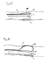
- a specially modified optical catheter 10 includes a rotatable inner shaft 12 and a transparent outer sheath 14.
- the inner shaft 12 houses a fiber array 15 and a mirror 16 near its distal end 18.
- a central fiber 20 in the fiber array connects to a fixed optical fiber 21 that extends from the catheter proximally to a light source 22.
- coherent light such as laser light
- beam-splitter 22a coherent light from light source 22 is transmitted via beam-splitter 22a, through the fixed optical fiber 21 and central fiber 20 and onto center 23 of mirror 16.
- the light is reflected to a tissue sample 24, such as a layer of static tissue over a layer of moving tissue, such as an atherosclerotic plaque.
- Outer sheath 14 can be placed directly in contact with sample 24 (near field), or can be positioned a short distance, e.g., 1 mm to 10 cm away from the sample (far field).
- Light enters sample 24, where it is reflected by molecules, cellular debris, proteins, compounds (e.g., cholesterol crystals), and cellular microstructures (such as organelles, microtubules) within the sample.
- Light remitted from the sample reflects from mirror 16 to the fibers of array 15, and is then transmitted by array 15 to a planar charge-coupled device (CCD), or a linear or two-dimensional detector 22b, via a beam-splitter 22a, e.g., located within light source 22.
- CCD charge-coupled device
- beam-splitter 22a e.g., located within light source 22.
- There may be one or multiple fibers for detection and illumination and detection may occur from the same fiber.
- illumination may occur through a fiber array where each fiber is selectively illuminated to generate multiple speckle patterns as a function of position on the sample. This method can provide a scanning of the incident light across a sample while keeping the probe stationary by illuminating one fiber after another in series.
- a speckle pattern forms at the CCD detector.
- the resulting speckle pattern is then digitized by an analog-digital converter, and analyzed using the procedures described in the analysis section below.
- the entire shaft 12 can rotate 360 degrees in the direction of arrow R, allowing catheter 10 to gather images around the entire circumference of a sample.
- catheter 10 can gather images of a plaque around the circumference of a vessel wall.
- the diameter of the catheter can be less than 500 ⁇ m. Larger diameters are also possible.
- the optics of catheter 10 can be integrated into other types of instruments, such as endoscopes or laparoscopes.
- the optics can also form a stand-alone unit passed into the accessory port of standard endoscopes or laparoscopes, or integrated into another type of catheter, such as dual-purpose intravascular ultrasound catheter.
- the optics can also include a lens that focuses the remitted light 26 onto the distal ends of the fibers in array 15.
- the lens would allow formation of a "near field image” (near the sample sight less than one wavelength) rather than a "far field image” (at the detector set more than a wavelength away from the surface of the tissue).
- the catheter can include a polarization filter to remove all but a certain type of polarized light.
- a cross-polarized filter would allow only light having a polarization perpendicular to the incident light to reach the detector, while a parallel polarized filter would allow only light having the same polarization as the incident light to pass.
- multiply scattered light is less likely to retain its initial polarization than single scattered light
- polarization filters can be used to bias the data toward multiply scattered or single scattered light. Such bias can be used to deduce information about the structure of the sample, since light which has penetrated deeper into the sample will be more highly scattered than light reflected from the surface or remitted from near the surface.
- the detector can be, e.g., a photographic plate, an array of photodetectors, or a single detector.
- the light source can illuminate the sample with continuous light or synchronized pulses.
- the light source can be, e.g., as far as one meter, or more, away from the sample.
- the temporal changes in the pattern should indicate movement of reflectors within the plaque, but not indicate movement of the plaque itself or movement of reflectors between the detector and the plaque.
- the changes in the plaque's speckle pattern preferably reflect microscopic or Brownian motion, but not macroscopic motion.
- the interval For a time interval to be sufficient to detect microscopic Brownian motion, the interval must be long enough to allow for movement of reflectors in the tissue, such as a lipid pool, but short enough that the random Brownian movements do not cancel out.
- an appropriate time interval is about 1-200 ms. Shorter time periods may also be possible. If the time intervals are longer, then changes in the speckle pattern may not adequately differentiate rapid Brownian movement (indicating low viscosity) from slower Brownian movement (indicating high viscosity).
- the fiber array 15 can be coupled to the plaque tissue using, e.g., an angioplasty balloon. This technique also compensates for minor patient movements.
- a balloon 28 is attached to outer sheath 14, on a far side 30 of the catheter. Once the catheter is positioned within a blood vessel in proximity to the plaque, the balloon is inflated. Referring to Fig. 2B , the inflated balloon abuts the vessel wall 32, and presses the catheter against plaque 24, such that a distal region of outer shaft 14 is in direct contact with the plaque. With the catheter coupled to plaque 24 as shown in Fig. 2B , fiber array 15 will move with the plaque when the heart beats, and the gross motion of the plaque will not significantly affect the speckle pattern.
- the balloon can surround the catheter.
- a transparent balloon surrounds outer sheath 14, but is also attached to the sheath.
- the balloon is squeezed between plaque 24 and wall 32 of the vessel. The balloon, therefore will be in direct contact with the plaque, and will move with the plaque when the heart beats. Since the balloon is attached to shaft 14, and shaft 14 is coupled to array 15, movement of the vessel wall will not significantly affect the speckle pattern. Additional methods of coupling the catheter to tissue can also be used, including methods that do not employ an angioplasty balloon.
- a second method of compensating for movement caused by heartbeats is to gather data between heartbeats.
- data is gathered during the relatively still PR interval of the diastole of the heartbeat (when the left ventricle is filling with blood).
- the PR interval lasts for about 0.12-0.2 seconds, providing sufficient time to detect Brownian motion.
- the timing can be computer-controlled or the detector can be linked to an ECG signal, and programmed to gather data only during the PR interval. Similar techniques can be used to compensate for other bodily movements such as peristalsis.
- the catheter can be placed in direct contact with the plaque tissue, as described above, thereby preventing blood from flowing between the detector and the plaque.
- blood flowing between the plaque and the catheter can be removed and replaced with clear saline solution or other clear solutions such as optically transparent blood substitutes.
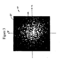
- Fig. 3 illustrates a typical speckle pattern 40 formed by reflecting light from the wall of a healthy blood vessel.
- X and Y coordinates overlay pattern 40 to facilitate mathematical description of the pattern.
- the pattern includes dark patches, where destructive interference dominates, and brighter patches, where constructive interference dominates. Very subtle movements of reflectors within the multiply scattering sample alter the speckle pattern.
- plaque's vulnerability to rupture can be assessed.
- This method includes: (1) gathering a series of speckle images at short, discrete time intervals; (2) eliminating diffuse reflectance from the data; (3) creating cross-correlation images comparing the speckle images in the series; (4) calculating the maximum correlation between each pair of images to create a one-dimensional data set over time; (5) calculating the rate of decorrelation from the data set; and (6) from the rate of decorrelation, assessing the plaque's viscosity and vulnerability to rupture.
- speckle images are gathered for a plaque at discrete intervals over a period of time.
- speckle images can be gathered, e.g., at intervals of every 1, 5, 10, 20, or 30 ms for a time period of, e.g., 200 ms.
- time period e.g. 200 ms.
- the shorter the time intervals the shorter the time period over which data can be gathered.
- time intervals such as 30 ms
- data can be gathered for, e.g., 1-2 seconds.
- the background, non-coherent diffuse reflectance is eliminated from the images.
- a number of techniques can be used to eliminate the tissue's diffuse reflectance.
- the raw data speckle images can be converted to edge images.
- Edge images are spatial derivatives of the raw data images; an edge image (high pass filter) reflects the change in intensity of an image as a function of space, at all points in the image, rather than the intensity itself.
- Known techniques of edge detection include convolution of the image by a kernel (e.g., Sobel or Robert), Morph gradient (subtraction of an eroded, dilated, closed, or opened image by its original), or high pass filtering.
- the cross-correlation images reflect the degree of correlation between the two images as a function of space.
- the cross-correlation images are reduced to a one-dimensional data set as a function of time (i.e., a series of correlation values, each value associated with a time t). From this series of correlation values, a time constant, ⁇ , is calculated, where ⁇ represents the rate of decorrelation.
- the time constant is the amount of time it takes g(t) to reach (1/e)g(0).
- the max function of equation (1) is not the only possible mechanism for reducing the cross-correlation images to a number.
- using a point to reduce the image to a value, rather than a max function would not compensate for the "memory effect" of first order correlation of speckle patterns in turbid media. This "memory effect" is described in Feng et al., Science 251: 633-39 (1991 ). Advantages to using a point are that a minimum number of fibers and detectors can be used.
- the viscosity of the plaque's lipid pool can be assessed.
- ⁇ the higher the Brownian motion in the lipid pool, and the greater the pool's viscosity.
- the smaller ⁇ the greater the Brownian motion in the lipid pool, and the lower the viscosity. The lower the viscosity, the more stresses are exerted on the cap, making the plaque more vulnerable
- This information can be used to identify plaques likely or vulnerable to rupture. Specifically, if ⁇ is about 40100 ms or lower, then the plaque is likely to rupture, and intervention is warranted. If the plaque ⁇ is about 100-200 ms, then the plaque is somewhat vulnerable, but not yet likely to rupture, and should be monitored over time. If the plaque ⁇ is about 200-300 ms, then the plaque is less vulnerable. Non-plaque covered, healthy vessel wall generally has a time constant greater than 300 ms or 500 ms.
- the speckle data can be analyzed to deduce spatial characteristics of the plaque, including the thickness of the fibrous cap, or the thickness of any tissue structure or tissue layer for that matter. As discussed above, a thin fibrous cap is another indication that a plaque is vulnerable to rupture. The combination of data relating to viscosity and cap thickness provides the most accurate assessment of plaque vulnerability, although the two characteristics can be assessed and analyzed independently.
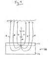
- a typical plaque 50 includes a fibrous cap 52 and a lipid pool 54. Photons that enter plaque 50 (arrow A) are internally scattered by reflectors within the plaque, such as collagen in fibrous cap 52 and lipids in pool 54. The various photons, therefore, exit the plaque at different locations (arrows B). As a result, the speckle pattern (see Fig. 2 ) has a diameter considerably larger than the width of the original light beam.
- the thickness of fibrous cap 52 can be deduced by comparing different regions of the resulting speckle pattern. Referring again to Fig. 3 , light forming intensity signals in the outer portion 60 of the pattern traveled greater distances than light forming signals near the center 62 of the pattern. Thus, outer portion 60 of the pattern is formed by photons that, on average, penetrated deeper into the plaque than photons forming center 62. By calculating separate time constants for separate regions of the speckle pattern, the viscosity of the plaque at different depths can be determined. Since the fibrous cap generally exhibits less Brownian motion than the lipid pool, the thickness of the fibrous cap can be estimated from spatially dependent data.
- Time constants are then calculated from the cross-correlation data for each window, in the manner described above.
- the variation of ⁇ as a function of the distance from the center of the speckle pattern i.e., as a function of (x o 2 +y o 2 ) 1 ⁇ 2
- ⁇ can then be analyzed to determine the thickness of the fibrous cap. Plaque cap thickness of less than about 60 ⁇ m is considered to be vulnerable, but this number can vary to some extent depending on the specific patient.
- any tissue structure e.g., a tissue layer that overlies or is adjacent to a different tissue, e.g., a plaque fibrous cap over a lipid pool
- a tissue layer that overlies or is adjacent to a different tissue e.g., a plaque fibrous cap over a lipid pool
- separately analyzing different regions of a speckle pattern also allows decorrelation caused by macroscopic motion to be identified and removed from the analysis.
- macroscopic motion caused by gross movement of the plaque tissue or blood flow will be directional, non-random, and global.
- Brownian motion will be non-directional and non-uniform (or random).
- the position of maximums of cross-correlation functions will shift along a vector v , which relates extrinsic motion of the sample with respect to the catheter or detection.
- Brownian motion will decorrelate the speckle patterns in many random directions, and will result in a broadening of the cross-correlation peak and a decrease in correlation maximum above that predicted by linear motion.
- the rate of decorrelation can be estimated from single pixel speckle images, rather than full, two-dimensional speckle patterns.
- a catheter with a single optical fiber could transmit data to a single detector, such as a photodiode.
- the speckle data gathered would be intensity at the spot as a function of time. From this data, a rate of decorrelation can be calculated directly or only as a function of time as opposed to space, without any spatial cross-correlation analysis.
- Imaging methods that detect single scattered light such as optical coherence tomography (OCT) and confocal microscopy, can also be used. While these imaging methods are less sensitive to speckle modulation than the multiple scattering methods described above, they have the advantage of allowing localization of data to a single point within the sample. Such localization would allow measurement of biomechanical properties of the tissue in three dimensions.
- OCT optical coherence tomography
- motion of the scatterers can produce a Doppler shift on the returned light. The Doppler shift can provide a further basis for measuring viscosity in the sample.
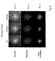
- speckle images formed by reflecting laser light from a cadaveric atherosclerotic plaque in a human aorta were analyzed to assess the plaque's viscosity. A portion of normal aorta was also analyzed for comparison.
- edge detection was performed on the 60 raw speckle images, generating 60 edge images.
- the edge images reflect the spatial derivative of the raw speckle images (i.e., the light patches in the edge images of row B are locations where the intensity is changing as a function of space).
- Each cross-correlation image was generated by multiplying the Fourier transform of the reference image 70 by the complex conjugate of the Fourier transform of the image in question, and then calculating an inverse Fourier transform of the product.
- Image 72 was formed by multiplying the Fourier transform of reference image 70 by the complex conjugate of the Fourier transform of image 70, and then calculating the inverse Fourier transform of the product.
- Each cross-correlation image represents the degree of correlation between the corresponding edge image and the reference edge image 70 (i.e., brighter spots are locations where there is a higher degree of correlation than at darker spots).
- the maximum cross-correlation peak (i.e., the correlation at the maximum point of correlation) was calculated using equation (1).
- the resulting data set included 60 cross-correlation values, each value associated with a time t.
- FIG. 6 A set of images of normal aorta tissue is shown in Fig. 6 . These images are comparable to the set of images in Fig. 5 for a lipid-rich plaque in the same aorta and were imaged and processed in the same manner.
- the decorrelation rate represented by the time constant ⁇
- the time constant was 40 ms.
- the time constant was 500 ms.
- the plaque was borderline vulnerable. Thus, had this plaque been analyzed in vivo, using the procedures described above, a physician would have determined that the plaque was a possible candidate for rupture, and may have chosen to intervene, preventing a possible infarction.
- the methods described herein can also be used to characterize diseased tissue other than atherosclerotic plaques.
- the microscopic and macroscopic constituents of diseased tissue differ from normal non-pathologic counterparts.
- speckle patterns can be used to diagnose and characterize other tissue pathology such as neoplasia (cancer), infection, tissue viability, or healing response to injury.
- neoplasia cancer
- tumors typically have an abnormal abundance of one cell type (clonal) and a surrounding abnormal supporting matrix. This cell type may produce and secrete a viscous fluid, such as mucin in adenocarcinoma, which would result in lower speckle decorrelation time constants than normal non-cancerous tissue.
- the surrounding matrix may be composed of necrotic tissue and an abundance of abnormal vessels that would also serve to decrease the speckle decorrelation time constant.
- Other tumors like osteosarcoma, produce osteoid or immature bone that would increase the time constant compared to normal tissue.
- Other forms of neoplasia would have increased time constants due to desmoplastic (abundant) fibrous stroma initiated by cytokines produced by the tumor. Indeed, many tumors, including bronchogenic carcinomas and breast carcinomas are firm upon gross examination due to the fibrous stroma surrounding the malignant cells. This fibrous stroma would increase the time constant relative to surrounding normal tissue.
- abscesses will be less viscous than surrounding tissue, enabling identification of the infected region by measuring a decrease in the time constant.
- Inflammation manifested by the influx of activated inflammatory cells will be characterized by a decrease in the speckle decorrelation time constant as these cells degrade the normal supporting tissue in response to a the presence of bacterial, viral, or foreign body antigens.
- Necrotic tissue such as burn eschar, diabetic ulcers, necrotic bowel, and ischemic myocardium will have longer time constants than viable tissue from the same organ due to the lack of intravascular and extravascular fluid and flow in these extracellular spaces.
- Speckle decorrelation times may also be used to estimate tissue hydration and provide a means for quantifying the state of hydration in a patient. While the above examples elucidate some of the mechanisms that explain how disease affects the biomechanical properties of pathologic tissue, many more exist and are well known in the field of gross anatomic pathology. These differing biomechanical properties and characteristics can be measured by speckle for the purpose of screening, intraoperative margin (e.g., tumor margin) identification, and primary diagnosis.
- intraoperative margin e.g., tumor margin
Landscapes
- Health & Medical Sciences (AREA)
- Life Sciences & Earth Sciences (AREA)
- Surgery (AREA)
- Engineering & Computer Science (AREA)
- General Health & Medical Sciences (AREA)
- Pathology (AREA)
- Physics & Mathematics (AREA)
- Biomedical Technology (AREA)
- Medical Informatics (AREA)
- Molecular Biology (AREA)
- Heart & Thoracic Surgery (AREA)
- Animal Behavior & Ethology (AREA)
- Biophysics (AREA)
- Public Health (AREA)
- Veterinary Medicine (AREA)
- Optics & Photonics (AREA)
- Radiology & Medical Imaging (AREA)
- Nuclear Medicine, Radiotherapy & Molecular Imaging (AREA)
- Physiology (AREA)
- Signal Processing (AREA)
- Psychiatry (AREA)
- Cardiology (AREA)
- Artificial Intelligence (AREA)
- Computer Vision & Pattern Recognition (AREA)
- Vascular Medicine (AREA)
- Chemical & Material Sciences (AREA)
- Analytical Chemistry (AREA)
- Biochemistry (AREA)
- General Physics & Mathematics (AREA)
- Immunology (AREA)
- Investigating Or Analysing Materials By Optical Means (AREA)
- Investigating Or Analysing Biological Materials (AREA)
- Investigating, Analyzing Materials By Fluorescence Or Luminescence (AREA)
Claims (11)
- Sonde à fibres optiques destinée à détecter des figures de granulation dans un échantillon, la sonde comprenant➢ un cathéter comprenant une tige interne rotative et une gaine externe transparente ;➢ un réseau de fibres accueilli à l'intérieur de la tige et comprenant une ou plusieurs premières fibres optiques destinées à transmettre à l'échantillon une lumière incidente, et une ou plusieurs secondes fibres optiques destinées à transmettre la lumière renvoyée par l'échantillon ;➢ un miroir agencé à proximité d'une extrémité distale de la tige pour réfléchir la lumière qui passe à travers le réseau de fibres sur un échantillon situé hors de la gaine externe transparente et de l'échantillon à travers le réseau de fibres ; et caractérisée par➢ une disposition de détection qui est configurée pour recevoir et détecter les figures de granulation associées à la lumière reçue à partir de l'échantillon.
- Sonde à fibres optiques selon la revendication 1, dans laquelle la tige peut tourner sur 360° à l'intérieur de la gaine.
- Sonde à fibres optiques selon la revendication 1, dans laquelle le réseau de fibres comprend une seule première fibre optique destinée à transmettre la lumière incidente à l'échantillon.
- Sonde à fibres optiques selon la revendication 1, dans laquelle le réseau de fibres comprend plusieurs premières fibres optiques destinées à transmettre la lumière incidente à l'échantillon.
- Sonde à fibres optiques selon la revendication 1, dans laquelle le réseau de fibres optiques comprend une seule seconde fibre optique destinée à transmettre la lumière renvoyée par l'échantillon.
- Sonde à fibres optiques selon la revendication 1, dans laquelle la (ou les) fibre(s) sélectionnée(s) pour transmettre la lumière incidente est (sont) la (les) même(s) que la (ou les) fibre(s) sélectionnée(s) pour transmettre la lumière renvoyée.
- Sonde à fibres optiques selon la revendication 1, dans laquelle une fibre du réseau est sélectionnée en tant que première fibre optique destinée à transmettre la lumière incidente à l'échantillon, puis une autre fibre est sélectionnée en tant que première fibre optique destinée à transmettre à l'échantillon la lumière incidente, effectuant ainsi un balayage de lumière à travers l'échantillon.
- Sonde à fibres optiques selon la revendication 1, comprenant en outre un ballon gonflable relié à la gaine.
- Système optique de détection de figures de granulation dans un échantillon, le système comprenant➢ une sonde à fibres optiques selon la revendication 1 ;➢ une source de lumière cohérente ou partiellement cohérente reliée à une fibre optique à l'intérieur du réseau de fibres ;➢ un détecteur destiné à recevoir la lumière renvoyée par l'échantillon ; et➢ un processeur destiné à traiter la lumière renvoyée et à analyser les figures de granulation renvoyées par l'échantillon.
- Système selon la revendication 9, dans lequel le processeur comprend une figure de granulation de référence.
- Système selon la revendication 9, dans lequel le processeur comprend un convertisseur analogique-numérique pour convertir la lumière analogique renvoyée en un signal numérique.
Applications Claiming Priority (3)
| Application Number | Priority Date | Filing Date | Title |
|---|---|---|---|
| US24425500P | 2000-10-30 | 2000-10-30 | |
| US244255P | 2000-10-30 | ||
| PCT/US2001/048332 WO2002036015A1 (fr) | 2000-10-30 | 2001-10-30 | Procedes et systemes optiques d'analyse de tissus |
Publications (3)
| Publication Number | Publication Date |
|---|---|
| EP1434522A1 EP1434522A1 (fr) | 2004-07-07 |
| EP1434522A4 EP1434522A4 (fr) | 2007-01-03 |
| EP1434522B1 true EP1434522B1 (fr) | 2010-01-13 |
Family
ID=22922006
Family Applications (1)
| Application Number | Title | Priority Date | Filing Date |
|---|---|---|---|
| EP01991092A Expired - Lifetime EP1434522B1 (fr) | 2000-10-30 | 2001-10-30 | Systemes optiques d'analyse de tissus |
Country Status (7)
| Country | Link |
|---|---|
| US (3) | US7231243B2 (fr) |
| EP (1) | EP1434522B1 (fr) |
| JP (1) | JP4241038B2 (fr) |
| AT (1) | ATE454845T1 (fr) |
| AU (1) | AU2002230842A1 (fr) |
| DE (1) | DE60141090D1 (fr) |
| WO (1) | WO2002036015A1 (fr) |
Families Citing this family (375)
| Publication number | Priority date | Publication date | Assignee | Title |
|---|---|---|---|---|
| EP1434522B1 (fr) * | 2000-10-30 | 2010-01-13 | The General Hospital Corporation | Systemes optiques d'analyse de tissus |
| US9295391B1 (en) | 2000-11-10 | 2016-03-29 | The General Hospital Corporation | Spectrally encoded miniature endoscopic imaging probe |
| EP1402244B1 (fr) | 2001-04-30 | 2020-03-11 | The General Hospital Corporation | Procede et appareil permettant d'ameliorer la clarte et la sensibilite de l'image en tomographie a coherence optique au moyen d'une interaction permettant de controler les proprietes focales et la synchronisation de coherence |
| DE10297689B4 (de) | 2001-05-01 | 2007-10-18 | The General Hospital Corp., Boston | Verfahren und Gerät zur Bestimmung von atherosklerotischem Belag durch Messung von optischen Gewebeeigenschaften |
| WO2004065943A1 (fr) * | 2003-01-22 | 2004-08-05 | Art, Advanced Research Technologies, Inc. | Acquisition simultanee de diverses portes temporelles dans une imagerie optique fondee sur tpsf |
| US6980299B1 (en) | 2001-10-16 | 2005-12-27 | General Hospital Corporation | Systems and methods for imaging a sample |
| CA2413483A1 (fr) | 2001-12-26 | 2003-06-26 | Kevin R. Forrester | Dispositif de mesure du mouvement |
| EP1324051A1 (fr) * | 2001-12-26 | 2003-07-02 | Kevin R. Forrester | Appareil pour la mesurage de mouvement |
| ATE503982T1 (de) | 2002-01-11 | 2011-04-15 | Gen Hospital Corp | Vorrichtung zur oct bildaufnahme mit axialem linienfokus für verbesserte auflösung und tiefenschärfe |
| US7355716B2 (en) | 2002-01-24 | 2008-04-08 | The General Hospital Corporation | Apparatus and method for ranging and noise reduction of low coherence interferometry LCI and optical coherence tomography OCT signals by parallel detection of spectral bands |
| DE60334231D1 (de) * | 2002-03-08 | 2010-10-28 | Univ Virginia | Intuitives ultraschallsystem und zugehöriges verfahren |
| FR2842407B1 (fr) * | 2002-07-18 | 2005-05-06 | Mauna Kea Technologies | "procede et appareillage d'imagerie de fluorescence confocale fibree" |
| US11243494B2 (en) | 2002-07-31 | 2022-02-08 | Abs Global, Inc. | Multiple laminar flow-based particle and cellular separation with laser steering |
| US7123363B2 (en) * | 2003-01-03 | 2006-10-17 | Rose-Hulman Institute Of Technology | Speckle pattern analysis method and system |
| EP2319404B1 (fr) | 2003-01-24 | 2015-03-11 | The General Hospital Corporation | Système et procédé pour identifier des tissus à l'aide d'interférométrie à faible cohérence |
| US8054468B2 (en) | 2003-01-24 | 2011-11-08 | The General Hospital Corporation | Apparatus and method for ranging and noise reduction of low coherence interferometry LCI and optical coherence tomography OCT signals by parallel detection of spectral bands |
| US20060165270A1 (en) * | 2003-02-25 | 2006-07-27 | Jorn Borgert | Intravascular imaging |
| AU2004225188B2 (en) | 2003-03-31 | 2010-04-15 | The General Hospital Corporation | Speckle reduction in optical coherence tomography by path length encoded angular compounding |
| EP2280258A3 (fr) | 2003-06-06 | 2015-09-02 | The General Hospital Corporation | Procédé et appareil pour une source de lumière à réglage de longueur d'onde |
| US20040260182A1 (en) * | 2003-06-23 | 2004-12-23 | Zuluaga Andres F. | Intraluminal spectroscope with wall contacting probe |
| CA2541297C (fr) | 2003-10-03 | 2013-12-10 | Academisch Medisch Centrum | Systeme et methode de representation en images de la reflectance d'un substrat |
| US7061618B2 (en) | 2003-10-17 | 2006-06-13 | Axsun Technologies, Inc. | Integrated spectroscopy system |
| CN103293126B (zh) | 2003-10-27 | 2017-03-01 | 通用医疗公司 | 用于使用频域干涉测量法进行光学成像的方法和设备 |
| US20050107694A1 (en) * | 2003-11-17 | 2005-05-19 | Jansen Floribertus H. | Method and system for ultrasonic tagging of fluorescence |
| WO2005054780A1 (fr) | 2003-11-28 | 2005-06-16 | The General Hospital Corporation | Procede et appareil d'imagerie codee de maniere spectrale tridimensionnelle |
| US7460737B2 (en) | 2004-02-12 | 2008-12-02 | Hoshiko Llc | Method and apparatus for photograph finding |
| US8060172B2 (en) * | 2004-03-29 | 2011-11-15 | Olympus Corporation | In-vivo information measurement apparatus |
| EP1744669B1 (fr) * | 2004-04-29 | 2019-03-27 | Koninklijke Philips N.V. | Appareil et procede de detection de flux sanguin |
| AU2004320269B2 (en) | 2004-05-29 | 2011-07-21 | The General Hospital Corporation | Process, system and software arrangement for a chromatic dispersion compensation using reflective layers in optical coherence tomography (OCT) imaging |
| EP1771755B1 (fr) | 2004-07-02 | 2016-09-21 | The General Hospital Corporation | Sonde d'imagerie endoscopique comprenant des fibres double gaine |
| JP5053845B2 (ja) | 2004-08-06 | 2012-10-24 | ザ ジェネラル ホスピタル コーポレイション | 光学コヒーレンス断層撮影法を使用して試料中の少なくとも1つの位置を決定するための方法、システムおよびソフトウェア装置 |
| EP1793730B1 (fr) * | 2004-08-24 | 2011-12-28 | The General Hospital Corporation | Ensemble procede, systeme et logiciel pour la détermination de module d 'élasticité |
| KR20070058523A (ko) | 2004-08-24 | 2007-06-08 | 더 제너럴 하스피탈 코포레이션 | 혈관절편 영상화 방법 및 장치 |
| JP5215664B2 (ja) | 2004-09-10 | 2013-06-19 | ザ ジェネラル ホスピタル コーポレイション | 光学コヒーレンス撮像のシステムおよび方法 |
| US7366376B2 (en) | 2004-09-29 | 2008-04-29 | The General Hospital Corporation | System and method for optical coherence imaging |
| US20060241364A1 (en) * | 2004-10-01 | 2006-10-26 | Academisch Medisch Centrum Of The University Van Amsterdam | System and method for imaging the reflectance of a substrate |
| JP5175101B2 (ja) * | 2004-10-29 | 2013-04-03 | ザ ジェネラル ホスピタル コーポレイション | 偏光感応性光コヒーレンストモグラフィを用いて偏光非解消の偏光パラメータを測定するジョーンズ行列に基づく解析を行うシステム及び方法 |
| EP1807722B1 (fr) | 2004-11-02 | 2022-08-10 | The General Hospital Corporation | Dispositif rotationnel a fibres optiques, systeme optique pour imager un echantillon |
| EP2278266A3 (fr) | 2004-11-24 | 2011-06-29 | The General Hospital Corporation | Interféromètre à chemin commun pour OCT endoscopique |
| EP1816949A1 (fr) | 2004-11-29 | 2007-08-15 | The General Hospital Corporation | Ensembles, dispositifs, endoscopes, catheters et methodes d'imagerie optique permettant d'eclairer et de detecter simultanement plusieurs points sur un echantillon |
| US9131861B2 (en) * | 2004-11-30 | 2015-09-15 | Academisch Medisch Centrum | Pulsed lighting imaging systems and methods |
| US7303280B2 (en) | 2004-12-17 | 2007-12-04 | The Regents Of The University Of California | High-resolution ophthalmic imaging system |
| WO2006069443A1 (fr) * | 2004-12-27 | 2006-07-06 | Bc Cancer Agency | Procedes et appareil de mesure de la rugosite d'une surface |
| US7336366B2 (en) * | 2005-01-20 | 2008-02-26 | Duke University | Methods and systems for reducing complex conjugate ambiguity in interferometric data |
| US7962198B2 (en) * | 2005-04-27 | 2011-06-14 | The Trustees Of Dartmouth College | System and method for spectral-encoded high-rate hemodynamic tomography |
| WO2006116769A1 (fr) | 2005-04-28 | 2006-11-02 | The General Hospital Corporation | Systemes, procedes et agencements logiciels servant a evaluer des informations associees a une structure anatomique au moyen d'une technique de telemetrie de coherence optique |
| WO2006130797A2 (fr) * | 2005-05-31 | 2006-12-07 | The General Hospital Corporation | Systeme, procede et dispositif qui peuvent utiliser des techniques d'interferometrie d'heterodyne a codage spectral pour l'imagerie |
| US9060689B2 (en) | 2005-06-01 | 2015-06-23 | The General Hospital Corporation | Apparatus, method and system for performing phase-resolved optical frequency domain imaging |
| WO2007014213A2 (fr) * | 2005-07-25 | 2007-02-01 | Massachusetts Institute Of Technology | Imagerie spectroscopique trimodale |
| ATE484727T1 (de) | 2005-08-09 | 2010-10-15 | Gen Hospital Corp | Gerät und verfahren zur durchführung von polarisationsbasierter quadraturdemodulation bei optischer kohärenztomographie |
| US8784336B2 (en) | 2005-08-24 | 2014-07-22 | C. R. Bard, Inc. | Stylet apparatuses and methods of manufacture |
| JP6046325B2 (ja) | 2005-09-29 | 2016-12-14 | ザ ジェネラル ホスピタル コーポレイション | 漸次的に解像度を増加させて1以上の生物学的サンプルの観察及び分析のための方法及びその方法のための装置 |
| US7911207B2 (en) * | 2005-11-16 | 2011-03-22 | Board Of Regents, The University Of Texas System | Method for determining location and movement of a moving object |
| EP1971848B1 (fr) | 2006-01-10 | 2019-12-04 | The General Hospital Corporation | Systèmes et procédés de génération de données basés sur une ou plusieurs technique(s) d'endoscopie spectralement codées |
| EP2289396A3 (fr) | 2006-01-19 | 2011-04-06 | The General Hospital Corporation | Procédés et systèmes pour visualiser de manière optique des organes intracavitaires épithéliaux lors d'un balayage au faisceau |
| US8145018B2 (en) | 2006-01-19 | 2012-03-27 | The General Hospital Corporation | Apparatus for obtaining information for a structure using spectrally-encoded endoscopy techniques and methods for producing one or more optical arrangements |
| WO2007149601A2 (fr) | 2006-02-01 | 2007-12-27 | The General Hospital Corporation | Appareil destiné à commander au moins l'une d'au moins deux sections d'au moins une fibre |
| US7538859B2 (en) | 2006-02-01 | 2009-05-26 | The General Hospital Corporation | Methods and systems for monitoring and obtaining information of at least one portion of a sample using conformal laser therapy procedures, and providing electromagnetic radiation thereto |
| JP5524487B2 (ja) | 2006-02-01 | 2014-06-18 | ザ ジェネラル ホスピタル コーポレイション | コンフォーマルレーザ治療手順を用いてサンプルの少なくとも一部分に電磁放射を放射する方法及びシステム。 |
| WO2007092911A2 (fr) | 2006-02-08 | 2007-08-16 | The General Hospital Corporation | procedes, dispositifs et systemes pour obtenir des informations associees a un prelevement anatomique utilisant la microscopie optique |
| EP1987318B1 (fr) | 2006-02-24 | 2015-08-12 | The General Hospital Corporation | Procédés et systèmes destinés à réaliser une tomographie par cohérence optique dans le domaine de fourier avec résolution angulaire |
| WO2007118129A1 (fr) | 2006-04-05 | 2007-10-18 | The General Hospital Corporation | Procedes, dispositifs et systemes pour effectuer une imagerie de domaine de frequence optique d'un echantillon sensible a la polarisation |
| WO2007133961A2 (fr) | 2006-05-10 | 2007-11-22 | The General Hospital Corporation | Processus, agencements et systèmes pour obtenir une imagerie de domaine de fréquence d'un échantillon |
| US7782464B2 (en) * | 2006-05-12 | 2010-08-24 | The General Hospital Corporation | Processes, arrangements and systems for providing a fiber layer thickness map based on optical coherence tomography images |
| US7920271B2 (en) | 2006-08-25 | 2011-04-05 | The General Hospital Corporation | Apparatus and methods for enhancing optical coherence tomography imaging using volumetric filtering techniques |
| WO2008049118A2 (fr) | 2006-10-19 | 2008-04-24 | The General Hospital Corporation | Dispositif et procédé d'obtention et de fourniture d'informations d'image associées à au moins une portion d' échantillon et permettant de réaliser une telle portion |
| US8388546B2 (en) | 2006-10-23 | 2013-03-05 | Bard Access Systems, Inc. | Method of locating the tip of a central venous catheter |
| US7794407B2 (en) | 2006-10-23 | 2010-09-14 | Bard Access Systems, Inc. | Method of locating the tip of a central venous catheter |
| JP2010508973A (ja) | 2006-11-08 | 2010-03-25 | ライトラブ イメージング, インコーポレイテッド | 光−音響イメージングデバイスおよび方法 |
| US8000774B2 (en) * | 2007-01-03 | 2011-08-16 | Infraredx, Inc. | Method and system for intra luminal thrombus detection |
| WO2008089406A2 (fr) | 2007-01-19 | 2008-07-24 | The General Hospital Corporation | Appareil et procédé pour contrôler la profondeur de télémétrie dans une imagerie de domaine de fréquence optique |
| EP2662674A3 (fr) | 2007-01-19 | 2014-06-25 | The General Hospital Corporation | Réflexion de disque rotatif pour balayage rapide de longueur d'onde de lumière à large bande dispersée |
| WO2008116010A1 (fr) * | 2007-03-19 | 2008-09-25 | The General Hospital Corporation | Système et procédé permettant un diagnostic non invasif du syndrome de compartiment, exemples de procédure d'imagerie de points par laser |
| EP2602651A3 (fr) | 2007-03-23 | 2014-08-27 | The General Hospital Corporation | Procédés, agencements et appareil pour utiliser un laser à balayage de longueur d'ondes utilisant un balayage angulaire et des procédures de dispersion |
| WO2008121844A1 (fr) | 2007-03-30 | 2008-10-09 | The General Hospital Corporation | Système et procédé pour fournir une imagerie à granularité laser en vue de détecter une plaque à risque |
| WO2008131082A1 (fr) | 2007-04-17 | 2008-10-30 | The General Hospital Corporation | Appareil et procédés de mesure des vibrations à l'aide de techniques d'endoscopie spectralement codées |
| FR2915077B1 (fr) * | 2007-04-19 | 2010-09-10 | Inst Radioprot Et De Surete Nu | Dispositif d'aide au diagnostic et pronostic de modifications physiopathologiques des tissus. |
| EP2139392B1 (fr) * | 2007-04-26 | 2014-02-26 | Koninklijke Philips N.V. | Système de localisation |
| US8115919B2 (en) | 2007-05-04 | 2012-02-14 | The General Hospital Corporation | Methods, arrangements and systems for obtaining information associated with a sample using optical microscopy |
| US7952719B2 (en) | 2007-06-08 | 2011-05-31 | Prescient Medical, Inc. | Optical catheter configurations combining raman spectroscopy with optical fiber-based low coherence reflectometry |
| WO2009005748A1 (fr) | 2007-06-29 | 2009-01-08 | The Trustees Of Columbia University In The City Ofnew York | Systèmes et procédés d'imagerie optique ou de spectroscopie |
| US7566173B2 (en) * | 2007-07-09 | 2009-07-28 | Alcon, Inc. | Multi-spot ophthalmic laser probe |
| US20090024040A1 (en) * | 2007-07-20 | 2009-01-22 | Prescient Medical, Inc. | Wall-Contacting Intravascular Ultrasound Probe Catheters |
| JP5917803B2 (ja) | 2007-07-31 | 2016-05-18 | ザ ジェネラル ホスピタル コーポレイション | 高速ドップラー光周波数領域撮像法のためのビーム走査パターンを放射するシステムおよび方法 |
| US8040608B2 (en) | 2007-08-31 | 2011-10-18 | The General Hospital Corporation | System and method for self-interference fluorescence microscopy, and computer-accessible medium associated therewith |
| WO2009039303A1 (fr) * | 2007-09-19 | 2009-03-26 | State University Of New York At Stony Brook | Systèmes et procédés de tomographie à cohérence optique |
| US20090131801A1 (en) * | 2007-10-12 | 2009-05-21 | The General Hospital Corporation | Systems and processes for optical imaging of luminal anatomic structures |
| EP2203733B1 (fr) | 2007-10-25 | 2017-05-03 | Washington University in St. Louis | Microscopie photo-acoustique confocale présentant une résolution latérale optique |
| WO2009059034A1 (fr) | 2007-10-30 | 2009-05-07 | The General Hospital Corporation | Système et procédé permettant une détection de mode de gaine |
| US7813609B2 (en) | 2007-11-12 | 2010-10-12 | Lightlab Imaging, Inc. | Imaging catheter with integrated reference reflector |
| US9521961B2 (en) | 2007-11-26 | 2016-12-20 | C. R. Bard, Inc. | Systems and methods for guiding a medical instrument |
| US9649048B2 (en) | 2007-11-26 | 2017-05-16 | C. R. Bard, Inc. | Systems and methods for breaching a sterile field for intravascular placement of a catheter |
| US10449330B2 (en) | 2007-11-26 | 2019-10-22 | C. R. Bard, Inc. | Magnetic element-equipped needle assemblies |
| US8849382B2 (en) | 2007-11-26 | 2014-09-30 | C. R. Bard, Inc. | Apparatus and display methods relating to intravascular placement of a catheter |
| US8781555B2 (en) | 2007-11-26 | 2014-07-15 | C. R. Bard, Inc. | System for placement of a catheter including a signal-generating stylet |
| US10751509B2 (en) | 2007-11-26 | 2020-08-25 | C. R. Bard, Inc. | Iconic representations for guidance of an indwelling medical device |
| US10524691B2 (en) | 2007-11-26 | 2020-01-07 | C. R. Bard, Inc. | Needle assembly including an aligned magnetic element |
| ES2557084T3 (es) | 2007-11-26 | 2016-01-21 | C. R. Bard, Inc. | Sistema integrado para la colocación intravascular de un catéter |
| US9492097B2 (en) | 2007-11-26 | 2016-11-15 | C. R. Bard, Inc. | Needle length determination and calibration for insertion guidance system |
| US8280496B2 (en) * | 2007-12-13 | 2012-10-02 | Boston Scientific Scimed, Inc. | Extended spectral sensitivity endoscope system and method of using the same |
| US11123047B2 (en) | 2008-01-28 | 2021-09-21 | The General Hospital Corporation | Hybrid systems and methods for multi-modal acquisition of intravascular imaging data and counteracting the effects of signal absorption in blood |
| US9332942B2 (en) | 2008-01-28 | 2016-05-10 | The General Hospital Corporation | Systems, processes and computer-accessible medium for providing hybrid flourescence and optical coherence tomography imaging |
| US8478382B2 (en) | 2008-02-11 | 2013-07-02 | C. R. Bard, Inc. | Systems and methods for positioning a catheter |
| US20090236541A1 (en) * | 2008-03-24 | 2009-09-24 | General Electric Company | System and Methods for Optical Imaging |
| WO2009137701A2 (fr) | 2008-05-07 | 2009-11-12 | The General Hospital Corporation | Système, procédé et support informatique permettant le suivi du mouvement des vaisseaux lors d'un examen en microscopie tridimensionnelle des artères coronaires |
| US8694266B2 (en) * | 2008-06-05 | 2014-04-08 | The Regents Of The University Of Michigan | Multimodal spectroscopic systems and methods for classifying biological tissue |
| US8861910B2 (en) | 2008-06-20 | 2014-10-14 | The General Hospital Corporation | Fused fiber optic coupler arrangement and method for use thereof |
| WO2010004365A1 (fr) | 2008-07-10 | 2010-01-14 | Ecole Polytechnique Federale De Lausanne (Epfl) | Imagerie optique cohérente fonctionnelle |
| WO2010009136A2 (fr) | 2008-07-14 | 2010-01-21 | The General Hospital Corporation | Appareil et procédés d'endoscopie couleur |
| ES2525525T3 (es) | 2008-08-22 | 2014-12-26 | C.R. Bard, Inc. | Conjunto de catéter que incluye conjuntos de sensor de ECG y magnético |
| KR101037628B1 (ko) * | 2008-08-28 | 2011-05-30 | 숭실대학교산학협력단 | 레이저 스펙클 내시경 |
| DE102008045634A1 (de) | 2008-09-03 | 2010-03-04 | Ludwig-Maximilians-Universität München | Wellenlängenabstimmbare Lichtquelle |
| US8437833B2 (en) | 2008-10-07 | 2013-05-07 | Bard Access Systems, Inc. | Percutaneous magnetic gastrostomy |
| US20100137711A1 (en) * | 2008-11-25 | 2010-06-03 | Trustees Of Boston University | Apparatus and Method of Analyzing Arterial Plaque |
| US8937724B2 (en) | 2008-12-10 | 2015-01-20 | The General Hospital Corporation | Systems and methods for extending imaging depth range of optical coherence tomography through optical sub-sampling |
| US9351705B2 (en) | 2009-01-09 | 2016-05-31 | Washington University | Miniaturized photoacoustic imaging apparatus including a rotatable reflector |
| EP2382456A4 (fr) | 2009-01-26 | 2012-07-25 | Gen Hospital Corp | Système, procédé et support accessible par ordinateur permettant de fournir une microscopie de super-résolution à large champ |
| JP6053284B2 (ja) | 2009-02-04 | 2016-12-27 | ザ ジェネラル ホスピタル コーポレイション | ハイスピード光学波長チューニング源の利用のための装置及び方法 |
| US9351642B2 (en) | 2009-03-12 | 2016-05-31 | The General Hospital Corporation | Non-contact optical system, computer-accessible medium and method for measurement at least one mechanical property of tissue using coherent speckle technique(s) |
| US9532724B2 (en) | 2009-06-12 | 2017-01-03 | Bard Access Systems, Inc. | Apparatus and method for catheter navigation using endovascular energy mapping |
| US9339206B2 (en) | 2009-06-12 | 2016-05-17 | Bard Access Systems, Inc. | Adaptor for endovascular electrocardiography |
| WO2011008822A2 (fr) * | 2009-07-14 | 2011-01-20 | The General Hospital Corporation | Appareil, systèmes et procédés permettant de mesurer le débit et la pression à lintérieur dune cuve |
| EP2464407A4 (fr) | 2009-08-10 | 2014-04-02 | Bard Access Systems Inc | Dispositifs et procédés pour électrographie endovasculaire |
| US8526472B2 (en) | 2009-09-03 | 2013-09-03 | Axsun Technologies, Inc. | ASE swept source with self-tracking filter for OCT medical imaging |
| US8670129B2 (en) | 2009-09-03 | 2014-03-11 | Axsun Technologies, Inc. | Filtered ASE swept source for OCT medical imaging |
| US12426789B2 (en) | 2009-09-23 | 2025-09-30 | Lightlab Imaging, Inc. | Blood vessel lumen morphology and minimum lumen area measurements data collection by intravascular imaging systems for stenosis or stent planning |
| WO2011038048A1 (fr) * | 2009-09-23 | 2011-03-31 | Lightlab Imaging, Inc. | Appareil, systèmes, et procédés de clarification du sang in vivo dans une lumière |
| ES2660570T3 (es) | 2009-09-23 | 2018-03-23 | Lightlab Imaging, Inc. | Sistemas, aparatos y métodos de recopilación de datos de medición de resistencia vascular y morfología luminal |
| US8923945B2 (en) * | 2009-09-24 | 2014-12-30 | Covidien Lp | Determination of a physiological parameter |
| US8855749B2 (en) | 2009-09-24 | 2014-10-07 | Covidien Lp | Determination of a physiological parameter |
| CN102665541B (zh) | 2009-09-29 | 2016-01-13 | C·R·巴德股份有限公司 | 与用于导管的血管内放置的设备一起使用的探针 |
| WO2011044421A1 (fr) | 2009-10-08 | 2011-04-14 | C. R. Bard, Inc. | Entretoises utilisées avec une sonde ultrasonore |
| MX338267B (es) * | 2009-11-24 | 2016-04-11 | Alcon Res Ltd | Sonda laser de multiples puntos de una sola fibra para endoiluminacion oftalimica. |
| US8619237B2 (en) | 2009-12-04 | 2013-12-31 | The Trustees Of Columbia University In The City Of New York | Laser-scanning intersecting plane tomography such as for high speed volumetric optical imaging |
| EP2512584B1 (fr) * | 2009-12-15 | 2016-06-08 | Alcon Research, Ltd | Sonde laser à points multiples |
| US8206377B2 (en) * | 2009-12-22 | 2012-06-26 | Lightlab Imaging, Inc. | Torque limiter for an OCT catheter |
| US8926590B2 (en) | 2009-12-22 | 2015-01-06 | Lightlab Imaging, Inc. | Torque limiter for an OCT catheter |
| WO2011097312A1 (fr) | 2010-02-02 | 2011-08-11 | C.R. Bard, Inc. | Appareil et procédé destinés à la navigation d'un cathéter et à la localisation d'une pointe |
| KR20130004326A (ko) | 2010-03-05 | 2013-01-09 | 더 제너럴 하스피탈 코포레이션 | 특정 해상도에서 적어도 하나의 해부학적 구조의 미세 영상을 제공하는 시스템, 방법 및 컴퓨터 접근 가능 매체 |
| AU2011227178B2 (en) * | 2010-03-17 | 2013-11-07 | Lightlab Imaging, Inc. | Intensity noise reduction methods and apparatus for interferometric sensing and imaging systems |
| EP2551664B1 (fr) * | 2010-03-24 | 2014-11-12 | Shimadzu Corporation | Système de mesure |
| US9086365B2 (en) | 2010-04-09 | 2015-07-21 | Lihong Wang | Quantification of optical absorption coefficients using acoustic spectra in photoacoustic tomography |
| US9069130B2 (en) | 2010-05-03 | 2015-06-30 | The General Hospital Corporation | Apparatus, method and system for generating optical radiation from biological gain media |
| US11105686B2 (en) | 2010-05-10 | 2021-08-31 | University of Pittshurgh-Of the Commonwealth System of Higher Education | Spatial-domain low-coherence quantitative phase microscopy |
| US9795301B2 (en) | 2010-05-25 | 2017-10-24 | The General Hospital Corporation | Apparatus, systems, methods and computer-accessible medium for spectral analysis of optical coherence tomography images |
| WO2011149972A2 (fr) | 2010-05-25 | 2011-12-01 | The General Hospital Corporation | Systèmes, dispositifs, procédés, appareil et supports accessibles par ordinateur pour fournir une imagerie optique de structures et de compositions |
| EP2912999B1 (fr) | 2010-05-28 | 2022-06-29 | C. R. Bard, Inc. | Appareil destiné à être utilisé avec un système de guidage d'insertion d'aiguille |
| CN103037761B (zh) | 2010-05-28 | 2016-11-02 | C·R·巴德股份有限公司 | 用于针和医疗部件的插入引导系统 |
| US10285568B2 (en) | 2010-06-03 | 2019-05-14 | The General Hospital Corporation | Apparatus and method for devices for imaging structures in or at one or more luminal organs |
| JP5417268B2 (ja) * | 2010-06-28 | 2014-02-12 | 富士フイルム株式会社 | 内視鏡システム |
| EP2603145A2 (fr) | 2010-08-09 | 2013-06-19 | C.R. Bard, Inc. | Structures de support et de capuchon pour tête de sonde à ultrasons |
| JP5845260B2 (ja) | 2010-08-20 | 2016-01-20 | シー・アール・バード・インコーポレーテッドC R Bard Incorporated | Ecg支援カテーテル先端配置の再確認 |
| JP5631671B2 (ja) | 2010-09-07 | 2014-11-26 | オリンパス株式会社 | 光イメージング装置 |
| JP5883018B2 (ja) | 2010-10-27 | 2016-03-09 | ザ ジェネラル ホスピタル コーポレイション | 少なくとも1つの血管内部の血圧を測定するための装置、システム、および方法 |
| WO2012058461A1 (fr) | 2010-10-29 | 2012-05-03 | C.R.Bard, Inc. | Mise en place assistée par bio-impédance d'un dispositif médical |
| US10908066B2 (en) | 2010-11-16 | 2021-02-02 | 1087 Systems, Inc. | Use of vibrational spectroscopy for microfluidic liquid measurement |
| US20120225475A1 (en) | 2010-11-16 | 2012-09-06 | 1087 Systems, Inc. | Cytometry system with quantum cascade laser source, acoustic detector, and micro-fluidic cell handling system configured for inspection of individual cells |
| FR2968762B1 (fr) * | 2010-12-10 | 2014-06-20 | Centre Nat Rech Scient | Procede et dispositif de caracterisation de la dynamique interne d'un echantillon de matiere en presence d'un deplacement rigide. |
| US9063074B2 (en) * | 2010-12-30 | 2015-06-23 | Empire Technology Development Llc | Analyzing tissue morphology in three dimensions |
| US20130278740A1 (en) * | 2011-01-05 | 2013-10-24 | Bar Ilan University | Imaging system and method using multicore fiber |
| US9271658B2 (en) | 2011-01-10 | 2016-03-01 | East Carolina University | Methods, systems and computer program products for non-invasive determination of blood flow distribution using speckle imaging techniques and hemodynamic modeling |
| US9226673B2 (en) | 2011-01-10 | 2016-01-05 | East Carolina University | Methods, systems and computer program products for non-invasive determination of blood flow distribution using speckle imaging techniques and hemodynamic modeling |
| US9636041B2 (en) * | 2011-01-28 | 2017-05-02 | Bar Ilan University | Method and system for non-invasively monitoring biological or biochemical parameters of individual |
| WO2012101644A2 (fr) | 2011-01-28 | 2012-08-02 | Bar Ilan University | Procédé et système de surveillance non invasive de paramètres biologiques et biochimiques d'un individu |
| US8997572B2 (en) | 2011-02-11 | 2015-04-07 | Washington University | Multi-focus optical-resolution photoacoustic microscopy with ultrasonic array detection |
| US9618319B2 (en) * | 2011-02-18 | 2017-04-11 | The General Hospital Corporation | Laser speckle microrheometer for measuring mechanical properties of biological tissue |
| US8582619B2 (en) | 2011-03-15 | 2013-11-12 | Lightlab Imaging, Inc. | Methods, systems, and devices for timing control in electromagnetic radiation sources |
| US9164240B2 (en) | 2011-03-31 | 2015-10-20 | Lightlab Imaging, Inc. | Optical buffering methods, apparatus, and systems for increasing the repetition rate of tunable light sources |
| US9138148B2 (en) * | 2011-04-13 | 2015-09-22 | St. Jude Medical, Inc. | High speed elastographic property mapping of lumens utilizing micropalpation delivered from an OCT-equipped catheter tip |
| US9179843B2 (en) | 2011-04-21 | 2015-11-10 | Hassan Ghaderi MOGHADDAM | Method and system for optically evaluating proximity to the inferior alveolar nerve in situ |
| JP6240064B2 (ja) | 2011-04-29 | 2017-11-29 | ザ ジェネラル ホスピタル コーポレイション | 散乱媒質の深さ分解した物理的及び/又は光学的特性を決定する方法 |
| JP6141264B2 (ja) | 2011-05-27 | 2017-06-07 | ライトラボ・イメージング・インコーポレーテッド | 光コヒーレンス断層撮影法及び圧力に基づくシステム及び方法 |
| CA2836790C (fr) | 2011-05-31 | 2019-04-23 | Desmond Adler | Systeme, appareil et procedes d'imagerie multimodale |
| WO2013013049A1 (fr) | 2011-07-19 | 2013-01-24 | The General Hospital Corporation | Systèmes, procédés, appareils et supports accessibles par ordinateur permettant de produire une compensation de dispersion en mode polarisation dans la tomographie à cohérence optique |
| US8582109B1 (en) | 2011-08-01 | 2013-11-12 | Lightlab Imaging, Inc. | Swept mode-hopping laser system, methods, and devices for frequency-domain optical coherence tomography |
| WO2013019840A1 (fr) | 2011-08-03 | 2013-02-07 | Lightlab Imaging, Inc. | Systèmes, procédés et appareil pour déterminer une réserve d'écoulement fractionnaire |
| USD699359S1 (en) | 2011-08-09 | 2014-02-11 | C. R. Bard, Inc. | Ultrasound probe head |
| USD724745S1 (en) | 2011-08-09 | 2015-03-17 | C. R. Bard, Inc. | Cap for an ultrasound probe |
| EP2748587B1 (fr) | 2011-08-25 | 2021-01-13 | The General Hospital Corporation | Procédés et arrangements permettant de mettre en oeuvre des procédures de tomographie par cohérence micro-optique |
| JP2013075035A (ja) * | 2011-09-30 | 2013-04-25 | Canon Inc | 光断層像撮像方法、光断層像撮像装置およびプログラム |
| WO2013066631A1 (fr) | 2011-10-18 | 2013-05-10 | The General Hospital Corporation | Appareil et procédés de production et/ou d'utilisation de retard(s) optique(s) de recirculation |
| US8953911B1 (en) | 2011-10-28 | 2015-02-10 | Lightlab Imaging, Inc. | Spectroscopic imaging probes, devices, and methods |
| US8831321B1 (en) | 2011-11-07 | 2014-09-09 | Lightlab Imaging, Inc. | Side branch detection methods, systems and devices |
| WO2013070775A1 (fr) | 2011-11-07 | 2013-05-16 | C.R. Bard, Inc | Insert à base d'hydrogel renforcé pour ultrasons |
| WO2013115231A1 (fr) * | 2012-01-31 | 2013-08-08 | オリンパス株式会社 | Dispositif détecteur de partie d'insertion et système détecteur de partie d'insertion |
| WO2013148306A1 (fr) | 2012-03-30 | 2013-10-03 | The General Hospital Corporation | Système d'imagerie, procédé et fixation distale permettant une endoscopie à champ de vision multidirectionnel |
| US10835127B2 (en) * | 2012-04-13 | 2020-11-17 | Baker Idi Heart & Diabetes Institute Holdings Limited | Atherosclerotic plaque detection |
| WO2013160861A1 (fr) | 2012-04-27 | 2013-10-31 | Aïmago S.A. | Dispositif médical d'imagerie par cohérence optique |
| US9186053B2 (en) * | 2012-05-03 | 2015-11-17 | Covidien Lp | Methods of using light to repair hernia defects |
| US11490797B2 (en) | 2012-05-21 | 2022-11-08 | The General Hospital Corporation | Apparatus, device and method for capsule microscopy |
| CN104837413B (zh) | 2012-06-15 | 2018-09-11 | C·R·巴德股份有限公司 | 检测超声探测器上可移除帽的装置及方法 |
| JP6023883B2 (ja) | 2012-07-10 | 2016-11-09 | アイマゴ ソシエテ アノニムAimago S.A. | 灌流評価マルチモダリティ光学医用デバイス |
| EP2879568A1 (fr) * | 2012-08-01 | 2015-06-10 | Bar Ilan University | Procédé et système pour la surveillance non invasive de paramètres biologiques ou biochimiques d'un individu |
| EP2888616A4 (fr) | 2012-08-22 | 2016-04-27 | Gen Hospital Corp | Système, procédé et support accessible par ordinateur pour fabriquer des endoscopes miniatures à l'aide d'une lithographie douce |
| WO2014063005A1 (fr) | 2012-10-18 | 2014-04-24 | Washington University | Imagerie du cerveau par tomographie photoacoustique/thermoacoustique transcrânienne renseignée par des données d'images complémentaires |
| JP6352287B2 (ja) | 2012-11-19 | 2018-07-04 | ライトラボ・イメージング・インコーポレーテッド | マルチモーダル・イメージングシステム、プローブ及び方法 |
| US9677869B2 (en) | 2012-12-05 | 2017-06-13 | Perimeter Medical Imaging, Inc. | System and method for generating a wide-field OCT image of a portion of a sample |
| JP6336471B2 (ja) | 2012-12-12 | 2018-06-06 | ライトラボ・イメージング・インコーポレーテッド | 血管の内腔輪郭の自動化された決定のための方法および装置 |
| US10245181B2 (en) | 2012-12-21 | 2019-04-02 | Alcon Research, Ltd. | Grin fiber multi-spot laser probe |
| WO2014120791A1 (fr) | 2013-01-29 | 2014-08-07 | The General Hospital Corporation | Appareil, systèmes et procédés pour donner des informations sur la valvule aortique |
| US11179028B2 (en) | 2013-02-01 | 2021-11-23 | The General Hospital Corporation | Objective lens arrangement for confocal endomicroscopy |
| US9173591B2 (en) | 2013-03-08 | 2015-11-03 | Lightlab Imaging, Inc. | Stent visualization and malapposition detection systems, devices, and methods |
| WO2014163601A1 (fr) | 2013-03-11 | 2014-10-09 | Lightlab Imaging, Inc. | Limiteur de couple de frottement pour cathéter d'imagerie |
| US9069396B2 (en) | 2013-03-12 | 2015-06-30 | Lightlab Imaging, Inc. | Controller and user interface device, systems, and methods |
| US9351698B2 (en) | 2013-03-12 | 2016-05-31 | Lightlab Imaging, Inc. | Vascular data processing and image registration systems, methods, and apparatuses |
| US10478072B2 (en) | 2013-03-15 | 2019-11-19 | The General Hospital Corporation | Methods and system for characterizing an object |
| US9400237B2 (en) | 2013-03-15 | 2016-07-26 | The United States Of America As Represented By The Administrator Of The National Aeronautics And Space Administration | Optical method for detecting displacements and strains at ultra-high temperatures during thermo-mechanical testing |
| US9833221B2 (en) | 2013-03-15 | 2017-12-05 | Lightlab Imaging, Inc. | Apparatus and method of image registration |
| US9702762B2 (en) | 2013-03-15 | 2017-07-11 | Lightlab Imaging, Inc. | Calibration and image processing devices, methods, and systems |
| KR20150131018A (ko) * | 2013-03-15 | 2015-11-24 | 세노 메디컬 인스투르먼츠 인코포레이티드 | 진단 벡터 분류 지원을 위한 시스템 및 방법 |
| US9476962B2 (en) * | 2013-05-02 | 2016-10-25 | The Boeing Company | Device, system and methods using angle of arrival measurements for ADS-B authentication and navigation |
| US9784681B2 (en) | 2013-05-13 | 2017-10-10 | The General Hospital Corporation | System and method for efficient detection of the phase and amplitude of a periodic modulation associated with self-interfering fluorescence |
| ES2834991T3 (es) | 2013-06-19 | 2021-06-21 | Massachusetts Gen Hospital | Aparato y método de visualización omnidireccional |
| EP3011315A4 (fr) | 2013-06-19 | 2017-02-22 | The General Hospital Corporation | Appareil, dispositifs et procédés pour obtenir une visualisation omnidirectionnelle par un cathéter |
| US8961904B2 (en) | 2013-07-16 | 2015-02-24 | Premium Genetics (Uk) Ltd. | Microfluidic chip |
| WO2015010133A1 (fr) | 2013-07-19 | 2015-01-22 | The General Hospital Corporation | Détermination de mouvement oculaire au moyen d'une imagerie de la rétine avec rétroaction r un mouvement de l'œil par imagerie de la rétine et fournir des informations en retour pour l'acquisition de signaux venant de la rétine |
| US11452433B2 (en) | 2013-07-19 | 2022-09-27 | The General Hospital Corporation | Imaging apparatus and method which utilizes multidirectional field of view endoscopy |
| US9668652B2 (en) | 2013-07-26 | 2017-06-06 | The General Hospital Corporation | System, apparatus and method for utilizing optical dispersion for fourier-domain optical coherence tomography |
| FR3011635B1 (fr) * | 2013-10-09 | 2017-01-27 | Areva | Sonde a ultrasons pour l'examen d'un objet par ultrasons et procede d'examen correspondant |
| US11796449B2 (en) | 2013-10-30 | 2023-10-24 | Abs Global, Inc. | Microfluidic system and method with focused energy apparatus |
| EP2871461A1 (fr) * | 2013-11-06 | 2015-05-13 | F. Hoffmann-La Roche AG | Procédé pour l'examen d'une pluralité de cellules de culture pour la présence de structures périodiques d'au moins un composant cible contenu dans les cellules en culture |
| JP6238690B2 (ja) | 2013-11-13 | 2017-11-29 | オリンパス株式会社 | 内視鏡システム |
| WO2015077355A1 (fr) | 2013-11-19 | 2015-05-28 | Washington University | Systèmes et procédés de microscopie photo-acoustique de relaxation de grueneisen et mise en forme du front d'onde photo-acoustique |
| KR20150077184A (ko) | 2013-12-27 | 2015-07-07 | 삼성전자주식회사 | 의료 영상의 병변 유사도 판단 장치 및 방법 |
| US9733460B2 (en) | 2014-01-08 | 2017-08-15 | The General Hospital Corporation | Method and apparatus for microscopic imaging |
| US10736494B2 (en) | 2014-01-31 | 2020-08-11 | The General Hospital Corporation | System and method for facilitating manual and/or automatic volumetric imaging with real-time tension or force feedback using a tethered imaging device |
| CN105979868B (zh) | 2014-02-06 | 2020-03-10 | C·R·巴德股份有限公司 | 用于血管内装置的导向和放置的系统和方法 |
| WO2015153982A1 (fr) | 2014-04-04 | 2015-10-08 | The General Hospital Corporation | Appareil et procédé de commande de la propagation et/ou de la transmission d'un rayonnement électromagnétique dans un ou des guides d'ondes flexibles |
| EP3861928B1 (fr) | 2014-04-04 | 2024-03-27 | St. Jude Medical Systems AB | Système de diagnostic des données de pression et de débit intravasculaires |
| US10111582B2 (en) | 2014-05-02 | 2018-10-30 | Kowa Company, Ltd. | Image processing device and method to identify disease in an ocular fundus image |
| JP6463903B2 (ja) | 2014-05-14 | 2019-02-06 | オリンパス株式会社 | 内視鏡システム |
| WO2015176294A1 (fr) * | 2014-05-23 | 2015-11-26 | Covidien Lp | Systèmes permettant l'imagerie de circulation sanguine en laparoscopie |
| WO2015183994A1 (fr) * | 2014-05-28 | 2015-12-03 | Santec Corporation | Mesure optique non-invasive d'analyte sanguin |
| US10178959B1 (en) * | 2014-07-10 | 2019-01-15 | Verily Life Sciences Llc | Non-invasive flow monitoring |
| ES2744900T3 (es) | 2014-07-24 | 2020-02-26 | Lightlab Imaging Inc | Visualización de endoprótesis vasculares y vasos sanguíneos y procedimientos de diagnóstico |
| ES2907287T3 (es) | 2014-07-25 | 2022-04-22 | Massachusetts Gen Hospital | Aparato para imagenología y diagnóstico in vivo |
| US11311200B1 (en) | 2014-08-27 | 2022-04-26 | Lightlab Imaging, Inc. | Systems and methods to measure physiological flow in coronary arteries |
| EP4574026A3 (fr) | 2014-08-27 | 2025-08-27 | St. Jude Medical Systems AB | Système et procédé d'évaluation d'un système cardiaque par détermination d'un rapport pd/pa minimal (pression distale/pression auriculaire) |
| US10624611B2 (en) * | 2014-08-29 | 2020-04-21 | Siemens Medical Solutions Usa, Inc. | Dynamic power doppler imaging in ultrasound |
| KR102299361B1 (ko) * | 2014-09-03 | 2021-09-07 | 삼성전자주식회사 | 혈압을 모니터링하는 장치 및 방법, 혈압 모니터링 기능을 갖는 웨어러블 디바이스 |
| US10499813B2 (en) | 2014-09-12 | 2019-12-10 | Lightlab Imaging, Inc. | Methods, systems and apparatus for temporal calibration of an intravascular imaging system |
| US11553844B2 (en) | 2014-10-14 | 2023-01-17 | East Carolina University | Methods, systems and computer program products for calculating MetaKG signals for regions having multiple sets of optical characteristics |
| CN107405094A (zh) | 2014-10-14 | 2017-11-28 | 东卡罗莱娜大学 | 用于使用成像技术来可视化解剖结构以及血流和灌注生理机能的方法、系统和计算机程序产品 |
| CN107257655B (zh) | 2014-10-14 | 2020-06-16 | 东卡罗莱娜大学 | 用于利用从多谱段血液流动和灌注成像获取的信号确定血液动力学状态参数的方法、系统和计算机程序产品 |
| EP3208600A4 (fr) * | 2014-10-16 | 2018-08-22 | National Institute of Advanced Industrial Science and Technology | Procédé d'imagerie tomographique optique, dispositif associé, et programme associé |
| ES2864714T3 (es) | 2014-12-12 | 2021-10-14 | Lightlab Imaging Inc | Método para detectar y representar visualmente características endovasculares |
| KR102411658B1 (ko) | 2015-01-15 | 2022-06-21 | 삼성전자주식회사 | 생체 정보 검출 장치 |
| US10973584B2 (en) | 2015-01-19 | 2021-04-13 | Bard Access Systems, Inc. | Device and method for vascular access |
| SG11201706777QA (en) | 2015-02-19 | 2017-09-28 | Premium Genetics (Uk) Ltd | Scanning infrared measurement system |
| KR102384225B1 (ko) | 2015-03-06 | 2022-04-07 | 삼성전자주식회사 | 혈압 측정 장치 및 방법 |
| US10058256B2 (en) | 2015-03-20 | 2018-08-28 | East Carolina University | Multi-spectral laser imaging (MSLI) methods and systems for blood flow and perfusion imaging and quantification |
| US10390718B2 (en) | 2015-03-20 | 2019-08-27 | East Carolina University | Multi-spectral physiologic visualization (MSPV) using laser imaging methods and systems for blood flow and perfusion imaging and quantification in an endoscopic design |
| EP3260037A4 (fr) * | 2015-03-31 | 2018-11-07 | Sony Corporation | Dispositif de transmission de lumière d'éclairage et procédé de transmission de lumière d'éclairage |
| US10548520B2 (en) | 2015-04-01 | 2020-02-04 | Santec Corporation | Non-invasive optical measurement of blood analyte |
| WO2016168605A1 (fr) | 2015-04-16 | 2016-10-20 | Gentuity, Llc | Sondes micro-optiques de neurologie |
| US10169862B2 (en) | 2015-05-07 | 2019-01-01 | Novadaq Technologies ULC | Methods and systems for laser speckle imaging of tissue using a color image sensor |
| US9996921B2 (en) | 2015-05-17 | 2018-06-12 | LIGHTLAB IMAGING, lNC. | Detection of metal stent struts |
| US10140712B2 (en) | 2015-05-17 | 2018-11-27 | Lightlab Imaging, Inc. | Detection of stent struts relative to side branches |
| US10222956B2 (en) | 2015-05-17 | 2019-03-05 | Lightlab Imaging, Inc. | Intravascular imaging user interface systems and methods |
| US10646198B2 (en) | 2015-05-17 | 2020-05-12 | Lightlab Imaging, Inc. | Intravascular imaging and guide catheter detection methods and systems |
| US10109058B2 (en) | 2015-05-17 | 2018-10-23 | Lightlab Imaging, Inc. | Intravascular imaging system interfaces and stent detection methods |
| JP6713149B2 (ja) | 2015-06-01 | 2020-06-24 | サンテック株式会社 | 2つの波長を合成する光コヒーレンストモグラフィーシステム |
| IL239113A (en) | 2015-06-01 | 2016-12-29 | Elbit Systems Land & C4I Ltd | A system and method for determining audio characteristics from a body |
| US10542961B2 (en) | 2015-06-15 | 2020-01-28 | The Research Foundation For The State University Of New York | System and method for infrasonic cardiac monitoring |
| WO2016210325A1 (fr) | 2015-06-26 | 2016-12-29 | C.R. Bard, Inc. | Interface de raccord pour système de positionnement de cathéter basé sur ecg |
| AU2016297930B2 (en) * | 2015-07-25 | 2021-02-18 | Lightlab Imaging, Inc. | Intravascular data visualization method |
| EP3324829B1 (fr) | 2015-07-25 | 2021-05-12 | Lightlab Imaging, Inc. | Systèmes, procédés et appareils de détection de fil-guide |
| EP4675259A3 (fr) | 2015-08-31 | 2026-04-08 | Spryte Medical, Inc. | Système d'imagerie comprenant une sonde d'imagerie et des dispositifs de distribution |
| KR102434701B1 (ko) | 2015-09-01 | 2022-08-22 | 삼성전자주식회사 | 생체 정보 획득 장치 및 생체 정보 획득 방법과 생체 정보 검사 장치 |
| US10921112B2 (en) * | 2015-09-17 | 2021-02-16 | Technion Research & Development Foundation Ltd. | Reflectance confocal microscopy of blood cells |
| KR101686766B1 (ko) * | 2015-11-17 | 2016-12-15 | 한국과학기술원 | 레이저 스페클을 이용한 세균 및 미생물 탐지 장치 및 방법 |
| JP7058837B2 (ja) * | 2015-11-17 | 2022-04-25 | 韓國科學技術院 | 混沌波センサを利用した試料特性探知装置 |
| EP3379234B1 (fr) * | 2015-11-17 | 2025-08-27 | Korea Advanced Institute of Science and Technology | Appareil permettant de détecter des propriétés d'échantillon à l'aide d'un capteur d'ondes chaotiques |
| EP3979189A1 (fr) | 2015-11-18 | 2022-04-06 | Lightlab Imaging, Inc. | Systèmes et méthodes de détection et d'enregistrement des caractéristiques d'une image radiographique |
| CA3005242A1 (fr) | 2015-11-18 | 2017-05-26 | Lightlab Imaging, Inc. | Detection d'entretoises d'endoprothese par rapport aux branches laterales |
| JP6898927B2 (ja) | 2015-11-23 | 2021-07-07 | ライトラボ・イメージング・インコーポレーテッド | 血管内画像における影の検出および検証 |
| TWI568408B (zh) * | 2015-12-23 | 2017-02-01 | 財團法人工業技術研究院 | 一種眼壓檢測裝置及其檢測方法 |
| US11000207B2 (en) | 2016-01-29 | 2021-05-11 | C. R. Bard, Inc. | Multiple coil system for tracking a medical device |
| US10593037B2 (en) | 2016-04-14 | 2020-03-17 | Lightlab Imaging, Inc. | Method, apparatus, and system to identify branches of a blood vessel |
| CN109314764B (zh) | 2016-04-20 | 2021-02-05 | 雷瑟联合科技有限公司 | 用于校准和修正散斑对比流量计的系统和方法 |
| US10677580B2 (en) | 2016-04-27 | 2020-06-09 | Santec Corporation | Optical coherence tomography system using polarization switching |
| WO2017201026A1 (fr) | 2016-05-16 | 2017-11-23 | Lightlab Imaging, Inc. | Procédés et systèmes de détection et de diagnostic de stent absorbable intravasculaire |
| GB201610594D0 (en) * | 2016-06-17 | 2016-08-03 | Ucl Business Plc | Method and apparatus for estimating the value of a physical parameter in a biological tissue |
| US9993153B2 (en) | 2016-07-06 | 2018-06-12 | Santec Corporation | Optical coherence tomography system and method with multiple apertures |
| WO2018064397A1 (fr) * | 2016-09-28 | 2018-04-05 | The Regents Of The University Of Colorado, A Body Corporate | Imagerie photo-acoustique haute résolution dans des milieux diffusants utilisant un éclairage structuré |
| CN117398183A (zh) | 2016-09-28 | 2024-01-16 | 光学实验室成像公司 | 利用血管表象的支架规划系统及方法 |
| KR102655671B1 (ko) | 2016-10-12 | 2024-04-05 | 삼성전자주식회사 | 생체정보 추정 장치 및 방법 |
| KR101920852B1 (ko) * | 2016-11-14 | 2018-11-21 | 주식회사 더웨이브톡 | 미생물 존재 검출용 포장용기, 이를 포함하는 미생물 존재 검출용 시스템 및 이를 이용한 포장용기 내의 미생물 존재 검출방법 |
| US10846852B2 (en) * | 2016-12-23 | 2020-11-24 | Bio-Rad Laboratories, Inc. | Reduction of background signal in blot images |
| US11069055B2 (en) * | 2017-01-24 | 2021-07-20 | Case Western Reserve University | Phase-decorrelation optical coherence tomography-based parameter measurement |
| US12140487B2 (en) | 2017-04-07 | 2024-11-12 | Bard Access Systems, Inc. | Optical fiber-based medical device tracking and monitoring system |
| US11672426B2 (en) | 2017-05-10 | 2023-06-13 | California Institute Of Technology | Snapshot photoacoustic photography using an ergodic relay |
| US10426337B2 (en) | 2017-06-01 | 2019-10-01 | Santec Corporation | Flow imaging in an optical coherence tomography (OCT) system |
| US10959627B2 (en) | 2017-06-20 | 2021-03-30 | Boston Scientific Scimed, Inc. | Devices and methods for determining blood flow around a body lumen |
| US10408600B2 (en) | 2017-06-22 | 2019-09-10 | Santec Corporation | Optical coherence tomography with a fizeau-type interferometer |
| US10206567B2 (en) | 2017-07-12 | 2019-02-19 | Santec Corporation | Dual wavelength resampling system and method |
| EP3655748B1 (fr) | 2017-07-18 | 2023-08-09 | Perimeter Medical Imaging, Inc. | Récipient d'échantillon pour stabiliser et aligner des échantillons de tissu biologique excisés pour analyse ex vivo |
| WO2019070702A1 (fr) | 2017-10-02 | 2019-04-11 | Lightlab Imaging, Inc. | Sondes de collecte de données intravasculaires et ensembles associés |
| US10747999B2 (en) * | 2017-10-18 | 2020-08-18 | The Trustees Of Columbia University In The City Of New York | Methods and systems for pattern characteristic detection |
| US10502546B2 (en) | 2017-11-07 | 2019-12-10 | Santec Corporation | Systems and methods for variable-range fourier domain imaging |
| GB201718699D0 (en) * | 2017-11-13 | 2017-12-27 | Rolls-Royce Ltd | Measuring surface roughness |
| US10299682B1 (en) | 2017-11-22 | 2019-05-28 | Hi Llc | Pulsed ultrasound modulated optical tomography with increased optical/ultrasound pulse ratio |
| US10016137B1 (en) | 2017-11-22 | 2018-07-10 | Hi Llc | System and method for simultaneously detecting phase modulated optical signals |
| JP7160935B2 (ja) | 2017-11-28 | 2022-10-25 | ジェンテュイティ・リミテッド・ライアビリティ・カンパニー | 撮像システム |
| US10368752B1 (en) | 2018-03-08 | 2019-08-06 | Hi Llc | Devices and methods to convert conventional imagers into lock-in cameras |
| US11213200B2 (en) | 2018-03-22 | 2022-01-04 | Santec Corporation | Topographical imaging using combined sensing inputs |
| US11206985B2 (en) | 2018-04-13 | 2021-12-28 | Hi Llc | Non-invasive optical detection systems and methods in highly scattering medium |
| US10838047B2 (en) | 2018-04-17 | 2020-11-17 | Santec Corporation | Systems and methods for LIDAR scanning of an environment over a sweep of wavelengths |
| US11067671B2 (en) | 2018-04-17 | 2021-07-20 | Santec Corporation | LIDAR sensing arrangements |
| US11857316B2 (en) | 2018-05-07 | 2024-01-02 | Hi Llc | Non-invasive optical detection system and method |
| WO2019215717A1 (fr) | 2018-05-10 | 2019-11-14 | ContinUse Biometrics Ltd. | Système et procédé d'inspection de flux profond |
| WO2019226790A1 (fr) | 2018-05-23 | 2019-11-28 | Abs Global, Inc. | Systèmes et procédés de focalisation de particules dans des microcanaux |
| US11344373B2 (en) | 2018-05-29 | 2022-05-31 | Lightlab Imaging, Inc. | Stent expansion display, systems, and methods |
| US11213206B2 (en) * | 2018-07-17 | 2022-01-04 | Hi Llc | Non-invasive measurement systems with single-photon counting camera |
| EP3836831A4 (fr) | 2018-08-14 | 2022-05-18 | California Institute of Technology | Microscopie photoacoustique multifocale par l'intermédiaire d'un relais ergodique |
| WO2020041622A1 (fr) | 2018-08-22 | 2020-02-27 | Bard Access Systems, Inc. | Systèmes et procédés de visualisation par ultrasons améliorés par infrarouge |
| WO2020051246A1 (fr) | 2018-09-04 | 2020-03-12 | California Institute Of Technology | Microscopie et spectroscopie photo-acoustique infrarouge à résolution améliorée |
| JP2022500174A (ja) | 2018-09-17 | 2022-01-04 | ジェンテュイティ・リミテッド・ライアビリティ・カンパニーGentuity, LLC | 光学経路を有する撮像システム |
| EP3852622B1 (fr) | 2018-10-16 | 2025-04-02 | Bard Access Systems, Inc. | Systèmes de connexion équipés de sécurité et leurs procédés d'établissement de connexions électriques |
| US10667693B1 (en) * | 2018-11-16 | 2020-06-02 | Perkinelmer Health Sciences, Inc. | Systems, methods, and apparatus for interference filter correction based on angle of incidence |
| WO2020112500A1 (fr) * | 2018-11-30 | 2020-06-04 | Verily Life Sciences Llc | Systèmes endoscopiques comprenant une fibre optique multimode et un classificateur à apprentissage automatique pour interroger une partie d'un organisme |
| EP3899489B1 (fr) | 2018-12-21 | 2025-04-09 | ABS Global, Inc. | Système et procédés pour identification de sous-populations |
| CN113473916A (zh) | 2019-01-30 | 2021-10-01 | 巴德阿克塞斯系统股份有限公司 | 用于跟踪医疗装置的系统和方法 |
| US11369280B2 (en) | 2019-03-01 | 2022-06-28 | California Institute Of Technology | Velocity-matched ultrasonic tagging in photoacoustic flowgraphy |
| KR102055310B1 (ko) * | 2019-03-15 | 2019-12-13 | 주식회사 더웨이브톡 | 혼돈파 센서를 이용한 항생제 적합성 검사 장치 |
| EP3955735B1 (fr) | 2019-04-18 | 2024-05-22 | ABS Global, Inc. | Système et procédé d'ajout en continu de cryoprotecteur |
| WO2020223433A1 (fr) | 2019-04-30 | 2020-11-05 | Gentuity, Llc | Sonde d'imagerie dotée d'un élément de mise sous pression de fluide |
| US12239412B2 (en) | 2019-05-21 | 2025-03-04 | Spryte Medical, Inc. | Systems and methods for OCT-guided treatment of a patient |
| CA3150788A1 (fr) | 2019-08-12 | 2021-02-18 | Bard Access Systems, Inc. | Systemes et procedes de detection de forme pour dispositifs medicaux |
| WO2021092250A1 (fr) | 2019-11-05 | 2021-05-14 | California Institute Of Technology | Anti-repliement spatiotemporel en tomographie photoacoustique assistée par ordinateur |
| WO2021108697A1 (fr) | 2019-11-25 | 2021-06-03 | Bard Access Systems, Inc. | Systèmes de suivi de pointe optique et leurs procédés |
| CN112826480B (zh) | 2019-11-25 | 2025-04-18 | 巴德阿克塞斯系统股份有限公司 | 具有滤波器的形状感测系统及其方法 |
| CA3164149A1 (fr) | 2020-01-08 | 2021-07-15 | John Oberlin | Estimation de retroaction de force de granularite laser |
| US11628439B2 (en) | 2020-01-13 | 2023-04-18 | Abs Global, Inc. | Single-sheath microfluidic chip |
| IL295510B2 (en) * | 2020-02-14 | 2025-08-01 | Activ Surgical Inc | Systems and methods for processing laser speckle signals |
| CN113318324B (zh) | 2020-02-28 | 2026-05-12 | 巴德阿克塞斯系统股份有限公司 | 具有光学形状感测能力的导管 |
| WO2021173861A1 (fr) | 2020-02-28 | 2021-09-02 | Bard Access Systems, Inc. | Systèmes de connexion optique et procédés associés |
| WO2021178578A1 (fr) | 2020-03-03 | 2021-09-10 | Bard Access Systems, Inc. | Système et procédé de détection de forme optique et de conduction de signal électrique |
| US11931179B2 (en) | 2020-03-30 | 2024-03-19 | Bard Access Systems, Inc. | Optical and electrical diagnostic systems and methods thereof |
| CN113842536B (zh) | 2020-06-26 | 2026-03-17 | 巴德阿克塞斯系统股份有限公司 | 错位检测系统 |
| CN113926050B (zh) | 2020-06-29 | 2026-04-14 | 巴德阿克塞斯系统股份有限公司 | 用于光纤的自动尺寸参考系 |
| CN216317552U (zh) | 2020-07-10 | 2022-04-19 | 巴德阿克塞斯系统股份有限公司 | 用于检测医疗装置的光纤技术的损坏和潜在损坏的医疗装置系统 |
| EP4188212A1 (fr) | 2020-08-03 | 2023-06-07 | Bard Access Systems, Inc. | Système de détection et de surveillance de fluctuation de fibre optique à réseau de bragg |
| US12514457B2 (en) | 2020-08-04 | 2026-01-06 | Baker Heart and Diabetes Institute | Guidewire sensing apparatus |
| US20220061644A1 (en) * | 2020-08-27 | 2022-03-03 | Nokia Technologies Oy | Holographic endoscope |
| WO2022067096A1 (fr) | 2020-09-25 | 2022-03-31 | Bard Access Systems, Inc. | Système d'oxymétrie à fibres optiques pour la détection et la confirmation |
| US11899249B2 (en) | 2020-10-13 | 2024-02-13 | Bard Access Systems, Inc. | Disinfecting covers for functional connectors of medical devices and methods thereof |
| CN114343843A (zh) | 2020-10-13 | 2022-04-15 | 巴德阿克塞斯系统股份有限公司 | 用于监测、评估和采集部署信息的支持光纤的可部署医疗装置 |
| US12497648B2 (en) | 2020-10-21 | 2025-12-16 | Abs Global, Inc. | Methods and systems for processing genetic samples to determine identity or detect contamination |
| CN216558785U (zh) | 2020-11-18 | 2022-05-17 | 巴德阿克塞斯系统股份有限公司 | 光纤管心针保持器 |
| WO2022108840A1 (fr) | 2020-11-23 | 2022-05-27 | Abs Global, Inc. | Systèmes modulaires de cytométrie en flux et procédés de traitement d'échantillons |
| WO2022115624A1 (fr) | 2020-11-24 | 2022-06-02 | Bard Access Systems, Inc. | Instrument médical allongé activé par détection de forme de fibre optique orientable |
| WO2022150411A1 (fr) | 2021-01-06 | 2022-07-14 | Bard Access Systems, Inc. | Guidage d'aiguille par détection de forme par fibre optique |
| EP4280995A1 (fr) | 2021-01-26 | 2023-11-29 | Bard Access Systems, Inc. | Système de détection de forme de fibre optique associé à un placement de port |
| US12490937B2 (en) | 2021-05-18 | 2025-12-09 | Bard Access Systems, Inc. | Anatomical oscillation and fluctuation sensing and confirmation system |
| US12504363B2 (en) | 2021-08-17 | 2025-12-23 | California Institute Of Technology | Three-dimensional contoured scanning photoacoustic imaging and virtual staining |
| CN115813547A (zh) | 2021-09-16 | 2023-03-21 | 巴德阿克塞斯系统股份有限公司 | 连接部件和执行医疗操作的方法 |
| USD1083949S1 (en) | 2021-10-15 | 2025-07-15 | Lightlab Imaging, Inc. | Display screen or portion thereof with graphical user interface |
| WO2023076143A1 (fr) | 2021-10-25 | 2023-05-04 | Bard Access Systems, Inc. | Plan de référence pour la mise en place de dispositif médical |
| USD1118965S1 (en) | 2021-11-12 | 2026-03-17 | Abs Global, Inc. | Flow cytometry device |
| JP2024543679A (ja) * | 2021-12-13 | 2024-11-21 | ユニバーシティ オブ サウス フロリダ | 干渉計ベースの合成多重露光スペックルイメージング(syMESI)方法及びシステム |
| US12318149B2 (en) | 2022-03-08 | 2025-06-03 | Bard Access Systems, Inc. | Medical shape sensing devices and systems |
| US12426956B2 (en) | 2022-03-16 | 2025-09-30 | Bard Access Systems, Inc. | Medical system and method for monitoring medical device insertion and illumination patterns |
| US12089815B2 (en) | 2022-03-17 | 2024-09-17 | Bard Access Systems, Inc. | Fiber optic medical systems and devices with atraumatic tip |
| US12605116B2 (en) | 2022-04-11 | 2026-04-21 | Bard Access Systems, Inc. | Fiber optic medical systems and devices with electrical tip |
| US12575893B2 (en) | 2022-06-24 | 2026-03-17 | Bard Access Systems, Inc. | Shape sensing fiber optic tip protection systems and devices |
| US12343117B2 (en) | 2022-06-28 | 2025-07-01 | Bard Access Systems, Inc. | Fiber optic medical systems and methods for identifying blood vessels |
| US12349984B2 (en) | 2022-06-29 | 2025-07-08 | Bard Access Systems, Inc. | System, method, and apparatus for improved confirm of an anatomical position of a medical instrument |
| US12564340B2 (en) | 2022-07-12 | 2026-03-03 | Bard Access Systems, Inc. | Eccentric single-core fiber-optic enabled medical device |
| US12492984B2 (en) * | 2022-10-12 | 2025-12-09 | Ciena Corporation | Method and apparatus for coherent spread spectrum optical spectroscopy |
| US12574137B2 (en) | 2022-10-12 | 2026-03-10 | Ciena Corporation | Method and apparatus for performing spread spectrum optical spectroscopy |
| CN115615590B (zh) * | 2022-12-02 | 2023-08-22 | 中国科学院武汉岩土力学研究所 | 基于多相机微变形监测的微型钻孔应力测量装置及方法 |
| US12313883B2 (en) | 2023-03-03 | 2025-05-27 | santec Holdings Corporation | Photonic beam steering device with wavelength sweep |
| US12593986B2 (en) | 2023-04-12 | 2026-04-07 | California Institute Of Technology | Transmission mode-photoacoustic tomography of the human brain through an acoustic window |
| CN117517261A (zh) * | 2024-01-03 | 2024-02-06 | 中国科学院长春光学精密机械与物理研究所 | 用于光学探测的多帧透过散射介质成像装置及其成像方法 |
Family Cites Families (604)
| Publication number | Priority date | Publication date | Assignee | Title |
|---|---|---|---|---|
| US2339754A (en) | 1941-03-04 | 1944-01-25 | Westinghouse Electric & Mfg Co | Supervisory apparatus |
| US3090753A (en) | 1960-08-02 | 1963-05-21 | Exxon Research Engineering Co | Ester oil compositions containing acid anhydride |
| GB1257778A (fr) | 1967-12-07 | 1971-12-22 | ||
| US3601480A (en) | 1968-07-10 | 1971-08-24 | Physics Int Co | Optical tunnel high-speed camera system |
| JPS4932484U (fr) | 1972-06-19 | 1974-03-20 | ||
| US3872407A (en) | 1972-09-01 | 1975-03-18 | Us Navy | Rapidly tunable laser |
| JPS584481Y2 (ja) | 1973-06-23 | 1983-01-26 | オリンパス光学工業株式会社 | ナイシキヨウシヤヘンカンコウガクケイ |
| FR2253410A5 (fr) | 1973-12-03 | 1975-06-27 | Inst Nat Sante Rech Med | |
| US3941121A (en) | 1974-12-20 | 1976-03-02 | The University Of Cincinnati | Focusing fiber-optic needle endoscope |
| US4030831A (en) | 1976-03-22 | 1977-06-21 | The United States Of America As Represented By The Secretary Of The Navy | Phase detector for optical figure sensing |
| US4141362A (en) | 1977-05-23 | 1979-02-27 | Richard Wolf Gmbh | Laser endoscope |
| US4224929A (en) | 1977-11-08 | 1980-09-30 | Olympus Optical Co., Ltd. | Endoscope with expansible cuff member and operation section |
| US4339954A (en) | 1978-03-09 | 1982-07-20 | National Research Development Corporation | Measurement of small movements |
| GB2030313A (en) | 1978-06-29 | 1980-04-02 | Wolf Gmbh Richard | Endoscopes |
| US4295738A (en) | 1979-08-30 | 1981-10-20 | United Technologies Corporation | Fiber optic strain sensor |
| US4300816A (en) | 1979-08-30 | 1981-11-17 | United Technologies Corporation | Wide band multicore optical fiber |
| US5065331A (en) * | 1981-05-18 | 1991-11-12 | Vachon Reginald I | Apparatus and method for determining the stress and strain in pipes, pressure vessels, structural members and other deformable bodies |
| US4479499A (en) | 1982-01-29 | 1984-10-30 | Alfano Robert R | Method and apparatus for detecting the presence of caries in teeth using visible light |
| US5302025A (en) | 1982-08-06 | 1994-04-12 | Kleinerman Marcos Y | Optical systems for sensing temperature and other physical parameters |
| US4601036A (en) | 1982-09-30 | 1986-07-15 | Honeywell Inc. | Rapidly tunable laser |
| HU187188B (en) | 1982-11-25 | 1985-11-28 | Koezponti Elelmiszeripari | Device for generating radiation of controllable spectral structure |
| CH663466A5 (fr) | 1983-09-12 | 1987-12-15 | Battelle Memorial Institute | Procede et dispositif pour determiner la position d'un objet par rapport a une reference. |
| DE3401535A1 (de) * | 1984-01-18 | 1985-07-25 | Gesellschaft für Strahlen- und Umweltforschung mbH, 8000 München | Verfahren und schaltungsanordnung zur beruehrungslosen real-time-bestimmung von geschwindigkeiten |
| US4639999A (en) | 1984-11-02 | 1987-02-03 | Xerox Corporation | High resolution, high efficiency I.R. LED printing array fabrication method |
| US4763977A (en) | 1985-01-09 | 1988-08-16 | Canadian Patents And Development Limited-Societe | Optical fiber coupler with tunable coupling ratio and method of making |
| EP0590268B1 (fr) * | 1985-03-22 | 1998-07-01 | Massachusetts Institute Of Technology | Sonde comprenant des fibres optiques destiné à l'analyse spectrale de tissus |
| US5318024A (en) | 1985-03-22 | 1994-06-07 | Massachusetts Institute Of Technology | Laser endoscope for spectroscopic imaging |
| DE3610165A1 (de) | 1985-03-27 | 1986-10-02 | Olympus Optical Co., Ltd., Tokio/Tokyo | Optisches abtastmikroskop |
| US4607622A (en) | 1985-04-11 | 1986-08-26 | Charles D. Fritch | Fiber optic ocular endoscope |
| US4631498A (en) | 1985-04-26 | 1986-12-23 | Hewlett-Packard Company | CW Laser wavemeter/frequency locking technique |
| US4650327A (en) | 1985-10-28 | 1987-03-17 | Oximetrix, Inc. | Optical catheter calibrating assembly |
| JPS62188001U (fr) | 1986-05-20 | 1987-11-30 | ||
| US5040889A (en) | 1986-05-30 | 1991-08-20 | Pacific Scientific Company | Spectrometer with combined visible and ultraviolet sample illumination |
| CA1290019C (fr) | 1986-06-20 | 1991-10-01 | Hideo Kuwahara | Recepteur de signaux lumineux a dedoublement |
| US4770492A (en) | 1986-10-28 | 1988-09-13 | Spectran Corporation | Pressure or strain sensitive optical fiber |
| JPH0824665B2 (ja) | 1986-11-28 | 1996-03-13 | オリンパス光学工業株式会社 | 内視鏡装置 |
| US4744656A (en) | 1986-12-08 | 1988-05-17 | Spectramed, Inc. | Disposable calibration boot for optical-type cardiovascular catheter |
| JPS63158363A (ja) | 1986-12-22 | 1988-07-01 | Daikin Mfg Co Ltd | エア回転継手のシ−ル装置 |
| US4751706A (en) | 1986-12-31 | 1988-06-14 | The United States Of America As Represented By The Secretary Of The Army | Laser for providing rapid sequence of different wavelengths |
| US4834111A (en) | 1987-01-12 | 1989-05-30 | The Trustees Of Columbia University In The City Of New York | Heterodyne interferometer |
| GB2209221B (en) | 1987-09-01 | 1991-10-23 | Litton Systems Inc | Hydrophone demodulator circuit and method |
| US5202931A (en) | 1987-10-06 | 1993-04-13 | Cell Analysis Systems, Inc. | Methods and apparatus for the quantitation of nuclear protein |
| US4909631A (en) | 1987-12-18 | 1990-03-20 | Tan Raul Y | Method for film thickness and refractive index determination |
| US4890901A (en) | 1987-12-22 | 1990-01-02 | Hughes Aircraft Company | Color corrector for embedded prisms |
| FR2626367B1 (fr) | 1988-01-25 | 1990-05-11 | Thomson Csf | Capteur de temperature multipoints a fibre optique |
| FR2626383B1 (fr) | 1988-01-27 | 1991-10-25 | Commissariat Energie Atomique | Procede de microscopie optique confocale a balayage et en profondeur de champ etendue et dispositifs pour la mise en oeuvre du procede |
| US4925302A (en) | 1988-04-13 | 1990-05-15 | Hewlett-Packard Company | Frequency locking device |
| US4998972A (en) | 1988-04-28 | 1991-03-12 | Thomas J. Fogarty | Real time angioscopy imaging system |
| US5730731A (en) | 1988-04-28 | 1998-03-24 | Thomas J. Fogarty | Pressure-based irrigation accumulator |
| US4905169A (en) | 1988-06-02 | 1990-02-27 | The United States Of America As Represented By The United States Department Of Energy | Method and apparatus for simultaneously measuring a plurality of spectral wavelengths present in electromagnetic radiation |
| US5242437A (en) | 1988-06-10 | 1993-09-07 | Trimedyne Laser Systems, Inc. | Medical device applying localized high intensity light and heat, particularly for destruction of the endometrium |
| EP1245987B1 (fr) | 1988-07-13 | 2008-01-23 | Optiscan Pty Ltd | Microscope confocal à balayage |
| US5214538A (en) | 1988-07-25 | 1993-05-25 | Keymed (Medical And Industrial Equipment) Limited | Optical apparatus |
| GB8817672D0 (en) | 1988-07-25 | 1988-09-01 | Sira Ltd | Optical apparatus |
| US4868834A (en) | 1988-09-14 | 1989-09-19 | The United States Of America As Represented By The Secretary Of The Army | System for rapidly tuning a low pressure pulsed laser |
| DE3833602A1 (de) | 1988-10-03 | 1990-02-15 | Krupp Gmbh | Spektrometer zur gleichzeitigen intensitaetsmessung in verschiedenen spektralbereichen |
| US4940328A (en) | 1988-11-04 | 1990-07-10 | Georgia Tech Research Corporation | Optical sensing apparatus and method |
| US4966589A (en) | 1988-11-14 | 1990-10-30 | Hemedix International, Inc. | Intravenous catheter placement device |
| ATE133545T1 (de) | 1988-12-21 | 1996-02-15 | Massachusetts Inst Technology | Verfahren für laserinduzierte fluoreszenz von gewebe |
| US5046501A (en) | 1989-01-18 | 1991-09-10 | Wayne State University | Atherosclerotic identification |
| US5085496A (en) | 1989-03-31 | 1992-02-04 | Sharp Kabushiki Kaisha | Optical element and optical pickup device comprising it |
| US5317389A (en) | 1989-06-12 | 1994-05-31 | California Institute Of Technology | Method and apparatus for white-light dispersed-fringe interferometric measurement of corneal topography |
| US5133035A (en) | 1989-11-14 | 1992-07-21 | Hicks John W | Multifiber endoscope with multiple scanning modes to produce an image free of fixed pattern noise |
| US4984888A (en) | 1989-12-13 | 1991-01-15 | Imo Industries, Inc. | Two-dimensional spectrometer |
| KR930003307B1 (ko) | 1989-12-14 | 1993-04-24 | 주식회사 금성사 | 입체용 프로젝터 |
| US5251009A (en) | 1990-01-22 | 1993-10-05 | Ciba-Geigy Corporation | Interferometric measuring arrangement for refractive index measurements in capillary tubes |
| US5039193A (en) | 1990-04-03 | 1991-08-13 | Focal Technologies Incorporated | Fibre optic single mode rotary joint |
| US5262644A (en) | 1990-06-29 | 1993-11-16 | Southwest Research Institute | Remote spectroscopy for raman and brillouin scattering |
| US5197470A (en) | 1990-07-16 | 1993-03-30 | Eastman Kodak Company | Near infrared diagnostic method and instrument |
| GB9015793D0 (en) | 1990-07-18 | 1990-09-05 | Medical Res Council | Confocal scanning optical microscope |
| US5127730A (en) | 1990-08-10 | 1992-07-07 | Regents Of The University Of Minnesota | Multi-color laser scanning confocal imaging system |
| US5845639A (en) | 1990-08-10 | 1998-12-08 | Board Of Regents Of The University Of Washington | Optical imaging methods |
| US5305759A (en) | 1990-09-26 | 1994-04-26 | Olympus Optical Co., Ltd. | Examined body interior information observing apparatus by using photo-pulses controlling gains for depths |
| US5241364A (en) | 1990-10-19 | 1993-08-31 | Fuji Photo Film Co., Ltd. | Confocal scanning type of phase contrast microscope and scanning microscope |
| US5250186A (en) | 1990-10-23 | 1993-10-05 | Cetus Corporation | HPLC light scattering detector for biopolymers |
| US5202745A (en) | 1990-11-07 | 1993-04-13 | Hewlett-Packard Company | Polarization independent optical coherence-domain reflectometry |
| US5275594A (en) | 1990-11-09 | 1994-01-04 | C. R. Bard, Inc. | Angioplasty system having means for identification of atherosclerotic plaque |
| JP3035336B2 (ja) * | 1990-11-27 | 2000-04-24 | 興和株式会社 | 血流測定装置 |
| US5784162A (en) | 1993-08-18 | 1998-07-21 | Applied Spectral Imaging Ltd. | Spectral bio-imaging methods for biological research, medical diagnostics and therapy |
| US6198532B1 (en) * | 1991-02-22 | 2001-03-06 | Applied Spectral Imaging Ltd. | Spectral bio-imaging of the eye |
| US5293872A (en) | 1991-04-03 | 1994-03-15 | Alfano Robert R | Method for distinguishing between calcified atherosclerotic tissue and fibrous atherosclerotic tissue or normal cardiovascular tissue using Raman spectroscopy |
| US5748598A (en) | 1995-12-22 | 1998-05-05 | Massachusetts Institute Of Technology | Apparatus and methods for reading multilayer storage media using short coherence length sources |
| US5465147A (en) | 1991-04-29 | 1995-11-07 | Massachusetts Institute Of Technology | Method and apparatus for acquiring images using a ccd detector array and no transverse scanner |
| US6485413B1 (en) | 1991-04-29 | 2002-11-26 | The General Hospital Corporation | Methods and apparatus for forward-directed optical scanning instruments |
| DE69227902T3 (de) | 1991-04-29 | 2010-04-22 | Massachusetts Institute Of Technology, Cambridge | Vorrichtung für optische abbildung und messung |
| US6134003A (en) | 1991-04-29 | 2000-10-17 | Massachusetts Institute Of Technology | Method and apparatus for performing optical measurements using a fiber optic imaging guidewire, catheter or endoscope |
| US5956355A (en) | 1991-04-29 | 1999-09-21 | Massachusetts Institute Of Technology | Method and apparatus for performing optical measurements using a rapidly frequency-tuned laser |
| US6111645A (en) | 1991-04-29 | 2000-08-29 | Massachusetts Institute Of Technology | Grating based phase control optical delay line |
| US6564087B1 (en) | 1991-04-29 | 2003-05-13 | Massachusetts Institute Of Technology | Fiber optic needle probes for optical coherence tomography imaging |
| US6501551B1 (en) | 1991-04-29 | 2002-12-31 | Massachusetts Institute Of Technology | Fiber optic imaging endoscope interferometer with at least one faraday rotator |
| US5441053A (en) | 1991-05-03 | 1995-08-15 | University Of Kentucky Research Foundation | Apparatus and method for multiple wavelength of tissue |
| US5281811A (en) | 1991-06-17 | 1994-01-25 | Litton Systems, Inc. | Digital wavelength division multiplex optical transducer having an improved decoder |
| US5208651A (en) | 1991-07-16 | 1993-05-04 | The Regents Of The University Of California | Apparatus and method for measuring fluorescence intensities at a plurality of wavelengths and lifetimes |
| AU2519892A (en) | 1991-08-20 | 1993-03-16 | Douglas C.B. Redd | Optical histochemical analysis, in vivo detection and real-time guidance for ablation of abnormal tissues using a raman spectroscopic detection system |
| DE4128744C1 (fr) | 1991-08-29 | 1993-04-22 | Siemens Ag, 8000 Muenchen, De | |
| US5177488A (en) | 1991-10-08 | 1993-01-05 | Hughes Aircraft Company | Programmable fiber optic delay line, and radar target simulation system incorporating the same |
| EP0550929B1 (fr) | 1991-12-30 | 1997-03-19 | Koninklijke Philips Electronics N.V. | Dispositif optique et appareil d'exploration d'une surface d'information muni d'un tel dispositif optique |
| US5353790A (en) | 1992-01-17 | 1994-10-11 | Board Of Regents, The University Of Texas System | Method and apparatus for optical measurement of bilirubin in tissue |
| US5212667A (en) | 1992-02-03 | 1993-05-18 | General Electric Company | Light imaging in a scattering medium, using ultrasonic probing and speckle image differencing |
| US5217456A (en) | 1992-02-24 | 1993-06-08 | Pdt Cardiovascular, Inc. | Device and method for intra-vascular optical radial imaging |
| US5248876A (en) | 1992-04-21 | 1993-09-28 | International Business Machines Corporation | Tandem linear scanning confocal imaging system with focal volumes at different heights |
| US5283795A (en) | 1992-04-21 | 1994-02-01 | Hughes Aircraft Company | Diffraction grating driven linear frequency chirped laser |
| US5486701A (en) | 1992-06-16 | 1996-01-23 | Prometrix Corporation | Method and apparatus for measuring reflectance in two wavelength bands to enable determination of thin film thickness |
| US5411025A (en) | 1992-06-30 | 1995-05-02 | Cordis Webster, Inc. | Cardiovascular catheter with laterally stable basket-shaped electrode array |
| US5716324A (en) | 1992-08-25 | 1998-02-10 | Fuji Photo Film Co., Ltd. | Endoscope with surface and deep portion imaging systems |
| US5348003A (en) | 1992-09-03 | 1994-09-20 | Sirraya, Inc. | Method and apparatus for chemical analysis |
| EP0587514A1 (fr) | 1992-09-11 | 1994-03-16 | Welch Allyn, Inc. | Module processeur pour sonde d'inspection à vidéo |
| US5698397A (en) | 1995-06-07 | 1997-12-16 | Sri International | Up-converting reporters for biological and other assays using laser excitation techniques |
| US5772597A (en) | 1992-09-14 | 1998-06-30 | Sextant Medical Corporation | Surgical tool end effector |
| US5439000A (en) | 1992-11-18 | 1995-08-08 | Spectrascience, Inc. | Method of diagnosing tissue with guidewire |
| US5383467A (en) | 1992-11-18 | 1995-01-24 | Spectrascience, Inc. | Guidewire catheter and apparatus for diagnostic imaging |
| US5785663A (en) | 1992-12-21 | 1998-07-28 | Artann Corporation | Method and device for mechanical imaging of prostate |
| US5400771A (en) | 1993-01-21 | 1995-03-28 | Pirak; Leon | Endotracheal intubation assembly and related method |
| JPH06222242A (ja) | 1993-01-27 | 1994-08-12 | Shin Etsu Chem Co Ltd | 光ファイバカプラおよびその製造方法 |
| US5987346A (en) | 1993-02-26 | 1999-11-16 | Benaron; David A. | Device and method for classification of tissue |
| US5414509A (en) | 1993-03-08 | 1995-05-09 | Associated Universities, Inc. | Optical pressure/density measuring means |
| JP3112595B2 (ja) | 1993-03-17 | 2000-11-27 | 安藤電気株式会社 | 光周波数シフタを用いる光ファイバ歪位置測定装置 |
| FI93781C (fi) | 1993-03-18 | 1995-05-26 | Wallac Oy | Biospesifinen multiparametrinen määritysmenetelmä |
| DE4309056B4 (de) | 1993-03-20 | 2006-05-24 | Häusler, Gerd, Prof. Dr. | Verfahren und Vorrichtung zur Ermittlung der Entfernung und Streuintensität von streuenden Punkten |
| DE4310209C2 (de) | 1993-03-29 | 1996-05-30 | Bruker Medizintech | Optische stationäre Bildgebung in stark streuenden Medien |
| US5485079A (en) | 1993-03-29 | 1996-01-16 | Matsushita Electric Industrial Co., Ltd. | Magneto-optical element and optical magnetic field sensor |
| US5424827A (en) | 1993-04-30 | 1995-06-13 | Litton Systems, Inc. | Optical system and method for eliminating overlap of diffraction spectra |
| DE4314189C1 (de) | 1993-04-30 | 1994-11-03 | Bodenseewerk Geraetetech | Vorrichtung zur Untersuchung von Lichtleitfasern aus Glas mittels Heterodyn-Brillouin-Spektroskopie |
| SE501932C2 (sv) | 1993-04-30 | 1995-06-26 | Ericsson Telefon Ab L M | Anordning och förfarande för dispersionskompensering i ett fiberoptiskt transmissionssystem |
| US5454807A (en) * | 1993-05-14 | 1995-10-03 | Boston Scientific Corporation | Medical treatment of deeply seated tissue using optical radiation |
| DE69418248T2 (de) | 1993-06-03 | 1999-10-14 | Hamamatsu Photonics Kk | Optisches Laser-Abtastsystem mit Axikon |
| JP3234353B2 (ja) | 1993-06-15 | 2001-12-04 | 富士写真フイルム株式会社 | 断層情報読取装置 |
| US5840031A (en) | 1993-07-01 | 1998-11-24 | Boston Scientific Corporation | Catheters for imaging, sensing electrical potentials and ablating tissue |
| US5995645A (en) | 1993-08-18 | 1999-11-30 | Applied Spectral Imaging Ltd. | Method of cancer cell detection |
| US5803082A (en) | 1993-11-09 | 1998-09-08 | Staplevision Inc. | Omnispectramammography |
| US5983125A (en) | 1993-12-13 | 1999-11-09 | The Research Foundation Of City College Of New York | Method and apparatus for in vivo examination of subcutaneous tissues inside an organ of a body using optical spectroscopy |
| US5450203A (en) | 1993-12-22 | 1995-09-12 | Electroglas, Inc. | Method and apparatus for determining an objects position, topography and for imaging |
| US5411016A (en) * | 1994-02-22 | 1995-05-02 | Scimed Life Systems, Inc. | Intravascular balloon catheter for use in combination with an angioscope |
| US5590660A (en) | 1994-03-28 | 1997-01-07 | Xillix Technologies Corp. | Apparatus and method for imaging diseased tissue using integrated autofluorescence |
| DE4411017C2 (de) | 1994-03-30 | 1995-06-08 | Alexander Dr Knuettel | Optische stationäre spektroskopische Bildgebung in stark streuenden Objekten durch spezielle Lichtfokussierung und Signal-Detektion von Licht unterschiedlicher Wellenlängen |
| TW275570B (fr) | 1994-05-05 | 1996-05-11 | Boehringer Mannheim Gmbh | |
| CA2195075C (fr) | 1994-07-14 | 2000-12-12 | Washington Research Foundation | Procede et appareil pour detecter la metaplasie de barrett de l'oesophage |
| US5459325A (en) | 1994-07-19 | 1995-10-17 | Molecular Dynamics, Inc. | High-speed fluorescence scanner |
| US6159445A (en) | 1994-07-20 | 2000-12-12 | Nycomed Imaging As | Light imaging contrast agents |
| CA2172284C (fr) | 1994-08-08 | 1999-09-28 | Richard J. Mammone | Traitement d'images keratoscopiques par phase spatiale locale |
| DE69528024T2 (de) | 1994-08-18 | 2003-10-09 | Carl Zeiss | Mit optischer Kohärenz-Tomographie gesteuerter chirurgischer Apparat |
| US5491524A (en) | 1994-10-05 | 1996-02-13 | Carl Zeiss, Inc. | Optical coherence tomography corneal mapping apparatus |
| US5740808A (en) * | 1996-10-28 | 1998-04-21 | Ep Technologies, Inc | Systems and methods for guilding diagnostic or therapeutic devices in interior tissue regions |
| US5817144A (en) | 1994-10-25 | 1998-10-06 | Latis, Inc. | Method for contemporaneous application OF laser energy and localized pharmacologic therapy |
| US6033721A (en) | 1994-10-26 | 2000-03-07 | Revise, Inc. | Image-based three-axis positioner for laser direct write microchemical reaction |
| JPH08136345A (ja) | 1994-11-10 | 1996-05-31 | Anritsu Corp | 複単色計 |
| JPH08160129A (ja) | 1994-12-05 | 1996-06-21 | Uniden Corp | 速度検出装置 |
| US5566267A (en) | 1994-12-15 | 1996-10-15 | Ceram Optec Industries Inc. | Flat surfaced optical fibers and diode laser medical delivery devices |
| US5600486A (en) | 1995-01-30 | 1997-02-04 | Lockheed Missiles And Space Company, Inc. | Color separation microlens |
| US5648848A (en) | 1995-02-01 | 1997-07-15 | Nikon Precision, Inc. | Beam delivery apparatus and method for interferometry using rotatable polarization chucks |
| DE19506484C2 (de) | 1995-02-24 | 1999-09-16 | Stiftung Fuer Lasertechnologie | Verfahren und Vorrichtung zur selektiven nichtinvasiven Lasermyographie (LMG) |
| RU2100787C1 (ru) * | 1995-03-01 | 1997-12-27 | Геликонов Валентин Михайлович | Оптоволоконный интерферометр и оптоволоконный пьезоэлектрический преобразователь |
| US5868731A (en) | 1996-03-04 | 1999-02-09 | Innotech Usa, Inc. | Laser surgical device and method of its use |
| US5526338A (en) | 1995-03-10 | 1996-06-11 | Yeda Research & Development Co. Ltd. | Method and apparatus for storage and retrieval with multilayer optical disks |
| US5697373A (en) | 1995-03-14 | 1997-12-16 | Board Of Regents, The University Of Texas System | Optical method and apparatus for the diagnosis of cervical precancers using raman and fluorescence spectroscopies |
| US5735276A (en) * | 1995-03-21 | 1998-04-07 | Lemelson; Jerome | Method and apparatus for scanning and evaluating matter |
| DE19681304T1 (de) | 1995-03-24 | 1998-04-16 | Optiscan Pty Ltd | Konfokales Abbildungssystem mit optischer Faser und variabler naher konfokaler Steuerung |
| US5565983A (en) | 1995-05-26 | 1996-10-15 | The Perkin-Elmer Corporation | Optical spectrometer for detecting spectra in separate ranges |
| US5785651A (en) | 1995-06-07 | 1998-07-28 | Keravision, Inc. | Distance measuring confocal microscope |
| US5621830A (en) | 1995-06-07 | 1997-04-15 | Smith & Nephew Dyonics Inc. | Rotatable fiber optic joint |
| WO1997001167A1 (fr) | 1995-06-21 | 1997-01-09 | Massachusetts Institute Of Technology | Appareil et procede permettant d'acceder a des donnees enregistrees sur des supports optiques multichouches |
| ATA107495A (de) | 1995-06-23 | 1996-06-15 | Fercher Adolf Friedrich Dr | Kohärenz-biometrie und -tomographie mit dynamischem kohärentem fokus |
| US6104945A (en) | 1995-08-01 | 2000-08-15 | Medispectra, Inc. | Spectral volume microprobe arrays |
| AU1130797A (en) | 1995-08-24 | 1997-03-19 | Purdue Research Foundation | Fluorescence lifetime-based imaging and spectroscopy in tissues and other random media |
| US6016197A (en) | 1995-08-25 | 2000-01-18 | Ceramoptec Industries Inc. | Compact, all-optical spectrum analyzer for chemical and biological fiber optic sensors |
| FR2738343B1 (fr) | 1995-08-30 | 1997-10-24 | Cohen Sabban Joseph | Dispositif de microstratigraphie optique |
| US6615071B1 (en) * | 1995-09-20 | 2003-09-02 | Board Of Regents, The University Of Texas System | Method and apparatus for detecting vulnerable atherosclerotic plaque |
| US6763261B2 (en) | 1995-09-20 | 2004-07-13 | Board Of Regents, The University Of Texas System | Method and apparatus for detecting vulnerable atherosclerotic plaque |
| EP0955883B1 (fr) | 1995-09-20 | 2002-07-31 | Texas Heart Institute | Detection d'ecarts thermiques dans les parois des vaisseaux |
| US5742419A (en) | 1995-11-07 | 1998-04-21 | The Board Of Trustees Of The Leland Stanford Junior Universtiy | Miniature scanning confocal microscope |
| DE19542955C2 (de) | 1995-11-17 | 1999-02-18 | Schwind Gmbh & Co Kg Herbert | Endoskop |
| US5719399A (en) | 1995-12-18 | 1998-02-17 | The Research Foundation Of City College Of New York | Imaging and characterization of tissue based upon the preservation of polarized light transmitted therethrough |
| JP3699761B2 (ja) | 1995-12-26 | 2005-09-28 | オリンパス株式会社 | 落射蛍光顕微鏡 |
| US5748318A (en) | 1996-01-23 | 1998-05-05 | Brown University Research Foundation | Optical stress generator and detector |
| US5840023A (en) | 1996-01-31 | 1998-11-24 | Oraevsky; Alexander A. | Optoacoustic imaging for medical diagnosis |
| US5642194A (en) | 1996-02-05 | 1997-06-24 | The Regents Of The University Of California | White light velocity interferometer |
| US5862273A (en) | 1996-02-23 | 1999-01-19 | Kaiser Optical Systems, Inc. | Fiber optic probe with integral optical filtering |
| US5956447A (en) * | 1996-05-07 | 1999-09-21 | Univ Central Florida | Device and method for image acquisition through multi-mode fiber |
| US5843000A (en) | 1996-05-07 | 1998-12-01 | The General Hospital Corporation | Optical biopsy forceps and method of diagnosing tissue |
| ATA84696A (de) | 1996-05-14 | 1998-03-15 | Adolf Friedrich Dr Fercher | Verfahren und anordnungen zur kontrastanhebung in der optischen kohärenztomographie |
| US6020963A (en) | 1996-06-04 | 2000-02-01 | Northeastern University | Optical quadrature Interferometer |
| US5795295A (en) | 1996-06-25 | 1998-08-18 | Carl Zeiss, Inc. | OCT-assisted surgical microscope with multi-coordinate manipulator |
| US5842995A (en) | 1996-06-28 | 1998-12-01 | Board Of Regents, The Univerisity Of Texas System | Spectroscopic probe for in vivo measurement of raman signals |
| US6296608B1 (en) | 1996-07-08 | 2001-10-02 | Boston Scientific Corporation | Diagnosing and performing interventional procedures on tissue in vivo |
| US6245026B1 (en) | 1996-07-29 | 2001-06-12 | Farallon Medsystems, Inc. | Thermography catheter |
| US6396941B1 (en) | 1996-08-23 | 2002-05-28 | Bacus Research Laboratories, Inc. | Method and apparatus for internet, intranet, and local viewing of virtual microscope slides |
| US6544193B2 (en) | 1996-09-04 | 2003-04-08 | Marcio Marc Abreu | Noninvasive measurement of chemical substances |
| JPH1090603A (ja) | 1996-09-18 | 1998-04-10 | Olympus Optical Co Ltd | 内視鏡光学系 |
| US5801831A (en) | 1996-09-20 | 1998-09-01 | Institute For Space And Terrestrial Science | Fabry-Perot spectrometer for detecting a spatially varying spectral signature of an extended source |
| US6249349B1 (en) | 1996-09-27 | 2001-06-19 | Vincent Lauer | Microscope generating a three-dimensional representation of an object |
| DE19640495C2 (de) | 1996-10-01 | 1999-12-16 | Leica Microsystems | Vorrichtung zur konfokalen Oberflächenvermessung |
| US5843052A (en) * | 1996-10-04 | 1998-12-01 | Benja-Athon; Anuthep | Irrigation kit for application of fluids and chemicals for cleansing and sterilizing wounds |
| US5904651A (en) | 1996-10-28 | 1999-05-18 | Ep Technologies, Inc. | Systems and methods for visualizing tissue during diagnostic or therapeutic procedures |
| US5752518A (en) | 1996-10-28 | 1998-05-19 | Ep Technologies, Inc. | Systems and methods for visualizing interior regions of the body |
| US6044288A (en) | 1996-11-08 | 2000-03-28 | Imaging Diagnostics Systems, Inc. | Apparatus and method for determining the perimeter of the surface of an object being scanned |
| US5872879A (en) | 1996-11-25 | 1999-02-16 | Boston Scientific Corporation | Rotatable connecting optical fibers |
| US6517532B1 (en) | 1997-05-15 | 2003-02-11 | Palomar Medical Technologies, Inc. | Light energy delivery head |
| US6437867B2 (en) | 1996-12-04 | 2002-08-20 | The Research Foundation Of The City University Of New York | Performing selected optical measurements with optical coherence domain reflectometry |
| US6249630B1 (en) | 1996-12-13 | 2001-06-19 | Imra America, Inc. | Apparatus and method for delivery of dispersion-compensated ultrashort optical pulses with high peak power |
| US5906759A (en) | 1996-12-26 | 1999-05-25 | Medinol Ltd. | Stent forming apparatus with stent deforming blades |
| US5871449A (en) | 1996-12-27 | 1999-02-16 | Brown; David Lloyd | Device and method for locating inflamed plaque in an artery |
| US5991697A (en) | 1996-12-31 | 1999-11-23 | The Regents Of The University Of California | Method and apparatus for optical Doppler tomographic imaging of fluid flow velocity in highly scattering media |
| AU721745B2 (en) | 1996-12-31 | 2000-07-13 | Corning Incorporated | Optical couplers with multilayer fibers |
| US5760901A (en) * | 1997-01-28 | 1998-06-02 | Zetetic Institute | Method and apparatus for confocal interference microscopy with background amplitude reduction and compensation |
| JP3213250B2 (ja) | 1997-01-29 | 2001-10-02 | 株式会社生体光情報研究所 | 光計測装置 |
| US5801826A (en) * | 1997-02-18 | 1998-09-01 | Williams Family Trust B | Spectrometric device and method for recognizing atomic and molecular signatures |
| US5836877A (en) | 1997-02-24 | 1998-11-17 | Lucid Inc | System for facilitating pathological examination of a lesion in tissue |
| US6120516A (en) | 1997-02-28 | 2000-09-19 | Lumend, Inc. | Method for treating vascular occlusion |
| US5968064A (en) | 1997-02-28 | 1999-10-19 | Lumend, Inc. | Catheter system for treating a vascular occlusion |
| US6010449A (en) | 1997-02-28 | 2000-01-04 | Lumend, Inc. | Intravascular catheter system for treating a vascular occlusion |
| WO1998038907A1 (fr) | 1997-03-06 | 1998-09-11 | Massachusetts Institute Of Technology | Instrument d'analyse a balayage optique de tissu vivant |
| EP0971624A1 (fr) | 1997-03-13 | 2000-01-19 | Biomax Technologies, Inc. | Procede et dispositif de detection du rejet d'un tissu greffe |
| US6078047A (en) | 1997-03-14 | 2000-06-20 | Lucent Technologies Inc. | Method and apparatus for terahertz tomographic imaging |
| US5994690A (en) | 1997-03-17 | 1999-11-30 | Kulkarni; Manish D. | Image enhancement in optical coherence tomography using deconvolution |
| JPH10267830A (ja) | 1997-03-26 | 1998-10-09 | Kowa Co | 光学測定装置 |
| JPH10267631A (ja) | 1997-03-26 | 1998-10-09 | Kowa Co | 光学測定装置 |
| GB9707414D0 (en) * | 1997-04-11 | 1997-05-28 | Imperial College | Anatomical probe |
| DE69820867T2 (de) | 1997-04-29 | 2004-11-18 | Amersham Health As | Lichtbilderzeugungskontrastmitteln |
| WO1998048846A1 (fr) | 1997-04-29 | 1998-11-05 | Nycomed Imaging As | Agents de contraste utilises dans des techniques d'imagerie basees sur la lumiere |
| US6117128A (en) | 1997-04-30 | 2000-09-12 | Kenton W. Gregory | Energy delivery catheter and method for the use thereof |
| US5887009A (en) | 1997-05-22 | 1999-03-23 | Optical Biopsy Technologies, Inc. | Confocal optical scanning system employing a fiber laser |
| EP1007901B1 (fr) | 1997-06-02 | 2009-04-29 | Joseph A. Izatt | Imagerie doppler d'ecoulement par representation tomographique de coherence optique |
| US6002480A (en) | 1997-06-02 | 1999-12-14 | Izatt; Joseph A. | Depth-resolved spectroscopic optical coherence tomography |
| US6208415B1 (en) | 1997-06-12 | 2001-03-27 | The Regents Of The University Of California | Birefringence imaging in biological tissue using polarization sensitive optical coherent tomography |
| JP2002516586A (ja) | 1997-06-23 | 2002-06-04 | ティーエイチエス インターナショナル,インコーポレイテッド | 音響止血を提供する方法及びその装置 |
| US5920390A (en) | 1997-06-26 | 1999-07-06 | University Of North Carolina | Fiberoptic interferometer and associated method for analyzing tissue |
| US6048349A (en) | 1997-07-09 | 2000-04-11 | Intraluminal Therapeutics, Inc. | Systems and methods for guiding a medical instrument through a body |
| US5921926A (en) * | 1997-07-28 | 1999-07-13 | University Of Central Florida | Three dimensional optical imaging colposcopy |
| US5892583A (en) * | 1997-08-21 | 1999-04-06 | Li; Ming-Chiang | High speed inspection of a sample using superbroad radiation coherent interferometer |
| US6014214A (en) | 1997-08-21 | 2000-01-11 | Li; Ming-Chiang | High speed inspection of a sample using coherence processing of scattered superbroad radiation |
| US6069698A (en) | 1997-08-28 | 2000-05-30 | Olympus Optical Co., Ltd. | Optical imaging apparatus which radiates a low coherence light beam onto a test object, receives optical information from light scattered by the object, and constructs therefrom a cross-sectional image of the object |
| US6297018B1 (en) | 1998-04-17 | 2001-10-02 | Ljl Biosystems, Inc. | Methods and apparatus for detecting nucleic acid polymorphisms |
| US5920373A (en) | 1997-09-24 | 1999-07-06 | Heidelberg Engineering Optische Messysteme Gmbh | Method and apparatus for determining optical characteristics of a cornea |
| US5951482A (en) | 1997-10-03 | 1999-09-14 | Intraluminal Therapeutics, Inc. | Assemblies and methods for advancing a guide wire through body tissue |
| US6193676B1 (en) | 1997-10-03 | 2001-02-27 | Intraluminal Therapeutics, Inc. | Guide wire assembly |
| US6091984A (en) | 1997-10-10 | 2000-07-18 | Massachusetts Institute Of Technology | Measuring tissue morphology |
| US5955737A (en) | 1997-10-27 | 1999-09-21 | Systems & Processes Engineering Corporation | Chemometric analysis for extraction of individual fluorescence spectrum and lifetimes from a target mixture |
| US6052186A (en) | 1997-11-05 | 2000-04-18 | Excel Precision, Inc. | Dual laser system for extended heterodyne interferometry |
| US6134010A (en) | 1997-11-07 | 2000-10-17 | Lucid, Inc. | Imaging system using polarization effects to enhance image quality |
| US6037579A (en) | 1997-11-13 | 2000-03-14 | Biophotonics Information Laboratories, Ltd. | Optical interferometer employing multiple detectors to detect spatially distorted wavefront in imaging of scattering media |
| US6107048A (en) | 1997-11-20 | 2000-08-22 | Medical College Of Georgia Research Institute, Inc. | Method of detecting and grading dysplasia in epithelial tissue |
| JP4662622B2 (ja) | 1998-01-28 | 2011-03-30 | イマージョン メディカル,インコーポレイティド | 医療処置シミュレーションシステムに器械をインタフェース接続するためのインタフェース装置及び方法 |
| US6134033A (en) * | 1998-02-26 | 2000-10-17 | Tyco Submarine Systems Ltd. | Method and apparatus for improving spectral efficiency in wavelength division multiplexed transmission systems |
| US6831781B2 (en) | 1998-02-26 | 2004-12-14 | The General Hospital Corporation | Confocal microscopy with multi-spectral encoding and system and apparatus for spectroscopically encoded confocal microscopy |
| JP4709969B2 (ja) | 1998-02-26 | 2011-06-29 | ザ ジェネラル ホスピタル コーポレイション | マルチスペクトル符号化を用いた共焦点顕微鏡 |
| US6048742A (en) | 1998-02-26 | 2000-04-11 | The United States Of America As Represented By The Secretary Of The Air Force | Process for measuring the thickness and composition of thin semiconductor films deposited on semiconductor wafers |
| RU2148378C1 (ru) | 1998-03-06 | 2000-05-10 | Геликонов Валентин Михайлович | Устройство для оптической когерентной томографии, оптоволоконное сканирующее устройство и способ диагностики биоткани in vivo |
| US6174291B1 (en) * | 1998-03-09 | 2001-01-16 | Spectrascience, Inc. | Optical biopsy system and methods for tissue diagnosis |
| US6066102A (en) | 1998-03-09 | 2000-05-23 | Spectrascience, Inc. | Optical biopsy forceps system and method of diagnosing tissue |
| US6151522A (en) | 1998-03-16 | 2000-11-21 | The Research Foundation Of Cuny | Method and system for examining biological materials using low power CW excitation raman spectroscopy |
| DE19814057B4 (de) | 1998-03-30 | 2009-01-02 | Carl Zeiss Meditec Ag | Anordnung zur optischen Kohärenztomographie und Kohärenztopographie |
| US6384915B1 (en) * | 1998-03-30 | 2002-05-07 | The Regents Of The University Of California | Catheter guided by optical coherence domain reflectometry |
| US6175669B1 (en) | 1998-03-30 | 2001-01-16 | The Regents Of The Universtiy Of California | Optical coherence domain reflectometry guidewire |
| US5975699A (en) | 1998-04-29 | 1999-11-02 | Carl Zeiss, Inc. | Method and apparatus for simultaneously measuring the length and refractive error of an eye |
| AU3781799A (en) | 1998-05-01 | 1999-11-23 | Board Of Regents, The University Of Texas System | Method and apparatus for subsurface imaging |
| US6996549B2 (en) | 1998-05-01 | 2006-02-07 | Health Discovery Corporation | Computer-aided image analysis |
| JPH11326826A (ja) | 1998-05-13 | 1999-11-26 | Sony Corp | 照明方法及び照明装置 |
| US6053613A (en) | 1998-05-15 | 2000-04-25 | Carl Zeiss, Inc. | Optical coherence tomography with new interferometer |
| FR2778838A1 (fr) | 1998-05-19 | 1999-11-26 | Koninkl Philips Electronics Nv | Procede de detection de variations d'elasticite et appareil echographique pour mettre en oeuvre ce procede |
| US5995223A (en) | 1998-06-01 | 1999-11-30 | Power; Joan Fleurette | Apparatus for rapid phase imaging interferometry and method therefor |
| JPH11352409A (ja) | 1998-06-05 | 1999-12-24 | Olympus Optical Co Ltd | 蛍光検出装置 |
| US6549801B1 (en) | 1998-06-11 | 2003-04-15 | The Regents Of The University Of California | Phase-resolved optical coherence tomography and optical doppler tomography for imaging fluid flow in tissue with fast scanning speed and high velocity sensitivity |
| AU5101699A (en) | 1998-07-15 | 2000-02-07 | Corazon Technologies, Inc. | Methods and devices for reducing the mineral content of vascular calcified lesions |
| US6166373A (en) | 1998-07-21 | 2000-12-26 | The Institute For Technology Development | Focal plane scanner with reciprocating spatial window |
| JP2000046729A (ja) | 1998-07-31 | 2000-02-18 | Takahisa Mitsui | 波長分散を用いた高速光断層像計測装置および計測方法 |
| US8024027B2 (en) | 1998-09-03 | 2011-09-20 | Hyperspectral Imaging, Inc. | Infrared endoscopic balloon probes |
| US6741884B1 (en) | 1998-09-03 | 2004-05-25 | Hypermed, Inc. | Infrared endoscopic balloon probes |
| AU6139199A (en) | 1998-09-11 | 2000-04-03 | Spectrx, Inc. | Multi-modal optical tissue diagnostic system |
| WO2000019889A1 (fr) * | 1998-10-08 | 2000-04-13 | University Of Kentucky Research Foundation | Methodes et appareil d'identification et de caracterisation in vivo de plaques d'atherosclerose vulnerables |
| JP2000121961A (ja) | 1998-10-13 | 2000-04-28 | Olympus Optical Co Ltd | 共焦点光走査プローブシステム |
| US6274871B1 (en) | 1998-10-22 | 2001-08-14 | Vysis, Inc. | Method and system for performing infrared study on a biological sample |
| US6324419B1 (en) * | 1998-10-27 | 2001-11-27 | Nejat Guzelsu | Apparatus and method for non-invasive measurement of stretch |
| JP2000126116A (ja) | 1998-10-28 | 2000-05-09 | Olympus Optical Co Ltd | 光診断システム |
| US6524249B2 (en) | 1998-11-11 | 2003-02-25 | Spentech, Inc. | Doppler ultrasound method and apparatus for monitoring blood flow and detecting emboli |
| AU1524700A (en) | 1998-11-13 | 2000-06-05 | Research And Development Institute, Inc. | Programmable frequency reference for laser frequency stabilization, and arbitrary optical clock generator, using persistent spectral hole burning |
| DE69932485T2 (de) | 1998-11-20 | 2007-01-11 | Fuji Photo Film Co. Ltd., Minamiashigara | Blutgefäss Bilddarstellungssystem |
| US5975697A (en) | 1998-11-25 | 1999-11-02 | Oti Ophthalmic Technologies, Inc. | Optical mapping apparatus with adjustable depth resolution |
| US6352502B1 (en) | 1998-12-03 | 2002-03-05 | Lightouch Medical, Inc. | Methods for obtaining enhanced spectroscopic information from living tissue, noninvasive assessment of skin condition and detection of skin abnormalities |
| RU2149464C1 (ru) | 1999-01-19 | 2000-05-20 | Таганрогский государственный радиотехнический университет | Динамическое запоминающее устройство радиосигналов |
| US6191862B1 (en) | 1999-01-20 | 2001-02-20 | Lightlab Imaging, Llc | Methods and apparatus for high speed longitudinal scanning in imaging systems |
| US6272376B1 (en) * | 1999-01-22 | 2001-08-07 | Cedars-Sinai Medical Center | Time-resolved, laser-induced fluorescence for the characterization of organic material |
| US6445944B1 (en) | 1999-02-01 | 2002-09-03 | Scimed Life Systems | Medical scanning system and related method of scanning |
| US6615072B1 (en) * | 1999-02-04 | 2003-09-02 | Olympus Optical Co., Ltd. | Optical imaging device |
| US6185271B1 (en) | 1999-02-16 | 2001-02-06 | Richard Estyn Kinsinger | Helical computed tomography with feedback scan control |
| DE19908883A1 (de) | 1999-03-02 | 2000-09-07 | Rainer Heintzmann | Verfahren zur Erhöhung der Auflösung optischer Abbildung |
| US20070048818A1 (en) | 1999-03-12 | 2007-03-01 | Human Genome Sciences, Inc. | Human secreted proteins |
| WO2000058766A1 (fr) | 1999-03-29 | 2000-10-05 | Scimed Life Systems, Inc. | Systemes de couplage a fibre optique monomode |
| US6859275B2 (en) | 1999-04-09 | 2005-02-22 | Plain Sight Systems, Inc. | System and method for encoded spatio-spectral information processing |
| IL129398A (en) * | 1999-04-12 | 2005-05-17 | Israel Atomic Energy Comm | Metabolism monitoring or body organs |
| US6264610B1 (en) | 1999-05-05 | 2001-07-24 | The University Of Connecticut | Combined ultrasound and near infrared diffused light imaging system |
| US6353693B1 (en) | 1999-05-31 | 2002-03-05 | Sanyo Electric Co., Ltd. | Optical communication device and slip ring unit for an electronic component-mounting apparatus |
| US6611833B1 (en) | 1999-06-23 | 2003-08-26 | Tissueinformatics, Inc. | Methods for profiling and classifying tissue using a database that includes indices representative of a tissue population |
| US6993170B2 (en) | 1999-06-23 | 2006-01-31 | Icoria, Inc. | Method for quantitative analysis of blood vessel structure |
| JP2001004447A (ja) | 1999-06-23 | 2001-01-12 | Yokogawa Electric Corp | 分光器 |
| US6208887B1 (en) | 1999-06-24 | 2001-03-27 | Richard H. Clarke | Catheter-delivered low resolution Raman scattering analyzing system for detecting lesions |
| US7426409B2 (en) * | 1999-06-25 | 2008-09-16 | Board Of Regents, The University Of Texas System | Method and apparatus for detecting vulnerable atherosclerotic plaque |
| GB9915082D0 (en) | 1999-06-28 | 1999-08-25 | Univ London | Optical fibre probe |
| AU6093400A (en) | 1999-07-13 | 2001-01-30 | Chromavision Medical Systems, Inc. | Automated detection of objects in a biological sample |
| EP1199986B1 (fr) | 1999-07-30 | 2005-06-01 | Boston Scientific Limited | Accouplement d'entrainement pouvant subir rotation et translation, destine a un ensemble catheter |
| US6445939B1 (en) | 1999-08-09 | 2002-09-03 | Lightlab Imaging, Llc | Ultra-small optical probes, imaging optics, and methods for using same |
| JP2001046321A (ja) | 1999-08-09 | 2001-02-20 | Asahi Optical Co Ltd | 内視鏡装置 |
| US6725073B1 (en) | 1999-08-17 | 2004-04-20 | Board Of Regents, The University Of Texas System | Methods for noninvasive analyte sensing |
| JP3869589B2 (ja) | 1999-09-02 | 2007-01-17 | ペンタックス株式会社 | ファイババンドル及び内視鏡装置 |
| US6687010B1 (en) | 1999-09-09 | 2004-02-03 | Olympus Corporation | Rapid depth scanning optical imaging device |
| JP4464519B2 (ja) | 2000-03-21 | 2010-05-19 | オリンパス株式会社 | 光イメージング装置 |
| US6198956B1 (en) * | 1999-09-30 | 2001-03-06 | Oti Ophthalmic Technologies Inc. | High speed sector scanning apparatus having digital electronic control |
| JP2001174744A (ja) | 1999-10-06 | 2001-06-29 | Olympus Optical Co Ltd | 光走査プローブ装置 |
| US6308092B1 (en) | 1999-10-13 | 2001-10-23 | C. R. Bard Inc. | Optical fiber tissue localization device |
| US6393312B1 (en) | 1999-10-13 | 2002-05-21 | C. R. Bard, Inc. | Connector for coupling an optical fiber tissue localization device to a light source |
| AU1182401A (en) | 1999-10-15 | 2001-04-23 | Cellavision Ab | Microscope and method for manufacturing a composite image with a high resolution |
| US6538817B1 (en) | 1999-10-25 | 2003-03-25 | Aculight Corporation | Method and apparatus for optical coherence tomography with a multispectral laser source |
| JP2001125009A (ja) | 1999-10-28 | 2001-05-11 | Asahi Optical Co Ltd | 内視鏡装置 |
| IL132687A0 (en) | 1999-11-01 | 2001-03-19 | Keren Mechkarim Ichilov Pnimit | System and method for evaluating body fluid samples |
| CA2392228A1 (fr) | 1999-11-19 | 2001-05-25 | Ming Xiao | Spectrofluorometre compact |
| US7236637B2 (en) | 1999-11-24 | 2007-06-26 | Ge Medical Systems Information Technologies, Inc. | Method and apparatus for transmission and display of a compressed digitized image |
| WO2001038820A1 (fr) | 1999-11-24 | 2001-05-31 | Haag-Streit Ag | Procede et dispositif pour mesurer les proprietes optiques d'au moins deux secteurs distants l'un de l'autre dans un objet transparent et/ou diffusant |
| JP2003516531A (ja) | 1999-12-09 | 2003-05-13 | オーティーアイ オフサルミック テクノロジーズ インク | 可変奥行き解像力を有する光学マッピング装置 |
| JP4315551B2 (ja) | 1999-12-14 | 2009-08-19 | イビデン株式会社 | セラミック成形体の乾燥装置 |
| JP2001174404A (ja) | 1999-12-15 | 2001-06-29 | Takahisa Mitsui | 光断層像計測装置および計測方法 |
| US6738144B1 (en) | 1999-12-17 | 2004-05-18 | University Of Central Florida | Non-invasive method and low-coherence apparatus system analysis and process control |
| US6680780B1 (en) | 1999-12-23 | 2004-01-20 | Agere Systems, Inc. | Interferometric probe stabilization relative to subject movement |
| US6445485B1 (en) | 2000-01-21 | 2002-09-03 | At&T Corp. | Micro-machine polarization-state controller |
| JP3660185B2 (ja) | 2000-02-07 | 2005-06-15 | 独立行政法人科学技術振興機構 | 断層像形成方法及びそのための装置 |
| US6475210B1 (en) | 2000-02-11 | 2002-11-05 | Medventure Technology Corp | Light treatment of vulnerable atherosclerosis plaque |
| US6556305B1 (en) | 2000-02-17 | 2003-04-29 | Veeco Instruments, Inc. | Pulsed source scanning interferometer |
| US6618143B2 (en) | 2000-02-18 | 2003-09-09 | Idexx Laboratories, Inc. | High numerical aperture flow cytometer and method of using same |
| US6751490B2 (en) | 2000-03-01 | 2004-06-15 | The Board Of Regents Of The University Of Texas System | Continuous optoacoustic monitoring of hemoglobin concentration and hematocrit |
| US6687013B2 (en) | 2000-03-28 | 2004-02-03 | Hitachi, Ltd. | Laser interferometer displacement measuring system, exposure apparatus, and electron beam lithography apparatus |
| WO2001072215A1 (fr) | 2000-03-28 | 2001-10-04 | Board Of Regents, The University Of Texas System | Amelioration du contraste en imagerie biologique |
| US6567585B2 (en) | 2000-04-04 | 2003-05-20 | Optiscan Pty Ltd | Z sharpening for fibre confocal microscopes |
| US6692430B2 (en) | 2000-04-10 | 2004-02-17 | C2Cure Inc. | Intra vascular imaging apparatus |
| WO2001080792A2 (fr) | 2000-04-27 | 2001-11-01 | Iridex Corporation | Procede et appareil de detection, de commande et d'enregistrement en temps reel de lesions laser therapeutiques sub-cliniques lors d'une photocoagulation laser oculaire |
| US6711283B1 (en) | 2000-05-03 | 2004-03-23 | Aperio Technologies, Inc. | Fully automatic rapid microscope slide scanner |
| WO2001082786A2 (fr) | 2000-05-03 | 2001-11-08 | Flock Stephen T | Imagerie optique de structures anatomiques internes et de biomolecules |
| US6441959B1 (en) | 2000-05-19 | 2002-08-27 | Avanex Corporation | Method and system for testing a tunable chromatic dispersion, dispersion slope, and polarization mode dispersion compensator utilizing a virtually imaged phased array |
| US6301048B1 (en) | 2000-05-19 | 2001-10-09 | Avanex Corporation | Tunable chromatic dispersion and dispersion slope compensator utilizing a virtually imaged phased array |
| US6560259B1 (en) | 2000-05-31 | 2003-05-06 | Applied Optoelectronics, Inc. | Spatially coherent surface-emitting, grating coupled quantum cascade laser with unstable resonance cavity |
| US6975898B2 (en) | 2000-06-19 | 2005-12-13 | University Of Washington | Medical imaging, diagnosis, and therapy using a scanning single optical fiber system |
| JP4460117B2 (ja) | 2000-06-29 | 2010-05-12 | 独立行政法人理化学研究所 | グリズム |
| JP2002035005A (ja) | 2000-07-21 | 2002-02-05 | Olympus Optical Co Ltd | 治療装置 |
| US6757467B1 (en) | 2000-07-25 | 2004-06-29 | Optical Air Data Systems, Lp | Optical fiber system |
| US6441356B1 (en) | 2000-07-28 | 2002-08-27 | Optical Biopsy Technologies | Fiber-coupled, high-speed, angled-dual-axis optical coherence scanning microscopes |
| US6882432B2 (en) | 2000-08-08 | 2005-04-19 | Zygo Corporation | Frequency transform phase shifting interferometry |
| US6972894B2 (en) | 2000-08-11 | 2005-12-06 | Crystal Fibre A/S | Optical wavelength converter |
| CA2424520C (fr) | 2000-08-21 | 2007-01-02 | The Cleveland Clinic Foundation | Reseau de microaiguilles et procede de fabrication |
| US7625335B2 (en) | 2000-08-25 | 2009-12-01 | 3Shape Aps | Method and apparatus for three-dimensional optical scanning of interior surfaces |
| DE10042840A1 (de) | 2000-08-30 | 2002-03-14 | Leica Microsystems | Vorrichtung und Verfahren zur Anregung von Fluoreszenzmikroskopmarkern bei der Mehrphotonen-Rastermikroskopie |
| JP2002095663A (ja) | 2000-09-26 | 2002-04-02 | Fuji Photo Film Co Ltd | センチネルリンパ節光断層画像取得方法および装置 |
| JP2002113017A (ja) | 2000-10-05 | 2002-04-16 | Fuji Photo Film Co Ltd | レーザ治療装置 |
| EP1434522B1 (fr) | 2000-10-30 | 2010-01-13 | The General Hospital Corporation | Systemes optiques d'analyse de tissus |
| JP3842101B2 (ja) | 2000-10-31 | 2006-11-08 | 富士写真フイルム株式会社 | 内視鏡装置 |
| DE60139346D1 (de) | 2000-10-31 | 2009-09-03 | Univ Danmarks Tekniske | Optische verstärkung bei der kohärenten optischen frequenzmodulierten dauer-reflektometrie |
| JP2002148185A (ja) | 2000-11-08 | 2002-05-22 | Fuji Photo Film Co Ltd | Oct装置 |
| US9295391B1 (en) | 2000-11-10 | 2016-03-29 | The General Hospital Corporation | Spectrally encoded miniature endoscopic imaging probe |
| WO2002038806A2 (fr) | 2000-11-13 | 2002-05-16 | Gnothis Holding Sa | Identification de polymorphismes d'acide nucleique |
| US6665075B2 (en) | 2000-11-14 | 2003-12-16 | Wm. Marshurice University | Interferometric imaging system and method |
| US6558324B1 (en) | 2000-11-22 | 2003-05-06 | Siemens Medical Solutions, Inc., Usa | System and method for strain image display |
| US6856712B2 (en) | 2000-11-27 | 2005-02-15 | University Of Washington | Micro-fabricated optical waveguide for use in scanning fiber displays and scanned fiber image acquisition |
| US7027633B2 (en) | 2000-11-30 | 2006-04-11 | Foran David J | Collaborative diagnostic systems |
| JP4786027B2 (ja) | 2000-12-08 | 2011-10-05 | オリンパス株式会社 | 光学系及び光学装置 |
| US6687007B1 (en) | 2000-12-14 | 2004-02-03 | Kestrel Corporation | Common path interferometer for spectral image generation |
| US6501878B2 (en) | 2000-12-14 | 2002-12-31 | Nortel Networks Limited | Optical fiber termination |
| US6515752B2 (en) | 2000-12-28 | 2003-02-04 | Coretek, Inc. | Wavelength monitoring system |
| US7230708B2 (en) | 2000-12-28 | 2007-06-12 | Dmitri Olegovich Lapotko | Method and device for photothermal examination of microinhomogeneities |
| CN101194856A (zh) | 2000-12-28 | 2008-06-11 | 帕洛玛医疗技术有限公司 | 用于皮肤的emr治疗处理的方法和装置 |
| EP1221581A1 (fr) | 2001-01-04 | 2002-07-10 | Universität Stuttgart | Interféromètre |
| JP2002205434A (ja) | 2001-01-10 | 2002-07-23 | Seiko Epson Corp | 画像出力装置及びプリンティングシステム |
| ATE333828T1 (de) | 2001-01-11 | 2006-08-15 | Univ Johns Hopkins | Erfassung der zahnstruktur mittels durch laser angeregtem ultraschall |
| US7177491B2 (en) | 2001-01-12 | 2007-02-13 | Board Of Regents The University Of Texas System | Fiber-based optical low coherence tomography |
| JP3628615B2 (ja) | 2001-01-16 | 2005-03-16 | 独立行政法人科学技術振興機構 | ヘテロダインビート画像同期測定装置 |
| US6697652B2 (en) | 2001-01-19 | 2004-02-24 | Massachusetts Institute Of Technology | Fluorescence, reflectance and light scattering spectroscopy for measuring tissue |
| US7826059B2 (en) | 2001-01-22 | 2010-11-02 | Roth Jonathan E | Method and apparatus for polarization-sensitive optical coherence tomography |
| US7973936B2 (en) | 2001-01-30 | 2011-07-05 | Board Of Trustees Of Michigan State University | Control system and apparatus for use with ultra-fast laser |
| US20020140942A1 (en) | 2001-02-17 | 2002-10-03 | Fee Michale Sean | Acousto-optic monitoring and imaging in a depth sensitive manner |
| GB0104378D0 (en) | 2001-02-22 | 2001-04-11 | Expro North Sea Ltd | Improved tubing coupling |
| US6654127B2 (en) | 2001-03-01 | 2003-11-25 | Carl Zeiss Ophthalmic Systems, Inc. | Optical delay line |
| US6721094B1 (en) | 2001-03-05 | 2004-04-13 | Sandia Corporation | Long working distance interference microscope |
| US7244232B2 (en) | 2001-03-07 | 2007-07-17 | Biomed Solutions, Llc | Process for identifying cancerous and/or metastatic cells of a living organism |
| IL142773A (en) | 2001-03-08 | 2007-10-31 | Xtellus Inc | Fiber optic damper |
| JP2002263055A (ja) | 2001-03-12 | 2002-09-17 | Olympus Optical Co Ltd | 内視鏡先端フード |
| US6563995B2 (en) | 2001-04-02 | 2003-05-13 | Lightwave Electronics | Optical wavelength filtering apparatus with depressed-index claddings |
| US6552796B2 (en) | 2001-04-06 | 2003-04-22 | Lightlab Imaging, Llc | Apparatus and method for selective data collection and signal to noise ratio enhancement using optical coherence tomography |
| WO2002083003A1 (fr) | 2001-04-11 | 2002-10-24 | Clarke Dana S | Identification de structures tissulaires lors de la progression d'un instrument |
| US7139598B2 (en) | 2002-04-04 | 2006-11-21 | Veralight, Inc. | Determination of a measure of a glycation end-product or disease state using tissue fluorescence |
| DE10118760A1 (de) | 2001-04-17 | 2002-10-31 | Med Laserzentrum Luebeck Gmbh | Verfahren zur Ermittlung der Laufzeitverteilung und Anordnung |
| EP1402244B1 (fr) | 2001-04-30 | 2020-03-11 | The General Hospital Corporation | Procede et appareil permettant d'ameliorer la clarte et la sensibilite de l'image en tomographie a coherence optique au moyen d'une interaction permettant de controler les proprietes focales et la synchronisation de coherence |
| US7616986B2 (en) | 2001-05-07 | 2009-11-10 | University Of Washington | Optical fiber scanner for performing multimodal optical imaging |
| US6701181B2 (en) | 2001-05-31 | 2004-03-02 | Infraredx, Inc. | Multi-path optical catheter |
| US6615062B2 (en) | 2001-05-31 | 2003-09-02 | Infraredx, Inc. | Referencing optical catheters |
| JP2005502618A (ja) | 2001-06-04 | 2005-01-27 | ザ・ジェネラル・ホスピタル・コーポレイション | 光力学的化合物を用いて脆弱なプラークを検出および治療する方法 |
| EP1191321B1 (fr) | 2001-06-07 | 2002-12-11 | Agilent Technologies, Inc. (a Delaware corporation) | Détermination des propriétés d'un dispositif optique |
| US6879851B2 (en) | 2001-06-07 | 2005-04-12 | Lightlab Imaging, Llc | Fiber optic endoscopic gastrointestinal probe |
| DE10129651B4 (de) | 2001-06-15 | 2010-07-08 | Carl Zeiss Jena Gmbh | Verfahren zur Kompensation der Dispersion in Signalen von Kurzkohärenz- und/oder OCT-Interferometern |
| US6702744B2 (en) | 2001-06-20 | 2004-03-09 | Advanced Cardiovascular Systems, Inc. | Agents that stimulate therapeutic angiogenesis and techniques and devices that enable their delivery |
| US6685885B2 (en) | 2001-06-22 | 2004-02-03 | Purdue Research Foundation | Bio-optical compact dist system |
| US20040166593A1 (en) | 2001-06-22 | 2004-08-26 | Nolte David D. | Adaptive interferometric multi-analyte high-speed biosensor |
| US6723090B2 (en) | 2001-07-02 | 2004-04-20 | Palomar Medical Technologies, Inc. | Fiber laser device for medical/cosmetic procedures |
| US6795199B2 (en) | 2001-07-18 | 2004-09-21 | Avraham Suhami | Method and apparatus for dispersion compensated reflected time-of-flight tomography |
| DE10137530A1 (de) | 2001-08-01 | 2003-02-13 | Presens Prec Sensing Gmbh | Anordnung und Verfahren zur Mehrfach-Fluoreszenzmessung |
| US7061622B2 (en) | 2001-08-03 | 2006-06-13 | Case Western Reserve University | Aspects of basic OCT engine technologies for high speed optical coherence tomography and light source and other improvements in optical coherence tomography |
| US20030030816A1 (en) | 2001-08-11 | 2003-02-13 | Eom Tae Bong | Nonlinearity error correcting method and phase angle measuring method for displacement measurement in two-freqency laser interferometer and displacement measurement system using the same |
| US6900899B2 (en) | 2001-08-20 | 2005-05-31 | Agilent Technologies, Inc. | Interferometers with coated polarizing beam splitters that are rotated to optimize extinction ratios |
| US20030045798A1 (en) | 2001-09-04 | 2003-03-06 | Richard Hular | Multisensor probe for tissue identification |
| EP1293925A1 (fr) | 2001-09-18 | 2003-03-19 | Agfa-Gevaert | Méthode d'évaluation de radiographies |
| US6961123B1 (en) | 2001-09-28 | 2005-11-01 | The Texas A&M University System | Method and apparatus for obtaining information from polarization-sensitive optical coherence tomography |
| JP2003102672A (ja) | 2001-10-01 | 2003-04-08 | Japan Science & Technology Corp | 病変等の対象部位を自動的に検知かつ治療または採取する方法およびその装置 |
| DE10150934A1 (de) | 2001-10-09 | 2003-04-10 | Zeiss Carl Jena Gmbh | Verfahren und Anordnung zur tiefenaufgelösten Erfassung von Proben |
| US7822470B2 (en) | 2001-10-11 | 2010-10-26 | Osypka Medical Gmbh | Method for determining the left-ventricular ejection time TLVE of a heart of a subject |
| US6980299B1 (en) | 2001-10-16 | 2005-12-27 | General Hospital Corporation | Systems and methods for imaging a sample |
| US6658278B2 (en) | 2001-10-17 | 2003-12-02 | Terumo Cardiovascular Systems Corporation | Steerable infrared imaging catheter having steering fins |
| US7006231B2 (en) | 2001-10-18 | 2006-02-28 | Scimed Life Systems, Inc. | Diffraction grating based interferometric systems and methods |
| US6749344B2 (en) | 2001-10-24 | 2004-06-15 | Scimed Life Systems, Inc. | Connection apparatus for optical coherence tomography catheters |
| US6661513B1 (en) | 2001-11-21 | 2003-12-09 | Roygbiv, Llc | Refractive-diffractive spectrometer |
| CA2469773A1 (fr) | 2001-12-11 | 2003-07-03 | C2Cure Inc. | Appareil, procede et systeme d'imagerie photographique intravasculaire |
| US20030216719A1 (en) | 2001-12-12 | 2003-11-20 | Len Debenedictis | Method and apparatus for treating skin using patterns of optical energy |
| AU2002250917A1 (en) | 2001-12-14 | 2003-06-30 | Agilent Technologies, Inc. | Retro-reflecting device in particular for tunable lasers |
| US7365858B2 (en) | 2001-12-18 | 2008-04-29 | Massachusetts Institute Of Technology | Systems and methods for phase measurements |
| US6947787B2 (en) | 2001-12-21 | 2005-09-20 | Advanced Cardiovascular Systems, Inc. | System and methods for imaging within a body lumen |
| US6975891B2 (en) | 2001-12-21 | 2005-12-13 | Nir Diagnostics Inc. | Raman spectroscopic system with integrating cavity |
| EP1324051A1 (fr) | 2001-12-26 | 2003-07-02 | Kevin R. Forrester | Appareil pour la mesurage de mouvement |
| US20080154090A1 (en) | 2005-01-04 | 2008-06-26 | Dune Medical Devices Ltd. | Endoscopic System for In-Vivo Procedures |
| ATE503982T1 (de) | 2002-01-11 | 2011-04-15 | Gen Hospital Corp | Vorrichtung zur oct bildaufnahme mit axialem linienfokus für verbesserte auflösung und tiefenschärfe |
| US7072045B2 (en) | 2002-01-16 | 2006-07-04 | The Regents Of The University Of California | High resolution optical coherence tomography with an improved depth range using an axicon lens |
| US7355716B2 (en) | 2002-01-24 | 2008-04-08 | The General Hospital Corporation | Apparatus and method for ranging and noise reduction of low coherence interferometry LCI and optical coherence tomography OCT signals by parallel detection of spectral bands |
| AU2003210669B2 (en) | 2002-01-24 | 2009-10-29 | The General Hospital Corporation | Apparatus and method for rangings and noise reduction of low coherence interferometry LCI and optical coherence tomography (OCT) signals by parallel detection of spectral bands |
| EP1475606A4 (fr) | 2002-02-14 | 2007-04-04 | Imalux Corp | Procede d'examen d'objet et interferometre optique permettant la mise en oeuvre dudit procede |
| US20030165263A1 (en) | 2002-02-19 | 2003-09-04 | Hamer Michael J. | Histological assessment |
| US7116887B2 (en) | 2002-03-19 | 2006-10-03 | Nufern | Optical fiber |
| US7006232B2 (en) | 2002-04-05 | 2006-02-28 | Case Western Reserve University | Phase-referenced doppler optical coherence tomography |
| US7016048B2 (en) | 2002-04-09 | 2006-03-21 | The Regents Of The University Of California | Phase-resolved functional optical coherence tomography: simultaneous imaging of the stokes vectors, structure, blood flow velocity, standard deviation and birefringence in biological samples |
| US20030236443A1 (en) | 2002-04-19 | 2003-12-25 | Cespedes Eduardo Ignacio | Methods and apparatus for the identification and stabilization of vulnerable plaque |
| US7503904B2 (en) | 2002-04-25 | 2009-03-17 | Cardiac Pacemakers, Inc. | Dual balloon telescoping guiding catheter |
| JP4135551B2 (ja) | 2002-05-07 | 2008-08-20 | 松下電工株式会社 | ポジションセンサ |
| JP3834789B2 (ja) | 2002-05-17 | 2006-10-18 | 独立行政法人科学技術振興機構 | 自律型超短光パルス圧縮・位相補償・波形整形装置 |
| RU2242710C2 (ru) | 2002-06-07 | 2004-12-20 | Геликонов Григорий Валентинович | Способ получения изображения объекта, устройство для его осуществления и устройство доставки низкокогерентного оптического излучения |
| US7272252B2 (en) | 2002-06-12 | 2007-09-18 | Clarient, Inc. | Automated system for combining bright field and fluorescent microscopy |
| AU2003245458A1 (en) | 2002-06-12 | 2003-12-31 | Advanced Research And Technology Institute, Inc. | Method and apparatus for improving both lateral and axial resolution in ophthalmoscopy |
| JP4045140B2 (ja) | 2002-06-21 | 2008-02-13 | 国立大学法人 筑波大学 | 偏光感受型光スペクトル干渉コヒーレンストモグラフィー装置及び該装置による試料内部の偏光情報の測定方法 |
| RU2213421C1 (ru) | 2002-06-21 | 2003-09-27 | Южно-Российский государственный университет экономики и сервиса | Динамическое запоминающее устройство радиосигналов |
| US20040039252A1 (en) | 2002-06-27 | 2004-02-26 | Koch Kenneth Elmon | Self-navigating endotracheal tube |
| JP3621693B2 (ja) | 2002-07-01 | 2005-02-16 | フジノン株式会社 | 干渉計装置 |
| US7072047B2 (en) | 2002-07-12 | 2006-07-04 | Case Western Reserve University | Method and system for quantitative image correction for optical coherence tomography |
| JP3950378B2 (ja) | 2002-07-19 | 2007-08-01 | 新日本製鐵株式会社 | 同期機 |
| JP4258015B2 (ja) | 2002-07-31 | 2009-04-30 | 毅 椎名 | 超音波診断システム、歪み分布表示方法及び弾性係数分布表示方法 |
| US20040110206A1 (en) | 2002-09-26 | 2004-06-10 | Bio Techplex Corporation | Waveform modulated light emitting diode (LED) light source for use in a method of and apparatus for screening to identify drug candidates |
| US6842254B2 (en) | 2002-10-16 | 2005-01-11 | Fiso Technologies Inc. | System and method for measuring an optical path difference in a sensing interferometer |
| US20040092829A1 (en) | 2002-11-07 | 2004-05-13 | Simon Furnish | Spectroscope with modified field-of-view |
| JP4246986B2 (ja) | 2002-11-18 | 2009-04-02 | 株式会社町田製作所 | 振動物体観察システム及び声帯観察用処理装置 |
| US6847449B2 (en) | 2002-11-27 | 2005-01-25 | The United States Of America As Represented By The Secretary Of The Navy | Method and apparatus for reducing speckle in optical coherence tomography images |
| EP1426799A3 (fr) | 2002-11-29 | 2005-05-18 | Matsushita Electric Industrial Co., Ltd. | Démultiplexeur optique, multiplexeur/démultiplexeur optique, et dispositif optique |
| DE10260256B9 (de) | 2002-12-20 | 2007-03-01 | Carl Zeiss | Interferometersystem und Meß-/Bearbeitungswerkzeug |
| GB0229734D0 (en) | 2002-12-23 | 2003-01-29 | Qinetiq Ltd | Grading oestrogen and progesterone receptors expression |
| US7123363B2 (en) | 2003-01-03 | 2006-10-17 | Rose-Hulman Institute Of Technology | Speckle pattern analysis method and system |
| US7075658B2 (en) | 2003-01-24 | 2006-07-11 | Duke University | Method for optical coherence tomography imaging with molecular contrast |
| US8054468B2 (en) | 2003-01-24 | 2011-11-08 | The General Hospital Corporation | Apparatus and method for ranging and noise reduction of low coherence interferometry LCI and optical coherence tomography OCT signals by parallel detection of spectral bands |
| EP2319404B1 (fr) | 2003-01-24 | 2015-03-11 | The General Hospital Corporation | Système et procédé pour identifier des tissus à l'aide d'interférométrie à faible cohérence |
| US6943892B2 (en) | 2003-01-29 | 2005-09-13 | Sarnoff Corporation | Instrument having a multi-mode optical element and method |
| WO2004073501A2 (fr) | 2003-02-20 | 2004-09-02 | Gutin Mikhail | Tomographie par coherence optique avec balayage par coherence 3d |
| JP4338412B2 (ja) | 2003-02-24 | 2009-10-07 | Hoya株式会社 | 共焦点プローブおよび共焦点顕微鏡 |
| US7271918B2 (en) | 2003-03-06 | 2007-09-18 | Zygo Corporation | Profiling complex surface structures using scanning interferometry |
| AU2004225188B2 (en) | 2003-03-31 | 2010-04-15 | The General Hospital Corporation | Speckle reduction in optical coherence tomography by path length encoded angular compounding |
| JP4135550B2 (ja) | 2003-04-18 | 2008-08-20 | 日立電線株式会社 | 半導体発光デバイス |
| US7110109B2 (en) | 2003-04-18 | 2006-09-19 | Ahura Corporation | Raman spectroscopy system and method and specimen holder therefor |
| JP2004317437A (ja) | 2003-04-18 | 2004-11-11 | Olympus Corp | 光イメージング装置 |
| US7347548B2 (en) | 2003-05-01 | 2008-03-25 | The Cleveland Clinic Foundation | Method and apparatus for measuring a retinal sublayer characteristic |
| AU2004237243B2 (en) | 2003-05-05 | 2010-11-11 | D4D Technologies, Llc | Optical coherence tomography imaging |
| CN100522043C (zh) | 2003-05-12 | 2009-08-05 | 富士能株式会社 | 气囊式内窥镜 |
| SE527164C2 (sv) | 2003-05-14 | 2006-01-10 | Spectracure Ab | Anordning och metod för terapi och diagnostik innefattande optiska komponenter för distribution av strålning |
| US7376455B2 (en) | 2003-05-22 | 2008-05-20 | Scimed Life Systems, Inc. | Systems and methods for dynamic optical imaging |
| WO2004111929A2 (fr) | 2003-05-28 | 2004-12-23 | Duke University | Systeme ameliore de tomographie par coherence optique dans le domaine de fourier |
| CN1795405A (zh) | 2003-05-29 | 2006-06-28 | 密歇根大学董事会 | 双包层光纤扫描显微镜 |
| WO2004111661A2 (fr) | 2003-05-30 | 2004-12-23 | Duke University | Systeme et procede d'interferometrie en quadrature a large bande et a faible coherence |
| US6943881B2 (en) | 2003-06-04 | 2005-09-13 | Tomophase Corporation | Measurements of optical inhomogeneity and other properties in substances using propagation modes of light |
| EP2280258A3 (fr) | 2003-06-06 | 2015-09-02 | The General Hospital Corporation | Procédé et appareil pour une source de lumière à réglage de longueur d'onde |
| US7458683B2 (en) | 2003-06-16 | 2008-12-02 | Amo Manufacturing Usa, Llc | Methods and devices for registering optical measurement datasets of an optical system |
| US7170913B2 (en) | 2003-06-19 | 2007-01-30 | Multiwave Photonics, Sa | Laser source with configurable output beam characteristics |
| US20040260182A1 (en) | 2003-06-23 | 2004-12-23 | Zuluaga Andres F. | Intraluminal spectroscope with wall contacting probe |
| JP4677208B2 (ja) | 2003-07-29 | 2011-04-27 | オリンパス株式会社 | 共焦点顕微鏡 |
| US7307734B2 (en) | 2003-08-14 | 2007-12-11 | University Of Central Florida | Interferometric sensor for characterizing materials |
| US7539530B2 (en) | 2003-08-22 | 2009-05-26 | Infraredx, Inc. | Method and system for spectral examination of vascular walls through blood during cardiac motion |
| JP2005077964A (ja) | 2003-09-03 | 2005-03-24 | Fujitsu Ltd | 分光装置 |
| US20050057680A1 (en) | 2003-09-16 | 2005-03-17 | Agan Martin J. | Method and apparatus for controlling integration time in imagers |
| US20050059894A1 (en) | 2003-09-16 | 2005-03-17 | Haishan Zeng | Automated endoscopy device, diagnostic method, and uses |
| US7935055B2 (en) | 2003-09-19 | 2011-05-03 | Siemens Medical Solutions Usa, Inc. | System and method of measuring disease severity of a patient before, during and after treatment |
| US6949072B2 (en) | 2003-09-22 | 2005-09-27 | Infraredx, Inc. | Devices for vulnerable plaque detection |
| US8172747B2 (en) | 2003-09-25 | 2012-05-08 | Hansen Medical, Inc. | Balloon visualization for traversing a tissue wall |
| JP3796550B2 (ja) | 2003-09-26 | 2006-07-12 | 日本電信電話株式会社 | 光干渉トモグラフィ装置 |
| US7142835B2 (en) | 2003-09-29 | 2006-11-28 | Silicon Laboratories, Inc. | Apparatus and method for digital image correction in a receiver |
| US7292792B2 (en) | 2003-09-30 | 2007-11-06 | Lucent Technologies Inc. | High speed modulation of optical subcarriers |
| DE10349230A1 (de) | 2003-10-23 | 2005-07-07 | Carl Zeiss Meditec Ag | Gerät zur interferometrischen Augenlängenmessung mit erhöhter Empfindlichkeit |
| CN103293126B (zh) | 2003-10-27 | 2017-03-01 | 通用医疗公司 | 用于使用频域干涉测量法进行光学成像的方法和设备 |
| DE10351319B4 (de) | 2003-10-31 | 2005-10-20 | Med Laserzentrum Luebeck Gmbh | Interferometer für die optische Kohärenztomographie |
| US7130320B2 (en) | 2003-11-13 | 2006-10-31 | Mitutoyo Corporation | External cavity laser with rotary tuning element |
| WO2005054780A1 (fr) | 2003-11-28 | 2005-06-16 | The General Hospital Corporation | Procede et appareil d'imagerie codee de maniere spectrale tridimensionnelle |
| US7359062B2 (en) | 2003-12-09 | 2008-04-15 | The Regents Of The University Of California | High speed spectral domain functional optical coherence tomography and optical doppler tomography for in vivo blood flow dynamics and tissue structure |
| DE10358735B4 (de) | 2003-12-15 | 2011-04-21 | Siemens Ag | Kathetereinrichtung umfassend einen Katheter, insbesondere einen intravaskulären Katheter |
| JP4414771B2 (ja) | 2004-01-08 | 2010-02-10 | オリンパス株式会社 | 共焦点顕微分光装置 |
| RU2255426C1 (ru) | 2004-02-19 | 2005-06-27 | Южно-Российский государственный университет экономики и сервиса | Динамическое запоминающее устройство радиосигналов с последовательной бинарной волоконно-оптической структурой |
| JP4462959B2 (ja) | 2004-02-25 | 2010-05-12 | 富士通株式会社 | 顕微鏡画像撮影システム及び方法 |
| US20110178409A1 (en) | 2004-02-27 | 2011-07-21 | Optiscan Pty Ltd | Optical Element |
| US7190464B2 (en) | 2004-05-14 | 2007-03-13 | Medeikon Corporation | Low coherence interferometry for detecting and characterizing plaques |
| US7242480B2 (en) | 2004-05-14 | 2007-07-10 | Medeikon Corporation | Low coherence interferometry for detecting and characterizing plaques |
| US20050254059A1 (en) | 2004-05-14 | 2005-11-17 | Alphonse Gerard A | Low coherence interferometric system for optical metrology |
| AU2004320269B2 (en) | 2004-05-29 | 2011-07-21 | The General Hospital Corporation | Process, system and software arrangement for a chromatic dispersion compensation using reflective layers in optical coherence tomography (OCT) imaging |
| EP1771755B1 (fr) | 2004-07-02 | 2016-09-21 | The General Hospital Corporation | Sonde d'imagerie endoscopique comprenant des fibres double gaine |
| DE102004035269A1 (de) | 2004-07-21 | 2006-02-16 | Rowiak Gmbh | Laryngoskop mit OCT |
| JP5053845B2 (ja) | 2004-08-06 | 2012-10-24 | ザ ジェネラル ホスピタル コーポレイション | 光学コヒーレンス断層撮影法を使用して試料中の少なくとも1つの位置を決定するための方法、システムおよびソフトウェア装置 |
| US20090227994A1 (en) | 2004-08-10 | 2009-09-10 | The Regents Of The University Of California | Device and method for the delivery and/or elimination of compounds in tissue |
| JP5215664B2 (ja) | 2004-09-10 | 2013-06-19 | ザ ジェネラル ホスピタル コーポレイション | 光学コヒーレンス撮像のシステムおよび方法 |
| US7366376B2 (en) | 2004-09-29 | 2008-04-29 | The General Hospital Corporation | System and method for optical coherence imaging |
| US7113625B2 (en) | 2004-10-01 | 2006-09-26 | U.S. Pathology Labs, Inc. | System and method for image analysis of slides |
| SE0402435L (sv) | 2004-10-08 | 2006-04-09 | Trajan Badju | Förfarande och system för alstring av tredimensionella bilder |
| JP2008517281A (ja) | 2004-10-22 | 2008-05-22 | ファーミスカン・オーストラリア・ピーティーワイ・リミテッド | 分析の方法および装置 |
| JP5175101B2 (ja) | 2004-10-29 | 2013-04-03 | ザ ジェネラル ホスピタル コーポレイション | 偏光感応性光コヒーレンストモグラフィを用いて偏光非解消の偏光パラメータを測定するジョーンズ行列に基づく解析を行うシステム及び方法 |
| EP1807722B1 (fr) | 2004-11-02 | 2022-08-10 | The General Hospital Corporation | Dispositif rotationnel a fibres optiques, systeme optique pour imager un echantillon |
| US7417740B2 (en) | 2004-11-12 | 2008-08-26 | Medeikon Corporation | Single trace multi-channel low coherence interferometric sensor |
| US8617152B2 (en) | 2004-11-15 | 2013-12-31 | Medtronic Ablation Frontiers Llc | Ablation system with feedback |
| GB0425419D0 (en) | 2004-11-18 | 2004-12-22 | Sira Ltd | Interference apparatus and method and probe |
| WO2006058187A2 (fr) | 2004-11-23 | 2006-06-01 | Robert Eric Betzig | Microscopie optique de reseau cristallin |
| GB0426609D0 (en) | 2004-12-03 | 2005-01-05 | Ic Innovations Ltd | Analysis |
| JP2006162366A (ja) | 2004-12-06 | 2006-06-22 | Fujinon Corp | 光断層映像装置 |
| US7450242B2 (en) | 2004-12-10 | 2008-11-11 | Fujifilm Corporation | Optical tomography apparatus |
| US7336366B2 (en) | 2005-01-20 | 2008-02-26 | Duke University | Methods and systems for reducing complex conjugate ambiguity in interferometric data |
| US8315282B2 (en) | 2005-01-20 | 2012-11-20 | Massachusetts Institute Of Technology | Fourier domain mode locking: method and apparatus for control and improved performance |
| US7342659B2 (en) | 2005-01-21 | 2008-03-11 | Carl Zeiss Meditec, Inc. | Cross-dispersed spectrometer in a spectral domain optical coherence tomography system |
| US7330270B2 (en) | 2005-01-21 | 2008-02-12 | Carl Zeiss Meditec, Inc. | Method to suppress artifacts in frequency-domain optical coherence tomography |
| HU227859B1 (en) | 2005-01-27 | 2012-05-02 | E Szilveszter Vizi | Real-time 3d nonlinear microscope measuring system and its application |
| US7267494B2 (en) | 2005-02-01 | 2007-09-11 | Finisar Corporation | Fiber stub for cladding mode coupling reduction |
| US7860555B2 (en) | 2005-02-02 | 2010-12-28 | Voyage Medical, Inc. | Tissue visualization and manipulation system |
| US7664300B2 (en) | 2005-02-03 | 2010-02-16 | Sti Medical Systems, Llc | Uterine cervical cancer computer-aided-diagnosis (CAD) |
| DE102005007574B3 (de) | 2005-02-18 | 2006-08-31 | Siemens Ag | Kathetereinrichtung |
| WO2006090320A1 (fr) | 2005-02-23 | 2006-08-31 | Lyncee Tec S.A. | Procede et dispositif de detection de front d'onde |
| JP4628820B2 (ja) | 2005-02-25 | 2011-02-09 | サンテック株式会社 | 波長走査型ファイバレーザ光源 |
| US7530948B2 (en) | 2005-02-28 | 2009-05-12 | University Of Washington | Tethered capsule endoscope for Barrett's Esophagus screening |
| DE102005010790A1 (de) | 2005-03-09 | 2006-09-14 | Basf Ag | Photovoltaische Zelle mit einem darin enthaltenen photovoltaisch aktiven Halbleitermaterial |
| JP2008538612A (ja) | 2005-04-22 | 2008-10-30 | ザ ジェネラル ホスピタル コーポレイション | スペクトルドメイン偏光感受型光コヒーレンストモグラフィを提供することの可能な構成、システム、及び方法 |
| US9599611B2 (en) | 2005-04-25 | 2017-03-21 | Trustees Of Boston University | Structured substrates for optical surface profiling |
| EP1886121A1 (fr) | 2005-05-13 | 2008-02-13 | The General Hospital Corporation | Arrangements, systemes et procedes permettant d'obtenir une reflectrometrie de coherence optique du domaine spectral pour une detection sensible d'un echantillon chimique et biologique |
| EP2453238B1 (fr) | 2005-05-23 | 2016-12-21 | Harald F. Hess | Microscopie optique avec marqueurs optiques phototransformables |
| WO2006130797A2 (fr) | 2005-05-31 | 2006-12-07 | The General Hospital Corporation | Systeme, procede et dispositif qui peuvent utiliser des techniques d'interferometrie d'heterodyne a codage spectral pour l'imagerie |
| US9060689B2 (en) | 2005-06-01 | 2015-06-23 | The General Hospital Corporation | Apparatus, method and system for performing phase-resolved optical frequency domain imaging |
| JP2008545500A (ja) | 2005-06-07 | 2008-12-18 | コーニンクレッカ フィリップス エレクトロニクス エヌ ヴィ | レーザ光学フィードバック断層撮影センサ及び方法 |
| US20080218696A1 (en) | 2005-07-01 | 2008-09-11 | Jose Mir | Non-Invasive Monitoring System |
| US7391520B2 (en) | 2005-07-01 | 2008-06-24 | Carl Zeiss Meditec, Inc. | Fourier domain optical coherence tomography employing a swept multi-wavelength laser and a multi-channel receiver |
| DE102005034443A1 (de) | 2005-07-22 | 2007-02-22 | Carl Zeiss Jena Gmbh | Auflösungsgesteigerte Lumineszenz-Mikroskopie |
| US7292347B2 (en) | 2005-08-01 | 2007-11-06 | Mitutoyo Corporation | Dual laser high precision interferometer |
| JP4376837B2 (ja) | 2005-08-05 | 2009-12-02 | サンテック株式会社 | 波長走査型レーザ光源 |
| ATE484727T1 (de) | 2005-08-09 | 2010-10-15 | Gen Hospital Corp | Gerät und verfahren zur durchführung von polarisationsbasierter quadraturdemodulation bei optischer kohärenztomographie |
| US7668342B2 (en) | 2005-09-09 | 2010-02-23 | Carl Zeiss Meditec, Inc. | Method of bioimage data processing for revealing more meaningful anatomic features of diseased tissues |
| WO2007030835A2 (fr) | 2005-09-10 | 2007-03-15 | Baer Stephen C | Microscopie a haute resolution utilisant un fluorophore commutable optiquement |
| JP4708937B2 (ja) | 2005-09-15 | 2011-06-22 | Hoya株式会社 | Oct観察器具、固定器具、及び、octシステム |
| US8114581B2 (en) | 2005-09-15 | 2012-02-14 | The Regents Of The University Of California | Methods and compositions for detecting neoplastic cells |
| KR100743591B1 (ko) | 2005-09-23 | 2007-07-27 | 한국과학기술원 | 사이드 로브가 제거된 공초점 자가 간섭 현미경 |
| JP6046325B2 (ja) | 2005-09-29 | 2016-12-14 | ザ ジェネラル ホスピタル コーポレイション | 漸次的に解像度を増加させて1以上の生物学的サンプルの観察及び分析のための方法及びその方法のための装置 |
| US7450241B2 (en) | 2005-09-30 | 2008-11-11 | Infraredx, Inc. | Detecting vulnerable plaque |
| US7400410B2 (en) | 2005-10-05 | 2008-07-15 | Carl Zeiss Meditec, Inc. | Optical coherence tomography for eye-length measurement |
| US7545504B2 (en) | 2005-10-07 | 2009-06-09 | Biotigen, Inc. | Imaging systems using unpolarized light and related methods and controllers |
| ES2402796T3 (es) | 2005-10-11 | 2013-05-09 | Duke University | Sistemas y método para interferometría endoscópica de baja coherencia resuelta en ángulo |
| WO2007044786A2 (fr) | 2005-10-11 | 2007-04-19 | Zygo Corporation | Procédé et système d'interférométrie à décomposition spectrale |
| US7408649B2 (en) | 2005-10-26 | 2008-08-05 | Kla-Tencor Technologies Corporation | Method and apparatus for optically analyzing a surface |
| EP2289396A3 (fr) | 2006-01-19 | 2011-04-06 | The General Hospital Corporation | Procédés et systèmes pour visualiser de manière optique des organes intracavitaires épithéliaux lors d'un balayage au faisceau |
| US8145018B2 (en) | 2006-01-19 | 2012-03-27 | The General Hospital Corporation | Apparatus for obtaining information for a structure using spectrally-encoded endoscopy techniques and methods for producing one or more optical arrangements |
| US20070223006A1 (en) | 2006-01-19 | 2007-09-27 | The General Hospital Corporation | Systems and methods for performing rapid fluorescence lifetime, excitation and emission spectral measurements |
| GB0601183D0 (en) | 2006-01-20 | 2006-03-01 | Perkinelmer Ltd | Improvements in and relating to imaging |
| US7787129B2 (en) | 2006-01-31 | 2010-08-31 | The Board Of Trustees Of The University Of Illinois | Method and apparatus for measurement of optical properties in tissue |
| WO2007092911A2 (fr) | 2006-02-08 | 2007-08-16 | The General Hospital Corporation | procedes, dispositifs et systemes pour obtenir des informations associees a un prelevement anatomique utilisant la microscopie optique |
| US8184367B2 (en) | 2006-02-15 | 2012-05-22 | University Of Central Florida Research Foundation | Dynamically focused optical instrument |
| DE102006008990B4 (de) | 2006-02-23 | 2008-05-21 | Atmos Medizintechnik Gmbh & Co. Kg | Verfahren und Anordnung zur Erzeugung eines dem Öffnungszustand der Stimmlippen des Kehlkopfes entsprechenden Signals |
| JP4347859B2 (ja) | 2006-03-24 | 2009-10-21 | 本田技研工業株式会社 | オイルポンプ |
| JP2007271761A (ja) | 2006-03-30 | 2007-10-18 | Fujitsu Ltd | 分光装置および波長分散制御装置 |
| WO2007118129A1 (fr) | 2006-04-05 | 2007-10-18 | The General Hospital Corporation | Procedes, dispositifs et systemes pour effectuer une imagerie de domaine de frequence optique d'un echantillon sensible a la polarisation |
| US20070253901A1 (en) | 2006-04-27 | 2007-11-01 | David Deng | Atherosclerosis genes and related reagents and methods of use thereof |
| WO2007127395A2 (fr) | 2006-04-28 | 2007-11-08 | Bioptigen, Inc. | Procédés, systèmes et produits-programmes informatiques pour tomographie par cohérence optique (oct) à l'aide d'une compensation de dispersion automatique |
| US7782464B2 (en) | 2006-05-12 | 2010-08-24 | The General Hospital Corporation | Processes, arrangements and systems for providing a fiber layer thickness map based on optical coherence tomography images |
| EP1859727A1 (fr) | 2006-05-26 | 2007-11-28 | Stichting voor de Technische Wetenschappen | Dispositif de déclenchement optique pour la stroboscopie et dispositif stroposcopique |
| US7599074B2 (en) | 2006-06-19 | 2009-10-06 | The Board Of Trustees Of The Leland Stanford Junior University | Grating angle magnification enhanced angular sensor and scanner |
| US20070291277A1 (en) | 2006-06-20 | 2007-12-20 | Everett Matthew J | Spectral domain optical coherence tomography system |
| JP5507247B2 (ja) | 2006-08-28 | 2014-05-28 | サーモ エレクトロン サイエンティフィック インストルメンツ リミテッド ライアビリティ カンパニー | 画像駆動分析による分光器顕微鏡法 |
| WO2008049118A2 (fr) | 2006-10-19 | 2008-04-24 | The General Hospital Corporation | Dispositif et procédé d'obtention et de fourniture d'informations d'image associées à au moins une portion d' échantillon et permettant de réaliser une telle portion |
| WO2008052155A2 (fr) | 2006-10-26 | 2008-05-02 | Cornell Research Foundation, Inc. | Système et procédé de production d'impulsions optiques d'une longueur d'onde voulue utilisant le rayonnement de cherenkov dans des fibres à mode d'ordre supérieur |
| JP2010508056A (ja) | 2006-10-30 | 2010-03-18 | エルフィ−テック リミテッド | 生物学的パラメータの体内での測定のためのシステム及び方法 |
| DE102006054556A1 (de) | 2006-11-20 | 2008-05-21 | Zimmer Medizinsysteme Gmbh | Vorrichtung und Verfahren zum nicht-invasiven, optischen Erfassen von chemischen und physikalischen Blutwerten und Körperinhaltsstoffen |
| US20080204762A1 (en) | 2007-01-17 | 2008-08-28 | Duke University | Methods, systems, and computer program products for removing undesired artifacts in fourier domain optical coherence tomography (FDOCT) systems using integrating buckets |
| WO2008089406A2 (fr) | 2007-01-19 | 2008-07-24 | The General Hospital Corporation | Appareil et procédé pour contrôler la profondeur de télémétrie dans une imagerie de domaine de fréquence optique |
| JP5227525B2 (ja) | 2007-03-23 | 2013-07-03 | 株式会社日立製作所 | 生体光計測装置 |
| JP5509417B2 (ja) | 2007-03-26 | 2014-06-04 | 国立大学法人東京海洋大学 | 魚類のVasa遺伝子を用いた生殖細胞マーカー |
| US8244334B2 (en) | 2007-04-10 | 2012-08-14 | University Of Southern California | Methods and systems for blood flow measurement using doppler optical coherence tomography |
| US8115919B2 (en) | 2007-05-04 | 2012-02-14 | The General Hospital Corporation | Methods, arrangements and systems for obtaining information associated with a sample using optical microscopy |
| US7799558B1 (en) | 2007-05-22 | 2010-09-21 | Dultz Shane C | Ligand binding assays on microarrays in closed multiwell plates |
| US8166967B2 (en) | 2007-08-15 | 2012-05-01 | Chunyuan Qiu | Systems and methods for intubation |
| US20090219544A1 (en) | 2007-09-05 | 2009-09-03 | The General Hospital Corporation | Systems, methods and computer-accessible medium for providing spectral-domain optical coherence phase microscopy for cell and deep tissue imaging |
| US20090131801A1 (en) | 2007-10-12 | 2009-05-21 | The General Hospital Corporation | Systems and processes for optical imaging of luminal anatomic structures |
| US9332942B2 (en) | 2008-01-28 | 2016-05-10 | The General Hospital Corporation | Systems, processes and computer-accessible medium for providing hybrid flourescence and optical coherence tomography imaging |
| JP5192247B2 (ja) | 2008-01-29 | 2013-05-08 | 並木精密宝石株式会社 | Octプローブ |
| US7898656B2 (en) | 2008-04-30 | 2011-03-01 | The General Hospital Corporation | Apparatus and method for cross axis parallel spectroscopy |
| US8184298B2 (en) | 2008-05-21 | 2012-05-22 | The Board Of Trustees Of The University Of Illinois | Spatial light interference microscopy and fourier transform light scattering for cell and tissue characterization |
| JP5324839B2 (ja) | 2008-06-19 | 2013-10-23 | 株式会社トプコン | 光画像計測装置 |
| JP5546112B2 (ja) | 2008-07-07 | 2014-07-09 | キヤノン株式会社 | 眼科撮像装置および眼科撮像方法 |
| US8133127B1 (en) | 2008-07-21 | 2012-03-13 | Synder Terrance W | Sports training device and methods of use |
| US20110160681A1 (en) | 2008-12-04 | 2011-06-30 | Searete Llc, A Limited Liability Corporation Of The State Of Delaware | Systems, devices, and methods including catheters having light removable coatings based on a sensed condition |
| US8457715B2 (en) | 2009-04-08 | 2013-06-04 | Covidien Lp | System and method for determining placement of a tracheal tube |
| US9089331B2 (en) | 2009-07-31 | 2015-07-28 | Case Western Reserve University | Characterizing ablation lesions using optical coherence tomography (OCT) |
| US20120228523A1 (en) | 2009-11-09 | 2012-09-13 | Tata Institute Of Fundamental Research | Biological laser plasma x-ray point source |
| KR20130004326A (ko) | 2010-03-05 | 2013-01-09 | 더 제너럴 하스피탈 코포레이션 | 특정 해상도에서 적어도 하나의 해부학적 구조의 미세 영상을 제공하는 시스템, 방법 및 컴퓨터 접근 가능 매체 |
-
2001
- 2001-10-30 EP EP01991092A patent/EP1434522B1/fr not_active Expired - Lifetime
- 2001-10-30 WO PCT/US2001/048332 patent/WO2002036015A1/fr not_active Ceased
- 2001-10-30 AT AT01991092T patent/ATE454845T1/de not_active IP Right Cessation
- 2001-10-30 JP JP2002538830A patent/JP4241038B2/ja not_active Expired - Fee Related
- 2001-10-30 AU AU2002230842A patent/AU2002230842A1/en not_active Abandoned
- 2001-10-30 DE DE60141090T patent/DE60141090D1/de not_active Expired - Lifetime
- 2001-10-30 US US10/016,244 patent/US7231243B2/en not_active Expired - Fee Related
-
2006
- 2006-09-21 US US11/534,095 patent/US8032200B2/en not_active Expired - Fee Related
-
2011
- 2011-10-03 US US13/251,594 patent/US9282931B2/en not_active Expired - Fee Related
Also Published As
| Publication number | Publication date |
|---|---|
| JP2004512538A (ja) | 2004-04-22 |
| US20120022381A1 (en) | 2012-01-26 |
| US8032200B2 (en) | 2011-10-04 |
| WO2002036015A1 (fr) | 2002-05-10 |
| EP1434522A1 (fr) | 2004-07-07 |
| US20020183601A1 (en) | 2002-12-05 |
| US7231243B2 (en) | 2007-06-12 |
| EP1434522A4 (fr) | 2007-01-03 |
| JP4241038B2 (ja) | 2009-03-18 |
| DE60141090D1 (de) | 2010-03-04 |
| US20070073162A1 (en) | 2007-03-29 |
| ATE454845T1 (de) | 2010-01-15 |
| US9282931B2 (en) | 2016-03-15 |
| AU2002230842A1 (en) | 2002-05-15 |
Similar Documents
| Publication | Publication Date | Title |
|---|---|---|
| US9282931B2 (en) | Methods for tissue analysis | |
| US8571640B2 (en) | Catheter based mid-infrared reflectance and reflectance generated absorption spectroscopy | |
| JP4870356B2 (ja) | 組織を測定するための高波数ラマン分光法の使用 | |
| US6507747B1 (en) | Method and apparatus for concomitant structural and biochemical characterization of tissue | |
| JP4217403B2 (ja) | 組織病巣の特徴付けおよびマッピングのためのシステム | |
| US6091984A (en) | Measuring tissue morphology | |
| US7072045B2 (en) | High resolution optical coherence tomography with an improved depth range using an axicon lens | |
| US7006220B2 (en) | Apparatus and method for determining tissue characteristics | |
| JP5579606B2 (ja) | 低コヒーレンス干渉法(lci)のための装置、システムおよび方法 | |
| US20070073156A1 (en) | Combined visual-optic and passive infra-red technologies and the corresponding systems for detection and identification of skin cancer precursors, nevi and tumors for early diagnosis | |
| EP2359745A1 (fr) | Procédé et dispositif pour imagerie photonique multi-spectrale | |
| US20170202462A1 (en) | Systems and methods for spectroscopy of biological tissue | |
| US20040010197A1 (en) | Multi-modal optical tissue diagnostic system | |
| EP2327978A2 (fr) | Systèmes et procédés pour spectroscopie d'un tissu biologique | |
| US20090192358A1 (en) | Systems, processes and computer-accessible medium for providing hybrid flourescence and optical coherence tomography imaging | |
| EP1495309A1 (fr) | Systemes et procedes de spectroscopie d un tissu biologique | |
| CN112168144A (zh) | 一种用于烧伤皮肤的光学相干层析成像系统 | |
| WO1995011624A2 (fr) | Endoscope raman | |
| JP2009545737A (ja) | 蛍光ベースのDNAイメージサイメトリーを用いたinvivoでの癌の検出及び/又は診断方法 | |
| Pope et al. | Dual imaging of arterial walls: intravascular ultrasound and fluorescence spectroscopy | |
| Chau | Development of an intracoronary Raman spectroscopy | |
| Stoykova et al. | USING OPTICAL TECHNIQUES IN SEPSIS DIAGNOSIS |
Legal Events
| Date | Code | Title | Description |
|---|---|---|---|
| PUAI | Public reference made under article 153(3) epc to a published international application that has entered the european phase |
Free format text: ORIGINAL CODE: 0009012 |
|
| 17P | Request for examination filed |
Effective date: 20030530 |
|
| AK | Designated contracting states |
Kind code of ref document: A1 Designated state(s): AT BE CH CY DE DK ES FI FR GB GR IE IT LI LU MC NL PT SE TR |
|
| AX | Request for extension of the european patent |
Extension state: AL LT LV MK RO SI |
|
| A4 | Supplementary search report drawn up and despatched |
Effective date: 20061201 |
|
| RIC1 | Information provided on ipc code assigned before grant |
Ipc: A61B 1/07 20060101AFI20061127BHEP Ipc: A61B 5/00 20060101ALI20061127BHEP |
|
| 17Q | First examination report despatched |
Effective date: 20080117 |
|
| GRAP | Despatch of communication of intention to grant a patent |
Free format text: ORIGINAL CODE: EPIDOSNIGR1 |
|
| RTI1 | Title (correction) |
Free format text: OPTICAL SYSTEMS FOR TISSUE ANALYSIS |
|
| GRAS | Grant fee paid |
Free format text: ORIGINAL CODE: EPIDOSNIGR3 |
|
| GRAA | (expected) grant |
Free format text: ORIGINAL CODE: 0009210 |
|
| AK | Designated contracting states |
Kind code of ref document: B1 Designated state(s): AT BE CH CY DE DK ES FI FR GB GR IE IT LI LU MC NL PT SE TR |
|
| REG | Reference to a national code |
Ref country code: GB Ref legal event code: FG4D |
|
| REG | Reference to a national code |
Ref country code: CH Ref legal event code: EP |
|
| REG | Reference to a national code |
Ref country code: IE Ref legal event code: FG4D |
|
| REF | Corresponds to: |
Ref document number: 60141090 Country of ref document: DE Date of ref document: 20100304 Kind code of ref document: P |
|
| REG | Reference to a national code |
Ref country code: NL Ref legal event code: VDEP Effective date: 20100113 |
|
| PG25 | Lapsed in a contracting state [announced via postgrant information from national office to epo] |
Ref country code: AT Free format text: LAPSE BECAUSE OF FAILURE TO SUBMIT A TRANSLATION OF THE DESCRIPTION OR TO PAY THE FEE WITHIN THE PRESCRIBED TIME-LIMIT Effective date: 20100113 |
|
| PG25 | Lapsed in a contracting state [announced via postgrant information from national office to epo] |
Ref country code: NL Free format text: LAPSE BECAUSE OF FAILURE TO SUBMIT A TRANSLATION OF THE DESCRIPTION OR TO PAY THE FEE WITHIN THE PRESCRIBED TIME-LIMIT Effective date: 20100113 Ref country code: ES Free format text: LAPSE BECAUSE OF FAILURE TO SUBMIT A TRANSLATION OF THE DESCRIPTION OR TO PAY THE FEE WITHIN THE PRESCRIBED TIME-LIMIT Effective date: 20100424 Ref country code: PT Free format text: LAPSE BECAUSE OF FAILURE TO SUBMIT A TRANSLATION OF THE DESCRIPTION OR TO PAY THE FEE WITHIN THE PRESCRIBED TIME-LIMIT Effective date: 20100513 |
|
| PG25 | Lapsed in a contracting state [announced via postgrant information from national office to epo] |
Ref country code: FI Free format text: LAPSE BECAUSE OF FAILURE TO SUBMIT A TRANSLATION OF THE DESCRIPTION OR TO PAY THE FEE WITHIN THE PRESCRIBED TIME-LIMIT Effective date: 20100113 |
|
| PG25 | Lapsed in a contracting state [announced via postgrant information from national office to epo] |
Ref country code: GR Free format text: LAPSE BECAUSE OF FAILURE TO SUBMIT A TRANSLATION OF THE DESCRIPTION OR TO PAY THE FEE WITHIN THE PRESCRIBED TIME-LIMIT Effective date: 20100414 Ref country code: SE Free format text: LAPSE BECAUSE OF FAILURE TO SUBMIT A TRANSLATION OF THE DESCRIPTION OR TO PAY THE FEE WITHIN THE PRESCRIBED TIME-LIMIT Effective date: 20100113 Ref country code: CY Free format text: LAPSE BECAUSE OF FAILURE TO SUBMIT A TRANSLATION OF THE DESCRIPTION OR TO PAY THE FEE WITHIN THE PRESCRIBED TIME-LIMIT Effective date: 20100113 Ref country code: BE Free format text: LAPSE BECAUSE OF FAILURE TO SUBMIT A TRANSLATION OF THE DESCRIPTION OR TO PAY THE FEE WITHIN THE PRESCRIBED TIME-LIMIT Effective date: 20100113 |
|
| PLBE | No opposition filed within time limit |
Free format text: ORIGINAL CODE: 0009261 |
|
| STAA | Information on the status of an ep patent application or granted ep patent |
Free format text: STATUS: NO OPPOSITION FILED WITHIN TIME LIMIT |
|
| 26N | No opposition filed |
Effective date: 20101014 |
|
| PG25 | Lapsed in a contracting state [announced via postgrant information from national office to epo] |
Ref country code: DK Free format text: LAPSE BECAUSE OF FAILURE TO SUBMIT A TRANSLATION OF THE DESCRIPTION OR TO PAY THE FEE WITHIN THE PRESCRIBED TIME-LIMIT Effective date: 20100113 |
|
| PG25 | Lapsed in a contracting state [announced via postgrant information from national office to epo] |
Ref country code: IT Free format text: LAPSE BECAUSE OF FAILURE TO SUBMIT A TRANSLATION OF THE DESCRIPTION OR TO PAY THE FEE WITHIN THE PRESCRIBED TIME-LIMIT Effective date: 20100113 |
|
| REG | Reference to a national code |
Ref country code: DE Ref legal event code: R409 Ref document number: 60141090 Country of ref document: DE |
|
| PG25 | Lapsed in a contracting state [announced via postgrant information from national office to epo] |
Ref country code: MC Free format text: LAPSE BECAUSE OF NON-PAYMENT OF DUE FEES Effective date: 20101031 |
|
| REG | Reference to a national code |
Ref country code: CH Ref legal event code: PL |
|
| GBPC | Gb: european patent ceased through non-payment of renewal fee |
Effective date: 20101030 |
|
| REG | Reference to a national code |
Ref country code: DE Ref legal event code: R073 Ref document number: 60141090 Country of ref document: DE |
|
| PG25 | Lapsed in a contracting state [announced via postgrant information from national office to epo] |
Ref country code: CH Free format text: LAPSE BECAUSE OF NON-PAYMENT OF DUE FEES Effective date: 20101031 Ref country code: LI Free format text: LAPSE BECAUSE OF NON-PAYMENT OF DUE FEES Effective date: 20101031 Ref country code: FR Free format text: LAPSE BECAUSE OF NON-PAYMENT OF DUE FEES Effective date: 20101102 |
|
| REG | Reference to a national code |
Ref country code: FR Ref legal event code: ST Effective date: 20110630 |
|
| REG | Reference to a national code |
Ref country code: FR Ref legal event code: RN |
|
| PG25 | Lapsed in a contracting state [announced via postgrant information from national office to epo] |
Ref country code: GB Free format text: LAPSE BECAUSE OF NON-PAYMENT OF DUE FEES Effective date: 20101030 |
|
| REG | Reference to a national code |
Ref country code: DE Ref legal event code: R119 Ref document number: 60141090 Country of ref document: DE Effective date: 20110502 |
|
| REG | Reference to a national code |
Ref country code: GB Ref legal event code: S28 Free format text: APPLICATION FILED |
|
| PG25 | Lapsed in a contracting state [announced via postgrant information from national office to epo] |
Ref country code: IE Free format text: LAPSE BECAUSE OF NON-PAYMENT OF DUE FEES Effective date: 20101030 |
|
| REG | Reference to a national code |
Ref country code: DE Ref legal event code: R409 Ref document number: 60141090 Country of ref document: DE |
|
| REG | Reference to a national code |
Ref country code: GB Ref legal event code: S28 Free format text: RESTORATION ALLOWED Effective date: 20111116 |
|
| PGRI | Patent reinstated in contracting state [announced from national office to epo] |
Ref country code: FR Effective date: 20111122 |
|
| PG25 | Lapsed in a contracting state [announced via postgrant information from national office to epo] |
Ref country code: LU Free format text: LAPSE BECAUSE OF NON-PAYMENT OF DUE FEES Effective date: 20101030 |
|
| PG25 | Lapsed in a contracting state [announced via postgrant information from national office to epo] |
Ref country code: TR Free format text: LAPSE BECAUSE OF FAILURE TO SUBMIT A TRANSLATION OF THE DESCRIPTION OR TO PAY THE FEE WITHIN THE PRESCRIBED TIME-LIMIT Effective date: 20100113 |
|
| REG | Reference to a national code |
Ref country code: DE Ref legal event code: R073 Ref document number: 60141090 Country of ref document: DE |
|
| REG | Reference to a national code |
Ref country code: DE Ref legal event code: R074 Ref document number: 60141090 Country of ref document: DE |
|
| REG | Reference to a national code |
Ref country code: DE Ref legal event code: R074 Ref document number: 60141090 Country of ref document: DE |
|
| REG | Reference to a national code |
Ref country code: FR Ref legal event code: PLFP Year of fee payment: 15 |
|
| REG | Reference to a national code |
Ref country code: FR Ref legal event code: PLFP Year of fee payment: 16 |
|
| REG | Reference to a national code |
Ref country code: FR Ref legal event code: PLFP Year of fee payment: 17 |
|
| PGFP | Annual fee paid to national office [announced via postgrant information from national office to epo] |
Ref country code: FR Payment date: 20171025 Year of fee payment: 17 Ref country code: DE Payment date: 20171027 Year of fee payment: 17 |
|
| PGFP | Annual fee paid to national office [announced via postgrant information from national office to epo] |
Ref country code: GB Payment date: 20171027 Year of fee payment: 17 |
|
| REG | Reference to a national code |
Ref country code: DE Ref legal event code: R119 Ref document number: 60141090 Country of ref document: DE |
|
| GBPC | Gb: european patent ceased through non-payment of renewal fee |
Effective date: 20181030 |
|
| PG25 | Lapsed in a contracting state [announced via postgrant information from national office to epo] |
Ref country code: DE Free format text: LAPSE BECAUSE OF NON-PAYMENT OF DUE FEES Effective date: 20190501 |
|
| PG25 | Lapsed in a contracting state [announced via postgrant information from national office to epo] |
Ref country code: FR Free format text: LAPSE BECAUSE OF NON-PAYMENT OF DUE FEES Effective date: 20181031 |
|
| PG25 | Lapsed in a contracting state [announced via postgrant information from national office to epo] |
Ref country code: GB Free format text: LAPSE BECAUSE OF NON-PAYMENT OF DUE FEES Effective date: 20181030 |