EP1568785A2 - Procédé pour détecter des oligonucléotides et détermination de la séquence des bases d'acides nucléiques - Google Patents
Procédé pour détecter des oligonucléotides et détermination de la séquence des bases d'acides nucléiques Download PDFInfo
- Publication number
- EP1568785A2 EP1568785A2 EP05075875A EP05075875A EP1568785A2 EP 1568785 A2 EP1568785 A2 EP 1568785A2 EP 05075875 A EP05075875 A EP 05075875A EP 05075875 A EP05075875 A EP 05075875A EP 1568785 A2 EP1568785 A2 EP 1568785A2
- Authority
- EP
- European Patent Office
- Prior art keywords
- base
- sample
- oligonucleotide fragments
- label
- length
- Prior art date
- Legal status (The legal status is an assumption and is not a legal conclusion. Google has not performed a legal analysis and makes no representation as to the accuracy of the status listed.)
- Withdrawn
Links
Images


Classifications
-
- G—PHYSICS
- G01—MEASURING; TESTING
- G01N—INVESTIGATING OR ANALYSING MATERIALS BY DETERMINING THEIR CHEMICAL OR PHYSICAL PROPERTIES
- G01N27/00—Investigating or analysing materials by the use of electric, electrochemical, or magnetic means
- G01N27/26—Investigating or analysing materials by the use of electric, electrochemical, or magnetic means by investigating electrochemical variables; by using electrolysis or electrophoresis
- G01N27/416—Systems
- G01N27/447—Systems using electrophoresis
- G01N27/44704—Details; Accessories
- G01N27/44717—Arrangements for investigating the separated zones, e.g. localising zones
- G01N27/44721—Arrangements for investigating the separated zones, e.g. localising zones by optical means
-
- C—CHEMISTRY; METALLURGY
- C12—BIOCHEMISTRY; BEER; SPIRITS; WINE; VINEGAR; MICROBIOLOGY; ENZYMOLOGY; MUTATION OR GENETIC ENGINEERING
- C12Q—MEASURING OR TESTING PROCESSES INVOLVING ENZYMES, NUCLEIC ACIDS OR MICROORGANISMS; COMPOSITIONS OR TEST PAPERS THEREFOR; PROCESSES OF PREPARING SUCH COMPOSITIONS; CONDITION-RESPONSIVE CONTROL IN MICROBIOLOGICAL OR ENZYMOLOGICAL PROCESSES
- C12Q1/00—Measuring or testing processes involving enzymes, nucleic acids or microorganisms; Compositions therefor; Processes of preparing such compositions
- C12Q1/68—Measuring or testing processes involving enzymes, nucleic acids or microorganisms; Compositions therefor; Processes of preparing such compositions involving nucleic acids
- C12Q1/6869—Methods for sequencing
-
- Y—GENERAL TAGGING OF NEW TECHNOLOGICAL DEVELOPMENTS; GENERAL TAGGING OF CROSS-SECTIONAL TECHNOLOGIES SPANNING OVER SEVERAL SECTIONS OF THE IPC; TECHNICAL SUBJECTS COVERED BY FORMER USPC CROSS-REFERENCE ART COLLECTIONS [XRACs] AND DIGESTS
- Y10—TECHNICAL SUBJECTS COVERED BY FORMER USPC
- Y10S—TECHNICAL SUBJECTS COVERED BY FORMER USPC CROSS-REFERENCE ART COLLECTIONS [XRACs] AND DIGESTS
- Y10S436/00—Chemistry: analytical and immunological testing
- Y10S436/80—Fluorescent dyes, e.g. rhodamine
Definitions
- the present invention relates to a method for determining the base sequence of nucleic acids, and an apparatus for said methods.
- nucleic acids particularly DNAs (deoxyribonucleic acids) and RNAs (ribonucleic acids) are important for elucidating genetic information and biological information of animals (human beings), plants, bacteria, viruses and the like.
- Representative conventional methods for determining the base sequence of nucleic acids include a method, called “Sanger method” or “chain termination method,” developed by F. Sanger and A. R. Coulson, and a method, called “Maxam-Gilbert method” or “chemical cleavage method,” developed by A. Maxam and W. Gilbert.
- a DNA fragment to be sequenced is first introduced into a single stranded DNA phage (for example, M13 phage) and cloned.
- This recombinant DNA is used as a template to anneal a primer DNA, and a complementary oligonucleotide fragment is synthesized using a DNA polymerase.
- a DNA polymerase for example, M13 phage
- four deoxynucleotide phosphates as substrates and dideoxynucleotide phosphate as a reaction terminator are added to four separate reaction systems.
- the synthesis of the oligonuoleotide is terminated.
- DNA fragments ending with dideoxynucleotide are synthesized.
- the DNA fragments are electrophoresed on a polyacrylamide gel to detect the fragments based on the label possessed by the DNA fragments, thereby determining the base sequence of the target DNA.
- the end of DNA to be sequenced is first labeled, and specific one or two bases are chemically cleaved.
- reaction conditions are set so that only several sites are nicked in any DNA.
- Treatment with piperidine permits DNA to be cut at the cleaved base site to provide various lengths of labeled fragments and unlabeled fragments. Only labeled fragments are involved in the sequencing.
- Electrophoresis permits the distance of the labeled one end to the cleaved base to vary according to the electrophoretic mobility. In this method, it is common practice to carry out the above chemical reaction so that four chemical reactions are carried out respectively to cleave only G, only G and A. only T and C, and only C.
- the number of capillaries should be further increased for each analyte.
- analysis of one analyte has been carried out using one capillary with a different label being used for each base.
- the number of capillaries should be simply increased.
- Most of multi-capillary electrophoresing devices are constructed so that a plurality of capillaries are scanned by a laser beam or the like to read labels. Therefore, increasing the number of capillaries results in increased width to be scanned, leading to a fear of the detection accuracy being lowered.
- the present inventors have now found that, in simultaneously analyzing a plurality of analytes according to the Sanger method or the Maxam-Gilbert method, use of a different label for each analyte and labeling of oligonucleotide fragments, which have been specifically fragmented with respect to four and bases and are derived from an identical analyte, with an identical labeling material can realize simultaneous analysis of a plurality of analytes with very high efficiency.
- a method for determining the base sequence of target nucleic acids contained in a plurality of analytes comprising the steps of:
- the method for determining the base sequence of a target nucleic acid in a plularity of analytes according to the present invention is based on that the information on the length of oligonucleotide is obtained one base by one base.
- a method for simultanaously analyzing the length of oligonucleotides, necessary for the determination of the final base sequence, one base by one base for a plurality of sample sets comprising the steps of:
- an apparatus for simultaneously analyzing the length of oligonucleotides one base by one base for a plurality of sample sets comprising:
- the base sequence of target nucleic acids contained in a plurality of samples containing the respective target nucleic acids is determined.
- Step (a) as the first step is to provide a plurality of nucleic acid analytes containing respective target nucleic acids.
- the length of target nucleic acids is generally about 1200 bP when a gel having a length of about 200 mm, a width of 360 mm, and a thickness of 0.2 mm is used.
- the length of target nucleic acid to be determined varies depending upon the length of gel used, constituents, and conditions for electrophoresis and hence is not Limited to the above length. This could be easily understood by a person having ordinary still in the art.
- the target nucleic acids contained in the plurality of analytes provided in this first step will generally have partial sequences of a nucleic acid having a certain sequence and are overlapped with each other or one another in a certain sequence range.
- these target nucleic acids may be such that the target nucleic acids are derived from utterly different genes and, unlike the above case, there is no overlapped sequence range in the target nucleic acids.
- the oligonucleotide fragments should be labeled with a different label for each of the nucleic acid analytes.
- nucleic acids consist of four bases of A (adenine), T (thymine), G (guanine), and C (cytosine). Consequently, end base information of at least four suffices for learning the base sequence of a certain nucleic acid from a large number of fragments thereof. Therefore, in the present invention "four samples wherein the oligonucleotide fragments at their end bases have been base-specifically fragmented" refers to four samples of oligonucleotide fragments respectively ending with four bases which provide information on the base sequence of the nucleic acid.
- the four samples may be those wherein the end of the oligonuclectide in one sample consists of A only, the end of the oligonucleotide in another sample consists of T only, the end of the oligonucleotide in still another sample consists of G only, and the end of the oligonucleotide in a further sample consists of C only.
- the base sequence may be learned from four samples wherein the end of the oligonucleotide consists of G only for one sample, G and A only for another sample, T and C only for still another sample, and C only for a further sample. Therefore, such four samples are also utilizable in the present invention.
- This step may be substantially the same as the step of preparation performed in the Sanger method or the Haxam-Cilbert method.
- the label should be changed for each of the nucleic acid analytic, and the four samples derived from an identical nucleic acid analyte should be labeled with an identical labeling material.
- the type of the label is not particularly limited so far as it, upon excitation by external energy or without such energy, can emit a recognizable signal.
- the label may be introduced by any method which has been properly selected according to the type of the label. It would be evident that when this step is carried out according to the Sanger method, the label should not inhibit the incorporation of the oligonucleotide and the elongation by polymerase, while when this step is carried out according to the Maxam-Gilbert method, the label should be introduced into one end of the target nucleic acid.
- Labels usable in the present invention include radioactive elements, fluorescent labels, and labeling by enzymatic reaction. According to the present invention, a plurality of analytes are simultaneously analyzed. Therefore, use of fluorescent labeling reagents is preferred, because a large number of labels having similar properties are known in the art. Specific examples of fluorescent labeling reagents, which may be preferably used in the present invention, include those indicated in the following table.
- the introduction of the label into the oligonucleotide (fragment) according to the present invention may be carried out by any proper method.
- a method reported by Prober et. al. may be used (see Prober et. al., (1987) Science 238, 336-341).
- step (b) four samples are prepared for each of the nucleic acid analytes.
- step (c) these samples are subjected to a separation method which can simultaneously distinguish oligonucleotide fragments for each of the types of end bases, independently of the nucleic acid analytes from which the oligonucleotide fragments are derived, based on a difference in length of one base.
- oligonucleotide consists of A only for one sample, C only for another sample, G only for still another sample, and T only for a further sample are prepared in the above step. These are subjected to a separation method (for example, electrophoresis) which can distinguish oligonucleotide fragments for each of the types of end bases (that is, for each of A, C, G, and T) based on a difference in length of one base.
- a separation method for example, electrophoresis
- Electrophoresis patterns respectively for the analyt es a and b are as shown in Figs. 1(A) and (B). In the drawing, the direction of electrophoresis is indicated by an arrow. According to the present invention, these samples are simultaneously electrophoresed for each of the types of end bases independently of analytes a and b. The resultant electrophoresis pattern is as shown in Fig. 1(C). According to the present invention, the oligonucleotide samples are labeled with an identical label when they are derived from an identical analyte, that is, analyte a or analyte b.
- the label for the oligonucleotide samples derived from analyte a is different from the label for the oligonucleotide samples derived from analyte b.
- all the ends of the shortest migration distance that is, all the ends of the longest oligonucleotides, are A.
- These oligonucleotide fragments are overlapped on lane A. Since, however, analytes a and b are labeled with different labels, the difference in label permits distinguishment of analytes a and b from each other.
- oligonucleotide fragments in analytes a and b are overlapping often occurs due to the same fragment length (in the drawing, bands appended with "a, b"). These fragments can be distinguished from each other by virtue of the difference in label.
- the present invention even though the number of analytes are increased, all the analytes can be distinguished from one another on four lanes by using a different label for each of the analytes.
- increasing the number of analytes has required increasing the number of lanes.
- the present invention is very advantageous in that a large number of analytes can be distinguished on four lanes. This advantage is particularly significant in multi-capillary electrophoresis using a plurality of capillary columns.
- the present invention when the present invention is applied to this method, basically, use of four capillaries suffices for the electrophoresis.
- the width to be scanned is the total width of the four capillaries. This is very advantageous from the viewpoint of optical reading accuracy.
- the "separation method which can distinguish oligonucleotide fragments based on a difference in length of one base" performed in this step is not particularly limited. However, currently widely used electrophoresis is preferred. Particularly preferred is gel electrophoresis using a gel as a support. Polyacrylamide gel/sodium dodecylsulfate electrophoresis is most preferred. The preparation of gel, voltage applied, temperature conditions and the like may be properly determined. Further, it is apparent that the preparation of gel and various conditions could be determined by a person having ordinary skill in the art without necessity of unreasonable experiments.
- step (d) the separated oligonucleotide fragments are detected based on the label, and the length of the oligonucleotide fragments is analyzed one base by one base to determine the base sequence of the target nucleic acid.
- the label emits a signal upon exposure to external energy or without such energy. Therefore, the signal output according to the properties of the label can be recognized to obtain information on the length of the oligonucleotide fragments. More specifically, for example, when the label is a fluorescent labeling reagent, the label is excited by application of light containing excitation wavelength, followed by detection of the resultant fluorescence.
- the difference in length of oligonucleotide fragments appears as the difference in mobility.
- the migration distance of the oligonucleotide fragments can be learned through the label, and the length of the oligonucleotide fragments can be learned one base by one base.
- the end bases are placed in the order of the length of the oligonucleotide fragments to obtain the base sequence of the target nucleic acid.
- a plurality of analytes are simultaneously analyzed.
- the method according to the first aspect of the present invention aims to determine the base sequence of certain nucleic acids.
- the method for determining the base sequence of target nucleic acids according to the first aspect of the present invention is based on the fact that information on the length of oligonucleotides is obtained one base by one base. Therefore, a method, which can obtain information on the length of oligonucleotides one base by one base, per se also is of technical significance. In the present invention, this method will be hereinafter referred to as the second aspect of the present invention.
- this method will be hereinafter referred to as the second aspect of the present invention.
- This method according to the second aspect of the present invention is quite the same as that according to the first aspect of the present invention, except that the step of determining the base sequence is excluded. Specifically, a plurality of sample sets each consisting of four samples containing various lengths of labeled oligonucleotide fragments with the oligonucleotide fragments at their end bases having been base-specifically fragmented are provided. This step is substantially identical to steps (a) and (b) in the first aspect of the present invention. The introduction of a label into the oligonucleotide fragments may also be carried out in the same manner as that in the step (b).
- the four samples for each of the sample sets are then subjected to a separation method which can simultaneously distinguish oligonucleotide fragments for each type of end bases, independently of the sample sets, based on a difference in length of one base.
- This step may be substantially identical to step (c) in the first aspect of the present invention.
- the detection of the separated oligonucleotide fragments based on the labels may also be carried in the same manner as in the step (d) according to the first aspect of the present invention.
- an apparatus for simultaneously analyzing the length of oligonucleotides one base by one base for a plurality of sample sets basically comprises: test sample holding means for holding a test sample obtained by separating labeled oligonucleotide fragments based on a difference in the length of one base; label excitation means for exciting a label when the label requires to be excited by external energy for emission of a signal; and detection means for detecting a signal from the label and analyzing the length of oligonucleotide fragments one base by one base.
- the test sample has been obtained by providing a plurality of sample sets each consisting of four samples containing various lengths of labeled oligonucleotide fragments, the oligonucleotide fragments at their end bases having been base-specifically fragmented, the oligonucleotide fragments having been labeled with a different label for each of the sample sets, and subjecting the four samples for each of the sample sets to a separation method which can simultaneously distinguish oligonucleotide fragments for each type of end bases, independently of the sample sets, based on a difference in length of one base.
- the label excitation means for exciting labels comprises: light irradiation means; and scanning means for scanning light emitted from the light irradiation means on the test sample.
- the detection means comprises: means for separating different fluorescences emitted from the different labels for respective sample sets; and means for detecting separated fluorescences.
- the test sample is an electrophoretic gel plate 21.
- the electrophoretic gel plate 21 obtained according to the first or second aspect of the present invention labeled oligonucleotide fragments derived from a plurality of analytes are electrophoresed in a direction indicated by an arrow in the drawing.
- This gel plate 21 is irradiated with light containing excitation lights for all the labels from an light irradiation apparatus 22 (for example, a laser beam irradiation device).
- a mirror 23 as the scanning means is moved to perform scanning in a direction indicated by an arrow in the drawing.
- the label emits fluorescence.
- This fluorescence is detected by detection means 24 to obtain positional information of the label.
- This detection means 24 can function to recognize different fluorescences attributable to the difference in the types of the labels in such a state that they are distinguished from each other. Therefore, even though there are a plurality of oligonucleotide fragments on the same position, the samples, from which the oligonucleotides are derived, can be detected according to the difference in label. Further, according to another embodiment of the present invention, the light irradiation device 22 applies different light for each of the labels.
- the detection means 24 there is no need for the detection means 24 to have such a function that can recognize different fluorescences attributable to the difference in the types of labels in the state of distinguishment of the different fluorescences from each other. This function, however, is preferred from the viewpoint of more accurate detection.
- Fig. 2 when the electrophoresis is carried out using a multi-capillary, four capillaries filled with a gel may be used as the test sample instead of the electrophoretic gel plate 21.
- a laser beam applied from a laser irradiation device 31 as an excitation source enters a tunnel mirror 32.
- the tunnel mirror 32 has in its center a hole, and the excitation light beam is passed through the hole.
- the laser beam passed through the tunnel mirror 32 enters a condensing lens 33 and applied to the electrophoretic gel plate 34.
- the condensing lens 33 performs both the application of the excitation light beam and the receipt of fluorescence in the single lens, and constitutes a reflection optical system.
- the fluorescence condensed by the condensing lens 33 is reflected from the specular surface of the tunnel mirror 32.
- An optical filter 35 cuts off the excitation light components from the reflected light and permits fluorescence to be passed therethrough.
- An edge filter for removing the excitation light and a color glass may be utilized as the optical filter 35.
- a pinhole slit 37 functions to limit the visual field for the detection and to prevent the entrance of stray light of the adjacent base.
- a stopping lens 36 serves to form an image of the fluorescence, passed through the optical filter 35, at the position of the pinhole slit 37. Image formation of the fluorescence creation point of the fluorescence-labeled base within the gel at the position of the pinhole slit 37 constitutes a confocal optical system.
- a two-divided lens panel 38 is disposed for dividing the fluorescent image formed at the pinhole slit 37 into two beams.
- the two-divided lens panel 38 comprises two lenses 38a and 38b which may be prepared by cutting single lenses and laminating them onto each other or prepared as a glass molded product.
- Filter panels 39 respectively comprising different spectroscopic filters for fluorescent labeling reagents corresponding to the two analytes are disposed on respective optical paths for the two-divided beams.
- the filter panels 39 are disposed so that two band-pass filters 39a and 39b having different wavelength characteristics corresponding to fluorescent labeling reagents for the two analytes are located on the respective optical paths.
- the filter panels 39 permit only fluorescences from the two labels to be passed therethrough.
- the fluorescences passed through the band-pass filters are detected respectively by two photomultiplier tubes 40a and 40b disposed on the optical paths.
- the above construction is for the case where the number of analytes is two.
- the number of analytes is increased, for example, to four or six, the four analytes or the six analytes can be distinguished from one another in such a manner that the divided lens panel 38 and the filter panel 39 in Fig. 3 each are divided into 4 parts or six parts and, in addition, the number of photomultiplier tubes 40 is increased to four or six.
- the apparatus further comprises a device for analyzing detection signals from the photomultiplier tubes 40 and a controller.
- the controller is as shown in Fig. 4.
- the controller comprises: CPU; an A/D converter for converting a detection signal from the photomultiplier tube to a digital signal and incorporating the digital signal into CPU; an I/O interface for outputting a signal to or receiving a signal from each section in response to instruction from CPU; and a temperature controller for controlling the temperature in the electrophoresis.
- the I/O interface is connected to the temperature controller, a high-pressure power supply for analyte injection and electrophoresis, a sensor and a motor for the scanning mechanism, a sensor and a motor for a sample stage, a power supply for a laser irradiation device, a safety switch for an interlock, an indicator lamp and the like.
- CPU is connected to an external personal computer (PC) which analyzes information on the length of the detected oligonucleotide fragments to determine the base sequence of the target nucleic acids.
- PC personal computer
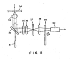
- an apparatus shown in Fig. 5 there is provided an apparatus shown in Fig. 5.
- a lens 58 and a band-pass filter array 59 comprising band-pass filters, corresponding respectively to a plurality of fluorescences, set in a circular form are disposed instead of the divided lens panel 38 and the filter panel 39 in the apparatus shown in Fig. 3.
- One photomultiplier tube 60 is installed behind the band-pass filter array 59.
- like members have the same reference numerals.
- This apparatus is advantageous in that rotation of the array 59 while applying light to one point of the gel plate 34 can realize detection and distinguishment of a plurality of fluorescences in a continuous manner by a single photomultiplier,
Landscapes
- Life Sciences & Earth Sciences (AREA)
- Chemical & Material Sciences (AREA)
- Health & Medical Sciences (AREA)
- Molecular Biology (AREA)
- Organic Chemistry (AREA)
- Proteomics, Peptides & Aminoacids (AREA)
- Physics & Mathematics (AREA)
- Engineering & Computer Science (AREA)
- Biochemistry (AREA)
- General Health & Medical Sciences (AREA)
- Zoology (AREA)
- Immunology (AREA)
- Wood Science & Technology (AREA)
- Analytical Chemistry (AREA)
- Microbiology (AREA)
- Biophysics (AREA)
- Biotechnology (AREA)
- Electrochemistry (AREA)
- Chemical Kinetics & Catalysis (AREA)
- Pathology (AREA)
- General Physics & Mathematics (AREA)
- Bioinformatics & Cheminformatics (AREA)
- General Engineering & Computer Science (AREA)
- Genetics & Genomics (AREA)
- Measuring Or Testing Involving Enzymes Or Micro-Organisms (AREA)
- Investigating Or Analysing Biological Materials (AREA)
- Investigating, Analyzing Materials By Fluorescence Or Luminescence (AREA)
Applications Claiming Priority (3)
| Application Number | Priority Date | Filing Date | Title |
|---|---|---|---|
| JP10203619A JP2000035432A (ja) | 1998-07-17 | 1998-07-17 | オリゴヌクレオチド検出および塩基配列決定法 |
| JP20361998 | 1998-07-17 | ||
| EP99305648A EP0979878A3 (fr) | 1998-07-17 | 1999-07-16 | Procédé pour détecter des oligonucléotides et détermination de la séquence des bases d'acides nucléiques |
Related Parent Applications (1)
| Application Number | Title | Priority Date | Filing Date |
|---|---|---|---|
| EP99305648A Division EP0979878A3 (fr) | 1998-07-17 | 1999-07-16 | Procédé pour détecter des oligonucléotides et détermination de la séquence des bases d'acides nucléiques |
Publications (1)
| Publication Number | Publication Date |
|---|---|
| EP1568785A2 true EP1568785A2 (fr) | 2005-08-31 |
Family
ID=16477054
Family Applications (2)
| Application Number | Title | Priority Date | Filing Date |
|---|---|---|---|
| EP99305648A Withdrawn EP0979878A3 (fr) | 1998-07-17 | 1999-07-16 | Procédé pour détecter des oligonucléotides et détermination de la séquence des bases d'acides nucléiques |
| EP05075875A Withdrawn EP1568785A2 (fr) | 1998-07-17 | 1999-07-16 | Procédé pour détecter des oligonucléotides et détermination de la séquence des bases d'acides nucléiques |
Family Applications Before (1)
| Application Number | Title | Priority Date | Filing Date |
|---|---|---|---|
| EP99305648A Withdrawn EP0979878A3 (fr) | 1998-07-17 | 1999-07-16 | Procédé pour détecter des oligonucléotides et détermination de la séquence des bases d'acides nucléiques |
Country Status (3)
| Country | Link |
|---|---|
| US (1) | US6333156B1 (fr) |
| EP (2) | EP0979878A3 (fr) |
| JP (1) | JP2000035432A (fr) |
Families Citing this family (2)
| Publication number | Priority date | Publication date | Assignee | Title |
|---|---|---|---|---|
| US20060084799A1 (en) * | 2003-09-24 | 2006-04-20 | Williams Lewis T | Human cDNA clones comprising polynucleotides encoding polypeptides and methods of their use |
| WO2005035569A2 (fr) * | 2003-10-10 | 2005-04-21 | Five Prime Therapeutics, Inc. | Kiaa0779, ses variantes epissees et procede de leur utilisation |
Family Cites Families (4)
| Publication number | Priority date | Publication date | Assignee | Title |
|---|---|---|---|---|
| JPH0799353B2 (ja) * | 1987-03-31 | 1995-10-25 | 株式会社島津製作所 | 塩基配列決定装置 |
| SE9203320D0 (sv) * | 1992-11-06 | 1992-11-06 | Pharmacia Lkb Biotech | A method of processing nucleic acid samples |
| DE19508366C2 (de) * | 1995-03-10 | 1998-01-29 | Evotec Biosystems Gmbh | Verfahren zum direkten Nachweisen weniger Nucleinsäurestränge |
| US5763175A (en) * | 1995-11-17 | 1998-06-09 | Lynx Therapeutics, Inc. | Simultaneous sequencing of tagged polynucleotides |
-
1998
- 1998-07-17 JP JP10203619A patent/JP2000035432A/ja active Pending
-
1999
- 1999-07-16 US US09/354,565 patent/US6333156B1/en not_active Expired - Fee Related
- 1999-07-16 EP EP99305648A patent/EP0979878A3/fr not_active Withdrawn
- 1999-07-16 EP EP05075875A patent/EP1568785A2/fr not_active Withdrawn
Also Published As
| Publication number | Publication date |
|---|---|
| US6333156B1 (en) | 2001-12-25 |
| EP0979878A3 (fr) | 2000-03-22 |
| EP0979878A2 (fr) | 2000-02-16 |
| JP2000035432A (ja) | 2000-02-02 |
Similar Documents
| Publication | Publication Date | Title |
|---|---|---|
| US6749734B1 (en) | Microfabricated capillary array electrophoresis device and method | |
| US6132578A (en) | Method and apparatus for electrophoresis separation and detection | |
| EP1178305B1 (fr) | Méthode pour détecter le flux de courant électrique à travers un canal de séparation et composition à utiliser dans celui-ci | |
| EP0624246B1 (fr) | Analyse par separation differentielle | |
| EP0854937B1 (fr) | Procedes et kit d'analyse par hybridation faisant appel a des sondes d'acides nucleiques peptidiques | |
| US6432634B1 (en) | Method, apparatus and kits for sequencing of nucleic acids using multiple dyes | |
| EP1024360A2 (fr) | Dispositif de balayage pour un système d'electrophorese multicapillaire de grande capacité | |
| CN1119275A (zh) | 电泳多试样组分采集器 | |
| JPH07209251A (ja) | 電気泳動装置 | |
| EP0294524A1 (fr) | Appareil en électrophorèse en balayage de temps réel pour la mise en séquence de DNA | |
| US6982029B2 (en) | Electrophoretic method and system having internal lane standards for color calibration | |
| JP2628571B2 (ja) | オリゴヌクレオチドの分析装置 | |
| US6333156B1 (en) | Method for detecting oligonucleotides and determining base sequence of nucleic acids | |
| JPH09512102A (ja) | 電気泳動分析用の方法および装置 | |
| JP2809227B2 (ja) | 電気泳動装置 | |
| US20030150728A1 (en) | Method and device for separating marked biopolymers | |
| Kambara et al. | REAL-TIME DNA DETECTION SYSTEMS | |
| JPH0591896A (ja) | 核酸ハイブリツド検定法 | |
| JP2649794C (fr) | ||
| JPH09140399A (ja) | 多種並行dnaシーケンシング方法 |
Legal Events
| Date | Code | Title | Description |
|---|---|---|---|
| PUAI | Public reference made under article 153(3) epc to a published international application that has entered the european phase |
Free format text: ORIGINAL CODE: 0009012 |
|
| 17P | Request for examination filed |
Effective date: 20050505 |
|
| AC | Divisional application: reference to earlier application |
Ref document number: 0979878 Country of ref document: EP Kind code of ref document: P |
|
| AK | Designated contracting states |
Kind code of ref document: A2 Designated state(s): AT BE CH CY DE DK ES FI FR GB GR IE IT LI LU MC NL PT SE |
|
| STAA | Information on the status of an ep patent application or granted ep patent |
Free format text: STATUS: THE APPLICATION IS DEEMED TO BE WITHDRAWN |
|
| 18D | Application deemed to be withdrawn |
Effective date: 20070301 |