EP1895301A1 - Analyseur pour une chaîne de sucres ou des glucides complexes - Google Patents
Analyseur pour une chaîne de sucres ou des glucides complexes Download PDFInfo
- Publication number
- EP1895301A1 EP1895301A1 EP06766719A EP06766719A EP1895301A1 EP 1895301 A1 EP1895301 A1 EP 1895301A1 EP 06766719 A EP06766719 A EP 06766719A EP 06766719 A EP06766719 A EP 06766719A EP 1895301 A1 EP1895301 A1 EP 1895301A1
- Authority
- EP
- European Patent Office
- Prior art keywords
- glycan
- substrate
- protein
- lectin
- immobilized
- Prior art date
- Legal status (The legal status is an assumption and is not a legal conclusion. Google has not performed a legal analysis and makes no representation as to the accuracy of the status listed.)
- Withdrawn
Links
Images






Classifications
-
- G—PHYSICS
- G01—MEASURING; TESTING
- G01N—INVESTIGATING OR ANALYSING MATERIALS BY DETERMINING THEIR CHEMICAL OR PHYSICAL PROPERTIES
- G01N33/00—Investigating or analysing materials by specific methods not covered by groups G01N1/00 - G01N31/00
- G01N33/48—Biological material, e.g. blood, urine; Haemocytometers
- G01N33/50—Chemical analysis of biological material, e.g. blood, urine; Testing involving biospecific ligand binding methods; Immunological testing
- G01N33/53—Immunoassay; Biospecific binding assay; Materials therefor
- G01N33/543—Immunoassay; Biospecific binding assay; Materials therefor with an insoluble carrier for immobilising immunochemicals
- G01N33/54366—Apparatus specially adapted for solid-phase testing
- G01N33/54373—Apparatus specially adapted for solid-phase testing involving physiochemical end-point determination, e.g. wave-guides, FETS, gratings
-
- G—PHYSICS
- G01—MEASURING; TESTING
- G01N—INVESTIGATING OR ANALYSING MATERIALS BY DETERMINING THEIR CHEMICAL OR PHYSICAL PROPERTIES
- G01N21/00—Investigating or analysing materials by the use of optical means, i.e. using sub-millimetre waves, infrared, visible or ultraviolet light
- G01N21/62—Systems in which the material investigated is excited whereby it emits light or causes a change in wavelength of the incident light
- G01N21/63—Systems in which the material investigated is excited whereby it emits light or causes a change in wavelength of the incident light optically excited
- G01N21/64—Fluorescence; Phosphorescence
- G01N21/6428—Measuring fluorescence of fluorescent products of reactions or of fluorochrome labelled reactive substances, e.g. measuring quenching effects, using measuring "optrodes"
-
- G—PHYSICS
- G01—MEASURING; TESTING
- G01N—INVESTIGATING OR ANALYSING MATERIALS BY DETERMINING THEIR CHEMICAL OR PHYSICAL PROPERTIES
- G01N21/00—Investigating or analysing materials by the use of optical means, i.e. using sub-millimetre waves, infrared, visible or ultraviolet light
- G01N21/62—Systems in which the material investigated is excited whereby it emits light or causes a change in wavelength of the incident light
- G01N21/63—Systems in which the material investigated is excited whereby it emits light or causes a change in wavelength of the incident light optically excited
- G01N21/64—Fluorescence; Phosphorescence
- G01N21/645—Specially adapted constructive features of fluorimeters
- G01N21/648—Specially adapted constructive features of fluorimeters using evanescent coupling or surface plasmon coupling for the excitation of fluorescence
-
- G—PHYSICS
- G01—MEASURING; TESTING
- G01N—INVESTIGATING OR ANALYSING MATERIALS BY DETERMINING THEIR CHEMICAL OR PHYSICAL PROPERTIES
- G01N33/00—Investigating or analysing materials by specific methods not covered by groups G01N1/00 - G01N31/00
- G01N33/48—Biological material, e.g. blood, urine; Haemocytometers
- G01N33/50—Chemical analysis of biological material, e.g. blood, urine; Testing involving biospecific ligand binding methods; Immunological testing
- G01N33/53—Immunoassay; Biospecific binding assay; Materials therefor
- G01N33/543—Immunoassay; Biospecific binding assay; Materials therefor with an insoluble carrier for immobilising immunochemicals
- G01N33/54393—Improving reaction conditions or stability, e.g. by coating or irradiation of surface, by reduction of non-specific binding, by promotion of specific binding
-
- G—PHYSICS
- G01—MEASURING; TESTING
- G01N—INVESTIGATING OR ANALYSING MATERIALS BY DETERMINING THEIR CHEMICAL OR PHYSICAL PROPERTIES
- G01N33/00—Investigating or analysing materials by specific methods not covered by groups G01N1/00 - G01N31/00
- G01N33/48—Biological material, e.g. blood, urine; Haemocytometers
- G01N33/50—Chemical analysis of biological material, e.g. blood, urine; Testing involving biospecific ligand binding methods; Immunological testing
- G01N33/53—Immunoassay; Biospecific binding assay; Materials therefor
- G01N33/543—Immunoassay; Biospecific binding assay; Materials therefor with an insoluble carrier for immobilising immunochemicals
- G01N33/544—Immunoassay; Biospecific binding assay; Materials therefor with an insoluble carrier for immobilising immunochemicals the carrier being organic
- G01N33/548—Carbohydrates, e.g. dextran
Definitions
- the present invention concerns an analyzer for a glycan or a complex carbohydrate, or a sample containing the same by utilizing an evanescent light, and a substrate which is used being mounted to the analyzer.
- the structural analysis for the glycan is first indispensable. It is expected that more importance will be attached to the glycan structure analysis method in the future. However, since the structural analysis of the glycan requires enormous time, energy, and experience it has been expected develop a system capable of extracting the features of various glycan structures more conveniently, quickly, at a higher sensitivity and higher accuracy and capable of distinguishing them from each other, instead of aiming at a complete determination of structure based on the existent method.
- a micro-array is a collective name of instruments in which immobilized specimens such as various types of DNA-proteins are immobilized spot-wise at a high density on a solid phase carrier (glass membrane silicon chip) and this can detect the presence or absence of molecules binding specifically to various types of immobilized specimen spots (hereinafter referred to as a probe).
- a probe those labeled with fluorescence are generally used and, by reacting a probe solution with an array surface and then observing by a fluorescent detection scanner, quantitative analysis can be conducted for the probe molecules binding to each of specimen spots. Since a DNA micro-array was developed by Affymetrix Co in USA, micro-arrays have been used in an extremely wide variety of fields of study to provide various novel findings to human beings.
- glycan binding protein for example, lectin
- the dissociation reaction proceeds between the glycan and the protein that exhibits interaction with the glycan at the instance of conducting the removing and cleaning operation for the probe solution and it is difficult to obtain an accurate interaction information under the equilibrium state. Accordingly, in the micro-array, the cleaning operation of the probe solution results in a significant problem in a case of precisely analyzing the interaction information in the equilibrium state between the glycan and the protein that exhibits interaction with the glycan.
- DNA micro-arrays have been utilized in an extremely wide range at present. Also for protein micro-arrays, future utilization is expected in the field of basic study such as clarification of the function of a protein as the product of DNA transcription in the living body, as well as in the application field such as diagnosis or judgment on the basis of quantitative or qualitative change of the protein, and vigorous researches have been conducted all over the world in view of the study. However, development and popularization for the protein micro-array is greatly delayed compared with that for the DNA micro-array. As one of the causes, it has been pointed out by various workers that a step of immobilizing protein specimens having various different natures at a predetermined ratio in a state of keeping the activity as it is is extremely difficult technically.
- Non-Patent Document 1 For the methods of immobilizing proteins on arrays, a method developed in the earliest stage includes a method of physically adsorbing a protein on a membrane typically represented by a PVDF (Non-Patent Document 1). It has been reported that some proteins such as transcription factors can maintain the activity to some extent but it lacks in generality. Further, in a case of immobilization on the membrane, increase in the density of the array has been limited. For obtaining higher density, while a study has been progressed in the direction of immobilizing the protein to the surface of a solid such as metal or glass, the protein generally has a nature tending to be denatured by the contact with the surface of the solid such as metal or glass. Accordingly, an immobilizing method using a certain linker for crosslinking the surface of the solid and the protein has been studied and developed vigorously.
- An example of the method of mitigating the problem of denaturation of the protein includes a method of joining a pad of a polyacrylamide of 10 to 100 ⁇ m thickness on a slide glass and spotting the protein thereto (Non-Patent Documents 2 and 3).
- the protein is immobilized in a three-dimensional space, it is said that improvement by 100 times or more may be expected in view of the quantity compared with a method of immobilizing on a two-dimensional surface.
- there is also a method of immobilizing the protein by way of an amino group in a porous polyacrylamide gel Non-Patent Document 4
- the methods described above have not yet been popularized generally since it is necessary to prepare a expensive and special slide glass.
- the immobilized protein layer has a thickness and this is sometimes not preferred depending on the detection method.
- Methods of immobilizing proteins to the solid phase that have been being studied most vigorously at present include a method of expressing a protein in a form attached with some or other tag and immobilizing the protein by way of its tag portion to a solid phase carrier. It has been said for the method that it can provide an effect of improving the effective ligand concentration of the protein or alignment for the direction of the protein in principle.
- An example of such method includes a method of immobilization by way of oligohistidine tag to a substrate modified at the surface with a nickel complex (Non-Patent Document 5) or a method of immobilization by way of avidin-biotin (Patent Document 1).
- a method of immobilization by way of amino groups in proteins to a solid phase carrier includes a method of immobilizing a protein by way of active ester groups bonded on the surface of the solid phase, or a method of immobilizing a protein by way of epoxy groups arranged on the surface of the solid phase (Non-Patent Document 6).
- the method of immobilization by way of the amino groups of the protein is a simple method, since commercial proteins, bio-body extract ingredients, or recombinant proteins with no particular tags can also be immobilized simply and conveniently, individual users can optionally select proteins in accordance with the object of their own and can use them with optimization to micro-arrays conforming with the purpose rapidly and at a low cost.
- Drawbacks of the immobilizing method by way of the amino groups of the protein include, for example, that the number of lysine residues in the proteins is different depending on individual proteins or that inactivation of the protein may possibly occur depending on the position of the lysine residues used for immobilization.
- An object of the present invention is to provide an apparatus for analyzing a glycan or a complex glycan, or a specimen containing the same more conveniently and quickly at high sensitivity and high accuracy, as well as a micro-array substrate mounted to the apparatus.
- the present inventors have made an earnest study for solving the subject described above. As a result, the inventors have manufactured an analyzer for a glycan or a complex glycan by using lectin as the glycan binding protein and utilizing a substrate in which multiple types of lectins are arranged and immobilized on the surface and evanescent waves as an excitation light for measuring the binding state of lectin on the substrate and a fluorescence-labeled glycan or glycoprotein, and have actually analyzed specimens containing the glycan or the complex glycan. According to the existent lectin plotting method, a cleaning step is necessary for the fluorescence-labeled glycan or the like not bonded with lectin.
- the cleaning and removing operation for the probe solution which resulted a significant problem in accurate analysis for the information on the interaction between a glycan binding protein such as a lectin and a glycan in an equilibrium state is not more necessary, and weak interaction that may be flushed off upon cleaning in the existent method can also be detected. That is, means for observing the interaction between the glycan binding protein and the glycan in the equilibrium state as it is has been put to practical use for the first time.
- the apparatus not the presence or absence (0 to 1) for the binding such as in the existent lectin plotting but information for an intermediate portion, that is, information for the binding intensity can be obtained (for example, as 0 to 6 stages).
- arrays for the analysis of interaction between glycan binding protein and glycan directed to various application uses, thereby analyzing, for example, the glycans of the glycoside proteins and the quantitative ratio thereof in living specimens such as stock solutions or dilute solutions of bloods, body fluids, tissue extracts, etc., they can be utilized to diagnosis and judgment of various diseases, as well as applied widely to the quality control of glycoside protein formulations.
- the present invention concerns an apparatus for measuring the interaction between a glycan or a complex carbohydrate and a glycan binding protein and analyzing the glycan or the complex carbohydrate.
- the apparatus of the invention is an analyzer for a glycan or a complex carbohydrate in a specimen, or a specimen containing the same, and has a substrate comprising a photoconductive material in which multiple types of glycan binding proteins are arranged and immobilized, means for introducing a light to the end face on the side of the substrate and generating evanescent waves to the surface of the substrate thereby exciting fluorescent labeling, and fluorescent intensity measuring means for measuring the intensity of fluorescence generated by the means described above on every position for arranging the glycan binding proteins.
- the chain includes, for example, glycoside protein type glycan (N-binding type glycan and O-binding type glycan), glucolipid type glycan, glycosamino glycane type glycan, and polysaccharide-derived oligo saccharide chain.
- N-binding type glycan comprising high mannose type-hybrid type-composite type as N-binding type glycan
- O-binding type glycan comprising mutin type (O-GalNAc) ⁇ O-Fuc type ⁇ O-Man type ⁇ O-Glc type as the O-binding type glycan
- hyaluronic acid ⁇ karatan sulfuric acid ⁇ hepalin ⁇ heparan sulfuric acid ⁇ chondroitin sulfuric acid ⁇ dermatan sulfuric acid as glucosamino glycan series glycan
- oligosaccharides derived from chitin, cellulose, curdlan, laminarin, dextran, starch, glycogen
- glycans include, for example, M3, M5A, Hybrid(monoagalacto, bisect), NA1, NA1( ⁇ 1-6Fuc), NA2(monoagalacto), NA2(monoagalacto, bisect), NA2, NA2( ⁇ 1-6Fuc), A2, NA2(bisect), NA3, NA3( ⁇ 1-6Fuc), NA4, NA4( ⁇ 1-6Fuc), NA5(pentaagalacto, bisect), Lactose, GA2, GA1, GM3-NeuAc, GM3-NeuGc, GM1, GM2, GD1a, DG1b, GD3, Gb3, Gb4, Forssman, LNnT, LNT, Galili pentasaccharide, B-hexasaccharide, LNFP-I, LNFP-II(Le a ), LNFP-III(Le X ), LNFP-II(Le b ), A-hexacchari
- the complex carbohydrate is a collective name of in-vivo polymers having glycans.
- the complex carbohydrate of the invention includes glycoprotein (also including glycopeptide), proteoglycan, and glycolipid.
- glycoprotein also including glycopeptide
- proteoglycan and glycolipid.
- lectin is preferred for example and the lectin includes lectins belonging to various molecule families obtained, for example, from animals and plants, fungus, bacteria, and virus, that is, lysine B chain-related "R type lectin” distributed in all biological fields including bacteria, "calnexin, calreticulin” present generally in eukaryotes and concerning folding of glycoside protein, calcium demanding "C-type lectin” present generally in multicellular animals and containing may typical lectins, "galectin” distributed generally in the animal field and showing specificity to galactose, "leguminous lectin” forming a large family in plant leguminous, and "L-lectin” having structural similar
- lectins include, for example, ACA(Amaranthus caudatus L.), BPL(Bauhinia purpurea L.), ConA(Canavalia ensiformis), DBA(Dolichos biflorus), DSA(Datura stramonium), ECA(Erythrina cristagalli), EEL(Euonymus europaeus), GNA(Galanthus nivalis), GSL I (Griffonia simplicifolia), GSL II(Griffonia simplicifolia), HHL(Hippeastrum hybridum), Jacalin(Artocarpus integrifolia), LBA(Phaseolus lunatus), LCA(Lens culinaris), LEL(Lycopersicon esculentum), LTL(Lotus tetragonolobus), MPA(Maclura pomifera), NPA(Narcissus pseudo-narcissus), PHA-E(Phaseolus
- lectins immobilized on the substrate include those modified from the lectins described above for the physical property, binding specificity and affinity by the method, for example, of introducing site specific mutation, or chemical modification.
- the lectin referred to in the specification also includes such lectin variants.
- the present invention concerns an apparatus for enabling analysis of a relatively weak interaction between a glycan and a protein compared with the antigen-antibody reaction and the glycan binding protein of weak interaction is not restricted to the lectin. That is, glycan recognition antibody generally having weak interaction (for example, glycan recognition antibody classified to the IgM class) is also included.
- the substrate in the invention may be any substrate having a photoconductivity and capable of generating evanescent waves on the surface thereof by the light irradiation means of the analyzer according to the invention and includes, for example, glass, quartz glass, and synthesis quartz glass, but it is not restricted to them.
- the substrate to which the lectin is immobilized in the invention is a compound, preferably, having an epoxy group as an active group, it is not restricted thereto but may be a substrate on which a glycan binding protein such as lectin is immobilized on the substrate coated with vinyl sulfonic group, active ester group, aldehyde group, carboxyl group, amino group, thiol group, isothiocyanate group, etc.
- the compound having the epoxy group as the active group includes, preferably, 3-glysidoxy propyl trimethoxy silane (GTMS) but is not restricted thereto.
- GTMS 3-glysidoxy propyl trimethoxy silane
- Other examples include, for example, 2- (3,4 epoxy cyclohexyl) ethyl trimethoxy silane, 3-glysidoxy propyl methyl diethoxy silane, 3-glysidoxy propyl triethoxy silane, or a silane coupling compound having a plurality of epoxy groups at the top ends of branched spacers and, preferably, compounds, for example, containing polyethylene glycol, protein, biotin ⁇ avidin, etc.
- the substrate used for the analysis of the glycan or the complex carbohydrate of the invention is a substrate in which multiple types of glycan binding proteins such as lectins are arranged and immobilized on the surface thereof and can be manufactured, for example, by the following method. Description is to be made to an example of using lectin as a glycan binding protein.
- a compound having an epoxy group as an active group is coated to the substrate.
- GTMS a compound having the epoxy group as the active group
- it can be conducted by the method described in the example to be described later. Specifically, a slide glass is dipped in a 10% KOH/MeOH solution, left for one hour in a state of being shaken together with the vessel thereby treating the glass surface, which is cleaned with a sufficient amount of pure water (milli Q water) and then dried in an oven at 60°C. Then, the slide glass is dipped into a 2% GTMS acetone solution, and reaction is taken place for one hour while being shaken together with a vessel under shielding of light.
- the alkoxy silyl group of GTMS is transformed into a silanol group by hydrolysis with water, the silanol group is instable and partially bonded into the state of an oligomer by change with time and, successively, adsorbed by way of hydrogen binding to the glass surface.
- the slide glass is dried in an oven at 110°C for 8 hours. By the drying treatment, dewatering condensation reaction with silanol groups on the glass surface occurs to form a strong covalent bond.
- Fig. 1 shows a series of GTMS coating method.
- a lectin is spotted to a substrate coated with the compound having the epoxy group as the active group and can be immobilized by reaction utilizing the amino group of the lectin.
- multiple types of lectins are spotted on one identical substrate.
- the plurality types of lectins are desirably arranged and immobilized by spotting in a predetermined pattern according to the types thereof.
- a commercially available pin type spotter for use in manufacture of DNA microarrays or a non-contact type ink jet spotter can be utilized. After spotting the lectins, unbonded lectins can be removed by cleaning with a Tween 20-containing PBS solution (PBST).
- PBST Tween 20-containing PBS solution
- the substrate to which the lectins are immobilized is preferably a substrate forming a plurality of reaction vessels (refer to Fig. 2 ). More preferably, it is a substrate formed with a plurality of reaction vessels by bonding rubber having a plurality of holes.
- a 8-hole rubber designed and developed by the present inventors is bonded to a predetermined position relative to a slide glass where a protein that exhibits interaction with the glycan to manufacture eight reaction vessels as described in the example.
- eight rectangular holes are formed regularly and a fluorescence labeling glycan probe solution can be filled accurately to the periphery of the spots by bringing it in close contact with the slight glass on an adjuster used exclusively therefor.
- the reaction vessel is preferably made by plurality and, more preferably, 12 or 16 reaction vessels are arranged each at an interval identical with the standard of the commercially available 96, 384 well plates.
- the material for forming the reaction vessel is not restricted to the silicone rubber but, for example, it is possible to form the reaction site by applying water repellent coating to a non-spot region of the glass surface, or utilizing the vessel of a 96 well plate standard.
- the substrate of the invention it is preferred to arrange and immobilize a plurality of lectins of an identical type.
- glycan binding proteins such as lectins are arranged and immobilized and, further, antibodies to the protein moieties of the glycoside proteins can be arranged and immobilized further (manufacture of hybrid array) upon analysis of glycoside proteins.
- analysis for the glycan portion of the glycoside protein and analysis for the protein portion can be conducted simultaneously. This can simply distinguish those of identical glycan although different in the protein or those of identical protein although different in the glycan.
- Fig. 3 is a view showing an example of an analyzer according to the invention.
- the apparatus has a substrate (1) comprising a photoconductive material in which multiple types of glycan binding proteins such as lectins are arranged and immobilized, means (2), (2') for introducing light to the end face on the side of the substrate and generating evanescent waves to the surface of the substrate thereby exciting the fluorescent labeling, and fluorescent intensity measuring means (3) for measuring the intensity of the fluorescence generated by the means described above on every position for arranging the glycan binding proteins, in which the substrate (1) is mounted detachably on a substrate mounting means (4), and the substrate mounting means preferably has a frame for guiding the fluorescence generated on the substrate to the fluorescent intensity measuring means without shielding the fluorescence.
- the fluorescence labeling excitation means (2), (2') are disposed facing the end face of the substrate.
- the evanescent waves are generated upon total reflection of a light at the boundary between substances, and the incident angle for causing total reflection is different depending on the substance and the material.
- the fluorescent labeling excitation means (2), (2') are provided such that the introduction angle of the light to the end face of the side of the substrate can be adjusted.
- the fluorescence-labeling excitation means may be disposed only one side of the end face of the substrate perpendicular to the row.
- the number of row of the glycan binding proteins is large, it is preferably disposed, for example, on both sides of the substrate to generate uniform evanescent waves on the surface of the substrate.
- the fluorescent intensity measuring means (3) has no particular restriction, and a charge coupled device (CCD) camera, image intensifier charge coupled device (ICCD) camera, cooled CCD camera, photomultiplier (PMT), etc. can also be used.
- CCD charge coupled device
- ICCD image intensifier charge coupled device
- PMT photomultiplier
- scanning is conducted by displacing the substrate mounting means (4) on the fluorescent intensity measuring means (3) to move the entire surface of a substrate for immobilizing glycan binding protein, for example, in directions XY, in which scanning may be conducted also by moving the fluorescent intensity measuring means (3) while fixing the substrate (1).
- Description is to be made specially to a case of analyzing glycans to be detected or complex carbohydrates to be detected by using the analyzer described above of the invention.
- a substrate (1) on which glycan binding proteins such as a plural types of lectins are arranged and immobilized is mounted on the substrate mounting means (4), a specimen containing fluorescence-labeled glycans to be detected or complex carbohydrates to be detected is introduced on the substrate (1), and each of the glycan binding proteins on the substrate (1) and the fluorescence-labeled glycans to be detected or complex carbohydrates to be detected are contacted with each other.
- the fluorescence labeling agent of the glycan to be detected or the complex carbohydrate to be detected includes 2-aminopyridine, Cy3, Cy3.5, Cy5, tetramethyl rhodamine, and various fluorescence dyes having fluorescein skeletons, Alexa series fluorescence dyes manufactured by Molecular Probes Co., and quantum dot fluorescence dyes but it is not restricted to them so long as it is a substance having a property of fluorescence labeling the glycan.
- the glycan to be detected or the complex carbohydrate to be detected can be fluorescence-labeled directly or indirectly.
- the glycan to be detected can be fluorescence-labeled indirectly.
- the complex carbohydrate to be detected can be fluorescence-labeled indirectly.
- the complex carbohydrate can be fluorescence-labeled indirectly also by binding a previously fluorescence-labeled glycan binding protein and a complex carbohydrate to be detected (in this case, in the glycans of the complex carbohydrate to be detected, glycans other than the glycan bonded with the glycan binding protein are bonded to the substrate).
- the target complex carbohydrate can be selectively fluorescence-labeled without separation or purification from the crude specimens. Further, by bringing a crude specimen acted with a previously fluorescence-labeled antibody into contact with a substrate on which glycan binding proteins such as lectins are immobilized for instance, only the glycan information of the target complex carbohydrate can be observed selectively.
- a light is then introduced from the fluorescence labeling excitation means (2), (2') to the end face on the side of the substrate without cleaning the substrate (1) to generate evanescent waves on the surface of the substrate and excite fluorescent labeling groups of the glycan or the complex carbohydrate to be detected.
- a weak light referred to as an evanescent light exudes within a range of 200 to 300 nm from the boundary (about one-half excitation wavelength) upon totally reflecting the fluorescent light in the inside of glass.
- the fluorescence labeling groups are excited by utilizing the evanescent light in a state of contacting a solution containing a glycan or a complex carbohydrate labeled with a fluorescent substance on a slide glass, probe molecules near the boundary contributing to the binding reaction can be observed selectively without scarcely exciting fluorescence-labeled molecules under Brownian motion, that is, molecules not contributing to the binding reaction.
- the present inventors have solved the problem described above by exciting the fluorescence labeling groups by the evanescence waves and measuring the intensity of fluorescence without cleaning the probe solution. Further, since scanning can be conducted not by way of the cleaning, the change with time of the interaction can be traced just after the addition of the probe by conducting scanning continuously on every predetermined time interval and it can be easily judged as to whether the interaction reaction between each of the glycan binding proteins immobilized on an identical glass and the probe reaches an equilibrium state or not.
- the fluorescence generated by the excitation of the evanescence waves is measured for the fluorescent intensity thereof by the fluorescent intensity measuring means (3) on every position for arranging each of the glycan binding proteins on the substrate (in a case of arranging and immobilizing multiple types of lectins on a substrate, the fluorescent intensity is measured on the position for arranging each of the lectins, that is, on every types of lectin). For example, by comparing the fluorescent intensity of the glycan to be detected to each of lectins and the fluorescent intensity of each of the known glycans to each of lectins measured in the same manner, difference, similarity, non-similarity, etc. thereof can be judged.
- the invention provides useful means as a glycan profiler for conducting the glycan structural analysis rapidly and conveniently. For example, a profiling method described in Protein, Nucleic Acid, Enzyme, in August 2003, special Vol. 48, No. 11 is applicable to the means described above.
- the measured values for the fluorescent intensity in the apparatus of the invention are used for the glycan structural analysis by conducting information processing using a computer.
- the apparatus of the invention includes such an information processing system by the computer.
- the structure of the glycan to be detected can be identified in a case where the glycan to be detected has a known structure.
- the glycan detected has an unknown structure
- a characteristic structure present in the glycan for example, ⁇ 2-3 sialic acid, ⁇ 2-6 sialic acid ⁇ 1-3 galactose ⁇ ⁇ 1-3 fucose ⁇ ⁇ 1-6 fucose ⁇ sulfated bisect N-acetyl glucosamine, etc.
- point out the similarity with a glycan of known structure for example, ⁇ 2-3 sialic acid, ⁇ 2-6 sialic acid ⁇ 1-3 galactose ⁇ ⁇ 1-3 fucose ⁇ ⁇ 1-6 fucose ⁇ sulfated bisect N-acetyl glucosamine, etc.
- This means is a system of automatically displaying a glycan structure when setting, to an apparatus, a substrate in which multiple types of glycan binding proteins, for example, multiple types of lectins which are brought into contact with a fluorescence-labeled glycan to be detected and exhibit interaction with the glycan or glycan to be detected.
- a step of bringing a fluorescence-labeled glycan to be detected to a substrate on which various proteins exhibiting interaction with the glycan are immobilized respectively can be automated.
- the step of blocking or cleaning and eliminating a blocking solution, and a contact step of the fluorescence-labeled glycan solution can be controlled uniformly. Since data of higher reliability can be obtained by using the methods, they are extremely useful. Further, in a case of estimating a partial glycan structure of a glycan to be detected by the apparatus according to the invention, data of higher reliability can be obtained by using enzymatic digestion by glycoside hydrolase.
- sialic acid is contained in the non-termination partial glycan structure of a glycan to be detected or not and, further, whether the binding site is ⁇ 2-3 or ⁇ 2-6 in a case where it is contained
- this can be conducted easily by digesting the glycan to be detected with a highly specific sialidase (for example, ⁇ 2-3 neuraminidase cleaves only ⁇ 2-3 sialic acid, and sialidase derived from Arthorobacter ureafaciens cleaves ⁇ 2-6 sialic acid more preferably than ⁇ 2-3 sialic acid) and examining the presence or absence for the change of the profile before and after the reaction.
- a highly specific sialidase for example, ⁇ 2-3 neuraminidase cleaves only ⁇ 2-3 sialic acid, and sialidase derived from Arthorobacter ureafaciens cleaves ⁇ 2-6 sialic acid more preferably than ⁇ 2-3 sialic acid
- partial glycan structures can be estimated in a wider range.
- By preparing eight reaction vessels on a glass it is possible to profile eight types of enzyme digestion reaction products simultaneously and this further facilitates estimation for the glycan structure.
- the glycoside hydrolase that can be used for the estimation of the partial glycan structure by enzymatic digestion includes, in addition, ⁇ -galactosidase derived from Jack Bean, ⁇ (1-3,4,6)-galactosidase derived from Green Coffee Bean, ⁇ -N-acetylhexosaminidase derived from Streptococcus pneumoniae, ⁇ -N-acetyl galactosaminidase derived from chicken liver, ⁇ (1-2,3,6)manosidase derived from Jack Bean, ⁇ mannosidase derived from Helix pomatia, ⁇ fucosidase derived from bovine kidney, ⁇ 1,2-L-fucosidase derived from Corynebacterium sp., ⁇ 1,2-xylosidase derived from Xanthomonas sp., and recombinants for all of them.
- the end type glycoside hydrolase usable in this case includes, for example, end glycosidase H derived from Streptomyces plicatus, PNGase F derived from Flavobacterium meningosepticum, O-glycanase derived from Streptococcus pneumoniae, endo glycosidase F1 derived from Chryseobacterium meningosepticum, and all of recombinants thereof.
- Fig. 4 shows an example of an information processing system of the invention.
- the system includes at least the following means (a) to (c).
- the data base in this system may include both a case where it is present outside of a computer as shown in Fig. 5 and a case where it is present in the inside of the computer as shown in Fig. 6 .
- Fig. 7 shows an example for the constitutional view of a computer in the system of the invention.
- Input means 1 and output means 2 are connected to a bus line 3.
- a temporary memory means 4 temporarily stores inputted information, calculated information, etc.
- a central processing unit (CPU) 5 receives instructions from the program of the invention and conducts various calculations.
- a memory means (data base) 7 stores fluorescent intensity data for various glycan binding proteins on known glycans or complex carbohydrates, or patterned information like those described above. In addition, information on the binding property between the glycans or the complex carbohydrates and the glycan binding proteins obtained by other experimental systems may also be stored.
- the memory means 6 accommodates various programs including a program for executing the processing of the invention.
- the program for executing the processing of the invention at least contains a program 61 for corresponding the inputted fluorescent intensity data of various glycan binding proteins for glycans or complex carbohydrates or patterned information thereof to similar fluorescent intensity information of known glycans or complex carbohydrates stored in the data base or the patterned information thereof, and selecting one or a plurality of glycans of known structures having identical or similar data or the pattern (information of glycans of known structures stored in the data base), a display program 62, and a program 63 for controlling them.
- values for the data may be compared with each other.
- the program 61 may be incorporated with a function of comparing the values for the inputted fluorescent intensity data with the values for the information of known glycans or complex carbohydrates stored in the data base and selecting one or a plurality of glycans of known structure in view of the close relation between the values.
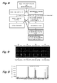
- Program 62 executes display for average fluorescent intensity data for each spot obtained for the glycan to be detected or the complex carbohydrate to be detected, signal noise ratio (S/N ratio) and fluctuation thereof, standard deviation, net luminance value calculated from the fluorescent intensity and patterned information obtained by pattering the same ( Fig. 9 ), information for the selected glycan of known structure, etc. Upon patterning, the information for the interaction can be normalized by using an appropriate internal standard.
- the programs described above can be collected in one program.
- the lectin immobilizing substrate (1) may be set in plurality to the analyzer, automatically transferred to the fluorescent intensity measuring section successively, the fluorescence labeling excitation means (3) is actuated to conduct scanning, and the obtained fluorescent intensity data may be automatically inputted to the computer.
- the inputted data may be stored in the memory means or the temporary memory means of the computer.
- the fluorescent intensity data or the patterned information formed by patterning the same are preferably stored together with the information derived from the glycan or complex carbohydrate to be detected in the data base.
- the calculation means such as the central processing unit (CPU) receives instructions from the program 62 stored in the memory means, reads out the fluorescent intensity information stored in the memory means or the temporary memory means to display the fluorescent intensity information.
- the calculation means such as the central processing unit (CPU) receives instructions from the program 62 stored in the memory means, reads out the fluorescent intensity information stored in the memory means or the temporary memory means to display the fluorescent intensity information.
- the internal standard spot may be present in plurality.
- the fluorescent intensity information inputted then is corresponded to the fluorescent intensity information of various glycans to various glycan binding proteins stored in the data base, and one or a plurality of glycans of known structures of identical patterns are selected.
- the calculation means such as the central processing unit (CPU) receives the instruction from the program 61 in the memory means, corresponds the fluorescent intensity information stored in the memory means or the temporary memory means and the fluorescent intensity information for the glycans or complex carbohydrates for the known glycans or complex carbohydrates stored in the data base, and selects one or a plurality of glycans of known structures identical or similar in the pattern of information.
- the selected information for the glycans of known structures can be stored in the memory means or the temporary memory means of the computer.
- the calculation means such as the central processing unit (CPU) receives the instruction from the program 61 in the memory means, inputs the fluorescent intensity information for the known glycans or complex carbohydrates stored in the data base into the computer, reads out the fluorescent intensity information stored in the memory means or the temporary memory means in the computer, corresponds the respective fluorescent intensity information, and selects one or a plurality of glycans of known structures identical in the pattern of information.
- CPU central processing unit
- the result of selection is then displayed by the display means.
- the calculation means such as the central processing unit (CPU) receives the instruction from the program 62 in the memory means, and reads out and displays the information of the glycans of known structures stored in the memory means or the temporary memory means.
- the invention is useful as a method of rapidly and conveniently examining the state of the glycan modification of an aimed protein from a mixture of different complex carbohydrates, for which application use may be considered in such fields as recognition for the situation of diagnosis or therapy by observing the state of the glycan modification of the aimed protein without purifying the mixed solution of various ingredients such as a stock solution or dilute solution of bloods, body fluids, living body extract ingredients, food ingredients, etc.
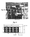
- All the prior art documents cited in the present specification are incorporated by reference to the present specification. Examples of the invention are to be shown below. In the examples, glycans were actually analyzed by using the analyzer of the invention shown in Fig. 3 . The appearance of the analyzer is shown by the photograph for the apparatus in Fig. 10 .
- a fluorescence-labeled glycoside protein probe was prepared by fluorescence-labeling Asialo-Fetuin (manufactured by SIGMA Co. hereinafter referred to as ASF) using Cy3 Mono-reactive Dye (manufactured by Amersham Pharmacia Biotech Co. hereinafter referred to as Cy3) as a fluorescent dye having an absorption maximum wavelength near 550 nm. It is known that ASF has a glycan structure having an N-binding type glycan and an O-binding type glycan each by the number of 3 in the molecular and in which sialic acid cap at the non-reducing terminal in the glycan is partially detached.
- GTMS 3-glysidoxypropyl trimethoxysilane having epoxy groups as an active group
- lectins were immobilized to the surface the glass.
- the GTMS coating was conducted by the following procedures using a slide glass manufactured by Matsunami Glass Industry Co., the slide glass was dipped into a 10% KOH/MeOH solution and left for 1 hour in a state being shaken together with a container to treat the glass surface.
- stamp pin operation program Upon procedure of immobilizing lectins to the slide glass, the following conditions were stored in a computer appended to the microarray spotter and a stamp pin operation program was executed. At first, after dipping the stamp pin into an immobilizing specimen solution in the 96-hole PCT micro titer plate for 1 sec and then it was pulled up and brought into contact with a predetermined position on the surface of the slide glass for 1 sec. While repeating the operation on every spot, after spotting for 4 points in one lateral row from an identical specimen solution, a cleaning step was applied to the stamp pin.
- the needle tip of the stamp pin was dipped in a 0.05% SDS solution for 2 sec, the stamp pin was dried in a vacuum device for 15 sec, further, dipped in pure water for 2 sec, then dried in a vacuum device for 15 sec and, finally, dipped into ethanol for 2 sec and, then applied with a drying operation in the vacuum device for 15 sec.
- lectins having various glycan glycoside-binding specificities RCA 120, SSA, xylane binding domain of xylanase derived from recombinant actinomycetes (hereinafter referred to as XBD) and C terminal end side domain derived from recombinant earth worm 29 kDa lectin (hereinafter referred to as EW 29(Ch)) and one type of negative control (hereinafter referred to as BSA) were spotted.
- RCA 120 and BSA purchased from SIGMA Co.
- SSA purchased from Biochemical Industry Co.
- XBD purchased from Biochemical Industry Co.
- XBD xylane binding domain of xylanase derived from recombinant actinomycetes
- EW 29(Ch) C terminal end side domain derived from recombinant earth worm 29 kDa lectin
- BSA negative control
- a 8-hole rubber designed and developed by the inventors is bonded at a predetermined position to manufacture an eight reaction vessels ( Fig. 11 ).
- the 8-hole rubber is made of black silicone rubber of 1 mm thickness in which eight rectangular holes each of 9.5 ⁇ 7.5 mm in length and width are opened regularly and 8 reaction vessels can be formed when bonded to the slide glass.
- the inside can be filled with a sufficient amount of the specimen solution.
- the blocking solution on the slide glass was removed and, after sufficiently cleaning by using PBS, water content was removed.
- PBS water content was removed.
- a probe solution of a fluorescence-labeled glycoside protein intended to be analyzed for the interaction was added to the reaction vessel on the lectin immobilizing slide glass prepared in (4).
- the probe for the fluorescence labeled glycoside protein was prepared by dissolving in PBS so as to be at a final concentration of 10 ⁇ g/mL and dropped by 50 ⁇ L to each of the reaction vessels.
- Array-Pro Analyzer as a commercial microarray analyzer software (version 4.0 for Windows (registered trade mark), manufactured by Media Cybernetics Co.) was used.
- the luminance for each spot was calculated by using the analyzer software described above, and the luminance at the non-spotted region was defined as a background value.
- the luminance for each of the spots subtracted with the background value was defined as a net luminance value and an average value and a standard deviation were calculated on every spot derived from identical specimen for 4 points arranged in a lateral row.
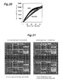
- the performance of the GTMS coated slide glass manufactured as described above was evaluated in comparison with existent slide glass (6 types). That is, previously Cy3-labeled lectin (100 ⁇ m/mL) was immobilized in an array to each slide glass applied with a surface coating and after by way of the steps (3) to (6), the S/N ratio was calculated based on the luminance value (S) for the spotting region and the luminance value (N) for the non-spotting surface. As a result, as shown in Table 1 while the luminance value for the GTMS coated slide glass manufactured by the step (2) remained at about 1/2 of the slide glass A showing the highest value, since the background was extremely low, the S/N was 16.1 showing the best value among the slide glass evaluated in this time.
- RCA 120 and ConA are typical lectins known to have high affinity to complex glycans and high mannose type glycans, respectively.
- the lectins were prepared in various concentrations, and spotted in an array each being arranged at four points laterally for an identical specimen. After applying 10 ⁇ g/mL of Cy3-ASF each by 50 ⁇ L to each of the reaction vessels in the array and causing binding reaction, fluorescence was observed by a scanner.
- ASF has a glycan structure having N-binding type glycans and O-binding type glycan each by three in the molecule and in which sialic acid at the non-reducing terminal in the glycan is decapped to protrude the lactosamine structure. Accordingly, in an experimental system of adding Cy3-ASF to the lectin array formed by immobilizing RCA 120 and ConA, it was expected that RCA 120 shows an extremely strong affinity, while ConA shows a weak affinity.
- lectins to be immobilized on a lectin array 6 types (RCA120, ECA, ConA, GNA, SSA, SNA) were selected as lectins having various glycan binding specificity. Further, BSA as a protein not binding with the glycan was selected as a negative control. Further, in this experiment, two types of anti- fetuin antibody and anti-RNase antibody recognizing the core protein portion of the probe were spotted in a compartment identical with that of the lectin. GNA and SNA purchased from VECTOR Co., BSA purchased from SIGMA Co., and RCA120, ECA, ConA, SSA purchased from Biochemical Industry were used.
- a model fluorescence-labeled glycoside protein probe was prepared by fluorescence-labeling ASF, FET, ribonuclease B (RNase B) derived from bovine pancreas, and ribonuclease A (RNase A) derived from bovine pancreas, and protein such as BSA as negative control (all purchased from SIGMA Co.) by using Cy3 Mono-reactive Dye as a fluorescence dye having an absorption maximum wavelength near 550 nm (manufactured by Amersham Pharmacia Biotech hereinafter referred to as Cy3).
- GTMS 3-glycidoxy propyl trimethoxysilane having epoxy groups as active groups
- GTMS coating was applied by the following procedures using a slide glass manufactured by Matsunami Glass Industry Co. The slide glass was dipped in a 10% KOH/MeOH solution and left for one hour in a state being shaken together with the vessel to treat the glass surface. After washing the same with a sufficient amount of pure water (milli Q water), it was dried in an oven at 60°C.
- the slide glass was dipped into a 2% GTMS acetone solution and reacted for 1 hour under shielding of light while being shaken together with the vessel. After the reaction and after drying in an oven at 110°C for 8 hours, it was washed with a sufficient amount of pure water and dried.
- Lectins were spotted to the GTMS coated slide glass manufactured in (2) to manufacture a lectin array.
- As a microarray spotter STAMPMAN manufactured by Nippon Laser & Electronics Lab Co., Ltd. was used and spots of about 0.5 mm diameter were arranged on the slide glass by using a stamp pin with a top end diameter of 0.40 mm.
- the following conditions were stored in a computer appended to the microarray spotter and a stamp pin operation program was executed.
- the needle tip of the stamp pin was dipped in a 0.05% SDS solution for 2 sec, the stamp pin was dried in a vacuum device for 15 sec, further, dipped in pure water for 2 sec, then dried in a vacuum device for 15 sec and, finally, dipped into ethanol for 2 sec and, then applied with a drying operation in the vacuum device for 15 sec.
- a 8-hole rubber made of silicone rubber was adhered on the slide glass spotted with lectins by the procedures described above to manufacture eight reaction vessels.
- a PBS solution containing 0.1% Tween 20 (PBST) By filling the reaction vessels with a PBS solution containing 0.1% Tween 20 (PBST), excess lectins not bonded to the slide solid layer were cleaned and removed. Then, after filling a PBS solution dissolving 1% BSA therein each by 200 ⁇ L to each of the reaction vessels, they were left for 1 hour in a preserving vessel kept at a humidity of 90% or higher at 4°C to conduct blocking procedure for the non-lectin spotted region.
- PBST PBS solution containing 0.1% Tween 20
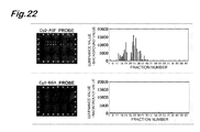
- Each of the fluorescence-labeled glycoside protein probe solution adjusted to about 100 mg/mL was added each by 50 ⁇ L to each of the reaction vessels to the slide after completing the blocking, thereby contacting the probe solution on the array.
- an excitation light was entered from the end face of the slide glass by using GTMAS Scan III as a evanescent excitation type microarray scanner (manufactured by Nippon Laser & Electronics Lab Co., Ltd) and the fluorescence emission generated by excitation was detected by an ICCD (charge coupled device with image intensifier) camera disposed to the lower surface of the slide glass.
- ICCD charge coupled device with image intensifier
- Array-Pro Analyzer as a commercial microarray analyzer software ver. 4.5 (manufactured by Media Cybernetics Co.) was used.
- the luminance for each spot was calculated by using the analyzer software described above, and the luminance at the non-spotted region was defined as a background value.
- the luminance for each of the spots subtracted with the background value was defined as a net luminance value and an average value and a standard deviation were calculated on every spot derived from identical specimen for 5 points arranged in a lateral row. Subsequently, for the binding of the probe to each of the lectin specimens, evaluation was conducted by using the average luminance value for five spots derived from the identical specimen.
- glycan profiles reflecting the glycan structures could be obtained.
- the obtained glycan profile was identical with ASF before enzymatic digestion showing that not only the glycoside protein but also peptide digestion products of the glycoside protein can be utilized for the lectin array.
- the glycan profile for each of the ingredients of the glycoside peptide can be observed and this is useful.
- the glycan addition state of the invivo glycoside protein is analyzed by conducting glycan profile for a glycoside protein mixture, particularly, biobody-derived mixture specimen by using the lectin array.
- the result of the experiment showed that the structural information of the glycan added to the glycoside peptide could be obtained at a high throughput and easily by arranging the glycoside peptide into an array.
- a method of manufacturing the glycoside peptide array it may be considered, for example, (1) immobilization of purified glycoside peptide, (2) immobilization of crude glycoside peptide, or (3) immobilization of a glycoside peptide fraction separated by HPLC.
- respective fractions separated by HPLC formed into an array could be manufactured and their usefulness could be shown.
- Glycan concerned arrays include a lectin array using the glycan for the probe, as well as a lectin array using glycoside protein for a probe, a glycoside peptide array using lectin for a probe, a glycoside protein array using lectin for a probe and an antibody array using lectin as a probe.
- Fig. 23 shows a schematic view for the interaction between various glycan concerned arrays and glycans.
Landscapes
- Health & Medical Sciences (AREA)
- Immunology (AREA)
- Life Sciences & Earth Sciences (AREA)
- Chemical & Material Sciences (AREA)
- Engineering & Computer Science (AREA)
- Molecular Biology (AREA)
- Urology & Nephrology (AREA)
- Biomedical Technology (AREA)
- Physics & Mathematics (AREA)
- Hematology (AREA)
- Analytical Chemistry (AREA)
- Biochemistry (AREA)
- Pathology (AREA)
- General Physics & Mathematics (AREA)
- General Health & Medical Sciences (AREA)
- Cell Biology (AREA)
- Microbiology (AREA)
- Biotechnology (AREA)
- Food Science & Technology (AREA)
- Medicinal Chemistry (AREA)
- Nuclear Medicine, Radiotherapy & Molecular Imaging (AREA)
- Chemical Kinetics & Catalysis (AREA)
- Optics & Photonics (AREA)
- Investigating, Analyzing Materials By Fluorescence Or Luminescence (AREA)
- Investigating Or Analysing Materials By The Use Of Chemical Reactions (AREA)
- Investigating Or Analysing Biological Materials (AREA)
Applications Claiming Priority (2)
| Application Number | Priority Date | Filing Date | Title |
|---|---|---|---|
| JP2005184171A JP4565237B2 (ja) | 2005-06-23 | 2005-06-23 | 糖鎖あるいは複合糖質の解析装置 |
| PCT/JP2006/311950 WO2006137311A1 (fr) | 2005-06-23 | 2006-06-14 | Analyseur pour une chaîne de sucres ou des glucides complexes |
Publications (2)
| Publication Number | Publication Date |
|---|---|
| EP1895301A1 true EP1895301A1 (fr) | 2008-03-05 |
| EP1895301A4 EP1895301A4 (fr) | 2010-10-06 |
Family
ID=37570341
Family Applications (1)
| Application Number | Title | Priority Date | Filing Date |
|---|---|---|---|
| EP06766719A Withdrawn EP1895301A4 (fr) | 2005-06-23 | 2006-06-14 | Analyseur pour une chaîne de sucres ou des glucides complexes |
Country Status (4)
| Country | Link |
|---|---|
| US (1) | US20090041630A1 (fr) |
| EP (1) | EP1895301A4 (fr) |
| JP (1) | JP4565237B2 (fr) |
| WO (1) | WO2006137311A1 (fr) |
Cited By (1)
| Publication number | Priority date | Publication date | Assignee | Title |
|---|---|---|---|---|
| CN105929162A (zh) * | 2016-05-13 | 2016-09-07 | 西北大学 | 一种基于唾液蛋白鉴别乳腺癌的凝集素芯片和试剂盒及其应用 |
Families Citing this family (10)
| Publication number | Priority date | Publication date | Assignee | Title |
|---|---|---|---|---|
| JP4515387B2 (ja) * | 2003-12-25 | 2010-07-28 | 独立行政法人産業技術総合研究所 | 糖鎖構造プロファイリング技術 |
| WO2005064333A1 (fr) * | 2003-12-25 | 2005-07-14 | National Institute Of Advanced Industrial Science And Technology | Procede d'analyse de l'interaction entre proteine et chaine de sucre |
| JP4982793B2 (ja) * | 2007-02-02 | 2012-07-25 | 独立行政法人産業技術総合研究所 | 生細胞表層分子解析方法及び部材 |
| EP2274613B1 (fr) * | 2008-04-02 | 2014-06-04 | Abbott Point Of Care, Inc. | Procédé d'agglutination sérologique effectués dans un échantillon de fluide en film mince |
| JP5539314B2 (ja) * | 2008-04-09 | 2014-07-02 | アボット ポイント オブ ケア インコーポレイテッド | 薄厚チャンバに含まれる薄膜状体液試料中の極めて低レベルの分析物を検出する方法 |
| EP2711689B1 (fr) * | 2011-05-19 | 2020-01-01 | Konica Minolta, Inc. | Dispositif de mesure de fluorescence améliorée par champ de plasmons de surface et procédé de détection de fluorescence l'utilisant |
| SG188698A1 (en) * | 2011-09-09 | 2013-04-30 | Albena Samokovlisky Ph D | Improved glycosylation assay, glycoanalysis array and an assay system |
| JP5726038B2 (ja) * | 2011-09-30 | 2015-05-27 | コニカミノルタ株式会社 | 表面プラズモン励起増強蛍光分光法を用いた前立腺特異抗原の定量方法 |
| JP6588055B2 (ja) * | 2017-06-09 | 2019-10-09 | 株式会社シバサキ | 細菌検出装置 |
| JP7298855B2 (ja) * | 2019-06-13 | 2023-06-27 | 株式会社島津製作所 | アフィニティ支持体及びそれを用いた物質の捕捉方法 |
Family Cites Families (6)
| Publication number | Priority date | Publication date | Assignee | Title |
|---|---|---|---|---|
| JPH08201382A (ja) | 1995-01-24 | 1996-08-09 | Nippon Kayaku Co Ltd | 体液中成分の分析法及び体液採取用シート |
| KR19990014709A (ko) * | 1995-05-12 | 1999-02-25 | 한스루돌프하우스 | 센서 플랫폼 및 감쇠 여기된 발광을 이용한 복수 분석물의병렬 검출 방법 |
| US5988587A (en) | 1997-10-10 | 1999-11-23 | Invasatec, Inc. | Control device for providing a variable control signal to a fluid-supplying machine |
| IL129835A (en) | 1999-05-06 | 2008-03-20 | Ofer Markman | Polysaccharide sequencing and structure determination |
| JP3983569B2 (ja) * | 2002-02-28 | 2007-09-26 | 独立行政法人科学技術振興機構 | 多変量解析システム、発現プロファイル解析方法、コンピュータプログラム、コンピュータ読み取り可能な記憶媒体 |
| WO2005064333A1 (fr) * | 2003-12-25 | 2005-07-14 | National Institute Of Advanced Industrial Science And Technology | Procede d'analyse de l'interaction entre proteine et chaine de sucre |
-
2005
- 2005-06-23 JP JP2005184171A patent/JP4565237B2/ja not_active Expired - Lifetime
-
2006
- 2006-06-14 WO PCT/JP2006/311950 patent/WO2006137311A1/fr not_active Ceased
- 2006-06-14 EP EP06766719A patent/EP1895301A4/fr not_active Withdrawn
- 2006-06-14 US US11/917,921 patent/US20090041630A1/en not_active Abandoned
Cited By (1)
| Publication number | Priority date | Publication date | Assignee | Title |
|---|---|---|---|---|
| CN105929162A (zh) * | 2016-05-13 | 2016-09-07 | 西北大学 | 一种基于唾液蛋白鉴别乳腺癌的凝集素芯片和试剂盒及其应用 |
Also Published As
| Publication number | Publication date |
|---|---|
| JP2007003357A (ja) | 2007-01-11 |
| EP1895301A4 (fr) | 2010-10-06 |
| WO2006137311A1 (fr) | 2006-12-28 |
| JP4565237B2 (ja) | 2010-10-20 |
| US20090041630A1 (en) | 2009-02-12 |
Similar Documents
| Publication | Publication Date | Title |
|---|---|---|
| US8008094B2 (en) | Methods for analyzing interactions between proteins and sugar chains | |
| JP4824119B2 (ja) | 糖鎖構造プロファイリング技術 | |
| US10697959B2 (en) | Sandwich assay using labeled lectin and kit therefor | |
| JP4982793B2 (ja) | 生細胞表層分子解析方法及び部材 | |
| EP1895301A1 (fr) | Analyseur pour une chaîne de sucres ou des glucides complexes | |
| EP2738556B1 (fr) | Procédé de mesure de quantité d'une substance à analyser et appareil de spfs | |
| CN106501225B (zh) | 凝集素组识别的糖链在区分胰腺黏液性囊性肿瘤和胰腺浆液性囊性肿瘤中的应用 | |
| US20120277116A1 (en) | Disease diagnosis according to saccharide binding | |
| HK1045727B (en) | Polysaccharide structure and sequence determination | |
| WO2001040796A2 (fr) | Matrices de molecules glycanes (glycomatrices) sur la surface de biopuces (glycopuces) et leurs utilisations | |
| AU2004299992B2 (en) | Method for analyzing a glycomolecule | |
| US8597576B2 (en) | Analyzer for glycan or complex carbohydrate | |
| US20060172339A1 (en) | Particle-based multiplex assay for identifying glycosylation | |
| JP2004506874A (ja) | 炭水化物ポリマーを分析するための方法および組成物 |
Legal Events
| Date | Code | Title | Description |
|---|---|---|---|
| PUAI | Public reference made under article 153(3) epc to a published international application that has entered the european phase |
Free format text: ORIGINAL CODE: 0009012 |
|
| 17P | Request for examination filed |
Effective date: 20080110 |
|
| AK | Designated contracting states |
Kind code of ref document: A1 Designated state(s): CH DE FR GB LI SE |
|
| DAX | Request for extension of the european patent (deleted) | ||
| DAX | Request for extension of the european patent (deleted) | ||
| RBV | Designated contracting states (corrected) |
Designated state(s): CH DE FR GB LI SE |
|
| RIC1 | Information provided on ipc code assigned before grant |
Ipc: G01N 33/543 20060101ALI20100728BHEP Ipc: G01N 33/548 20060101ALI20100728BHEP Ipc: G01N 21/64 20060101ALI20100728BHEP Ipc: G01N 33/53 20060101AFI20070217BHEP Ipc: G01N 33/547 20060101ALI20100728BHEP Ipc: G01N 37/00 20060101ALI20100728BHEP |
|
| A4 | Supplementary search report drawn up and despatched |
Effective date: 20100902 |
|
| STAA | Information on the status of an ep patent application or granted ep patent |
Free format text: STATUS: THE APPLICATION IS DEEMED TO BE WITHDRAWN |
|
| 18D | Application deemed to be withdrawn |
Effective date: 20110302 |