EP2000802A1 - Nouveau marqueur de l'activation plaquettaire et son procédé de détermination - Google Patents
Nouveau marqueur de l'activation plaquettaire et son procédé de détermination Download PDFInfo
- Publication number
- EP2000802A1 EP2000802A1 EP07739968A EP07739968A EP2000802A1 EP 2000802 A1 EP2000802 A1 EP 2000802A1 EP 07739968 A EP07739968 A EP 07739968A EP 07739968 A EP07739968 A EP 07739968A EP 2000802 A1 EP2000802 A1 EP 2000802A1
- Authority
- EP
- European Patent Office
- Prior art keywords
- gpvi
- antibody
- specifically binds
- determination
- sgpvi
- Prior art date
- Legal status (The legal status is an assumption and is not a legal conclusion. Google has not performed a legal analysis and makes no representation as to the accuracy of the status listed.)
- Withdrawn
Links
Images









Classifications
-
- C—CHEMISTRY; METALLURGY
- C07—ORGANIC CHEMISTRY
- C07K—PEPTIDES
- C07K16/00—Immunoglobulins [IG], e.g. monoclonal or polyclonal antibodies
- C07K16/18—Immunoglobulins [IG], e.g. monoclonal or polyclonal antibodies against material from animals or humans
- C07K16/28—Immunoglobulins [IG], e.g. monoclonal or polyclonal antibodies against material from animals or humans against receptors, cell surface antigens or cell surface determinants
-
- G—PHYSICS
- G01—MEASURING; TESTING
- G01N—INVESTIGATING OR ANALYSING MATERIALS BY DETERMINING THEIR CHEMICAL OR PHYSICAL PROPERTIES
- G01N33/00—Investigating or analysing materials by specific methods not covered by groups G01N1/00 - G01N31/00
- G01N33/48—Biological material, e.g. blood, urine; Haemocytometers
- G01N33/50—Chemical analysis of biological material, e.g. blood, urine; Testing involving biospecific ligand binding methods; Immunological testing
- G01N33/86—Chemical analysis of biological material, e.g. blood, urine; Testing involving biospecific ligand binding methods; Immunological testing involving blood coagulating time or factors, or their receptors
-
- C—CHEMISTRY; METALLURGY
- C07—ORGANIC CHEMISTRY
- C07K—PEPTIDES
- C07K2317/00—Immunoglobulins specific features
- C07K2317/30—Immunoglobulins specific features characterized by aspects of specificity or valency
- C07K2317/34—Identification of a linear epitope shorter than 20 amino acid residues or of a conformational epitope defined by amino acid residues
-
- G—PHYSICS
- G01—MEASURING; TESTING
- G01N—INVESTIGATING OR ANALYSING MATERIALS BY DETERMINING THEIR CHEMICAL OR PHYSICAL PROPERTIES
- G01N2333/00—Assays involving biological materials from specific organisms or of a specific nature
- G01N2333/435—Assays involving biological materials from specific organisms or of a specific nature from animals; from humans
- G01N2333/705—Assays involving receptors, cell surface antigens or cell surface determinants
- G01N2333/70503—Immunoglobulin superfamily, e.g. VCAMs, PECAM, LFA-3
-
- G—PHYSICS
- G01—MEASURING; TESTING
- G01N—INVESTIGATING OR ANALYSING MATERIALS BY DETERMINING THEIR CHEMICAL OR PHYSICAL PROPERTIES
- G01N2800/00—Detection or diagnosis of diseases
- G01N2800/32—Cardiovascular disorders
Definitions
- the present invention relates to a novel platelet activation marker, a reagent and method for determining this novel platelet activation marker, and novel applications of the marker to the diagnosis of diseases associated with platelet activation/vascular endothelial injury.
- the leading cause of death in Japan is malignant neoplasm, while the second leading cause is heart disease and the third leading cause is cerebrovascular disease.
- the majority of heart disease and cerebrovascular disease is accounted for by thrombosis, such as myocardial infarction and cerebral infarction, and together the two of these rival the number of deaths from cancer.
- the prevention and treatment of thrombosis continues to be an important issue. Controlling platelet activation is crucial for the prevention and treatment of thrombosis, and antiplatelet drugs are aggressively prescribed. However, the clinical efficacy have by no means come up to satisfactory levels.
- Platelet function can be divided into two reactions: adhesion to subendothelial tissue at a site of vascular injury, and aggregation, in which the platelets adhere to one another. In particular, the latter is accompanied by the release of the contents of granules that are present in platelets.
- various molecules participate in the expression of function in the platelet activation process, and these are utilized as platelet activation markers. The effective utilization of these markers not only enables greater clarity in the diagnosis of abnormal thrombosis, but also enables a more appropriate selection of the treatment protocol.
- ⁇ -thromboglobulin and platelet factor 4 which are stored in the ⁇ -granules of platelets, has been carried out in the past as an indicator of platelet activation.
- these require consideration from the influence of the blood sampling technique and the fact that they assume high values when renal function declines, and thus they are not always categorized as an appropriate markers.
- ⁇ -thromboglobulin and platelet factor 4 it must be treated with caution due to the high possibility of change induced by in vitro platelet activation. Therefore, there has been desire for the appearance of a more sensitive and specific platelet activation marker.
- P-selectin is a membrane protein existing in the ⁇ -granule membrane of platelets and in the Weibel-Palade bodies of vascular endothelial cells; it migrates to the cell surface when these cells are suffered from a stimulus.
- P-selectin is currently categorized into two types, a membrane bound type and a soluble type.
- Membrane-bound P-selectin is directly detected using a fluorescence-labeled anti-P-selectin antibody and flow cytometry.
- sample handling can cause a pseudo-positive result for the expression of P-selectin in platelets, making it difficult to obtain consistent results.
- Soluble P-selectin on the other hand, can be detected by sandwich ELISA.
- P-selectin originates in platelets and vascular endothelial cells, the problem of its origin arises when the soluble P-selectin in plasma is measured.
- Microscopic membrane vesicles are formed when platelets undergo activation under a variety of circumstances or are suffered from a physical stimulus; these membrane vesicles are called microparticles. Platelet microparticles are generally measured by flow cytometry, and this is important for comprehending the pathological condition of various thrombotic diseases.
- flow cytometry measures flow cytometry, and this is important for comprehending the pathological condition of various thrombotic diseases.
- problems have been noted here, such as (1) differences in sensitivity between instruments, (2) how the microparticle region is gated, (3) the fact that detection limit is near the size of the microparticles, and (4) differences between facilities based on the antibody used, and standardization of the measurement method has not been achieved.
- the glycoprotein VI (GPVI) present on the platelet membrane is a collagen receptor of platelets, and it has been shown to have a central role in collagen-induced platelet activation (refer to Takayama H. Japanese Journal of Thrombosis and Hemostasis, Volume 14 (2003), Number 2, pp. 75-81 ).
- anti-mouse GPVI antibody has been reported to exhibit an antithrombotic activity by specifically inhibiting collagen-induced platelet aggregation without remarkable prolongation of bleeding time (refer to Nieswandt B. et al., J. Exp. Med., Volume 194 (2001), Number 4, pp. 459-469 ).
- anti-human GPVI antibody exhibits an antithrombotic activity without causing a remarkable prolongation of bleeding time ( Takayama H. et al., Blood, Volume 106 (2005), Number 11, p. 612a ; WO 2005/111083 A2 ). Accordingly, there is an expectation that a drug that specifically inhibits collagen-mediated platelet aggregation, for example, an anti-GPVI antibody, could be a safe and highly effective antiplatelet drug.
- An object of the present invention is to provide a novel platelet activation marker and a reagent and method for its determination, and is to provide novel applications of the marker in, for example, the diagnosis of diseases associated with platelet activation/vascular endothelial injury.
- the present inventors conceived of the idea that a convenient, high-sensitivity measurement of sGPVI present in plasma could be accessed by establishing a plurality of mouse hybridomas that produce antibody against GPVI and using a combination of the antibodies produced therefrom.
- the present inventors succeeded in establishing a specific and highly sensitive method of determining GPVI present on the platelet membrane (in some cases referred to as mGPVI in the following) and soluble GPVI present in body fluids (in some cases referred to as sGPVI in the following) and discovered that this can be a platelet activation marker, and accomplished the present invention.
- the present invention is described in the following.
- the first embodiment of the present invention is
- GPVI in a sample and particularly sGPVI or mGPVI in a biological sample can be determined at a high sensitivity by a determination method, a determination reagent, or a kit according to the present invention.
- the detection, determination, or quantitation of sGPVI or mGPVI using a determination method, a determination reagent, or a kit according to the present invention makes it possible, inter alia, to detect, determine, quantitate, assess, or evaluate platelet activation or vascular endothelial injury; to diagnose a disease associated with platelet activation or vascular endothelial injury, or assess the susceptibility to such a disease or evaluate the risk of developing such a disease; to select patients who respond to antiplatelet drugs and particularly anti-GPVI antibody; to decide the time for the (beginning of) administration of such drugs; and to predict, monitor, or prognostically assess the therapeutic efficacy of such drugs or their occurrence of side effects.
- the animal species that is the source of GPVI for determination is not particularly limited and is a mammal, for example, mouse, rat, or monkey, wherein human GPVI is preferred.
- the nucleotide sequences and amino acid sequences of these GPVIs are known, and the amino acid sequences of mouse, rat, and human GPVI are shown in Figures 1 to 3 .
- mGPVI is present as a transmembrane protein on the platelet surface; is generally a full-length GPVI molecule; and comprises an amino acid sequence as cited above.
- sGPVI has been released from the platelet due to some cause and is believed to be rendered soluble by enzymatic cleavage, particularly by an enzyme, preferably a protease and more preferably a metalloprotease derived from platelets.
- an enzyme preferably a protease and more preferably a metalloprotease derived from platelets.
- its molecular weight is, for example, about 55 kd by SDS-PAGE under a reducing condition, and it can be detected by, for example, western blotting using an anti-GPVI antibody.
- the samples (in some cases referred to below as the specimen) in the present invention, particularly the biological samples are not necessarily limited and include body fluids, cells, and tissues.
- the body fluid can be exemplified by blood, plasma, platelet-rich plasma (PRP), platelet-poor plasma (PPP), urine, and so forth, while the cells can be exemplified by platelets.
- a platelet-containing biological sample is preferred, particularly blood, platelet-rich plasma (PRP), and platelets.
- a body fluid is preferred, particularly blood, plasma, platelet-poor plasma (PPP), urine, and so forth.
- the source animal species for the biological sample is also not necessarily limited and comprises mammals, for example, human, mouse, rat, rabbit, and so forth, wherein human is preferred.
- the method of determining GPVI in a sample in accordance with the first embodiment of the present invention characteristically comprises determining GPVI in a sample, particularly sGPVI or mGPVI in a biological sample, using at least one substance that specifically binds GPVI.
- the reagent or kit for determining GPVI in a sample in accordance with the second embodiment of the present invention characteristically comprises at least one substance that specifically binds GPVI.
- the substance that specifically binds GPVI in the present invention encompasses collagen, CRP, convulxin, and so forth, and is preferably an anti-GPVI antibody.
- the anti-GPVI antibody is an antibody that specifically recognizes or binds GPVI, particularly sGPVI or mGPVI, but is not otherwise necessarily limited.
- the binding affinity (Kd) with GPVI is no greater than 10 -7 M, preferably no greater than 3 ⁇ 10 -8 M, more preferably no greater than 1 ⁇ 10 -8 M, even more preferably no greater than 3 ⁇ 10 -9 M, and particularly preferably no greater than 1 ⁇ 10 -9 M.
- the animal source species for the antibody is not necessarily limited and can be, for example, a mammal and particularly the mouse, rat, hamster, rabbit, and so forth; moreover, the antibody can also be produced by gene recombination techniques.
- Monoclonal antibody or polyclonal antibody can be used, wherein monoclonal antibody is preferred from the standpoints of physical uniformity and properties such as the specificity.
- the form of the antibody in the present invention is not necessarily limited, and various form can be employed.
- the antibody in the present invention may - as long as its activity (e.g., capacity to bind GPVI) is present - be a fragment, portion, or derivative of an antibody.
- the fragment can be, for example, Fab (fragment antigen binding), Fab', (Fab') 2 , and so forth, while the derivative can be, for example, a single-chain antibody (scFv), disulfide-stabilized antibody (dsFv), diabody, sc(Fv) 2 , (refer, for example, to Orita T., Blood, 2005; 105:562-566 ), nanobody (refer, for example, to Cortez-Retamozo V., Cancer Research 64, 2853-2857, 2004 ), CDR-comprising peptide, and so forth. These can be produced by known methods.
- mouse anti-human GPVI monoclonal antibody (refer, inter alia, to WO 01/810 , WO 02/80968 , and WO 2005/111083 )
- rat anti-mouse GPVI monoclonal antibody (Nieswandt et al.)
- An antibody having an identified binding region, binding site, or epitope is preferred, for example, an antibody that binds with a specific domain, for example, domain 1 or domain 2, or an antibody that recognizes a particular loop, for example, at least a portion of loop 2 or loop 9.
- a specific domain for example, domain 1 or domain 2
- an antibody that recognizes a particular loop for example, at least a portion of loop 2 or loop 9.
- Specific examples are the antibodies provided in the examples herein and the antibodies described in the examples of WO 2006/117910 and WO 2006/118350 .
- a preferred method uses, as its immunizing antigen or antigen for antibody identification, mutants in which a specific site on GPVI, for example, a specific domain, preferably a specific loop region, has been substituted by another amino acid sequence, specifically by the corresponding amino acid sequence from GPVI of another animal species.
- a specific site on GPVI for example, a specific domain, preferably a specific loop region
- the methods described in this application or the methods described in the examples of WO 2006/117910 or WO 2006/118350 can be used.
- the recognition sites and so forth of these antibodies can be estimated by measurement, in accordance with known methods, of the binding capacity between the said substitution mutants and the antibody and the binding capacity between GPVI and the antibody.
- a highly specific, highly selective, and/or highly sensitive detection or determination of GPVI is made possible by the use of one or two or more of these antibodies in which, for example, the binding site has been identified, preferably by the use of two species that have different, for example, binding sites, preferably by the use of a combination of antibodies that do not compete with one another in their binding with GPVI, for example: the combination of an antibody that binds to domain 1 with an antibody that binds to domain 2, or the combination of an antibody that binds to at least a portion of loop 2 with an antibody that binds to at least a portion of loop 9.
- a specific GPVI molecular species therein for example, sGPVI, can thereby be determined specifically, selectively, and/or sensitively. Suitable examples are shown in the examples hereinbelow.
- the determination sensitivity in the present invention is not necessarily limited, and, expressed as the concentration in the sample, is 1 ng/mL or less, preferably 300 pg/mL or less, more preferably 100 pg/mL or less, even more preferably 30 pg/mL or less, particularly preferably 10 pg/mL or less, very preferably 3.0 pg/mL or less, and most preferably 1.0 pg/mL or less.
- the detection limit concentration is 1 ng/mL or less, preferably 300 pg/mL or less, more preferably 100 pg/mL or less, even more preferably 30 pg/mL or less, particularly preferably 10 pg/mL or less, very preferably 3.0 pg/mL or less, and most preferably 1.0 pg/mL or less.
- this is 1 ng/mL or less, preferably 300 pg/mL or less, more preferably 100 pg/mL or less, still more preferably 30 pg/mL or less, particularly preferably 10 pg/mL or less, very preferably 3.0 pg/mL or less, and most preferably 1.0 pg/mL or less.
- the concentration in the sample may be expressed in terms of the quantity of the standard material, for example, GPVI-Fc, used to construct the standard curve.
- the concentration in the sample is the value post-dilution.
- GPVI released from the platelet When soluble GPVI in a sample and particularly in plasma is to be determined, GPVI released from the platelet must be specifically determined free of influence from the plasma components. A sandwich immunoassay procedure using a highly specific enzyme is therefore generally employed. In order to accurately determine GPVI in the determination method of the present invention, it is important here to reduce the influence of the plasma components as much as possible, and dilution of the specimen is primarily selected as the means for doing this. While a component such as, for example, surfactant, can also be added to the reaction fluid for the purpose of reducing this influence, such components have the potential for influencing the determination system.
- a component such as, for example, surfactant
- the determination is preferably carried out with specimen dilution, and a high sensitivity is then required as a matter of necessity.
- the determination must be run with the specimen diluted in general at least 5-fold, preferably at least 10-fold, more preferably at least 50-fold, and even more preferably at least 100-fold.
- the sensitivity of the determination system desirably is 30 pg/mL or less, preferably 10 pg/mL or less, and more preferably several pg/mL or less.
- an absorbance difference of about 40 mAbs is obtained at 3 pg/mL standard substance, and it is thus possible to obtain a satisfactory sensitivity using the present invention.
- the anti-GPVI antibody in the determination method, determination reagent, or kit according to the present invention may be a labeled antibody or may be an unlabeled antibody, but the use of at least one labeled antibody is preferred.
- the labeling substance and the labeling method employed for this labeling can be a known substance and a known method, and any of radioactive substances, enzymes, fluorescent substances, chemiluminescent substances, and so forth, can be used and applicable.
- Methods that use enzyme-labeled antibody are preferred among the preceding because they do not require special facilities or expensive detection instrumentation and because they also provide convenience in handling. Direct and indirect methods are available for labeling, and either of these can be used.
- the determination principle and so forth in the determination method of the present invention is not necessarily limited, and is generally an immunoassay method and can be exemplified by known methods, for example, enzyme-antibody methods, ELISA methods, sandwich immunoassay methods, agglutination methods, solid-phase direct methods, solid-phase binding methods, solution reaction methods, competitive assays, non-competitive assays, immunochromatographic methods, and flow-through methods; a single one of these methods can be used or combinations can be used (refer, for example, to Ultrasensitive Enzyme Immunoassay Methods, Eiji ISHIKAWA, Japan Scientific Societies Press (1993 ); New Uses of Immunoassay Methods and Their Applications to the Development of Diagnostic Reagents and Therapeutic Drugs, Association for Research and Development in Immunoassay Methods, Keiei Kyoiku Publishing ; and Enzymatic Immunoassay Methods (Third Edition), edited by Eiji ISHIKAWA et al., Igaku Shoin
- sandwich immunoassay methods particularly sandwich ELISA methods that use an insoluble carrier, e.g., a microwell plate and so forth - are convenient and are also preferred from the standpoint of sensitivity.
- the known technology can be employed for the sandwich immunoassay method.
- the principles and applications of this determination method and improvements thereto are described in, for example, Ultrasensitive Enzyme Immunoassay Methods, Eiji ISHIKAWA, Japan Scientific Societies Press (1993 ); New Uses of Immunoassay Methods and Their Applications to the Development of Diagnostic Reagents and Therapeutic Drugs, Association for Research and Development in Immunoassay Methods, Keiei Kyoiku Publishing ; and Enzymatic Immunoassay Methods (Third Edition), edited by Eiji ISHIKAWA et al., Igaku Shoin (1987 ).
- the sandwich immunoassay method generally is a method in which determination is carried out by the formation of an antibody-antigen-antibody complex using at least two antibody species that differ in their recognition sites for the protein being determined.
- Sandwich immunoassay methods generally employ an insoluble carrier, and in such a case the first antibody, which is bonded to the insoluble carrier, is referred to as the immobilized antibody or as the antibody for immobilization or as the capture antibody, while the second antibody is referred to as the non-immobilized antibody, the liquid-phase antibody, the detection antibody, or the labeled antibody when directly or indirectly labeled.
- An insoluble carrier having the first antibody bonded thereto is first prepared to give a solid-phase or reaction site. The specimen is added to this solid-phase insoluble carrier to carry out a reaction.
- This sandwich method can be carried out by a procedure in which two steps are implemented as described above (a two-step method) or by a single step procedure in which the antigen and labeled antibody are added at the same time (one-step method).
- the immobilized antibody is, for example, preferably an antibody that specifically binds to domain 2, more preferably to loop 9 or loop 11, particularly to loop 9.
- the non-immobilized antibody is, for example, preferably an antibody that specifically binds to domain 1, more preferably to loop 2 or loop 5, particularly to loop 2.
- preferred is the combination of the antibody that specifically binds to loop 9 for the immobilized antibody and the antibody that specifically binds to loop 2 for the non-immobilized antibody.
- the sandwich immunoassay method can also be carried out in solution without using an insoluble carrier.
- An example here is a procedure in which antigen is reacted in the liquid phase with a labeled antibody and a second binding substance tagged with a second label and the interaction between the label and the second label is determined.
- determination can also be carried out in the sandwich immunoassay method and so forth by a competitive method. This is a method in which determination is carried out based on the competition, during the formation of the antigen-antibody complex, between the antigen in the sample and a labeled antigen or a labeled antigen-like substance.
- determination can be carried out in the sandwich immunoassay method using a second specific binding.
- the second specific binding substance-second specific binding partner combination can be, inter alia, an antigen and its antibody, a ligand and its receptor, a sugar chain-containing substance and a lectin, and biotin and avidin or streptavidin.
- a system is exemplified, in which using biotin and avidin or streptavidin as a second specific binding substance-second specific binding partner, one antibody, in particular the non-immobilized antibody is biotinylated and detected with a label, particularly with poly-HRP-labeled streptavidin.
- the insoluble carrier used in the sandwich immunoassay system of the present invention may be a bead, latex particle, magnetic particle, plate, tube, membrane, and so forth.
- the material of the bead, plate, or tube can be, for example, polystyrene, nylon, glass, silicone rubber, stainless steel, plastic, and so forth.
- the membrane can be, for example, cellulose, a cellulose derivative, nitrocellulose, a porous synthetic polymer, glass fiber, a fabric, a nonwoven fabric, filter paper, and so forth.
- shape a spherical shape can be used for the bead, latex particle, magnetic particle, and so forth. This is advantageous in terms of preserving space during storage.
- a well shape can be used for the plate and tube. This is advantageous in terms of being applicable to commercial automatic assay instrumentation, plate readers, and so forth.
- a membrane can also be used in immunochromatographic and flow-through methods.
- Bonding to the insoluble carrier of the antibody, second binding substance, second specific binding substance or its partner, or anti-immunoglobulin antibody can be effected by, for example, thermal adsorption, chemical bonding, and so forth.
- a blocking treatment with a substance that does not influence the determination system is preferably carried out on its non-adsorbing surfaces of the insoluble carrier, where the aforementioned substances are not bound.
- proteins such as bovine serum albumin (BSA) and casein and surfactants such as Tween 20 and NP-40.
- the label used in the sandwich immunoassay system kit of the present invention can be exemplified by enzymes such as peroxidase and particularly horseradish peroxidase (HRP), alkaline phosphatase, ⁇ -D-galactosidase, oxidase, uricase, and so forth; chemiluminescent substances such as acridinium and derivatives thereof, aequorin and variants thereof, and so forth; fluorescent substances e.g. FITC, lanthanoid, such as europium (Eu), samarium (Sm), and so forth; as well as dyes, colloidal gold, colored latexes, and isotopes.
- enzymes such as peroxidase and particularly horseradish peroxidase (HRP), alkaline phosphatase, ⁇ -D-galactosidase, oxidase, uricase, and so forth; chemiluminescent substances such as acridinium
- 3,3',5,5'-tetrabenzidine and 1,2-phenylenediamine are examples of the chromogenic substrate when a peroxidase is used as the enzyme; 4-nitrophenyl phosphate is an example of the chromogenic substrate when an alkaline phosphatase is used as the enzyme; and 2-nitrophenyl ⁇ -D-galactoside is an example of the chromogenic substrate when ⁇ -D-galactosidase is used as the enzyme.
- Labeling with the enzyme of the antibody, second binding substance, second specific binding substance or partner therefore, or anti-immunoglobulin antibody can be carried out by the two-step glutaraldehyde method, periodic acid method, maleimide method, pyridyl ⁇ disulfide method, and so forth.
- this can be done using the known art, e.g., thermal adsorption, chemical bonding, and so forth.
- the use of an enzyme label with a chromogenic substrate as exemplified above makes it possible to carry out the determination using the usual absorbance measurement systems and also provides a relatively high sensitivity and for these reasons is preferred.
- the determination can be carried out by measurement instrumentation conforming to the particular label when a chemiluminescent substance, fluorescent substance, colored label, or isotope is used as the label.
- a fluorescent substance such as Eu, for example, a cryptate (Eu 3+ cryptate)
- the fluorescent resonance energy transfer can be determined using an allophycocyanine derivative, for example, XL665, as a second label.
- Dyes, colloidal gold, and colored lattices can also be visually evaluated and for this reason are preferred for the label used in easy-to-use determination kits, for example, kits that employ an immunochromatographic method or a flow-through method.
- the particles used as the insoluble carrier in an agglutination method can be those particles that are ordinarily used, for example, a latex, erythrocytes (for example, sheep erythrocytes), gelatin, microbeads, carbon particles, and so forth.
- the determination reagent or kit of the present invention may contain optional constituent components or constituent elements in addition to the at least one substance that specifically binds GPVI.
- the optional constituent components in the determination reagent or kit can be exemplified by additives such as stabilizers, vehicles, preservatives, and so forth, while the optional constituent elements in the kit can be exemplified by diluents or buffer solutions for the standard substance, specimen, labeled antibody, and so forth, chromogenic substrate adapted to the enzyme when an enzyme is used in the labeled antibody, blocking agent, reaction stopper, rinsing agent, and so forth. While the diluent is not particularly limited, a diluent containing a substance present in the specimen is preferred.
- the same amount of EDTA or citric acid is preferably also present in the diluent.
- 0.2 to 1 mg/mL EDTA is preferably present in the diluent.
- the standard substance is, for example, GPVI, particularly sGPVI or mGPVI, prepared from a biological sample or recombinant GPVI, particularly sGPVI, mGPVI, the GPVI extracellular domain, or a fusion protein (GPVI-Fc) consisting of the GPVI extracellular domain and an immunoglobulin Fc fragment, prepared by genetic engineering. These can be prepared by known methods.
- the determination reagent or kit of the present invention is particularly useful, for example, in the diagnostic methods described herebelow.
- sGPVI and mGPVI exhibit variations - particularly a quantitative variation - accompanying the effects, inter alia, platelet activation induced by various in vivo and ex vivo platelet stimuli, they can be utilized as biomarkers, that is, platelet activation markers that estimate the status of platelet activation in vivo, particularly GPVI-mediated activation.
- biomarkers that is, platelet activation markers that estimate the status of platelet activation in vivo, particularly GPVI-mediated activation.
- sGPVI in a biological sample has a high value, it can be inferred that platelet activation has been induced in vivo due to some cause, particularly due to a GPVI-mediated mechanism.
- the platelet mGPVI has a high value
- the cause, mechanism, or stimulus cited here can be exemplified by the exposure of vascular subendothelial collagen due to vascular endothelial injury.
- the present invention provides a platelet activation marker comprising soluble GPVI (sGPVI) or GPVI present on the platelet membrane (mGPVI) and further provides the use of sGPVI or mGPVI as a platelet activation marker.
- diseases associated with platelet activation or vascular endothelial injury can, inter alia, be diagnosed based on the detection, determination, or quantitation of sGPVI or mGPVI. That is, the present invention provides a method of detecting, determining, quantitating, assessing, or evaluating platelet activation or vascular endothelial injury, comprising detecting, determining, or quantitating sGPVI or mGPVI in a sample (the fifth embodiment of the present invention); a method of diagnosing, a method of assessing the susceptibility to, or a method of evaluating the risk of developing, a disease associated with platelet activation or vascular endothelial injury, comprising detecting, determining, or quantitating sGPVI or mGPVI in a sample (the sixth embodiment of the present invention); a method of selecting for patients who respond to an antiplatelet drugs, preferably antiplatelet drugs that inhibit collagen-induced platelet activation or aggregation, more preferably an anti-GPVI antibody,
- the aforementioned methods of the fifth to ninth embodiments of the present invention comprise at least the detection, determination, or quantitation of sGPVI or mGPVI in a sample, particularly in a biological sample, and this comprises at least the step of bringing the sample into contact with at least one substance that specifically binds GPVI.
- the detection, determination, or quantitation of sGPVI or mGPVI in a sample can be carried out in accordance with the description of the first and second embodiments of the present invention, supra, using a determination method, determination reagent, or kit of the present invention.
- a step can be included comprising comparison with the value determined for healthy individuals, or comparison of the value determined pre-onset with the value determined post-onset, or comparison of the value determined prior to treatment - and particularly treatment with a drug such as an antiplatelet drug - with the value determined after treatment.
- the results of the determination in a plurality of normal individuals are obtained in advance; a standardized normal value or normal range, obtained by taking the average of these results or a range of these results, is made the standard normal value; and this is compared with the determined value.
- the standard normal value may be acquired by designating the average value for normal individuals + 2 SD or 3 SD as the cutoff value.
- a step may be implemented in which a reference value is established in advance for the patients of a disease, for example, a thrombotic disease, and this is compared with the determined value. This step may be carried out in place of the step of comparison with a standard normal value.
- the aforementioned methods of the fifth to ninth embodiments of the present invention can be effectively executed by carrying out these comparisons of the determination and/or the determined value.
- the above-cited platelet activation is not necessarily limited in the present invention, but is preferably platelet activation due to a platelet agonist, more preferably is platelet activation due to a GPVI-specific agonist.
- Said platelet agonist can be exemplified by collagen, particularly the vascular subendothelial collagen associated with vascular endothelial injury, and otherwise by an anti-GPVI antibody that exhibits a platelet activation function, CRP, convulxin, and so forth.
- the diseases associated with platelet activation or vascular endothelial injury encompass abnormalities of the blood coagulation system and diseases that are caused by or associated with platelet activation or aggregation (particularly collagen-induced platelet activation or aggregation), vascular endothelial injury, and arteriosclerotic reactions, for example, diseases caused by or associated with up-regulation of the blood coagulation system.
- diseases caused by or associated with thrombus or embolus or arteriosclerosis i.e., thrombotic or embolic diseases or arteriosclerotic diseases, for example, thrombosis, embolism, or arteriosclerosis.
- These diseases encompass not only arterial thrombosis, but also venous thrombosis and cerebral infarction caused by atrial fibrillation.
- Specific examples are angina pectoris; myocardial infarction; and vascular endothelial hypertrophy, vessel restenosis, angina pectoris, and myocardial infarction during and after thrombolytic therapy, percutaneous transluminal coronary angioplasty, stent operation, bypass surgery, and artificial blood vessel operation.
- Additional examples are atrial fibrillation and atrial flutter and the thrombosis, embolism, particularly cerebral infarction caused thereby.
- thromboangiitis obliterans thromboangiitis obliterans, acute arterial occlusion, arteriosclerosis obliterans, deep venous thrombosis, and so forth, as well as cerebral infarction (atheromatous thrombotic infarction, lacunar infarction, cardiogenic infarction), transient cerebral ischemic attack, cerebrovascular spasm after subarachnoid bleeding, pulmonary thrombosis, pulmonary embolism, vascular purpura, idiopathic thrombocytopenic purpura, thrombotic thrombocytopenic purpura, disseminated intravascular coagulation syndrome, inhibition of blood coagulation during extracorporeal circulation (dialysis), systemic lupus erythematosus, multiple arteritis, anti-phospholipid antibody syndrome, purpura nephritis, endothelial cell injury associated with diabetes mellitus, diabetic nephritis, diabet
- Thrombotic diseases, embolic diseases, and arteriosclerotic diseases are preferred, while angina pectoris, myocardial infarction, heart disease, cerebral infarction, and dementia are more preferred.
- a determination of GPVI in a sample - particularly sGPVI or mGPVI in a biological sample - using the determination method, determination reagent, or kit of the present invention makes it possible to detect, determine, quantitate, assess, or evaluate platelet activation or vascular endothelial injury and also makes it possible to diagnose a disease associated with platelet activation or vascular endothelial injury, or to assess the susceptibility to such a disease, or to evaluate the risk of developing such a disease.
- the implementation of the preceding preferably is accompanied by a step of comparison with the value determined for healthy individuals, or comparison of the value determined pre-onset with the value determined post-onset, or comparison of the value determined prior to treatment - particularly treatment with a drug such as an antiplatelet drug - with the value determined after treatment.
- the antiplatelet drug is not necessarily limited in the present invention and can be a known drug, for example, aspirin, ticlopidine, GPIIb/IIIa antagonist, and so forth.
- Various antiplatelet drugs can be used as the antiplatelet drug that specifically inhibits collagen-induced platelet activation or aggregation.
- Examples in this regard are antibodies against collagen; collagen peptide and fragments and derivatives thereof (refer, for example, to WO 99/50281 and so forth); and GPVI and fragments and derivatives thereof, for example, GPVI-Fc (refer, for example, to WO 01/810 , WO 03/104282 , and so forth).
- Anti-GPVI antibodies and GPVI-Fc are preferred, while anti-GPVI antibodies are more preferred.
- Known anti-GPVI antibodies can be used as this anti-GPVI antibody, for example, mouse anti-human GPVI monoclonal antibody (refer, inter alia, to WO 01/810 , WO 02/80968 , and WO 2005/111083 ), rat anti-mouse GPVI monoclonal antibody (Nieswandt et al.), rat anti-human GPVI monoclonal antibody hGP5C4 (refer to WO 2005/54294 and so forth), human single-chain antibody (scFv) (refer to WO 01/810 , WO 03/54020 , and so forth), and human anti-human GPVI monoclonal antibody ( WO 05/7800 ).
- mouse anti-human GPVI monoclonal antibody (refer, inter alia, to WO 01/810 , WO 02/80968 , and WO 2005/111083 )
- rat anti-mouse GPVI monoclonal antibody (Nieswandt e
- Preferred examples are antibodies that inhibit collagen-induced platelet aggregation, antibodies that exhibit a GPVI-depleting function, and antibodies that recognize loop 9 of GPVI.
- Specific examples here are the antibodies provided in the examples of this application and the antibodies described in the examples of WO 2006/117910 and WO 2006/118350 .
- the detection, determination, or quantitation of sGPVI or mGPVI in a sample in accordance with the determination method and so forth of the present invention makes it possible to screen for patients who respond to these drugs, to establish the (beginning) time for administration of these drugs, and to predict, monitor, or prognostically assess the therapeutic effects of these drugs or the occurrence of side effects by them.
- the implementation of the preceding is preferably accompanied by a step of comparison with a value determined in health individuals or comparison of the value determined prior to treatment - particularly treatment with a drug such as an antiplatelet drug - with the value determined after treatment.
- the determination of soluble GPVI in the plasma and/or urine, selection of patients who exhibit high values, and administration to such patients of an anti-GPVI antibody or soluble GPVI-Fc is predicted to provide a higher efficacy ratio than that without such patient selection. That is, the determination of the quantity of soluble GPVI in the blood and/or urine makes possible the selection of patients at high risk for thrombosis (e.g., myocardial infarction, cerebral infarction, peripheral arterial occlusion, and so forth) and also makes possible the selection of patients in whom an anti-GPVI antibody or soluble GPVI-Fc will be effective.
- thrombosis e.g., myocardial infarction, cerebral infarction, peripheral arterial occlusion, and so forth
- the amount of soluble GPVI in the plasma and/or urine may be determined after the execution of PCI.
- the rate of occurrence of cardiovascular events is predicted to be lower in the case in which continuation is carried out than in the case in which continuation is not carried out. That is, determination of the quantity of soluble GPVI in the plasma and/or urine enables an evaluation of the risk of the occurrence of thrombosis and enables a decision of the term for the administration of antiplatelet drugs.
- primers (6 pairs) capable of amplifying each of the exons of the mouse GPVI gene were designed based on the known data of the mouse GPVI gene. With these primers, PCRs were carried out using rat genomic DNA as the template; the gene fragments that were specifically amplified were sequenced; and the nucleotide sequence of rat GPVI gene was estimated by connecting them.
- ratGPVI-#a, mGPVI-d primers for rat GPVI
- ratGPVI-#a, mGPVI-d primers for rat GPVI
- rat bone marrow cDNA reverse transcripted from rat bone marrow RNA with oligo dT primer
- the amplificated product was extracted from the gel and was cloned into TA-cloning vector pT7-Blue(T) (TAKARA BIO INC.), and its nucleotide sequence was determined. It was confirmed to be the same as the estimated sequence; this plasmid was named pTK-2478.
- the nucleotide sequence of the rat GPVI gene is shown in Figure 1 .
- a fragment 'A' encoding D1 and D2 of rat GPVI was amplified by PCR with the primer pair (rat GPVI-#a and rat GPVI-#t).
- pTK-2440 described in WO 2006/117910 and WO 2006/118350
- a fragment 'B' encoding D3 of mouse GPVI was amplified by PCR using the primer pair (rat GPVI-#s and IgG1-i).
- a fragment 'C' which was ligated rat GPVI (D1 and D2) and mouse GPVI (D3), was obtained by re-PCR using the primer pair (rat GPVI-#a and IgG1-i).
- the fragment 'C' was inserted into Eco RI and Bam HI sites of the plasmid (pTK-2299: described in WO 2006/117910 and WO 2006/118350 ) which comprises the mouse Fc region (mFc) at downstream from an EF promoter, then TK-2483, which was capable of expressing rat GPVI-mFc fusion protein, was constructed
- COS-1 cells were maintained in Dulbecco's MEM culture medium containing 10% fetal bovine serum. After mixing of the transfection reagent (FuGENE6, Roche Diagnostics) and serum-free Dulbecco's MEM culture medium, suitable amount ofpTK-2483 was added and mixed, and this mixture was added to COS-1 cells that had been medium-exchanged to Hybridoma-SFM culture medium (Gibco). The supernatant was collected after cultivation for 3 days at 37°C/5% CO 2 ; cultivation was then carried out for an additional 3 days in fresh culture medium; and these supernatants were purified with a protein A column (Prosep-vA, Millipore) to yield antigen for use in the preparation of anti-rat GPVI antibody.
- a protein A column Protein A column
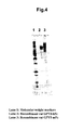
- the obtained rGPVI-mFc fusion protein was subjected to SDS-PAGE analysis by an established method.
- the sample and molecular weight markers Bio-Rad, Precision Plus Protein Unstained Standard
- rGPVI-hFc fusion protein mGPVI-hFc fusion protein
- hGPVI-hFc fusion protein hGPVI-hFc fusion protein
- rGPVI-hL2,5-hFc substituted mutant protein rGPVI-hL9,11-hFc substituted mutant protein
- hGPVI-mL9-hFc substituted mutant protein hGPVI-mL9-hFc substituted mutant protein.
- alum (Pierce), 1 mg CpG adjuvant, and 20 ⁇ g purified rat GPVI-mFc fusion protein were mixed and 100 ⁇ L of antigen for administration was provided.
- 50 ⁇ L of this antigen for administration was injected into each footpad on both feet of ddY mice (female, 8 weeks, SLC).
- 20 ⁇ g of the antigen for administration was diluted with 50 ⁇ L physiological saline and 25 ⁇ L of diluted antigen was administered into each of the footpads.
- lymphocytes were isolated from the iliac lymph nodes; the obtained lymphocytes were mixed with P3 ⁇ 63-Ag.8.U1 (ATTC) and cell fusion was then carried out using polyethylene glycol according to Introduction to Monoclonal Antibody Experimental Procedures , Tamie ANDO and Takeshi CHIBA (Kodansha).
- the hybridomas were selected using HAT culture medium, and after 1 weeks hybridomas producing the desired antibody were screened .
- purified rat GPVI-hFc fusion protein was diluted to 3 ⁇ g/mL wi th 0.076 M phosphate buffer solution (pH 7.4) (abbreviated below as PBS), and this solution was added to an immunoplate (Maxisorb, NUNC) at 50 ⁇ L/well. After reaction at 4°C overnight, plate was washed three times with ion-exchanged water and then blocked by addition of 100 ⁇ L PBS containing 2% Stabilguard in each well. Culture supernatant was then added to each well, and plate was washed three times with physiological saline containing 0.05% Tween 20 after the incubation for 1 hour at 37°C.
- PBS phosphate buffer solution
- Peroxidase-labeled anti-mouse immunoglobulin antibody (Dako) was diluted 1000X with PBS containing 10% rabbit serum, and 50 ⁇ L of this solution was added to each well. After reaction for 1 hour at 37°C, plate was washed five times in the same manner and TMB solution (BioFix) was added to each well. After reaction for 10 minutes at room temperature, the reaction was stopped with 0.5 M sulfuric acid solution. The absorbance at 450 nm was measured with a plate spectrophotometer (NJ-2100, Nippon InterMed). Cells that had reacted with the purified rat GPVI-hFc fusion protein were selected and cloned by the limiting dilution method. After 11 days, 17 hybridoma clones producing the antibody that reacted with purified rat GPVI-hFc fusion protein were screened by same method (Table 3).
- a culture supernatant was obtained by the clarification, at room temperature, of the mouse hybridoma culture fluid using a capsule cartridge filter having a pore diameter of 1 ⁇ m as a prefilter ( Toyo Roshi Kaisha, Ltd. (Toyo Filter paper Ltd.)) and a Fluorodyne filter (Pall) having a pore diameter of 0.22 ⁇ m and a Millipak 200 (MILLIPORE) as the main filter, respectively.
- This culture supernatant was adsorbed on a protein A column (rmp Protein A Sepharose Fast Flow, GE Healthcare Biosciences) that had been preliminarily equilibrated with PBS (Sigma).
- the non-specifically adsorbed protein was eluted with 10 ⁇ PBS (Sigma).
- the protein A-bound antibody was subsequently eluted with 100 mM glycine-HCl buffer (pH 3.0).
- the volume of the eluate was measured and one-tenth volume of 2 M Tris-HCl (pH 8.5) was immediately added to return the pH to neutrality and a purified antibody solution was yielded.
- This purified antibody solution was concentrated with a Biomax PB ultrafiltration membrane (Millipore) having a molecular weight cut-off of 30,000 using a stirred cell, followed by dialysis against physiological saline (Otsuka Normal Saline, Otsuka Pharmaceutical Co., Ltd.), and finally obtained the purified antibody solution.
- the obtained antibody was analyzed with SDS-PAGE according to an established method.
- the sample and molecular weight markers Bio-Rad, Precision Plus Protein Unstained Standard
- the antibodies used in the following all examples were purified by this same method.
- the recognition domain of the anti-rat GPVI monoclonal antibodies obtained in Example 4 was analyzed.
- the purified rGPVI-hFc fusion protein was diluted to 2 ⁇ g/mL with PBS, and this solution was added to immunoplate (Maxisorb, NUNC) at 50 ⁇ L/well. After reaction for 1 hour at 37°C, plate was washed five times with ion-exchanged water and then blocked by addition of 100 ⁇ L PBS containing 2% Stabilgurd in each well.
- NUNC polypropylene plate
- Peroxidase-labeled anti-mouse kappa chain antibody (Rockland) was diluted to 1000X with PBS containing 10% rabbit serum and 50 ⁇ L of this solution was added to each well. After reaction for 1 hour at 37°C, plate was washed five times in the same manner, and TMB solution (BioFix) was added to each well. After reaction for 10 minutes at room temperature, the reaction was stopped with 0.5 M sulfuric acid solution. The absorbance at 450 nm was measured using a plate spectrophotometer (NJ-2100, Nippon InterMed).
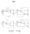
- loop 11 of rat GPVI is identical to the amino acid sequence of loop 11 of mouse GPVI, it is difficult to regard loop 11 as an important recognition region, and the aforementioned antibodies are presumed to recognize a structure that contains at least a portion of loop 9 of rat GPVI.
- a sandwich ELISA system was constructed using the mouse anti-rat GPVI antibody prepared in Example 4.
- the purified F1239-6-1 antibody was diluted to 10 ⁇ g/ ⁇ L with PBS, and this solution was added to immunoplate (Maxisorb, NUNC) at 75 ⁇ L/well. After reaction overnight at 4°C, plate was washed five times with ion-exchanged water and blocked by addition of 200 ⁇ L PBS containing 5% Stabilgurd and 0.1% Tween 20 in each well.
- the sample and recombinant rGPVI-hFc fusion protein, used as the standard were diluted with 0.1% BSA, 0.3 M sodium chloride, 0.05% Tween 20/PBS.
- plasma When plasma was used as the sample for determination, plasma was diluted to five times or more. Each sample was added at 50 ⁇ L/well. After reaction for 2 hours at 37°C with stirring, plate was washed five times with physiological saline containing 0.05% Tween 20. Biotin-labeled F1239-7-1 F(ab')2 antibody was diluted with PBS containing 20% Poly-HRP DILUENT (Fitzgerald), 0.3 M sodium chloride and 0.05% Tween 20, and 50 ⁇ L of this solution was added to each well. After reaction for 1 hour at 37°C with stirring, plate was washed five times in the same manner.
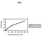
- a sandwich ELISA system was constructed using mouse anti-human GPVI antibody.
- the purified F1232-7-1 antibody was diluted to 10 ⁇ g/mL with PBS, and this solution was added to immunoplate (Maxisorb, NUNC) at 75 ⁇ L/well. After reaction overnight at 4°C, plate was washed five times with ion-exchanged water and blocked by addition of 200 ⁇ L PBS containing 5% Stabilgurd and 0.1% Tween 20 in each well.
- the sample and recombinant hGPVI-hFc fusion protein, used as the standard were diluted with 0.1% BSA, 0.3 M sodium chloride, 0.05% Tween 20/PBS.
- plasma When plasma was used as the sample for determination, plasma was diluted to five times or more. Each sample was added at 50 ⁇ L/well. After reaction for 2 hours at 37°C with stirring, plate was washed five times with physiological saline containing 0.05% Tween 20. Biotin-labeled F1232-10-2 F(ab')2 antibody was diluted with PBS containing 20% Poly-HRP DILUENT (Fitzgerald), 0.3 M sodium chloride and 0.05% Tween 20, and 50 ⁇ L of this solution was added to each well. After reaction for 1 hour at 37°C with stirring, plate was washed five times in the same manner.
- Citrated blood was collected from the normal rat, and platelet rich plasma (PRP) was prepared by centrifugation at 110 ⁇ g for 15 minutes at 25°C. After adjusting the pH of the PRP to 6.5 with ACD-A (acid-citrate-dextrose), the platelets were recovered by centrifugation at 830 ⁇ g for 10 minutes at 25°C. The recovered platelets were washed twice by the addition of HEPES buffer (137 mM NaCl, 1 mM MgCl 2 , 5.5 mM glucose, 3 mM NaH 2 PO 4 , 10 mM HEPES, 1 mg/mL BSA) adjusted to pH 6.5 with ACD-A.
- HEPES buffer 137 mM NaCl, 1 mM MgCl 2 , 5.5 mM glucose, 3 mM NaH 2 PO 4 , 10 mM HEPES, 1 mg/mL BSA
- the platelets were suspended by the addition of HEPES buffer (pH 7.3) to obtain washed platelets.
- Convulxin (Alexis), CRP (synthetic peptide), or ADP was added to the washed platelets in the presence of 1 mM CaCl 2 and 1 mM MgCl 2 , and the platelets were stimulated for 1 hour at room temperature.
- the reaction was stopped by the addition of Protease Inhibitor Cocktail solution containing 25 mM EDTA, and this reaction mixture was followed by centrifugation at 18,000 ⁇ g for 1 minute at 25°C, and a supernatant fraction and a platelet fraction were recovered.
- Washed platelets were stimulated with convulxin, CRP, or ADP by the method described in Example 9. Washed platelets added with buffer only were served as the control, and the reaction mixture was stationary incubated for 1 hour at room temperature. After the reaction had been stopped by the addition of Protease Inhibitor Cocktail solution containing 25 mM EDTA, the stimulated platelets were washed with PBS containing 0.5% inactivated FBS and 2.5 mM EDTA (abbreviated below as FACS buffer).
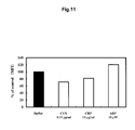
- the quantity of GPVI expression was calculated by subtracting the value obtained for the detection of PE-labeled mouse IgG2a ⁇ antibody from the value obtained for the detection of PE-labeled F1239-11-1. The results are shown in Figure 9 . In comparison to the platelets added with buffer only, a substantial reduction in GPVI expression was observed for the convulxin (CVX)-stimulated platelets and the CRP-stimulated platelets, while no reduction in GPVI expression was seen for the ADP-stimulated platelets.
- CVX convulxin
- Example 9 washed platelets were stimulated with convulxin, CRP, or ADP and the supernatant fraction was recovered.
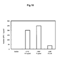
- the quantity of soluble GPVI in the supernatant was determined by the method described in Example 7. The results are shown in Figure 10 . A substantial increase in the quantity of soluble GPVI in the supernatant was observed for the platelets stimulated by convulxin and the platelets stimulated by CRP. On the other, increase in the quantity of soluble GPVI in the supernatant was weak for the platelets stimulated by ADP.
- Citrated blood was collected from the normal monkey, and platelet rich plasma (PRP) was obtained by centrifugation at 115 ⁇ g for 15 minutes at 25°C.
- PRP platelet rich plasma
- ACD-A acid-citrate-dextrose
- the platelets were recovered by centrifugation at 830 ⁇ g for 10 minutes at 25°C.
- the recovered platelets were washed twice by the addition of HEPES buffer (137 mM NaCl, 1 mM MgCl 2 , 5.5 mM glucose, 3 mM NaH 2 PO 4 , 10 mM HEPES, 1 mg/mL BSA) adjusted to pH 6.5 with ACD-A.
- the platelets were suspended by the addition of HEPES buffer (pH 7.3) to obtain washed platelets.
- Convulxin (Alexis), CRP (synthetic peptide), or ADP was added to the washed platelets in the presence of 1 mM CaCl 2 and 1 mM MgCl 2 , and the platelets were stimulated for 1 hour at room temperature.
- the reaction was stopped by the addition of Protease Inhibitor Cocktail solution containing 25 mM EDTA, and this reaction mixture was followed by centrifugation at 18,000 ⁇ g for 1 minute at 25°C. A supernatant fraction and a platelet fraction were recovered.
- Washed platelets were stimulated with convulxin, CRP, or ADP by the method described in Example 12. Washed platelets added with buffer only were served as the control, and the reaction mixture was stationary incubated for 1 hour at room temperature. After the reaction had been stopped by the addition of Protease Inhibitor Cocktail solution containing 25 mM EDTA, the stimulated platelets were washed with FACS buffer. Then, 1 ⁇ g per 4 ⁇ 10 5 platelets of chimeric anti-human GPVI antibody F1232-37-2 labeled with the fluorescent dye PE or the PE-labeled chimeric anti-human GPVI antibody F1232-10-1 was added, and the reaction mixture was stationary incubated for 30 minutes at room temperature in the dark.
- a sample was also prepared by the addition of the same quantity of PE-labeled human IgG4 antibody. After 30 minutes, the platelets were diluted with FACS buffer and the quantity of GPVI expression on the membrane surface of the platelets was analyzed by determination of the fluorescent intensity of the platelets with a Cytomics FC500 flow cytometer (Beckman Coulter). The quantity of GPVI expression was calculated by subtracting the value obtained for the detection of PE-labeled human IgG4 antibody from the value obtained for the detection of PE-labeled chimeric F1232-37-2 or PE-labeled chimeric F1232-10-1.
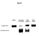
- Example 12 washed platelets were stimulated with convulxin, CRP, or ADP. Then, a supernatant fraction and a platelet fraction were recovered. The soluble GPVI present in the supernatant fraction and the GPVI present in the platelet fraction (referred to hereafter as platelet GPVI) were detected by western blotting. In brief, 4 ⁇ L of 4 ⁇ Sample Buffer (+ ⁇ -mercaptoethanol, Protease Inhibitor Cocktail (Roche), Phosphatase Inhibitor Cocktail (Pierce)) was added to 12 ⁇ L of the supernatant fraction.
- Sample Buffer (+ ⁇ -mercaptoethanol, Protease Inhibitor Cocktail (Roche), Phosphatase Inhibitor Cocktail (Pierce)
- the gel was blotted onto a low-fluorescence membrane (Immobilon-FL, PVDF, Millipore) by the semi-dry method. After blotting, the membrane was blocked with BlockAce (Dainippon Pharmaceutical Co., Ltd.) overnight at 4°C. After blocking, the primary antibody (polyclonal antibody prepared using human GPVI synthetic peptide as an antigen), diluted with PBS containing 10% BlockAce/0.1% Tween 20 (TPBS), was added and the membrane was incubated for 1 hour at room temperature.
- BlockAce Dainippon Pharmaceutical Co., Ltd.
- TPBS BlockAce/0.1% Tween 20
- Example 12 washed platelets were stimulated with convulxin, CRP, or ADP and the supernatant was recovered.
- the quantity of soluble GPVI in the supernatant was determined by the method described in Example 8. The results are shown in Figure 13 . A substantial increase in the quantity of soluble GPVI in the supernatant was observed for the platelets stimulated by convulxin and the platelets stimulated by CRP. On the other, the increase in the quantity of soluble GPVI in the supernatant was very weak for the platelets stimulated by ADP.
- Citrated blood was collected from the normal human, and platelet rich plasma (PRP) was obtained by centrifugation at 170 ⁇ g for 15 minutes at 25°C. After adjusting the pH of the PRP to 6.5 with ACD-A (acid-citrate-dextrose), the platelets were recovered by centrifugation at 830 ⁇ g for 10 minutes at 25°C. The recovered platelets were washed twice by the addition of HEPES buffer (137 mM NaCl, 1 mM MgCl 2 , 5.5 mM glucose, 3 mM NaH 2 PO 4 , 10 mM HEPES, 1 mg/mL BSA) adjusted to pH 6.5 with ACD-A.
- HEPES buffer 137 mM NaCl, 1 mM MgCl 2 , 5.5 mM glucose, 3 mM NaH 2 PO 4 , 10 mM HEPES, 1 mg/mL BSA
- HEPES buffer pH 7.3
- Collagen Horm collagen reagent, Nycomed
- convulxin Alexis
- CRP synthetic peptide
- ADP ADP was added to the washed platelets in the presence of 1 mM CaCl 2 and 1 mM MgCl 2 , and the platelets were stimulated for 1 hour at room temperature.
- the reaction was stopped by the addition of Protease Inhibitor Cocktail solution containing 25 mM EDTA, and this reaction mixture was followed by centrifugation at 18,000 ⁇ g for 1 minute at 25°C. A supernatant fraction and a platelet fraction were recovered.
- Washed platelets were stimulated with collagen, convulxin, CRP, or ADP by the method described in Example 16. Washed platelets added with buffer only were served as the control, and the reaction mixture was stationary incubated for 1 hour at room temperature. After the reaction had been stopped by the addition of Protease Inhibitor Cocktail solution containing 25 mM EDTA, the stimulated platelets were washed with FACS buffer. Then, 1 ⁇ g per 4 ⁇ 10 5 platelets of chimeric anti-human GPVI antibody F1232-37-2 labeled with the fluorescent dye PE was added, and the mixture was stationary incubated for 30 minutes at room temperature in the dark.
- Example 16 washed platelets were stimulated with collagen, convulxin, CRP, or ADP. Then, a supernatant fraction and a platelet fraction were recovered. The soluble GPVI present in the supernatant fraction and the GPVI present in the platelet fraction (referred to hereafter as platelet GPVI) were detected by western blotting. In brief, 4 ⁇ L of 4x Sample Buffer (+ ⁇ -mercaptoethanol, Protease Inhibitor Cocktail (Roche), Phosphatase Inhibitor Cocktail (Pierce)) was added to 12 ⁇ L of the supernatant fraction.
- 4x Sample Buffer (+ ⁇ -mercaptoethanol, Protease Inhibitor Cocktail (Roche), Phosphatase Inhibitor Cocktail (Pierce)
- the gel was blotted onto a low-fluorescence membrane (Immobilon-FL, PVDF, Millipore) by the semi-dry method. After blotting, the membrane was blocked with BlockAce (Dainippon Pharmaceutical Co., Ltd.) overnight at 4°C. After blocking, the primary antibody (polyclonal antibody prepared using human GPVI synthetic peptide as an antigen), diluted with PBS containing 10% BlockAce/0.1% Tween 20 (TPBS), was added and the membrane was incubated for 1 hour at room temperature.
- BlockAce Dainippon Pharmaceutical Co., Ltd.
- TPBS BlockAce/0.1% Tween 20
- Example 16 washed platelets were stimulated with collagen, convulxin, CRP, or ADP and the supernatant was recovered.
- the quantity of soluble GPVI in the supernatant was determined by the method described in Example 8. The results are shown in Figure 16 . A substantial increase in the quantity of soluble GPVI in the supernatant was observed for the platelets stimulated by collagen, the platelets stimulated by convulxin, and the platelets stimulated by CRP. On the other, an increase in the soluble GPVI in the supernatant was not observed in the platelets stimulated by ADP.
- Example 20 Example 20
- Endotoxin (a dose of 20 mg/kg in 2 hours or a dose of 40 mg/kg in 4 hours) was intravenously infused via right femoral vein.
- blood was collected from the abdominal aorta and the platelet count was determined using an automated hemocytometer (Sysmex, Toa Medical Electronics).
- the plasma was separated by centrifugation of the blood and the soluble GPVI was determined by the method described in Example 7 and the soluble ICAM-1 was determined using an ELISA kit (R & D System, Rat sICAM-1 (CD54) Immunoassay).
- a decline in the platelet count was observed by the administration of collagen. It indicates that platelet is activated by collagen administration.
- the quantity of soluble GPVI and soluble ICAM-1 in the plasma were increased by collagen administration.
- the degree of the decline in the platelet count agreed with the degree of the increase in the quantity of soluble GPVI in the plasma.
- male SD rats were anesthetized with pentobarbital sodium (50 mg/kg) and collagen ( a dose of 0.1 mg/kg) was rapidly administered via right femoral vein. Blood was collected from the jugular vein at 15 minutes, 30 minutes, and 60 minutes post-collagen administration and the platelet count was determined using an automated hemocytometer (Sysmex, Toa Medical Electronics).
- Anti-rat GPVI antibody (F1239-6-1) was administered to male SD rats. After 6 days, collagen was rapidly and intravenously administered at a dose of 0.8 mg/kg. The mortality was observed from post-collagen administration to 15 minutes. The results are shown in Table 4.
- the anti-rat GPVI antibody (F1239-6-1) completely prevented animal death at a dose of 0.3 mg/kg in an animal thrombosis model in which the quantity of soluble GPVI in the plasma undergoes elevation.
- determination of the sGPVI in plasma by the determination method of the present invention enables the detection, determination, or quantitation of platelet activation or vascular endothelial injury prior to disease onset; enables the diagnosis of diseases associated with platelet activation or vascular endothelial injury, such as thrombosis and so forth; and enables the selection of patients for treatment.
- a high rate of effectiveness will presumably be obtained for the administration of an antiplatelet drug, e.g., an anti-GPVI antibody, to such patients.
- Table 4 group number of survivors/number of animals physiological saline 0/15 F1239-6-1 0.1 mg/kg 1/7 F1239-6-1 0.3 mg/kg 7/7 F1239-6-1 1.0 mg/kg 8/8
- Example 22 The effect of anti-rat GPVI antibody in the animal model described in Example 22 was examined.
- 3 mg/kg F1239-6-1 was administered subcutaneously, and the vascular intima was injured at 6 days after the administration using the method of Example 22.
- the right and left carotid arteries were collected under anesthesia by pentobarbital and were fixed in 10% neutral buffered formalin, and paraffin blocks were prepared. Then, the paraffin blocks were thin sectioned to prepare the specimens. After staining with hematoxylin-eosin, the specimens were observed under a microscope.
- the observed image was recorded as a digital image using a CCD camera and analyzed using WinROOF image analysis software to calculate the cross-sectional areas of the intima and the media.
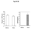
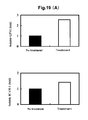
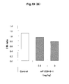
- the results are shown in Figure 19(B) .
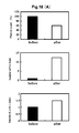
- the anti-rat GPVI antibody (F1239-6-1) inhibited intimal thickening in an animal arteriosclerosis model that presented an increase in soluble GPVI in the plasma.
- the determination of the sGPVI in plasma by the determination method of the present invention enables the detection, determination, or quantitation of platelet activation or vascular endothelial injury prior to disease onset; enables the diagnosis of diseases associated with platelet activation or vascular endothelial injury, such as arteriosclerotic diseases and so forth; and enables the selection of patients for treatment.
- a high rate of effectiveness will presumably be obtained for the administration of an antiplatelet drug, e.g., an anti-GPVI antibody, to such patients.
- Female Wistar rats were anesthetized with pentobarbital sodium (50 mg/kg) and endotoxin (40 mg/kg in 4 hours) was intravenously infused via right femoral vein. After the completion of endotoxin infusion, urine was collected and the soluble GPVI in the urine was determined using the method described in Example 7. The results are shown in Figure 20 . Increase in the quantity of soluble GPVI in the urine was also observed by the administration of endotoxin.
- the quantity of soluble GPVI in the plasma was determined by the method described in Example 8 for various human pathologies. The results are shown in Figure 21 .
- hGPVI-mFc 20 ⁇ g purified hGPVI-mFc was mixed with Freund's complete adjuvant (Difco) or alum (Pierce) and oligo CpG to provide the antigen for administration.
- ddY mice female, 8 weeks, SLC
- the obtained lymphocytes were mixed with P3 ⁇ 63-Ag.8.U1 (ATTC) and cell fusion was then carried out using polyethylene glycol (PEG1500, Sigma) according to Introduction to Monoclonal Antibody Experimental Procedures , Tamie ANDO and Takeshi CHIBA (Kodansha), page 83.
- hybridomas were selected using HAT culture medium, and after 1 week Hybidomas producing the desired antibody were screened by the binding activity to hGPVI-hFc and hGPVI-mL9-hFc as the indicator.
- purified hGPVI-hFc or hGPVI-mL9-hFc was diluted to 1 ⁇ g/mL with D-PBS (pH 7.4). This solution was added to immunoplate (Maxisorb, NUNC) at 50 ⁇ L/well. Immobilization was carried out using the same method as described in Example 6. Culture supernatant was subsequently added to each well, and plate was incubated for 1 hour at room temperature.
- the GPVI binding activity of each antibody is shown (Table 5), assigning - to an absorbance of 0.5 or less in the aforementioned ELISA system, + to an absorbance of 0.5 up to 1.0, + + to an absorbance of 1.0 to 2.0, and + + + to an absorbance greater than 2.0.
- the same procedure was used to obtain L2-specific mouse anti-human GPVI antibody hybridomas (F1232-10-1 and F1232-10-2, which was considered to originate from the same clone).
- Single-strand cDNA was synthesized from hybridomas F1232-37-2 and F1232-10-1 by the standard methods.
- the DNA encoding the variable regions of the anti-GPVI antibodies was amplified by PCR using Mouse Ig-Primer Set (Novagen) and single-strand cDNA as the template, and the nucleotide sequense was determined.
- the nucleotide sequence and putative amino acid sequence of the heavy-chain and light-chain variable regions of the monoclonal antibodies F1232-37-2 and F1232-10-1 secreted from each hybridomas, are shown in sequence table (SEQ ID NO: 18-25).
- F1232-7-1 and F1232-10-2 were able to identify the presence of human soluble GPVI in the supernatant fraction sample from convulxin-stimulated washed platelets in the same manner as the polyclonal antibody prepared from human GPVI synthetic peptide as an antigen. Moreover, in the case of the supernatant fraction sample from the buffer-added washed platelets, human soluble GPVI was not detected by F1232-7-1 or F1232-10-2 or the polyclonal antibody prepared from human GPVI synthetic peptide as an antigen. It indicates that F1232-7-1 and F1232-10-2 used in the ELISA system for human soluble GPVI, recognize human soluble GPVI.
- GPVI in a sample can be measured at high sensitivities by the determination method, determination reagent, or kit of the present invention.
- the detection, determination, or quantitation of sGPVI or mGPVI using the determination method, determination reagent, or kit of the present invention enables, inter alia, the detection, determination, quantitation, assessment, or evaluation of platelet activation or vascular endothelial injury; the diagnosis of diseases associated with platelet activation or vascular endothelial injury, the assessment of susceptibility to diseases associated with platelet activation or vascular endothelial injury, and the evaluation of the risk of onset of diseases associated with platelet activation or vascular endothelial injury; the selection of patients who respond to an antiplatelet drug, particularly an anti-GPVI antibody; the decision of the time of (beginning) the administration of such drugs; and the prediction, monitoring, or prognostic assessment of the therapeutic effects of, or the manifestation of side
Landscapes
- Health & Medical Sciences (AREA)
- Life Sciences & Earth Sciences (AREA)
- Chemical & Material Sciences (AREA)
- Immunology (AREA)
- Molecular Biology (AREA)
- Engineering & Computer Science (AREA)
- Hematology (AREA)
- General Health & Medical Sciences (AREA)
- Biochemistry (AREA)
- Urology & Nephrology (AREA)
- Medicinal Chemistry (AREA)
- Organic Chemistry (AREA)
- Biomedical Technology (AREA)
- Cell Biology (AREA)
- Biophysics (AREA)
- Biotechnology (AREA)
- Proteomics, Peptides & Aminoacids (AREA)
- Microbiology (AREA)
- Genetics & Genomics (AREA)
- Food Science & Technology (AREA)
- Physics & Mathematics (AREA)
- Analytical Chemistry (AREA)
- General Physics & Mathematics (AREA)
- Pathology (AREA)
- Peptides Or Proteins (AREA)
Applications Claiming Priority (2)
| Application Number | Priority Date | Filing Date | Title |
|---|---|---|---|
| JP2006100241 | 2006-03-31 | ||
| PCT/JP2007/056530 WO2007116779A1 (fr) | 2006-03-31 | 2007-03-20 | Nouveau marqueur de l'activation plaquettaire et son procede de determination |
Publications (2)
| Publication Number | Publication Date |
|---|---|
| EP2000802A1 true EP2000802A1 (fr) | 2008-12-10 |
| EP2000802A4 EP2000802A4 (fr) | 2009-04-15 |
Family
ID=38581075
Family Applications (1)
| Application Number | Title | Priority Date | Filing Date |
|---|---|---|---|
| EP07739968A Withdrawn EP2000802A4 (fr) | 2006-03-31 | 2007-03-20 | Nouveau marqueur de l'activation plaquettaire et son procédé de détermination |
Country Status (5)
| Country | Link |
|---|---|
| US (2) | US8323905B2 (fr) |
| EP (1) | EP2000802A4 (fr) |
| JP (2) | JPWO2007116779A1 (fr) |
| CA (1) | CA2646807A1 (fr) |
| WO (1) | WO2007116779A1 (fr) |
Cited By (8)
| Publication number | Priority date | Publication date | Assignee | Title |
|---|---|---|---|---|
| WO2010000756A1 (fr) * | 2008-06-30 | 2010-01-07 | Institut National De La Sante Et De La Recherche Medicale (Inserm) | Détection de cd31 détachées, diagnostic de l’athérothrombose et de maladies auto-immunes, et procédé d’analyse de voies de signalisation |
| EP2336188A1 (fr) * | 2009-12-18 | 2011-06-22 | Sanofi-Aventis | Nouveaux anticorps antagonistes et leurs fragments Fab contre le GPVI et leurs utilisations |
| EP2397495A1 (fr) * | 2010-06-21 | 2011-12-21 | Sanofi | Nouveaux anticorps antagonistes et leurs fragments Fab contre le GPVI et leurs utilisations |
| WO2011073954A3 (fr) * | 2009-12-18 | 2012-03-08 | Sanofi-Aventis | Nouveaux anticorps antagonistes et leurs fragments fab dirigés contre gpvi, et utilisations de ceux-ci |
| WO2012072743A1 (fr) * | 2010-12-01 | 2012-06-07 | INSERM (Institut National de la Santé et de la Recherche Médicale) | Procédé et trousses pour déterminer la sensibilité des plaquettes à une activation dans un patient |
| EP2615458A4 (fr) * | 2010-09-07 | 2014-01-01 | Mochida Pharm Co Ltd | Méthode de diagnostic de la maladie d'alzheimer à l'aide de la gpvi soluble dans le sang |
| WO2013150375A3 (fr) * | 2012-04-04 | 2014-01-09 | Assistance Publique Hôpitaux De Paris | Biomarqueurs d'activation des cellules endothéliales caractérisant le rejet médié par anticorps et utilisations associées |
| WO2022263330A1 (fr) * | 2021-06-18 | 2022-12-22 | Advancecor Gmbh | Composition pharmaceutique |
Families Citing this family (12)
| Publication number | Priority date | Publication date | Assignee | Title |
|---|---|---|---|---|
| JPWO2007116779A1 (ja) * | 2006-03-31 | 2009-08-20 | 持田製薬株式会社 | 新規血小板活性化マーカー及びその測定方法 |
| DE102011016143A1 (de) * | 2011-03-25 | 2012-09-27 | Eberhard-Karls-Universität Tübingen Universitätsklinikum | Diagnostische Differenzierung zwischen einem akuten Koronarsyndrom und einer stabilen Angina pectoris |
| US20160264684A1 (en) * | 2013-10-10 | 2016-09-15 | Siamab Therapeutics, Inc. | Glycan-interacting compounds and methods of use |
| US9879087B2 (en) | 2014-11-12 | 2018-01-30 | Siamab Therapeutics, Inc. | Glycan-interacting compounds and methods of use |
| FI3218005T3 (fi) | 2014-11-12 | 2023-03-31 | Seagen Inc | Glykaanin kanssa vuorovaikutteisia yhdisteitä ja käyttömenetelmiä |
| CA3002097A1 (fr) | 2015-11-12 | 2017-05-18 | Siamab Therapeutics, Inc. | Composes interagissant avec le glycane et methodes d'utilisation |
| WO2018094143A1 (fr) | 2016-11-17 | 2018-05-24 | Siamab Therapeutics, Inc. | Composés interagissant avec le glycane et méthodes d'utilisation |
| WO2018160909A1 (fr) | 2017-03-03 | 2018-09-07 | Siamab Therapeutics, Inc. | Composés interagissant avec le glycane et méthodes d'utilisation |
| US20190388548A1 (en) * | 2018-06-26 | 2019-12-26 | Tzu Chi University | Method for providing ocular neuroprotection or for preventing, treating or alleviating the effects of, an ocular disease associated with retinal ganglion cell death |
| CA3128368A1 (fr) * | 2019-02-01 | 2020-08-06 | Memorial Sloan Kettering Cancer Center | Cellules car-t antisenescence ciblant upar, surface cellulaire et biomarqueur de senescence secrete |
| WO2022163124A1 (fr) * | 2021-01-29 | 2022-08-04 | 東洋濾紙株式会社 | Membrane pour dosages immunochromatographiques, bandelette réactive pour dosages immunochromatographiques et procédé de test |
| JP2024027767A (ja) * | 2022-08-19 | 2024-03-01 | 株式会社Lsiメディエンス | 可溶型clec2を用いた血小板減少性疾患の診断法 |
Family Cites Families (18)
| Publication number | Priority date | Publication date | Assignee | Title |
|---|---|---|---|---|
| US5989811A (en) * | 1994-09-29 | 1999-11-23 | Urocor, Inc. | Sextant core biopsy predictive mechanism for non-organ confined disease status |
| US5750345A (en) * | 1995-10-31 | 1998-05-12 | Evanston Hospital Corporation | Detection of human α-thalassemia mutations and their use as predictors of blood-related disorders |
| US20040001826A1 (en) * | 1999-06-30 | 2004-01-01 | Millennium Pharmaceuticals, Inc. | Glycoprotein VI and uses thereof |
| AU7097400A (en) | 1999-09-01 | 2001-03-26 | Otsuka Pharmaceutical Co., Ltd. | Platelet membrane glycoprotein vi (gpvi) dna and protein sequences, and uses thereof |
| EP1224942A1 (fr) | 2001-01-23 | 2002-07-24 | Bernhard Dr. Nieswandt | Utilisation de JAQ1 (anticorps monoclonal anti-GPVI) en tant que médicament actif pour la protection contre les maladies thrombotiques |
| US7713705B2 (en) * | 2002-12-24 | 2010-05-11 | Biosite, Inc. | Markers for differential diagnosis and methods of use thereof |
| GB0130543D0 (en) | 2001-12-20 | 2002-02-06 | Univ Cambridge Tech | Human antibodies and their use |
| EP1538165A1 (fr) | 2003-12-03 | 2005-06-08 | Procorde GmbH | Inhibiteurs de la glycoprotéine VI construits à partir de l'anticorps monoclonal hgp 5c4 |
| US20090130021A1 (en) | 2002-06-07 | 2009-05-21 | Gotz Munch | Methods, products and uses involving platelets and/or the vasculature |
| GB0511590D0 (en) | 2005-06-07 | 2005-07-13 | Procorde Gmbh | Anti-thrombotic agents |
| US20070071744A1 (en) | 2002-06-07 | 2007-03-29 | Gotz Munch | Agents which bind to epitopes of glycoprotein VI |
| WO2006061650A2 (fr) | 2004-12-10 | 2006-06-15 | Trigen Gmbh | Procedes, produits et utilisations relatifs a des plaquettes et/ou la vasculature |
| US20070025992A1 (en) | 2003-07-18 | 2007-02-01 | Mochida Pharmaceutical Co., Ltd | Monoclonal antibody against platelet membrane glycoprotein VI |
| US7645592B2 (en) | 2004-04-29 | 2010-01-12 | Otsuka Pharmaceutical Co., Ltd. | Glycoprotein VI antibodies and methods of use thereof |
| US7611707B2 (en) | 2004-04-29 | 2009-11-03 | Otsuka Pharmaceutical Co., Ltd. | Glycoprotein VI antibodies and methods thereof |
| CA2606450A1 (fr) * | 2005-04-28 | 2006-11-09 | Mochida Pharmaceutical Co., Ltd. | Anticorps monoclonal anti-glycoproteine vi de membrane plaquettaire |
| US20090041783A1 (en) * | 2005-04-28 | 2009-02-12 | Mochida Pharmaceutical Co., Ltd. | Anti-platelet membrane glycoprotein vi monoclonal antibody |
| JPWO2007116779A1 (ja) * | 2006-03-31 | 2009-08-20 | 持田製薬株式会社 | 新規血小板活性化マーカー及びその測定方法 |
-
2007
- 2007-03-20 JP JP2008509792A patent/JPWO2007116779A1/ja not_active Ceased
- 2007-03-20 WO PCT/JP2007/056530 patent/WO2007116779A1/fr not_active Ceased
- 2007-03-20 CA CA002646807A patent/CA2646807A1/fr not_active Abandoned
- 2007-03-20 EP EP07739968A patent/EP2000802A4/fr not_active Withdrawn
- 2007-03-20 US US12/225,722 patent/US8323905B2/en not_active Expired - Fee Related
-
2012
- 2012-09-14 US US13/617,409 patent/US20130011868A1/en not_active Abandoned
-
2013
- 2013-08-26 JP JP2013174247A patent/JP2013257338A/ja not_active Withdrawn
Cited By (16)
| Publication number | Priority date | Publication date | Assignee | Title |
|---|---|---|---|---|
| US8951743B2 (en) | 2008-06-30 | 2015-02-10 | Institut National De La Sante Et De La Recherche Medicale (Inserm) | Detection of shed CD31, diagnosis of atherothrombosis and autoimmune disorders, and methods for analyzing signaling pathways |
| US9977030B2 (en) | 2008-06-30 | 2018-05-22 | Institut National De La Sante Et De La Recherche Medicale (Inserm) | Detection of shed CD31, diagnosis of atherothrombosis and autoimmune disorders, and methods for analyzing signaling pathways |
| WO2010000756A1 (fr) * | 2008-06-30 | 2010-01-07 | Institut National De La Sante Et De La Recherche Medicale (Inserm) | Détection de cd31 détachées, diagnostic de l’athérothrombose et de maladies auto-immunes, et procédé d’analyse de voies de signalisation |
| US9441040B2 (en) | 2009-12-18 | 2016-09-13 | Sanofi | Antagonist antibodies and their fab fragments against GPVI and uses thereof |
| US8852593B2 (en) | 2009-12-18 | 2014-10-07 | Sanofi | Antagonist antibodies and their fab fragments against GPVI and uses thereof |
| WO2011073954A3 (fr) * | 2009-12-18 | 2012-03-08 | Sanofi-Aventis | Nouveaux anticorps antagonistes et leurs fragments fab dirigés contre gpvi, et utilisations de ceux-ci |
| EP2933269A1 (fr) * | 2009-12-18 | 2015-10-21 | Sanofi | Nouveaux anticorps antagonistes et leurs fragments fab contre le gpvi et utilisation de ceux-ci |
| AU2010331807B2 (en) * | 2009-12-18 | 2016-08-04 | Sanofi | Novel antagonist antibodies and their Fab fragments against GPVI and uses thereof |
| EP2336188A1 (fr) * | 2009-12-18 | 2011-06-22 | Sanofi-Aventis | Nouveaux anticorps antagonistes et leurs fragments Fab contre le GPVI et leurs utilisations |
| EP2397495A1 (fr) * | 2010-06-21 | 2011-12-21 | Sanofi | Nouveaux anticorps antagonistes et leurs fragments Fab contre le GPVI et leurs utilisations |
| EP2615458A4 (fr) * | 2010-09-07 | 2014-01-01 | Mochida Pharm Co Ltd | Méthode de diagnostic de la maladie d'alzheimer à l'aide de la gpvi soluble dans le sang |
| WO2012072743A1 (fr) * | 2010-12-01 | 2012-06-07 | INSERM (Institut National de la Santé et de la Recherche Médicale) | Procédé et trousses pour déterminer la sensibilité des plaquettes à une activation dans un patient |
| US9029095B2 (en) | 2010-12-01 | 2015-05-12 | Institut National De La Sante Et De La Recherche Medicale (Inserm) | Method and kits for determining platelet susceptibility to activation in a patient |
| WO2013150375A3 (fr) * | 2012-04-04 | 2014-01-09 | Assistance Publique Hôpitaux De Paris | Biomarqueurs d'activation des cellules endothéliales caractérisant le rejet médié par anticorps et utilisations associées |
| WO2022263330A1 (fr) * | 2021-06-18 | 2022-12-22 | Advancecor Gmbh | Composition pharmaceutique |
| WO2022263057A1 (fr) * | 2021-06-18 | 2022-12-22 | Advancecor Gmbh | Utilisation d'une composition pharmaceutique |
Also Published As
| Publication number | Publication date |
|---|---|
| JP2013257338A (ja) | 2013-12-26 |
| CA2646807A1 (fr) | 2007-10-18 |
| US20130011868A1 (en) | 2013-01-10 |
| JPWO2007116779A1 (ja) | 2009-08-20 |
| EP2000802A4 (fr) | 2009-04-15 |
| US20090311728A1 (en) | 2009-12-17 |
| WO2007116779A1 (fr) | 2007-10-18 |
| US8323905B2 (en) | 2012-12-04 |
Similar Documents
| Publication | Publication Date | Title |
|---|---|---|
| US8323905B2 (en) | Methods of detection GPVI | |
| Wautier et al. | Increased adhesion to endothelial cells of erythrocytes from patients with polycythemia vera is mediated by laminin α5 chain and Lu/BCAM | |
| Johnson-Léger et al. | Junctional adhesion molecule-2 (JAM-2) promotes lymphocyte transendothelial migration | |
| AU2018275235B2 (en) | Methods for aiding in diagnosing and evaluating a mild traumatic brain injury in a human subject using cardiac troponin I and early biomarkers | |
| RU2549684C2 (ru) | Антитела, связывающиеся с лизилоксидазоподобным ферментом-2 (loxl2), и способы их применения | |
| JP4829897B2 (ja) | 血小板減少及び/又はフォンビルブラント因子(vWF)と血小板との間の自発的相互作用によって特徴づけられる疾患及び障害の異なる形態を区別する方法及びアッセイ | |
| JP5766606B2 (ja) | 輸血関連急性肺障害(trali)に関するスクリーニング方法 | |
| JPS59183696A (ja) | 架橋フイブリン誘導体特異性モノクロ−ナル抗体 | |
| JPS61192285A (ja) | 活性化血小板に対するモノクロ−ナル抗体 | |
| JP6078845B2 (ja) | 可溶型clec−2に基づく血小板活性化測定方法 | |
| DE69128966T2 (de) | Charakterisierung von störungen der blutplättchenaggregation | |
| AU696118B2 (en) | Antibodies for the detection of HLA-G | |
| RU2770006C2 (ru) | Антитела к фактору ix padua | |
| Moroi et al. | Activation‐induced changes in platelet surface receptor expression and the contribution of the large‐platelet subpopulation to activation | |
| Costanzo et al. | Repositioning the early pathology of Type 1 diabetes to the extraislet vasculature | |
| JP2002501201A (ja) | インテグリンアンタゴニスト/アゴニスト仲介性疾患のリスク評価 | |
| EP3006942A1 (fr) | Réactif contenant des anticorps anti-lgr6 pour la détection et le diagnostic du cancer | |
| Juch et al. | A novel sandwich ELISA for α1 domain based detection of soluble HLA-G heavy chains | |
| EP0497881A1 (fr) | Procedes d'evaluation du metabolisme du cholesterol et reactifs destines a ceux-ci | |
| EP2609431B1 (fr) | Procédé de validation de cibles et de médicaments pour des affections inflammatoires et/ou cardiovasculaires | |
| JP5339676B2 (ja) | 細胞を検出する方法、およびこれに有用な薬剤 | |
| JP2006526762A (ja) | アジポネクチンとそのレセプターとの間の相互作用を調節するための方法及び組成物 | |
| EP1174498A2 (fr) | Protéine activante du facteur de coagulation sanguin IX et son anticorps | |
| JP2001526774A (ja) | P―セレクチンアッセイおよびその使用方法 | |
| JP2003532896A (ja) | Ipgアッセイ |
Legal Events
| Date | Code | Title | Description |
|---|---|---|---|
| PUAI | Public reference made under article 153(3) epc to a published international application that has entered the european phase |
Free format text: ORIGINAL CODE: 0009012 |
|
| 17P | Request for examination filed |
Effective date: 20080922 |
|
| AK | Designated contracting states |
Kind code of ref document: A1 Designated state(s): AT BE BG CH CY CZ DE DK EE ES FI FR GB GR HU IE IS IT LI LT LU LV MC MT NL PL PT RO SE SI SK TR |
|
| A4 | Supplementary search report drawn up and despatched |
Effective date: 20090313 |
|
| 17Q | First examination report despatched |
Effective date: 20090710 |
|
| DAX | Request for extension of the european patent (deleted) | ||
| GRAP | Despatch of communication of intention to grant a patent |
Free format text: ORIGINAL CODE: EPIDOSNIGR1 |
|
| GRAP | Despatch of communication of intention to grant a patent |
Free format text: ORIGINAL CODE: EPIDOSNIGR1 |
|
| INTG | Intention to grant announced |
Effective date: 20130613 |
|
| GRAP | Despatch of communication of intention to grant a patent |
Free format text: ORIGINAL CODE: EPIDOSNIGR1 |
|
| INTG | Intention to grant announced |
Effective date: 20131002 |
|
| STAA | Information on the status of an ep patent application or granted ep patent |
Free format text: STATUS: THE APPLICATION IS DEEMED TO BE WITHDRAWN |
|
| 18D | Application deemed to be withdrawn |
Effective date: 20140213 |