EP2001998B1 - Verfahren und zusammensetzungen zur reparatur bzw. regenerierung von beschädigtem herzmuskel - Google Patents
Verfahren und zusammensetzungen zur reparatur bzw. regenerierung von beschädigtem herzmuskel Download PDFInfo
- Publication number
- EP2001998B1 EP2001998B1 EP07751073.3A EP07751073A EP2001998B1 EP 2001998 B1 EP2001998 B1 EP 2001998B1 EP 07751073 A EP07751073 A EP 07751073A EP 2001998 B1 EP2001998 B1 EP 2001998B1
- Authority
- EP
- European Patent Office
- Prior art keywords
- cells
- stem cells
- cardiac
- myocardium
- myocytes
- Prior art date
- Legal status (The legal status is an assumption and is not a legal conclusion. Google has not performed a legal analysis and makes no representation as to the accuracy of the status listed.)
- Active
Links
- 210000004165 myocardium Anatomy 0.000 title claims description 217
- 239000000203 mixture Substances 0.000 title description 93
- 238000000034 method Methods 0.000 title description 88
- 230000008439 repair process Effects 0.000 title description 39
- 230000008929 regeneration Effects 0.000 title description 26
- 238000011069 regeneration method Methods 0.000 title description 26
- 210000004027 cell Anatomy 0.000 claims description 493
- 210000000130 stem cell Anatomy 0.000 claims description 285
- 230000000747 cardiac effect Effects 0.000 claims description 167
- 208000010125 myocardial infarction Diseases 0.000 claims description 134
- 108010014608 Proto-Oncogene Proteins c-kit Proteins 0.000 claims description 116
- 102000016971 Proto-Oncogene Proteins c-kit Human genes 0.000 claims description 116
- 102000003745 Hepatocyte Growth Factor Human genes 0.000 claims description 68
- 108090000100 Hepatocyte Growth Factor Proteins 0.000 claims description 68
- 210000004351 coronary vessel Anatomy 0.000 claims description 57
- 230000002107 myocardial effect Effects 0.000 claims description 57
- 238000002347 injection Methods 0.000 claims description 55
- 239000007924 injection Substances 0.000 claims description 55
- 101000599951 Homo sapiens Insulin-like growth factor I Proteins 0.000 claims description 42
- 102100037852 Insulin-like growth factor I Human genes 0.000 claims description 42
- 108010069241 Connexin 43 Proteins 0.000 claims description 29
- 102000001045 Connexin 43 Human genes 0.000 claims description 29
- 210000001367 artery Anatomy 0.000 claims description 29
- 102000000905 Cadherin Human genes 0.000 claims description 21
- 108050007957 Cadherin Proteins 0.000 claims description 21
- 210000005166 vasculature Anatomy 0.000 claims description 19
- 108050000637 N-cadherin Proteins 0.000 claims description 17
- 238000001727 in vivo Methods 0.000 claims description 17
- 108010053099 Vascular Endothelial Growth Factor Receptor-2 Proteins 0.000 claims description 14
- 230000006907 apoptotic process Effects 0.000 claims description 14
- 102000016549 Vascular Endothelial Growth Factor Receptor-2 Human genes 0.000 claims description 13
- 208000037265 diseases, disorders, signs and symptoms Diseases 0.000 claims description 12
- 208000031225 myocardial ischemia Diseases 0.000 claims description 11
- 230000002829 reductive effect Effects 0.000 claims description 11
- 108090000723 Insulin-Like Growth Factor I Proteins 0.000 claims description 6
- 208000035475 disorder Diseases 0.000 claims description 6
- 102000004218 Insulin-Like Growth Factor I Human genes 0.000 claims description 5
- 108010092694 L-Selectin Proteins 0.000 claims description 5
- 238000001361 intraarterial administration Methods 0.000 claims description 3
- 102100033467 L-selectin Human genes 0.000 claims 1
- 210000000107 myocyte Anatomy 0.000 description 242
- 210000002216 heart Anatomy 0.000 description 157
- 206010061216 Infarction Diseases 0.000 description 144
- 230000007574 infarction Effects 0.000 description 144
- 210000004940 nucleus Anatomy 0.000 description 139
- 241000699670 Mus sp. Species 0.000 description 135
- 102000004127 Cytokines Human genes 0.000 description 129
- 108090000695 Cytokines Proteins 0.000 description 129
- 210000001519 tissue Anatomy 0.000 description 101
- 210000002889 endothelial cell Anatomy 0.000 description 91
- XJMOSONTPMZWPB-UHFFFAOYSA-M propidium iodide Chemical compound [I-].[I-].C12=CC(N)=CC=C2C2=CC=C(N)C=C2[N+](CCC[N+](C)(CC)CC)=C1C1=CC=CC=C1 XJMOSONTPMZWPB-UHFFFAOYSA-M 0.000 description 90
- 210000000329 smooth muscle myocyte Anatomy 0.000 description 87
- 241000700159 Rattus Species 0.000 description 76
- 210000005240 left ventricle Anatomy 0.000 description 71
- 238000002372 labelling Methods 0.000 description 69
- 102000007469 Actins Human genes 0.000 description 65
- 108010085238 Actins Proteins 0.000 description 65
- WOVKYSAHUYNSMH-RRKCRQDMSA-N 5-bromodeoxyuridine Chemical compound C1[C@H](O)[C@@H](CO)O[C@H]1N1C(=O)NC(=O)C(Br)=C1 WOVKYSAHUYNSMH-RRKCRQDMSA-N 0.000 description 57
- 108010048367 enhanced green fluorescent protein Proteins 0.000 description 57
- 238000011282 treatment Methods 0.000 description 56
- 108010051609 Cardiac Myosins Proteins 0.000 description 55
- 102000013602 Cardiac Myosins Human genes 0.000 description 55
- 210000003205 muscle Anatomy 0.000 description 52
- 210000002837 heart atrium Anatomy 0.000 description 50
- 241000699666 Mus <mouse, genus> Species 0.000 description 44
- 210000002565 arteriole Anatomy 0.000 description 43
- 210000001736 capillary Anatomy 0.000 description 40
- 239000003102 growth factor Substances 0.000 description 37
- 230000002861 ventricular Effects 0.000 description 37
- 230000004069 differentiation Effects 0.000 description 36
- 230000001172 regenerating effect Effects 0.000 description 35
- 241001465754 Metazoa Species 0.000 description 32
- 101150114527 Nkx2-5 gene Proteins 0.000 description 32
- 230000014509 gene expression Effects 0.000 description 29
- 238000002513 implantation Methods 0.000 description 29
- 238000000151 deposition Methods 0.000 description 28
- 210000002064 heart cell Anatomy 0.000 description 27
- 238000002560 therapeutic procedure Methods 0.000 description 27
- 230000006870 function Effects 0.000 description 26
- 102000055120 MEF2 Transcription Factors Human genes 0.000 description 25
- 108010018650 MEF2 Transcription Factors Proteins 0.000 description 25
- 230000000694 effects Effects 0.000 description 25
- 230000001965 increasing effect Effects 0.000 description 25
- 230000035755 proliferation Effects 0.000 description 25
- 108090000623 proteins and genes Proteins 0.000 description 25
- 102100021242 Dymeclin Human genes 0.000 description 24
- 101000817629 Homo sapiens Dymeclin Proteins 0.000 description 24
- 239000002096 quantum dot Substances 0.000 description 24
- 238000004624 confocal microscopy Methods 0.000 description 22
- 210000000805 cytoplasm Anatomy 0.000 description 22
- 230000012010 growth Effects 0.000 description 21
- 210000003958 hematopoietic stem cell Anatomy 0.000 description 21
- 238000000338 in vitro Methods 0.000 description 21
- 230000005012 migration Effects 0.000 description 21
- 230000002265 prevention Effects 0.000 description 21
- 102000004169 proteins and genes Human genes 0.000 description 21
- 101100013973 Mus musculus Gata4 gene Proteins 0.000 description 20
- 101100460507 Xenopus laevis nkx-2.5 gene Proteins 0.000 description 20
- 229960000301 factor viii Drugs 0.000 description 20
- 238000013508 migration Methods 0.000 description 19
- 210000005055 nestin Anatomy 0.000 description 19
- 239000008194 pharmaceutical composition Substances 0.000 description 19
- 102000001690 Factor VIII Human genes 0.000 description 18
- 108010054218 Factor VIII Proteins 0.000 description 18
- 108010088225 Nestin Proteins 0.000 description 18
- 102000008730 Nestin Human genes 0.000 description 18
- 239000002609 medium Substances 0.000 description 18
- 102100036912 Desmin Human genes 0.000 description 17
- 108010044052 Desmin Proteins 0.000 description 17
- 230000004913 activation Effects 0.000 description 17
- 210000005045 desmin Anatomy 0.000 description 17
- 238000002360 preparation method Methods 0.000 description 17
- 108010017080 Granulocyte Colony-Stimulating Factor Proteins 0.000 description 16
- 102000004269 Granulocyte Colony-Stimulating Factor Human genes 0.000 description 16
- 230000015572 biosynthetic process Effects 0.000 description 16
- 239000003814 drug Substances 0.000 description 16
- 239000002953 phosphate buffered saline Substances 0.000 description 16
- 230000002792 vascular Effects 0.000 description 16
- 206010011086 Coronary artery occlusion Diseases 0.000 description 15
- 102000013127 Vimentin Human genes 0.000 description 15
- 108010065472 Vimentin Proteins 0.000 description 15
- 238000013459 approach Methods 0.000 description 15
- 210000001185 bone marrow Anatomy 0.000 description 15
- 210000001988 somatic stem cell Anatomy 0.000 description 15
- 210000005048 vimentin Anatomy 0.000 description 15
- FAPWRFPIFSIZLT-UHFFFAOYSA-M Sodium chloride Chemical compound [Na+].[Cl-] FAPWRFPIFSIZLT-UHFFFAOYSA-M 0.000 description 14
- -1 artherosclerosis Diseases 0.000 description 14
- 239000011324 bead Substances 0.000 description 14
- 210000004369 blood Anatomy 0.000 description 14
- 239000008280 blood Substances 0.000 description 14
- 230000001351 cycling effect Effects 0.000 description 14
- 238000001356 surgical procedure Methods 0.000 description 14
- 210000002593 Y chromosome Anatomy 0.000 description 13
- 230000003021 clonogenic effect Effects 0.000 description 13
- 230000008602 contraction Effects 0.000 description 13
- 230000003247 decreasing effect Effects 0.000 description 13
- 239000000243 solution Substances 0.000 description 13
- 230000004936 stimulating effect Effects 0.000 description 13
- 230000001225 therapeutic effect Effects 0.000 description 13
- 108020004414 DNA Proteins 0.000 description 12
- 241000283973 Oryctolagus cuniculus Species 0.000 description 12
- 210000004504 adult stem cell Anatomy 0.000 description 12
- 238000009826 distribution Methods 0.000 description 12
- 239000001963 growth medium Substances 0.000 description 12
- 239000000523 sample Substances 0.000 description 12
- 230000037390 scarring Effects 0.000 description 12
- 238000004904 shortening Methods 0.000 description 12
- 210000003462 vein Anatomy 0.000 description 12
- 102000008186 Collagen Human genes 0.000 description 11
- 108010035532 Collagen Proteins 0.000 description 11
- 102100039620 Granulocyte-macrophage colony-stimulating factor Human genes 0.000 description 11
- 102100021669 Stromal cell-derived factor 1 Human genes 0.000 description 11
- 229920001436 collagen Polymers 0.000 description 11
- 230000001086 cytosolic effect Effects 0.000 description 11
- 230000006378 damage Effects 0.000 description 11
- 230000003205 diastolic effect Effects 0.000 description 11
- 238000002592 echocardiography Methods 0.000 description 11
- 239000000499 gel Substances 0.000 description 11
- 208000028867 ischemia Diseases 0.000 description 11
- 239000007788 liquid Substances 0.000 description 11
- 238000005259 measurement Methods 0.000 description 11
- 102000005962 receptors Human genes 0.000 description 11
- 108020003175 receptors Proteins 0.000 description 11
- 238000013456 study Methods 0.000 description 11
- WSFSSNUMVMOOMR-UHFFFAOYSA-N Formaldehyde Chemical compound O=C WSFSSNUMVMOOMR-UHFFFAOYSA-N 0.000 description 10
- 108010017213 Granulocyte-Macrophage Colony-Stimulating Factor Proteins 0.000 description 10
- 241000124008 Mammalia Species 0.000 description 10
- 102000040945 Transcription factor Human genes 0.000 description 10
- 108091023040 Transcription factor Proteins 0.000 description 10
- DRTQHJPVMGBUCF-XVFCMESISA-N Uridine Chemical compound O[C@@H]1[C@H](O)[C@@H](CO)O[C@H]1N1C(=O)NC(=O)C=C1 DRTQHJPVMGBUCF-XVFCMESISA-N 0.000 description 10
- 150000001875 compounds Chemical class 0.000 description 10
- 238000001514 detection method Methods 0.000 description 10
- MHMNJMPURVTYEJ-UHFFFAOYSA-N fluorescein-5-isothiocyanate Chemical compound O1C(=O)C2=CC(N=C=S)=CC=C2C21C1=CC=C(O)C=C1OC1=CC(O)=CC=C21 MHMNJMPURVTYEJ-UHFFFAOYSA-N 0.000 description 10
- 238000009472 formulation Methods 0.000 description 10
- 210000005003 heart tissue Anatomy 0.000 description 10
- 230000000004 hemodynamic effect Effects 0.000 description 10
- 210000005260 human cell Anatomy 0.000 description 10
- NOESYZHRGYRDHS-UHFFFAOYSA-N insulin Chemical compound N1C(=O)C(NC(=O)C(CCC(N)=O)NC(=O)C(CCC(O)=O)NC(=O)C(C(C)C)NC(=O)C(NC(=O)CN)C(C)CC)CSSCC(C(NC(CO)C(=O)NC(CC(C)C)C(=O)NC(CC=2C=CC(O)=CC=2)C(=O)NC(CCC(N)=O)C(=O)NC(CC(C)C)C(=O)NC(CCC(O)=O)C(=O)NC(CC(N)=O)C(=O)NC(CC=2C=CC(O)=CC=2)C(=O)NC(CSSCC(NC(=O)C(C(C)C)NC(=O)C(CC(C)C)NC(=O)C(CC=2C=CC(O)=CC=2)NC(=O)C(CC(C)C)NC(=O)C(C)NC(=O)C(CCC(O)=O)NC(=O)C(C(C)C)NC(=O)C(CC(C)C)NC(=O)C(CC=2NC=NC=2)NC(=O)C(CO)NC(=O)CNC2=O)C(=O)NCC(=O)NC(CCC(O)=O)C(=O)NC(CCCNC(N)=N)C(=O)NCC(=O)NC(CC=3C=CC=CC=3)C(=O)NC(CC=3C=CC=CC=3)C(=O)NC(CC=3C=CC(O)=CC=3)C(=O)NC(C(C)O)C(=O)N3C(CCC3)C(=O)NC(CCCCN)C(=O)NC(C)C(O)=O)C(=O)NC(CC(N)=O)C(O)=O)=O)NC(=O)C(C(C)CC)NC(=O)C(CO)NC(=O)C(C(C)O)NC(=O)C1CSSCC2NC(=O)C(CC(C)C)NC(=O)C(NC(=O)C(CCC(N)=O)NC(=O)C(CC(N)=O)NC(=O)C(NC(=O)C(N)CC=1C=CC=CC=1)C(C)C)CC1=CN=CN1 NOESYZHRGYRDHS-UHFFFAOYSA-N 0.000 description 10
- 230000000638 stimulation Effects 0.000 description 10
- 230000004083 survival effect Effects 0.000 description 10
- 239000000725 suspension Substances 0.000 description 10
- 230000017423 tissue regeneration Effects 0.000 description 10
- 230000005945 translocation Effects 0.000 description 10
- LFQSCWFLJHTTHZ-UHFFFAOYSA-N Ethanol Chemical compound CCO LFQSCWFLJHTTHZ-UHFFFAOYSA-N 0.000 description 9
- 101000819074 Homo sapiens Transcription factor GATA-4 Proteins 0.000 description 9
- 102000003505 Myosin Human genes 0.000 description 9
- 108060008487 Myosin Proteins 0.000 description 9
- 210000001766 X chromosome Anatomy 0.000 description 9
- 210000002798 bone marrow cell Anatomy 0.000 description 9
- 210000004413 cardiac myocyte Anatomy 0.000 description 9
- 230000010261 cell growth Effects 0.000 description 9
- 230000004087 circulation Effects 0.000 description 9
- 210000002950 fibroblast Anatomy 0.000 description 9
- 238000001943 fluorescence-activated cell sorting Methods 0.000 description 9
- 210000000056 organ Anatomy 0.000 description 9
- 238000010186 staining Methods 0.000 description 9
- 230000035882 stress Effects 0.000 description 9
- 238000002054 transplantation Methods 0.000 description 9
- 208000001778 Coronary Occlusion Diseases 0.000 description 8
- 108010010803 Gelatin Proteins 0.000 description 8
- 101000738771 Homo sapiens Receptor-type tyrosine-protein phosphatase C Proteins 0.000 description 8
- 102100037422 Receptor-type tyrosine-protein phosphatase C Human genes 0.000 description 8
- 102100021380 Transcription factor GATA-4 Human genes 0.000 description 8
- 102000005789 Vascular Endothelial Growth Factors Human genes 0.000 description 8
- 108010019530 Vascular Endothelial Growth Factors Proteins 0.000 description 8
- 210000000709 aorta Anatomy 0.000 description 8
- 238000003556 assay Methods 0.000 description 8
- 230000001413 cellular effect Effects 0.000 description 8
- 230000034994 death Effects 0.000 description 8
- 231100000517 death Toxicity 0.000 description 8
- 238000011161 development Methods 0.000 description 8
- 230000018109 developmental process Effects 0.000 description 8
- 239000008273 gelatin Substances 0.000 description 8
- 229920000159 gelatin Polymers 0.000 description 8
- 235000019322 gelatine Nutrition 0.000 description 8
- 235000011852 gelatine desserts Nutrition 0.000 description 8
- 230000003394 haemopoietic effect Effects 0.000 description 8
- 239000010410 layer Substances 0.000 description 8
- 230000001338 necrotic effect Effects 0.000 description 8
- 230000008569 process Effects 0.000 description 8
- 210000002966 serum Anatomy 0.000 description 8
- 239000011780 sodium chloride Substances 0.000 description 8
- XLYOFNOQVPJJNP-UHFFFAOYSA-N water Substances O XLYOFNOQVPJJNP-UHFFFAOYSA-N 0.000 description 8
- 241000282472 Canis lupus familiaris Species 0.000 description 7
- 208000024172 Cardiovascular disease Diseases 0.000 description 7
- 102000001187 Collagen Type III Human genes 0.000 description 7
- 108010069502 Collagen Type III Proteins 0.000 description 7
- 241000282412 Homo Species 0.000 description 7
- 108010073929 Vascular Endothelial Growth Factor A Proteins 0.000 description 7
- 238000009825 accumulation Methods 0.000 description 7
- 239000000654 additive Substances 0.000 description 7
- 230000001640 apoptogenic effect Effects 0.000 description 7
- 230000002238 attenuated effect Effects 0.000 description 7
- 229940096422 collagen type i Drugs 0.000 description 7
- 230000010339 dilation Effects 0.000 description 7
- 230000035876 healing Effects 0.000 description 7
- 230000036722 left ventricular developed pressure Effects 0.000 description 7
- 230000004807 localization Effects 0.000 description 7
- 230000033001 locomotion Effects 0.000 description 7
- 108010047303 von Willebrand Factor Proteins 0.000 description 7
- 102100036537 von Willebrand factor Human genes 0.000 description 7
- 229960001134 von willebrand factor Drugs 0.000 description 7
- 102000012422 Collagen Type I Human genes 0.000 description 6
- 108010022452 Collagen Type I Proteins 0.000 description 6
- IAZDPXIOMUYVGZ-UHFFFAOYSA-N Dimethylsulphoxide Chemical compound CS(C)=O IAZDPXIOMUYVGZ-UHFFFAOYSA-N 0.000 description 6
- 102000009024 Epidermal Growth Factor Human genes 0.000 description 6
- 102000016878 Hypoxia-Inducible Factor 1 Human genes 0.000 description 6
- 108010028501 Hypoxia-Inducible Factor 1 Proteins 0.000 description 6
- 108010002386 Interleukin-3 Proteins 0.000 description 6
- 102000000646 Interleukin-3 Human genes 0.000 description 6
- 102000002274 Matrix Metalloproteinases Human genes 0.000 description 6
- 108010000684 Matrix Metalloproteinases Proteins 0.000 description 6
- DNIAPMSPPWPWGF-UHFFFAOYSA-N Propylene glycol Chemical compound CC(O)CO DNIAPMSPPWPWGF-UHFFFAOYSA-N 0.000 description 6
- 101710088580 Stromal cell-derived factor 1 Proteins 0.000 description 6
- 210000003484 anatomy Anatomy 0.000 description 6
- 239000000427 antigen Substances 0.000 description 6
- 102000036639 antigens Human genes 0.000 description 6
- 108091007433 antigens Proteins 0.000 description 6
- 230000009286 beneficial effect Effects 0.000 description 6
- 201000010099 disease Diseases 0.000 description 6
- 238000002474 experimental method Methods 0.000 description 6
- 230000001976 improved effect Effects 0.000 description 6
- 229940076264 interleukin-3 Drugs 0.000 description 6
- 230000000302 ischemic effect Effects 0.000 description 6
- 238000002955 isolation Methods 0.000 description 6
- 230000001404 mediated effect Effects 0.000 description 6
- 239000012528 membrane Substances 0.000 description 6
- 238000010232 migration assay Methods 0.000 description 6
- 210000001616 monocyte Anatomy 0.000 description 6
- 230000010412 perfusion Effects 0.000 description 6
- 239000008177 pharmaceutical agent Substances 0.000 description 6
- 239000003755 preservative agent Substances 0.000 description 6
- 238000011084 recovery Methods 0.000 description 6
- 210000002027 skeletal muscle Anatomy 0.000 description 6
- 239000002562 thickening agent Substances 0.000 description 6
- 102000010825 Actinin Human genes 0.000 description 5
- 108010063503 Actinin Proteins 0.000 description 5
- 206010002383 Angina Pectoris Diseases 0.000 description 5
- 238000012935 Averaging Methods 0.000 description 5
- 108091003079 Bovine Serum Albumin Proteins 0.000 description 5
- 108010008951 Chemokine CXCL12 Proteins 0.000 description 5
- 102000010834 Extracellular Matrix Proteins Human genes 0.000 description 5
- 108010037362 Extracellular Matrix Proteins Proteins 0.000 description 5
- 102100024785 Fibroblast growth factor 2 Human genes 0.000 description 5
- 108090000379 Fibroblast growth factor 2 Proteins 0.000 description 5
- 238000012413 Fluorescence activated cell sorting analysis Methods 0.000 description 5
- ZWQVYZXPYSYPJD-RYUDHWBXSA-N Glu-Gly-Phe Chemical compound OC(=O)CC[C@H](N)C(=O)NCC(=O)N[C@H](C(O)=O)CC1=CC=CC=C1 ZWQVYZXPYSYPJD-RYUDHWBXSA-N 0.000 description 5
- 206010020772 Hypertension Diseases 0.000 description 5
- 206010020880 Hypertrophy Diseases 0.000 description 5
- 229930010555 Inosine Natural products 0.000 description 5
- UGQMRVRMYYASKQ-KQYNXXCUSA-N Inosine Chemical compound O[C@@H]1[C@H](O)[C@@H](CO)O[C@H]1N1C2=NC=NC(O)=C2N=C1 UGQMRVRMYYASKQ-KQYNXXCUSA-N 0.000 description 5
- 102000004877 Insulin Human genes 0.000 description 5
- 108090001061 Insulin Proteins 0.000 description 5
- 102100020880 Kit ligand Human genes 0.000 description 5
- 102000007651 Macrophage Colony-Stimulating Factor Human genes 0.000 description 5
- 108010046938 Macrophage Colony-Stimulating Factor Proteins 0.000 description 5
- 102100038380 Myogenic factor 5 Human genes 0.000 description 5
- 101710099061 Myogenic factor 5 Proteins 0.000 description 5
- 102100032970 Myogenin Human genes 0.000 description 5
- 108010056785 Myogenin Proteins 0.000 description 5
- 102000016349 Myosin Light Chains Human genes 0.000 description 5
- 108010067385 Myosin Light Chains Proteins 0.000 description 5
- 102100024616 Platelet endothelial cell adhesion molecule Human genes 0.000 description 5
- 108010039445 Stem Cell Factor Proteins 0.000 description 5
- 241000282887 Suidae Species 0.000 description 5
- 238000010171 animal model Methods 0.000 description 5
- QVGXLLKOCUKJST-UHFFFAOYSA-N atomic oxygen Chemical compound [O] QVGXLLKOCUKJST-UHFFFAOYSA-N 0.000 description 5
- DRTQHJPVMGBUCF-PSQAKQOGSA-N beta-L-uridine Natural products O[C@H]1[C@@H](O)[C@H](CO)O[C@@H]1N1C(=O)NC(=O)C=C1 DRTQHJPVMGBUCF-PSQAKQOGSA-N 0.000 description 5
- 210000004204 blood vessel Anatomy 0.000 description 5
- 230000037396 body weight Effects 0.000 description 5
- 239000000872 buffer Substances 0.000 description 5
- YKYOUMDCQGMQQO-UHFFFAOYSA-L cadmium dichloride Chemical compound Cl[Cd]Cl YKYOUMDCQGMQQO-UHFFFAOYSA-L 0.000 description 5
- 230000024245 cell differentiation Effects 0.000 description 5
- 230000007910 cell fusion Effects 0.000 description 5
- 230000012292 cell migration Effects 0.000 description 5
- 230000004663 cell proliferation Effects 0.000 description 5
- 239000003795 chemical substances by application Substances 0.000 description 5
- 230000003399 chemotactic effect Effects 0.000 description 5
- 238000010367 cloning Methods 0.000 description 5
- 238000000942 confocal micrograph Methods 0.000 description 5
- 230000008828 contractile function Effects 0.000 description 5
- 239000003085 diluting agent Substances 0.000 description 5
- BFMYDTVEBKDAKJ-UHFFFAOYSA-L disodium;(2',7'-dibromo-3',6'-dioxido-3-oxospiro[2-benzofuran-1,9'-xanthene]-4'-yl)mercury;hydrate Chemical compound O.[Na+].[Na+].O1C(=O)C2=CC=CC=C2C21C1=CC(Br)=C([O-])C([Hg])=C1OC1=C2C=C(Br)C([O-])=C1 BFMYDTVEBKDAKJ-UHFFFAOYSA-L 0.000 description 5
- 238000011156 evaluation Methods 0.000 description 5
- 210000002744 extracellular matrix Anatomy 0.000 description 5
- 230000002068 genetic effect Effects 0.000 description 5
- 230000004217 heart function Effects 0.000 description 5
- 210000004090 human X chromosome Anatomy 0.000 description 5
- 229960003786 inosine Drugs 0.000 description 5
- 229940125396 insulin Drugs 0.000 description 5
- 238000001990 intravenous administration Methods 0.000 description 5
- 230000000670 limiting effect Effects 0.000 description 5
- 239000003550 marker Substances 0.000 description 5
- 238000002844 melting Methods 0.000 description 5
- 230000008018 melting Effects 0.000 description 5
- 230000011278 mitosis Effects 0.000 description 5
- 108010091047 neurofilament protein H Proteins 0.000 description 5
- 229910052760 oxygen Inorganic materials 0.000 description 5
- 239000001301 oxygen Substances 0.000 description 5
- 230000010410 reperfusion Effects 0.000 description 5
- 230000004044 response Effects 0.000 description 5
- 239000007787 solid Substances 0.000 description 5
- 238000003860 storage Methods 0.000 description 5
- 210000000115 thoracic cavity Anatomy 0.000 description 5
- DRTQHJPVMGBUCF-UHFFFAOYSA-N uracil arabinoside Natural products OC1C(O)C(CO)OC1N1C(=O)NC(=O)C=C1 DRTQHJPVMGBUCF-UHFFFAOYSA-N 0.000 description 5
- 229940045145 uridine Drugs 0.000 description 5
- 239000013598 vector Substances 0.000 description 5
- 101710193519 Glial fibrillary acidic protein Proteins 0.000 description 4
- 102100039289 Glial fibrillary acidic protein Human genes 0.000 description 4
- WZUVPPKBWHMQCE-UHFFFAOYSA-N Haematoxylin Chemical compound C12=CC(O)=C(O)C=C2CC2(O)C1C1=CC=C(O)C(O)=C1OC2 WZUVPPKBWHMQCE-UHFFFAOYSA-N 0.000 description 4
- 102000016551 L-selectin Human genes 0.000 description 4
- 108091028043 Nucleic acid sequence Proteins 0.000 description 4
- 239000004698 Polyethylene Substances 0.000 description 4
- 108010050808 Procollagen Proteins 0.000 description 4
- 101001023030 Toxoplasma gondii Myosin-D Proteins 0.000 description 4
- 108010065729 Troponin I Proteins 0.000 description 4
- 102000013394 Troponin I Human genes 0.000 description 4
- 102000008790 VE-cadherin Human genes 0.000 description 4
- 238000004458 analytical method Methods 0.000 description 4
- 230000004071 biological effect Effects 0.000 description 4
- 210000000988 bone and bone Anatomy 0.000 description 4
- 108010018828 cadherin 5 Proteins 0.000 description 4
- 229910052799 carbon Inorganic materials 0.000 description 4
- 230000032823 cell division Effects 0.000 description 4
- 210000000038 chest Anatomy 0.000 description 4
- OSASVXMJTNOKOY-UHFFFAOYSA-N chlorobutanol Chemical compound CC(C)(O)C(Cl)(Cl)Cl OSASVXMJTNOKOY-UHFFFAOYSA-N 0.000 description 4
- 230000007423 decrease Effects 0.000 description 4
- BVTBRVFYZUCAKH-UHFFFAOYSA-L disodium selenite Chemical compound [Na+].[Na+].[O-][Se]([O-])=O BVTBRVFYZUCAKH-UHFFFAOYSA-L 0.000 description 4
- 229940079593 drug Drugs 0.000 description 4
- 230000002526 effect on cardiovascular system Effects 0.000 description 4
- 239000000839 emulsion Substances 0.000 description 4
- 210000001174 endocardium Anatomy 0.000 description 4
- 230000003511 endothelial effect Effects 0.000 description 4
- 210000003743 erythrocyte Anatomy 0.000 description 4
- 239000012894 fetal calf serum Substances 0.000 description 4
- 210000005046 glial fibrillary acidic protein Anatomy 0.000 description 4
- 230000010354 integration Effects 0.000 description 4
- 238000007912 intraperitoneal administration Methods 0.000 description 4
- 230000009545 invasion Effects 0.000 description 4
- 239000003446 ligand Substances 0.000 description 4
- KJJZZJSZUJXYEA-UHFFFAOYSA-N losartan Chemical compound CCCCC1=NC(Cl)=C(CO)N1CC1=CC=C(C=2C(=CC=CC=2)C=2[N]N=NN=2)C=C1 KJJZZJSZUJXYEA-UHFFFAOYSA-N 0.000 description 4
- 210000004698 lymphocyte Anatomy 0.000 description 4
- 238000012423 maintenance Methods 0.000 description 4
- 210000004379 membrane Anatomy 0.000 description 4
- 230000001483 mobilizing effect Effects 0.000 description 4
- 230000036457 multidrug resistance Effects 0.000 description 4
- 210000003365 myofibril Anatomy 0.000 description 4
- XEPXGZZWVKNRGS-GQYPCLOQSA-N n-[(3r,4s,5s,6r)-3,4,5-trihydroxy-6-(hydroxymethyl)oxan-2-yl]octanamide Chemical compound CCCCCCCC(=O)NC1O[C@H](CO)[C@@H](O)[C@H](O)[C@H]1O XEPXGZZWVKNRGS-GQYPCLOQSA-N 0.000 description 4
- 230000001537 neural effect Effects 0.000 description 4
- 239000013641 positive control Substances 0.000 description 4
- 238000007634 remodeling Methods 0.000 description 4
- 230000003362 replicative effect Effects 0.000 description 4
- 210000005241 right ventricle Anatomy 0.000 description 4
- 210000002235 sarcomere Anatomy 0.000 description 4
- 210000002363 skeletal muscle cell Anatomy 0.000 description 4
- 229960001471 sodium selenite Drugs 0.000 description 4
- 239000011781 sodium selenite Substances 0.000 description 4
- 235000015921 sodium selenite Nutrition 0.000 description 4
- 238000007920 subcutaneous administration Methods 0.000 description 4
- 239000000126 substance Substances 0.000 description 4
- 239000003981 vehicle Substances 0.000 description 4
- 108091023043 Alu Element Proteins 0.000 description 3
- 206010002091 Anaesthesia Diseases 0.000 description 3
- 102400000345 Angiotensin-2 Human genes 0.000 description 3
- 101800000733 Angiotensin-2 Proteins 0.000 description 3
- 201000001320 Atherosclerosis Diseases 0.000 description 3
- 102100022005 B-lymphocyte antigen CD20 Human genes 0.000 description 3
- WVDDGKGOMKODPV-UHFFFAOYSA-N Benzyl alcohol Chemical compound OCC1=CC=CC=C1 WVDDGKGOMKODPV-UHFFFAOYSA-N 0.000 description 3
- 239000002083 C09CA01 - Losartan Substances 0.000 description 3
- 241000283707 Capra Species 0.000 description 3
- 229920002307 Dextran Polymers 0.000 description 3
- 239000006144 Dulbecco’s modified Eagle's medium Substances 0.000 description 3
- 102100037362 Fibronectin Human genes 0.000 description 3
- 108010067306 Fibronectins Proteins 0.000 description 3
- 108010052320 GATA4 Transcription Factor Proteins 0.000 description 3
- 102000018715 GATA4 Transcription Factor Human genes 0.000 description 3
- 102000008412 GATA5 Transcription Factor Human genes 0.000 description 3
- 108010021779 GATA5 Transcription Factor Proteins 0.000 description 3
- 102100036646 Glutamyl-tRNA(Gln) amidotransferase subunit A, mitochondrial Human genes 0.000 description 3
- PEDCQBHIVMGVHV-UHFFFAOYSA-N Glycerine Chemical compound OCC(O)CO PEDCQBHIVMGVHV-UHFFFAOYSA-N 0.000 description 3
- 102100035716 Glycophorin-A Human genes 0.000 description 3
- 108091005250 Glycophorins Proteins 0.000 description 3
- 101000897405 Homo sapiens B-lymphocyte antigen CD20 Proteins 0.000 description 3
- 101001072655 Homo sapiens Glutamyl-tRNA(Gln) amidotransferase subunit A, mitochondrial Proteins 0.000 description 3
- CZGUSIXMZVURDU-JZXHSEFVSA-N Ile(5)-angiotensin II Chemical compound C([C@@H](C(=O)N[C@@H]([C@@H](C)CC)C(=O)N[C@@H](CC=1NC=NC=1)C(=O)N1[C@@H](CCC1)C(=O)N[C@@H](CC=1C=CC=CC=1)C([O-])=O)NC(=O)[C@@H](NC(=O)[C@H](CCCNC(N)=[NH2+])NC(=O)[C@@H]([NH3+])CC([O-])=O)C(C)C)C1=CC=C(O)C=C1 CZGUSIXMZVURDU-JZXHSEFVSA-N 0.000 description 3
- 102100027754 Mast/stem cell growth factor receptor Kit Human genes 0.000 description 3
- 241000699660 Mus musculus Species 0.000 description 3
- 206010028851 Necrosis Diseases 0.000 description 3
- 108010069381 Platelet Endothelial Cell Adhesion Molecule-1 Proteins 0.000 description 3
- 102000037602 Platelet Endothelial Cell Adhesion Molecule-1 Human genes 0.000 description 3
- 241000283984 Rodentia Species 0.000 description 3
- HEMHJVSKTPXQMS-UHFFFAOYSA-M Sodium hydroxide Chemical compound [OH-].[Na+] HEMHJVSKTPXQMS-UHFFFAOYSA-M 0.000 description 3
- 238000000692 Student's t-test Methods 0.000 description 3
- 210000001744 T-lymphocyte Anatomy 0.000 description 3
- 102100026144 Transferrin receptor protein 1 Human genes 0.000 description 3
- 102100033177 Vascular endothelial growth factor receptor 2 Human genes 0.000 description 3
- 208000033774 Ventricular Remodeling Diseases 0.000 description 3
- 238000010521 absorption reaction Methods 0.000 description 3
- 230000037005 anaesthesia Effects 0.000 description 3
- 229950006323 angiotensin ii Drugs 0.000 description 3
- 210000001008 atrial appendage Anatomy 0.000 description 3
- 230000001746 atrial effect Effects 0.000 description 3
- 230000017531 blood circulation Effects 0.000 description 3
- 239000002775 capsule Substances 0.000 description 3
- 230000030833 cell death Effects 0.000 description 3
- 210000003855 cell nucleus Anatomy 0.000 description 3
- 238000002659 cell therapy Methods 0.000 description 3
- 238000006243 chemical reaction Methods 0.000 description 3
- 210000000349 chromosome Anatomy 0.000 description 3
- 230000008045 co-localization Effects 0.000 description 3
- 239000003086 colorant Substances 0.000 description 3
- 239000006185 dispersion Substances 0.000 description 3
- 239000003937 drug carrier Substances 0.000 description 3
- 238000005516 engineering process Methods 0.000 description 3
- 230000005284 excitation Effects 0.000 description 3
- 239000000796 flavoring agent Substances 0.000 description 3
- 238000009650 gentamicin protection assay Methods 0.000 description 3
- 230000011132 hemopoiesis Effects 0.000 description 3
- 238000003365 immunocytochemistry Methods 0.000 description 3
- 238000007901 in situ hybridization Methods 0.000 description 3
- 238000011534 incubation Methods 0.000 description 3
- 239000004615 ingredient Substances 0.000 description 3
- 238000007918 intramuscular administration Methods 0.000 description 3
- 230000002427 irreversible effect Effects 0.000 description 3
- 229960004773 losartan Drugs 0.000 description 3
- 210000002540 macrophage Anatomy 0.000 description 3
- 238000004519 manufacturing process Methods 0.000 description 3
- 210000002901 mesenchymal stem cell Anatomy 0.000 description 3
- 230000031864 metaphase Effects 0.000 description 3
- 229920000609 methyl cellulose Polymers 0.000 description 3
- 239000001923 methylcellulose Substances 0.000 description 3
- 238000002324 minimally invasive surgery Methods 0.000 description 3
- 230000002297 mitogenic effect Effects 0.000 description 3
- 230000000394 mitotic effect Effects 0.000 description 3
- 230000000921 morphogenic effect Effects 0.000 description 3
- 230000017074 necrotic cell death Effects 0.000 description 3
- 210000003061 neural cell Anatomy 0.000 description 3
- 210000001178 neural stem cell Anatomy 0.000 description 3
- 238000010899 nucleation Methods 0.000 description 3
- 239000002245 particle Substances 0.000 description 3
- 210000005259 peripheral blood Anatomy 0.000 description 3
- 239000011886 peripheral blood Substances 0.000 description 3
- 239000000546 pharmaceutical excipient Substances 0.000 description 3
- 239000006187 pill Substances 0.000 description 3
- 239000002243 precursor Substances 0.000 description 3
- 238000004321 preservation Methods 0.000 description 3
- 239000000047 product Substances 0.000 description 3
- 230000001681 protective effect Effects 0.000 description 3
- 230000010076 replication Effects 0.000 description 3
- 238000011160 research Methods 0.000 description 3
- 238000012552 review Methods 0.000 description 3
- PYWVYCXTNDRMGF-UHFFFAOYSA-N rhodamine B Chemical compound [Cl-].C=12C=CC(=[N+](CC)CC)C=C2OC2=CC(N(CC)CC)=CC=C2C=1C1=CC=CC=C1C(O)=O PYWVYCXTNDRMGF-UHFFFAOYSA-N 0.000 description 3
- 231100000241 scar Toxicity 0.000 description 3
- 230000036573 scar formation Effects 0.000 description 3
- 230000009758 senescence Effects 0.000 description 3
- 238000000926 separation method Methods 0.000 description 3
- 239000012679 serum free medium Substances 0.000 description 3
- 239000002904 solvent Substances 0.000 description 3
- 238000003786 synthesis reaction Methods 0.000 description 3
- 239000003826 tablet Substances 0.000 description 3
- 238000011830 transgenic mouse model Methods 0.000 description 3
- 230000004862 vasculogenesis Effects 0.000 description 3
- 230000035899 viability Effects 0.000 description 3
- SFLSHLFXELFNJZ-QMMMGPOBSA-N (-)-norepinephrine Chemical compound NC[C@H](O)C1=CC=C(O)C(O)=C1 SFLSHLFXELFNJZ-QMMMGPOBSA-N 0.000 description 2
- LRFVTYWOQMYALW-UHFFFAOYSA-N 9H-xanthine Chemical compound O=C1NC(=O)NC2=C1NC=N2 LRFVTYWOQMYALW-UHFFFAOYSA-N 0.000 description 2
- 102100033350 ATP-dependent translocase ABCB1 Human genes 0.000 description 2
- 208000036490 Arterial inflammations Diseases 0.000 description 2
- IJGRMHOSHXDMSA-UHFFFAOYSA-N Atomic nitrogen Chemical compound N#N IJGRMHOSHXDMSA-UHFFFAOYSA-N 0.000 description 2
- 102100021943 C-C motif chemokine 2 Human genes 0.000 description 2
- 101710155857 C-C motif chemokine 2 Proteins 0.000 description 2
- 238000011740 C57BL/6 mouse Methods 0.000 description 2
- 206010068051 Chimerism Diseases 0.000 description 2
- 108010071942 Colony-Stimulating Factors Proteins 0.000 description 2
- 206010010356 Congenital anomaly Diseases 0.000 description 2
- RTZKZFJDLAIYFH-UHFFFAOYSA-N Diethyl ether Chemical compound CCOCC RTZKZFJDLAIYFH-UHFFFAOYSA-N 0.000 description 2
- 102000004190 Enzymes Human genes 0.000 description 2
- 108090000790 Enzymes Proteins 0.000 description 2
- 101150031329 Ets1 gene Proteins 0.000 description 2
- ZHNUHDYFZUAESO-UHFFFAOYSA-N Formamide Chemical compound NC=O ZHNUHDYFZUAESO-UHFFFAOYSA-N 0.000 description 2
- WQZGKKKJIJFFOK-GASJEMHNSA-N Glucose Natural products OC[C@H]1OC(O)[C@H](O)[C@@H](O)[C@@H]1O WQZGKKKJIJFFOK-GASJEMHNSA-N 0.000 description 2
- NYHBQMYGNKIUIF-UUOKFMHZSA-N Guanosine Chemical compound C1=NC=2C(=O)NC(N)=NC=2N1[C@@H]1O[C@H](CO)[C@@H](O)[C@H]1O NYHBQMYGNKIUIF-UUOKFMHZSA-N 0.000 description 2
- 206010019280 Heart failures Diseases 0.000 description 2
- 102100031573 Hematopoietic progenitor cell antigen CD34 Human genes 0.000 description 2
- 101000777663 Homo sapiens Hematopoietic progenitor cell antigen CD34 Proteins 0.000 description 2
- 101000610551 Homo sapiens Prominin-1 Proteins 0.000 description 2
- 101000819088 Homo sapiens Transcription factor GATA-6 Proteins 0.000 description 2
- 206010021143 Hypoxia Diseases 0.000 description 2
- 108090001005 Interleukin-6 Proteins 0.000 description 2
- 102000004889 Interleukin-6 Human genes 0.000 description 2
- 102000004890 Interleukin-8 Human genes 0.000 description 2
- 108090001007 Interleukin-8 Proteins 0.000 description 2
- 108090000581 Leukemia inhibitory factor Proteins 0.000 description 2
- 102000004058 Leukemia inhibitory factor Human genes 0.000 description 2
- 102000000424 Matrix Metalloproteinase 2 Human genes 0.000 description 2
- 108010016165 Matrix Metalloproteinase 2 Proteins 0.000 description 2
- 108010052285 Membrane Proteins Proteins 0.000 description 2
- 101001065566 Mus musculus Lymphocyte antigen 6A-2/6E-1 Proteins 0.000 description 2
- ISWSIDIOOBJBQZ-UHFFFAOYSA-N Phenol Chemical compound OC1=CC=CC=C1 ISWSIDIOOBJBQZ-UHFFFAOYSA-N 0.000 description 2
- 108010004729 Phycoerythrin Proteins 0.000 description 2
- 244000203593 Piper nigrum Species 0.000 description 2
- RJKFOVLPORLFTN-LEKSSAKUSA-N Progesterone Chemical compound C1CC2=CC(=O)CC[C@]2(C)[C@@H]2[C@@H]1[C@@H]1CC[C@H](C(=O)C)[C@@]1(C)CC2 RJKFOVLPORLFTN-LEKSSAKUSA-N 0.000 description 2
- 102100040120 Prominin-1 Human genes 0.000 description 2
- 241001116459 Sequoia Species 0.000 description 2
- 102000036693 Thrombopoietin Human genes 0.000 description 2
- 108010041111 Thrombopoietin Proteins 0.000 description 2
- IQFYYKKMVGJFEH-XLPZGREQSA-N Thymidine Chemical compound O=C1NC(=O)C(C)=CN1[C@@H]1O[C@H](CO)[C@@H](O)C1 IQFYYKKMVGJFEH-XLPZGREQSA-N 0.000 description 2
- 108090000373 Tissue Plasminogen Activator Proteins 0.000 description 2
- 102000003978 Tissue Plasminogen Activator Human genes 0.000 description 2
- 102100021382 Transcription factor GATA-6 Human genes 0.000 description 2
- 102000004338 Transferrin Human genes 0.000 description 2
- 108090000901 Transferrin Proteins 0.000 description 2
- 108010009583 Transforming Growth Factors Proteins 0.000 description 2
- 102000009618 Transforming Growth Factors Human genes 0.000 description 2
- 230000003213 activating effect Effects 0.000 description 2
- OIRDTQYFTABQOQ-KQYNXXCUSA-N adenosine Chemical compound C1=NC=2C(N)=NC=NC=2N1[C@@H]1O[C@H](CO)[C@@H](O)[C@H]1O OIRDTQYFTABQOQ-KQYNXXCUSA-N 0.000 description 2
- 230000032683 aging Effects 0.000 description 2
- 230000033115 angiogenesis Effects 0.000 description 2
- 239000002870 angiogenesis inducing agent Substances 0.000 description 2
- 238000002399 angioplasty Methods 0.000 description 2
- 230000002424 anti-apoptotic effect Effects 0.000 description 2
- 210000000702 aorta abdominal Anatomy 0.000 description 2
- 239000007864 aqueous solution Substances 0.000 description 2
- 210000003719 b-lymphocyte Anatomy 0.000 description 2
- 230000004888 barrier function Effects 0.000 description 2
- 230000008901 benefit Effects 0.000 description 2
- WQZGKKKJIJFFOK-VFUOTHLCSA-N beta-D-glucose Chemical compound OC[C@H]1O[C@@H](O)[C@H](O)[C@@H](O)[C@@H]1O WQZGKKKJIJFFOK-VFUOTHLCSA-N 0.000 description 2
- 230000027455 binding Effects 0.000 description 2
- 229960002685 biotin Drugs 0.000 description 2
- 239000011616 biotin Substances 0.000 description 2
- 210000000601 blood cell Anatomy 0.000 description 2
- 230000036770 blood supply Effects 0.000 description 2
- 239000007894 caplet Substances 0.000 description 2
- 238000007675 cardiac surgery Methods 0.000 description 2
- 206010007625 cardiogenic shock Diseases 0.000 description 2
- 230000007555 cardiovascular defect Effects 0.000 description 2
- OGHNVEJMJSYVRP-UHFFFAOYSA-N carvedilol Chemical compound COC1=CC=CC=C1OCCNCC(O)COC1=CC=CC2=C1C1=CC=CC=C1N2 OGHNVEJMJSYVRP-UHFFFAOYSA-N 0.000 description 2
- 239000006285 cell suspension Substances 0.000 description 2
- 238000012512 characterization method Methods 0.000 description 2
- RNFNDJAIBTYOQL-UHFFFAOYSA-N chloral hydrate Chemical compound OC(O)C(Cl)(Cl)Cl RNFNDJAIBTYOQL-UHFFFAOYSA-N 0.000 description 2
- 229960002327 chloral hydrate Drugs 0.000 description 2
- 229960004926 chlorobutanol Drugs 0.000 description 2
- 239000011248 coating agent Substances 0.000 description 2
- 238000000576 coating method Methods 0.000 description 2
- 239000003636 conditioned culture medium Substances 0.000 description 2
- 230000008878 coupling Effects 0.000 description 2
- 238000010168 coupling process Methods 0.000 description 2
- 238000005859 coupling reaction Methods 0.000 description 2
- 210000004748 cultured cell Anatomy 0.000 description 2
- 238000012258 culturing Methods 0.000 description 2
- 230000001186 cumulative effect Effects 0.000 description 2
- 230000021953 cytokinesis Effects 0.000 description 2
- 230000007812 deficiency Effects 0.000 description 2
- 230000001419 dependent effect Effects 0.000 description 2
- 230000002999 depolarising effect Effects 0.000 description 2
- UREBDLICKHMUKA-CXSFZGCWSA-N dexamethasone Chemical compound C1CC2=CC(=O)C=C[C@]2(C)[C@]2(F)[C@@H]1[C@@H]1C[C@@H](C)[C@@](C(=O)CO)(O)[C@@]1(C)C[C@@H]2O UREBDLICKHMUKA-CXSFZGCWSA-N 0.000 description 2
- 229960003957 dexamethasone Drugs 0.000 description 2
- 230000029087 digestion Effects 0.000 description 2
- VYFYYTLLBUKUHU-UHFFFAOYSA-N dopamine Chemical compound NCCC1=CC=C(O)C(O)=C1 VYFYYTLLBUKUHU-UHFFFAOYSA-N 0.000 description 2
- 231100000673 dose–response relationship Toxicity 0.000 description 2
- 239000003995 emulsifying agent Substances 0.000 description 2
- 230000002708 enhancing effect Effects 0.000 description 2
- 229940088598 enzyme Drugs 0.000 description 2
- YQGOJNYOYNNSMM-UHFFFAOYSA-N eosin Chemical compound [Na+].OC(=O)C1=CC=CC=C1C1=C2C=C(Br)C(=O)C(Br)=C2OC2=C(Br)C(O)=C(Br)C=C21 YQGOJNYOYNNSMM-UHFFFAOYSA-N 0.000 description 2
- 210000003617 erythrocyte membrane Anatomy 0.000 description 2
- 239000000284 extract Substances 0.000 description 2
- 210000003195 fascia Anatomy 0.000 description 2
- 210000003754 fetus Anatomy 0.000 description 2
- 239000003527 fibrinolytic agent Substances 0.000 description 2
- 239000000834 fixative Substances 0.000 description 2
- 235000019634 flavors Nutrition 0.000 description 2
- 239000012530 fluid Substances 0.000 description 2
- 239000012634 fragment Substances 0.000 description 2
- 239000012595 freezing medium Substances 0.000 description 2
- 230000004927 fusion Effects 0.000 description 2
- 210000001035 gastrointestinal tract Anatomy 0.000 description 2
- 210000003714 granulocyte Anatomy 0.000 description 2
- 238000003306 harvesting Methods 0.000 description 2
- 230000036541 health Effects 0.000 description 2
- 210000003897 hepatic stem cell Anatomy 0.000 description 2
- 229910052739 hydrogen Inorganic materials 0.000 description 2
- 230000007954 hypoxia Effects 0.000 description 2
- 230000006872 improvement Effects 0.000 description 2
- 229910052738 indium Inorganic materials 0.000 description 2
- 238000001802 infusion Methods 0.000 description 2
- 208000014674 injury Diseases 0.000 description 2
- 238000007913 intrathecal administration Methods 0.000 description 2
- 231100000636 lethal dose Toxicity 0.000 description 2
- 210000004185 liver Anatomy 0.000 description 2
- 238000001325 log-rank test Methods 0.000 description 2
- 210000005244 lower chamber Anatomy 0.000 description 2
- 230000035800 maturation Effects 0.000 description 2
- 230000007246 mechanism Effects 0.000 description 2
- 239000011325 microbead Substances 0.000 description 2
- 238000001000 micrograph Methods 0.000 description 2
- 239000004005 microsphere Substances 0.000 description 2
- 230000004899 motility Effects 0.000 description 2
- 238000010172 mouse model Methods 0.000 description 2
- 210000003098 myoblast Anatomy 0.000 description 2
- 239000013642 negative control Substances 0.000 description 2
- 210000002569 neuron Anatomy 0.000 description 2
- 229960002748 norepinephrine Drugs 0.000 description 2
- SFLSHLFXELFNJZ-UHFFFAOYSA-N norepinephrine Natural products NCC(O)C1=CC=C(O)C(O)=C1 SFLSHLFXELFNJZ-UHFFFAOYSA-N 0.000 description 2
- 230000002018 overexpression Effects 0.000 description 2
- 210000003540 papillary muscle Anatomy 0.000 description 2
- 210000004738 parenchymal cell Anatomy 0.000 description 2
- 238000002135 phase contrast microscopy Methods 0.000 description 2
- 238000007747 plating Methods 0.000 description 2
- BASFCYQUMIYNBI-UHFFFAOYSA-N platinum Chemical compound [Pt] BASFCYQUMIYNBI-UHFFFAOYSA-N 0.000 description 2
- 239000004417 polycarbonate Substances 0.000 description 2
- 229920000515 polycarbonate Polymers 0.000 description 2
- 239000011148 porous material Substances 0.000 description 2
- 230000003389 potentiating effect Effects 0.000 description 2
- 239000000843 powder Substances 0.000 description 2
- 230000002335 preservative effect Effects 0.000 description 2
- 230000002062 proliferating effect Effects 0.000 description 2
- KIDHWZJUCRJVML-UHFFFAOYSA-N putrescine Chemical compound NCCCCN KIDHWZJUCRJVML-UHFFFAOYSA-N 0.000 description 2
- 208000037803 restenosis Diseases 0.000 description 2
- 108010051412 reteplase Proteins 0.000 description 2
- 230000002207 retinal effect Effects 0.000 description 2
- 208000004124 rheumatic heart disease Diseases 0.000 description 2
- 238000005070 sampling Methods 0.000 description 2
- 210000004683 skeletal myoblast Anatomy 0.000 description 2
- 210000002784 stomach Anatomy 0.000 description 2
- 239000006188 syrup Substances 0.000 description 2
- 235000020357 syrup Nutrition 0.000 description 2
- 229940124597 therapeutic agent Drugs 0.000 description 2
- 229960000103 thrombolytic agent Drugs 0.000 description 2
- 210000002303 tibia Anatomy 0.000 description 2
- 230000000451 tissue damage Effects 0.000 description 2
- 231100000827 tissue damage Toxicity 0.000 description 2
- 239000012581 transferrin Substances 0.000 description 2
- 210000000689 upper leg Anatomy 0.000 description 2
- 230000006444 vascular growth Effects 0.000 description 2
- 230000007998 vessel formation Effects 0.000 description 2
- 238000012800 visualization Methods 0.000 description 2
- 238000001262 western blot Methods 0.000 description 2
- 239000000080 wetting agent Substances 0.000 description 2
- JWZZKOKVBUJMES-UHFFFAOYSA-N (+-)-Isoprenaline Chemical compound CC(C)NCC(O)C1=CC=C(O)C(O)=C1 JWZZKOKVBUJMES-UHFFFAOYSA-N 0.000 description 1
- NMWKYTGJWUAZPZ-WWHBDHEGSA-N (4S)-4-[[(4R,7S,10S,16S,19S,25S,28S,31R)-31-[[(2S)-2-[[(1R,6R,9S,12S,18S,21S,24S,27S,30S,33S,36S,39S,42R,47R,53S,56S,59S,62S,65S,68S,71S,76S,79S,85S)-47-[[(2S)-2-[[(2S)-4-amino-2-[[(2S)-2-[[(2S)-2-[[(2S)-2-[[(2S)-2-[[(2S)-2-amino-3-methylbutanoyl]amino]-3-methylbutanoyl]amino]-3-hydroxypropanoyl]amino]-3-(1H-imidazol-4-yl)propanoyl]amino]-3-phenylpropanoyl]amino]-4-oxobutanoyl]amino]-3-carboxypropanoyl]amino]-18-(4-aminobutyl)-27,68-bis(3-amino-3-oxopropyl)-36,71,76-tribenzyl-39-(3-carbamimidamidopropyl)-24-(2-carboxyethyl)-21,56-bis(carboxymethyl)-65,85-bis[(1R)-1-hydroxyethyl]-59-(hydroxymethyl)-62,79-bis(1H-imidazol-4-ylmethyl)-9-methyl-33-(2-methylpropyl)-8,11,17,20,23,26,29,32,35,38,41,48,54,57,60,63,66,69,72,74,77,80,83,86-tetracosaoxo-30-propan-2-yl-3,4,44,45-tetrathia-7,10,16,19,22,25,28,31,34,37,40,49,55,58,61,64,67,70,73,75,78,81,84,87-tetracosazatetracyclo[40.31.14.012,16.049,53]heptaoctacontane-6-carbonyl]amino]-3-methylbutanoyl]amino]-7-(3-carbamimidamidopropyl)-25-(hydroxymethyl)-19-[(4-hydroxyphenyl)methyl]-28-(1H-imidazol-4-ylmethyl)-10-methyl-6,9,12,15,18,21,24,27,30-nonaoxo-16-propan-2-yl-1,2-dithia-5,8,11,14,17,20,23,26,29-nonazacyclodotriacontane-4-carbonyl]amino]-5-[[(2S)-1-[[(2S)-1-[[(2S)-3-carboxy-1-[[(2S)-1-[[(2S)-1-[[(1S)-1-carboxyethyl]amino]-4-methyl-1-oxopentan-2-yl]amino]-4-methyl-1-oxopentan-2-yl]amino]-1-oxopropan-2-yl]amino]-1-oxopropan-2-yl]amino]-3-(1H-imidazol-4-yl)-1-oxopropan-2-yl]amino]-5-oxopentanoic acid Chemical compound CC(C)C[C@H](NC(=O)[C@H](CC(C)C)NC(=O)[C@H](CC(O)=O)NC(=O)[C@H](C)NC(=O)[C@H](Cc1c[nH]cn1)NC(=O)[C@H](CCC(O)=O)NC(=O)[C@@H]1CSSC[C@H](NC(=O)[C@@H](NC(=O)[C@@H]2CSSC[C@@H]3NC(=O)[C@H](Cc4ccccc4)NC(=O)[C@H](CCC(N)=O)NC(=O)[C@@H](NC(=O)[C@H](Cc4c[nH]cn4)NC(=O)[C@H](CO)NC(=O)[C@H](CC(O)=O)NC(=O)[C@@H]4CCCN4C(=O)[C@H](CSSC[C@H](NC(=O)[C@@H](NC(=O)CNC(=O)[C@H](Cc4c[nH]cn4)NC(=O)[C@H](Cc4ccccc4)NC3=O)[C@@H](C)O)C(=O)N[C@@H](CCCNC(N)=N)C(=O)N[C@@H](Cc3ccccc3)C(=O)N[C@@H](CC(C)C)C(=O)N[C@@H](C(C)C)C(=O)N[C@@H](CCC(N)=O)C(=O)N[C@@H](CCC(O)=O)C(=O)N[C@@H](CC(O)=O)C(=O)N[C@@H](CCCCN)C(=O)N3CCC[C@H]3C(=O)N[C@@H](C)C(=O)N2)NC(=O)[C@H](CC(O)=O)NC(=O)[C@H](CC(N)=O)NC(=O)[C@H](Cc2ccccc2)NC(=O)[C@H](Cc2c[nH]cn2)NC(=O)[C@H](CO)NC(=O)[C@@H](NC(=O)[C@@H](N)C(C)C)C(C)C)[C@@H](C)O)C(C)C)C(=O)N[C@@H](Cc2c[nH]cn2)C(=O)N[C@@H](CO)C(=O)NCC(=O)N[C@@H](Cc2ccc(O)cc2)C(=O)N[C@@H](C(C)C)C(=O)NCC(=O)N[C@@H](C)C(=O)N[C@@H](CCCNC(N)=N)C(=O)N1)C(=O)N[C@@H](C)C(O)=O NMWKYTGJWUAZPZ-WWHBDHEGSA-N 0.000 description 1
- UHDGCWIWMRVCDJ-UHFFFAOYSA-N 1-beta-D-Xylofuranosyl-NH-Cytosine Natural products O=C1N=C(N)C=CN1C1C(O)C(O)C(CO)O1 UHDGCWIWMRVCDJ-UHFFFAOYSA-N 0.000 description 1
- IIZPXYDJLKNOIY-JXPKJXOSSA-N 1-palmitoyl-2-arachidonoyl-sn-glycero-3-phosphocholine Chemical compound CCCCCCCCCCCCCCCC(=O)OC[C@H](COP([O-])(=O)OCC[N+](C)(C)C)OC(=O)CCC\C=C/C\C=C/C\C=C/C\C=C/CCCCC IIZPXYDJLKNOIY-JXPKJXOSSA-N 0.000 description 1
- JKMHFZQWWAIEOD-UHFFFAOYSA-N 2-[4-(2-hydroxyethyl)piperazin-1-yl]ethanesulfonic acid Chemical compound OCC[NH+]1CCN(CCS([O-])(=O)=O)CC1 JKMHFZQWWAIEOD-UHFFFAOYSA-N 0.000 description 1
- HIQIXEFWDLTDED-UHFFFAOYSA-N 4-hydroxy-1-piperidin-4-ylpyrrolidin-2-one Chemical compound O=C1CC(O)CN1C1CCNCC1 HIQIXEFWDLTDED-UHFFFAOYSA-N 0.000 description 1
- NIXOWILDQLNWCW-UHFFFAOYSA-N Acrylic acid Chemical compound OC(=O)C=C NIXOWILDQLNWCW-UHFFFAOYSA-N 0.000 description 1
- 241000251468 Actinopterygii Species 0.000 description 1
- 229920001817 Agar Polymers 0.000 description 1
- 241001439211 Almeida Species 0.000 description 1
- 101800001288 Atrial natriuretic factor Proteins 0.000 description 1
- 102400001282 Atrial natriuretic peptide Human genes 0.000 description 1
- 101800001890 Atrial natriuretic peptide Proteins 0.000 description 1
- 108010081589 Becaplermin Proteins 0.000 description 1
- DWRXFEITVBNRMK-UHFFFAOYSA-N Beta-D-1-Arabinofuranosylthymine Natural products O=C1NC(=O)C(C)=CN1C1C(O)C(O)C(CO)O1 DWRXFEITVBNRMK-UHFFFAOYSA-N 0.000 description 1
- 241000283690 Bos taurus Species 0.000 description 1
- 101710155856 C-C motif chemokine 3 Proteins 0.000 description 1
- 239000002126 C01EB10 - Adenosine Substances 0.000 description 1
- 101100289995 Caenorhabditis elegans mac-1 gene Proteins 0.000 description 1
- OYPRJOBELJOOCE-UHFFFAOYSA-N Calcium Chemical compound [Ca] OYPRJOBELJOOCE-UHFFFAOYSA-N 0.000 description 1
- 241000178270 Canarypox virus Species 0.000 description 1
- 241000282465 Canis Species 0.000 description 1
- 239000004215 Carbon black (E152) Substances 0.000 description 1
- 229920002134 Carboxymethyl cellulose Polymers 0.000 description 1
- 206010007559 Cardiac failure congestive Diseases 0.000 description 1
- 206010007572 Cardiac hypertrophy Diseases 0.000 description 1
- 208000006029 Cardiomegaly Diseases 0.000 description 1
- 102100028892 Cardiotrophin-1 Human genes 0.000 description 1
- 108090000397 Caspase 3 Proteins 0.000 description 1
- 102100029855 Caspase-3 Human genes 0.000 description 1
- 102000005483 Cell Cycle Proteins Human genes 0.000 description 1
- 108010031896 Cell Cycle Proteins Proteins 0.000 description 1
- 102000000013 Chemokine CCL3 Human genes 0.000 description 1
- 102000019034 Chemokines Human genes 0.000 description 1
- 108010012236 Chemokines Proteins 0.000 description 1
- 108010077544 Chromatin Proteins 0.000 description 1
- 102000029816 Collagenase Human genes 0.000 description 1
- 108060005980 Collagenase Proteins 0.000 description 1
- 102000007644 Colony-Stimulating Factors Human genes 0.000 description 1
- 238000011537 Coomassie blue staining Methods 0.000 description 1
- 241000938605 Crocodylia Species 0.000 description 1
- MIKUYHXYGGJMLM-GIMIYPNGSA-N Crotonoside Natural products C1=NC2=C(N)NC(=O)N=C2N1[C@H]1O[C@@H](CO)[C@H](O)[C@@H]1O MIKUYHXYGGJMLM-GIMIYPNGSA-N 0.000 description 1
- UHDGCWIWMRVCDJ-PSQAKQOGSA-N Cytidine Natural products O=C1N=C(N)C=CN1[C@@H]1[C@@H](O)[C@@H](O)[C@H](CO)O1 UHDGCWIWMRVCDJ-PSQAKQOGSA-N 0.000 description 1
- NYHBQMYGNKIUIF-UHFFFAOYSA-N D-guanosine Natural products C1=2NC(N)=NC(=O)C=2N=CN1C1OC(CO)C(O)C1O NYHBQMYGNKIUIF-UHFFFAOYSA-N 0.000 description 1
- 108020003215 DNA Probes Proteins 0.000 description 1
- 239000003298 DNA probe Substances 0.000 description 1
- 230000004568 DNA-binding Effects 0.000 description 1
- 241000252212 Danio rerio Species 0.000 description 1
- 101100540419 Danio rerio kdrl gene Proteins 0.000 description 1
- 102000007260 Deoxyribonuclease I Human genes 0.000 description 1
- 108010008532 Deoxyribonuclease I Proteins 0.000 description 1
- 101100347633 Drosophila melanogaster Mhc gene Proteins 0.000 description 1
- 101100521338 Drosophila melanogaster prominin-like gene Proteins 0.000 description 1
- 102000001039 Dystrophin Human genes 0.000 description 1
- 108010069091 Dystrophin Proteins 0.000 description 1
- 238000002965 ELISA Methods 0.000 description 1
- 108010061435 Enalapril Proteins 0.000 description 1
- 102100036448 Endothelial PAS domain-containing protein 1 Human genes 0.000 description 1
- 102100031785 Endothelial transcription factor GATA-2 Human genes 0.000 description 1
- 101800003838 Epidermal growth factor Proteins 0.000 description 1
- 241000283073 Equus caballus Species 0.000 description 1
- 102100031690 Erythroid transcription factor Human genes 0.000 description 1
- 102000003951 Erythropoietin Human genes 0.000 description 1
- 108090000394 Erythropoietin Proteins 0.000 description 1
- 241000588724 Escherichia coli Species 0.000 description 1
- 241000282326 Felis catus Species 0.000 description 1
- 102000018233 Fibroblast Growth Factor Human genes 0.000 description 1
- 108050007372 Fibroblast Growth Factor Proteins 0.000 description 1
- 108090000386 Fibroblast Growth Factor 1 Proteins 0.000 description 1
- 102100031706 Fibroblast growth factor 1 Human genes 0.000 description 1
- 102100028072 Fibroblast growth factor 4 Human genes 0.000 description 1
- 108090000381 Fibroblast growth factor 4 Proteins 0.000 description 1
- 102100020715 Fms-related tyrosine kinase 3 ligand protein Human genes 0.000 description 1
- 101710162577 Fms-related tyrosine kinase 3 ligand protein Proteins 0.000 description 1
- 241000287828 Gallus gallus Species 0.000 description 1
- 206010017711 Gangrene Diseases 0.000 description 1
- 102100039290 Gap junction gamma-1 protein Human genes 0.000 description 1
- 102000034615 Glial cell line-derived neurotrophic factor Human genes 0.000 description 1
- 108091010837 Glial cell line-derived neurotrophic factor Proteins 0.000 description 1
- 102000009465 Growth Factor Receptors Human genes 0.000 description 1
- 108010009202 Growth Factor Receptors Proteins 0.000 description 1
- 239000007995 HEPES buffer Substances 0.000 description 1
- HTTJABKRGRZYRN-UHFFFAOYSA-N Heparin Chemical compound OC1C(NC(=O)C)C(O)OC(COS(O)(=O)=O)C1OC1C(OS(O)(=O)=O)C(O)C(OC2C(C(OS(O)(=O)=O)C(OC3C(C(O)C(O)C(O3)C(O)=O)OS(O)(=O)=O)C(CO)O2)NS(O)(=O)=O)C(C(O)=O)O1 HTTJABKRGRZYRN-UHFFFAOYSA-N 0.000 description 1
- 101000851937 Homo sapiens Endothelial PAS domain-containing protein 1 Proteins 0.000 description 1
- 101001066265 Homo sapiens Endothelial transcription factor GATA-2 Proteins 0.000 description 1
- 101001066268 Homo sapiens Erythroid transcription factor Proteins 0.000 description 1
- 101000746367 Homo sapiens Granulocyte colony-stimulating factor Proteins 0.000 description 1
- 101000946843 Homo sapiens T-cell surface glycoprotein CD8 alpha chain Proteins 0.000 description 1
- 101000835093 Homo sapiens Transferrin receptor protein 1 Proteins 0.000 description 1
- 229920002153 Hydroxypropyl cellulose Polymers 0.000 description 1
- 108060003951 Immunoglobulin Proteins 0.000 description 1
- 108090001117 Insulin-Like Growth Factor II Proteins 0.000 description 1
- 102100025947 Insulin-like growth factor II Human genes 0.000 description 1
- 108090000965 Insulin-like growth factor binding protein 3 Proteins 0.000 description 1
- 108090000961 Insulin-like growth factor binding protein 5 Proteins 0.000 description 1
- 102000004371 Insulin-like growth factor binding protein 5 Human genes 0.000 description 1
- 102100022708 Insulin-like growth factor-binding protein 3 Human genes 0.000 description 1
- 102100020881 Interleukin-1 alpha Human genes 0.000 description 1
- 102000003777 Interleukin-1 beta Human genes 0.000 description 1
- 108090000193 Interleukin-1 beta Proteins 0.000 description 1
- 102000003815 Interleukin-11 Human genes 0.000 description 1
- 108090000177 Interleukin-11 Proteins 0.000 description 1
- 102000003816 Interleukin-13 Human genes 0.000 description 1
- 108090000176 Interleukin-13 Proteins 0.000 description 1
- 108010082786 Interleukin-1alpha Proteins 0.000 description 1
- 108010002586 Interleukin-7 Proteins 0.000 description 1
- 102100021592 Interleukin-7 Human genes 0.000 description 1
- 108090000862 Ion Channels Proteins 0.000 description 1
- 102000004310 Ion Channels Human genes 0.000 description 1
- 206010048858 Ischaemic cardiomyopathy Diseases 0.000 description 1
- 101150088608 Kdr gene Proteins 0.000 description 1
- YQEZLKZALYSWHR-UHFFFAOYSA-N Ketamine Chemical compound C=1C=CC=C(Cl)C=1C1(NC)CCCCC1=O YQEZLKZALYSWHR-UHFFFAOYSA-N 0.000 description 1
- 102000004856 Lectins Human genes 0.000 description 1
- 108090001090 Lectins Proteins 0.000 description 1
- 241000713666 Lentivirus Species 0.000 description 1
- 108010047230 Member 1 Subfamily B ATP Binding Cassette Transporter Proteins 0.000 description 1
- 102000018697 Membrane Proteins Human genes 0.000 description 1
- 108010092801 Midkine Proteins 0.000 description 1
- 102000016776 Midkine Human genes 0.000 description 1
- 102000005604 Myosin Heavy Chains Human genes 0.000 description 1
- 108010084498 Myosin Heavy Chains Proteins 0.000 description 1
- SQVRNKJHWKZAKO-PFQGKNLYSA-N N-acetyl-beta-neuraminic acid Chemical compound CC(=O)N[C@@H]1[C@@H](O)C[C@@](O)(C(O)=O)O[C@H]1[C@H](O)[C@H](O)CO SQVRNKJHWKZAKO-PFQGKNLYSA-N 0.000 description 1
- 229920002274 Nalgene Polymers 0.000 description 1
- 206010028980 Neoplasm Diseases 0.000 description 1
- 206010029113 Neovascularisation Diseases 0.000 description 1
- 108010025020 Nerve Growth Factor Proteins 0.000 description 1
- 102000015336 Nerve Growth Factor Human genes 0.000 description 1
- 102100028452 Nitric oxide synthase, endothelial Human genes 0.000 description 1
- 102000007999 Nuclear Proteins Human genes 0.000 description 1
- 108010089610 Nuclear Proteins Proteins 0.000 description 1
- 108020005187 Oligonucleotide Probes Proteins 0.000 description 1
- 102000004140 Oncostatin M Human genes 0.000 description 1
- 108090000630 Oncostatin M Proteins 0.000 description 1
- 229930040373 Paraformaldehyde Natural products 0.000 description 1
- 208000027089 Parkinsonian disease Diseases 0.000 description 1
- 206010034010 Parkinsonism Diseases 0.000 description 1
- 235000019483 Peanut oil Nutrition 0.000 description 1
- 241001494479 Pecora Species 0.000 description 1
- 108010038512 Platelet-Derived Growth Factor Proteins 0.000 description 1
- 102000010780 Platelet-Derived Growth Factor Human genes 0.000 description 1
- 239000002202 Polyethylene glycol Substances 0.000 description 1
- 241000288906 Primates Species 0.000 description 1
- 239000005700 Putrescine Substances 0.000 description 1
- 101100008069 Rattus norvegicus Cuzd1 gene Proteins 0.000 description 1
- 101000716735 Rattus norvegicus Kit ligand Proteins 0.000 description 1
- 101100517253 Rattus norvegicus Nsf gene Proteins 0.000 description 1
- 102000004278 Receptor Protein-Tyrosine Kinases Human genes 0.000 description 1
- 108090000873 Receptor Protein-Tyrosine Kinases Proteins 0.000 description 1
- 230000018199 S phase Effects 0.000 description 1
- 101150076597 SQLE gene Proteins 0.000 description 1
- 102000012010 Sialomucins Human genes 0.000 description 1
- 108010061228 Sialomucins Proteins 0.000 description 1
- 241000219289 Silene Species 0.000 description 1
- 229920002125 Sokalan® Polymers 0.000 description 1
- 102000013275 Somatomedins Human genes 0.000 description 1
- 108010090804 Streptavidin Proteins 0.000 description 1
- 108010023197 Streptokinase Proteins 0.000 description 1
- 208000006011 Stroke Diseases 0.000 description 1
- 235000019486 Sunflower oil Nutrition 0.000 description 1
- 241000282898 Sus scrofa Species 0.000 description 1
- 102100034922 T-cell surface glycoprotein CD8 alpha chain Human genes 0.000 description 1
- 108700026226 TATA Box Proteins 0.000 description 1
- 208000007536 Thrombosis Diseases 0.000 description 1
- 108010033576 Transferrin Receptors Proteins 0.000 description 1
- 102000046299 Transforming Growth Factor beta1 Human genes 0.000 description 1
- 102000011117 Transforming Growth Factor beta2 Human genes 0.000 description 1
- 101800004564 Transforming growth factor alpha Proteins 0.000 description 1
- 102400001320 Transforming growth factor alpha Human genes 0.000 description 1
- 101800002279 Transforming growth factor beta-1 Proteins 0.000 description 1
- 101800000304 Transforming growth factor beta-2 Proteins 0.000 description 1
- 102000056172 Transforming growth factor beta-3 Human genes 0.000 description 1
- 108090000097 Transforming growth factor beta-3 Proteins 0.000 description 1
- 102000004987 Troponin T Human genes 0.000 description 1
- 108090001108 Troponin T Proteins 0.000 description 1
- 102000004142 Trypsin Human genes 0.000 description 1
- 108090000631 Trypsin Proteins 0.000 description 1
- 108060008682 Tumor Necrosis Factor Proteins 0.000 description 1
- 102000000852 Tumor Necrosis Factor-alpha Human genes 0.000 description 1
- 206010046865 Vaccinia virus infection Diseases 0.000 description 1
- 108010051583 Ventricular Myosins Proteins 0.000 description 1
- 241000251539 Vertebrata <Metazoa> Species 0.000 description 1
- 101150109862 WNT-5A gene Proteins 0.000 description 1
- 108700020483 Wnt-5a Proteins 0.000 description 1
- 102000043366 Wnt-5a Human genes 0.000 description 1
- 208000027418 Wounds and injury Diseases 0.000 description 1
- 244000126002 Ziziphus vulgaris Species 0.000 description 1
- 235000008529 Ziziphus vulgaris Nutrition 0.000 description 1
- 229960000446 abciximab Drugs 0.000 description 1
- 230000005856 abnormality Effects 0.000 description 1
- NOSIYYJFMPDDSA-UHFFFAOYSA-N acepromazine Chemical compound C1=C(C(C)=O)C=C2N(CCCN(C)C)C3=CC=CC=C3SC2=C1 NOSIYYJFMPDDSA-UHFFFAOYSA-N 0.000 description 1
- 229960005054 acepromazine Drugs 0.000 description 1
- 230000009471 action Effects 0.000 description 1
- 229940099983 activase Drugs 0.000 description 1
- 239000004480 active ingredient Substances 0.000 description 1
- 108010023082 activin A Proteins 0.000 description 1
- 230000001154 acute effect Effects 0.000 description 1
- 206010000891 acute myocardial infarction Diseases 0.000 description 1
- 230000006978 adaptation Effects 0.000 description 1
- 230000002730 additional effect Effects 0.000 description 1
- 230000000996 additive effect Effects 0.000 description 1
- 229960005305 adenosine Drugs 0.000 description 1
- 230000001464 adherent effect Effects 0.000 description 1
- 239000002671 adjuvant Substances 0.000 description 1
- 239000000443 aerosol Substances 0.000 description 1
- 239000004479 aerosol dispenser Substances 0.000 description 1
- 239000008272 agar Substances 0.000 description 1
- 238000000246 agarose gel electrophoresis Methods 0.000 description 1
- 229960003318 alteplase Drugs 0.000 description 1
- 230000004075 alteration Effects 0.000 description 1
- 230000001668 ameliorated effect Effects 0.000 description 1
- 230000003698 anagen phase Effects 0.000 description 1
- 230000031016 anaphase Effects 0.000 description 1
- 230000003872 anastomosis Effects 0.000 description 1
- 208000007502 anemia Diseases 0.000 description 1
- 239000000400 angiotensin II type 1 receptor blocker Substances 0.000 description 1
- 230000025164 anoikis Effects 0.000 description 1
- 239000003242 anti bacterial agent Substances 0.000 description 1
- 230000000844 anti-bacterial effect Effects 0.000 description 1
- 230000003367 anti-collagen effect Effects 0.000 description 1
- 230000001675 anti-mdr Effects 0.000 description 1
- 230000000845 anti-microbial effect Effects 0.000 description 1
- 229940121375 antifungal agent Drugs 0.000 description 1
- 239000003429 antifungal agent Substances 0.000 description 1
- 239000003963 antioxidant agent Substances 0.000 description 1
- 235000006708 antioxidants Nutrition 0.000 description 1
- 238000003782 apoptosis assay Methods 0.000 description 1
- XKRFYHLGVUSROY-UHFFFAOYSA-N argon Substances [Ar] XKRFYHLGVUSROY-UHFFFAOYSA-N 0.000 description 1
- 229910052786 argon Inorganic materials 0.000 description 1
- 210000001130 astrocyte Anatomy 0.000 description 1
- 230000003143 atherosclerotic effect Effects 0.000 description 1
- 230000001580 bacterial effect Effects 0.000 description 1
- 229960000686 benzalkonium chloride Drugs 0.000 description 1
- 235000019445 benzyl alcohol Nutrition 0.000 description 1
- CADWTSSKOVRVJC-UHFFFAOYSA-N benzyl(dimethyl)azanium;chloride Chemical compound [Cl-].C[NH+](C)CC1=CC=CC=C1 CADWTSSKOVRVJC-UHFFFAOYSA-N 0.000 description 1
- IQFYYKKMVGJFEH-UHFFFAOYSA-N beta-L-thymidine Natural products O=C1NC(=O)C(C)=CN1C1OC(CO)C(O)C1 IQFYYKKMVGJFEH-UHFFFAOYSA-N 0.000 description 1
- 210000000625 blastula Anatomy 0.000 description 1
- 230000000903 blocking effect Effects 0.000 description 1
- 230000036765 blood level Effects 0.000 description 1
- 230000036772 blood pressure Effects 0.000 description 1
- 238000010322 bone marrow transplantation Methods 0.000 description 1
- KGBXLFKZBHKPEV-UHFFFAOYSA-N boric acid Chemical compound OB(O)O KGBXLFKZBHKPEV-UHFFFAOYSA-N 0.000 description 1
- 239000004327 boric acid Substances 0.000 description 1
- 210000004556 brain Anatomy 0.000 description 1
- 239000006172 buffering agent Substances 0.000 description 1
- UIZLQMLDSWKZGC-UHFFFAOYSA-N cadmium helium Chemical compound [He].[Cd] UIZLQMLDSWKZGC-UHFFFAOYSA-N 0.000 description 1
- 239000011575 calcium Substances 0.000 description 1
- 229910052791 calcium Inorganic materials 0.000 description 1
- 229940023860 canarypox virus HIV vaccine Drugs 0.000 description 1
- 201000011510 cancer Diseases 0.000 description 1
- 229960001631 carbomer Drugs 0.000 description 1
- 239000001768 carboxy methyl cellulose Substances 0.000 description 1
- 235000010948 carboxy methyl cellulose Nutrition 0.000 description 1
- 239000008112 carboxymethyl-cellulose Substances 0.000 description 1
- 229940105329 carboxymethylcellulose Drugs 0.000 description 1
- 230000003683 cardiac damage Effects 0.000 description 1
- 230000001269 cardiogenic effect Effects 0.000 description 1
- 108010041776 cardiotrophin 1 Proteins 0.000 description 1
- 210000001715 carotid artery Anatomy 0.000 description 1
- NSQLIUXCMFBZME-MPVJKSABSA-N carperitide Chemical compound C([C@H]1C(=O)NCC(=O)NCC(=O)N[C@@H](CCCNC(N)=N)C(=O)N[C@@H](CCSC)C(=O)N[C@@H](CC(O)=O)C(=O)N[C@@H](CCCNC(N)=N)C(=O)N[C@H](C(NCC(=O)N[C@@H](C)C(=O)N[C@@H](CCC(N)=O)C(=O)N[C@@H](CO)C(=O)NCC(=O)N[C@@H](CC(C)C)C(=O)NCC(=O)N[C@@H](CSSC[C@@H](C(=O)N1)NC(=O)[C@H](CO)NC(=O)[C@H](CO)NC(=O)[C@H](CCCNC(N)=N)NC(=O)[C@H](CCCNC(N)=N)NC(=O)[C@H](CC(C)C)NC(=O)[C@@H](N)CO)C(=O)N[C@@H](CC(N)=O)C(=O)N[C@@H](CO)C(=O)N[C@@H](CC=1C=CC=CC=1)C(=O)N[C@@H](CCCNC(N)=N)C(=O)N[C@@H](CC=1C=CC(O)=CC=1)C(O)=O)=O)[C@@H](C)CC)C1=CC=CC=C1 NSQLIUXCMFBZME-MPVJKSABSA-N 0.000 description 1
- 239000000969 carrier Substances 0.000 description 1
- 210000000845 cartilage Anatomy 0.000 description 1
- 229960004195 carvedilol Drugs 0.000 description 1
- 230000020411 cell activation Effects 0.000 description 1
- 238000004113 cell culture Methods 0.000 description 1
- 230000022131 cell cycle Effects 0.000 description 1
- 230000004709 cell invasion Effects 0.000 description 1
- 239000013592 cell lysate Substances 0.000 description 1
- 210000000170 cell membrane Anatomy 0.000 description 1
- 230000009087 cell motility Effects 0.000 description 1
- 230000003822 cell turnover Effects 0.000 description 1
- 230000003833 cell viability Effects 0.000 description 1
- 230000017455 cell-cell adhesion Effects 0.000 description 1
- 108091092356 cellular DNA Proteins 0.000 description 1
- 230000010094 cellular senescence Effects 0.000 description 1
- 230000005754 cellular signaling Effects 0.000 description 1
- 239000002738 chelating agent Substances 0.000 description 1
- 239000003153 chemical reaction reagent Substances 0.000 description 1
- 239000002975 chemoattractant Substances 0.000 description 1
- 230000001659 chemokinetic effect Effects 0.000 description 1
- 230000035605 chemotaxis Effects 0.000 description 1
- 210000004978 chinese hamster ovary cell Anatomy 0.000 description 1
- 210000003483 chromatin Anatomy 0.000 description 1
- 230000001684 chronic effect Effects 0.000 description 1
- 229960002424 collagenase Drugs 0.000 description 1
- 229940047120 colony stimulating factors Drugs 0.000 description 1
- 210000001608 connective tissue cell Anatomy 0.000 description 1
- 108010015426 connexin 45 Proteins 0.000 description 1
- 230000037020 contractile activity Effects 0.000 description 1
- 238000007796 conventional method Methods 0.000 description 1
- 235000005687 corn oil Nutrition 0.000 description 1
- 239000002285 corn oil Substances 0.000 description 1
- 238000007887 coronary angioplasty Methods 0.000 description 1
- 235000012343 cottonseed oil Nutrition 0.000 description 1
- 239000002385 cottonseed oil Substances 0.000 description 1
- 239000006071 cream Substances 0.000 description 1
- 230000009260 cross reactivity Effects 0.000 description 1
- 239000013078 crystal Substances 0.000 description 1
- UHDGCWIWMRVCDJ-ZAKLUEHWSA-N cytidine Chemical compound O=C1N=C(N)C=CN1[C@H]1[C@H](O)[C@@H](O)[C@H](CO)O1 UHDGCWIWMRVCDJ-ZAKLUEHWSA-N 0.000 description 1
- 230000018044 dehydration Effects 0.000 description 1
- 238000006297 dehydration reaction Methods 0.000 description 1
- 230000002939 deleterious effect Effects 0.000 description 1
- 230000000994 depressogenic effect Effects 0.000 description 1
- 230000001627 detrimental effect Effects 0.000 description 1
- 239000008121 dextrose Substances 0.000 description 1
- 238000009792 diffusion process Methods 0.000 description 1
- UGMCXQCYOVCMTB-UHFFFAOYSA-K dihydroxy(stearato)aluminium Chemical compound CCCCCCCCCCCCCCCCCC(=O)O[Al](O)O UGMCXQCYOVCMTB-UHFFFAOYSA-K 0.000 description 1
- 230000003292 diminished effect Effects 0.000 description 1
- LOKCTEFSRHRXRJ-UHFFFAOYSA-I dipotassium trisodium dihydrogen phosphate hydrogen phosphate dichloride Chemical compound P(=O)(O)(O)[O-].[K+].P(=O)(O)([O-])[O-].[Na+].[Na+].[Cl-].[K+].[Cl-].[Na+] LOKCTEFSRHRXRJ-UHFFFAOYSA-I 0.000 description 1
- 230000008034 disappearance Effects 0.000 description 1
- 239000002270 dispersing agent Substances 0.000 description 1
- 239000012153 distilled water Substances 0.000 description 1
- 229960003638 dopamine Drugs 0.000 description 1
- 239000002552 dosage form Substances 0.000 description 1
- 230000009977 dual effect Effects 0.000 description 1
- 239000000975 dye Substances 0.000 description 1
- 230000005684 electric field Effects 0.000 description 1
- 230000005662 electromechanics Effects 0.000 description 1
- 210000002253 embryonic cardiomyocyte Anatomy 0.000 description 1
- 210000002308 embryonic cell Anatomy 0.000 description 1
- 210000001671 embryonic stem cell Anatomy 0.000 description 1
- 210000002257 embryonic structure Anatomy 0.000 description 1
- 229960000309 enalapril maleate Drugs 0.000 description 1
- OYFJQPXVCSSHAI-QFPUQLAESA-N enalapril maleate Chemical compound OC(=O)\C=C/C(O)=O.C([C@@H](C(=O)OCC)N[C@@H](C)C(=O)N1[C@@H](CCC1)C(O)=O)CC1=CC=CC=C1 OYFJQPXVCSSHAI-QFPUQLAESA-N 0.000 description 1
- 230000008497 endothelial barrier function Effects 0.000 description 1
- 210000003989 endothelium vascular Anatomy 0.000 description 1
- 239000002158 endotoxin Substances 0.000 description 1
- 229940116977 epidermal growth factor Drugs 0.000 description 1
- 210000000267 erythroid cell Anatomy 0.000 description 1
- 229940105423 erythropoietin Drugs 0.000 description 1
- 150000002148 esters Chemical class 0.000 description 1
- BEFDCLMNVWHSGT-UHFFFAOYSA-N ethenylcyclopentane Chemical compound C=CC1CCCC1 BEFDCLMNVWHSGT-UHFFFAOYSA-N 0.000 description 1
- 210000003414 extremity Anatomy 0.000 description 1
- 230000002349 favourable effect Effects 0.000 description 1
- 239000012091 fetal bovine serum Substances 0.000 description 1
- 230000001605 fetal effect Effects 0.000 description 1
- 229940029303 fibroblast growth factor-1 Drugs 0.000 description 1
- 230000003176 fibrotic effect Effects 0.000 description 1
- 238000011049 filling Methods 0.000 description 1
- 239000007850 fluorescent dye Substances 0.000 description 1
- 238000001215 fluorescent labelling Methods 0.000 description 1
- SXXHYAMKYMNIHI-UHFFFAOYSA-N fluoroimino(sulfanylidene)methane Chemical compound FN=C=S SXXHYAMKYMNIHI-UHFFFAOYSA-N 0.000 description 1
- 235000013355 food flavoring agent Nutrition 0.000 description 1
- 230000037406 food intake Effects 0.000 description 1
- 239000012737 fresh medium Substances 0.000 description 1
- 210000003976 gap junction Anatomy 0.000 description 1
- 230000000477 gelanolytic effect Effects 0.000 description 1
- 238000007804 gelatin zymography Methods 0.000 description 1
- 210000001654 germ layer Anatomy 0.000 description 1
- 239000008103 glucose Substances 0.000 description 1
- 239000005090 green fluorescent protein Substances 0.000 description 1
- 229940029575 guanosine Drugs 0.000 description 1
- 229960002897 heparin Drugs 0.000 description 1
- 229920000669 heparin Polymers 0.000 description 1
- 210000003494 hepatocyte Anatomy 0.000 description 1
- 229960000900 human factor viii Drugs 0.000 description 1
- 229930195733 hydrocarbon Natural products 0.000 description 1
- 150000002430 hydrocarbons Chemical class 0.000 description 1
- 239000001863 hydroxypropyl cellulose Substances 0.000 description 1
- 235000010977 hydroxypropyl cellulose Nutrition 0.000 description 1
- 229940071676 hydroxypropylcellulose Drugs 0.000 description 1
- 238000003384 imaging method Methods 0.000 description 1
- 238000007654 immersion Methods 0.000 description 1
- 230000001900 immune effect Effects 0.000 description 1
- 102000018358 immunoglobulin Human genes 0.000 description 1
- 238000001114 immunoprecipitation Methods 0.000 description 1
- 230000001506 immunosuppresive effect Effects 0.000 description 1
- 239000007943 implant Substances 0.000 description 1
- 238000000099 in vitro assay Methods 0.000 description 1
- 238000011065 in-situ storage Methods 0.000 description 1
- 238000010348 incorporation Methods 0.000 description 1
- 230000006698 induction Effects 0.000 description 1
- 230000001939 inductive effect Effects 0.000 description 1
- 208000000509 infertility Diseases 0.000 description 1
- 230000036512 infertility Effects 0.000 description 1
- 208000021267 infertility disease Diseases 0.000 description 1
- 210000004969 inflammatory cell Anatomy 0.000 description 1
- 239000007972 injectable composition Substances 0.000 description 1
- 230000003993 interaction Effects 0.000 description 1
- 229940100601 interleukin-6 Drugs 0.000 description 1
- 229940096397 interleukin-8 Drugs 0.000 description 1
- XKTZWUACRZHVAN-VADRZIEHSA-N interleukin-8 Chemical compound C([C@H](NC(=O)[C@H](CC(O)=O)NC(=O)[C@H](CC=1C2=CC=CC=C2NC=1)NC(=O)[C@@H](NC(C)=O)CCSC)C(=O)N[C@@H](CC(O)=O)C(=O)N[C@@H](CC(O)=O)C(=O)N[C@@H](CC(C)C)C(=O)N[C@@H](CC(N)=O)C(=O)N[C@@H](CC=1C=CC=CC=1)C(=O)N[C@@H]([C@@H](C)O)C(=O)NCC(=O)N[C@@H](CCSC)C(=O)N1[C@H](CCC1)C(=O)N1[C@H](CCC1)C(=O)N[C@@H](C)C(=O)N[C@H](CC(O)=O)C(=O)N[C@H](CCC(O)=O)C(=O)N[C@H](CC(O)=O)C(=O)N[C@H](CC=1C=CC(O)=CC=1)C(=O)N[C@H](CO)C(=O)N1[C@H](CCC1)C(N)=O)C1=CC=CC=C1 XKTZWUACRZHVAN-VADRZIEHSA-N 0.000 description 1
- 238000010253 intravenous injection Methods 0.000 description 1
- 230000001057 ionotropic effect Effects 0.000 description 1
- 210000004153 islets of langerhan Anatomy 0.000 description 1
- 229940039009 isoproterenol Drugs 0.000 description 1
- 239000007951 isotonicity adjuster Substances 0.000 description 1
- 229960003299 ketamine Drugs 0.000 description 1
- 239000000787 lecithin Substances 0.000 description 1
- 229940067606 lecithin Drugs 0.000 description 1
- 235000010445 lecithin Nutrition 0.000 description 1
- 239000002523 lectin Substances 0.000 description 1
- 230000003902 lesion Effects 0.000 description 1
- 210000000265 leukocyte Anatomy 0.000 description 1
- 210000003041 ligament Anatomy 0.000 description 1
- 239000002502 liposome Substances 0.000 description 1
- 239000008297 liquid dosage form Substances 0.000 description 1
- 238000011068 loading method Methods 0.000 description 1
- 230000007774 longterm Effects 0.000 description 1
- 239000006210 lotion Substances 0.000 description 1
- 210000004962 mammalian cell Anatomy 0.000 description 1
- 210000001161 mammalian embryo Anatomy 0.000 description 1
- 238000013507 mapping Methods 0.000 description 1
- 239000000463 material Substances 0.000 description 1
- 239000011159 matrix material Substances 0.000 description 1
- 230000007721 medicinal effect Effects 0.000 description 1
- 210000001501 megacaryocyte Anatomy 0.000 description 1
- MYWUZJCMWCOHBA-VIFPVBQESA-N methamphetamine Chemical compound CN[C@@H](C)CC1=CC=CC=C1 MYWUZJCMWCOHBA-VIFPVBQESA-N 0.000 description 1
- 244000005700 microbiome Species 0.000 description 1
- 238000000386 microscopy Methods 0.000 description 1
- 230000000116 mitigating effect Effects 0.000 description 1
- 238000002156 mixing Methods 0.000 description 1
- 230000004048 modification Effects 0.000 description 1
- 238000012986 modification Methods 0.000 description 1
- 230000004001 molecular interaction Effects 0.000 description 1
- 239000003068 molecular probe Substances 0.000 description 1
- 238000012544 monitoring process Methods 0.000 description 1
- UXOUKMQIEVGVLY-UHFFFAOYSA-N morin Natural products OC1=CC(O)=CC(C2=C(C(=O)C3=C(O)C=C(O)C=C3O2)O)=C1 UXOUKMQIEVGVLY-UHFFFAOYSA-N 0.000 description 1
- 230000001181 motogenic effect Effects 0.000 description 1
- 229940126619 mouse monoclonal antibody Drugs 0.000 description 1
- 210000004877 mucosa Anatomy 0.000 description 1
- 210000002464 muscle smooth vascular Anatomy 0.000 description 1
- 210000000066 myeloid cell Anatomy 0.000 description 1
- 230000003680 myocardial damage Effects 0.000 description 1
- 230000001114 myogenic effect Effects 0.000 description 1
- 210000002850 nasal mucosa Anatomy 0.000 description 1
- 229940053128 nerve growth factor Drugs 0.000 description 1
- 229910052757 nitrogen Inorganic materials 0.000 description 1
- 239000002687 nonaqueous vehicle Substances 0.000 description 1
- 210000000633 nuclear envelope Anatomy 0.000 description 1
- 239000002773 nucleotide Substances 0.000 description 1
- 125000003729 nucleotide group Chemical group 0.000 description 1
- 239000002674 ointment Substances 0.000 description 1
- 210000001706 olfactory mucosa Anatomy 0.000 description 1
- 239000002751 oligonucleotide probe Substances 0.000 description 1
- 239000004006 olive oil Substances 0.000 description 1
- 235000008390 olive oil Nutrition 0.000 description 1
- 230000003287 optical effect Effects 0.000 description 1
- 239000006186 oral dosage form Substances 0.000 description 1
- 230000008520 organization Effects 0.000 description 1
- 230000003204 osmotic effect Effects 0.000 description 1
- 239000006179 pH buffering agent Substances 0.000 description 1
- 230000003076 paracrine Effects 0.000 description 1
- 230000000803 paradoxical effect Effects 0.000 description 1
- 239000012188 paraffin wax Substances 0.000 description 1
- 229920002866 paraformaldehyde Polymers 0.000 description 1
- 230000001575 pathological effect Effects 0.000 description 1
- 239000000312 peanut oil Substances 0.000 description 1
- 239000008188 pellet Substances 0.000 description 1
- 230000000737 periodic effect Effects 0.000 description 1
- 230000003836 peripheral circulation Effects 0.000 description 1
- JTJMJGYZQZDUJJ-UHFFFAOYSA-N phencyclidine Chemical compound C1CCCCN1C1(C=2C=CC=CC=2)CCCCC1 JTJMJGYZQZDUJJ-UHFFFAOYSA-N 0.000 description 1
- 229960003742 phenol Drugs 0.000 description 1
- 229910052698 phosphorus Inorganic materials 0.000 description 1
- 239000002504 physiological saline solution Substances 0.000 description 1
- 239000013600 plasmid vector Substances 0.000 description 1
- 108010017843 platelet-derived growth factor A Proteins 0.000 description 1
- 229910052697 platinum Inorganic materials 0.000 description 1
- 231100000614 poison Toxicity 0.000 description 1
- 229920002401 polyacrylamide Polymers 0.000 description 1
- 229920001223 polyethylene glycol Polymers 0.000 description 1
- 229920000642 polymer Polymers 0.000 description 1
- 229920005862 polyol Polymers 0.000 description 1
- 150000003077 polyols Chemical class 0.000 description 1
- 230000008092 positive effect Effects 0.000 description 1
- OXCMYAYHXIHQOA-UHFFFAOYSA-N potassium;[2-butyl-5-chloro-3-[[4-[2-(1,2,4-triaza-3-azanidacyclopenta-1,4-dien-5-yl)phenyl]phenyl]methyl]imidazol-4-yl]methanol Chemical compound [K+].CCCCC1=NC(Cl)=C(CO)N1CC1=CC=C(C=2C(=CC=CC=2)C2=N[N-]N=N2)C=C1 OXCMYAYHXIHQOA-UHFFFAOYSA-N 0.000 description 1
- 102000004196 processed proteins & peptides Human genes 0.000 description 1
- 108090000765 processed proteins & peptides Proteins 0.000 description 1
- 229960003387 progesterone Drugs 0.000 description 1
- 239000000186 progesterone Substances 0.000 description 1
- 230000005522 programmed cell death Effects 0.000 description 1
- 230000000750 progressive effect Effects 0.000 description 1
- 230000002035 prolonged effect Effects 0.000 description 1
- 230000031877 prophase Effects 0.000 description 1
- 230000000069 prophylactic effect Effects 0.000 description 1
- 239000008213 purified water Substances 0.000 description 1
- 238000004445 quantitative analysis Methods 0.000 description 1
- 230000005855 radiation Effects 0.000 description 1
- 230000036647 reaction Effects 0.000 description 1
- 210000001567 regular cardiac muscle cell of ventricle Anatomy 0.000 description 1
- 230000001105 regulatory effect Effects 0.000 description 1
- 238000009877 rendering Methods 0.000 description 1
- 229940107685 reopro Drugs 0.000 description 1
- 230000000241 respiratory effect Effects 0.000 description 1
- 210000001533 respiratory mucosa Anatomy 0.000 description 1
- 229960002917 reteplase Drugs 0.000 description 1
- 238000013341 scale-up Methods 0.000 description 1
- 230000028327 secretion Effects 0.000 description 1
- 229940082569 selenite Drugs 0.000 description 1
- 239000004065 semiconductor Substances 0.000 description 1
- 239000008159 sesame oil Substances 0.000 description 1
- 235000011803 sesame oil Nutrition 0.000 description 1
- 210000003765 sex chromosome Anatomy 0.000 description 1
- 239000002356 single layer Substances 0.000 description 1
- 210000002460 smooth muscle Anatomy 0.000 description 1
- HELHAJAZNSDZJO-OLXYHTOASA-L sodium L-tartrate Chemical compound [Na+].[Na+].[O-]C(=O)[C@H](O)[C@@H](O)C([O-])=O HELHAJAZNSDZJO-OLXYHTOASA-L 0.000 description 1
- 229910001415 sodium ion Inorganic materials 0.000 description 1
- 239000001433 sodium tartrate Substances 0.000 description 1
- 229960002167 sodium tartrate Drugs 0.000 description 1
- 235000011004 sodium tartrates Nutrition 0.000 description 1
- 210000004872 soft tissue Anatomy 0.000 description 1
- 239000007909 solid dosage form Substances 0.000 description 1
- 239000008247 solid mixture Substances 0.000 description 1
- 239000004334 sorbic acid Substances 0.000 description 1
- 229940075582 sorbic acid Drugs 0.000 description 1
- 235000010199 sorbic acid Nutrition 0.000 description 1
- 239000003549 soybean oil Substances 0.000 description 1
- 235000012424 soybean oil Nutrition 0.000 description 1
- 241000894007 species Species 0.000 description 1
- 238000001228 spectrum Methods 0.000 description 1
- 230000002269 spontaneous effect Effects 0.000 description 1
- 239000007921 spray Substances 0.000 description 1
- 239000012192 staining solution Substances 0.000 description 1
- 238000010561 standard procedure Methods 0.000 description 1
- 239000008223 sterile water Substances 0.000 description 1
- 229960005202 streptokinase Drugs 0.000 description 1
- 238000010254 subcutaneous injection Methods 0.000 description 1
- 239000007929 subcutaneous injection Substances 0.000 description 1
- 238000006467 substitution reaction Methods 0.000 description 1
- 239000000758 substrate Substances 0.000 description 1
- 235000000346 sugar Nutrition 0.000 description 1
- 150000008163 sugars Chemical class 0.000 description 1
- 239000002600 sunflower oil Substances 0.000 description 1
- 239000006228 supernatant Substances 0.000 description 1
- 239000004094 surface-active agent Substances 0.000 description 1
- 230000002459 sustained effect Effects 0.000 description 1
- 230000002194 synthesizing effect Effects 0.000 description 1
- 230000009885 systemic effect Effects 0.000 description 1
- 230000035488 systolic blood pressure Effects 0.000 description 1
- 230000016853 telophase Effects 0.000 description 1
- 238000012360 testing method Methods 0.000 description 1
- RTKIYNMVFMVABJ-UHFFFAOYSA-L thimerosal Chemical compound [Na+].CC[Hg]SC1=CC=CC=C1C([O-])=O RTKIYNMVFMVABJ-UHFFFAOYSA-L 0.000 description 1
- 229940033663 thimerosal Drugs 0.000 description 1
- 229940104230 thymidine Drugs 0.000 description 1
- 230000036962 time dependent Effects 0.000 description 1
- 210000003014 totipotent stem cell Anatomy 0.000 description 1
- 239000003440 toxic substance Substances 0.000 description 1
- 231100000419 toxicity Toxicity 0.000 description 1
- 230000001988 toxicity Effects 0.000 description 1
- 230000002103 transcriptional effect Effects 0.000 description 1
- 229940099456 transforming growth factor beta 1 Drugs 0.000 description 1
- 229940072041 transforming growth factor beta 2 Drugs 0.000 description 1
- 230000008733 trauma Effects 0.000 description 1
- 230000001960 triggered effect Effects 0.000 description 1
- 239000012588 trypsin Substances 0.000 description 1
- 230000007306 turnover Effects 0.000 description 1
- 241000701161 unidentified adenovirus Species 0.000 description 1
- 241000701447 unidentified baculovirus Species 0.000 description 1
- 238000011144 upstream manufacturing Methods 0.000 description 1
- VBEQCZHXXJYVRD-GACYYNSASA-N uroanthelone Chemical compound C([C@@H](C(=O)N[C@H](C(=O)N[C@@H](CS)C(=O)N[C@@H](CC(N)=O)C(=O)N[C@@H](CS)C(=O)N[C@H](C(=O)N[C@@H]([C@@H](C)CC)C(=O)NCC(=O)N[C@@H](CC=1C=CC(O)=CC=1)C(=O)N[C@@H](CO)C(=O)NCC(=O)N[C@@H](CC(O)=O)C(=O)N[C@@H](CCCNC(N)=N)C(=O)N[C@@H](CS)C(=O)N[C@@H](CCC(N)=O)C(=O)N[C@@H]([C@@H](C)O)C(=O)N[C@@H](CCCNC(N)=N)C(=O)N[C@@H](CC(O)=O)C(=O)N[C@@H](CC(C)C)C(=O)N[C@@H](CCCNC(N)=N)C(=O)N[C@@H](CC=1C2=CC=CC=C2NC=1)C(=O)N[C@@H](CC=1C2=CC=CC=C2NC=1)C(=O)N[C@@H](CCC(O)=O)C(=O)N[C@@H](CC(C)C)C(=O)N[C@@H](CCCNC(N)=N)C(O)=O)C(C)C)[C@@H](C)O)NC(=O)[C@H](CO)NC(=O)[C@H](CC(O)=O)NC(=O)[C@H](CC(C)C)NC(=O)[C@H](CO)NC(=O)[C@H](CCC(O)=O)NC(=O)[C@@H](NC(=O)[C@H](CC=1NC=NC=1)NC(=O)[C@H](CCSC)NC(=O)[C@H](CS)NC(=O)[C@@H](NC(=O)CNC(=O)CNC(=O)[C@H](CC(N)=O)NC(=O)[C@H](CC(C)C)NC(=O)[C@H](CS)NC(=O)[C@H](CC=1C=CC(O)=CC=1)NC(=O)CNC(=O)[C@H](CC(O)=O)NC(=O)[C@H](CC=1C=CC(O)=CC=1)NC(=O)[C@H](CO)NC(=O)[C@H](CO)NC(=O)[C@H]1N(CCC1)C(=O)[C@H](CS)NC(=O)CNC(=O)[C@H]1N(CCC1)C(=O)[C@H](CC=1C=CC(O)=CC=1)NC(=O)[C@H](CO)NC(=O)[C@@H](N)CC(N)=O)C(C)C)[C@@H](C)CC)C1=CC=C(O)C=C1 VBEQCZHXXJYVRD-GACYYNSASA-N 0.000 description 1
- 208000007089 vaccinia Diseases 0.000 description 1
- 210000004509 vascular smooth muscle cell Anatomy 0.000 description 1
- 235000015112 vegetable and seed oil Nutrition 0.000 description 1
- 239000008158 vegetable oil Substances 0.000 description 1
- 239000013603 viral vector Substances 0.000 description 1
- 210000001835 viscera Anatomy 0.000 description 1
- 238000009736 wetting Methods 0.000 description 1
- 239000000230 xanthan gum Substances 0.000 description 1
- 229920001285 xanthan gum Polymers 0.000 description 1
- 235000010493 xanthan gum Nutrition 0.000 description 1
- 229940082509 xanthan gum Drugs 0.000 description 1
- 229940075420 xanthine Drugs 0.000 description 1
- 238000007805 zymography Methods 0.000 description 1
Images







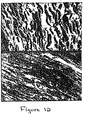

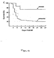
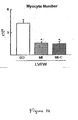
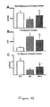


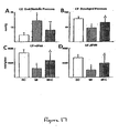




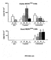
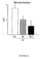

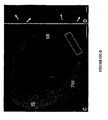

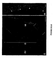
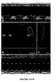


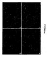






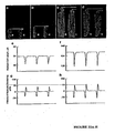
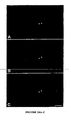
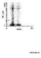






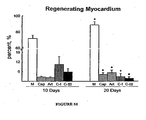
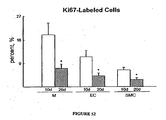



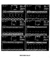
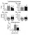


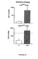
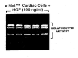
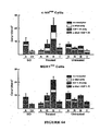
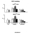

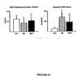

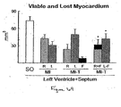


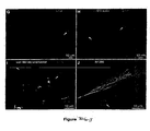


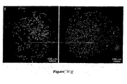
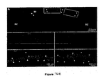
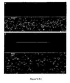
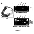
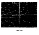
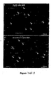

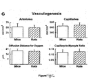

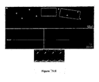
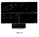
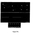


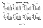
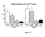





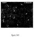
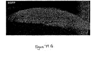
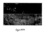


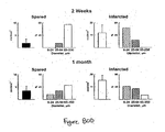


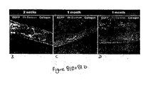


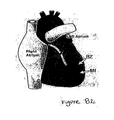
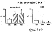


Classifications
-
- A—HUMAN NECESSITIES
- A61—MEDICAL OR VETERINARY SCIENCE; HYGIENE
- A61K—PREPARATIONS FOR MEDICAL, DENTAL OR TOILETRY PURPOSES
- A61K38/00—Medicinal preparations containing peptides
- A61K38/16—Peptides having more than 20 amino acids; Gastrins; Somatostatins; Melanotropins; Derivatives thereof
- A61K38/17—Peptides having more than 20 amino acids; Gastrins; Somatostatins; Melanotropins; Derivatives thereof from animals; from humans
- A61K38/19—Cytokines; Lymphokines; Interferons
-
- A—HUMAN NECESSITIES
- A61—MEDICAL OR VETERINARY SCIENCE; HYGIENE
- A61K—PREPARATIONS FOR MEDICAL, DENTAL OR TOILETRY PURPOSES
- A61K35/00—Medicinal preparations containing materials or reaction products thereof with undetermined constitution
- A61K35/12—Materials from mammals; Compositions comprising non-specified tissues or cells; Compositions comprising non-embryonic stem cells; Genetically modified cells
- A61K35/34—Muscles; Smooth muscle cells; Heart; Cardiac stem cells; Myoblasts; Myocytes; Cardiomyocytes
-
- A—HUMAN NECESSITIES
- A61—MEDICAL OR VETERINARY SCIENCE; HYGIENE
- A61P—SPECIFIC THERAPEUTIC ACTIVITY OF CHEMICAL COMPOUNDS OR MEDICINAL PREPARATIONS
- A61P29/00—Non-central analgesic, antipyretic or antiinflammatory agents, e.g. antirheumatic agents; Non-steroidal antiinflammatory drugs [NSAID]
-
- A—HUMAN NECESSITIES
- A61—MEDICAL OR VETERINARY SCIENCE; HYGIENE
- A61P—SPECIFIC THERAPEUTIC ACTIVITY OF CHEMICAL COMPOUNDS OR MEDICINAL PREPARATIONS
- A61P43/00—Drugs for specific purposes, not provided for in groups A61P1/00-A61P41/00
-
- A—HUMAN NECESSITIES
- A61—MEDICAL OR VETERINARY SCIENCE; HYGIENE
- A61P—SPECIFIC THERAPEUTIC ACTIVITY OF CHEMICAL COMPOUNDS OR MEDICINAL PREPARATIONS
- A61P9/00—Drugs for disorders of the cardiovascular system
-
- A—HUMAN NECESSITIES
- A61—MEDICAL OR VETERINARY SCIENCE; HYGIENE
- A61P—SPECIFIC THERAPEUTIC ACTIVITY OF CHEMICAL COMPOUNDS OR MEDICINAL PREPARATIONS
- A61P9/00—Drugs for disorders of the cardiovascular system
- A61P9/10—Drugs for disorders of the cardiovascular system for treating ischaemic or atherosclerotic diseases, e.g. antianginal drugs, coronary vasodilators, drugs for myocardial infarction, retinopathy, cerebrovascula insufficiency, renal arteriosclerosis
-
- A—HUMAN NECESSITIES
- A61—MEDICAL OR VETERINARY SCIENCE; HYGIENE
- A61P—SPECIFIC THERAPEUTIC ACTIVITY OF CHEMICAL COMPOUNDS OR MEDICINAL PREPARATIONS
- A61P9/00—Drugs for disorders of the cardiovascular system
- A61P9/12—Antihypertensives
-
- C—CHEMISTRY; METALLURGY
- C12—BIOCHEMISTRY; BEER; SPIRITS; WINE; VINEGAR; MICROBIOLOGY; ENZYMOLOGY; MUTATION OR GENETIC ENGINEERING
- C12N—MICROORGANISMS OR ENZYMES; COMPOSITIONS THEREOF; PROPAGATING, PRESERVING, OR MAINTAINING MICROORGANISMS; MUTATION OR GENETIC ENGINEERING; CULTURE MEDIA
- C12N5/00—Undifferentiated human, animal or plant cells, e.g. cell lines; Tissues; Cultivation or maintenance thereof; Culture media therefor
- C12N5/06—Animal cells or tissues; Human cells or tissues
- C12N5/0602—Vertebrate cells
- C12N5/0634—Cells from the blood or the immune system
- C12N5/0647—Haematopoietic stem cells; Uncommitted or multipotent progenitors
-
- C—CHEMISTRY; METALLURGY
- C12—BIOCHEMISTRY; BEER; SPIRITS; WINE; VINEGAR; MICROBIOLOGY; ENZYMOLOGY; MUTATION OR GENETIC ENGINEERING
- C12N—MICROORGANISMS OR ENZYMES; COMPOSITIONS THEREOF; PROPAGATING, PRESERVING, OR MAINTAINING MICROORGANISMS; MUTATION OR GENETIC ENGINEERING; CULTURE MEDIA
- C12N5/00—Undifferentiated human, animal or plant cells, e.g. cell lines; Tissues; Cultivation or maintenance thereof; Culture media therefor
- C12N5/06—Animal cells or tissues; Human cells or tissues
- C12N5/0602—Vertebrate cells
- C12N5/0652—Cells of skeletal and connective tissues; Mesenchyme
- C12N5/0657—Cardiomyocytes; Heart cells
-
- C—CHEMISTRY; METALLURGY
- C12—BIOCHEMISTRY; BEER; SPIRITS; WINE; VINEGAR; MICROBIOLOGY; ENZYMOLOGY; MUTATION OR GENETIC ENGINEERING
- C12N—MICROORGANISMS OR ENZYMES; COMPOSITIONS THEREOF; PROPAGATING, PRESERVING, OR MAINTAINING MICROORGANISMS; MUTATION OR GENETIC ENGINEERING; CULTURE MEDIA
- C12N5/00—Undifferentiated human, animal or plant cells, e.g. cell lines; Tissues; Cultivation or maintenance thereof; Culture media therefor
- C12N5/06—Animal cells or tissues; Human cells or tissues
- C12N5/0602—Vertebrate cells
- C12N5/0652—Cells of skeletal and connective tissues; Mesenchyme
- C12N5/0662—Stem cells
-
- A—HUMAN NECESSITIES
- A61—MEDICAL OR VETERINARY SCIENCE; HYGIENE
- A61K—PREPARATIONS FOR MEDICAL, DENTAL OR TOILETRY PURPOSES
- A61K35/00—Medicinal preparations containing materials or reaction products thereof with undetermined constitution
- A61K35/12—Materials from mammals; Compositions comprising non-specified tissues or cells; Compositions comprising non-embryonic stem cells; Genetically modified cells
-
- C—CHEMISTRY; METALLURGY
- C12—BIOCHEMISTRY; BEER; SPIRITS; WINE; VINEGAR; MICROBIOLOGY; ENZYMOLOGY; MUTATION OR GENETIC ENGINEERING
- C12N—MICROORGANISMS OR ENZYMES; COMPOSITIONS THEREOF; PROPAGATING, PRESERVING, OR MAINTAINING MICROORGANISMS; MUTATION OR GENETIC ENGINEERING; CULTURE MEDIA
- C12N2500/00—Specific components of cell culture medium
- C12N2500/05—Inorganic components
- C12N2500/10—Metals; Metal chelators
- C12N2500/20—Transition metals
- C12N2500/24—Iron; Fe chelators; Transferrin
- C12N2500/25—Insulin-transferrin; Insulin-transferrin-selenium
-
- C—CHEMISTRY; METALLURGY
- C12—BIOCHEMISTRY; BEER; SPIRITS; WINE; VINEGAR; MICROBIOLOGY; ENZYMOLOGY; MUTATION OR GENETIC ENGINEERING
- C12N—MICROORGANISMS OR ENZYMES; COMPOSITIONS THEREOF; PROPAGATING, PRESERVING, OR MAINTAINING MICROORGANISMS; MUTATION OR GENETIC ENGINEERING; CULTURE MEDIA
- C12N2500/00—Specific components of cell culture medium
- C12N2500/30—Organic components
- C12N2500/40—Nucleotides, nucleosides or bases
-
- C—CHEMISTRY; METALLURGY
- C12—BIOCHEMISTRY; BEER; SPIRITS; WINE; VINEGAR; MICROBIOLOGY; ENZYMOLOGY; MUTATION OR GENETIC ENGINEERING
- C12N—MICROORGANISMS OR ENZYMES; COMPOSITIONS THEREOF; PROPAGATING, PRESERVING, OR MAINTAINING MICROORGANISMS; MUTATION OR GENETIC ENGINEERING; CULTURE MEDIA
- C12N2501/00—Active agents used in cell culture processes, e.g. differentation
- C12N2501/10—Growth factors
- C12N2501/105—Insulin-like growth factors [IGF]
-
- C—CHEMISTRY; METALLURGY
- C12—BIOCHEMISTRY; BEER; SPIRITS; WINE; VINEGAR; MICROBIOLOGY; ENZYMOLOGY; MUTATION OR GENETIC ENGINEERING
- C12N—MICROORGANISMS OR ENZYMES; COMPOSITIONS THEREOF; PROPAGATING, PRESERVING, OR MAINTAINING MICROORGANISMS; MUTATION OR GENETIC ENGINEERING; CULTURE MEDIA
- C12N2501/00—Active agents used in cell culture processes, e.g. differentation
- C12N2501/10—Growth factors
- C12N2501/11—Epidermal growth factor [EGF]
-
- C—CHEMISTRY; METALLURGY
- C12—BIOCHEMISTRY; BEER; SPIRITS; WINE; VINEGAR; MICROBIOLOGY; ENZYMOLOGY; MUTATION OR GENETIC ENGINEERING
- C12N—MICROORGANISMS OR ENZYMES; COMPOSITIONS THEREOF; PROPAGATING, PRESERVING, OR MAINTAINING MICROORGANISMS; MUTATION OR GENETIC ENGINEERING; CULTURE MEDIA
- C12N2501/00—Active agents used in cell culture processes, e.g. differentation
- C12N2501/10—Growth factors
- C12N2501/115—Basic fibroblast growth factor (bFGF, FGF-2)
-
- C—CHEMISTRY; METALLURGY
- C12—BIOCHEMISTRY; BEER; SPIRITS; WINE; VINEGAR; MICROBIOLOGY; ENZYMOLOGY; MUTATION OR GENETIC ENGINEERING
- C12N—MICROORGANISMS OR ENZYMES; COMPOSITIONS THEREOF; PROPAGATING, PRESERVING, OR MAINTAINING MICROORGANISMS; MUTATION OR GENETIC ENGINEERING; CULTURE MEDIA
- C12N2501/00—Active agents used in cell culture processes, e.g. differentation
- C12N2501/10—Growth factors
- C12N2501/12—Hepatocyte growth factor [HGF]
Definitions
- the invention relates to the embodiments as defined in the claims. Furthermore, the present invention relates generally to the field of cardiology, and more particularly relates to methods and cellular compositions for treatment of a patient suffering from a cardiovascular disease, including, but not limited to, artherosclerosis, ischemia, hypertension, restenosis, angina pectoris, rheumatic heart disease, congenital cardiovascular defects and arterial inflammation and other disease of the arteries, arterioles and capillaries.
- a cardiovascular disease including, but not limited to, artherosclerosis, ischemia, hypertension, restenosis, angina pectoris, rheumatic heart disease, congenital cardiovascular defects and arterial inflammation and other disease of the arteries, arterioles and capillaries.
- the present invention contemplates treatments, therapeutics and methodologies that can be used in place of, or in conjunction with, traditional, invasive therapeutic treatments such as cardiac or vascular bypass surgery.
- the present invention provides for isolated cardiac stem cells for use in treating a vasculature disorder by forming large coronary vessels or arteries in vivo in a patient, wherein the cardiac stem cells are activated by exposure to hepatocyte growth factor (HGF) and/or insulin-like growth factor-1 (IGF-1) prior to administration to the patient.
- HGF hepatocyte growth factor
- IGF-1 insulin-like growth factor-1
- the present invention also provides for isolated cardiac stem cells for use in treating myocardial infarction or myocardial ischemia by generating myocardium and/or myocardial cells in a patient, wherein the cardiac stem cells are activated by exposure to HGF and/or IGF-1 prior to administration to the patient.
- compositions comprising a therapeutically effective amount of somatic stem cells alone or in combination with a cytokine such as a cytokine selected from the group consisting of stem cell factor (SCF), granulocyte-colony stimulating factor (G-CSF), granulocyte-macrophage colony stimulating factor (GM-CSF), stromal cell-derived factor-1, steel factor, vascular endothelial growth factor, macrophage colony stimulating factor, granulocyte-macrophage stimulating factor or Interleukin-3 or any cytokine capable of the stimulating and/or mobilizing stem cells.
- a cytokine selected from the group consisting of stem cell factor (SCF), granulocyte-colony stimulating factor (G-CSF), granulocyte-macrophage colony stimulating factor (GM-CSF), stromal cell-derived factor-1, steel factor, vascular endothelial growth factor, macrophage colony stimulating factor, granulocyte-macrophage stimulating factor or Interleuk
- Cytokines may be administered alone or in combination of with any other cytokine capable of: the stimulation and/or mobilization of stem cells; the maintenance of early and late hematopoiesis (see below); the activation of monocytes (see below), macrophage/monocyte proliferation; differentiation, motility and survival (see below) and a pharmaceutically acceptable carrier, diluent or excipient (including combinations thereof).
- the stem cells are advantageously adult stem cells, such as hematopoietic or cardiac stem cells or a combination thereof or a combination of cardiac stem cells and any other type of stem cells.
- the implanting, depositing, administering or causing of implanting or depositing or administering of stem cells such as adult stem cells, for instance hematopoietic or cardiac stem cells or a combination thereof or any combination of cardiac stem cells (e.g., adult cardiac stem cells) and stem cells of another type of (e.g., adult stem cells of another type), alone or with a cytokine such as a cytokine selected from the group consisting of stem cell factor (SCF), granulocyte-colony stimulating factor (G-CSF), granulocyte-macrophage colony stimulating factor (GM-CSF), stromal cell-derived factor-1, steel factor, vascular endothelial growth factor, macrophage colony stimulating factor, granulocyte-macrophage stimulating factor or Interleukin-3 or any cytokine capable of the stimulating and/or mobilizing stem cells (wherein "with a cytokine " can include sequential implanting, depositing administering or causing of implanting or depositing or administering of
- This implanting, depositing, or administering or causing of implanting, depositing or administering can be in conjunction with grafts.
- Such implanting, depositing or administering or causing of implanting, depositing or administering is advantageously employed in the treatment or therapy or prevention of cardiac conditions, such as to treat areas of weakness or scarring in the heart or prevent the occurrence or further occurrence of such areas or to treat conditions which cause or irritate such areas, for instance myocardial infarction or ischemia or other e.g., genetic, conditions that impart weakness or scarring to the heart ( see also cardiac conditions mentioned infra ).
- stem cells alone or in combination with said cytokine(s), in the formulation of medicaments for such treatment, therapy or prevention.
- Medicaments for use in such treatment, therapy or prevention comprising the stem cells and optionally the cytokine(s).
- Kits comprising the stem cells and optionally the cytokine(s) for formulations for use in such treatment, therapy or prevention.
- compositions comprising such stem cells and optionally at least one cytokine and kits for preparing such compositions.
- compositions comprising a therapeutically effective amount of one or more cytokines for causing the migration and/or proliferation of cardiac stem cells or cardiac primative cells into circulatory tissue or muscle tissue or circulatory muscle tissue, e.g., cardiac tissue, such as the heart or blood vessels - e.g., veins, arteries, that go to or come from the heart such as veins and arteries directly connected or attached or flowing into the heart, for instance the aorta.
- cardiac tissue such as the heart or blood vessels - e.g., veins, arteries, that go to or come from the heart such as veins and arteries directly connected or attached or flowing into the heart, for instance the aorta.
- This migration and/or proliferation is advantageously employed in the treatment or therapy or prevention of cardiac conditions, such as to treat areas of weakness or scarring in the heart or prevent the occurrence or further occurrence of such areas or to treat conditions which cause or irritate such areas, for instance myocardial infarction or ischemia or other e.g., genetic, conditions that impart weakness or scarring to the heart ( see also cardiac conditions mentioned infra ).
- Medicaments for use in such treatment, therapy or prevention comprising the two or more cytokines.
- Kits comprising the cytokines for formulations for use in such treatment, therapy or prevention.
- compositions comprising the cytokines and kits for preparing such compositions. Methods of making the kits and compositions described herein.
- compositions comprising a therapeutically effective amount of one or more cytokines for causing the migration and/or proliferation of cardiac stem cells or cardiac primative cells into circulatory tissue or muscle tissue or circulatory muscle tissue, e.g., cardiac tissue, such as the heart or blood vessels - e.g., veins, arteries, that go to or come from the heart such as veins and arteries directly connected or attached or flowing into the heart, for instance the aorta in combination with a therapeutically effective amount of a pharmaceutical agent useful in treating hypertension, myocardial infarction, ischemia, angina, or other coronary or vascular ailments, such as AT 1 receptor blockers such as losartan, streptokinase, ReoPro (abciximab), enalapril maleate, Rapilysin (reteplase), Dilatrend (carvedilol), Activase (alteplase), and other drugs for similar uses which would be known by one skilled in the art.
- cardiac tissue such as the heart
- Kits comprising one or more cytokines in combination with a pharmaceutical agent useful in treating hypertension, myocardial infarction, ischemia, angina, or other coronary or vascular ailments.
- Methods of isolating, expanding and activating stem cells in particular cardiac stem cells.
- Methods of treating occlusions or blockages in arteries and/or vessels comprising the administration of stem cells, in particular cardiac stem cells, more in particular activated cardiac stem cells, and optionally in the presence of one or more cytokines.
- Cardiovascular disease is a major health risk throughout the industrialized world.
- Atherosclerosis the most prevalent of cardiovascular diseases, is the principal cause of heart attack, stroke, and gangrene of the extremities, and thereby the principal cause of death in the United States.
- Atherosclerosis is a complex disease involving many cell types and molecular factors (for a detailed review, see Ross, 1993, Nature 362: 801-809 ).
- Ischemia is a condition characterized by a lack of oxygen supply in tissues of organs due to inadequate perfusion. Such inadequate perfusion can have number of natural causes, including atherosclerotic or restenotic lesions, anemia, or stroke, to name a few. Many medical interventions, such as the interruption of the flow of blood during bypass surgery, for example, also lead to ischemia. In addition to sometimes being caused by diseased cardiovascular tissue, ischemia may sometimes affect cardiovascular tissue, such as in ischemic heart disease. Ischemia may occur in any organ, however, that is suffering a lack of oxygen supply.
- MI myocardial infarction
- a heart attack is one of the most well-known types of cardiovascular disease.
- MI is caused by a sudden and sustained lack of blood flow to an area of the heart, commonly caused by narrowing of a coronary artery. Without adequate blood supply, the tissue becomes ischemic, leading to the death of myocytes and vascular structures.
- This area of necrotic tissue is referred to as the infarct site, and will eventually become scar tissue. Survival is dependent on the size of this infarct site, with the probability of recovery decreasing with increasing infarct size. For example, in humans, an infarct of 46% or more of the left ventricle triggers irreversible cardiogenic shock and death (99).
- progenitors very immature cells
- stem cells progenitor cells themselves derive from a class of progenitor cells called stem cells.
- stem cells have the capacity, upon division, for both self-renewal and differentiation into progenitors. Thus, dividing stem cells generate both additional primitive stem cells and somewhat more differentiated progenitor cells.
- stem cells also give rise to cells found in other tissues, including but not limited to the liver, brain, and heart.
- Stem cells have the ability to divide indefinitely, and to specialize into specific types of cells.
- Totipotent stem cells which exist after an egg is fertilized and begins dividing, have total potential, and are able to become any type of cell. Once the cells have reached the blastula stage, the potential of the cells has lessened, with the cells still able to develop into any cell within the body, however they are unable to develop into the support tissues needed for development of an embryo.
- the cells are considered pluripotent, as they may still develop into many types of cells. During development, these cells become more specialized, committing to give rise to cells with a specific function.
- These cells, considered multipotent are found in human adults and referred to as adult stem cells. It is well known that stem cells are located in the bone marrow, and that there is a small amount of peripheral blood stem cells that circulate throughout the blood stream (National Institutes of Health, 2000).
- stem cells Due to the regenerative properties of stem cells, they have been considered an untapped resource for potential engineering of tissues and organs. It would be an advance to provide uses of stem cells with respect to addressing cardiac conditions.
- stem cells in medicine are for the treatment of cancer.
- bone marrow is transplanted into a patient whose own marrow has been destroyed by radiation, allowing the stem cells in the transplanted bone marrow to produce new, healthy, white blood cells.
- stem cells are transplanted into their normal environment, where they continue to function as normal.
- any particular stem cell line was only capable of producing three or four types of cells, and as such, they were only utilized in treatments where the stem cell was required to become one of the types of cells for which their ability was already proven.
- researchers are beginning to explore other options for treatments of myriad disorders, where the role of the stem cell is not well defined. Examples of such work will be presented in support of the present invention.
- Organ transplantation has been widely used to replace diseased, nonfunctional tissue. More recently, cellular transplantation to augment deficiencies in host tissue function has emerged as a potential therapeutic paradigm.
- One example of this approach is the well publicized use of fetal tissue in individuals with Parkinsonism (reviewed in Tompson, 1992), where dopamine secretion from transplanted cells alleviates the deficiency in patients.
- transplanted myoblasts from uneffected siblings fused with endogenous myotubes in Duchenne's patients; importantly the grafted myotubes expressed wild-type dystrophin (Gussoni et al., 1992).
- somatic stem cells into the myocardium surrounding an infarct following a myocardial infarction, migrate into the damaged area, where they differentiate into myocytes, endothelial cells and smooth muscle cells and then proliferate and form structures including myocardium, coronary arteries, arterioles, and capillaries, restoring the structural and functional integrity of the infarct.
- CSCs resident cardiac stem cells
- atria 82
- rat 83, 84
- CSCs express surface antigens commonly found in hematopoietic and skeletal muscle stem cells (85, 86).
- CSCs are clonogenic, self-renewing and multipotent giving rise to all cardiac lineages (84).
- the injured heart has the potential to repair itself.
- this possibility had been limited by our lack of understanding of CSC colonization, proliferation and differentiation in new organized, functioning myocardium (61, 87). Identical obstacles apply to any other source of stem cells in the organism (88).
- HGF hepatocyte growth factor
- IGF-1 insulin-like growth factor-1
- IGF-1 is mitogenic, antiapoptotic and is necessary for neural stem cell multiplication and differentiation (96, 97, 98).
- IGF-1 impacts CSCs by increasing their number and protecting their viability.
- IGF-1 overexpression is characterized by myocyte proliferation in the adult mouse heart (65) and this cell growth may depend on CSC activation, differentiation and survival.
- the invention provides for isolated cardiac stem cells for use in treating a vasculature disorder by forming large coronary vessels or arteries in vivo in a patient, wherein the cardiac stem cells are activated by exposure to hepatocyte growth factor (HGF) and/or insulin-like growth factor-1 (IGF-1) prior to administration to the patient.
- HGF hepatocyte growth factor
- IGF-1 insulin-like growth factor-1
- the invention also provides for isolated cardiac stem cells for use in treating myocardial infarction or myocardial ischemia by generating myocardium and/or myocardial cells in a patient, wherein the cardiac stem cells are activated by exposure to HGF and/or IGF-1 prior to administration to the patient.
- compositions for repairing and/or regenerating recently damaged myocardium and/or myocardial cells comprising the administration of an effective amount of one or more cytokines, e.g. HGF and IGF-1 for causing the mitigation and/or proliferation of cardiac stem cells or cardiac primative cells into circulatory tissue or muscle tissue or circulatory muscle tissue are provided.
- This migration and/or proliferation is advantageously employed in the treatment or therapy or prevention of cardiac conditions, such as to treat areas of weakness or scarring in the heart or prevent the occurrence or further occurrence of such areas or to treat conditions which cause or irritate such areas, for instance myocardial infarction or ischemia or other, e.g. genetic, conditions that impart weakness or scarring to the heart.
- compositions comprising a therapeutically effective amount of one or more cytokines for causing the migration and/or proliferation of cardiac stem cells or cardiac primative cells into circulatory tissue or muscle tissue or circulatory muscle tissue.
- This migration and/or proliferation is advantageously employed in the treatment or therapy or prevention of cardiac conditions, such as to treat areas of weakness or scarring in the heart or prevent the occurrence or further occurrence of such areas or to treat conditions which cause or irritate such areas, for instance myocardial infarction or ischemia or other, e.g. genetic, conditions that impart weakness or scarring to the heart.
- medicaments for use in such treatment, therapy or prevention are also disclosed.
- kits comprising one or more cytokines for formulation for use in such treatment, therapy or prevention.
- compositions and/or kits comprising one or more cytokines in combination with a therapeutic agent for treating cardiac or vascular conditions for formulation for use in such treatment, therapy or prevention.
- Disclosed are methods and/or compositions for repairing and/or regenerating recently damaged myocardium and/or myocardial cells comprising the administration of somatic stem cells, e.g., adult stem cells or cardiac stem cells or hematopoietic stem cells or a combination thereof, such as adult cardiac or adult hematopoietic stem cells or a combination thereof or a combination of cardiac stem cells and a stem cell of another type, such as a combination of adult cardiac stem cells and adult stem cells of another type.
- somatic stem cells e.g., adult stem cells or cardiac stem cells or hematopoietic stem cells or a combination thereof, such as adult cardiac or adult hematopoietic stem cells or a combination thereof or a combination of cardiac stem cells and a stem cell of another type, such as a combination of adult cardiac stem cells and adult stem cells of another type.
- media for use in the culturing and/or expansion of stem cells in vitro, prior to the administration of the stem cells are described.
- compositions for repairing and/or regenerating recently damaged myocardium and/or myocardial cells comprising the administration of at least one cytokine.
- compositions for repairing and/or regenerating recently damaged myocardium and/or myocardial cells comprising the administration of at least one cytokine in combination with a pharmaceutical agent useful in the treatment of cardiac or vascular conditions.
- a method and/or compositions for repairing and/or regenerating recently damaged myocardium comprising the administration of somatic stem cells, e.g., adult stem cells or cardiac stem cells or hematopoietic stem cells or a combination thereof, such as adult cardiac or adult hematopoietic stem cells or a combination thereof or a combination of cardiac stem cells and a stem cell of another type, such as a combination of adult cardiac stem cells and adult stem cells of another type and a cytokine.
- somatic stem cells e.g., adult stem cells or cardiac stem cells or hematopoietic stem cells or a combination thereof, such as adult cardiac or adult hematopoietic stem cells or a combination thereof or a combination of cardiac stem cells and a stem cell of another type, such as a combination of adult cardiac stem cells and adult stem cells of another type and a cytokine.
- compositions comprising admixing the pharmaceutically acceptable carrier and the somatic stem cells and/or cytokines.
- kits comprising a pharmaceutical composition for use in repairing and/or regenerating recently damaged myocardium and/or myocardial cells.
- a cytokine such as a cytokine selected from the group consisting of stem cell factor (SCF), granulocyte-colony stimulating factor (G-CSF), granulocyte-macrophage colony stimulating factor (GM-CSF), stromal cell-derived factor-1, steel factor, vascular endothelial growth factor, macrophage colony stimulating factor, granulocyte-macrophage stimulating factor or interleukin-3 or any cytokine capable of the stimulating and/or mobilizing stem cells (wherein "with a cytokine " can include sequential implanting, depositing administering or causing of implanting or
- Such implanting, depositing or administering or causing of implanting, depositing or administering is advantageously employed in the treatment or therapy or prevention of cardiac conditions, such as to treat areas of weakness or scarring in the heart or prevent the occurrence or further occurrence of such areas or to treat conditions which cause or irritate such areas, for instance myocardial infarction or ischemia or other e.g., genetic, conditions that impart weakness or scarring to the heart ( see also cardiac conditions mentioned supra ).
- stem cells alone or in combination with said cytokine(s), in the formulation of medicaments for such treatment, therapy or prevention.
- medicaments for use in such treatment, therapy or prevention comprising the stem cells and optionally the cytokine(s).
- kits comprising the stem cells and optionally the cytokine(s) for formulations for use in such treatment, therapy or prevention.
- the stem cells and the cytokine(s) can be in separate containers in a package or in one container in a package; and, the kit can optionally include a device for administration (e.g., syringe) and/or instructions for administration and/or admixture.
- a device for administration e.g., syringe
- somatic stem cells and heart tissue are cultured in vitro, optionally in the presence of a ctyokine.
- the somatic stem cells differentiate into myocytes, smooth muscle cells and endothelial cells, and proliferate in vitro, forming myocardial tissue and/or cells.
- These tissues and cells may assemble into cardiac structures including arteries, arterioles, capillaries, and myocardium.
- the tissue and/or cells formed in vitro may then be implanted into a patient, e.g. via a graft, to restore structural and functional integrity.
- the methods of the invention herein relate to the isolation, expansion, and activation of cardiac stem cells, wherein the cardiac stem cells are activated through contact with one or more stem cells. The activated cardiac stem cells are then delivered or implanted at the site of the blockage or occlusion.
- growth media that can be used the culture and expansion of stem cells, in particular cardiac stem cells.
- growth media that can be used to activate stem cells, in particular cardiac stem cells.
- Non-activated stem cells grown in said media can be administered to regenerate myocardium or vasculature.
- Activated stem cells grown in the media can also be administered to regemate myocardium or vasculature, wherein vasculaure includes large arteries and veins, such as in a biological bypass.
- the methods of the present invention are considered biotechnology methods in under 35 U.S.C. ⁇ 287(c)(2)(A)(iii) which provides that the infringement exception for medical activity does not apply to the practice of a process in violation of a biotechnology patent.
- the present invention provides for isolated cardiac stem cells for use in treating a vasculature disorder by forming large coronary vessels or arteries in vivo in a patient, wherein the cardiac stem cells are activated by exposure to hepatocyte growth factor (HGF) and/or insulin-like growth factor-1 (IGF-1) prior to administration to the patient.
- HGF hepatocyte growth factor
- IGF-1 insulin-like growth factor-1
- the invention also provides for isolated cardiac stem cells for use in treating myocardial infarction or myocardial ischemia by generating myocardium and/or myocardial cells in a patient, wherein the cardiac stem cells are activated by exposure to HGF and/or IGF-1 prior to administration to the patient.
- methods and/or pharmaceutical composition comprising a therapeutically effective amount of somatic stem cells alone or in combination with a cytokine selected from the group consisting of stem cell factor (SCF), granulocyte-colony stimulating factor (G-CSF), granulocyte-macrophage colony stimulating factor (GM-CSF), stromal cell-derived factor-1, steel factor, vascular endothelial growth factor, macrophage colony stimulating factor, granulocyte-macrophage stimulating factor, hepatocyte growth factor (HGF), insulin-like growth factor (IGF-1) or Interleukin-3 or any cytokine capable of the stimulating and/or mobilizing stem cells are provided.
- a cytokine selected from the group consisting of stem cell factor (SCF), granulocyte-colony stimulating factor (G-CSF), granulocyte-macrophage colony stimulating factor (GM-CSF), stromal cell-derived factor-1, steel factor, vascular endothelial growth factor, macrophage colony
- Cytokines may be administered alone or in combination or with any other cytokine or pharmaceutical agent capable of: the stimulation and/or mobilization of stem cells; the maintenance of early and late hematopoiesis (see below); the activation of monocytes (see below), macrophage/monocyte proliferation; differentiation, motility and survival (see below); treatment of cardiac or vascular conditions; and a pharmaceutically acceptable carrier, diluent or excipient (including combinations thereof).
- compositions comprising a therapeutically effective amount of one or more cytokines for causing the migration and/or proliferation of cardiac stern cells or cardiac primative cells into circulatory tissue or muscle tissue or circulatory muscle tissue, e.g., cardiac tissue, such as the heart or blood vessels - e.g., veins, arteries, that go to or come from the heart such as veins and arteries directly connected or attached or flowing into the heart, for instance the aorta.
- cardiac tissue such as the heart or blood vessels - e.g., veins, arteries, that go to or come from the heart such as veins and arteries directly connected or attached or flowing into the heart, for instance the aorta.
- the methods and/or compositions comprise effective amounts of two or more cytokines. More specifically, the methods and/or compositions preferably comprise effective amounts of hepatocyte growth factor and insulin-like growth factor-1.
- the cytokines in the pharmaceutical composition may also include mediators known to be involved in the maintenance of early and late hematopoiesis such as IL-1 alpha and IL-1 beta, IL-6, IL-7, IL-8, IL-11 and IL-13; colony-stimulating factors, thrombopoietin, erythropoietin, stem cell factor, fit 3-ligand, hepatocyte cell growth factor, tumor necrosis factor alpha, leukemia inhibitory factor, transforming growth factors beta 1 and beta 3; and macrophage inflammatory protein 1 alpha), angiogenic factors (fibroblast growth factors 1 and 2, vascular endothelial growth factor) and mediators whose usual target (and source) is the connective tissue-forming cells (platelet-derived growth factor A, epidermal growth factor, transforming growth factors alpha and beta 2, oncostatin M and insulin-like growth factor-1), or neuronal cells (nerve growth factor) ( Sensebe, L., et al., Stem Cells
- the methods and/or compositions comprise effective amounts of two or more cytokines in combination with an appropriate pharmaceutical agent useful in treating cardiac and/or vascular conditions.
- the pharmaceutical composition is delivered via injection.
- routes for administration include, but are not limited to subcutaneous or parenteral including intravenous, intraarterial, intramuscular, intraperitoneal, intramyocardial, transendocardial, trans-epicardial, intranasal administration as well as intrathecal, and infusion techniques.
- the pharmaceutical composition is in a form that is suitable for injection.
- a therapeutic of the present invention When administering a therapeutic of the present invention parenterally, it will generally be formulated in a unit dosage injectable form (solution, suspension, emulsion).
- the pharmaceutical formulations suitable for injection include sterile aqueous solutions or dispersions and sterile powders for reconstitution into sterile injectable solutions or dispersions.
- the carrier can be a solvent or dispersing medium containing, for example, water, ethanol, polyol (for example, glycerol, propylene glycol, liquid polyethylene glycol, and the like) , suitable mixtures thereof, and vegetable oils.
- Proper fluidity can be maintained, for example, by the use of a coating such as lecithin, by the maintenance of the required particle size in the case of dispersion and by the use of surfactants.
- Nonaqueous vehicles such as cottonseed oil, sesame oil, olive oil, soybean oil, corn oil, sunflower oil, or peanut oil and esters, such as isopropyl myristate, may also be used as solvent systems for compound compositions
- various additives which enhance the stability, sterility, and isotonicity of the compositions including antimicrobial preservatives, antioxidants, chelating agents, and buffers, can be added.
- Prevention of the action of microorganisms can be ensured by various antibacterial and antifungal agents, for example, parabens, chlorobutanol, phenol, sorbic acid, and the like.
- isotonic agents for example, sugars, sodium chloride, and the like.
- Prolonged absorption of the injectable pharmaceutical form can be brought about by the use of agents delaying absorption, for example, aluminum monostearate and gelatin. According to the present invention, however, any vehicle, diluent, or additive used would have to be compatible with the compounds.
- Sterile injectable solutions can be prepared by incorporating the compounds utilized in practicing the present invention in the required amount of the appropriate solvent with various amounts of the other ingredients, as desired.
- the pharmaceutical composition e.g., comprising a therapeutic compound
- any compatible carrier such as various vehicles, adjuvants, additives, and diluents
- the compounds utilized in the present invention can be administered parenterally to the patient in the form of slow-release subcutaneous implants or targeted delivery systems such as monoclonal antibodies, iontophoretic, polymer matrices, liposomes, and microspheres.
- the pharmaceutical composition utilized in the present invention can be administered orally to the patient.
- Conventional methods such as administering the compounds in tablets, suspensions, solutions, emulsions, capsules, powders, syrups and the like are usable.
- Known techniques which deliver the compound orally or intravenously and retain the biological activity are preferred.
- a composition can be administered initially, and thereafter maintained by further administration.
- a composition can be administered in one type of composition and thereafter further administered in a different or the same type of composition.
- a composition can be administered by intravenous injection to bring blood levels to a suitable level. The patient's levels are then maintained by an oral dosage form, although other forms of administration, dependent upon the patient's condition, can be used.
- mice are treated generally longer than the mice or other experimental animals which treatment has a length proportional to the length of the disease process and drug effectiveness.
- the doses may be single doses or multiple doses over a period of several days, but single doses are preferred.
- animal experiments e.g., rats, mice, and the like, to humans, by techniques from this disclosure and documents cited herein and the knowledge in the art, without undue experimentation.
- the treatment generally has a length proportional to the length of the disease process and drug effectiveness and the patient being treated.
- the quantity of the pharmaceutical composition to be administered will vary for the patient being treated.
- 2 x 10 4 - 1 x 10 5 stem cells and 50-500 ⁇ g/kg per day of a cytokine were administered to the patient. While there would be an obvious size difference between the hearts of a mouse and a human, it is possible that 2 x 10 4 -1 x 10 5 stem cells would be sufficient in a human as well.
- the precise determination of what would be considered an effective dose may be based on factors individual to each patient, including their size, age, size of the infarct, and amount of time since damage. Therefore, dosages can be readily ascertained by those skilled in the art from this disclosure and the knowledge in the art.
- any additives in addition to the active stem cell(s) and/or cytokine(s) are present in an amount of 0.001 to 50 wt% solution in phosphate buffered saline, and the active ingredient is present in the order of micrograms to milligrams, such as about 0.0001 to about 5 wt%, preferably about 0.0001 to about 1 wt%, most preferably about 0.0001 to about 0.05 wt% or about 0.001 to about 20 wt%, preferably about 0.01 to about 10 wt%, and most preferably about 0.05 to about 5 wt%.
- any composition to be administered to an animal or human it is preferred to determine therefore: toxicity, such as by determining the lethal dose (LD) and LD 50 in a suitable animal model e.g., rodent such as mouse; and, the dosage of the composition(s), concentration of components therein and timing of administering the composition(s), which elicit a suitable response.
- toxicity such as by determining the lethal dose (LD) and LD 50 in a suitable animal model e.g., rodent such as mouse
- LD 50 lethal dose
- LD 50 low-d dose
- suitable animal model e.g., rodent such as mouse
- the dosage of the composition(s), concentration of components therein and timing of administering the composition(s) which elicit a suitable response.
- compositions comprising a therapeutic include liquid preparations for orifice, e.g., oral, nasal, anal, vaginal, peroral, intragastric, mucosal (e.g., perlingual, alveolar, gingival, olfactory or respiratory mucosa) etc., administration such as suspensions, syrups or elixirs; and, preparations for parenteral, subcutaneous, intradermal, intramuscular or intravenous administration (e.g., injectable administration), such as sterile suspensions or emulsions.
- Such compositions may be in admixture with a suitable carrier, diluent, or excipient such as sterile water, physiological saline, glucose or the like.
- compositions can also be lyophilized.
- the compositions can contain auxiliary substances such as wetting or emulsifying agents, pH buffering agents, gelling or viscosity enhancing additives, preservatives, flavoring agents, colors, and the like, depending upon the route of administration and the preparation desired. Standard texts, such as " REMINGTON'S PHARMACEUTICAL SCIENCE", 17th edition, 1985 , incorporated herein by reference, may be consulted to prepare suitable preparations, without undue experimentation.
- compositions are conveniently provided as liquid preparations, e.g., isotonic aqueous solutions, suspensions, emulsions or viscous compositions which may be buffered to a selected pH. If digestive tract absorption is preferred, compositions of the invention can be in the "solid" form of pills, tablets, capsules, caplets and the like, including “solid” preparations which are time-released or which have a liquid filling, e.g., gelatin covered liquid, whereby the gelatin is dissolved in the stomach for delivery to the gut. If nasal or respiratory (mucosal) administration is desired, compositions may be in a form and dispensed by a squeeze spray dispenser, pump dispenser or aerosol dispenser. Aerosols are usually under pressure by means of a hydrocarbon. Pump dispensers can preferably dispense a metered dose or, a dose having a particular particle size.
- compositions can contain pharmaceutically acceptable flavors and/or colors for rendering them more appealing, especially if they are administered orally.
- the viscous compositions may be in the form of gels, lotions, ointments, creams and the like (e.g., for transdermal administration) and will typically contain a sufficient amount of a thickening agent so that the viscosity is from about 2500 to 6500 cps, although more viscous compositions, even up to 10,000 cps may be employed.
- Viscous compositions have a viscosity preferably of 2500 to 5000 cps, since above that range they become more difficult to administer. However, above that range, the compositions can approach solid or gelatin forms which are then easily administered as a swallowed pill for oral ingestion.
- Liquid preparations are normally easier to prepare than gels, other viscous compositions, and solid compositions. Additionally, liquid compositions are somewhat more convenient to administer, especially by injection or orally. Viscous compositions, on the other hand, can be formulated within the appropriate viscosity range to provide longer contact periods with mucosa, such as the lining of the stomach or nasal mucosa.
- suitable carriers and other additives will depend on the exact route of administration and the nature of the particular dosage form, e.g., liquid dosage form (e.g., whether the composition is to be formulated into a solution, a suspension, gel or another liquid form), or solid dosage form (e.g., whether the composition is to be formulated into a pill, tablet, capsule, caplet, time release form or liquid-filled form).
- liquid dosage form e.g., whether the composition is to be formulated into a solution, a suspension, gel or another liquid form
- solid dosage form e.g., whether the composition is to be formulated into a pill, tablet, capsule, caplet, time release form or liquid-filled form.
- Solutions, suspensions and gels normally contain a major amount of water (preferably purified water) in addition to the active compound. Minor amounts of other ingredients such as pH adjusters (e.g., a base such as NaOH), emulsifiers or dispersing agents, buffering agents, preservatives, wetting agents, jelling agents, (e.g., methylcellulose), colors and/or flavors may also be present.
- pH adjusters e.g., a base such as NaOH
- emulsifiers or dispersing agents e.g., a base such as NaOH
- buffering agents e.g., preservatives
- wetting agents e.g., methylcellulose
- jelling agents e.g., methylcellulose
- colors and/or flavors e.g., methylcellulose
- the desired isotonicity of the compositions may be accomplished using sodium chloride, or other pharmaceutically acceptable agents such as dextrose, boric acid, sodium tartrate, propylene glycol or other inorganic or organic solutes.
- sodium chloride is preferred particularly for buffers containing sodium ions.
- Viscosity of the compositions may be maintained at the selected level using a pharmaceutically acceptable thickening agent.
- Methylcellulose is preferred because it is readily and economically available and is easy to work with.
- suitable thickening agents include, for example, xanthan gum, carboxymethyl cellulose, hydroxypropyl cellulose, carbomer, and the like. The preferred concentration of the thickener will depend upon the agent selected. The important point is to use an amount which will achieve the selected viscosity. Viscous compositions are normally prepared from solutions by the addition of such thickening agents.
- a pharmaceutically acceptable preservative can be employed to increase the shelf-life of the compositions.
- Benzyl alcohol may be suitable, although a variety of preservatives including, for example, parabens, thimerosal, chlorobutanol, or benzalkonium chloride may also be employed.
- a suitable concentration of the preservative will be from 0.02% to 2% based on the total weight although there may be appreciable variation depending upon the agent selected.
- compositions should be selected to be chemically inert with respect to the active compound. This will present no problem to those skilled in chemical and pharmaceutical principles, or problems can be readily avoided by reference to standard texts or by simple experiments (not involving undue experimentation), from this disclosure and the documents cited herein.
- compositions are prepared by mixing the ingredients following generally accepted procedures.
- the selected components may be simply mixed in a blender, or other standard device to produce a concentrated mixture which may then be adjusted to the final concentration and viscosity by the addition of water or thickening agent and possibly a buffer to control pH or an additional solute to control tonicity.
- the pH may be from about 3 to 7.5.
- Compositions can be administered in dosages and by techniques well known to those skilled in the medical and veterinary arts taking into consideration such factors as the age, sex, weight, and condition of the particular patient, and the composition form used for administration (e.g., solid vs. liquid). Dosages for humans or other mammals can be determined without undue experimentation by the skilled artisan, from this disclosure, the documents cited herein, and the knowledge in the art.
- Suitable regimes for initial administration and further doses or for sequential administrations also are variable, may include an initial administration followed by subsequent administrations; but nonetheless, may be ascertained by the skilled artisan, from this disclosure, the documents cited herein, and the knowledge in the art.
- compositions are used to treat cardiovascular diseases, including, but not limited to, atherosclerosis, ischemia, hypertension, restenosis, angina pectoris, rheumatic heart disease, congenital cardiovascular defects and arterial inflammation and other diseases of the arteries, arterioles and capillaries or related complaint.
- cardiovascular diseases including, but not limited to, atherosclerosis, ischemia, hypertension, restenosis, angina pectoris, rheumatic heart disease, congenital cardiovascular defects and arterial inflammation and other diseases of the arteries, arterioles and capillaries or related complaint.
- the invention involves the administration of stem cells as herein discussed, alone or in combination with one or more cytokine, as herein discussed, for the treatment or prevention of any one or more of these conditions or other conditions involving weakness in the heart.
- compositions for such treatment or prevention use of stem cells as herein discussed, alone or in combination with one or more cytokine, as herein discussed, for formulating such compositions, and kits involving stem cells as herein discussed, alone or in combination with one or more cytokine, as herein discussed, for preparing such compositions and/or for such treatment, or prevention.
- advantageous routes of administration involves those best suited for treating these conditions, such as via injection, including, but are not limited to subcutaneous or parenteral including intravenous, intraarterial, intramuscular, intraperitoneal, intramyocardial, transendocardial, trans-epicardial, intranasal administration as well as intrathecal, and infusion techniques.
- the present invention provides methods and compositions that can be used for such therapeutic treatment as an alternative to, or in combination with, cardiact bypass surgery.
- the present invention provides for the isolation, expansion, activation, and implantation or delivery of activated stem cells, preferably activated cardiac stem cells, to an area of the vasculature in need thereof.
- Such delivery or implantation can be accomplished by any of the methods described herein or which are known to those of skill in the art, including, but not limited to, the use of a NOGA catheter system such that visualization of the area to be treated is possible, and the therapeutic is delivered via a retractable needle associated with such catheter system.
- One of skill in the art will recognize other useful methods of delivery or implantation which can be utilized with the present invention, including those described in Dawn, 2005, the contents of which are incorporated herein in their entirety.
- cardiac tissue is harvested from a patient in need of therapeutic treatment for one of the cardiac or vasculature conditions described herein.
- the present disclosure describes the isolation of stem cells, preferably cardiac stem cells, more preferably c-kit pos cardiac stem cells, which are cultured and expanded in vitro.
- the present disclosure provides media for use in the culture and expansion of stem cells, preferably cardiac stem cells, more preferably c-kit pos cardiac stem cells.
- Such media can comprise DMEM/F12, patient serum, insulin, transferring and sodium selenite.
- the media can further comprise one or more of human recombinant bFGF, human recombinant EGF, uridine and inosine.
- components of the medium can be present in approximate ranges as follows: Component Final Concentration Patient serum 5-20% by weight Human recombinant bFGF 10-100 ng/ml Human recombinant EGF 10-100 ng/ml Insulin 2-20 ⁇ g/ml Transferrin 2-20 ⁇ g/ml Sodium selenite 2-10 ng/ml Uridine 0.24-2.44 mg/ml Inosine, 0.27-2.68 mg/ml
- substitutions of the components of the media may be made as known by those of skill in the art.
- insulin can be substituted with insulin-like growth factor I.
- Uridine and inosine can be substituted with mixtures of other nucleotides, including adenosine, guanosine, xanthine, thymidine, and cytidine.
- the above media can be utilized during the culturing and expansion of stem cells that are to be administered in order to regenerate or create new myocardium in a damaged or infacted area of the heart.
- the cultured and expanded stem cells are activated prior to their implantation or delivery.
- the stem cells are contacted with one or more growth factors.
- Suitable growth factors can be any of those described herein, including, but not limited to: Activin A, Angiotensin II, Bone Morphogenic Protein 2, Bone Morphogenic Protein 4, Bone Morphogenic Protein 6, Cardiotrophin-1, Fibroblast Growth Factor 1, Fibroblast Growth Factor 4, Flt3 Ligand, Glial-Derived Neurotrophic Factor, Heparin, Hepatocyte Growth Factor, Insulin-like Growth Factor-I, Insulin-like Growth Factor-II, Insulin-Like Growth Factor Binding Protein-3, Insulin-Like Growth Factor Binding Protein-5, Interleukin-3, Interleukin-6, Interleukin-8, Leukemia Inhibitory Factor, Midkine, Platelet-Derived Growth Factor AA
- the stem cells are contacted with hepatocyte growth factor (HGF) and/or insulin-like growth factor-1 (IGF-1).
- HGF hepatocyte growth factor
- IGF-1 insulin-like growth factor-1
- the HGF is present in an amount of about 0-400 ng/ml.
- the HGF is present in an amount of about 25, about 50, about 75, about 100, about 125, about 150, about 175, about 200, about 225, about 250, about 275, about 300, about 325, about 350, about 375 or about 400 ng/ml.
- the IGF-1 is present in an amount of about 0-500 ng/ml.
- the IGF-1 is present in an amount of about 25, about 50, about 75, about 100, about 125, about 150, about 175, about 200, about 225, about 250, about 275, about 300, about 325, about 350, about 375, about 400, about 425, about 450, about 475, or about 500 ng/ml.
- the one or more growth factors can be present in the media provided herein, such that in one embodiment, the media comprisies one or more growth factors, DMEM/F12, patient serum, insulin, transferring and sodium selenite and optionally one or more of human recombinant bFGF, human recombinant EGF, uridine and inosine. It is contemplated that the components of the media can be present in the amounts described herein, and one of skill in the art will be able to determine a sufficient amount of the one or more growth factors in order to obtain activation of any stem cells contacted therewith.
- activated cardiac stem cells are delivered to, or implanted in, an area of the vasculature in need of therapy or repair.
- the activated stem cells are delivered to, or implanted in, the site of an occluded or blocked cardiac vessel or artery.
- cardiac stem cells that are ckitpos and contain the flk-1 epitope are delivered to, or implanted in, the area in need of therapy or repair.
- the activated stem cells form into an artery or vessel at the site at which the stem cells were delivered or implanted.
- the formed artery or vessel has a diameter of over 100 ⁇ m. In yet a further embodiment, the formed artery or vessel has a diameter of at least 125, at least 150, at least 175, at least 200, at least 225, at least 250 or at least 275 ⁇ m. In yet another embodiment of the present invention, the formed artery or vessel provides a "biological bypass" around the area in need of therapy or repair, including around an occlusion or blockage such that blood flow, blood pressure, and circulation are restored or improved. In yet a further still embodiment of the present invention, the administration of activated stem cells can be done in conjunction with other therapeutic means, including but not limited to the administration of other therapeutics, including one or more growth factors.
- compositions may be used as therapeutic agents - i.e. in therapy applications.
- treatment and “therapy” include curative effects, alleviation effects, and prophylactic effects.
- patient may encompass any vertebrate including but not limited to humans, mammals, reptiles, amphibians and fish.
- the patient is a mammal such as a human, or an animal mammal such as a domesticated mammal, e.g., dog, cat, horse, and the like, or production mammal, e.g., cow, sheep, pig, and the like
- stem cell or “stem cell” or “hematopoietic cell” refers to either autologous or allogenic stem cells, which may be obtained from the bone marrow, peripheral blood, or other source.
- adult stem cells refers to stem cells that are not embryonic in origin nor derived from embryos or fetal tissue.
- myocardium refers to myocardium which has been damaged within one week of treatment being started. In a preferred embodiment, the myocardium has been damaged within three days of the start of treatment. In a further preferred embodiment, the myocardium has been damaged within 12 hours of the start of treatment. It is advantageous to employ stem cells alone or in combination with cytokine(s) as herein disclosed to a recently damaged myocardium.
- damaged myocardium refers to myocardial cells which have been exposed to ischemic conditions. These ischemic conditions may be caused by a myocardial infarction, or other cardiovascular disease or related complaint. The lack of oxygen causes the death of the cells in the surrounding area, leaving an infarct, which will eventually scar.
- home refers to the attraction and mobilization of somatic stem cells towards damaged myocardium and/or myocardial cells.
- assemble refers to the assembly of differentiated somatic stem cells into functional structures i.e., myocardium and/or myocardial cells, coronary arteries, arterioles, and capillaries etc. This assembly provides functionality to the differentiated myocardium and/or myocardial cells, coronary arteries, arterioles and capillaries.
- the invention involves the use of somatic stem cells. These are present in animals in small amounts, but methods of collecting stem cells are known to those skilled in the art.
- the stem cells are selected to be lineage negative.
- lineage negative is known to one skilled in the art as meaning the cell does not express antigens characteristic of specific cell lineages.
- the lineage negative stem cells are selected to be c-kit positive.
- c-kit is known to one skilled in the art as being a receptor which is known to be present on the surface of stem cells, and which is routinely utilized in the process of identifying and separating stem cells from other surrounding cells.
- the invention further involves a therapeutically effective dose or amount of stem cells applied to the heart.
- An effective dose is an amount sufficient to effect a beneficial or desired clinical result. Said dose could be administered in one or more administrations.
- 2 x 10 4 - 1 x 10 5 stem cells were administered in the mouse model. While there would be an obvious size difference between the hearts of a mouse and a human, it is possible that this range of stem cells would be sufficient in a human as well. However, the precise determination of what would be considered an effective dose may be based on factors individual to each patient, including their size, age, size of the infarct, and amount of time since damage.
- One skilled in the art specifically a physician or cardiologist, would be able to determine the number of stem cells that would constitute an effective dose without undue experimentation.
- the stem cells are delivered to the heart, specifically to the border area of the infarct.
- the infarcted area is visible grossly, allowing this specific placement of stem cells to be possible.
- the stem cells are advantageously administered by injection, specifically an intramyocardial injection. As one skilled in the art would be aware, this is the preferred method of delivery for stem cells as the heart is a functioning muscle. Injection of the stem cells into the heart ensures that they will not be lost due to the contracting movements of the heart.
- the stem cells are administered by injection transendocardially or trans-epicardially.
- This preferred embodiment allows the stem cells to penetrate the protective surrounding membrane, necessitated by the embodiment in which the cells are injected intramyocardially.
- a preferred embodiment of the invention includes use of a catheter-based approach to deliver the trans-endocardial injection.
- the use of a catheter precludes more invasive methods of delivery wherein the opening of the chest cavity would be necessitated.
- optimum time of recovery would be allowed by the more minimally invasive procedure, which as outlined here, includes a catheter approach.
- a cathether approach includes the use of such techniques as the NOGA catheter or similar systems.
- the NOGA cathether system facilitates guided administration by providing electromechanic mapping of the area of interest, as well as a retractable needle that can be used to deliver targeted injections or to bathe a targeted area with a therapeutic. Any of the embodiments of the present invention can be administered through the use of such a system to deliver injections or provide a therapeutic.
- One of skill in the art will recognize alternate systems that also provide the ability to provide targeted treatment through the integration of imaging and a cathether delivery system that can be used with the present invention.
- Information regarding the use of NOGA and similar systems can be found in, for example, Sherman, 2003; Patel, 2005; and Perrin, 2003; the text of each of which are incorporated herein in their entirety.
- stem cells to migrate into the infarcted region and differentiate into myocytes, smooth muscle cells, and endothelial cells. It is known in the art that these types of cells must be present to restore both structural and functional integrity. Other approaches to repairing infarcted or ischemic tissue have involved the implantation of these cells directly into the heart, or as cultured grafts, such as in U.S. Patent No. 6,110,459 , and 6,099,832 .
- Another embodiment of the invention includes the proliferation of the differentiated cells and the formation of the cells into cardiac structures including coronary arteries, arterioles, capillaries, and myocardium. As one skilled in the art is aware, all of these structures are essential for proper function in the heart. It has been shown in the literature that implantation of cells including endothelial cells and smooth muscle cells will allow for the implanted cells to live within the infarcted region, however they do not form the necessary structures to enable the heart to regain full functionality. The ability to restore both functional and structural integrity is yet another aspect of this invention.
- cytokine may be chosen from a group of cytokines, or may include combinations of cytokines.
- SCF Stem cell factor
- G-CSF granulocyte-colony stimulating factor
- Stromal cell-derived factor-1 has been shown to stimulate stem cell mobilization chemotactically, while steel factor has both chemotactic and chemokinetic properties (Caceres-Cortes et al, 2001, Jo et al, 2000, Kim and Broxmeyer, 1998, Ikuta et al, 1991).
- Vascular endothelial growth factor has been surmised to engage a paracrine loop that helps facilitate migration during mobilization (Bautz et al, 2000, Janowska-Wieczorek et al, 2001).
- Macrophage colony stimulating factor and granulocyte-macrophage stimulating factor have been shown to function in the same manner of SCF and G-CSF, by stimulating mobilization of stem cells.
- Interleukin-3 has also been shown to stimulate mobilization of stem cells, and is especially potent in combination with other cytokines.
- the cytokine can be administered via a vector that expresses the cytokine in vivo.
- a vector for in vivo expression can be a vector or cells or an expression system as cited in any document incorporated herein by reference or used in the art, such as a viral vector, e.g., an adenovirus, poxvirus (such as vaccinia, canarypox virus, MVA, NYVAC, ALVAC, and the like), lentivirus or a DNA plasmid vector; and, the cytokine can also be from in vitro expression via such a vector or cells or expression system or others such as a baculovirus expression system, bacterial vectors such as E. coli, and mammalian cells such as CHO cells.
- a viral vector e.g., an adenovirus, poxvirus (such as vaccinia, canarypox virus, MVA, NYVAC, ALVAC, and the like), lentivirus or a DNA
- cytokine compositions may lend themselves to administration by routes outside of those stated to be advantageous or preferred for stem cell preparations; but, cytokine compositions may also be advantageously administered by routes stated to be advantageous or preferred for stem cell preparations.
- a further aspect of the present disclosure involves administration of a therapeutically effective dose or amount of a cytokine.
- An effective dose is an amount sufficient to effect a beneficial or desired clinical result.
- Said dose could be administered in one or more administrations.
- the dose would be given over the course of about two or three days following the beginning of treatment.
- the precise determination of what would be considered an effective dose may be based on factors individual to each patient, including their size, age, size of the infarct, the cytokine or combination of cytokines being administered, and amount of time since damage.
- One skilled in the art specifically a physician or cardiologist, would be able to determine a sufficient amount of cytokine that would constitute an effective dose without being subjected to undue experimentation.
- a cytokine being delivered by injection, specifically subcutaneously or intravenously.
- a person skilled in the art will be aware that subcutaneous injection or intravenous delivery are extremely common and offer an effective method of delivering the specific dose in a manner which allows for timely uptake and circulation in the blood stream.
- a further aspect includes the administered cytokine stimulating the patient's stem cells and causing mobilization into the blood stream.
- the given cytokines are well-known to one skilled in the art for their ability to promote said mobilization.
- the stem cells once the stem cells have mobilized into the bloodstream, they home to the damaged area of the heart, as will become clear through the following examples.
- FIG. 1 Further embodiments of the invention involve the stem cells migrating into the infarcted region and differentiating into myocytes, smooth muscle cells, and endothelial cells. It is known in the art that these types of cells must be present to restore both structural and functional integrity.
- a further aspect of the disclosure includes administering an effective amount of one or more cytokines to the infarcted region.
- An effective dose is an amount sufficient to effect a beneficial or desired clinical result. Said dose could be administered in one or more administrations. However, the precise determination of what would be considered an effective dose may be based on factors individual to each patient, including their size, age, size of the infarct, the cytokine or combination of cytokines being administered, and amount of time since damage.
- One skilled in the art specifically a physician or cardiologist, would be able to determine a sufficient amount of cytokine that would constitute an effective dose without being subjected to undue experimentation.
- a still further aspect of the disclosure includes the administering of an effective amount of one or more cytokines to the heart by injection.
- the cytokines are delivered to the infarcted region or to the area bordering the infarcted region.
- the infarcted area is visible grossly, allowing this specific placement of cytokines to be possible.
- the cytokines are advantageously administered by injection, specifically an intramyocardial injection. As one skilled in the art would be aware, this is the preferred method of delivery for cytokines as the heart is a functioning muscle. Injection of the cytokines into the heart ensures that they will not be lost due to the contracting movements of the heart.
- the cytokines are administered by injection transendocardially or trans-epicardially. This allows the cytokines to penetrate the protective surrounding membrane, necessitated by the embodiment in which the cytokines are injected intramyocardially.
- a preferred embodiment of the invention includes use of a catheter-based approach to deliver the trans-endocardial injection.
- the use of a catheter precludes more invasive methods of delivery wherein the opening of the chest cavity would be necessitated.
- optimum time of recovery would be allowed by the more minimally invasive procedure, which as outlined here, includes a catheter approach.
- a further embodiment of the invention includes the delivery of the cytokines by a single administration.
- a still further embodiment of the invention includes multiple administrations of the same dosage of cytokines to the heart.
- a still further embodiment of the invention includes administration of multiple doses of the cytokines to the heart, such that a gradient is formed.
- a still further embodiment of the invention includes the stimulation, migration, proliferation and/or differentiation of the resident cardiac stem cells.
- Another embodiment of the invention includes the proliferation of the differentiated cells and the formation of the cells into cardiac structures including coronary arteries, arterioles, capillaries, and myocardium. As one skilled in the art is aware, all of these structures are important for proper function in the heart. It has been shown in the literature that implantation of cells including endothelial cells and smooth muscle cells will allow for the implanted cells to live within the infarcted region, however they do not form the necessary structures to enable the heart to regain full functionality. The ability to restore both functional and structural integrity or better functional and structural integrity than previously achieved in the art is yet another aspect of this invention.
- Stem cells employed in the invention are advantageously selected to be lineage negative.
- lineage negative is known to one skilled in the art as meaning the cell does not express antigens characteristic of specific cell lineages.
- the lineage negative stem cells are selected to be c-kit positive.
- c-kit is known to one skilled in the art as being a receptor which is known to be present on the surface of stem cells, and which is routinely utilized in the process of identifying and separating stem cells from other surrounding cells.
- a therapeutically effective dose of stem cells is applied, delivered, or administered to the heart or implanted into the heart.
- An effective dose or amount is an amount sufficient to effect a beneficial or desired clinical result.
- Said dose could be administered in one or more administrations.
- 2 x 10 4 - 1x10 5 stem cells were administered in the mouse model. While there would be an obvious size difference between the hearts of a mouse and a human, it is possible that 2 x 10 4 - 1 x 10 5 stem cells would be sufficient in a human as well. However, the precise determination of what would be considered an effective dose may be based on factors individual to each patient, including their size, age, size of the infarct, and amount of time since damage.
- the stem cells are advantageously bone marrow or are cardiac stem cells; and even more advantageously, the stem cells are adult bone marrow (hematopoietic stem cells) or adult cardiac stem cells or a combination thereof or a combination of cardiac stem cells such as adult cardiac stem cells and another type of stem cell such as another type of adult stem cells.
- the stem cells are adult bone marrow (hematopoietic stem cells) or adult cardiac stem cells or a combination thereof or a combination of cardiac stem cells such as adult cardiac stem cells and another type of stem cell such as another type of adult stem cells.
- the stem cells are delivered to the heart, specifically to the border area of the infarct.
- the infarcted area is visible grossly, allowing this specific placement of stem cells to be possible.
- the stem cells are advantageously administered by injection, specifically an intramyocardial injection. As one skilled in the art would be aware, this is the preferred method of delivery for stem cells as the heart is a functioning muscle. Injection of the stem cells into the heart ensures that they will not be lost due to the contracting movements of the heart.
- the stem cells are administered by injection transendocardially or trans-epicardially.
- This preferred embodiment allows the stem cells to penetrate the protective surrounding membrane, necessitated by the embodiment in which the cells are injected intramyocardially.
- a preferred embodiment of the invention includes use of a catheter-based approach to deliver the trans-endocardial injection.
- the use of a catheter precludes more invasive methods of delivery wherein the opening of the chest cavity would be necessitated.
- optimum time of recovery would be allowed by the more minimally invasive procedure, which as outlined here, includes a catheter approach.
- Disclosure is also the administration of a cytokine.
- This cytokine may be chosen from a group of cytokines, or may include combinations of cytokines.
- a further aspect of the present disclosure involves administration of a therapeutically effective dose of a cytokine.
- An effective dose or amount is an amount sufficient to effect a beneficial or desired clinical result.
- Said dose could be administered in one or more administrations.
- the dose would be given over the course of about two or three days following the beginning of treatment.
- the precise determination of what would be considered an effective dose may be based on factors individual to each patient, including their size, age, size of the infarct, the cytokine or combination of cytokines being administered, and amount of time since damage.
- One skilled in the art specifically a physician or cardiologist, would be able to determine a sufficient amount of cytokine that would constitute an effective dose without being subjected to undue experimentation, especially in view of the disclosure herein and the knowledge in the art.
- the administration of the therapeutically effective dose of at least one cytokine is advantageously by injection, specifically subcutaneously or intravenously.
- a person skilled in the art will be aware that subcutenous injection or intravenous delivery are extremely common and offer an effective method of delivering the specific dose in a manner which allows for timely uptake and circulation in the blood stream.
- a further aspect of the present disclosure includes the administered cytokine stimulating the patient's stem cells and causing mobilization into the blood stream.
- the given cytokines are well known to one skilled in the art for their ability to promote said mobilization.
- the stem cells Once the stem cells have mobilized into the bloodstream, they home to the damaged area of the heart.
- both the implanted stem cells and the mobilized stem cells migrate into the infarct region and differentiate into myocytes, smooth muscle cells, and endothelial cells. It is known in the art that these types of cells are advantageously present to restore both structural and functional integrity.
- Another embodiment of the invention includes the proliferation of the differentiated cells and the formation of the cells into cardiac structures including coronary arteries, arterioles, capillaries, and myocardium. As one skilled in the art is aware, all of these structures are essential for proper function in the heart. It has been shown in the literature that implantation of cells including endothelial cells and smooth muscle cells will allow for the implanted cells to live within the infarcted region, however they do not form the necessary structures to enable the heart to regain full functionality.
- Cardiac structures can be generated ex vivo and then implanted in the form of a graft; with the implantation of the graft being alone or in combination with stem cells or stem cells and at least one cytokine as in this disclosure, e.g., advantageously adult or cardiac or hematopoietic stem cells such as adult cardiac and/or adult hematpoietic stem cells or adult cardiac stem cells with another type of stem cell e.g. another type of adult stem cell.
- the means of generating and/or regenerating myocardium ex vivo may incorporate somatic stem cells and heart tissue being cultured in vitro, optionally in the presence of a ctyokine.
- the somatic stem cells differentiate into myocytes, smooth muscle cells and endothelial cells, and proliferate in vitro, forming myocardial tissue and/or cells. These tissues and cells may assemble into cardiac structures including arteries, arterioles, capillaries, and myocardium. The tissue and/or cells formed in vitro may then be implanted into a patient, e.g. via a graft, to restore structural and functional integrity.
- the source of the tissue being grafted can be from other sources of tissue used in grafts of the heart.
- compositions such as pharmaceutical compositions including somatic stem cells and/or at least one cytokine, for instance, for use in inventive methods for treating cardiovascular disease or conditions or cardiac conditions.
- Bone marrow was harvested from the femurs and tibias of male transgenic mice expressing enhanced green fluorescent protein (EGFP). After surgical removal of the femurs and tibias, the muscle was dissected and the upper and lower surface of the bone was cut on the surface to allow the collecting buffer to infiltrate the bone marrow. The fluid containing buffer and cells was collected in tubes such as 1.5 ml Epindorf tubes.
- EGFP enhanced green fluorescent protein
- Bone marrow cells were suspended in PBS containing 5% fetal calf serum (FCS) and incubated on ice with rat anti-mouse monoclonal antibodies specific for the following hematopoietic lineages: CD4 and CD8 (T-lymphocytes), B-220 (B-lymphocytes), Mac-1 (macrophages), GR-1 (granulocytes) (Caltag Laboratories) and TER-119 (erythrocytes) (Pharmingen). Cells were then rinsed in PBS and incubated for 30 minutes with magnetic beads coated with goat anti-rat immunoglobulin (Polysciences Inc.).
- FCS fetal calf serum
- Lineage positive cells were removed by a biomagnet and lineage negative cells (Lin - ) were stained with ACK-4-biotin (anti-c- kit mAb).
- Cells were rinsed in PBS, stained with streptavidin-conjugated phycoerythrin (SA-PE) (Caltag Labs.) and sorted by fluorescence activated cell sorting (FACS) using a FACSVantage instrument (Becton Dickinson). Excitation of EGFP and ACK-4-biotin-SA-EP occurred at a wavelength of 488 nm.
- the Lin - cells were sorted as c- kit positive (c- kit pos ) and c- kit negative (C- kit NEG ) with a 1-2 log difference in staining intensity ( Figure 1 ).
- the c- kit POS cells were suspended at 2 x 10 4 to 1 x 10 5 cells in 5 ⁇ l of PBS and the c- kit NEG cells were suspended at a concentration of 1 x 10 5 in 5 ⁇ l of PBS.
- Myocardial infarction was induced in female C57BL/6 mice at 2 months of age as described by Li et al. (1997). Three to five hours after infarction, the thorax of the mice was reopened and 2.5 ⁇ l of PBS containing Lin - c- kit POS cells were injected in the anterior and posterior aspects of the viable myocardium bordering the infarct ( Figure 2 ). Infarcted mice, left uninjected or injected with Lin - c- kit - NEG , cells, and sham-operated mice i.e., mice where the chest cavity was opened but no infarction was induced, were used as controls. All animals were sacrificed 9 ⁇ 2 days after surgery. Protocols were approved by institutional review board. Results are presented as mean ⁇ SD. Significance between two measurements was determined by the Student's t test, and in multiple comparisons was evaluated by the Bonferroni method (Scholzen and Gerdes, 2000). P ⁇ 0.05 was considered significant.
- mice were anesthetized with chloral hydrate (400 mg/kg body weight, i.p.), and the right carotid artery was cannulated with a microtip pressure transducer (model SPR-671, Millar) for the measurements of left ventricular (LV) pressures and LV + and -dP/dt in the closed-chest preparation to determine whether developing myocytes derived from the HSC transplant had an impact on function.
- LV left ventricular
- LV + and -dP/dt left ventricular
- Infarcted mice non-injected or injected with Lin - c- kit NEG cells were combined in the statistics. In comparison with sham-operated groups, the infarcted groups exhibited indices of cardiac failure ( Figure 3 ).
- LV end-diastolic pressure (LVEDP) was 36% lower, and developed pressure (LVDP) and LV+ and -dP/dt were 32%, 40%, and 41 % higher, respectively ( Figure 4A ).
- the abdominal aorta was cannulated, the heart was arrested in diastole by injection of cadmium chloride (CdCl 2 ), and the myocardium was perfused retrogradely with 10% buffered formalin.
- CdCl 2 cadmium chloride
- the infarcted portion of the ventricle was easily identifiable grossly and histologically (see Fig. 2A ).
- the lengths of the endocardial and epicardial surfaces delimiting the infarcted region, and the endocardium and epicardium of the entire left ventricle were measured in each section.
- EGFP was detected with a rabbit polyclonal anti-GFP (Molecular Probes).
- Myocytes were recognized with a mouse monoclonal anti-cardiac myosin heavy chain (MAB 1548; Chemicon) or a mouse monoclonal anti- ⁇ -sarcomeric actin (clone 5C5; Sigma), endothelial cells with a rabbit polyclonal anti-human factor VIII (Sigma) and smooth muscle cells with a mouse monoclonal anti- ⁇ -smooth muscle actin (clone 1A4; Sigma). Nuclei were stained with propidium iodide (PI), 10 ⁇ g/ml.
- PI propidium iodide
- the percentage of myocytes in the regenerating myocardium was determined by delineating the area occupied by cardiac myosin stained cells divided by the total area represented by the infarcted region in each case.
- Myocyte proliferation was 93% (p ⁇ 0.001) and 60% (p ⁇ 0.001) higher than in endothelial cells, and 225% (p ⁇ 0.001 and 176% (p ⁇ 0.001) higher than smooth muscle cells, when measured by BrdU and Ki67, respectively.
- EGFP The origin of the cells in the forming myocardium was determined by the expression of EGFP ( Fig. 7 and 8 ).
- EGFP expression was restricted to the cytoplasm and the Y chromosome to nuclei of new cardiac cells.
- EGFP was combined with labeling of proteins specific for myocytes, endothelial cells and smooth muscle cells. This allowed the identification of each cardiac cell type and the recognition of endothelial cells and smooth muscle cells organized in coronary vessels ( Figs. 5 , 7 , and 8 ).
- FISH fluorescence in situ hybridization
- Y-chromosomes were not detected in cells from the surviving portion of the ventricle. However, the Y-chromosome was detected in the newly formed myocytes, indicating their origin as from the injected bone marrow cells ( Fig. 9 ).
- Sections were incubated with rabbit polyclonal anti-MEF2 (C-21; Santa Cruz), rabbit polyclonal anti-GATA-4 (H-112; Santa Cruz), rabbit polyclonal anti-Csx/Nkx2.5 (obtained from Dr. Izumo) and rabbit polyclonal anti-connexin 43 (Sigma).
- FITC-conjugated goat anti-rabbit IgG (Sigma) was used as secondary antibody.
- MEF2 myocyte enhancer factor 2
- GATA-4 cardiac specific transcription factor 4
- the early marker of myocyte development Csx/Nkx2.5 was examined.
- MEF2 proteins are recruited by GATA-4 to synergistically activate the promoters of several cardiac genes such as myosin light chain, troponin T, troponin I, ⁇ -myosin heavy chain, desmin, atrial natriuretic factor and ⁇ -actin (Durocher et al., 1997; Morin et al., 2000).
- Csx/Nkx2.5 is a transcription factor restricted to the initial phases of myocyte differentiation (D.r et al., 1997).
- EXAMPLE 2 Mobilization of Bone Marrow Cells to Repair Infarcted Myocardium
- mice at 2 months of age were splenectomized and 2 weeks later were injected subcutaneously with recombinant rat stem cell factor (SCF), 200 ⁇ g/kg/day, and recombinant human granulocyte colony stimulating factor (G-CSF), 50 ⁇ g/kg/day (Amgen), once a day for 5 days (Bodine et al., 1994; Orlic et al., 1993). Under ether anesthesia, the left ventricle (LV) was exposed and the coronary artery was ligated (Orlic et al., 2001; Li et al., 1997; Li et al., 1999). SCF and G-CSF were given for 3 more days.
- SCF rat stem cell factor
- G-CSF human granulocyte colony stimulating factor
- Controls consisted of splenectomized infarcted and sham-operated (SO) mice injected with saline.
- BrdU 50 mg/kg body weight, was given once a day, for 13 days, before sacrifice; mice were killed at 27 days. Protocols were approved by New York Medical College. Results are mean ⁇ SD. Significance was determined by the Student's t test and Bonferroni method (Li et al., 1999). Mortality was computed with log-rank test. P ⁇ 0.05 was significant.
- Echocardiography was performed in conscious mice using a Sequoia 256c (Acuson) equipped with a 13-MHz linear transducer (15L8).
- the anterior chest area was shaved and two dimensional (2D) images and M-mode tracings were recorded from the parasternal short axis view at the level of papillary muscles. From M-mode tracings, anatomical parameters in diastole and systole were obtained (Pollick et al., 1995).
- Mice were anesthetized with chloral hydrate (400 mg/kg body weight, ip) and a microtip pressure transducer (SPR-671, Millar) connected to a chart recorder was advanced into the LV for the evaluation of pressures and + and - dP/dt in the closed-chest preparation (Orlic et al., 2001; Li et al., 1997; Li et al., 1999).
- EF was 48%, 62% and 114% higher in treated than in non-treated mice at 9, 16 and 26 days after coronary occlusion, respectively ( Fig. 15D ).
- contractile function developed with time in the infarcted region of the wall ( Figs. 15E-M ; Figs. 16H-P , www.pnas.org).
- LVEDP LV end-diastolic pressure
- the changes in LV systolic pressure (not shown), developed pressure (LVDP), + and -dP/dt were also more severe in the absence of cytokine treatment ( Figs. 17A-D ).
- cytokine-mediated infarct repair restored a noticeable level of contraction in the regenerating myocardium, decreasing diastolic wall stress and increasing ventricular performance.
- Myocardial regeneration attenuated cavitary dilation and mural thinning during the evolution of the infarcted heart in vivo.
- LV end-systolic (LVESD) and end-diastolic (LVEDD) diameters increased more in non-treated than in cytokine-treated mice, at 9, 16 and 26 days after infarction ( Figs. 16A-B ).
- Infarction prevented the evaluation of systolic (AWST) and diastolic (AWDT) anterior wall thickness.
- AWST systolic
- AWDT diastolic
- PWST posterior wall thickness in systole
- PWDT diastole
- BMC-induced repair resulted in a 42% higher wall thickness-to-chamber radius ratio ( FIG. 15A ). Additionally, tissue regeneration decreased the expansion in cavitary diameter, -14%, longitudinal axis, -5% ( Figs.16F-G ), and chamber volume, -26% ( Fig. 15B ). Importantly, ventricular mass-to-chamber volume ratio was 36% higher in treated animals ( Fig. 15C ). Therefore, BMC mobilization that led to proliferation and differentiation of a new population of myocytes and vascular structures attenuated the anatomical variables which define cardiac decompensation.
- the abdominal aorta was cannulated, the heart was arrested in diastole with CdCl 2 and the myocardium was perfused with 10% formalin.
- the LV chamber was filled with fixative at a pressure equal to the in vivo measured end-diastolic pressure (Li et al., 1997; Li et al., 1999).
- the LV intracavitary axis was measured and three transverse slices from the base, mid-region and apex were embedded in paraffin. The mid-section was used to measure LV thickness, chamber diameter and volume (Li et al., 1997; Li et al., 1999). Infarct size was determined by the number of myocytes lost from the LVFW (Olivetti et al., 1991; Beltrami et al., 1994).
- the volume of the LVFW (weight divided by 1.06 g/ml) was determined in each group of mice.
- the volume of regenerating myocardium was determined by measuring in each of three sections the area occupied by the restored tissue and section thickness. The product of these two variables yielded the volume of tissue repair in each section. Values in the three sections were added and the total volume of formed myocardium was obtained. Additionally, the volume of 400 myocytes was measured in each heart. Sections were stained with desmin and laminin antibodies and propidium iodide (PI). Only longitudinally oriented cells with centrally located nuclei were included. The length and diameter across the nucleus were collected in each myocyte to compute cell volume, assuming a cylindrical shape (Olivetti et al., 1991; Beltrami et al., 1994).
- Myocytes were divided in classes and the number of myocytes in each class was calculated from the quotient of total myocyte class volume and average cell volume (Kajstura et al., 1995; Reiss et al., 1996). Number of arteriole and capillary profiles per unit area of myocardium was measured as previously done (Olivetti et al., 1991; Beltrami et al., 1994).
- M Myocytes
- EC endothelial cells
- SMC smooth muscle cells
- BrdU was injected daily between days 14 to 26 to measure the cumulative extent of cell proliferation while Ki67 was assayed to determine the number of cycling cells at sacrifice. Ki67 identifies cells in G1, S, G2, prophase and metaphase, decreasing in anaphase and telophase (Orlic et al., 2001).
- the percentages of BrdU and Ki67 positive myocytes were 1.6- and 1.4-fold higher than EC, and 2.8- and 2.2-fold higher than SMC, respectively ( Fig. 18C , 19 ).
- the forming myocardium occupied 76 ⁇ 11% of the infarct; myocytes constituted 61 ⁇ 12%, new vessels 12 ⁇ 5% and other components 3 ⁇ 2%.
- the band contained 15x10 6 regenerating myocytes that were in an active growing phase and had a wide size distribution ( FIGs. 18D-E ).
- EC and SMC growth resulted in the formation of 15 ⁇ 5 arterioles and 348 ⁇ 82 capillaries per mm 2 of new myocardium.
- Thick wall arterioles with several layers of SMC and luminal diameters of 10-30 ⁇ m represented vessels in early differentiation.
- arterioles and capillaries containing erythrocytes Figs. 18F-H ).
- Cytoplasmic and nuclear markers were used.
- Myocyte nuclei rabbit polyclonal Csx/Nkx2.5, MEF2, and GATA4 antibodies (Orlic et al., 2001; Lin et al., 1997; Kasahara et al., 1998);
- cytoplasm mouse monoclonal nestin (Kachinsky et al., 1995), rabbit polyclonal desmin (Hermann and Aebi, 1998), cardiac myosin, mouse monoclonal ⁇ -sarcomeric actin and rabbit polyclonal connexin 43 antibodies (Orlic et al., 2001).
- EC cytoplasm mouse monoclonal flk-1, VE-cadherin and factor VIII antibodies (Orlic et al., 2001; Yamaguchi et al., 1993; Breier et al., 1996).
- SMC cytoplasm flk-1 and ⁇ -smooth muscle actin antibodies (Orlic et al., 2001; Couper et al., 1997). Scar was detected by a mixture of collagen type I and type III antibodies.
- cytoplasmic proteins Five cytoplasmic proteins were identified to establish the state of differentiation of myocytes (Orlic et al., 2001; Kachinsky et al., 1995; Hermann and Aebi, 1998): nestin, desmin, ⁇ -sarcomeric actin, cardiac myosin and connexin 43. Nestin was recognized in individual cells scattered across the forming band ( Fig. 20A ). With this exception, all other myocytes expressed desmin ( Fig. 20B ), ⁇ -sarcomeric actin, cardiac myosin and connexin 43 ( Fig. 20C ).
- This tyrosine kinase receptor promotes migration of SMC during angiogenesis (Couper et al., 1997). Therefore, repair of the infarcted heart involved growth and differentiation of all cardiac cell populations resulting in de novo myocardium.
- HGF hepatocyte growth factor
- SCF stem cell factor
- GM-CSF granulocyte monocyte colony stimulating factor
- HGF did not mobilize a larger number of cells at a concentration of 100 ng/ml.
- the cells that showed a chemotactic response to HGF consisted of 15% of c-kit positive (c- kit POS ) cells, 50% of multidrug resistance -1 (MDR-1) labeled cells and 30% of stem cell antigen-1 (Sca-1) expressing cells.
- MDR-1 multidrug resistance -1
- Sca-1 stem cell antigen-1
- Cardiac myosin positive myocytes constituted 50% of the preparation, while factor VIII labeled cells included 15%, alpha-smooth muscle actin stained cells 4%, and vimentin positive factor VIII negative fibroblasts 20%. The remaining cells were small undifferentiated and did not stain with these four antibodies.
- the mouse heart possesses primitive cells which are mobilized by growth factors. HGF translocates cells that in vitro differentiate into the four cardiac cell lineages.
- infarcted Fischer 344 rats were injected with these BrdU positive cells in the damaged region, 3-5 hours after coronary artery occlusion. Two weeks later, animals were sacrificed and the characteristics of the infarcted area were examined. Myocytes containing parallel arranged myofibrils along their longitudinal axis were recognized, in combination with BrdU labeling of nuclei. Moreover, vascular structures comprising arterioles and capillary profiles were present and were also positive to BrdU.
- primitive c-kit positive cells reside in the senescent heart and maintain the ability to proliferate and differentiate into parenchymal cells and coronary vessels when implanted into injured functionally depressed myocardium.
- the heart is not a post-mitotic organ but contains a subpopulation of myocytes that physiologically undergo cell division to replace dying cells.
- Myocyte multiplication is enhanced during pathologic overloads to expand the muscle mass and maintain cardiac performance.
- the origin of these replicating myocytes remains to be identified. Therefore, primitive cells with characteristics of stem/progenitor cells were searched for in the myocardium of of Fischer 344 rats. Young and old animals were studied to determine whether aging had an impact on the size population of stem cells and dividing myocytes.
- the numbers of c-kit and MDR1 positive cells in rats at 4 months were 11 ⁇ 3, and 18 ⁇ 6/100 mm 2 of tissue, respectively. Values in rats at 27 months were 35 ⁇ 10, and 42 ⁇ 13/100 mm 2 .
- Ki67 protein which is expressed in nuclei of cycling cells was detected in 1.3 ⁇ 0.3% and 4.1 ⁇ 1.5% of myocytes at 4 and 27 months, respectively.
- the critical role played by resident primitive cells in the remodeling of the injured heart is well appreciated when organ chimerism, associated with transplantation of a female heart in a male recipient, is considered.
- organ chimerism associated with transplantation of a female heart in a male recipient
- 8 female hearts implanted in male hosts were analyzed.
- Translocation of male cells to the grafted female heart was identified by FISH for Y chromosome (see Example 1E).
- FISH FISH for Y chromosome
- the percentages of myocytes, coronary arterioles and capillary profiles labeled by Y chromosome were 9%, 14% and 7%, respectively.
- the numbers of undifferentiated c-kit and multidrug resistance-1 (MDR1) positive cells in the implanted female hearts were measured. Additionally, the possibility that these cells contained the Y chromosome was established.
- Cardiac transplantation involves the preservation of portions of the atria of the recipient on which the donor heart with part of its own atria is attached. This surgical procedure is critical for understanding whether the atria from the host and donor contained undifferentiated cells that may contribute to the complex remodeling process of the implanted heart. Quantitatively, the values of c-kit and, MDR1 labeled cells were very low in control non-transplanted hearts: 3 c-kit and 5 MDR1/100 mm 2 of left ventricular myocardium. In contrast, the numbers of c-kit and MDR1 cells in the atria of the recipient were 15 and 42/100 mm 2 .
- the number of MDR1 positive cells was higher than those expressing c-kit , but followed a similar localization pattern; 43 ⁇ 14, 29 ⁇ 16, 14 ⁇ 7 and 12 ⁇ 10/100 mm 2 in the atria, apex, base and mid-section. Again, the values in the atria and apex were greater than in the other two areas. Sca-1 labeled cells showed the highest value; 150 ⁇ 36/100 mm 2 positive cells were found in the atria. Cells positive for c-kit, MDR1 and Sca-1 were negative for CD45, and for myocyte, endothelial cell, smooth muscle cell and fibroblast cytoplasmic proteins.
- the number of cells positive to both c-kit and MDR1 was measured to recognize cells that possessed two stem cell markers.
- 36% of c-kit labeled cells expressed MDR1 and 19% of MDR1 cells had also c-kit.
- stem cells are distributed throughout the mouse heart, but tend to accumulate in the regions at low stress, such as the atria and the apex.
- EXAMPLE 8 Repair of Infarcted Myocardium by Resident Cardiac Stem Cells Migration, Invasion and Expression Assays
- HGF HGF-Met
- the receptor of HGF, c-Met has been identified on hematopoietic and hepatic stem cells ( 126, 90 ) and, most importantly, on satellite skeletal muscle cells ( 92 ) and embryonic cardiomyocytes ( 127 ). These findings prompted us to determine whether c-Met was present in CSCs and its ligand HGF had a biological effect on these undifferentiated cells.
- HGF promotes migration and invasion of CSCs in vitro and favors their translocation from storage areas to sites of infarcted myocardium in vivo.
- HGF influences cell migration ( 128 ) through the expression and activation of matrix metalloproteinase-2 ( 94, 95 ). This enzyme family may destroy barriers in the extracellular matrix facilitating CSC movement, homing and tissue restoration.
- IGF-1 is mitogenic, antiapoptotic and is necessary for neural stem cell multiplication and differentiation ( 96, 97, 98 ). If CSCs express IGF-1R, IGF-1 may impact in a comparable manner on CSCs protecting their viability during migration to the damaged myocardium. IGF-1 overexpression is characterized by myocyte proliferation in the adult mouse heart ( 65 ) and this form of cell growth may depend on CSC activation, differentiation and survival.
- SFM serum-free medium
- the filter for the 48-well plate consisted of gelatin-coated polycarbonate membrane with pores of 5 ⁇ m in diameter.
- the bottom well was filled with SFM containing 0.1% BSA and HGF at increasing concentrations; 50 ⁇ l of small cell suspension were placed in the upper well. Five hours later, filters were fixed in 4% paraformaldheyde for 40 minutes and stained with PI, and c-kit and MDR1 antibodies.
- FITC-conjugated anti-IgG was used as a secondary antibody.
- Six separate experiments were done at each HGF concentration. Forty randomly chosen fields were counted in each well in each assay to generate a dose-response curve ( fig. 61 ). The motogenic effects of IGF-1 on small cells was excluded by performing migration assays with IGF-1 alone or in combination with HGF (data not shown). Invasion assays were done utilizing a chamber with 24-wells and 12 cell culture inserts (Chemicon, Temecula, CA). A thin layer of growth factor-depleted extracellular matrix was spread on the surface of the inserts. Conversely, 100 ng/ml of HGF were placed in the lower chamber.
- Invading cells digested the coating and clung to the bottom of the polycarbonate membrane. The number of translocated cells was measured 48 hours later following the same protocol described in the migration assay. Four separate experiments were done ( fig. 62 ). Consistent with the results obtained in the migration assay, IGF-1 had no effects on cell invasion (data not shown).
- IGF-1 had no effect on the mobility of these CSCs at concentrations varying from 25 to 400 ng/ml.
- the addition of IGF-1 to HGF did not modify the migration and invasion characteristics of c-kit POS and MDR1 POS cells obtained by HGF alone.
- Myocardial infarction was produced in mice and 5 hours later 4 separate injections of a solution containing HGF and IGF-1 were performed from the atria to the border zone. HGF was administrated at increasing concentrations to create a chemotactic gradient between the stored CSCs and the dead tissue. This protocol was introduced to enhance homing of CSCs to the injured area and to generate new myocardium. If this were the case, large infarcts associated with animal death may be rapidly reduced and the limits of infarct size and survival extended by this intervention.
- mice Female 129 SV-EV mice were used. Following anesthesia (150 mg ketamine-1 mg acepromazine/kg b.w., i.m.), mice were ventilated, the heart was exposed and the left coronary artery was ligated (61, 87). Coronary ligation in animals to be treated with growth factors was performed as close as possible to the aortic origin to induce very large infarcts. Subsequently, the chest was closed and animals were allowed to recover. Five hours later, mice were anesthetized, the chest was reopened and four injections of HGF-IGF-1, each of 2.5 ⁇ l, were made from the atria to the region bordering the infarct.
- mice were injected with BrdU (50 mg/kg b.w.) from day 6 to day 16 to identify small, newly formed, proliferating myocytes during this interval. Sham-operated and infarcted-untreated mice were injected with normal saline in the same four sites.
- BrdU 50 mg/kg b.w.
- a large fraction of c-kit POS and MDR1 POS cells expressed c-Met and IGF-1R alone or in combination. Myocardial infarction and the administration of growth factors did not alter in a consistent manner the relative proportion of CSCs with and without c-Met and IGF-1R in the myocardium ( Figure 64 ).
- Hairpin 1 (apoptosis) and hairpin 2 (necrosis) labeling and Ki67 expression in nuclei were used to establish the viability and activation of c-kit POS and MDR1 POS cells in the various portions of the damaged and non-damaged heart, respectively ( Fig. 22 , G to L).
- CSCs were more numerous in the atria than in the ventricle of control mice.
- Acute myocardial infarction and growth factor administration markedly changed the number and the distribution of primitive cells in the heart.
- Viable c-kit POS and MDR1 POS cells significantly increased in the spared myocardium of the border zone and remote tissue as well as in the dead myocardium of the infarcted region.
- CSCs decreased in the atria ( Fig. 22 , M and N), suggesting that a translocation of primitive cells occurred from this site of storage to the stressed viable and dead myocardium.
- a different phenomenon was noted in infarcted-untreated mice, in which viable CSCs remained higher in the atria than in the ventricle.
- the heart was arrested in diastole with CdCl 2 , and the myocardium was perfused with 10% formalin.
- the LV chamber was filled with fixative at a pressure equal to the in vivo measured end-diastolic pressure.
- the LV intracavitary axis was determined and the mid-section was used to obtain LV thickness and chamber diameter. Infarct size was measured by the number of myocytes lost from the LV inclusive of the interventricular septum ( 87 ).
- mice that received growth factors survived with infarcts affecting more than 60% of the LV. Seven of these mice had infarcts that involved 75% to 86% of LV. Untreated mice had infarcts that never exceeded 60% ( Fig. 23 , C and D). In contrast to injected mice, a portion of the posterior aspect of the LV wall and the entire interventricular septum had to be preserved for untreated animals to survive. An infarct larger than 60% is incompatible with life in mice, rats, dogs and any other mammalian species. Irreversible cardiogenic shock and death supervene in humans with a 46% infarct ( 99 ).
- HGF-IGF-1 The chemotactic and mitogenic properties of HGF-IGF-1 resulted in the mobilization, proliferation and differentiation of primitive cells in the infarcted region of the wall creating new myocardium.
- the band occupied 65 ⁇ 8% of the damaged area and was located in the mid-portion of the infarct equally distant from the inner and outer layer of the wall.
- the entire thickness of the wall was replaced by developing myocardium ( Fig. 23 , E to H).
- the composition of the repairing myocardium was evaluated morphometrically. Antibodies specific for myocytes, endothelial cells and smooth muscle cells were employed for the recognition of parenchymal cells and vessel profiles ( 61, 87 ). Moreover, BrdU labeling of cells was used as a marker of regenerating tissue over time. Myocytes occupied 84 ⁇ 3% of the band, the coronary vasculature 12 ⁇ 3%, and other structural components 4 ⁇ 1%. New myocytes varied from 600 to 7,200 ⁇ m 3 , with an average volume of 2,200 ⁇ 400 ⁇ m 3 ( Figures 68 and 69 ). Together, 3.1 ⁇ 1.1 million myocytes were formed to compensate for a loss of 2.4 ⁇ 0.8 million cells.
- mice were anesthetized and a Millar microtip pressure transducer connected to a chart recorder was advanced into the LV for the evaluation of pressures and + and - dP/dt in the closed-chest preparation. Echocardiography performed at day 15 showed that contractile activity was partially restored in the regenerating portion of the wall of treated infarcts. Ejection fraction was also higher in treated than in untreated mice ( Fig. 24 , A to E). Thus, structural repair was coupled with functional repair.
- myocytes were small with myofibrils located at the periphery of the cell in the subsarcolemmal region.
- the new myocytes resembled neonatal cells actively replicating DNA. They were markedly smaller than the spared hypertrophied ventricular myocytes ( Fig. 25 , A and B).
- growing cells showed a higher peak shortening and velocity of shortening, and a lower time to peak shortening ( Fig. 25 , C to J).
- the isolated newly generated myocytes were stained by Ki67 to determine whether these cells were cycling and, therefore, synthesizing DNA.
- An identical protocol was applied to the isolated surviving hypertrophied myocytes of infarcted-treated mice.
- the DNA content of each myocyte nucleus in mononucleated and binucleated cells was evaluated by PI staining and confocal microscopy (see Fig. 25 , A and B).
- Control diploid mouse lymphocytes were used as baseline. The objective was to establish if cell fusion occurred in CSCs before their commitment to cell lineages. This possibility has recently been suggested by in vitro studies ( 131, 132 ). Non-cycling new myocytes and enlarged spared myocytes had only diploid nuclei, excluding that such a phenomenon played a role in cardiac repair ( Figure 66 ).
- N-cadherin identifies the fascia adherens and connexin 43 the gap junctions in the intercalated discs. These proteins are developmentally regulated. Connexin 43 is also critical for electrical coupling and synchrony of contraction of myocytes. These 6 proteins were detected in essentially all newly formed myocytes ( Fig. 26 , A to N). The percentage of myocytes labeled by BrdU was 84 ⁇ 9%, indicating that cell proliferation was ongoing in the regenerating tissue.
- Cardiac repair included the formation of capillaries and arterioles ( Fig. 27 , A to D).
- the presence of red blood cells within the lumen indicated that the vessels were connected with the coronary circulation.
- This phase of myocardial restoration was characterized by a prevailing growth of resistance arterioles than capillary structures. There were 59 ⁇ 29 arterioles and 137 ⁇ 80 capillaries per mm 2 of new myocardium.
- c-kit POS cells scored negative for myocyte ⁇ -sarcomeric actin, cardiac myosin, desmin, ⁇ -cardiac actinin, connexin 43
- endothelial cell EC; factor VIII, CD31, vimentin
- smooth muscle cell SMC; ⁇ -smooth muscle actin, desmin
- fibroblast F; vimentin
- Nuclear markers of myocyte lineage Nkx2.5, MEF2, GATA-4) were detected in 7-10% and cytoplasmic proteins in 1-2% of the cells.
- c-kit POS cells did not express skeletal muscle transcription factors (MyoD, myogenin, Myf5) or markers of the myeloid, lymphoid and erythroid cell lineages (CD45, CD45RO, CD8, TER-119), indicating the cells were Lin - c- kit POS cells.
- c-kit POS cells were plated at 1-2x10 4 cells/ml NSCM utilized for selection and growth of neural stem cells (122). This was composed by Dulbecco's MEM and Ham's F12 (ratio 1:1), bFGF, 10ng/ml, EGF, 20ng/ml, HEPES, 5mM, insulin-transferrin-selenite.
- c-kit POS cells attached in two weeks and began to proliferate ( Fig. 28 a,b ). NSCM was then substituted with differentiating medium (DM) and confluence was reached in 7-10 days. Cells were passaged by trypsinization.
- DM differentiating medium
- fibronectin procollagen type I and vimentin were absent excluding the fibroblast lineage. Individual colonies were detached with cloning cylinders and plated. Multiple clones developed and one clone in each preparation was chosen for characterization. MEM containing 10% FCS and 10 -8 M dexamethasone was employed to induce differentiation (DM). For subcloning, cells from multiple clones were plated at 10-50 cells/ml NSCM. Single subclones were isolated and plated in DM. At each subcloning step, an aliquot of cells was grown in suspension to develop clonal spheres.
- Each clone contained groups of 2-3 Lin - c- kit POS cells ( Fig. 29 a ), although the majority of these cells ( ⁇ 20-50) were dispersed among c- kit NEG cells. Some cells were Ki67 positive and occasionally in mitosis ( Fig. 29b -d). Myocytes expressing cardiac myosin and ⁇ -sarcomeric actin, EC expressing factor VIII, CD31 and vimentin, SMC expressing ⁇ -smooth muscle actin and F expressing vimentin alone were identified in each clone ( Fig. 29 e-h ). Aggregates of small cells containing nestin were also present (Supplementary Information).
- Lin - c- kit POS cells isolated from the myocardium possessed the properties expected for stem cells. They were clonogenic, self-renewing and multipotent and gave origin to the main cardiac cell types. Subclonal analysis of several primary clones confirmed the stability of the phenotype of the primary clones: clonogenicity, self-renewal and multipotentiality. The phenotype of most subclones was indistinguishable from that of the primary clones. However, in two of eight subclones, only myocytes were obtained in one case and exclusively EC were identified in the other.
- spherical clones ( FIG. 30 a ). This anchorage independent growth is typical of stem cells 14,15 .
- Spheroids consisted of clusters of c- kit POS and c- kit NEG cells and large amounts of nestin ( Fig. 30 b-d ).
- spheroids readily attached, and cells migrated out of the spheres and differentiated ( Fig. 30 e-h ).
- Cells were fixed in 4% paraformaldehyde and undifferentiated cells were labeled with c-kit antibody.
- Markers for myocytes included Nkx2.5, MEF2, GATA-4, GATA-5, nestin, ⁇ -sarcomeric actin, ⁇ -cardiac actinin, desmin and cardiac myosin heavy chain.
- Markers for SMC comprised ⁇ -smooth muscle actin and desmin, for EC factor VIII, CD31 and vimentin, and for F vimentin in the absence of factor VIII, fibronectin and procollagen type I.
- MyoD, myogenin and Myf5 were utilized as markers of skeletal muscle cells.
- CD45, CD45RO, CD8 and TER-119 were employed to exclude hematopoietic cell lineages.
- MAP1b, neurofilament 200 and GFAP were used to recognize neural cell lineages.
- BrdU and Ki67 were employed to identify cycling cells (61, 87). Nuclei were stained by PI.
- Hearts were arrested in diastole and fixed with formalin. Infarct size was determined by the fraction of myocytes lost from the left ventricle (87), 53 ⁇ 7% and 49 ⁇ 10% (NS) in treated and untreated rats at 10 days, and 70 ⁇ 9% and 55 ⁇ 10% ( P ⁇ 0.001) in treated and untreated rats at 20 days, respectively. The volume of 400 new myocytes was measured in each heart. Sections were stained with desmin and laminin and PI. In longitudinally oriented myocytes with centrally located nuclei, cell length and diameter across the nucleus were collected to compute cell volume (87).
- a band of regenerating myocardium was identified in 9 of 12 treated infarcts at 10 days, and in all 10 treated infarcts at 20 days. At 10 days, the band was thin and discontinuous and, at 20 days, was thicker and present throughout the infarcted area ( Figure 31 a-c ).
- Myocytes (M), EC, SMC and F were identified by cardiac myosin, factor VIII, ⁇ -smooth muscle actin and vimentin in the absence of factor VIII, respectively. Myocytes were also identified by cardiac myosin antibody and propidium iodide (PI).
- Tissue regeneration reduced infarct size from 53 ⁇ 7% to 40 ⁇ 5% ( P ⁇ 0.001) at 10 days, and from 70 ⁇ 9% to 48 ⁇ 7% ( P ⁇ 0.001) at 20 days
- Cells labeled by BrdU and Ki67 were identified by confocal microscopy (103, 105).
- Myocyte differentiation was established with cardiac myosin, ⁇ -sarcomeric actin, ⁇ -cardiac actinin, N-cadherin and connexin 43. Collagen was detected by collagen type I and type III antibodies.
- Cardiac myosin, ⁇ -sarcomeric actin, ⁇ -cardiac actinin, N-cadherin and connexin 43 were detected in myocytes ( Fig. 31 m-t ; Supplementary Information). At 10 days, myocytes were small, sarcomeres were rarely detectable and N-cadherin and connexin 43 were mostly located in the cytoplasm ( Fig. 31 m,n ,q,r ). Myocyte volume averaged 1,500 ⁇ m 3 and 13.9x10 6 myocytes were formed.
- N-cadherin and connexin 43 defined the fascia adherens and nexuses in intercalated discs ( Fig. 31 o,p ,s,t ).
- Myocyte volume averaged 3,400 ⁇ m 3 and 13x10 6 myocytes were present
- Myocyte apoptosis was measured by in situ ligation of hairpin oligonucleotide probe with single base overhang.
- the number of nuclei sampled for apoptosis was 30,464 at 10 days and 12,760 at 20 days.
- the preservation of myocyte number from 10 to 20 days was consistent with a decrease in Ki67 labeling and an increase in apoptosis (0.33 ⁇ 0.23% to 0.85 ⁇ 0.31%, P ⁇ 0.001).
- myocyte proliferation prevailed early and myocyte hypertrophy later. From 10-20 days, the number of vessels nearly doubled.
- Mechanical parameters were obtained from video images stored in a computer.
- the mechanical behavior of myocytes isolated from the infarcted and non-infarcted regions of treated hearts was measured at 20 days ( Fig. 32 a-e ). New cells were calcium tolerant and responded to stimulation.
- Colonization, replication, differentiation of the transplanted cells and tissue regeneration required c- kit POS cells and damaged myocardium.
- Injection of c- kif NEG cells in the border of infarcts had no effect on cardiac repair.
- EXAMPLE 10 Mobilization of Cardiac Stem Cells (CSC) by Growth Factors Promotes Repair of Infarcted Myocardium Improving Regional and Global Cardiac Function in Conscious Dogs
- Myocardial regeneration after infarction in rodents by stem cell homing and differentiation has left unanswered the question whether a similar type of cardiac repair would occur in large mammals. Moreover, whether new myocardium can affect the functional abnormality of infarcted segments restoring contraction is not known.
- dogs were chronically instrumented for measurements of hemodynamics and regional wall function. Stroke volume and EF were also determined. Myocardial infarction was induced by inflating a hydraulic occluder around the left anterior descending coronary artery. Four hours later, HGF and IGF-1 were injected in the border zone to mobilize and activate stem cells; dogs were then monitored up to 30 days.
- Ki67 labeling was detected in 48%, 46% and 26% of c-kit positive cells in the remote, border and infarcted myocardium, respectively. Thus, high levels of these cells were replicating. These effects were essentially absent in infarcted untreated dogs.
- Acute experiments were complemented with the quantitative analysis of the infarcted myocardium defined by the implanted crystals 10-30 days after coronary occlusion. Changes from paradoxical movement to regular contraction in the new myocardium were characterized by the production of myocytes, varying in size from 400 to 16,000 with a mean volume of 2,000 ⁇ 640 ⁇ m 3 . Resistance vessels with BrdU-labeled endothelial and smooth muscle cells were 87 ⁇ 48 per mm 2 of tissue.
- Capillaries were 2-3-fold higher than arterioles. Together, 16 ⁇ 9% of the infarct was replaced by healthy myocardium. Thus, canine resident primitive cells can be mobilized from the site of storage to reach dead myocardium. Stem cell activation and differentiation promotes repair of the infarcted heart improving local wall motion and systemic hemodynamics.
- EXAMPLE 11 Mobilization of Resident Cardiac Stem Cells Constitutes an Important Additional Treatment to Angiotensin II Blockade in the Infarcted Heart
- MI myocardial infarction
- CSC resident cardiac stem cells
- MI treated with Los and CSC resulted in a more favorable outcome of the damaged heart in terms of chamber diameter: - 17% vs MI and -12% vs MI-Los; longitudinal axis: -26% (p ⁇ 0.001) vs MI and -8% (p ⁇ 0.02).
- MI-Los-CSC vs MI-Los
- chamber volume -40% (p ⁇ 0.01) vs MI and -35% (p ⁇ 0.04) vs MI-Los.
- the LV-mass-to-chamber volume ratio was 47% (p ⁇ 0.01) and 56% (p ⁇ 0.01) higher in MI-Los-CSC than in MI and MI-Los, respectively.
- Tissue repair in MI-Los-CSC was made of 10 x 10 6 new myocytes of 900 ⁇ m 3 .
- the production of 9 mm 3 of new myocardium reduced MI size by 22% from 53% to 41% of LV.
- HGF Hepatocyte Growth Factor
- CSCs positive for c-kit or MDR-1 expressed the surface receptor c-met.
- c-met is the receptor of HGF and ligand binding promoted cell motility via the synthesis of matrix metalloproteinases.
- c-met activation had additional effects on CSCs biology and function.
- c-met on CSCs exposed to 50 ng/ml of HGF in NSCM responded to the growth factor by internalization and translocation within the cell.
- a localization of c-met in the nucleus was detected by confocal microscopy in these stimulated cells which maintained primitive characteristics.
- a shifted band was obtained utilizing a probe containing the GATA sequence.
- the addition of GATA-4 antibody resulted in a supershifted band.
- the inclusion of c-met antibody attenuated the optical density of the GATA band. Since a GATA sequence upstream to the TATA box was identified in the c-met promoter, a second mobility shift assay was performed. In this case, nuclear extracts from HGF stimulated cells resulted in a shifted band which was diminished by c-met antibody. In contrast, GATA-4 antibody induced a supershifted band.
- HGF-mediated translocation of c-met at the level of the nucleus may confer to c-met a function of transcription factor and future studies will demonstrate whether this DNA binding enhances the expression of GATA-4 leading to the differentiation of immature cardiac cells.
- Example 13 Isolation and Expansion of Human Cardiac Stem Cells and preparation of media useful therein
- Myocardial tissue (averaging 1 g or less in weight) was harvested under sterile conditions in the operating room.
- Growth media was prepared using 425-450 ml of DMEM/F12 (Cambrex 12-719F), 5-10% patient serum (50-75 ml of serum derived from 100-150 ml of patient's blood, obtained along with the atrial appendage tissue), 20 ng/ml human recombinant bFGF (Peprotech 100-18B), 20 ng/ml human recombinant EGF (Sigma E9644), 5 ⁇ g/ml insulin (RayBiotech IP-01-270), 5 ⁇ g/ml transferrin (RayBiotech IP-03-363), 5 ng/ml sodium selenite (Sigma S5261), 1.22 mg/ml uridine (Sigma U-3003) and 1.34 mg/ml inosine (Sigma I-1024).
- the tissue was immersed inside a sterile Petri dish filled with growth medium, and then cut under sterile conditions into small pieces (200-400 mg). Each tissue piece was then transferred into 1.2 ml cryogenic vials containing 1 ml of freezing medium (the freezing medium is composed of the growth culture medium mixed with DMSO in a 9:1 volumetric mixture; e.g., 9 ml of medium mixed with 1 ml of DMSO).
- the freezing medium is composed of the growth culture medium mixed with DMSO in a 9:1 volumetric mixture; e.g., 9 ml of medium mixed with 1 ml of DMSO).
- cryogenic vials were frozen in a nalgene container pre-cooled at -70 °C to -80 °C and then stored at -70 to -80 °C for at least 3 days.
- Samples were thawed (at 37°C) via immersion in a container containing 70% ethanol in distilled water placed in a water bath warmed to 37 °C. After 2 minutes, the vial was taken under the hood and opened, and the supernatant was removed by pipetting and substituted with normal saline solution kept at room temperature. The sample was then transferred to a 100 mm Petri dish and washed twice with saline solution. Forceps sterilized in Steri 250 (Inotech) were used to manually separate fibrotic tissue and fat from the cardiac specimen. Samples were then transferred to the growth medium and minced in 1-2 mm 2 slices.
- Slices were plated in uncoated dishes under a cover slide containing growth medium enriched with 5-10% human serum as described above. Petri dishes were placed in an incubator at 37 °C, under 5% C0 2 .
- the growth medium was removed and cells were detached with 4 ml of trypsin (0.25%) [Carnbrex cat # 10170; negligible level of endotoxin] per dish for 5-7 minutes.
- the reaction was stopped with 6 ml of medium containing serum.
- Cells were then sorted to obtain c-kit POS cells using Myltenyi immunomagnetic beads.
- Cell sorting was performed through the indirect technique utilizing anti-c-kit H-300 (sc-5535 Santa Cruz) as the primary antibody and anti-rabbit conjugated with microbeads as the secondary antibody (130048602 Miltenyi).
- Cells outgrown from the myocardial sample were placed in 15 ml Falcon tubes and centrifuged at 850 g at 4°C for 10 minutes. The medium was discarded and cells were re-suspended in 10 ml PBS. Cells were centrifuged again at 850 g at 4 °C for 10 minutes as a wash.
- the PBS was removed and the pellet of cells re-suspended in 975 ⁇ l PBS before being transferred to a 1.5 ml tube.
- 25 ⁇ l of anti-c-kit antibody (corresponding to 25 ⁇ g of antibody) H-300 (sc-5535 Santa Cruz) was added.
- the incubation with the antibody was carried out at 4 °C for one hour with the vials in a shaker having 360 degree rotation.
- c-kit+ cells were then plated in growth medium for expansion. After 3-4 months ( ⁇ 1 month), approximately 1 million cells was obtained. The growth medium was changed twice a week for the entire period of cell expansion. The medium was stored at 4°C and was again warmed at 37°C prior to use. A total of I ml of medium was used in each of the 24 wells. To obtain the desired number of cells to be injected, cells were passaged three times at subconfluence: 1) 35-mm Petri dishes filled with 2 ml of medium; 2) 60-mm Petri dishes filled with 4 ml of medium; and 3) 100-mm Petri dishes filled with 8 ml of medium. To preserve the conditioned medium created by the cultured cells, only 2/3 of the medium was changed at each passage.
- CSCs c-kit+ cells
- markers of cardiac lineage commitment i.e., cardiac myocytes, endothelial cells, smooth muscle cells
- the CSCs were incubated for two hours with growth media additionally containing 200 ng/ml hepatocyte growth factor and 200 ng/ml insulin-like growth factor-1.
- EXAMPLE 14 Isolation and Expansion of Human Cardiac Stem Cells and Use in Treatment of Myocardial Infarction
- Cell phenotype was defined by FACS and immunocytochemistry as described above (Beltrami, 2003; Orlic, 2001; Urbanek, 2005). Sorted-c-kit POS -cells were fixed and tested for markers of cardiac, skeletal muscle, neural and hematopoietic cell lineages (Table 1, below) to detect lineage negative (Lin - )-hCSCs (Beltrami, 2003; Linke, 2005; Urbanek, 2005). A fraction of cells outgrown from the myocardial samples at P0 expressed the stem cell antigens c-kit, MDR1 and Sca-1-like ( Fig. 70D-F ); they constituted 1.8 ⁇ 1.7, 0.5 ⁇ 0.7, and 1.3 ⁇ 1.9 percent of the entire cell population, respectively. These cells were negative for hematopoietic cell markers including CD133, CD34, CD45, CD45RO, CD8, CD20, and glycophorin A (Table 1, below).
- Direct labeling technique corresponds to the utilization of a fluorochrome-conjugated primary antibody while indirect labeling technique requires the use of nonconjugated primary antibody and fluorochrome-conjugated secondary antibody.
- Mixtures of fluorochrome-conjugated primary antibodies were utilized: *Cocktail 1, ⁇ Cocktail 2, ⁇ Cocktail 3 and ⁇ Cocktail 4.
- QD indicates direct labeling of primary antibodies with quantum dots (QD); indirect/QD indicates that both indirect labeling and direct labeling with QD were employed.
- the cardiac transcription factor GATA4 and the myocyte transcription factor MEF2C were present in some of these cells.
- FACS analysis of unfractionated cells confirmed the data obtained by immunolabeling.
- antibodies were directly labeled by fluorochromes or quantum dots to avoid cross-reactivity and autofluorescence (Table 1, online) (Linke, 2005; Urbanek, 2005).
- Antibodies employed for FACS of unfractionated cells and c-kit POS cells are listed in Table 2 (Beltrami, 2003; Urbanek, 2005).
- Direct labeling technique corresponds to the utilization of a fluorochrome-conjugated primary antibody while indirect labeling technique requires the use of a nonconjugated primary antibody and a fluorochrome-conjugated secondary antibody (PE: phycoerythrin; FITC: fluoroisothiocyanate).
- PE phycoerythrin
- FITC fluoroisothiocyanate
- MDR multidrug resistance
- PECAM-1 platelet endothelial cell adhesion molecule 1
- VEGF-R2 vascular endothelial growth factor receptor 2.
- c-kit POS -cells included Lin - cells, 52 ⁇ 12 percent, and early committed cells, 48 ⁇ 12 percent ( Fig. 71 A) .
- c-kit POS -cells continued to be negative for hematopoietic cell lineages and a large fraction expressed the transferrin receptor CD71, which correlates closely with Ki67 ( Fig. 71 D) .
- Human c-kit POS -cells were sorted at P0 and, under microscopic control, individual c-kit POS cells were seeded in single wells of Terasaki plates at a density of 0.25-0.5 cells/well ( Fig. 71H ) (Beltrami, 2003; Linke, 2005). Wells containing more than one cell were excluded; 50 ⁇ 10 percent of the c-kit POS -cells to be deposited were Lin - . BrdU (10 ⁇ M) was added 3 times a day for 5 days (Beltrami, 2003; Linke, 2005). After ⁇ 3-4 weeks, 53 small clones were generated from 6,700 single seeded cells.
- c-kit POS -hCSCs had 0.8 percent cloning efficiency.
- the number of cells in the clones varied from 200 to 1,000 ( Fig. 71I ). Of the 53 clones, 12 did not grow further. The remaining 41 clones were expanded and characterized by immunocytochemistry. Doubling time was 29 ⁇ 10 hours and 90 ⁇ 7 percent of cells after 5 days were BrdU POS .
- Dexamethasone was employed to induce differentiation (Beltrami, 2003; Linke, 2005), and as a result, cardiac cell lineages were detected. They included myocytes, SMCs, and ECs ( Fig. 71J ). Myocytes were the predominant cell population and were followed by ECs and SMCs ( fig. 75 ).
- C-kit POS -cells were isolated and expanded from myocardial samples of 8 patients ( ⁇ 3 specimens/patient) who underwent cardiac surgery as described above. In these studies, c-kit POS -cells were collected at P2 when ⁇ 200,000 c-kit POS -cells were obtained from each sample. This protocol required ⁇ 7 weeks.
- Human myocardium was present in all cases in which human c-kit POS -cells were delivered properly within the border zone of infarcted mice and rats. These foci of human myocardium were located within the infarct and were recognized by the detection of human DNA sequences with an Alu probe (Just, 2003). The extent of reconstitution of the lost myocardium was 1.3 ⁇ 0.9 mm 3 in mice and 3.7 ⁇ 2.9 mm 3 in rats ( Fig. 72A-C ). The accumulation of newly formed cells was also determined by BrdU labeling of structures; BrdU was given to the animals throughout the period of observation. Although human c-kit POS -cells were obtained from 8 patients, there were no apparent differences in terms of degree of cardiac repair with the various human cells. The variability in tissue regeneration was independent from the source of the cells, suggesting that other factors affected the recovery of the treated heart.
- human myocardium was confirmed by the recognition of human Alu DNA sequence in the infarcted portion of the wall of treated rats. Additionally, human MLC2v DNA sequence was identified together with the human Alu DNA ( Fig. 72D ). The surviving myocardium in the same animals did not contain human Alu or human MLC2v DNA sequences. The viable myocardium showed rat MLC2v DNA.
- human myocardium In treated-mice, human myocardium consisted of closely packed myocytes, which occupied 84 ⁇ 6 percent of the new tissue while resistance arterioles and capillary profiles together accounted for 7 ⁇ 3 percent. Corresponding values in treated-rats were 83 ⁇ 8 and 8 ⁇ 4 percent. Dispersed human myocytes, SMCs and ECs together with isolated human vascular profiles were detected, scattered throughout the infarct ( fig. 76 ). Human myocytes, SMCs and ECs were not found in unsuccessfully injected infarcted mice and rats or in animals treated with PBS.
- Human cells were detected by in situ hybridization with FITC-labeled probe against the human-specific Alu repeat sequences (Biogenex) (Just, 2003). Additionally, human X-chromosomes, and mouse and rat X-chromosomes were identified (Quaini, 2002). DNA was extracted from tissue sections of the viable and infarcted LV of rats treated with human cells. PCR was conducted for human Alu (approximately 300 base pairs in length; specficially found in primate genomes; is present in more than 10 % of the human genome; and is located with an average distance of 4 kb in humans), and rat and human myosin light chain 2v sequences (See Table 3 below).
- Table 3 Each sample was mixed with 15 ⁇ l Platinum PCR BlueMix solution (Invitrogen) and 0.2 ⁇ M of each primer and subject to PCR.
- the PCR reaction was performed as follows: 94°C for 30 sec; 35 cycles of 94°C for 30 sec, 60°C for 30 sec, and 72°C for 1 min; 72°C for 3 min.
- PCR products were separated on 2% agarose gel electrophoresis.
- hMLC2-A TGTCGTGACCAAATACACGACCTC (24mer, melting temperature 58°C)
- cardiac myosin heavy chain and troponin I were detected in new myocytes together with the transcription factors GATA4 and MEF2C. Additionally, the junctional proteins connexin 43 and N-cadherin were identified at the surface of these developing myocytes ( Fig. 72E-J ). Laminin was also apparent in the interstitium. Human myocytes varied significantly in size from 100 to 2,900 ⁇ m 3 in both animal models ( fig. 77 ).
- mice and rats Female human cells were injected in female infarcted mice and rats. Therefore, human X-chromosomes were identified together with mouse and rat X-chromosomes to detect fusion of human cells with mouse or rat cells. No colocalization of human X-chromosome with a mouse or rat X-chromosome was found in newly formed myocytes, coronary arterioles, and capillary profiles ( Fig. 73H-M ). Importantly, human myocytes, SMCs and ECs carried at most two X-chromosomes. Therefore, cell fusion did not play a significant role in the formation of human myocardium in the chimeric infarcted hearts.
- Vasculogenesis mediated by the injection of human c-kit POS -cells was documented by coronary arterioles and capillaries constituted exclusively by human SMCs and ECs ( Fig. 73A-F ). There was no visible integration of human SMCs and ECs in mouse or rat coronary vasculature. In no case, we found vessels formed by human and non-human cells. The number of human arterioles and capillaries was comparable in rats and mice and there was one capillary per 8 myocytes in both cases ( Fig. 73G ). Additionally, the diffusion distance for oxygen averaged 18 ⁇ m. These capillary parameters are similar to those found in the late fetal and newborn human heart (Anversa, 2002).
- results provided throughout this example are mean ⁇ SD. Significance was determined by Student's t test and Bonferroni method (Anversa, 2002).
- EXAMPLE 15 Formation of large coronary arteries by cardiac stem cells - a biological bypass
- Non-activated-CSCs or activated-CSCs activated-CSCs (activation occurred 2 hours prior to their implantation) were implanted in proximity to the occluded left coronary artery. Because of the anatomical location of the ligature, the sites of cell implantation were away from the infarcted region of the ventricular wall that resulted from the ligature ( Fig.
- Non-activated-CSCs seeded within the myocardium showed a high apoptotic rate that increased progressively from 12 and 24 to 48 hours after delivery ( Fig. 83 ).
- Cell death led to a complete disappearance of the implanted cells in a period of 1-2 weeks.
- striking positive effects were detected with implantation of CSCs activated by growth factors ( Fig. 79a ).
- the activated CSCs homed to the myocardium where, acutely, apoptosis prevailed on cell replication and, subsequently, cell division exceeded cell death ( Fig. 79b-d ).
- activated-CSCs were injected in the intact myocardium of control non-infarcted rats.
- a large quantity of cells was present in the epicardial region of the heart ( Fig. 79g ). These cells expressed connexin 43 and 45, N- and E-cadherin and L-selectin.
- the implanted cells preserved their undifferentiated phenotype, most likely due to the absence of tissue damage and the necessity to regenerate lost myocardium (Beltrami, 2003; Orlic, 2001; Mouquet, 2005).
- hypoxia-inducible factor-1 which is a transcriptional regulator of the SDF-1 chemokine 12
- SDF-1 hypoxia-inducible factor-1
- HIF-1 and SDF-1 were minimal in the dead myocardium of the apex, modest in the mid-region, and highly apparent towards the ischemic but viable myocardium of the base. HIF-1 and SDF-1 were restricted to the endothelial lining of the vessel wall. Immunolabeling was consistent with the regional expression of HIF-1 and SDF-1 by Western blotting and the levels of SDF-1 measured by ELISA.
- Fig. 81g The improvement in coronary circulation with cell treatment was associated with attenuation of ventricular dilation and relative increases in wall thickness-to-chamber radius ratio and ventricular mass-to-chamber volume ratio. These anatomical variables have dramatic impacts on ventricular function and myocardial loading (Pfeffer, 1990). As expected, regeneration of the coronary circulation did not decrease infarct size (Fig. 81g). Cell therapy was introduced shortly after coronary ligation and the cardiomyocytes supplied by the occluded coronary artery were dead by 4-6 hours (Anversa, 2002).
- EXAMPLE 16 Catheter-based intracoronarv delivery of cardiac stem cells in a large animal model
- CSCs were harvested from atrial appendages, cultured and expanded ex vivo as described above, and then injected intracoronarily in the same pig 2-3 months later (average, 86 days). 7 pigs received intracoronary CSCs injections while 8 pigs received vehicle injections.
Landscapes
- Health & Medical Sciences (AREA)
- Life Sciences & Earth Sciences (AREA)
- Engineering & Computer Science (AREA)
- Chemical & Material Sciences (AREA)
- Biomedical Technology (AREA)
- Bioinformatics & Cheminformatics (AREA)
- Organic Chemistry (AREA)
- Zoology (AREA)
- General Health & Medical Sciences (AREA)
- Biotechnology (AREA)
- Genetics & Genomics (AREA)
- Wood Science & Technology (AREA)
- Cell Biology (AREA)
- Animal Behavior & Ethology (AREA)
- Veterinary Medicine (AREA)
- Public Health (AREA)
- Medicinal Chemistry (AREA)
- Pharmacology & Pharmacy (AREA)
- Developmental Biology & Embryology (AREA)
- Cardiology (AREA)
- Immunology (AREA)
- General Engineering & Computer Science (AREA)
- Microbiology (AREA)
- Biochemistry (AREA)
- Chemical Kinetics & Catalysis (AREA)
- Rheumatology (AREA)
- General Chemical & Material Sciences (AREA)
- Nuclear Medicine, Radiotherapy & Molecular Imaging (AREA)
- Epidemiology (AREA)
- Hematology (AREA)
- Vascular Medicine (AREA)
- Heart & Thoracic Surgery (AREA)
- Gastroenterology & Hepatology (AREA)
- Proteomics, Peptides & Aminoacids (AREA)
- Virology (AREA)
- Urology & Nephrology (AREA)
- Pain & Pain Management (AREA)
- Medicines Containing Material From Animals Or Micro-Organisms (AREA)
- Medicines That Contain Protein Lipid Enzymes And Other Medicines (AREA)
- Micro-Organisms Or Cultivation Processes Thereof (AREA)
Claims (14)
- Isolierte kardiale Stammzellen zur Verwendung in der Behandlung einer Gefäßsystemerkrankung durch Bildung großer Koronargefäße oder Arterien in vivo in einem Patienten, wobei die kardialen Stammzellen durch Exposition mit Hepatocytenwachstumsfaktor (HGF) und/oder insulin-ähnlichem Wachstumsfaktor I (IGF-1) vor Verabreichung an den Patienten aktiviert sind.
- Kardiale Stammzellen nach Anspruch 1, wobei das/die gebildete Gefäß oder Arterie einen Bypass für ein(e) verschlossene(s) oder verstopfte(s) Arterie oder Gefäß bereitstellt.
- Isolierte kardiale Stammzellen zur Verwendung in der Behandlung von Myokardinfarkt oder Myokardischämie durch Generierung von Myokard und/oder myokardialen Zellen in einem Patienten, wobei die kardialen Stammzellen durch Exposition mit HGF und/oder IGF-1 vor Verabreichung an den Patienten aktiviert sind.
- Kardiale Stammzellen nach einem der Ansprüche 1 bis 3, wobei die kardialen Stammzellen aus myokardialem Gewebe isoliert und in Kultur expandiert werden.
- Kardiale Stammzellen nach einem der Ansprüche 1 bis 4, welche autolog sind.
- Kardiale Stammzellen nach einem der Ansprüche 1 bis 5, welche durch ein Kathetersystem zugeführt werden.
- Kardiale Stammzellen nach Anspruch 6, welche intrakoronar zugeführt werden.
- Kardiale Stammzellen nach einem der Ansprüche 1 bis 7, wobei die aktivierten kardialen Stammzellen eine reduzierte Apoptoserate im Vergleich zu nichtaktivierten kardialen Stammzellen durchmachen und wobei die Zellen durch Exposition mit HGF und IGF-1 vor Verabreichung an den Patienten aktiviert werden.
- Kardiale Stammzellen nach einem der Ansprüche 1 bis 8, wobei die aktivierten kardialen Stammzellen einen oder mehrere Marker ausgewählt aus der Gruppe bestehend aus Connexin 43, N-Cadherin, E-Cadherin, und L-Selectin exprimieren und wobei die Zellen durch Exposition mit HGF und IGF-1 vor Verabreichung an den Patienten aktiviert werden.
- Kardiale Stammzellen nach einem der Ansprüche 1 bis 9, welche durch eine intramyokardiale, intraarterielle, transendokardiale oder transepikardiale Injektion zugeführt werden.
- Kardiale Stammzellen nach einem der Ansprüche 1 bis 10, wobei die kardialen Stammzellen c-Kit-positiv sind.
- Kardiale Stammzellen nach Anspruch 11, wobei die kardialen Stammzellen Flk-1-positiv sind.
- Kardiale Stammzellen nach einem der Ansprüche 1 bis 12, wobei die kardialen Stammzellen Lineage-negativ sind.
- Kardiale Stammzellen nach einem der Ansprüche 1 bis 13, wobei die kardialen Stammzellen menschliche sind.
Applications Claiming Priority (2)
| Application Number | Priority Date | Filing Date | Title |
|---|---|---|---|
| US11/357,898 US7862810B2 (en) | 2000-07-31 | 2006-02-16 | Methods and compositions for the repair and/or regeneration of damaged myocardium |
| PCT/US2007/004287 WO2007100530A2 (en) | 2006-02-16 | 2007-02-15 | Methods and compositions for the repair and/or regeneration of damaged myocardium |
Publications (3)
| Publication Number | Publication Date |
|---|---|
| EP2001998A2 EP2001998A2 (de) | 2008-12-17 |
| EP2001998A4 EP2001998A4 (de) | 2010-04-14 |
| EP2001998B1 true EP2001998B1 (de) | 2013-07-24 |
Family
ID=38459510
Family Applications (1)
| Application Number | Title | Priority Date | Filing Date |
|---|---|---|---|
| EP07751073.3A Active EP2001998B1 (de) | 2006-02-16 | 2007-02-15 | Verfahren und zusammensetzungen zur reparatur bzw. regenerierung von beschädigtem herzmuskel |
Country Status (9)
| Country | Link |
|---|---|
| US (3) | US7862810B2 (de) |
| EP (1) | EP2001998B1 (de) |
| JP (1) | JP5292106B2 (de) |
| CN (1) | CN101460610B (de) |
| AU (1) | AU2007221361B2 (de) |
| CA (1) | CA2642564A1 (de) |
| IL (1) | IL193492A0 (de) |
| NZ (1) | NZ571226A (de) |
| WO (1) | WO2007100530A2 (de) |
Families Citing this family (65)
| Publication number | Priority date | Publication date | Assignee | Title |
|---|---|---|---|---|
| US20110091428A1 (en) * | 2000-07-31 | 2011-04-21 | New York Medical College | Compositions of adult organ stem cells and uses thereof |
| US20060153813A1 (en) * | 2003-07-06 | 2006-07-13 | Quesenberry Peter J | Methods of producing differentiated hematopoietic cells for treatment of cytopenia |
| ITRM20030376A1 (it) | 2003-07-31 | 2005-02-01 | Univ Roma | Procedimento per l'isolamento e l'espansione di cellule staminali cardiache da biopsia. |
| US11660317B2 (en) | 2004-11-08 | 2023-05-30 | The Johns Hopkins University | Compositions comprising cardiosphere-derived cells for use in cell therapy |
| JP4783909B2 (ja) | 2005-03-04 | 2011-09-28 | 国立大学法人京都大学 | 心臓組織由来の多能性幹細胞 |
| US20080241111A1 (en) * | 2005-03-04 | 2008-10-02 | Kyoto University | Pluripotent Stem Cell Derived from Cardiac Tissue |
| US8637005B2 (en) | 2005-11-07 | 2014-01-28 | Amorcyte, Inc. | Compositions and methods of vascular injury repair |
| SI1951864T1 (sl) * | 2005-11-07 | 2014-09-30 | Amorcyte, Inc. | Sestavki in postopki za reparacijo vaskularne poškodbe |
| US20110076255A1 (en) | 2005-11-07 | 2011-03-31 | Pecora Andrew L | Compositions and methods for treating progressive myocardial injury due to a vascular insufficiency |
| JP2009531021A (ja) * | 2006-02-16 | 2009-09-03 | フォンダッチォーネ・セントロ・サン・ラファエル・デル・モンテ・タボール | 骨格筋周囲血管芽細胞および心筋中胚葉性血管芽細胞、それらの単離および使用方法 |
| WO2008058273A2 (en) * | 2006-11-09 | 2008-05-15 | The Johns Hopkins University | Dedifferentiation of adult mammalian cardiomyocytes into cardiac stem cells |
| US20100158805A1 (en) * | 2007-03-23 | 2010-06-24 | The Trustees Of Columbia University In The City Of | Quantum dot labeled stem cells for use in cardiac repair |
| US20100291042A1 (en) | 2007-05-03 | 2010-11-18 | The Brigham And Women's Hospital, Inc. | Multipotent stem cells and uses thereof |
| US8574567B2 (en) | 2007-05-03 | 2013-11-05 | The Brigham And Women's Hospital, Inc. | Multipotent stem cells and uses thereof |
| US8247374B2 (en) * | 2007-11-09 | 2012-08-21 | New York Medical College | Methods and compositions for the repair and/or regeneration of damaged myocardium using cytokines and variants thereof |
| CA2743682A1 (en) | 2007-11-30 | 2009-06-11 | New York Medical College | Methods of reducing transplant rejection and cardiac allograft vasculopathy by implanting autologous stem cells |
| US8512696B2 (en) | 2007-11-30 | 2013-08-20 | Autologous, Llc | Methods of isolating non-senescent cardiac stem cells and uses thereof |
| EP2245140A2 (de) * | 2007-11-30 | 2010-11-03 | New York Medical College | Zusammensetzungen mit hdac-inhibitoren und verfahren zu deren verwendung bei der wiederherstellung der stammzellenfunktion und vorbeugung von herzversagen |
| CA2743697A1 (en) * | 2007-11-30 | 2009-06-11 | New York Medical College | Methods of isolating non-senescent cardiac stem cells and uses thereof |
| CA2743698A1 (en) * | 2007-11-30 | 2009-06-11 | New York Medical College | Compositions comprising vascular and myocyte progenitor cells and methods of their use |
| US8241898B2 (en) * | 2007-12-10 | 2012-08-14 | The Board Of Trustees Of The Leland Stanford Junior University | Regenerative dot cells |
| WO2009089476A1 (en) * | 2008-01-11 | 2009-07-16 | Kardia Therapeutics, Inc. | Adult human cardiac-derived progenitor cells |
| RU2369399C1 (ru) * | 2008-04-16 | 2009-10-10 | Государственное образовательное учреждение высшего профессионального образования Военно-медицинская академия им. С.М. Кирова | Способ лечения терминальной ишемии конечностей |
| US8193161B2 (en) * | 2008-06-09 | 2012-06-05 | New York Medical College | Compositions comprising cardiac stem cells overexpressing specific micrornas and methods of their use in repairing damaged myocardium |
| EP2367432B1 (de) * | 2008-12-03 | 2017-08-30 | Amorcyte, Inc. | Zusammensetzung für verbesserte infarktarealperfusion |
| WO2010138180A2 (en) * | 2009-05-26 | 2010-12-02 | The University Of Vermont And State Agriculture College | Compositions and methods for cardiac tissue repair |
| WO2011056658A1 (en) * | 2009-10-27 | 2011-05-12 | Duke University | Multi-photon microscopy via air interface objective lens |
| US20120321595A1 (en) | 2009-11-09 | 2012-12-20 | The Brigham And Women's Hospital, Inc. | Treatment of heart disease |
| CN102154202A (zh) * | 2010-04-06 | 2011-08-17 | 杭州易文赛生物技术有限公司 | 储存宫内膜干细胞的方法 |
| US9249392B2 (en) | 2010-04-30 | 2016-02-02 | Cedars-Sinai Medical Center | Methods and compositions for maintaining genomic stability in cultured stem cells |
| US9845457B2 (en) | 2010-04-30 | 2017-12-19 | Cedars-Sinai Medical Center | Maintenance of genomic stability in cultured stem cells |
| CA2844109C (en) | 2010-08-18 | 2018-11-06 | Theresa Deisher | Compositions and methods to inhibit stem cell binding to active germinal centers in lymphatic tissues |
| DK2624847T3 (en) | 2010-10-05 | 2017-10-09 | Aal Scient Inc | HUMAN LUNG STEM CELLS AND APPLICATIONS THEREOF |
| WO2013184527A1 (en) | 2012-06-05 | 2013-12-12 | Capricor, Inc. | Optimized methods for generation of cardiac stem cells from cardiac tissue and their use in cardiac therapy |
| US9957557B2 (en) | 2012-08-13 | 2018-05-01 | Life Genetics Lab, Llc | Development of a highly sensitive quantification system for assessing DNA degradation and quality in forensic samples |
| US10993418B2 (en) | 2012-08-13 | 2021-05-04 | Life Genetics Lab, Llc | Method for measuring tumor burden in patient derived xenograft (PDX) mice |
| AU2013302799B2 (en) | 2012-08-13 | 2018-03-01 | Cedars-Sinai Medical Center | Exosomes and micro-ribonucleic acids for tissue regeneration |
| EP2943206A4 (de) * | 2013-01-12 | 2017-01-04 | Cesca Therapeutics Inc. | Schnelle infusion von autologen knochenmarksstammzellen |
| GB201304831D0 (en) * | 2013-03-15 | 2013-05-01 | Coretherapix Slu | Adult cardiac stem cell population |
| US20160230143A1 (en) * | 2013-09-19 | 2016-08-11 | The U.S.A., As Represented By The Secretary, Department Of Health & Human Services | Chemically defined culture medium for stem cell maintenance and differentiation |
| US9987310B2 (en) | 2013-11-27 | 2018-06-05 | University Of Louisville Research Foundation, Inc. | Cardiac progenitor cells and methods of use therefor |
| US20150328263A1 (en) * | 2014-05-14 | 2015-11-19 | University Of Maryland, Baltimore | Cardiac stem cells for cardiac repair |
| WO2016054591A1 (en) | 2014-10-03 | 2016-04-07 | Cedars-Sinai Medical Center | Cardiosphere-derived cells and exosomes secreted by such cells in the treatment of muscular dystrophy |
| US10071121B2 (en) * | 2014-11-14 | 2018-09-11 | San Diego State University (Sdsu) Foundation | Cardiac, mesenchymal and endothelial progenitor cell (CPC) chimeras and methods for making and using them |
| KR20240056789A (ko) * | 2014-12-23 | 2024-04-30 | 메조블라스트 인터내셔널 에스에이알엘 | 진행성 심부전증의 예방 |
| CN104587528A (zh) * | 2014-12-26 | 2015-05-06 | 上海交通大学医学院附属仁济医院 | 人心脏瓣膜组织脱细胞基质及其制备和应用 |
| CN108601810B (zh) * | 2015-04-09 | 2022-07-05 | 雷根科公司 | 用于修复心脏组织的心外膜衍生的旁分泌因子 |
| RU2633487C2 (ru) * | 2015-10-26 | 2017-10-12 | Федеральное государственное бюджетное учреждение "Российский научно-исследовательский институт гематологии и трансфузиологии Федерального медико-биологического агентства России" (ФГБУ РосНИИГТ ФМБА РФ) | Способ лечения ишемической болезни нижних конечностей |
| US11253551B2 (en) | 2016-01-11 | 2022-02-22 | Cedars-Sinai Medical Center | Cardiosphere-derived cells and exosomes secreted by such cells in the treatment of heart failure with preserved ejection fraction |
| US11534466B2 (en) | 2016-03-09 | 2022-12-27 | Aal Scientifics, Inc. | Pancreatic stem cells and uses thereof |
| CA2968946A1 (en) | 2016-05-30 | 2017-11-30 | Ottawa Heart Institute Research Corporation | Serum-free and xenogen-free human cardiac explant-derived stem cells and uses and methods for the production thereof |
| WO2017210652A1 (en) | 2016-06-03 | 2017-12-07 | Cedars-Sinai Medical Center | Cdc-derived exosomes for treatment of ventricular tachyarrythmias |
| EP3515459A4 (de) | 2016-09-20 | 2020-08-05 | Cedars-Sinai Medical Center | Kardiosphärenzellen und deren extrazelluläre vesikel zur verzögerung oder umkehr des alterungsprozesses und altersbedingten erkrankungen |
| CA3042382A1 (en) | 2016-11-02 | 2018-05-11 | Aal Scientifics, Inc. | Non-mesenchymal human lung stem cells and methods of their use for treating respiratory diseases |
| CN106754671B (zh) * | 2016-11-30 | 2020-02-07 | 张晓南 | 一种用于培养心肌祖细胞的试剂盒 |
| US20190343888A1 (en) * | 2017-02-01 | 2019-11-14 | Aal Scientifics, Inc. | C-kit-positive bone marrow cells and uses thereof |
| CA3059910A1 (en) | 2017-04-19 | 2018-10-25 | Cedars-Sinai Medical Center | Methods and compositions for treating skeletal muscular dystrophy |
| CN107941678B (zh) * | 2017-11-09 | 2020-06-30 | 东南大学 | 基于非密堆积光子晶体薄膜的心肌细胞检测方法及其应用 |
| US11660355B2 (en) | 2017-12-20 | 2023-05-30 | Cedars-Sinai Medical Center | Engineered extracellular vesicles for enhanced tissue delivery |
| WO2019152549A1 (en) | 2018-02-05 | 2019-08-08 | Cedars-Sinai Medical Center | Methods for therapeutic use of exosomes and y-rnas |
| AU2019265019B2 (en) | 2018-05-11 | 2025-11-06 | Beam Therapeutics Inc. | Methods of substituting pathogenic amino acids using programmable base editor systems |
| CN108753685B (zh) * | 2018-06-20 | 2022-06-28 | 首都医科大学 | 一种表达c-Kit的人主动脉血管壁干细胞的分离、筛选、培养及功能鉴定方法 |
| CN110237250A (zh) * | 2019-06-10 | 2019-09-17 | 上海交通大学医学院附属上海儿童医学中心 | 抗-cd8抗体作为制备治疗心肌梗塞药物的应用 |
| EP4552690A3 (de) * | 2019-11-15 | 2025-07-30 | Secretome Therapeutics, Inc. | Immortalisierte herzstammzellen zur herzreparatur |
| CN115232784B (zh) * | 2022-07-25 | 2024-06-07 | 复旦大学附属中山医院 | 一种诱导心肌细胞容量超负荷的药物及其用于体外心肌细胞的容量超负荷模型制备方法 |
Family Cites Families (56)
| Publication number | Priority date | Publication date | Assignee | Title |
|---|---|---|---|---|
| US5833975A (en) | 1989-03-08 | 1998-11-10 | Virogenetics Corporation | Canarypox virus expressing cytokine and/or tumor-associated antigen DNA sequence |
| US5202120A (en) | 1987-09-11 | 1993-04-13 | Case Western Reserve University | Methods of reducing glial scar formation and promoting axon and blood vessel growth and/or regeneration through the use of activated immature astrocytes |
| US5197985A (en) | 1990-11-16 | 1993-03-30 | Caplan Arnold I | Method for enhancing the implantation and differentiation of marrow-derived mesenchymal cells |
| SE9100099D0 (sv) * | 1991-01-11 | 1991-01-11 | Kabi Pharmacia Ab | Use of growth factor |
| US5199942A (en) | 1991-06-07 | 1993-04-06 | Immunex Corporation | Method for improving autologous transplantation |
| US5543318A (en) | 1991-06-12 | 1996-08-06 | Smith; David A. | Method of isolation, culture and proliferation of human atrial myocytes |
| WO1992022636A1 (en) | 1991-06-12 | 1992-12-23 | Smith David A | Method for inducing human myocardial cell proliferation |
| WO1995012979A1 (en) | 1993-11-08 | 1995-05-18 | The University Of Southern California | Compositions and methods for transduction of cells |
| US5602301A (en) | 1993-11-16 | 1997-02-11 | Indiana University Foundation | Non-human mammal having a graft and methods of delivering protein to myocardial tissue |
| US5610134A (en) | 1994-04-15 | 1997-03-11 | Genentech, Inc. | Treatment of congestive heart failure |
| US6174333B1 (en) | 1994-06-06 | 2001-01-16 | Osiris Therapeutics, Inc. | Biomatrix for soft tissue regeneration using mesenchymal stem cells |
| US5785966A (en) | 1994-06-15 | 1998-07-28 | Coles; John G. | Inhibition of human xenogenic or allogenic antibodies to reduce xenograft or allograft rejection in human recipients |
| EP0997477B1 (de) | 1994-07-29 | 2006-03-15 | Sunol Molecular Corporation | MHC-Komplexe und ihre Verwendung |
| DE4441327C1 (de) * | 1994-11-22 | 1995-11-09 | Inst Pflanzengenetik & Kultur | Embryonale Herzmuskelzellen, ihre Herstellung und ihre Verwendung |
| US5906934A (en) | 1995-03-14 | 1999-05-25 | Morphogen Pharmaceuticals, Inc. | Mesenchymal stem cells for cartilage repair |
| US5712258A (en) | 1995-03-23 | 1998-01-27 | The Trustees Of The University Of Pennsylvania | Inotropic ADP and ATP analogues and their pharmaceutical compositions |
| US5919449A (en) | 1995-05-30 | 1999-07-06 | Diacrin, Inc. | Porcine cardiomyocytes and their use in treatment of insufficient cardiac function |
| US5908782A (en) * | 1995-06-05 | 1999-06-01 | Osiris Therapeutics, Inc. | Chemically defined medium for human mesenchymal stem cells |
| US20050158859A1 (en) | 1995-09-29 | 2005-07-21 | Yale University | Manipulation of non-terminally differentiated cells using the Notch pathway |
| CA2216439A1 (en) | 1996-09-25 | 1998-03-25 | Derek Van Der Kooy | Pharmaceuticals containing retinal stem cells |
| US5990091A (en) | 1997-03-12 | 1999-11-23 | Virogenetics Corporation | Vectors having enhanced expression, and methods of making and uses thereof |
| US6004777A (en) | 1997-03-12 | 1999-12-21 | Virogenetics Corporation | Vectors having enhanced expression, and methods of making and uses thereof |
| US6547787B1 (en) | 1997-03-13 | 2003-04-15 | Biocardia, Inc. | Drug delivery catheters that attach to tissue and methods for their use |
| CA2218145C (en) * | 1997-04-14 | 2008-01-08 | Toshikazu Nakamura | Method of treating dilated cardiomyopathy |
| US6099832A (en) | 1997-05-28 | 2000-08-08 | Genzyme Corporation | Transplants for myocardial scars |
| US6110459A (en) | 1997-05-28 | 2000-08-29 | Mickle; Donald A. G. | Transplants for myocardial scars and methods and cellular preparations |
| US5990901A (en) * | 1997-06-27 | 1999-11-23 | Microsoft Corporation | Model based image editing and correction |
| ATE307195T1 (de) * | 1997-07-14 | 2005-11-15 | Osiris Therapeutics Inc | Herzmuskelregenerierung unter verwendung mesenchymaler stammzellen |
| US6001934A (en) | 1997-09-03 | 1999-12-14 | Tonen Chemical Co. | Process for the preparation of a functional group-containing polyarylene sulfide resin |
| IL137304A (en) | 1998-02-05 | 2005-09-25 | Novartis Ag | Expanded and genetically modified populations of human hematopoietic stem cells |
| JPH11246433A (ja) | 1998-03-03 | 1999-09-14 | Sumitomo Pharmaceut Co Ltd | 心筋梗塞治療剤 |
| WO1999045775A1 (en) | 1998-03-09 | 1999-09-16 | St. Elizabeth's Medical Center | Compositions and methods for modulating vascularization |
| WO1999049015A2 (en) * | 1998-03-23 | 1999-09-30 | Zymogenetics, Inc. | Cardiac-derived stem cells |
| US20020122792A1 (en) | 1998-07-24 | 2002-09-05 | Thomas J. Stegmann | Induction of neoangiogenesis in ischemic myocardium |
| US6569619B1 (en) | 1998-07-29 | 2003-05-27 | Tularik, Inc. | High-throughput in vitro screening assays for modulators of nucleic acid helicases |
| DE60035035T2 (de) | 1999-03-30 | 2008-01-31 | Kornowski, Ran | Injektion von autologem knochenmark in den herzmuskel |
| US6468543B1 (en) | 1999-05-03 | 2002-10-22 | Zymogenetics, Inc. | Methods for promoting growth of bone using ZVEGF4 |
| ATE395087T1 (de) | 1999-10-08 | 2008-05-15 | Anges Mg Inc | Hgf zur gentherapie der karidomyopathie |
| US6329348B1 (en) | 1999-11-08 | 2001-12-11 | Cornell Research Foundation, Inc. | Method of inducing angiogenesis |
| JP2003523323A (ja) | 1999-12-06 | 2003-08-05 | ザ ジェネラル ホスピタル コーポレーション | 膵臓幹細胞および移植におけるその使用 |
| ATE473751T1 (de) | 2000-02-11 | 2010-07-15 | Schepens Eye Res Inst | Isolierung und transplantation von retinalen stammzellen |
| CA2412436C (en) | 2000-06-05 | 2013-05-21 | The Trustees Of Columbia University In The City Of New York | Identification and use of human bone marrow-derived endothelial progenitor cells to improve myocardial function after ischemic injury |
| AU2001284695A1 (en) | 2000-07-31 | 2002-02-13 | New York Medical College | Methods and compositions for the repair and/or regeneration of damaged myocardium |
| US7547674B2 (en) | 2001-06-06 | 2009-06-16 | New York Medical College | Methods and compositions for the repair and/or regeneration of damaged myocardium |
| CA2505251A1 (en) * | 2002-11-05 | 2004-05-27 | Brigham And Women's Hospital, Inc. | Mesenchymal stem cells and methods of use thereof |
| AU2004249340B2 (en) * | 2003-06-25 | 2011-04-21 | Ottawa Hospital Research Institute | Use of cardiotrophin to modulate stem cell proliferation |
| EP1660644A1 (de) * | 2003-08-29 | 2006-05-31 | Regents Of The University Of Minnesota | Aus der niere stammende stammzellen und methoden zu deren isolierung, differenzierung und verwendung |
| US20080044392A1 (en) | 2003-10-17 | 2008-02-21 | Innovative Dairy Products Pty Ltd As Trustee For The Participants Of The Coooperative Research Ctr | Isolation of Stem Cell-Like Cells and Use Thereof |
| MX2007010008A (es) * | 2005-02-16 | 2008-01-18 | Lentigen Corp | Vectores lentivirales y su uso. |
| US7776592B2 (en) * | 2005-08-31 | 2010-08-17 | Stc.Unm | Human renal stem cells |
| ITFI20060099A1 (it) * | 2006-04-28 | 2007-10-29 | Azienda Ospedaliera Careggi | Popolazione di cellule staminali renali, sua identificazione ed uso per finalita' terapeutiche |
| ES2432395T3 (es) * | 2006-10-12 | 2013-12-03 | Ethicon, Inc. | Células derivadas de riñon y metodo de uso en la reparación y regeneración tisular |
| US8247374B2 (en) * | 2007-11-09 | 2012-08-21 | New York Medical College | Methods and compositions for the repair and/or regeneration of damaged myocardium using cytokines and variants thereof |
| CA2743697A1 (en) * | 2007-11-30 | 2009-06-11 | New York Medical College | Methods of isolating non-senescent cardiac stem cells and uses thereof |
| WO2009114826A2 (en) * | 2008-03-13 | 2009-09-17 | Angiodynamics, Inc. | Treatment systems and methods for renal-related diseases |
| US20100111908A1 (en) * | 2008-11-03 | 2010-05-06 | Fangming Lin | Induction of Renal Cells for Treatment of Kidney Disease |
-
2006
- 2006-02-16 US US11/357,898 patent/US7862810B2/en not_active Expired - Fee Related
-
2007
- 2007-02-15 CA CA002642564A patent/CA2642564A1/en not_active Abandoned
- 2007-02-15 EP EP07751073.3A patent/EP2001998B1/de active Active
- 2007-02-15 CN CN2007800134589A patent/CN101460610B/zh not_active Expired - Fee Related
- 2007-02-15 AU AU2007221361A patent/AU2007221361B2/en not_active Ceased
- 2007-02-15 NZ NZ571226A patent/NZ571226A/en not_active IP Right Cessation
- 2007-02-15 WO PCT/US2007/004287 patent/WO2007100530A2/en not_active Ceased
- 2007-02-15 JP JP2008555409A patent/JP5292106B2/ja not_active Expired - Fee Related
-
2008
- 2008-08-17 IL IL193492A patent/IL193492A0/en not_active IP Right Cessation
-
2010
- 2010-10-27 US US12/913,631 patent/US8663627B2/en not_active Expired - Lifetime
-
2013
- 2013-03-15 US US13/842,289 patent/US20130288962A1/en not_active Abandoned
Also Published As
| Publication number | Publication date |
|---|---|
| WO2007100530A3 (en) | 2008-11-13 |
| EP2001998A4 (de) | 2010-04-14 |
| EP2001998A2 (de) | 2008-12-17 |
| US20110152835A1 (en) | 2011-06-23 |
| CN101460610A (zh) | 2009-06-17 |
| US20130288962A1 (en) | 2013-10-31 |
| WO2007100530A2 (en) | 2007-09-07 |
| NZ571226A (en) | 2012-01-12 |
| US20060239983A1 (en) | 2006-10-26 |
| CN101460610B (zh) | 2011-09-07 |
| JP2009527482A (ja) | 2009-07-30 |
| JP5292106B2 (ja) | 2013-09-18 |
| US8663627B2 (en) | 2014-03-04 |
| IL193492A0 (en) | 2011-08-01 |
| AU2007221361B2 (en) | 2013-04-18 |
| US7862810B2 (en) | 2011-01-04 |
| AU2007221361A1 (en) | 2007-09-07 |
| CA2642564A1 (en) | 2007-09-07 |
Similar Documents
| Publication | Publication Date | Title |
|---|---|---|
| AU2007221361B2 (en) | Methods and compositions for the repair and/or regeneration of damaged myocardium | |
| AU2003220238B2 (en) | Methods and compositions for the repair and/or regeneration of damaged myocardium | |
| US20110091428A1 (en) | Compositions of adult organ stem cells and uses thereof | |
| US8617889B2 (en) | Methods and compositions for the repair and/or regeneration of damaged myocardium using cytokines and variants thereof | |
| US20060083712A1 (en) | Methods and compositions for the repair and/or regeneration of damaged myocardium | |
| CA2423592A1 (en) | Methods and compositions for the repair and/or regeneration of damaged myocardium | |
| AU2008331501B2 (en) | Methods of isolating non-senescent cardiac stem cells and uses thereof | |
| Class et al. | Patent application title: METHODS AND COMPOSITIONS FOR THE REPAIR AND/OR REGENERATION OF DAMAGED MYOCARDIUM Inventors: Piero Anversa (Boston, MA, US) | |
| AU2013206670A1 (en) | Methods for the repair and/or regeneration of damaged myocardium using variants of hepatocyte growth factor |
Legal Events
| Date | Code | Title | Description |
|---|---|---|---|
| PUAI | Public reference made under article 153(3) epc to a published international application that has entered the european phase |
Free format text: ORIGINAL CODE: 0009012 |
|
| 17P | Request for examination filed |
Effective date: 20080916 |
|
| AK | Designated contracting states |
Kind code of ref document: A2 Designated state(s): AT BE BG CH CY CZ DE DK EE ES FI FR GB GR HU IE IS IT LI LT LU LV MC NL PL PT RO SE SI SK TR |
|
| AX | Request for extension of the european patent |
Extension state: AL BA HR MK RS |
|
| R17D | Deferred search report published (corrected) |
Effective date: 20081113 |
|
| A4 | Supplementary search report drawn up and despatched |
Effective date: 20100317 |
|
| 17Q | First examination report despatched |
Effective date: 20100907 |
|
| REG | Reference to a national code |
Ref country code: DE Ref legal event code: R079 Ref document number: 602007031858 Country of ref document: DE Free format text: PREVIOUS MAIN CLASS: C12N0005080000 Ipc: C12N0005077500 |
|
| GRAP | Despatch of communication of intention to grant a patent |
Free format text: ORIGINAL CODE: EPIDOSNIGR1 |
|
| RIC1 | Information provided on ipc code assigned before grant |
Ipc: C12N 5/0775 20100101AFI20130131BHEP |
|
| GRAS | Grant fee paid |
Free format text: ORIGINAL CODE: EPIDOSNIGR3 |
|
| GRAA | (expected) grant |
Free format text: ORIGINAL CODE: 0009210 |
|
| AK | Designated contracting states |
Kind code of ref document: B1 Designated state(s): AT BE BG CH CY CZ DE DK EE ES FI FR GB GR HU IE IS IT LI LT LU LV MC NL PL PT RO SE SI SK TR |
|
| AX | Request for extension of the european patent |
Extension state: AL BA HR MK RS |
|
| REG | Reference to a national code |
Ref country code: GB Ref legal event code: FG4D |
|
| REG | Reference to a national code |
Ref country code: CH Ref legal event code: EP |
|
| REG | Reference to a national code |
Ref country code: AT Ref legal event code: REF Ref document number: 623495 Country of ref document: AT Kind code of ref document: T Effective date: 20130815 |
|
| REG | Reference to a national code |
Ref country code: IE Ref legal event code: FG4D |
|
| REG | Reference to a national code |
Ref country code: DE Ref legal event code: R096 Ref document number: 602007031858 Country of ref document: DE Effective date: 20130919 |
|
| REG | Reference to a national code |
Ref country code: NL Ref legal event code: T3 |
|
| REG | Reference to a national code |
Ref country code: AT Ref legal event code: MK05 Ref document number: 623495 Country of ref document: AT Kind code of ref document: T Effective date: 20130724 |
|
| REG | Reference to a national code |
Ref country code: LT Ref legal event code: MG4D |
|
| REG | Reference to a national code |
Ref country code: CH Ref legal event code: NV Representative=s name: VOSSIUS AND PARTNER, CH |
|
| PG25 | Lapsed in a contracting state [announced via postgrant information from national office to epo] |
Ref country code: PT Free format text: LAPSE BECAUSE OF FAILURE TO SUBMIT A TRANSLATION OF THE DESCRIPTION OR TO PAY THE FEE WITHIN THE PRESCRIBED TIME-LIMIT Effective date: 20131125 Ref country code: LT Free format text: LAPSE BECAUSE OF FAILURE TO SUBMIT A TRANSLATION OF THE DESCRIPTION OR TO PAY THE FEE WITHIN THE PRESCRIBED TIME-LIMIT Effective date: 20130724 Ref country code: BE Free format text: LAPSE BECAUSE OF FAILURE TO SUBMIT A TRANSLATION OF THE DESCRIPTION OR TO PAY THE FEE WITHIN THE PRESCRIBED TIME-LIMIT Effective date: 20130724 Ref country code: IS Free format text: LAPSE BECAUSE OF FAILURE TO SUBMIT A TRANSLATION OF THE DESCRIPTION OR TO PAY THE FEE WITHIN THE PRESCRIBED TIME-LIMIT Effective date: 20131124 Ref country code: CY Free format text: LAPSE BECAUSE OF FAILURE TO SUBMIT A TRANSLATION OF THE DESCRIPTION OR TO PAY THE FEE WITHIN THE PRESCRIBED TIME-LIMIT Effective date: 20130619 Ref country code: AT Free format text: LAPSE BECAUSE OF FAILURE TO SUBMIT A TRANSLATION OF THE DESCRIPTION OR TO PAY THE FEE WITHIN THE PRESCRIBED TIME-LIMIT Effective date: 20130724 Ref country code: SE Free format text: LAPSE BECAUSE OF FAILURE TO SUBMIT A TRANSLATION OF THE DESCRIPTION OR TO PAY THE FEE WITHIN THE PRESCRIBED TIME-LIMIT Effective date: 20130724 |
|
| PG25 | Lapsed in a contracting state [announced via postgrant information from national office to epo] |
Ref country code: GR Free format text: LAPSE BECAUSE OF FAILURE TO SUBMIT A TRANSLATION OF THE DESCRIPTION OR TO PAY THE FEE WITHIN THE PRESCRIBED TIME-LIMIT Effective date: 20131025 Ref country code: PL Free format text: LAPSE BECAUSE OF FAILURE TO SUBMIT A TRANSLATION OF THE DESCRIPTION OR TO PAY THE FEE WITHIN THE PRESCRIBED TIME-LIMIT Effective date: 20130724 Ref country code: SI Free format text: LAPSE BECAUSE OF FAILURE TO SUBMIT A TRANSLATION OF THE DESCRIPTION OR TO PAY THE FEE WITHIN THE PRESCRIBED TIME-LIMIT Effective date: 20130724 Ref country code: LV Free format text: LAPSE BECAUSE OF FAILURE TO SUBMIT A TRANSLATION OF THE DESCRIPTION OR TO PAY THE FEE WITHIN THE PRESCRIBED TIME-LIMIT Effective date: 20130724 Ref country code: FI Free format text: LAPSE BECAUSE OF FAILURE TO SUBMIT A TRANSLATION OF THE DESCRIPTION OR TO PAY THE FEE WITHIN THE PRESCRIBED TIME-LIMIT Effective date: 20130724 |
|
| PG25 | Lapsed in a contracting state [announced via postgrant information from national office to epo] |
Ref country code: CY Free format text: LAPSE BECAUSE OF FAILURE TO SUBMIT A TRANSLATION OF THE DESCRIPTION OR TO PAY THE FEE WITHIN THE PRESCRIBED TIME-LIMIT Effective date: 20130724 |
|
| PG25 | Lapsed in a contracting state [announced via postgrant information from national office to epo] |
Ref country code: SK Free format text: LAPSE BECAUSE OF FAILURE TO SUBMIT A TRANSLATION OF THE DESCRIPTION OR TO PAY THE FEE WITHIN THE PRESCRIBED TIME-LIMIT Effective date: 20130724 Ref country code: DK Free format text: LAPSE BECAUSE OF FAILURE TO SUBMIT A TRANSLATION OF THE DESCRIPTION OR TO PAY THE FEE WITHIN THE PRESCRIBED TIME-LIMIT Effective date: 20130724 Ref country code: CZ Free format text: LAPSE BECAUSE OF FAILURE TO SUBMIT A TRANSLATION OF THE DESCRIPTION OR TO PAY THE FEE WITHIN THE PRESCRIBED TIME-LIMIT Effective date: 20130724 Ref country code: EE Free format text: LAPSE BECAUSE OF FAILURE TO SUBMIT A TRANSLATION OF THE DESCRIPTION OR TO PAY THE FEE WITHIN THE PRESCRIBED TIME-LIMIT Effective date: 20130724 Ref country code: RO Free format text: LAPSE BECAUSE OF FAILURE TO SUBMIT A TRANSLATION OF THE DESCRIPTION OR TO PAY THE FEE WITHIN THE PRESCRIBED TIME-LIMIT Effective date: 20130724 |
|
| PG25 | Lapsed in a contracting state [announced via postgrant information from national office to epo] |
Ref country code: IT Free format text: LAPSE BECAUSE OF FAILURE TO SUBMIT A TRANSLATION OF THE DESCRIPTION OR TO PAY THE FEE WITHIN THE PRESCRIBED TIME-LIMIT Effective date: 20130724 Ref country code: ES Free format text: LAPSE BECAUSE OF FAILURE TO SUBMIT A TRANSLATION OF THE DESCRIPTION OR TO PAY THE FEE WITHIN THE PRESCRIBED TIME-LIMIT Effective date: 20130724 |
|
| PLBE | No opposition filed within time limit |
Free format text: ORIGINAL CODE: 0009261 |
|
| STAA | Information on the status of an ep patent application or granted ep patent |
Free format text: STATUS: NO OPPOSITION FILED WITHIN TIME LIMIT |
|
| 26N | No opposition filed |
Effective date: 20140425 |
|
| REG | Reference to a national code |
Ref country code: DE Ref legal event code: R097 Ref document number: 602007031858 Country of ref document: DE Effective date: 20140425 |
|
| PG25 | Lapsed in a contracting state [announced via postgrant information from national office to epo] |
Ref country code: LU Free format text: LAPSE BECAUSE OF FAILURE TO SUBMIT A TRANSLATION OF THE DESCRIPTION OR TO PAY THE FEE WITHIN THE PRESCRIBED TIME-LIMIT Effective date: 20140215 Ref country code: MC Free format text: LAPSE BECAUSE OF FAILURE TO SUBMIT A TRANSLATION OF THE DESCRIPTION OR TO PAY THE FEE WITHIN THE PRESCRIBED TIME-LIMIT Effective date: 20130724 |
|
| PGFP | Annual fee paid to national office [announced via postgrant information from national office to epo] |
Ref country code: NL Payment date: 20150220 Year of fee payment: 9 Ref country code: IE Payment date: 20150220 Year of fee payment: 9 |
|
| PG25 | Lapsed in a contracting state [announced via postgrant information from national office to epo] |
Ref country code: BG Free format text: LAPSE BECAUSE OF FAILURE TO SUBMIT A TRANSLATION OF THE DESCRIPTION OR TO PAY THE FEE WITHIN THE PRESCRIBED TIME-LIMIT Effective date: 20130724 |
|
| REG | Reference to a national code |
Ref country code: CH Ref legal event code: PFA Owner name: NEW YORK MEDICAL COLLEGE, US Free format text: FORMER OWNER: NEW YORK MEDICAL COLLEGE, US |
|
| PG25 | Lapsed in a contracting state [announced via postgrant information from national office to epo] |
Ref country code: TR Free format text: LAPSE BECAUSE OF FAILURE TO SUBMIT A TRANSLATION OF THE DESCRIPTION OR TO PAY THE FEE WITHIN THE PRESCRIBED TIME-LIMIT Effective date: 20130724 Ref country code: HU Free format text: LAPSE BECAUSE OF FAILURE TO SUBMIT A TRANSLATION OF THE DESCRIPTION OR TO PAY THE FEE WITHIN THE PRESCRIBED TIME-LIMIT; INVALID AB INITIO Effective date: 20070215 |
|
| REG | Reference to a national code |
Ref country code: FR Ref legal event code: PLFP Year of fee payment: 10 |
|
| REG | Reference to a national code |
Ref country code: NL Ref legal event code: MM Effective date: 20160301 |
|
| REG | Reference to a national code |
Ref country code: IE Ref legal event code: MM4A |
|
| PG25 | Lapsed in a contracting state [announced via postgrant information from national office to epo] |
Ref country code: NL Free format text: LAPSE BECAUSE OF NON-PAYMENT OF DUE FEES Effective date: 20160301 Ref country code: IE Free format text: LAPSE BECAUSE OF NON-PAYMENT OF DUE FEES Effective date: 20160215 |
|
| REG | Reference to a national code |
Ref country code: FR Ref legal event code: PLFP Year of fee payment: 11 |
|
| REG | Reference to a national code |
Ref country code: FR Ref legal event code: PLFP Year of fee payment: 12 |
|
| PGFP | Annual fee paid to national office [announced via postgrant information from national office to epo] |
Ref country code: FR Payment date: 20180807 Year of fee payment: 12 Ref country code: DE Payment date: 20180814 Year of fee payment: 12 |
|
| PGFP | Annual fee paid to national office [announced via postgrant information from national office to epo] |
Ref country code: GB Payment date: 20180820 Year of fee payment: 12 Ref country code: CH Payment date: 20180816 Year of fee payment: 12 |
|
| REG | Reference to a national code |
Ref country code: DE Ref legal event code: R119 Ref document number: 602007031858 Country of ref document: DE |
|
| REG | Reference to a national code |
Ref country code: CH Ref legal event code: PL |
|
| GBPC | Gb: european patent ceased through non-payment of renewal fee |
Effective date: 20190215 |
|
| PG25 | Lapsed in a contracting state [announced via postgrant information from national office to epo] |
Ref country code: CH Free format text: LAPSE BECAUSE OF NON-PAYMENT OF DUE FEES Effective date: 20190228 Ref country code: LI Free format text: LAPSE BECAUSE OF NON-PAYMENT OF DUE FEES Effective date: 20190228 |
|
| PG25 | Lapsed in a contracting state [announced via postgrant information from national office to epo] |
Ref country code: GB Free format text: LAPSE BECAUSE OF NON-PAYMENT OF DUE FEES Effective date: 20190215 Ref country code: DE Free format text: LAPSE BECAUSE OF NON-PAYMENT OF DUE FEES Effective date: 20190903 |
|
| PG25 | Lapsed in a contracting state [announced via postgrant information from national office to epo] |
Ref country code: FR Free format text: LAPSE BECAUSE OF NON-PAYMENT OF DUE FEES Effective date: 20190228 |