EP2105138A2 - Régénération et augmentation de l'os utilisant des cellules souches mésenchymateuses - Google Patents
Régénération et augmentation de l'os utilisant des cellules souches mésenchymateuses Download PDFInfo
- Publication number
- EP2105138A2 EP2105138A2 EP09009351A EP09009351A EP2105138A2 EP 2105138 A2 EP2105138 A2 EP 2105138A2 EP 09009351 A EP09009351 A EP 09009351A EP 09009351 A EP09009351 A EP 09009351A EP 2105138 A2 EP2105138 A2 EP 2105138A2
- Authority
- EP
- European Patent Office
- Prior art keywords
- bone
- marrow
- cells
- implant
- mscs
- Prior art date
- Legal status (The legal status is an assumption and is not a legal conclusion. Google has not performed a legal analysis and makes no representation as to the accuracy of the status listed.)
- Withdrawn
Links
Images


Classifications
-
- A—HUMAN NECESSITIES
- A61—MEDICAL OR VETERINARY SCIENCE; HYGIENE
- A61L—METHODS OR APPARATUS FOR STERILISING MATERIALS OR OBJECTS IN GENERAL; DISINFECTION, STERILISATION OR DEODORISATION OF AIR; CHEMICAL ASPECTS OF BANDAGES, DRESSINGS, ABSORBENT PADS OR SURGICAL ARTICLES; MATERIALS FOR BANDAGES, DRESSINGS, ABSORBENT PADS OR SURGICAL ARTICLES
- A61L27/00—Materials for grafts or prostheses or for coating grafts or prostheses
- A61L27/36—Materials for grafts or prostheses or for coating grafts or prostheses containing ingredients of undetermined constitution or reaction products thereof, e.g. transplant tissue, natural bone, extracellular matrix
- A61L27/38—Materials for grafts or prostheses or for coating grafts or prostheses containing ingredients of undetermined constitution or reaction products thereof, e.g. transplant tissue, natural bone, extracellular matrix containing added animal cells
- A61L27/3839—Materials for grafts or prostheses or for coating grafts or prostheses containing ingredients of undetermined constitution or reaction products thereof, e.g. transplant tissue, natural bone, extracellular matrix containing added animal cells characterised by the site of application in the body
- A61L27/3843—Connective tissue
- A61L27/3847—Bones
-
- A—HUMAN NECESSITIES
- A61—MEDICAL OR VETERINARY SCIENCE; HYGIENE
- A61K—PREPARATIONS FOR MEDICAL, DENTAL OR TOILETRY PURPOSES
- A61K38/00—Medicinal preparations containing peptides
- A61K38/16—Peptides having more than 20 amino acids; Gastrins; Somatostatins; Melanotropins; Derivatives thereof
- A61K38/17—Peptides having more than 20 amino acids; Gastrins; Somatostatins; Melanotropins; Derivatives thereof from animals; from humans
- A61K38/18—Growth factors; Growth regulators
- A61K38/1875—Bone morphogenic factor; Osteogenins; Osteogenic factor; Bone-inducing factor
-
- A—HUMAN NECESSITIES
- A61—MEDICAL OR VETERINARY SCIENCE; HYGIENE
- A61L—METHODS OR APPARATUS FOR STERILISING MATERIALS OR OBJECTS IN GENERAL; DISINFECTION, STERILISATION OR DEODORISATION OF AIR; CHEMICAL ASPECTS OF BANDAGES, DRESSINGS, ABSORBENT PADS OR SURGICAL ARTICLES; MATERIALS FOR BANDAGES, DRESSINGS, ABSORBENT PADS OR SURGICAL ARTICLES
- A61L27/00—Materials for grafts or prostheses or for coating grafts or prostheses
- A61L27/02—Inorganic materials
- A61L27/12—Phosphorus-containing materials, e.g. apatite
-
- A—HUMAN NECESSITIES
- A61—MEDICAL OR VETERINARY SCIENCE; HYGIENE
- A61L—METHODS OR APPARATUS FOR STERILISING MATERIALS OR OBJECTS IN GENERAL; DISINFECTION, STERILISATION OR DEODORISATION OF AIR; CHEMICAL ASPECTS OF BANDAGES, DRESSINGS, ABSORBENT PADS OR SURGICAL ARTICLES; MATERIALS FOR BANDAGES, DRESSINGS, ABSORBENT PADS OR SURGICAL ARTICLES
- A61L27/00—Materials for grafts or prostheses or for coating grafts or prostheses
- A61L27/36—Materials for grafts or prostheses or for coating grafts or prostheses containing ingredients of undetermined constitution or reaction products thereof, e.g. transplant tissue, natural bone, extracellular matrix
- A61L27/38—Materials for grafts or prostheses or for coating grafts or prostheses containing ingredients of undetermined constitution or reaction products thereof, e.g. transplant tissue, natural bone, extracellular matrix containing added animal cells
- A61L27/3804—Materials for grafts or prostheses or for coating grafts or prostheses containing ingredients of undetermined constitution or reaction products thereof, e.g. transplant tissue, natural bone, extracellular matrix containing added animal cells characterised by specific cells or progenitors thereof, e.g. fibroblasts, connective tissue cells, kidney cells
- A61L27/3808—Endothelial cells
-
- A—HUMAN NECESSITIES
- A61—MEDICAL OR VETERINARY SCIENCE; HYGIENE
- A61L—METHODS OR APPARATUS FOR STERILISING MATERIALS OR OBJECTS IN GENERAL; DISINFECTION, STERILISATION OR DEODORISATION OF AIR; CHEMICAL ASPECTS OF BANDAGES, DRESSINGS, ABSORBENT PADS OR SURGICAL ARTICLES; MATERIALS FOR BANDAGES, DRESSINGS, ABSORBENT PADS OR SURGICAL ARTICLES
- A61L27/00—Materials for grafts or prostheses or for coating grafts or prostheses
- A61L27/36—Materials for grafts or prostheses or for coating grafts or prostheses containing ingredients of undetermined constitution or reaction products thereof, e.g. transplant tissue, natural bone, extracellular matrix
- A61L27/38—Materials for grafts or prostheses or for coating grafts or prostheses containing ingredients of undetermined constitution or reaction products thereof, e.g. transplant tissue, natural bone, extracellular matrix containing added animal cells
- A61L27/3804—Materials for grafts or prostheses or for coating grafts or prostheses containing ingredients of undetermined constitution or reaction products thereof, e.g. transplant tissue, natural bone, extracellular matrix containing added animal cells characterised by specific cells or progenitors thereof, e.g. fibroblasts, connective tissue cells, kidney cells
- A61L27/3821—Bone-forming cells, e.g. osteoblasts, osteocytes, osteoprogenitor cells
-
- A—HUMAN NECESSITIES
- A61—MEDICAL OR VETERINARY SCIENCE; HYGIENE
- A61L—METHODS OR APPARATUS FOR STERILISING MATERIALS OR OBJECTS IN GENERAL; DISINFECTION, STERILISATION OR DEODORISATION OF AIR; CHEMICAL ASPECTS OF BANDAGES, DRESSINGS, ABSORBENT PADS OR SURGICAL ARTICLES; MATERIALS FOR BANDAGES, DRESSINGS, ABSORBENT PADS OR SURGICAL ARTICLES
- A61L27/00—Materials for grafts or prostheses or for coating grafts or prostheses
- A61L27/36—Materials for grafts or prostheses or for coating grafts or prostheses containing ingredients of undetermined constitution or reaction products thereof, e.g. transplant tissue, natural bone, extracellular matrix
- A61L27/38—Materials for grafts or prostheses or for coating grafts or prostheses containing ingredients of undetermined constitution or reaction products thereof, e.g. transplant tissue, natural bone, extracellular matrix containing added animal cells
- A61L27/3895—Materials for grafts or prostheses or for coating grafts or prostheses containing ingredients of undetermined constitution or reaction products thereof, e.g. transplant tissue, natural bone, extracellular matrix containing added animal cells using specific culture conditions, e.g. stimulating differentiation of stem cells, pulsatile flow conditions
-
- A—HUMAN NECESSITIES
- A61—MEDICAL OR VETERINARY SCIENCE; HYGIENE
- A61L—METHODS OR APPARATUS FOR STERILISING MATERIALS OR OBJECTS IN GENERAL; DISINFECTION, STERILISATION OR DEODORISATION OF AIR; CHEMICAL ASPECTS OF BANDAGES, DRESSINGS, ABSORBENT PADS OR SURGICAL ARTICLES; MATERIALS FOR BANDAGES, DRESSINGS, ABSORBENT PADS OR SURGICAL ARTICLES
- A61L27/00—Materials for grafts or prostheses or for coating grafts or prostheses
- A61L27/50—Materials characterised by their function or physical properties, e.g. injectable or lubricating compositions, shape-memory materials, surface modified materials
- A61L27/58—Materials at least partially resorbable by the body
-
- A—HUMAN NECESSITIES
- A61—MEDICAL OR VETERINARY SCIENCE; HYGIENE
- A61P—SPECIFIC THERAPEUTIC ACTIVITY OF CHEMICAL COMPOUNDS OR MEDICINAL PREPARATIONS
- A61P19/00—Drugs for skeletal disorders
-
- C—CHEMISTRY; METALLURGY
- C12—BIOCHEMISTRY; BEER; SPIRITS; WINE; VINEGAR; MICROBIOLOGY; ENZYMOLOGY; MUTATION OR GENETIC ENGINEERING
- C12N—MICROORGANISMS OR ENZYMES; COMPOSITIONS THEREOF; PROPAGATING, PRESERVING, OR MAINTAINING MICROORGANISMS; MUTATION OR GENETIC ENGINEERING; CULTURE MEDIA
- C12N5/00—Undifferentiated human, animal or plant cells, e.g. cell lines; Tissues; Cultivation or maintenance thereof; Culture media therefor
- C12N5/06—Animal cells or tissues; Human cells or tissues
- C12N5/0602—Vertebrate cells
- C12N5/0652—Cells of skeletal and connective tissues; Mesenchyme
- C12N5/0654—Osteocytes, Osteoblasts, Odontocytes; Bones, Teeth
-
- C—CHEMISTRY; METALLURGY
- C12—BIOCHEMISTRY; BEER; SPIRITS; WINE; VINEGAR; MICROBIOLOGY; ENZYMOLOGY; MUTATION OR GENETIC ENGINEERING
- C12N—MICROORGANISMS OR ENZYMES; COMPOSITIONS THEREOF; PROPAGATING, PRESERVING, OR MAINTAINING MICROORGANISMS; MUTATION OR GENETIC ENGINEERING; CULTURE MEDIA
- C12N5/00—Undifferentiated human, animal or plant cells, e.g. cell lines; Tissues; Cultivation or maintenance thereof; Culture media therefor
- C12N5/06—Animal cells or tissues; Human cells or tissues
- C12N5/0602—Vertebrate cells
- C12N5/0652—Cells of skeletal and connective tissues; Mesenchyme
- C12N5/0662—Stem cells
- C12N5/0663—Bone marrow mesenchymal stem cells (BM-MSC)
-
- A—HUMAN NECESSITIES
- A61—MEDICAL OR VETERINARY SCIENCE; HYGIENE
- A61F—FILTERS IMPLANTABLE INTO BLOOD VESSELS; PROSTHESES; DEVICES PROVIDING PATENCY TO, OR PREVENTING COLLAPSING OF, TUBULAR STRUCTURES OF THE BODY, e.g. STENTS; ORTHOPAEDIC, NURSING OR CONTRACEPTIVE DEVICES; FOMENTATION; TREATMENT OR PROTECTION OF EYES OR EARS; BANDAGES, DRESSINGS OR ABSORBENT PADS; FIRST-AID KITS
- A61F2310/00—Prostheses classified in A61F2/28 or A61F2/30 - A61F2/44 being constructed from or coated with a particular material
- A61F2310/00005—The prosthesis being constructed from a particular material
- A61F2310/00179—Ceramics or ceramic-like structures
- A61F2310/00293—Ceramics or ceramic-like structures containing a phosphorus-containing compound, e.g. apatite
-
- A—HUMAN NECESSITIES
- A61—MEDICAL OR VETERINARY SCIENCE; HYGIENE
- A61K—PREPARATIONS FOR MEDICAL, DENTAL OR TOILETRY PURPOSES
- A61K35/00—Medicinal preparations containing materials or reaction products thereof with undetermined constitution
- A61K35/12—Materials from mammals; Compositions comprising non-specified tissues or cells; Compositions comprising non-embryonic stem cells; Genetically modified cells
- A61K2035/124—Materials from mammals; Compositions comprising non-specified tissues or cells; Compositions comprising non-embryonic stem cells; Genetically modified cells the cells being hematopoietic, bone marrow derived or blood cells
-
- A—HUMAN NECESSITIES
- A61—MEDICAL OR VETERINARY SCIENCE; HYGIENE
- A61L—METHODS OR APPARATUS FOR STERILISING MATERIALS OR OBJECTS IN GENERAL; DISINFECTION, STERILISATION OR DEODORISATION OF AIR; CHEMICAL ASPECTS OF BANDAGES, DRESSINGS, ABSORBENT PADS OR SURGICAL ARTICLES; MATERIALS FOR BANDAGES, DRESSINGS, ABSORBENT PADS OR SURGICAL ARTICLES
- A61L2430/00—Materials or treatment for tissue regeneration
- A61L2430/02—Materials or treatment for tissue regeneration for reconstruction of bones; weight-bearing implants
-
- C—CHEMISTRY; METALLURGY
- C12—BIOCHEMISTRY; BEER; SPIRITS; WINE; VINEGAR; MICROBIOLOGY; ENZYMOLOGY; MUTATION OR GENETIC ENGINEERING
- C12N—MICROORGANISMS OR ENZYMES; COMPOSITIONS THEREOF; PROPAGATING, PRESERVING, OR MAINTAINING MICROORGANISMS; MUTATION OR GENETIC ENGINEERING; CULTURE MEDIA
- C12N2500/00—Specific components of cell culture medium
- C12N2500/30—Organic components
- C12N2500/38—Vitamins
-
- C—CHEMISTRY; METALLURGY
- C12—BIOCHEMISTRY; BEER; SPIRITS; WINE; VINEGAR; MICROBIOLOGY; ENZYMOLOGY; MUTATION OR GENETIC ENGINEERING
- C12N—MICROORGANISMS OR ENZYMES; COMPOSITIONS THEREOF; PROPAGATING, PRESERVING, OR MAINTAINING MICROORGANISMS; MUTATION OR GENETIC ENGINEERING; CULTURE MEDIA
- C12N2500/00—Specific components of cell culture medium
- C12N2500/30—Organic components
- C12N2500/42—Organic phosphate, e.g. beta glycerophosphate
-
- C—CHEMISTRY; METALLURGY
- C12—BIOCHEMISTRY; BEER; SPIRITS; WINE; VINEGAR; MICROBIOLOGY; ENZYMOLOGY; MUTATION OR GENETIC ENGINEERING
- C12N—MICROORGANISMS OR ENZYMES; COMPOSITIONS THEREOF; PROPAGATING, PRESERVING, OR MAINTAINING MICROORGANISMS; MUTATION OR GENETIC ENGINEERING; CULTURE MEDIA
- C12N2501/00—Active agents used in cell culture processes, e.g. differentation
- C12N2501/10—Growth factors
- C12N2501/155—Bone morphogenic proteins [BMP]; Osteogenins; Osteogenic factor; Bone inducing factor
-
- C—CHEMISTRY; METALLURGY
- C12—BIOCHEMISTRY; BEER; SPIRITS; WINE; VINEGAR; MICROBIOLOGY; ENZYMOLOGY; MUTATION OR GENETIC ENGINEERING
- C12N—MICROORGANISMS OR ENZYMES; COMPOSITIONS THEREOF; PROPAGATING, PRESERVING, OR MAINTAINING MICROORGANISMS; MUTATION OR GENETIC ENGINEERING; CULTURE MEDIA
- C12N2501/00—Active agents used in cell culture processes, e.g. differentation
- C12N2501/30—Hormones
- C12N2501/38—Hormones with nuclear receptors
- C12N2501/39—Steroid hormones
-
- C—CHEMISTRY; METALLURGY
- C12—BIOCHEMISTRY; BEER; SPIRITS; WINE; VINEGAR; MICROBIOLOGY; ENZYMOLOGY; MUTATION OR GENETIC ENGINEERING
- C12N—MICROORGANISMS OR ENZYMES; COMPOSITIONS THEREOF; PROPAGATING, PRESERVING, OR MAINTAINING MICROORGANISMS; MUTATION OR GENETIC ENGINEERING; CULTURE MEDIA
- C12N2533/00—Supports or coatings for cell culture, characterised by material
- C12N2533/10—Mineral substrates
- C12N2533/18—Calcium salts, e.g. apatite, Mineral components from bones, teeth, shells
-
- C—CHEMISTRY; METALLURGY
- C12—BIOCHEMISTRY; BEER; SPIRITS; WINE; VINEGAR; MICROBIOLOGY; ENZYMOLOGY; MUTATION OR GENETIC ENGINEERING
- C12N—MICROORGANISMS OR ENZYMES; COMPOSITIONS THEREOF; PROPAGATING, PRESERVING, OR MAINTAINING MICROORGANISMS; MUTATION OR GENETIC ENGINEERING; CULTURE MEDIA
- C12N2533/00—Supports or coatings for cell culture, characterised by material
- C12N2533/50—Proteins
- C12N2533/52—Fibronectin; Laminin
-
- C—CHEMISTRY; METALLURGY
- C12—BIOCHEMISTRY; BEER; SPIRITS; WINE; VINEGAR; MICROBIOLOGY; ENZYMOLOGY; MUTATION OR GENETIC ENGINEERING
- C12N—MICROORGANISMS OR ENZYMES; COMPOSITIONS THEREOF; PROPAGATING, PRESERVING, OR MAINTAINING MICROORGANISMS; MUTATION OR GENETIC ENGINEERING; CULTURE MEDIA
- C12N2533/00—Supports or coatings for cell culture, characterised by material
- C12N2533/50—Proteins
- C12N2533/54—Collagen; Gelatin
Definitions
- Allograft bone is probably the best known type of osteoconductive implant. Although widely used for many years, the risk of disease transmission, host rejection, and lack of osteoinduction compromise its desirability (76).
- Synthetic osteoconductive implants include titanium fibermetals and ceramics composed of hydroxyapatite and/or tricalcium phosphate. The favorably porous nature of these implants facilitate bony ingrowth, but their lack of osteoinductive potential limits their utility.
- demineralized bone matrix 34,45
- BMP bone morphogenic proteins
- osteoinductive approach is the implantation of living cells which are directly osteogenic. Since bone marrow has been shown to contain a population of cells which possess osteogenic potential, some have devised experimental therapies based on the implantation of fresh autologous or syngeneic marrow at sites in need of skeletal repair (26,27,49,105,113,144,145). Though sound in principle, the practicality of obtaining enough bone marrow with the requisite number of osteoprogenitor cells is limiting.
- the present invention provides compositions and methods for directing MSCs cultivated in vitro to differentiate into specific cell lineage pathways prior to, and/or at the time of, their implantation for the therapeutic treatment of elective procedures or pathologic conditions in humans and other species.
- the use of both autologous and allogenic MSCs is contemplated in this invention.
- the invention provides a method for augmenting bone formation in an individual in need thereof by administering isolated human mesenchymal stem cells with a matrix which supports the differentiation of such stem cells into the osteogenic lineage to an extent sufficient to generate bone formation therefrom.
- the matrix is preferably selected from a ceramic and a resorbable biopolymer.
- the ceramic can be in particulate form or can be in the form of a structurally stable, three dimensional implant.
- the structurally stable, three dimensional implant can be, for example, a cube, cylinder, block or an appropriate anatomical form.
- the resorbable biopolymer is a gelatin, collagen or cellulose matrix, can be in the form of a powder or sponge, and is preferably a bovine skin-derived gelatin.
- the invention provides a method for effecting the repair or regeneration of bone defects in an animal or individual in need thereof.
- defects include, for example, segmental bone defects, non-unions, malunions or delayed unions, cysts, tumors, necroses or developmental abnormalities.
- Other conditions requiring bone augmentation such as joint reconstruction, cosmetic reconstruction or bone fusion, such as spinal fusion or joint fusion, are treated in an individual by administering, for example into the site of bone in need of augmentation, fresh whole marrow and/or isolated human mesenchymal stem cells or combinations thereof in the gelatin, cellulose or collagen based medium to an extent sufficient to augment bone formation therefrom.
- the composition can also contain one or more other components which degrade, resorb or remodel at rates approximating the formation of new tissue.
- the invention also contemplates the use of other extracellular matrix components, along with the cells, so as to achieve osteoconduction or osteoinduction.
- surgical handling properties of the cell-biomatrix implants can be adjusted in a range from a dimensionally stable matrix, such as a sponge or film, to a powder.
- the above method can further comprise administering to the individual at least one bioactive factor which induces or accelerates the differentiation of mesenchymal stem cells into the osteogenic lineage.
- the MSCs can be contacted with the bioactive factor ex vivo and are preferably contacted with the bioactive factor when the MSCs are in contact with the matrix.
- the bioactive factor can be, for example, a synthetic glucocorticoid, such as dexamethasone, or a bone morphogenic protein, such as BMP-2, BMP-3, BMP-4, BMP-6 or BMP-7.
- the bone morphogenic protein can be in a liquid or semi-solid carrier suitable for intramuscular, intravenous, intramedullary or intra-articular injection.
- the invention further provides a composition for augmenting bone formation, which composition comprises a matrix selected from the group consisting of absorbable gelatin, cellulose and collagen in combination with at least one of fresh bone marrow and/or isolated mesenchymal stem cells.
- the composition can be used in the form of a sponge, strip, powder, gel or web.
- the invention also provides a method for augmenting bone formation in an individual in need thereof by administering to said individual a bone formation augmenting amount of the composition.
- the invention provides a method for effecting the repair of segmental bone defects, non-unions, malunions or delayed unions in an individual in need thereof by administering into the bone defect of said person isolated human mesenchymal stem cells in a porous ceramic carrier, thereby inducing the differentiation of such stem cells into the osteogenic lineage to an extent sufficient to generate bone formation therefrom.
- the porous ceramic carrier comprises hydroxyapatite and, more preferably, the porous ceramic carrier further comprises ⁇ -tricalcium phosphate.
- the porous ceramic carrier may also contain one or more other biodegradable carrier components which degrade, resorb or remodel at rates approximating the formation of new tissue extracellular matrix or normal bone turnover.
- the invention also provides for the use of other extracellular matrix components, or other constituents, so as to achieve osteoconductive or osteoinductive properties similar to natural extracellular matrix.
- the composition is an absorbable gelatin, cellulose and/or collagen-based matrix in combination with bone marrow and/or isolated mesenchymal stem cells.
- the composition can be used in the form of a sponge, strip, powder, gel, web or other physical format.
- the composition is, for example, inserted in the defect and results in osteogenic healing of the defect.
- surgical handling properties of the cell-biomatrix implants can be adjusted in a range from a porous ceramic block or a moldable, putty-like consistency to a pliable gel or slurry.
- the invention comprises a rigid cell-matrix implant for large segmental defects, spinal fusions or non-unions, gel or slurry cell-matrix implants, or infusions for stabilized fractures and other segmental bone defects.
- Custom cell-matrix implants containing autologous or allogeneic MSCs can be administered using open or arthroscopic surgical techniques or percutaneous insertion, e . g . direct injection, cannulation or catheterization.
- a composition of human mesenchymal stem cells is obtained from either homogeneous, culture-expanded preparations derived from whole-marrow (or other pre-natal or post-natal source of autologous or allogeneic hMSCs), or from enriched or heterogenous cultures containing an effective dose of hMSCs.
- the key to effective clinical outcomes using MSC therapy is to provide that number of mesenchymal stem cells to the patient which repairs the bone or other tissue defect. This is referred to as the "Regenerative MSC Threshold", or that concentration of MSCs necessary to achieve direct repair of the tissue defect.
- the Regenerative MSC Threshold will vary by: 1) type of tissue (i.e ., bone, cartilage, ligament, tendon, muscle, marrow stroma, dermis and other connective tissue); 2) size or extent of tissue defect; 3) formulation with pharmaceutical carrier; and 4) age of the patient.
- tissue i.e ., bone, cartilage, ligament, tendon, muscle, marrow stroma, dermis and other connective tissue
- size or extent of tissue defect i.e ., bone, cartilage, ligament, tendon, muscle, marrow stroma, dermis and other connective tissue
- 3) formulation with pharmaceutical carrier i.e a complete medium or chemically defined serum-free medium
- isolated, culturally-expanded hMSCs are capable of augmenting bone formation.
- an osteoconductive or other optimized medium such as a resorbable biopolymer
- fresh whole bone marrow containing about 10 4 MSCs per ml of marrow is also capable of augmenting bone
- the invention contemplates the delivery of (i) isolated, culture-expanded, human mesenchymal stem cells; (ii) freshly aspirated bone marrow; or (iii) their combination in a carrier material or matrix to provide for improved bone fusion area and fusion mass, when compared to the matrix alone.
- a composition comprising purified mesenchymal stem cells and fresh bone marrow aspirates delivered in a carrier material or matrix to provide for improved bone fusion area and fusion mass.
- composition of the invention is envisioned as a combination of materials implanted in order to effect bone repair, osseous fusion, or bone augmentation.
- the components of this implanted material include, in part, porous granular ceramic, ranging in size from 0.5 mm to 4 mm in diameter, with a preferred size ranging from 1.0 to 2.5 mm in diameter.
- the composition of the ceramic may range from 100% hydroxyapatite to 100% tricalcium phosphate, and in the preferred form, consists of a 60/40 mixture of hydroxyapatite and tricalcium phosphate.
- the ceramic material may be uncoated, or coated with a variety of materials including autologous serum, purified fibronectin, purified laminin, or other molecules that support cell adhesion.
- the granular ceramic material can be combined with MSCs ranging in concentration from 10 thousand to 30 million cells per cc of ceramic, with a preferred range between 3 and 15 million cells per cc. It is also envisioned that the cells may be in the form of fresh marrow obtained intraoperatively, without ex vivo culture-expansion.
- Bone marrow cells may be obtained from iliac crest, femora, tibiae, spine, rib or other medullary spaces.
- Other sources of human mesenchymal stem cells include embryonic yolk sac, placenta, umbilical cord, periosteum, fetal and adolescent skin, and blood.
- the cells are incubated at 37°C with the ceramic for 0 to 5 hours, preferably 3 hours.
- the cell-loaded granules Prior to implant, can be combined with either fresh peripheral blood, human fibrin, fresh bone marrow, obtained by routine aspiration, or other biological adjuvant. These final combinations are allowed to form a soft blood clot which helps to keep the material together at the graft site.
- Implant or delivery methods include open or arthroscopic surgery and direct implant by injection, e . g . syringe or cannula. Finally, these implants may be used in the presence or absence of fixation devices, which themselves may be internally or externally placed and secured.
- the composition can also contain additional components, such as osteoinductive factors.
- osteoinductive factors include, for example, dexamethasone, ascorbic acid-2-phosphate, ⁇ -glycerophosphate and TGF superfamily proteins, such as the bone morphogenic proteins (BMPs).
- BMPs bone morphogenic proteins
- the composition can also contain antibiotic, antimycotic, antiinflammatory, immunosuppressive and other types of therapeutic, preservative and excipient agents.
- Bone grafting procedures are widely used to treat acute fractures, fracture non-unions, bone defects, and to achieve therapeutic arthrodesis.
- Autogenous cancellous bone is the current "gold standard" for clinical bone grafting.
- Contemporary dogma attributes this effectiveness to three primary intrinsic properties: osteoconduction, osteogenic cells, and osteoinduction (76,96), which can be defined as follows:
- the marrow or isolated mesenchymal stem cells can be autologous, allogeneic or from xenogeneic sources, and can be embryonic or from post-natal sources. Bone marrow cells may be obtained from iliac crest, femora, tibiae, spine, rib or other medullary spaces. Other sources of human mesenchymal stem cells include embryonic yolk sac, placenta, umbilical cord, periosteum, fetal and adolescent skin, and blood. In order to obtain mesenchymal stem cells, it is necessary to isolate rare pluripotent mesenchymal stem cells from other cells in the bone marrow or other MSC source.
- the present invention provides a composition for the repair of bone defects by the rapid regeneration of healthy bone.
- the composition is an absorbable gelatin, cellulose and/or collagen-based matrix in combination with bone marrow and/or isolated mesenchymal stem cells.
- the composition can be used in the form of a sponge, strip, powder, gel, web or other physical format.
- the composition is, for example, inserted in the defect and results in osteogenic healing of the defect.
- the composition can also contain additional components, such as osteoinductive factors.
- osteoinductive factors include, for example, dexamethasone, ascorbic acid-2-phosphate, ⁇ -glycerophosphate and TGF superfamily proteins, such as the bone morphogenic proteins (BMPs).
- BMPs bone morphogenic proteins
- the composition can also contain antibiotic, antimycotic, antiinflammatory, immunosuppressive and other types of therapeutic, preservative and excipient agents.
- the invention also provides a method for treating a bone defect in an animal, particularly a mammal and even more particularly a human, in need thereof which comprises administering to the bone defect of said animal a bone defect-regenerative amount of the composition of the invention.
- the invention also contemplates the use of other extracellular matrix components, along with the cells , so as to achieve osteoconductive or osteoinductive properties.
- surgical handling properties of the cell-biomatrix implants can be adjusted in a range from a dimensionally stable matrix, such as a sponge or film, to a moldable, putty-like consistency to a pliable gel or slurry to a powder.
- the marrow or isolated mesenchymal stem cells can be autologous, allogeneic or from xenogeneic sources, and can be embryonic or from post-natal sources. Bone marrow cells may be obtained from iliac crest, femora, tibiae, spine, rib or other medullary spaces. Other sources of human mesenchymal stem cells include embryonic yolk sac, placenta, umbilical cord, periosteum, fetal and adolescent skin, and blood. In order to obtain mesenchymal stem cells, it is necessary to isolate rare pluripotent mesenchymal stem cells from other cells in the bone marrow or other MSC source.
- the composition of the invention comprises an absorbable implant, containing whole marrow and/or isolated MSCs for repair of segmental defects, spinal fusions or non-unions and other bone defects.
- Custom cell-matrix implants containing autologous, allogeneic or xenogeneic bone marrow and/or MSCs can be administered using open surgical techniques, arthroscopic techniques or percutaneous injection.
- Human mesenchymal stem cells can be provided as either homogeneous, culture-expanded preparations derived from whole-marrow (or other pre-natal or post-natal source of autologous or allogeneic hMSCs), from hMSC-enriched or heterogenous cultures or fresh, whole marrow (when combined with an osteoinductive or other optimized medium) containing an effective dose of at least about 10', preferably about 10 4 , MSCs per milliliter of the composition.
- the key to effective clinical outcomes, in this embodiment using MSC therapy, is to provide that number of enriched or culture-expanded mesenchymal stem cells to the patient, or about the same number in an optimized medium, which repairs the bone or other tissue defect beyond that in a volume of whole marrow equivalent to that of the defect.
- This is referred to as the "Regenerative MSC Threshold", or that concentration of MSCs necessary to achieve direct repair of the tissue defect.
- the Regenerative MSC Threshold will vary by: 1) type of tissue (i . e ., bone, cartilage, ligament, tendon, muscle, marrow stroma, dermis and other connective tissue); 2) size or extent of tissue defect; 3) formulation with pharmaceutical carrier; and 4) age of the patient.
- the method further comprises administering at least one bioactive factor which further induces or accelerates the differentiation of such mesenchymal stem cells into the osteogenic lineage.
- the cells are contacted with the bioactive factor ex vivo , while in the matrix, or injected into the defect site at or following the implantation of the composition of the invention.
- the bioactive factor is a member of the TGF- ⁇ superfamily comprising various tissue growth factors, particularly bone morphogenic proteins, such as at least one selected from the group consisting of BMP-2, BMP-3, BMP-4, BMP-6 and BMP-7.
- an appropriate absorbable gelatin sponge, powder or film is cross-linked gelatin, for example, Gelfoam ® (Upjohn, Inc., Kalamazoo, MI) which is formed from denatured collagen.
- the absorbable gelatin-based matrix can be combined with the bone reparative cells and, optionally, other active ingredients by soaking the absorbable gelatin sponge in a cell suspension of the bone marrow and/or MSC cells, where the suspension liquid can have other active ingredients dissolved therein.
- a predetermined amount of a cell suspension can be transferred on top of the gelatin sponge, and the cell suspension can be absorbed.
- an appropriate absorbable cellulose is regenerated oxidized cellulose sheet material, for example, Surgicel ® (Johnson & Johnson, New Brunswick, NJ.) which is available in the form of various sized strips or Oxycel ® (Becton Dickinson, Franklin Lakes, NJ) which is available in the form of various sized pads, pledgets and strips.
- the absorbable cellulose-based matrix can be combined with the bone reparative cells and, optionally, other active ingredients by soaking the absorbable cellulose-based matrix in a cell suspension of the bone marrow and/or MSC cells, where the suspension liquid can have other active ingredients dissolved therein.
- a predetermined amount of a cell suspension can be transferred on top of the cellulose-based matrix, and the cell suspension can be absorbed.
- an appropriate resorbable collagen is purified bovine corium collagen, for example, Avitene ® (MedChem, Woburn, MA) which is available in various sizes of nonwoven web and fibrous foam, Helistat ® (Marion Merrell Dow, Kansas City, MO) which is available in various size sponges or Hemotene ® (Astra, Westborough, MA) which is available in powder form.
- the resorbable collagen-based matrix can be combined with the bone reparative cells and, optionally, other active ingredients by soaking the resorbable collagen-based matrix in a cell suspension of the bone marrow and/or MSC cells, where the suspension liquid can have other active ingredients dissolved therein.
- a predetermined amount of a cell suspension can be transferred on top of the collagen-based matrix, and the cell suspension can be absorbed.
- gelatin-based, cellulose-based and collagen-based matrices may, optionally, possess hemostatic properties.
- Preferred active ingredients are those biological agents which enhance wound healing or regeneration of bone, particularly recombinant proteins. Such active ingredients are present in an amount sufficient to enhance healing of a wound, i . e ., a wound healing-effective amount.
- the actual amount of the active ingredient will be determined by the attending clinician and will depend on various factors such as the severity of the wound, the condition of the patient, the age of the patient and any collateral injuries or medical ailments possessed by the patient. Generally, the amount of active ingredient will be in the range of about 1 pg/cm 2 to 5 mg/cm 2 .
- the present invention provides a method for augmenting bone formation in an individual in need thereof by administering to said individual isolated human mesenchymal stem cells with a medium which supports the differentiation of such stem cells into the osteogenic lineage to an extent sufficient to generate bone formation therefrom.
- the medium may be a porous ceramic or resorbable biopolymer.
- the ceramic may be selected from the group consisting of hydroxyapatite, ⁇ -tricalcium phosphate and combinations thereof, may be in particulate form, or may be a structurally stable, three dimensional implant, wherein the structurally stable, three dimensional implant may be a cube, cylinder, block or in the shape of an anatomical form.
- the resorbable biopolymer may be selected from the group consisting of gelatin, collagen and cellulose.
- the medium may be a powder, sponge, strip, film, gel or web or a structurally stable, three dimensional implant in the form of a cube, cylinder or block or in the shape of an anatomical form, or the gelatin may be a bovine skin-derived gelatin.
- the method of the invention in one embodiment further comprises administering to said individual at least one bioactive factor which induces or accelerates the differentiation of such mesenchymal stem cells into the osteogenic lineage.
- the cells are contacted with the bioactive factor ex viva.
- the cells are contacted with the bioactive factor when in contact with the matrix which supports the differentiation of such stem cells into the osteogenic lineage to an extent sufficient to generate bone formation therefrom.
- the bioactive factor is a synthetic glucocorticoid, preferably dexamethasone, or a bone morphogenic protein.
- the bone morphogenic protein is in a liquid or semi-solid carrier suitable for intramuscular, intravenous, intramedullary or intra-articular injection.
- the bone morphogenic protein is selected from the group consisting of BMP-2, BMP-3, BMP-4, BMP-6 and BMP-7.
- the present invention further provides a composition for augmenting bone formation, which composition comprises a porous ceramic in combination with at least one of fresh bone marrow and isolated mesenchymal stem cells.
- the porous ceramic may be in particulate form or may be a structurally stable, three dimensional implant.
- the present invention further provides a composition for augmenting bone formation, which composition comprises a resorbable biopolymer selected from the group consisting of gelatin, cellulose and collagen in combination with at least one of fresh bone marrow and isolated mesenchymal stem cells.
- the resorbable biopolymer may be in particulate form or may be a sponge, strip, film, gel or web or a structurally stable, three dimensional implant.
- the present invention further provides a method for augmenting bone formation in an individual in need thereof which comprises administering to said individual a bone formation augmenting amount of a composition for augmenting bone formation, which composition comprises a porous ceramic in combination with at least one of fresh bone marrow and isolated mesenchymal stem cells.
- a composition for augmenting bone formation which composition comprises a porous ceramic in combination with at least one of fresh bone marrow and isolated mesenchymal stem cells.
- the porous ceramic may be in particulate form or may be a structurally stable, three dimensional implant.
- the present invention further provides a method for augmenting bone formation in an individual in need thereof which comprises administering to said individual a bone formation augmenting amount of a composition for augmenting bone formation, which composition comprises a resorbable biopolymer selected from the group consisting of gelatin, cellulose and collagen in combination with at least one of fresh bone marrow and isolated mesenchymal stem cells.
- a composition for augmenting bone formation which composition comprises a resorbable biopolymer selected from the group consisting of gelatin, cellulose and collagen in combination with at least one of fresh bone marrow and isolated mesenchymal stem cells.
- the resorbable biopolymer may be in particulate form, or may be a sponge, strip, film, gel or web or a structurally stable, three dimensional implant.
- Dexamethasone (Dex), sodium ⁇ -glycerophosphate ( ⁇ GP), antibiotic penicillin/streptomycin, and alkaline phosphatase histochemistry kit #85 were purchased from Sigma Chemical Co. (St. Louis, MO), DMEM-LG (DMEM) tissue culture medium from GIBCO Laboratories (Grand Island, NY), and L-ascorbic acid-2-phosphate (AsAP) from Wako Chemical (Osaka, Japan).
- Fetal bovine serum (FBS) was purchased from GIBCO following an extensive testing and selection protocol (80).
- MSC isolation and culture expansion was performed according to previously published methods (32). Briefly, male Fisher F344 rats (200-275 g) were sacrificed by pentobarbital overdose. The tibias and the femurs were recovered by dissection under sterile conditions, the metaphyseal ends of the bones were cut, and the marrow plugs were flushed out by passing saline through a needle inserted into one end of the bone. Pooled marrow clots were dispersed by gentle pipetting, followed by sequential passage through a series of smaller needles yielding a single-cell suspension. The cells were then centrifuged for ten minutes at 900 xg, and resuspended in DMEM containing 10% FBS (Control Medium).
- Fifty million nucleated cells were plated onto petri-dishes (sixty cm 2 ) in seven milliliters of Control Medium, and grown at 37°C in the presence of 5 % CO 2 .
- Non-adherent cells were removed at the time of the first medium change, four days post plating, and cells were routinely fed twice weekly thereafter. These primary cultures approached confluence typically at thirteen days, were then released by a five minute exposure to 0.25% trypsin containing one millimolar EDTA, and subcultivated at a density of 10 4 cells/cm 2 .
- Cells for implantation were derived from these first passage cultures ten days after replating, at which time they were approximately 85% confluent.
- MSCs were replated into six-well plates at a density of 10 4 cells/cm 2 in Control Medium.
- Control Medium was provided, and the cells were grown in the absence or presence of Osteogenic Supplements (OS) (100 nanomolar Dex, 0.05 millimolar AsAP and ten millimolar ⁇ -GP) (64).
- OS Osteogenic Supplements
- Media changes were performed twice weekly, and at days seven, fourteen, twenty-one, and twenty-eight, cultures were assayed for cell number, alkaline phosphatase (APase) histochemistry, and mineralized matrix production utilizing techniques previously described (64).
- HA/TCP blocks were shaped into cylinders approximately four millimeter in diameter and eight millimeter in length. A central canal roughly one millimeter in diameter was bored through the length of the entire cylinder using an eighteen gauge hypodermic needle. Cylinders were cleaned by sonication and rinsing in distilled water, and then sterilized by 220°C dry heat for five hours. The cylinders were subsequently coated with human plasma fibronectin (Cal-Biochem, Irvine, CA) by soaking in a 100 microgram per milliliter solution for sixteen hours at 4°C. The implants were then air dried at room temperature overnight in a sterile biosafety cabinet, and stored at 4°C. HA/TCP cubes, measuring three millimeter per side, were similarly prepared and coated with fibronectin as described above for use in the ectopic osteogenesis assay.
- HA/TCP implants both in cube and cylinder form, were loaded with MSCs using a modification of a technique previously described (32,83). Briefly, implants were placed in a suspension of MSCs (7.5 x 10 6 cells/ml) in serum free DMEM. The loading vessel was capped, and the implants were subjected to a vacuum in three bursts of five seconds each to remove air present within the pores of the HA/TCP, and to facilitate fluid flow into the pores. The loading vessels were capped loosely, placed in a tissue culture incubator for two hours, and gently agitated every thirty minutes until the time of surgery. Cell-free control cylinders were treated identically, with the notable exception that the serum free DMEM contained no cells.
- the third implant group was designed to generously approximate the clinically relevant control of a fresh bone marrow aspirate.
- fresh marrow cell suspensions were obtained as previously described, centrifuged for ten minutes at 900 xg, and resuspended in a volume of serum free DMEM which would coat each cylinder with the number of bone marrow cells derived from one entire femur, approximately fifty million (144,145).
- the HA/TCP implants were loaded with this fresh marrow by rolling them in the congealed marrow suspension.
- the rat femoral gap model described here is a modification of one used extensively to study long bone repair (34,37,61,83,105,126,129,144,145,147). Briefly, both femurs of male F344 rats (300-350 g) were exposed by an anterolateral approach. Soft tissue and muscle was elevated while keeping the periosteum intact along the surface of the bone. A polyethylene fixation plate (four by four by twenty-three millimeters) (Hospital for Special Surgery, New York, NY) was secured to the anterolateral aspect of each femur by four threaded Kirschner-wires and two cerclage wires (Zimmer, Warsaw, IN).
- Rats implanted with MSC-loaded cylinders also received subcutaneous implants of the MSC-loaded HA/TCP cubes to correlate the ectopic osteogenesis assay with orthotopic bone regeneration and the in vitro osteogenic potential of syngeneic MSCs.
- Rats implanted with marrow-loaded cylinders similarly received subcutaneous implants of marrow-loaded cubes. The animals were allowed full activity in their cages post-operatively. No animals experienced failure of fixation or other post-operative complications. At least six limbs were used for each of the implant groups, randomly selected between left or right. Upon sacrifice at four and eight weeks, the vascular tree of some animals was perfused with India ink, and the entire femur and surrounding soft tissue was carefully dissected. Specimens were immediately evaluated radiographically, and subsequently processed for undecalcified histology.
- the specimens were radiographed using a high resolution Faxitron Imaging system (Buffalo Grove, IL) with an exposure of thirty-five kVP for thirty seconds.
- the radiographs were independently evaluated by two of the authors who were blinded with respect to the duration and type of implant. Bone formation was scored on a semiquantitative scale with ranges as follows: distal host-implant union (0-2); proximal host-implant union (0-2); and implant core density (0-4). The union scores and the core density scores were added to give a maximum possible score of eight for each implant. Results from both examiners were averaged to give final scores.
- Rat MSC cultures were established from syngeneic animals and, by seven days, formed characteristic colonies on the surface of the culture dish ( FIG. 1A ). Several hundred MSC colonies arose from the fifty million nucleated cells seeded on each sixty cm 2 dish. On the basis of this observation, rat MSCs, like human MSCs (13,54), appear to be present at a frequency of approximately one in 10 5 nucleated marrow cells. Primary MSC cultures subcultivated on day fourteen attached uniformly to the surface of new dishes, and were allowed to divide for roughly ten days, or until the dishes became ⁇ 85 % confluent. Passaged cells also demonstrate a characteristic morphology ( Fig. 1B ), and uniformly divide upon the dish resulting in an even distribution of MSCs throughout the plate. Cells derived from this first passage were used for preparing implants as described above, and an aliquot was used to confirm the in vitro osteogenic potential of rat MSCs.
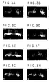
- FIG. 3 shows representative radiographs of the femurs from each of the groups recovered at four and eight weeks post-implantation. As demonstrated in these radiographs, the fixation remained intact in all the samples and there were no fractures in any of the femurs. In animals whose femoral defects were left empty, reactive bone formation at the transversely cut edges of the host femur was observed at four weeks ( Fig. 3A ). By eight weeks, slightly more bone was present within the gap, however, most of this bone appeared to form along the edge of the fixation plate which was in contact with the periosteum ( Fig. 3B ). Every specimen which was left empty resulted in the formation of a radiographic non-union. Some limbs, irrespective of the group, also contained an eccentric spicule of bone which was usually on the outside of the defect opposite the fixation plate.
- Table 1 Average of Radiographic Scores for Each Implant Group Four Weeks Eight Weeks C only C+MSCs C+M C only C+MSCs C+M Proximal Union 1.5 0.8 0.2 1.2 1.3 1.0 Distal Union 0.5 1.4 0.7 1.2 2.0 1.6 Core Density 0.7 2.0 0.3 0.6 3.7 0.7 Total Score 2.7 4.2 1.2 3.0 7.0* 3.3
- the low scores indicate the absence of any radiodense material within the pores, and minimal union of the implant with the host bone.
- Loading the HA/TCP implant with fresh marrow did not result in an improvement in the healing of the defect, and the low scores reflect the similarity of this group to that of the carrier alone.
- loading the HA/TCP carrier with MSCs produces a vigorous osteogenic response. Even at four weeks, pore filling was observed and is reflected in the considerably higher scores of these implants. Interestingly, even in this case the host-implant union was modest compared to controls. By the eight week time point, the pores of the implant were filled with new bone and the host-implant union was well established.
- the bone formed within the pores and at the ends of these implants represents de novo bone formation, is highly cellular, and is presented in higher magnification photomicrographs in Figure 5 .
- New woven and lamellar bone can be seen in intimate contact with the cut edge of the host cortex at eight weeks ( Fig. 5A ).
- this region of union is directly contiguous with bone formed throughout the pores of implant.
- filling of the pores with new bone is evident, as is the association of vasculature which orients the secretory activity of the differentiating osteoblasts ( Fig. 5B ).
- the cell-free HA/TCP implants had a bone fraction of only 2.3 % and 10.4% at four and eight weeks, respectively. Importantly, this fraction of bone at eight weeks correlates with previously published results (126). These fractions primarily represent the bone ingrowth from the cut ends of the host cortices.
- the marrow-loaded HA/TCP cylinders did exhibit modest osteogenesis within the body of the implant and consequently had a slightly higher value of 17.2% at eight weeks.
- the MSC-loaded samples exceeded the eight week value for the other two groups. The 19.3% bone fill at this four week time point is most likely attributable to MSC-mediated osteogenesis.
- the average bone fraction within the implant increased over time, reaching 43 % by eight weeks.
- progenitor cells are capable of healing a clinically significant bone defect in a well established animal model.
- These progenitor cells are referred to as mesenchymal stem cells since they give rise not only to bone (11,32,54,64), but to cartilage (32,66,80,142), muscle (121,143), tendon (23), and a stromal tissue which supports hematopoietic differentiation (87). While the osteogenic potential of both animal and human MSCs has been proven via subcutaneous implants in ectopic assays, rigorous and quantitative studies establishing the ability of culture-expanded MSCs to regenerate large segmental bone defects have not been reported to our knowledge.
- MSCs with a porous HA/TCP implant material are shown in the present study to be an effective strategy for healing large segmental bone defects.
- the current investigation further substantiates that compared to fresh marrow, MSCs produce significantly more bone when placed in either an ectopic or an orthotopic site. With these results as a foundation, we may begin to refine our approach to autologous cell therapies for the regeneration of skeletal defects.
- the bone and cartilage formed in cubes implanted subcutaneously not only confirms the osteochondral potential of the MSCs, but acts as an internal control to verify that every host rat was capable of providing an environment which could support osteogenesis within these combined cell:matrix implants. Additional experiments documenting the multilineage potential of these cells were not included as part of the current study because previous publications have focused on describing such potential in greater detail (32,79,80,121,143).
- the isolation and selection procedures for rat MSCs are similar to those used for human MSCs (32,54,80), and result in the formation of characteristic primary colonies illustrated in Figure 1A . These cells are mitotically expanded to yield a morphologically homogeneous population which divides uniformly across the dish.
- the radiographic findings in this study establish a precedent for obtaining non-invasive evidence of bone regeneration in animals, or humans, which receive MSCs in an orthotopic location.
- new bone which forms within the interstices of the material is readily apparent radiographically by four weeks, in spite of the inherent radiodensity of the HA/TCP material.
- the progressive increase in radiodensity evident by eight weeks correlates well with the histological observations of processed limbs.
- integration at the host-implant interface was not observed until eight weeks.
- the mean radiographic scores for the three implant groups document a significant (p ⁇ 0.05) difference between MSCs and either marrow-loaded and ceramic implants at eight weeks, while no significant difference was observed between marrow-loaded and ceramic implants at either time point.
- the histologic studies demonstrate appositional bone growth on the surface of the HA/TCP throughout the core of the implant, consistent with previous observations of osteogenesis in ectopic implants loaded with MSCs (32,47,54).
- the bone which is formed at four and eight weeks in MSC-loaded samples is woven in many areas, but lamellar bone can also be appreciated ( Figs. 5A and 5B ). It is critical to note that in the process of regenerating this osseous defect, bone formation occurs by a direct conversion of mesenchymal cells into osteoblasts rather than by an endochondral sequence.
- the pores of the ceramic are filled with significantly more bone, which is laid down upon the walls of the implant or existing bone, and oriented by the invading vasculature.
- These blood vessels visualized by India inking of animals immediately prior to sacrifice, also provide a portal for the entry and establishment of new marrow islands which contain hematopoietic elements, as well as host-derived MSCs.
- the process of bone remodeling ensues, and eventually the donor bone is replaced by host bone (47).
- integration of the implant is achieved with direct continuity between the cut edge of the host cortex and the new bone formed upon the surface of the implant ( Fig. 5A ).
- MSCs ability to regenerate a large segmental defect in this experimental model compares favorably with other investigations testing implants such as demineralized bone matrix, bone marrow, purified or recombinant BMPs, allograft, ceramics, and fibermetals (34,37,74,83,105,126,129,144,145,147). While the use of recombinant BMP has received considerable attention, the precise mechanism of action has only recently been appreciated. These powerful inductive molecules act on undifferentiated mesenchymal cells to initiate the endochondral cascade, ultimately resulting in the formation of bone.
- implants such as demineralized bone matrix, bone marrow, purified or recombinant BMPs, allograft, ceramics, and fibermetals (34,37,74,83,105,126,129,144,145,147). While the use of recombinant BMP has received considerable attention, the precise mechanism of action has only recently been appreciated. These powerful inductive molecules act on undifferentiated mesenchy
- MSCs marrow-derived mesenchymal progenitors
- the histomorphometric data generated in this study provides a basis for comparison to other investigations.
- fresh marrow from one femur equivalent is loaded on an HA/TCP implant
- no significant difference in bone formation is observed when compared to implants which receive no cells. This is true for both time points in our study, and likely reflects an inadequate number of MSCs in the volume of marrow applied.
- we and others would predict that greater healing of the bone defect would have occurred (74,144).
- an appropriate clinically relevant control is generously approximated by applying the total cell population obtained from one long bone since removing all the marrow from multiple long bones for the repair of a focal defect is contradictory to sound clinical judgment.
- MSCs produced a bone fill of 19.3 % and 43.2 % , respectively, at four and eight weeks.
- the bone fill was 21 % at four weeks, and only 22 % by eight weeks (126).
- These BMP-coated HA/TCP implants did not achieve a bone fill of 43 percent until 16 weeks following implantation.
- MSCs produce twice as much bone as BMP by the eight week time point. In this formulation, it took BMP sixteen weeks to form the same amount of bone which MSCs produce in only eight weeks.
- MSCs offer a considerable advantage to the use of BMP alone, although some combination of BMP and MSCs could provide an even faster, more vigorous bone repair as discussed above.
- the number of progenitor cells present at the site of repair is a critical factor, it is obligatory to estimate how the MSC-loaded implants compare with marrow-loaded implants in this regard.
- the number of nucleated marrow cells which were placed on an implant was approximately fifty million; the same number harvested from one long bone. Another fifty million cells were used to initiate the MSC culture which eventually provided cells for one implant. From these fifty million cells, roughly 500 MSC colonies develop, and these cells are mitotically expanded to three million by the end of first passage. This represents a 6,000-fold increase in MSC number due to approximately twelve population doublings. Using the current technique to load these type of implants, it appears that only about 150,000 cells become adherent following incubation with the MSC suspension (32).
- MSCs syngeneic bone marrow-derived mesenchymal stem cells
- a 15cc bone marrow aspirate was obtained from the iliac crest of each animal, according to an IACUC-approved protocol, and shipped on ice by overnight courier to the cell culture facilities.
- Isolation of canine MSCs was achieved by centrifuging whole marrow aspirates over a Percoll cushion, using procedures analogous to those developed for human MSC isolation (54).
- Tissue culture flasks (185 cm 2 ) were seeded with 10' nucleated cells isolated from the cushion, and cultured with DMEM containing 10% fetal calf serum from a selected lot (80). Cells were passaged at 8 x 10 3 cells/cm 2 , and transported back to the veterinary hospital where they were maintained until the time of implantation.
- Cell-loaded implants were prepared by incubating fibronectin-coated porous hydroxyapatitetricalcium phosphate (HA/TCP) cylinders (Zimmer, Inc.) in a 7.5 x 10 6 cells/ml suspension of MSCs for 3 hr at 37°C. The interval between marrow harvest and implantation was 16 days. An aliquot of cells from each preparation was also cultured under osteoinductive conditions to quantify aspects of osteoblastic differentiation.
- HA/TCP hydroxyapatitetricalcium phosphate
- a unilateral segmental femoral defect model was developed for this study following IACUC approval. Under general anesthesia, thirty-six skeletally mature female purpose-bred hounds (20 kg) underwent resection of a 21 mm long osteoperiosteal segment from their mid-diaphysis. A 4.5 mm Synthes* 8-hole lengthening plate was contoured to the lateral aspect of the bone, and secured with bicortical screws. The defect was filled with one of three materials; 1) a cell-free HA/TCP cylinder, 2) an MSC-loaded HA/TCP cylinder, or 3) cancellous bone harvested from the iliac crest. HA/TCP implants were secured by placing two sutures around the implant and the plate. Animals received peri-operative antibiotics, and analgesics were administered for three days post-operatively.
- Standard radiographic images were obtained at pre-op, immediately post-op, and at 4 week intervals until termination of the study. All samples contained a radiodensity step wedge to provide a basis for comparing changes over time, and between dogs. Upon sacrifice, specimens were subjected to high resolution Faxitron radiography, and subsequently processed for biomechanical evaluation. Following torsion testing, undecalcified longitudinal sections will be processed for quantitative histomorphometry.
- the present study demonstrates that MSCs from a large animal may be culture-expanded, and implanted for the successful repair of large diaphyseal bone defects.
- Radiographic and histologic evidence indicates that not only do the MSCs form bone within and around the implant directly, but their presence elicits a response in the host periosteum to form additional bone.
- the mechanism of this is currently not known, but is consistent with our observation that MSCs undergoing osteogenic differentiation secrete a paracrine factor(s) which is osteoinductive (63).
- the conspicuous lack of callus formation and periosteal reaction in the cell-free implants was an unexpected finding.
- Radiographic evidence suggests that MSC-mediated bone regeneration is faster than autograft throughout the study period. In addition to establishing a new standardized model for large animal bone repair, this study illustrates the feasibility of translating autologous stem cell therapy from the laboratory into the clinic.
- rat MSCs have been shown to synthesize structurally competent bone in an orthotopic site (68)
- human MSCs have only been shown to form bone in vitro (12,64) and in an ectopic implantation site in immunodeficient mice (55). Since fracture healing and bone repair depend on the ability to amass enough cells at the defect site to form a repair blastema, one therapeutic strategy is to directly administer the precursor cells to the site in need of repair. This approach is particularly attractive for patients who have fractures which are difficult to heal, or patients who have a decline in their MSC repository as a result of age (72,118), osteoporosis (128), or other metabolic derangement. With this in mind, the goal of the current study was to show that purified, culture-expanded human MSCs are capable of regenerating bone at the site of a clinically significant defect.
- Human MSCs were replated into six-well dishes at a density of 3x10 3 cells/cm 2 . The following day (Day 0), fresh medium was provided, and the cells were grown in the absence or presence of Osteogenic Supplements (OS) (12,64), Media changes were performed twice weekly, and at days 4, 8, 12 and 16, cultures were assayed for cell number, alkaline phosphatase (APase) biochemistry and histochemistry, and mineralized matrix production utilizing techniques previously described (64).
- OS Osteogenic Supplements
- Porous hydroxyapatite/ ⁇ -tricalcium phosphate (HA/TCP) ceramic blocks mean pore size 200-450 ⁇ m (Zimmer, Inc., Warsaw, IN), were shaped into cylinders approximately 4 mm in diameter and 8 mm in length with a 1 mm central canal, or cut into cubes 3 mm per side.
- MSC-loaded implants were prepared by incubating human fibronectin-coated HA/TCP cubes and cylinders in a 7.5 x 10 6 cell/ml suspension of first passage MSCs for 2 hr at 37°C as previously described (68). Cell-free control cylinders were prepared identically.
- the femoral gap surgical model employed here has been used extensively in euthymic rats to study long bone repair (68,129,34,147). Briefly, both femurs of Harlan Nude (Hsd:Rh- rnu ) rats (325 g) were exposed by an anterolateral approach. A polyethylene fixation plate was attached to each femur by four Kirschner wires, and an 8 mm transverse segment of the central diaphysis, along with its adherent periosteum, was removed by using a rotary osteotomy burr under saline irrigation.
- Each animal then received a cell-free HA/TCP cylinder in one femoral defect, an identical cylinder loaded with human MSCs in the contralateral defect, and a subcutaneous implant of a MSC-loaded HA/TCP cube along the dorsum.
- MSCs cultured with OS underwent a dramatic change in cellular morphology from that of spindle-shaped to cuboidal, which was accompanied by an increase in APase activity and production of an extracellular matrix rich in bone hydroxyapatite ( Fig. 6B ).
- a significant increase in APase activity was observed after 4 days of OS treatment with maximal activity occurring on day 8, followed by a decline through day 16 ( Fig. 6C ). This late decrease in APase activity of OS cultures correlates with increasing mineral deposition and terminal differentiation of cells into osteocytes.
- Figure 6C illustrates that MSCs grown with OS deposited a significant amount of calcium by days 12 (60 ⁇ 5.1 ⁇ g/dish) and 16 (98 ⁇ 5.0 ⁇ g/dish).
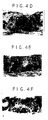
- Figure 8A illustrates the segmental defect model used in this study.
- the polyethylene fixation plate on top of the femur provides stability following creation of the 8 mm diaphyseal defect. No animals experienced failure of fixation or other post-operative complications throughout the course of study.
- Previous studies have established that femoral defects that are not implanted with a bioactive material give rise to a fibrous non-union devoid of bone (68,34,147).
- High resolution Faxitron radiographs provided sufficient clarity and detail to discern subtle changes occurring within the implant and the surrounding host bone. Representative radiographs of the femurs from the 2 groups recovered 12 weeks post-implantation demonstrate substantially more bone in animals which received MSC-loaded HA/TCP cylinders ( Fig.
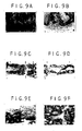
- Immunocytochemical staining with antibody 6E2 demonstrates that, at 4 weeks, virtually all the cells within the pores of the implant were reactive on their surface and were, therefore, of human origin ( Fig. 9A ).
- the host rat cells were intermingled with the human donor cells, but as the distance away from the surface of the implant increased, the representation of donor cells precipitously declined.
- the presence of these peripheral cells which are not immunostained also serves as a negative control for this established antibody.
- the ceramic material itself which appears black in the phase contrast micrograph ( Fig. 9B ), displays a high level of background fluorescence.
- Table 3 Bone Fill in HA/TCP Implants as a Percentage of Available Space Four Weeks Eight Weeks Twelve Weeks Carrier Alone 1.89 ⁇ 1.00 11.47 ⁇ 7.08 29.51 ⁇ 8.93 Carrier plus MSCs 1.95 ⁇ 1.92 26.46 ⁇ 3.60* 46.61 ⁇ 14.83*
- Bone present in the cell-free HA/TCP implants primarily represents the bony ingrowth from the cut ends of the host cortices.
- the MSC-loaded samples contained significantly more bone than the cell-free group, and the average bone fraction within the implant increased over time, reaching 26.5% and 46.6% by the 8 and 12 week time points, respectively.
- This increased bone fraction at 8 weeks is 2.3-fold higher than that measured in cell-free implants at the same time, and by 12 weeks, is over 23-fold higher than that observed in either condition at 4 weeks.
- the volume fraction of the HA/TCP carrier remained constant, and served as an internal control for histomorphometry.
- results presented here demonstrate that purified, culture-expanded human MSCs are capable of healing a clinically significant bone defect in a well-established model for bone repair. While the osteogenic potential of human MSCs has been proven by neo-osteogenesis in subcutaneous implants (54), as well as in studies of isolated MSCs in vitro (12,64), this is the first demonstration that human MSCs can form bone at an orthotopic site in need of repair.
- the combination of MSCs with a porous HA/TCP carrier possesses regenerative potential which is histomorphometrically and biomechanically superior to the carrier alone. This investigation paves the way for the clinical application of autologous MSC-therapy for the treatment of orthopedic defects in man.
- the pores of the ceramic are filled with an increasing amount of bone, which is laid down upon the walls of the implant or existing bone, and oriented by the invading vasculature that provides a portal for the entry and establishment of new marrow islands containing hematopoietic elements and host-derived MSCs.
- the rate of bone regeneration is lower than that observed in euthymic rats implanted with syngeneic MSCs (68), suggesting that immunocompromised rats are not the ideal hosts to assess the bone-forming potential of human MSCs. This may be due in part to the xenogeneic nature of the implant and the increased natural killer cell activity, which may be a compensatory mechanism for the animal to cope with its deficient T-cell-mediated immunity (123). Nevertheless, a significantly higher amount of bone was formed in the defect which received MSCs compared to those limbs receiving the carrier only. The extent of host-implant union was greater in the MSC-loaded implants, which likely reflects the combined contributions of implanted MSCs and host-derived cells.
- recombinant human BMP have been implanted in experimental bone defect models in an effort to stimulate bone repair (147,78,29).
- recombinant BMPs are capable of inducing the endochondral cascade in ectopic implants (146), their ability to reproducibly direct bone formation at orthotopic sites has been hampered by the problems associated with the design and selection of an appropriate carrier.
- BMP delivered in the same HA/TCP carrier did not increase implant strength over the carrier alone (126). The brittle nature of this ceramic, combined with its slow resorption and complex porous structure, may explain why even in the presence of significant bone formation mechanical strength remains less than intact limbs.
- Implantation of culture-expanded autologous MSCs offers the advantage of directly delivering the cellular machinery responsible for synthesizing new bone, and circumventing the otherwise slow steps leading to bone repair. Even in patients with a reduced ability to regenerate connective tissue, presumably due to a low titer of endogenous MSCs (72,128,144,11), these rare MSCs may be isolated and culture-expanded over one billion-fold without a loss in their osteogenic potential (13), thus restoring or enhancing a patient's ability to heal tissue defects.
- the studies presented here suggest that MSC-based cell therapies will be useful for the reconstruction of a variety of tissue defects in man.
- This experiment was performed in an attempt to establish that uncoated HA/TCP cubes are equivalent to fibronectin- or autologous serum-coated HA/TCP cubes in supporting MSC-mediated osteogenesis.
- the data reported here provides important information regarding the value of fresh bone marrow and of culture expanded purified mesenchymal stem cells as a means of improving existing graft materials for spinal fusion applications. This technology may result in a significant improvement in the efficacy of bone grafting materials and procedures. Furthermore, use of bone grafting procedures which do not require harvest of autogenous bone graft will significantly reduce the morbidity of bone grafting procedures.
- Canine bone is a better model for human bone than rodents (35), and avoids many of the limitations associated with defect models discussed herein.
- Use of a larger graft volume and a common site for clinical graft procedures (the spine) are significant advantages. Testing three materials in each animal allows control for interanimal variation and reduces the number of animals required.
- the model allows each fusion site to be mechanically tested without artifacts from internal fixation.
- Union score of the cross sectional area of the fusion mass at the site of failure has also proven to be a sensitive means for comparison between materials.
- Quantitative CT image analysis of each fusion site prior to destructive testing was developed in response to the first review. This now allows quantitative analysis of fusion mass volume, mean electron density (mineralization), and cross sectional area.
- a collagen/ceramic (CC) composite composed of Type I bovine fibrillar skin collagen and 0.5-1.0 mm diameter granules of a biphasic calcium phosphate ceramic (60% hydroxyapatite, 40% tri-calcium phosphate), is ineffective as a graft material. Used alone it is no better than an ungrafted defect. Addition of an extract of bone matrix proteins or adding autogenous bone improved efficacy (p ⁇ 0.01), but the result remained inferior to pure autogenous bone (p ⁇ 0.01)(55). We also showed that adding this collagen/ceramic composite to autogenous bone as a "bone graft expander" significantly reduced autograft performance (p ⁇ 0.01)(58). Interestingly, successful fusions resulting from the CC composite had similar mechanical properties to fusions resulting from autograft, indicating that residual unresorbed ceramic granules had no evident adverse effect on the material properties of the fusion mass.
- a biphasic calcium phosphate ceramic 60% hydroxy
- the specific aims of this project address the comparison of three graft materials: Granular Ceramic matrix loaded with autogenous mesenchymal stem cells; Granular Ceramic matrix loaded with fresh bone marrow; and Granular Ceramic matrix loaded with fresh bone marrrow and autogenous mesenchymal stem cells.
- Twelve male beagle dogs (age 10-14 months, 12-14 kg) are used for the experiment. Localized fusions are performed at three spinal fusion sites, L1-2, L3-4, and L5-6. Each site is internally fixed using dual plates immobilizing adjacent spinous processes, separated by one mobile segment. Each animal is grafted with one of the three materials under evaluation. To limit the potential for surgical bias and to insure distribution of materials at each of the three graft sites, twelve cards, two sets of the six possible combinations of the three materials in three sites, are prepared at the beginning of each experiment and placed in an envelope. Site assignments are then made intraoperatively by blind drawing after site preparation is complete. Internal fixation is applied to each segment using plates placed on either side of the spinous processes. No external immobilization is used.
- each animal in the study undergoes spinal fusion at three separate levels. Mesenchymal stem cells are provided, and fresh bone marrow will be harvested again, this time from the right iliac crest.
- Each material composite is then prepared intraoperatively. Following preparation of the surgical bed and application of internal fixation, a 2 cc volume of each material is grafted at one level in each animal. In this way each animal has one fusion site for each of the materials. The materials are distributed according to a randomization protocol to prevent surgical bias and insure uniform distribution of materials by site.
- a small (3 mm) stab incision is made using a #11 blade.
- a Lee-Lok bone marrow aspiration needle (Lee-Lok Inc., Minneapolis, MN), is advanced into the bone cavity. The obturator is removed. A 2 cc volume of bone marrow is then aspirated promptly into a 10 cc syringe containing 1 cc of heparinized saline-(1000 units/ml). The syringe is detached and inverted several times to insure-mixing. Subsequent aspirates are taken using identical technique through the same skin incision and the same cortical window, but redirecting the needle tip to intramedullary sites separated by at least 1 cm. Pressure is held on the site for 3-5 minutes to insure hemostasis. No dressing is necessary. No post-operative immobilization or restriction is required.
- Iliac crest marrow aspirates (10 ml) were obtained from 15-25 kg random source canines under Propofol anesthesia (Diprivan 1%, Stuart Pharm., DE). The marrow was drawn into a 10 ml syringe containing 7500 units of Heparin (Elkins-Simm, Cherry Hill, NJ) to prevent clotting. For some canines, the marrow was placed on ice and shipped overnight to the cell culture facility.
- the marrow was mixed with two volumes of Complete media, consisting of 10% FBS and antibiotics (100 U/ml penicillin G, 100 ⁇ g/ml streptomycin sulfate, and 0.25 ⁇ g/ml amphotericin B) in low-glucose DMEM.
- the nucleated cell fraction of the marrow was enriched for MSCs based on density separation on a 1.063 g/cc Percoll cushion. Two hundred million nucleated cells in 5 ml of media were carefully layered over 20 ml of Percoll (Sigma, St. Louis, MO). Separation was achieved by centrifugation at 400 x g for 20 minutes.
- a mid-line posterior longitudinal incision is made from T10 to L7.
- Cutting cautery is used to outline the tips of spinous processes L1 through L6 and subperiosteal elevation of paraspinal muscles is performed.
- the interspinous ligament and interlaminar tissue are excised at the L1-2, L3-4, and L5-6 interspaces preserving the ligamentum flavum.
- a dental burr is used to excise facet cartilage to the level of subchondral bone and to perform a superficial decortication of the adjoining laminar surfaces under continuous saline irrigation. Entry into the neural canal is avoided.
- site preparation is complete, a saline soaked gauze is placed in each fusion site, the wound is covered; and an unscrubbed assistant blindly selects a card containing the material/site assignment for that animal.
- Porous hydroxyapatite/tricalcium phosphate (HA/TCP) ceramic material supplied from Zimmer Inc. as a 60/40 combination, is lightly crushed and sieved to select for a particle size ranging from 1.0 to 2.5 mm in diameter. Following sterilization of the granules, 15 million MSCs are incubated with the 1 cc of ceramic for 3 hours at 37 degrees Celsius with agitation every 30 minutes.
- implant groups consist of 1) ceramic granules combined with autologous MSCs, 2) ceramic granules combined with autologous MSCs and fresh bone marrow obtained by aspiration, and 3) ceramic granules combined with fresh bone marrow alone.
- each graft Following preparation of all graft materials, materials are carefully placed in their appropriate sites. Approximately 1 cc of each graft is used to fill the area of the excised facets and the interlaminar space. The remainder of each graft is layered over the dorsal surface of the adjoining lamina.
- Fixation at each site is then applied.
- a 1 mm burr is used to place a hole in the central region of the distal spinous process at each site.
- 316L stainless steel plates (0.125" x 0.4" x 1.4") are placed on either side of the adjoining spinous processes and fixed to the caudal spinous process using a stainless steel bolt and nut (size 2-56, 0.5" long).
- Four symmetrical holes in each plate allow selection among three potential fixation lengths to accommodate variation in the interspinous distance at individual sites (0.75", 0.90", and 1.05").
- the fixation to the cranial spinous process at each level is accomplished by drilling through the appropriate hole in the plates to create a hole in the spinous process and by passing a second bolt and nut.
- Both bolts are tightened firmly without crushing the spinous processes.
- a second locking nut is then applied at each site to prevent loosening.
- the spinal wound and the autograft donor site is then closed using O-Dexon Plus sutures in deep fascia, interrupted subcutaneous sutures of 2-0 Dexon Plus, followed by staples in the skin.
- a small (3 mm) stab incision is made using a #11 blade.
- a Lee-Lok bone marrow aspiration needle (Lee-Lok Inc., Minneapolis, MN), is advanced into the bone cavity. The obturator is removed. Bone marrow is then aspirated promptly into a 10 cc syringe, and mixed with the granular ceramic matrix and allowed to set for 10-15 minutes to allow clot formation. Excess fluid is then pressed out with a gauze sponge and a 2 cc volume of the matrix impregnated with fresh bone marrow is implanted.
- Each animal receives prophylactic antibiotics of Penicillin G 500,000 units IM preoperatively and Ampicillin 250 mg po per day for five days post-operatively. Acepromazine and Tylenol are used for perioperative pain. No external immobilization is used. Animals are housed in cages for three days post-operatively and then moved to runs where they are exercised daily.
- Quantitative assessment of the fusion mass is performed using helical X-ray Computed Tomography (CT) and automated 3-dimensional image processing techniques.
- Quantitative measures at each fusion site includes: 1) fusion mass volume, 2) area measured at the mid-disk cross-sectional plane, and 3) mineralization density of the fusion mass.
- Scanning is done on a Somatome Plus 40 CT scanner (Siemens Medical Systems).
- the specimen is placed with the cranial-caudal axis perpendicular to the direction of table travel. In this orientation the intervertebral spaces are oriented perpendicular to the scan plane. This reduces the chance of missing features of nonunions occurring in the transaxial plane and allows the midline of the intervertebral space to be identified more accurately.
- Scans will be done at 120 kVp, 210 mA, 1 sec helical mode, 2 mm collimation, and table speed 2 mm/sec, for 30 secs. This will produce images with 2 mm section thickness and 1 mm 2 pixel area.
- Images are then reconstructed using the bone algorithm and an image-to-image overlap of 1 mm. This provides the highest possible spatial resolution available in three dimensions (i.e. a voxel size of 1 mm 3 ).
- a Siemens bone mineral density phantom is placed beneath each specimen to provide a reference for the quantitation of bone mineralization density.
- Bone and soft tissue CT values are easily differentiable (CT bone - 150-1000, CT soft tissue - 0 ⁇ 20). Therefore, three-dimensional segmentation of the reconstructed volume data sets is performed using a basic automated threshold algorithm in each two dimensional slice followed by a connectivity algorithm between slices. Once the three-dimensional data set has been segmented, the volume of the fusion mass is measured in a specified region of interest, defined by anatomic fiducial markers. The user selects two points which define the dorsal most points of the left and right neural foramen at the level of interest and one level above and below the site of interest. The user also identifies the midpoint of the intervertebral disk.
- the interactive tools for manipulating and visualizing the 3-D data sets are developed using the GL library and Iris Inventor Toolkit available on a Silicon graphics workstation.
- the volume of the fusion mass is calculated by summing the segmented voxels (bone) within the specified region of interest and multiplying this sum by the appropriate voxel volume (1 mm 3 ).
- the area of the fusion mass will be measured at the mid-fusion cross-sectional plane by summing the number of segmented pixels (bone) in the mid-fusion slice and multiplying the sum by the appropriate pixel area (1 mm 2 ).
- a mean-mineralization density is calculated for the entire fusion mass and referenced to the density of the phantom.
- a 2-D topographic map of bone mineral density at the mid-disk cross-sectional plane is provided.
- the original volume of bone and bony cross section from the region of interest for each segment can be estimated by measuring the same region in the adjacent normal segment(s). This is used to normalize data from each fusion segment.
- Software for this quantitative analysis requires four modules: 3-D segmentation, animation to view cross-sectional planes and mark fiducial points, calculation of rectangular region of interest and quantitative measures of bone fusion, and an interactive display module to view reconstructed data in 3-D.
- each specimen Prior to testing, each specimen is thawed for 24 hours at room temperature. Testing is performed on an MTS TESTAR Bionix System using a custom four point bending device. After three sinusoidal conditioning cycles, load displacement data are collected nondestructively in flexion-extension and left-right bending. Failure testing is then performed in right bending using the ramp function at 8 mm/sec. Bending has been selected as the mode of failure because bending stiffness was found to be most closely correlated with union status in prior studies (94). Load-displacement curves are used to derive stiffness, maximum load, displacement to failure, and total energy to failure.
- the surfaces of the fractured specimen are examined using a metal probe.
- the degree of union is scored from zero to four based on a regional grid system ( Figure 11 ).
- a score of four is defined as complete fusion of both facet joints and the entire lamina. One half point is added for union in either half of each facet joint or any of four quadrants of the adjoining laminar surfaces. Therefore, scores of 0, 1, 2, 3 and 4 represent union of roughly 0, 25, 50, 75 and 100 percent of the cross-sectional area of the grafted volume, respectively. Scoring is done by consensus of two observers who examine the specimens,together and are blinded with respect to the material grafted at each site.
- Union score and CT generated data are analyzed to determine whether outcome differs statistically due to site and/or material effects.
- the data set is considered "incomplete" since only one material can be tested at each site within an animal. Therefore, both site and material are repeated factors but with missing observations.
- a regression model for repeated measures data is used. This modeling strategy uses the Generalized Estimating Equation (GEE) approach described by Zeger et al . (150) and Ou et al . (108). Since union scores for a given dog are potentially not independent of each other, a working covariance structure of independence and a robust variance estimate are used in fitting the model.
- GEE Generalized Estimating Equation
- the objectives of this study were to demonstrate efficacy of bone marrow and/or mesenchymal stem cells (MSCs) in healing clinically significant bone defects in an established animal model.
- MSCs mesenchymal stem cells
- Fisher 344 rats (Charles River Laboratories, Wilmington, MA) of approximately 325 grams in weight were used.
- a bilateral femoral gap 8 mm in length was created in each femur. This length is approximately towards the diameter of the mid-diaphysis of the femur.
- An internal fixation plate was applied with four Kirschner wires.
- the groups for comparison were separately treated with one of the following:
Landscapes
- Health & Medical Sciences (AREA)
- Life Sciences & Earth Sciences (AREA)
- Engineering & Computer Science (AREA)
- Biomedical Technology (AREA)
- Chemical & Material Sciences (AREA)
- General Health & Medical Sciences (AREA)
- Zoology (AREA)
- Cell Biology (AREA)
- Animal Behavior & Ethology (AREA)
- Veterinary Medicine (AREA)
- Medicinal Chemistry (AREA)
- Public Health (AREA)
- Epidemiology (AREA)
- Transplantation (AREA)
- Oral & Maxillofacial Surgery (AREA)
- Dermatology (AREA)
- Bioinformatics & Cheminformatics (AREA)
- Chemical Kinetics & Catalysis (AREA)
- Organic Chemistry (AREA)
- Botany (AREA)
- Biotechnology (AREA)
- Genetics & Genomics (AREA)
- Wood Science & Technology (AREA)
- Orthopedic Medicine & Surgery (AREA)
- Developmental Biology & Embryology (AREA)
- Microbiology (AREA)
- General Engineering & Computer Science (AREA)
- Biochemistry (AREA)
- Urology & Nephrology (AREA)
- Rheumatology (AREA)
- Immunology (AREA)
- Pharmacology & Pharmacy (AREA)
- Gastroenterology & Hepatology (AREA)
- Proteomics, Peptides & Aminoacids (AREA)
- Inorganic Chemistry (AREA)
- Vascular Medicine (AREA)
- Hematology (AREA)
- Physical Education & Sports Medicine (AREA)
- General Chemical & Material Sciences (AREA)
- Nuclear Medicine, Radiotherapy & Molecular Imaging (AREA)
Priority Applications (1)
| Application Number | Priority Date | Filing Date | Title |
|---|---|---|---|
| EP10011227.5A EP2311471A3 (fr) | 1996-04-19 | 1997-04-17 | Régéneration et augmentation d'os utilisant des cellules souches mésenchymateuses |
Applications Claiming Priority (3)
| Application Number | Priority Date | Filing Date | Title |
|---|---|---|---|
| US1624596P | 1996-04-19 | 1996-04-19 | |
| US2983896P | 1996-10-28 | 1996-10-28 | |
| EP97920427A EP0906415B1 (fr) | 1996-04-19 | 1997-04-17 | Regeneration et croissance osseuse obtenues a l'aide de cellules souches du mesenchyme |
Related Parent Applications (2)
| Application Number | Title | Priority Date | Filing Date |
|---|---|---|---|
| EP97920427A Division EP0906415B1 (fr) | 1996-04-19 | 1997-04-17 | Regeneration et croissance osseuse obtenues a l'aide de cellules souches du mesenchyme |
| EP97920427.8 Division | 1997-10-30 |
Related Child Applications (1)
| Application Number | Title | Priority Date | Filing Date |
|---|---|---|---|
| EP10011227.5 Division-Into | 2010-09-28 |
Publications (2)
| Publication Number | Publication Date |
|---|---|
| EP2105138A2 true EP2105138A2 (fr) | 2009-09-30 |
| EP2105138A3 EP2105138A3 (fr) | 2013-06-05 |
Family
ID=26688354
Family Applications (3)
| Application Number | Title | Priority Date | Filing Date |
|---|---|---|---|
| EP97920427A Expired - Lifetime EP0906415B1 (fr) | 1996-04-19 | 1997-04-17 | Regeneration et croissance osseuse obtenues a l'aide de cellules souches du mesenchyme |
| EP09009351.9A Withdrawn EP2105138A3 (fr) | 1996-04-19 | 1997-04-17 | Régénération et augmentation de l'os utilisant des cellules souches mésenchymateuses |
| EP10011227.5A Withdrawn EP2311471A3 (fr) | 1996-04-19 | 1997-04-17 | Régéneration et augmentation d'os utilisant des cellules souches mésenchymateuses |
Family Applications Before (1)
| Application Number | Title | Priority Date | Filing Date |
|---|---|---|---|
| EP97920427A Expired - Lifetime EP0906415B1 (fr) | 1996-04-19 | 1997-04-17 | Regeneration et croissance osseuse obtenues a l'aide de cellules souches du mesenchyme |
Family Applications After (1)
| Application Number | Title | Priority Date | Filing Date |
|---|---|---|---|
| EP10011227.5A Withdrawn EP2311471A3 (fr) | 1996-04-19 | 1997-04-17 | Régéneration et augmentation d'os utilisant des cellules souches mésenchymateuses |
Country Status (9)
| Country | Link |
|---|---|
| US (2) | US6541024B1 (fr) |
| EP (3) | EP0906415B1 (fr) |
| JP (2) | JP2000508911A (fr) |
| AT (1) | ATE439849T1 (fr) |
| AU (1) | AU731468B2 (fr) |
| CA (1) | CA2251983C (fr) |
| DE (1) | DE69739540D1 (fr) |
| ES (1) | ES2329953T3 (fr) |
| WO (1) | WO1997040137A1 (fr) |
Families Citing this family (250)
| Publication number | Priority date | Publication date | Assignee | Title |
|---|---|---|---|---|
| US6150328A (en) | 1986-07-01 | 2000-11-21 | Genetics Institute, Inc. | BMP products |
| US6291206B1 (en) | 1993-09-17 | 2001-09-18 | Genetics Institute, Inc. | BMP receptor proteins |
| AU689184B2 (en) | 1993-12-07 | 1998-03-26 | Genetics Institute, Llc | BMP-12, BMP-13 and tendon-inducing compositions thereof |
| US20050186673A1 (en) * | 1995-02-22 | 2005-08-25 | Ed. Geistlich Soehne Ag Fuer Chemistrie Industrie | Collagen carrier of therapeutic genetic material, and method |
| CA2198978A1 (fr) * | 1996-03-01 | 1997-09-01 | Clemens Antoni Van Blitterswijk | Methode pour la production d'os in vitro |
| US20030180705A1 (en) * | 1997-01-24 | 2003-09-25 | Asahi Medical Co., Ltd. | Method of regenerating blood vessels |
| US20040224300A1 (en) * | 1997-01-24 | 2004-11-11 | Shuji Terashima | Method for separating nucleated cells |
| US20020031757A1 (en) * | 1997-01-24 | 2002-03-14 | Asahi Medical Co., Ltd. | Method of regenerating a tissue |
| US20030180263A1 (en) * | 2002-02-21 | 2003-09-25 | Peter Geistlich | Resorbable extracellular matrix for reconstruction of bone |
| US20050186283A1 (en) * | 1997-10-10 | 2005-08-25 | Ed. Geistlich Soehne Ag Fuer Chemistrie Industrie | Collagen carrier of therapeutic genetic material, and method |
| US9034315B2 (en) * | 1997-10-10 | 2015-05-19 | Ed. Geistlich Soehne Ag Fuer Chemische Industrie | Cell-charged multi-layer collagen membrane |
| US8858981B2 (en) * | 1997-10-10 | 2014-10-14 | Ed. Geistlich Soehne Fuer Chemistrie Industrie | Bone healing material comprising matrix carrying bone-forming cells |
| AU749675B2 (en) * | 1998-03-13 | 2002-07-04 | Mesoblast International Sarl | Uses for human non-autologous mesenchymal stem cells |
| EP1064016A2 (fr) * | 1998-03-14 | 2001-01-03 | Curis, Inc. | Compositions pour moduler la differentiation des cellules comprenant un lipide et un morphogene |
| US6171610B1 (en) | 1998-04-24 | 2001-01-09 | University Of Massachusetts | Guided development and support of hydrogel-cell compositions |
| US6727224B1 (en) | 1999-02-01 | 2004-04-27 | Genetics Institute, Llc. | Methods and compositions for healing and repair of articular cartilage |
| CA2362049A1 (fr) * | 1999-02-04 | 2000-08-10 | Sdgi Holdings, Inc. | Compositions spongieuses osteogeniques extremement mineralisees et leurs utilisations |
| AU770196B2 (en) | 1999-02-04 | 2004-02-12 | Warsaw Orthopedic, Inc. | Osteogenic paste compositions and uses thereof |
| US20030082152A1 (en) | 1999-03-10 | 2003-05-01 | Hedrick Marc H. | Adipose-derived stem cells and lattices |
| US6777231B1 (en) | 1999-03-10 | 2004-08-17 | The Regents Of The University Of California | Adipose-derived stem cells and lattices |
| US8389017B1 (en) | 1999-07-08 | 2013-03-05 | Cap Biotechnology, Inc. | Calcium-containing structures and methods of making and using the same |
| US20020095157A1 (en) | 1999-07-23 | 2002-07-18 | Bowman Steven M. | Graft fixation device combination |
| US6179840B1 (en) | 1999-07-23 | 2001-01-30 | Ethicon, Inc. | Graft fixation device and method |
| EP1475109A1 (fr) * | 1999-10-15 | 2004-11-10 | Genetics Institute, LLC | Formulations destinées a l'administration de protéines osteogéniques |
| WO2001028603A1 (fr) * | 1999-10-15 | 2001-04-26 | Genetics Institute, Inc. | Formulations destinees a l'administration de proteines osteogeniques |
| DK1223990T3 (da) | 1999-10-15 | 2004-11-29 | Inst Genetics Llc | Formuleringer af hyaluronsyre til tilförsel af osteogene proteiner |
| DE60028070T2 (de) * | 1999-11-11 | 2007-01-11 | Japan Tissue Engineering Co., Ltd., Gamagori | Transplantatmaterial und ein herstellungsverfahren dafür |
| US7108721B2 (en) | 2000-05-11 | 2006-09-19 | Massachusetts Institute Of Technology | Tissue regrafting |
| GB0011244D0 (en) * | 2000-05-11 | 2000-06-28 | Smith & Nephew Inc | Tissue regrafting |
| GB0015479D0 (en) * | 2000-06-23 | 2000-08-16 | Univ London | Transcutaneous Prosthesis |
| AU7107301A (en) * | 2000-07-18 | 2002-01-30 | Nichimo Co. Ltd. | Stem cell reinforcing material |
| MY133943A (en) | 2000-08-22 | 2007-11-30 | Synthes Gmbh | Bone replacement material |
| US20080152629A1 (en) * | 2000-12-06 | 2008-06-26 | James Edinger | Placental stem cell populations |
| DK1349918T3 (da) * | 2000-12-06 | 2014-11-10 | Anthrogenesis Corp | Fremgangsmåde til indsamling af stamceller fra moderkagen |
| US7311905B2 (en) * | 2002-02-13 | 2007-12-25 | Anthrogenesis Corporation | Embryonic-like stem cells derived from post-partum mammalian placenta, and uses and methods of treatment using said cells |
| CA2365376C (fr) * | 2000-12-21 | 2006-03-28 | Ethicon, Inc. | Utilisation d'implants en mousse renforces ayant une meilleure integrite pour la reparation et la regeneration de tissus mous |
| US6852330B2 (en) * | 2000-12-21 | 2005-02-08 | Depuy Mitek, Inc. | Reinforced foam implants with enhanced integrity for soft tissue repair and regeneration |
| US6599323B2 (en) * | 2000-12-21 | 2003-07-29 | Ethicon, Inc. | Reinforced tissue implants and methods of manufacture and use |
| EP3246396B1 (fr) * | 2001-02-14 | 2020-01-29 | Celularity, Inc. | Rénovation et repopulation de tissus décellularisés et d'organes cadavériques par des cellules souches |
| EP2316919B1 (fr) * | 2001-02-14 | 2015-10-07 | Anthrogenesis Corporation | Placenta post-gravidique de mammifère, son utilisation et céllules souches placentaires correspondantes |
| US6723131B2 (en) * | 2001-02-28 | 2004-04-20 | The Cleveland Clinic Foundation | Composite bone marrow graft material with method and kit |
| US6949251B2 (en) * | 2001-03-02 | 2005-09-27 | Stryker Corporation | Porous β-tricalcium phosphate granules for regeneration of bone tissue |
| IL141813A (en) * | 2001-03-05 | 2010-04-15 | Hadasit Med Res Service | Mixture comprising bone marrow cells together with demineralized and/or mineralized bone matrix and uses thereof in the preparation of compositions for the treatment of hematopoietic dusirders |
| US20020182189A1 (en) * | 2001-04-19 | 2002-12-05 | Chang Chia Ning (Sophia) | Compositions and methods for the repair and construction of bone and other tissue |
| US7011829B2 (en) | 2001-05-18 | 2006-03-14 | Kanemaru Shin-Ichi | Tissue filler |
| US7226587B2 (en) | 2001-06-01 | 2007-06-05 | Wyeth | Compositions and methods for systemic administration of sequences encoding bone morphogenetic proteins |
| TWI267378B (en) | 2001-06-08 | 2006-12-01 | Wyeth Corp | Calcium phosphate delivery vehicles for osteoinductive proteins |
| JP2003018984A (ja) * | 2001-07-06 | 2003-01-21 | Mitsubishi Chemicals Corp | 多分化能を有する細胞の製造法 |
| AU2002325758A1 (en) * | 2001-09-20 | 2003-04-01 | Centre For Translational Research In Cancer | Cultured stromal cells and uses thereof |
| US7585670B2 (en) | 2001-12-07 | 2009-09-08 | Cytori Therapeutics, Inc. | Automated methods for isolating and using clinically safe adipose derived regenerative cells |
| US7514075B2 (en) | 2001-12-07 | 2009-04-07 | Cytori Therapeutics, Inc. | Systems and methods for separating and concentrating adipose derived stem cells from tissue |
| US7651684B2 (en) | 2001-12-07 | 2010-01-26 | Cytori Therapeutics, Inc. | Methods of using adipose tissue-derived cells in augmenting autologous fat transfer |
| US20050048035A1 (en) | 2001-12-07 | 2005-03-03 | Fraser John K. | Methods of using regenerative cells in the treatment of stroke and related diseases and disorders |
| KR100930139B1 (ko) | 2001-12-07 | 2009-12-07 | 사이토리 테라퓨틱스, 인크. | 처리된 리포애스퍼레이트 세포로 환자를 치료하기 위한시스템 및 방법 |
| US7595043B2 (en) | 2001-12-07 | 2009-09-29 | Cytori Therapeutics, Inc. | Method for processing and using adipose-derived stem cells |
| US7771716B2 (en) | 2001-12-07 | 2010-08-10 | Cytori Therapeutics, Inc. | Methods of using regenerative cells in the treatment of musculoskeletal disorders |
| US9597395B2 (en) | 2001-12-07 | 2017-03-21 | Cytori Therapeutics, Inc. | Methods of using adipose tissue-derived cells in the treatment of cardiovascular conditions |
| US20050095228A1 (en) | 2001-12-07 | 2005-05-05 | Fraser John K. | Methods of using regenerative cells in the treatment of peripheral vascular disease and related disorders |
| JP3646167B2 (ja) * | 2002-02-19 | 2005-05-11 | 独立行政法人産業技術総合研究所 | フォスフォフォリンを含む複合生体材料 |
| AU2003223214B2 (en) * | 2002-03-01 | 2008-09-18 | Celltech R & D, Inc. | Methods to increase or decrease bone density |
| US7046835B2 (en) * | 2002-03-07 | 2006-05-16 | Ge Medical Systems Global Technology Company Llc | Method and system for processing vascular radiographic images which have been reconstructed by three-dimensional modelling |
| US8313742B2 (en) | 2002-03-29 | 2012-11-20 | Depuy Acromed, Inc. | Cell-containing bone graft material |
| US7223414B1 (en) * | 2002-06-03 | 2007-05-29 | Ahmed El-Ghannam | Silica-calcium phosphate bioactive composite for improved synthetic graft resorbability and tissue regeneration |
| US20040037819A1 (en) * | 2002-06-06 | 2004-02-26 | The Brigham And Women's Hospital, Inc. | Non-polymeric hematopoeitic cell clots for delivery of active agents |
| JP2004016045A (ja) * | 2002-06-13 | 2004-01-22 | Olympus Corp | 培養装置 |
| US7744597B2 (en) * | 2002-06-26 | 2010-06-29 | Lifenet Health | Device and process for producing fiber products and fiber products produced thereby |
| US7498040B2 (en) | 2005-10-12 | 2009-03-03 | Lifenet Health | Compositions for repair of defects in osseous tissues, and methods of making the same |
| EP1556057A4 (fr) * | 2002-08-29 | 2009-07-15 | Univ California | Agents et procedes pour stimuler la formation osseuse |
| US20040078090A1 (en) * | 2002-10-18 | 2004-04-22 | Francois Binette | Biocompatible scaffolds with tissue fragments |
| US7824701B2 (en) * | 2002-10-18 | 2010-11-02 | Ethicon, Inc. | Biocompatible scaffold for ligament or tendon repair |
| WO2004071283A2 (fr) * | 2003-02-13 | 2004-08-26 | Anthrogenesis Corporation | Utilisation de sang de cordon ombilical pour traiter des individus presentant une maladie, un trouble ou une pathologie |
| US8197837B2 (en) | 2003-03-07 | 2012-06-12 | Depuy Mitek, Inc. | Method of preparation of bioabsorbable porous reinforced tissue implants and implants thereof |
| US20040193270A1 (en) * | 2003-03-31 | 2004-09-30 | Depuyacromed, Inc. | Implantable bone graft |
| EP1608411B1 (fr) * | 2003-04-01 | 2010-09-29 | United States Government as represented by the Department of Veteran's Affaires | Traitement de defaillance multiviscerale faisant intervenir des cellules souches, des cellules precurseurs ou des cellules cibles. |
| US7067123B2 (en) | 2003-04-29 | 2006-06-27 | Musculoskeletal Transplant Foundation | Glue for cartilage repair |
| US7494811B2 (en) | 2003-05-01 | 2009-02-24 | Lifenet Health | In vitro growth of tissues suitable to the formation of bone and bone forming tissue formed thereby |
| US7901457B2 (en) | 2003-05-16 | 2011-03-08 | Musculoskeletal Transplant Foundation | Cartilage allograft plug |
| US8226715B2 (en) * | 2003-06-30 | 2012-07-24 | Depuy Mitek, Inc. | Scaffold for connective tissue repair |
| AU2004255245B2 (en) * | 2003-07-09 | 2009-10-22 | Warsaw Orthopedic, Inc. | Isolation of bone marrow fraction rich in connective tissue growth components and the use thereof to promote connective tissue formation |
| US20050074877A1 (en) * | 2003-07-28 | 2005-04-07 | Mao Jeremy Jian | Biological engineering of articular structures containing both cartilage and bone |
| AU2004261899A1 (en) | 2003-08-01 | 2005-02-10 | Norimasa Nakamura | Scaffold-free self-organized 3D synthetic tissue |
| WO2007083504A1 (fr) * | 2003-08-01 | 2007-07-26 | Norimasa Nakamura | Tissu synthétique en 3d auto-organisé sans échafaudage |
| DE10336152B4 (de) * | 2003-08-06 | 2007-02-15 | Nephrogen LLC, Salt Lake City | Aufreinigungsverfahren für humane mesenchymale Stammzellen |
| US10583220B2 (en) | 2003-08-11 | 2020-03-10 | DePuy Synthes Products, Inc. | Method and apparatus for resurfacing an articular surface |
| US20050084962A1 (en) * | 2003-08-20 | 2005-04-21 | Bruce Simon | Methods of treatment using electromagnetic field stimulated stem cells |
| US7744869B2 (en) * | 2003-08-20 | 2010-06-29 | Ebi, Llc | Methods of treatment using electromagnetic field stimulated mesenchymal stem cells |
| WO2005020928A2 (fr) | 2003-08-29 | 2005-03-10 | The Regents Of The University Of California | Agents et procedes permettant d'ameliorer la formation osseuse a l'aide d'oxysterols combines a des proteines morphogeniques osseuses |
| GB0321337D0 (en) * | 2003-09-11 | 2003-10-15 | Massone Mobile Advertising Sys | Method and system for distributing advertisements |
| JP2007508018A (ja) | 2003-10-08 | 2007-04-05 | べト−ステム インコーポレイテッド | 新規の幹細胞組成物の調製法および使用法、ならびにこの組成物を含有するキット |
| US7316822B2 (en) | 2003-11-26 | 2008-01-08 | Ethicon, Inc. | Conformable tissue repair implant capable of injection delivery |
| US7901461B2 (en) * | 2003-12-05 | 2011-03-08 | Ethicon, Inc. | Viable tissue repair implants and methods of use |
| US8188219B2 (en) | 2004-01-28 | 2012-05-29 | The Regents Of The University Of California | Bone morphogenic protein binding peptide |
| US8415302B2 (en) | 2004-01-28 | 2013-04-09 | The Regents Of The University Of California | Surgical applications for BMP binding protein |
| US8193312B2 (en) | 2004-01-28 | 2012-06-05 | The Regents Of The University Of California | Bone morphogenic protein binding peptide |
| US11395865B2 (en) * | 2004-02-09 | 2022-07-26 | DePuy Synthes Products, Inc. | Scaffolds with viable tissue |
| US20070190101A1 (en) * | 2004-03-31 | 2007-08-16 | Chunlin Yang | Flowable bone grafts |
| US8137686B2 (en) * | 2004-04-20 | 2012-03-20 | Depuy Mitek, Inc. | Nonwoven tissue scaffold |
| US8221780B2 (en) * | 2004-04-20 | 2012-07-17 | Depuy Mitek, Inc. | Nonwoven tissue scaffold |
| US8657881B2 (en) * | 2004-04-20 | 2014-02-25 | Depuy Mitek, Llc | Meniscal repair scaffold |
| CA2563872C (fr) * | 2004-05-07 | 2014-11-25 | Wisconsin Alumni Research Foundation | Procede permettant de former des cellules souches mesenchymateuses a partir de cellules souches embryonnaires |
| US7722895B1 (en) | 2004-09-20 | 2010-05-25 | Warsaw Orthopedic, Inc. | Osteogenic implants with combined implant materials, and materials and methods for same |
| US7837740B2 (en) | 2007-01-24 | 2010-11-23 | Musculoskeletal Transplant Foundation | Two piece cancellous construct for cartilage repair |
| JP4279233B2 (ja) * | 2004-10-25 | 2009-06-17 | 国立大学法人広島大学 | 間葉系組織再生誘導用シート及びその製造方法 |
| US7871978B2 (en) * | 2004-11-04 | 2011-01-18 | University Of Virginia Patent Foundation | Bone tropic peptides |
| WO2006093545A1 (fr) * | 2004-11-29 | 2006-09-08 | The Regents Of The University Of California | Activation de l'extraction de la matrice osseuse demineralisee |
| CA2596957C (fr) * | 2004-12-03 | 2015-04-14 | New Jersey Institute Of Technology | Reconnaissance de substrat par cellules souches mesenchymateuses humaines differenciables |
| CA2512667A1 (fr) * | 2005-01-07 | 2006-07-07 | Takahiro Ochiya | Cellules semblables aux hepatocytes humains et utilisations connexes |
| JP2006230817A (ja) * | 2005-02-25 | 2006-09-07 | Tokyo Medical & Dental Univ | 生体組織補填材、生体組織補填体およびその製造方法 |
| WO2006089359A1 (fr) * | 2005-02-25 | 2006-08-31 | Eugene Sherry | Tissu osseux de remplacement |
| JP2006289062A (ja) * | 2005-03-18 | 2006-10-26 | Pentax Corp | 肥大化能を有する軟骨細胞と足場を用いた骨補填材料 |
| US8506646B2 (en) | 2005-04-29 | 2013-08-13 | Warsaw Orthopedic, Inc. | Multi-purpose medical implant devices |
| CA2608048A1 (fr) * | 2005-05-10 | 2006-11-16 | United States Of America Department Of Veteran's Affairs | Traitement de nephropathies et de defaillance polyviscerale a l'aide de cellules souches mesenchymateuses et de milieux conditionnes par des cellules souches mesenchymateuses |
| DE102005030614B4 (de) * | 2005-06-30 | 2014-05-08 | Biotissue Ag | Zellfreies Transplantat, dessen Verwendung, Verfahren zu dessen Herstellung, dabei hergestellte Matrix mit Gel und Verfahren zur Herstellung dieser Matrix mit Gel |
| US7815926B2 (en) | 2005-07-11 | 2010-10-19 | Musculoskeletal Transplant Foundation | Implant for articular cartilage repair |
| US7531355B2 (en) | 2005-07-29 | 2009-05-12 | The Regents Of The University Of California | Methods and compositions for smooth muscle reconstruction |
| JP2009508596A (ja) | 2005-09-19 | 2009-03-05 | ヒストジェニックス コーポレイション | 細胞支持基材及びその調製方法 |
| JP2007097956A (ja) * | 2005-10-06 | 2007-04-19 | Toshie Tsuchiya | 生体組織補填材とその製造方法 |
| DK1957633T3 (en) | 2005-10-13 | 2014-03-17 | Anthrogenesis Corp | Immunomodulation USING PLACE SPEECH STEM CELLS |
| US8641667B2 (en) | 2005-10-20 | 2014-02-04 | DePuy Synthes Products, LLC | Perfusion device and method |
| US20070105769A1 (en) * | 2005-11-07 | 2007-05-10 | Ebi, L.P. | Methods of treating tissue defects |
| CN1990054B (zh) * | 2005-12-27 | 2011-09-07 | 上海国睿生命科技有限公司 | 一种体外诱导人骨髓基质干细胞构建软骨的方法 |
| PT2471904T (pt) * | 2005-12-29 | 2019-02-25 | Celularity Inc | População de células estaminais placentárias |
| CN101389754A (zh) | 2005-12-29 | 2009-03-18 | 人类起源公司 | 胎盘干细胞和第二来源干细胞的联合培养 |
| GB0600972D0 (en) * | 2006-01-18 | 2006-03-01 | Univ Leeds | Enrichment of cells |
| CA2643732C (fr) | 2006-02-27 | 2012-08-21 | The Regents Of The University Of California | Composes oxysterols et voie |
| WO2007119240A2 (fr) * | 2006-04-17 | 2007-10-25 | Hepacore Ltd. | Méthodes de régénération osseuse à l'aide de préparations de cellules progénitrices endothéliales |
| US8734827B2 (en) * | 2006-04-28 | 2014-05-27 | University Of Hong Kong | Bioengineered intervertebral discs and methods for their preparation |
| HUE059011T2 (hu) * | 2006-05-31 | 2022-09-28 | Childrens Medical Center | Immunmodulátorként szolgáló ABC5-pozitív mezenchimális õssejtek |
| CN101501185A (zh) * | 2006-06-09 | 2009-08-05 | 人类起源公司 | 胎盘巢(placental niche)及其培养干细胞的用途 |
| DE102006033168A1 (de) | 2006-07-10 | 2008-01-17 | Gelita Ag | Verwendung von Gelatine und einem Vernetzungsmittel zur Herstellung einer vernetzenden therapeutischen Zusammensetzung |
| DE102006033167A1 (de) | 2006-07-10 | 2008-01-24 | Gelita Ag | Verwendung von Gelatine und einem Vernetzungsmittel zur Herstellung eines vernetzenden medizinischen Klebers |
| US7993918B2 (en) * | 2006-08-04 | 2011-08-09 | Anthrogenesis Corporation | Tumor suppression using placental stem cells |
| US20100040667A1 (en) * | 2006-09-07 | 2010-02-18 | Ed. Geistlich Soehne Ag Fuer Chemische Industrie | Method of treating bone cancer |
| DE102006047346A1 (de) * | 2006-10-06 | 2008-04-10 | Transtissue Technologies Gmbh | Matrix-Gel-Transplantat ohne Zellen |
| ZA200902233B (en) | 2006-10-23 | 2010-07-28 | Anthrogenesis Corp | Methods and compositions for treatment of bone defects with placental cell populations |
| CA2913024A1 (fr) | 2006-11-03 | 2008-05-08 | Aastrom Biosciences, Inc. | Populations de cellules mixtes pour technique de reparation et de separation tissulaire en traitement cellulaire |
| EP2111225A4 (fr) | 2006-12-19 | 2010-02-10 | Univ California | Inhibition de l'expression de ppar gamma par des oxystérols ostéogènes spécifiques |
| EP3763376A1 (fr) * | 2007-02-12 | 2021-01-13 | Celularity, Inc. | Traitement de maladies inflammatoires au moyen de cellules souches placentaires |
| US8034014B2 (en) | 2007-03-06 | 2011-10-11 | Biomet Biologics, Llc | Angiogenesis initation and growth |
| US8435551B2 (en) | 2007-03-06 | 2013-05-07 | Musculoskeletal Transplant Foundation | Cancellous construct with support ring for repair of osteochondral defects |
| WO2008124494A1 (fr) | 2007-04-03 | 2008-10-16 | The Cleveland Clinic Foundation | Enrichissement de cellules souches adultes dérivées du tissu sur la base de matériau matriciel extracellulaire retenu |
| EP2607477B1 (fr) | 2007-05-03 | 2020-09-23 | The Brigham and Women's Hospital, Inc. | Cellules souches multipotentes et leurs utilisations |
| CA2685956A1 (fr) * | 2007-05-04 | 2008-11-13 | Perth Bone & Tissue Bank | Procede et materiau a liberation controlee pour traiter l'inflammation |
| TWM322542U (en) * | 2007-05-23 | 2007-11-21 | Universal Scient Ind Co Ltd | Testing machine |
| US8574825B2 (en) | 2007-06-01 | 2013-11-05 | Bacterin International, Inc. | Process for demineralization of bone matrix with preservation of natural growth factors |
| ES2327480B1 (es) | 2007-06-15 | 2010-08-10 | Bioiberica, S.A. | "disacaridos para el tratamiento de tendones, ligamentos y huesos". |
| WO2008157594A2 (fr) | 2007-06-18 | 2008-12-24 | New Jersey Institute Of Technology | Composite céramique-polymère électrofilé comme structure pour réparation tissulaire |
| US8785191B2 (en) * | 2007-08-27 | 2014-07-22 | University Of Connecticut | Concentration of stem cells obtained during orthopaedic surgeries |
| CA2700613C (fr) * | 2007-09-26 | 2022-09-20 | Celgene Cellular Therapeutics | Cellules angiogeniques provenant d'un perfusat placentaire humain |
| NZ599825A (en) | 2007-09-28 | 2014-10-31 | Anthrogenesis Corp | Tumor suppression using human placental perfusate and human placenta-derived intermediate natural killer cells |
| KR20170116221A (ko) * | 2007-11-07 | 2017-10-18 | 안트로제네시스 코포레이션 | 조산 합병증의 치료에 있어서의 제대혈의 용도 |
| CN101951915A (zh) | 2007-12-03 | 2011-01-19 | 加利福尼亚大学董事会 | 用于刺猬蛋白信号、骨诱导、抗脂肪形成和wnt信号的激活的氧固醇 |
| WO2009111069A1 (fr) | 2008-03-05 | 2009-09-11 | Musculoskeletal Transplant Foundation | Produits de recombinaison spongieux, particules de cartilage et associations de produits de recombinaison spongieux et de particules de cartilage |
| US20120219531A1 (en) * | 2008-03-17 | 2012-08-30 | Agency For Science, Technology And Research | Microcarriers for Stem Cell Culture |
| JP2011514169A (ja) | 2008-03-17 | 2011-05-06 | エージェンシー フォー サイエンス,テクノロジー アンド リサーチ | 幹細胞培養のためのマイクロキャリア |
| US9458431B2 (en) | 2008-03-17 | 2016-10-04 | Agency For Science, Technology And Research | Microcarriers for stem cell culture |
| US20090325296A1 (en) | 2008-03-25 | 2009-12-31 | New Jersey Institute Of Technology | Electrospun electroactive polymers for regenerative medicine applications |
| RU2491076C2 (ru) * | 2008-03-31 | 2013-08-27 | Аугустинус БАДЕР | Способ и композиция для регенерации тканей с помощью стволовых или костномозговых клеток |
| EP2274023B1 (fr) | 2008-04-10 | 2019-12-11 | Bonus Therapeutics Ltd | Implants prothétiques de type osseux |
| EP3124601A1 (fr) * | 2008-08-14 | 2017-02-01 | Mesoblast International Sàrl | Compositions de cellules souches mésenchymateuses purifiées, et procédés de purification de compositions de cellules souches mésenchymateuses |
| US20100055078A1 (en) * | 2008-08-15 | 2010-03-04 | The Government of the U.S.A, as represented by the Department of Veterans Affairs | Methods of making a transplantable bone repair system using multipotent stem cells |
| WO2010021993A1 (fr) | 2008-08-19 | 2010-02-25 | Cytori Therapeutics, Inc. | Procédés d'utilisation de cellules issues du tissu adipeux dans le traitement du système lymphatique et d'une maladie maligne |
| KR20240052847A (ko) | 2008-08-20 | 2024-04-23 | 셀룰래리티 인코포레이티드 | 개선된 세포 조성물 및 그의 제조 방법 |
| WO2010021715A1 (fr) * | 2008-08-20 | 2010-02-25 | Anthrogenesis Corporation | Traitement d'un accident vasculaire cérébral à l'aide de cellules placentaires isolées |
| CA2734446C (fr) | 2008-08-22 | 2017-06-20 | Anthrogenesis Corporation | Methodes et compositions pour le traitement de deficits osseux au moyen de populations de cellules placentaires |
| IT1391252B1 (it) * | 2008-10-16 | 2011-12-01 | Univ Bari | Peocedimento per l'identificazione e l'isolamento di cellule mesenchimali a spiccata capacita' osteorigenerativa |
| AU2009311251A1 (en) * | 2008-11-04 | 2010-05-14 | Perth Bone & Tissue Bank | Carrier material for bone forming cells |
| WO2010056336A1 (fr) * | 2008-11-14 | 2010-05-20 | Howmedica Osteonics Corp. | Cellules pour liquide articulaire |
| AU2009316541B2 (en) * | 2008-11-19 | 2015-08-06 | Celularity Inc. | Amnion derived adherent cells |
| US9192695B2 (en) | 2008-11-20 | 2015-11-24 | Allosource | Allografts combined with tissue derived stem cells for bone healing |
| NZ592839A (en) * | 2008-11-21 | 2012-10-26 | Anthrogenesis Corp | Treatment of diseases, disorders or conditions of the lung using placental cells |
| US20100172951A1 (en) | 2009-01-03 | 2010-07-08 | Ray Wasielewski | Enhanced Medical Implant |
| WO2010083051A2 (fr) * | 2009-01-15 | 2010-07-22 | ProChon Biotech, Ltd. | Mélanges de tissus cartilagineux particulaires éventuellement associés à une structure spongieuse |
| US9192655B2 (en) | 2009-03-12 | 2015-11-24 | New Jersey Institute Of Technology | System and method for a hydrogel and hydrogel composite for cartilage repair applications |
| US9334476B2 (en) | 2009-03-12 | 2016-05-10 | New Jersey Institute Of Technology | Method for nerve growth and repair using a piezoelectric scaffold |
| US9476026B2 (en) | 2009-03-12 | 2016-10-25 | New Jersey Institute Of Technology | Method of tissue repair using a piezoelectric scaffold |
| US9771557B2 (en) | 2009-03-12 | 2017-09-26 | New Jersey Institute Of Technology | Piezoelectric scaffold for nerve growth and repair |
| US9301975B2 (en) | 2009-05-01 | 2016-04-05 | Biocardia, Inc. | Method of preparing autologous cells and method of use for therapy |
| AU2010242780B2 (en) | 2009-05-01 | 2016-04-21 | Puregraft Llc | Systems, methods and compositions for optimizing tissue and cell enriched grafts |
| EP2445512B1 (fr) | 2009-06-23 | 2018-08-29 | The Regents of The University of California | Amélioration de la rétention des protéines morphogénétiques osseuses (bmp) |
| AU2010278737B2 (en) * | 2009-07-31 | 2015-09-24 | University Of Southern California | Use of collagen type VII for maintaining and re-inducing osteogenic abilities of mesenchymal stem cells |
| KR20120100962A (ko) | 2009-10-02 | 2012-09-12 | 박스터 헬쓰케어 에스.에이. | 신장 손상을 치료하는데 사용하기 위한 조혈 줄기 세포 |
| US9707318B2 (en) * | 2009-10-29 | 2017-07-18 | Shaker A. Mousa | Compositions of novel bone patch in bone and vascular regeneration |
| EP2499240A1 (fr) | 2009-11-12 | 2012-09-19 | Vbi Technologies, LLC | Sous-populations de cellules du type spore et leurs utilisations |
| AU2010335005B2 (en) * | 2009-12-22 | 2016-04-21 | Agency For Science, Technology And Research | Treatment of bone fracture |
| US9163212B2 (en) * | 2010-01-25 | 2015-10-20 | Warsaw Orthopedic, Inc. | Osteogenic cell delivery matrix |
| DK3284818T3 (da) * | 2010-01-26 | 2022-06-20 | Celularity Inc | Behandling af knoglerelateret kræft ved hjælp af placenta stamceller |
| US9180166B2 (en) | 2010-03-12 | 2015-11-10 | New Jersey Institute Of Technology | Cartilage repair systems and applications utilizing a glycosaminoglycan mimic |
| US11078248B2 (en) | 2010-03-19 | 2021-08-03 | Lifenet Health | BMP peptides and methods of use |
| LT2556145T (lt) | 2010-04-07 | 2016-11-10 | Anthrogenesis Corporation | Angiogenezė naudojant placentos kamienines ląsteles |
| MX2012011543A (es) | 2010-04-08 | 2013-05-06 | Anthrogenesis Corp | Tratamiento de sarcoidosis empleando celulas madre placentarias. |
| IT1401457B1 (it) * | 2010-06-11 | 2013-07-26 | Fond I R C C S Istituto Neurologico Carlo Besta | Carrier di tipo cellulare per il trasporto mirato di almeno una molecola e/o almeno un composto molecolare ad almeno una cellula bersaglio in un mammifero umano o non umano |
| ES2666746T3 (es) | 2010-07-13 | 2018-05-07 | Anthrogenesis Corporation | Métodos para generar linfocitos citolíticos naturales |
| US20120087868A1 (en) | 2010-10-08 | 2012-04-12 | Gabriele Todd | Nanoparticle-loaded cells |
| WO2012048276A2 (fr) | 2010-10-08 | 2012-04-12 | Caridianbct, Inc. | Procédés et systèmes configurables pour la culture et la récolte de cellules dans un système de bioréacteur à fibres creuses |
| US8969315B2 (en) | 2010-12-31 | 2015-03-03 | Anthrogenesis Corporation | Enhancement of placental stem cell potency using modulatory RNA molecules |
| US8709401B2 (en) | 2011-02-25 | 2014-04-29 | Howmedica Osteonics Corp. | Primed stem cells and uses thereof to treat inflammatory conditions in joints |
| WO2012142533A1 (fr) | 2011-04-13 | 2012-10-18 | New Jersey Institute Of Technology | Système et procédé pour échafaudage biodégradable électrofilé destiné à la réparation osseuse |
| JP6104896B2 (ja) | 2011-06-01 | 2017-03-29 | アントフロゲネシス コーポレーション | 胎盤幹細胞を使用する疼痛の治療 |
| EP2731555B1 (fr) | 2011-07-13 | 2018-11-07 | Vivex Biomedical, Inc. | Implants rachidiens avec cellules souches |
| WO2013055476A1 (fr) | 2011-09-09 | 2013-04-18 | Anthrogenesis Corporation | Traitement de la sclérose latérale amyotrophique au moyen de cellules souches placentaires |
| US9642935B1 (en) * | 2012-01-27 | 2017-05-09 | Robert L. Bundy | Synthetic, composite osteogenic bone graft |
| JP2015508101A (ja) | 2012-02-21 | 2015-03-16 | バクスター・インターナショナル・インコーポレイテッドBaxter International Incorp0Rated | Cd34+細胞を含む薬学的組成物 |
| CA2872751A1 (fr) | 2012-05-07 | 2013-11-14 | The Regents Of The University Of California | Oxy133, un analogue de l'oxysterol, induisant l'osteo-genese et la signalisation hedgehog et inhibant l'adipogenese |
| WO2014110284A1 (fr) | 2013-01-09 | 2014-07-17 | Bacterin International, Inc. | Substitut de greffe osseuse contenant un agent de contraste temporaire et un procédé de génération et un procédé d'utilisation associés |
| CN115137753A (zh) | 2013-02-05 | 2022-10-04 | 细胞结构公司 | 来自胎盘的自然杀伤细胞 |
| AU2014219026B2 (en) | 2013-02-19 | 2018-11-29 | Children's Medical Center Corporation | ABCB5(+) stem cells for treating ocular disease |
| CA2894750C (fr) * | 2013-02-22 | 2021-12-14 | Allosource | Compositions de mosaique de cartilage et procedes afferents |
| WO2014143383A1 (fr) | 2013-03-13 | 2014-09-18 | Agilent Technologies, Inc. | Transposome attaché à un véhicule d'administration de gène |
| CA2899713C (fr) | 2013-03-15 | 2022-07-19 | Allosource | Matrice de collagene repeuplee de cellules pour reparation et regeneration des tissus mous |
| SG10201913448UA (en) * | 2013-03-15 | 2020-02-27 | Human Biomolecular Res Institute | Compounds and matrices for use in bone growth and repair |
| JP2016517888A (ja) | 2013-05-02 | 2016-06-20 | ザ リージェンツ オブ ザ ユニバーシティ オブ カリフォルニア | 骨選択的骨形成のオキシステロール骨標的薬剤 |
| EP4008369A1 (fr) | 2013-05-10 | 2022-06-08 | Children's Medical Center Corporation | Cicatrisation de plaies et génie tissulaire |
| US9913929B2 (en) | 2013-10-18 | 2018-03-13 | Fortus Medical, Inc. | Bone marrow aspirate enhanced bone graft |
| WO2015073913A1 (fr) | 2013-11-16 | 2015-05-21 | Terumo Bct, Inc. | Expansion de cellules dans un bioréacteur |
| US10173375B2 (en) | 2014-03-05 | 2019-01-08 | Bacterin International, Inc. | Shaped fiber-based products and method of manufacture thereof |
| WO2015148704A1 (fr) | 2014-03-25 | 2015-10-01 | Terumo Bct, Inc. | Remplacement passif de milieu |
| US11433163B2 (en) | 2014-04-10 | 2022-09-06 | Bonus Therapeutics Ltd. | Bone repair compositions |
| EP3198006B1 (fr) | 2014-09-26 | 2021-03-24 | Terumo BCT, Inc. | Alimentation programmée |
| US10077420B2 (en) | 2014-12-02 | 2018-09-18 | Histogenics Corporation | Cell and tissue culture container |
| WO2016172026A1 (fr) * | 2015-04-20 | 2016-10-27 | The Board Of Regents Of The University Of Texas System | Clec11a en tant qu'agent de croissance osseuse |
| WO2016183019A1 (fr) | 2015-05-08 | 2016-11-17 | Fortus Medical, Inc. | Système de traitement de fragment osseux et de tissu |
| US10821004B2 (en) | 2015-06-30 | 2020-11-03 | Bacterin Interational, Inc. | Expandable bone grafts and methods of manufacture thereof |
| WO2017004592A1 (fr) | 2015-07-02 | 2017-01-05 | Terumo Bct, Inc. | Croissance cellulaire à l'aide de stimuli mécaniques |
| CN104941001A (zh) * | 2015-07-10 | 2015-09-30 | 奥思达干细胞有限公司 | 一种生物膜角膜贴片及其制备方法和应用 |
| US11965175B2 (en) | 2016-05-25 | 2024-04-23 | Terumo Bct, Inc. | Cell expansion |
| US11685883B2 (en) | 2016-06-07 | 2023-06-27 | Terumo Bct, Inc. | Methods and systems for coating a cell growth surface |
| US11104874B2 (en) | 2016-06-07 | 2021-08-31 | Terumo Bct, Inc. | Coating a bioreactor |
| IL264205B2 (en) | 2016-07-11 | 2023-12-01 | Bonus Therapeutics Ltd | Cellular compounds for tissue regeneration |
| WO2018049082A1 (fr) | 2016-09-07 | 2018-03-15 | Fortus Medical, Inc. | Système de préparation de remplissage de vide osseux |
| US20200108099A1 (en) | 2017-02-15 | 2020-04-09 | Rohto Pharmaceutical Co., Ltd. | Therapeutic agent for pulmonary fibrosis, ptprr expression-promotor, and kit for treatment of pulmonary fibrosis |
| US11624046B2 (en) | 2017-03-31 | 2023-04-11 | Terumo Bct, Inc. | Cell expansion |
| US12234441B2 (en) | 2017-03-31 | 2025-02-25 | Terumo Bct, Inc. | Cell expansion |
| CN117247899A (zh) | 2017-03-31 | 2023-12-19 | 泰尔茂比司特公司 | 细胞扩增 |
| US11602588B2 (en) | 2017-06-07 | 2023-03-14 | Forcyte Medical, Llc | Connective tissue progenitor cell aspiration and processing system |
| WO2018225440A1 (fr) | 2017-06-09 | 2018-12-13 | 国立大学法人大阪大学 | Agent thérapeutique contre la cardiomyopathie dilatée |
| US11464804B1 (en) * | 2017-11-15 | 2022-10-11 | Enrico Stazzone | Intrathecal multifactorial infusion of heterogeneous autologous cell therapy |
| SG11202004680UA (en) | 2017-11-22 | 2020-06-29 | Mesoblast Int Sarl | Cellular compositions and methods of treatment i |
| WO2019182788A1 (fr) | 2018-03-22 | 2019-09-26 | Fortus Medical, Inc. | Système de traitement de tissu ostéo-médullaire |
| WO2019210109A1 (fr) | 2018-04-25 | 2019-10-31 | Children's Medical Center Corporation | Ligands abcb5 et substrats |
| US11583402B2 (en) | 2019-07-16 | 2023-02-21 | William Baumgartl | Method for treating joint pain |
| JP7385664B2 (ja) * | 2019-07-26 | 2023-11-22 | 富士フイルム株式会社 | 生体移植材料 |
| US11927393B2 (en) * | 2020-04-30 | 2024-03-12 | Dci Donor Services, Inc. | Fiber slurry tray and process |
| CN112957523B (zh) * | 2021-02-09 | 2021-12-07 | 浙江大学 | 一种用于同步修复软硬组织缺损的仿生复合支架及其基于3d打印的成型方法 |
| EP4314244B1 (fr) | 2021-03-23 | 2025-07-23 | Terumo BCT, Inc. | Capture et multiplication cellulaire |
| CN113350486A (zh) * | 2021-07-06 | 2021-09-07 | 深圳市人民医院 | 一种具有骨缺损修复功效的药物组合物及其应用 |
| US12209689B2 (en) | 2022-02-28 | 2025-01-28 | Terumo Kabushiki Kaisha | Multiple-tube pinch valve assembly |
| JP7408000B1 (ja) | 2022-06-20 | 2024-01-04 | 威久 山本 | 骨粗鬆症による骨折の推測及び予測の少なくとも一方の方法、骨折スコア出力方法、学習モデル生成方法、学習モデル、骨粗鬆症による骨折のリスク因子推定方法、グラフ作成方法、プログラム、情報処理装置、並びに、学習データセット作成方法 |
| USD1099116S1 (en) | 2022-09-01 | 2025-10-21 | Terumo Bct, Inc. | Display screen or portion thereof with a graphical user interface for displaying cell culture process steps and measurements of an associated bioreactor device |
| EP4732856A1 (fr) * | 2023-06-21 | 2026-04-29 | Kagoshima University | Complexe pour régénération ou augmentation osseuse, et procédé pour la production de celui-ci |
Citations (2)
| Publication number | Priority date | Publication date | Assignee | Title |
|---|---|---|---|---|
| US1624596A (en) | 1920-12-24 | 1927-04-12 | Western Electric Co | Signaling method and system |
| US2983896A (en) | 1958-09-10 | 1961-05-09 | Continental Connector Corp | Multiple electrical connector with selectively positionable polarizing member |
Family Cites Families (3)
| Publication number | Priority date | Publication date | Assignee | Title |
|---|---|---|---|---|
| US5486359A (en) * | 1990-11-16 | 1996-01-23 | Osiris Therapeutics, Inc. | Human mesenchymal stem cells |
| JPH05339167A (ja) * | 1991-08-08 | 1993-12-21 | Hajime Ogushi | 骨形成補助剤 |
| WO1994000567A1 (fr) * | 1992-06-25 | 1994-01-06 | David Scheer | Stockage de cellules souches et de lymphocytes |
-
1997
- 1997-04-17 AT AT97920427T patent/ATE439849T1/de not_active IP Right Cessation
- 1997-04-17 ES ES97920427T patent/ES2329953T3/es not_active Expired - Lifetime
- 1997-04-17 DE DE69739540T patent/DE69739540D1/de not_active Expired - Lifetime
- 1997-04-17 EP EP97920427A patent/EP0906415B1/fr not_active Expired - Lifetime
- 1997-04-17 EP EP09009351.9A patent/EP2105138A3/fr not_active Withdrawn
- 1997-04-17 EP EP10011227.5A patent/EP2311471A3/fr not_active Withdrawn
- 1997-04-17 CA CA002251983A patent/CA2251983C/fr not_active Expired - Lifetime
- 1997-04-17 WO PCT/US1997/006433 patent/WO1997040137A1/fr not_active Ceased
- 1997-04-17 JP JP9538185A patent/JP2000508911A/ja active Pending
- 1997-04-17 AU AU24622/97A patent/AU731468B2/en not_active Expired
-
1998
- 1998-03-13 US US09/042,275 patent/US6541024B1/en not_active Expired - Lifetime
-
2002
- 2002-06-26 US US09/840,284 patent/US6863900B2/en not_active Expired - Lifetime
-
2005
- 2005-12-07 JP JP2005353709A patent/JP2006158975A/ja not_active Withdrawn
Patent Citations (2)
| Publication number | Priority date | Publication date | Assignee | Title |
|---|---|---|---|---|
| US1624596A (en) | 1920-12-24 | 1927-04-12 | Western Electric Co | Signaling method and system |
| US2983896A (en) | 1958-09-10 | 1961-05-09 | Continental Connector Corp | Multiple electrical connector with selectively positionable polarizing member |
Non-Patent Citations (153)
| Title |
|---|
| "Leads from the MMWR. Transmission of HIV through bone transplantation: Case report and publich health recommendations", JAMA., vol. 260, 1988, pages 2487 - 2488 |
| ARMITAGE JO; KLASSEN LW.: "Bone Marrow transplantation", 1984, LABORATORY HEMATOLOGY, pages: 1085 - 1111 |
| ASHTON BA; ABDULLAH F; CAVE J ET AL.: "Characterization of cells with high alkaline phosphatase activity derived from human bone and marrow: preliminary assessment of their osteogenicity", BONE, vol. 6, 1985, pages 313 - 9 |
| AURORI BF ET AL.: "Pseudarthrosis after spinal fusion for scoliosis. A comparison of autogeneic and allogeneic bone grafts", CLINICAL ORTHOPAEDICS & RELATED RESEARCH, vol. 199, 1985, pages 153 - 158 |
| BELLOWS CG ET AL.: "Mineralized bone nodules formed in vitro from enzymatically released rat calvaria cell populations", CALCIFIED TISSUE INTERNATIONAL, vol. 38, 1986, pages 143 - 54 |
| BERESFORD, J.N.: "Osteogenic stem cells and the stromal system of bone and marrow", CLIN. ORTHOP. REL. RES., vol. 240, 1989, pages 270 - 280 |
| BIOMETRICS, vol. 44, 1988, pages 1049 - 60 |
| BOLANDER ME; BALIAN G.: "The use of demineralized bone matrix in the repair of segmental defects. Augmentation with extracted matrix proteins and a comparison with autologous grafts", JOURNAL OF BONE & JOINT SURGERY AMERICAN, vol. 68, 1986, pages 1264 - 74 |
| BOS GD ET AL.: "Immune responses of rats to frozen bone allografts", JOURNAL OF BONE & JOINT SURGERY AMERICAN, vol. 65, 1983, pages 239 - 246 |
| BOS GD ET AL.: "The effect of histocompatibility matching on canine frozen bone allografts", JOURNAL OF BONE & JOINT SURGERY AMERICAN, vol. 65, 1983, pages 89 - 96 |
| BRANDWEIN JM ET AL.: "An evaluation of outpatient bone marrow harvesting [see comments]", JOURNAL OF CLINICAL ONCOLOGY, vol. 7, 1989, pages 648 - 50 |
| BRUDER, S.P.; CAPLAN, A.I., BONE, vol. 11, 1990, pages 133 - 139 |
| BRUDER, S.P.; EAMES, B.F.; HAYNESWORTH, S.E.: "Osteogenic induction of purified human mesenchymal stem cells in vitro: Quantitative assessment of the osteoblastic phenotype", TRANS. ORTHO. RES. SOC., vol. 20, 1995, pages 464 |
| BRUDER, S.P.; FINK, D.J.; CAPLAN, A.I.: "Mesenchymal stem cells in bone development, bone repair, and skeletal regeneration therapy", J. CELL. BIOCHEM., vol. 56, 1994, pages 283 - 294 |
| BRUDER, S.P.; JAISWAL, N.; HAYNESWORTH, S.E.: "Growth kinetics, self-renewal and the osteogenic potential of purified human mesenchymal stem cells during extensive subcultivation and following cryopreservation", J. CELL BIOCHEM., vol. 64, no. 2, 1997, pages 278 - 294 |
| BRUDER, S.P.; LAWRENCE, E.G.; HAYNESWORTH, S.E., TRANS. ORTHO. RES. SOC., vol. 20, 1995, pages 8 |
| BUCHOLZ RW; CARLTON A; HOLMES R.: "Interporous hydroxyapatite as a bone graft substitute in tibial plateau fractures.", CLINICAL ORTHOPAEDICS & RELATED RESEARCH, vol. 240, 1989, pages 53 - 62 |
| BUCHOLZ, R.W.; CARLTON, A.; HOLMES, R.E., ORTHOP. CLIN. NORTH AM., vol. 18, 1987, pages 323 - 334 |
| BUCKNER CD; CLIFT RA; SANDERS JE ET AL.: "Marrow harvesting from normal donors", BLOOD, vol. 64, 1984, pages 630 - 4 |
| BURTON CV ET AL.: "Causes of failure of surgery on the lumbar spine", CLINICAL ORTHOPAEDICS & RELATED RESEARCH, vol. 157, 1981, pages 191 - 199 |
| BURWELL RG.: "Studies in the transplantation of bone. 8. Treated composite homograft-autografts of cancellous bone: an analysis of inductive mechanisms in bone transplantation", JOURNAL OF BONE & JOINT SURGERY BRITISH, vol. 48, 1966, pages 532 - 66 |
| BURWELL RG.: "Studies in the transplantation of bone.7", JOURNAL OF BONE & JOINT SURGERY BRITISH, vol. 46, 1964, pages 110 |
| BURWELL RG.: "The function of bone marrow in the incorporation of a bone graft. [Review].", CLINICAL ORTHOPAEDICS & RELATED RESEARCH, vol. 200, 1985, pages 125 - 141 |
| CAPLAN, A.I. ET AL.: "The Anterior Cruciate Ligament: Current and Future Concepts", 1993, RAVEN PRESS, LTD., article "Mesenchymal stem cells and tissue repair", pages: 405 - 417 |
| CAPLAN, A.I.: "Mesenchymal stem cells.", J. ORTHOP. RES., vol. 9, 1991, pages 641 - 650 |
| CAPLAN, A.I.; BRUDER, S.P.: "Textbook of Tissue Engineering", 1997, R.G. LANDES COMPANY, pages: 603 - 618 |
| CAPLAN, A.I.; PECHAK, D.: "Bone and Mineral Research", 1987, ELSEVIER, pages: 117 - 183 |
| CONNOLLY, J.F. ET AL.: "Autologous marrow injection a a substitute for operative grafting of tibial nonunions", CLIN. ORTHOP. REL. RES., vol. 266, 1991, pages 259 - 270 |
| CONNOLLY, J.F. ET AL.: "Development of an osteogenic bone-marrow preparation", J. BONE JOINT SURG., vol. 71, no. 5, 1989, pages 684 - 691 |
| COOK SD; REYNOLDS MC; WHITECLOUD TS ET AL.: "Evaluation of hydroxylapatite graft materials in canine cervical spine fusions", SPINE, vol. 11, 1986, pages 305 - 9 |
| COOK, S.D. ET AL., J. BONE JOINT SURG., vol. 77-A, 1995, pages 734 - 750 |
| CORNELL CN; LANE JM; CHAPMAN M ET AL.: "Multicenter trial of Collagraft as bone graft substitute", JOURNAL OF ORTHOPAEDIC TRAUMA, vol. 5, 1991, pages 1 - 8 |
| DENNIS JE; CAPLAN AL.: "Porous ceramic vehicles for rat-marrow-derived (Rattus norvegicus) osteogenic cell delivery: effects of pretreatment with fibronectin or laminin", JOURNAL OF ORAL IMPLANTOLOGY, vol. 19, 1993, pages 106 - 15 |
| DENNIS, J.E. ET AL.: "Osteogenesis in marrow-derived Mesenchymal cell porous ceramic composites transplanted subcutaneously: Effect of fibronectin and laminin on cell retention and rate of osteogenic expression", CELL TRANSPLANT, vol. 1, 1992, pages 23 - 32 |
| EGRISE, D. ET AL.: "The number of fibroblastic colonies formed from bone marrow is decreased and the in vitro proliferation rate of trabecular bone cells increased in aged rats", BONE, vol. 13, 1992, pages 355 - 361 |
| EINHORN, T. A. ET AL.: "The healing of segmental bone defects induced by demineralized bone matrix", J. BONE JOINT SURG., vol. 66, no. 2, 1984, pages 274 - 279 |
| EITEL F; SELLER H; SCHWEIBERER L.: "Morphological examination of animal-experiment results: comparison with regeneration of the human bone-structure. 11", RESEARCH RESULTS (AUTHOR'S TRANSL), vol. 84, 1981, pages 255 - 64 |
| FANG, J. ET AL., PROC. NATL. ACAD. SCI. USA, vol. 93, 1996, pages 5753 - 5758 |
| FEIGHAN, J.E. ET AL.: "Induction of bone by a demineralized bone matrix gel: a study in a rat femoral defect model.", J. ORTHOP. RES., vol. 13, 1995, pages 881 - 891 |
| FLATLEY TJ; LYNCH KL; BENSON M.: "Tissue response to implants of calcium phosphate ceramic in the rabbit spine", CLINICAL ORTHOPAEDICS & RELATED RESEARCH, vol. 179, 1983, pages 246 - 52 |
| FRIEDENSTEIN AJ ET AL.: "Stromal cells responsible for transferring the microenvironment of the hemopoietic tissues. Cloning in vitro and retransplantation in vivo", TRANSPLANTATION, vol. 17, 1974, pages 331 - 40 |
| FRIEDENSTEIN AJ.: "Precursor cells of mechanocytes", INTERNATIONAL REVIEW OF CYTOLOGY, vol. 47, 1976, pages 327 - 59 |
| FRIEDENSTEIN AJ; PIATETZKY SI; PETRAKOVA KV.: "Osteogenesis in transplants of bone marrow cells", JOURNAL OF EMBRYOLOGY & EXPERIMENTAL MORPHOLOGY, vol. 16, 1966, pages 381 - 90 |
| FRIEDLAENDER GE; STRONG DM; SELL KW.: "Studies on the antigenicity of bone. 11. Donor-specific anti-HLA antibodies in human recipients of freeze-dried allografts", JOURNAL OF BONE & JOINT SURGERY AMERICAN, vol. 66, 1984, pages 107 - 12 |
| GEPSTEIN R; WEISS RE; HALLEL T.: "Bridging large defects in bone by demineralized bone matrix in the form of a powder. A radiographic, histological, and radioisotope-uptake study in rats", JOURNAL OF BONE & JOINT SURGERY AMERICAN, vol. 69, 1987, pages 98492 |
| GERHART, T.N. ET AL.: "Healing segmental femoral defects in sheep using recombinant human bone morphogenic protein.", CLIN. ORTHOP. REL. RES., vol. 293, 1993, pages 317 - 326 |
| GLOWACKI J; MULTIKEN JB.: "Demineralized bone implants", CLINICS IN PLASTIC SURGERY, vol. 12, 1985, pages 233 - 41 |
| GLOWACKI, J. ET AL.: "Application of the biological principle of induced osteogenesis for craniofacial defects.", LANCET, vol. 1, 1981, pages 959 - 968 |
| GOSHIMA, J.; GOLDBERG, V.M.; CAPLAN, A.I.: "The origin of bone formed in composite grafts of porous calcium phosphate ceramic loaded with marrow cells", CLIN. ORTHOP. REL. RES., vol. 269, 1991, pages 274 - 283 |
| GRANDE, D.A. ET AL., TISSUE ENGIN., vol. 1, no. 4, 1995, pages 345 - 353 |
| GRUNDEL, R.E. ET AL.: "Autogeneic bone marrow and porous biphasic calcium phosphate ceramic for segmental bone defects in the canine ulna", CLIN. ORTHOP. REL. RES., vol. 266, 1991, pages 244 - 258 |
| HARAKAS NK.: "Demineralized bone-matrix-induced osteogenesis", CLINICAL ORTHOPAEDICS & RELATED RESEARCH, vol. 188, 1984, pages 239 - 251 |
| HAYNESWORTH SE; BABER MA; CAPLAN AL.: "Cell surface antigens on human marrow-derived mesenchymal cells are detected by monocional antibodies", BONE, vol. 13, 1992, pages 69 - 80 |
| HAYNESWORTH, S.E. ET AL.: "Characterization of cells with osteogenic potential from human marrow", BONE, vol. 13, 1992, pages 81 - 88 |
| HAYNESWORTH, S.E.; BABER, M.A; CAPLAN, A.I., J. CELL PHYSIOL., vol. 166, no. 3, 1996, pages 585 - 592 |
| HAYNESWORTH, S.E.; BABER, M.A; CAPLAN, A.I., TRANS. ORTHO. RES. SOC., vol. 20, 1995, pages 7 |
| HEALEY JH ET AL.: "Percutaneous bone marrow grafting of delayed union and nonunion in cancer patients", CLINICAL ORTHOPAEDICS & RELATED RESEARCH, vol. 256, 1990, pages 280 - 285 |
| HECKMAN JD ET AL.: "The use of bone morphogenetic protein in the treatment of non-union in a canine model.", JOURNAL OF BONE & JOINT SURGERY AMERICAN, vol. 73, 1991, pages 750 - 64 |
| HOLMES RE; BUCHOLZ RW; MOONEY V.: "Porous hydroxyapatite as a bone graft substitute in diaphyseal defects: a histometric study", JOURNAL OF ORTHOPAEDIC RESEARCH, vol. 5, 1987, pages 114 - 21 |
| HUANG S; TERSTAPPEN LW., NATURE, vol. 360, no. 6406, 24 December 1992 (1992-12-24), pages 745 - 9 |
| HUANG S; TERSTAPPEN LW., NATURE, vol. 368, no. 6472, 14 April 1994 (1994-04-14), pages 664 |
| HUNT, T.R.; SCHWAPPACH, J.R.; ANDERSON, H.C.: "Healing of a segmental defect in the rat femur with use of an extract from a cultured human osteosarcoma cell-line (Saos-2)", J. BONE JOINT SURG., vol. 78, no. 1, 1996, pages 41 - 48 |
| ISHIDA H ET AL.: "Characterization of the 1,25-(OH)2D3-induced inhibition of bone nodule formation in long-term cultures of fetal rat calvaria cells", ENDOCRINOLOGY, vol. 132, 1993, pages 61 - 6 |
| JAISWAL, N. ET AL.: "Osteogenic differentiation of purified, culture-expanded human mesenchymal stem cells in vitro", J. CELL BIOCHEM., vol. 64, no. 2, 1997, pages 295 - 312 |
| JAISWAL, N.; BRUDER, S.P., TRANS. O.R.S., vol. 524, 1997 |
| JOHNSON KA ET AL.: "Osteogenesis by canine and rabbit bone marrow in diffusion chambers", CALCIFIED TISSUE INTERNATIONAL, vol. 42, 1988, pages 113 - 8 |
| JOHNSTONE, B.; YOO, J. U.; BARRY, F.P.: "In vitro chondrogenesis of bone marrow-derived .mesenchymal cells", TRANS. ORTHO. RES. SOC., vol. 21, 1996, pages 65 |
| JOYCE ME ET AL.: "Transforming growth factor-beta and the initiation of chondrogenesis and osteogenesis in the rat femur", JOURNAL OF CELL BIOLOGY, vol. 110, 1990, pages 2195 - 207 |
| KADIYALA, S. ET AL., CELL TRANSPLANT., vol. 6, 1997 |
| KADIYALA, S.; JAISWAL, N.; BRUDER, S.P., TISSUE. ENGIN., vol. 3, 1997 |
| KADIYALA, S.; KRAUS, K.H.; BRUDER, S.P., TRANS. TISSUE. ENGIN. SOC., vol. 1, 1996, pages 20 |
| KAHANOVITZ N; ARNOCZKY SP.: "The efficacy of direct current electrical stimulation to enhance canine spinal fusions", CLINICAL ORTHOPAEDICS & RELATED RESEARCH, vol. 251, 1990, pages 295 - 299 |
| KAHN, A. ET AL.: "Age-related bone loss: A hypothesis and initial assessment in mice", CLIN. ORTHOP. REL. RES., vol. 313, 1995, pages 69 - 75 |
| LANE JM; SANDHU HS.: "Current approaches to experimental bone grafting", ORTHOPEDIC CLINICS OF NORTH AMERICA, vol. 18, 1987, pages 213 - 25 |
| LANE, J. M. ET AL.: "Orthopaedic application of BMP-2 in fracture healing", FIRST INTERNATIONAL CONFERENCE ON BONE MORPHOGENIC PROTEINS, 8 June 1994 (1994-06-08) |
| LAURIE, S.W.S. ET AL.: "Donor-site morbidity after harvesting rib and iliac bone", PLAST. RECONSTR. SURG., vol. 73, no. 6, 1984, pages 933 - 938 |
| LEBOY, P.S. ET AL.: "Dexamethasone induction of osteoblast mRNAs in rat marrow stromal cell cultures", J. CELL PHYSIOL., vol. 146, 1991, pages 370 - 378 |
| LEE, S.C. ET AL., J. BIOMED. MATER. RES., vol. 28, 1994, pages 1149 - 1156 |
| LENNON, D.P. ET AL.: "A chemically defined medium supports in vitro proliferation and maintains the osteochondral potential of rat marrow-derived mesenchymal stem cells", EXP. CELL RES., vol. 219, 1995, pages 211 - 222 |
| LENNON, D.P. ET AL.: "Human and animal mesenchymal progenitor cells from bone marrow: Identification of serum for optimal selection and proliferation.", IN VITRO CELL. DEV. BIOL., vol. 32, no. 10, 1996, pages 602 - 611 |
| LIAN JB; STEIN GS.: "Concepts of osteoblast growth and differentiation: basis for modulation of bone cell development and tissue formation", CRITICAL REVIEWS IN ORAL BIOLOGY & MEDICINE, vol. 3, 1992, pages 269 - 305 |
| LIANG, C.T. ET AL.: "Impaired bone activity in aged rats: Alterations at the cellular and molecular levels", BONE, vol. 13, 1992, pages 435 - 441 |
| LIEBERGALL, M. ET AL.: "Bone Formation and Repair.", vol. II, 1994, AMERICAN ACADEMY OF ORTHOPAEDIC SURGEONS, article "The effects of cellular manipulation and TGF-§ in a composite bone graft.", pages: 367 - 378 |
| LINDHOLM TS; RAGNI P; LINDHOLM TC.: "Response of bone marrow stroma cells to demineralized cortical bone matrix in experimental spinal fusion in rabbits", CLINICAL ORTHOPAEDICS & RELATED RESEARCH, vol. 150, 1988, pages 296 - 302 |
| LINDHOLM TS; URIST MR.: "A quantitative analysis of new bone formation by induction in compositive grafts of bone marrow and bone matrix.", CLINICAL ORTHOPAEDICS & RELATED RESEARCH, vol. 150, 1980, pages 288 - 300 |
| LOVELL TP ET AL.: "Augmentation of spinal fusion with bone morphogenetic protein in dogs", CLINICAL ORTHOPAEDICS & RELATED RESEARCH, vol. 243, 1989, pages 266 - 274 |
| MAJUMDAR, M.K. ET AL.: "Culture-expanded human mesenchymal stem cells (MSCs) express cytokines and support hematopoiesis in vitro", BLOOD, vol. 86, no. 10, 1995, pages 494A |
| MALAVAL, L. ET AL.: "Cellular expression of bone-related proteins during in vitro osteogenesis in rat bone marrow stromal cell cultures", J. CELL PHYSIOL., vol. 158, 1994, pages 555 - 572 |
| MANIATOPOLOUS, C.; SODEK, J.; MELCHER, A.H.: "Bone formation in vitro by stromal cells obtained from bone marrow of young adult rats", CELL TISS. RES., vol. 254, 1988, pages 317 - 330 |
| MCDAVID PT ET AL.: "Effect of autogenous marrow and calcitonin on reactions to a ceramic.", JOURNAL OF DENTAL RESEARCH, vol. 58, 1979, pages 147883 |
| MOORE DC; CHAPMAN MW; MANSKE D.: "The evaluation of a biphasic calcium phosphate ceramic for use in grafting long-bone diaphyseal defects", JOURNAL OF ORTHOPAEDIC RESEARCH, vol. 5, 1987, pages 356 - 65 |
| MOSCA, J.D. ET AL., BLOOD, vol. 88, no. 10, 1997, pages 186A |
| MULLIKEN JB; KABAN LB; GLOWACKI J.: "Induced osteogenesis-the biological principle and clinical applications", JOURNAL OF SURGICAL RESEARCH, vol. 37, 1984, pages 487 - 96 |
| MUSCHLER GF ET AL.: "Evaluation of human bone morphogenetic protein 2 in a canine spinal fusion model.", CLINICAL ORTHOPAEDICS & RELATED RESEARCH, vol. 308, 1994, pages 229 - 240 |
| MUSCHLER GF ET AL.: "The evaluation of collagen and ceramic composites as bone graft materials in a canine posterior segmental spinal fusion model.", SUBMITTED TO CLINICAL ORTHOPAEDICS & RELATED RESEARCH |
| MUSCHLER GF; HUBER B; ULLMAN T: "Evaluation of bone-grafting materials in a new canine segmental spinal fusion model.", JOURNAL OF ORTHOPAEDIC RESEARCH, vol. 11, 1993, pages 514 - 24 |
| MUSCHLER GF; LANE JM; DAWSON EG.: "Science and techniques", 1990, SPRINGER-VERLAG, article "The biology of spinal fusion.", pages: 9 - 21 |
| MUSCOLO DL ET AL.: "Tissue-typing in human massive allografts of frozen bone", JOURNAL OF BONE & JOINT SURGERY AMERICAN, vol. 69, 1987, pages 583 - 95 |
| MUTHUKUMARAN N; REDDI AH.: "Bone matrix-induced local bone induction", CLINICAL ORTHOPAEDICS & RELATED RESEARCH, vol. 200, 1985, pages 159 - 64 |
| NADE S ET AL.: "Osteogenesis after bone and bone marrow transplantation. The ability of ceramic materials @o sustain osteogenesis from transplanted bone marrow cells: preliminary studies", CLINICAL ORTHOPAEDICS & RELATED RESEARCH, vol. 181, 1983, pages 255 - 63 |
| NADE S; BURWELL RG.: "Decalcified bone as a substrate for osteogenesis. An appraisal of the interrelation of bone and marrow in combined grafts", JOURNAL OF BONE & JOINT SURGERY BRITISH, vol. 59, 1977, pages 189 - 96 |
| NAKAHARA H ET AL.: "in vitro differentiation of bone and hypertrophic cartilage from periosteal-derived cells", EXPERIMENTAL CELL RESEARCH, vol. 195, 1991, pages 492 - 503 |
| NATURE, vol. 360, 1992, pages 745 - 9 |
| NATURE, vol. 368, 1994 |
| NIEDZWIEDZKI, T. ET AL.: "Bone healing after bone marrow stromal cell transplantation to the bone defect", BIOMATERIALS, vol. 14, 1993, pages 115 - 121 |
| NILSSON OS ET AL.: "Bone repair induced by bone morphogenetic protein in uinar defects in dogs", JOURNAL OF BONE & JOINT SURGERY BRITISH, vol. 68, 1986, pages 635 - 42 |
| OHGUSHI H; GOLDBERG VM; CAPLAN AL.: "Heterotopic osteogenesis in porous ceramics induced by marrow cells", JOURNAL OF ORTHOPAEDIC RESEARCH, vol. 7, 1989, pages 568 - 78 |
| OHGUSHI, H.; GOLDBERG, V.M.; CAPLAN, A. I.: "Repair of bone defects with marrow cells and porous ceramic: Experiments in rats", ACTA. ORTHOP. SCAND., vol. 60, 1989, pages 334 - 339 |
| OIKARINEN J.: "Experimental spinal fusion with decalcified bone matrix and deep-frozen allogeneic bone in rabbits", CLINICAL ORTHOPAEDICS & RELATED RESEARCH, vol. 162, 1982, pages 210 - 218 |
| OU Y; PIEDMONTE MR; MEDENDROP SV.: "Latent variable models for clustered ordinal data", SUBMITTED TO BIOMETRICS |
| OWEN M; FRIEDENSTEIN AJ.: "Stromal stem cells: marrow-derived osteogenic precursors", CIBA FOUNDATION SYMPOSIUM, vol. 136, 1988, pages 42 - 60 |
| OWEN, M.: "Bone and Mineral", vol. 3, 1985, ELSEVIER, article "Lineage of osteogenic cells and their relationship to the stromal system", pages: 1 - 25 |
| OWEN, M.: "Marrow stromal stem cells.", J. CELL SCI., vol. 10, 1988, pages 63 - 76 |
| PAIK D.: "Repeated measurement analysis for non-normal data in small samples", COMMUN STATIST SIMULATION, vol. 17, 1988, pages 1155 - 1171 |
| PALEY, D. ET AL.: "Percutaneous bone marrow grafting of fractures and bone defects", CLIN. ORTHOP. REL. RES., vol. 208, 1986, pages 300 - 311 |
| PELKER RR ET AL.: "Allograft incorporation- a biomechanical evaluation in a rat model", JOURNAL OF ORTHOPAEDIC RESEARCH, vol. 7, 1989, pages 585 - 9 |
| PEREIRA, R. F. ET AL.: "Culture adherent cells from marrow can serve as long-lasting precursor cells for bone, cartilage, and lung in irradiated mice", PROC. NATL. ACAD. SCI. USA., vol. 92, 1988, pages 4857 - 4861 |
| PITTENGER, M.F.; MACKAY, A.M.; BECK, S.C., MOL. BIOL. CELL., vol. 7, 1996, pages 582A |
| PREIFFER CA.: "Development of bone from transplanted marrow in mice", ANAT REC, vol. 102, 1948, pages 225 |
| QUARTO, R.; THOMAS, D.; LIANG, T.: "Bone progenitor cell deficits and the age-associated 'decline in bone repair capacity", CALCIF. TISSUE INT., vol. 56, 1995, pages 123 - 129 |
| RAGNI P; LINDHOLM TS; LINDHOLM TC.: "Vertebral fusion dynamics in the thoracic and lumbar spine induced by allogenic demineralized bone matrix combined with autogenous bone marrow. An experimental study in rabbits", ITALIAN JOURNAL OF ORTHOPAEDICS & TRAUMATOLOGY, vol. 13, 1987, pages 241 - 51 |
| REJDA BV; PEELEN JG; DE GK.: "Tri-calcium phosphate as a bone substitute", JOURNAL OF BIOENGINEERING, vol. 1, 1977, pages 93 - 7 |
| SAITO, T. ET AL.: "Myogenic expression of mesenchymal stem cells within myotubes of mdx mice in vitro and in vivo", TISSUE. ENGIN., vol. 1, no. 4, 1995, pages 327 - 343 |
| SALAMA R; WEISSMAN SL.: "The clinical use of combined xenografts of bone and autologous red marrow. A preliminary report", JOURNAL OF BONE & JOINT SURGERY BRITISH, vol. 60, 1978, pages 111 - 5 |
| SCHUURMAN, H.-J.; HOUGEN, H.P.; VAN LOVEREN, H., ILAR JOURNAL., vol. 34, no. 1-2, 1992, pages 3 - 12 |
| SIMMONS DJ ET AL.: "The bone inductive potential of a composite bone allograft-marrow autograft in rabbits", CLINICAL ORTHOPAEDICS & RELATED RESEARCH, vol. 97, 1973, pages 237 - 47 |
| STABLER CL ET AL.: "Failure of posterior cervical fusions using cadaveric bone graft in children", JOURNAL OF BONE & JOINT SURGERY AMERICAN, vol. 67, 1985, pages 371 - 5 |
| STEVENSON S; HOHN RB; TEMPLETON JW.: "Effects of tissue antigen matching on the healing of fresh cancellous bone allografts in dogs", AMERICAN JOURNAL OF VETERINARY RESEARCH, vol. 44, 1983, pages 201 - 6 |
| STEVENSON, S. ET AL.: "The effect of osteogenin (a bone morphogenic protein) on the formation of bone in orthotopic segmental defects in rats", J. BONE JOINT SURG., vol. 76, no. 11, 1994, pages 1676 - 1687 |
| TABUCHI, C. ET AL.: "Bone deficit in ovariectomized rats.", J. CLIN. INVEST., vol. 78, 1986, pages 637 - 642 |
| TAKAGI, K.; URIST, M. R.: "The role of bone marrow in bone morphogenic protein-induced repair of femoral massive diaphyseal defects", CLIN. ORTHOP. REL. RES., vol. 171, 1982, pages 224 - 231 |
| THOMAS 1 ET AL.: "Experimental spinal fusion in guinea pigs and dogs: the effect of immobilization", CLINICAL ORTHOPAEDICS & RELATED RESEARCH, vol. 112, 1975, pages 363 - 375 |
| THOMAS ED; STORB R.: "Technique for human marrow grafting.", BLOOD, vol. 36, 1970, pages 507 - 15 |
| TIEDEMAN, J.J. ET AL.: "Healing of a large nonossifying fibroma after grafting with bone matrix and marrow", CLIN. ORTHOP. REL. RES., vol. 265, 1991, pages 302 - 305 |
| TIEDEMAN, J.J. ET AL.: "Treatment of nonunion by percutaneous injection of bone marrow and demineralized bone matrix.", CLIN. ORTHOP. REL. RES., vol. 268, 1991, pages 294 - 302 |
| TOMFORD WW; STARKWEATHER RJ; GOLDMAN MH.: "A study of the clinical incidence of infection in the use of banked allograft bone", JOURNAL OF BONE & JOINT SURGERY AMERICAN, vol. 63, 1981, pages 244 - 8 |
| TSUJI, T. ET AL.: "Effect of donor age on osteogenic cells of rat bone marrow in vitro.", MECH. AGEING DEV., vol. 51, 1990, pages 121 - 132 |
| TULI SM; SINGH AD.: "The osteoninductive property of decalcified bone matrix. An experimental study", JOURNAL OF BONE & JOINT SURGERY BRITISH, vol. 60, 1978, pages 116 - 23 |
| TURKSEN K; AUBIN JE.: "Positive and negative immunoselection for enrichment of two classes of osteoprogenitor cells", JOURNAL OF CELL BIOLOGY, vol. 114, 1991, pages 37384 |
| URIST MR; DAWSON E.: "Intertransverse process fusion with the aid of chemosterilized autolyzed antigen-extracted allogeneic (AAA) bone", CLINICAL ORTHOPAEDICS & RELATED RESEARCH, vol. 154, 1981, pages 97 - 113 |
| URIST MR; DELANGE RJ; FINERMAN GA.: "Bone cell differentiation and growth factors", SCIENCE, vol. 220, 1983, pages 680 - 6 |
| URIST, M.R.: "Bone: Formation by autoinduction.", SCIENCE, vol. 150, 1965, pages 893 - 899 |
| VILLANUEVA JE; NIMNI ME.: "Promotion of calvarial cell osteogenesis by endothelial cells", JOURNAL OF BONE & MINERAL RESEARCH, vol. 5, 1990, pages 733 - 9 |
| WAKITANI, S. ET AL.: "Mesenchymal cell-based repair of large, full-thickness defects of articular cartilage", J. BONE JOINT SURG., vol. 76A, 1994, pages 579 - 592 |
| WAKITANI, S.; SAITO, T.; CAPLAN, A.I.: "Myogenic cells derived from rat bone marrow mesenchymal stem cells exposed to 5-azacytidine", MUSCLE & NERVE, vol. 18, 1995, pages 1417 - 1426 |
| WERNTZ, J.R. ET AL.: "Qualitative and quantitative analysis of orthotopic bone regeneration by marrow", J. ORTHOP. RES., vol. 14, 1996, pages 85 - 93 |
| WOLFF, D.; GOLDBERG, V. M.; STEVENSON, S.: "Histomorphometric analysis of the repair of a segmental diaphyseal defect with ceramic and titanium fibermetal implants: Effects of bone marrow", J. ORTHOP. RES., vol. 12, 1994, pages 439 - 446 |
| WOZNEY, J.M. ET AL.: "Novel regulators of bone formation: Molecular clones and activities", SCIENCE, vol. 242, 1988, pages 1528 - 1534 |
| YASKO, A. W. ET AL.: "The healing of segmental bone defects, induced by recombinant human bone morphogenic protein (rhBMP-2)", J. BONE JOINT SURG., vol. 74, no. 5, 1992, pages 659 - 671 |
| YOUNG, R.G. ET AL., TRANS. ORTHO. RES. SOC., vol. 22, 1997, pages 249 |
| YOUNGER, E.M.; CHAPMAN, M.W.: "Morbidity at bone graft donor sites.", J. ORTHOP. TRAUMA., vol. 3, 1989, pages 192 - 195 |
| ZEGER SL; LIANG KY; ALBERT PS.: "Models for longitudinal data: a generalized estimating equation approach", BIOMETRICS, vol. 45, no. 1, March 1989 (1989-03-01), pages 347 |
| ZERWEKH JE; KOUROSH S; SCHEINBERG R ET AL.: "Fibrillar collagen-biphasic calcium phosphate composite as a bone graft substitute for spinal fusion", JOURNAL OF ORTHOPAEDIC RESEARCH, vol. 10, 1992, pages 562 - 72 |
Also Published As
| Publication number | Publication date |
|---|---|
| ES2329953T3 (es) | 2009-12-02 |
| EP0906415A1 (fr) | 1999-04-07 |
| EP0906415B1 (fr) | 2009-08-19 |
| EP2311471A2 (fr) | 2011-04-20 |
| AU2462297A (en) | 1997-11-12 |
| CA2251983C (fr) | 2003-12-16 |
| WO1997040137A1 (fr) | 1997-10-30 |
| US6863900B2 (en) | 2005-03-08 |
| ATE439849T1 (de) | 2009-09-15 |
| US20030031695A1 (en) | 2003-02-13 |
| EP2105138A3 (fr) | 2013-06-05 |
| EP0906415A4 (fr) | 2005-12-21 |
| JP2000508911A (ja) | 2000-07-18 |
| DE69739540D1 (de) | 2009-10-01 |
| US6541024B1 (en) | 2003-04-01 |
| EP2311471A3 (fr) | 2013-05-15 |
| CA2251983A1 (fr) | 1997-10-30 |
| JP2006158975A (ja) | 2006-06-22 |
| AU731468B2 (en) | 2001-03-29 |
Similar Documents
| Publication | Publication Date | Title |
|---|---|---|
| EP0906415B1 (fr) | Regeneration et croissance osseuse obtenues a l'aide de cellules souches du mesenchyme | |
| Kadiyala et al. | Culture-expanded, bone marrow-derived mesenchymal stem cells can regenerate a critical-sized segmental bone defect | |
| Kuhbier et al. | Isolation, characterization, differentiation, and application of adipose-derived stem cells | |
| Prins et al. | Bone regeneration using the freshly isolated autologous stromal vascular fraction of adipose tissue in combination with calcium phosphate ceramics | |
| Arinzeh et al. | Allogeneic mesenchymal stem cells regenerate bone in a critical-sized canine segmental defect | |
| US6835377B2 (en) | Osteoarthritis cartilage regeneration | |
| EP1940423B1 (fr) | Methodes et compositions de genie tissulaire | |
| EP2110431A1 (fr) | Régénération de cartilage utilisant des cellules souches mésenchymateuses | |
| Sun et al. | Maxillary sinus floor elevation using a tissue engineered bone complex with BMP-2 gene modified bMSCs and a novel porous ceramic scaffold in rabbits | |
| US20140112891A1 (en) | AUTOLOGOUS HUMAN ADULT PLURIPOTENT VERY SMALL EMBRYONIC-LIKE (hVSEL) STEM CELL REGENERATION OF BONE AND CARTILAGE | |
| KR102729200B1 (ko) | 지방 유래 줄기세포를 포함하는 생체재료 및 이를 생산하는 방법 | |
| Dilogo et al. | Role of mesenchymal stem cell-conditioned medium (MSC-CM) in the bone regeneration: a systematic review from 2007-2018 | |
| Mesgarzadeh et al. | Evaluation of bone regeneration in mandible large defect using undifferentiated adipose stem cells loaded on gelatin carrier: An animal model case study | |
| EP2179738A2 (fr) | Utilisation de cellules souche mesenchymales pour le traitement de l'osteoporose | |
| HK1132679A (en) | Regeneration and augmentation of bone using mesenchymal stem cells | |
| Ben-David et al. | Autologous cell-coated particles for the treatment of segmental bone defects—a new cell therapy approach | |
| Kasper et al. | Flow perfusion culture of mesenchymal stem cells for bone tissue engineering | |
| WO2012048755A1 (fr) | Peptide er-alpha-17p, effets sur la différenciation des cellules souches | |
| Hanada | Observing the health need of the community | |
| He | Mediation of Stem Cell Behavior by Composite Substrates for Bone Repair | |
| Maki | Towards an autologous tissue engineering construct for craniofacial bone repair | |
| HK1132762A (en) | Cartilage regeneration using human mesenchymal stem cells |
Legal Events
| Date | Code | Title | Description |
|---|---|---|---|
| PUAI | Public reference made under article 153(3) epc to a published international application that has entered the european phase |
Free format text: ORIGINAL CODE: 0009012 |
|
| AC | Divisional application: reference to earlier application |
Ref document number: 0906415 Country of ref document: EP Kind code of ref document: P |
|
| AK | Designated contracting states |
Kind code of ref document: A2 Designated state(s): AT BE CH DE DK ES FI FR GB GR IE IT LI LU MC NL PT SE |
|
| REG | Reference to a national code |
Ref country code: HK Ref legal event code: DE Ref document number: 1132679 Country of ref document: HK |
|
| PUAL | Search report despatched |
Free format text: ORIGINAL CODE: 0009013 |
|
| AK | Designated contracting states |
Kind code of ref document: A3 Designated state(s): AT BE CH DE DK ES FI FR GB GR IE IT LI LU MC NL PT SE |
|
| RIC1 | Information provided on ipc code assigned before grant |
Ipc: A61L 27/12 20060101ALI20130430BHEP Ipc: C12N 5/071 20100101ALI20130430BHEP Ipc: A61L 27/38 20060101ALI20130430BHEP Ipc: A61K 35/28 20060101AFI20130430BHEP Ipc: C12N 5/00 20060101ALI20130430BHEP Ipc: A61K 35/12 20060101ALI20130430BHEP Ipc: A61K 38/18 20060101ALI20130430BHEP Ipc: A61L 27/58 20060101ALI20130430BHEP |
|
| 17P | Request for examination filed |
Effective date: 20131122 |
|
| RBV | Designated contracting states (corrected) |
Designated state(s): AT BE CH DE DK ES FI FR GB GR IE IT LI LU MC NL PT SE |
|
| RAP1 | Party data changed (applicant data changed or rights of an application transferred) |
Owner name: MESOBLAST INTERNATIONAL SARL |
|
| 17Q | First examination report despatched |
Effective date: 20160511 |
|
| STAA | Information on the status of an ep patent application or granted ep patent |
Free format text: STATUS: THE APPLICATION HAS BEEN WITHDRAWN |
|
| 18W | Application withdrawn |
Effective date: 20170804 |
|
| REG | Reference to a national code |
Ref country code: HK Ref legal event code: WD Ref document number: 1132679 Country of ref document: HK |