EP2105843A1 - Aktives Medizingerät, das verbesserte Mittel zur Unterscheidung zwischen Tachykardien ventrikulären Ursprungs und Tachykardien supraventrikulären Ursprungs enthält - Google Patents
Aktives Medizingerät, das verbesserte Mittel zur Unterscheidung zwischen Tachykardien ventrikulären Ursprungs und Tachykardien supraventrikulären Ursprungs enthält Download PDFInfo
- Publication number
- EP2105843A1 EP2105843A1 EP09290016A EP09290016A EP2105843A1 EP 2105843 A1 EP2105843 A1 EP 2105843A1 EP 09290016 A EP09290016 A EP 09290016A EP 09290016 A EP09290016 A EP 09290016A EP 2105843 A1 EP2105843 A1 EP 2105843A1
- Authority
- EP
- European Patent Office
- Prior art keywords
- tachycardia
- characteristic
- beats
- ventricular
- beat
- Prior art date
- Legal status (The legal status is an assumption and is not a legal conclusion. Google has not performed a legal analysis and makes no representation as to the accuracy of the status listed.)
- Granted
Links
Images



Classifications
-
- A—HUMAN NECESSITIES
- A61—MEDICAL OR VETERINARY SCIENCE; HYGIENE
- A61B—DIAGNOSIS; SURGERY; IDENTIFICATION
- A61B5/00—Measuring for diagnostic purposes; Identification of persons
- A61B5/72—Signal processing specially adapted for physiological signals or for diagnostic purposes
- A61B5/7235—Details of waveform analysis
- A61B5/7264—Classification of physiological signals or data, e.g. using neural networks, statistical classifiers, expert systems or fuzzy systems
-
- A—HUMAN NECESSITIES
- A61—MEDICAL OR VETERINARY SCIENCE; HYGIENE
- A61B—DIAGNOSIS; SURGERY; IDENTIFICATION
- A61B5/00—Measuring for diagnostic purposes; Identification of persons
- A61B5/24—Detecting, measuring or recording bioelectric or biomagnetic signals of the body or parts thereof
- A61B5/316—Modalities, i.e. specific diagnostic methods
- A61B5/318—Heart-related electrical modalities, e.g. electrocardiography [ECG]
- A61B5/346—Analysis of electrocardiograms
- A61B5/349—Detecting specific parameters of the electrocardiograph cycle
- A61B5/363—Detecting tachycardia or bradycardia
-
- A—HUMAN NECESSITIES
- A61—MEDICAL OR VETERINARY SCIENCE; HYGIENE
- A61B—DIAGNOSIS; SURGERY; IDENTIFICATION
- A61B5/00—Measuring for diagnostic purposes; Identification of persons
- A61B5/24—Detecting, measuring or recording bioelectric or biomagnetic signals of the body or parts thereof
- A61B5/316—Modalities, i.e. specific diagnostic methods
- A61B5/318—Heart-related electrical modalities, e.g. electrocardiography [ECG]
- A61B5/339—Displays specially adapted therefor
- A61B5/341—Vectorcardiography [VCG]
-
- A—HUMAN NECESSITIES
- A61—MEDICAL OR VETERINARY SCIENCE; HYGIENE
- A61B—DIAGNOSIS; SURGERY; IDENTIFICATION
- A61B5/00—Measuring for diagnostic purposes; Identification of persons
- A61B5/72—Signal processing specially adapted for physiological signals or for diagnostic purposes
- A61B5/7235—Details of waveform analysis
- A61B5/7264—Classification of physiological signals or data, e.g. using neural networks, statistical classifiers, expert systems or fuzzy systems
- A61B5/7267—Classification of physiological signals or data, e.g. using neural networks, statistical classifiers, expert systems or fuzzy systems involving training the classification device
-
- A—HUMAN NECESSITIES
- A61—MEDICAL OR VETERINARY SCIENCE; HYGIENE
- A61N—ELECTROTHERAPY; MAGNETOTHERAPY; RADIATION THERAPY; ULTRASOUND THERAPY
- A61N1/00—Electrotherapy; Circuits therefor
- A61N1/18—Applying electric currents by contact electrodes
- A61N1/32—Applying electric currents by contact electrodes alternating or intermittent currents
- A61N1/36—Applying electric currents by contact electrodes alternating or intermittent currents for stimulation
- A61N1/362—Heart stimulators
- A61N1/3621—Heart stimulators for treating or preventing abnormally high heart rate
- A61N1/3622—Heart stimulators for treating or preventing abnormally high heart rate comprising two or more electrodes co-operating with different heart regions
-
- G—PHYSICS
- G06—COMPUTING OR CALCULATING; COUNTING
- G06F—ELECTRIC DIGITAL DATA PROCESSING
- G06F2218/00—Aspects of pattern recognition specially adapted for signal processing
- G06F2218/12—Classification; Matching
- G06F2218/16—Classification; Matching by matching signal segments
- G06F2218/18—Classification; Matching by matching signal segments by plotting the signal segments against each other, e.g. analysing scattergrams
-
- G—PHYSICS
- G16—INFORMATION AND COMMUNICATION TECHNOLOGY [ICT] SPECIALLY ADAPTED FOR SPECIFIC APPLICATION FIELDS
- G16H—HEALTHCARE INFORMATICS, i.e. INFORMATION AND COMMUNICATION TECHNOLOGY [ICT] SPECIALLY ADAPTED FOR THE HANDLING OR PROCESSING OF MEDICAL OR HEALTHCARE DATA
- G16H50/00—ICT specially adapted for medical diagnosis, medical simulation or medical data mining; ICT specially adapted for detecting, monitoring or modelling epidemics or pandemics
- G16H50/20—ICT specially adapted for medical diagnosis, medical simulation or medical data mining; ICT specially adapted for detecting, monitoring or modelling epidemics or pandemics for computer-aided diagnosis, e.g. based on medical expert systems
Definitions
- the invention relates to a technique for analyzing ventricular tachyarrhythmias.
- the invention can be implemented not only within an implant, but also outside the patient's body, for example in an external programmer used by a practitioner to download and analyze the cardiac signals collected. and stored by the implant.
- the invention can also be implemented in a "home monitoring" monitor, which is a particular type of programmer whose operation is fully automated; such a monitor does not require the use of a practitioner, and allows in particular to remotely transmit to a remote site data collected by an implant, for analysis and monitoring of the patient.
- the invention can also be implemented at the data server of this site, from the data transmitted as is by the patient monitor.
- a tachyarrhythmia (abnormal rapid heartbeat) may be of sinus, atrial or ventricular origin.
- VT true ventricular tachycardia
- SVT supraventricular tachycardia
- the application of a shock of defibrillation in a conscious patient is extremely painful and distressing, the applied energies being indeed well beyond the threshold of pain.
- the application of a defibrillation shock is not devoid of side effects on the heart rate (risk of occurrence of secondary disorders), on the functional integrity of the myocardium, and generally on the physiological balance of the patient.
- tachycardia can include various forms of cardiac arrhythmias: when tachyarrhythmia is present, it may be due to ventricular fibrillation (VF), ventricular tachycardia (VT), sinus tachycardia (TS) or supraventricular tachycardia (TSV). TSV itself covers atrial tachycardia, atrial flutter and atrial fibrillation (AF). These disorders can also be superimposed and it is called "bitachycardia", especially in the presence of atrial fibrillation combined with ventricular tachycardia.
- VF ventricular fibrillation
- VT ventricular tachycardia
- TS sinus tachycardia
- TSV supraventricular tachycardia
- the difficulty lies in the fact that, in many pathological situations, certain events that are present are not visible because they are masked by other concomitant events.
- the large complexes of a fast TV often make it difficult to recognize the P waves, which does not always make it possible to differentiate them from those of a flutter associated with a functional branch block.
- the discrimination criteria employed by these devices include in particular the ventricular rate, the stability of the ventricular intervals (RR intervals), the analysis of the atrioventricular association (revealed by the stability of the PR interval) and the mode of start of tachycardias (presence of sudden acceleration and original cavity, ventricular or atrial).
- EP 0 626 182 A1 (ELA Medical), which describes an algorithm for the detection and classification of tachyarrhythmias called PARAD / PARAD + implemented in particular in the Defender and Ovatio models of ELA Medical.
- the EP 0 838 235 A1 , EP 0 813 888 A1 and EP 1 208 873 A1 (all in the name of ELA Medical) describe various improvements to this algorithm, allowing to further improve the discrimination between TV and TSV, in particular to avoid false positive diagnoses (indication of a TV when it was a question of 'a TSV) or negative (indication of a TSV while it was a TV).
- QRS morphological analysis
- VTC Electrogram Vector Timing and Correlation
- the algorithm creates a reference beat in sinus rhythm, by the steps of: (i) collecting a certain number of complexes of a unipolar VD signal (between the housing and an electrode on the probe), (ii ) align these complexes with each other by means of the corresponding bipolar VD signal (collected between two electrodes on the probe), (iii) average the complexes thus aligned, and finally (iv) extract from the average reference beat eight representative points (minimum , maximum, inflection points, etc.) to define a template or " template ".
- the VTC algorithm calculates the correlation coefficient between these eight model reference points and the eight similar points of the tachycardia beat collected on the (single) unipolar VD signal path. If for a given tachycardia the algorithm finds a sufficient number of uncorrelated beats, then this tachycardia is classified as being of ventricular origin - which may therefore warrant the application of a defibrillation shock. In the case of a dual chamber defibrillator, the VTC morphological analysis algorithm can be improved by taking into account additional, non-morphological criteria (V> A and stability).
- MD Morphology Discrimination
- US 7,149,569 B1 Pieretter Inc.
- a percent match between a model beat and each arrhythmia beat to be analyzed which percentage is a function of amplitude, polarity and order of peaks. If at least five beats out of eight have a match percentage lower than a threshold value, then the arrhythmia is characterized as a ventricular tachycardia (the threshold can be programmed with values between 30% and 95%).
- this algorithm must be programmed so that it also takes into account non-morphological criteria (acceleration, stability) in order to obtain satisfactory results.
- the WO 00/69517 A1 (Medtronic Inc.) describes a third method, called Wavelet Dynamic Discrimination, which compares the morphology of a basic rhythm and the morphology of tachycardia based on the difference between wavelet coefficients, this difference being expressed in terms of a percentage of correspondence. Beats for which this percentage is less than a nominal value of 70% are classified as being of ventricular origin, after which tachycardia is classified as being of ventricular origin if at least six out of eight beats meet this criterion. .
- the object of the invention is to overcome the aforementioned drawbacks, by proposing a new analysis technique to virtually eliminate any risk of false diagnosis of TV (false positive or false negative) when discrimination between TV and TSV, so to reduce the number of inappropriate shocks due to poor discrimination, and this in all clinical situations likely to be encountered, thus ensuring greater reliability of tachyarrhythmia analysis.
- the object of the invention is to improve the decision-making of implantable defibrillators in the discrimination between TV and
- the basic idea of the invention lies in the observation that relevant discrimination parameters between TV and SVV can be obtained by analyzing EGM signals originating from the same cavity (the ventricle) and collected concurrently on two distinct pathways, these signals being combined with each other in the form of two respective components applied to a two-dimensional analysis - thus disregarding the temporal dimension.
- the two different EGM channels are for example that of the unipolar signal (collected between the housing and one of the distal or proximal electrodes), and that of the bipolar signal (collected between the two distal and proximal electrodes).
- the invention proposes, as with existing methods, to discriminate TV / SVV from a measurement of the similarity of the signals recorded during the arrhythmia to those recorded in sinus rhythm.
- this discrimination TV / TSV is operated from the "cardiac loop” or "vectogram", which is the representation of one of these signals with respect to the other, in a space to two dimensions.
- This space is typically a space "unipolar path (ordinate) vs. bipolar path (abscissa)”.
- Each beat (or significant fraction of beat) is then represented by its vectogram in the plane thus defined.
- the beat is compared to a corresponding reference vectogram obtained in sinus rhythm.
- the algorithm estimates the greater or less similarity with this reference vectogram and discriminates accordingly the type of arrhythmia, TV (low similarity) or TSV (strong similarity).
- the invention proposes an active medical device of the type disclosed by the US 2005/0159781 A1 above, corresponding to the preamble of claim 1.
- the characterizing part of this claim 1 states the characteristic elements of the invention.
- each vectogram sinus rhythm and arrhythmia
- tangent vector and curvature at each point.
- Subclaims 14 to 29 are directed to a second embodiment of the invention, wherein the vectogram is characterized by projection into an orthonormal basis defined from sinus rhythm by principal component analysis.
- the invention can be implemented by appropriate programming of the control software of a known device, for example of the pacemaker or defibrillator type, comprising means for acquiring a signal provided. by endocavitary probes.
- the invention can in particular be applied to implantable devices such as the devices of the Ovatio range marketed by ELA Medical, Montrouge, France. These are programmable microprocessor devices to which it is possible to transmit by telemetry software that will be stored in memory and executed to implement the functions of the invention which will be described below.
- telemetry software that will be stored in memory and executed to implement the functions of the invention which will be described below.
- the adaptation of these devices to the implementation of the functions of the invention is within the abilities of those skilled in the art, and it will not be described in detail.
- the assay technique of the invention consists in discriminating between ventricular TV (or VT) tachycardias and supraventricular tachycardias TSV (or SVT) from the two-way EGM electrogram signals. distinct and analyzed in two dimensions.
- the Figure 1 illustrates, in the case of a patient presenting a sinus rhythm RS (or SR), the plot of the BipV and UnipV electrograms respectively observed on the ventricular bipolar pathway ( Fig. 1a ) and on the unipolar ventricular pathway ( Fig. 1b ).
- RS sinus rhythm
- the Figure 2 illustrates, in the same way, the corresponding signals BipV and UnipV observed in the case of a patient having a TSV.
- the next step is, once these signals are collected (in the time domain), to draw one of the signals according to the other.
- the characteristic obtained, called “cardiac loop”, is illustrated Figure 3 , from in the case of a sinus rhythm (continuous loop) and in the case of a TSV episode (dashed loop), in the same patient.
- Each of these loops is representative of a complete heart beat, either in sinus rhythm or in TSV.
- the invention proposes, essentially, to systematize this approach by analyzing the characteristics of the 2D vectogram, with respect to a corresponding reference vectogram obtained in sinus rhythm.
- This typical value of 80 ms makes it possible to isolate the QRS complex to analyze the morphology, without too much noise around, noise corresponding to the baseline after the end of the QRS.
- the device stores a plurality of successive beats, for example the last eight beats B 1 to B 8 , as illustrated. Figure 5 these beats being recorded simultaneously on the ventricular bipolar pathway (BipV) and the unipolar ventricular pathway (UnipV).
- the fraction of each of these beats included inside the window W is then represented in the form of a vectogram, considered in the plane consisting of the bipolar path on the abscissa and the unipolar path on the ordinate. It should be noted that the vectogram corresponding to each of the beats is not a closed loop, since it corresponds only to a part of the complete cardiac loop, namely the QRS complex isolated inside the window W .
- the analysis requires the prior creation of a reference beat, averaged from a succession of beats in sinus rhythm.
- the device stores the eight successive waves (step 12) and isolates the QRS complex in the window W for each of the beats collected on the bipolar path (step 14) .
- test 18 the algorithm determines whether or not to create or update the reference beat (test 18). Indeed, even if there is already a reference beat, it may be desirable to update it at regular intervals (typically at least once a day, or at least once an hour after implantation so as to take into account account the phenomenon of maturation of the electrodes after implantation of the probe), and / or depending on the patient's condition (rest / exercise, etc.).
- the algorithm selects representative beats from the eight stored beats, isolating and eliminating ESVs and miscellaneous artifacts such as poorly centered windows, and so on.
- a first, simple method for selecting representative beats consists in keeping only the complexes for which the RR intervals are stable, and averaging the complexes satisfying these criteria point by point.
- Another method illustrated with reference to Figures 8 and 9 consists in analyzing the morphology of the eight beats by cross correlation.
- a beat is taken at random as a reference, for example the fourth of the eight beats of the Figure 5 (which in this case is an ESV).
- a correlation coefficient is calculated between this reference beat and each of the other seven beats, and this for both the bipolar signal and the unipolar signal.
- the slow rate reference beat is calculated by averaging the eight beats, for each of the two bipolar and unipolar pathways (steps 22 and 22 'on the Figure 7 ).
- the algorithm chooses the cluster with the largest number of elements, namely the cluster at the bottom left on the Figure 8 .
- the reference beat in slow rhythm is calculated on each of the two bipolar and unipolar paths (steps 22 and 22 ') by means of point by point the beats corresponding to the selected cluster : in the example, it will be the vectograms referenced "RS" of the Figure 9 , corresponding to the "RS" cluster of the Figure 8 , which will be averaged point-to-point to get the reference beat, while the two vectograms referenced "ESV" on the Figure 9 , corresponding to the two beats B 4 and B 8 of the "ESV" cluster of the Figure 8 , will be eliminated because they correspond to ESV (or artifacts).
- the algorithm determines a vectogram of the reference beat (step 24 of the Figure 7 ), by plotting the variations of the bipolar path as the abscissa and the unipolar path as the ordinate, for each of the sampling points of the signals inside the window W.
- This vectogram is then characterized in each of its points.
- the invention proposes, for example, to carry out this characterization with two descriptors ( Figure 10 ): the unit tangent vector e T and the curvature c (inverse of the radius of curvature r ) at the point P of the reference vectogram VG REF , and this for the different sampled successive points of the vectogram (steps 26 and 26 'of the Figure 7 ).
- Another possible descriptor is the norm of the tangent vector.
- the unit tangent vector e T at a given point can be determined by a technique in itself known, in particular with a discrete filter which approximates the first derivatives, for example on four points for a sampling frequency of 1000 Hz, this filtering being followed by a normalization (so that the tangent vector is unitary).
- the curvature c it can be calculated at a given point of the vectogram from the first derivatives and the second derivatives, calculated analogously to the first derivatives.
- the curvature is then weighted by a power of the distance between the points. This distance is calculated from a discrete filter applied to the Euclidean distances between two successive points in the vectogram space. Finally the curvature is normalized.
- the reference vectogram was thus determined, and characterized by its tangent vector and its curvature at each point.
- the device will then be able to determine the nature of this tachycardia by a morphological analysis involving a comparison with the reference vectogram thus defined.
- the general tachycardia classification algorithm is illustrated Figure 11 .
- the device detects and stores the last eight beats, keeping only the information centered on a window W around the depolarization peak of the bipolar signal (steps 30, 32 and 34).
- the procedure is the same as that explained above with reference to the Figure 5 , steps 30, 32 and 34 being similar to steps 10, 12 and 14 previously explained.
- the algorithm can possibly condition the continuation of the morphological analysis to the existence of a TV previously confirmed by rhythmic analysis (test 36); for example, by algorithms such as PARAD, PARAD + or STABILITY + implemented in medical ELA devices and described in the aforementioned documents EP 0 626 182 A1 (ELA Medical) and others.
- rhythm analysis and morphological analysis will be described later with reference to the Figure 14 , but it may already be noted that the conditioning of the morphological analysis to the prior detection of a TV by rhythmic analysis is not a necessary characteristic of the implementation of the invention, and that step 36 so is an optional step.
- the next step (step 38) is to trace the vectograms of the last eight beats, and to characterize them in each of their points by the two descriptors (unit tangent vector and weighted and normalized curvature).
- this corresponds to a quadratic function of the heart rate, which is calculated on the training set by conventional supervised classification methods, such as the least squares method.
- Supervised classification consists in inferring from a sample of classified data a decision boundary separating the two classes by minimizing the quadratic (least squares) error between the true values (for example 1 for TV and -1 for TSV) and the values predicted by the classifier.
- the Figure 14 is a block diagram showing how it is possible to combine rhythmic analysis (according to known techniques) and morphological analysis (according to the invention) to allow the device to make a global decision on the classification of the arrhythmia, and thus on whether or not to apply a defibrillation shock to the patient.
- atrio-ventricular association is 1: 1, because in this case the acceleration is sudden and the origin of this acceleration is not obvious (atrial tachycardia ventricular tachycardia). Or when the RR intervals are stable and there is no atrio-ventricular association (atrial fibrillation / ventricular tachycardia), because the rhythmic analysis is often insufficient to determine for sure the origin of the arrhythmia.
- morphological analysis helps to avoid inappropriate shocks. Indeed, the conjunction of a situation with stable RR intervals, sudden acceleration and absence of a long cycle, considered by rhythmic analysis as requiring therapy, can very well characterize in certain situations a supraventricular tachycardia, which does not justify such a situation. therapy.
- the morphological analysis according to the invention will, precisely, to discriminate such a situation.
- the device operates concurrently a rhythm analysis (step 62) and a morphological analysis (step 64 in the manner explained above on the Figure 11 ).
- the rhythmic analysis operates the classification between TV, TSV or non-significant arrhythmia (no majority on the eight beats) and the morphological analysis does the same.
- This second embodiment is also based on the analysis and characterization of the vectogram, but on the basis of criteria other than those described above with regard to the first embodiment (unit tangent vector and normalized weighted curvature). in each point). Considerations as to how it is possible to combine rhythmic analysis and morphological analysis, explained in particular in relation to the Figure 14 , remain however perfectly applicable to this second mode of implementation.
- This ACP analysis which is a technique in itself known, can be performed for each beat, and it allows to deduce the electric axis of the heart, which is an indicator of the general direction that the electric wave takes when it spreads in the ventricles: the path of greatest dynamics is the one with the largest projection, the corresponding direction being called the "main axis"; this axis can be completed by two other axes called “secondary axes, perpendicular to each other and to the main axis.
- the technique of the invention makes it possible to discriminate between TV and TSV from only two electrodes, which allows an implementation of this technique in a single chamber defibrillator.
- the analysis of a 2D characteristic is sufficient to achieve the desired goal, in an improved implementation the analysis can be performed on the basis of a 3D characteristic, obtained from three electrodes.
- S1 and S2 be the two signals on the respective channels A (BipV) and B (UnipV) representing a cardiac beat averaged for example over fifteen successive sinus beats.
- Each signal consists of N points represented in the database of the electrodes (A, B), the coordinates of the ith point being (S 1 (i), S 2 (i)).
- the eigenvector with the greatest eigenvalue thus gives the direction of the greatest scatter scatter.
- the eigenvalues ( ⁇ i ) i 1.2 and the eigenvectors ( V1 , V2) associated with the matrix C are then calculated.
- P - 1 P 1 P 2
- P i the column vector i of the base ACP (that is to say the eigenvector associated with the eigenvalue ⁇ i ) expressed in the base ( A , B ).
- the base ACP is calculated on the basis of the sinus rhythm, before projecting the sinus rhythm data and the tachycardia data on this same basis.
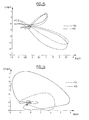
- the Figure 15 illustrates the result obtained after this basic change, for a patient with RS sinus rhythm (continuous loop) and episodes of supraventricular tachycardia TSV (dashed loop).
- the Figure 16 is counterpart of the Figure 15 , for a patient with RS sinus rhythm (continuous loop) and TV ventricular tachycardia episodes (dashed loop).
- the next step of the analysis consists in determining a certain number of descriptors parameters of the morphology of these loops so as to be able, under the best conditions, to discriminate between TV and TSV for a patient presenting episodes of tachycardia.
- each signal (sinus rhythm and tachycardia) is then projected on its own base, so as to be able to observe the corresponding one-dimensional signal (which is therefore a signal in the time domain), then compare the shapes in order to extract the morphological parameters that differentiate the SVTs from the VTs.
- the algorithm then calculates correlation coefficients between, on the one hand, the signals RS and TSV and, on the other hand, the signals RS and TV, these coefficients being calculated on the main channel and the secondary way.
- the mean squared error with respect to the sinus rhythm is also calculated, for the TSV beats and for the TV beats.
- D 1 ⁇ 1 / ⁇ 2 SR ⁇ 1 / ⁇ 2 TR
- D 2 ⁇ 1 / ⁇ 1 + ⁇ 2 SR ⁇ 1 / ⁇ 1 + ⁇ 2 TR
- D 4 R 1 , SR R 1 , TR
- D 5 R 2 , SR R 2 , TR
- the discrimination between TV and TSV can then be performed by various types of classifiers, in particular by a linear classifier or by a neural classifier.
- a first mode of implementation consists in constructing a linear classifier in the 3D space formed for example by the three descriptors EQM, M 1 and D 1 (this method can be applied to other descriptors).
- Such a classifier is characterized by the equation of the plane separating in this space the two families of arrhythmias, TV and TSV.
- a robust separator plane can be obtained by minimizing the least squares of the distance of each example to the plane.
- Matrix X is the matrix containing for each arrhythmia the value of the three descriptors in columns, and a fourth column of 1. This matrix has the following structure, assuming that we have a database of patients with 1 to N arrhythmias: MSE 1 ⁇ M 1 1 ⁇ D 1 1 1 MSE 2 ⁇ M 1 2 ⁇ D 1 2 1 . .. . . . MSE NOT ⁇ M 1 NOT ⁇ D 1 NOT 1
- the matrix Y is the vector consisting of -1 when the point corresponds to a TSV, and of 1 if the point corresponds to a TV.
- the ratios R 1, SR and R 2, SR respectively between the maximum amplitude and the minimum amplitude of the depolarization complex on the BipV and UnipV axes from the patient's sinus rhythm EGM, as well as the ratios R 1, TR and R 2, TR for tachycardia beats can be determined without the use of principal components analysis.
- the correlation maxima M 1 and M 2 can be determined between the sinus beat and the tachycardia beat plots, respectively on the BipV and UnipV axes.
- the database is scalable and continuously complete in the device, each arrhythmia to be added or replace the arrhythmia of the database. Moreover, the device, implant or programmer recalculates the matrix A at regular intervals.
- Another embodiment may, alternatively, implement a neural classifier, which allows in particular to operate by means of an adaptive network, instead of a purely mathematical calculation.
- This classifier is constructed in the 3D space formed for example by the three descriptors EQM, M 1 and D 1 (this method can be applied to other descriptors).
- the database is scalable and continuously complete in the device, each arrhythmia to be added or replace the arrhythmia of the database. Moreover, the device, implant or programmer remakes at regular intervals the training of W.
- the device may comprise complementary means for checking a posteriori classification arrhythmia, the same type as those discussed above, leading to a new learning of W at regular intervals in the event of misclassification.
Landscapes
- Health & Medical Sciences (AREA)
- Life Sciences & Earth Sciences (AREA)
- Engineering & Computer Science (AREA)
- Cardiology (AREA)
- Physics & Mathematics (AREA)
- Animal Behavior & Ethology (AREA)
- Pathology (AREA)
- Biomedical Technology (AREA)
- Heart & Thoracic Surgery (AREA)
- Medical Informatics (AREA)
- Molecular Biology (AREA)
- Surgery (AREA)
- Biophysics (AREA)
- General Health & Medical Sciences (AREA)
- Public Health (AREA)
- Veterinary Medicine (AREA)
- Artificial Intelligence (AREA)
- Evolutionary Computation (AREA)
- Fuzzy Systems (AREA)
- Mathematical Physics (AREA)
- Computer Vision & Pattern Recognition (AREA)
- Physiology (AREA)
- Psychiatry (AREA)
- Signal Processing (AREA)
- Measurement And Recording Of Electrical Phenomena And Electrical Characteristics Of The Living Body (AREA)
- Electrotherapy Devices (AREA)
Applications Claiming Priority (1)
| Application Number | Priority Date | Filing Date | Title |
|---|---|---|---|
| FR0801691 | 2008-03-28 |
Publications (2)
| Publication Number | Publication Date |
|---|---|
| EP2105843A1 true EP2105843A1 (de) | 2009-09-30 |
| EP2105843B1 EP2105843B1 (de) | 2011-03-16 |
Family
ID=39679351
Family Applications (1)
| Application Number | Title | Priority Date | Filing Date |
|---|---|---|---|
| EP09290016A Active EP2105843B1 (de) | 2008-03-28 | 2009-01-09 | Aktives Medizingerät, das verbesserte Mittel zur Unterscheidung zwischen Tachykardien ventrikulären Ursprungs und Tachykardien supraventrikulären Ursprungs enthält |
Country Status (4)
| Country | Link |
|---|---|
| US (1) | US8195281B2 (de) |
| EP (1) | EP2105843B1 (de) |
| AT (1) | ATE502339T1 (de) |
| DE (1) | DE602009000871D1 (de) |
Cited By (8)
| Publication number | Priority date | Publication date | Assignee | Title |
|---|---|---|---|---|
| EP2324885A1 (de) | 2009-11-18 | 2011-05-25 | Sorin CRM SAS | Aktives medizinisches Gerät mit Mitteln zum Einfangtesten durch Auswertung eines kardialen Vektogramms |
| EP2332462A1 (de) * | 2009-12-11 | 2011-06-15 | Sorin CRM SAS | Einfacher implantierbarer Einkammer-Defibrillator/Kardioverter mit Erfassung der aurikularen Aktivität über Monokörper-Sonde |
| FR2957512A1 (fr) * | 2010-03-22 | 2011-09-23 | Sorin Crm Sas | Dispositif medical actif comprenant des moyens de detection et de filtrage d'artefacts par analyse du vectogramme cardiaque |
| EP2742973A1 (de) | 2012-12-14 | 2014-06-18 | Sorin CRM SAS | Aktive implantierbare medizinische Vorrichtung vom Typ Herzschrittmacher mit Nachweis der anodischen Stimulation durch Vektogrammanalyse |
| EP3075413A1 (de) | 2015-04-02 | 2016-10-05 | Sorin CRM SAS | Implantierbares medizinisches gerät für die biventrikuläre stimulation, umfassend mittel zur messung intraventrikuläre leitungszeit für die diagnose von herzdekompensation |
| EP3081257A1 (de) | 2015-04-17 | 2016-10-19 | Sorin CRM SAS | Aktive implantierbare medizinische vorrichtung zur herzstimulation, die detektionsmittel für remodellierungserscheinungen oder inversen remodellierungserscheinungen des patienten umfasst |
| EP3760114A1 (de) | 2019-07-05 | 2021-01-06 | Sorin CRM SAS | Subkutan implantierbare medizinische vorrichtung und verfahren zur verarbeitung der signale einer subkutan implantierbaren medizinischen vorrichtung |
| EP2412401B1 (de) * | 2010-07-26 | 2022-01-05 | Sorin CRM SAS | Aktives implantierbares medizinisches gerät mit mitteln zum ventrikulären einfangtesten durch auswertung eines endokardialen beschleunigungssignals |
Families Citing this family (13)
| Publication number | Priority date | Publication date | Assignee | Title |
|---|---|---|---|---|
| US8903490B2 (en) * | 2010-03-03 | 2014-12-02 | Cardiac Pacemakers, Inc. | Methods and systems for recognizing arrhythmias using neural stimulation |
| US10517494B2 (en) * | 2014-11-14 | 2019-12-31 | Beth Israel Deaconess Medical Center, Inc. | Method and system to access inapparent conduction abnormalities to identify risk of ventricular tachycardia |
| US9675270B2 (en) | 2015-04-23 | 2017-06-13 | Medtronic, Inc. | Method and apparatus for determining a premature ventricular contraction in a medical monitoring device |
| CN119423777A (zh) | 2018-01-23 | 2025-02-14 | 程杰 | 使用由可植入装置感测和存储的内部心电图(ecg)信号对心脏心律失常的精确定位 |
| FR3079405B1 (fr) * | 2018-03-30 | 2023-10-27 | Substrate Hd | Dispositif informatique de detection de troubles du rythme cardiaque |
| WO2020014715A2 (en) * | 2018-06-21 | 2020-01-16 | Mayo Foundation For Medical Education And Research | Apparatus and method for distinguishing wide complex heart beats |
| US11154233B2 (en) | 2018-06-21 | 2021-10-26 | Mayo Foundation For Medical Education And Research | Apparatus and method for differentiating wide complex heart beats |
| EP3914345A1 (de) * | 2019-01-23 | 2021-12-01 | Impulse Dynamics NV | Unterscheidung von supraventrikulären tachykardien in kombinierter ccm-icd-vorrichtung |
| EP3760277B1 (de) | 2019-07-05 | 2022-09-21 | Sorin CRM SAS | Subkutan implantierbares kardiales defibrillationssystem |
| EP4073818A1 (de) * | 2019-12-09 | 2022-10-19 | Sanofi | Datenverarbeitungssysteme und verfahren für das repurposing von arzneimitteln |
| CN111714771B (zh) * | 2019-12-16 | 2023-12-29 | 苏州无双医疗设备有限公司 | 具有室速诊断功能的植入医疗设备 |
| KR102910027B1 (ko) * | 2022-03-30 | 2026-01-09 | 연세대학교 산학협력단 | 생체 전류 분석 방법 및 장치 |
| US20240278020A1 (en) * | 2023-02-22 | 2024-08-22 | Impulse Dynamics Nv | Electrocardiogram morphology evaluation management in implantable device |
Citations (9)
| Publication number | Priority date | Publication date | Assignee | Title |
|---|---|---|---|---|
| EP0626182A1 (de) | 1993-05-28 | 1994-11-30 | ELA MEDICAL (Société anonyme) | Verfahren zur Herztätigkeit-Analyse zur Bestimmung, ob eine Tachyarrythmie durch Stimulation unterbrochen werden kann |
| EP0813888A1 (de) | 1996-06-18 | 1997-12-29 | ELA MEDICAL (Société anonyme) | Aktive medizinische Vorrichtung von der Art eines Defibrillator-Kardiovertierer mit verbesserter Unterscheidung von Tachykardien |
| EP0838235A1 (de) | 1996-10-25 | 1998-04-29 | ELA MEDICAL (Société anonyme) | Aktive medizinische Vorrichtung von der Art eines Defibrillator/Kardiovertierrer mit verbesserter Unterscheidung von Tachykardien |
| WO2000069517A1 (en) | 1999-05-12 | 2000-11-23 | Medtronic, Inc. | Monitoring apparatus using wavelet transforms for the analysis of heart rhythms |
| EP1208873A1 (de) | 2000-11-27 | 2002-05-29 | Ela Medical | Aktive, medizinische Vorrichtung des Typs implantierbarer Defibrillator/Kardioverter mit verbesserter Unterscheidung von Vorhoffibrillation |
| US20030083587A1 (en) * | 2001-10-31 | 2003-05-01 | Bozidar Ferek-Petric | Method and apparatus for developing a vectorcardiograph in an implantable medical devicee |
| US20050159781A1 (en) | 1999-07-14 | 2005-07-21 | Cardiac Pacemakers, Inc. | Classification of supraventricular and ventricular cardiac rhythms using cross channel timing algorithm |
| WO2006039693A1 (en) * | 2004-09-30 | 2006-04-13 | Cardiac Pacemakers, Inc. | Cardiac activation sequence monitoring and tracking |
| US7149569B1 (en) | 2003-12-15 | 2006-12-12 | Pacesetter, Inc. | Apparatus and method for improved morphology discrimination in an implantable cardioverter defibrillator |
Family Cites Families (4)
| Publication number | Priority date | Publication date | Assignee | Title |
|---|---|---|---|---|
| US5766131A (en) * | 1995-08-04 | 1998-06-16 | Seiko Epson Corporation | Pulse-wave measuring apparatus |
| US7457664B2 (en) | 2005-05-09 | 2008-11-25 | Cardiac Pacemakers, Inc. | Closed loop cardiac resynchronization therapy using cardiac activation sequence information |
| FR2906123A1 (fr) | 2006-09-25 | 2008-03-28 | Ela Medical Soc Par Actions Si | Procede de reconstruction d'un electrocardiogramme de surface a partir d'un electrogramme endocavitaire |
| US7751873B2 (en) * | 2006-11-08 | 2010-07-06 | Biotronik Crm Patent Ag | Wavelet based feature extraction and dimension reduction for the classification of human cardiac electrogram depolarization waveforms |
-
2009
- 2009-01-09 AT AT09290016T patent/ATE502339T1/de not_active IP Right Cessation
- 2009-01-09 DE DE602009000871T patent/DE602009000871D1/de active Active
- 2009-01-09 EP EP09290016A patent/EP2105843B1/de active Active
- 2009-03-27 US US12/412,808 patent/US8195281B2/en active Active
Patent Citations (9)
| Publication number | Priority date | Publication date | Assignee | Title |
|---|---|---|---|---|
| EP0626182A1 (de) | 1993-05-28 | 1994-11-30 | ELA MEDICAL (Société anonyme) | Verfahren zur Herztätigkeit-Analyse zur Bestimmung, ob eine Tachyarrythmie durch Stimulation unterbrochen werden kann |
| EP0813888A1 (de) | 1996-06-18 | 1997-12-29 | ELA MEDICAL (Société anonyme) | Aktive medizinische Vorrichtung von der Art eines Defibrillator-Kardiovertierer mit verbesserter Unterscheidung von Tachykardien |
| EP0838235A1 (de) | 1996-10-25 | 1998-04-29 | ELA MEDICAL (Société anonyme) | Aktive medizinische Vorrichtung von der Art eines Defibrillator/Kardiovertierrer mit verbesserter Unterscheidung von Tachykardien |
| WO2000069517A1 (en) | 1999-05-12 | 2000-11-23 | Medtronic, Inc. | Monitoring apparatus using wavelet transforms for the analysis of heart rhythms |
| US20050159781A1 (en) | 1999-07-14 | 2005-07-21 | Cardiac Pacemakers, Inc. | Classification of supraventricular and ventricular cardiac rhythms using cross channel timing algorithm |
| EP1208873A1 (de) | 2000-11-27 | 2002-05-29 | Ela Medical | Aktive, medizinische Vorrichtung des Typs implantierbarer Defibrillator/Kardioverter mit verbesserter Unterscheidung von Vorhoffibrillation |
| US20030083587A1 (en) * | 2001-10-31 | 2003-05-01 | Bozidar Ferek-Petric | Method and apparatus for developing a vectorcardiograph in an implantable medical devicee |
| US7149569B1 (en) | 2003-12-15 | 2006-12-12 | Pacesetter, Inc. | Apparatus and method for improved morphology discrimination in an implantable cardioverter defibrillator |
| WO2006039693A1 (en) * | 2004-09-30 | 2006-04-13 | Cardiac Pacemakers, Inc. | Cardiac activation sequence monitoring and tracking |
Cited By (22)
| Publication number | Priority date | Publication date | Assignee | Title |
|---|---|---|---|---|
| US9014806B2 (en) | 2009-11-18 | 2015-04-21 | Sorin Crm Sas | System and method for performing a capture test based on the analysis of a cardiac vectogram |
| EP2324885A1 (de) | 2009-11-18 | 2011-05-25 | Sorin CRM SAS | Aktives medizinisches Gerät mit Mitteln zum Einfangtesten durch Auswertung eines kardialen Vektogramms |
| EP2332462A1 (de) * | 2009-12-11 | 2011-06-15 | Sorin CRM SAS | Einfacher implantierbarer Einkammer-Defibrillator/Kardioverter mit Erfassung der aurikularen Aktivität über Monokörper-Sonde |
| US9446256B2 (en) | 2009-12-11 | 2016-09-20 | Sorin Crm S.A.S. | Atrial activity signal estimation using independent component analysis |
| FR2957512A1 (fr) * | 2010-03-22 | 2011-09-23 | Sorin Crm Sas | Dispositif medical actif comprenant des moyens de detection et de filtrage d'artefacts par analyse du vectogramme cardiaque |
| EP2368493A1 (de) | 2010-03-22 | 2011-09-28 | Sorin CRM SAS | Aktive medizinische Vorrichtung mit Erfassungs- und Filtermitteln für Artefakte durch Analyse eines Vektor-Kardiogramms |
| US8359091B2 (en) | 2010-03-22 | 2013-01-22 | Sorin Crm S.A.S. | Apparatus and method for detecting and filtering artifacts by analysis of a cardiac vectogram |
| EP2412401B1 (de) * | 2010-07-26 | 2022-01-05 | Sorin CRM SAS | Aktives implantierbares medizinisches gerät mit mitteln zum ventrikulären einfangtesten durch auswertung eines endokardialen beschleunigungssignals |
| EP2742973A1 (de) | 2012-12-14 | 2014-06-18 | Sorin CRM SAS | Aktive implantierbare medizinische Vorrichtung vom Typ Herzschrittmacher mit Nachweis der anodischen Stimulation durch Vektogrammanalyse |
| FR2999440A1 (fr) * | 2012-12-14 | 2014-06-20 | Sorin Crm Sas | Dispositif medical implantable actif de type stimulateur cardiaque a detection de stimulation anodique par analyse de vectogramme |
| US8874212B2 (en) | 2012-12-14 | 2014-10-28 | Sorin Crm S.A.S. | Active implantable medical device type such as a pacemaker with detection of anodal stimulation by analysis of a vectogram |
| EP3075413A1 (de) | 2015-04-02 | 2016-10-05 | Sorin CRM SAS | Implantierbares medizinisches gerät für die biventrikuläre stimulation, umfassend mittel zur messung intraventrikuläre leitungszeit für die diagnose von herzdekompensation |
| US9757568B2 (en) | 2015-04-02 | 2017-09-12 | Sorin Crm Sas | Active implantable medical device for the diagnosis of cardiac decompensation |
| US10022547B2 (en) | 2015-04-02 | 2018-07-17 | Sorin Crm Sas | Active implantable medical device for the diagnosis of cardiac decompensation |
| US10328269B2 (en) | 2015-04-02 | 2019-06-25 | Sorin Crm Sas | Active implantable medical device for the diagnosis of cardiac decompensation |
| US11058881B2 (en) | 2015-04-02 | 2021-07-13 | Sorin Crm Sas | Active implantable medical device for the diagnosis of cardiac decompensation |
| FR3034319A1 (fr) * | 2015-04-02 | 2016-10-07 | Sorin Crm Sas | Dispositif medical implantable actif a stimulation biventriculaire, comprenant des moyens de mesure du temps de conduction interventriculaire pour le diagnostic de la decompensation cardiaque. |
| EP3081257A1 (de) | 2015-04-17 | 2016-10-19 | Sorin CRM SAS | Aktive implantierbare medizinische vorrichtung zur herzstimulation, die detektionsmittel für remodellierungserscheinungen oder inversen remodellierungserscheinungen des patienten umfasst |
| US9757566B2 (en) | 2015-04-17 | 2017-09-12 | Sorin Crm Sas | Active implantable medical device for detecting a remodeling or reverse remodeling phenomenon of the patient |
| US10420945B2 (en) | 2015-04-17 | 2019-09-24 | Sorin Crm Sas | Active implantable medical device for detecting a remodeling or reverse remodeling phenomenon of the patient |
| US11198009B2 (en) | 2015-04-17 | 2021-12-14 | Sorin Crm Sas | Active implantable medical device for detecting a remodeling or reverse remodeling phenomenon of the patient |
| EP3760114A1 (de) | 2019-07-05 | 2021-01-06 | Sorin CRM SAS | Subkutan implantierbare medizinische vorrichtung und verfahren zur verarbeitung der signale einer subkutan implantierbaren medizinischen vorrichtung |
Also Published As
| Publication number | Publication date |
|---|---|
| US20100249626A1 (en) | 2010-09-30 |
| DE602009000871D1 (de) | 2011-04-28 |
| EP2105843B1 (de) | 2011-03-16 |
| ATE502339T1 (de) | 2011-04-15 |
| US8195281B2 (en) | 2012-06-05 |
Similar Documents
| Publication | Publication Date | Title |
|---|---|---|
| EP2105843B1 (de) | Aktives Medizingerät, das verbesserte Mittel zur Unterscheidung zwischen Tachykardien ventrikulären Ursprungs und Tachykardien supraventrikulären Ursprungs enthält | |
| EP1867360B1 (de) | Aktives medizinisches Implantat, insbesondere Vorrichtung zur Stimulation, Resynchronisation, Defibrillation und/oder Kardioversion, die Mittel zur prädiktiven Warnung über eine Verschlimmerung des Krankheitszustands des Patienten umfasst | |
| EP2070562B1 (de) | Medizinische Vorrichtung zur Bestimmung des Herzzustands eines Patienten, dem ein aktives Implantat zur biventrikulären Stimulation eingesetzt wurde | |
| EP2412401B1 (de) | Aktives implantierbares medizinisches gerät mit mitteln zum ventrikulären einfangtesten durch auswertung eines endokardialen beschleunigungssignals | |
| EP2052680B1 (de) | Elektrokardiologisches Diagnosehilfsgerät, insbesondere zur Diagnose des Brugada-Syndroms und von ERS | |
| EP2324885B1 (de) | Aktives medizinisches gerät mit mitteln zum einfangtesten durch auswertung eines kardialen vektogramms | |
| EP2263747B1 (de) | Implantierbare Herzprothese mit Mittel zur Analyse der Patiententoleranz bezüglich der Stimulationsart die spontane Atrio-Ventrukuläre Leitung bevorzugend | |
| EP2368493B1 (de) | Aktive medizinische Vorrichtung mit Erfassungs- und Filtermitteln für Artefakte durch Analyse eines Vektor-Kardiogramms | |
| EP2499971B1 (de) | Adaptative Datenverarbeitungsvorrichtung eines endokardialen Beschleunigungssignals | |
| EP2623023B1 (de) | Aktives medizinisches Implantat, das mit Diagnosemitteln für Herzinsuffizienz ausgestattet ist | |
| EP2742971B1 (de) | Aktive implantierbare medizinische Vorrichtung vom Typ Herzschrittmacher mit Nachweistest durch vereinfachte Analyse eines Vektogramms | |
| EP2357020A1 (de) | Aktive medizinische Vorrichtung vom Typ Herzschrittmacher, Defibrillator und/oder Resynchronisator mit automatischer Optimierung der atrioventrikulären Verzögerung | |
| EP3669773B1 (de) | Vorrichtung und verfahren zur erkennung von herzkammerflimmern | |
| EP1405654B1 (de) | Aktives implantierbares medizinisches Gerät, wie Herzschrittmacher, Defibrillatoren, Kardiovertierer und/oder Multisitevorrichtungen, mit Mitteln zur Feststellung der durchschnittlichen hemodynamischen Kennzahl | |
| WO2017121702A1 (fr) | Dispositif médical implantable actif de défibrillation, comprenant des moyens perfectionnés de discrimination entre bruit externe et rupture de sonde et de caractérisation des tachyarythmies | |
| EP2742973A1 (de) | Aktive implantierbare medizinische Vorrichtung vom Typ Herzschrittmacher mit Nachweis der anodischen Stimulation durch Vektogrammanalyse | |
| EP2684515B1 (de) | Aktive medizinische Vorrichtung, die Mittel zur Überwachung des Zustands eines Patienten mit Herzinsuffizienzrisiko umfasst | |
| EP3081257B1 (de) | Aktive implantierbare medizinische vorrichtung zur herzstimulation, die detektionsmittel für remodellierungserscheinungen oder inversen remodellierungserscheinungen des patienten umfasst | |
| WO2002069178A2 (fr) | Procede et dispositif de filtrage d'une serie rr issue d'un signal cardiaque, et plus particulierement d'un signal ecg | |
| EP3025758B1 (de) | Aktive implantierbare medizinische vorrichtung, insbesondere vom typ leadless-kapsel, mit detektion der zyklusweisen erfassung durch analyse eines signals für die endokardiale beschleunigung | |
| EP3075413B1 (de) | Implantierbares medizinisches gerät für die biventrikuläre stimulation, umfassend mittel zur messung intraventrikulärer leitungszeit für die diagnose von herzdekompensation | |
| EP3760114B1 (de) | Subkutan implantierbare medizinische vorrichtung zur verarbeitung der signale einer subkutan implantierbaren medizinischen vorrichtung | |
| EP2839859A1 (de) | Aktive medizinische Vorrichtung, insbesondere Einheit für kardiale Resynchronisationstherapie (CRT), die Alarmmittel zur Vorhersage einer kardialen Dekompensation im Fall einer zentralen Schlafapnoe umfasst | |
| EP3165160B1 (de) | Aktive implantierbare medizinische vorrichtung, die mittel für die erfassung und quantifizierung von fusionssituationen umfasst | |
| EP2332462A1 (de) | Einfacher implantierbarer Einkammer-Defibrillator/Kardioverter mit Erfassung der aurikularen Aktivität über Monokörper-Sonde |
Legal Events
| Date | Code | Title | Description |
|---|---|---|---|
| PUAI | Public reference made under article 153(3) epc to a published international application that has entered the european phase |
Free format text: ORIGINAL CODE: 0009012 |
|
| AK | Designated contracting states |
Kind code of ref document: A1 Designated state(s): AT BE BG CH CY CZ DE DK EE ES FI FR GB GR HR HU IE IS IT LI LT LU LV MC MK MT NL NO PL PT RO SE SI SK TR |
|
| AX | Request for extension of the european patent |
Extension state: AL BA RS |
|
| 17P | Request for examination filed |
Effective date: 20091028 |
|
| 17Q | First examination report despatched |
Effective date: 20091124 |
|
| AKX | Designation fees paid |
Designated state(s): AT BE BG CH CY CZ DE DK EE ES FI FR GB GR HR HU IE IS IT LI LT LU LV MC MK MT NL NO PL PT RO SE SI SK TR |
|
| GRAP | Despatch of communication of intention to grant a patent |
Free format text: ORIGINAL CODE: EPIDOSNIGR1 |
|
| GRAS | Grant fee paid |
Free format text: ORIGINAL CODE: EPIDOSNIGR3 |
|
| GRAA | (expected) grant |
Free format text: ORIGINAL CODE: 0009210 |
|
| AK | Designated contracting states |
Kind code of ref document: B1 Designated state(s): AT BE BG CH CY CZ DE DK EE ES FI FR GB GR HR HU IE IS IT LI LT LU LV MC MK MT NL NO PL PT RO SE SI SK TR |
|
| REG | Reference to a national code |
Ref country code: GB Ref legal event code: FG4D Free format text: NOT ENGLISH |
|
| REG | Reference to a national code |
Ref country code: CH Ref legal event code: EP |
|
| REG | Reference to a national code |
Ref country code: IE Ref legal event code: FG4D |
|
| REF | Corresponds to: |
Ref document number: 602009000871 Country of ref document: DE Date of ref document: 20110428 Kind code of ref document: P |
|
| REG | Reference to a national code |
Ref country code: DE Ref legal event code: R096 Ref document number: 602009000871 Country of ref document: DE Effective date: 20110428 |
|
| REG | Reference to a national code |
Ref country code: SE Ref legal event code: TRGR |
|
| REG | Reference to a national code |
Ref country code: NL Ref legal event code: VDEP Effective date: 20110316 |
|
| PG25 | Lapsed in a contracting state [announced via postgrant information from national office to epo] |
Ref country code: LV Free format text: LAPSE BECAUSE OF FAILURE TO SUBMIT A TRANSLATION OF THE DESCRIPTION OR TO PAY THE FEE WITHIN THE PRESCRIBED TIME-LIMIT Effective date: 20110316 Ref country code: ES Free format text: LAPSE BECAUSE OF FAILURE TO SUBMIT A TRANSLATION OF THE DESCRIPTION OR TO PAY THE FEE WITHIN THE PRESCRIBED TIME-LIMIT Effective date: 20110627 Ref country code: NO Free format text: LAPSE BECAUSE OF FAILURE TO SUBMIT A TRANSLATION OF THE DESCRIPTION OR TO PAY THE FEE WITHIN THE PRESCRIBED TIME-LIMIT Effective date: 20110616 Ref country code: GR Free format text: LAPSE BECAUSE OF FAILURE TO SUBMIT A TRANSLATION OF THE DESCRIPTION OR TO PAY THE FEE WITHIN THE PRESCRIBED TIME-LIMIT Effective date: 20110617 Ref country code: LT Free format text: LAPSE BECAUSE OF FAILURE TO SUBMIT A TRANSLATION OF THE DESCRIPTION OR TO PAY THE FEE WITHIN THE PRESCRIBED TIME-LIMIT Effective date: 20110316 Ref country code: HR Free format text: LAPSE BECAUSE OF FAILURE TO SUBMIT A TRANSLATION OF THE DESCRIPTION OR TO PAY THE FEE WITHIN THE PRESCRIBED TIME-LIMIT Effective date: 20110316 |
|
| LTIE | Lt: invalidation of european patent or patent extension |
Effective date: 20110316 |
|
| PG25 | Lapsed in a contracting state [announced via postgrant information from national office to epo] |
Ref country code: BG Free format text: LAPSE BECAUSE OF FAILURE TO SUBMIT A TRANSLATION OF THE DESCRIPTION OR TO PAY THE FEE WITHIN THE PRESCRIBED TIME-LIMIT Effective date: 20110616 Ref country code: AT Free format text: LAPSE BECAUSE OF FAILURE TO SUBMIT A TRANSLATION OF THE DESCRIPTION OR TO PAY THE FEE WITHIN THE PRESCRIBED TIME-LIMIT Effective date: 20110316 Ref country code: CY Free format text: LAPSE BECAUSE OF FAILURE TO SUBMIT A TRANSLATION OF THE DESCRIPTION OR TO PAY THE FEE WITHIN THE PRESCRIBED TIME-LIMIT Effective date: 20110316 Ref country code: FI Free format text: LAPSE BECAUSE OF FAILURE TO SUBMIT A TRANSLATION OF THE DESCRIPTION OR TO PAY THE FEE WITHIN THE PRESCRIBED TIME-LIMIT Effective date: 20110316 Ref country code: SI Free format text: LAPSE BECAUSE OF FAILURE TO SUBMIT A TRANSLATION OF THE DESCRIPTION OR TO PAY THE FEE WITHIN THE PRESCRIBED TIME-LIMIT Effective date: 20110316 |
|
| PG25 | Lapsed in a contracting state [announced via postgrant information from national office to epo] |
Ref country code: EE Free format text: LAPSE BECAUSE OF FAILURE TO SUBMIT A TRANSLATION OF THE DESCRIPTION OR TO PAY THE FEE WITHIN THE PRESCRIBED TIME-LIMIT Effective date: 20110316 Ref country code: PT Free format text: LAPSE BECAUSE OF FAILURE TO SUBMIT A TRANSLATION OF THE DESCRIPTION OR TO PAY THE FEE WITHIN THE PRESCRIBED TIME-LIMIT Effective date: 20110718 |
|
| PG25 | Lapsed in a contracting state [announced via postgrant information from national office to epo] |
Ref country code: CZ Free format text: LAPSE BECAUSE OF FAILURE TO SUBMIT A TRANSLATION OF THE DESCRIPTION OR TO PAY THE FEE WITHIN THE PRESCRIBED TIME-LIMIT Effective date: 20110316 Ref country code: IS Free format text: LAPSE BECAUSE OF FAILURE TO SUBMIT A TRANSLATION OF THE DESCRIPTION OR TO PAY THE FEE WITHIN THE PRESCRIBED TIME-LIMIT Effective date: 20110716 Ref country code: RO Free format text: LAPSE BECAUSE OF FAILURE TO SUBMIT A TRANSLATION OF THE DESCRIPTION OR TO PAY THE FEE WITHIN THE PRESCRIBED TIME-LIMIT Effective date: 20110316 Ref country code: SK Free format text: LAPSE BECAUSE OF FAILURE TO SUBMIT A TRANSLATION OF THE DESCRIPTION OR TO PAY THE FEE WITHIN THE PRESCRIBED TIME-LIMIT Effective date: 20110316 |
|
| PG25 | Lapsed in a contracting state [announced via postgrant information from national office to epo] |
Ref country code: NL Free format text: LAPSE BECAUSE OF FAILURE TO SUBMIT A TRANSLATION OF THE DESCRIPTION OR TO PAY THE FEE WITHIN THE PRESCRIBED TIME-LIMIT Effective date: 20110316 |
|
| PLBE | No opposition filed within time limit |
Free format text: ORIGINAL CODE: 0009261 |
|
| STAA | Information on the status of an ep patent application or granted ep patent |
Free format text: STATUS: NO OPPOSITION FILED WITHIN TIME LIMIT |
|
| 26N | No opposition filed |
Effective date: 20111219 |
|
| PG25 | Lapsed in a contracting state [announced via postgrant information from national office to epo] |
Ref country code: PL Free format text: LAPSE BECAUSE OF FAILURE TO SUBMIT A TRANSLATION OF THE DESCRIPTION OR TO PAY THE FEE WITHIN THE PRESCRIBED TIME-LIMIT Effective date: 20110316 |
|
| REG | Reference to a national code |
Ref country code: DE Ref legal event code: R097 Ref document number: 602009000871 Country of ref document: DE Effective date: 20111219 |
|
| PGFP | Annual fee paid to national office [announced via postgrant information from national office to epo] |
Ref country code: SE Payment date: 20120112 Year of fee payment: 4 |
|
| BERE | Be: lapsed |
Owner name: ELA MEDICAL Effective date: 20120131 |
|
| PG25 | Lapsed in a contracting state [announced via postgrant information from national office to epo] |
Ref country code: MC Free format text: LAPSE BECAUSE OF NON-PAYMENT OF DUE FEES Effective date: 20120131 |
|
| PG25 | Lapsed in a contracting state [announced via postgrant information from national office to epo] |
Ref country code: BE Free format text: LAPSE BECAUSE OF NON-PAYMENT OF DUE FEES Effective date: 20120131 |
|
| PG25 | Lapsed in a contracting state [announced via postgrant information from national office to epo] |
Ref country code: MK Free format text: LAPSE BECAUSE OF FAILURE TO SUBMIT A TRANSLATION OF THE DESCRIPTION OR TO PAY THE FEE WITHIN THE PRESCRIBED TIME-LIMIT Effective date: 20110316 |
|
| PGFP | Annual fee paid to national office [announced via postgrant information from national office to epo] |
Ref country code: IE Payment date: 20130110 Year of fee payment: 5 |
|
| PG25 | Lapsed in a contracting state [announced via postgrant information from national office to epo] |
Ref country code: MT Free format text: LAPSE BECAUSE OF FAILURE TO SUBMIT A TRANSLATION OF THE DESCRIPTION OR TO PAY THE FEE WITHIN THE PRESCRIBED TIME-LIMIT Effective date: 20110316 |
|
| REG | Reference to a national code |
Ref country code: CH Ref legal event code: PL |
|
| REG | Reference to a national code |
Ref country code: SE Ref legal event code: EUG |
|
| PG25 | Lapsed in a contracting state [announced via postgrant information from national office to epo] |
Ref country code: CH Free format text: LAPSE BECAUSE OF NON-PAYMENT OF DUE FEES Effective date: 20130131 Ref country code: LI Free format text: LAPSE BECAUSE OF NON-PAYMENT OF DUE FEES Effective date: 20130131 Ref country code: SE Free format text: LAPSE BECAUSE OF NON-PAYMENT OF DUE FEES Effective date: 20130110 |
|
| PG25 | Lapsed in a contracting state [announced via postgrant information from national office to epo] |
Ref country code: TR Free format text: LAPSE BECAUSE OF FAILURE TO SUBMIT A TRANSLATION OF THE DESCRIPTION OR TO PAY THE FEE WITHIN THE PRESCRIBED TIME-LIMIT Effective date: 20110316 |
|
| PG25 | Lapsed in a contracting state [announced via postgrant information from national office to epo] |
Ref country code: LU Free format text: LAPSE BECAUSE OF NON-PAYMENT OF DUE FEES Effective date: 20120109 |
|
| PG25 | Lapsed in a contracting state [announced via postgrant information from national office to epo] |
Ref country code: HU Free format text: LAPSE BECAUSE OF FAILURE TO SUBMIT A TRANSLATION OF THE DESCRIPTION OR TO PAY THE FEE WITHIN THE PRESCRIBED TIME-LIMIT Effective date: 20090109 |
|
| REG | Reference to a national code |
Ref country code: IE Ref legal event code: MM4A |
|
| PG25 | Lapsed in a contracting state [announced via postgrant information from national office to epo] |
Ref country code: IE Free format text: LAPSE BECAUSE OF NON-PAYMENT OF DUE FEES Effective date: 20140109 |
|
| REG | Reference to a national code |
Ref country code: FR Ref legal event code: PLFP Year of fee payment: 8 |
|
| REG | Reference to a national code |
Ref country code: FR Ref legal event code: PLFP Year of fee payment: 9 |
|
| REG | Reference to a national code |
Ref country code: FR Ref legal event code: PLFP Year of fee payment: 10 |
|
| P01 | Opt-out of the competence of the unified patent court (upc) registered |
Effective date: 20230714 |
|
| PGFP | Annual fee paid to national office [announced via postgrant information from national office to epo] |
Ref country code: GB Payment date: 20260219 Year of fee payment: 18 |
|
| PGFP | Annual fee paid to national office [announced via postgrant information from national office to epo] |
Ref country code: DE Payment date: 20260206 Year of fee payment: 18 |
|
| PGFP | Annual fee paid to national office [announced via postgrant information from national office to epo] |
Ref country code: IT Payment date: 20260115 Year of fee payment: 18 |
|
| PGFP | Annual fee paid to national office [announced via postgrant information from national office to epo] |
Ref country code: FR Payment date: 20260130 Year of fee payment: 18 |