EP2143464A2 - Implantierbare Elektrodenleitung oder Elektrodenleitungsanordnung - Google Patents
Implantierbare Elektrodenleitung oder Elektrodenleitungsanordnung Download PDFInfo
- Publication number
- EP2143464A2 EP2143464A2 EP09162536A EP09162536A EP2143464A2 EP 2143464 A2 EP2143464 A2 EP 2143464A2 EP 09162536 A EP09162536 A EP 09162536A EP 09162536 A EP09162536 A EP 09162536A EP 2143464 A2 EP2143464 A2 EP 2143464A2
- Authority
- EP
- European Patent Office
- Prior art keywords
- lead
- septum
- conduit
- closure element
- line
- Prior art date
- Legal status (The legal status is an assumption and is not a legal conclusion. Google has not performed a legal analysis and makes no representation as to the accuracy of the status listed.)
- Granted
Links
- 210000005240 left ventricle Anatomy 0.000 claims abstract description 19
- 230000000638 stimulation Effects 0.000 claims abstract description 18
- 210000000596 ventricular septum Anatomy 0.000 claims abstract description 12
- 210000003157 atrial septum Anatomy 0.000 claims abstract description 8
- 230000001746 atrial effect Effects 0.000 claims abstract description 7
- 238000002513 implantation Methods 0.000 claims description 13
- 238000007789 sealing Methods 0.000 claims description 4
- 210000002808 connective tissue Anatomy 0.000 claims description 2
- 239000000463 material Substances 0.000 claims description 2
- 238000012377 drug delivery Methods 0.000 claims 3
- 239000004480 active ingredient Substances 0.000 claims 1
- 239000003795 chemical substances by application Substances 0.000 claims 1
- 239000003550 marker Substances 0.000 claims 1
- 230000001737 promoting effect Effects 0.000 claims 1
- 230000004936 stimulating effect Effects 0.000 claims 1
- 230000009772 tissue formation Effects 0.000 claims 1
- 239000003814 drug Substances 0.000 abstract 1
- 238000004088 simulation Methods 0.000 abstract 1
- 239000000523 sample Substances 0.000 description 17
- 230000002861 ventricular Effects 0.000 description 15
- 210000003748 coronary sinus Anatomy 0.000 description 8
- 238000009125 cardiac resynchronization therapy Methods 0.000 description 6
- 238000000034 method Methods 0.000 description 5
- 230000008901 benefit Effects 0.000 description 3
- 230000002349 favourable effect Effects 0.000 description 2
- 210000005246 left atrium Anatomy 0.000 description 2
- 230000035939 shock Effects 0.000 description 2
- 238000002560 therapeutic procedure Methods 0.000 description 2
- 238000005299 abrasion Methods 0.000 description 1
- 230000009471 action Effects 0.000 description 1
- 238000004873 anchoring Methods 0.000 description 1
- 230000015572 biosynthetic process Effects 0.000 description 1
- 238000009530 blood pressure measurement Methods 0.000 description 1
- 230000036471 bradycardia Effects 0.000 description 1
- 208000006218 bradycardia Diseases 0.000 description 1
- 230000001419 dependent effect Effects 0.000 description 1
- 238000013461 design Methods 0.000 description 1
- 238000011161 development Methods 0.000 description 1
- 230000018109 developmental process Effects 0.000 description 1
- 238000002592 echocardiography Methods 0.000 description 1
- 230000001435 haemodynamic effect Effects 0.000 description 1
- 230000004217 heart function Effects 0.000 description 1
- 238000003780 insertion Methods 0.000 description 1
- 230000037431 insertion Effects 0.000 description 1
- 238000012986 modification Methods 0.000 description 1
- 230000004048 modification Effects 0.000 description 1
- 210000003205 muscle Anatomy 0.000 description 1
- 210000004165 myocardium Anatomy 0.000 description 1
- 238000005457 optimization Methods 0.000 description 1
- 230000008447 perception Effects 0.000 description 1
- 229920001296 polysiloxane Polymers 0.000 description 1
- 229920002635 polyurethane Polymers 0.000 description 1
- 239000004814 polyurethane Substances 0.000 description 1
- 230000033764 rhythmic process Effects 0.000 description 1
- 210000005241 right ventricle Anatomy 0.000 description 1
- 210000003462 vein Anatomy 0.000 description 1
Images


Classifications
-
- A—HUMAN NECESSITIES
- A61—MEDICAL OR VETERINARY SCIENCE; HYGIENE
- A61N—ELECTROTHERAPY; MAGNETOTHERAPY; RADIATION THERAPY; ULTRASOUND THERAPY
- A61N1/00—Electrotherapy; Circuits therefor
- A61N1/02—Details
- A61N1/04—Electrodes
- A61N1/05—Electrodes for implantation or insertion into the body, e.g. heart electrode
- A61N1/056—Transvascular endocardial electrode systems
- A61N1/057—Anchoring means; Means for fixing the head inside the heart
- A61N1/0573—Anchoring means; Means for fixing the head inside the heart chacterised by means penetrating the heart tissue, e.g. helix needle or hook
Definitions
- the invention relates to an implantable lead or lead assembly for implantation in the left ventricle of a heart with perforation of the atrial or ventricular septum.
- the state of the art for left ventricular stimulation and perception is currently the implantation of an electrode into a left ventricular vein via the coronary sinus. These so-called coronary sinus electrodes are mainly used for cardiac resynchronization therapy.
- WO 2006/105395 A2 describes a transseptal / trans-myocardial ventricular stimulation electrode.
- the reliable implantation of a left ventricular coronary sinus electrode is not possible.
- the rate of dislocation of left ventricular leads implanted across the coronary sinus for cardiac resynchronization therapy (CRT) is greater than that of a conventional right ventricular pacing lead.
- CRT cardiac resynchronization therapy
- ICDs automatic cardioverters / defibrillators
- Another disadvantage of a coronary sinus electrode is the very limited placement options. Usually only 1 to 2 different positions are given for fixation of the probe. This is considered a major cause of the z. Poor responder rate (60-70%) of the CRT.
- the transmural LV pressure measurement here represents only the possibility of permanently introducing a system through the heart muscle into the left ventricle.
- a very short probe is placed in the left ventricle with a pressure sensor that can not be used for electrical stimulation of the heart.
- the probe described is constructed in such a way that the active stimulation surface lies only in the region of the left ventricular septum and is not freely positionable in the left ventricle.
- WO 2006/105395 A2 did not address the repositioning and explantability of an electrode.
- the object of the invention is to construct a left ventricular probe which can be pushed through the atrial or ventricular septum from the right ventricle into the left ventricle and freely maneuvered and fixed within the left ventricle the implantation risk of a left-right shunt is minimized by appropriate design measures on the probe body. Furthermore, the possibility of repositioning such a probe and its explantation must be considered.
- the invention includes the essential idea at the perforation site in the atrial or ventricular septum through which the electrode lead is laid to provide a suitable element which ensures reliable mutual sealing of the areas of the heart adjacent to the septum, at least during a growing-in phase.
- this is done by the electrode line itself, which then carries a suitable closure element.
- a separate closure element is provided for this purpose, which is placed in the septum prior to the introduction of the electrode line and is then pierced by the electrode line when it is laid.
- the invention is also applicable to a lead-in conduit provided for use in the left atrium that is passed through a septum into the left atrium.
- the closure body can be firmly connected to the electrode, so that a longitudinal movement of the electrode body in the septum is avoided.
- the closure body is pronounced in another embodiment as expandable Schirmchen.
- the closure body is shaped as an expandable screen and as an expandable, electrode-displaceable anchor (proximal to the screen).
- the probe is preferably coated in the region of the closure body in order to promote rapid formation of connective tissue in the area of the perforation site.
- the "working channel” preferably has a maximum free diameter of 2mm when no electrode is pushed through (acceptable left-right shunt).
- the "working channel" is marked in a further embodiment with X-ray markers or made of an X-ray visible material.
- the "working channel” is secured in the septum by expandable fixators (such as stents) and the LV probe can also be secured in the "working channel” by expandable fixators. These fixators are attached to the probe.
- the expandable fixators can also be used for uni- and bipolar stimulation of the septum.
- FIG. 1 an arrangement 1 according to the invention is shown.
- a left ventricular electrode lead 3 is thereby advanced through an implantable transseptal passage ["working channel"] 5 into the left ventricle and fixed there by means of a conventional screw 7.
- the left ventricular Electrode lead 3 a bipolar stimulation electrode 9 and additionally two shock electrodes (distal: 11, proximal 13).
- the diameter of the distal shock electrode 11 is selected so that it can not slip through the passage 5 in the left ventricle.
- the lead 3 is connected to an implantable defibrillator 17 with an electrode plug 15 (IS-4 or IS-1 and DF-1).
- Fig. 2 shows as a further embodiment of the invention specifically an electrode lead 19, which can be implanted via a guide wire 21 with proximal handle 22 and its structure with a flexible (here U-shaped bent shown) electrode lead body 23, a tip electrode 25 and distal fixation barbs 27 is known per se for anchoring in the wall of the ventricle.
- a new element on this electrode line is a closure element 29, here represented symbolically as two sealing disks with a small distance from one another, for closing a perforation point in the septum, via which the electrode line (as in FIG Fig. 1 shown) is guided in the left ventricle.
- Fig. 3 shows as a special embodiment of this embodiment, an electrode line 19 ', in which the closure element 29 by two oppositely spreadable screens 29a' 29b 'is formed, which spreads thanks to an additional control wire 30' next to the guide wire 21, after insertion of the electrode line on both sides of the perforation in the septum can be and seal in the spread state, the environment of the puncture point.
- the proximal screen can additionally be displaced on the probe by means of the additional stylet 30 'so that the ventricular septum is fixed between the two screens.
- the two screens 29a ', 29b' can also be used as (for example bipolar) active stimulation electrodes.
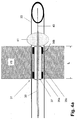
- FIG. 4A-C shown septal implementation (“working channel") 31 is first prepared by means of a Brockenbrough needle 33 by puncture of the ventricular septum VS.
- the implementation itself consists of 2 expandable stents 35a, 35b embedded in a flexible biocompatible tube 35c, eg silicone or polyurethane. This passage is mounted on an expandable balloon 37 of a balloon catheter 39 and is fixed by balloon expansion in the septum.
- the balloon catheter which expands after puncturing the left ventricle and retracting the Brockenbrough needle. Subsequently, the balloon catheter is withdrawn so far that the larger balloon 41 rests against the ventricular septum VS. Thereafter, the lead-through 31 whose length L was previously determined by echocardiography is fixed by the expansion of the smaller balloon 37 by the expanding balloon 37 placing the stems 35a, 35b with the tube portion 35c therebetween against the wall of the hole formed by the Brockenbrough needle in the septum moves.
- the leadthrough 31 and possibly a guide wire 43 remains in the puncture site (FIG. 4B ).
- the two stents 35a, 35b are visible in the X-ray image, so that a subsequent navigation of the LV probe by this transsepthal implementation is easily possible.
- the left-right shunt volume is minimized by the flexible part of the bushing 31. Namely, when no pipe is inserted, the inside width d is reduced by inward buckling of the flexible hose portion 35c between the stems 35a, 35b.
- the transseptal passage 31 described above is shown along with a left ventricular lead 45.
- a self-expanding fixator 47 This fixator is realized in the embodiment as a radiopaque spring element, which is released by retracting a tube 49 and thus clamps the electrode line in the implementation. In this way, abrasion of the electrode line or the fixator is avoided.
- FIG. 5 An alternative to the LV probes described above in combination with a feedthrough is in Fig. 5 an LV probe 51 is shown, which can be attached independently, without a separate closure element by means of directly attached fixators 53 in the septum, these fixators 53 are first encased by a tube 55 and then expanded by retraction of this tube by springs 57 in the septum VS ,
- the tube 55 is designed such that it can be completely removed from the electrode line after implantation (for example by peeling).
- the fixators can be "retracted” by a suitable tube - after cutting off the electrode plug.
- the fixators can be used as active stimulation electrodes by being connected to the electrical leads 57 of the electrode lead.
- the implantation in this line is also done by Brockenbrough needle.
Landscapes
- Health & Medical Sciences (AREA)
- Heart & Thoracic Surgery (AREA)
- Vascular Medicine (AREA)
- Cardiology (AREA)
- Engineering & Computer Science (AREA)
- Biomedical Technology (AREA)
- Nuclear Medicine, Radiotherapy & Molecular Imaging (AREA)
- Radiology & Medical Imaging (AREA)
- Life Sciences & Earth Sciences (AREA)
- Animal Behavior & Ethology (AREA)
- General Health & Medical Sciences (AREA)
- Public Health (AREA)
- Veterinary Medicine (AREA)
- Electrotherapy Devices (AREA)
- Materials For Medical Uses (AREA)
Abstract
Description
- Die Erfindung betrifft eine implantierbare Leitung oder Leitungsanordnung zur Implantation im linken Ventrikel eines Herzens unter Perforation des atrialen oder ventrikulären Septums.
- Als Stand der Technik für eine linksventrikuläre Stimulation und Wahrnehmung ist derzeit die Implantation einer Elektrode in eine linksventrikuläre Vene via Koronarsinus anzusehen. Diese sog. Koronarsinuselektroden kommen vorwiegend für die kardiale Resynchronisationstherapie zum Einsatz.
- In der medizinischen Literatur finden sich vermehrt Fallberichte von transseptaler Implantation linksventrikulären Stimulationselektroden zur Resynchronisationstherapie. Diese Implantationstechniken wurden immer unter Zuhilfenahme vorhandener Katheter, Führungsdrähte und Elektroden durchgeführt. Als Zugang zum linken Ventrikel wurden entweder die Punktion des atrialen Septums oder des Ventrikelseptums beschrieben, vgl. z.B. Transseptal endocardial left ventricular pacing: An alternative technique for coronary sinus lead placement in cardiac resynchronization therapy, van Gelder BM, Scheffer MG, Meijer A, et al, Heart Rhythm 2007 Apr; 4(4):454-60.
- Ferner werden derzeit Konzepte zur transmuralen linksventrikulären Druckmessung in klinischen Studien untersucht. Hier werden Drucksensoren transmural im linken Ventrikel zur dauerhaften telemetrischen Druckmessung im linken Ventrikel platziert; vgl. unter www.transomamedical.com oder folgende Fachveröffentlichungen:
- A Novel Technique For Assessing Load-Dependent Cardiac Function During LVAD Support Using Telemetered Left Ventricular Pressure. McConnell, PI, Del Rio, CL, Kwiatkowski, P, Farrar, D, Shipkowitz, T, Michler, RE, Sun, B. ASAIO Journal. 51(2): 31A, March/April 2005;
- In Vivo Safety and Accuracy of a Clinically Applicable Telemetered Left Ventricular Pressure Module: Intermediate-Term Results. McConnell, PI, de Cunha, D, Shipkowitz, T, Van Hee, J, Long, P and Hamlin, R. Heart Failure Society Meeting, September 2004;
- A System for Long-Term Measurement of Left Ventricular Pressure in Heart Failure Patients Living at Home. Sweitzer, N, Park, S. Heart Failure Society Meeting, September 2002:
- Automated Non-Invasive Monitoring of Left Ventricular Hemodynamics During Onset of Heart Failure in an Ambulatory Yucatan Mini Pig Model Using a New System Under Development for Assessing Heart Failure Patients at Home. Park, S, Sweitzer, N. Heart Failure Society Meeting, September 2002, oder
- A System For Long-Term Measurement Of Left Ventricular Pressure In Heart Failure Patients Living At Home. Park, S, Sweitzer, N and May, G. Heart Failure & Circulatory Support Summit, Cleveland, OH, August 2002.
- Für angeborene Atriumsseptumdefekte, offene Foramen oder ovale und Ventrikelseptumdefekte werden derzeit eine Vielzahl von kommerziellen Verschlusssystemen (z.B. Premere™ PFO, SJM) angeboten, die mittels Kathetertechnik platziert werden können und einen zuverlässigen Verschluss des Septumdefektes gewährleisten; vgl. dazu Transcatheter patent foramen ovale closure using the premere PFO occlusion system. Andrea Donti, Alessandro Giardini, Luisa Salomone, Roberto Formigari, Fernando M. Picchio. Catheterization and Cardiovascular Interventions, vol 68/5 2006.
- In
WO 2006/105395 A2 wird eine transseptale/trans-myocardiale ventrikuläre Stimulationselektrode beschrieben. - In ca. 10-15% der Implantationen ist aufgrund der anatomischen Gegebenheiten die zuverlässige Implantation einer linksventrikulären Koronarsinus-Elektrode nicht möglich. Des Weiteren ist die Dislokationsrate von linksventrikulären Elektroden, die für die kardiale Resynchronisationstherapie (CRT) über den Koronarsinus implantiert würden, größer als die einer herkömmlichen rechtventrikulären Schrittmacherelektrode. Aus diesen Gründen ist derzeit die Einführung einer rein linksventrikulären Stimulation - unter Verwendung einer Koronarsinuselektrode - für die Bradykardietherapie oder die Implantation von automatischen Kardiovertern/Defibrillatoren (ICD) nicht gegeben, da sowohl der Implantationserfolg als auch die Sicherheit mit dieser Art linksventrikulärer Elektrode nicht gegeben ist. Ein weiterer Nachteil einer Koronarsinuselektrode sind die sehr eingeschränkten Platzierungsoptionen. Meist sind nur 1 bis 2 verschiedene Positionen zu Fixierung der Sonde gegeben. Dies wird als eine wesentliche Ursache für die z. T. schlechte Responderrate (60-70%) der CRT diskutiert.
- Die weiter oben vorgestellten Techniken zur Elektrodenimplantation in den linken Ventrikel via atrialem oder ventrikulärem Septum sind sehr aufwändig und haben sich aufgrund der bestehen Risiken (RV Shunt, Thromben) bislang nicht durchgesetzt, obwohl hier eine freie Platzierung der Elektrode im linken Ventrikel möglich ist und damit die Nachteile der zuverlässigen Sondenfixierung, der Responderrate und anatomischen Einschränkungen behoben werden.
- Die transmurale LV-Druckmessung stellt hier nur die Möglichkeit dar, durch den Herzmuskel ein System dauerhaft in den linken Ventrikel einzubringen. Allerdings wird hier eine sehr kurze Sonde mit einem Drucksensor in den linken Ventrikel gebracht, die nicht für die elektrische Stimulation des Herzens verwendet werden kann. Die in
WO 2006/105395 A2 beschriebene Sonde ist jedoch derart konstruiert, dass aktive Stimulationsfläche nur im Bereich des links-ventrikulären Septums liegt und nicht im linken Ventrikel frei positionierbar ist. Weiter wird inWO 2006/105395 A2 nicht auf die Repositionierung und auf die Explantationsfähigkeit einer Elektrode eingegangen. - Um die oben genannten Nachteile zu beheben, besteht die Aufgabe der Erfindung darin, eine linksventrikuläre Sonde zu konstruieren, die durch das atriale oder ventrikuläre Septum vom rechten Ventrikel in den linken Ventrikel geschoben werden und frei innerhalb des linken Ventrikel manövriert und fixiert werden kann, wobei das Implantationsrisiko eines Links-Rechts-Shunts durch geeignete konstruktive Maßnahmen am Sondenkörper minimiert wird. Des Weiteren sind die Möglichkeit der Repositionierung einer solchen Sonde und deren Explantation zu berücksichtigen.
- Diese Aufgabe wird durch eine implantierbare Leitung gemäß Anspruch 1 bzw. eine Leitungsanordnung gemäß Anspruch 9 gelöst. Zweckmäßige Fortbildungen des Erfindungsgedankens sind Gegenstand der abhängigen Ansprüche.
- Die Erfindung schließt den wesentlichen Gedanken ein, an der Perforationsstelle im atrialen oder ventrikulären Septum, durch die die Elektrodenleitung verlegt wird, ein geeignetes Element vorzusehen, die zumindest während einer Einwachsphase eine zuverlässige gegenseitige Abdichtung der an das Septum angrenzenden Bereiche des Herzens sichert. In einer ersten Ausprägung des Erfindungsgedankens geschieht dies durch die Elektrodenleitung selbst, welche dann ein geeignetes Verschlusselement trägt. In einer relativ selbstständigen zweiten Ausprägung des Erfindungsgedankens ist hierzu ein separates Verschlusselement vorgesehen, welches vor der Einführung der Elektrodenleitung im Septum platziert und anschließend beim Verlegen der Elektrodenleitung von dieser durchstoßen wird.
- Es wird ausdrücklich angemerkt, dass die Erfindung auch bei einer zum Einsatz im linken Atrium vorgesehenen Leitung bzw. Leitungsanordnung anwendbar ist, die durch ein Septum in das linke Atrium geführt ist.
- Ein wichtiger Vorteil der erfindungsgemäßen Lösung ist der sichere und zuverlässige Zugang zum linken Ventrikel ohne die anatomischen Einschränkungen des Koronarsinus-Zugangs. Mit dieser Technik ist es dann möglich, primär linksventrikulär gesteuerte Herzschrittmacher, ICDs und CRT-Geräte anzubieten. Vorteil der primär linksgesteuerten Systeme sind:
- Physiologisch günstigerer Stimulationsort;
- bessere Sensingsignale aufgrund der größeren Muskelmasse;
- günstigere Bedingungen zu Sondenfixierung und geringeres Perforationsrisiko aufgrund der größeren Wandstärke;
- bessere Möglichkeiten der haemodynamischen Optimierung durch Stimulation;
- der Nachteil der RV-Stimulation wird weitgehend aufgehoben!
- Der Verschlusskörper kann fest mit der Elektrode verbunden werden, so dass eine longitudinale Bewegung des Elektrodenkörpers im Septum vermieden wird.
- Der Verschlusskörper ist in einer weiteren Ausführung ausgeprägt als expandierbares Schirmchen.
- In einer ähnlichen Ausführung ist der Verschlusskörper ausgeprägt als ein expandierbares Schirmchen und als ein expandierbarer, auf der Elektrode verschiebbarer Anker (proximal des Schirmchens).
- Die Sonde ist im Bereich des Verschlußkörpers bevorzugt beschichtet, um eine schnelle Bindegewebsbildung im Bereich der Perforationsstelle zu fördern.
- Der ,,Arbeitskanal" hat bevorzugt einen maximalen freien Durchmesser von 2mm, wenn keine Elektrode durchgeschoben ist. (akzeptabler Links-Rechts Shunt).
- Der ,,Arbeitskanal" ist in einer weiteren Ausführung mit Röntgenmarkern gekennzeichnet bzw. aus einem Röntgensichtbaren Material gefertigt.
- Der ,,Arbeitskanal" wird z.B. durch expandierbare Fixatoren (wie z.B. Stents) im Septum befestigt. Auch die LV-Sonde kann im ,,Arbeitskanal" durch expandierbare Fixatoren befestigt werden. Diese Fixatoren sind an der Sonde angebracht. Die expandierbaren Fixatoren können zudem zur uni- und bipolaren Stimulation des Septums verwendet werden.
- Vorteile und Zweckmäßigkeiten der Erfindung ergeben sich im Übrigen aus der nachfolgenden Beschreibung von Ausführungsbeispielen anhand der Figuren. Von diesen zeigen:
-
Fig. 1 die Gesamtansicht einer Defibrillationsanordnung mit einer Ausführungsform der Erfindung, -
Fig. 2 eine Elektrodenleitung gemäß einer Ausführungsform der Erfindung, -
Fig. 3 eine Elektrodenleitung gemäß einer alternativen Ausführungsform der Erfindung, -
Fig. 4A bis 4C Darstellungen einer weiteren Ausführung der Erfindung, in verschiedenen Realisierungsphasen, und -
Fig. 5 eine Detailansicht einer weiteren Ausführungsform der Erfindung. - In
Fig. 1 ist eine erfindungsgemäße Anordnung 1 dargestellt. Eine linksventrikuläre Elektrodenleitung 3 ist dabei durch eine implantierbare transseptale Durchführung ["Arbeitskanal"] 5 in den linken Ventrikel vorgeschoben und dort mittels einer konventionellen Schraube 7 fixiert. In der gezeigten Ausführung hat die linksventrikuläre Elektrodenleitung 3 eine bipolare Stimulationselektrode 9 und zusätzlich zwei Schockelektroden (distal: 11, proximal 13). Der Durchmesser der distalen Schockelektrode 11 ist so gewählt, dass diese nicht durch die Durchführung 5 in den linken Ventrikel rutschen kann. Die Leitung 3 ist mit einem Elektrodenstecker 15 (IS-4 bzw. IS-1 und DF-1) mit einem implantierbaren Defibrillator 17 verbunden. -
Fig. 2 zeigt als weitere Ausführungsform der Erfindung speziell eine Elektrodenleitung 19, die über einen Führungsdraht 21 mit proximaler Handhabe 22 implantiert werden kann und deren Aufbau mit einem flexiblen (hier U-förmig gebogen dargestellten) Elektrodenleitungskörper 23, einer Tip-Elektrode 25 und distalen Fixierungs-Widerhaken 27 zur Verankerung in der Wandung des Ventrikels an sich bekannt ist. Neu an dieser Elektrodenleitung ist ein - hier symbolisch als zwei Dichtscheiben mit kleinem Abstand voneinander dargestelltes - Verschlusselement 29 zum Verschluss einer Perforationsstelle im Septum, über die die Elektrodenleitung (wie inFig. 1 gezeigt) in den linken Ventrikel geführt ist. -
Fig. 3 zeigt als spezielle Ausgestaltung dieser Ausführung eine Elektrodenleitung 19', bei der das Verschlusselement 29 durch zwei gegensinnig aufspreizbare Schirme 29a' 29b' gebildet ist, die dank eines zusätzlichen Steuerdrahtes 30' neben dem Führungsdraht 21, nach Einführung der Elektrodenleitung beidseitig der Perforationsstelle im Septum aufgespreizt werden können und im aufgespreizten Zustand die Umgebung des Durchstoßpunktes abdichten. - Das proximale Schirmchen kann mittels des zusätzlichen Mandrins 30' zusätzlich auf der Sonde verschoben werden, so dass das Ventrikelseptum zwischen den beiden Schirmchen fixiert wird. Die beiden Schirmchen 29a', 29b' können dabei auch als (z.B. bipolare) aktive Stimulationselektroden eingesetzt werden.
- Die in
Fig. 4A-C dargestellte septale Durchführung ("Arbeitskanal") 31 wird zunächst mittels einer Brockenbrough-Nadel 33 durch Punktion des Ventrikelseptums VS hergestellt. Die Durchführung selbst besteht aus 2 expandierbaren Stents 35a, 35b, eingebettet in einen flexiblen biokompatiblen Schlauch 35c, z.B. Silikon oder Polyurethan. Diese Durchführung ist auf einem expandierbaren Ballon 37 eines Ballonkatheters 39 montiert und wird durch Ballonexpansion im Septum fixiert. - Um die korrekte Position der Durchführung zu gewähleisten, befindet sich auf dem Ballonkatheder ein zweiter Ballon 41 größeren Durchmessers, der nach Punktion des linken Ventrikels und Zurückziehen der Brockenbrough-Nadel expandiert wird. Anschließend wird der Ballonkatheter so weit zurückgezogen, dass der größere Ballon 41 am Ventrikelseptum VS anliegt. Danach wird die Durchführung 31, deren Länge L vorab mittels Echokardiographie bestimmt wurde, durch die Expansion des kleineren Ballons 37 fixiert, indem der expandierende Ballon 37 die Stens 35a, 35b mit dem Schlauchabschnitt 35c dazwischen gegen die Wandung des durch die Brockenbrough-Nadel gebildeten Loches im Septum rückt.
- Nach dem Entfernen des Ballonkatheters 39 verbleibt die Durchführung 31 und ggf. ein Führungsdraht 43 in der Punktionsstelle (
Fig 4B ). Die beiden Stents 35a, 35b sind dabei im Röntgenbild sichtbar, so dass eine anschließende Navigation der LV-Sonde durch diese transseptale Durchführung einfach möglich ist. Das Links-Rechts-Shuntvolumen wird dabei durch den flexiblen Teil der Durchführung 31 minimiert. Wenn keine Leitung eingeführt ist, wird nämlich die lichte Weite d durch ein Sich-Nach-Innen-Wölben des flexiblen Schlauchabschnitts 35c zwischen den Stens 35a, 35b reduziert. - In der
Fig. 4C ist die oben beschriebene transseptale Durchführung 31 zusammen mit einer linksventrikulären Leitung bzw. Sonde 45 dargestellt. Um eine Längsbewegung der Sonde 45 innerhalb der transseptalen Durchführung zu vermeiden, kann sie durch einen selbstexpandierenden Fixator 47 innerhalb der Durchführung befestigt werden. Dieser Fixator ist in dem Ausführungsbeispiel als ein röntgensichtbares Federelement realisiert, das durch Zurückziehen eines Schlauches 49 freigegeben wird und so die Elektrodenleitung in der Durchführung festklemmt. Auf diese Weise wird ein Abrieb der Elektrodenleitung oder des Fixators vermieden. - Alternativ zu den weiter oben beschriebenen LV-Sonden in Kombination mit einer Durchführung ist in
Fig. 5 eine LV-Sonde 51 dargestellt, die selbstständig, ohne separates Verschlusselement mittels direkt angebrachter Fixatoren 53 im Septum befestigt werden kann, wobei diese Fixatoren 53 zunächst durch einen Schlauch 55 ummantelt sind und durch das Zurückziehen dieses Schlauches dann durch Federn 57 im Septum VS expandiert werden. - Der Schlauch 55 ist dabei derart ausgeführt, dass er nach der Implantation (z.B. durch Peeling) vollständig von der Elektrodenleitung entfernt werden kann. Für den Fall einer Explantation einer solchen Elektrodenleitung können die Fixatoren durch einen entsprechenden Schlauch - nach Abschneiden des Elektrodensteckers - wieder "eingefahren" werden. Die Fixatoren können als aktive Stimulationselektroden eingesetzt werden, indem sie mit den elektrischen Zuleitungen 57 der Elektrodenleitung verbunden sind. Die Implantation in dieser Leitung erfolgt ebenfalls mittels Brockenbrough-Nadel.
- Die Ausführung der Erfindung ist nicht auf die oben beschriebenen Beispiele und hervorgehobenen Aspekte beschränkt, sondern ebenso in einer Vielzahl von Abwandlungen möglich, die im Rahmen fachgemäßen Handelns liegen.
Claims (15)
- Implantierbare Leitung (3, 19, 19', 45), insbesondere Elektroden- und/oder Sensor-und/oder Medikamentenzufuhrleitung, zur Implantation im linken Ventrikel eines Herzens unter Perforation des atrialen oder ventrikulären Septums, mit
einem langgestreckten, flexiblen Leitungskörper (23, 23'),
einer Elektrode (9, 25, 25') und/oder einem Sensor und/oder einer Medikamentenapplikationseinrichtung am oder nahe einem distalen Ende des Leitungskörpers und
einem an den Leitungskörper angeformten oder mit diesem verbundenen Verschlusselement (29, 29', 31) zur Abdichtung der Perforationsstelle im Septum. - Leitung nach Anspruch 1, mit am oder nahe dem distalen Ende des Leitungskörpers angeordneten Fixierungsmitteln (27, 27', 31), die zur Fixierung der Leitung in oder an einer Wand des linken Ventrikels ausgebildet sind.
- Leitung nach Anspruch 1 oder 2, mit mehreren Abfühl- und/oder Stimulationselektroden.
- Leitung nach Anspruch 1, wobei das Verschlusselement (29, 29', 31) fest mit dem Leitungskörper verbunden ist.
- Leitung nach einem der Ansprüche 1 oder 4, wobei das Verschlusselement (29, 29', 31) einen expandier- bzw. aufspreizbaren Abschnitt (47, 53) aufweist oder insgesamt expandier- bzw. aufspreizbar ausgebildet ist.
- Leitung nach einem der vorangehenden Ansprüche, wobei das Verschlusselement (29, 29', 31) ein, insbesondere auf dem Leitungskörper verschiebbares, Ankerelement (47, 53) zur Fixierung im oder am Septum aufweist.
- Leitung nach Anspruch 6, wobei das Ankerelement am Verschlusselement zugleich als Stimulationselektrode zur Stimulation des Septums ausgebildet ist.
- Leitung nach einem der vorangehenden Ansprüche, wobei mindestens ein Abschnitt des Verschlusselementes und/oder ein Abschnitt des Leitungskörpers nahe dem Verschlusselement mit einem eine schnelle Bindegewebsbildung im Bereich der Perforationsstelle fördernden Wirkstoff in einer Form beschichtet ist, die den Wirkstoff während einer Einwachsphase wirksam werden lässt.
- Implantierbare Leitungsanordnung zur Implantation im linken Ventrikel eines Herzens unter Perforation des atrialen oder ventrikulären Septums, mit
einer Leitung (3, 19, 19', 45), die einen langgestreckten, flexiblen Leitungskörper (23, 23') und eine Elektrode (9, 25, 25') und/oder einen Sensor und/oder eine Medikamentenapplikationseinrichtung am oder nahe einem distalen Ende des Leitungskörpers aufweist, und
einem separaten Verschlusselement (29, 29', 31) zur Abdichtung der Perforationsstelle, welches zur Fixierung im Septum ausgebildet ist und ein an den Außendurchmesser der Leitung angepasstes Lumen aufweist. - Leitungsanordnung nach Anspruch 9, wobei die lichte Weite des Lumens (31) im separaten Verschlusselement ohne eingeführte Leitung maximal 2 mm beträgt.
- Leitungsanordnung nach Anspruch 9 oder 10, wobei das Lumen (31) des Verschlusselementes ein Röntgenmarker-Material aufweist.
- Leitungsanordnung nach einem der Ansprüche 9 bis 11, wobei das Verschlusselement ein expandier- oder aufspreizbares Fixierungsmittel (47, 53) zur Fixierung im Septum aufweist.
- Leitungsanordnung nach einem der Ansprüche 9 bis 12, wobei der Leitungskörper Mittel zur Fixierung (47, 53) der Leitung im separaten Verschlusselement aufweist.
- Leitungsanordnung nach Anspruch 13, wobei die Fixierungsmittel am Leitungskörper zugleich als Stimulationselektrode zur elektrischen Stimulation des Septums ausgebildet sind.
- Leitungsanordnung nach einem der Ansprüche 9 bis 14, mit einer Leitung, welche am oder nahe dem distalen Ende des Leitungskörpers angeordnete Fixierungsmittel (7, 25, 25') zur Fixierung der Leitung in oder an einer Wand des linken Ventrikels und/oder mehrere Abfühl- und/oder Stimulationselektroden aufweist.
Priority Applications (1)
| Application Number | Priority Date | Filing Date | Title |
|---|---|---|---|
| EP16195063.9A EP3141279A1 (de) | 2008-07-10 | 2009-06-12 | Verschlusselement zur platzierung und fixierung im septum eines herzens sowie ballonkatheter zur fixierung eines verschlusselementes im septum eines herzens |
Applications Claiming Priority (1)
| Application Number | Priority Date | Filing Date | Title |
|---|---|---|---|
| DE102008040304A DE102008040304A1 (de) | 2008-07-10 | 2008-07-10 | Implantierbare Elektrodenleitung oder Elektrodenleitungsanordnung |
Related Child Applications (2)
| Application Number | Title | Priority Date | Filing Date |
|---|---|---|---|
| EP16195063.9A Division EP3141279A1 (de) | 2008-07-10 | 2009-06-12 | Verschlusselement zur platzierung und fixierung im septum eines herzens sowie ballonkatheter zur fixierung eines verschlusselementes im septum eines herzens |
| EP16195063.9A Division-Into EP3141279A1 (de) | 2008-07-10 | 2009-06-12 | Verschlusselement zur platzierung und fixierung im septum eines herzens sowie ballonkatheter zur fixierung eines verschlusselementes im septum eines herzens |
Publications (3)
| Publication Number | Publication Date |
|---|---|
| EP2143464A2 true EP2143464A2 (de) | 2010-01-13 |
| EP2143464A3 EP2143464A3 (de) | 2010-10-20 |
| EP2143464B1 EP2143464B1 (de) | 2017-01-18 |
Family
ID=41226205
Family Applications (2)
| Application Number | Title | Priority Date | Filing Date |
|---|---|---|---|
| EP16195063.9A Withdrawn EP3141279A1 (de) | 2008-07-10 | 2009-06-12 | Verschlusselement zur platzierung und fixierung im septum eines herzens sowie ballonkatheter zur fixierung eines verschlusselementes im septum eines herzens |
| EP09162536.8A Not-in-force EP2143464B1 (de) | 2008-07-10 | 2009-06-12 | Implantierbare Elektrodenleitung oder Elektrodenleitungsanordnung |
Family Applications Before (1)
| Application Number | Title | Priority Date | Filing Date |
|---|---|---|---|
| EP16195063.9A Withdrawn EP3141279A1 (de) | 2008-07-10 | 2009-06-12 | Verschlusselement zur platzierung und fixierung im septum eines herzens sowie ballonkatheter zur fixierung eines verschlusselementes im septum eines herzens |
Country Status (3)
| Country | Link |
|---|---|
| US (1) | US8509921B2 (de) |
| EP (2) | EP3141279A1 (de) |
| DE (1) | DE102008040304A1 (de) |
Cited By (1)
| Publication number | Priority date | Publication date | Assignee | Title |
|---|---|---|---|---|
| US8417332B2 (en) | 2010-05-05 | 2013-04-09 | Sorin Crm S.A.S. | Endocardial stimulation/defibrillation system of the left ventricle |
Families Citing this family (6)
| Publication number | Priority date | Publication date | Assignee | Title |
|---|---|---|---|---|
| DE102008043513A1 (de) | 2008-11-06 | 2010-05-12 | Biotronik Crm Patent Ag | Implantierbare Leitung |
| DE102009030340B4 (de) * | 2009-06-25 | 2011-12-01 | Peter Osypka | Elektrodenanordnung mit einer Stimulationselektrode für den linken Ventrikel |
| EP2510975A1 (de) | 2011-04-14 | 2012-10-17 | BIOTRONIK SE & Co. KG | Herzstimulator |
| US20170312537A1 (en) * | 2014-11-19 | 2017-11-02 | Sharp Kabushiki Kaisha | Photodynamic therapy device |
| EP3870267B1 (de) | 2018-10-25 | 2024-06-19 | Pacesetter, Inc. | Biostimulatordurchführung mit integriertem elektrodennapf |
| US11918820B2 (en) | 2020-01-21 | 2024-03-05 | Pacesetter, Inc. | Leadless biostimulator having overmolded header assembly |
Citations (1)
| Publication number | Priority date | Publication date | Assignee | Title |
|---|---|---|---|---|
| WO2006105395A2 (en) | 2005-03-31 | 2006-10-05 | Medtronic, Inc. | Trans-septal/trans-myocardial ventricular pacing lead |
Family Cites Families (22)
| Publication number | Priority date | Publication date | Assignee | Title |
|---|---|---|---|---|
| US4836204A (en) * | 1987-07-06 | 1989-06-06 | Landymore Roderick W | Method for effecting closure of a perforation in the septum of the heart |
| US5634936A (en) * | 1995-02-06 | 1997-06-03 | Scimed Life Systems, Inc. | Device for closing a septal defect |
| US5853422A (en) * | 1996-03-22 | 1998-12-29 | Scimed Life Systems, Inc. | Apparatus and method for closing a septal defect |
| US5728140A (en) * | 1996-06-17 | 1998-03-17 | Cardiac Pacemakers, Inc. | Method for evoking capture of left ventricle using transeptal pacing lead |
| US5976174A (en) * | 1997-12-15 | 1999-11-02 | Ruiz; Carlos E. | Medical hole closure device and methods of use |
| US6634364B2 (en) * | 2000-12-15 | 2003-10-21 | Cardiac Pacemakers, Inc. | Method of deploying a ventricular lead containing a hemostasis mechanism |
| US6309350B1 (en) * | 1999-05-03 | 2001-10-30 | Tricardia, L.L.C. | Pressure/temperature/monitor device for heart implantation |
| US6478776B1 (en) * | 2000-04-05 | 2002-11-12 | Biocardia, Inc. | Implant delivery catheter system and methods for its use |
| US6746404B2 (en) * | 2000-12-18 | 2004-06-08 | Biosense, Inc. | Method for anchoring a medical device between tissue |
| US20060122633A1 (en) * | 2002-06-13 | 2006-06-08 | John To | Methods and devices for termination |
| US9480839B2 (en) * | 2002-09-24 | 2016-11-01 | Medtronic, Inc. | Lead delivery device and method |
| FR2859912B1 (fr) * | 2003-09-22 | 2005-11-18 | Ela Medical Sa | Necessaire de percage du septum cardiaque et de mise en place d'un dispositif transseptal, notamment d'une sonde de stimulation d'une cavite gauche |
| US7515970B2 (en) * | 2004-08-18 | 2009-04-07 | Cardiac Pacemakers, Inc. | Transeptal lead |
| US7840266B2 (en) * | 2005-03-11 | 2010-11-23 | Cardiac Pacemakers, Inc. | Integrated lead for applying cardiac resynchronization therapy and neural stimulation therapy |
| US7305270B1 (en) * | 2005-04-21 | 2007-12-04 | Pacesetter, Inc. | Cardiac pacing/sensing lead providing far-field signal rejection |
| US7840281B2 (en) * | 2006-07-21 | 2010-11-23 | Boston Scientific Scimed, Inc. | Delivery of cardiac stimulation devices |
| US20080046059A1 (en) * | 2006-08-04 | 2008-02-21 | Zarembo Paul E | Lead including a heat fused or formed lead body |
| US8244377B1 (en) * | 2006-09-27 | 2012-08-14 | Boston Scientific Neuromodulation Corporation | Fixation arrangements for implantable leads and methods of making and using |
| US8123668B2 (en) * | 2006-09-28 | 2012-02-28 | Bioventrix (A Chf Technologies' Company) | Signal transmitting and lesion excluding heart implants for pacing defibrillating and/or sensing of heart beat |
| WO2008058265A2 (en) * | 2006-11-08 | 2008-05-15 | Emerge Medsystems Llc | Transmuscular left ventricular cardiac stimulation leads and related systems and methods |
| AU2008200861A1 (en) * | 2007-03-12 | 2008-10-02 | Cathrx Ltd | A medical use electrical lead including a radio opaque marker |
| US8639357B2 (en) * | 2008-06-19 | 2014-01-28 | Cardiac Pacemakers, Inc. | Pacing catheter with stent electrode |
-
2008
- 2008-07-10 DE DE102008040304A patent/DE102008040304A1/de not_active Withdrawn
-
2009
- 2009-06-12 EP EP16195063.9A patent/EP3141279A1/de not_active Withdrawn
- 2009-06-12 EP EP09162536.8A patent/EP2143464B1/de not_active Not-in-force
- 2009-06-29 US US12/493,264 patent/US8509921B2/en not_active Expired - Fee Related
Patent Citations (1)
| Publication number | Priority date | Publication date | Assignee | Title |
|---|---|---|---|---|
| WO2006105395A2 (en) | 2005-03-31 | 2006-10-05 | Medtronic, Inc. | Trans-septal/trans-myocardial ventricular pacing lead |
Non-Patent Citations (7)
| Title |
|---|
| ANDREA DONTI ET AL., CATHETERIZATION AND CARDIOVASCULAR INTERVENTIONS, vol. 68, no. 5, 2006 |
| MCCONNELL, PI ET AL., ASAIO JOURNAL, vol. 51, no. 2, March 2005 (2005-03-01), pages 31 |
| MCCONNELL, PI ET AL., HEART FAILURE SOCIETY MEETING, September 2004 (2004-09-01) |
| PARK, S; SWEITZER, N, HEART FAILURE SOCIETY MEETING, September 2002 (2002-09-01) |
| PARK, S; SWEITZER, N; MAY, G., HEART FAILURE & CIRCULATORY SUPPORT SUMMIT, August 2002 (2002-08-01) |
| SWEITZER, N; PARK, S., HEART FAILURE SOCIETY MEETING, September 2002 (2002-09-01) |
| VAN GELDER BM; SCHEFFER MG; MEIJER A ET AL., HEART RHYTHM, vol. 4, no. 4, April 2007 (2007-04-01), pages 454 - 60 |
Cited By (1)
| Publication number | Priority date | Publication date | Assignee | Title |
|---|---|---|---|---|
| US8417332B2 (en) | 2010-05-05 | 2013-04-09 | Sorin Crm S.A.S. | Endocardial stimulation/defibrillation system of the left ventricle |
Also Published As
| Publication number | Publication date |
|---|---|
| EP3141279A1 (de) | 2017-03-15 |
| DE102008040304A1 (de) | 2010-01-14 |
| EP2143464B1 (de) | 2017-01-18 |
| EP2143464A3 (de) | 2010-10-20 |
| US8509921B2 (en) | 2013-08-13 |
| US20100010607A1 (en) | 2010-01-14 |
Similar Documents
| Publication | Publication Date | Title |
|---|---|---|
| DE60311487T2 (de) | Zuführsystem für medizinische vorrichtungen | |
| EP1477203B1 (de) | Epicard-Elektrode | |
| EP2143464B1 (de) | Implantierbare Elektrodenleitung oder Elektrodenleitungsanordnung | |
| DE69725302T2 (de) | Medizinische elektrische zuleitung | |
| DE2918741C2 (de) | In den rechten Vorhof des Herzens transvenös einführbarer Katheter mit verschiebbarem Draht | |
| DE60007629T2 (de) | Mit führungsdraht plazierte implantierbare leitung mit dichtungselement an der spitze | |
| DE69628663T2 (de) | Minimalinvasive, medizinische, elektrische leitung | |
| DE69825450T2 (de) | Medizinisch elektrische zuleitung | |
| DE69102709T2 (de) | In einem blutgefäss implantierbare elektrode. | |
| EP0951920B1 (de) | Gefässelektrodenleitung | |
| DE19957241B4 (de) | Elektrische Leitung für medizinische Zwecke und System zur Einführung derselben | |
| EP2674189B1 (de) | Implantierbarer Elektroden Pol | |
| US9192317B2 (en) | Implantable active fixation lead with biodegradable helical tip | |
| EP0038484A1 (de) | Endocard-Elektrodenanordnung | |
| EP2123323A1 (de) | Implantierbare Elektrodenleitung | |
| DE29909082U1 (de) | Stimulations-, Sensing- und/oder Defibrillationselektrode sowie Ballon-Katheter zum Einbringen der Elektrode | |
| EP2740412A1 (de) | Implantierbares Verschlusssystem mit membranbespanntem Formgedächtnisgeflecht | |
| DE69718677T2 (de) | Einführungsvorrichtung von leitungen mit einer defibrillierungselektrode zur atrialen defibrillierung | |
| EP0779079B1 (de) | Einzel-Elektrodensonde für Zweikammer-Herzschrittmachersysteme, insbesondere für DDD-Herzschrittmachersysteme | |
| DE102008043513A1 (de) | Implantierbare Leitung | |
| EP1285678A2 (de) | Einzel-Elektrodensonde für Herzschrittmachersysteme | |
| DE60301439T2 (de) | Koronarsonde mit verbesserten Haltemitteln | |
| DE102004036397A1 (de) | Stimulationselektrodenleitung | |
| EP2862594A1 (de) | Aktive, reversible Fixierung von CRT Elektroden | |
| EP2497526B1 (de) | Implantierbare Elektrodenleitung |
Legal Events
| Date | Code | Title | Description |
|---|---|---|---|
| PUAI | Public reference made under article 153(3) epc to a published international application that has entered the european phase |
Free format text: ORIGINAL CODE: 0009012 |
|
| AK | Designated contracting states |
Kind code of ref document: A2 Designated state(s): AT BE BG CH CY CZ DE DK EE ES FI FR GB GR HR HU IE IS IT LI LT LU LV MC MK MT NL NO PL PT RO SE SI SK TR |
|
| PUAL | Search report despatched |
Free format text: ORIGINAL CODE: 0009013 |
|
| AK | Designated contracting states |
Kind code of ref document: A3 Designated state(s): AT BE BG CH CY CZ DE DK EE ES FI FR GB GR HR HU IE IS IT LI LT LU LV MC MK MT NL NO PL PT RO SE SI SK TR |
|
| 17P | Request for examination filed |
Effective date: 20110420 |
|
| 17Q | First examination report despatched |
Effective date: 20150326 |
|
| GRAP | Despatch of communication of intention to grant a patent |
Free format text: ORIGINAL CODE: EPIDOSNIGR1 |
|
| INTG | Intention to grant announced |
Effective date: 20160919 |
|
| GRAS | Grant fee paid |
Free format text: ORIGINAL CODE: EPIDOSNIGR3 |
|
| GRAA | (expected) grant |
Free format text: ORIGINAL CODE: 0009210 |
|
| AK | Designated contracting states |
Kind code of ref document: B1 Designated state(s): AT BE BG CH CY CZ DE DK EE ES FI FR GB GR HR HU IE IS IT LI LT LU LV MC MK MT NL NO PL PT RO SE SI SK TR |
|
| REG | Reference to a national code |
Ref country code: GB Ref legal event code: FG4D Free format text: NOT ENGLISH |
|
| REG | Reference to a national code |
Ref country code: CH Ref legal event code: EP |
|
| REG | Reference to a national code |
Ref country code: AT Ref legal event code: REF Ref document number: 862546 Country of ref document: AT Kind code of ref document: T Effective date: 20170215 |
|
| REG | Reference to a national code |
Ref country code: IE Ref legal event code: FG4D Free format text: LANGUAGE OF EP DOCUMENT: GERMAN |
|
| REG | Reference to a national code |
Ref country code: DE Ref legal event code: R096 Ref document number: 502009013578 Country of ref document: DE |
|
| REG | Reference to a national code |
Ref country code: NL Ref legal event code: MP Effective date: 20170118 |
|
| REG | Reference to a national code |
Ref country code: LT Ref legal event code: MG4D |
|
| PG25 | Lapsed in a contracting state [announced via postgrant information from national office to epo] |
Ref country code: NL Free format text: LAPSE BECAUSE OF FAILURE TO SUBMIT A TRANSLATION OF THE DESCRIPTION OR TO PAY THE FEE WITHIN THE PRESCRIBED TIME-LIMIT Effective date: 20170118 |
|
| PG25 | Lapsed in a contracting state [announced via postgrant information from national office to epo] |
Ref country code: LT Free format text: LAPSE BECAUSE OF FAILURE TO SUBMIT A TRANSLATION OF THE DESCRIPTION OR TO PAY THE FEE WITHIN THE PRESCRIBED TIME-LIMIT Effective date: 20170118 Ref country code: IS Free format text: LAPSE BECAUSE OF FAILURE TO SUBMIT A TRANSLATION OF THE DESCRIPTION OR TO PAY THE FEE WITHIN THE PRESCRIBED TIME-LIMIT Effective date: 20170518 Ref country code: HR Free format text: LAPSE BECAUSE OF FAILURE TO SUBMIT A TRANSLATION OF THE DESCRIPTION OR TO PAY THE FEE WITHIN THE PRESCRIBED TIME-LIMIT Effective date: 20170118 Ref country code: GR Free format text: LAPSE BECAUSE OF FAILURE TO SUBMIT A TRANSLATION OF THE DESCRIPTION OR TO PAY THE FEE WITHIN THE PRESCRIBED TIME-LIMIT Effective date: 20170419 Ref country code: FI Free format text: LAPSE BECAUSE OF FAILURE TO SUBMIT A TRANSLATION OF THE DESCRIPTION OR TO PAY THE FEE WITHIN THE PRESCRIBED TIME-LIMIT Effective date: 20170118 Ref country code: NO Free format text: LAPSE BECAUSE OF FAILURE TO SUBMIT A TRANSLATION OF THE DESCRIPTION OR TO PAY THE FEE WITHIN THE PRESCRIBED TIME-LIMIT Effective date: 20170418 |
|
| PGFP | Annual fee paid to national office [announced via postgrant information from national office to epo] |
Ref country code: IE Payment date: 20170622 Year of fee payment: 9 Ref country code: CH Payment date: 20170626 Year of fee payment: 9 |
|
| PG25 | Lapsed in a contracting state [announced via postgrant information from national office to epo] |
Ref country code: ES Free format text: LAPSE BECAUSE OF FAILURE TO SUBMIT A TRANSLATION OF THE DESCRIPTION OR TO PAY THE FEE WITHIN THE PRESCRIBED TIME-LIMIT Effective date: 20170118 Ref country code: PT Free format text: LAPSE BECAUSE OF FAILURE TO SUBMIT A TRANSLATION OF THE DESCRIPTION OR TO PAY THE FEE WITHIN THE PRESCRIBED TIME-LIMIT Effective date: 20170518 Ref country code: LV Free format text: LAPSE BECAUSE OF FAILURE TO SUBMIT A TRANSLATION OF THE DESCRIPTION OR TO PAY THE FEE WITHIN THE PRESCRIBED TIME-LIMIT Effective date: 20170118 Ref country code: BG Free format text: LAPSE BECAUSE OF FAILURE TO SUBMIT A TRANSLATION OF THE DESCRIPTION OR TO PAY THE FEE WITHIN THE PRESCRIBED TIME-LIMIT Effective date: 20170418 Ref country code: SE Free format text: LAPSE BECAUSE OF FAILURE TO SUBMIT A TRANSLATION OF THE DESCRIPTION OR TO PAY THE FEE WITHIN THE PRESCRIBED TIME-LIMIT Effective date: 20170118 Ref country code: PL Free format text: LAPSE BECAUSE OF FAILURE TO SUBMIT A TRANSLATION OF THE DESCRIPTION OR TO PAY THE FEE WITHIN THE PRESCRIBED TIME-LIMIT Effective date: 20170118 |
|
| REG | Reference to a national code |
Ref country code: DE Ref legal event code: R097 Ref document number: 502009013578 Country of ref document: DE |
|
| PG25 | Lapsed in a contracting state [announced via postgrant information from national office to epo] |
Ref country code: IT Free format text: LAPSE BECAUSE OF FAILURE TO SUBMIT A TRANSLATION OF THE DESCRIPTION OR TO PAY THE FEE WITHIN THE PRESCRIBED TIME-LIMIT Effective date: 20170118 Ref country code: EE Free format text: LAPSE BECAUSE OF FAILURE TO SUBMIT A TRANSLATION OF THE DESCRIPTION OR TO PAY THE FEE WITHIN THE PRESCRIBED TIME-LIMIT Effective date: 20170118 Ref country code: CZ Free format text: LAPSE BECAUSE OF FAILURE TO SUBMIT A TRANSLATION OF THE DESCRIPTION OR TO PAY THE FEE WITHIN THE PRESCRIBED TIME-LIMIT Effective date: 20170118 Ref country code: RO Free format text: LAPSE BECAUSE OF FAILURE TO SUBMIT A TRANSLATION OF THE DESCRIPTION OR TO PAY THE FEE WITHIN THE PRESCRIBED TIME-LIMIT Effective date: 20170118 Ref country code: SK Free format text: LAPSE BECAUSE OF FAILURE TO SUBMIT A TRANSLATION OF THE DESCRIPTION OR TO PAY THE FEE WITHIN THE PRESCRIBED TIME-LIMIT Effective date: 20170118 |
|
| PGFP | Annual fee paid to national office [announced via postgrant information from national office to epo] |
Ref country code: DE Payment date: 20170626 Year of fee payment: 9 |
|
| PLBE | No opposition filed within time limit |
Free format text: ORIGINAL CODE: 0009261 |
|
| STAA | Information on the status of an ep patent application or granted ep patent |
Free format text: STATUS: NO OPPOSITION FILED WITHIN TIME LIMIT |
|
| PG25 | Lapsed in a contracting state [announced via postgrant information from national office to epo] |
Ref country code: DK Free format text: LAPSE BECAUSE OF FAILURE TO SUBMIT A TRANSLATION OF THE DESCRIPTION OR TO PAY THE FEE WITHIN THE PRESCRIBED TIME-LIMIT Effective date: 20170118 |
|
| 26N | No opposition filed |
Effective date: 20171019 |
|
| PG25 | Lapsed in a contracting state [announced via postgrant information from national office to epo] |
Ref country code: MC Free format text: LAPSE BECAUSE OF FAILURE TO SUBMIT A TRANSLATION OF THE DESCRIPTION OR TO PAY THE FEE WITHIN THE PRESCRIBED TIME-LIMIT Effective date: 20170118 |
|
| GBPC | Gb: european patent ceased through non-payment of renewal fee |
Effective date: 20170612 |
|
| PG25 | Lapsed in a contracting state [announced via postgrant information from national office to epo] |
Ref country code: SI Free format text: LAPSE BECAUSE OF FAILURE TO SUBMIT A TRANSLATION OF THE DESCRIPTION OR TO PAY THE FEE WITHIN THE PRESCRIBED TIME-LIMIT Effective date: 20170118 |
|
| REG | Reference to a national code |
Ref country code: FR Ref legal event code: ST Effective date: 20180228 |
|
| REG | Reference to a national code |
Ref country code: DE Ref legal event code: R082 Ref document number: 502009013578 Country of ref document: DE Ref country code: DE Ref legal event code: R081 Ref document number: 502009013578 Country of ref document: DE Owner name: BIOTRONIK SE & CO. KG, DE Free format text: FORMER OWNER: BIOTRONIK CRM PATENT AG, BAAR, CH |
|
| PG25 | Lapsed in a contracting state [announced via postgrant information from national office to epo] |
Ref country code: LU Free format text: LAPSE BECAUSE OF NON-PAYMENT OF DUE FEES Effective date: 20170612 Ref country code: GB Free format text: LAPSE BECAUSE OF NON-PAYMENT OF DUE FEES Effective date: 20170612 |
|
| REG | Reference to a national code |
Ref country code: BE Ref legal event code: MM Effective date: 20170630 |
|
| PG25 | Lapsed in a contracting state [announced via postgrant information from national office to epo] |
Ref country code: FR Free format text: LAPSE BECAUSE OF NON-PAYMENT OF DUE FEES Effective date: 20170630 |
|
| REG | Reference to a national code |
Ref country code: AT Ref legal event code: MM01 Ref document number: 862546 Country of ref document: AT Kind code of ref document: T Effective date: 20170612 |
|
| PG25 | Lapsed in a contracting state [announced via postgrant information from national office to epo] |
Ref country code: BE Free format text: LAPSE BECAUSE OF NON-PAYMENT OF DUE FEES Effective date: 20170630 |
|
| PG25 | Lapsed in a contracting state [announced via postgrant information from national office to epo] |
Ref country code: MT Free format text: LAPSE BECAUSE OF FAILURE TO SUBMIT A TRANSLATION OF THE DESCRIPTION OR TO PAY THE FEE WITHIN THE PRESCRIBED TIME-LIMIT Effective date: 20170118 |
|
| PG25 | Lapsed in a contracting state [announced via postgrant information from national office to epo] |
Ref country code: AT Free format text: LAPSE BECAUSE OF NON-PAYMENT OF DUE FEES Effective date: 20170612 |
|
| REG | Reference to a national code |
Ref country code: DE Ref legal event code: R119 Ref document number: 502009013578 Country of ref document: DE |
|
| REG | Reference to a national code |
Ref country code: CH Ref legal event code: PL |
|
| REG | Reference to a national code |
Ref country code: IE Ref legal event code: MM4A |
|
| PG25 | Lapsed in a contracting state [announced via postgrant information from national office to epo] |
Ref country code: CH Free format text: LAPSE BECAUSE OF NON-PAYMENT OF DUE FEES Effective date: 20180630 Ref country code: LI Free format text: LAPSE BECAUSE OF NON-PAYMENT OF DUE FEES Effective date: 20180630 Ref country code: IE Free format text: LAPSE BECAUSE OF NON-PAYMENT OF DUE FEES Effective date: 20180612 Ref country code: DE Free format text: LAPSE BECAUSE OF NON-PAYMENT OF DUE FEES Effective date: 20190101 |
|
| PG25 | Lapsed in a contracting state [announced via postgrant information from national office to epo] |
Ref country code: HU Free format text: LAPSE BECAUSE OF FAILURE TO SUBMIT A TRANSLATION OF THE DESCRIPTION OR TO PAY THE FEE WITHIN THE PRESCRIBED TIME-LIMIT; INVALID AB INITIO Effective date: 20090612 |
|
| PG25 | Lapsed in a contracting state [announced via postgrant information from national office to epo] |
Ref country code: CY Free format text: LAPSE BECAUSE OF NON-PAYMENT OF DUE FEES Effective date: 20170118 |
|
| PG25 | Lapsed in a contracting state [announced via postgrant information from national office to epo] |
Ref country code: MK Free format text: LAPSE BECAUSE OF FAILURE TO SUBMIT A TRANSLATION OF THE DESCRIPTION OR TO PAY THE FEE WITHIN THE PRESCRIBED TIME-LIMIT Effective date: 20170118 |
|
| PG25 | Lapsed in a contracting state [announced via postgrant information from national office to epo] |
Ref country code: TR Free format text: LAPSE BECAUSE OF FAILURE TO SUBMIT A TRANSLATION OF THE DESCRIPTION OR TO PAY THE FEE WITHIN THE PRESCRIBED TIME-LIMIT Effective date: 20170118 |