EP2201099B1 - Procédé pour la propagation de chondrocytes avec rétention de phénotype - Google Patents
Procédé pour la propagation de chondrocytes avec rétention de phénotype Download PDFInfo
- Publication number
- EP2201099B1 EP2201099B1 EP08831789.6A EP08831789A EP2201099B1 EP 2201099 B1 EP2201099 B1 EP 2201099B1 EP 08831789 A EP08831789 A EP 08831789A EP 2201099 B1 EP2201099 B1 EP 2201099B1
- Authority
- EP
- European Patent Office
- Prior art keywords
- chondrocytes
- cells
- population
- expansion
- tissue
- Prior art date
- Legal status (The legal status is an assumption and is not a legal conclusion. Google has not performed a legal analysis and makes no representation as to the accuracy of the status listed.)
- Not-in-force
Links
- 210000001612 chondrocyte Anatomy 0.000 title claims description 288
- 238000000034 method Methods 0.000 title claims description 87
- 230000014759 maintenance of location Effects 0.000 title description 8
- 210000004027 cell Anatomy 0.000 claims description 209
- 239000000758 substrate Substances 0.000 claims description 108
- 210000001519 tissue Anatomy 0.000 claims description 103
- KIUKXJAPPMFGSW-DNGZLQJQSA-N (2S,3S,4S,5R,6R)-6-[(2S,3R,4R,5S,6R)-3-Acetamido-2-[(2S,3S,4R,5R,6R)-6-[(2R,3R,4R,5S,6R)-3-acetamido-2,5-dihydroxy-6-(hydroxymethyl)oxan-4-yl]oxy-2-carboxy-4,5-dihydroxyoxan-3-yl]oxy-5-hydroxy-6-(hydroxymethyl)oxan-4-yl]oxy-3,4,5-trihydroxyoxane-2-carboxylic acid Chemical compound CC(=O)N[C@H]1[C@H](O)O[C@H](CO)[C@@H](O)[C@@H]1O[C@H]1[C@H](O)[C@@H](O)[C@H](O[C@H]2[C@@H]([C@@H](O[C@H]3[C@@H]([C@@H](O)[C@H](O)[C@H](O3)C(O)=O)O)[C@H](O)[C@@H](CO)O2)NC(C)=O)[C@@H](C(O)=O)O1 KIUKXJAPPMFGSW-DNGZLQJQSA-N 0.000 claims description 83
- 229920002674 hyaluronan Polymers 0.000 claims description 83
- 229960003160 hyaluronic acid Drugs 0.000 claims description 81
- 239000002609 medium Substances 0.000 claims description 68
- 210000000845 cartilage Anatomy 0.000 claims description 58
- CIWBSHSKHKDKBQ-JLAZNSOCSA-N Ascorbic acid Chemical compound OC[C@H](O)[C@H]1OC(=O)C(O)=C1O CIWBSHSKHKDKBQ-JLAZNSOCSA-N 0.000 claims description 47
- 230000014509 gene expression Effects 0.000 claims description 38
- 102000010834 Extracellular Matrix Proteins Human genes 0.000 claims description 32
- 108010037362 Extracellular Matrix Proteins Proteins 0.000 claims description 32
- 210000002744 extracellular matrix Anatomy 0.000 claims description 32
- 238000004519 manufacturing process Methods 0.000 claims description 30
- 239000004417 polycarbonate Substances 0.000 claims description 25
- 229920000515 polycarbonate Polymers 0.000 claims description 25
- 102000004887 Transforming Growth Factor beta Human genes 0.000 claims description 23
- 108090001012 Transforming Growth Factor beta Proteins 0.000 claims description 23
- 239000011148 porous material Substances 0.000 claims description 23
- 239000012679 serum free medium Substances 0.000 claims description 23
- ZRKFYGHZFMAOKI-QMGMOQQFSA-N tgfbeta Chemical compound C([C@H](NC(=O)[C@H](C(C)C)NC(=O)CNC(=O)[C@H](CCC(O)=O)NC(=O)[C@H](CCCNC(N)=N)NC(=O)[C@H](CC(N)=O)NC(=O)[C@H](CC(C)C)NC(=O)[C@H]([C@@H](C)O)NC(=O)[C@H](CCC(O)=O)NC(=O)[C@H]([C@@H](C)O)NC(=O)[C@H](CC(C)C)NC(=O)CNC(=O)[C@H](C)NC(=O)[C@H](CO)NC(=O)[C@H](CCC(N)=O)NC(=O)[C@@H](NC(=O)[C@H](C)NC(=O)[C@H](C)NC(=O)[C@@H](NC(=O)[C@H](CC(C)C)NC(=O)[C@@H](N)CCSC)C(C)C)[C@@H](C)CC)C(=O)N[C@@H]([C@@H](C)O)C(=O)N[C@@H](C(C)C)C(=O)N[C@@H](CC=1C=CC=CC=1)C(=O)N[C@@H](C)C(=O)N1[C@@H](CCC1)C(=O)N[C@@H]([C@@H](C)O)C(=O)N[C@@H](CC(N)=O)C(=O)N[C@@H](CCC(O)=O)C(=O)N[C@@H](C)C(=O)N[C@@H](CC=1C=CC=CC=1)C(=O)N[C@@H](CCCNC(N)=N)C(=O)N[C@@H](C)C(=O)N[C@@H](CC(C)C)C(=O)N1[C@@H](CCC1)C(=O)N1[C@@H](CCC1)C(=O)N[C@@H](CCCNC(N)=N)C(=O)N[C@@H](CCC(O)=O)C(=O)N[C@@H](CCCNC(N)=N)C(=O)N[C@@H](CO)C(=O)N[C@@H](CCCNC(N)=N)C(=O)N[C@@H](CC(C)C)C(=O)N[C@@H](CC(C)C)C(O)=O)C1=CC=C(O)C=C1 ZRKFYGHZFMAOKI-QMGMOQQFSA-N 0.000 claims description 20
- 239000013587 production medium Substances 0.000 claims description 19
- 238000000338 in vitro Methods 0.000 claims description 17
- ZZZCUOFIHGPKAK-UHFFFAOYSA-N D-erythro-ascorbic acid Natural products OCC1OC(=O)C(O)=C1O ZZZCUOFIHGPKAK-UHFFFAOYSA-N 0.000 claims description 16
- 229930003268 Vitamin C Natural products 0.000 claims description 16
- 210000003035 hyaline cartilage Anatomy 0.000 claims description 16
- 235000019154 vitamin C Nutrition 0.000 claims description 16
- 239000011718 vitamin C Substances 0.000 claims description 16
- 238000000576 coating method Methods 0.000 claims description 9
- 239000012528 membrane Substances 0.000 claims description 9
- CURLTUGMZLYLDI-UHFFFAOYSA-N Carbon dioxide Chemical compound O=C=O CURLTUGMZLYLDI-UHFFFAOYSA-N 0.000 claims description 8
- 238000004113 cell culture Methods 0.000 claims description 8
- 230000000366 juvenile effect Effects 0.000 claims description 8
- ZDXPYRJPNDTMRX-VKHMYHEASA-N L-glutamine Chemical compound OC(=O)[C@@H](N)CCC(N)=O ZDXPYRJPNDTMRX-VKHMYHEASA-N 0.000 claims description 7
- 229930182816 L-glutamine Natural products 0.000 claims description 7
- 230000010261 cell growth Effects 0.000 claims description 7
- 239000011248 coating agent Substances 0.000 claims description 7
- 230000001404 mediated effect Effects 0.000 claims description 7
- 238000010899 nucleation Methods 0.000 claims description 7
- 229910002092 carbon dioxide Inorganic materials 0.000 claims description 6
- 238000012258 culturing Methods 0.000 claims description 5
- 208000006735 Periostitis Diseases 0.000 claims description 4
- 230000008520 organization Effects 0.000 claims description 4
- 210000003460 periosteum Anatomy 0.000 claims description 4
- 230000009261 transgenic effect Effects 0.000 claims description 3
- QVGXLLKOCUKJST-UHFFFAOYSA-N atomic oxygen Chemical compound [O] QVGXLLKOCUKJST-UHFFFAOYSA-N 0.000 claims description 2
- 239000001569 carbon dioxide Substances 0.000 claims description 2
- 239000001301 oxygen Substances 0.000 claims description 2
- 229910052760 oxygen Inorganic materials 0.000 claims description 2
- 210000000281 joint capsule Anatomy 0.000 claims 2
- 108020004414 DNA Proteins 0.000 description 59
- 239000004793 Polystyrene Substances 0.000 description 46
- 229920002223 polystyrene Polymers 0.000 description 46
- 239000011159 matrix material Substances 0.000 description 33
- 230000015572 biosynthetic process Effects 0.000 description 32
- 210000001188 articular cartilage Anatomy 0.000 description 25
- 239000000203 mixture Substances 0.000 description 25
- 239000004971 Cross linker Substances 0.000 description 23
- 230000004069 differentiation Effects 0.000 description 23
- 239000000243 solution Substances 0.000 description 22
- 229920002385 Sodium hyaluronate Polymers 0.000 description 21
- 239000004033 plastic Substances 0.000 description 21
- 229920003023 plastic Polymers 0.000 description 21
- 229940010747 sodium hyaluronate Drugs 0.000 description 21
- YWIVKILSMZOHHF-QJZPQSOGSA-N sodium;(2s,3s,4s,5r,6r)-6-[(2s,3r,4r,5s,6r)-3-acetamido-2-[(2s,3s,4r,5r,6r)-6-[(2r,3r,4r,5s,6r)-3-acetamido-2,5-dihydroxy-6-(hydroxymethyl)oxan-4-yl]oxy-2-carboxy-4,5-dihydroxyoxan-3-yl]oxy-5-hydroxy-6-(hydroxymethyl)oxan-4-yl]oxy-3,4,5-trihydroxyoxane-2- Chemical compound [Na+].CC(=O)N[C@H]1[C@H](O)O[C@H](CO)[C@@H](O)[C@@H]1O[C@H]1[C@H](O)[C@@H](O)[C@H](O[C@H]2[C@@H]([C@@H](O[C@H]3[C@@H]([C@@H](O)[C@H](O)[C@H](O3)C(O)=O)O)[C@H](O)[C@@H](CO)O2)NC(C)=O)[C@@H](C(O)=O)O1 YWIVKILSMZOHHF-QJZPQSOGSA-N 0.000 description 21
- 102000018233 Fibroblast Growth Factor Human genes 0.000 description 20
- 108050007372 Fibroblast Growth Factor Proteins 0.000 description 20
- 102000016611 Proteoglycans Human genes 0.000 description 20
- 108010067787 Proteoglycans Proteins 0.000 description 20
- 239000000463 material Substances 0.000 description 20
- 102000008186 Collagen Human genes 0.000 description 19
- 108010035532 Collagen Proteins 0.000 description 19
- 229920001436 collagen Polymers 0.000 description 19
- 230000000694 effects Effects 0.000 description 19
- 230000008439 repair process Effects 0.000 description 18
- 102100036601 Aggrecan core protein Human genes 0.000 description 17
- 108090000623 proteins and genes Proteins 0.000 description 17
- 210000002966 serum Anatomy 0.000 description 17
- 229940126864 fibroblast growth factor Drugs 0.000 description 16
- 238000003786 synthesis reaction Methods 0.000 description 16
- 229920002683 Glycosaminoglycan Polymers 0.000 description 14
- 238000004458 analytical method Methods 0.000 description 14
- 238000005138 cryopreservation Methods 0.000 description 14
- 230000012010 growth Effects 0.000 description 14
- 230000000877 morphologic effect Effects 0.000 description 14
- 102000029816 Collagenase Human genes 0.000 description 13
- 108060005980 Collagenase Proteins 0.000 description 13
- 102000004127 Cytokines Human genes 0.000 description 13
- 108090000695 Cytokines Proteins 0.000 description 13
- 102000004190 Enzymes Human genes 0.000 description 13
- 108090000790 Enzymes Proteins 0.000 description 13
- 102000003974 Fibroblast growth factor 2 Human genes 0.000 description 13
- 108090000379 Fibroblast growth factor 2 Proteins 0.000 description 13
- 229940088598 enzyme Drugs 0.000 description 13
- 239000006144 Dulbecco’s modified Eagle's medium Substances 0.000 description 12
- 102100031181 Glyceraldehyde-3-phosphate dehydrogenase Human genes 0.000 description 12
- 108020004445 glyceraldehyde-3-phosphate dehydrogenase Proteins 0.000 description 12
- 229960002424 collagenase Drugs 0.000 description 11
- 238000010494 dissociation reaction Methods 0.000 description 11
- 230000005593 dissociations Effects 0.000 description 11
- 238000012423 maintenance Methods 0.000 description 11
- 230000008569 process Effects 0.000 description 11
- 102000004169 proteins and genes Human genes 0.000 description 11
- 108010067219 Aggrecans Proteins 0.000 description 10
- 241000508269 Psidium Species 0.000 description 10
- 239000000047 product Substances 0.000 description 10
- 235000018102 proteins Nutrition 0.000 description 10
- 230000035899 viability Effects 0.000 description 10
- 108091003079 Bovine Serum Albumin Proteins 0.000 description 9
- 238000010240 RT-PCR analysis Methods 0.000 description 9
- 238000013459 approach Methods 0.000 description 9
- 230000004888 barrier function Effects 0.000 description 9
- 239000003153 chemical reaction reagent Substances 0.000 description 9
- 238000004132 cross linking Methods 0.000 description 9
- 229960003957 dexamethasone Drugs 0.000 description 9
- UREBDLICKHMUKA-CXSFZGCWSA-N dexamethasone Chemical class C1CC2=CC(=O)C=C[C@]2(C)[C@]2(F)[C@@H]1[C@@H]1C[C@@H](C)[C@@](C(=O)CO)(O)[C@@]1(C)C[C@@H]2O UREBDLICKHMUKA-CXSFZGCWSA-N 0.000 description 9
- 230000006870 function Effects 0.000 description 9
- XLYOFNOQVPJJNP-UHFFFAOYSA-N water Substances O XLYOFNOQVPJJNP-UHFFFAOYSA-N 0.000 description 9
- 101710192389 Aggrecan core protein Proteins 0.000 description 8
- 102000000905 Cadherin Human genes 0.000 description 8
- 108050007957 Cadherin Proteins 0.000 description 8
- 108010003272 Hyaluronate lyase Proteins 0.000 description 8
- 102000001974 Hyaluronidases Human genes 0.000 description 8
- 101100096242 Mus musculus Sox9 gene Proteins 0.000 description 8
- 108050000637 N-cadherin Proteins 0.000 description 8
- 101710094000 Programmed cell death 1 ligand 1 Proteins 0.000 description 8
- 210000000988 bone and bone Anatomy 0.000 description 8
- 238000009792 diffusion process Methods 0.000 description 8
- 238000002474 experimental method Methods 0.000 description 8
- 229960002773 hyaluronidase Drugs 0.000 description 8
- 238000002054 transplantation Methods 0.000 description 8
- WCDDVEOXEIYWFB-VXORFPGASA-N (2s,3s,4r,5r,6r)-3-[(2s,3r,5s,6r)-3-acetamido-5-hydroxy-6-(hydroxymethyl)oxan-2-yl]oxy-4,5,6-trihydroxyoxane-2-carboxylic acid Chemical compound CC(=O)N[C@@H]1C[C@H](O)[C@@H](CO)O[C@H]1O[C@@H]1[C@@H](C(O)=O)O[C@@H](O)[C@H](O)[C@H]1O WCDDVEOXEIYWFB-VXORFPGASA-N 0.000 description 7
- 235000010323 ascorbic acid Nutrition 0.000 description 7
- 239000011668 ascorbic acid Substances 0.000 description 7
- 239000003795 chemical substances by application Substances 0.000 description 7
- 238000009472 formulation Methods 0.000 description 7
- 239000001963 growth medium Substances 0.000 description 7
- 238000003306 harvesting Methods 0.000 description 7
- 229940014041 hyaluronate Drugs 0.000 description 7
- 230000004048 modification Effects 0.000 description 7
- 238000012986 modification Methods 0.000 description 7
- FHVDTGUDJYJELY-UHFFFAOYSA-N 6-{[2-carboxy-4,5-dihydroxy-6-(phosphanyloxy)oxan-3-yl]oxy}-4,5-dihydroxy-3-phosphanyloxane-2-carboxylic acid Chemical compound O1C(C(O)=O)C(P)C(O)C(O)C1OC1C(C(O)=O)OC(OP)C(O)C1O FHVDTGUDJYJELY-UHFFFAOYSA-N 0.000 description 6
- 102000012422 Collagen Type I Human genes 0.000 description 6
- 108010022452 Collagen Type I Proteins 0.000 description 6
- 229930182566 Gentamicin Natural products 0.000 description 6
- CEAZRRDELHUEMR-URQXQFDESA-N Gentamicin Chemical compound O1[C@H](C(C)NC)CC[C@@H](N)[C@H]1O[C@H]1[C@H](O)[C@@H](O[C@@H]2[C@@H]([C@@H](NC)[C@@](C)(O)CO2)O)[C@H](N)C[C@@H]1N CEAZRRDELHUEMR-URQXQFDESA-N 0.000 description 6
- 230000022159 cartilage development Effects 0.000 description 6
- 238000005516 engineering process Methods 0.000 description 6
- 230000002255 enzymatic effect Effects 0.000 description 6
- 239000012091 fetal bovine serum Substances 0.000 description 6
- 210000002950 fibroblast Anatomy 0.000 description 6
- 125000000524 functional group Chemical group 0.000 description 6
- 229960002518 gentamicin Drugs 0.000 description 6
- 238000001727 in vivo Methods 0.000 description 6
- 108020004999 messenger RNA Proteins 0.000 description 6
- 230000007480 spreading Effects 0.000 description 6
- 238000003892 spreading Methods 0.000 description 6
- 108091032973 (ribonucleotides)n+m Proteins 0.000 description 5
- 108090000145 Bacillolysin Proteins 0.000 description 5
- 108091008794 FGF receptors Proteins 0.000 description 5
- 101000711846 Homo sapiens Transcription factor SOX-9 Proteins 0.000 description 5
- 102000035092 Neutral proteases Human genes 0.000 description 5
- 108091005507 Neutral proteases Proteins 0.000 description 5
- 102000035195 Peptidases Human genes 0.000 description 5
- 108091005804 Peptidases Proteins 0.000 description 5
- 239000004365 Protease Substances 0.000 description 5
- QAOWNCQODCNURD-UHFFFAOYSA-N Sulfuric acid Chemical compound OS(O)(=O)=O QAOWNCQODCNURD-UHFFFAOYSA-N 0.000 description 5
- 102100034204 Transcription factor SOX-9 Human genes 0.000 description 5
- 229940072056 alginate Drugs 0.000 description 5
- 229920000615 alginic acid Polymers 0.000 description 5
- 235000010443 alginic acid Nutrition 0.000 description 5
- 150000001412 amines Chemical class 0.000 description 5
- 230000003833 cell viability Effects 0.000 description 5
- 230000008859 change Effects 0.000 description 5
- 230000009816 chondrogenic differentiation Effects 0.000 description 5
- 230000002648 chondrogenic effect Effects 0.000 description 5
- 230000032459 dedifferentiation Effects 0.000 description 5
- 239000000017 hydrogel Substances 0.000 description 5
- 238000002513 implantation Methods 0.000 description 5
- 125000003588 lysine group Chemical group [H]N([H])C([H])([H])C([H])([H])C([H])([H])C([H])([H])C([H])(N([H])[H])C(*)=O 0.000 description 5
- 229920002521 macromolecule Polymers 0.000 description 5
- 229920001184 polypeptide Polymers 0.000 description 5
- 238000002360 preparation method Methods 0.000 description 5
- 102000004196 processed proteins & peptides Human genes 0.000 description 5
- 108090000765 processed proteins & peptides Proteins 0.000 description 5
- 125000006850 spacer group Chemical group 0.000 description 5
- 239000000126 substance Substances 0.000 description 5
- -1 succinimidyl ester Chemical class 0.000 description 5
- 125000003396 thiol group Chemical group [H]S* 0.000 description 5
- ABFYEILPZWAIBN-UHFFFAOYSA-N 3-(iminomethylideneamino)-n,n-dimethylpropan-1-amine;hydrochloride Chemical compound Cl.CN(C)CCCN=C=N ABFYEILPZWAIBN-UHFFFAOYSA-N 0.000 description 4
- 102000000503 Collagen Type II Human genes 0.000 description 4
- 108010041390 Collagen Type II Proteins 0.000 description 4
- JOCBASBOOFNAJA-UHFFFAOYSA-N N-tris(hydroxymethyl)methyl-2-aminoethanesulfonic acid Chemical compound OCC(CO)(CO)NCCS(O)(=O)=O JOCBASBOOFNAJA-UHFFFAOYSA-N 0.000 description 4
- 229920001744 Polyaldehyde Polymers 0.000 description 4
- 229940072107 ascorbate Drugs 0.000 description 4
- 239000007640 basal medium Substances 0.000 description 4
- 125000003178 carboxy group Chemical group [H]OC(*)=O 0.000 description 4
- 230000001413 cellular effect Effects 0.000 description 4
- 238000009833 condensation Methods 0.000 description 4
- 230000005494 condensation Effects 0.000 description 4
- 230000008878 coupling Effects 0.000 description 4
- 238000010168 coupling process Methods 0.000 description 4
- 238000005859 coupling reaction Methods 0.000 description 4
- 230000001186 cumulative effect Effects 0.000 description 4
- 230000007547 defect Effects 0.000 description 4
- 238000011161 development Methods 0.000 description 4
- 230000018109 developmental process Effects 0.000 description 4
- 230000029087 digestion Effects 0.000 description 4
- 230000006862 enzymatic digestion Effects 0.000 description 4
- 238000000855 fermentation Methods 0.000 description 4
- 230000004151 fermentation Effects 0.000 description 4
- 102000052178 fibroblast growth factor receptor activity proteins Human genes 0.000 description 4
- 210000000968 fibrocartilage Anatomy 0.000 description 4
- 239000003102 growth factor Substances 0.000 description 4
- 238000002347 injection Methods 0.000 description 4
- 239000007924 injection Substances 0.000 description 4
- 210000002901 mesenchymal stem cell Anatomy 0.000 description 4
- 238000004264 monolayer culture Methods 0.000 description 4
- 201000008482 osteoarthritis Diseases 0.000 description 4
- 229920000642 polymer Polymers 0.000 description 4
- 150000003141 primary amines Chemical class 0.000 description 4
- JQWHASGSAFIOCM-UHFFFAOYSA-M sodium periodate Chemical compound [Na+].[O-]I(=O)(=O)=O JQWHASGSAFIOCM-UHFFFAOYSA-M 0.000 description 4
- 210000000130 stem cell Anatomy 0.000 description 4
- IHPYMWDTONKSCO-UHFFFAOYSA-N 2,2'-piperazine-1,4-diylbisethanesulfonic acid Chemical compound OS(=O)(=O)CCN1CCN(CCS(O)(=O)=O)CC1 IHPYMWDTONKSCO-UHFFFAOYSA-N 0.000 description 3
- 102000007350 Bone Morphogenetic Proteins Human genes 0.000 description 3
- 108010007726 Bone Morphogenetic Proteins Proteins 0.000 description 3
- 102000053602 DNA Human genes 0.000 description 3
- WQZGKKKJIJFFOK-GASJEMHNSA-N Glucose Natural products OC[C@H]1OC(O)[C@H](O)[C@@H](O)[C@@H]1O WQZGKKKJIJFFOK-GASJEMHNSA-N 0.000 description 3
- 101000914484 Homo sapiens T-lymphocyte activation antigen CD80 Proteins 0.000 description 3
- PMMYEEVYMWASQN-DMTCNVIQSA-N Hydroxyproline Chemical compound O[C@H]1CN[C@H](C(O)=O)C1 PMMYEEVYMWASQN-DMTCNVIQSA-N 0.000 description 3
- 108010072582 Matrilin Proteins Proteins 0.000 description 3
- 102000055008 Matrilin Proteins Human genes 0.000 description 3
- SEQKRHFRPICQDD-UHFFFAOYSA-N N-tris(hydroxymethyl)methylglycine Chemical compound OCC(CO)(CO)[NH2+]CC([O-])=O SEQKRHFRPICQDD-UHFFFAOYSA-N 0.000 description 3
- 102100027222 T-lymphocyte activation antigen CD80 Human genes 0.000 description 3
- 230000004931 aggregating effect Effects 0.000 description 3
- 229960005070 ascorbic acid Drugs 0.000 description 3
- 239000012298 atmosphere Substances 0.000 description 3
- 229940112869 bone morphogenetic protein Drugs 0.000 description 3
- 229940098773 bovine serum albumin Drugs 0.000 description 3
- 150000001718 carbodiimides Chemical class 0.000 description 3
- 238000012512 characterization method Methods 0.000 description 3
- 239000003431 cross linking reagent Substances 0.000 description 3
- 230000006378 damage Effects 0.000 description 3
- 238000004925 denaturation Methods 0.000 description 3
- 230000036425 denaturation Effects 0.000 description 3
- 201000010099 disease Diseases 0.000 description 3
- 208000037265 diseases, disorders, signs and symptoms Diseases 0.000 description 3
- 239000012153 distilled water Substances 0.000 description 3
- PMMYEEVYMWASQN-UHFFFAOYSA-N dl-hydroxyproline Natural products OC1C[NH2+]C(C([O-])=O)C1 PMMYEEVYMWASQN-UHFFFAOYSA-N 0.000 description 3
- 210000001162 elastic cartilage Anatomy 0.000 description 3
- 230000013020 embryo development Effects 0.000 description 3
- 238000010195 expression analysis Methods 0.000 description 3
- 230000003328 fibroblastic effect Effects 0.000 description 3
- 239000008103 glucose Substances 0.000 description 3
- 229960002743 glutamine Drugs 0.000 description 3
- 210000004276 hyalin Anatomy 0.000 description 3
- 210000004408 hybridoma Anatomy 0.000 description 3
- 229960002591 hydroxyproline Drugs 0.000 description 3
- 230000004054 inflammatory process Effects 0.000 description 3
- 208000014674 injury Diseases 0.000 description 3
- 238000002955 isolation Methods 0.000 description 3
- 229950006780 n-acetylglucosamine Drugs 0.000 description 3
- 239000008188 pellet Substances 0.000 description 3
- 238000007747 plating Methods 0.000 description 3
- 230000035755 proliferation Effects 0.000 description 3
- 235000019419 proteases Nutrition 0.000 description 3
- 230000009467 reduction Effects 0.000 description 3
- 230000001105 regulatory effect Effects 0.000 description 3
- 230000000717 retained effect Effects 0.000 description 3
- 210000002536 stromal cell Anatomy 0.000 description 3
- 229940124530 sulfonamide Drugs 0.000 description 3
- 150000003456 sulfonamides Chemical class 0.000 description 3
- 239000000725 suspension Substances 0.000 description 3
- 230000008467 tissue growth Effects 0.000 description 3
- FGMPLJWBKKVCDB-UHFFFAOYSA-N trans-L-hydroxy-proline Natural products ON1CCCC1C(O)=O FGMPLJWBKKVCDB-UHFFFAOYSA-N 0.000 description 3
- VBEQCZHXXJYVRD-GACYYNSASA-N uroanthelone Chemical compound C([C@@H](C(=O)N[C@H](C(=O)N[C@@H](CS)C(=O)N[C@@H](CC(N)=O)C(=O)N[C@@H](CS)C(=O)N[C@H](C(=O)N[C@@H]([C@@H](C)CC)C(=O)NCC(=O)N[C@@H](CC=1C=CC(O)=CC=1)C(=O)N[C@@H](CO)C(=O)NCC(=O)N[C@@H](CC(O)=O)C(=O)N[C@@H](CCCNC(N)=N)C(=O)N[C@@H](CS)C(=O)N[C@@H](CCC(N)=O)C(=O)N[C@@H]([C@@H](C)O)C(=O)N[C@@H](CCCNC(N)=N)C(=O)N[C@@H](CC(O)=O)C(=O)N[C@@H](CC(C)C)C(=O)N[C@@H](CCCNC(N)=N)C(=O)N[C@@H](CC=1C2=CC=CC=C2NC=1)C(=O)N[C@@H](CC=1C2=CC=CC=C2NC=1)C(=O)N[C@@H](CCC(O)=O)C(=O)N[C@@H](CC(C)C)C(=O)N[C@@H](CCCNC(N)=N)C(O)=O)C(C)C)[C@@H](C)O)NC(=O)[C@H](CO)NC(=O)[C@H](CC(O)=O)NC(=O)[C@H](CC(C)C)NC(=O)[C@H](CO)NC(=O)[C@H](CCC(O)=O)NC(=O)[C@@H](NC(=O)[C@H](CC=1NC=NC=1)NC(=O)[C@H](CCSC)NC(=O)[C@H](CS)NC(=O)[C@@H](NC(=O)CNC(=O)CNC(=O)[C@H](CC(N)=O)NC(=O)[C@H](CC(C)C)NC(=O)[C@H](CS)NC(=O)[C@H](CC=1C=CC(O)=CC=1)NC(=O)CNC(=O)[C@H](CC(O)=O)NC(=O)[C@H](CC=1C=CC(O)=CC=1)NC(=O)[C@H](CO)NC(=O)[C@H](CO)NC(=O)[C@H]1N(CCC1)C(=O)[C@H](CS)NC(=O)CNC(=O)[C@H]1N(CCC1)C(=O)[C@H](CC=1C=CC(O)=CC=1)NC(=O)[C@H](CO)NC(=O)[C@@H](N)CC(N)=O)C(C)C)[C@@H](C)CC)C1=CC=C(O)C=C1 VBEQCZHXXJYVRD-GACYYNSASA-N 0.000 description 3
- HZAXFHJVJLSVMW-UHFFFAOYSA-N 2-Aminoethan-1-ol Chemical compound NCCO HZAXFHJVJLSVMW-UHFFFAOYSA-N 0.000 description 2
- JKMHFZQWWAIEOD-UHFFFAOYSA-N 2-[4-(2-hydroxyethyl)piperazin-1-yl]ethanesulfonic acid Chemical compound OCC[NH+]1CCN(CCS([O-])(=O)=O)CC1 JKMHFZQWWAIEOD-UHFFFAOYSA-N 0.000 description 2
- AJTVSSFTXWNIRG-UHFFFAOYSA-N 2-[bis(2-hydroxyethyl)amino]ethanesulfonic acid Chemical compound OCC[NH+](CCO)CCS([O-])(=O)=O AJTVSSFTXWNIRG-UHFFFAOYSA-N 0.000 description 2
- DVLFYONBTKHTER-UHFFFAOYSA-N 3-(N-morpholino)propanesulfonic acid Chemical compound OS(=O)(=O)CCCN1CCOCC1 DVLFYONBTKHTER-UHFFFAOYSA-N 0.000 description 2
- RZQXOGQSPBYUKH-UHFFFAOYSA-N 3-[[1,3-dihydroxy-2-(hydroxymethyl)propan-2-yl]azaniumyl]-2-hydroxypropane-1-sulfonate Chemical compound OCC(CO)(CO)NCC(O)CS(O)(=O)=O RZQXOGQSPBYUKH-UHFFFAOYSA-N 0.000 description 2
- 208000010444 Acidosis Diseases 0.000 description 2
- QGZKDVFQNNGYKY-UHFFFAOYSA-N Ammonia Chemical compound N QGZKDVFQNNGYKY-UHFFFAOYSA-N 0.000 description 2
- VHUUQVKOLVNVRT-UHFFFAOYSA-N Ammonium hydroxide Chemical compound [NH4+].[OH-] VHUUQVKOLVNVRT-UHFFFAOYSA-N 0.000 description 2
- LSNNMFCWUKXFEE-UHFFFAOYSA-M Bisulfite Chemical compound OS([O-])=O LSNNMFCWUKXFEE-UHFFFAOYSA-M 0.000 description 2
- 101710120270 Bone morphogenetic protein receptor type-1A Proteins 0.000 description 2
- 102100025423 Bone morphogenetic protein receptor type-1A Human genes 0.000 description 2
- 241000283690 Bos taurus Species 0.000 description 2
- 206010061762 Chondropathy Diseases 0.000 description 2
- KCXVZYZYPLLWCC-UHFFFAOYSA-N EDTA Chemical compound OC(=O)CN(CC(O)=O)CCN(CC(O)=O)CC(O)=O KCXVZYZYPLLWCC-UHFFFAOYSA-N 0.000 description 2
- 102000016942 Elastin Human genes 0.000 description 2
- 108010014258 Elastin Proteins 0.000 description 2
- 102400001368 Epidermal growth factor Human genes 0.000 description 2
- 101800003838 Epidermal growth factor Proteins 0.000 description 2
- 101150021185 FGF gene Proteins 0.000 description 2
- 239000007995 HEPES buffer Substances 0.000 description 2
- OWXMKDGYPWMGEB-UHFFFAOYSA-N HEPPS Chemical compound OCCN1CCN(CCCS(O)(=O)=O)CC1 OWXMKDGYPWMGEB-UHFFFAOYSA-N 0.000 description 2
- 229920002971 Heparan sulfate Polymers 0.000 description 2
- 101001052035 Homo sapiens Fibroblast growth factor 2 Proteins 0.000 description 2
- 101000976075 Homo sapiens Insulin Proteins 0.000 description 2
- DGAQECJNVWCQMB-PUAWFVPOSA-M Ilexoside XXIX Chemical compound C[C@@H]1CC[C@@]2(CC[C@@]3(C(=CC[C@H]4[C@]3(CC[C@@H]5[C@@]4(CC[C@@H](C5(C)C)OS(=O)(=O)[O-])C)C)[C@@H]2[C@]1(C)O)C)C(=O)O[C@H]6[C@@H]([C@H]([C@@H]([C@H](O6)CO)O)O)O.[Na+] DGAQECJNVWCQMB-PUAWFVPOSA-M 0.000 description 2
- 206010061218 Inflammation Diseases 0.000 description 2
- 241000124008 Mammalia Species 0.000 description 2
- OVRNDRQMDRJTHS-UHFFFAOYSA-N N-acelyl-D-glucosamine Natural products CC(=O)NC1C(O)OC(CO)C(O)C1O OVRNDRQMDRJTHS-UHFFFAOYSA-N 0.000 description 2
- OVRNDRQMDRJTHS-FMDGEEDCSA-N N-acetyl-beta-D-glucosamine Chemical compound CC(=O)N[C@H]1[C@H](O)O[C@H](CO)[C@@H](O)[C@@H]1O OVRNDRQMDRJTHS-FMDGEEDCSA-N 0.000 description 2
- MBLBDJOUHNCFQT-LXGUWJNJSA-N N-acetylglucosamine Natural products CC(=O)N[C@@H](C=O)[C@@H](O)[C@H](O)[C@H](O)CO MBLBDJOUHNCFQT-LXGUWJNJSA-N 0.000 description 2
- 208000002193 Pain Diseases 0.000 description 2
- 102000010780 Platelet-Derived Growth Factor Human genes 0.000 description 2
- 108010038512 Platelet-Derived Growth Factor Proteins 0.000 description 2
- 238000002123 RNA extraction Methods 0.000 description 2
- 238000011529 RT qPCR Methods 0.000 description 2
- 101150106167 SOX9 gene Proteins 0.000 description 2
- 239000002262 Schiff base Substances 0.000 description 2
- 150000004753 Schiff bases Chemical class 0.000 description 2
- 241000187392 Streptomyces griseus Species 0.000 description 2
- 108091005735 TGF-beta receptors Proteins 0.000 description 2
- 108090001109 Thermolysin Proteins 0.000 description 2
- 108091023040 Transcription factor Proteins 0.000 description 2
- 102000040945 Transcription factor Human genes 0.000 description 2
- 102000004338 Transferrin Human genes 0.000 description 2
- 108090000901 Transferrin Proteins 0.000 description 2
- 102000016715 Transforming Growth Factor beta Receptors Human genes 0.000 description 2
- 102000009618 Transforming Growth Factors Human genes 0.000 description 2
- 108010009583 Transforming Growth Factors Proteins 0.000 description 2
- 230000007950 acidosis Effects 0.000 description 2
- 208000026545 acidosis disease Diseases 0.000 description 2
- 230000001154 acute effect Effects 0.000 description 2
- 239000011543 agarose gel Substances 0.000 description 2
- 150000001299 aldehydes Chemical class 0.000 description 2
- 239000000908 ammonium hydroxide Substances 0.000 description 2
- 230000033115 angiogenesis Effects 0.000 description 2
- 239000007864 aqueous solution Substances 0.000 description 2
- YZXBAPSDXZZRGB-DOFZRALJSA-N arachidonic acid Chemical compound CCCCC\C=C/C\C=C/C\C=C/C\C=C/CCCC(O)=O YZXBAPSDXZZRGB-DOFZRALJSA-N 0.000 description 2
- 206010003246 arthritis Diseases 0.000 description 2
- 238000011882 arthroplasty Methods 0.000 description 2
- 238000003556 assay Methods 0.000 description 2
- 239000002585 base Substances 0.000 description 2
- 238000003287 bathing Methods 0.000 description 2
- 239000011324 bead Substances 0.000 description 2
- 230000008901 benefit Effects 0.000 description 2
- 230000027455 binding Effects 0.000 description 2
- 230000000975 bioactive effect Effects 0.000 description 2
- 229920000249 biocompatible polymer Polymers 0.000 description 2
- 239000012620 biological material Substances 0.000 description 2
- 238000001574 biopsy Methods 0.000 description 2
- OWMVSZAMULFTJU-UHFFFAOYSA-N bis-tris Chemical compound OCCN(CCO)C(CO)(CO)CO OWMVSZAMULFTJU-UHFFFAOYSA-N 0.000 description 2
- 239000000872 buffer Substances 0.000 description 2
- 150000001720 carbohydrates Chemical class 0.000 description 2
- 210000003321 cartilage cell Anatomy 0.000 description 2
- 230000003915 cell function Effects 0.000 description 2
- 239000006285 cell suspension Substances 0.000 description 2
- 238000006243 chemical reaction Methods 0.000 description 2
- 230000030944 contact inhibition Effects 0.000 description 2
- 239000008367 deionised water Substances 0.000 description 2
- 229910021641 deionized water Inorganic materials 0.000 description 2
- 238000001514 detection method Methods 0.000 description 2
- VQODGRNSFPNSQE-CXSFZGCWSA-N dexamethasone phosphate Chemical compound C1CC2=CC(=O)C=C[C@]2(C)[C@]2(F)[C@@H]1[C@@H]1C[C@@H](C)[C@@](C(=O)COP(O)(O)=O)(O)[C@@]1(C)C[C@@H]2O VQODGRNSFPNSQE-CXSFZGCWSA-N 0.000 description 2
- 238000009826 distribution Methods 0.000 description 2
- 229920002549 elastin Polymers 0.000 description 2
- 229940116977 epidermal growth factor Drugs 0.000 description 2
- DEFVIWRASFVYLL-UHFFFAOYSA-N ethylene glycol bis(2-aminoethyl)tetraacetic acid Chemical compound OC(=O)CN(CC(O)=O)CCOCCOCCN(CC(O)=O)CC(O)=O DEFVIWRASFVYLL-UHFFFAOYSA-N 0.000 description 2
- 210000003414 extremity Anatomy 0.000 description 2
- 229940099552 hyaluronan Drugs 0.000 description 2
- KIUKXJAPPMFGSW-MNSSHETKSA-N hyaluronan Chemical compound CC(=O)N[C@H]1[C@H](O)O[C@H](CO)[C@@H](O)C1O[C@H]1[C@H](O)[C@@H](O)[C@H](O[C@H]2[C@@H](C(O[C@H]3[C@@H]([C@@H](O)[C@H](O)[C@H](O3)C(O)=O)O)[C@H](O)[C@@H](CO)O2)NC(C)=O)[C@@H](C(O)=O)O1 KIUKXJAPPMFGSW-MNSSHETKSA-N 0.000 description 2
- 239000007943 implant Substances 0.000 description 2
- 230000002757 inflammatory effect Effects 0.000 description 2
- 230000000977 initiatory effect Effects 0.000 description 2
- NOESYZHRGYRDHS-UHFFFAOYSA-N insulin Chemical compound N1C(=O)C(NC(=O)C(CCC(N)=O)NC(=O)C(CCC(O)=O)NC(=O)C(C(C)C)NC(=O)C(NC(=O)CN)C(C)CC)CSSCC(C(NC(CO)C(=O)NC(CC(C)C)C(=O)NC(CC=2C=CC(O)=CC=2)C(=O)NC(CCC(N)=O)C(=O)NC(CC(C)C)C(=O)NC(CCC(O)=O)C(=O)NC(CC(N)=O)C(=O)NC(CC=2C=CC(O)=CC=2)C(=O)NC(CSSCC(NC(=O)C(C(C)C)NC(=O)C(CC(C)C)NC(=O)C(CC=2C=CC(O)=CC=2)NC(=O)C(CC(C)C)NC(=O)C(C)NC(=O)C(CCC(O)=O)NC(=O)C(C(C)C)NC(=O)C(CC(C)C)NC(=O)C(CC=2NC=NC=2)NC(=O)C(CO)NC(=O)CNC2=O)C(=O)NCC(=O)NC(CCC(O)=O)C(=O)NC(CCCNC(N)=N)C(=O)NCC(=O)NC(CC=3C=CC=CC=3)C(=O)NC(CC=3C=CC=CC=3)C(=O)NC(CC=3C=CC(O)=CC=3)C(=O)NC(C(C)O)C(=O)N3C(CCC3)C(=O)NC(CCCCN)C(=O)NC(C)C(O)=O)C(=O)NC(CC(N)=O)C(O)=O)=O)NC(=O)C(C(C)CC)NC(=O)C(CO)NC(=O)C(C(C)O)NC(=O)C1CSSCC2NC(=O)C(CC(C)C)NC(=O)C(NC(=O)C(CCC(N)=O)NC(=O)C(CC(N)=O)NC(=O)C(NC(=O)C(N)CC=1C=CC=CC=1)C(C)C)CC1=CN=CN1 NOESYZHRGYRDHS-UHFFFAOYSA-N 0.000 description 2
- PBGKTOXHQIOBKM-FHFVDXKLSA-N insulin (human) Chemical compound C([C@@H](C(=O)N[C@@H](CC(C)C)C(=O)N[C@H]1CSSC[C@H]2C(=O)N[C@H](C(=O)N[C@@H](CO)C(=O)N[C@H](C(=O)N[C@H](C(N[C@@H](CO)C(=O)N[C@@H](CC(C)C)C(=O)N[C@@H](CC=3C=CC(O)=CC=3)C(=O)N[C@@H](CCC(N)=O)C(=O)N[C@@H](CC(C)C)C(=O)N[C@@H](CCC(O)=O)C(=O)N[C@@H](CC(N)=O)C(=O)N[C@@H](CC=3C=CC(O)=CC=3)C(=O)N[C@@H](CSSC[C@H](NC(=O)[C@H](C(C)C)NC(=O)[C@H](CC(C)C)NC(=O)[C@H](CC=3C=CC(O)=CC=3)NC(=O)[C@H](CC(C)C)NC(=O)[C@H](C)NC(=O)[C@H](CCC(O)=O)NC(=O)[C@H](C(C)C)NC(=O)[C@H](CC(C)C)NC(=O)[C@H](CC=3NC=NC=3)NC(=O)[C@H](CO)NC(=O)CNC1=O)C(=O)NCC(=O)N[C@@H](CCC(O)=O)C(=O)N[C@@H](CCCNC(N)=N)C(=O)NCC(=O)N[C@@H](CC=1C=CC=CC=1)C(=O)N[C@@H](CC=1C=CC=CC=1)C(=O)N[C@@H](CC=1C=CC(O)=CC=1)C(=O)N[C@@H]([C@@H](C)O)C(=O)N1[C@@H](CCC1)C(=O)N[C@@H](CCCCN)C(=O)N[C@@H]([C@@H](C)O)C(O)=O)C(=O)N[C@@H](CC(N)=O)C(O)=O)=O)CSSC[C@@H](C(N2)=O)NC(=O)[C@H](CCC(N)=O)NC(=O)[C@H](CCC(O)=O)NC(=O)[C@H](C(C)C)NC(=O)[C@@H](NC(=O)CN)[C@@H](C)CC)[C@@H](C)CC)[C@@H](C)O)NC(=O)[C@H](CCC(N)=O)NC(=O)[C@H](CC(N)=O)NC(=O)[C@@H](NC(=O)[C@@H](N)CC=1C=CC=CC=1)C(C)C)C1=CN=CN1 PBGKTOXHQIOBKM-FHFVDXKLSA-N 0.000 description 2
- 230000003993 interaction Effects 0.000 description 2
- 230000008407 joint function Effects 0.000 description 2
- 210000000867 larynx Anatomy 0.000 description 2
- 230000002045 lasting effect Effects 0.000 description 2
- 230000003902 lesion Effects 0.000 description 2
- 230000007774 longterm Effects 0.000 description 2
- 238000005259 measurement Methods 0.000 description 2
- 230000005012 migration Effects 0.000 description 2
- 238000013508 migration Methods 0.000 description 2
- 102000039446 nucleic acids Human genes 0.000 description 2
- 108020004707 nucleic acids Proteins 0.000 description 2
- 150000007523 nucleic acids Chemical class 0.000 description 2
- 235000015097 nutrients Nutrition 0.000 description 2
- 230000035699 permeability Effects 0.000 description 2
- WTJKGGKOPKCXLL-RRHRGVEJSA-N phosphatidylcholine Chemical compound CCCCCCCCCCCCCCCC(=O)OC[C@H](COP([O-])(=O)OCC[N+](C)(C)C)OC(=O)CCCCCCCC=CCCCCCCCC WTJKGGKOPKCXLL-RRHRGVEJSA-N 0.000 description 2
- ZCCUUQDIBDJBTK-UHFFFAOYSA-N psoralen Chemical compound C1=C2OC(=O)C=CC2=CC2=C1OC=C2 ZCCUUQDIBDJBTK-UHFFFAOYSA-N 0.000 description 2
- 238000011160 research Methods 0.000 description 2
- 150000003839 salts Chemical class 0.000 description 2
- 230000000405 serological effect Effects 0.000 description 2
- 239000011734 sodium Substances 0.000 description 2
- 229910052708 sodium Inorganic materials 0.000 description 2
- 235000010378 sodium ascorbate Nutrition 0.000 description 2
- PPASLZSBLFJQEF-RKJRWTFHSA-M sodium ascorbate Substances [Na+].OC[C@@H](O)[C@H]1OC(=O)C(O)=C1[O-] PPASLZSBLFJQEF-RKJRWTFHSA-M 0.000 description 2
- 229960005055 sodium ascorbate Drugs 0.000 description 2
- DAEPDZWVDSPTHF-UHFFFAOYSA-M sodium pyruvate Chemical compound [Na+].CC(=O)C([O-])=O DAEPDZWVDSPTHF-UHFFFAOYSA-M 0.000 description 2
- PPASLZSBLFJQEF-RXSVEWSESA-M sodium-L-ascorbate Chemical compound [Na+].OC[C@H](O)[C@H]1OC(=O)C(O)=C1[O-] PPASLZSBLFJQEF-RXSVEWSESA-M 0.000 description 2
- 238000012289 standard assay Methods 0.000 description 2
- 230000001954 sterilising effect Effects 0.000 description 2
- 238000004659 sterilization and disinfection Methods 0.000 description 2
- 238000001356 surgical procedure Methods 0.000 description 2
- 210000001258 synovial membrane Anatomy 0.000 description 2
- 238000002560 therapeutic procedure Methods 0.000 description 2
- RWQNBRDOKXIBIV-UHFFFAOYSA-N thymine Chemical compound CC1=CNC(=O)NC1=O RWQNBRDOKXIBIV-UHFFFAOYSA-N 0.000 description 2
- 230000009772 tissue formation Effects 0.000 description 2
- 230000017423 tissue regeneration Effects 0.000 description 2
- 210000003437 trachea Anatomy 0.000 description 2
- 239000012581 transferrin Substances 0.000 description 2
- 230000008733 trauma Effects 0.000 description 2
- LENZDBCJOHFCAS-UHFFFAOYSA-N tris Chemical compound OCC(N)(CO)CO LENZDBCJOHFCAS-UHFFFAOYSA-N 0.000 description 2
- UNSRRHDPHVZAHH-YOILPLPUSA-N (5Z,8Z,11Z)-icosatrienoic acid Chemical compound CCCCCCCC\C=C/C\C=C/C\C=C/CCCC(O)=O UNSRRHDPHVZAHH-YOILPLPUSA-N 0.000 description 1
- JLPULHDHAOZNQI-ZTIMHPMXSA-N 1-hexadecanoyl-2-(9Z,12Z-octadecadienoyl)-sn-glycero-3-phosphocholine Chemical compound CCCCCCCCCCCCCCCC(=O)OC[C@H](COP([O-])(=O)OCC[N+](C)(C)C)OC(=O)CCCCCCC\C=C/C\C=C/CCCCC JLPULHDHAOZNQI-ZTIMHPMXSA-N 0.000 description 1
- NVKAWKQGWWIWPM-ABEVXSGRSA-N 17-β-hydroxy-5-α-Androstan-3-one Chemical compound C1C(=O)CC[C@]2(C)[C@H]3CC[C@](C)([C@H](CC4)O)[C@@H]4[C@@H]3CC[C@H]21 NVKAWKQGWWIWPM-ABEVXSGRSA-N 0.000 description 1
- WEEMDRWIKYCTQM-UHFFFAOYSA-N 2,6-dimethoxybenzenecarbothioamide Chemical compound COC1=CC=CC(OC)=C1C(N)=S WEEMDRWIKYCTQM-UHFFFAOYSA-N 0.000 description 1
- ISCMYZGMRHODRP-UHFFFAOYSA-N 3-(iminomethylideneamino)-n,n-dimethylpropan-1-amine Chemical compound CN(C)CCCN=C=N ISCMYZGMRHODRP-UHFFFAOYSA-N 0.000 description 1
- VXGRJERITKFWPL-UHFFFAOYSA-N 4',5'-Dihydropsoralen Natural products C1=C2OC(=O)C=CC2=CC2=C1OCC2 VXGRJERITKFWPL-UHFFFAOYSA-N 0.000 description 1
- UNSRRHDPHVZAHH-UHFFFAOYSA-N 6beta,11alpha-Dihydroxy-3alpha,5alpha-cyclopregnan-20-on Natural products CCCCCCCCC=CCC=CCC=CCCCC(O)=O UNSRRHDPHVZAHH-UHFFFAOYSA-N 0.000 description 1
- 229920000936 Agarose Polymers 0.000 description 1
- 108010088751 Albumins Proteins 0.000 description 1
- 102000009027 Albumins Human genes 0.000 description 1
- 108700016232 Arg(2)-Sar(4)- dermorphin (1-4) Proteins 0.000 description 1
- 208000006820 Arthralgia Diseases 0.000 description 1
- 241000271566 Aves Species 0.000 description 1
- 241000193389 Bacillus thermoproteolyticus Species 0.000 description 1
- 102000004954 Biglycan Human genes 0.000 description 1
- 108090001138 Biglycan Proteins 0.000 description 1
- 102100027052 Bone morphogenetic protein receptor type-1B Human genes 0.000 description 1
- 101710120271 Bone morphogenetic protein receptor type-1B Proteins 0.000 description 1
- 102100032912 CD44 antigen Human genes 0.000 description 1
- 101150008656 COL1A1 gene Proteins 0.000 description 1
- 102100036364 Cadherin-2 Human genes 0.000 description 1
- 101710132601 Capsid protein Proteins 0.000 description 1
- 206010057248 Cell death Diseases 0.000 description 1
- 229920002567 Chondroitin Polymers 0.000 description 1
- 102000004427 Collagen Type IX Human genes 0.000 description 1
- 108010042106 Collagen Type IX Proteins 0.000 description 1
- 102000009736 Collagen Type XI Human genes 0.000 description 1
- 108010034789 Collagen Type XI Proteins 0.000 description 1
- 230000004568 DNA-binding Effects 0.000 description 1
- 101710088194 Dehydrogenase Proteins 0.000 description 1
- 102000016911 Deoxyribonucleases Human genes 0.000 description 1
- 108010053770 Deoxyribonucleases Proteins 0.000 description 1
- 229920002307 Dextran Polymers 0.000 description 1
- 238000002965 ELISA Methods 0.000 description 1
- 108700039887 Essential Genes Proteins 0.000 description 1
- 102000009123 Fibrin Human genes 0.000 description 1
- 108010073385 Fibrin Proteins 0.000 description 1
- BWGVNKXGVNDBDI-UHFFFAOYSA-N Fibrin monomer Chemical compound CNC(=O)CNC(=O)CN BWGVNKXGVNDBDI-UHFFFAOYSA-N 0.000 description 1
- 102000044168 Fibroblast Growth Factor Receptor Human genes 0.000 description 1
- 108010067306 Fibronectins Proteins 0.000 description 1
- 102000016359 Fibronectins Human genes 0.000 description 1
- IAJILQKETJEXLJ-UHFFFAOYSA-N Galacturonsaeure Natural products O=CC(O)C(O)C(O)C(O)C(O)=O IAJILQKETJEXLJ-UHFFFAOYSA-N 0.000 description 1
- 241000287828 Gallus gallus Species 0.000 description 1
- 239000007996 HEPPS buffer Substances 0.000 description 1
- 241000193159 Hathewaya histolytica Species 0.000 description 1
- 108010011593 Healos Proteins 0.000 description 1
- 102000008055 Heparan Sulfate Proteoglycans Human genes 0.000 description 1
- HTTJABKRGRZYRN-UHFFFAOYSA-N Heparin Chemical compound OC1C(NC(=O)C)C(O)OC(COS(O)(=O)=O)C1OC1C(OS(O)(=O)=O)C(O)C(OC2C(C(OS(O)(=O)=O)C(OC3C(C(O)C(O)C(O3)C(O)=O)OS(O)(=O)=O)C(CO)O2)NS(O)(=O)=O)C(C(O)=O)O1 HTTJABKRGRZYRN-UHFFFAOYSA-N 0.000 description 1
- 241000282412 Homo Species 0.000 description 1
- 101000868273 Homo sapiens CD44 antigen Proteins 0.000 description 1
- 101000714537 Homo sapiens Cadherin-2 Proteins 0.000 description 1
- 101000987581 Homo sapiens Perforin-1 Proteins 0.000 description 1
- 101000741967 Homo sapiens Presequence protease, mitochondrial Proteins 0.000 description 1
- 101500025614 Homo sapiens Transforming growth factor beta-1 Proteins 0.000 description 1
- 101500026551 Homo sapiens Transforming growth factor beta-3 Proteins 0.000 description 1
- VEXZGXHMUGYJMC-UHFFFAOYSA-N Hydrochloric acid Chemical compound Cl VEXZGXHMUGYJMC-UHFFFAOYSA-N 0.000 description 1
- 102000004877 Insulin Human genes 0.000 description 1
- 108090001061 Insulin Proteins 0.000 description 1
- 102100034343 Integrase Human genes 0.000 description 1
- MIJPAVRNWPDMOR-ZAFYKAAXSA-N L-ascorbic acid 2-phosphate Chemical compound OC[C@H](O)[C@H]1OC(=O)C(OP(O)(O)=O)=C1O MIJPAVRNWPDMOR-ZAFYKAAXSA-N 0.000 description 1
- 108010063045 Lactoferrin Proteins 0.000 description 1
- 102100032241 Lactotransferrin Human genes 0.000 description 1
- 108010052014 Liberase Proteins 0.000 description 1
- OYHQOLUKZRVURQ-HZJYTTRNSA-N Linoleic acid Chemical compound CCCCC\C=C/C\C=C/CCCCCCCC(O)=O OYHQOLUKZRVURQ-HZJYTTRNSA-N 0.000 description 1
- 239000007993 MOPS buffer Substances 0.000 description 1
- PEEHTFAAVSWFBL-UHFFFAOYSA-N Maleimide Chemical compound O=C1NC(=O)C=C1 PEEHTFAAVSWFBL-UHFFFAOYSA-N 0.000 description 1
- 102000012750 Membrane Glycoproteins Human genes 0.000 description 1
- 108010090054 Membrane Glycoproteins Proteins 0.000 description 1
- 108010052285 Membrane Proteins Proteins 0.000 description 1
- 102000018697 Membrane Proteins Human genes 0.000 description 1
- 241001465754 Metazoa Species 0.000 description 1
- 125000001429 N-terminal alpha-amino-acid group Chemical group 0.000 description 1
- 206010028980 Neoplasm Diseases 0.000 description 1
- 238000000636 Northern blotting Methods 0.000 description 1
- 108091034117 Oligonucleotide Proteins 0.000 description 1
- 241000906034 Orthops Species 0.000 description 1
- 238000009004 PCR Kit Methods 0.000 description 1
- 229910019142 PO4 Inorganic materials 0.000 description 1
- 241000194105 Paenibacillus polymyxa Species 0.000 description 1
- 108090000526 Papain Proteins 0.000 description 1
- 108010022233 Plasminogen Activator Inhibitor 1 Proteins 0.000 description 1
- 102100039418 Plasminogen activator inhibitor 1 Human genes 0.000 description 1
- 102100038632 Presequence protease, mitochondrial Human genes 0.000 description 1
- 108010059712 Pronase Proteins 0.000 description 1
- 102000001253 Protein Kinase Human genes 0.000 description 1
- 102000003923 Protein Kinase C Human genes 0.000 description 1
- 108090000315 Protein Kinase C Proteins 0.000 description 1
- 108010092799 RNA-directed DNA polymerase Proteins 0.000 description 1
- 238000011530 RNeasy Mini Kit Methods 0.000 description 1
- 239000012980 RPMI-1640 medium Substances 0.000 description 1
- 101100326696 Rattus norvegicus Capn8 gene Proteins 0.000 description 1
- 108010036426 SOX9 Transcription Factor Proteins 0.000 description 1
- 102000012083 SOX9 Transcription Factor Human genes 0.000 description 1
- 101100495263 Saccharomyces cerevisiae (strain ATCC 204508 / S288c) CDC24 gene Proteins 0.000 description 1
- 102000007374 Smad Proteins Human genes 0.000 description 1
- 108010007945 Smad Proteins Proteins 0.000 description 1
- UIIMBOGNXHQVGW-DEQYMQKBSA-M Sodium bicarbonate-14C Chemical compound [Na+].O[14C]([O-])=O UIIMBOGNXHQVGW-DEQYMQKBSA-M 0.000 description 1
- FAPWRFPIFSIZLT-UHFFFAOYSA-M Sodium chloride Chemical compound [Na+].[Cl-] FAPWRFPIFSIZLT-UHFFFAOYSA-M 0.000 description 1
- 101100043719 Streptomyces griseus strB1 gene Proteins 0.000 description 1
- 108090000054 Syndecan-2 Proteins 0.000 description 1
- UZMAPBJVXOGOFT-UHFFFAOYSA-N Syringetin Natural products COC1=C(O)C(OC)=CC(C2=C(C(=O)C3=C(O)C=C(O)C=C3O2)O)=C1 UZMAPBJVXOGOFT-UHFFFAOYSA-N 0.000 description 1
- 102000007000 Tenascin Human genes 0.000 description 1
- 108010008125 Tenascin Proteins 0.000 description 1
- 241000589499 Thermus thermophilus Species 0.000 description 1
- 108060008245 Thrombospondin Proteins 0.000 description 1
- 102000002938 Thrombospondin Human genes 0.000 description 1
- 102000046299 Transforming Growth Factor beta1 Human genes 0.000 description 1
- 102000011117 Transforming Growth Factor beta2 Human genes 0.000 description 1
- 101800002279 Transforming growth factor beta-1 Proteins 0.000 description 1
- 101800000304 Transforming growth factor beta-2 Proteins 0.000 description 1
- 102000056172 Transforming growth factor beta-3 Human genes 0.000 description 1
- 239000007997 Tricine buffer Substances 0.000 description 1
- 239000007983 Tris buffer Substances 0.000 description 1
- 239000002253 acid Substances 0.000 description 1
- 230000004913 activation Effects 0.000 description 1
- 239000000654 additive Substances 0.000 description 1
- 230000000996 additive effect Effects 0.000 description 1
- 230000001464 adherent effect Effects 0.000 description 1
- 210000000577 adipose tissue Anatomy 0.000 description 1
- 238000001042 affinity chromatography Methods 0.000 description 1
- 125000003172 aldehyde group Chemical group 0.000 description 1
- IAJILQKETJEXLJ-QTBDOELSSA-N aldehydo-D-glucuronic acid Chemical compound O=C[C@H](O)[C@@H](O)[C@H](O)[C@H](O)C(O)=O IAJILQKETJEXLJ-QTBDOELSSA-N 0.000 description 1
- 125000000539 amino acid group Chemical group 0.000 description 1
- 230000003321 amplification Effects 0.000 description 1
- 230000019552 anatomical structure morphogenesis Effects 0.000 description 1
- 229960003473 androstanolone Drugs 0.000 description 1
- 238000000137 annealing Methods 0.000 description 1
- 239000005557 antagonist Substances 0.000 description 1
- 239000003242 anti bacterial agent Substances 0.000 description 1
- 229940088710 antibiotic agent Drugs 0.000 description 1
- 230000006907 apoptotic process Effects 0.000 description 1
- 239000012736 aqueous medium Substances 0.000 description 1
- 229940114079 arachidonic acid Drugs 0.000 description 1
- 235000021342 arachidonic acid Nutrition 0.000 description 1
- 230000003305 autocrine Effects 0.000 description 1
- 230000008003 autocrine effect Effects 0.000 description 1
- 230000001580 bacterial effect Effects 0.000 description 1
- FCPVYOBCFFNJFS-LQDWTQKMSA-M benzylpenicillin sodium Chemical compound [Na+].N([C@H]1[C@H]2SC([C@@H](N2C1=O)C([O-])=O)(C)C)C(=O)CC1=CC=CC=C1 FCPVYOBCFFNJFS-LQDWTQKMSA-M 0.000 description 1
- WQZGKKKJIJFFOK-VFUOTHLCSA-N beta-D-glucose Chemical compound OC[C@H]1O[C@@H](O)[C@H](O)[C@@H](O)[C@@H]1O WQZGKKKJIJFFOK-VFUOTHLCSA-N 0.000 description 1
- AFYNADDZULBEJA-UHFFFAOYSA-N bicinchoninic acid Chemical compound C1=CC=CC2=NC(C=3C=C(C4=CC=CC=C4N=3)C(=O)O)=CC(C(O)=O)=C21 AFYNADDZULBEJA-UHFFFAOYSA-N 0.000 description 1
- 238000012742 biochemical analysis Methods 0.000 description 1
- 238000002306 biochemical method Methods 0.000 description 1
- 230000003115 biocidal effect Effects 0.000 description 1
- 229920002988 biodegradable polymer Polymers 0.000 description 1
- 239000004621 biodegradable polymer Substances 0.000 description 1
- 230000004071 biological effect Effects 0.000 description 1
- 230000031018 biological processes and functions Effects 0.000 description 1
- 229960000074 biopharmaceutical Drugs 0.000 description 1
- 230000036770 blood supply Effects 0.000 description 1
- 210000002449 bone cell Anatomy 0.000 description 1
- 210000001185 bone marrow Anatomy 0.000 description 1
- 210000004271 bone marrow stromal cell Anatomy 0.000 description 1
- 230000010478 bone regeneration Effects 0.000 description 1
- 210000000621 bronchi Anatomy 0.000 description 1
- 235000010376 calcium ascorbate Nutrition 0.000 description 1
- 229940047036 calcium ascorbate Drugs 0.000 description 1
- 239000011692 calcium ascorbate Substances 0.000 description 1
- BLORRZQTHNGFTI-ZZMNMWMASA-L calcium-L-ascorbate Chemical compound [Ca+2].OC[C@H](O)[C@H]1OC(=O)C(O)=C1[O-].OC[C@H](O)[C@H]1OC(=O)C(O)=C1[O-] BLORRZQTHNGFTI-ZZMNMWMASA-L 0.000 description 1
- 150000007942 carboxylates Chemical class 0.000 description 1
- 230000015556 catabolic process Effects 0.000 description 1
- 230000022131 cell cycle Effects 0.000 description 1
- 230000030833 cell death Effects 0.000 description 1
- 230000006037 cell lysis Effects 0.000 description 1
- 230000012292 cell migration Effects 0.000 description 1
- 239000002738 chelating agent Substances 0.000 description 1
- 238000007385 chemical modification Methods 0.000 description 1
- 239000007795 chemical reaction product Substances 0.000 description 1
- DLGJWSVWTWEWBJ-HGGSSLSASA-N chondroitin Chemical compound CC(O)=N[C@@H]1[C@H](O)O[C@H](CO)[C@H](O)[C@@H]1OC1[C@H](O)[C@H](O)C=C(C(O)=O)O1 DLGJWSVWTWEWBJ-HGGSSLSASA-N 0.000 description 1
- 229940096422 collagen type i Drugs 0.000 description 1
- 230000005757 colony formation Effects 0.000 description 1
- 210000001520 comb Anatomy 0.000 description 1
- 230000000295 complement effect Effects 0.000 description 1
- 150000001875 compounds Chemical class 0.000 description 1
- 230000001143 conditioned effect Effects 0.000 description 1
- 210000002808 connective tissue Anatomy 0.000 description 1
- 239000000356 contaminant Substances 0.000 description 1
- 230000001276 controlling effect Effects 0.000 description 1
- 229920001577 copolymer Polymers 0.000 description 1
- IDLFZVILOHSSID-OVLDLUHVSA-N corticotropin Chemical compound C([C@@H](C(=O)N[C@@H](CO)C(=O)N[C@@H](CCSC)C(=O)N[C@@H](CCC(O)=O)C(=O)N[C@@H](CC=1NC=NC=1)C(=O)N[C@@H](CC=1C=CC=CC=1)C(=O)N[C@@H](CCCNC(N)=N)C(=O)N[C@@H](CC=1C2=CC=CC=C2NC=1)C(=O)NCC(=O)N[C@@H](CCCCN)C(=O)N1[C@@H](CCC1)C(=O)N[C@@H](C(C)C)C(=O)NCC(=O)N[C@@H](CCCCN)C(=O)N[C@@H](CCCCN)C(=O)N[C@@H](CCCNC(N)=N)C(=O)N[C@@H](CCCNC(N)=N)C(=O)N1[C@@H](CCC1)C(=O)N[C@@H](C(C)C)C(=O)N[C@@H](CCCCN)C(=O)N[C@@H](C(C)C)C(=O)N[C@@H](CC=1C=CC(O)=CC=1)C(=O)N1[C@@H](CCC1)C(=O)N[C@@H](CC(N)=O)C(=O)NCC(=O)N[C@@H](C)C(=O)N[C@@H](CCC(O)=O)C(=O)N[C@@H](CC(O)=O)C(=O)N[C@@H](CCC(O)=O)C(=O)N[C@@H](CO)C(=O)N[C@@H](C)C(=O)N[C@@H](CCC(O)=O)C(=O)N[C@@H](C)C(=O)N[C@@H](CC=1C=CC=CC=1)C(=O)N1[C@@H](CCC1)C(=O)N[C@@H](CC(C)C)C(=O)N[C@@H](CCC(O)=O)C(=O)N[C@@H](CC=1C=CC=CC=1)C(O)=O)NC(=O)[C@@H](N)CO)C1=CC=C(O)C=C1 IDLFZVILOHSSID-OVLDLUHVSA-N 0.000 description 1
- 238000002316 cosmetic surgery Methods 0.000 description 1
- QTCANKDTWWSCMR-UHFFFAOYSA-N costic aldehyde Natural products C1CCC(=C)C2CC(C(=C)C=O)CCC21C QTCANKDTWWSCMR-UHFFFAOYSA-N 0.000 description 1
- 239000002577 cryoprotective agent Substances 0.000 description 1
- 239000013078 crystal Substances 0.000 description 1
- 230000001351 cycling effect Effects 0.000 description 1
- 230000009089 cytolysis Effects 0.000 description 1
- 231100000433 cytotoxic Toxicity 0.000 description 1
- 230000001472 cytotoxic effect Effects 0.000 description 1
- 230000007850 degeneration Effects 0.000 description 1
- 230000003412 degenerative effect Effects 0.000 description 1
- 239000007857 degradation product Substances 0.000 description 1
- 230000002939 deleterious effect Effects 0.000 description 1
- 230000001419 dependent effect Effects 0.000 description 1
- 238000000502 dialysis Methods 0.000 description 1
- KCFYHBSOLOXZIF-UHFFFAOYSA-N dihydrochrysin Natural products COC1=C(O)C(OC)=CC(C2OC3=CC(O)=CC(O)=C3C(=O)C2)=C1 KCFYHBSOLOXZIF-UHFFFAOYSA-N 0.000 description 1
- IPZJQDSFZGZEOY-UHFFFAOYSA-N dimethylmethylene Chemical group C[C]C IPZJQDSFZGZEOY-UHFFFAOYSA-N 0.000 description 1
- 150000002016 disaccharides Chemical group 0.000 description 1
- BVTBRVFYZUCAKH-UHFFFAOYSA-L disodium selenite Chemical compound [Na+].[Na+].[O-][Se]([O-])=O BVTBRVFYZUCAKH-UHFFFAOYSA-L 0.000 description 1
- 108010007093 dispase Proteins 0.000 description 1
- 229940079593 drug Drugs 0.000 description 1
- 239000003814 drug Substances 0.000 description 1
- 239000000975 dye Substances 0.000 description 1
- 238000001962 electrophoresis Methods 0.000 description 1
- 210000002889 endothelial cell Anatomy 0.000 description 1
- 230000003511 endothelial effect Effects 0.000 description 1
- 239000002158 endotoxin Substances 0.000 description 1
- 210000002409 epiglottis Anatomy 0.000 description 1
- ZMMJGEGLRURXTF-UHFFFAOYSA-N ethidium bromide Chemical compound [Br-].C12=CC(N)=CC=C2C2=CC=C(N)C=C2[N+](CC)=C1C1=CC=CC=C1 ZMMJGEGLRURXTF-UHFFFAOYSA-N 0.000 description 1
- 229960005542 ethidium bromide Drugs 0.000 description 1
- 238000011156 evaluation Methods 0.000 description 1
- 210000004700 fetal blood Anatomy 0.000 description 1
- 230000001605 fetal effect Effects 0.000 description 1
- 229950003499 fibrin Drugs 0.000 description 1
- 230000003176 fibrotic effect Effects 0.000 description 1
- 238000001943 fluorescence-activated cell sorting Methods 0.000 description 1
- 125000002485 formyl group Chemical group [H]C(*)=O 0.000 description 1
- 239000012737 fresh medium Substances 0.000 description 1
- 230000005714 functional activity Effects 0.000 description 1
- 238000002825 functional assay Methods 0.000 description 1
- 239000007789 gas Substances 0.000 description 1
- 239000000499 gel Substances 0.000 description 1
- 238000011223 gene expression profiling Methods 0.000 description 1
- 238000001415 gene therapy Methods 0.000 description 1
- 230000002068 genetic effect Effects 0.000 description 1
- 229940097043 glucuronic acid Drugs 0.000 description 1
- 150000004676 glycans Chemical class 0.000 description 1
- 230000013595 glycosylation Effects 0.000 description 1
- 238000006206 glycosylation reaction Methods 0.000 description 1
- 210000004349 growth plate Anatomy 0.000 description 1
- 210000003709 heart valve Anatomy 0.000 description 1
- 229960002897 heparin Drugs 0.000 description 1
- 229920000669 heparin Polymers 0.000 description 1
- 229920006158 high molecular weight polymer Polymers 0.000 description 1
- 229940079826 hydrogen sulfite Drugs 0.000 description 1
- 125000002887 hydroxy group Chemical group [H]O* 0.000 description 1
- 125000004029 hydroxymethyl group Chemical group [H]OC([H])([H])* 0.000 description 1
- 238000010166 immunofluorescence Methods 0.000 description 1
- 238000001114 immunoprecipitation Methods 0.000 description 1
- 230000001771 impaired effect Effects 0.000 description 1
- 238000011534 incubation Methods 0.000 description 1
- 239000003112 inhibitor Substances 0.000 description 1
- 229940125396 insulin Drugs 0.000 description 1
- ISTFUJWTQAMRGA-UHFFFAOYSA-N iso-beta-costal Natural products C1C(C(=C)C=O)CCC2(C)CCCC(C)=C21 ISTFUJWTQAMRGA-UHFFFAOYSA-N 0.000 description 1
- 239000012948 isocyanate Substances 0.000 description 1
- 150000002513 isocyanates Chemical class 0.000 description 1
- 210000003127 knee Anatomy 0.000 description 1
- CSSYQJWUGATIHM-IKGCZBKSSA-N l-phenylalanyl-l-lysyl-l-cysteinyl-l-arginyl-l-arginyl-l-tryptophyl-l-glutaminyl-l-tryptophyl-l-arginyl-l-methionyl-l-lysyl-l-lysyl-l-leucylglycyl-l-alanyl-l-prolyl-l-seryl-l-isoleucyl-l-threonyl-l-cysteinyl-l-valyl-l-arginyl-l-arginyl-l-alanyl-l-phenylal Chemical compound C([C@H](N)C(=O)N[C@@H](CCCCN)C(=O)N[C@@H](CS)C(=O)N[C@@H](CCCNC(N)=N)C(=O)N[C@@H](CCCNC(N)=N)C(=O)N[C@@H](CC=1C2=CC=CC=C2NC=1)C(=O)N[C@@H](CCC(N)=O)C(=O)N[C@@H](CC=1C2=CC=CC=C2NC=1)C(=O)N[C@@H](CCCNC(N)=N)C(=O)N[C@@H](CCSC)C(=O)N[C@@H](CCCCN)C(=O)N[C@@H](CCCCN)C(=O)N[C@@H](CC(C)C)C(=O)NCC(=O)N[C@@H](C)C(=O)N1CCC[C@H]1C(=O)N[C@@H](CO)C(=O)N[C@@H]([C@@H](C)CC)C(=O)N[C@@H]([C@@H](C)O)C(=O)N[C@@H](CS)C(=O)N[C@@H](C(C)C)C(=O)N[C@@H](CCCNC(N)=N)C(=O)N[C@@H](CCCNC(N)=N)C(=O)N[C@@H](C)C(=O)N[C@@H](CC=1C=CC=CC=1)C(O)=O)C1=CC=CC=C1 CSSYQJWUGATIHM-IKGCZBKSSA-N 0.000 description 1
- 229940078795 lactoferrin Drugs 0.000 description 1
- 235000021242 lactoferrin Nutrition 0.000 description 1
- 239000010410 layer Substances 0.000 description 1
- 210000003041 ligament Anatomy 0.000 description 1
- 108700041430 link Proteins 0.000 description 1
- 125000005647 linker group Chemical group 0.000 description 1
- 235000020778 linoleic acid Nutrition 0.000 description 1
- OYHQOLUKZRVURQ-IXWMQOLASA-N linoleic acid Natural products CCCCC\C=C/C\C=C\CCCCCCCC(O)=O OYHQOLUKZRVURQ-IXWMQOLASA-N 0.000 description 1
- 230000033001 locomotion Effects 0.000 description 1
- 230000005923 long-lasting effect Effects 0.000 description 1
- 229940074358 magnesium ascorbate Drugs 0.000 description 1
- AIOKQVJVNPDJKA-ZZMNMWMASA-L magnesium;(2r)-2-[(1s)-1,2-dihydroxyethyl]-4-hydroxy-5-oxo-2h-furan-3-olate Chemical compound [Mg+2].OC[C@H](O)[C@H]1OC(=O)C(O)=C1[O-].OC[C@H](O)[C@H]1OC(=O)C(O)=C1[O-] AIOKQVJVNPDJKA-ZZMNMWMASA-L 0.000 description 1
- 239000003550 marker Substances 0.000 description 1
- 230000035800 maturation Effects 0.000 description 1
- 238000011093 media selection Methods 0.000 description 1
- 229940127554 medical product Drugs 0.000 description 1
- 102000006240 membrane receptors Human genes 0.000 description 1
- 108020004084 membrane receptors Proteins 0.000 description 1
- 210000003716 mesoderm Anatomy 0.000 description 1
- 230000003278 mimic effect Effects 0.000 description 1
- 239000007758 minimum essential medium Substances 0.000 description 1
- 239000003226 mitogen Substances 0.000 description 1
- 230000011278 mitosis Effects 0.000 description 1
- 238000002156 mixing Methods 0.000 description 1
- 230000009826 neoplastic cell growth Effects 0.000 description 1
- 210000001640 nerve ending Anatomy 0.000 description 1
- 208000015122 neurodegenerative disease Diseases 0.000 description 1
- 238000003199 nucleic acid amplification method Methods 0.000 description 1
- 210000000056 organ Anatomy 0.000 description 1
- 230000000278 osteoconductive effect Effects 0.000 description 1
- 230000002018 overexpression Effects 0.000 description 1
- 230000001590 oxidative effect Effects 0.000 description 1
- 238000004806 packaging method and process Methods 0.000 description 1
- 229940055729 papain Drugs 0.000 description 1
- 235000019834 papain Nutrition 0.000 description 1
- 230000003076 paracrine Effects 0.000 description 1
- 125000000538 pentafluorophenyl group Chemical group FC1=C(F)C(F)=C(*)C(F)=C1F 0.000 description 1
- 239000010452 phosphate Substances 0.000 description 1
- 150000003904 phospholipids Chemical class 0.000 description 1
- 210000002826 placenta Anatomy 0.000 description 1
- 230000003169 placental effect Effects 0.000 description 1
- 229920001282 polysaccharide Polymers 0.000 description 1
- 239000005017 polysaccharide Substances 0.000 description 1
- 230000000750 progressive effect Effects 0.000 description 1
- 235000004252 protein component Nutrition 0.000 description 1
- 108060006633 protein kinase Proteins 0.000 description 1
- 238000001243 protein synthesis Methods 0.000 description 1
- 230000002797 proteolythic effect Effects 0.000 description 1
- 230000006337 proteolytic cleavage Effects 0.000 description 1
- 229940024999 proteolytic enzymes for treatment of wounds and ulcers Drugs 0.000 description 1
- 238000000746 purification Methods 0.000 description 1
- 238000013139 quantization Methods 0.000 description 1
- 239000011541 reaction mixture Substances 0.000 description 1
- 230000035484 reaction time Effects 0.000 description 1
- 102000005962 receptors Human genes 0.000 description 1
- 108020003175 receptors Proteins 0.000 description 1
- 238000002278 reconstructive surgery Methods 0.000 description 1
- 230000001172 regenerating effect Effects 0.000 description 1
- 238000007634 remodeling Methods 0.000 description 1
- 238000012552 review Methods 0.000 description 1
- 229920006395 saturated elastomer Polymers 0.000 description 1
- 235000003441 saturated fatty acids Nutrition 0.000 description 1
- 150000004671 saturated fatty acids Chemical class 0.000 description 1
- 238000010963 scalable process Methods 0.000 description 1
- 238000013341 scale-up Methods 0.000 description 1
- 229940082569 selenite Drugs 0.000 description 1
- MCAHWIHFGHIESP-UHFFFAOYSA-L selenite(2-) Chemical compound [O-][Se]([O-])=O MCAHWIHFGHIESP-UHFFFAOYSA-L 0.000 description 1
- 230000011664 signaling Effects 0.000 description 1
- 239000002356 single layer Substances 0.000 description 1
- 230000022379 skeletal muscle tissue development Effects 0.000 description 1
- 230000012488 skeletal system development Effects 0.000 description 1
- 229940054269 sodium pyruvate Drugs 0.000 description 1
- 159000000000 sodium salts Chemical group 0.000 description 1
- 229960001471 sodium selenite Drugs 0.000 description 1
- 235000015921 sodium selenite Nutrition 0.000 description 1
- 239000011781 sodium selenite Substances 0.000 description 1
- 230000000392 somatic effect Effects 0.000 description 1
- 229940083466 soybean lecithin Drugs 0.000 description 1
- 238000002798 spectrophotometry method Methods 0.000 description 1
- 230000002269 spontaneous effect Effects 0.000 description 1
- 230000000087 stabilizing effect Effects 0.000 description 1
- 230000000638 stimulation Effects 0.000 description 1
- 238000003860 storage Methods 0.000 description 1
- 229960002385 streptomycin sulfate Drugs 0.000 description 1
- 210000005065 subchondral bone plate Anatomy 0.000 description 1
- 239000013589 supplement Substances 0.000 description 1
- 230000009469 supplementation Effects 0.000 description 1
- 230000003319 supportive effect Effects 0.000 description 1
- 230000004083 survival effect Effects 0.000 description 1
- 238000004114 suspension culture Methods 0.000 description 1
- 210000002437 synoviocyte Anatomy 0.000 description 1
- 238000012360 testing method Methods 0.000 description 1
- 210000001550 testis Anatomy 0.000 description 1
- 229940113082 thymine Drugs 0.000 description 1
- 230000030968 tissue homeostasis Effects 0.000 description 1
- 238000010361 transduction Methods 0.000 description 1
- 230000026683 transduction Effects 0.000 description 1
- 238000001890 transfection Methods 0.000 description 1
- 238000012546 transfer Methods 0.000 description 1
- 229940072041 transforming growth factor beta 2 Drugs 0.000 description 1
- 238000012250 transgenic expression Methods 0.000 description 1
- 230000014616 translation Effects 0.000 description 1
- 230000008736 traumatic injury Effects 0.000 description 1
- 230000004614 tumor growth Effects 0.000 description 1
- 210000003954 umbilical cord Anatomy 0.000 description 1
- 235000021122 unsaturated fatty acids Nutrition 0.000 description 1
- 150000004670 unsaturated fatty acids Chemical class 0.000 description 1
- 238000011144 upstream manufacturing Methods 0.000 description 1
- 238000005406 washing Methods 0.000 description 1
- 230000003442 weekly effect Effects 0.000 description 1
- 230000029663 wound healing Effects 0.000 description 1
Images


Classifications
-
- C—CHEMISTRY; METALLURGY
- C12—BIOCHEMISTRY; BEER; SPIRITS; WINE; VINEGAR; MICROBIOLOGY; ENZYMOLOGY; MUTATION OR GENETIC ENGINEERING
- C12N—MICROORGANISMS OR ENZYMES; COMPOSITIONS THEREOF; PROPAGATING, PRESERVING, OR MAINTAINING MICROORGANISMS; MUTATION OR GENETIC ENGINEERING; CULTURE MEDIA
- C12N5/00—Undifferentiated human, animal or plant cells, e.g. cell lines; Tissues; Cultivation or maintenance thereof; Culture media therefor
- C12N5/06—Animal cells or tissues; Human cells or tissues
- C12N5/0602—Vertebrate cells
- C12N5/0652—Cells of skeletal and connective tissues; Mesenchyme
- C12N5/0655—Chondrocytes; Cartilage
-
- C—CHEMISTRY; METALLURGY
- C12—BIOCHEMISTRY; BEER; SPIRITS; WINE; VINEGAR; MICROBIOLOGY; ENZYMOLOGY; MUTATION OR GENETIC ENGINEERING
- C12N—MICROORGANISMS OR ENZYMES; COMPOSITIONS THEREOF; PROPAGATING, PRESERVING, OR MAINTAINING MICROORGANISMS; MUTATION OR GENETIC ENGINEERING; CULTURE MEDIA
- C12N2500/00—Specific components of cell culture medium
- C12N2500/90—Serum-free medium, which may still contain naturally-sourced components
-
- C—CHEMISTRY; METALLURGY
- C12—BIOCHEMISTRY; BEER; SPIRITS; WINE; VINEGAR; MICROBIOLOGY; ENZYMOLOGY; MUTATION OR GENETIC ENGINEERING
- C12N—MICROORGANISMS OR ENZYMES; COMPOSITIONS THEREOF; PROPAGATING, PRESERVING, OR MAINTAINING MICROORGANISMS; MUTATION OR GENETIC ENGINEERING; CULTURE MEDIA
- C12N2501/00—Active agents used in cell culture processes, e.g. differentation
- C12N2501/10—Growth factors
- C12N2501/115—Basic fibroblast growth factor (bFGF, FGF-2)
-
- C—CHEMISTRY; METALLURGY
- C12—BIOCHEMISTRY; BEER; SPIRITS; WINE; VINEGAR; MICROBIOLOGY; ENZYMOLOGY; MUTATION OR GENETIC ENGINEERING
- C12N—MICROORGANISMS OR ENZYMES; COMPOSITIONS THEREOF; PROPAGATING, PRESERVING, OR MAINTAINING MICROORGANISMS; MUTATION OR GENETIC ENGINEERING; CULTURE MEDIA
- C12N2501/00—Active agents used in cell culture processes, e.g. differentation
- C12N2501/10—Growth factors
- C12N2501/15—Transforming growth factor beta (TGF-β)
-
- C—CHEMISTRY; METALLURGY
- C12—BIOCHEMISTRY; BEER; SPIRITS; WINE; VINEGAR; MICROBIOLOGY; ENZYMOLOGY; MUTATION OR GENETIC ENGINEERING
- C12N—MICROORGANISMS OR ENZYMES; COMPOSITIONS THEREOF; PROPAGATING, PRESERVING, OR MAINTAINING MICROORGANISMS; MUTATION OR GENETIC ENGINEERING; CULTURE MEDIA
- C12N2533/00—Supports or coatings for cell culture, characterised by material
- C12N2533/30—Synthetic polymers
-
- C—CHEMISTRY; METALLURGY
- C12—BIOCHEMISTRY; BEER; SPIRITS; WINE; VINEGAR; MICROBIOLOGY; ENZYMOLOGY; MUTATION OR GENETIC ENGINEERING
- C12N—MICROORGANISMS OR ENZYMES; COMPOSITIONS THEREOF; PROPAGATING, PRESERVING, OR MAINTAINING MICROORGANISMS; MUTATION OR GENETIC ENGINEERING; CULTURE MEDIA
- C12N2533/00—Supports or coatings for cell culture, characterised by material
- C12N2533/70—Polysaccharides
- C12N2533/80—Hyaluronan
Definitions
- Cartilage is a smooth, flexible connective tissue covering the ends of a joint which functions to cushion the bone and allow the joint to move easily without pain. Loss of joint articular cartilage due to traumatic injury or disease ultimately results in joint stiffening caused by "bone on bone movement" and painful exposure of nerve endings in subchondral bone.
- cartilage contributes to the structure of several organs and systems like the articular surface of joints and other joint-associated structures, including the ear, the nose, the larynx, the trachea, the bronchi, structures of the heart valves, etc.
- cartilage in mammals, fibro-cartilage, elastic cartilage and hyaline cartilage.
- Fibrocartilage contains an abundance of type I collagen and is found in the intervertebral disks and ligaments.
- Elastic cartilage contains elastin fibrils and is found in the pinna of the ear and in the epiglottis.
- Hyaline cartilage a semi-transparent and clear cartilage tissue found in the cartilagenous walls of the trachea and bronchia, the costal cartilage and growth plate, as well as in cartilage of the nose, larynx and diarthroidal joints, contains neither type I collagen nor elastin.
- Hyaline cartilage having a distinctive combination of cartilage-specific collagens (types II, VI, IX, and XI) and aggregating proteoglycans (aggrecan) that give it the unique ability to withstand compressive forces is called articular cartilage.
- articular cartilage defects can be the result of various aetiologies, including inflammatory processes, neoplasias, post-traumatic and degenerative events, etc.
- Adult articular cartilage has a major shortcoming: unlike most tissues, it cannot repair itself. Lack of a blood supply in large part restricts the tissue's ability to recruit chondroprogenitor cells that can act to repair articular cartilage defects. Consequently, articular cartilage defects that have progressed to advanced degenerative disease require total joint arthroplasty to eliminate pain and to restore normal joint function.
- Tissue-engineered growth of articular cartilage represents a biologic solution which may delay or reduce the need for metal- and polymer-based materials currently used in total joint arthroplasty.
- Several biologic approaches have attempted to repair or regenerate articular cartilage that is damaged by trauma or disease (Hunziker, 2003). The majority of these approaches combine cell-based therapy with biodegradable polymers to create a three-dimensional construct that can be transplanted into the knee. However, these experimental therapies have not produced long-lasting repair of hyaline cartilage (Buckwatler et al., 1990; Hunziker, 2002).
- chondrocytes (cartilage cells) are isolated and expanded ex vivo for subsequent reimplantation into the patient in a second surgical procedure.
- a key limitation of this method is the relatively small number of donor cells that can be obtained at biopsy, and chondrocytes derived from adult articular cartilage appear to have a limited ability to produce cartilage matrix after expansion.
- a successful tissue engineered approach to cartilage repair must make use of cells that can be expanded in a scalable process that is both efficient and reproducible and which retains the ability of the expanded chondrocytes to synthesize functional cartilage for use in transplantation.
- the most widely used technique for chondrocyte expansion is monolayer culture ( U.S. Pat. No. 4,356,261 ).
- chondrocytes grown in monolayer culture using serum-containing medium undergo a process of dedifferentiation in which chondrocytes lose their spherical shape and acquire an elongated fibroblastic morphology.
- Biochemical changes associated with loss of native chondrocyte shape include arrested synthesis of cartilage-specific collagens and proteoglycans, subsequent initiation of type I and III collagen synthesis and increased synthesis of small non-aggregating proteoglycans.
- chondrocytes traditionally have been suspended in three-dimensional environments such as hydrogels, e.g., agarose (Benya and Shafer, 1982) or alginate (Hauselmann et al., 1994 and 1996), pellet culture (Mackay et al., 1998; Jakob et al., 2001; Barbero et al., 2004), or three-dimensional scaffolds (Vacanti et al., 1998).
- hydrogels e.g., agarose (Benya and Shafer, 1982) or alginate (Hauselmann et al., 1994 and 1996)
- pellet culture Mackay et al., 1998; Jakob et al., 2001; Barbero et al., 2004
- three-dimensional scaffolds Vacanti et al., 1998.
- Chondrocytes are reported to better retain their native rounded morphologic appearance and to synthesize macromolecules characteristic of hyaline cartilage when maintained in three-dimensional suspension culture after expansion.
- many of such cultured chondrocytes still produce type I collagen and small proteoglycans, indicating an "incompletely" restored cartilage phenotype.
- Alginate for example, is reported to induce inflammation and may be cytotoxic when used in vivo.
- a logical approach to retain chondrocyte phenotype during in vitro expansion would be to recapitulate the in vivo microenvironment to which chondrocytes are naturally exposed during embryonic development.
- matrices such as hyaluronic acid, type II and VI collagen or aggregating proteoglycans may serve as excellent substrates for chondrocyte expansion and growth, particularly in the absence of serum-derived factors.
- chondrocyte expansion and growth particularly in the absence of serum-derived factors.
- condensation and proliferation of mesenchymal progenitor cells forms the cartilage anlagen through a process known as chondrogenesis. Further differentiation of the cartilage template results in formation of joint articular cartilage and bone.
- hyaluronic acid plays a critical role in cartilage development and in the maintenance of tissue homeostasis.
- HA is a non-sulfated, linear glycosaminoglycan of the extracellular matrix consisting of repeating units of ( ⁇ , 1-4) D-glucuronic acid-( ⁇ 1-3)-N-acetyl-D-glucosamine (Laurent, 1970). HA is ubiquitously distributed in body tissues and has been shown to play an important role in a number of biological processes including embryonic development, wound healing and tumor growth by providing a provisional matrix that supports cellular migration, adherence, proliferation and differentiation (Laurent and Fraser, 1992). In its native state, HA exists as a high molecular weight polymer, usually in excess of Ix10 6 daltons.
- the present invention provides a method for producing cartilage tissue in vitro, the method comprising:
- Expanding the population of cells under low attachment conditions includes expanding the population of cells using conditions established by using one of the following: an HA-modified substrateor an unmodified substrate using an expansion medium comprising a low attachment agent in solution.
- the low attachment agent is, for example, hyaluronic acid (HA).
- the expansion medium is serum-free medium, which optionally includes an amount of TGF- ⁇ . In an illustrative embodiment the expansion medium contains 10 ng/ml TGF- ⁇ .
- the expansion medium further includes FGF, and in an illustrative embodiment includes 100 ng/ml FGF by optionally includes up to 200 ng/ml FGF.
- the expansion medium further comprises L-Glutamine and Vitamin C.
- the method further includes injecting the expanded population of chondrocytes into a subject in need of cartilage repair.
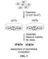
- expanding the population of chondrocytes includes a primary cell expansion and a secondary cell expansion that are optionally separated by a period of cryopreservation.
- the method further includes exposing the population of chondrocytes to TGF- ⁇ in the tissue production medium for a period of about seven (7) days.
- the method further includes maintaining the population of chondrocytes for a culture period of about forty-five (45) to about sixty-five (65) days in the tissue production medium. Maintaining the population of chondrocytes in a tissue production medium includes, for example, maintaining the population of chondrocytes at a predetermined level of oxygen and carbon dioxide in the tissue production medium.
- tissue culture apparatus including a polycarbonate substrate having multiple pores therethrough, the pores characterized by an inner diameter of at least about 1 micron to about 12 microns.
- the tissue culture apparatus may be a perforated polycarbonate membrane.
- the perforated polycarbonate membrane may have a thickness of about 10 microns.
- the pores in the perforated polycarbonate membrane may be characterized by inner diameters distributed around 3 microns.
- the tissue culture apparatus may further include side walls forming a perimeter around the polycarbonate substrate, the perimeter of a predetermined shape selected as a shape for a cultured tissue.
- the side walls are fabricated, for example, from a biocompatible polymer, which in one embodiment is semipermeable, and in another embodiment is impermeable.
- the biocompatible polymer can be, for example, polystyrene.
- a tissue culture insert including a polycarbonate substrate having a plurality of pores therethrough, the pores each having an inner diameter of at least about 1 micron to about 12 microns, and side walls forming a perimeter around the polycarbonate substrate, the perimeter of a predetermined shape selected as a shape for the cultured tissue.
- chondrocyte refers to cartilage-specific cells that give rise to normal cartilage tissue growth in vivo; these cells synthesize and deposit the supportive matrix (composed principally of collagen and proteoglycan) of cartilage.
- phenotype refers to the observable characteristics at any level-physical, morphologic, biochemical or molecular-of a cell or tissue.
- noncartilage refers to cartilage grown ex vivo and characterized by one or more of the following attributes: containing membrane phospholipids enriched in Mead acid but substantially depleted of linoleic or arachidonic acid, being substantially free of endothelial, bone and synovial cells, having a sulfated glycosaminoglycan (S-GAG) content of at least 40 ⁇ g/ ⁇ g of DNA; being substantially free of types I, III and X collagen; containing a matrix substantially free of biglycan; having multiple layers of cells randomly arranged, rather than separated, into distinct zones of chondrocyte maturation; being enriched in high molecular weight aggrecan, or being characterized by having multiple layers of cells surrounded by a substantially continuous insoluble glycosaminoglycan and collagen-enriched hyaline extracellular matrix.
- S-GAG sulfated glycosaminoglycan
- hyaluronate substrate refers to a substrate containing a hyaluronate mixture that is in large part comprised of a natural or synthetic, highly purified hyaluronate, such as sodium hyaluronate purified either from rooster combs or from bacterial fermentation.
- Hyaluronic acid is a polysaccharide composed of repeating disaccharide units of N-acetylglucasamine and glucuronic acid. Commercial HA is commonly its sodium salt form.
- cytokine refers to a vast array of relatively low molecular weight, pharmacologically active proteins that are secreted by one cell for the purpose of altering either its own function(s) (autocrine effect) or those of adjacent cells (paracrine effect).
- individual cytokines can have multiple biological activities. Different cytokines can also have redundant activity.
- FGF indicates the fibroblast growth factor family of related proteins, which currently numbers 22 members (in humans, FGF-1-14 and FGF-16-23).
- FGF-2 refers to the basic form of fibroblast growth factor (FGF).
- FGFs generally have a high affinity for heparan sulfate proteoglycans; interactions between FGF and heparin appear necessary for activation of each the FGF receptors (Ornitz and Itoh 2001).
- FGFR1-4 There are four FGFRs (FGFR1-4), each having many splice variants.
- an "active form" of an FGF polypeptide is a polypeptide that has significant (greater than 70%) homology to at least a portion of a conserved region of any FGF polypeptide and possesses the same activity as at least one of its homologues. The degree of activity can be greater or less than at least one of the homologues. In most cases, an internal core region containing approximately 28 highly conserved and six identical amino-acid residues can be identified, ten of which habitually interact with FGFRs (Ornitz, 2000 and Plotnikov et al., 2000).
- FGF-like activity refers to an activity of a molecules, such as a polypeptide, that acts on at least one cell type in a similar manner as the cognate FGF molecule in at least one aspect.
- a molecules having FGF-like activity can be substituted for FGF during the expansion of chondrocytes in the methods of the invention.
- TGF- ⁇ indicates the transforming growth factor family of related proteins. TGF- ⁇ proteins are recognizable by C-terminus polypeptide homology and their signaling via the Similar to Mothers against Decapentaplegic (SMAD) proteins after binding TGF- ⁇ receptors. Examples includes TGF- ⁇ 1-3, bone morphogenetic proteins (BMPs), etc.
- An "active form" of a TGF- ⁇ polypeptide is a polypeptide displaying significant (greater than 70%) homology to at least a portion of a conserved region of a TGF- ⁇ polypeptide and possesses the same activity as at least one of its homologues. The degree of activity can be greater or less than at least one of the homologues.
- TGF- ⁇ -like activity refers to an activity of a molecule (such as a polypeptide) that acts on at least one cell type in a similar manner as a cognate TGF- ⁇ . molecule.
- a molecule that signals via the SMAD proteins regardless if the molecule has bound a TGF- ⁇ receptor or not, has TGF- ⁇ -like activity.
- TGF- ⁇ molecules stimulate synthesis of collagens, fibronectin proteoglycans, tenascin, thrombospondin, plasminogen activator inhibitor-1 and tissue inhibitor of metalloprotease-1 proteins.
- a molecule having TGF- ⁇ -like activity such as recombinant lactoferrin, can be substituted for TGF- ⁇ during the expansion of chondrocytes in the methods of the invention.
- low cell attachment refers to a characteristic of chondrocyte culture conditions under which a majority of cells seeded onto a tissue substrate do not adhere to the tissue substrate.
- the underlying principle is to maintain native chondrocyte phenotype during ex vivo expansion by growing dissociated chondrocytes or chondroprogenitor cells under low Attachment conditions that limit cell attachment and spreading.
- Low attachment conditions as described herein, including but not limited to use of certain substrate surfaces, have been found to support maintenance of chondrocyte morphology, phenotype and function during the process of expansion.
- the cartilaginous tissue produced therefrom is both biocompatible and safe for use in medical applications.
- Use of a low attachment substrate provides a microenvironment that more closely mimics that of native articular cartilage, resulting in retention of rounded cell shape and hyaline cartilage gene expression once the expanded cells exit the cell cycle.
- a suitable low attachment condition is use of an HA-modified substrate, e.g. use of a polystyrene surface modified by covalent attachment of sodium hyaluronate, Chondrocytes adhere to immobilized HA via a cell surface glycoprotein receptor for hyaluronan (CD44) and can utilize the HA substrate to assemble the large proteoglycan aggregates typically found in normal, healthy articular cartilage,
- HA-modified substrate e.g. use of a polystyrene surface modified by covalent attachment of sodium hyaluronate
- Chondrocytes adhere to immobilized HA via a cell surface glycoprotein receptor for hyaluronan (CD44) and can utilize the HA substrate to assemble the large proteoglycan aggregates typically found in normal, healthy articular cartilage,
- High molecular weight HA (>400,000 MW) can be used as a substrate, or in solution, for in vitro expansion culture to increase total cell yield, while preventing loss of differentiated cell function.
- the expansion of chondrocytes starting at low density on an HA modified substrate in chemically defined media significantly increases chondrocyte cell number.
- chondrocytes expanded under other low attachment conditions such as use of HA in solution or use of a polystyrene substrate having an Ultra-Low Attachment coating such as those manufactured by Corning, produce an expanded population that maintains phenotype and morphology as determined and described elsewhere herein.
- chondrocytes expanded in this manner retain their native chondrogenic differentiation potential to produce neocartilage tissue, and these cells or the neocartilage produced therefrom can be used for clinical application.
- the present disclosure thus provides a method of producing chondrocytes under low attachment conditions such that chondrocytes remain rounded and loosely attached during the expansion process. In time, the chondrocytes are observed to undergo condensation, forming tissue aggregates or clusters. This unique method of expanding chondrocytes allows the cells to retain their functional activity after expansion.
- the invention also provides for kits for the expansion of chondrocytes. Therefore, this method serves to address the challenges identified in earlier cell-based approaches to articular cartilage repair.
- one embodiment of the invention is a method in which isolated chondrocytes are expanded under low cell attachment conditions on a substrate modified by covalent attachment of HA as described herein, or on an unmodified substrate using soluble HA in solution in the expansion medium, or on a substrate coated with a commercially available low cell attachment composition, such as those commercially available from Corning, Nunc, and CellSeed, Inc..
- a novel method for serially expanding chondrocytes in vitro on a substrate includes the steps of: isolating chondrocytes; growing the chondrocytes in a serum-free expansion medium containing cytokines and under low attachment conditions such as on a substrate modified to reduce cell attachment; and then using the expanded chondrocytes to produce hyaline-like cartilage tissue.
- the expanded population of cells is also well-suited for transplantation, and particularly for transplantation by injection.
- chondrocytes are isolated from cartilage derived from immature donors, including neonatal, infant, or pre-adolescent donors for subsequent plating.
- Chondrocytes can be isolated by methods known in the art such as by sequential enzymatic digestion techniques (Adkisson et al., 2001). Isolated chondrocytes are then seeded onto a substrate under one of multiple possible low attachment conditions. Suitable low attachment conditions include e.g., use of a polystyrene substrate to which sodium hyaluronate is covalently attached, use of ultra low attachment plates such as those commercially available from Corning, Nunc, and CellSeed, Inc.
- tissue culture-treated polystyrene in the presence of serum-free expansion medium containing soluble HA
- tissue culture-treated polystyrene in the presence of serum-free expansion medium containing a low attachment agent other than HA, such as phosphatidyl choline.
- Chondrocytes can be avian or mammalian chondrocytes, preferably human chondrocytes. Chondrocytes can be derived from transgenic animals that have been genetically engineered to prevent immune-mediated xenograft rejection (Sandrin et al., 1995; Sandrin et al., 1996 and Osman et al., 1997). Cartilage can be obtained from any tissue containing hyaline, elastic or fibro-cartilage.
- chondrocytes In contrast to other methods of expanding chondrocytes known in the art, such as seeding cells on three dimensional scaffolds, further exogenous material beyond sodium hyaluronate is not required to retain differentiated chondrocyte morphology during expansion.
- the expansion method of the present invention provides for seeding chondrocytes in direct contact with an appropriate tissue culture surface. Although scaffold material is unnecessary, it can be used, for example in the case of polystyrene microcarrier beads.
- an expansion culture is produced by isolating immature chondrocytes, e.g., neonatal, infant or pre-adolescent, from donor articular cartilage and plating the dissociated cells onto a tissue culture substrate that is first modified via covalent attachment of sodium hyaluronate, or using a serum-free expansion medium that contains HA in solution. Reduction of cell attachment and subsequent spreading is sufficient when a majority of cells in the starting population are observed not to attach to the substrate.
- immature chondrocytes e.g., neonatal, infant or pre-adolescent
- chondrocytes are then seeded directly on a tissue culture vessel in a suitable medium.
- a basal medium containing serum is used, but because serum has been observed to encourage the cells to de-differentiate (see, e.g. Example 1, Figure 1 ), any serum-containing media is exchanged for a chemically defined serum-free expansion medium.
- “Chemically defined” refers to a serum-free medium, the chemical components of which are known. As used herein the term refers to that characteristic of expansion media used for expansion and also of tissue production media used.
- Suitable substrates for expansion are any tissue culture plastics, such as virgin polystyrene, tissue-culture treated polystyrene or polycarbonate that can be modified to covalently attach hyaluronic acid.
- tissue culture plastics such as virgin polystyrene, tissue-culture treated polystyrene or polycarbonate that can be modified to covalently attach hyaluronic acid.
- the only requirement for a suitable substrate is that it be amenable to modification with hyaluronic acid or N-acetylglucosamine, the natural building block to HA biosynthesis.
- hyaluronic acid or N-acetylglucosamine is covalently attached, although a covalent linkage is not necessary in most cases, except for direct attachment to polystyrene substrates.
- the covalent modification of the substrate with HA provides a substrate that no longer is soluble in aqueous media and therefore maintains native chondrocyte phenotype during cytokine-mediated expansion of chondrocytes in serum-free expansion medium during two-dimensional culture.
- Hyaluronic acid recapitulates a cartilage microenvironment, whereas unmodified tissue culture plastic (polystyrene) does not.
- tissue culture plastic polystyrene
- any other molecules that promote aggrecan assembly can be used, for example recombinant link protein.
- Cross-linking two or more molecules is to covalently join them.
- Cross-linkers, cross-linking reagents, etc. have portions that react with specific functional groups, such as primary amines, sulfhydryls, carboxyls, etc.
- Most commonly used cross-linkers come in two forms: homobifunctional and heterobifunctional, although cross-linking reagents can have more than two reactive groups (e.g., trifunctional cross-linkers).
- Homobifunctional linkers have two identical reactive groups, whereas heterobifunctional cross-linkers have two different reactive groups.
- Homobifunctional cross-linkers are used in "single-stage" cross-linking.
- heterobifunctional cross-linkers To take advantage of heterobifunctional cross-linkers, "sequential-stage" cross-linking procedures are used. Most commonly used cross-linkers have chemically reactive groups; however, cross-linkers with photoreactive groups can be used. An advantage of photoreactive groups is that they can form conjugates that otherwise cannot be formed using chemically reactive groups, although the efficiency of photoreactive groups can be low, most often 10%, sometimes up to 70%.
- Common heterobifunctional cross-linkers include those that have amine-reactive succinimidyl ester at one end, and a sulfhydrylreactive group at the other.
- Another example is the carbodiimides, which are "zero-length" cross-linkers and effect direct coupling between carboxylates and primary amines.
- cross-linker is based on the target functional groups of the molecules to be cross-linked and the compatibility of the reaction to the application. The following characteristics are also considered: chemical specificity, spacer arm length, reagent solubility (and in the case of cells, membrane permeability), whether a homo- or hetero-bifunctional agent is appropriate or desired, chemically reactive or photoreactive groups, desirability of cleavable links after cross-linking, and whether the reagent can be conjugated to a label (e.g., a radiolabel).
- a label e.g., a radiolabel
- MHS-ester homobifunctional cross-linkers If only lysine residues or N-terminal amino acids are available, a logical choice is MHS-ester homobifunctional cross-linkers. If one has lysine residues and the other sulfhydryls, a maleimide NHS-ester cross-linker is appropriate. If only lysine residues are available on both molecules, modification to introduce sulfhydryls via the lysine residues on one would allow for sequential coupling. If both molecules have free sulfhydryls, a homobifunctional sulfhydryl reactive cross-linker is appropriate.
- Reactive cross-linker groups and their functional group targets Reactive group (classes) Functional group (classes) Reactive group (classes) Function group (classes) aryl azide non-selective (for primary amine) Maleimide sulfhydryl carbodiimide amine/carboxyl NHS-ester 1 amine hydrazide oxidized carbohydrate PFP-ester 2 amine Hydroxymethyl prosphine amine Psoralen thymine (photoreactive intercaltor) Isocyanate non-aqueous hydroxyl vinyl sulfone sulfhydryl, amine, hydroxyl 1 NHS, aminosuccinimidyl 2 PFP, pentafluorophenyl
- Spacer arm lengths can play an important role in the success of cross-linking, and usually requires empirical determination. Usually, a short spacer arm (4-8 is is first used and then the success of cross-linking is assessed. If linking is poor, then cross-linkers with longer spacer arms can be explored. In some instances, a cleavable cross-linking agent is desirable; for example, if one would want to release a culture of chondrocytes intact with the HA substrate. TABLE II identifies an expanded list of applicable double-agent cross-linkers. TABLE III identifies an expanded list of applicable single-agent cross-linkers.
- Sodium periodate can be used to create reactive aldehyde groups for crosslinking of glycosaminoglycans to a variety of structures containing available primary amines through the formation of a stable Schiff base.
- Glycosaminoglycans such as chondroitin sulfated, dextran and sodium hyaluronate have been modified using such procedures.
- Liu et al, 2003 described the preparation of hyaluronate-polyaldehydes to form a tissue engineering scaffold useful for bone repair (Healos ® ) (Liu et al., 2003).
- activated hyaluronate-polyaldehyde is prepared by oxidizing sodium hyaluronate with sodium periodate, followed by extensive dialysis.
- the density of reactive aldehydes (formyl groups) on the hyaluronate polymer can be controlled by altering the reaction time, and can be monitored quantitatively using a modification of the bicinchoninic acid method ( Hermanson GT Bioconjugate Techniques, San Diego, Academic Press, Inc., 1996, p 622 ).
- an alternative method for cross linking sodium hyaluronate, to polystyrene surfaces involves Schiff base formation between the sulfonamide created in the the first step with the reactive aldehydes of periodate-oxidized HA.
- the lyophilized HA-polyaldehyde is stable at 4 °C (dark storage).
- An aqueous solution of the polyaldehyde is added to the modified polystyrene surface and allowed to react either at room temperature or at 37 °C, depending on the extent of cross-linking desired. Unreacted material is then removed by aspiration, and the plates are washed extensively in distilled water and culture medium prior to seeding with cells.
- chondrocytes have been isolated from cartilage tissue by sequential enzymatic digestion, using a nonspecific protease followed by a mixture of collagenase and hyaluronidase.
- most preparations of proteolytic enzymes used for research purposes contain significant levels of endotoxin, as well as a variety of undesirable proteolytic activities that can damage cell membrane proteins, leading to apoptotic cell death.
- Blendzyme 2 contains a combination of collagenase and neutral protease activity that works well for harvesting primary chondrocytes from articular cartilage. Reduced levels of the same enzyme are preferred when chondrocytes are released from a culture substrate.
- Blendzymes are blends of purified collagenases I and II and a neutral protease.
- the collagenases are purified from the fermentation of Clostridium histolyticum.
- Blendzyme 1 contains the neutral protease dispase, which is purified from Bacillus polymyxa fermentation.
- Blendzymes 2, 3 and 4 contain the neutral protease thermolysin, purified from Bacillus thermoproteolyticus fermentation.
- the methods for purifying and blending these components for Blendyme preparation have been described in US, Pat. Nos. 5,753,485 and 5,830,741 .
- the use of Blendzyme mixes can be optimized using the criteria presented in TABLE IV.
- BlendzymeTM Use Observation 1 Observation 2 Possible cause Recommendation Low cell viability Dissociation very rapid Enzyme concentration too high Reduce enzyme concentration by 50%. Blendzyme number too high Move down one Blendzyme level, keep initial concentration. Dissociation very slow Enzyme concentration too low Increase enzyme concentration by 50%. Blendzyme number too low Move up one Blendzyme level, keep initial concentration. Impaired cell function Cell viability >80%, cell yield is reasonable Enzyme concentration too high Reduce enzyme concentration by 25%. Blendzyme number too high Move down one Blendzyme level, keep initial concentration. Low cell yield Cell viability >80% Enzyme concentration too low Increase enzyme concentration by 25-50%, Blendzyme number too low Move up one Blendzyme level, keep initial concentration.
- Blendzymes are preferred, any method that safely releases the cells can be used, including those that use chelating agents, with and without enzymes.
- Ethylenediaminetetraacetic acid (EDTA) and ethylene-bis (oxyethylenenitrilo) tetraacetic acid (EGTA) are two such common reagents that can be used singly, or in combination.
- chondrocytes are isolated from immature donors such as neonatal, infants, or pre-adolescents, and expanded on a substrate surface material, such as polystyrene, that is covalently modified via covalent attachment of sodium hyaluronate, or on a substrate under other suitably low attachment conditions as described elsewhere herein.
- the expanded cells are subsequently grown in a chemically defined serum-free tissue production medium to produce neocartilage for cartilage repair.
- a "chemically defined" medium is a serum-free medium the chemical components of which are specified.
- the resulting expanded population of cells can be frozen for future transplantation, for example by injection, or can be frozen in a cryopreservation step before a second phase of expansion.
- a population of cells that has undergone two phases of expansion may also be frozen in a second cryopreservation step prior to being seeded under appropriate culture conditions for tissue growth and extracellular matrix production.
- Other cells that are suitable for the methods of the invention include those isolated from placenta (Kogler et al., 2004), bone marrow mesenchymal stromal cells (Mackay et al., 1998; Kavalkovick et al., 2002), adipose stromal cells (Huang et al., 2004), synovium (DeBari et al., 2004) and periosteum (DeBari et al., 2001).
- the serum-free tissue production medium can also comprise vitamin C. ascorbate, exogenous autocrine growth factors or conditioned growth media as described below.
- Vitamin C can be supplied in any active form, as a free acid or as a salt. Examples include calcium ascorbate, magnesium ascorbate, sodium ascorbate and L-ascorbic acid 2-phosphate or an esterified derivative thereof.
- concentration to be used can be determined empirically, depending on the source of the cells, but usually, concentrations range from 5.6x10 -4 g/L to 0.1 g/L when supplied as sodium ascorbate.
- the serum free tissue production medium can comprise dexamethasone, or a salt derivative thereof.
- Dexamethasone enhances SOX 9 expression and presumably will increase the capacity for cartilage matrix production by the expanded cells (Sekiya et al., 2001; Malpeli et al., 2004).
- the cell culture can be grown under suitable culture conditions such as growing the cell culture at 37 °C in a humidified atmosphere with the addition of 2-10% CO 2 , preferably 5%.
- the doubling time for a particular group of cells may vary.
- the doubling time observed for chondrocytes that are maintained as described in the present invention is generally 3 to 4 days, when plated at a density of 3 x 10 4 /cm 2 .
- the length of time for expansion culture is increased to day 14 and 17, as many as 4-6 chondrocytes population doublings can be achieved.
- the same cells grown in serum-supplemented media are limited by contact inhibition and produce no greater than two population doublings.
- a preferred defined expansion medium used in the present invention is HL-1, a proprietary formulation containing insulin-transferrin-selenium-complex as its only source of protein (HL-1TM of Lonzo Walkersville, Inc., (formerly Cambrex), Walkersville, Md.).
- the serum-free expansion medium containing cytokines is either completely or partially replaced every three to four days after the initial seed.
- Optimal media selection depends on the cell type: that media used to culture the cells usually represents a preferred option.
- suitable culture media include Iscove's Modified Dulbecoo's Medium (IMDM), Dulbecco's Modified Eagle's Medium (DMEM), Minimal Essential Medium Eagle (MEM), Basal Medium Eagle (BME), Click's Medium, L-15 Medium Leibovitz, McCoy's 5A Medium, Glasgow Minimum Essential Medium (GMEM), NCTC 109 Medium, Williams' Medium E, RPMI-1640, Medium 199, Ham's F12 and SFM (Gibco).
- IMDM Iscove's Modified Dulbecoo's Medium
- DMEM Dulbecco's Modified Eagle's Medium
- MEM Minimal Essential Medium Eagle
- BME Basal Medium Eagle
- Click's Medium L-15 Medium Leibovitz
- McCoy's 5A Medium Glasgow Minimum Essential Medium (GMEM), NCTC 109 Medium, Williams' Medium E, RPMI-16
- a medium specifically developed for a particular cell type/line other than chondrocytes may not be useful in the practice of this invention, particularly if it contains epidermal growth factor which is reported to induce chondrogenic dedifferentiation (Yoon et al, 2002).
- a protein can be added to support the cells, such as various albumins, including bovine serum albumin.
- the media can be further supplemented with reagents that limit acidosis of the cultures, such as buffer addition to the medium (such as N,N-bis(2-hydroxyethyl)-2-aminoethanesulfonic acid (BES), bis(2-hydroxyethyl)amino-tris(hydroxymethyl)methane (BIS-Tris), N-(2-hydroxyethyl)piperazine-N'3-propanesulfonic acid (EPPS or HEPPS), glyciclycine, N-2-hydroxyehtylpiperazine-N'-2-ethanesulfonic acid (HEPES), 3-(N-morpholino)propane sulfonic acid (MOPS), piperazine-N,N'-bis(2-ethane-sulfonic acid) (PIPES), sodium bicarbonate, 3-(N-tris(hydroxy)-2-a
- Pronase-E (Type XIV, from Streptomyces griseus), hyaluronidase (type VIII, bovine testes), N-tris [hydroxymethyl]methyl-2-aminoethanesulfonic acid (TES), and MILLET-GS syringe sterilization filters were obtained from Sigma Chemical Company (St. Louis, Mo.).
- Collagenase (CLS 4) was purchased from Worthington Biochemicals (Freehold, N.J.). Tissue-culture dishes (35 mm dia., 100 mm dia, 12 and 24 well clusters) and bottle top sterilization filter units (type CA) were obtained from Coster Corporation (Cambridge, Mass.).
- Bovine serum albumin (fraction V, fatty acid-free) was from Calbiochem (San Diego, Calif.).
- HL-1TM Complete Serum-free Medium was obtained from Cambrex (now Lonzo Walkersville, inc.).
- chondrocyte expansion ex vivo is for cartilage repair.
- cartilage is removed from a non-damaged cartilage area around a damage site and digested with collagenase.
- the resulting chondrocytes are expanded on the modified substrate of the invention, then injected or implanted into the cartilage defect site.
- marrow stromal fibroblasts can be isolated from a normal area adjacent a bone defect, expanded ex vivo as described and administered at the site of a bone defect.
- chondrocytes of the invention U.S. Pat. No. 6,617,161 containing platelet derived growth factor (PDGF), epidermal growth factor (EGF), fibroblast growth factor (FGF), and bone morphogenetic proteins results in the loss of two main cartilage-specific phenotypic markers, type II collagen and proteoglycan aggrecan, which are critical components for cartilage assembly and repair. Discontinued expression of these molecules indicates that the cells have undergone phenotypic dedifferentiation Yoon et al., 2000 recently demonstrated that EGF promotes loss of native chondrocyte phenotype.
- PDGF platelet derived growth factor
- EGF epidermal growth factor
- FGF fibroblast growth factor
- bone morphogenetic proteins results in the loss of two main cartilage-specific phenotypic markers, type II collagen and proteoglycan aggrecan, which are critical components for cartilage assembly and repair. Discontinued expression of these molecules indicates that the cells have undergone phenotypic
- Chondrocytes cultured on unmodified polystyrene develop a fibroblastic appearance, displacing an elongated cell shape relative to their native rounded morphology.
- the characterization of phenotypic markers in the expanded chondrocyte population is necessary to establish the potential functional properties of the manipulated cells.
- Food and Drug Administration (FDA)/Center for Biologics Evaluation and Research (CBER) requires that protocols relating to ex vivo expansion of articular chondrocytes for repair of joint surfaces prove that the expanded cell populations are very similar to the native chondrocytes.
- mRNA expression levels of aggrecan core protein and type II collagen can be assessed by measuring mRNA expression levels of aggrecan core protein and type II collagen at the end of the expansion protocol or after neocartilage production by any appropriate method, including molecular methods (e.g., Northern blotting, RT-PCR) and biochemical methods (e.g., immunoprecipitation; affinity chromatography) or immunocytochemical methods (e.g ., ELISA or immunofluorescence-activated cell sorting (FACS)).
- molecular methods e.g., Northern blotting, RT-PCR
- biochemical methods e.g., immunoprecipitation; affinity chromatography
- immunocytochemical methods e.g ., ELISA or immunofluorescence-activated cell sorting (FACS)
- bioactive molecule that improves the proliferation, differentiation potential or quality of the resulting regenerated tissue can be used according to the present invention.
- Expansion of cells under low attachment conditions, such as on an HA-modified substrate, also improves the efficiency of transfection of nucleic acids into the cells.
- nucleic acid transfer is carried out during monolayer expansion, and cells in active mitosis are more amenable to DNA transduction. Therefore, applications where tissue engineering techniques are combined with gene therapy can be utilized in accordance with the teachings of the present invention.
- cells can be transfected with a vector that confers resistance to a variety of biological and chemical compounds, such as antibiotics, cytokines and inflammatory agents, or with a vector that result in the overexpression and synthesis of a particular growth factor or matrix component.
- Dissociated cells isolated from the expansion process of the present invention are typically grown without scaffold support to create a three-dimensional tissue for cartilage repair ( U.S. Pat. No. 6,235,316 ).
- cells expanded via this method can be implanted in combination with suitable biodegradable, polymeric matrix or hydrogel to form new cartilage tissue.
- suitable biodegradable, polymeric matrix or hydrogel there are two forms of matrices which can be used: a polymeric hydrogel formed of a material, such as fibrin or alginate, having cells suspended therein, and a fibrous matrix having an interstitial spacing between about 40 and 200 microns.
- Preferred polymeric matrices are those that degrade in about one to two months after implantation; such as polylactic acid-glycolic acid copolymers ( U.S. Pat.
- the matrices can be seeded prior to implantation or implanted, allowed to vascularize, then seeded with cells, (See, e . g ., Cima et al., 1991; Vacanti et al., 1988; and Vacanti et al., 1988).
- Other materials such as bioactive molecules that enhance vascularization of the implanted tissue and/or inhibit fibrotic tissue ingrowth can be implanted with the matrix to enhance development of more normal tissue.
- Chondrocytes regardless of tissue origin, rapidly lose the ability to synthesize cartilage specific macromolecules with serial expansion in vitro (Homicz et al., 2002; Mandi et al., 2002). Although juvenile chondrocytes are thought to better retain the ability to synthesize matrix macromolecules than adult articular chondrocytes, the following experiment was performed to determine the effect of serial expansion on chondrocyte matrix synthesis using chondrocytes derived from immature articular cartilage. A variety of media containing serum were evaluated to demonstrate the deleterious effects of serum on chondrocyte differentiation potential.
- Dissociated cells (1x10 6 viable cells) were subsequently seeded in 48 well plates using HL-1 complete serum-free medium (formerly available from Cambrex, now HL-1TM Serum-free Hybridoma Medium available from Lonza Walkersville, Inc., Walkersville, Md) to produce neocartilage as described by Adkisson et al, 2001. Cultures were harvested for biochemical composition analysis on day 45 of culture.
- FIG. 1 is a graphic representation showing loss of chondrocyte differentiation potential as a function of serial expansion in serum-containing medium. Two of the groups were expanded through two passages to a maximum of 2.8 population doublings (nearly an eight-fold increase in total cell number). A marked loss in the ability of juvenile articular chondrocytes to produce cartilageneous proteoglycan was observed. An overall regression line was plotted from the data which demonstrated a greater than 70% reduction in chondrocyte matrix synthesis after nearly three population doublings, relative to non-expanded chondrocytes derived from the same individuals.
- juvenile chondrocytes were grown on a modified substrate prepared via covalent attachment of high molecular weight sodium hyaluronate to polystyrene. It was hypothesized that immobilized sodium hyaluronate, in contrast to unmodified tissue culture plastic, would provide a microenvironment that could more closely mimic that of native articular cartilage.
- the following method for chemical modification of polystyrene with sodium hyaluronate is a modification of that originally described by Turley and Roth (Turley and Roth, 1979).
- FIG. 2 the virgin polystyrene plates were treated with sulfuric acid at 37 °C for two hours. After removing the sulfuric acid and extensive washing of the plates in deionized distilled water, the reactive sulfonamide was created by adding aqueous ammonium hydroxide (24 hours at room temp.). The base was removed, and the plates again were washed extensively with water.
- Example 3 Chondrocytes Phenotype Retained by Serial Expansion on HA-coated Plastic in Serum-free Medium
- Chondrocytes derived from a six week-old subject were taken through two passages using plates that were modified by the method illustrated in Example 2. Differentiation potential of the expanded chondrocytes was assessed after enzymatic dissociation and estimation of total cell number and viability.
- a proprietary serum-free differentiation medium developed at is to Technologies, Inc. was used to stimulate chondrocyte differentiation.
- this medium contains neither TGF ⁇ nor dexamethasone to enhance chondrogenic differentiation potential, and the only protein component in this formulation is recombinant human insulin (Serologicals Corporation, Milford, Mass.).
- FIG. 3 shows a schematic representation of the process of cytokine-mediated chondrocyte expansion in which covalent attachment of sodium hyaluronate serves as a substrate for maintenance of chondrocyte phenotype during expansion.
- Primary chondrocytes are isolated, washed and resuspended at a final density of 3x10 4 cells/cm 51 in HL-1 Complete Serum-free Medium containing 100 ng/mL FGF-2 and 20/ng/mL TGF- ⁇ and vitamin C.
- Control and HA-treated plates were harvested after 14-17 days of expansion using Blendzyme 2 as described in Example 7. An aliquot of the dissociated cells was plated in differentiation medium to assess neocartilage formation potential, while the remaining cells underwent serial expansion.
- Neocartilage was harvested from differentiation cultures between day 28 and 45 for measurement of total proteoglycan, DNA and collagen content in the neocartilage matrix. These values were compared to those obtained for neocartilage prepared from non-expanded chondrocytes using the same methods. The total cumulative increase in chondrocytes number was 85-fold or 6.5 population doublings.
- FIG. 4 illustrates the biochemical composition data that was collected on grafts produced from the freshly dissociated unpassaged cells (P0) and passage two cells (P2).
- Neocartilage grafts were initiated at a density of 1x10 8 cells/well in untreated 48-well plates using freshly dissociated cells and cells from each successive passage to P2.
- the rates of glycosaminoglycan (S-GAG/DNA/Day) and collagen (hydroxyproline/DNA/Day) synthesis are plotted on the left- and right-hand axis, respectively.
- Data represent the mean of replicates of four (4) to five (5) +/- STD DEV.
- Estimated population doublings for each expansion was 2.25 and 4.25 at passage one cells P1 and P2, respectively, at day 14 and 17 of culture.
- this example illustrates that a plastic surface modified by covalent attachment of sodium hyaluronate limits chondrocyte spreading during expansion and retains the functional properties of the expanded chondrocytes.
- Example 4 Chondrocytes Expanded on Covalently Bound Sodium Hyaluronate Maintain Rounded Morphology and Native Function
- chondrocytes cultured on an HA-coated substrate in defined medium results in maintenance of native chondrocyte phenotype by retaining rounded cell shape and function.
- Human P1 chondrocytes derived from a five year-old, having first been expanded on unmodified plastic in the presence of cytokines, were used in this pilot experiment to determine if immobilized HA could restore both rounded chondrocyte morphology and the functional properties of the cells after enzymatic dissociation. Thirty-five mm diameter polystyrene dishes were modified via covalent attachment of sodium HA as described.
- chondrocytes were harvested from an expansion of primary cells and subsequently seeded onto HA-modified and unmodified polystyrene surfaces for further expansion in HL-1 Complete Serum-free Medium (Cambrex) containing 2 ng/mL TGF ⁇ -2 and 10 ng/ml variant human FGF-2 (ProChon, Ltd., Rehovat, Israel) and vitamin C. Cultures were harvested on day 10 for enumeration and estimation of viability using Guava ViaCount® reagents. Greater than 2.5 population doublings were achieved at day 10 of culture.
- FIG. 5A illustrates that chondrocyte expanded in the presence of cytokines on HA-modified polystyrene form chondrocyte islands or aggregates in which the chondrocytes appear to maintain a rounded morphologic appearance. Formation of these cartilage aggregates is reminiscent of the condensation of mesenchymal tissue which normally occurs in vivo during skeletal development (Singley and Solursh, 1981). The relative opacity of the chondrocyte aggregates suggests that the chondrocytes continue to actively synthesize extracellular matrix components. Thus, chondrocytes grown on the HA modified surface formed clusters of cells that retained a rounded morphology, characteristics of the chondrocyte phenotype.
- Chondrocytes from each of these groups were subsequently passaged by dissociation in collagenase and hyaluronidase and seeded (1 x 10 6 per well) into 48-well culture dishes (unmodified) to measure the capacity to which the P2 cells synthesize neocartilage matrix.
- the chondrocytes were grown for 45 days, at which time they were harvested for biochemical composition analysis.
- FIG. 6 shows a photomicrograph of neocartilage obtained at Day 10 of culture, illustrating the rounded morphologic appearance of P2 cells that were harvested from expansion culture on HA-modified polystyrene.
- Chondrocytes expanded by this method and placed in differentiation culture displayed a rounded morphologic appearance characteristic of freshly dissociated chondrocytes.
- FIG. 7 compares the biochemical composition of newly synthesized NC matrix obtained for chondrocytes that were expanded on HA modified plastic to that of the same cells that were expanded on unmodified polystyrene.
- Substrate containing covalently bound HA appears to preserve native chondrocyte morphology (rounded phenotype) during neocartilage tissue formation, and more importantly, to restore chondrocyte extracellular matrix synthesis in expanded cells to a level that approached that of the primary culture population (approximately 1.75 to 2 ⁇ g S-GAG/ ⁇ g DNA/Day).
- chondrocyte phenotype in cells that had been expanded on HA-modified polystyrene was confirmed at the molecular level by gene expression analysis. Chondrocytes harvested at each passage in scale-up experiments were used for RNA isolation, and the relative mRNA levels corresponding to a set of cartilage-specific extracellular matrix macromolecules and the cartilage transcription factor, SOX 9, were analyzed using semiquantitative reverse transcriptase polymerase chain reaction (RT-PCR).
- RT-PCR semiquantitative reverse transcriptase polymerase chain reaction
- FIGS. 8A and 8B show representative gene expression profiles obtained for P0 and P2 chondrocytes that were harvested from the cells used in Example 4 above. Note that P2 cells have gone through a total of 6.5 population doublings. No discernable difference was observed in the pattern of gene expression obtained from freshly dissociated chondrocytes (upper panel) and the same cells after 6.5 population doublings (lower panel). mRNA levels for SOX 9 and B7-H1 remained unchanged after serial expansion. B7-H1 is the putative negative regulator of alloreactlvity that is thought to maintain the immune privilege status of chondrocytes in vitro and in vivo (Adkisson et al., unpublished observations).
- chondrocytes expanded in serum containing media also lose their functional capacity to synthesize and assemble neocartilage matrix.
- chondrocyte expansion on a hyaluronate-modified substrate represents a preferred microenvironment that results in maintenance of native phenotype as demonstrated by morphological analysis of chondrocyte cell shape and gene expression profiling.
- chondrocytes that were expanded in serum containing medium showed marked changes in mRNA levels for aggrecan core protein and type II collagen, two macromolecules essential for cartilage formation and repair.
- Previous examples 4 and 5 demonstrate the unique ability of a modified polystyrene surface to support chondrocyte expansion during anchorage-independent growth, i.e. under a low attachment condition. Chondrocytes maintained under low attachment conditions undergo extensive growth while retaining their native rounded morphological appearance and the ability to synthesize engineered tissues ex vivo. Conformation of phenotype retention was demonstrated at both the biochemical and molecular level.
- the purpose was to demonstrate that different methods can be used to achieve the same result of limiting chondrocyte attachment and spreading during ex vivo expansion, thus allowing cells to maintain phenotype and hyaline cartilage gene expression during the process of expansion.
- Three different conditions were used for comparison.
- Group A covalently bound HA
- Group B soluble HA added to the expansion medium at 1 mg/mL using tissue culture-treated plastic ware
- Group C Ultra Low Attachment plates manufactured by Corning.
- Juvenile particular chondrocytes were plated at a density of 3x10 4 cells/cm 2 in T-75 tissue culture flasks containing HL-1 supplemented medium as defined in earlier examples. Expansion cultures were maintained to day 21.
- Figure 10 shows a comparison of the morphologic appearance of culture-expanded chondrocytes imaged at 100x prior to harvest. Arrows indicate suspended clusters observed for each expansion condition. Nearly all cells cultured in each condition were observed to maintain a rounded morphologic appearance and do not attach and spread on the substrate.
- FIG. 11 shows a comparison of the rate of proteoglycan synthesis measured for triplicate samples of neocartilage produced using cells obtained by each expansion condition. Data were normalized to DNA content and the number of days in culture. Equivalent rates of matrix proteoglycan production were observed for each of the expanded chondrocyte populations, with means ranging between 1.24 and 1.37 micrograms of S-GAG per microgram of DNA per day of culture. No statistically significant difference in the rate of proteoglycan synthesis was observed among the populations of chondrocytes subsequently used to produce neocartilage.
- Table V sets forth the relative level of gene expression for articular chondrocytes harvested from a two month-old donor. Relative gene expression for Col2A1, Aggecan core protein, SOX9 and Col1A1 is normalized to the expression of the housekeeping gene, GAPDH (GAP).
- Gene profile analysis confirms the hyaline nature of a population of chondrocytes that had been expanded ex vivo using low attachment conditions that maintain chondrocytes in suspension prior to ex vivo growth of neocartilage.
- the described conditions used to maintain either low or ultra low adherence of chondrocytes to the culture vessel successfully generated progeny that retain the ability to produce tissue engineered neocartilage ( Figure 12 ).
- Each of the genes characteristically expressed by articular chondrocytes (Col21A and aggrecan core protein) was expressed to nearly the same level as that of the starting cell population, and in fact was somewhat greater than that of the starting cell population.
- chondrocyte-specific transcription factor SOX9
- Col1A1 gene expression typically a marker of fibroblastic cells, measured for neocartilage generated from each of the P2 populations of cells showed little change from levels measured for freshly isolated chondrocytes.
- Col1A1 gene expression by chondrocytes contained within neocartilage was measured to be 12% to 16% of that measured for freshly isolated chondrocytes and is clearly distinguishable from fibroblasts.
- chondrocytes were isolated from the indicated sources by sequential enzymatic digestion as described previously ( U.S. Pat. No. 6,235,316 ). Briefly, minced articular cartilage was exposed to protease from Streptomyces Griseus at 37 °C for 20 min., followed by overnight digestion (16-18 hrs) in HL-1 medium (15 mL) containing 2000 units CLS4 collagenase (Worthington, Lakewood, N.J.) and 1200 units hyaluronidase Type VIII (Sigma), Gentamicin (50 ⁇ g/mL) and ascorbic acid (50 ⁇ g/mL). The next morning, the chondrocyte suspension was gently titrated and pelleted at 500 ⁇ g for 8 minutes in a clinical centrifuge.
- chondrocytes from expansion culture utilized a reduced concentration of collagenase/hyaluronidase (60/50 units/mL), respectively, Protease from Streptomyces Griseus was not necessary for this isolation procedure. Viability and total cell number were estimated using a Guava Personal Cell Analysis System (Guava Technologies, Inc, Hayward, Calif.) and fluorescent detection prior to using the cells in subsequent expansion or differentiation culture.
- the experiments presented in Example 6 utilized Liberase Enzymes manufactured by Roche Diagnostics Corporation (Indianapolis, Ind.), The concentration of Blendzyme 2 used to isolate chondrocytes from young donor cartilage was established empirically at 6.38 Wunsch units per gram of tissue (5 mL volume).
- the neutral protease activity of this preparation is 1.4 units thermolysin per gram of tissue.
- the final concentration of Blendzyme 2 was 0.2 Wunsch units per mL. Tissue digestion required 8-10 hrs to reach completion, whereas expansion culture digestion was complete at 3-4 hrs.
- HL-1 Complete Serum-free Medium was purchased from Cambrex Bio Science (now Lonzo Walkersville Inc., Walkersville, Md.).
- HL-1 medium is a chemically defined complete serum-free medium containing less than 30 ⁇ g protein/ml.
- Components of HL-1 include a modified DMEM/F12 base, HEPES buffer, known amounts of insulin, transferrin, sodium selenite and proprietary levels of dihydrotestosterone, ethanolamine, a variety of saturated and unsaturated fatty acids and stabilizing proteins. It contains no bovine serum albumin or other undefined protein mixtures. HL-1 was formulated to support the serum-free growth of various hybridomas and certain other differentiated cells of lymphoid origin.
- Chondrocyte expansion cultures were established in HL-1 medium containing gentamicin (50 ⁇ g/mL), ascorbic acid (50 ⁇ g/mL) and the indicated concentration of recombinant human TGF- ⁇ 1 or TGF- ⁇ 3 (10-20 ng/ml) and recombinant human FGF-2 (100 ng/ml). These cytokines were purchased from R&D Systems (Minneapolis, Minn.). A variant form of FGF-2, purchased from ProChon, Ltd (Rehovat, Israel), was used as described in Example 4.
- neocartilage for biochemical and gene expression profile analyses was optimized using a proprietary formulation developed at Isto Technologies, Inc. (Media A). This medium was also chemically defined, containing no additional protein source other than 1xinsulin, transferrin, selenite mixture available from Sigma (Cat No. I-3146) or recombinant human insulin alone (Serologicals Corporation). Cells were plated at 1x10 6 per well in 48-well Tissue Culture plates manufactured by Corning (either untreated or treated with HA as indicated). Media exchange occurred twice weekly, and the newly synthesized neocartilage was harvested at day 45, unless otherwise indicated. The rate of proteoglycan synthesis for freshly dissociated chondrocytes derived from young donor cartilage typically was 2 ⁇ g S-GAG/ ⁇ g DNA per day or greater.
- Freshly dissociated chondrocyte were seeded in the indicated medium containing ascorbate and Gentamicin at a final density of 0.5 to 3x10 4 /cm 2 using T-75 flasks purchased from Corning. The cultures were maintained for 21 days, with complete exchange of medium occurring twice per week. At culture day 21, chondrocytes were enzymatically dissociated (5-8 hrs) using the collagenase/hyaluronidase mixture described above. Total cell number and viability was estimated using Guava ViaCount® fluorescent cell counting solution and a Guava Personal Cell Analysis system. Guava Viacount® solution distinguishes viable and non-viable cells based on the differential permeability of a combination of DNA-binding dyes in the reagent.
- glycosaminoglycan hyaluronic acid was covalently linked to polystyrene culture dishes (6 well plates and 100 mm diameter dishes, Corning) or T75 and T150 flasks, also purchased from Corning, using the procedure defined in Example 2. Briefly, each dish was treated for 2 hr with concentrated sulfuric acid at 37 °C, washed extensively with distilled deionized water and then treated with aqueous ammonium hydroxide (30%, v/v) at room temperature for 24 hr.
- the reactive polysulfonamide (on the plastic surface) was crosslinked to sodium hyaluronate (5 mg/mL; Hylumed Medical, Genzyme; Cambridge, Mass.) using an aqueous solution of 1-ethy)-3(3-dimethylaminopropyl) carbodiimide (25 mg/mL, Sigma).
- the plates were placed in a humidified atmosphere at 37 °C for 48 hrs. Underivatized HA was removed by aspiration, and the plates were washed extensively with distilled deionized water, followed by two washes with HL-1.
- the differentiation potential of expanded chondrocytes was assessed by measuring the total proteoglycan and collagen content of newly synthesized neocartilage matrix. Sulfated glycosaminoglycans, hydroxyproline and deoxyribonucleic acid content were measured using established spectrophotometric methods after papain digestion as described previously, (Farndate and Murray, 1986; Stegemann and Stalder, 1967).
- chondrocytes were first expanded on unmodified tissue culture plastic (Example 4 only) in HL-1 Complete Serum-free Medium containing the indicate cytokines. ascorbate and gentamicin. An equal volume of fully supplemented medium was added on day 3, with 50% exchange of medium every 3 to 4 days until harvest. Chondrocytes were dissociated using the collagenase/hyaluronidase mixture described above, and subsequently transferred to 35 mm polystyrene dishes that had been modified via covalent attachment of sodium HA as described above in Derivitization. These cells were expanded to day 10 at which time they were dissociated and viability characterized using Guava ViaCount® reagents as described above. Morphological characterization was performed using a Nikon TMS light microscope outfitted with a Nikon CoolPix995 camera.
- Neocartilage derived chondrocytes were harvested as indicated for total RNA extraction from disrupted cell pellets using Qbiogene RNApro Solution (Qbiogene Inc, Carlsbad, Calif.), followed by RNeasy Mini Kit (Qiagen Inc, Valencia, Calif.) column purification. Semi-quantitative RT-PCR was performed using 0.5 ⁇ g of RNA in a 25 ⁇ L reaction mixture with RNA specific primers and the EZ rTth RNA PCR Kit (Perkin Elmer/Applied Biosystems, Foster City, Calif.). All experiments were performed three times on individual donors.
- Primers sets used for PCR were selected using the LaserGene PrimerSelect Software, (DNAStar Inc., Madison, Wis.). All PCR primers were designed such that PCR products amplified by a given primer were specific for the gene of interest.
- the following oligonucleotide primer pairs for human collagen 1A1, 2A1, 9A1, 11A1, aggrecan core protein, N-cadherin, SOX-9, B7-H1 and GAPOH were synthesized at Sigma Genosys (Woodlands, Tex.). Primer pairs are listed in Table VI.
- Col9A1 X54412 Col9A1-5S AAGCACAACTCAGTGCCCCAACAAAAC Col9A1-6AS ATCCCATCACGGCCATCACA [SEQ ID NO. 7] Col11A1 J04177 Col11A1-2S AAGCACAACTCAGTGCCCCAACAAAAC CoL11A1-2AS CTACCCGATGCCACTTCCCGTCAG [SEQ ID NO. 8] GAPDH NM_002046 GAPDH-1S GCAAATTCCATGGCACCGTCA GAPDH-1AS CAGGGGTGCTAAGCAGTTGG [SEQ ID NO. 9] N-cadherin BC036470 NCAD-1S GGAAAAGTGGCAAGTGGCAGTAAAAT [SEQ ID NO.
- NCAD-1AS CCGAGATGGGGTTGATAATGAAGATA [SEQ ID NO. 11] SOX-9 Z46629 SOX9-1S GTCAACGGCTCCAGCAAGAACAA [SEQ ID NO. 12] SOX9-1AS GCTCCGCCTCCTCCACCAA [SEQ ID NO. 13]
- RT-PCR Semi-quantitative RT-PCR was performed using 0.5 ⁇ g of total RNA with 1 ⁇ l Thermus Thermophilus Polymerase (2.5 U/ ⁇ l), 0.75 ⁇ L of each 10 mM dNTP, 5 ⁇ L of 5xEZ RT-PCR Buffer, 2.5 ⁇ L of 25 mM Mn(OAc) 2 , and 2 ⁇ L of 5 ⁇ M stocks (0.4 ⁇ M final conc.) of upstream and downstream primers in a total volume of 25 ⁇ l. The final concentration of Mn(OAc) 2 in the buffer was 2.5 mM. The solutions were denatured at 94" C.
- thermocycler 1-cycler, Bio-Rad Labs Inc., Hercules, Calif.
- Denaturation 94° C. for 1 min.
- reverse transcriptase reaction at 58° C. for 30 min.
- denaturation at 94° C, for 1 min.
- PCR Cycling with 94° C. denaturation for 45 sec., and annealing and extension at 58° C. for 1 min. 10 seconds, for a total of 42 cycles.
- a final extension at 58" C. for 8 min. was performed, and the reaction products were stored at -80° C.
- PCR products were separated by electrophoresis on 2.5% agarose gels containing ethidium bromide. Gels were run at 80 V for approximately 90 min and photographed under UV light. Each experiment was performed a minimum of three times to confirm reproducibility.
- Prism software (version 3.0), manufactured by Graph Pad Software (San Diego, Calif.), was used to create graphic data. Data are presented as the mean +/- SD with replicates of three (3) to five (5). Significance between groups was determined using the statistical package within the Prism suite.
- Example 8 Adult Chondrocytes Expanded on Hyaluronate Modified Substrates Show Enhanced Matrix Formation Potential
- Chondrocytes derived from the articular cartilage of a 47 year-old male were expanded in HL-1 Complete Serum-free medium containing 100 ng/mL FGF-2 and 20 ng/mL TGF- ⁇ 1, 50 ⁇ g/mL Gentamicin and 50 ⁇ g/mL ascorbic acid. Two million cells were seeded into 100 mm diameter plates that had been modified by covalent attachment of sodium hyaluronate. Expansion cultures were maintained for a total of 17 days as described above (at each passage). Chondrocytes were subsequently released from the culture surface using a combination of collagenase/hyaluronidase, washed in HL-1 medium and total ceil number and viability were measured using Guava Viacount® reagents.
- a portion of the first passage cells were placed into differentiation (tissue production) culture as described above to measure matrix formation potential for comparison with neocartilage produced from primary cells ( FIG. 11 ).
- Two additional 100 mm diameter plates were set up (2 million cells per plate) for serial expansion to passage two, and the matrix formation potential of these expanded cells was assayed using the same method. Under these conditions, passage one and two cells demonstrated 3 and 2.7 population doublings, respectively, resulting in a cumulative increase of nearly 64-fold at day 34 of culture.
- the biochemical composition of newly synthesized neocartilage matrix produced (at day 45) from both the freshly isolated and expanded cells is shown in FIG. 13 . Chondrocytes were isolated from a 47-year-old male displaying Grade 1 osteoarthritis.
- Differentiation (tissue production) medium containing Vitamin C (Medium A) further contained either dexamethasone (10 -8 M; Sigma) or a combination of dexamethasone and FGF-2 (10 ng/mL) as indicated to further induce proteoglycan synthesis relative to the primary cells maintained in Medium A containing Vitamin C alone.
- the standard assay compared matrix formation in Medium A containing vitamin C, with the two additional groups of cells (primary culture alone) that were grown in Medium A further supplemented with dexamethasone 21-phosphate (10 -5 M; Sigma) or the combination of dexamethasone and FGF-2 (10 ng/mL).
- dexamethasone 21-phosphate 10 -5 M; Sigma
- FGF recombinant human fibroblast basic growth factor-2
- Each population of expanded cells produced neocartilage that contained four-fold greater levels of sulfated glycosaminoglycans than the freshly isolated cells derived from the same donor ( FIG. 13 ).
- the level of proteoglycan synthesis in the expanded cells was indistinguishable from that of primary cells that had been maintained in Media A containing dexamethasone alone or the combination of basic FGF and dexamethasone.
- Alternative embodiments of the method include certain modifications in the tissue culture process during expansion and extracellular matrix production, which have been found to facilitate phenotype retention by the expanded population of chondrocytes.
- the alternative embodiments described herein all share at least one initial expansion phase followed by a period of extracellular matrix production and harvesting of the final tissue product.
- One or more periods of expansion are involved, and cryopreservation steps may also be incorporated.
- a first expansion phase is optionally followed by a period of cryopreservation after which the population of cells having undergone the first expansion are thawed and returned to expansion conditions for a second phase of expansion.
- An expanded population of cells having undergone two phases of expansion optionally undergoes a second period of cryopreservation, or is seeded onto a tissue culture substrate under appropriate conditions for tissue growth and extracellular matrix production to produce neocartilage.
- the expanded population of cells is ready for transplant into a subject for tissue repair.
- the expanded population of cells is maintained for a period under cryopreservation and later retrieved for transplant.
- the expanded population is well-suited for transplant by injection, for example by directly injecting the expanded population of cells into a joint of a subject in need of cartilage repair.
- the expanded population of cells is suitable for transplantation into a subject without the requirement of a subsequent period of cell culture for ECM production.
- the chondrocytes are selected from cartilaginous tissue that is isolated from either juveniles or adults, such as but not limited to articular cartilage.
- suitable sources of chondroprogenitor cells include, without limitation, mesenchymal stem cells, cartilage cells, umbilical cord stem cells, bone marrow stromal cells, adipose stromal cells or chondrogenic progenitor cells derived from periosteum or synovium, and also transgenic chondrocytes resistant to immune-mediated xenograft rejection.
- an initial chondrocyte expansion phase lasting at least about fourteen (14) days up to twenty-one (21) days is performed under low attachment conditions that limit or prevent chondrocyte attachment to the tissue culture substrate.
- This is accomplished, for example, by using an HA-modified substrate as described elsewhere herein, use of an unmodified substrate in the presence of an expansion medium containing HA in solution, or use of other substrates to which a coating of a commercially available low attachment material has been applied.
- the method contemplates use of an unmodified substrate in the presence of an expansion medium containing another low-attachment additive other than HA in solution, such as the soybean lecithin phosphatidyl choline.
- a low attachment substrate is any substrate comprised of a suitable material, or to which a surface coating has been applied, such that a majority of isolated cells seeded onto the substrate do not attach to the substrate.
- Such low attachment substrates like other low attachment conditions described herein, have been shown to result in colony formation (clusters) by chondrocytes undergoing expansion, as well as phenotype retention.
- suitable low attachment coatings include any coating material such as those described herein and elsewhere that limits chondrocyte attachment during the expansion of the initial population of chondrocytes, and thereby limits or prevents dedifferentiation and so results in phenotype retention by the chondrocytes.
- Conditions of low cell attachment encompass conditions established using commercially available products and materials sometimes referred to in the literature and elsewhere as “ultra low cell attachment” and “ULA”, such as, for example, products having the “Ultra-Low Attachment” surface from Corning, Inc. (Acton, Massachusetts, USA), comparable products also with surface coatings from Nalge Nunc International (Rochester, NY) and products available from CellSeed (Japan).
- a serum-free expansion medium (SFM, Gibco) is used for bathing the cells.
- TGF- ⁇ , FGF, L-glutamine and Vitamin C are added to the SFM at concentrations in solution of 10 ng/ml, 100 ng/ml, 4mM and 50 ⁇ g/ml respectively.
- the supplemented SFM is either replaced or further supplemented with TGF- ⁇ , FGF, L-glutamine and Vitamin C at regular three to four day intervals to maintain the final concentrations in solution for each as set forth above.
- the expanded cells are harvested by enzymatic dissociation using a purified collagenase, for example LiberaseTM Blendzyme 2 available from Roche Diagnostics Corporation, Roche Applied Science (Indianapolis, IN, USA), and placed into cryopreservation.
- a purified collagenase for example LiberaseTM Blendzyme 2 available from Roche Diagnostics Corporation, Roche Applied Science (Indianapolis, IN, USA)
- phenotype retention is demonstrated by observation of a rounded, not spread morphology and low expression of Type I collagen as determined by measurement of mRNA levels.
- at least 50% of the chondrocytes retain rounded morphology and hyaline cartilage gene expression.
- cells may be expanded through more than 8 to 10 doublings while at least 50% of cells retain phenotype as determined by cell morphology and hyaline cartilage gene expression as described elsewhere herein.
- the harvested expanded chondrocyte population is suspended in a suitable commercially available cryoprotectant such as CryostorTM to produce a cell suspension having a density of no more than about 17x10 8 cells per milliliter of suspension. After a suitable incubation time, one-milliliter aliquots of the cell suspension are delivered to cryovials so that each cryovial contains as many as 1.7x10 8 cells.
- a suitable commercially available cryoprotectant such as CryostorTM to produce a cell suspension having a density of no more than about 17x10 8 cells per milliliter of suspension.
- cryovials are frozen and placed in cryostorage at -150°C for later retrieval, thaw and transplantation by injection, or extracellular matrix production or for a second round of expansion by repeating the same expansion steps using low attachment conditions and SFM supplemented as noted above with TGF- ⁇ , FGF, L-glutamine and Vitamin C in concentrations as set forth above.
- an exemplary embodiment of the method includes a primary cell expansion followed by a first period of cryopreservation, then a secondary cell expansion and finally a second period of cryopreservation before the process of extracellular matrix production is started.
- the intermediate hold-step (cryopreservation) at passage 1 is optionally eliminated.
- the initial expansion and cryopreservation phases are followed by extracellular matrix production in a transwell apparatus as described herein.
- the transwell apparatus was developed to solve the problem presented by the diffusion barriers created when a typical tissue culture substrate is used.
- the extracellular matrix itself creates a diffusion barrier as tissue is grown from an initial population of chondrocytes and the extracellular matrix is deposited around the cells.
- the extracellular matrix is a diffusion barrier to molecules moving into and out of the cells which are critical to cells survival and growth.
- the typically used impermeable tissue culture substrate magnifies the diffusion problem by creating a barrier to diffusion over about half of the cellular surface area. i.e. the cell surfaces that contact the substrate.
- Described herein is a novel approach to culturing the chondrocytes that minimizes the diffusion barrier problem.
- the inventors have found success in culturing chondrocytes in a bathing solution of suitable tissue production medium on a permeable, uncoated polycarbonate membrane having multiple pores therethrough.
- suitable tissue production medium on a permeable, uncoated polycarbonate membrane having multiple pores therethrough.
- any commonly commercially available polycarbonate substrate without any surface modification has been found to perform remarkably well as a chondroconductive substrate.
- the polycarbonate substrate is a membrane having a thickness of 10 microns.
- the pores of the polycarbonate tissue culture substrate are characterized by an inner diameter of at least about 1 micron to about 12 microns.
- the population of pores is characterized by inner diameters having a normal distribution centered around 3 microns, with hardly any or no pores having inner diameters of less than 1 micron. Pore density across the surface of the substrate is about 2 x 10 6 / cm 2 .
- the inventors have succeeded in demonstrating that a population of chondrocytes expanded according to the methods as described herein and then maintained in tissue production medium on a perforated substrate of polycarbonate with pores having an inner diameter of at least about 1 micron, at currently commercially available pore densities, are observed to both retain phenotype and to demonstrate random cellular organization under microscopic examination, as described in further detail, for example, in U.S. Pat. No. 6,235,316 .
- the random cellular organization stands in contrast to other methods that produce stratified tissue having distinct superficial, mid- and deep regions as determined under microscopic examination, ( e.g . U.S. Pat. No. 5,326,357 ).
- the permeable polycarbonate substrate is used in a transwell configuration for extracellular matrix production.
- a substrate is placed within a well of a multiwell plate, for example a six well plate, and cells from the expanded population of chondrocytes are seeded onto the substrate within the well. Cells are seeded, for example, at a density of about 1 x 10 8 cells / cm 2 .
- Extracellular matrix production takes place during a predetermined serum-free culture period of at least about 45 days, typically lasting 60 - 65 days, after which the cultured tissue is ready for harvest, packaging and ultimately implantation. For the first seven days of the extracellular matrix production period, cells are maintained in the transwell plates under conditions appropriate to minimize exposure to contaminants.
- tissue production medium is supplied to the well and refreshed regularly by manual delivery or using an automated equivalent method.
- the tissue production medium optionally includes an amount of TGF- ⁇ in solution, preferably at a concentration of about 10 ng/ml of solution.
- the cells in culture on the permeable polycarbonate substrate, and in process of producing extracellular matrix are transferred together with the substrate to a polystyrene or similar culture well of a predetermined shape and selected dimensions.
- Multiple culture wells each containing cells seeded onto the permeable polycarbonate substrate are then maintained in a serum-free tissue production medium without added TGF- ⁇ , at a controlled temperature (37 °C) and atmosphere (10% CO 2 in air) during the remainder of the predetermined culture period of at least 45 days.
- the shape and dimensions of the cartilaginous tissue implants (neocartilage) resulting after the period of extracellular matrix production are determined by the shape and dimensions of the culture well inasmuch as side walls of the well constrain the outgrowth of the tissue by forming a barrier to cell migration.
- the culture well can be fabricated from a suitable material such as polystyrene, or can be fabricated from other materials that form a migration barrier to the cells while still allowing certain nutrients such as gases and cell supplements in solution to pass through the barrier.
- the material forming the side walls of the well can be selected for the ability to allow selected cell nutrients to pass through while preventing other unhelpful or perhaps damaging molecules from passing through.
- the culture well can be configured in a variety of different shapes and sizes as determined by the intended geometrical characteristics of the cultured tissue product and implant needs.
- FIGS. 10A, 10B and 10C are each, respectively, a photomicrograph of cells expanded under one of three low cell attachment conditions: using an HA-modified polystyrene substrate ( FIG. 10A ), using an unmodified polystyrene substrate in combination with HA in solution in the expansion medium ( FIG. 10B ). and using a commercially available Ulta-Low Attachment substrate ( FIG. 10C ).
- FIG. 10A HA-modified polystyrene substrate
- FIG. 10B unmodified polystyrene substrate in combination with HA in solution in the expansion medium
- Ulta-Low Attachment substrate FIG. 10C
- a rounded chondrocyte appearance and cluster formation (arrows) was observed for each of the three low attachment expansion conditions used, each of which controlled cell attachment and spreading during expansion.
- FIG. 12 is a photomicrograph illustrating the histological appearance of neocartilage produced from chondrocytes that were expanded through three (3) to four (4) population doublings under a low attachment condition. The expanded population was then transferred to a perforated polycarbonate membrane characterized by pores having an inner diameter of 3 microns. The photomicrograph shows that the chondrocytes have produced an extensive extracellular matrix and further that the cells are randomly organized within the newly synthesized matrix.
- NM_006889 7 ctctctggtg ctgctcctct gaa 23 8 27 DNA Artificial B7-1-6AS primer, B7-2 (CD86), GenBank ID No. NM_006889 8 ctgtgggctt tttgtgatgg atgatac 27 9 20 DNA Artificial Col1A1-3S primer, CollA1, GenBank ID No. NM_000088 9 cgagggccaa gacgaagaca 20 10 22 DNA Artificial Col1A1-3AS primer, CollA1, GenBank ID No. NM_000088 10 cttggtcggt gggtgactct ga 22 11 26 DNA Artificial Col2A1-8S primer, Col2A1, GenBank ID No.
- NM_033150 11 caccctgagt ggaagagtgg agctac 26 12 23 DNA Artificial Col2A1-8AS primer, Col2A1, GenBank ID No. NM_033150 12 cagtgttggg agccagattg tca 23 13 27 DNA Artificial Col9A1-5S primer, Col9A1, GenBank ID No. X54412 13 aagcacaact cagtgcccca acaaaac 27 14 20 DNA Artificial Col9A1-6AS primer, Col9A1, GenBank ID No. X54412 14 atcccatcac ggccatcaca 20 15 27 DNA Artificial Col11A1-2S primer, Col11A1, GenBank ID No.
Landscapes
- Health & Medical Sciences (AREA)
- Engineering & Computer Science (AREA)
- Life Sciences & Earth Sciences (AREA)
- Biomedical Technology (AREA)
- Wood Science & Technology (AREA)
- Zoology (AREA)
- Organic Chemistry (AREA)
- Bioinformatics & Cheminformatics (AREA)
- Genetics & Genomics (AREA)
- Chemical & Material Sciences (AREA)
- Rheumatology (AREA)
- Biotechnology (AREA)
- Microbiology (AREA)
- Biochemistry (AREA)
- General Engineering & Computer Science (AREA)
- General Health & Medical Sciences (AREA)
- Cell Biology (AREA)
- Micro-Organisms Or Cultivation Processes Thereof (AREA)
- Materials For Medical Uses (AREA)
- Apparatus Associated With Microorganisms And Enzymes (AREA)
Claims (11)
- Méthode permettant de produire du tissu cartilagineux in vitro, comprenant les étapes suivantes :a) l'isolement d'une population de chondrocytes à partir de tissus cartilagineux provenant d'un donneur isolé ;b) la mise en culture de la population de chondrocytes cartilagineux dans un milieu de culture et sur un substrat dans des conditions efficaces pour empêcher la fixation d'une majorité des chondrocytes au substrat, dans lequel les conditions suivantes permettent une culture de la population de cellules :(i) un substrat comportant une couche d'acide hyaluronique; ou(ii) un substrat non modifié à l'aide d'un milieu de culture composé d'acide hyaluronique en solution;c) l'ensemencement d'une grand nombre de chondrocytes à partir de la population cultivée de chondrocytes cartilagineux dans un milieu de production de tissu composé de TGF-ß, sur un substrat de polycarbonate traversé par un grand nombre de pores, les pores ayant un diamètre interne d'au moins 1 micron jusqu'à environ 12 microns; etd) la mise en culture des chondrocytes ensemencés dans le milieu de production de tissus sur le substrat de polycarbonate poreux pendant une période suffisante pour que les chondrocytes produisent une matrice extracellulaire, produisant ainsi un tissu cartilagineux cultivé in vitro caractérisé par une organisation aléatoire de cellules.
- Méthode selon la revendication 1 dans laquelle après au moins 3,8 doublements de la population de chondrocytes, au moins 50% des chondrocytes conservent leur morphologie arrondie et l'expression génétique du cartilage hyalin.
- Méthode selon toute revendication précédente dans laquelle le tissu cartilagineux du donneur comporte des tissus cartilagineux hyalins immatures et les chondrocytes comportent des chondrocytes cartilagineux hyalins immatures.
- Méthode selon l'une quelconque des revendications précédentes où la culture de la population de cellules dans des conditions de faible fixation comporte une culture de la population de cellules à l'aide d'un substrat qui présente une couche d'acide hyaluronique.
- Méthode selon l'une quelconque des revendications précédentes où la culture de la population de cellules dans des conditions de faible fixation comporte l'utilisation par la population de cellules d'un substrat non modifié et d'un milieu de culture contenant de l'acide hyaluronique en solution.
- Méthode selon l'une quelconque des revendications précédentes où le milieu de culture comporte un milieu sans sérum et contient facultativement du TGF-ß, du FGF, de la L-Glutamine et de la Vitamine C.
- Méthode selon l'une quelconque des revendications précédentes où la population de chondrocytes comporte des chondrocytes de la capsule synoviale, des chondrocytes du périoste, des chondrocytes articulaires juvéniles ou des chondrocytes articulaires adultes.
- Méthode selon l'une quelconque des revendications précédentes où la culture de la population de chondrocytes comporte: une culture cellulaire primaire et une culture cellulaire secondaire.
- Méthode selon l'une quelconque des revendications précédentes comportant en outre, après ensemencement de la population de chondrocytes sur un substrat de polycarbonate pour produire une matrice extracellulaire, une exposition de la population de chondrocytes au TGF-ß dans le milieu de production du tissu pendant une période d'environ sept (7) jours.
- Méthode selon la revendication 9 comportant en outre le maintien de la population de chondrocytes dans le milieu de production du tissu à un taux prédéfini d'oxygène et de dioxyde de carbone pendant une période de culture d'environ quarante-cinq (45) à environ soixante-cinq (65) jours.
- Une culture cellulaire comportant une population cultivée de chondrocytes exprimant le phénotype natif et le taux natif d'expression génétique cartilagineuse hyaline que l'on peut se procurer par n'importe laquelle des revendications 1 à 10, la population de chondrocytes comprenant de préférence des chondrocytes de la capsule synoviale, des chondrocytes du périoste, des chondrocytes articulaires adultes, des chondrocytes articulaires juvéniles ou des chondrocytes transgéniques résistants au rejet de xénogreffe à médiation immunitaire, ainsi qu'une membrane en polycarbonate traversée par un grand nombre de pores, les pores ayant un diamètre interne d'au moins 1 µm jusqu'à environ 12 µm.
Applications Claiming Priority (2)
| Application Number | Priority Date | Filing Date | Title |
|---|---|---|---|
| US11/859,524 US8017394B2 (en) | 2004-10-01 | 2007-09-21 | Method for chondrocyte expansion with phenotype retention |
| PCT/US2008/077161 WO2009039469A1 (fr) | 2007-09-21 | 2008-09-22 | Procédé pour la propagation de chondrocytes avec rétention de phénotype |
Publications (3)
| Publication Number | Publication Date |
|---|---|
| EP2201099A1 EP2201099A1 (fr) | 2010-06-30 |
| EP2201099A4 EP2201099A4 (fr) | 2011-06-01 |
| EP2201099B1 true EP2201099B1 (fr) | 2014-11-19 |
Family
ID=40469879
Family Applications (1)
| Application Number | Title | Priority Date | Filing Date |
|---|---|---|---|
| EP08831789.6A Not-in-force EP2201099B1 (fr) | 2007-09-21 | 2008-09-22 | Procédé pour la propagation de chondrocytes avec rétention de phénotype |
Country Status (7)
| Country | Link |
|---|---|
| US (1) | US8017394B2 (fr) |
| EP (1) | EP2201099B1 (fr) |
| JP (1) | JP2010539905A (fr) |
| AU (1) | AU2008302088B2 (fr) |
| DK (1) | DK2201099T3 (fr) |
| ES (1) | ES2530757T3 (fr) |
| WO (1) | WO2009039469A1 (fr) |
Families Citing this family (33)
| Publication number | Priority date | Publication date | Assignee | Title |
|---|---|---|---|---|
| EP0896825B1 (fr) * | 1997-08-14 | 2002-07-17 | Sulzer Innotec Ag | Composition et dispositif pour la réparation de cartilage in vivo comprenant des nanocapsules avec des facteurs ostéoinductifs et/ou chondroinductifs |
| US7622562B2 (en) | 2002-06-26 | 2009-11-24 | Zimmer Orthobiologics, Inc. | Rapid isolation of osteoinductive protein mixtures from mammalian bone tissue |
| ES2396689T3 (es) | 2003-12-11 | 2013-02-25 | Isto Technologies Inc. | Sistema de cartílago particulado |
| US8889127B2 (en) | 2004-07-01 | 2014-11-18 | Icahn School Of Medicine At Mount Sinai | Targeted protein replacement for the treatment of lysosomal storage disorders |
| EP1916964A4 (fr) | 2005-08-26 | 2015-11-04 | Zimmer Inc | Implants et procedes pour la reparation, le remplacement et le traitement de maladies articulaires |
| EP1764117A1 (fr) * | 2005-09-20 | 2007-03-21 | Zimmer GmbH | Implant pour la réparation de défauts cartilagineux et son procédé de préparation |
| US8163549B2 (en) | 2006-12-20 | 2012-04-24 | Zimmer Orthobiologics, Inc. | Method of obtaining viable small tissue particles and use for tissue repair |
| US20080154233A1 (en) * | 2006-12-20 | 2008-06-26 | Zimmer Orthobiologics, Inc. | Apparatus for delivering a biocompatible material to a surgical site and method of using same |
| AU2008240191B2 (en) | 2007-04-12 | 2013-09-19 | Zimmer, Inc. | Compositions and methods for tissue repair |
| US8426196B2 (en) * | 2008-06-05 | 2013-04-23 | Lynn L. H. Huang | Method for regulating proliferation of cells |
| US20100041149A1 (en) * | 2008-08-15 | 2010-02-18 | Mark Weidenbecher | Bioreactor and method for generating cartilage tissue constructs |
| KR20110089876A (ko) * | 2008-11-25 | 2011-08-09 | 티슈진, 인코포레이티드 | 감작 세포 치료요법 |
| JP5721319B2 (ja) * | 2009-08-17 | 2015-05-20 | 国立大学法人北海道大学 | 軟骨細胞培養基材及び培養方法 |
| JP5544635B2 (ja) * | 2009-09-30 | 2014-07-09 | 国立大学法人北海道大学 | 軟骨細胞再分化誘導用基材およびこれを用いた軟骨細胞の製造方法 |
| US9352003B1 (en) | 2010-05-14 | 2016-05-31 | Musculoskeletal Transplant Foundation | Tissue-derived tissuegenic implants, and methods of fabricating and using same |
| US10130736B1 (en) | 2010-05-14 | 2018-11-20 | Musculoskeletal Transplant Foundation | Tissue-derived tissuegenic implants, and methods of fabricating and using same |
| US8883210B1 (en) | 2010-05-14 | 2014-11-11 | Musculoskeletal Transplant Foundation | Tissue-derived tissuegenic implants, and methods of fabricating and using same |
| GB201010422D0 (en) * | 2010-06-22 | 2010-08-04 | Univ Cardiff | Cartilage repair |
| US8834928B1 (en) | 2011-05-16 | 2014-09-16 | Musculoskeletal Transplant Foundation | Tissue-derived tissugenic implants, and methods of fabricating and using same |
| JP6243839B2 (ja) | 2011-06-29 | 2017-12-06 | バイオリストーラティブ セラピーズ, インコーポレイテッド | 褐色脂肪細胞の組成物および方法 |
| LT2753346T (lt) | 2011-09-07 | 2020-08-10 | Mount Sinai School Of Medicine | Ceramidazė ir ląstelių diferenciacija |
| ES2897825T3 (es) | 2012-06-01 | 2022-03-02 | Icahn School Med Mount Sinai | Niveles de ceramida en el tratamiento y prevención de infecciones |
| CA2918486C (fr) | 2012-07-20 | 2020-12-08 | The Regents Of The University Of California | Procedes de production de cartilage et d'os |
| US20140178343A1 (en) | 2012-12-21 | 2014-06-26 | Jian Q. Yao | Supports and methods for promoting integration of cartilage tissue explants |
| PT2968479T (pt) | 2013-03-14 | 2019-08-07 | Icahn School Med Mount Sinai | Composições terapêuticas de ceramidase ácida e métodos de fabrico e utilização |
| JP6887249B2 (ja) | 2013-04-19 | 2021-06-16 | バイオリストーラティブ セラピーズ, インコーポレイテッド | ヒト褐色脂肪由来幹細胞およびその使用 |
| CA2914618A1 (fr) * | 2013-06-07 | 2014-12-11 | Nissan Chemical Industries, Ltd. | Recipient de culture de cellules |
| WO2016019168A1 (fr) * | 2014-08-01 | 2016-02-04 | Isto Technologies, Inc. | Procédés et systèmes d'expansion de cellules |
| JP6921737B2 (ja) * | 2014-08-01 | 2021-08-18 | イスト・テクノロジーズ・インコーポレイテッドIsto Technologies, Inc. | 軟骨組成物及びプロテオグリカン含量の修正方法 |
| CA2986702C (fr) | 2015-05-21 | 2023-04-04 | David Wang | Fibres osseuses corticales demineralisees modifiees |
| JP6911852B2 (ja) | 2016-06-15 | 2021-07-28 | 日産化学株式会社 | 凍結保存用容器 |
| WO2018021362A1 (fr) * | 2016-07-25 | 2018-02-01 | 宇部興産株式会社 | Procédé de suppression de la dédifférenciation de cellules qui se dédifférencient facilement, procédé de préparation desdites cellules, et procédé de production de substance |
| TW201923065A (zh) | 2017-09-26 | 2019-06-16 | 日商日產化學股份有限公司 | 具有微小容積之細胞培養容器 |
Family Cites Families (23)
| Publication number | Priority date | Publication date | Assignee | Title |
|---|---|---|---|---|
| US4356261A (en) | 1980-04-22 | 1982-10-26 | Rush-Presbyterian-St. Luke's Medical Center | Anti-invasion factor containing cultures |
| IT1219587B (it) | 1988-05-13 | 1990-05-18 | Fidia Farmaceutici | Polisaccaridi carbossiilici autoreticolati |
| US5326357A (en) * | 1992-03-18 | 1994-07-05 | Mount Sinai Hospital Corporation | Reconstituted cartridge tissue |
| IT1263316B (it) | 1993-02-12 | 1996-08-05 | Fidia Advanced Biopolymers Srl | Tessuto non tessuto multistrato in cui uno degli strati e' costituito essenzialmente da esteri dell'acido ialuronico |
| US5723331A (en) * | 1994-05-05 | 1998-03-03 | Genzyme Corporation | Methods and compositions for the repair of articular cartilage defects in mammals |
| CA2189646C (fr) | 1994-06-24 | 1999-05-25 | Francis E. Dwulet | Melange purifie de collagenases et de deux autres proteases tirees de clostridium histolyticum |
| US5716404A (en) | 1994-12-16 | 1998-02-10 | Massachusetts Institute Of Technology | Breast tissue engineering |
| IT1288290B1 (it) | 1996-06-21 | 1998-09-11 | Fidia Spa In Amministrazione S | Acido ialuronico autoreticolato e relative composizioni farmaceutiche per il trattamento delle artropatie |
| WO1998004681A2 (fr) * | 1996-07-25 | 1998-02-05 | Genzyme Corporation | Formulations de milieux de culture de chondrocytes et procedes de culture |
| US5830741A (en) | 1996-12-06 | 1998-11-03 | Boehringer Mannheim Corporation | Composition for tissue dissociation containing collagenase I and II from clostridium histolyticum and a neutral protease |
| US5866165A (en) * | 1997-01-15 | 1999-02-02 | Orquest, Inc. | Collagen-polysaccharide matrix for bone and cartilage repair |
| JP4394169B2 (ja) | 1997-04-04 | 2010-01-06 | バーンズ − ジューウィッシュ・ホスピタル | 新軟骨および使用方法 |
| WO1998059035A2 (fr) | 1997-06-25 | 1998-12-30 | The Goverment Of The United States Of America, Represented By The Secretary, Dept. Of Health And Human Services, National Institutes Of Health | Milieu de croissance cellulaire ne contenant pas de serum |
| US6582960B1 (en) | 1998-09-18 | 2003-06-24 | Massachusetts Institute Of Technology | Use of fibroblast growth factor 2 for expansion of chondrocytes and tissue engineering |
| IT1302534B1 (it) | 1998-12-21 | 2000-09-05 | Fidia Advanced Biopolymers Srl | Composizioni iniettabili, biocompatibili e biodegradabili comprendentialmeno un derivato dell'acido ialuronico, cellule condrogeniche, per |
| US6197061B1 (en) * | 1999-03-01 | 2001-03-06 | Koichi Masuda | In vitro production of transplantable cartilage tissue cohesive cartilage produced thereby, and method for the surgical repair of cartilage damage |
| WO2001035968A1 (fr) * | 1999-11-19 | 2001-05-25 | Children's Medical Center Corporation | Procedes permettant d'induire la chondrogenese et de produire du cartilage de novo in vitro |
| US6932977B2 (en) * | 2000-04-14 | 2005-08-23 | Depuy Spine, Inc. | Method of inducing or enhancing chondrogenesis with extracellular matrix containing BMP-4 |
| WO2003024463A1 (fr) | 2001-09-15 | 2003-03-27 | Rush-Presbyterian-St. Luke's Medical Center | Tissu cartilagineux stratifie et son procede de fabrication |
| AU2003226201A1 (en) | 2002-04-02 | 2003-10-20 | William Marsh Rice University | Redifferentiated cells for repairing cartilage defects |
| US7074615B2 (en) | 2003-08-15 | 2006-07-11 | Becton, Dickinson And Company | Peptides for enhanced cell attachment and cell growth |
| US7273756B2 (en) | 2004-10-01 | 2007-09-25 | Isto Technologies, Inc. | Method for chondrocyte expansion with phenotype retention |
| JP5269612B2 (ja) | 2006-02-07 | 2013-08-21 | スパイナルサイト, エルエルシー | インビボバイオリアクターを使用する軟骨の修復のための方法および組成物 |
-
2007
- 2007-09-21 US US11/859,524 patent/US8017394B2/en active Active
-
2008
- 2008-09-22 ES ES08831789T patent/ES2530757T3/es active Active
- 2008-09-22 JP JP2010526037A patent/JP2010539905A/ja active Pending
- 2008-09-22 WO PCT/US2008/077161 patent/WO2009039469A1/fr not_active Ceased
- 2008-09-22 AU AU2008302088A patent/AU2008302088B2/en active Active
- 2008-09-22 EP EP08831789.6A patent/EP2201099B1/fr not_active Not-in-force
- 2008-09-22 DK DK08831789.6T patent/DK2201099T3/da active
Also Published As
| Publication number | Publication date |
|---|---|
| WO2009039469A1 (fr) | 2009-03-26 |
| EP2201099A4 (fr) | 2011-06-01 |
| AU2008302088A1 (en) | 2009-03-26 |
| ES2530757T3 (es) | 2015-03-05 |
| JP2010539905A (ja) | 2010-12-24 |
| EP2201099A1 (fr) | 2010-06-30 |
| DK2201099T3 (da) | 2015-02-23 |
| US20080081369A1 (en) | 2008-04-03 |
| AU2008302088B2 (en) | 2014-02-20 |
| US8017394B2 (en) | 2011-09-13 |
Similar Documents
| Publication | Publication Date | Title |
|---|---|---|
| EP2201099B1 (fr) | Procédé pour la propagation de chondrocytes avec rétention de phénotype | |
| EP1794284B1 (fr) | Methode d'expansion des chondrocytes avec retention du phenotype | |
| US6582960B1 (en) | Use of fibroblast growth factor 2 for expansion of chondrocytes and tissue engineering | |
| Blunk et al. | Differential effects of growth factors on tissue-engineered cartilage | |
| Malda et al. | Expansion of human nasal chondrocytes on macroporous microcarriers enhances redifferentiation | |
| US20100028308A1 (en) | Methods to maintain, improve and restore the cartilage phenotype of chondrocytes | |
| US20170100511A1 (en) | Collagen matrix for cell therapy | |
| JP2016028756A (ja) | 足場 | |
| Kisiday | Expansion of chondrocytes for cartilage tissue engineering: A review of chondrocyte dedifferentiation and redifferentiation as a function of growth in expansion culture | |
| Melero-Martin et al. | In vitro expansion of chondrocytes | |
| CN113692282A (zh) | 一种生物活性物质组合物、包含所述组合物的无血清培养基及其用途 | |
| Bujia et al. | Effect of growth factors on matrix synthesis by human nasal chondrocytes cultured in monolayer and in agar | |
| US8431399B2 (en) | Method to restore cartilaginous phenotype of chondrocytes after cultured and expanded in vitro | |
| WO2009017267A1 (fr) | Procédé de différenciation de cellules souches mésenchymes et de culture de chondrocytes par le biais de composite fibrine/ha revêtu d'alginate | |
| KR100926975B1 (ko) | 알지네이트로 코팅된 피브린/ha 혼합체 스캐폴드를이용한 중간엽줄기세포의 분화방법 및 연골세포의 배양방법 | |
| US20230295573A1 (en) | Engineered neocartilage tissue compositions | |
| Capito et al. | Effect of expansion medium on ex vivo gene transfer and chondrogenesis in type II collagen–glycosaminoglycan scaffolds in vitro | |
| Eremeev et al. | Key parameters of autologous biomedical product for cartilage tissue repair | |
| Wong et al. | Phenotypic re-expression of near quiescent chondrocytes: The effects of type II collagen and growth factors | |
| JP2001224678A (ja) | 軟骨移植用材料の製造方法 | |
| TWI477605B (zh) | 使體外繼代培養增生之軟骨細胞再度表現軟骨性表型之方法 | |
| I Malinin et al. | Cartilage allograft materials for cartilage defect repair and tissue engineering |
Legal Events
| Date | Code | Title | Description |
|---|---|---|---|
| PUAI | Public reference made under article 153(3) epc to a published international application that has entered the european phase |
Free format text: ORIGINAL CODE: 0009012 |
|
| 17P | Request for examination filed |
Effective date: 20100324 |
|
| AK | Designated contracting states |
Kind code of ref document: A1 Designated state(s): AT BE BG CH CY CZ DE DK EE ES FI FR GB GR HR HU IE IS IT LI LT LU LV MC MT NL NO PL PT RO SE SI SK TR |
|
| AX | Request for extension of the european patent |
Extension state: AL BA MK RS |
|
| DAX | Request for extension of the european patent (deleted) | ||
| A4 | Supplementary search report drawn up and despatched |
Effective date: 20110503 |
|
| 17Q | First examination report despatched |
Effective date: 20120420 |
|
| REG | Reference to a national code |
Ref country code: DE Ref legal event code: R079 Ref document number: 602008035507 Country of ref document: DE Free format text: PREVIOUS MAIN CLASS: C12N0005000000 Ipc: C12N0005077000 |
|
| GRAP | Despatch of communication of intention to grant a patent |
Free format text: ORIGINAL CODE: EPIDOSNIGR1 |
|
| RIC1 | Information provided on ipc code assigned before grant |
Ipc: C12N 5/077 20100101AFI20140502BHEP |
|
| INTG | Intention to grant announced |
Effective date: 20140603 |
|
| GRAS | Grant fee paid |
Free format text: ORIGINAL CODE: EPIDOSNIGR3 |
|
| GRAA | (expected) grant |
Free format text: ORIGINAL CODE: 0009210 |
|
| AK | Designated contracting states |
Kind code of ref document: B1 Designated state(s): AT BE BG CH CY CZ DE DK EE ES FI FR GB GR HR HU IE IS IT LI LT LU LV MC MT NL NO PL PT RO SE SI SK TR |
|
| REG | Reference to a national code |
Ref country code: GB Ref legal event code: FG4D |
|
| REG | Reference to a national code |
Ref country code: CH Ref legal event code: EP |
|
| REG | Reference to a national code |
Ref country code: AT Ref legal event code: REF Ref document number: 697057 Country of ref document: AT Kind code of ref document: T Effective date: 20141215 |
|
| REG | Reference to a national code |
Ref country code: IE Ref legal event code: FG4D |
|
| REG | Reference to a national code |
Ref country code: DE Ref legal event code: R096 Ref document number: 602008035507 Country of ref document: DE Effective date: 20141231 |
|
| REG | Reference to a national code |
Ref country code: CH Ref legal event code: NV Representative=s name: E. BLUM AND CO. AG PATENT- UND MARKENANWAELTE , CH |
|
| REG | Reference to a national code |
Ref country code: DK Ref legal event code: T3 Effective date: 20150217 |
|
| REG | Reference to a national code |
Ref country code: NL Ref legal event code: T3 |
|
| REG | Reference to a national code |
Ref country code: ES Ref legal event code: FG2A Ref document number: 2530757 Country of ref document: ES Kind code of ref document: T3 Effective date: 20150305 |
|
| REG | Reference to a national code |
Ref country code: SE Ref legal event code: TRGR |
|
| REG | Reference to a national code |
Ref country code: AT Ref legal event code: MK05 Ref document number: 697057 Country of ref document: AT Kind code of ref document: T Effective date: 20141119 |
|
| REG | Reference to a national code |
Ref country code: LT Ref legal event code: MG4D |
|
| PG25 | Lapsed in a contracting state [announced via postgrant information from national office to epo] |
Ref country code: PT Free format text: LAPSE BECAUSE OF FAILURE TO SUBMIT A TRANSLATION OF THE DESCRIPTION OR TO PAY THE FEE WITHIN THE PRESCRIBED TIME-LIMIT Effective date: 20150319 Ref country code: IS Free format text: LAPSE BECAUSE OF FAILURE TO SUBMIT A TRANSLATION OF THE DESCRIPTION OR TO PAY THE FEE WITHIN THE PRESCRIBED TIME-LIMIT Effective date: 20150319 Ref country code: LT Free format text: LAPSE BECAUSE OF FAILURE TO SUBMIT A TRANSLATION OF THE DESCRIPTION OR TO PAY THE FEE WITHIN THE PRESCRIBED TIME-LIMIT Effective date: 20141119 Ref country code: NO Free format text: LAPSE BECAUSE OF FAILURE TO SUBMIT A TRANSLATION OF THE DESCRIPTION OR TO PAY THE FEE WITHIN THE PRESCRIBED TIME-LIMIT Effective date: 20150219 |
|
| PG25 | Lapsed in a contracting state [announced via postgrant information from national office to epo] |
Ref country code: AT Free format text: LAPSE BECAUSE OF FAILURE TO SUBMIT A TRANSLATION OF THE DESCRIPTION OR TO PAY THE FEE WITHIN THE PRESCRIBED TIME-LIMIT Effective date: 20141119 Ref country code: GR Free format text: LAPSE BECAUSE OF FAILURE TO SUBMIT A TRANSLATION OF THE DESCRIPTION OR TO PAY THE FEE WITHIN THE PRESCRIBED TIME-LIMIT Effective date: 20150220 Ref country code: HR Free format text: LAPSE BECAUSE OF FAILURE TO SUBMIT A TRANSLATION OF THE DESCRIPTION OR TO PAY THE FEE WITHIN THE PRESCRIBED TIME-LIMIT Effective date: 20141119 Ref country code: PL Free format text: LAPSE BECAUSE OF FAILURE TO SUBMIT A TRANSLATION OF THE DESCRIPTION OR TO PAY THE FEE WITHIN THE PRESCRIBED TIME-LIMIT Effective date: 20141119 Ref country code: LV Free format text: LAPSE BECAUSE OF FAILURE TO SUBMIT A TRANSLATION OF THE DESCRIPTION OR TO PAY THE FEE WITHIN THE PRESCRIBED TIME-LIMIT Effective date: 20141119 Ref country code: CY Free format text: LAPSE BECAUSE OF FAILURE TO SUBMIT A TRANSLATION OF THE DESCRIPTION OR TO PAY THE FEE WITHIN THE PRESCRIBED TIME-LIMIT Effective date: 20141119 |
|
| PG25 | Lapsed in a contracting state [announced via postgrant information from national office to epo] |
Ref country code: RO Free format text: LAPSE BECAUSE OF FAILURE TO SUBMIT A TRANSLATION OF THE DESCRIPTION OR TO PAY THE FEE WITHIN THE PRESCRIBED TIME-LIMIT Effective date: 20141119 Ref country code: SK Free format text: LAPSE BECAUSE OF FAILURE TO SUBMIT A TRANSLATION OF THE DESCRIPTION OR TO PAY THE FEE WITHIN THE PRESCRIBED TIME-LIMIT Effective date: 20141119 Ref country code: EE Free format text: LAPSE BECAUSE OF FAILURE TO SUBMIT A TRANSLATION OF THE DESCRIPTION OR TO PAY THE FEE WITHIN THE PRESCRIBED TIME-LIMIT Effective date: 20141119 Ref country code: CZ Free format text: LAPSE BECAUSE OF FAILURE TO SUBMIT A TRANSLATION OF THE DESCRIPTION OR TO PAY THE FEE WITHIN THE PRESCRIBED TIME-LIMIT Effective date: 20141119 |
|
| REG | Reference to a national code |
Ref country code: DE Ref legal event code: R097 Ref document number: 602008035507 Country of ref document: DE |
|
| PLBE | No opposition filed within time limit |
Free format text: ORIGINAL CODE: 0009261 |
|
| STAA | Information on the status of an ep patent application or granted ep patent |
Free format text: STATUS: NO OPPOSITION FILED WITHIN TIME LIMIT |
|
| 26N | No opposition filed |
Effective date: 20150820 |
|
| PG25 | Lapsed in a contracting state [announced via postgrant information from national office to epo] |
Ref country code: SI Free format text: LAPSE BECAUSE OF FAILURE TO SUBMIT A TRANSLATION OF THE DESCRIPTION OR TO PAY THE FEE WITHIN THE PRESCRIBED TIME-LIMIT Effective date: 20141119 |
|
| REG | Reference to a national code |
Ref country code: FR Ref legal event code: PLFP Year of fee payment: 8 |
|
| PG25 | Lapsed in a contracting state [announced via postgrant information from national office to epo] |
Ref country code: MC Free format text: LAPSE BECAUSE OF FAILURE TO SUBMIT A TRANSLATION OF THE DESCRIPTION OR TO PAY THE FEE WITHIN THE PRESCRIBED TIME-LIMIT Effective date: 20141119 |
|
| PGFP | Annual fee paid to national office [announced via postgrant information from national office to epo] |
Ref country code: LU Payment date: 20160323 Year of fee payment: 8 |
|
| PGFP | Annual fee paid to national office [announced via postgrant information from national office to epo] |
Ref country code: FI Payment date: 20160322 Year of fee payment: 8 |
|
| REG | Reference to a national code |
Ref country code: FR Ref legal event code: PLFP Year of fee payment: 9 |
|
| PG25 | Lapsed in a contracting state [announced via postgrant information from national office to epo] |
Ref country code: BE Free format text: LAPSE BECAUSE OF NON-PAYMENT OF DUE FEES Effective date: 20160930 |
|
| PG25 | Lapsed in a contracting state [announced via postgrant information from national office to epo] |
Ref country code: MT Free format text: LAPSE BECAUSE OF FAILURE TO SUBMIT A TRANSLATION OF THE DESCRIPTION OR TO PAY THE FEE WITHIN THE PRESCRIBED TIME-LIMIT Effective date: 20141119 |
|
| PG25 | Lapsed in a contracting state [announced via postgrant information from national office to epo] |
Ref country code: FI Free format text: LAPSE BECAUSE OF NON-PAYMENT OF DUE FEES Effective date: 20160922 |
|
| PGFP | Annual fee paid to national office [announced via postgrant information from national office to epo] |
Ref country code: NL Payment date: 20170308 Year of fee payment: 9 Ref country code: SE Payment date: 20170308 Year of fee payment: 9 |
|
| PG25 | Lapsed in a contracting state [announced via postgrant information from national office to epo] |
Ref country code: BE Free format text: LAPSE BECAUSE OF NON-PAYMENT OF DUE FEES Effective date: 20160930 Ref country code: HU Free format text: LAPSE BECAUSE OF FAILURE TO SUBMIT A TRANSLATION OF THE DESCRIPTION OR TO PAY THE FEE WITHIN THE PRESCRIBED TIME-LIMIT; INVALID AB INITIO Effective date: 20080922 Ref country code: BG Free format text: LAPSE BECAUSE OF FAILURE TO SUBMIT A TRANSLATION OF THE DESCRIPTION OR TO PAY THE FEE WITHIN THE PRESCRIBED TIME-LIMIT Effective date: 20141119 |
|
| PGFP | Annual fee paid to national office [announced via postgrant information from national office to epo] |
Ref country code: DK Payment date: 20170308 Year of fee payment: 9 Ref country code: BE Payment date: 20170308 Year of fee payment: 9 |
|
| PGRI | Patent reinstated in contracting state [announced from national office to epo] |
Ref country code: BE Effective date: 20170310 |
|
| PG25 | Lapsed in a contracting state [announced via postgrant information from national office to epo] |
Ref country code: TR Free format text: LAPSE BECAUSE OF FAILURE TO SUBMIT A TRANSLATION OF THE DESCRIPTION OR TO PAY THE FEE WITHIN THE PRESCRIBED TIME-LIMIT Effective date: 20141119 Ref country code: LU Free format text: LAPSE BECAUSE OF NON-PAYMENT OF DUE FEES Effective date: 20160922 |
|
| PGFP | Annual fee paid to national office [announced via postgrant information from national office to epo] |
Ref country code: IT Payment date: 20170314 Year of fee payment: 9 |
|
| REG | Reference to a national code |
Ref country code: FR Ref legal event code: PLFP Year of fee payment: 10 |
|
| REG | Reference to a national code |
Ref country code: DK Ref legal event code: EBP Effective date: 20170930 |
|
| REG | Reference to a national code |
Ref country code: SE Ref legal event code: EUG |
|
| REG | Reference to a national code |
Ref country code: NL Ref legal event code: MM Effective date: 20171001 |
|
| REG | Reference to a national code |
Ref country code: BE Ref legal event code: FP Effective date: 20150217 Ref country code: BE Ref legal event code: MM Effective date: 20170930 |
|
| PG25 | Lapsed in a contracting state [announced via postgrant information from national office to epo] |
Ref country code: NL Free format text: LAPSE BECAUSE OF NON-PAYMENT OF DUE FEES Effective date: 20171001 |
|
| PG25 | Lapsed in a contracting state [announced via postgrant information from national office to epo] |
Ref country code: IT Free format text: LAPSE BECAUSE OF NON-PAYMENT OF DUE FEES Effective date: 20170922 Ref country code: BE Free format text: LAPSE BECAUSE OF NON-PAYMENT OF DUE FEES Effective date: 20170930 |
|
| REG | Reference to a national code |
Ref country code: FR Ref legal event code: PLFP Year of fee payment: 11 |
|
| PG25 | Lapsed in a contracting state [announced via postgrant information from national office to epo] |
Ref country code: DK Free format text: LAPSE BECAUSE OF NON-PAYMENT OF DUE FEES Effective date: 20170930 |
|
| PG25 | Lapsed in a contracting state [announced via postgrant information from national office to epo] |
Ref country code: SE Free format text: LAPSE BECAUSE OF NON-PAYMENT OF DUE FEES Effective date: 20170923 |
|
| PGFP | Annual fee paid to national office [announced via postgrant information from national office to epo] |
Ref country code: GB Payment date: 20200930 Year of fee payment: 13 Ref country code: DE Payment date: 20200929 Year of fee payment: 13 Ref country code: FR Payment date: 20200924 Year of fee payment: 13 Ref country code: IE Payment date: 20200924 Year of fee payment: 13 |
|
| PGFP | Annual fee paid to national office [announced via postgrant information from national office to epo] |
Ref country code: CH Payment date: 20200929 Year of fee payment: 13 |
|
| PGFP | Annual fee paid to national office [announced via postgrant information from national office to epo] |
Ref country code: ES Payment date: 20201015 Year of fee payment: 13 |
|
| REG | Reference to a national code |
Ref country code: DE Ref legal event code: R119 Ref document number: 602008035507 Country of ref document: DE |
|
| REG | Reference to a national code |
Ref country code: CH Ref legal event code: PL |
|
| GBPC | Gb: european patent ceased through non-payment of renewal fee |
Effective date: 20210922 |
|
| PG25 | Lapsed in a contracting state [announced via postgrant information from national office to epo] |
Ref country code: IE Free format text: LAPSE BECAUSE OF NON-PAYMENT OF DUE FEES Effective date: 20210922 Ref country code: GB Free format text: LAPSE BECAUSE OF NON-PAYMENT OF DUE FEES Effective date: 20210922 Ref country code: FR Free format text: LAPSE BECAUSE OF NON-PAYMENT OF DUE FEES Effective date: 20210930 Ref country code: DE Free format text: LAPSE BECAUSE OF NON-PAYMENT OF DUE FEES Effective date: 20220401 |
|
| PG25 | Lapsed in a contracting state [announced via postgrant information from national office to epo] |
Ref country code: LI Free format text: LAPSE BECAUSE OF NON-PAYMENT OF DUE FEES Effective date: 20210930 Ref country code: CH Free format text: LAPSE BECAUSE OF NON-PAYMENT OF DUE FEES Effective date: 20210930 |
|
| REG | Reference to a national code |
Ref country code: ES Ref legal event code: FD2A Effective date: 20221125 |
|
| PG25 | Lapsed in a contracting state [announced via postgrant information from national office to epo] |
Ref country code: ES Free format text: LAPSE BECAUSE OF NON-PAYMENT OF DUE FEES Effective date: 20210923 |
|
| P01 | Opt-out of the competence of the unified patent court (upc) registered |
Effective date: 20230529 |