EP2277992A2 - Cellules progénitrices issues de la gelée de Wharton du cordon ombilical humain - Google Patents
Cellules progénitrices issues de la gelée de Wharton du cordon ombilical humain Download PDFInfo
- Publication number
- EP2277992A2 EP2277992A2 EP10173753A EP10173753A EP2277992A2 EP 2277992 A2 EP2277992 A2 EP 2277992A2 EP 10173753 A EP10173753 A EP 10173753A EP 10173753 A EP10173753 A EP 10173753A EP 2277992 A2 EP2277992 A2 EP 2277992A2
- Authority
- EP
- European Patent Office
- Prior art keywords
- cells
- human
- progenitor cells
- wharton
- jelly
- Prior art date
- Legal status (The legal status is an assumption and is not a legal conclusion. Google has not performed a legal analysis and makes no representation as to the accuracy of the status listed.)
- Withdrawn
Links
- 210000000130 stem cell Anatomy 0.000 title claims abstract description 64
- 235000015110 jellies Nutrition 0.000 title claims abstract description 38
- 239000008274 jelly Substances 0.000 title claims abstract description 38
- 210000003954 umbilical cord Anatomy 0.000 title claims abstract description 36
- 210000000988 bone and bone Anatomy 0.000 claims abstract description 26
- 210000004663 osteoprogenitor cell Anatomy 0.000 claims abstract description 17
- 239000000284 extract Substances 0.000 claims description 33
- 238000012258 culturing Methods 0.000 claims description 16
- 210000004700 fetal blood Anatomy 0.000 claims description 10
- 238000004519 manufacturing process Methods 0.000 claims description 10
- 210000002808 connective tissue Anatomy 0.000 claims description 9
- 230000002188 osteogenic effect Effects 0.000 claims description 8
- 239000013589 supplement Substances 0.000 claims description 7
- 239000003814 drug Substances 0.000 claims 1
- 210000004027 cell Anatomy 0.000 abstract description 163
- 210000001519 tissue Anatomy 0.000 abstract description 21
- 238000000034 method Methods 0.000 description 27
- 230000020382 suppression by virus of host antigen processing and presentation of peptide antigen via MHC class I Effects 0.000 description 23
- 108091003079 Bovine Serum Albumin Proteins 0.000 description 21
- LOKCTEFSRHRXRJ-UHFFFAOYSA-I dipotassium trisodium dihydrogen phosphate hydrogen phosphate dichloride Chemical compound P(=O)(O)(O)[O-].[K+].P(=O)(O)([O-])[O-].[Na+].[Na+].[Cl-].[K+].[Cl-].[Na+] LOKCTEFSRHRXRJ-UHFFFAOYSA-I 0.000 description 21
- 239000002953 phosphate buffered saline Substances 0.000 description 21
- 108700018351 Major Histocompatibility Complex Proteins 0.000 description 20
- 239000002609 medium Substances 0.000 description 20
- 239000012091 fetal bovine serum Substances 0.000 description 19
- 239000011159 matrix material Substances 0.000 description 15
- 239000004793 Polystyrene Substances 0.000 description 13
- 229920002223 polystyrene Polymers 0.000 description 13
- 210000000981 epithelium Anatomy 0.000 description 12
- 239000004098 Tetracycline Substances 0.000 description 11
- 230000015572 biosynthetic process Effects 0.000 description 11
- 238000000605 extraction Methods 0.000 description 11
- 229960002180 tetracycline Drugs 0.000 description 11
- 229930101283 tetracycline Natural products 0.000 description 11
- 235000019364 tetracycline Nutrition 0.000 description 11
- 150000003522 tetracyclines Chemical class 0.000 description 11
- 102000008186 Collagen Human genes 0.000 description 9
- 108010035532 Collagen Proteins 0.000 description 9
- 102000029816 Collagenase Human genes 0.000 description 9
- 108060005980 Collagenase Proteins 0.000 description 9
- 229920001436 collagen Polymers 0.000 description 9
- 229960002424 collagenase Drugs 0.000 description 9
- 230000004069 differentiation Effects 0.000 description 9
- CIWBSHSKHKDKBQ-JLAZNSOCSA-N Ascorbic acid Chemical compound OC[C@H](O)[C@H]1OC(=O)C(O)=C1O CIWBSHSKHKDKBQ-JLAZNSOCSA-N 0.000 description 8
- 238000000684 flow cytometry Methods 0.000 description 8
- 210000005166 vasculature Anatomy 0.000 description 8
- 239000000243 solution Substances 0.000 description 7
- DHCLVCXQIBBOPH-UHFFFAOYSA-N Glycerol 2-phosphate Chemical compound OCC(CO)OP(O)(O)=O DHCLVCXQIBBOPH-UHFFFAOYSA-N 0.000 description 6
- 210000002950 fibroblast Anatomy 0.000 description 6
- 238000001000 micrograph Methods 0.000 description 6
- 230000035755 proliferation Effects 0.000 description 6
- 238000001878 scanning electron micrograph Methods 0.000 description 6
- 239000003242 anti bacterial agent Substances 0.000 description 5
- 229940088710 antibiotic agent Drugs 0.000 description 5
- 239000006285 cell suspension Substances 0.000 description 5
- UREBDLICKHMUKA-CXSFZGCWSA-N dexamethasone Chemical compound C1CC2=CC(=O)C=C[C@]2(C)[C@]2(F)[C@@H]1[C@@H]1C[C@@H](C)[C@@](C(=O)CO)(O)[C@@]1(C)C[C@@H]2O UREBDLICKHMUKA-CXSFZGCWSA-N 0.000 description 5
- 229960003957 dexamethasone Drugs 0.000 description 5
- 238000007710 freezing Methods 0.000 description 5
- 230000008014 freezing Effects 0.000 description 5
- 238000002372 labelling Methods 0.000 description 5
- 238000004626 scanning electron microscopy Methods 0.000 description 5
- 239000000725 suspension Substances 0.000 description 5
- 238000012360 testing method Methods 0.000 description 5
- 230000002792 vascular Effects 0.000 description 5
- YBJHBAHKTGYVGT-ZKWXMUAHSA-N (+)-Biotin Chemical compound N1C(=O)N[C@@H]2[C@H](CCCCC(=O)O)SC[C@@H]21 YBJHBAHKTGYVGT-ZKWXMUAHSA-N 0.000 description 4
- LFQSCWFLJHTTHZ-UHFFFAOYSA-N Ethanol Chemical compound CCO LFQSCWFLJHTTHZ-UHFFFAOYSA-N 0.000 description 4
- JGSARLDLIJGVTE-MBNYWOFBSA-N Penicillin G Chemical compound N([C@H]1[C@H]2SC([C@@H](N2C1=O)C(O)=O)(C)C)C(=O)CC1=CC=CC=C1 JGSARLDLIJGVTE-MBNYWOFBSA-N 0.000 description 4
- 210000001789 adipocyte Anatomy 0.000 description 4
- 238000004458 analytical method Methods 0.000 description 4
- 239000000427 antigen Substances 0.000 description 4
- 108091007433 antigens Proteins 0.000 description 4
- 102000036639 antigens Human genes 0.000 description 4
- 229960005070 ascorbic acid Drugs 0.000 description 4
- 230000006862 enzymatic digestion Effects 0.000 description 4
- 238000003306 harvesting Methods 0.000 description 4
- UCSJYZPVAKXKNQ-HZYVHMACSA-N streptomycin Chemical compound CN[C@H]1[C@H](O)[C@@H](O)[C@H](CO)O[C@H]1O[C@@H]1[C@](C=O)(O)[C@H](C)O[C@H]1O[C@@H]1[C@@H](NC(N)=N)[C@H](O)[C@@H](NC(N)=N)[C@H](O)[C@H]1O UCSJYZPVAKXKNQ-HZYVHMACSA-N 0.000 description 4
- 238000010257 thawing Methods 0.000 description 4
- JKMHFZQWWAIEOD-UHFFFAOYSA-N 2-[4-(2-hydroxyethyl)piperazin-1-yl]ethanesulfonic acid Chemical compound OCC[NH+]1CCN(CCS([O-])(=O)=O)CC1 JKMHFZQWWAIEOD-UHFFFAOYSA-N 0.000 description 3
- APKFDSVGJQXUKY-KKGHZKTASA-N Amphotericin-B Natural products O[C@H]1[C@@H](N)[C@H](O)[C@@H](C)O[C@H]1O[C@H]1C=CC=CC=CC=CC=CC=CC=C[C@H](C)[C@@H](O)[C@@H](C)[C@H](C)OC(=O)C[C@H](O)C[C@H](O)CC[C@@H](O)[C@H](O)C[C@H](O)C[C@](O)(C[C@H](O)[C@H]2C(O)=O)O[C@H]2C1 APKFDSVGJQXUKY-KKGHZKTASA-N 0.000 description 3
- 239000006144 Dulbecco’s modified Eagle's medium Substances 0.000 description 3
- 239000007995 HEPES buffer Substances 0.000 description 3
- 101000738771 Homo sapiens Receptor-type tyrosine-protein phosphatase C Proteins 0.000 description 3
- 108060008487 Myosin Proteins 0.000 description 3
- 102000003505 Myosin Human genes 0.000 description 3
- 101710204040 Myosin-3 Proteins 0.000 description 3
- 102100037422 Receptor-type tyrosine-protein phosphatase C Human genes 0.000 description 3
- 230000001464 adherent effect Effects 0.000 description 3
- APKFDSVGJQXUKY-INPOYWNPSA-N amphotericin B Chemical compound O[C@H]1[C@@H](N)[C@H](O)[C@@H](C)O[C@H]1O[C@H]1/C=C/C=C/C=C/C=C/C=C/C=C/C=C/[C@H](C)[C@@H](O)[C@@H](C)[C@H](C)OC(=O)C[C@H](O)C[C@H](O)CC[C@@H](O)[C@H](O)C[C@H](O)C[C@](O)(C[C@H](O)[C@H]2C(O)=O)O[C@H]2C1 APKFDSVGJQXUKY-INPOYWNPSA-N 0.000 description 3
- 229960003942 amphotericin b Drugs 0.000 description 3
- 235000010323 ascorbic acid Nutrition 0.000 description 3
- 239000011668 ascorbic acid Substances 0.000 description 3
- 210000002805 bone matrix Anatomy 0.000 description 3
- 238000004113 cell culture Methods 0.000 description 3
- 239000003795 chemical substances by application Substances 0.000 description 3
- 210000001612 chondrocyte Anatomy 0.000 description 3
- 238000005138 cryopreservation Methods 0.000 description 3
- 230000001086 cytosolic effect Effects 0.000 description 3
- 238000002224 dissection Methods 0.000 description 3
- 210000002889 endothelial cell Anatomy 0.000 description 3
- 230000012010 growth Effects 0.000 description 3
- 210000002901 mesenchymal stem cell Anatomy 0.000 description 3
- 238000000386 microscopy Methods 0.000 description 3
- 210000003098 myoblast Anatomy 0.000 description 3
- 230000002062 proliferating effect Effects 0.000 description 3
- 238000010186 staining Methods 0.000 description 3
- 238000002560 therapeutic procedure Methods 0.000 description 3
- 108010085238 Actins Proteins 0.000 description 2
- 102000007469 Actins Human genes 0.000 description 2
- 108010043137 Actomyosin Proteins 0.000 description 2
- 208000020084 Bone disease Diseases 0.000 description 2
- 102100036912 Desmin Human genes 0.000 description 2
- 108010044052 Desmin Proteins 0.000 description 2
- IAZDPXIOMUYVGZ-UHFFFAOYSA-N Dimethylsulphoxide Chemical compound CS(C)=O IAZDPXIOMUYVGZ-UHFFFAOYSA-N 0.000 description 2
- 102000010834 Extracellular Matrix Proteins Human genes 0.000 description 2
- 108010037362 Extracellular Matrix Proteins Proteins 0.000 description 2
- 102000011786 HLA-A Antigens Human genes 0.000 description 2
- 108010075704 HLA-A Antigens Proteins 0.000 description 2
- 102000006354 HLA-DR Antigens Human genes 0.000 description 2
- 108010058597 HLA-DR Antigens Proteins 0.000 description 2
- 101000976075 Homo sapiens Insulin Proteins 0.000 description 2
- 229930182555 Penicillin Natural products 0.000 description 2
- 102000016611 Proteoglycans Human genes 0.000 description 2
- 108010067787 Proteoglycans Proteins 0.000 description 2
- 102000004142 Trypsin Human genes 0.000 description 2
- 108090000631 Trypsin Proteins 0.000 description 2
- 102000013127 Vimentin Human genes 0.000 description 2
- 108010065472 Vimentin Proteins 0.000 description 2
- 230000002293 adipogenic effect Effects 0.000 description 2
- 238000003556 assay Methods 0.000 description 2
- 210000004369 blood Anatomy 0.000 description 2
- 239000008280 blood Substances 0.000 description 2
- 210000004204 blood vessel Anatomy 0.000 description 2
- 210000001185 bone marrow Anatomy 0.000 description 2
- 210000000845 cartilage Anatomy 0.000 description 2
- 238000012512 characterization method Methods 0.000 description 2
- 239000011248 coating agent Substances 0.000 description 2
- 238000000576 coating method Methods 0.000 description 2
- 210000005045 desmin Anatomy 0.000 description 2
- 230000029087 digestion Effects 0.000 description 2
- 238000010790 dilution Methods 0.000 description 2
- 239000012895 dilution Substances 0.000 description 2
- 229960001760 dimethyl sulfoxide Drugs 0.000 description 2
- 208000037765 diseases and disorders Diseases 0.000 description 2
- 238000009826 distribution Methods 0.000 description 2
- 210000002919 epithelial cell Anatomy 0.000 description 2
- 210000002744 extracellular matrix Anatomy 0.000 description 2
- 230000035611 feeding Effects 0.000 description 2
- 239000012894 fetal calf serum Substances 0.000 description 2
- 239000000834 fixative Substances 0.000 description 2
- 238000000799 fluorescence microscopy Methods 0.000 description 2
- 239000012634 fragment Substances 0.000 description 2
- 239000001963 growth medium Substances 0.000 description 2
- 230000002055 immunohistochemical effect Effects 0.000 description 2
- 229910052500 inorganic mineral Inorganic materials 0.000 description 2
- PBGKTOXHQIOBKM-FHFVDXKLSA-N insulin (human) Chemical compound C([C@@H](C(=O)N[C@@H](CC(C)C)C(=O)N[C@H]1CSSC[C@H]2C(=O)N[C@H](C(=O)N[C@@H](CO)C(=O)N[C@H](C(=O)N[C@H](C(N[C@@H](CO)C(=O)N[C@@H](CC(C)C)C(=O)N[C@@H](CC=3C=CC(O)=CC=3)C(=O)N[C@@H](CCC(N)=O)C(=O)N[C@@H](CC(C)C)C(=O)N[C@@H](CCC(O)=O)C(=O)N[C@@H](CC(N)=O)C(=O)N[C@@H](CC=3C=CC(O)=CC=3)C(=O)N[C@@H](CSSC[C@H](NC(=O)[C@H](C(C)C)NC(=O)[C@H](CC(C)C)NC(=O)[C@H](CC=3C=CC(O)=CC=3)NC(=O)[C@H](CC(C)C)NC(=O)[C@H](C)NC(=O)[C@H](CCC(O)=O)NC(=O)[C@H](C(C)C)NC(=O)[C@H](CC(C)C)NC(=O)[C@H](CC=3NC=NC=3)NC(=O)[C@H](CO)NC(=O)CNC1=O)C(=O)NCC(=O)N[C@@H](CCC(O)=O)C(=O)N[C@@H](CCCNC(N)=N)C(=O)NCC(=O)N[C@@H](CC=1C=CC=CC=1)C(=O)N[C@@H](CC=1C=CC=CC=1)C(=O)N[C@@H](CC=1C=CC(O)=CC=1)C(=O)N[C@@H]([C@@H](C)O)C(=O)N1[C@@H](CCC1)C(=O)N[C@@H](CCCCN)C(=O)N[C@@H]([C@@H](C)O)C(O)=O)C(=O)N[C@@H](CC(N)=O)C(O)=O)=O)CSSC[C@@H](C(N2)=O)NC(=O)[C@H](CCC(N)=O)NC(=O)[C@H](CCC(O)=O)NC(=O)[C@H](C(C)C)NC(=O)[C@@H](NC(=O)CN)[C@@H](C)CC)[C@@H](C)CC)[C@@H](C)O)NC(=O)[C@H](CCC(N)=O)NC(=O)[C@H](CC(N)=O)NC(=O)[C@@H](NC(=O)[C@@H](N)CC=1C=CC=CC=1)C(C)C)C1=CN=CN1 PBGKTOXHQIOBKM-FHFVDXKLSA-N 0.000 description 2
- 239000011707 mineral Substances 0.000 description 2
- 238000010899 nucleation Methods 0.000 description 2
- 210000005009 osteogenic cell Anatomy 0.000 description 2
- 229940014662 pantothenate Drugs 0.000 description 2
- 239000011713 pantothenic acid Substances 0.000 description 2
- 239000008188 pellet Substances 0.000 description 2
- 229940049954 penicillin Drugs 0.000 description 2
- 230000008569 process Effects 0.000 description 2
- 238000009738 saturating Methods 0.000 description 2
- 210000002966 serum Anatomy 0.000 description 2
- 210000000329 smooth muscle myocyte Anatomy 0.000 description 2
- 229960005322 streptomycin Drugs 0.000 description 2
- 239000000126 substance Substances 0.000 description 2
- 238000002054 transplantation Methods 0.000 description 2
- 239000012588 trypsin Substances 0.000 description 2
- 210000005048 vimentin Anatomy 0.000 description 2
- 230000003442 weekly effect Effects 0.000 description 2
- KIUKXJAPPMFGSW-DNGZLQJQSA-N (2S,3S,4S,5R,6R)-6-[(2S,3R,4R,5S,6R)-3-Acetamido-2-[(2S,3S,4R,5R,6R)-6-[(2R,3R,4R,5S,6R)-3-acetamido-2,5-dihydroxy-6-(hydroxymethyl)oxan-4-yl]oxy-2-carboxy-4,5-dihydroxyoxan-3-yl]oxy-5-hydroxy-6-(hydroxymethyl)oxan-4-yl]oxy-3,4,5-trihydroxyoxane-2-carboxylic acid Chemical compound CC(=O)N[C@H]1[C@H](O)O[C@H](CO)[C@@H](O)[C@@H]1O[C@H]1[C@H](O)[C@@H](O)[C@H](O[C@H]2[C@@H]([C@@H](O[C@H]3[C@@H]([C@@H](O)[C@H](O)[C@H](O3)C(O)=O)O)[C@H](O)[C@@H](CO)O2)NC(C)=O)[C@@H](C(O)=O)O1 KIUKXJAPPMFGSW-DNGZLQJQSA-N 0.000 description 1
- GHOKWGTUZJEAQD-ZETCQYMHSA-N (D)-(+)-Pantothenic acid Chemical compound OCC(C)(C)[C@@H](O)C(=O)NCCC(O)=O GHOKWGTUZJEAQD-ZETCQYMHSA-N 0.000 description 1
- YXHLJMWYDTXDHS-IRFLANFNSA-N 7-aminoactinomycin D Chemical compound C[C@H]1OC(=O)[C@H](C(C)C)N(C)C(=O)CN(C)C(=O)[C@@H]2CCCN2C(=O)[C@@H](C(C)C)NC(=O)[C@H]1NC(=O)C1=C(N)C(=O)C(C)=C2OC(C(C)=C(N)C=C3C(=O)N[C@@H]4C(=O)N[C@@H](C(N5CCC[C@H]5C(=O)N(C)CC(=O)N(C)[C@@H](C(C)C)C(=O)O[C@@H]4C)=O)C(C)C)=C3N=C21 YXHLJMWYDTXDHS-IRFLANFNSA-N 0.000 description 1
- 108700012813 7-aminoactinomycin D Proteins 0.000 description 1
- 208000010392 Bone Fractures Diseases 0.000 description 1
- 108010068426 Contractile Proteins Proteins 0.000 description 1
- 102000002585 Contractile Proteins Human genes 0.000 description 1
- 235000000638 D-biotin Nutrition 0.000 description 1
- 239000011665 D-biotin Substances 0.000 description 1
- 241000255925 Diptera Species 0.000 description 1
- 102000004190 Enzymes Human genes 0.000 description 1
- 108090000790 Enzymes Proteins 0.000 description 1
- 229930182566 Gentamicin Natural products 0.000 description 1
- CEAZRRDELHUEMR-URQXQFDESA-N Gentamicin Chemical compound O1[C@H](C(C)NC)CC[C@@H](N)[C@H]1O[C@H]1[C@H](O)[C@@H](O[C@@H]2[C@@H]([C@@H](NC)[C@@](C)(O)CO2)O)[C@H](N)C[C@@H]1N CEAZRRDELHUEMR-URQXQFDESA-N 0.000 description 1
- 108010002386 Interleukin-3 Proteins 0.000 description 1
- 239000002211 L-ascorbic acid Substances 0.000 description 1
- 235000000069 L-ascorbic acid Nutrition 0.000 description 1
- NPGIHFRTRXVWOY-UHFFFAOYSA-N Oil red O Chemical compound Cc1ccc(C)c(c1)N=Nc1cc(C)c(cc1C)N=Nc1c(O)ccc2ccccc12 NPGIHFRTRXVWOY-UHFFFAOYSA-N 0.000 description 1
- 208000001132 Osteoporosis Diseases 0.000 description 1
- 239000004743 Polypropylene Substances 0.000 description 1
- 102000056172 Transforming growth factor beta-3 Human genes 0.000 description 1
- 108090000097 Transforming growth factor beta-3 Proteins 0.000 description 1
- 210000000577 adipose tissue Anatomy 0.000 description 1
- 230000002776 aggregation Effects 0.000 description 1
- 238000004220 aggregation Methods 0.000 description 1
- 239000000556 agonist Substances 0.000 description 1
- 230000000735 allogeneic effect Effects 0.000 description 1
- 238000013459 approach Methods 0.000 description 1
- 239000007640 basal medium Substances 0.000 description 1
- 230000008901 benefit Effects 0.000 description 1
- 230000003115 biocidal effect Effects 0.000 description 1
- 235000020958 biotin Nutrition 0.000 description 1
- 239000011616 biotin Substances 0.000 description 1
- 229960002685 biotin Drugs 0.000 description 1
- 230000000903 blocking effect Effects 0.000 description 1
- 210000002449 bone cell Anatomy 0.000 description 1
- 230000034127 bone morphogenesis Effects 0.000 description 1
- 239000001506 calcium phosphate Substances 0.000 description 1
- 229910000389 calcium phosphate Inorganic materials 0.000 description 1
- 235000011010 calcium phosphates Nutrition 0.000 description 1
- 230000010261 cell growth Effects 0.000 description 1
- 238000011072 cell harvest Methods 0.000 description 1
- 230000036978 cell physiology Effects 0.000 description 1
- 238000001516 cell proliferation assay Methods 0.000 description 1
- 230000008859 change Effects 0.000 description 1
- 230000002648 chondrogenic effect Effects 0.000 description 1
- 230000001332 colony forming effect Effects 0.000 description 1
- 239000000356 contaminant Substances 0.000 description 1
- 238000011109 contamination Methods 0.000 description 1
- 239000007799 cork Substances 0.000 description 1
- 238000007405 data analysis Methods 0.000 description 1
- 238000009795 derivation Methods 0.000 description 1
- 230000000694 effects Effects 0.000 description 1
- 230000013020 embryo development Effects 0.000 description 1
- 230000003511 endothelial effect Effects 0.000 description 1
- 229940088598 enzyme Drugs 0.000 description 1
- 210000005081 epithelial layer Anatomy 0.000 description 1
- 230000008175 fetal development Effects 0.000 description 1
- 239000012530 fluid Substances 0.000 description 1
- 238000002073 fluorescence micrograph Methods 0.000 description 1
- 238000011010 flushing procedure Methods 0.000 description 1
- 238000005194 fractionation Methods 0.000 description 1
- 230000007045 gastrulation Effects 0.000 description 1
- 229960002518 gentamicin Drugs 0.000 description 1
- 210000001654 germ layer Anatomy 0.000 description 1
- 239000011521 glass Substances 0.000 description 1
- PCHJSUWPFVWCPO-UHFFFAOYSA-N gold Chemical compound [Au] PCHJSUWPFVWCPO-UHFFFAOYSA-N 0.000 description 1
- 239000010931 gold Substances 0.000 description 1
- 229910052737 gold Inorganic materials 0.000 description 1
- 210000005260 human cell Anatomy 0.000 description 1
- 229920002674 hyaluronan Polymers 0.000 description 1
- 229960003160 hyaluronic acid Drugs 0.000 description 1
- 238000007654 immersion Methods 0.000 description 1
- 238000011532 immunohistochemical staining Methods 0.000 description 1
- 238000002513 implantation Methods 0.000 description 1
- 238000000338 in vitro Methods 0.000 description 1
- 238000001727 in vivo Methods 0.000 description 1
- 238000010952 in-situ formation Methods 0.000 description 1
- 230000006698 induction Effects 0.000 description 1
- 230000000977 initiatory effect Effects 0.000 description 1
- 238000011835 investigation Methods 0.000 description 1
- 238000002955 isolation Methods 0.000 description 1
- 210000000265 leukocyte Anatomy 0.000 description 1
- 150000002632 lipids Chemical class 0.000 description 1
- 230000007774 longterm Effects 0.000 description 1
- 230000001404 mediated effect Effects 0.000 description 1
- 210000003205 muscle Anatomy 0.000 description 1
- 210000000651 myofibroblast Anatomy 0.000 description 1
- 230000001114 myogenic effect Effects 0.000 description 1
- 239000013642 negative control Substances 0.000 description 1
- VOFUROIFQGPCGE-UHFFFAOYSA-N nile red Chemical compound C1=CC=C2C3=NC4=CC=C(N(CC)CC)C=C4OC3=CC(=O)C2=C1 VOFUROIFQGPCGE-UHFFFAOYSA-N 0.000 description 1
- 230000024121 nodulation Effects 0.000 description 1
- 235000015097 nutrients Nutrition 0.000 description 1
- 230000003287 optical effect Effects 0.000 description 1
- 230000011164 ossification Effects 0.000 description 1
- 210000000963 osteoblast Anatomy 0.000 description 1
- 210000004409 osteocyte Anatomy 0.000 description 1
- 235000019161 pantothenic acid Nutrition 0.000 description 1
- 229940056360 penicillin g Drugs 0.000 description 1
- 210000003668 pericyte Anatomy 0.000 description 1
- 230000002093 peripheral effect Effects 0.000 description 1
- 210000001316 polygonal cell Anatomy 0.000 description 1
- -1 polypropylene Polymers 0.000 description 1
- 229920001155 polypropylene Polymers 0.000 description 1
- 210000000229 preadipocyte Anatomy 0.000 description 1
- 230000035935 pregnancy Effects 0.000 description 1
- 238000002360 preparation method Methods 0.000 description 1
- 230000004044 response Effects 0.000 description 1
- 230000004936 stimulating effect Effects 0.000 description 1
- 239000011550 stock solution Substances 0.000 description 1
- 238000003860 storage Methods 0.000 description 1
- 239000000758 substrate Substances 0.000 description 1
- 239000006228 supernatant Substances 0.000 description 1
- 210000002435 tendon Anatomy 0.000 description 1
- 230000001225 therapeutic effect Effects 0.000 description 1
- 230000009772 tissue formation Effects 0.000 description 1
- 230000017423 tissue regeneration Effects 0.000 description 1
- 238000012546 transfer Methods 0.000 description 1
- QORWJWZARLRLPR-UHFFFAOYSA-H tricalcium bis(phosphate) Chemical compound [Ca+2].[Ca+2].[Ca+2].[O-]P([O-])([O-])=O.[O-]P([O-])([O-])=O QORWJWZARLRLPR-UHFFFAOYSA-H 0.000 description 1
- 210000003606 umbilical vein Anatomy 0.000 description 1
- 238000005406 washing Methods 0.000 description 1
- 239000002699 waste material Substances 0.000 description 1
- 210000001325 yolk sac Anatomy 0.000 description 1
Images


Classifications
-
- A—HUMAN NECESSITIES
- A61—MEDICAL OR VETERINARY SCIENCE; HYGIENE
- A61K—PREPARATIONS FOR MEDICAL, DENTAL OR TOILETRY PURPOSES
- A61K35/00—Medicinal preparations containing materials or reaction products thereof with undetermined constitution
- A61K35/12—Materials from mammals; Compositions comprising non-specified tissues or cells; Compositions comprising non-embryonic stem cells; Genetically modified cells
- A61K35/48—Reproductive organs
- A61K35/51—Umbilical cord; Umbilical cord blood; Umbilical stem cells
-
- C—CHEMISTRY; METALLURGY
- C12—BIOCHEMISTRY; BEER; SPIRITS; WINE; VINEGAR; MICROBIOLOGY; ENZYMOLOGY; MUTATION OR GENETIC ENGINEERING
- C12N—MICROORGANISMS OR ENZYMES; COMPOSITIONS THEREOF; PROPAGATING, PRESERVING, OR MAINTAINING MICROORGANISMS; MUTATION OR GENETIC ENGINEERING; CULTURE MEDIA
- C12N5/00—Undifferentiated human, animal or plant cells, e.g. cell lines; Tissues; Cultivation or maintenance thereof; Culture media therefor
- C12N5/06—Animal cells or tissues; Human cells or tissues
- C12N5/0602—Vertebrate cells
- C12N5/0603—Embryonic cells ; Embryoid bodies
- C12N5/0605—Cells from extra-embryonic tissues, e.g. placenta, amnion, yolk sac, Wharton's jelly
-
- C—CHEMISTRY; METALLURGY
- C12—BIOCHEMISTRY; BEER; SPIRITS; WINE; VINEGAR; MICROBIOLOGY; ENZYMOLOGY; MUTATION OR GENETIC ENGINEERING
- C12N—MICROORGANISMS OR ENZYMES; COMPOSITIONS THEREOF; PROPAGATING, PRESERVING, OR MAINTAINING MICROORGANISMS; MUTATION OR GENETIC ENGINEERING; CULTURE MEDIA
- C12N5/00—Undifferentiated human, animal or plant cells, e.g. cell lines; Tissues; Cultivation or maintenance thereof; Culture media therefor
- C12N5/06—Animal cells or tissues; Human cells or tissues
- C12N5/0602—Vertebrate cells
- C12N5/0652—Cells of skeletal and connective tissues; Mesenchyme
- C12N5/0653—Adipocytes; Adipose tissue
-
- C—CHEMISTRY; METALLURGY
- C12—BIOCHEMISTRY; BEER; SPIRITS; WINE; VINEGAR; MICROBIOLOGY; ENZYMOLOGY; MUTATION OR GENETIC ENGINEERING
- C12N—MICROORGANISMS OR ENZYMES; COMPOSITIONS THEREOF; PROPAGATING, PRESERVING, OR MAINTAINING MICROORGANISMS; MUTATION OR GENETIC ENGINEERING; CULTURE MEDIA
- C12N5/00—Undifferentiated human, animal or plant cells, e.g. cell lines; Tissues; Cultivation or maintenance thereof; Culture media therefor
- C12N5/06—Animal cells or tissues; Human cells or tissues
- C12N5/0602—Vertebrate cells
- C12N5/0652—Cells of skeletal and connective tissues; Mesenchyme
- C12N5/0654—Osteocytes, Osteoblasts, Odontocytes; Bones, Teeth
-
- C—CHEMISTRY; METALLURGY
- C12—BIOCHEMISTRY; BEER; SPIRITS; WINE; VINEGAR; MICROBIOLOGY; ENZYMOLOGY; MUTATION OR GENETIC ENGINEERING
- C12N—MICROORGANISMS OR ENZYMES; COMPOSITIONS THEREOF; PROPAGATING, PRESERVING, OR MAINTAINING MICROORGANISMS; MUTATION OR GENETIC ENGINEERING; CULTURE MEDIA
- C12N5/00—Undifferentiated human, animal or plant cells, e.g. cell lines; Tissues; Cultivation or maintenance thereof; Culture media therefor
- C12N5/06—Animal cells or tissues; Human cells or tissues
- C12N5/0602—Vertebrate cells
- C12N5/0652—Cells of skeletal and connective tissues; Mesenchyme
- C12N5/0662—Stem cells
- C12N5/0668—Mesenchymal stem cells from other natural sources
-
- C—CHEMISTRY; METALLURGY
- C12—BIOCHEMISTRY; BEER; SPIRITS; WINE; VINEGAR; MICROBIOLOGY; ENZYMOLOGY; MUTATION OR GENETIC ENGINEERING
- C12N—MICROORGANISMS OR ENZYMES; COMPOSITIONS THEREOF; PROPAGATING, PRESERVING, OR MAINTAINING MICROORGANISMS; MUTATION OR GENETIC ENGINEERING; CULTURE MEDIA
- C12N2509/00—Methods for the dissociation of cells, e.g. specific use of enzymes
-
- C—CHEMISTRY; METALLURGY
- C12—BIOCHEMISTRY; BEER; SPIRITS; WINE; VINEGAR; MICROBIOLOGY; ENZYMOLOGY; MUTATION OR GENETIC ENGINEERING
- C12N—MICROORGANISMS OR ENZYMES; COMPOSITIONS THEREOF; PROPAGATING, PRESERVING, OR MAINTAINING MICROORGANISMS; MUTATION OR GENETIC ENGINEERING; CULTURE MEDIA
- C12N2523/00—Culture process characterised by temperature
Definitions
- This invention focuses on the harvesting of a population of rapidly proliferating human cells from the connective tissue of the umbilical cord (UC); the culture of such cells in osteogenic, or bone-forming conditions; the demonstration of a high percentage of cells within these populations that are immunologically incompetent, as shown by their lack of cell surface histocompatibility antigens; and the ability of these cells to be used as a source of cells for various cell-based therapies.
- UC connective tissue of the umbilical cord
- the UC is one of the first structures to form following gastrulation (formation of the three embryonic germ layers).
- the embryonic disc becomes connected, by the primitive midgut (embryonic origin) to the primitive yolk sac (extra-embryonic origin) via the vitelline and allantoic vessels which in turn develop to form the umbilical vessels ⁇ Pereda, 2002 50 /id ⁇ Tuchmann-Duplessis, 1972 77 /id ⁇ Tuchmann-Duplessis, 1972 77 /id ⁇ Haynesworth, 1998 51 /id ⁇ .
- WJ Wharton's Jelly
- WJ was first described by Thomas Wharton, who published his treatise Adenographia in 1656. It has subsequently been defined as a gelatinous, loose mucous connective tissue composed of cells dispersed in an amorphous ground substance composed of proteoglycans, including hyaluronic acid ⁇ Schoenberg, 1960 81 /id ⁇ , and different types of collagens (Nanaev et al., 1997). The cells dispersed in the matrix have been described as "fibroblast-like" that are stellate in shape in collapsed cord and elongate in distended cord (Parry, 1970).
- Romanov et al. (Romanov et al., 2003) indicate they were successful in isolating mesenchymal stem cell-like cells from cord vasculature, although they also indicate their cultures do not contain cells from WJ. Specifically, they employ a single, 15min, collagenase digestion from within the umbilical vein, which yields a mixed population of vascular endothelial and sub-endothelial cells. Romanov et al. show that sparse numbers of fibroblast-like cells appear from this cell harvest after 7 days.
- US patent 5,919,702 describes a method of isolating "pre-chondrocytes" from the WJ of human UC, and their use to produce cartilage.
- the method comprises slicing open a one inch section of cord longitudinally, dissecting away the blood vessels and 'casing', which are then discarded, and collecting the WJ into a sterile container where it was cut into 2-3mm 3 sections for culturing.
- cells are isolated by placing a 2-3mm 3 section of the WJ on a glass slide on the bottom of a Petri dish, covering it with another slide, and culturing it for 10-12 days in order to allow the 'pre-chondrocytes' to migrate out to the culture dish surface.
- a Wharton's jelly extract wherein the extract comprises human progenitor cells and is obtained by enzymatic digestion of the Wharton's jelly proximal to the vasculature of human umbilical cord, in a region usefully termed the perivascular zone.
- the extraction procedure suitably results in an extract that is essentially free from cells of umbilical cord blood, epithelial cells or endothelial cells of the UC and cells derived from the vascular structure of the cord, where vascular structure is defined as the tunicae intima, media and adventia of arteriolar or venous vessels.
- the present invention provides a method for obtaining a human progenitor cell, comprising the step of isolating the cell from the Wharton's extract obtained in accordance with the invention.
- the present invention provides a cell population obtained by culturing of the cells present in the Wharton's jelly extract.
- a population of osteoprogenitor cells there is provided a population of immune-incompetent progenitor cells.
- Also provided by the present invention is a population of committed osteoprogenitor cells characterized by the property of differentiating into bone cells when cultured in the absence of supplements otherwise required for such differentiation,
- the present invention provides a method for producing connective tissue and especially bone tissue, which comprises the step of subjecting cells obtained from the Wharton's jelly extract to conditions conducive to differentiation of those cells into the desired connective tissue phenotype.
- the invention further provides for the use of such cells in cell-based therapies including cell transplantation-mediated treatment of medical conditions, diseases and disorders.
- the present invention provides an extract of Wharton's jelly (WJ), as a source of a rapidly proliferating cell population comprising human progenitor cells including osteoprogenitor cells, as well as immuno-incompetent cells.
- WJ Wharton's jelly
- progenitor cells refers to cells that will differentiate under controlled and/or defined conditions into cells of a given phenotype.
- an osteoprogenitor cell is a progenitor cell that will commit to the osteoblast lineage, and ultimately form bone tissue when cultured under conditions established for such commitment and differentiation.
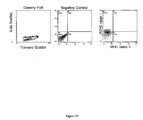
- a progenitor cell that is "immuno-incompetent” is a cell having a phenotype that is negative for surface antigens associated with class I and class II major histocompatibility complexes (MHC). Such a progenitor cell is also referred to herein as an HLA double negative.
- the cell population extracted from WJ is also characterized by "rapid proliferation", which refers to the rate at which the extracted cells will grow relative to other known progenitor cell populations, under conditions that are standard for progenitor cell expansion.
- rapid proliferation refers to the rate at which the extracted cells will grow relative to other known progenitor cell populations, under conditions that are standard for progenitor cell expansion.
- the present progenitor cell population can double within at least about 25 hours and as quickly as 15 hours, and thus expands far more rapidly than other known osteoprogenitor cell populations and other progenitor cell populations extracted from WJ.
- the cells and cell populations of the present invention can be obtained by extraction from WJ of human umbilical cord. Unlike the prior art, and in accordance with the present invention, such cells are extracted from the WJ that is associated with, i.e., proximal to, the exterior wall of the umbilical vasculature.
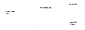
- the Wharton's jelly that is associated with or very near to the external surface of the cord vasculature lies within a region termed the perivascular zone, and typically remains associated with the vasculature when the vessels are excised from the cord, as is done for instance either to extract Wharton's jelly from the cord, or to extract the vessels from the cord and associated Wharton's jelly.
- the Wharton's jelly within this perivascular zone is a rich source of progenitor cells having the characteristics herein described. Accordingly, the present invention exploits the Wharton's jelly from this perivascular zone as a source for useful human progenitor cells.
- care is taking during the extraction process to avoid extracting cells of the umbilical cord blood, epithelial cells or endothelial cells of the UC, and cells derived from the vascular structure of the cord, where vascular structure is defined as the tunicae intima, media and adventia of arterial or venous vessels.
- vascular structure is defined as the tunicae intima, media and adventia of arterial or venous vessels.
- the WJ that lies within the perivascular zone is the Wharton's jelly proximal to the external wall of the umbilical vasculature, and lies typically within a zone extending to about 3mm from the external wall of the vessels.
- the target extraction zone can lie within about 2mm, e.g., about 1mm from the external wall of any one of the three vessels.
- the extraction of WJ from this region can be readily achieved using the technique described in the examples. In this technique the vessels are used as a carrier for the WJ, and the jelly-bearing vessels per se are used as the substrate from which the progenitor cells are extracted.
- cord vessels bearing a thin coating of WJ are excised either surgically or manually from fresh umbilical cord that has been washed thoroughly to remove essentially all cord blood contaminants.
- the vessels bearing the proximal Wharton's jelly, or sections thereof are then incubated at about 37°C in an extraction medium such as phosphate buffered saline (PBS) containing an enzyme suitable for digesting the collagen matrix of the WJ in which the desired cells reside.
- PBS phosphate buffered saline
- digestion with a collagenase is suitable, at a concentration within the range from about 0.1mg/mL to 1.0mg/mL or more, e.g., 0.5mg/mL.
- the ends of the vessels are tied, or clipped, off and can be suspended above the extraction medium to avoid contamination by agents contained within the vessel. It will thus be appreciated that the present Wharton's jelly extract is essentially free from cord blood cells and vessel endothelial cells.
- the vessels are removed, leaving a Wharton's jelly extract that contains human progenitor cells.
- These cells are expanded under conditions standard for expansion of progenitor cells.
- the cells can, for instance, be selected on polystyrene to select for adherent cells, such as in polystyrene dishes or flasks and then maintained in a suitable culturing medium.
- the extracted cells are cultured for expansion, with or without prior selection for adherent cells, under conditions of stirred suspension, as described for instance by Baksh et al in WO02/086104 .
- the cells present in the extract can, either directly or after their expansion, be sorted using established techniques to provide expandable subpopulations enriched for cells of a given phenotype.
- the present invention further provides WJ extracted cell populations that are enriched for osteoprogenitor cells, cell populations that are enriched for immuno-incompetent progenitor cells, and cell populations that are enriched for osteoprogenitor cells that are immuno-incompetent.
- the present invention thus further provides a method for producing MHC double negative progenitor cells, by obtaining a Wharton's jelly extract as herein described, or an MHC double negative-enriched fraction thereof, subjecting the extract or fraction thereof to freezing, and then culturing the frozen cells.
- the resulting cells as noted are potentially useful to induce tissue formation or repair in human subjects.
- the cell populations obtained from the extract or from a suitably enriched fraction thereof are useful either directly or following their expansion to provide differentiated cell populations. All of the procedures suitable for their fractionation and enrichment, and for their expansion are established in the prior art, and are exemplified herein. Expansion can proceed, for instance, in the presence of factors such as IL-3 and Stem Cell Factor, and similar agents know in the art.
- the present cell population, and particularly the osteoprogenitor cells therein are subjected to differentiation using conditions established for the growth of bone tissue therefrom.
- osteoprogenitor cells that arise from the culturing of the present progenitor cell population, referred to as committed osteoprogenitors, have shown the ability to differentiate in the absence of osteogenic supplements.
- the osteoprogenitor cells are cultured in a medium supplemented with one or more agents that stimulate osteogenesis, such as dexamethasone.
- the progenitor cells can also be cultured with supplements suitable for stimulating differentiation into other mesenchymally-derived connective tissues (Caplan, 1991), including cartilage, muscle, tendon, adipose etc., all in accordance with standard practice in the art.
- the cells can be transplanted in vivo to induce the formation of a desired tissue directly within a patient.
- the in situ formation of bone is provided by implanting osteoprogenitor, for the benefit of patients suffering from various bone conditions, diseases and disorders, particularly including bone fracture and osteoporosis.
- the immuno-incompetent progenitor cells present in the cell population are particularly valuable in this respect, given the substantially reduced rejection response that can be expected following their implantation.
- the UCs were collected from full-term caesarian section infants immediately upon delivery at Sunnybrook & Women' College Hospital, Toronto, Canada.
- the UC was transferred by the surgeon into a sterile vessel containing medium (75% ⁇ -MEM, 15% Fetal Bovine Serum (FBS), 10% antibiotics), and immediately transported to our laboratories at the Institute of Biomaterials & Biomedical Engineering, University of Toronto.
- medium 75% ⁇ -MEM, 15% Fetal Bovine Serum (FBS), 10% antibiotics
- the UC was washed in Phosphate Buffered Saline (PBS) (-Mg 2+ , -Ca 2+ ) three times to remove as much of the UC blood as possible, and transferred back into a container with medium.
- PBS Phosphate Buffered Saline
- a length of approximately 6 cm of cord was cut with sterile scissors and placed onto a sterile cork dissection board.
- the remaining cord (30-45 cm) was returned to the medium-filled container and placed into an incubator at 37°C.
- the 6 cm section of cord was 'twisted' against its helix, and pinned at both ends to reveal a smooth and straight surface of the UC epithelium.
- the UC was cut approximately 1-2 mm deep along its length to reveal the WJ.
- the WJ was teased from its inner surface using the blunt edge of a scalpel, and the teased away epithelium (approximately 0.5mm thick) was pinned down. This procedure resulted in the WJ being exposed, and with its three vessels embedded in it running straight from end to end rather than helically along its longitudinal axis. Care was taken to constantly bathe the section with 37°C PBS. Isolating one of the ends of a vessel with tweezers, it was teased away from the WJ along its length until it was free of the bulk of the WJ matrix.
- the middle of the vessel could be dissected from the matrix, held with tweezers, and teased from the matrix in each direction toward its ends.
- the vessel was surrounded with approximately 1-2mm of the cell-bearing WJ matrix.
- the dissected vessel was then clipped at both ends with either a surgical clamp, mosquito clip or sutured to create a 'loop,' blocking the passage of fluid either into or out of the vessel.
- the 'loop' was immediately placed along with the scissors into a 15ml tube containing a 0.5mg/ml collagenase solution with PBS (-Mg 2+ , -Ca 2+ ), and placed into an incubator at 37°C.
- the remaining two vessels were dissected in a similar fashion, looped, and also placed in the collagenase solution in the incubator. Subsequent to the removal of the vessels, strips of WJ could easily be dissected off the epithelium and placed into 15 ml tubes with the collagenase solution. The remaining epithelial layer was then disposed of in a biohazard waste container. The same protocol was used with the remaining 30-45 cm of UC, producing 15 to 25 tubes with either 'loops' or WJ strips.
- the 'loops' were removed with the aid of their attached suspension clamp or suture and a pipette, and the remaining suspensions were plated directly onto T-75 cm 2 tissue culture polystyrene dishes, and allowed to sit for 72 hours in order to allow the cells to attach to the polystyrene surface. The medium was then changed every two days.
- the attached cells were passaged using 1% trypsin solution after 7 days, at which point they exhibited 80-90% confluency, as observed by light microscopy, and there was evidence of 'mineralized' aggregate formation, as revealed under phase microscopy and indicated by expected changes in optical properties.
- cells were plated either in 35mm tissue culture polystyrene dishes or 6 well plates at 1x10 4 cells/cm 2 in 75% ⁇ -MEM, 15% FBS, 10% antibiotics and treated with 10 -8 M Dex, 5mM ⁇ -GP and 50 ⁇ g/ml ascorbic acid to test the osteogenic capacity of these cells. These plates were observed on days 2, 3, 4 and 5 of culture for CFU-O otherwise referred to as 'bone nodule' formation.
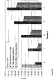
- the perivascular Wharton's Jelly (PVWJ)-derived progenitors comprise different sub-populations of progenitor cells.
- the so-called "committed” osteoprogenitor cells characterized by an ability to form bone nodules, as shown herein, in the absence of the culturing supplements normally required to induce such differentiation, such as culturing in the presence of dexamethasone.
- These committed osteoprogenitors thus differentiate spontaneously to form bone nodules.
- the "total osteoprotenitor” sub population graphed in Figure 15 includes the "committed osteoprogenitor” population, as well as the "uncommitted osteoprogenitor” population, and reveals the total number of cells that can be induced to differentiate down the osteogenic lineage.
- the actual ratio between the "committed” and “uncommitted” population is approximately 1:1, and so the ratio between the "total osteoprogenitor” population and the “committed osteoprogenitor” population is about 2:1.
- test cell populations were directly plated on tissue-culture polystyrene in bone forming medium containing 75% ⁇ -MEM, 15% FBS (StemCell Batch #: S13E40), 10% antibiotic stock solution containing penicillin G (167 units/ml), gentamicin (50 ⁇ g/ml) and amphotericin B (0.3 ⁇ g/ml), and Dex (10 -8 M), ⁇ -glycerophosphate (5mM) and L-ascorbic acid (50ug/ml), at a cell seeding density of 1 x 10 4 cells/cm 2 . Cultures were re-fed every two days for a period of 12 days.
- the cultures were maintained until mineralized nodular areas, detected as bone nodules, were observed (usually 3 days) at which point the cultures were re-fed with tetracycline containing medium at the last culture re-feed, then fixed in Karnovsky's fixative and prepared for analysis.
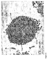
- a Leitz Aristoplan microscope (Esselte Leitz GmbH & Co KG, Stuttgart, Germany) was used to visualize the tetracycline labelled cultures under phase contrast as well as UV fluorescence and a Hitachi S-2000 scanning electron microscope at an accelerating voltage of 15 kV was used to generate images to demonstrate the presence of morphologically identifiable bone matrix.
- Tetracycline (9 ⁇ g/ml) was added to the cultures prior to termination.
- the cells were fixed in Karnovsky's fixative overnight and then viewed by UV-excited fluorescence imaging for tetracycline labeling of the mineral component of the nodular areas.
- Representative samples of CFU-O cultures were prepared for SEM by first placing them in 70%, 80%, 90% and 95% ethanol for 1 hour, followed by immersion in 100% ethanol for 3 hours. They were then critical point dried. A layer of gold approximately 3 nm layer was sputter coated with a Polaron SC515 SEM Coating System onto the specimens, which were then examined at various magnifications in a Hitachi S-2000 scanning electron microscope at an accelerating voltage of 15 kV. The images generated are used to demonstrate the presence of morphologically identifiable bone matrix.
- Test cell populations of >1 x 10 5 cells were washed in PBS containing 2% FBS (StemCell Batch #: S13E40) and re-suspended in PBS + 2% FBS with saturating concentrations (1:100 dilution) of the following conjugated mouse IgG1 HLA-A,B,C-PE and HLA-DR,DP,DQ-FITC for 30 minutes at 4°C.
- the cell suspension was washed twice with PBS + 2% FBS, stained with 1 ⁇ g/ml 7-AAD (BD Biosciences) and re-suspended in PBS + 2% FBS for analysis on a flow cytometer (XL, Beckman-Coulter, Miami, FL) using the ExpoADCXL4 software (Beckman-Coulter). Positive staining was defined as the emission of a fluorescence signal that exceeded levels obtained by >99% of cells from the control population stained with matched isotype antibodies (FITC- and PE-conjugated mouse IgG1, ⁇ monoclonal isotype standards, BD Biosciences). For each sample, at least 10,000 list mode events were collected. All plots were generated in EXPO 32 ADC Analysis software.
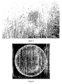
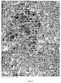
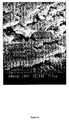
- FIGS 3, 4 and 5 illustrate CFU-O's that were present in the cultures on day 3 and day 5. They demonstrated the confluent layer of "fibroblastic-like" cells surrounding a nodular area represented by an 'aggregation' of polygonal cells that were producing the bone-matrix. These CFU-O's were observed in both the Dex (+) and Dex (-) cultures, and displayed similar morphology over successive passages.
- Tetracycline labeling of cultures was used for labeling newly formed calcium phosphate associated with the biological mineral phase of bone.
- the tetracycline labeling of the cultures coincide with the mineralized nodular areas, which is visualized by exposing the cultures to UV light.
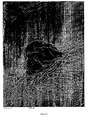
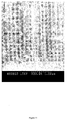
- Figures 6 and 7 depict tetracycline labeled CFU-O cultures of Day 3 and Day 5 cultures of WJ progenitor cells. These images were generated by UV-excited fluorescence imaging, and photographed.
- the attached cells were sub-cultured (passaged) using 0.1% trypsin solution after 7 days, at which point they exhibited 80-90% confluency as observed by light microscopy.
- the PVWJ peripheral vein Wharton's jelly
- MHC-A,B,C, MHC-DR,DP,DQ, and CD45 were then plated in T-75 tissue culture polystyrene flasks at 4x10 3 cells/cm 2 in SM, and treated with 10 -8 M Dex, 5mM ⁇ -GP and 50 ⁇ g/ml ascorbic acid to test the osteogenic capacity of these cells. These flasks were observed on days 2, 3, 4, 5 and 6 of culture for CFU-O or bone nodule, formation. Any residual cells from the passaging procedure also were cryopreserved for future use.
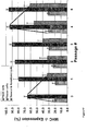
- PVWJ cells were prepared in 1ml total volume consisting of 90% FBS, 10% dimethyl sulphoxide (DMSO) (Sigma D-2650, Lot# 11K2320), and pipetted into 1ml polypropylene cryo-vials. The vials were placed into a -70°C freezer overnight, and transferred the following day to a -150°C freezer for long-term storage. After one week of cryo-preservation, the PVWJ cells were thawed and observed by flow cytometry for expression of MHC-A,B,C, MHC-DR,DP,DQ, and CD45.
- DMSO dimethyl sulphoxide
- PVWJ cells were thawed after one week of cryopreservation, recultured for one week, sub-cultured then reanalyzed by flow cytometry for expression of MHC-A,B,C, MHC-DR,DP,DQ, and CD45.
- the PVWJ progenitor cell population may also be exploited to give rise to mesenchymal cells and tissues other than bone, by culturing under conditions appropriate for such differentiation.
- the progenitors are prepared at a concentration of 10 4 cells/cm 2 and plated in 35 mm tissue culture dishes. The cells are maintained in Preadipocyte Medium (PM) (DMEM/Ham's F-10 (1:1, vol/vol), 10% fetal calf serum, 15 mM HEPES, 100 U/ml penicillin, 100 ⁇ g/ml streptomycin, 0.25 ⁇ g/ml amphotericin B) for 3 days.
- PM Preadipocyte Medium
- Adipogenic medium DMEM/ Ham's F-10 nutrient broth, 1:1, v/v; HEPES buffer (15 mM); Fetal Bovine Serum (3%); Biotin (33 ⁇ M), Pantothenate (17 ⁇ M), human insulin (100 nM), dexamethasone (0.5 ⁇ M), PPARY agonist (1 ⁇ M) and antibiotics
- DMEM/ Ham's F-10 nutrient broth, 1:1, v/v; HEPES buffer 15 mM
- Fetal Bovine Serum 3%
- Biotin 33 ⁇ M
- Pantothenate 17 ⁇ M
- human insulin 100 nM
- dexamethasone 0.5 ⁇ M
- PPARY agonist PPARY agonist
- Adipocyte Medium (DMEM/Ham's F-10 (1:1, vol/vol), 3% fetal calf serum, 1 ⁇ M dexamethasone, 100 nM human insulin, 33 ⁇ M D-biotin, 17 ⁇ M Na-pantothenate, 15 mM HEPES, 100 U/ml penicillin , 100 ⁇ g/ml streptomycin, 0.25 ⁇ g/ml amphotericin B), with regular feeding every 3 days, ensuring to only remove half the medium, replenishing with an equal volume of AM since adipocytes will float if all the media is removed. After four feedings (12 days), cells appear rounded with lipid droplets. Positive identification of differentiated mesenchymal cells into adipocytes can be confirmed by staining with Oil Red O and Nile Red.
- chondrocytes may be generated using cell suspensions prepared at a concentration of 10 4 cells/cm 2 and plated in 35 mm tissue culture dishes. To promote chondrogenic cells are cultured without serum and with transforming growth factor- ⁇ 3. The cell pellets develop a multilayered matrix-rich morphology and histologically show an increased proteoglycan-rich extracellular matrix during culture.
- cell suspensions are prepared at a concentration of 10 4 cells/cm 2 and plated in 35 mm tissue culture dishes.
- the cells are maintained in MCDB 120 medium completed with 15% fetal bovine serum (FBS) for 1 week (myoblast proliferation medium, MPM).
- FBS fetal bovine serum
- MPM myoblast proliferation medium
- MDM myoblast differentiation medium
- the present invention provides human progenitor cells having properties useful in the production of various connective tissues including bone, and further provides progenitor cells that are immune incompetent and ideal for transplantation into human patients to treat connective tissue conditions including bone diseases and disorders.
- the human progenitor cells are generated from extracts of a particular zone of human umbilical cord Wharton's jelly, termed the perivascular zone, extending proximally from the external wall of the cord vessels.
- the cell population extracted from this zone displays remarkable properties, including rapid proliferation, changes in cell morphology, as witnessed by the formation of cell colonies occurring before day 7 in all subcultured flasks (approximately 7-10 doublings) and the appearance of bone nodule formation without the addition of osteogenic supplements to the culture medium, as well as relatively high frequency of MHC double negative cells, the frequency of which is increased upon culturing of cells that have been frozen.
Landscapes
- Health & Medical Sciences (AREA)
- Life Sciences & Earth Sciences (AREA)
- Engineering & Computer Science (AREA)
- Biomedical Technology (AREA)
- Chemical & Material Sciences (AREA)
- Zoology (AREA)
- Biotechnology (AREA)
- Organic Chemistry (AREA)
- Bioinformatics & Cheminformatics (AREA)
- Genetics & Genomics (AREA)
- Wood Science & Technology (AREA)
- Developmental Biology & Embryology (AREA)
- Cell Biology (AREA)
- General Health & Medical Sciences (AREA)
- Biochemistry (AREA)
- Microbiology (AREA)
- General Engineering & Computer Science (AREA)
- Reproductive Health (AREA)
- Gynecology & Obstetrics (AREA)
- Rheumatology (AREA)
- Pregnancy & Childbirth (AREA)
- Medicinal Chemistry (AREA)
- Virology (AREA)
- Immunology (AREA)
- Pharmacology & Pharmacy (AREA)
- Epidemiology (AREA)
- Animal Behavior & Ethology (AREA)
- Public Health (AREA)
- Veterinary Medicine (AREA)
- Hematology (AREA)
- Orthopedic Medicine & Surgery (AREA)
- Micro-Organisms Or Cultivation Processes Thereof (AREA)
- Medicines Containing Material From Animals Or Micro-Organisms (AREA)
- Materials For Medical Uses (AREA)
Applications Claiming Priority (2)
| Application Number | Priority Date | Filing Date | Title |
|---|---|---|---|
| US44627503P | 2003-02-11 | 2003-02-11 | |
| EP04709560A EP1594957B1 (fr) | 2003-02-11 | 2004-02-10 | Cellules progenitrices provenant de la gelee de wharton de cordon ombilical humain |
Related Parent Applications (1)
| Application Number | Title | Priority Date | Filing Date |
|---|---|---|---|
| EP04709560.9 Division | 2004-02-10 |
Publications (2)
| Publication Number | Publication Date |
|---|---|
| EP2277992A2 true EP2277992A2 (fr) | 2011-01-26 |
| EP2277992A3 EP2277992A3 (fr) | 2011-03-02 |
Family
ID=32869477
Family Applications (2)
| Application Number | Title | Priority Date | Filing Date |
|---|---|---|---|
| EP10173753A Withdrawn EP2277992A3 (fr) | 2003-02-11 | 2004-02-10 | Cellules progénitrices issues de la gelée de Wharton du cordon ombilical humain |
| EP04709560A Expired - Lifetime EP1594957B1 (fr) | 2003-02-11 | 2004-02-10 | Cellules progenitrices provenant de la gelee de wharton de cordon ombilical humain |
Family Applications After (1)
| Application Number | Title | Priority Date | Filing Date |
|---|---|---|---|
| EP04709560A Expired - Lifetime EP1594957B1 (fr) | 2003-02-11 | 2004-02-10 | Cellules progenitrices provenant de la gelee de wharton de cordon ombilical humain |
Country Status (13)
| Country | Link |
|---|---|
| US (5) | US7547546B2 (fr) |
| EP (2) | EP2277992A3 (fr) |
| JP (1) | JP4790592B2 (fr) |
| KR (1) | KR101077760B1 (fr) |
| AT (1) | ATE478941T1 (fr) |
| AU (2) | AU2004210891B2 (fr) |
| BR (1) | BRPI0407221A (fr) |
| CA (1) | CA2515469C (fr) |
| DE (1) | DE602004028803D1 (fr) |
| ES (1) | ES2351386T3 (fr) |
| IL (2) | IL170221A (fr) |
| PT (1) | PT1594957E (fr) |
| WO (1) | WO2004072273A1 (fr) |
Cited By (1)
| Publication number | Priority date | Publication date | Assignee | Title |
|---|---|---|---|---|
| WO2019080942A1 (fr) * | 2017-10-27 | 2019-05-02 | Alis Pharma Ltd. | Extrait de tissus fœtaux, procédés de production de l'extrait et utilisation associée |
Families Citing this family (97)
| Publication number | Priority date | Publication date | Assignee | Title |
|---|---|---|---|---|
| US7311905B2 (en) | 2002-02-13 | 2007-12-25 | Anthrogenesis Corporation | Embryonic-like stem cells derived from post-partum mammalian placenta, and uses and methods of treatment using said cells |
| DK1349918T3 (da) | 2000-12-06 | 2014-11-10 | Anthrogenesis Corp | Fremgangsmåde til indsamling af stamceller fra moderkagen |
| EP2316919B1 (fr) * | 2001-02-14 | 2015-10-07 | Anthrogenesis Corporation | Placenta post-gravidique de mammifère, son utilisation et céllules souches placentaires correspondantes |
| EP3246396B1 (fr) * | 2001-02-14 | 2020-01-29 | Celularity, Inc. | Rénovation et repopulation de tissus décellularisés et d'organes cadavériques par des cellules souches |
| WO2004072273A1 (fr) | 2003-02-11 | 2004-08-26 | Davies John E | Cellules progenitrices provenant de la gelee de wharton de cordon ombilical humain |
| US8518390B2 (en) | 2003-06-27 | 2013-08-27 | Advanced Technologies And Regenerative Medicine, Llc | Treatment of stroke and other acute neural degenerative disorders via intranasal administration of umbilical cord-derived cells |
| WO2006071794A2 (fr) * | 2004-12-23 | 2006-07-06 | Ethicon Incorporated | Cellules postnatales derivees de tissus du cordon ombilical, et procedes de production et d'utilisation de celles-ci |
| US9572840B2 (en) | 2003-06-27 | 2017-02-21 | DePuy Synthes Products, Inc. | Regeneration and repair of neural tissue using postpartum-derived cells |
| US9592258B2 (en) | 2003-06-27 | 2017-03-14 | DePuy Synthes Products, Inc. | Treatment of neurological injury by administration of human umbilical cord tissue-derived cells |
| US7875272B2 (en) * | 2003-06-27 | 2011-01-25 | Ethicon, Incorporated | Treatment of stroke and other acute neuraldegenerative disorders using postpartum derived cells |
| US8790637B2 (en) | 2003-06-27 | 2014-07-29 | DePuy Synthes Products, LLC | Repair and regeneration of ocular tissue using postpartum-derived cells |
| US8491883B2 (en) | 2003-06-27 | 2013-07-23 | Advanced Technologies And Regenerative Medicine, Llc | Treatment of amyotrophic lateral sclerosis using umbilical derived cells |
| WO2005001080A2 (fr) | 2003-06-27 | 2005-01-06 | Ethicon, Incorporated | Cellules derivees de tissus post-partum destinees a etre utilisees pour traiter des maladies du coeur et du systeme circulatoire |
| JP2007508018A (ja) | 2003-10-08 | 2007-04-05 | べト−ステム インコーポレイテッド | 新規の幹細胞組成物の調製法および使用法、ならびにこの組成物を含有するキット |
| PL1789534T3 (pl) * | 2004-08-16 | 2015-03-31 | Cellresearch Corp Pte Ltd | Wyodrębnienie komórek macierzystych/progenitorowych z błony owodniowej pępowiny |
| US20060166361A1 (en) * | 2004-12-21 | 2006-07-27 | Agnieszka Seyda | Postpartum cells derived from placental tissue, and methods of making, culturing, and using the same |
| US20060171930A1 (en) * | 2004-12-21 | 2006-08-03 | Agnieszka Seyda | Postpartum cells derived from umbilical cord tissue, and methods of making, culturing, and using the same |
| US20060153815A1 (en) * | 2004-12-21 | 2006-07-13 | Agnieszka Seyda | Tissue engineering devices for the repair and regeneration of tissue |
| WO2006071778A2 (fr) | 2004-12-23 | 2006-07-06 | Ethicon Incorporated | Traitement de la maladie de parkinson et de troubles associes au moyen de cellules derivees postnatales |
| EP1831355A2 (fr) * | 2004-12-23 | 2007-09-12 | Ethicon, Inc. | Reparation et regeneration de tissus mous au moyen de cellules derivees du post-partum et produits cellulaires |
| WO2006071773A2 (fr) * | 2004-12-23 | 2006-07-06 | Ethicon Incoporated | Traitement de maladies osteochondrales utilisant des cellules derivees de post-partum et des produits de ceux-ci |
| DK1957633T3 (en) | 2005-10-13 | 2014-03-17 | Anthrogenesis Corp | Immunomodulation USING PLACE SPEECH STEM CELLS |
| WO2007059084A2 (fr) * | 2005-11-14 | 2007-05-24 | The New England Medical Center Hospitals, Inc. | Procedes de preparation de cellules souches matricielles de cordon (cmsc) en vue d'un stockage a long terme et de preparation d'un segment de cordon ombilical en vue d'une cryoconservation |
| WO2007070870A1 (fr) | 2005-12-16 | 2007-06-21 | Ethicon, Inc. | Compositions et procedes d'inhibition d'une reponse immunitaire negative en transplantation avec defaut d'histocompatibilite |
| JP5179376B2 (ja) * | 2005-12-19 | 2013-04-10 | エシコン・インコーポレイテッド | ローラーボトルでの分娩後取り出し細胞の体外増殖 |
| CN103173401A (zh) * | 2005-12-22 | 2013-06-26 | 简·恩尼斯 | 来自冷冻的脐带组织的活细胞 |
| US9125906B2 (en) | 2005-12-28 | 2015-09-08 | DePuy Synthes Products, Inc. | Treatment of peripheral vascular disease using umbilical cord tissue-derived cells |
| WO2007076522A2 (fr) * | 2005-12-28 | 2007-07-05 | Ethicon, Incorporated | Traitement de maladies vasculaires peripheriques a l'aide de cellules derivees du post-partum |
| CN101389754A (zh) | 2005-12-29 | 2009-03-18 | 人类起源公司 | 胎盘干细胞和第二来源干细胞的联合培养 |
| PT2471904T (pt) | 2005-12-29 | 2019-02-25 | Celularity Inc | População de células estaminais placentárias |
| WO2007099534A2 (fr) * | 2006-03-01 | 2007-09-07 | The Regenerative Medicine Institute | Compositions et populations de cellules obtenues à partir du cordon ombilical et procédés de production de celles-ci |
| CA2649874C (fr) * | 2006-05-05 | 2015-01-27 | John E. Davies | Cellules progenitrices immuno privilegiees et modulatrices |
| US20080064098A1 (en) * | 2006-06-05 | 2008-03-13 | Cryo-Cell International, Inc. | Procurement, isolation and cryopreservation of maternal placental cells |
| US20080050814A1 (en) * | 2006-06-05 | 2008-02-28 | Cryo-Cell International, Inc. | Procurement, isolation and cryopreservation of fetal placental cells |
| CN101501185A (zh) * | 2006-06-09 | 2009-08-05 | 人类起源公司 | 胎盘巢(placental niche)及其培养干细胞的用途 |
| US7993918B2 (en) * | 2006-08-04 | 2011-08-09 | Anthrogenesis Corporation | Tumor suppression using placental stem cells |
| WO2008060037A1 (fr) * | 2006-11-15 | 2008-05-22 | Seoul National University Industry Foundation | Procédé d'isolement primaire et d'expansion de cellule endothéliale souche/parente et de cellule souche mésenchyme derivées d'un cordon ombilical de mammifère, notamment humain |
| KR100902569B1 (ko) * | 2007-01-19 | 2009-06-11 | 재단법인서울대학교산학협력재단 | 인간 탯줄 유래 중간엽 줄기세포 및 이의 확립방법 |
| EP2099290A4 (fr) * | 2006-12-07 | 2010-01-13 | Teva Pharma | Procede de generation et d'expansion de cellules progenitrices de tissus et de cellules tissulaires mures a partir de moelle osseuse intacte ou de tissu de cordon ombilical intact |
| EP3763376A1 (fr) | 2007-02-12 | 2021-01-13 | Celularity, Inc. | Traitement de maladies inflammatoires au moyen de cellules souches placentaires |
| WO2009021333A1 (fr) * | 2007-08-14 | 2009-02-19 | Tissue Regeneration Therapeutics Inc. | Extraction des vaisseaux sanguins du cordon ombilical |
| US12129481B2 (en) | 2007-09-13 | 2024-10-29 | Reprobiogen Inc. | Use of cells derived from first trimester umbilical cord tissue |
| US10925903B2 (en) | 2007-09-13 | 2021-02-23 | Reprobiogen Inc. | Use of cells derived from first trimester umbilical cord tissue |
| CA2630708C (fr) * | 2007-09-13 | 2017-08-01 | Clifford L. Librach | Methode d'isolement et d'utilisation de cellules formees a partir de tissus foetaux du cordon ombilical preleves au premier trimestre de la grossesse |
| NZ599825A (en) | 2007-09-28 | 2014-10-31 | Anthrogenesis Corp | Tumor suppression using human placental perfusate and human placenta-derived intermediate natural killer cells |
| JP5323845B2 (ja) * | 2007-10-05 | 2013-10-23 | エシコン・インコーポレイテッド | ヒト臍帯組織由来細胞を用いた腎組織の修復および再建 |
| JP5795166B2 (ja) * | 2007-11-28 | 2015-10-14 | オルガノジェネシス インク. | 生命工学による組織コンストラクト並びに生産及び使用のための方法 |
| US8236538B2 (en) * | 2007-12-20 | 2012-08-07 | Advanced Technologies And Regenerative Medicine, Llc | Methods for sterilizing materials containing biologically active agents |
| BRPI0910686A2 (pt) | 2008-04-21 | 2015-09-29 | Tissue Regeneration Therapeutics Inc | células perivasculares do cordão umbilical humano geneticamente modificadas para a profilaxia contra agentes biológicos e químicos ou para o tratamento dos mesmos. |
| KR20240052847A (ko) | 2008-08-20 | 2024-04-23 | 셀룰래리티 인코포레이티드 | 개선된 세포 조성물 및 그의 제조 방법 |
| WO2010021715A1 (fr) | 2008-08-20 | 2010-02-25 | Anthrogenesis Corporation | Traitement d'un accident vasculaire cérébral à l'aide de cellules placentaires isolées |
| CA2734446C (fr) | 2008-08-22 | 2017-06-20 | Anthrogenesis Corporation | Methodes et compositions pour le traitement de deficits osseux au moyen de populations de cellules placentaires |
| AU2009316541B2 (en) | 2008-11-19 | 2015-08-06 | Celularity Inc. | Amnion derived adherent cells |
| CN102387807A (zh) | 2008-12-19 | 2012-03-21 | 先进科技及再生医学有限责任公司 | 肺部疾病和病症的治疗 |
| US10179900B2 (en) | 2008-12-19 | 2019-01-15 | DePuy Synthes Products, Inc. | Conditioned media and methods of making a conditioned media |
| US8771677B2 (en) | 2008-12-29 | 2014-07-08 | Vladimir B Serikov | Colony-forming unit cell of human chorion and method to obtain and use thereof |
| SG174551A1 (en) * | 2009-03-26 | 2011-10-28 | Ethicon Inc | Human umbilical cord tissue cells as therapy for alzheimer' s disease |
| JP2013514072A (ja) * | 2009-12-18 | 2013-04-25 | シー.ビー.ビー.ライフライン バイオテク リミテッド | 臍帯組織から間葉系前駆細胞の亜集団を含む単核細胞および内皮前駆細胞の亜集団を含む血管細胞を単離する方法 |
| EP2524034A1 (fr) * | 2010-01-14 | 2012-11-21 | Organogenesis, Inc. | Constructions tissulaires obtenues par bio-ingénierie et leurs procédés de fabrication et d'utilisation |
| DK3284818T3 (da) | 2010-01-26 | 2022-06-20 | Celularity Inc | Behandling af knoglerelateret kræft ved hjælp af placenta stamceller |
| EP2552457B1 (fr) | 2010-03-30 | 2015-07-29 | Histocell, S.L. | Biomatériau provenant de la gelée de wharton de cordon ombilical |
| LT2556145T (lt) | 2010-04-07 | 2016-11-10 | Anthrogenesis Corporation | Angiogenezė naudojant placentos kamienines ląsteles |
| MX2012011543A (es) | 2010-04-08 | 2013-05-06 | Anthrogenesis Corp | Tratamiento de sarcoidosis empleando celulas madre placentarias. |
| LT2576768T (lt) | 2010-06-01 | 2017-08-25 | Auxocell Laboratories, Inc. | Gamtinės wharton`s jelly kamieninės ląstelės ir jų gryninimas |
| ES2666746T3 (es) | 2010-07-13 | 2018-05-07 | Anthrogenesis Corporation | Métodos para generar linfocitos citolíticos naturales |
| CA2838330C (fr) | 2010-08-23 | 2021-01-26 | President And Fellows Of Harvard College | Sondes optogenetiques pour mesurer le potentiel de membrane |
| US8969315B2 (en) | 2010-12-31 | 2015-03-03 | Anthrogenesis Corporation | Enhancement of placental stem cell potency using modulatory RNA molecules |
| JP6104896B2 (ja) | 2011-06-01 | 2017-03-29 | アントフロゲネシス コーポレーション | 胎盤幹細胞を使用する疼痛の治療 |
| WO2013055476A1 (fr) | 2011-09-09 | 2013-04-18 | Anthrogenesis Corporation | Traitement de la sclérose latérale amyotrophique au moyen de cellules souches placentaires |
| WO2013070899A1 (fr) | 2011-11-08 | 2013-05-16 | Auxocell Laboratories, Inc. | Systèmes et procédés pour le traitement de cellules |
| AU2012358810B2 (en) | 2011-12-23 | 2018-03-15 | DePuy Synthes Products, Inc. | Detection of human umbilical cord tissue-derived cells |
| PT3321355T (pt) | 2011-12-30 | 2021-09-28 | Patel Amit | Métodos e composições para a derivação clínica de uma célula alogénica e utilizações terapêuticas |
| US8940294B2 (en) | 2012-03-02 | 2015-01-27 | Tissuetech, Inc. | Methods of isolating and culturing stem cells |
| EP2831577B1 (fr) | 2012-03-28 | 2018-08-08 | Purdue Research Foundation | Procédés et systèmes pouvant être utilisés en vue de la détection d'agents pathogènes contaminant les aliments |
| US9670457B2 (en) * | 2012-05-08 | 2017-06-06 | Stem Cell Reserve Lp | Stem cells and matrix from cord tissue |
| EP2756754B1 (fr) | 2013-01-17 | 2017-01-04 | Vita 34 Ag | Procédé de traitement du tissu du cordon ombilical, notamment en rapport avec la conservation du tissu |
| CN115137753A (zh) | 2013-02-05 | 2022-10-04 | 细胞结构公司 | 来自胎盘的自然杀伤细胞 |
| SG11201507515YA (en) | 2013-03-15 | 2015-10-29 | Marcus Kare Torleif Larsson | Cells, methods and apparatuses for umbilical cord blood collection and isolation of cells |
| US10400211B1 (en) | 2014-04-28 | 2019-09-03 | Donnie Rudd | Cell composition for tissue regeneration |
| USD748462S1 (en) | 2014-08-11 | 2016-02-02 | Auxocell Laboratories, Inc. | Centrifuge clip |
| US9993748B2 (en) | 2014-08-11 | 2018-06-12 | Auxocell Laboratories, Inc. | Centrifuge clip and method |
| US10542743B2 (en) | 2015-01-05 | 2020-01-28 | Hygieia Therapeutics Sdn Bhd | Isolation, expansion and characterization of wharton's jelly mesenchymal stem cells |
| JP2018527394A (ja) | 2015-09-23 | 2018-09-20 | オシリス セラピューティクス,インコーポレイテッド | 臍組織組成物及び使用方法 |
| WO2018073615A1 (fr) | 2016-10-21 | 2018-04-26 | Longboat Explorers Ab | Procédés et compositions de production de cellules hématopoïétiques |
| ES2985827T3 (es) * | 2017-12-22 | 2024-11-07 | Chiesi Farm Spa | Células estromales mesenquimales y procedimientos de obtención de células estromales mesenquimales a partir del cordón umbilical |
| US11285177B2 (en) | 2018-01-03 | 2022-03-29 | Globus Medical, Inc. | Allografts containing viable cells and methods thereof |
| TWI882956B (zh) | 2018-04-12 | 2025-05-11 | 新加坡商細胞研究私人有限公司 | 一種誘導或改善間質幹細胞傷口癒合特性的方法 |
| US12460178B2 (en) | 2018-04-12 | 2025-11-04 | Cellresearch Corporation Pte. Ltd. | Method of inducing or improving wound healing properties of mesenchymal stem cells |
| US20210269768A1 (en) | 2018-07-02 | 2021-09-02 | Vetbiobank | Neonatal stromal cells having low mhc-i expression and uses therof |
| US12465878B2 (en) | 2019-06-20 | 2025-11-11 | Amniotics Ab | Apparatus for filtering amniotic fluid |
| WO2021003254A1 (fr) * | 2019-07-01 | 2021-01-07 | Auxocell Laboratories, Inc. | Milieu de cryoconservation comprenant un extrait de tissu |
| EP4234019A3 (fr) | 2019-10-18 | 2023-09-13 | Amniotics AB | Procédés et appareils pour obtenir des cellules souches mésenchymateuses amniotiques à partir de liquide amniotique et de cellules dérivées de celui-ci |
| WO2021092199A1 (fr) | 2019-11-08 | 2021-05-14 | Kansas State University Research Foundation | Isolement, conservation et multiplication de cellules stromales mésenchymateuses de cordon ombilical canines |
| EP3909593A1 (fr) | 2020-05-15 | 2021-11-17 | Rigshospitalet | Cellules souches pour le traitement de lésions cutanées |
| US12435308B2 (en) | 2020-11-06 | 2025-10-07 | Amniotics Ab | Immunomodulation by amniotic fluid mesenchymal stem cells |
| EP4405375A1 (fr) | 2021-09-23 | 2024-07-31 | President and Fellows of Harvard College | Indicateurs de tension codés génétiquement et leurs utilisations |
| WO2023223303A1 (fr) | 2022-05-20 | 2023-11-23 | Alt Atlas Ltd. | Nouvelles lignées cellulaires et systèmes et procédés pour une plateforme logicielle de fabrication par apprentissage automatique qui optimisent des ingrédients fonctionnels uniques et des solutions pour les industries de biotechnologies et de techniques agroalimentaires de pointe |
Family Cites Families (19)
| Publication number | Priority date | Publication date | Assignee | Title |
|---|---|---|---|---|
| FR2578743B3 (fr) * | 1985-03-13 | 1987-06-12 | Bontemps Raymond | Procede de fabrication d'une preparation medicamenteuse a base de substance mesenchymateuse du type microsepteurs |
| SU1708328A1 (ru) * | 1989-07-11 | 1992-01-30 | Кемеровский государственный медицинский институт | Способ изготовлени сосудистого протеза |
| US5919702A (en) * | 1996-10-23 | 1999-07-06 | Advanced Tissue Science, Inc. | Production of cartilage tissue using cells isolated from Wharton's jelly |
| AU2003901668A0 (en) | 2003-03-28 | 2003-05-01 | Medvet Science Pty. Ltd. | Non-haemopoietic precursor cells |
| AUPQ147799A0 (en) | 1999-07-07 | 1999-07-29 | Medvet Science Pty. Ltd. | Mesenchymal precursor cell |
| US7670628B2 (en) | 1999-07-07 | 2010-03-02 | Angioblast Systems, Inc. | Mesenchymal precursor cell |
| US20050158289A1 (en) | 1999-07-07 | 2005-07-21 | Simmons Paul J. | Mesenchymal precursor cell and use thereof in the repair of bone defects and fractures in mammals |
| WO2001011011A2 (fr) * | 1999-08-05 | 2001-02-15 | Mcl Llc | Cellules souches adultes toutes-puissantes et procede d'isolement |
| US20020045260A1 (en) * | 2000-10-17 | 2002-04-18 | Shih-Chieh Hung | Method of isolating mesenchymal stem cells |
| EP1383869B1 (fr) | 2001-04-24 | 2009-06-10 | Dolores Baksh | Populations de cellules souches, leur expansion, et production de tissus et d'especes de cellules non hematopoietiques a partir desdites cellules souches |
| US7736892B2 (en) | 2002-02-25 | 2010-06-15 | Kansas State University Research Foundation | Cultures, products and methods using umbilical cord matrix cells |
| US20030161818A1 (en) * | 2002-02-25 | 2003-08-28 | Kansas State University Research Foundation | Cultures, products and methods using stem cells |
| WO2004072273A1 (fr) | 2003-02-11 | 2004-08-26 | Davies John E | Cellules progenitrices provenant de la gelee de wharton de cordon ombilical humain |
| WO2005001080A2 (fr) | 2003-06-27 | 2005-01-06 | Ethicon, Incorporated | Cellules derivees de tissus post-partum destinees a etre utilisees pour traiter des maladies du coeur et du systeme circulatoire |
| CA2529718C (fr) | 2003-06-27 | 2012-10-23 | Universite Laval | Methode permettant d'isoler des cellules du cordon ombilical |
| WO2005085428A1 (fr) | 2004-03-05 | 2005-09-15 | Davies John E | Systeme de culture en suspension exempte de serum pour des cellules souches mesenchymateuses |
| CN103173401A (zh) | 2005-12-22 | 2013-06-26 | 简·恩尼斯 | 来自冷冻的脐带组织的活细胞 |
| WO2007099534A2 (fr) | 2006-03-01 | 2007-09-07 | The Regenerative Medicine Institute | Compositions et populations de cellules obtenues à partir du cordon ombilical et procédés de production de celles-ci |
| CA2649874C (fr) | 2006-05-05 | 2015-01-27 | John E. Davies | Cellules progenitrices immuno privilegiees et modulatrices |
-
2004
- 2004-02-10 WO PCT/CA2004/000182 patent/WO2004072273A1/fr not_active Ceased
- 2004-02-10 AU AU2004210891A patent/AU2004210891B2/en not_active Ceased
- 2004-02-10 AT AT04709560T patent/ATE478941T1/de not_active IP Right Cessation
- 2004-02-10 EP EP10173753A patent/EP2277992A3/fr not_active Withdrawn
- 2004-02-10 BR BR0407221-9A patent/BRPI0407221A/pt not_active Application Discontinuation
- 2004-02-10 CA CA2515469A patent/CA2515469C/fr not_active Expired - Lifetime
- 2004-02-10 JP JP2006501414A patent/JP4790592B2/ja not_active Expired - Fee Related
- 2004-02-10 EP EP04709560A patent/EP1594957B1/fr not_active Expired - Lifetime
- 2004-02-10 ES ES04709560T patent/ES2351386T3/es not_active Expired - Lifetime
- 2004-02-10 PT PT04709560T patent/PT1594957E/pt unknown
- 2004-02-10 DE DE602004028803T patent/DE602004028803D1/de not_active Expired - Lifetime
- 2004-02-10 KR KR1020057014825A patent/KR101077760B1/ko not_active Expired - Fee Related
- 2004-10-08 US US10/961,919 patent/US7547546B2/en not_active Expired - Lifetime
-
2005
- 2005-08-10 IL IL170221A patent/IL170221A/en not_active IP Right Cessation
-
2008
- 2008-12-19 AU AU2008261123A patent/AU2008261123A1/en not_active Abandoned
-
2009
- 2009-06-11 US US12/482,963 patent/US8481311B2/en not_active Expired - Fee Related
- 2009-11-25 IL IL202331A patent/IL202331A/en not_active IP Right Cessation
-
2013
- 2013-05-28 US US13/903,575 patent/US9567564B2/en not_active Expired - Lifetime
-
2015
- 2015-10-15 US US14/884,339 patent/US9611456B2/en not_active Expired - Fee Related
-
2017
- 2017-02-23 US US15/440,170 patent/US20170157180A1/en not_active Abandoned
Non-Patent Citations (17)
| Title |
|---|
| AUBIN, JE: "Bone stem cells", J CELL BIOCHEM, vol. 30-31, 1998, pages 73 - 82, XP002222386, DOI: doi:10.1002/(SICI)1097-4644(1998)72:30/31+<73::AID-JCB11>3.0.CO;2-L |
| BECKSTEAD,JH; D F BAINTON: "Enzyme histochemistry on bone marrow biopsies: reactions useful in the differential diagnosis of leukemia and lymphoma applied to 2- micron plastic sections", BLOOD, vol. 55, 1980, pages 386 - 394 |
| BECKSTEAD,JH; P S HALVERSON; C A RIES; D F BAINTON: "Enzyme histochemistry and immunohistochemistry on biopsy specimens of pathologic human bone marrow", BLOOD, vol. 57, 1981, pages 1088 - 1098 |
| CAN ALP ET AL: "Concise review: human umbilical cord stroma with regard to the source of fetus-derived stem cells.", STEM CELLS (DAYTON, OHIO) NOV 2007, vol. 25, no. 11, November 2007 (2007-11-01), pages 2886 - 2895, XP002521370, ISSN: 1549-4918, DOI: 10.1634/STEMCELLS.2007-0417 * |
| CAPLAN,AI: "Mesenchymal stem cells", J ORTHOP.RES, vol. 9, 1991, pages 641 - 650 |
| CHACKO,AW; S R M REYNOLDS: "Architecture of distended and nondistended human umbilical cord tissues, with special reference to the arteries and veins.: Carnegie Institution of Washington", CONTRIBUTIONS TO EMBRYOLOGY, vol. 35, 1954, pages 135 - 150 |
| CONGET,P; J J MINGUELL: "Phenotypical and functional properties of human bone marrow mesenchymal progenitor cells", JOURNAL OF CELL PHYSIOLOGY, vol. 181, 1999, pages 67 - 73, XP002967649, DOI: doi:10.1002/(SICI)1097-4652(199910)181:1<67::AID-JCP7>3.0.CO;2-C |
| FRIEDENSTEIN,AJ; J F GORSKAJA; N N KULAGINA: "Fibroblast precursors in normal and irradiated mouse hematopoietic organs", EXP HEMATOL, vol. 4, 1976, pages 267 - 274, XP009091334 |
| KIMIHIRO TAKECHI ET AL: "Ultrastructural and immunohistochemical studies of Wharton's jelly umbilical cord cells", PLACENTA, vol. 14, no. 2, 1 March 1993 (1993-03-01), GB, pages 235 - 245, XP055260265, ISSN: 0143-4004, DOI: 10.1016/S0143-4004(05)80264-4 * |
| MITCHELL,KE; M L WEISS; B M MITCHELL; P MARTIN; D DAVIS; L MORALES; B HELWIG; M BEERENSTRAUCH; K ABOU-EASA; T HILDRETH: "Matrix Cells from Wharton's Jelly Form Neurons and Glia", STEM CELLS, vol. 21, 2003, pages 50 - 60, XP002281007 |
| NANAEV,AK; G KOHNEN; A P MILOVANOV; S P DOMOGATSKY; P KAUFMANN: "Stromal Differentiation and Architecture of the Human Umbilical Cord", PLACENTA, vol. 18, 1997, pages 53 - 64, XP008029778, DOI: doi:10.1016/S0143-4004(97)90071-0 |
| PARRY,EW: "Some electron microscope observations on the mesenchymal structures of lull-term umbilical cord", JOURNAL OF ANATOMY, vol. 107, 1970, pages 505 - 518 |
| PEREDA,J; P M MOTTA: "New advances in human embryology: morphofunctional relationship between the embryo and the yolk sac", MEDICAL ELECTRON MICROSCOPY, vol. 32, 2002, pages 67 - 78 |
| ROMANOV,YA; V A SVINTSITSKAYA; V N SMIMOV: "Searching for Alternative Sources of Postnatal Human Mesenchymal Stem Cells: Candidate MSC-Like Cells from Umbilical Cord", STEM CELLS, vol. 21, 2003, pages 105 - 110, XP002331940, DOI: doi:10.1634/stemcells.21-1-105 |
| SCHOENBERG,MD; A HINMAN; R D MOORE: "Studies on connective tissue V, Feber formation in Wharton's Jelly.", LABORATORY INVESTIGATION, vol. 9, 1960, pages 350 - 355 |
| SEN,A, J. CELL BIOCHEM., vol. 81, pages 312 - 319 |
| TAKECHI,K; Y KUWABARA; M MIZUNO: "Ultrastructural and Immunohistochemical Studies of Wharton's Jelly Umbilical Cord Cells", PLACENTA, vol. 14, 1993, pages 235 - 245, XP055260265, DOI: doi:10.1016/S0143-4004(05)80264-4 |
Cited By (1)
| Publication number | Priority date | Publication date | Assignee | Title |
|---|---|---|---|---|
| WO2019080942A1 (fr) * | 2017-10-27 | 2019-05-02 | Alis Pharma Ltd. | Extrait de tissus fœtaux, procédés de production de l'extrait et utilisation associée |
Also Published As
| Publication number | Publication date |
|---|---|
| US20170157180A1 (en) | 2017-06-08 |
| AU2004210891B2 (en) | 2008-09-25 |
| US20090269318A1 (en) | 2009-10-29 |
| IL202331A (en) | 2011-08-31 |
| JP4790592B2 (ja) | 2011-10-12 |
| US20050148074A1 (en) | 2005-07-07 |
| JP2006517102A (ja) | 2006-07-20 |
| KR20050115862A (ko) | 2005-12-08 |
| US20130259840A1 (en) | 2013-10-03 |
| IL170221A (en) | 2010-11-30 |
| EP1594957B1 (fr) | 2010-08-25 |
| US20160032241A1 (en) | 2016-02-04 |
| DE602004028803D1 (de) | 2010-10-07 |
| AU2004210891A1 (en) | 2004-08-26 |
| EP1594957A1 (fr) | 2005-11-16 |
| PT1594957E (pt) | 2010-11-30 |
| HK1084417A1 (en) | 2006-07-28 |
| US9567564B2 (en) | 2017-02-14 |
| CA2515469A1 (fr) | 2004-08-26 |
| BRPI0407221A (pt) | 2006-01-31 |
| ATE478941T1 (de) | 2010-09-15 |
| ES2351386T3 (es) | 2011-02-03 |
| CA2515469C (fr) | 2013-12-17 |
| US9611456B2 (en) | 2017-04-04 |
| EP2277992A3 (fr) | 2011-03-02 |
| KR101077760B1 (ko) | 2011-10-27 |
| US7547546B2 (en) | 2009-06-16 |
| WO2004072273A1 (fr) | 2004-08-26 |
| US8481311B2 (en) | 2013-07-09 |
| AU2008261123A1 (en) | 2009-01-15 |
Similar Documents
| Publication | Publication Date | Title |
|---|---|---|
| EP1594957B1 (fr) | Cellules progenitrices provenant de la gelee de wharton de cordon ombilical humain | |
| EP1976980B1 (fr) | Cellules viables obtenues de tissu de cordon ombilical congele | |
| US20090068153A1 (en) | Cell composition for tissue regeneration | |
| AU2012227326B2 (en) | Progenitor cells from wharton's jelly of human umbilical cord | |
| HK1084417B (en) | Progenitor cells from wharton's jelly of human umbilical cord |
Legal Events
| Date | Code | Title | Description |
|---|---|---|---|
| PUAI | Public reference made under article 153(3) epc to a published international application that has entered the european phase |
Free format text: ORIGINAL CODE: 0009012 |
|
| AC | Divisional application: reference to earlier application |
Ref document number: 1594957 Country of ref document: EP Kind code of ref document: P |
|
| AK | Designated contracting states |
Kind code of ref document: A2 Designated state(s): AT BE BG CH CY CZ DE DK EE ES FI FR GB GR HU IE IT LI LU MC NL PT RO SE SI SK TR |
|
| AX | Request for extension of the european patent |
Extension state: AL LT LV MK |
|
| PUAL | Search report despatched |
Free format text: ORIGINAL CODE: 0009013 |
|
| AK | Designated contracting states |
Kind code of ref document: A3 Designated state(s): AT BE BG CH CY CZ DE DK EE ES FI FR GB GR HU IE IT LI LU MC NL PT RO SE SI SK TR |
|
| AX | Request for extension of the european patent |
Extension state: AL LT LV MK |
|
| 17P | Request for examination filed |
Effective date: 20110902 |
|
| 17Q | First examination report despatched |
Effective date: 20160330 |
|
| STAA | Information on the status of an ep patent application or granted ep patent |
Free format text: STATUS: THE APPLICATION IS DEEMED TO BE WITHDRAWN |
|
| 18D | Application deemed to be withdrawn |
Effective date: 20180901 |