EP2554663A2 - Method for obtaining oligodendrocyte precursor cells - Google Patents
Method for obtaining oligodendrocyte precursor cells Download PDFInfo
- Publication number
- EP2554663A2 EP2554663A2 EP11721797A EP11721797A EP2554663A2 EP 2554663 A2 EP2554663 A2 EP 2554663A2 EP 11721797 A EP11721797 A EP 11721797A EP 11721797 A EP11721797 A EP 11721797A EP 2554663 A2 EP2554663 A2 EP 2554663A2
- Authority
- EP
- European Patent Office
- Prior art keywords
- opcs
- cell
- enzymatic digestion
- cells
- papain
- Prior art date
- Legal status (The legal status is an assumption and is not a legal conclusion. Google has not performed a legal analysis and makes no representation as to the accuracy of the status listed.)
- Withdrawn
Links
- 238000000034 method Methods 0.000 title claims abstract description 62
- 210000000535 oligodendrocyte precursor cell Anatomy 0.000 title description 71
- 210000004556 brain Anatomy 0.000 claims abstract description 22
- 241000124008 Mammalia Species 0.000 claims abstract description 14
- 210000003710 cerebral cortex Anatomy 0.000 claims abstract description 12
- 239000000523 sample Substances 0.000 claims abstract description 10
- 230000005012 migration Effects 0.000 claims abstract description 6
- 238000013508 migration Methods 0.000 claims abstract description 6
- 239000012472 biological sample Substances 0.000 claims abstract description 3
- 230000037230 mobility Effects 0.000 claims abstract 2
- 230000006862 enzymatic digestion Effects 0.000 claims description 20
- 108090000526 Papain Proteins 0.000 claims description 18
- 239000004365 Protease Substances 0.000 claims description 17
- 229940055729 papain Drugs 0.000 claims description 17
- 235000019834 papain Nutrition 0.000 claims description 17
- 210000002418 meninge Anatomy 0.000 claims description 11
- 210000002987 choroid plexus Anatomy 0.000 claims description 9
- 238000004113 cell culture Methods 0.000 claims description 6
- 239000003814 drug Substances 0.000 claims description 6
- 230000035755 proliferation Effects 0.000 claims description 6
- 239000000126 substance Substances 0.000 claims description 6
- 229940079593 drug Drugs 0.000 claims description 5
- 210000000944 nerve tissue Anatomy 0.000 claims description 5
- 230000000694 effects Effects 0.000 claims description 4
- 230000024245 cell differentiation Effects 0.000 claims description 3
- 238000012637 gene transfection Methods 0.000 claims description 3
- 102000016911 Deoxyribonucleases Human genes 0.000 claims description 2
- 108010053770 Deoxyribonucleases Proteins 0.000 claims description 2
- 102000004190 Enzymes Human genes 0.000 claims description 2
- 108090000790 Enzymes Proteins 0.000 claims description 2
- 238000012258 culturing Methods 0.000 claims description 2
- 229940088598 enzyme Drugs 0.000 claims description 2
- 230000001413 cellular effect Effects 0.000 claims 1
- 210000004027 cell Anatomy 0.000 abstract description 66
- 210000004248 oligodendroglia Anatomy 0.000 abstract description 28
- 230000004069 differentiation Effects 0.000 abstract description 7
- 239000002243 precursor Substances 0.000 abstract description 3
- 230000004663 cell proliferation Effects 0.000 abstract description 2
- 238000000746 purification Methods 0.000 description 17
- 241001465754 Metazoa Species 0.000 description 16
- 210000001519 tissue Anatomy 0.000 description 16
- 239000002609 medium Substances 0.000 description 13
- 230000008569 process Effects 0.000 description 12
- 210000003050 axon Anatomy 0.000 description 10
- 210000003169 central nervous system Anatomy 0.000 description 10
- 102000001393 Platelet-Derived Growth Factor alpha Receptor Human genes 0.000 description 9
- 108010068588 Platelet-Derived Growth Factor alpha Receptor Proteins 0.000 description 9
- 108090000623 proteins and genes Proteins 0.000 description 9
- 208000016192 Demyelinating disease Diseases 0.000 description 8
- 241000699670 Mus sp. Species 0.000 description 8
- 239000012981 Hank's balanced salt solution Substances 0.000 description 7
- 239000006285 cell suspension Substances 0.000 description 7
- 238000001943 fluorescence-activated cell sorting Methods 0.000 description 7
- 102000006386 Myelin Proteins Human genes 0.000 description 6
- 108010083674 Myelin Proteins Proteins 0.000 description 6
- 102000008730 Nestin Human genes 0.000 description 6
- 108010088225 Nestin Proteins 0.000 description 6
- 238000011161 development Methods 0.000 description 6
- 230000018109 developmental process Effects 0.000 description 6
- 239000012528 membrane Substances 0.000 description 6
- 210000005012 myelin Anatomy 0.000 description 6
- 210000005055 nestin Anatomy 0.000 description 6
- 230000003287 optical effect Effects 0.000 description 6
- 208000035346 Margins of Excision Diseases 0.000 description 5
- 206010028980 Neoplasm Diseases 0.000 description 5
- 230000006378 damage Effects 0.000 description 5
- 230000003210 demyelinating effect Effects 0.000 description 5
- 230000023105 myelination Effects 0.000 description 5
- 210000005036 nerve Anatomy 0.000 description 5
- 238000010899 nucleation Methods 0.000 description 5
- 238000000926 separation method Methods 0.000 description 5
- 238000012360 testing method Methods 0.000 description 5
- 239000006144 Dulbecco’s modified Eagle's medium Substances 0.000 description 4
- 241000699666 Mus <mouse, genus> Species 0.000 description 4
- -1 PLP Proteins 0.000 description 4
- 108010004729 Phycoerythrin Proteins 0.000 description 4
- 208000027418 Wounds and injury Diseases 0.000 description 4
- 230000030833 cell death Effects 0.000 description 4
- 230000029087 digestion Effects 0.000 description 4
- 238000010790 dilution Methods 0.000 description 4
- 239000012895 dilution Substances 0.000 description 4
- 208000014674 injury Diseases 0.000 description 4
- 230000003902 lesion Effects 0.000 description 4
- 210000003007 myelin sheath Anatomy 0.000 description 4
- 230000001537 neural effect Effects 0.000 description 4
- 210000004498 neuroglial cell Anatomy 0.000 description 4
- 230000009984 peri-natal effect Effects 0.000 description 4
- 230000004044 response Effects 0.000 description 4
- 239000000243 solution Substances 0.000 description 4
- UCSJYZPVAKXKNQ-HZYVHMACSA-N streptomycin Chemical compound CN[C@H]1[C@H](O)[C@@H](O)[C@H](CO)O[C@H]1O[C@@H]1[C@](C=O)(O)[C@H](C)O[C@H]1O[C@@H]1[C@@H](NC(N)=N)[C@H](O)[C@@H](NC(N)=N)[C@H](O)[C@H]1O UCSJYZPVAKXKNQ-HZYVHMACSA-N 0.000 description 4
- 230000004083 survival effect Effects 0.000 description 4
- 102000007260 Deoxyribonuclease I Human genes 0.000 description 3
- 108010008532 Deoxyribonuclease I Proteins 0.000 description 3
- 102000010780 Platelet-Derived Growth Factor Human genes 0.000 description 3
- 108010038512 Platelet-Derived Growth Factor Proteins 0.000 description 3
- 238000002659 cell therapy Methods 0.000 description 3
- 230000007423 decrease Effects 0.000 description 3
- 239000012909 foetal bovine serum Substances 0.000 description 3
- 150000002254 galactosylceramides Chemical class 0.000 description 3
- 238000011534 incubation Methods 0.000 description 3
- 239000010410 layer Substances 0.000 description 3
- 210000002569 neuron Anatomy 0.000 description 3
- 108010055896 polyornithine Proteins 0.000 description 3
- 108010057417 polysialyl neural cell adhesion molecule Proteins 0.000 description 3
- OYPRJOBELJOOCE-UHFFFAOYSA-N Calcium Chemical compound [Ca] OYPRJOBELJOOCE-UHFFFAOYSA-N 0.000 description 2
- 206010012305 Demyelination Diseases 0.000 description 2
- KCXVZYZYPLLWCC-UHFFFAOYSA-N EDTA Chemical compound OC(=O)CN(CC(O)=O)CCN(CC(O)=O)CC(O)=O KCXVZYZYPLLWCC-UHFFFAOYSA-N 0.000 description 2
- 241000282412 Homo Species 0.000 description 2
- 241000701044 Human gammaherpesvirus 4 Species 0.000 description 2
- XUJNEKJLAYXESH-REOHCLBHSA-N L-Cysteine Chemical compound SC[C@H](N)C(O)=O XUJNEKJLAYXESH-REOHCLBHSA-N 0.000 description 2
- FYYHWMGAXLPEAU-UHFFFAOYSA-N Magnesium Chemical compound [Mg] FYYHWMGAXLPEAU-UHFFFAOYSA-N 0.000 description 2
- 229930182555 Penicillin Natural products 0.000 description 2
- JGSARLDLIJGVTE-MBNYWOFBSA-N Penicillin G Chemical compound N([C@H]1[C@H]2SC([C@@H](N2C1=O)C(O)=O)(C)C)C(=O)CC1=CC=CC=C1 JGSARLDLIJGVTE-MBNYWOFBSA-N 0.000 description 2
- RJKFOVLPORLFTN-LEKSSAKUSA-N Progesterone Chemical compound C1CC2=CC(=O)CC[C@]2(C)[C@@H]2[C@@H]1[C@@H]1CC[C@H](C(=O)C)[C@@]1(C)CC2 RJKFOVLPORLFTN-LEKSSAKUSA-N 0.000 description 2
- 241000283984 Rodentia Species 0.000 description 2
- 238000000692 Student's t-test Methods 0.000 description 2
- 108010017842 Telomerase Proteins 0.000 description 2
- 108700019146 Transgenes Proteins 0.000 description 2
- 230000009471 action Effects 0.000 description 2
- 210000001130 astrocyte Anatomy 0.000 description 2
- 230000008901 benefit Effects 0.000 description 2
- 238000001574 biopsy Methods 0.000 description 2
- 230000015572 biosynthetic process Effects 0.000 description 2
- 239000011575 calcium Substances 0.000 description 2
- 229910052791 calcium Inorganic materials 0.000 description 2
- 210000001175 cerebrospinal fluid Anatomy 0.000 description 2
- 210000000805 cytoplasm Anatomy 0.000 description 2
- 239000003102 growth factor Substances 0.000 description 2
- 238000000338 in vitro Methods 0.000 description 2
- 238000001727 in vivo Methods 0.000 description 2
- NOESYZHRGYRDHS-UHFFFAOYSA-N insulin Chemical compound N1C(=O)C(NC(=O)C(CCC(N)=O)NC(=O)C(CCC(O)=O)NC(=O)C(C(C)C)NC(=O)C(NC(=O)CN)C(C)CC)CSSCC(C(NC(CO)C(=O)NC(CC(C)C)C(=O)NC(CC=2C=CC(O)=CC=2)C(=O)NC(CCC(N)=O)C(=O)NC(CC(C)C)C(=O)NC(CCC(O)=O)C(=O)NC(CC(N)=O)C(=O)NC(CC=2C=CC(O)=CC=2)C(=O)NC(CSSCC(NC(=O)C(C(C)C)NC(=O)C(CC(C)C)NC(=O)C(CC=2C=CC(O)=CC=2)NC(=O)C(CC(C)C)NC(=O)C(C)NC(=O)C(CCC(O)=O)NC(=O)C(C(C)C)NC(=O)C(CC(C)C)NC(=O)C(CC=2NC=NC=2)NC(=O)C(CO)NC(=O)CNC2=O)C(=O)NCC(=O)NC(CCC(O)=O)C(=O)NC(CCCNC(N)=N)C(=O)NCC(=O)NC(CC=3C=CC=CC=3)C(=O)NC(CC=3C=CC=CC=3)C(=O)NC(CC=3C=CC(O)=CC=3)C(=O)NC(C(C)O)C(=O)N3C(CCC3)C(=O)NC(CCCCN)C(=O)NC(C)C(O)=O)C(=O)NC(CC(N)=O)C(O)=O)=O)NC(=O)C(C(C)CC)NC(=O)C(CO)NC(=O)C(C(C)O)NC(=O)C1CSSCC2NC(=O)C(CC(C)C)NC(=O)C(NC(=O)C(CCC(N)=O)NC(=O)C(CC(N)=O)NC(=O)C(NC(=O)C(N)CC=1C=CC=CC=1)C(C)C)CC1=CN=CN1 NOESYZHRGYRDHS-UHFFFAOYSA-N 0.000 description 2
- 239000011777 magnesium Substances 0.000 description 2
- 229910052749 magnesium Inorganic materials 0.000 description 2
- 210000000274 microglia Anatomy 0.000 description 2
- 230000009456 molecular mechanism Effects 0.000 description 2
- 201000006417 multiple sclerosis Diseases 0.000 description 2
- 229940049954 penicillin Drugs 0.000 description 2
- 230000002062 proliferating effect Effects 0.000 description 2
- 102000004169 proteins and genes Human genes 0.000 description 2
- KIDHWZJUCRJVML-UHFFFAOYSA-N putrescine Chemical compound NCCCCN KIDHWZJUCRJVML-UHFFFAOYSA-N 0.000 description 2
- 238000012216 screening Methods 0.000 description 2
- 210000002966 serum Anatomy 0.000 description 2
- 229960005322 streptomycin Drugs 0.000 description 2
- 239000000758 substrate Substances 0.000 description 2
- 239000000725 suspension Substances 0.000 description 2
- 230000001225 therapeutic effect Effects 0.000 description 2
- JKMHFZQWWAIEOD-UHFFFAOYSA-N 2-[4-(2-hydroxyethyl)piperazin-1-yl]ethanesulfonic acid Chemical compound OCC[NH+]1CCN(CCS([O-])(=O)=O)CC1 JKMHFZQWWAIEOD-UHFFFAOYSA-N 0.000 description 1
- KISWVXRQTGLFGD-UHFFFAOYSA-N 2-[[2-[[6-amino-2-[[2-[[2-[[5-amino-2-[[2-[[1-[2-[[6-amino-2-[(2,5-diamino-5-oxopentanoyl)amino]hexanoyl]amino]-5-(diaminomethylideneamino)pentanoyl]pyrrolidine-2-carbonyl]amino]-3-hydroxypropanoyl]amino]-5-oxopentanoyl]amino]-5-(diaminomethylideneamino)p Chemical compound C1CCN(C(=O)C(CCCN=C(N)N)NC(=O)C(CCCCN)NC(=O)C(N)CCC(N)=O)C1C(=O)NC(CO)C(=O)NC(CCC(N)=O)C(=O)NC(CCCN=C(N)N)C(=O)NC(CO)C(=O)NC(CCCCN)C(=O)NC(C(=O)NC(CC(C)C)C(O)=O)CC1=CC=C(O)C=C1 KISWVXRQTGLFGD-UHFFFAOYSA-N 0.000 description 1
- 206010003497 Asphyxia Diseases 0.000 description 1
- 241000283690 Bos taurus Species 0.000 description 1
- 241000282465 Canis Species 0.000 description 1
- 241000251556 Chordata Species 0.000 description 1
- 241000199632 Craniata <brachiopoda> Species 0.000 description 1
- 241000283073 Equus caballus Species 0.000 description 1
- 241000206602 Eukaryota Species 0.000 description 1
- 241000282324 Felis Species 0.000 description 1
- 208000032612 Glial tumor Diseases 0.000 description 1
- 206010018338 Glioma Diseases 0.000 description 1
- 229920002527 Glycogen Polymers 0.000 description 1
- 241000251728 Gnathostomata <vertebrate> Species 0.000 description 1
- 239000007995 HEPES buffer Substances 0.000 description 1
- 102000004877 Insulin Human genes 0.000 description 1
- 108090001061 Insulin Proteins 0.000 description 1
- 102100034343 Integrase Human genes 0.000 description 1
- 235000013878 L-cysteine Nutrition 0.000 description 1
- 239000004201 L-cysteine Substances 0.000 description 1
- ZDXPYRJPNDTMRX-VKHMYHEASA-N L-glutamine Chemical compound OC(=O)[C@@H](N)CCC(N)=O ZDXPYRJPNDTMRX-VKHMYHEASA-N 0.000 description 1
- 229930182816 L-glutamine Natural products 0.000 description 1
- 241000283953 Lagomorpha Species 0.000 description 1
- 241000829100 Macaca mulatta polyomavirus 1 Species 0.000 description 1
- 102000029749 Microtubule Human genes 0.000 description 1
- 108091022875 Microtubule Proteins 0.000 description 1
- 201000010133 Oligodendroglioma Diseases 0.000 description 1
- 108010046016 Peanut Agglutinin Proteins 0.000 description 1
- 108010010974 Proteolipids Proteins 0.000 description 1
- 102000016202 Proteolipids Human genes 0.000 description 1
- 239000005700 Putrescine Substances 0.000 description 1
- 108010092799 RNA-directed DNA polymerase Proteins 0.000 description 1
- 241000612118 Samolus valerandi Species 0.000 description 1
- 108010052164 Sodium Channels Proteins 0.000 description 1
- 102000018674 Sodium Channels Human genes 0.000 description 1
- FAPWRFPIFSIZLT-UHFFFAOYSA-M Sodium chloride Chemical compound [Na+].[Cl-] FAPWRFPIFSIZLT-UHFFFAOYSA-M 0.000 description 1
- 229930006000 Sucrose Natural products 0.000 description 1
- CZMRCDWAGMRECN-UGDNZRGBSA-N Sucrose Chemical compound O[C@H]1[C@H](O)[C@@H](CO)O[C@@]1(CO)O[C@@H]1[C@H](O)[C@@H](O)[C@H](O)[C@@H](CO)O1 CZMRCDWAGMRECN-UGDNZRGBSA-N 0.000 description 1
- 102000004338 Transferrin Human genes 0.000 description 1
- 108090000901 Transferrin Proteins 0.000 description 1
- 241000251539 Vertebrata <Metazoa> Species 0.000 description 1
- 108700005077 Viral Genes Proteins 0.000 description 1
- 208000036142 Viral infection Diseases 0.000 description 1
- 230000036982 action potential Effects 0.000 description 1
- 230000004913 activation Effects 0.000 description 1
- 239000000654 additive Substances 0.000 description 1
- 230000032683 aging Effects 0.000 description 1
- VREFGVBLTWBCJP-UHFFFAOYSA-N alprazolam Chemical compound C12=CC(Cl)=CC=C2N2C(C)=NN=C2CN=C1C1=CC=CC=C1 VREFGVBLTWBCJP-UHFFFAOYSA-N 0.000 description 1
- 238000010171 animal model Methods 0.000 description 1
- 239000000427 antigen Substances 0.000 description 1
- 108091007433 antigens Proteins 0.000 description 1
- 102000036639 antigens Human genes 0.000 description 1
- 210000000576 arachnoid Anatomy 0.000 description 1
- 230000004071 biological effect Effects 0.000 description 1
- 210000004369 blood Anatomy 0.000 description 1
- 239000008280 blood Substances 0.000 description 1
- 210000004958 brain cell Anatomy 0.000 description 1
- 230000011712 cell development Effects 0.000 description 1
- 230000012292 cell migration Effects 0.000 description 1
- 230000003833 cell viability Effects 0.000 description 1
- 230000007248 cellular mechanism Effects 0.000 description 1
- 210000001638 cerebellum Anatomy 0.000 description 1
- 210000004289 cerebral ventricle Anatomy 0.000 description 1
- 210000002808 connective tissue Anatomy 0.000 description 1
- 238000011109 contamination Methods 0.000 description 1
- 230000001086 cytosolic effect Effects 0.000 description 1
- 230000001419 dependent effect Effects 0.000 description 1
- 238000001514 detection method Methods 0.000 description 1
- 210000002451 diencephalon Anatomy 0.000 description 1
- BVTBRVFYZUCAKH-UHFFFAOYSA-L disodium selenite Chemical compound [Na+].[Na+].[O-][Se]([O-])=O BVTBRVFYZUCAKH-UHFFFAOYSA-L 0.000 description 1
- 238000002224 dissection Methods 0.000 description 1
- 238000010494 dissociation reaction Methods 0.000 description 1
- 230000005593 dissociations Effects 0.000 description 1
- 210000001951 dura mater Anatomy 0.000 description 1
- 230000013020 embryo development Effects 0.000 description 1
- 210000002257 embryonic structure Anatomy 0.000 description 1
- 210000002889 endothelial cell Anatomy 0.000 description 1
- 230000002255 enzymatic effect Effects 0.000 description 1
- 210000000981 epithelium Anatomy 0.000 description 1
- 238000001914 filtration Methods 0.000 description 1
- 229940096919 glycogen Drugs 0.000 description 1
- 210000004884 grey matter Anatomy 0.000 description 1
- 239000001963 growth medium Substances 0.000 description 1
- 210000001320 hippocampus Anatomy 0.000 description 1
- 210000003016 hypothalamus Anatomy 0.000 description 1
- 208000015181 infectious disease Diseases 0.000 description 1
- 229940125396 insulin Drugs 0.000 description 1
- 230000010354 integration Effects 0.000 description 1
- 210000003963 intermediate filament Anatomy 0.000 description 1
- 238000002955 isolation Methods 0.000 description 1
- 230000009191 jumping Effects 0.000 description 1
- 210000000627 locus coeruleus Anatomy 0.000 description 1
- 230000007774 longterm Effects 0.000 description 1
- 230000007246 mechanism Effects 0.000 description 1
- 230000028161 membrane depolarization Effects 0.000 description 1
- 210000001259 mesencephalon Anatomy 0.000 description 1
- 230000002503 metabolic effect Effects 0.000 description 1
- 210000004688 microtubule Anatomy 0.000 description 1
- 230000001617 migratory effect Effects 0.000 description 1
- 238000012986 modification Methods 0.000 description 1
- 230000004048 modification Effects 0.000 description 1
- 108700024542 myc Genes Proteins 0.000 description 1
- 210000001577 neostriatum Anatomy 0.000 description 1
- 210000000276 neural tube Anatomy 0.000 description 1
- 239000002547 new drug Substances 0.000 description 1
- 230000007170 pathology Effects 0.000 description 1
- 230000000149 penetrating effect Effects 0.000 description 1
- 238000011422 pharmacological therapy Methods 0.000 description 1
- 238000000053 physical method Methods 0.000 description 1
- 230000035479 physiological effects, processes and functions Effects 0.000 description 1
- 210000003446 pia mater Anatomy 0.000 description 1
- 229960003387 progesterone Drugs 0.000 description 1
- 239000000186 progesterone Substances 0.000 description 1
- 230000000644 propagated effect Effects 0.000 description 1
- 102000005962 receptors Human genes 0.000 description 1
- 108020003175 receptors Proteins 0.000 description 1
- 230000001172 regenerating effect Effects 0.000 description 1
- 238000011160 research Methods 0.000 description 1
- 210000001202 rhombencephalon Anatomy 0.000 description 1
- 230000009758 senescence Effects 0.000 description 1
- 239000002356 single layer Substances 0.000 description 1
- 229960001471 sodium selenite Drugs 0.000 description 1
- 235000015921 sodium selenite Nutrition 0.000 description 1
- 239000011781 sodium selenite Substances 0.000 description 1
- 210000000130 stem cell Anatomy 0.000 description 1
- 238000003756 stirring Methods 0.000 description 1
- 230000035882 stress Effects 0.000 description 1
- 210000002330 subarachnoid space Anatomy 0.000 description 1
- 239000005720 sucrose Substances 0.000 description 1
- 239000006228 supernatant Substances 0.000 description 1
- 238000003786 synthesis reaction Methods 0.000 description 1
- 210000001587 telencephalon Anatomy 0.000 description 1
- 238000001890 transfection Methods 0.000 description 1
- 239000012581 transferrin Substances 0.000 description 1
- 230000009466 transformation Effects 0.000 description 1
- 210000004881 tumor cell Anatomy 0.000 description 1
- 230000009385 viral infection Effects 0.000 description 1
- 239000013603 viral vector Substances 0.000 description 1
- 210000004885 white matter Anatomy 0.000 description 1
Images


Classifications
-
- C—CHEMISTRY; METALLURGY
- C12—BIOCHEMISTRY; BEER; SPIRITS; WINE; VINEGAR; MICROBIOLOGY; ENZYMOLOGY; MUTATION OR GENETIC ENGINEERING
- C12N—MICROORGANISMS OR ENZYMES; COMPOSITIONS THEREOF; PROPAGATING, PRESERVING, OR MAINTAINING MICROORGANISMS; MUTATION OR GENETIC ENGINEERING; CULTURE MEDIA
- C12N5/00—Undifferentiated human, animal or plant cells, e.g. cell lines; Tissues; Cultivation or maintenance thereof; Culture media therefor
- C12N5/06—Animal cells or tissues; Human cells or tissues
- C12N5/0602—Vertebrate cells
- C12N5/0618—Cells of the nervous system
- C12N5/0622—Glial cells, e.g. astrocytes, oligodendrocytes; Schwann cells
-
- C—CHEMISTRY; METALLURGY
- C12—BIOCHEMISTRY; BEER; SPIRITS; WINE; VINEGAR; MICROBIOLOGY; ENZYMOLOGY; MUTATION OR GENETIC ENGINEERING
- C12N—MICROORGANISMS OR ENZYMES; COMPOSITIONS THEREOF; PROPAGATING, PRESERVING, OR MAINTAINING MICROORGANISMS; MUTATION OR GENETIC ENGINEERING; CULTURE MEDIA
- C12N5/00—Undifferentiated human, animal or plant cells, e.g. cell lines; Tissues; Cultivation or maintenance thereof; Culture media therefor
- C12N5/06—Animal cells or tissues; Human cells or tissues
- C12N5/0602—Vertebrate cells
- C12N5/0618—Cells of the nervous system
- C12N5/0623—Stem cells
-
- C—CHEMISTRY; METALLURGY
- C12—BIOCHEMISTRY; BEER; SPIRITS; WINE; VINEGAR; MICROBIOLOGY; ENZYMOLOGY; MUTATION OR GENETIC ENGINEERING
- C12N—MICROORGANISMS OR ENZYMES; COMPOSITIONS THEREOF; PROPAGATING, PRESERVING, OR MAINTAINING MICROORGANISMS; MUTATION OR GENETIC ENGINEERING; CULTURE MEDIA
- C12N2501/00—Active agents used in cell culture processes, e.g. differentation
- C12N2501/10—Growth factors
- C12N2501/135—Platelet-derived growth factor [PDGF]
-
- C—CHEMISTRY; METALLURGY
- C12—BIOCHEMISTRY; BEER; SPIRITS; WINE; VINEGAR; MICROBIOLOGY; ENZYMOLOGY; MUTATION OR GENETIC ENGINEERING
- C12N—MICROORGANISMS OR ENZYMES; COMPOSITIONS THEREOF; PROPAGATING, PRESERVING, OR MAINTAINING MICROORGANISMS; MUTATION OR GENETIC ENGINEERING; CULTURE MEDIA
- C12N2509/00—Methods for the dissociation of cells, e.g. specific use of enzymes
-
- C—CHEMISTRY; METALLURGY
- C12—BIOCHEMISTRY; BEER; SPIRITS; WINE; VINEGAR; MICROBIOLOGY; ENZYMOLOGY; MUTATION OR GENETIC ENGINEERING
- C12N—MICROORGANISMS OR ENZYMES; COMPOSITIONS THEREOF; PROPAGATING, PRESERVING, OR MAINTAINING MICROORGANISMS; MUTATION OR GENETIC ENGINEERING; CULTURE MEDIA
- C12N2533/00—Supports or coatings for cell culture, characterised by material
- C12N2533/30—Synthetic polymers
- C12N2533/32—Polylysine, polyornithine
Definitions
- the present invention is within the field of neurobiology and biomedicine. It specifically relates to a method for the purification of oligodendrocyte precursor cells (OPCs) of cerebral cortex from samples of mature age.
- OPCs oligodendrocyte precursor cells
- oligodendrocytes are responsible for the formation of myelin in the central nervous system (CNS). These oligodendrocytes envelope the axon layer by layer with their own membrane until forming a narrow spiral, the sheath, which isolates the membrane of the axon so that it barely loses intensity of the electrical impulse.
- the sheath is interrupted in the Nodes of Ranvier, which are regularly spaced and are points where almost all sodium channels are concentrated.
- the OPCs which have the capacity of proliferating, migrating and differentiating, offer the possibility of studying the molecular and cellular mechanisms of morphofunctional differentiation, as well as the processes of myelination, demyelination and remyelination in vitro, and may be a source to promote the myelination and/or the remyelination in vivo, with special importance in demyelinating pathologies.
- CNS cells come from neuroepithelial stem cells. These give rise both to neurons and macroglia.
- the glial cells are astrocytes and oligodendrocytes, which are distinguished by several characteristics. Oligodendrocytes are of smaller size and greater density in their nucleus and their cytoplasm. They lack intermediate filaments and glycogen in their cytoplasm and have a high number of microtubules ( Peters et al., 1991, Anat Rec; 229(3):384-98 ).
- the oligodendrocyte may have many cell processes and extensions and each one of them contacts and envelopes a portion of neuronal axon repeated times. When these envelopes are condensed, the membrane spiral forms the myelin sheath. In the same axon, the adjacent segments of myelin belong to different oligodendrocytes and each myelin unit ends up close to a Node of Ranvier ( Bunge, 1968, Physiol Rev. 48(1):197-251 ).
- the oligodendrocytes Before maturing until forming the myelin sheath, the oligodendrocytes pass through several stages of development defined by the expression of specific surface receptors and by their response to different growth factors, among other molecules.
- the PDGF platelet-derived growth factor
- the PDGF platelet-derived growth factor
- the OPCs are characterized in that they express Nestin + , A2B5 + , NG2/AN2 + , plp-dm20 + , PDGFR ⁇ + , GD3 + , CNP + . Later, the OPCs give rise to preoligodendrocytes that express the same markers except Nestin and also have 04 ( Rowitch, 2004, Nat Rev Neurosci. 5:409-417 ; de Castro and Bribián, 2005, Brain Res Brain Res Rev. 49(2):227-41 )
- the start of terminal differentiation is associated to the synthesis and presence on the cell surface of galactosylceramides (GalC), which recognizes the antibody against O1.
- the mature oligodendrocytes express myelin basic protein (MBP) and proteolipid protein (PLP) and synthesize the myelin membrane.
- MBP myelin basic protein
- PGP proteolipid protein
- Other molecular markers expressed by the mature oligodendrocytes are O4 + , RIP + , GalC + , ACNP + , MAG + ( Rowitch, 2004, Nat Rev Neurosci. 5:409-417 ; de Castro and Bribián, 2005, Brain Res Brain Res Rev. 49(2):227-41 ).
- oligodendrocytes and OPCs are homogeneously distributed throughout the CNS, mainly in the white matter, but also in the grey matter.
- the proliferation and differentiation of the OPCs follow a caudo-rostral gradient throughout the neural tube.
- Each step of the differentiation process from the precursor phase to the mature oligodendrocyte, can be identified by changes in the cell morphology and in the expression of immunocytochemical markers, which accompanies a gradual decrease in the proliferative and migratory capacity ( Almazán, G et al., 2001, Microsc Res Tech. 15; 52(6):753-65 ; Rowitch, D, 2004, Nat Rev Neurosci. 5(5):409-19 ).
- the expression of these markers even serves to differentiate between various populations of oligodendrocytes.
- oligodendrocytes are generated from OPCs.
- This cell type has been described in the CNS of adult humans and constitutes an interesting source for its use in the treatment of demyelinating lesions, such as, for example, multiple sclerosis.
- This potential is especially interesting as it has been demonstrated that the axons can be myelinated throughout the life of the neurons, even after having undergone a demyelinating process ( Setzu et al.., 2004, Glia 45:307-311 ).
- the endogenous OPCs migrate towards the zone of the lesion, but are not capable of penetrating and effectively remyelinating, but they remain in the periphery or, if they penetrate in the lesion zone, they die ( Chang et al., 2002, N Engl J Med. 346:165-173 ).
- the efficacy of any purification process of a given cell type is inversely proportional to the time elapsed from the obtaining of the sample until the time of the seeding of the cells obtained in the combination of a culture medium and a substrate that allows their survival.
- the purification of OPCs from embryonic or perinatal tissue is a much more efficient process than from mature or adult tissue, since in the embryonic or young age stage the absolute quantity of the relative proportion of OPCs are significantly greater, and with age, furthermore, the potential for cell survival decreases.
- Cell lines are a useful tool, frequently used in the search for new drugs. Cell lines are obtained spontaneously or artificially from primary cell cultures. These cells may proliferate indefinitely and are frequently used as models for the study of the cells they come from.
- oligodendroglia In the case of oligodendroglia, rat oligodendroglial cells have been described derived from 2-day old animal brain cells, such as the CG-4 line, described by Louis et al. in 1992 (J Neurosci Res. 1992; 31(1):193-204 ) or the OL-1 line, described by Lagarde et al. in 2007 (Int J Dev Neurosci. 2007 25(2):95-105 ).
- Human oligodendroglial cell lines come from glioma, oligodendroglioma or oligodendrocytes fused with human tumour cells ( Buntinx et al. 2003, J Neurocytol. 32(1):25-38 ).
- the CNS is surrounded by three layers of connective tissue of mesodermal origin known as the meninges (dura mater, arachnoid mater and pia mater), that protect the CNS, among other things, from infections and contain the cerebrospinal fluid (CSF) in the subarachnoid space, also mechanically protecting the CNS.
- the choroid plexuses are the areas of the cerebral ventricles where the LCR is produced and they are formed by blood capillaries folded over themselves, limited by endothelial cells, coated with relaxed conjunctive tissue and finally coated with an epithelial tissue.
- the authors of the present invention provide a method for the isolation and purification of OPCs from mature and adult brains that allows:
- the process of the present invention makes it possible to decrease the duration of the cell purification process and considerably increases survival of the OPCs and, with this, the efficacy of the method evaluated in the final number of cells obtained.
- a first aspect of the invention relates to a method for obtaining OPCs from the cerebral cortex of a mammal of age older than one week, comprising:
- oligodendrocyte precursor cell or OPC as used in the present invention relates to a cell which has not yet differentiated into a mature oligodendrocyte, and which has potential to differentiate into oligodendrocytes.
- the OPC can be characterized in that it has the phenotype Nestin + /PSA-NCAM + /plp-dm20 + /PDGFR ⁇ + .
- the OPC can be characterized in that it has the phenotype Nestin + /A2B5 + /NG2/AN2 + /plp-dm20 + /PDGFR ⁇ + /GD3 + /CNP + .
- the OPC can be characterized in that it has the phenotype A2B5 + /NG2/AN2 + /plp-dm20 + /PDGFR ⁇ + /O4 + /GD3 + /CNP + .
- Nestin, PSA-NCAM, PLP, PDGFR ⁇ , A2B5, NG2, 04, GD3 and CNP relate to the expression of cytoplasmic or surface markers that are proteins that are recognized by antibodies against Nestin, PSA-NCAM, PLP, PDGFR ⁇ , A2B5, NG2, 04, GD3 and CNP, respectively.
- brain as used in the present invention, relates to mammal neural tissue older than 7 days, including the proencephalon, the hippocampus, the cerebellum, the cortex, the striatum, the telencephalon, the diencephalon, the mesencephalon, the hypothalamus, the locus coeruleus and the rhombencephalon.
- mammal as used in the present invention, is used to refer to any organism in the superkingdom Eukarya, kingdom Metazoa, phylum Chordata, subphylum Craniata, superclass Gnathostomata and class Mammalia to any mammal species, including humans, non-human primates, rodents, equines, canines, felines, bovines, porcines, ovines and lagomorphs.
- step (b) comprises the enzymatic digestion of the sample obtained according to (a). More preferably, the enzyme used for the digestion of step (b) is papain. Even more preferably, the concentration of papain used for the digestion of step (b) is between 50 and 200 micrograms/millilitre.
- step (b) comprises:
- the concentration of papain used in step (i) is between 150 and 200 micrograms/millilitre. More preferably, the concentration of papain used in step (i) is between 160 and 190 micrograms/millilitre. Even more preferably, the concentration of papain used in step (i) is between 170 and 185 micrograms/millilitre.
- the concentration of papain used in step (iv) is between 50 and 120 micrograms/millilitre. More preferably, the concentration of papain used in step (iv) is between 60 and 110 micrograms/millilitre. Even more preferably, the concentration of papain used in step (iv) is between 70 and 100 micrograms/millilitre.
- the duration of the enzymatic digestion used in step (b) is between 2 and 22 minutes.
- the duration of the enzymatic digestion used in step (i) is between 3 and 7 minutes. More preferably, the duration of the enzymatic digestion used in step (i) is between 4 and 6 minutes.
- the duration of the enzymatic digestion used in step (iv) is between 8 and 18 minutes. More preferably, the duration of the enzymatic digestion used in step (iv) is between 9 and 16 minutes.
- step (b) comprises an enzymatic digestion of the sample obtained according to (a) with DNase. More preferably, step (b) comprises an enzymatic digestion of the sample obtained according to (a) with DNase-I.
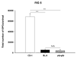
- the efficiency of the method of the invention has been assessed by the cell purification method FACS (Fluorescence Activated Cell Sorting) and with the commercial kit called "Oligodendrocyte Selection Kit” from Heraclitus Biosciences (HB), as described in examples 1-3. Furthermore, the efficiency of the method of the invention has also been compared to the one described in example 4, where OPCs from the rat optical nerve are purified by immunopanning. Table 1. Comparison of the efficiency of the method described by the invention with that of other already described methods (FACS, Kit from Heraclitus Biosciences (Kit HB) and immunopanning). Example Method Efficiency in adult (thousands of cells/animal) Efficiency in perinatal (thousands of cells/animal) 1 Invention 68.5 263 2 FACS 10 - 3 HB kit 7.5 250 4 Immunopanning 2.5 -
- the efficiency of the method of the invention is much higher than that of any method described to date for the purification of OPCs from mature or adult tissue.
- the invention presents a method almost 7 times more efficient than the method of the example, almost 10 times more efficient than the method of example 3 and more than 27 times more efficient than the method of example 4.
- Another aspect of the invention relates to the cell obtained or obtainable by the method of the invention, hereinafter called "cell of the invention".
- a preferred embodiment of this aspect of the invention relates to a cell culture derived from the cell of the invention.
- Another more preferred embodiment relates to a cell line derived from the cell of the invention. More preferably, the cell line has been immortalized by gene transfection.
- the term "immortalization” relates to the transformation process of a cell consisting of an acquisition of said cell of dividing itself without limitation in time, without suffering aging or senescence.
- transfection relates to any technique or process that allows the integration in a series of cells of a live organism of an exogenous gene, or "transgene", and which gives said cells a new biological property. This process is very well known by a person skilled in the art and may be carried out by viral vectors, by chemical or physical methods. Preferably, the method of transfection is viral infection.
- the transgene or exogenous gene relates to DNA normally not resident, nor present in the cell to be transformed. In the particular case of the present invention the DNA codes for a cell immortalization gene.
- Cell immortalization genes can be considered, but without being limited to, the genes that code for: telomerase or the reverse transcriptase of telomerase, myc genes, or viral genes such as SV40T (antigen T of the simian virus 40), EBV (Epstein-Barr virus), adenoviral genes, genes of the Papilomavirus E6 and E7.
- SV40T antigen T of the simian virus 40
- EBV Epstein-Barr virus
- adenoviral genes genes of the Papilomavirus E6 and E7.
- Another aspect of the invention relates to a method of discovery or screening of drugs comprising:
- the term "studying the effect of the chemical substance” comprises, but without being limited to, studying cell survival, cell viability, cell stress, cell proliferation, cell migration, cell differentiation or studies of demyelination, myelination or remyelination.
- EXAMPLE 1 Purification of OPCs of mouse cerebral cortex of 15 days, 2 months or 6 months old.
- mice of strain CD1 were supplied by Charles River Laboratories. All work was carried out according to the Spanish (RD 1201/2005 (BOE 252/34367-91, 2005)) and European (86/ 609/ EEC and 2003/65/EC) legislations on animal handling and use.
- the flasks where the cells were seeded were coated with poly-ornithine at a concentration of 10 mg/l. Before seeding the cells, the poly-ornithine was removed and the flasks were washed to eliminate the remains.
- the animals were sacrificed by cervical dislocation and/or asphyxia with CO 2 and the brains were extracted and placed in a dish with saline solution with calcium and magnesium (HBSS or Hanks' Balanced Salt Solution) in ice.
- HBSS calcium and magnesium
- the brains were incubated in different dilutions of a papain solution (SP, composed of: papain (0.9 mg/ml), L-cysteine (0.2 mg/ml), EDTA (0.2 mg/ml) in HBSS) at 37° C during 5 minutes (see Table 2).
- SP a papain solution
- the remains of meninges and plexuses were finally removed with forceps and with the aid of a stereoscopic microscope.
- the brain cortexes were taken to a new dish with HBSS in ice. Table 2.
- the tissue was mechanically dissociated with the aid of scissors and preincubated for 5 minutes at 37° C.
- the pieces of tissue were incubated in different dilutions of the aforementioned papain solution but in HBSS without calcium or magnesium.
- the dilution and the time of incubation used for each animal age are detailed in Table 2.
- the enzymatic digestion was stopped by the addition of medium with serum and DNase-I (DMEM, Dulbecco's Modified Eagle Medium, with 10% of foetal bovine serum and 1% of DNase-I).
- the cell suspension was incubated, during 5 minutes, at 37° C and it was then centrifuged at 900 rpm, during 10 minutes. The supernatant and the remains of myelin were eliminated and the cells were resuspended in medium with serum.
- the cell suspension was filtered through a 100-micron mesh and was seeded in a final volume of 10 ml of OPCs medium (DMEM with 10% foetal bovine serum (SFB), 10 ng/ml PDGF ⁇ and 1% penicillin and streptomycin) for each flask of 75 cm 2 of surface, previously coated with poly-ornithine, as described above.
- the seeding cell density depends on each stage. The number of animals necessary for each flask is detailed in Table 2.
- the medium was collected with the OPCs in suspension and was filtered through a 40-micron mesh, then centrifuging the cell suspension at 900 rpm during 10 minutes.
- the cells were resuspended in OPCs medium and seeded in petri dishes during 45 minutes at 37° C to eliminate the microglia cells, which quickly adhere to the substrate.
- the cell suspension was collected; it was again filtered through a 40-micron mesh and was centrifuged as before.
- the cells were resuspended and reseeded in OPCs medium and in petri dishes during another 30 minutes at 37° C to remove the microglia cells that may remain.
- This medium enriched in OPCs was collected and centrifuged as described above. The cells were finally resuspended in BS medium (Bottenstein-Sato, for 10 ml of medium: 9.6 ml of DMEM, 100 microlitres of SFB, 100 microlitres of L-Glutamine (200mM), 100 microlitres of Penicillin/Streptomycin, 60 microlitres of 5% BSA, 10 microlitres of transferrin (100mg/ml), 1.86 microlitres of Insulin (50 mg/ml), 1 microlitre of Progesterone (0.62mg/ml), 1 microlitre of putrescine (160 mg/ml), 1 microlitre of T3 3 (mg/ml), 1 microlitre of T4 (4 mg/ml), 1 microlitre of sodium selenite (0.39 ng/ml), counted and seeded in different manners for the study either of proliferation, migration or differentiation to mature oligodendrocytes ( Bribián et al.,
- EXAMPLE 2 Purification of OPCs of 2-month old mouse cerebral cortex by FACS.
- the fluorescence activation cell separation was carried out with plp -GFP genetically modified mice (Spassky et al, 1998; Bribián et al, 2006, Merchán et al, 2007).
- the animals were sacrificed as in example 1 and the brains were extracted in the same manner.
- the meninges and the choroid plexuses were removed as in example 1 and the brain cortexes were mechanically dissected with the "Neural Tissue Dissociation Kit” from Miltenyi Biotec, according to the manufacturer's instructions.
- the remains of myelin were eliminated by filtering the cell suspension through a 40-micron mesh followed by centrifuging in sucrose (0.9 M) in HBSS in ice.
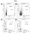
- the cells were stained with anti-A2B5 antibody conjugated with PE and they were analysed in a FACSAria apparatus from BD.
- the cells were separated in HBSS with 2% foetal bovine serum, 25 mM HEPES and 5mM EDTA.
- EXAMPLE 3 Purification of OPCs of 2-month old mouse cerebral cortex by the "Oligodendrocyte Selection Kit "from HB.
- this kit is described by the manufacturer for the obtainment of immature dendrocytes with O4 + /GalC - phenotype from P3 rat or mice brains, i.e. 3 days old, which we have previously called perinatal stage.
- the animals were sacrificed as in example 1 and the brains were extracted in the same manner. Then, the manufacturer's instructions detailed in the user manual were followed.
- EXAMPLE 4 Purification of OPCs of rat optical nerved by immunopanning.
- This method is disclosed in patent application PCT/US1996/013279 and consists of the purification of the OPCs of adult rat optical nerve from a mixed suspension of cells from the optical nerve.
- This cell suspension is first placed in contact with a plate where an anti-Thy1 antibody had previously been absorbed, which enables the removal of non-glial cells.
- the cell suspension is then placed in contact with a second plate previously absorbed with anti-A2B5n antibody, anti-NG2 antibody or with peanut agglutinin, which is going to permit that only the cells with these proteins on their surface are absorbed in the plate.
- These cells will mainly be OPCs although, in the case of A2B5, they can also be O-2A precursors.
- the efficiency of this method is of 60,000 OPCs for each gram of fresh tissue used in the case of tumour resection margins and 15,000 OPCs for each gram of fresh tissue from injuries.
Landscapes
- Health & Medical Sciences (AREA)
- Engineering & Computer Science (AREA)
- Biomedical Technology (AREA)
- Life Sciences & Earth Sciences (AREA)
- Organic Chemistry (AREA)
- Zoology (AREA)
- Chemical & Material Sciences (AREA)
- Biotechnology (AREA)
- Wood Science & Technology (AREA)
- Bioinformatics & Cheminformatics (AREA)
- Genetics & Genomics (AREA)
- General Health & Medical Sciences (AREA)
- Cell Biology (AREA)
- Neurology (AREA)
- Neurosurgery (AREA)
- Biochemistry (AREA)
- General Engineering & Computer Science (AREA)
- Microbiology (AREA)
- Developmental Biology & Embryology (AREA)
- Micro-Organisms Or Cultivation Processes Thereof (AREA)
- Preparation Of Compounds By Using Micro-Organisms (AREA)
- Medicines Containing Material From Animals Or Micro-Organisms (AREA)
- Peptides Or Proteins (AREA)
- Medicines That Contain Protein Lipid Enzymes And Other Medicines (AREA)
Applications Claiming Priority (2)
| Application Number | Priority Date | Filing Date | Title |
|---|---|---|---|
| ES201030493A ES2375860B8 (es) | 2010-03-31 | 2010-03-31 | Método para la obtención de células precursoras de oligodendrocitos. |
| PCT/ES2011/070191 WO2011121158A2 (es) | 2010-03-31 | 2011-03-22 | Método para la obtención de células precursoras de oligodendrocitos |
Publications (1)
| Publication Number | Publication Date |
|---|---|
| EP2554663A2 true EP2554663A2 (en) | 2013-02-06 |
Family
ID=44484795
Family Applications (1)
| Application Number | Title | Priority Date | Filing Date |
|---|---|---|---|
| EP11721797A Withdrawn EP2554663A2 (en) | 2010-03-31 | 2011-03-22 | Method for obtaining oligodendrocyte precursor cells |
Country Status (3)
| Country | Link |
|---|---|
| EP (1) | EP2554663A2 (es) |
| ES (1) | ES2375860B8 (es) |
| WO (1) | WO2011121158A2 (es) |
Cited By (1)
| Publication number | Priority date | Publication date | Assignee | Title |
|---|---|---|---|---|
| CN115340982A (zh) * | 2022-08-23 | 2022-11-15 | 徐州医科大学 | 一种分离并纯化新生小鼠皮质区少突胶质前体细胞的方法 |
Family Cites Families (4)
| Publication number | Priority date | Publication date | Assignee | Title |
|---|---|---|---|---|
| WO1997007200A1 (en) | 1995-08-16 | 1997-02-27 | The Board Of Trustees Of The Leland Stanford Junior University | Adult oligodendrocyte precursor cell compositions and methods |
| AR046076A1 (es) * | 2003-07-18 | 2005-11-23 | Otsuka Pharma Co Ltd | Procedimiento para obtener una poblacion homogenea de celulas precursoras de oligodendrocitos y poblacion obtenida |
| EP1684706A4 (en) * | 2003-11-10 | 2008-01-09 | Cornell Res Foundation Inc | GENES DIFFERENTIALLY EXPRESSED BY INSULATED PRECURSOR CELLS OF HUMAN WHITE MATTER |
| US20060183147A1 (en) * | 2005-02-11 | 2006-08-17 | Elan Pharmaceuticals, Inc. | Methods of identifying genes which modulate myelination |
-
2010
- 2010-03-31 ES ES201030493A patent/ES2375860B8/es not_active Expired - Fee Related
-
2011
- 2011-03-22 WO PCT/ES2011/070191 patent/WO2011121158A2/es not_active Ceased
- 2011-03-22 EP EP11721797A patent/EP2554663A2/en not_active Withdrawn
Non-Patent Citations (1)
| Title |
|---|
| See references of WO2011121158A2 * |
Cited By (1)
| Publication number | Priority date | Publication date | Assignee | Title |
|---|---|---|---|---|
| CN115340982A (zh) * | 2022-08-23 | 2022-11-15 | 徐州医科大学 | 一种分离并纯化新生小鼠皮质区少突胶质前体细胞的方法 |
Also Published As
| Publication number | Publication date |
|---|---|
| WO2011121158A3 (es) | 2011-12-08 |
| ES2375860B1 (es) | 2013-01-25 |
| ES2375860B8 (es) | 2013-06-10 |
| WO2011121158A2 (es) | 2011-10-06 |
| ES2375860A1 (es) | 2012-03-07 |
Similar Documents
| Publication | Publication Date | Title |
|---|---|---|
| JP4621410B2 (ja) | 神経細胞へ分化しうる細胞分画の調製方法及び神経変性疾患治療薬の製造方法 | |
| JP6673966B2 (ja) | 神経変性を治療するための方法及び組成物 | |
| US6638763B1 (en) | Isolated mammalian neural stem cells, methods of making such cells | |
| JP6346629B2 (ja) | 哺乳動物由来グリア制限前駆細胞を増大、同定、特徴付けおよび能力増強するための方法および組成物 | |
| US20150087058A1 (en) | Stem cell culture medium and method for culturing stem cells using same | |
| JP2004511266A (ja) | 間葉間質細胞に対する治療的利用法 | |
| Westerlund et al. | Endoscopically harvested stem cells: a putative method in future autotransplantation | |
| Deshpande et al. | Isolation of neural stem cells from whole brain tissues of adult mice | |
| WO2004108907A1 (ja) | Es細胞の電気パルス処理によって得られた神経細胞 | |
| Lu et al. | Retrovirus delivered neurotrophin-3 promotes survival, proliferation and neuronal differentiation of human fetal neural stem cells in vitro | |
| Zhu et al. | Isolation and Long‐Term Expansion of Functional, Myelinating Oligodendrocyte Progenitor Cells from Neonatal Rat Brain | |
| CN112154203B (zh) | 高效分离和培养神经干细胞的方法 | |
| EP2554663A2 (en) | Method for obtaining oligodendrocyte precursor cells | |
| Poduslo et al. | Purification and characterization of cultures of oligodendroglia from rat brain | |
| KR101910269B1 (ko) | 인간 뇌 조직 유래 신경줄기세포의 고효율 분리 방법 | |
| KR101738715B1 (ko) | 식물 줄기세포 또는 식물 역분화 줄기 세포의 추출물을 이용한 맞춤형 만능줄기세포의 유도 방법 및 상기 방법에 의해 제조된 만능줄기세포 | |
| Beythien et al. | Bridging the gap: Canine in vitro models of the central nervous system as tools to study pathogenetic mechanisms in neurodegenerative disease and neuroinfectiology | |
| Tao et al. | Culture and identification of monoclonal neural stem cells derived from cerebral cortex | |
| WO2003038074A1 (fr) | Procede d'induction de differenciation de cellules souches de mesoblastes ou de cellules es dans des cellules du systeme nerveux | |
| WO2002014479A2 (en) | Isolated mammalian neural stem cells, methods of making such cells, and methods of using such cells | |
| Wu et al. | In vitro culture and characterization of oligodendrocyte precursor cells derived from neonatal rats | |
| WO2004013315A1 (en) | Method for the preparation, purification and differentiation of neurospheres from mammalian stem cells | |
| US20040115807A1 (en) | O-2a progenitors multipotent cells from neurohypophysis | |
| US20050118143A1 (en) | Isolated mammalian neural stem cells, methods of making such cells, and methods of using such cells | |
| Meas | Regeneration of Neurons in the Inner Ear by Reprogramming Spiral Ganglion Glial Cells |
Legal Events
| Date | Code | Title | Description |
|---|---|---|---|
| PUAI | Public reference made under article 153(3) epc to a published international application that has entered the european phase |
Free format text: ORIGINAL CODE: 0009012 |
|
| 17P | Request for examination filed |
Effective date: 20121031 |
|
| AK | Designated contracting states |
Kind code of ref document: A2 Designated state(s): AL AT BE BG CH CY CZ DE DK EE ES FI FR GB GR HR HU IE IS IT LI LT LU LV MC MK MT NL NO PL PT RO RS SE SI SK SM TR |
|
| DAX | Request for extension of the european patent (deleted) | ||
| 17Q | First examination report despatched |
Effective date: 20140122 |
|
| STAA | Information on the status of an ep patent application or granted ep patent |
Free format text: STATUS: THE APPLICATION IS DEEMED TO BE WITHDRAWN |
|
| 18D | Application deemed to be withdrawn |
Effective date: 20140603 |