EP2848252A2 - Inhibiteurs d'aldose-réductase pour le traitement de l'uvéite - Google Patents
Inhibiteurs d'aldose-réductase pour le traitement de l'uvéite Download PDFInfo
- Publication number
- EP2848252A2 EP2848252A2 EP20140185108 EP14185108A EP2848252A2 EP 2848252 A2 EP2848252 A2 EP 2848252A2 EP 20140185108 EP20140185108 EP 20140185108 EP 14185108 A EP14185108 A EP 14185108A EP 2848252 A2 EP2848252 A2 EP 2848252A2
- Authority
- EP
- European Patent Office
- Prior art keywords
- cells
- inhibition
- aldose reductase
- induced
- tnf
- Prior art date
- Legal status (The legal status is an assumption and is not a legal conclusion. Google has not performed a legal analysis and makes no representation as to the accuracy of the status listed.)
- Withdrawn
Links
Images











Classifications
-
- A—HUMAN NECESSITIES
- A61—MEDICAL OR VETERINARY SCIENCE; HYGIENE
- A61K—PREPARATIONS FOR MEDICAL, DENTAL OR TOILETRY PURPOSES
- A61K31/00—Medicinal preparations containing organic active ingredients
- A61K31/33—Heterocyclic compounds
- A61K31/395—Heterocyclic compounds having nitrogen as a ring hetero atom, e.g. guanethidine or rifamycins
- A61K31/535—Heterocyclic compounds having nitrogen as a ring hetero atom, e.g. guanethidine or rifamycins having six-membered rings with at least one nitrogen and one oxygen as the ring hetero atoms, e.g. 1,2-oxazines
- A61K31/5375—1,4-Oxazines, e.g. morpholine
- A61K31/538—1,4-Oxazines, e.g. morpholine ortho- or peri-condensed with carbocyclic ring systems
-
- A—HUMAN NECESSITIES
- A61—MEDICAL OR VETERINARY SCIENCE; HYGIENE
- A61K—PREPARATIONS FOR MEDICAL, DENTAL OR TOILETRY PURPOSES
- A61K31/00—Medicinal preparations containing organic active ingredients
- A61K31/33—Heterocyclic compounds
- A61K31/395—Heterocyclic compounds having nitrogen as a ring hetero atom, e.g. guanethidine or rifamycins
- A61K31/41—Heterocyclic compounds having nitrogen as a ring hetero atom, e.g. guanethidine or rifamycins having five-membered rings with two or more ring hetero atoms, at least one of which being nitrogen, e.g. tetrazole
- A61K31/4164—1,3-Diazoles
-
- A—HUMAN NECESSITIES
- A61—MEDICAL OR VETERINARY SCIENCE; HYGIENE
- A61K—PREPARATIONS FOR MEDICAL, DENTAL OR TOILETRY PURPOSES
- A61K31/00—Medicinal preparations containing organic active ingredients
- A61K31/33—Heterocyclic compounds
- A61K31/395—Heterocyclic compounds having nitrogen as a ring hetero atom, e.g. guanethidine or rifamycins
- A61K31/41—Heterocyclic compounds having nitrogen as a ring hetero atom, e.g. guanethidine or rifamycins having five-membered rings with two or more ring hetero atoms, at least one of which being nitrogen, e.g. tetrazole
- A61K31/4164—1,3-Diazoles
- A61K31/4166—1,3-Diazoles having oxo groups directly attached to the heterocyclic ring, e.g. phenytoin
-
- A—HUMAN NECESSITIES
- A61—MEDICAL OR VETERINARY SCIENCE; HYGIENE
- A61K—PREPARATIONS FOR MEDICAL, DENTAL OR TOILETRY PURPOSES
- A61K31/00—Medicinal preparations containing organic active ingredients
- A61K31/33—Heterocyclic compounds
- A61K31/395—Heterocyclic compounds having nitrogen as a ring hetero atom, e.g. guanethidine or rifamycins
- A61K31/41—Heterocyclic compounds having nitrogen as a ring hetero atom, e.g. guanethidine or rifamycins having five-membered rings with two or more ring hetero atoms, at least one of which being nitrogen, e.g. tetrazole
- A61K31/4164—1,3-Diazoles
- A61K31/4188—1,3-Diazoles condensed with other heterocyclic ring systems, e.g. biotin, sorbinil
-
- A—HUMAN NECESSITIES
- A61—MEDICAL OR VETERINARY SCIENCE; HYGIENE
- A61K—PREPARATIONS FOR MEDICAL, DENTAL OR TOILETRY PURPOSES
- A61K31/00—Medicinal preparations containing organic active ingredients
- A61K31/33—Heterocyclic compounds
- A61K31/395—Heterocyclic compounds having nitrogen as a ring hetero atom, e.g. guanethidine or rifamycins
- A61K31/495—Heterocyclic compounds having nitrogen as a ring hetero atom, e.g. guanethidine or rifamycins having six-membered rings with two or more nitrogen atoms as the only ring heteroatoms, e.g. piperazine or tetrazines
- A61K31/499—Spiro-condensed pyrazines or piperazines
-
- A—HUMAN NECESSITIES
- A61—MEDICAL OR VETERINARY SCIENCE; HYGIENE
- A61K—PREPARATIONS FOR MEDICAL, DENTAL OR TOILETRY PURPOSES
- A61K31/00—Medicinal preparations containing organic active ingredients
- A61K31/33—Heterocyclic compounds
- A61K31/395—Heterocyclic compounds having nitrogen as a ring hetero atom, e.g. guanethidine or rifamycins
- A61K31/495—Heterocyclic compounds having nitrogen as a ring hetero atom, e.g. guanethidine or rifamycins having six-membered rings with two or more nitrogen atoms as the only ring heteroatoms, e.g. piperazine or tetrazines
- A61K31/50—Pyridazines; Hydrogenated pyridazines
-
- A—HUMAN NECESSITIES
- A61—MEDICAL OR VETERINARY SCIENCE; HYGIENE
- A61K—PREPARATIONS FOR MEDICAL, DENTAL OR TOILETRY PURPOSES
- A61K31/00—Medicinal preparations containing organic active ingredients
- A61K31/33—Heterocyclic compounds
- A61K31/395—Heterocyclic compounds having nitrogen as a ring hetero atom, e.g. guanethidine or rifamycins
- A61K31/495—Heterocyclic compounds having nitrogen as a ring hetero atom, e.g. guanethidine or rifamycins having six-membered rings with two or more nitrogen atoms as the only ring heteroatoms, e.g. piperazine or tetrazines
- A61K31/50—Pyridazines; Hydrogenated pyridazines
- A61K31/502—Pyridazines; Hydrogenated pyridazines ortho- or peri-condensed with carbocyclic ring systems, e.g. cinnoline, phthalazine
-
- A—HUMAN NECESSITIES
- A61—MEDICAL OR VETERINARY SCIENCE; HYGIENE
- A61K—PREPARATIONS FOR MEDICAL, DENTAL OR TOILETRY PURPOSES
- A61K31/00—Medicinal preparations containing organic active ingredients
- A61K31/33—Heterocyclic compounds
- A61K31/395—Heterocyclic compounds having nitrogen as a ring hetero atom, e.g. guanethidine or rifamycins
- A61K31/535—Heterocyclic compounds having nitrogen as a ring hetero atom, e.g. guanethidine or rifamycins having six-membered rings with at least one nitrogen and one oxygen as the ring hetero atoms, e.g. 1,2-oxazines
-
- A—HUMAN NECESSITIES
- A61—MEDICAL OR VETERINARY SCIENCE; HYGIENE
- A61P—SPECIFIC THERAPEUTIC ACTIVITY OF CHEMICAL COMPOUNDS OR MEDICINAL PREPARATIONS
- A61P11/00—Drugs for disorders of the respiratory system
- A61P11/06—Antiasthmatics
-
- A—HUMAN NECESSITIES
- A61—MEDICAL OR VETERINARY SCIENCE; HYGIENE
- A61P—SPECIFIC THERAPEUTIC ACTIVITY OF CHEMICAL COMPOUNDS OR MEDICINAL PREPARATIONS
- A61P27/00—Drugs for disorders of the senses
-
- A—HUMAN NECESSITIES
- A61—MEDICAL OR VETERINARY SCIENCE; HYGIENE
- A61P—SPECIFIC THERAPEUTIC ACTIVITY OF CHEMICAL COMPOUNDS OR MEDICINAL PREPARATIONS
- A61P27/00—Drugs for disorders of the senses
- A61P27/02—Ophthalmic agents
-
- A—HUMAN NECESSITIES
- A61—MEDICAL OR VETERINARY SCIENCE; HYGIENE
- A61P—SPECIFIC THERAPEUTIC ACTIVITY OF CHEMICAL COMPOUNDS OR MEDICINAL PREPARATIONS
- A61P29/00—Non-central analgesic, antipyretic or antiinflammatory agents, e.g. antirheumatic agents; Non-steroidal antiinflammatory drugs [NSAID]
-
- C—CHEMISTRY; METALLURGY
- C12—BIOCHEMISTRY; BEER; SPIRITS; WINE; VINEGAR; MICROBIOLOGY; ENZYMOLOGY; MUTATION OR GENETIC ENGINEERING
- C12N—MICROORGANISMS OR ENZYMES; COMPOSITIONS THEREOF; PROPAGATING, PRESERVING, OR MAINTAINING MICROORGANISMS; MUTATION OR GENETIC ENGINEERING; CULTURE MEDIA
- C12N15/00—Mutation or genetic engineering; DNA or RNA concerning genetic engineering, vectors, e.g. plasmids, or their isolation, preparation or purification; Use of hosts therefor
- C12N15/09—Recombinant DNA-technology
- C12N15/11—DNA or RNA fragments; Modified forms thereof; Non-coding nucleic acids having a biological activity
- C12N15/113—Non-coding nucleic acids modulating the expression of genes, e.g. antisense oligonucleotides; Antisense DNA or RNA; Triplex- forming oligonucleotides; Catalytic nucleic acids, e.g. ribozymes; Nucleic acids used in co-suppression or gene silencing
- C12N15/1137—Non-coding nucleic acids modulating the expression of genes, e.g. antisense oligonucleotides; Antisense DNA or RNA; Triplex- forming oligonucleotides; Catalytic nucleic acids, e.g. ribozymes; Nucleic acids used in co-suppression or gene silencing against enzymes
-
- C—CHEMISTRY; METALLURGY
- C12—BIOCHEMISTRY; BEER; SPIRITS; WINE; VINEGAR; MICROBIOLOGY; ENZYMOLOGY; MUTATION OR GENETIC ENGINEERING
- C12N—MICROORGANISMS OR ENZYMES; COMPOSITIONS THEREOF; PROPAGATING, PRESERVING, OR MAINTAINING MICROORGANISMS; MUTATION OR GENETIC ENGINEERING; CULTURE MEDIA
- C12N2310/00—Structure or type of the nucleic acid
- C12N2310/10—Type of nucleic acid
- C12N2310/14—Type of nucleic acid interfering nucleic acids [NA]
-
- C—CHEMISTRY; METALLURGY
- C12—BIOCHEMISTRY; BEER; SPIRITS; WINE; VINEGAR; MICROBIOLOGY; ENZYMOLOGY; MUTATION OR GENETIC ENGINEERING
- C12Y—ENZYMES
- C12Y101/00—Oxidoreductases acting on the CH-OH group of donors (1.1)
- C12Y101/01—Oxidoreductases acting on the CH-OH group of donors (1.1) with NAD+ or NADP+ as acceptor (1.1.1)
- C12Y101/01021—Aldehyde reductase (1.1.1.21), i.e. aldose-reductase
Definitions
- Embodiments of this invention are related generally to physiology and medicine. More specifically, this invention is related to aldose reductase inhibitors (ARIs) and their use in treating and ameliorating inflammation.
- ARIs aldose reductase inhibitors
- Aldose reductase catalyzes the reduction of a wide range of aldehydes (Bhatnager and Srivastava, 1992).
- the substrates of the enzyme range from aromatic and aliphatic aldehydes to aldoses such as glucose, galactose, and ribose.
- the reduction of glucose by AR is particularly significant during hyperglycemia and increased flux of glucose via AR has been etiologically linked to the development of secondary diabetic complications (Bhatnager and Srivastava, 1992; Yabe-Nishimura, 1998).
- AR is upregulated by oxidants such as hydrogen peroxide (Spycher et al ., 1997), lipid peroxidation-derived aldehydes (Ruef et al ., 2000; Rittner et al ., 1999), advanced glycosylation end products (Nakamura et al ., 2000) and nitric oxide (Seo et al ., 2000).
- oxidants such as hydrogen peroxide (Spycher et al ., 1997), lipid peroxidation-derived aldehydes (Ruef et al ., 2000; Rittner et al ., 1999), advanced glycosylation end products (Nakamura et al ., 2000) and nitric oxide (Seo et al ., 2000).
- the expression of the enzyme is also increased under several pathological conditions associated with increased oxidative or electrophilic stress such as iron overload (Barisani et al ., 2000), alcoholic liver disease (O'Connor et al ., 1999), heart failure (Yang et al ., 2000), myocardial ischemia (Shinmura et al ., 2000), vascular inflammation (Rittner et al ., 1999) and restenosis (Ruef et al ., 2000), and various forms of cancer.
- iron overload Barisani et al ., 2000
- alcoholic liver disease O'Connor et al ., 1999
- heart failure Yang et al ., 2000
- myocardial ischemia Shinmura et al ., 2000
- vascular inflammation Rostner et al ., 1999
- restenosis Ruef et al ., 2000
- Inhibitors of aldose reductase have been indicated for some conditions and diseases, such as diabetes complications, ischemic damage to non-cardiac tissue, Huntington's disease. See U.S. Patents 6,696,407 , 6,127,367 , 6,380,200 , which are all hereby incorporated by reference. In some cases, the role in which aldose reductase plays in mechanisms involved in the condition or disease are known. For example, in U.S. Patent 6,696,407 indicates that an aldose reductase inhibitors increase striatal ciliary neurotrophic factor (CNTF), which has ramifications for the treatment of Huntington's Disease. In other cases, however, the way in which aldose reductase or aldose reductase inhibitors work with respect to a particular disease or condition are not known.
- CNTF striatal ciliary neurotrophic factor
- Embodiments of the invention are directed to methods of preventing or reducing uveitis or conjuctivitis in a subject comprising administering to a subject diagnosed with or at risk of developing uveitis or conjunctivitis an amount of a pharmaceutically acceptable composition comprising an aldose reductase inhibitor (ARI) sufficient to prevent, ameliorate, or attenuate uveitis or conjunctivitis.
- a pharmaceutically acceptable composition comprising an aldose reductase inhibitor (ARI) sufficient to prevent, ameliorate, or attenuate uveitis or conjunctivitis.
- the composition may be administered 1, 2, 3, 4, 5, 6, or more times and may be administered over 1, 2, 3, 4, 5, 6, 7, or more minutes, hours, days or weeks.
- the aldose reductase inhibitor is administered to the patient as a prodrug.
- prodrug is an inactive or less active form of a drug that is metabolized or converted in vivo to an active or more active form.
- ARI compositions can be administered directly, locally, topically, orally, ocularly, endoscopically, intratracheally, intravitreously, intrabronchially, intratumorally, intravenously, intralesionally, intramuscularly, intraperitoneally, regionally, percutaneously, or subcutaneously.
- the ARI is administered orally or by inhalation or instillation, e.g ., by inhaler or other aersol delivery devices.
- inventions are directed to methods of preventing or reducing asthma associated inflammation in a subject comprising administering to a subject diagnosed with or at risk of developing an inflammatory condition associated with asthma an amount of a pharmaceutically acceptable composition comprising an aldose reductase inhibitor sufficient to prevent, ameliorate, or attenuate inflammation.
- the aldose reductase inhibitor is a peptide, a peptide mimetic, a small molecule, or an inhibitory RNA.
- the aldose reductase inhibitor can be a siRNA or other inhibitory nucleic acid, a carboxylic acid, a hydantoins, a pyridazinone, or a pharmaceutically acceptable derivative thereof.
- the aldose reductase inhibitor is sorbinil, epalrestat, ponalrestat, methosorbinil, risarestat, imirestat, ALO-1567, quercetin, zopolrestat, AD-5467, NZ-314, M-16209, minalrestat, AS-3201, WP-921, luteolin, tolrestat, EBPC, fidarestat, siRNA, or a pharmaceutically acceptable derivatives thereof.
- the aldose reductase inhibitor is sorbinil or zopolrestat.
- the aldose reductase inhibitor can be administered at a dose of 0.1, 1, 5, 10, 20, 40, 50, 100, 200, 400, 800, 1200 to 1500 ng/day, mg/day, ng/kg/day, or mg/kg/day, including all ranges and values there between.
- compositions and kits of the invention can be used to achieve methods of the invention.
- the words “comprising” (and any form of comprising, such as “comprise” and “comprises”), “having” (and any form of having, such as “have” and “has”), "including” (and any form of including, such as “includes” and “include”) or “containing” (and any form of containing, such as “contains” and “contain”) are inclusive or open-ended and do not exclude additional, unrecited elements or method steps.
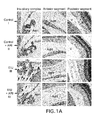
- FIGs. 1A-1B Inhibition of AR prevents LPS-induced inflammatory cell infiltration and protein concentration in aqueous humour.
- FIG. 1A Histo-pathological changes in the anterior chamber of EIU rat eyes in the absence and presence of Sorbinil or zopolrestat. Serial sections of para-formaldehyde- fixed rat eyes were stained with Hematoxylin and Eosin (H&E) and observed under light microscope (Magnification 200X).
- FIG. 1B I The inflammatory cells and ( FIG. 1B II ) total protein concentration in the AqH were measured using trypan-blue exclusion cell counting and Bradford methods, respectively as described in the Methods.
- AqH Aqueous humor
- CB ciliary body
- I Iris
- C cornea
- R Retina
- V Vitrious.
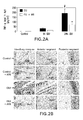
- FIGs. 2A -2B Inhibition of AR prevents TNF- ⁇ secretion in LPS.
- AqH Aqueous humor
- CB Ciliary body
- I Iris
- C Cornea
- V Vitreous
- R Retina.
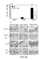
- FIGs. 3A-3B Inhibition of AR prevents NO and PGE 2 secretion in EIU.
- AqH aqueous humor
- C Cornea
- CB ciliary body
- I Iris
- V Vitreous
- R Retina.
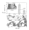
- FIGs. 4A -4B Inhibition of AR prevents AR expression and activation of NF- ⁇ B in EIU.
- FIG. 5 Inhibition of AR prevents ROS generation in EIU.
- AqH Aqueous humor
- I Iris
- CB Ciliary body
- C Cornea
- R Retina.
- FIGs. 6A -6B Effect of inhibition of AR on the LPS-induced inflammatory response in human monocytic cells.
- FIG. 6A Growth arrested U-937 cells without or with zopolrestat (10 ⁇ M each) were incubated with 1 ⁇ g/ml of LPS for 24 h. The expression of Cox-2 and iNOS proteins was determined by western blot analysis using specific antibodies as described in the Methods.
- FIG. 6B U-937 cells were transiently transfected with pNF- ⁇ B-SEAP reporter vector. The cells treated without or with sorbinil and zopolrestat (10 ⁇ M each) were incubated with 1 ⁇ g/ml of LPS.
- FIG. 7 Role of aldose reductase (AR) in inflammatory signals.
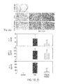
- FIGs. 8A -8B AR inhibition prevents accumulation of eosinophils in airways and in sub-epithelial regions (inset) induced by Ragweed Pollen extract in mice model of asthma.
- FIG. 8A The lungs were lavaged with ice-cold PBS and cells were counted and (inset) differential cell counts were performed on cytocentrifuge preparations stained with hematoxylin and eosin.
- FIG. 8B Perivascular and peribronchial inflammation and cell composition in the BAL were evaluated by a pathologist blinded to treatment groups to obtain data for each lung.
- FIGs. 9A -9B AR inhibition prevents Ragweed Pollen extract-induced ( FIG. 9A ) accumulation of Muc5ac levels in the BAL and ( FIG. 9B ) hyperresponsiveness in mice model of asthma.
- FIG. 9A MUC5AC levels in the BAL assessed by ELISA using anti-MUC5AC monoclonal antibody and (inset in A) mucin productions in the epithelial cells was assessed by periodic acid Schiff (PAS)-staining of lung sections.
- FIG. 9B Airway responsiveness was measured in unrestrained, conscious mice 3 days after the last challenge. Mice will be placed in a barometric plethysmographic chamber, and baseline readings will be taken and averaged for 3 min.
- Aerosolized methacholine in increasing concentrations were nebulized through an inlet of the main chamber for 3 min. Readings were taken and averaged for 3 min after each nebulization and enhanced pause (Penh) was determined. Penh, calculated as (expiratory time/relaxation time - 1) x (peak expiratory flow/peak inspiratory flow) according to the manufacturers' protocol. Penh was used as a measure of airway responsiveness to methacholine.
- FIGs. 10A -10B Effect of AR inhibition on ( FIG. 10A ) Th2 cytokine production and ( FIG. 10B ) inflammatory cell (eosinophils) infiltration in ovalbumin (OVA)-induced murine model of asthma.
- FIG. 10B Fixed lungs from the different experimental groups were sectioned, stained with hematoxylin and eosin. Arrows indicate sites of inflammatory cell infiltration.
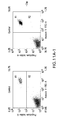
- FIGs. 11A-11B Inhibition of AR prevents RW-induced apoptosis and cell death in SAEC.
- FIG. 11A Growth-arrested SAEC, pretreated without or with zopolrestat (20 ⁇ M), were incubated with 150 ⁇ g/ml of RW for 18 h to induce apoptotic cell death. The cells were stained with annexin-V FITC (FL-1) and propidium iodide (PI) (FL-2). R1 denotes dead cells (PI positive) and R2 represents early apoptotic cells (annexin-V positive) and R1+R2 represents total dead cells.
- DHE dihydroethidium
- FIGs. 13A -13C Inhibition of AR prevents RW-induced secretion of inflammatory markers IL-6, IL-8 and PGE2 in SAEC.
- FIGs. 14A -14C Genetic ablation of AR prevents RW-induced secretion of inflammatory markers IL-6, IL-8 and PGE2.
- FIG. 15 Inhibition of AR prevents RW-induced expression of IL-6 and IL-8 and COX-2 mRNA in SAEC.
- Approximately 2x10 5 SAEC were seeded in 6-well plates and incubated until 80% confluency. The cells were starved in serum-free basal medium with or without zopolrestat for 24 h. The cells were treated with RW (50 ⁇ g/ml) for 6 h.
- Total RNA was extracted as described in the methods and IL-6, IL-8 and Cox-2 mRNA expression was determined using Qiagen RT-PCR kit. ⁇ -Actin was used as loading control.
- FIGs. 16A -16C Inhibition of AR prevents RW-induced expression and activation of inflammatory, apoptosis and cell cycle proteins in SAEC.
- Approximately 2x10 5 SAEC were seeded in 6-well plates and incubated until 80% confluency. The cells were starved in serum-free basal medium with or without zopolrestat for 24 h. The cells were treated with RW (50 ⁇ g/ml) for 24 h.
- FIGs. 17A -17B AR inhibition attenuates the activation of redox-sensitive transcription factors NF-kB and AP-1 SAEC.
- FIG. 17A For EMSA, approximately 2x10 6 SAEC were seeded in T-150 cm 2 flasks and incubated until 80% confluency. The cells were starved in serum-free basal medium with or without zopolrestat for 24 h. The cells were treated with RW (50 ⁇ g/ml) for 3 h. Nuclear extract was prepared as described in the methods and EMSA was performed to determine the activation of NF- ⁇ B and AP-1. Lanes: 1, Control; 2, Ragweed; 3, Control+ Zop; 4, RW+ Zop. ( FIG. 17B ).
- Approximately, 1x10 5 SAEC were plated in 24 well plate and growth-arrested by preincubatin in serum-free basal medium with AR inhibitors or carrier for 24 h followed by transfection with NF- ⁇ B-pSEAP vector or control (pTAL) vector. After 6 h, transfected cells were incubated with RW (50 ⁇ g/ml) for 48 hours. Medium was collected and NF- ⁇ B-dependent reporter SEAP activity was measured by chemiluminescence method essentially as described by the manufacturer. Bars represent Mean ⁇ SD (n 4). #p ⁇ 0.001 Vs. Control; *p ⁇ 0.01 vs RW.
- FIGs. 18A -18B AR inhibition prevents RW-induced accumulation of eosinophils in airways in mice model of asthma.
- FIG. 18A The BALB/c mice were sensitized and challenged with RW, subsequently lungs were lavaged with ice-cold PBS and cells were counted and differential cell counts were performed on cytocentrifuge preparations stained with hematoxylin and eosin (inset in FIG. 18A).
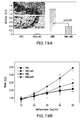
- FIG. 19A MUC5AC levels in the BAL were assessed by ELISA using anti-MUC5AC monoclonal antibody and mucin productions in the epithelial cells was assessed by periodic acid Schiff (PAS)-staining of lung sections (inset in FIG. 19A).
- FIG. 19B Airway responsiveness was measured in unrestrained, conscious mice 3 days after the last challenge. Mice were placed in a barometric plethysmographic chamber, and baseline readings were taken and averaged for 3 min.
- Aerosolized methacholine in increasing concentrations were nebulized through an inlet of the main chamber for 3 min. Readings were taken and averaged for 3 min after each nebulization and enhanced pause (Penh) was determined and plotted against the increasing concentration of methacholine as a measure of airway responsiveness to methacholine. Penh, calculated as (expiratory time/relaxation time -1 ) x (peak expiratory flow/peak inspiratory flow) according to the manufacturers' protocol.
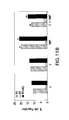
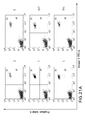
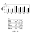
- FIGs. 21A -21B Inhibition of AR prevents TNF- ⁇ - and LPS-induced apoptosis and cell death in SAEC.
- FIG. 21A Approximately 2x10 5 SAEC were seeded on 6 well plates (4 well per group) and growth arrested with sorbinil (20 ⁇ M) or carrier (DMSO). Subsequently, the cells were incubated with TNF- ⁇ or LPS for 18 h. The cells were harvested, pooled together and stained with annexin V-FITC (FL-1) and propidium iodide (PI) (FL-2). R1 represents dead cells (PI positive) and R2 denotes early apoptotic cells (annexin-V positive) and R1+R2 represents total dead cells.
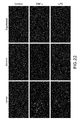
- FIG. 22 Inhibition of AR prevents TNF- ⁇ - and LPS-induced ROS generation in SAEC.
- Approximately 1x10 5 cells were seeded on chambered slides and after the cells attached they were starved in serum free basal medium with AR inhibitor or carrier (DMSO) for overnight.
- the cells were treated with TNF and LPS or carrier (PBS) for 16 h.
- the cells were washed with cold PBS (pH 7.2) and stained with ROS-sensitive dye dihydroethidium (DHE) for 15 min at 37° C.
- DHE ROS-sensitive dye dihydroethidium
- the cells were washed again and mounted with floursave (with DAPI) mounting medium.
- a fluorescence microscope Nakon
- FIG. 23 AR inhibition prevents TNF- ⁇ - and LPS-induced secretion of inflammatory markers IL-6, IL-8 and PGE2.
- IL-6, IL-8 and PGE2 inflammatory markers
- Approximately 2x10 5 cells were seed in 6-well plates and incubated until 80% confluence. Medium was replaced with basal medium with AR inhibitor or carrier for overnight. The cells were treated with TNF- ⁇ (2 nM) and LPS (1 ⁇ g/ml) and incubated for 24h. Medium was harvested, centrifuged and supernatant was used for determination of IL-6, IL-8 and PGE2 with respective ELISA kits following supplier's protocol.
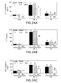
- FIGs. 24A -24C AR ablation by siRNA prevents TNF- ⁇ - and LPS-induced secretion of inflammatory markers IL-6, IL-8 and PGE2.
- the SAEC ⁇ 2x10 5
- the SAEC were seeded in 6-well plates and incubated until 80% confluence. Medium was replaced with basal medium and cells were transfected with either AR-siRNA or scrambled-siRNA. After 48 h cells were incubated with TNF- ⁇ (2 nM) or LPS (1 ⁇ g/ml) for additional 24 h. At the end of incubation medium was harvested, centrifuged and supernatant was used for determination of IL-6, IL-8 and PGE2 with respective ELISA kits following supplier's protocol.
- FIG. 25 AR inhibition prevents TNF- ⁇ - and LPS-induced expression of inflammatory markers IL-6 and IL-8 and COX-2.
- Approximately 2x10 5 SAEC were seeded in 6-well plates and incubated until 80% confluence. Medium was replaced with basal (serum-free) medium with AR inhibitor or carrier and incubated for 24 h. The cells were treated with TNF- ⁇ (2 nM) and LPS (1 ⁇ g/ml) for an additional 6 h. Total RNA was extracted as described in the methods and levels of expression of IL-6, IL-8 and Cox-2 mRNA was determined using Qiagen RT-PCR kit. ⁇ -Actin was used as loading control. The bar diagram above each blot represents relative fold change in band intensity.
- C Control
- T TNF- ⁇
- L LPS
- S sorbinil.
- FIG. 26 AR inhibition prevents TNF- ⁇ - and LPS-induced expression of inflammatory, apoptosis and cell cycle proteins in SAEC.
- Approximately 2x10 5 SAEC were seeded in 6-well plates and incubated until 80% confluence. Medium was replaced with basal (serum-free) medium with AR inhibitor or carrier for 24 h.
- the cells were treated with TNF- ⁇ (2 nM) and LPS (1 ⁇ g/ml) for further 24 h.
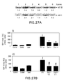
- FIGs. 27A -27B AR inhibition attenuates TNF- ⁇ - and LPS-induced activation of redox-sensitive transcription factors NF-kB and AP-1 in SAEC.
- FIG. 27A Approximately 2x10 6 SAEC were seeded in T-150 cm 2 flasks and incubated till >90% confluence. Medium was replaced with basal (serum-free) medium with AR inhibitors or carrier for 24 h. The cells were treated with TNF- ⁇ (2 nM) for 1 h and LPS (1 ⁇ g/ml) for 3 h. Nuclear extracts were prepared as described in the methods and EMSA was performed to determine the activation and translocation of NF- ⁇ B and AP-1 to nucleus.
- FIG. 27B Lanes: 1, Control; 2, Ragweed; 3, C+ Zop; 4, RW+ Zop.
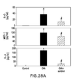
- FIGs. 28A -28B Effect of AR inhibition on ( FIG. 28A ) Th2 cytokine production and ( FIG. 28B ) inflammatory cell (eosinophils) infiltration in lungs of ovalbumin (OVA)-induced murine model of asthma.
- FIG. 28B Fixed lungs from the different experimental groups were sectioned, stained with hematoxylin and eosin. Arrows indicate sites of inflammatory cells infiltration induced by OVA-challenge.
- AR Aldose Reductase
- ARI aldose reductase inhibitors
- Uveitis is the major cause of severe visual impairment and has been estimated to account for 5-15% of all cases of total blindness in the U.S. (Read, 2006; Curi et al ., 2005). It's even more prevalent in developing nations with limited access to health care (Rathinam and Cunningham, 2000).
- the uvea has good vasculature that nourishes the eye and inflammation in uvea can affect ocular functions.
- the cause of uveitis can include autoimmune disorders, infection, or exposure to toxins, in a number of cases the etiology remains unknown (Gupta and Murray, 2006). However, the ocular inflammation due to autoimmune diseases and infections is considered the major source (Nussenblatt, 1990).
- EIU Endotoxin-induced uveitis
- LPS lipopolysaccharide
- EIU was originally used as a model of anterior uveitis, increasing evidences suggest that it also involves inflammation in the posterior segment of the eye with recruitment of leukocytes that adhere to the retinal vasculature and infiltrate the vitreous cavity (Read, 2006; Rosenbaum et al ., 1980; Altan-Yaycioglu et al ., 2006).
- This phenomenon serves as a model for certain types of human uveitis such as those associated with seronegative arthritis, where gram-negative bacteria may play a role in the pathogenesis (Rosenbaum et al. , 1980).
- LPS enhances the expression of various inflammatory cytokines and chemokines such as TNF- ⁇ , IL-6, MIF, IFN- ⁇ , MCP-1 as well as the production of PGE 2 and nitric oxide resulting in the breakdown of the blood-ocular barrier and in the infiltration of leukocytes and monocytes in ocular tissues which contribute to the development of EIU (Rosenbaum et al ., 1980).
- ROS reactive oxygen species
- TNF- ⁇ is known to be recognized as a central mediator in the pathophysiology of chronic inflammatory bowel diseases such as Crohn's and ulcerative colitis which cause increased risk of uveitis (Lin et al ., 2006; Orchard et al. , 2002).
- Recent studies have shown the use of anti-TNF- ⁇ therapy to treat uveitis (Dick et al ., 2004; Hale and Lightman, 2006; El-Shabrawi and Hermann, 2002; Joseph et al ., 2003).
- inflammation-associated increase in free radicals could cause activation of NF- ⁇ B.
- AR polyol pathway enzyme-aldose reductase
- vascular smooth muscle cells vascular smooth muscle cells
- VEC vascular endothelial cells
- macrophages vascular smooth muscle cells
- the inventors have also demonstrated that TNF- ⁇ - and high glucose-induced activation of NF- ⁇ B and apoptosis of human lens epithelial cells (HLEC) were significantly prevented by AR inhibition (Ramana et al ., 2003).
- AR inhibition prevents LPS-induced expression of TNF- ⁇ , MMP2, MMP9 and Cox-2 in HLEC which indicates the role of AR in mediating inflammatory signals in lens epithelial cells (Pladzyk et al ., 2006).
- the inventors have investigated the effect of AR inhibition on the ocular inflammation caused by LPS during EIU in Lewis rats. The results show that inhibition of AR prevents EIU-induced activation of NF- ⁇ B and production of inflammatory markers such as NO, PGE 2 , Cox-2, and TNF- ⁇ , and accumulation of infiltrating cells in various ocular tissues indicating therapeutic applications of AR inhibitors in ocular inflammation.
- Asthma is one of the most common chronic respiratory diseases, with more than 100 million sufferers worldwide. This inflammatory disorder is caused by a hypersensitive immune system that results from a number of triggers, such as dust, pollen, viruses and changes in the weather. While it is not clear how asthma is initiated in the setting of chronic inflammation, accumulating evidences strongly support the association of airway inflammation to asthma (Rosi et al. , 2006).
- cytokines such as TNF- ⁇ , IL-4, IL-5, IL-6, IL-13
- chemokines such as MCP-1, MIP-1, adhesion molecules such as ICAM-1, and E and P-Selectins (Nakajima and Takatsu, 2006; Riffo-Vasquez and Spina, 2002).
- cytokines such as TNF- ⁇ , IL-4, IL-5, IL-6, IL-13
- chemokines such as MCP-1, MIP-1, adhesion molecules such as ICAM-1, and E and P-Selectins
- NF- ⁇ B and AP1 transcription factors such as NF- ⁇ B and AP1 are known to regulate the expression of multiple proinflammatory genes associated with the pathogenesis of asthma, drugs and antioxidants that inhibit NF- ⁇ B and AP1 activation are being used to treat asthma pathogenesis (Gagliardo et al ., 2003; Frode-Saleh and Calixto, 2000; Chang and Crapo, 2002).
- ROS reactive Oxygen Species
- polyol pathway enzyme-AR is a regulator of oxidative stress signals induced by cytokines, chemokines and growth factors (GF), leading to cytotoxicity, such as apoptosis of epithelial cells and vascular endothelial cells (VEC)(Srivastava et al ., 2005; Ramana et al ., 2003; Ramana et al. ,. 2004).
- cytokines cytokines
- chemokines and growth factors growth factors
- VEC vascular endothelial cells
- LDAs such as the most abundant and toxic, 4-hydroxynonenal (HNE)
- HNE 4-hydroxynonenal
- kinetic studies have demonstrated that AR catalyzes the reduction of glutathione (GSH)-aldehydes with 10 to 100-fold higher catalytic efficiency than their parent aldehydes, because the active site of AR shows high affinity for GSH-conjugates (Srivastava et al ., 1995).
- the inventors have demonstrated by computer modeling and site-directed mutagenesis that besides carbonyl binding, the AR active site also shows specific binding for GSH (Ramana et al ., 2000; Dixit et al ., 2000).
- the inventors have recently crystallized AR conjugated with a GS-analog and solved its crystal structure, which shows a specific binding site for GSH (Singh et al ., 2006). This has opened up an entirely novel approach of synthesizing structure-based AR inhibitors, which could decrease or abolish GSH binding without affecting the binding and reduction of aldehydes.
- Such compounds should be able to attenuate signaling that causes cellular cytotoxicity including transcription of inflammatory markers, without affecting the detoxification of unconjugated LDAs generated by oxidants that could otherwise be toxic.
- the novel AR inhibitors could be excellent anti-inflammatory drugs with less or minimum side effects upon long term use in respiratory disorders such as asthma.
- the inventors contemplate that AR regulates the cellular redox state via AR/NADPH-catalyzed reduction of lipid aldehydes and their conjugates with GSH which mediate ROS-induced NF- ⁇ B and AP1 activation.
- This is based upon earlier studies demonstrating that AR catalyzed GS-LDAs mediate cytotoxic signals (Srivastava et al ., 2005).
- AR is a significant component of redox cell signaling and is induced by cytokines such as TNF- ⁇ which is known to induce the generation of ROS (Srivastava et al ., 2005).
- VECs vascular endothelial cells
- the inhibitors of aldose reductase can be any compound that inhibits the enzyme aldose reductase.
- the aldose reductase inhibitor compounds of this invention are readily available or can be easily synthesized by those skilled in the art using conventional methods of organic synthesis, particularly in view of the pertinent patent specifications.
- AR inhibitors are commercially available, such as Tolrestat, N-[[6-methoxy-5-(trifluoromethyl)-1-naphthalenyl]thioxomethyl]-N-methylglycine, [Wyeth-Ayerst, Princeton, N.J.; other designations are Tolrestatin, CAS Registry Number 82964-04-3, Drug Code AY-27,773, and brand names ALREDASE (Am.
- ARIs have been described.
- ARIs used in the diabetes context see Humber, Leslie “Aldose Reductase Inhibition: An Approach to the Prevention of Diabetes Complications", Porte, ed., Ch. 5, pp. 325-353 ; Tomlinson et al ., 1992), such as spirohydantoins and related structures, spiro-imidazolidine-2',5'-diones; and heterocycloic alkanoic acids.
- aldose reductase inhibitors are ONO-2235; zopolrestat; SNK-860; 5-3-thienyltetrazol-1-yl (TAT); WAY-121,509; ZENECA ZD5522; M16209; (5-(3'-indolal)-2-thiohydantoin; zenarestat; zenarestat 1-O-acylglucuronide; SPR-210; (2S,4S)-6-fluoro-2',5'-dioxospiro-[chroman-4,4'-imidazolidine]-2-carboxami de (SNK-880); arylsulfonylamino acids; 2,7-difluorospirofluorene-9,5'-imidazolidine-2',4'-dione (imiriestat, A111576, HOE 843); and isoliquiritigenin.
- the aldose reductase inhibitor is an compound that directly inhibits the bioconversion of glucose to sorbitol catalyzed by the enzyme aldose reductase.
- Such aldose reductase inhibitors are direct inhibitors, which are contemplated as part of the invention. Direct inhibition is readily determined by those skilled in the art according to standard assays (Malone, 1980).
- aldose reductase inhibitors are specifically described and referenced below, however, other aldose reductase inhibitors will be known to those skilled in the art. Also, common chemical names or other designations are in parentheses where applicable, together with reference to appropriate patent literature disclosing the compound. Accordingly, examples of aldose reductase inhibitors useful in the compositions, methods and kits of this invention include, but are not limited to: 3-(4-bromo-2-fluorobenzyl)-3,4-dihydro-4-oxo-1-phthalazineacetic acid (ponalrestat, U.S.
- Patent 4,251,528 N[[(5-trifluoromethyl)-6-methoxy-1-naphthalenyl]thioxomethyl ⁇ -N-methylglycine (tolrestat, U.S. Patent 4,600,724 ); 5-[(Z,E)- ⁇ -methylcinnamylidene]-4-oxo-2-thioxo-3-thiazolideneacetic acid (epalrestat, U.S. Patent 4,464,382 , U.S. Patent 4,791,126 , U.S.
- Patent 4,831,045 3-(4-bromo-2-fluorobenzyl)-7-chloro-3,4-dihydro-2,4-dioxo-1(2H)-quinazolineacetic acid (zenarestat, U.S. Patent 4,734,419 , and U.S. Patent 4,883,800 ); 2R,4R-6,7-dichloro-4-hydroxy-2-methylchroman-4-acetic acid ( U.S. Patent 4,883,410 ); 2R,4R-6,7-dichloro-6-fluoro-4-hydroxy-2-methylchroman-4-acetic acid ( U.S.
- Patent 4,883,410 3,4-dihydro-2,8-diisopropyl-3-oxo-2H-1,4-benzoxazine-4-acetic acid ( U.S. Patent 4,771,050 ); 3,4-dihydro-3-oxo-4-[(4,5,7-trifluoro-2-benzothiazolyl)methyl]-2H-1,4-benzothiazine-2-acetic acid (SPR-210, U.S. Patent 5,252,572 ); N-[3,5-dimethyl-4-[(nitromethyl)sulfonyl]phenyl]-2-methyl-benzeneacetamide (ZD5522, U.S. Patent 5,270,342 and U.S.
- Patent 5,430,060 (S)-6-fluorospiro[chroman-4,4'-imidazolidine]-2,5'-dione (sorbinil, U.S. Patent 4,130,714 ); d-2-methyl-6-fluoro-spiro(chroman-4', 4'-imidazolidine)-2', 5'-dione ( U.S. Patent 4,540,704 ); 2-fluoro-spiro(9H-fluorene-9,4'-imidazolidine)-2', 5'-dione ( U.S.
- Patent 4,438,272 2,7-difluoro-spiro(9H-fluorene-9,4'-imidazolidine)-2', 5'-dione ( U.S. Patent 4,436,745 , U.S. Patent 4,438,272 ); 2,7-di-fluoro-5-methoxy-spiro(9H-fluorene-9,4'-imidazolidine)-2', 5'-dione ( U.S. Patent 4,436,745 , U.S. Patent 4,438,272 ); 7-fluoro-spiro(5H-indenol[1,2-b]pyridine-5,3'-pyrrolidine)-2,5'-dione ( U.S.
- Patent 4,436,745 U.S. Patent 4,438,272
- d-cis-6'-chloro-2', 3'-dihydro-2'-methyl-spiro-(imidazolidine-4,4'-4'H-pyrano(2,3-b)pyridine)-2,5-dione U.S. Patent 4,980,357
- spiro[imidazolidine-4,5'(6H)-quinoline]-2,5-dione-3'-chloro-7,'8'-dihydro-7'-methyl-(5'-cis) U.S.
- Patent 5,066,659 (2S,4S)-6-fluoro-2', 5'-dioxospiro(chroman-4,4'-imidazolidine)-2-carboxamide (fidarestat, U.S. Patent 5,447,946 ); and 2-[(4-bromo-2-fluorophenyl)methyl]-6-fluorospiro[isoquinoline-4(1H),3'-pyrrolidine]-1,2', 3,5'(2H)-tetrone (minalrestat, U.S. Patent 5,037,831 ).
- Other compounds include those described in U.S. Patents 6,720,348 , 6,380,200 , and 5,990,111 , which are hereby incorporated by reference. Moreover, in other embodiments it is specifically contemplated that any of these may be excluded as part of the invention.
- Embodiments of the invention contemplate inhibitors of aldose reductase that are peptides or proteins that form a proteinaceous composition. It is contemplated that any teaching with respect to one particular proteinaceous composition may apply generally to other proteinaceous compositions described herein.
- a "proteinaceous molecule,” “proteinaceous composition,” “proteinaceous compound,” or “proteinaceous material” generally refers, but is not limited to, a protein of greater than about 200 amino acids or the full length endogenous sequence translated from a gene; a polypeptide of greater than about 100 amino acids; and/or a peptide of from about 3 to about 100 amino acids. All the “proteinaceous” terms described above may be used interchangeably herein.
- the proteinaceous composition may include such molecules that may comprise, but is not limited to 1, 2, 3, 4, 5, 6, 7, 8, 9, 10, 11, 12, 13, 14, 15, 16, 17, 18, 19, 20, 21, 22, 23, 24, 25, 26, 27, 28, 29, 30, 31, 32, 33, 34, 35, 36, 37, 38, 39, 40, 41, 42, 43, 44, 45, 46, 47, 48, 49, 50, 51, 52, 53, 54, 55, 56, 57, 58, 59, 60, 61, 62, 63, 64, 65, 66, 67, 68, 69, 70, 71, 72, 73, 74, 75, 76, 77, 78, 79, 80, 81, 82, 83, 84, 85, 86, 87, 88, 89, 90, 91, 92, 93, 94, 95, 96, 97, 98, 99, 100, 110, 120, 130, 140, 150, 160, 170, 180, 190, 200, 210, 220,
- an aldose reductase inhibitor may specifically bind or recognize a particular region of AR, including 5, 6, 7, 8, 9, 10, 11, 12, 13, 14, 15, 16, 17, 18, 19, 20, 21, 22, 23, 24, 25, 26, 27, 28, 29, 30, 31, 32, 33, 34, 35, 36, 37, 38, 39, 40, 41, 42, 43, 44, 45, 46, 47, 48, 49, 50, 51, 52, 53, 54, 55, 56, 57, 58, 59, 60, 61, 62, 63, 64, 65, 66, 67, 68, 69, 70, 71, 72, 73, 74, 75, 76, 77, 78, 79, 80, 81, 82, 83, 84, 85, 86, 87, 88, 89, 90, 91, 92, 93, 94, 95, 96, 97, 98, 99, 100, 110, 120, 130, 140, 150
- Aldose reductase may be from any organism, including mammals, such as a human, monkey, mouse, rat, hamster, cow, pig, rabbit, and may be from other cultured cells readily available. AR inhibitors may also affect polypeptides in pathways involving AR but found further upstream or downstream from AR in the pathway.
- amino acid sequences or nucleic acid sequences of AR, AR polypeptide inhibitors, or screening proteins may include additional residues, such as additional N - or C - terminal amino acids or 5' or 3' sequences, or various combinations thereof, and yet still be essentially as set forth in one of the sequences disclosed herein, so long as the sequence meets the criteria set forth above, including the maintenance of biological protein, polypeptide or peptide activity where expression of a proteinaceous composition is concerned.
- terminal sequences particularly applies to nucleic acid sequences that may, for example, include various non-coding sequences flanking either of the 5' and/or 3' portions of the coding region or may include various internal sequences, i.e., introns, which are known to occur within genes.
- peptide mimetics are peptide-containing compounds, that mimic elements of protein secondary structure.
- Mimetics are peptide-containing compounds, that mimic elements of protein secondary structure.
- the underlying rationale behind the use of peptide mimetics is that the peptide backbone of proteins exists chiefly to orient amino acid side chains in such a way as to facilitate molecular interactions, such as those of antibody and antigen.
- a peptide mimetic is expected to permit molecular interactions similar to the natural molecule.
- the present invention also contemplates the synthesis of peptides that can directly or indirectly inhibit AR. Because of their relatively small size, the peptides of the invention can also be synthesized in solution or on a solid support in accordance with conventional techniques. Various automatic synthesizers are commercially available and can be used in accordance with known protocols. See, for example, Tam et al ., (1983); Merrifield, (1986); and Barany and Merrifield (1979). Short peptide sequences, or libraries of overlapping peptides, usually from about 6 up to about 35 to 50 amino acids, which correspond to the selected regions described herein, can be readily synthesized and then screened in screening assays designed to identify reactive peptides. Alternatively, recombinant DNA technology may be employed wherein a nucleotide sequence which encodes a peptide of the invention is inserted into an expression vector, transformed or transfected into an appropriate host cell and cultivated under conditions suitable for expression.
- nucleic acid sequences complementary to at least a portion of the nucleic acid encoding AR will find utility as AR inhibitors.
- a probe or primer of between 13 and 100 nucleotides, preferably between 17 and 100 nucleotides in length, or in some aspects of the invention up to 1-2 kilobases or more in length allows the formation of a duplex molecule that is both stable and selective.
- these probes consist of oligonucleotide fragments. Such fragments should be of sufficient length to provide specific hybridization to a RNA or DNA tissue sample.
- the sequences typically will be 10-20 nucleotides, but may be longer. Longer sequences, e.g., 40, 50, 100, 500 and even up to full length, are preferred for certain embodiments.
- Antisense methodology takes advantage of the fact that nucleic acids tend to pair with "complementary" sequences.
- complementary it is meant that polynucleotides are those which are capable of base-pairing according to the standard Watson-Crick complementarity rules. That is, the larger purines will base pair with the smaller pyrimidines to form combinations of guanine paired with cytosine (G:C) and adenine paired with either thymine (A:T) in the case of DNA, or adenine paired with uracil (A:U) in the case of RNA. Inclusion of less common bases such as inosine, 5-methylcytosine, 6-methyladenine, hypoxanthine and others in hybridizing sequences does not interfere with pairing.
- Antisense polynucleotides when introduced into a target cell, specifically bind to their target polynucleotide and interfere with transcription, RNA processing, transport, translation and/or stability.
- Antisense RNA constructs, or DNA encoding such antisense RNAs may be employed to inhibit gene transcription or translation or both within a host cell, either in vitro or in vivo, such as within a host animal, including a human subject.
- Antisense constructs may be designed to bind to the promoter and other control regions, exons, introns or even exon-intron boundaries of a gene. It is contemplated that the most effective antisense constructs may include regions complementary to intron/exon splice junctions. Thus, antisense constructs with complementarity to regions within 50-200 bases of an intron-exon splice junction may be used. It has been observed that some exon sequences can be included in the construct without seriously affecting the target selectivity thereof. The amount of exonic material included will vary depending on the particular exon and intron sequences used. One can readily test whether too much exon DNA is included simply by testing the constructs in vitro to determine whether normal cellular function is affected or whether the expression of related genes having complementary sequences is affected.
- complementary or “antisense” means polynucleotide sequences that are substantially complementary over their entire length and have very few base mismatches. For example, sequences of fifteen bases in length may be termed complementary when they have complementary nucleotides at thirteen or fourteen positions. Naturally, sequences which are completely complementary will be sequences which are entirely complementary throughout their entire length and have no base mismatches. Other sequences with lower degrees of homology also are contemplated. For example, an antisense construct which has limited regions of high homology, but also contains a non-homologous region (e.g., ribozyme) could be designed. These molecules, though having less than 50% homology, would bind to target sequences under appropriate conditions.
- Ribozymes are RNA-protein complexes that cleave nucleic acids in a site-specific fashion. Ribozymes have specific catalytic domains that possess endonuclease activity (Kim and Cech, 1987; Forster and Symons, 1987).
- ribozymes accelerate phosphoester transfer reactions with a high degree of specificity, often cleaving only one of several phosphoesters in an oligonucleotide substrate (Cech et al ., 1981; Michel and Westhof, 1990; Reinhold-Hurek and Shub, 1992).
- This specificity has been attributed to the requirement that the substrate bind via specific base-pairing interactions to the internal guide sequence ("IGS") of the ribozyme prior to chemical reaction.
- IGS internal guide sequence
- Ribozyme catalysis has primarily been observed as part of sequence specific cleavage/ligation reactions involving nucleic acids (Joyce, 1989; Cech et al ., 1981).
- U.S. Patent 5,354,855 reports that certain ribozymes can act as endonucleases with a sequence specificity greater than that of known ribonucleases and approaching that of the DNA restriction enzymes.
- sequence-specific ribozyme-mediated inhibition of gene expression may be particularly suited to therapeutic applications (Scanlon et al ., 1991; Sarver et al ., 1990; Sioud et al ., 1992).
- the preparation and use of additional ribozymes that are specifically targeted to a given gene e.g., AR gene, will now be straightforward.
- RNA cleavage activity examples include sequences from the Group I self splicing introns including tobacco ringspot virus (Prody et al ., 1986), avocado sunblotch viroid (Symons, 1981), and Lucerne transient streak virus (Forster and Symons, 1987). Sequences from these and related viruses are referred to as hammerhead ribozymes based on a predicted folded secondary structure. Other suitable ribozymes include sequences from RNase P with RNA cleavage activity (Yuan et al.
- Ribozymes are targeted to a given sequence by virtue of annealing to a site by complimentary base pair interactions. Two stretches of homology are required for this targeting. These stretches of homologous sequences flank the catalytic ribozyme structure defined above. Each stretch of homologous sequence can vary in length from 7 to 15 nucleotides. The only requirement for defining the homologous sequences is that, on the target RNA, they are separated by a specific sequence which is the cleavage site.
- the cleavage site is a dinucleotide sequence on the target RNA, uracil (U) followed by either an adenine, cytosine or uracil (A,C or U; Perriman, et al ., 1992; Thompson, et al ., 1995).
- the frequency of this dinucleotide occurring in any given RNA is statistically 3 out of 16. Therefore, for a given target messenger RNA of 1000 bases, 187 dinucleotide cleavage sites are statistically possible.
- Designing and testing ribozymes for efficient cleavage of a target RNA is a process well known to those skilled in the art. Examples of scientific methods for designing and testing ribozymes are described by Chowrira et al ., (1994) and Lieber and Strauss (1995), each incorporated by reference. The identification of operative and preferred sequences for use in AR-targeted ribozymes is simply a matter of preparing and testing a given sequence, and is a routinely practiced "screening" method known to those of skill in the art.
- RNA molecule capable of mediating RNA interference in a cell is referred to as "siRNA.”
- Elbashir et al. (2001) discovered a clever method to bypass the anti viral response and induce gene specific silencing in mammalian cells.
- 21-nucleotide dsRNAs with 2 nucleotide 3' overhangs were transfected into mammalian cells without inducing the antiviral response.
- the small dsRNA molecules also referred to as "siRNA” were capable of inducing the specific suppression of target genes.
- siRNA directed against AR, NF- ⁇ B, and TNF- ⁇ are specifically contemplated.
- the siRNA can target a particular sequence because of a region of complementarity between the siRNA and the RNA transcript encoding the polypeptide whose expression will be decreased, inhibited, or eliminated.
- siRNA may be a double-stranded compound comprising two separate, but complementary strands of RNA or it may be a single RNA strand that has a region that self-hybridizes such that there is a double-stranded intramolecular region of 7 basepairs or longer (see Sui et al ., 2002 and Brummelkamp et al ., 2002 in which a single strand with a hairpin loop is used as a dsRNA for RNAi).
- a double-stranded RNA molecule may be processed in the cell into different and separate siRNA molecules.
- the strand or strands of dsRNA are 100 bases (or basepairs) or less, in which case they may also be referred to as "siRNA.” In specific embodiments the strand or strands of the dsRNA are less than 70 bases in length. With respect to those embodiments, the dsRNA strand or strands may be from 5-70, 10-65, 20-60, 30-55, 40-50 bases or basepairs in length.
- a dsRNA that has a complementarity region equal to or less than 30 basepairs (such as a single stranded hairpin RNA in which the stem or complementary portion is less than or equal to 30 basepairs) or one in which the strands are 30 bases or fewer in length is specifically contemplated, as such molecules evade a mammalian's cell antiviral response.
- a hairpin dsRNA (one strand) may be 70 or fewer bases in length with a complementary region of 30 basepairs or fewer.
- siRNA molecules to a particular target which in this case includes AR, NF- ⁇ B, and TNF- ⁇ . Kits such as SilencerTM Express, SilencerTM siRNA Cocktail, SilencerTM siRNA Construction, MEGAScript® RNAi are readily available from Ambion, Inc.
- aptamers and aptazymes are synthetic nucleic acid ligands.
- the methods of the present invention may involve nucleic acids that modulate AR, NF- ⁇ B, and TNF- ⁇ .
- a nucleic acid may comprise or encode an aptamer.
- An "aptamer” as used herein refers to a nucleic acid that binds a target molecule through interactions or conformations other than those of nucleic acid annealing/hybridization described herein.
- Methods for making and modifying aptamers, and assaying the binding of an aptamer to a target molecule may be assayed or screened for by any mechanism known to those of skill in the art (see for example, U.S. Patent Nos. 5,840,867 , 5,792,613 , 5,780,610 , 5,756,291 and 5,582,981 , Burgstaller et al., 2002, which are incorporated herein by reference.
- compositions of the present invention may comprise an effective amount of one or more AR inhibitors dissolved or dispersed in a pharmaceutically acceptable carrier to a subject.
- pharmaceutically acceptable refers to molecular entities and compositions that do not produce an adverse, allergic or other untoward reaction when administered to an animal, such as, for example, a human, as appropriate.
- the preparation of a pharmaceutical composition that contains at least one AR inhibitor or additional active ingredient will be known to those of skill in the art in light of the present disclosure, and as exemplified by Remington's Pharmaceutical Sciences, 18th Ed. Mack Printing Company, 1990 , incorporated herein by reference.
- preparations should meet sterility, pyrogenicity, general safety and purity standards as required by FDA Office of Biological Standards.
- pharmaceutically acceptable carrier includes any and all solvents, dispersion media, coatings, surfactants, antioxidants, preservatives (e.g., antibacterial agents, antifungal agents), isotonic agents, absorption delaying agents, salts, preservatives, drugs, drug stabilizers, gels, binders, excipients, disintegration agents, lubricants, sweetening agents, flavoring agents, dyes, such like materials and combinations thereof, as would be known to one of ordinary skill in the art (see, for example, Remington's Pharmaceutical Sciences, 18th Ed. Mack Printing Company, 1990, pp. 1289-1329 , incorporated herein by reference). Except insofar as any conventional carrier is incompatible with the active ingredient, its use in the therapeutic or pharmaceutical compositions is contemplated.
- An AR inhibitor can be administered in the form of a pharmaceutically acceptable salt or with a pharmaceutically acceptable salt.
- pharmaceutically acceptable salts includes both pharmaceutically acceptable acid addition salts and pharmaceutically acceptable cationic salts, where appropriate.
- pharmaceutically-acceptable cationic salts is intended to define, but is not limited to such salts as the alkali metal salts, (e.g ., sodium and potassium), alkaline earth metal salts (e.g ., calcium and magnesium), aluminum salts, ammonium salts, and salts with organic amines such as benzathine (N,N'-dibenzylethylenediamine), choline, diethanolamine, ethylenediamine, meglumine (N-methylglucamine), benethamine (N-benzylphenethylamine), diethylamine, piperazine, tromethamine (2-amino-2-hydroxymethyl-1,3-propanediol) and procaine.
- alkali metal salts e.g ., sodium and potassium
- alkaline earth metal salts e.g ., calcium and magnesium
- salts are intended to define but is not limited to such salts as the hydrochloride, hydrobromide, sulfate, hydrogen sulfate, phosphate, hydrogen phosphate, dihydrogenphosphate, acetate, succinate, citrate, methanesulfonate (mesylate) and p-toluenesulfonate (tosylate) salts.
- compositions of the aldose reductase inhibitors of this invention may be readily prepared by reacting the free acid form of the aldose reductase inhibitor with an appropriate base, usually one equivalent, in a co-solvent.
- bases are sodium hydroxide, sodium methoxide, sodium ethoxide, sodium hydride, potassium methoxide, magnesium hydroxide, calcium hydroxide, benzathine, choline, diethanolamine, piperazine and tromethamine.
- the salt is isolated by concentration to dryness or by addition of a non-solvent.
- salts are preferably prepared by mixing a solution of the acid with a solution of a different salt of the cation (sodium or potassium ethylhexanoate, magnesium oleate), and employing a solvent (e.g ., ethyl acetate) from which the desired cationic salt precipitates, or can be otherwise isolated by concentration and/or addition of a non-solvent.
- a solvent e.g ., ethyl acetate
- the acid addition salts of the aldose reductase inhibitors of this invention may be readily prepared by reacting the free base form of said aldose reductase inhibitor with the appropriate acid.
- the salt is of a monobasic acid (e.g., the hydrochloride, the hydrobromide, the p-toluenesulfonate, the acetate)
- the hydrogen form of a dibasic acid e.g., the hydrogen sulfate, the succinate
- the dihydrogen form of a tribasic acid e.g., the dihydrogen phosphate, the citrate
- salts as the sulfate, the hemisuccinate, the hydrogen phosphate, or the phosphate
- the appropriate and exact chemical equivalents of acid will generally be used.
- the free base and the acid are usually combined in a co-solvent from which the desired salt precipitates, or can be otherwise isolated by concentration and/or addition of a non-solvent.
- aldose reductase inhibitors that may be used in accordance with this invention, prodrugs thereof, and pharmaceutically acceptable salts thereof or of said prodrugs, may occur as hydrates or solvates. These hydrates and solvates are also within the scope of the invention.
- a pharmaceutical composition of the present invention may comprise different types of carriers depending on whether it is to be administered in solid, liquid or aerosol form, and whether it needs to be sterile for such routes of administration as injection.
- a pharmaceutical composition of the present invention can be administered intravenously, intradermally, intraarterially, intraperitoneally, intraarticularly, intrapleurally, intrabronchially, intrapleurally, intranasally, topically, intramuscularly, intraperitoneally, subcutaneously, subconjunctival, intravesicularlly, mucosally, intrapericardially, intraumbilically, orally, topically, locally, inhalation ( e.g ., aerosol inhalation), instillation, injection, infusion, continuous infusion, via a catheter, via a lavage, in lipid compositions ( e.g ., liposomes), or by other method or any combination of the forgoing as would be known to one of ordinary skill in the art (see, for example, Remington's Pharmaceutical Sciences, 18th
- the actual dosage amount of a composition of the present invention administered to a subject can be determined by physical and physiological factors such as body weight, severity of condition, the type of disease being treated, previous or concurrent therapeutic interventions, idiopathy of the patient and on the route of administration.
- the number of doses and the period of time over which the dose may be given may vary.
- the practitioner responsible for administration will, in any event, determine the concentration of active ingredient(s) in a composition and appropriate dose(s), as well as the length of time for administration for the individual subject.
- An amount of an aldose reductase inhibitor that is effective for inhibiting aldose reductase activity is used.
- an effective dosage for the inhibitors is in the range of about 0.01 mg/kg/day to 100 mg/kg/day in single or divided doses, preferably 0.1 mg/kg/day to 20 mg/kg/day in single or divided doses. Doses of about, at least about, or at most about 0.01, 0.05, 0.1, 0.15, 0.20, 0.25, 0.30, 0.35, 0.40, 0.45, 0.50, 0.55, 0.60, 0.65, 0.70, 0.75, 0.80, 0.85, 0.90.
- compositions may comprise, for example, at least about 0.1% of an active compound.
- the an active compound may comprise between about 2% to about 75% of the weight of the unit, or between about 25% to about 60%, for example, and any range derivable therein.
- a dose may also comprise from about 1 microgram/kg/body weight, about 5 microgram/kg/body weight, about 10 microgram/kg/body weight, about 50 microgram/kg/body weight, about 100 microgram/kg/body weight, about 200 microgram/kg/body weight, about 350 microgram/kg/body weight, about 500 microgram/kg/body weight, about 1 milligram/kg/body weight, about 5 milligram/kg/body weight, about 10 milligram/kg/body weight, about 50 milligram/kg/body weight, about 100 milligram/kg/body weight, about 200 milligram/kg/body weight, about 350 milligram/kg/body weight, about 500 milligram/kg/body weight, to about 1000 mg/kg/body weight or more per administration, and any range derivable therein.
- a range of about 5 mg/kg/body weight to about 100 mg/kg/body weight, about 5 microgram/kg/body weight to about 500 milligram/kg/body weight, etc. can be administered, based on the numbers described above.
- the composition may comprise various antioxidants to retard oxidation of one or more component.
- the prevention of the action of microorganisms can be brought about by preservatives such as various antibacterial and antifungal agents, including but not limited to parabens (e.g., methylparabens, propylparabens), chlorobutanol, phenol, sorbic acid, thimerosal or combinations thereof.
- parabens e.g., methylparabens, propylparabens
- chlorobutanol phenol
- sorbic acid thimerosal or combinations thereof.
- An AR inhibitor(s) of the present invention may be formulated into a composition in a free base, neutral or salt form.
- Pharmaceutically acceptable salts include the acid addition salts, e.g., those formed with the free amino groups of a proteinaceous composition, or which are formed with inorganic acids such as for example, hydrochloric or phosphoric acids, or such organic acids as acetic, oxalic, tartaric or mandelic acid. Salts formed with the free carboxyl groups can also be derived from inorganic bases such as for example, sodium, potassium, ammonium, calcium or ferric hydroxides; or such organic bases as isopropylamine, trimethylamine, histidine or procaine.
- a carrier can be a solvent or dispersion medium comprising but not limited to, water, ethanol, polyol (e.g., glycerol, propylene glycol, liquid polyethylene glycol, etc), lipids (e.g., triglycerides, vegetable oils, liposomes) and combinations thereof.
- the proper fluidity can be maintained, for example, by the use of a coating, such as lecithin; by the maintenance of the required particle size by dispersion in carriers such as, for example liquid polyol or lipids; by the use of surfactants such as, for example hydroxypropylcellulose; or combinations thereof such methods.
- isotonic agents such as, for example, sugars, sodium chloride or combinations thereof.
- the AR inhibitors are prepared for administration by such routes as oral ingestion.
- the solid composition may comprise, for example, solutions, suspensions, emulsions, tablets, pills, capsules (e.g., hard or soft shelled gelatin capsules), sustained release formulations, buccal compositions, troches, elixirs, suspensions, syrups, wafers, or combinations thereof.
- Oral compositions may be incorporated directly with the food of the diet.
- Preferred carriers for oral administration comprise inert diluents, assimilable edible carriers or combinations thereof.
- the oral composition may be prepared as a syrup or elixir.
- a syrup or elixir and may comprise, for example, at least one active agent, a sweetening agent, a preservative, a flavoring agent, a dye, a preservative, or combinations thereof.
- an oral composition may comprise one or more binders, excipients, disintegration agents, lubricants, flavoring agents, and combinations thereof.
- a composition may comprise one or more of the following: a binder, such as, for example, gum tragacanth, acacia, cornstarch, gelatin or combinations thereof; an excipient, such as, for example, dicalcium phosphate, mannitol, lactose, starch, magnesium stearate, sodium saccharine, cellulose, magnesium carbonate or combinations thereof; a disintegrating agent, such as, for example, corn starch, potato starch, alginic acid or combinations thereof; a lubricant, such as, for example, magnesium stearate; a sweetening agent, such as, for example, sucrose, lactose, saccharin or combinations thereof; a flavoring agent, such as, for example peppermint, oil of wintergreen, cherry flavoring, orange flavoring, etc.; or combinations thereof the for
- the dosage unit form When the dosage unit form is a capsule, it may contain, in addition to materials of the above type, carriers such as a liquid carrier. Various other materials may be present as coatings or to otherwise modify the physical form of the dosage unit. For instance, tablets, pills, or capsules may be coated with shellac, sugar or both.
- Sterile injectable solutions are prepared by incorporating the active compounds in the required amount in the appropriate solvent with various of the other ingredients enumerated above, as required, followed by filtered sterilization.
- dispersions are prepared by incorporating the various sterilized active ingredients into a sterile vehicle which contains the basic dispersion medium and/or the other ingredients.
- the preferred methods of preparation are vacuum-drying or freeze-drying techniques which yield a powder of the active ingredient plus any additional desired ingredient from a previously sterile-filtered liquid medium thereof.
- the liquid medium should be suitably buffered if necessary and the liquid diluent first rendered isotonic prior to injection with sufficient saline or glucose.
- the preparation of highly concentrated compositions for direct injection is also contemplated, where the use of DMSO as solvent is envisioned to result in extremely rapid penetration, delivering high concentrations of the active agents to a small area.
- composition must be stable under the conditions of manufacture and storage, and preserved against the contaminating action of microorganisms, such as bacteria and fungi. It will be appreciated that endotoxin contamination should be kept minimally at a safe level, for example, less that 0.5 ng/mg protein.
- prolonged absorption of an injectable composition can be brought about by the use in the compositions of agents delaying absorption, such as, for example, aluminum monostearate, gelatin or combinations thereof.
- compositions of the present invention such as an AR inhibitor
- This process may involve contacting the cell(s) with an AR inhibitor and a therapeutic agent at the same time or within a period of time wherein separate administration of the modulator and an agent to a cell, tissue or organism produces a desired therapeutic benefit.
- the terms "contacted” and "exposed,” when applied to a cell, tissue or organism, are used herein to describe the process by which a AR inhibitor and/or therapeutic agent are delivered to a target cell, tissue or organism or are placed in direct juxtaposition with the target cell, tissue or organism.
- the cell, tissue or organism may be contacted (e.g., by administration) with a single composition or pharmacological formulation that includes both a AR inhibitor and one or more agents, or by contacting the cell with two or more distinct compositions or formulations, wherein one composition includes an AR inhibitor and the other includes one or more agents.
- the AR inhibitor may precede, be concurrent with and/or follow the other agent(s) by intervals ranging from minutes to weeks.
- the AR inhibitor and other agent(s) are applied separately to a cell, tissue or organism, one would generally ensure that a significant period of time did not expire between the time of each delivery, such that the inhibitor and agent(s) would still be able to exert an advantageously combined effect on the cell, tissue or organism.
- one may contact the cell, tissue or organism with two, three, four or more modalities substantially simultaneously ( i.e., within less than about a minute) as the modulator.
- one or more agents may be administered within of from substantially simultaneously, about 1 minute, about 5 minutes, about 10 minutes, about 20 minutes about 30 minutes, about 45 minutes, about 60 minutes, about 2 hours, or more hours, or about 1 day or more days, or about 4 weeks or more weeks, or about 3 months or more months, or about one or more years, and any range derivable therein, prior to and/or after administering the AR inhibitor.
- a AR inhibitor is "A” and the secondary agent, such as a antibiotic or other anti-inflammatory treatment, is "B": A/B/A B/A/B B/B/A A/A/B A/B/B B/A/A B/B/B/A B/B/A/B A/A/B/B A/B/A/B A/B/B/A B/B/A B/B/A B/A/B/A B/A/A/B A/A/A/B B/A/A/A A/B/A/A A/B/A/A A/B/A A/B/A A/B/A A/B/A A/B/A A/B/A A/B/A A/B/A A/B/A A/B/A A/B/A A/B/A A/B/A A/B/B B/A/B/B
- modulators to a cell, tissue or organism may follow general protocols for the administration of agents for the treatment of uveitis, conjunctivitis, or asthma. It is expected that the treatment cycles would be repeated as necessary. In particular embodiments, it is contemplated that various additional agents may be applied in any combination with the present invention. Agents include antibiotics (for gram-positive and gram negative bacteria), anti-inflammatory drugs, and immunosuppressant drugs, which are well known to those of skill in the art and frequently commerically available.
- AR inhibitors and other active agents may be administered together or separately.
- the administration of one agent may be prior to, concurrent to, or subsequent to the administration of other agent(s).
- RPMI-1640 medium, phosphate-buffered saline (PBS), gentamicin sulfate solution, trypsin/EDTA solution and fetal bovine serum (FBS) were purchased from GIBCO BRL Life Technologies (Grand Island, NY). Sorbinil and zopolrestat were obtained as gift from Pfizer (New York, NY). Dimethyl sulfoxide (DMSO) was obtained from Fischer scientific (Pittsburgh, PA). Nitrite/Nitrate and PGE 2 assay kits were obtained from Cayman Chemical Inc (Ann Arbor, MI). Rat TNF- ⁇ ELISA kit was obtained from BD Biosciences (San Diego, CA).
- PBS phosphate-buffered saline
- FBS fetal bovine serum
- LPS from Escherichia coli was obtained from Sigma (Sigma-Aldrich, Saint Louise, MO). Antibodies against TNF- ⁇ , and phospho-p65 (serine 536) were purchased from cell signaling (Danvers, MA), iNOS was from Cayman Chemicals (Ann Arbor, MI), Cox-2 and GAPDH were from Santacruz biotech inc. (Santa Cruz, CA), and polyclonal antibodies against human recombinant AR were made for the inventors by Alpha Diagnostic Intl. San Antonio, TX. All other reagents used were of analytical grade.
- EIU was induced by a subcutaneous injection of Escherichia coli LPS (200 ⁇ g) dissolved in phosphate-buffered saline (PBS, pH 7.4). Rats in ARI and EIU + ARI groups were injected intraperitoneally with AR inhibitor zopolrestat (25 mg/kg body weight) dissolved in dimethyl- sulfoxide (DMSO) 24 h before and immediately after LPS injection. Rats of control group received carrier (PBS + 20% DMSO) injection.
- Infiltrating cells and proteins in aqueous humor The rats were euthanized after 3, 6, and 24 h after LPS injection and the aqueous humor (AqH) was collected immediately from eye by an anterior chamber puncture using a 30-gauge needle under the surgical microscope.
- AqH samples were suspended in an equal amount of Trypan-blue solution, and the cells were counted using a Hemocytometer under a light microscope (Olympus Optical Ltd).
- the total protein concentration in the AqH samples was measured using a Biorad protein assay kit (Biorad, CA, USA).
- the AqH samples were stored in ice water until testing, cell counts and total protein concentrations were measured on the day of sample collection. Rest of the AqH was stored at -80°C until used.
- TNF- ⁇ , NO and PGE 2 in aqueous humor The levels of TNF- ⁇ in the AqH (stored at -80°C) were assessed with commercially available ELISA kit, according to the manufacturer's instructions. The total level of nitrate plus nitrite in the AqH was measured by using a total nitrite colorimetric assay (LDH) kit according to the manufacturer's instructions. PGE 2 production was measured by enzyme immunoassay kit following the manufacturer's instructions.
- LDH total nitrite colorimetric assay
- Rats were euthanized 24 h after LPS injection and the eyes were enucleated immediately and stored in 4% para-formaldehyde solution for 48 h at 4°C. The eyes were washed in ice-cold PBS twice and kept in 70% alcohol at 4°C until they were embedded in paraffin. Sagittal sections (5 ⁇ m) were cut and stained with hematoxylin and eosin (H&E). The iris-ciliary body complex, anterior chamber, vitreous and retina were observed under light microscope.
- H&E hematoxylin and eosin
- ROS The levels of ROS in rat eye were quantified by dihydroethidium (DHE) (Molecular Probes, Eugene, OR, U.S.A.) which gives red fluorescence when oxidized to ethidium in the presence of ROS.
- DHE dihydroethidium
- U-937 a human monocytic cell line, was obtained from ATCC (Manassas, VA, USA). The cells were cultured in RPMI-1640 medium supplemented with 2 mM glutamine, 1 mM sodium pyruvate, 25 mM HEPES, antibiotics (100 U/mL penicillin and 100 ⁇ g/mL streptomycin) and 10% heat-inactivated fetal bovine serum and maintained at 37°C in a humidified incubator containing 95% O 2 and 5% CO 2 . The cells were pretreated with 10 ⁇ M AR inhibitor, zopolrestat for overnight in serum-free medium and subsequently stimulated with 1 ⁇ g/mL LPS from E. coli for 24 h, unless otherwise stated.
- Lysates were separated on 10% SDS-polyacrylamide gels and transferred to polyvinylidene difluoride membranes (Immobilon; Millipore, Bedford, MA). The membranes were then incubated in blocking solution containing 5% wt/vol dried fat-free milk and 0.1% vol/vol Tween-20 in Tris-buffered Saline. Subsequently, the membranes were incubated with anti-Cox-2, -iNOS, and -GAPDH antibodies. The membranes were then probed with horseradish peroxidase- conjugated secondary antibody (GE Healthcare, Piscataway, NJ) and visualized by chemiluminescence (Pierce biotechnology, Rockford, IL).
- horseradish peroxidase- conjugated secondary antibody GE Healthcare, Piscataway, NJ
- the cell culture media were centrifuged at 5000 rpm and supernatants were stored at -80°C.
- the media was thawed and used for chemiluminescent secretory alkaline phosphatase (SEAP) assay using Great EscAPeTM SEAP reporter assay system according to protocol essentially as described by the manufacturer, (BD Biosciences, Palo Alto, CA) using a 96-well chemiluminescence plate reader. All the suggested controls by manufacturers were used in the assay.
- SEAP chemiluminescent secretory alkaline phosphatase
- infiltrating cells were also present in the vitreous chamber (VC) but none were observed in AR inhibitor + EIU or control rats.
- the accumulation of infiltrating cells in AqH was also confirmed by manually counting the cells in AqH by using a hemocytometer ( FIG. 1B (I) ).
- the manual cell counting also demonstrated a significant (>200-folds) increase in the infiltration of inflammatory cells in the aqueous humor of EIU-rat eyes which was significantly (>80%) prevented by AR inhibitor treatment of the EIU-rats ( FIG. 1B (I) ).
- the EIU - rats showed a significant intensity of antibody staining in iris-ciliary complex, and AqH region whereas, AR inhibitor-treated animals showed diminished antibody staining, indicating that AR inhibition prevents accumulation of TNF- ⁇ ( FIG. 2B , left panel).
- EIU-induced acute inflammation is not restricted to anterior chamber only (Rosenbaum et al ., 1980; Altan-Yaycioglu et al ., 2006)
- the inventors immunohistochemically examined the levels of TNF- ⁇ in vitreous region as well.
- the results shown in FIG. 2B show increased levels of TNF- ⁇ in vitreous, and retina of LPS-treated rats as compared to control groups which was significantly prevented by AR inhibitor.
- NO and PGE 2 are produced by inducible nitric oxide synthase (iNOS) and cyclooxygenase-2 (Cox-2) enzymes respectively
- iNOS inducible nitric oxide synthase
- Cox-2 cyclooxygenase-2
- the inventors immunohistochemically examined the expression of iNOS and Cox-2 proteins in the various regions of eye.
- the EIU rat eyes showed increased expression of iNOS and Cox-2 proteins in the iris-ciliary body complex, corneal epithelium in anterior segment and retinal wall in the posterior segment ( FIG. 3A (II) and FIG. 3B (II) ).
- AR inhibitor significantly prevented the expression of iNOS as well as Cox-2.
- NF- ⁇ B positive cells were observed in the iris-ciliary body complex, corneal epithelium in anterior segment, and retina in posterior segment of the eye ( FIG. 4B ). In contrast the number of NF- ⁇ B positive cells in the anterior as well as posterior chambers of AR inhibitor-treated EIU eyes were significantly decreased ( FIG. 4B ).
- AR plays a pivotal role in inflammation, irrespective of the cause such as cytokines, chemokines, endotoxins and growth factors. Since anti-inflammatory drugs are being used therapeutically in asthma and AR inhibitors prevent the formation of inflammatory markers as well as their cellular effects (Ramana et al ., 2006a; Ramana et al ., 2006b), it was reasoned that they should be excellent drugs to treat asthma pathogenesis, irrespective of the source or causative factor.
- AR inhibitors significantly attenuated TNF- ⁇ -induced proliferation of VSMCs (Ramana et al. , 2002) and apoptosis of HLECs (Ramana et al ., 2003).
- AR inhibitors also prevented the activation of caspase-3 and degradation of nucleosomal histones by high glucose or TNF- ⁇ in HLECs and by LPS in macrophages (Ramana et al ., 2006a; Pladzyk et al ., 2006). Also, AR inhibition attenuated cytokine- and high glucose-induced NF- ⁇ B and AP1 activation in all the cell lines studied (Srivastava et al ., 2005).
- LPS is known to cause apoptosis through the expression of proinflammatory markers such as PGE2, Cox-2 and iNOS
- the effect of AR inhibition was investigated in cellular models.
- the inventors demonstrate that LPS-induced apoptosis of HLECs and macrophages and inflammatory response, as determined by a significant increase in the TNF, MMP2, MMP9, PGE2 and Cox-2 levels, are prevented (75 to 95%) by inhibiting or ablating AR (Ramana et al ., 2006a; Pladzyk et al ., 2006).
- the in vitro studies were extended to mice.
- mice were injected intraperitoneally with a single dose of LPS (4 ⁇ g/kg body wt) ⁇ ARI, sorbinil, 25 mg/Kg body wt/day, and killed them on days 1, 3, and 7 after LPS injection.
- Cytokine and chemokine levels were determined in the serum, as well as in such tissues as liver, heart and spleen (Ramana et al ., 2006a). The results showed that LPS significantly increased various cytokines, chemokines, cAMP, Cox-2 and PGE2 in the serum and various tissues, and that the increases were 75 to 95% decreased by AR inhibition.
- AR inhibition significantly increased the LD50 of LPS in mice from 14 mg to 20 mg (Ramana et al ., 2006b).
- the investigations have been extended to other inflammatory diseases such as colon cancer and uveitis and have shown that inhibition of AR prevents colon cancer progression in nude mice xenografts and uveitis in rats (Tammali et al ., 2006).
- the inventors contemplate that AR plays pivotal role in the pathophysiology of inflammation, which is backed by strong evidences obtained using cellular as well as animal models.
- AR inhibition would prevent asthma.
- AR inhibitors can be used to prevent chronic inflammation mediated by cytokines, chemokines and other proinflammatory markers such as Cox-2, PGE2 and iNOS, irrespective of the initiating cause such as autoimmune or infection. Since such inflammatory markers are major risk factors for airway inflammation leading to respiratory complications, AR inhibition is an excellent therapeutic strategy in such disorders.
- Bronchial asthma is a chronic inflammatory disease of the airways characterized by airway eosinophilia, goblet cell hyperplasia with mucus hypersecretion, and hyperresponsiveness to inhaled allergens.
- the inventors studied AR inhibition and asthma pathogenesis using mouse models for asthma to determine effects of two structurally distinct AR inhibitors (sorbinil and zopolrestat) on OVA-induced bronchial inflammation and airway hyper-responsiveness.
- mice receive i.p. injections of sorbinil or zopolrestat (25 mg/kg body wt) 24 h prior and 12 h post challenge.
- Control groups are not sensitized or challenged with OVA instead are given saline (Mustapha et al ., 2006).
- Bronchoalveolar Lavage (BAL): In the mice killed by CO 2 asphyxiation, the trachea is cannulated just below the larynx. A flexible polyurethane tube (outer diameter, 0.4 mm) attached to a blunt 23-gauge needle is inserted ⁇ 6 mm into the trachea. BAL is performed by injecting 0.3 ml of saline at room temperature; the fluid is withdrawn and stored on ice. This procedure is repeated a total of three times and the collected fluid pooled.
- BAL fluid is centrifuged at 1000 x g for 10 min at 4°C, and the resulting supernatant stored immediately at -80°C.
- the cell pellets are resuspended in 250 ⁇ l of phosphate-buffered saline (PBS) containing 2% bovine serum albumin (BSA), and the total cell count is determined with an automated counter (Coulter Electronics, Hialeah, FL) and recorded as the total number of inflammatory cells per milliliter.
- PBS phosphate-buffered saline
- BSA bovine serum albumin
- the cell suspension is adjusted to a density of 200 cells/ ⁇ l, and 100 ⁇ l of the diluted suspension is centrifuged at 800 x g for 10 min with a Cytospin onto coated Superfrost Plus microscope slides.
- the cells on the slides are air-dried, fixed for 30 s with Diff-Quik fixative, and then stained first with Diff-Quik solution I for 60 s and then with Diff-Quik solution II for 60 s. After washing with deionized water for 30 s, the slides are allowed to dry before application of the mounting media and a cover slip. Differential counts are performed for ⁇ 200 cells according to standard morphological criteria. A pathologist, assessing the different inflammatory cell types is blinded to the treatment groups.
- Immunohistochemistry Lung sections are immunostained for the expression of protein-HNE adducts, AR, p65 NF- ⁇ B, ICAM-1, VCAM-1 and other inflammatory markers such as Cox-2 and iNOS as described in Mustapha et al ., 2006. Western blot analysis of the lung homogenates are carried out to confirm immunohistochemical studies using specific antibodies.
- Cytokine assessment The concentrations of total IL-4, IL-5, GM-CSF, IL-10, IFN- ⁇ , IL-12, IL-13 are determined using the Bio-Rad Bioplex System for mouse Th1 and Th2 cytokines as described in Mustapha et al ., 2006. The levels of MMP2, MMP9, Cox, PGE-2 and NO are measured in BAL by using specific ELISA kits.
- IgE quantification IgE is quantified by sandwich ELISA technique. The plates are coated overnight with rabbit anti-OVA antibodies (Serotec, Raleigh, NC) at 4°C and then blocked with 1% BSA in PBS for 1h at 37°C. Samples, along with IgE Standards are prepared and added to appropriate wells and incubated for 3h at 4°C. Polyclonal goat anti-IgE antibodies, followed by HRP-conjugated rabbit anti-goat antibodies are used to detect bound IgE. 3,3'5,5'-Tetramethylbenzidine is used to develop the plates and values determined by an ELISA plate reader at 490nm.
- Airway responsiveness is measured in unrestrained, conscious mice 3 days after the last challenge (Prieto et al ., 2006). Mice are placed in a barometric plethysmographic chamber, and baseline readings taken and averaged for 3 min. Aerosolized methacholine in increasing concentrations (from 2.5 to 50 mg/ml) will be nebulized through an inlet of the main chamber for 3 min. Readings are taken and averaged for 3 min after each nebulization and enhanced pause (Penh) is determined.
- Penh calculated as (expiratory time/relaxation time - 1) x (peak expiratory flow/peak inspiratory flow) according to the manufacturers' protocol, is a dimensionless value that represents a function of the proportion of maximal expiratory to maximal inspiratory box pressure signals and a function of the timing of expiration. Penh is used as a measure of airway responsiveness to methacholine. Results are expressed as the percentage increase of Penh following challenge with each concentration of methacholine, where the baseline Penh (after saline challenge) is expressed as 100%. Penh values averaged for 3 min after each nebulization are evaluated.
- Mucin production in the epithelial cells is assessed by periodic acid Schiff (PAS)-staining of formalin-fixed, paraffin-embedded lung sections. The stained sections are analyzed as above and representative fields are photographed with a Photometrix CoolSNAP Fx camera mounted on a NIKON Eclipse TE 200 UV microscope. MUC5AC levels in the BAL is assessed by ELISA using commercially available anti-MUC5AC monoclonal antibody (Lab Vision, CA, USA) as described (Mustapha et al ., 2006; Boldogh et al ., 2005).
- AR participates in cytotoxic signaling initiated by various oxidants such as LPS, RW, TNF and HNE and by regulating the activation of transcription factors such as NF- ⁇ B and AP1 via protein kinase network comprised of PKC, MAPK, JNK, and IKK and expression of inflammatory markers, leading to airway epithelial cell apoptosis.
- oxidants such as LPS, RW, TNF and HNE
- transcription factors such as NF- ⁇ B and AP1
- protein kinase network comprised of PKC, MAPK, JNK, and IKK
- inflammatory markers leading to airway epithelial cell apoptosis.
- SAEC Human small airway epithelial cells
- SAEC Small Airway growth medium
- SAGM small airway growth medium
- the cells are pretreated with 10 ⁇ M AR inhibitors for 24 h and then stimulated with LPS, RW, TNF and HNE. After different incubation periods (0, 15, 30, 60, 90, 120 min for protein kinases and transcription factors and 24 h for expression of inflammatory markers), the effect of oxidants ⁇ AR inhibitors or siRNA on the activation of PKC, MAPK, ERK, P38, JNK, and IKK are determined. The effects of inhibiting AR on the activation of transcription factors such as NF- ⁇ B, AP-1, SP1 and OCT1 and expression of inflammatory markers are determined as described herein.
- Apoptosis Cell viability is determined by cell counting, MTT assays and [ 3 H]-thymidine incorporation (Ramana et al. , 2002). Cell death is determined by "cell Death ELISA kit", caspase-3 activation, and Hoechst nuclear staining that determines the morphological changes in the nuclei undergoing apoptotic death (Ramana, et al. , 2006a; Pladzyk et al., 2006).
- Activation of caspase-3 is a marker for apoptosis, and is measured by two methods [1] in vitro cleavage of caspase-3 substrate and [2] the in vivo activation of caspase-3 that leads to the cleavage of PARP.
- the cleavage products are identified by Western blot analysis with anti-PARP antibodies.
- Apoptosis is also determined by "Annexin V staining and DNA content by FACS" in the absence and presence of AR inhibitors or siRNA.
- Oxidative stress is the main mediator of antigen-induced airway inflammation. Therefore, the inventors examine the effects of inhibiting or ablating AR on the oxidative stress; lipid peroxidation, generation of toxic LDAs such as HNE, as well as activation of NADPH oxidase and formation of superoxide radicals. As a crude indicator of oxidative stress, the inventors quantify the endogenous GSH levels after treating the SAEC cells with LPS ⁇ AR inhibitors or AR SiRNA. Several studies show that a decrease in the levels of GSH may cause cytotoxic effects under various stress conditions; this is an accepted index of increased oxidative stress (Wisnewski et al., 2005; Kloek et al., 2003).
- Total ROS production in the cells is measured fluorimetrically, as well as by flow cytometry, using 2',7'-Dichlorodihydrofluorescein (H2DCF), a fluorescent dye that interacts with oxygen-free radicals, using a fluorometry or FACS scan analysis (Pladzyk et al., 2006).
- H2DCF 2',7'-Dichlorodihydrofluorescein
- FACS scan analysis Fluorometry or FACS scan analysis.
- the inventors use the flow cytometry core facility available to all UTMB investigators on a fee-for-service basis.
- the generation of superoxide is measured by a spectrophotometric assay, as well as by a luminescence assay using NBT and coelenterazine, respectively (Ramana et al., 2006c).
- the inventors will measure the NADPH oxidase activity in the SAEC.
- Lipid peroxidation and HNE levels Several stress conditions show increased HNE generation and protein-HNE formation, especially in cells and tissues undergoing cell death (Rahman et al., 2002; Hamilton et al., 1996). Since the inventors have previously shown that AR efficiently catalyzes the reduction of these aldehydes and their GS-conjugates with a Km in the low micromolar range (Srivastava et al., 1995; Ramana et al., 2000; Dixit et al., 2000; Singh et al., 2006), the inventors contemplate that by inhibiting or ablating AR they are able to modulate oxidative injury and alter its cytotoxic consequences.
- HNE formation is used as a marker for lipid peroxidation.
- HNE-protein adducts are quantified using an ELISA kit per the supplier's instructions.
- concentration of GS-HNE and its AR-reduced product GS-DHN is measured by HPLC analysis of culture media.
- the GS-HNE and GS-DHN peaks are identified by ESI/MS as described by Ramana et al., 2006c.
- NF- ⁇ B and AP-1 The activation of NF- ⁇ B and AP1 is measured by EMSA, and also with a NF- ⁇ B reporter gene (Clontech) as described earlier (Ramana et al., 2006a; Ramana et al., 2006b; Ramana et al., 2006c; Pladzyk et al., 2006).
- NF- ⁇ B upstream signals The enzymes PKC, IKK, and MAPK are the main kinases for the phosphorylation of I ⁇ B- ⁇ , whereas p38 MAPK and JNK are the main kinases involved in the activation of AP1.
- Polyclonal antibodies against phosphorylated and unphosphorylated anti-p44/42 MAPK, ERK1/2 and the IKK isoforms (IKK- ⁇ , IKK ⁇ and IKK- ⁇ ) are used to determine the effects of AR inhibition on oxidant-induced activation of kinases.
- the phosphorylation of JNK and p38 is studied by using phospho-specific Abs against JNK and p38.
- the inventors determine total PKC activity using Promega SignaTECT PKC assay system, per the manufacturer's instructions (Ramana et al., 2006a).
- Inflammatory marker expression Inflammatory cytokines and chemokines will be measured and confirmed by RT-PCR (Ramana et al., 2006a, Tammali et al., 2006).
- GS-HNE a representative of GS-LDAs
- PLC pathway that activates PKC
- NF- ⁇ B and AP1 the main mediator for the activation of PLC pathway that activates PKC
- NF- ⁇ B and AP1 the main mediator for the activation of PLC pathway that activates PKC
- NF- ⁇ B and AP1 the main mediator for the activation of PLC pathway that activates PKC
- NF- ⁇ B and AP1 NF- ⁇ B and AP1 and cause inflammation. Therefore, the GS-DHN-induced activation of PLC, PKC and other kinases that activate NF- ⁇ B and AP1 are investigated in airway epithelial cells.
- the SAEC are incubated with GS-DHN (1 ⁇ M) for different time intervals (0-180 min).
- the cells are harvested, cytoplasmic and nuclear extracts prepared as described (Ramana et al., 2006a).
- SABM Small airway epithelial basal medium
- SAGMTM small airway epithelial growth media
- ReagentpackTM containing Trypsin 0.025%/EDTA 0.01%, Trypsin neutralizing solution and HEPES buffered saline solution were purchaged from Cambrex Bio Sciences Walkersvillle, Inc. (Walkersville, MD). Sorbinil and Zopolrestat were obtained as gift from Pfizer (New York, NY). Dimethyl sulfoxide (DMSO) was obtained from Fischer scientific (Pittsburgh, PA). Ragweed pollens (RW) were purchased from Greer's laboratory (Lenoir, NC).
- Nitrite/Nitrate and PGE 2 assay kits were obtained from Cayman Chemical Inc (Ann Arbor, MI).
- Human IL-6 and IL-8 ELISA kits were from Diaclone (Stamford, CT) and R&D systems, respectively.
- Antibodies against COX2, iNOS, Bcl-XL, Bax, GAPDH, cyclin-D1 and E2F2 were from Santa Cruz Biotechnology Inc. (Santa Cruz, CA); and antibodies against phospho-I ⁇ B were from Cell signaling (Danvers, MA).
- Dihydroethidium (DHE) fluorescent dye was purchased from Molecular Probes, Invitrogen (Carlsbad, CA) and polyclonal antibodies against human recombinant AR were made for us by Alpha diagnostic intl. (San Antonio, TX).
- the reagents used in the electrophoretic mobility shift assay (EMSA) and Western blot analysis were obtained from Sigma. All other reagents used were of analytical grade.
- SAEC Small Airway Epithelial cells
- SABM small airway epithelial basal medium
- EGF epidermal growth factor
- RA retinoic acid
- RA 6.5 ng/ml triiodothyronine
- Annexin V Staining and Flow cytometry Approximately 2x10 5 SAEC/well were plated in 6-well plates in triplicate for each group. After 24 hours, the medium was replaced with serum-free SABM with or without zopolrestat (20 ⁇ M). The cells were induced by RW (150 ⁇ g/mL) and incubated for 18 h. Apoptotic cell death was examined using the annexin V-FITC/PI, (molecular probes, Invitrogen) according to the manufacturer's instructions. Twenty thousand events were acquired for each sample and analyzed by flow cytometry using the LYSIS II software (FACScan, BD Pharmingen).
- DHE Dihydroethidium staining for superoxide was carried out as described previously. Briefly, Approximately 1x10 5 cells were seeded on chambered slides and starved in serum-free SABM with or without AR inhibitor for 24 h. The cells were treated with RW for 16 h. SAEC were rinsed with cold PBS and incubated in PBS containing HEt (2.5 ⁇ mol/L) at 37°C for 15 minutes. Cells were rinsed in PBS and mounted with Vectashield hard setTM mounting medium with DAPI (Vector Laboratories Inc., Burlingame, CA). The image of ethidium staining was measured with a Nikon epifluorescence microscope with a 585 nm long-pass filter. Generation of superoxide in the cells was demonstrated by strong red fluorescent labeling.
- DHE Dihydroethidium
- Electrophoretic Mobility Shift Assay The SAEC were pretreated with or without AR inhibitors for 24 h in starving medium, followed by treatment with Ragweed pollens (RW) (50 ⁇ g/ml) for 2 h at 37°C.
- RW Ragweed pollens
- the nuclear extracts were prepared as described. Briefly, SAEC were harvested and washed with cold PBS and suspended in 0.1 ml of hypotonic lysis buffer containing protease inhibitors for 10 min. The cells were then lysed with 5 ⁇ l of 10% Nonidet P-40. The homogenate was centrifuged (6000rpm; 1min.), and supernatant containing the cytoplasmic extracts was stored frozen at -80°C.
- the nuclear pellet was resuspended in 50 ⁇ l ice-cold nuclear extraction buffer. After 30 min of intermittent mixing, the extract was centrifuged (12000rpm; 15 min.), and supernatants containing nuclear extracts were secured. The protein content was measured by the Bradford method. If they were not used immediately, they were stored at -80°C.
- the Consensus oligonucleotides for NF- ⁇ B and AP-1 transcription factors were 5'-end labeled using T4 polynucleotide kinase. EMSA was performed as described. The specificity of the assay was examined by competition with an excess of unlabeled oligonucleotide and supershift assays with antibodies to p65.
- NF- ⁇ B-dependent reporter secretary alkaline phosphatase expression Assay The SAEC (1x10 5 cells/well) were plated in 24-well plates, serum-starved in SABM for 24 hours with or without AR inhibitors, sorbinil and zopolrestat (20 ⁇ M), and transiently transfected with pNF- ⁇ B-secretory alkaline phosphatase (SEAP) construct or control plasmid pTAL-SEAP DNA (Clontech, Palo Alto, CA) using the LipofectAMINE Plus reagent. After 6 hours of transfection, medium was replaced with fresh medium and cells were treated with RW (50 ⁇ g/mL) for 48 hours. The cell culture medium was harvested, centrifuged and supernatant was analyzed for SEAP activity, essentially as described by the manufacturer (Clontech, Palo Alto, CA), using a 96-well chemiluminescence plate reader.
- SEAP pNF- ⁇ B-secretory alkaline phosphat
- RNA Interference Ablation of AR in SAECs The ablation of AR mRNA was essentially carried out as described in the art. Briefly, 2x10 5 SAECs were plated in a 6-well plate and grown until ⁇ 80% confluency. The cells were incubated with serum-free medium containing the AR-siRNA (AATCGGTGTCTCCAACTTCAA (SEQ ID NO:1)) or scrambled siRNA (AAAATCTCCCTAAATCA TACA (SEQ ID NO:2); control) to a final concentration of 100 nM and the RNAiFectTM transfection reagent (Qiagen). The cells were cultured for 48 h at 37 °C, and AR expression was determined by measuring AR protein by Western blot analysis using anti-AR antibodies.
- Prostaglandin E2, IL-6 and IL-8 Assays The SAEC were plated in 6-well plates at a density of 2x10 5 cells/well in triplicate for each group. After 24 hours, the medium was replaced with serum-free SABM with or without zopolrestat (20 ⁇ M). The growth-arrested and AR ablated cells were stimulated with RW (50 ⁇ g/mL) for another 24 hours.
- the medium was collected from each well , centrifuged and supernatant was analyzed for PGE2 (Cayman Chemical Co., Ann Arbor, Michigan); IL-6 (Diaclone, Stamford, CT) and IL-8 (R&D systems Inc, Minneapolis, MN) by using an ELISA kits according to the manufacturer's instructions.
- PGE2 Chemical Chemical Co., Ann Arbor, Michigan
- IL-6 Diaclone, Stamford, CT
- IL-8 R&D systems Inc, Minneapolis, MN
- RNA from SAECs was isolated by using RNeasy kit (Qiagen) as per supplier's instructions.
- RNA 0.5-1.0 ⁇ g isolated from each sample were reverse transcribed with Omniscript and Sensiscript reverse transcriptase one-step RT-PCR system with HotStar Taq DNApolymerase (Qiagen) at 55°C for 30 min followed by PCR amplification.
- oligonucleotide primer sequences were as follows: 5'-ATGAACTCCT TCTCCACAAGCGC-3' (SEQ ID NO:3) (sense) and 5'-GAAGAGCCCTCAGGCTGGACTG-3' (SEQ ID NO:4) (antisense) for IL-6; 5'-ATGACTTCCAAGCTGGCCGTGGCT-3' (SEQ ID NO:5) (sense) and 5'-TCT CAGCCCTCTTCAAAAACTTCTC-3' (SEQ ID NO:6) (antisense) for IL-8; 5'-TGAAACCCACTCCA AACACAG-3' (SEQ ID NO:7) (sense) and 5'-TCATCAGGCACAGGAGGAAG-3' (SEQ ID NO:8) (antisense) for Cox-2; and 5'-ATCTGGCACCACACCTTCTACAATGAGCTGCG-3' (SEQ ID NO:9) (sense) and 5'-CGTC ATACTCCTGCTTGCTGATCCACATCTGC- 3
- PCR was carried out in a PCR Sprint thermal cycler (Thermo electron corporation, Milford, MA) under the following conditions: initial denaturation at 95°C for 15 min followed by 35 cycles of 94°C 1 min, 62°C 1 min, 72°C 1 min, and then 72°C 10 min for final extension.
- PCR products were electrophoresed with 1.5% agarose-1X TAE gels containing 0.5 ⁇ g/ml ethidium bromide.
- the densitometric analyses of the blots were performed by using Kodak 1D image analysis software.
- mice BALB/c mice were purchased from Harlan Sprague-Dawley (San Diego, CA, USA). All animal experiments were performed according to the National Institutes of Health Guide for Care and Use of Experimental Animals and approved by UTMB Animal Care and Use Committee (#9708038-05).
- RW ragweed pollens
- Mucin production in the epithelial cells was assessed by periodic acid Schiff (PAS)-staining of formalin-fixed, paraffin-embedded lung sections. The stained sections were analyzed as above and representative fields were photographed with a Photometrix CoolSNAP Fx camera mounted on a NIKON Eclipse TE 200 UV microscope (Boldogh et al., 2005, Bacsi et al., 2006).
- PAS periodic acid Schiff
- MUC5AC levels in the BAL were assessed by ELISA using commercially available anti-MUC5AC monoclonal antibody (I-13M1) (Lab Vision, Fremont, CA, USA). Briefly, MUC5AC present in the BAL was captured to a microtiter plate and a second antibody conjugated to biotin was added. After 30 min' incubation with streptavidin-horseradish peroxidase (HRP) plates were washed and peroxidase substrate was added to obtain colorimetric product, which was quantified by spectrometry. Data are expressed as arbitrary units relative to a MUC5AC standard curve that was included on each plate (Boldogh et al., 2005).
- HRP streptavidin-horseradish peroxidase
- Airway responsiveness was measured in unrestrained, conscious mice 3 days after the last challenge. Mice were placed in a barometric plethysmographic chamber, and baseline readings were taken and averaged for 3 min. Aerosolized methacholine in increasing concentrations (from 10 to 80 mg/ml) were nebulized through an inlet of the main chamber for 3 min. Readings were taken and averaged for 3 min after each nebulization and enhanced pause (Penh) was determined.
- Penh a dimensionless value, represents a function of the ratio of peak expiratory flow to peak inspiratory flow and a function of the timing of expiration, was calculated as (expiratory time/relaxation time -1 ) x (peak expiratory flow/peak inspiratory flow) according to the manufacturers' protocol. Penh correlates with pulmonary airflow resistance or obstruction and was used as a measure of airway responsiveness to methacholine.
- AR inhibition prevents RW-induced ROS generation in SAEC: To examine the nature of the RW-induced decrease in SAEC viability, the level of RW-induced ROS in SAEC was measured. RW (150 ⁇ g/mL) caused increase in cellular ROS levels as evident by increased fluorescence by ROS sensitive DHE causing oxidative stress, which could be responsible for the loss in cell viability. Preincubation of SAEC with two different AR inhibitors, sorbinil or zopolrestat, prevented these changes ( FIG. 12 ). Under the similar conditions, AR inhibition alone caused no significant changes in the ROS levels of SAEC. These results suggest that AR mediates RW-induced ROS formation and resultant cell death.
- AR inhibition prevents RW-induced production of inflammatory markers (chemokines and cytokines) by SAEC: Since RW is known to elevate the levels of inflammatory markers in the airway epithelial cells that cause inflammation and aggravate the allergic condition, the effect of AR inhibition on the RW-induced increase in the levels of various inflammatory markers was examined in SAEC culture medium. As shown in FIG. 13 , treatment of SAEC with 50 ⁇ g/mL RW for 24 hours caused 3 and 4-fold increases in the synthesis of IL-6, and IL-8, respectively, and inhibition of AR significantly prevented these changes. A more than 2-fold increase in the RW-induced PGE2 levels in SAEC was also significantly (>75%) prevented by AR inhibition ( FIG. 13 ).
- inflammatory markers chemokines and cytokines
- AR siRNA antisense oligonucleotides
- AR inhibition prevents expression of RW-induced inflammatory markers in SAEC The effects of AR inhibition on the expression of these inflammatory markers at RNA levels was further examined using RT-PCR. As shown in FIG. 15 , treatment of SAEC with RW caused a 3-4 fold increase in the expression of IL-6 and IL-8 mRNA levels and zopolrestat prevented it by >70% suggesting that AR could regulate the transcriptional activation of inflammatory marker genes. Also, since PGE2 is synthesized by COX2, the effect of AR inhibition on the transcriptional activation of COX2 was determined by quantification of its mRNA in response to RW in SAEC by RT-PCR. As shown in FIG.
- AR inhibition prevents RW-induced synthesis of inflammatory markers proteins in SAEC: Since biosynthesis of PGE2 and NO from their precursors is catalyzed by COX-2 and iNOS enzymes, respectively, the effect of AR inhibition on RW-induced COX-2 and iNOS expression in SAEC was next examined by immunoblotting. As shown in FIG. 16A , treatment of SAEC with RW significantly ( ⁇ 3-folds) increased COX-2 and iNOS protein expression and pre-treatment of SAEC with AR inhibitor, zopolrestat, significantly prevented the increase. This indicated that RW-induced COX-2 and iNOS overexpression is mediated by AR which is obligatory for RW-induced PGE2 and NO production that leads to Cytotoxicity and tissue damage during pollen allergy.
- AR inhibition maintains the ratio of pro- and anti-apoptotic proteins in SAEC: Since apoptosis is regulated by the fine balance between the pro-apoptotic (Bax) and anti-apoptotic (Bcl-XL) proteins, the effect of AR inhibition on the expression of these proteins was next examined. As shown in FIG. 16B RW caused more than 2-fold increased expression of pro-apoptotic protein Bax while the expression of anti-apoptotic protein Bcl-XL decreased by 50%. The over-all ratio of these two proteins in control cells was 1 which increased significantly (to 4.2) in RW-treated cells. Inhibition of AR not only controlled the expression of these proteins but it also maintained the ratio to less than 1. These results suggest that AR inhibition prevents the RW- induced alteration in the ratio of pro- and anti-apoptotic proteins and thereby inhibits apoptosis in these cells.
- AR inhibition prevents RW-induced over-expression of cell cycle proteins in SAEC: Under oxidative stress number of cells entering the cell division cycle is altered which is regulated by a fine balance of cell cycle proteins. Whether AR inhibition will affect the level of cell cycle proteins in RW treated SAEC was next examined. As shown in FIG. 16C , RW caused >3.5 fold increased expression of cyclin D1 and >2.5 fold increased expression in E2F2 proteins. Inhibition of AR by zopolrestat prevented the increase in expression of these cell cycle protein significantly (>90%).
- AR inhibition prevents RW-induced activation of NF- ⁇ B and AP1 in SAEC: The effect of AR inhibition on RW-induced activation of NF- ⁇ B and AP1 was next examined because these transcription factors are responsible for the transcription of various inflammatory markers. As shown in FIG. 17A , RW caused approximately 4-fold activation of NF- ⁇ B and AP-1 and zopolrestat significantly (>80%) prevented the RW-induced NF- ⁇ B activation and nuclear translocation. Zopolrestat alone did not affect the basal NF- ⁇ B activity in the SAEC.
- NF- ⁇ B-dependent secretory alkaline phosphatase (SEAP) reporter assay were employed. It was found that RW significantly (>3-fold) induced NF- ⁇ B-dependent SEAP activation in SAEC and sorbinil as well as zopolrestat caused >60% inhibition ( FIG. 17B ). However, sorbinil and zopolrestat alone did not affect the NF- ⁇ B-SEAP activity. These results validated our measurement of DNA binding activity of NF- ⁇ B by gel shift assay. Based on these observations, the inventors concluded that inhibition of AR prevents RW-induced activation of NF- ⁇ B, which could activate the expression and synthesis of inflammatory markers in SAEC.
- SEAP secretory alkaline phosphatase
- AR inhibition prevents RW-induced accumulation of eosinophils in airways in mice model of asthma: Since inflammatory response of RW challenge to SAEC was blocked significantly by AR inhibition, the inventors tested whether this approach would work in the animal models as well.
- FIG. 18A there was a robust airway inflammtion as measured by accumulation of inflammatory cells in BAL fluid and subepithelial spaces in RW-sensitized and challenged mice.
- the mice treated with AR inhibitor there was significantly less inflammation as determined by the number of eosinophils which decresed significantly (p ⁇ 0.002).
- Perivascular and peribronchial inflammation and cell composition in the BAL fluid induced by RW challenge was significantly prevented by AR inhibitor treatement ( FIG. 18B ).
- AR inhibition prevents RW-induced accumulation of MUC5AC in the BAL fluid: Excessive mucin production by airway epithelium is characteristic of allergic asthma and its prevention is the main goal to treat allergic episodes in susceptible individuals. Therefore, the MUC5AC levels in the BAL fluid by ELISA were next examined using anti-MUC5AC monoclonal antibodies and found that MUC5AC increased ⁇ 25-folds in RW-challenged mice as compared to control and AR inhibition prevented it significantly (p ⁇ 0.003). Similarly, mucin production in the epithelial cells as assessed by periodic acid Schiff (PAS)-staining of lung sections ( FIG. 19A , inset) was also prevented by AR inhibition.
- PAS periodic acid Schiff
- SABM Small airway epithelial basal medium
- SAGMTM small airway epithelial growth media
- ReagentpackTM containing Trypsin 0.025%/EDTA 0.01%, Trypsin neutralizing solution and HEPES buffered saline solution were purchaged from Cambrex Bio Sciences Walkersvillle, Inc. (Walkersville, MD).
- Aldose reductase inhibitors Sorbinil and Zopolrestat were obtained as gift from Pfizer (New York, NY).
- Dimethyl sulfoxide (DMSO) was obtained from Fischer scientific (Pittsburgh, PA).
- TNF- ⁇ was purchased from Research diagnostics Inc (Concord, MA)
- LPS from Escherichia coli was obtained from Sigma (Sigma-Aldrich, Saint Louise, MO).
- Nitrite/Nitrate and PGE 2 assay kits were obtained from Cayman Chemical Inc (Ann Arbor, MI).
- Human IL-6 and IL-8 ELISA kits were from Diaclone (Stamford, CT) and R&D systems, respectively.
- Antibodies against Cox2, iNOS, Bcl-XL, Bax, GAPDH, cyclin-D1 and E2F2 were from Santa Cruz Biotechnology Inc. (Santa Cruz, CA); and antibodies against phospho-I ⁇ B were from Cell signaling (Danvers, MA).
- Dihydroethidium (DHE) fluorescent dye was purchased from Molecular Probes, Invitrogen (Carlsbad, CA) and polyclonal antibodies against human recombinant AR were made for us by Alpha diagnostic intl. San Antonio, TX.
- the reagents used in the electrophoretic mobility shift assay (EMSA) and Western blot analysis were obtained from Sigma. All other reagents used were of analytical grade.
- SAEC Small Airway Epithelial cells obtained from Cambrex Bio Science Walkersville, Inc. (Walkersville, MD) from distal airspace of an 18yr old male donor.
- the cells were cultured according to the supplier's instructions at 37°C in humidified incubator with 95% O 2 and 5% CO 2 in small airway epithelial basal medium (SABM) supplemented with 52 ⁇ g/ml bovine pituitary extract, 0.5 ng/ml human recombinant epidermal growth factor (EGF), 0.5 ⁇ g/ml epinephrine, 1 ⁇ g/ml hydrocortisone, 10 ⁇ g/ml transferrin, 5 ⁇ g/ml insulin, 0.1 ng/ml retinoic acid (RA), 6.5 ng/ml triiodothyronine, 50 ⁇ g/ml Gentamicin/Amphotericin-B (GA-1000), and 50 ⁇ g/ml fatty acid-free bovine
- the SAEC were plated at 5000 cells/well in a 96-well plate. After they attached, cells were growth-arrested for 24 h by replacing complete medium with fresh basal medium with AR inhibitor sorbinil (20 ⁇ M) or carrier. The cells were incubated with TNF- ⁇ (2 nM) or LPS (1 ⁇ g/mL) or carrier for an additional 24 hours. Cell viability was detected by the MTT assay as described earlier. After the incubation, 10 ⁇ L of MTT (5 mg/ml) was added to each well and incubated at 37°C for 2 h. The medium was removed and the formazan granules obtained were dissolved in 100% dimethyl sulfoxide (DMSO). Absorbance was detected at 570 nm with a 96-well ELISA reader.
- DMSO dimethyl sulfoxide
- DHE Dihydroethidium staining for superoxide was carried out as described previously. Briefly, approximately 1x10 5 cells were seeded on chambered slides and starved in serum-free basal medium with AR inhibitor or carrier for 24 h. The cells were treated with TNF- ⁇ (2 nM) or LPS (1 ⁇ g/mL) or carrier for an additional 16 h. SAEC were rinsed with cold PBS and incubated in PBS containing HEt (2.5 ⁇ mol/L) at 37°C for 15 min. Cells were rinsed in PBS and mounted with fluorsave mounting medium containing DAPI (Vector Laboratories Inc., Burlingame, CA). The image of ethidium staining was measured with a Nikon epifluorescence microscope with a 585 nm long-pass filter. Generation of superoxide in the cells was demonstrated by strong red fluorescent labeling.
- DHE Dihydroethidium staining for superoxide was carried out as described previously. Briefly, approximately 1x10
- Prostaglandin E2, IL-6 and IL-8 Assays Approximately 2x10 5 SAEC/well were seeded in 6-well plates in triplicate for each group. The medium was replaced with serum-free basal medium with AR inhibitor, sorbinil (20 ⁇ M) or carrier. The growth-arrested cells were treated with either TNF- ⁇ (2nM) or LPS (1 ⁇ g/mL) or carrier for another 24 h.
- the medium was collected from each well, centrifuged and the supernatant was analyzed for PGE2 (Cayman Chemical Co., Ann Arbor, Michigan); IL-6 (Diaclone, Stamford, CT) and IL-8 (R&D systems Inc, Minneapolis, MN) by using respective ELISA kits according to the manufacturer's instructions.
- RNA from SAEC was isolated by using RNeasy kit (Qiagen) as per supplier's instructions.
- RNA 0.5-1.0 ⁇ g isolated from each sample were reverse transcribed with Omniscript and Sensiscript reverse transcriptase one-step RT-PCR system with HotStar Taq DNApolymerase (Qiagen) at 55°C for 30 min followed by PCR amplification.
- oligonucleotide primer sequences were as follows: 5'-ATGAACTCCTTCTCCACAAGCGC-3' (SEQ ID NO:3) (sense) and 5'-GAAGAGCCCTCAGGCTGGACTG-3' (SEQ ID NO:4) (antisense) for IL-6; 5'-ATGACTTCCAAGCTGGCCGTGGCT-3' (SEQ ID NO:5) (sense) and 5'-TCT CAGCCCTCTTCAAAAACTTCTC-3' (SEQ ID NO:6) (antisense) for IL-8; 5'-TGAAACCCACTCCA AACACAG-3' (SEQ ID NO:7) (sense) and 5'-TCATCAGGCACAGGAGGAAG-3' (SEQ ID NO:8) (antisense) for Cox-2; and 5'-ATCTGGCACCACACCTTCTACAATGAGCTGCG-3' (SEQ ID NO:9) (sense) and 5'-CGTC ATACTCCTGCTTGCTGATCCACATCTGC- 3'
- PCR was carried out in a PCR Sprint thermal cycler (Thermo electron corporation, Milford, MA) under the following conditions: initial denaturation at 95°C for 15 min followed by 35 cycles of 94°C 1 min, 62°C 1 min, 72°C 1 min, and then 72°C 10 min for final extension.
- PCR products were electrophoresed with 1.5% agarose-1X TAE gels containing 0.5 ⁇ g/ml ethidium bromide.
- the densitometry analyses of the blots were performed by using Kodak 1D image analysis software.
- Electrophoresis mobility gel shift assay The SAEC were plated in T-150 cm 2 culture flasks and incubated until 80% confluence and pretreated with AR inhibitor sorbinil or carrier for 24 h in starving medium, followed by treatment with TNF- ⁇ (2nM) for 1 h and LPS (1 ⁇ g/ml) for 2 h at 37°C.
- the nuclear extracts were prepared as described. Briefly, SAEC cells were harvested and washed with cold PBS and suspended in 0.1 ml of hypotonic lysis buffer containing protease inhibitors for 10 min. The cells were then lysed with 5 ⁇ l of 10% Nonidet P-40.
- the homogenate was centrifuged (6000 rpm; 1 min), and supernatant containing the cytoplasmic extracts was stored frozen at -80°C.
- the nuclear pellet was resuspended in 50 ⁇ l ice-cold nuclear extraction buffer. After 30 min of intermittent mixing, the extract was centrifuged (12000 rpm; 15 min), and supernatants containing nuclear extracts were secured. The protein concentration was measured by the Bradford method. If they were not used immediately, nuclear extracts were stored at -80°C.
- the Consensus oligonucleotides for NF- ⁇ B and AP-1 transcription factors were 5'-end labeled using T4 polynucleotide kinase. EMSA was performed as described in the art. The specificity of the assay was examined by competition with an excess of unlabeled oligonucleotide and supershift assays with antibodies to p65.
- NF-KB-dependent reporter secretory alkaline phosphatase expression Assay SAEC cells (1x10 5 cells/well) were plated in 24-well plates, serum starved in basal medium with AR inhibitor, sorbinil and zopolrestate (20 ⁇ M) or carrier for 24 h, and transiently transfected with pNF- ⁇ B-secretory alkaline phosphatase (SEAP) construct or control plasmid pTALSEAP DNA (Clontech, Palo Alto, CA) using the LipofectAMINE Plus reagent. After 6 hours of transfection medium was replaced with fresh medium and cells were incubated with either TNF- ⁇ (2nM) or LPS (1 ⁇ g/mL) or carrier for 48 h. The cell culture medium was then harvested and analyzed for SEAP activity, essentially as described by the manufacturer (Clontech, Palo Alto, CA), using a 96-well chemiluminescence plate reader.
- SEAP pNF- ⁇ B-secretory
- RNA Interference Ablation of AR in SAECs The ablation of AR mRNA was essentially carried out as described in the art. Briefly, 2x10 5 SAECs were plated in a 6-well plateand grown till >80% confluency. The cells were incubated with serum-free medium containing the AR-siRNA (AATCGGTGTCTCCAACTTCAA) (SEQ ID NO:1) or scrambled siRNA (AAAATCTCCCTAAATCA TACA (SEQ ID NO:2); control) to a final concentration of 100 nM and the RNAiFectTM transfection reagent (Qiagen). The cells were cultured for 48 h at 37°C, and AR expression was determined by measuring AR protein by Western blot analysis using anti-AR antibodies.
- the blot was then washed, exposed to HRP-conjugated secondary antibodies (1:5,000 dilution) for 1 hr, and the antigen-antibody complex was detected by enhanced chemiluminescence (Amersham Pharmacia Biotech, Piscataway, NJ, USA).
- mice C57BL/6; wild type
- LSUHSC New Lat.
- LA New Lat.
- mice C57BL/6; wild type mice were bred in a specific-pathogen free facility at LSUHSC, New La, LA, and allowed unlimited access to sterilized chow and water. Maintenance, experimental protocols, and procedures were all approved by the LSUHSC Animal Care & Use Committee.
- mice Six to eight-weeks old C57BL/6 wild type mice were sensitized with injections (i.p.) of 100 ⁇ g Grade V chicken ovalbumin (OVA) (Sigma-Aldrich, St. Louis MO), mixed with 2 mg aluminum hydroxide in saline, once a week for 2 consecutive weeks followed by a challenge with aerosolized OVA a week after the second sensitization.
- OVA Grade V chicken ovalbumin
- the mice were challenged by placing them in groups of six in a Plexiglas chamber and were exposed for 30 min to aerosolized OVA (3% OVA in saline).
- the OVA aerosol was generated by a Bennett nebulizer (DeVilbiss, PA).
- a group of mice received an injection of 25 mg/kg AR inhibitor (i.p.) prior to challenge. Control groups were not sensitized or challenged. The mice that were used in each experiment were of the same litter.
- BAL fluids were centrifuged at 1000 x g for 10 min at 4°C, and the supernatant was stored immediately at -80°C.
- the cell pellets were resuspended in 250 ⁇ l of phosphate-buffered saline (PBS) containing 2% bovine serum albumin (BSA), and the total cell count was determined with an automated counter (Coulter Electronics, Hialeah, FL) and recorded as the total number of inflammatory cells per milliliter.
- PBS phosphate-buffered saline
- BSA bovine serum albumin
- the cell suspension was adjusted to a density of 200 cells/ ⁇ l, and 100 ⁇ l of the diluted suspension was centrifuged at 800 x g for 10 min with a Cytospin (International Equipment, Needham Heights, MA) onto coated Superfrost Plus microscope slides (Baxter Diagnostics, Deerfield, IL).
- the cells on the slides were air-dried, fixed for 30 s with Diff-Quik fixative (American Scientific Products, McGaw, IL), and stained first with Diff-Quik solution I for 60 s and then with Diff-Quik solution II for 60 s. After washing with deionized water for 30 s, the slides were allowed to dry before the application of mounting medium and a cover slip. Differential counts were performed for ⁇ 200 cells according to standard morphological criteria by a pathologist blinded to the treatment groups.
- Cytokine assessment The concentration of total IL-4, IL-5, and MCP1 was determined using the Bio-Rad Bioplex System for mouse according to the manufacturer instructions.
- AR inhibition prevents TNF- ⁇ - and LPS-Induced ROS generation in SAEC: Since it is known that both TNF- ⁇ and LPS cause oxidative stress which could cause decrease in SAEC viability, the levels of reactive oxygen species in SAEC were measured. As shown in FIG. 22 , both TNF- ⁇ and LPS caused a significant increase in the ROS levels as indicated by increased ethidium staining in TNF- ⁇ and LPS treated cells, suggesting increased oxidative stress which would explain increased death of SAEC. Pre-incubation of the cells with two different AR inhibitors sorbinil or zopolrestat prevented these changes ( FIG. 22 ). AR inhibition alone caused no significant changes in the ROS levels of SAEC.
- TNF- ⁇ and LPS are known to elevate the levels of inflammatory markers in the airway epithelial cells that cause inflammation aggravating the allergic condition. Therefore, the effect of AR inhibition on the TNF- ⁇ and LPS -induced increase in the levels of various inflammatory markers in the SAEC culture medium were examined. As shown in FIG. 23 , treatment of SAEC with TNF- ⁇ (2 nM) for 24 hours caused approximately 4-fold increase in the synthesis of IL-6, and >19fold increase in IL-8, respectively, and inhibition of AR significantly (>80%) prevented these changes.
- AR in SAEC was ablated by antisense oligonucleotides (AR siRNA) to study whether phenotypic absence of AR will have similar effects in SAEC as does AR inhibitor.
- Transient transfection of SACE with AR antisense abolished AR protein by >95% ( FIG. 23 ) while with scrambled antisense oligonucleotides AR activity as well as protein expression were not affected.
- Antisense ablation of AR also significantly prevented the TNF- ⁇ and LPS -induced IL-6, IL-8 and PGE2 production in SAEC similar to the pharmacological inhibitor of AR ( FIG. 24 ).
- AR inhibition prevents TNF- ⁇ - and LPS-Induced synthesis of inflammatory marker proteins in SAEC: Since biosynthesis of PGE2 and NO from their precursors is catalyzed by Cox-2 and iNOS enzymes, respectively the effect of AR inhibition on TNF- ⁇ - and LPS -induced Cox-2 and iNOS expression in SAEC was next examined by immunoblotting. Treatment of SAEC with TNF- ⁇ and LPS significantly increased Cox-2 and iNOS protein expression by ⁇ 3.5 and ⁇ 2-folds and pre-treatment of cells with AR inhibitor zopolrestat significantly prevented this increase ( FIG. 26 ). These results indicate that AR-dependent Cox-2 and iNOS over-expression is required for PGE2 and iNOS production that leads to Cytotoxicity and tissue damage during airway inflammation.
- AR inhibition prevents TNF- ⁇ - and LPS -Induced imbalance in the ratio of pro- and anti-apoptotic proteins in SAEC: Since apoptosis is regulated by the fine balance between the pro-apoptotic (Bax) and anti-apoptotic (Bcl-xl) proteins, the effect of AR inhibition on the expression of these proteins was next examined. As shown in FIG. 26 , TNF- ⁇ and LPS caused more than ⁇ 2-fold increased expression of pro-apoptotic protein, Bax while the expression of anti-apoptotic protein, Bcl-xl decreased by 50%. The over-all ratio of pro- and anti-apoptotic proteins in control cells was 1 which increased significantly to approximately 3 in TNF- ⁇ - and LPS- treated cells.
- AR inhibition prevents TNF- ⁇ - and LPS -Induced over-expression of cell cycle proteins in SAEC: Since under oxidative stress, the number of cells entering cell cycle is altered, the inventors examined whether AR inhibition will affect the level of cell cycle proteins in SAECs. As shown in FIG. 26 , TNF- ⁇ and LPS caused ⁇ 2.5-fold increase in the expression of cyclin D1 and E2F2 proteins and inhibition of AR significantly (>90%) prevented the increase indicating that inhibition of AR is critical to maintaining the cell cycle in the event of oxidative stress and prevention of apoptosis.
- AR Inhibition prevents TNF- ⁇ - and LPS -Induced Activation of NF- ⁇ B and AP1 in SAEC:
- the redox-sensitive transcription factors such as NF- ⁇ B and AP1 are responsible for the transcription of various inflammatory markers. Therefore, the effect of AR inhibition on TNF- ⁇ - and LPS-induced activation of NF- ⁇ B and AP1 was examined. As shown in FIG. 27A , TNF- ⁇ and LPS respectively caused ⁇ 3 and 2-fold activation of NF- ⁇ B as well as AP-1 and AR inhibition significantly prevented it.
- AR inhibitor alone did not affect the basal NF- ⁇ B activity in the SAECs.
- NF- ⁇ B-dependent secretory alkaline Phosphatase (SEAP) reporter assay was used. As shown in FIG. 28 , TNF- ⁇ and LPS, respectively caused more than 7 and 2.5 fold increased NF- ⁇ B-dependent reporter (SEAP) activation in SAECs.
- SEAP NF- ⁇ B-dependent secretory alkaline Phosphatase
- Two structurally different inhibitors of AR, sorbinil and zopolrestat caused >60% inhibition ( FIG. 27B ) of NF- ⁇ B-dependent SEAP activation. However, sorbinil and zopolrestat alone did not affect the basal NF- ⁇ B-SEAP activity.
- AR inhibition prevents cytokine production and inflammatory cells (eosinophils) infiltration in ovalbumin -induced murine model of asthma:
- the levels of Cytokines such as IL-4, IL-5 and chemokine such as MCP-1 in BAL fluids were significantly elevated in OVA-challenged mice as compared to control mice.
- Treatment with AR inhibitor significantly (p ⁇ 0.05) prevented the increase in cytokines and chemokines significantly (p ⁇ 0.05) ( FIG. 28A ).
- FIG. 28B shows that OVA-sensitization and challenge induced a clear and marked perivascular and peribronchial infiltration of eosinophils, a trait of allergic airway inflammation, into the lungs of C57BL/6 mice.
Landscapes
- Health & Medical Sciences (AREA)
- Life Sciences & Earth Sciences (AREA)
- Chemical & Material Sciences (AREA)
- Veterinary Medicine (AREA)
- Public Health (AREA)
- Medicinal Chemistry (AREA)
- General Health & Medical Sciences (AREA)
- Animal Behavior & Ethology (AREA)
- Pharmacology & Pharmacy (AREA)
- Epidemiology (AREA)
- General Chemical & Material Sciences (AREA)
- Organic Chemistry (AREA)
- Nuclear Medicine, Radiotherapy & Molecular Imaging (AREA)
- Chemical Kinetics & Catalysis (AREA)
- Engineering & Computer Science (AREA)
- Bioinformatics & Cheminformatics (AREA)
- Pulmonology (AREA)
- Ophthalmology & Optometry (AREA)
- Pain & Pain Management (AREA)
- Rheumatology (AREA)
- Pharmaceuticals Containing Other Organic And Inorganic Compounds (AREA)
- Medicines That Contain Protein Lipid Enzymes And Other Medicines (AREA)
- Acyclic And Carbocyclic Compounds In Medicinal Compositions (AREA)
Applications Claiming Priority (2)
| Application Number | Priority Date | Filing Date | Title |
|---|---|---|---|
| US89675207P | 2007-03-23 | 2007-03-23 | |
| EP08744242.2A EP2139330B1 (fr) | 2007-03-23 | 2008-03-24 | Procédés engageant des inhibiteurs d'aldose réductase |
Related Parent Applications (1)
| Application Number | Title | Priority Date | Filing Date |
|---|---|---|---|
| EP08744242.2A Division EP2139330B1 (fr) | 2007-03-23 | 2008-03-24 | Procédés engageant des inhibiteurs d'aldose réductase |
Publications (2)
| Publication Number | Publication Date |
|---|---|
| EP2848252A2 true EP2848252A2 (fr) | 2015-03-18 |
| EP2848252A3 EP2848252A3 (fr) | 2015-06-17 |
Family
ID=39788970
Family Applications (2)
| Application Number | Title | Priority Date | Filing Date |
|---|---|---|---|
| EP08744242.2A Not-in-force EP2139330B1 (fr) | 2007-03-23 | 2008-03-24 | Procédés engageant des inhibiteurs d'aldose réductase |
| EP14185108.9A Withdrawn EP2848252A3 (fr) | 2007-03-23 | 2008-03-24 | Inhibiteurs d'aldose-réductase pour le traitement de l'uvéite |
Family Applications Before (1)
| Application Number | Title | Priority Date | Filing Date |
|---|---|---|---|
| EP08744242.2A Not-in-force EP2139330B1 (fr) | 2007-03-23 | 2008-03-24 | Procédés engageant des inhibiteurs d'aldose réductase |
Country Status (5)
| Country | Link |
|---|---|
| US (3) | US8273746B2 (fr) |
| EP (2) | EP2139330B1 (fr) |
| AU (1) | AU2008230949B2 (fr) |
| CA (1) | CA2685202C (fr) |
| WO (1) | WO2008118844A1 (fr) |
Families Citing this family (2)
| Publication number | Priority date | Publication date | Assignee | Title |
|---|---|---|---|---|
| CA2822672C (fr) * | 2010-12-23 | 2018-06-05 | The Board Of Regents Of The University Of Texas System | Methodes de traitement de copd |
| SK288508B6 (sk) | 2013-10-15 | 2017-10-03 | Ústav Experimentálnej Farmakológie A Toxikológie Sav | Použitie 5-karboxymetyl-3-merkapto-1,2,4-triazino-[5,6-b]indolov a farmaceutický prostriedok s ich obsahom |
Citations (31)
| Publication number | Priority date | Publication date | Assignee | Title |
|---|---|---|---|---|
| US4130714A (en) | 1977-05-23 | 1978-12-19 | Pfizer Inc. | Hydantoin therapeutic agents |
| US4251528A (en) | 1977-12-29 | 1981-02-17 | Imperial Chemical Industries Limited | Phthalazin-4-ylacetic acid derivatives |
| US4436745A (en) | 1982-04-15 | 1984-03-13 | Alcon Laboratories, Inc. | Method of inhibiting aldose reductase activity |
| US4438272A (en) | 1982-04-15 | 1984-03-20 | Alcon Laboratories, Inc. | Spiro-(fluoren-9,4'-imidazolidine)-2',5'-diones |
| US4464382A (en) | 1980-08-22 | 1984-08-07 | Ono Pharmaceutical Co., Ltd. | Rhodanine derivatives, process for their preparation, and aldose reductase inhibitor containing the rhodanine derivatives as active ingredient |
| US4540704A (en) | 1980-07-21 | 1985-09-10 | Eisai Co., Ltd. | Treating complications of diabetes mellitus with hydantoin derivatives |
| US4600724A (en) | 1981-03-02 | 1986-07-15 | Ayerst, Mckenna & Harrison, Inc. | N-naphthoylglycine derivatives |
| US4734419A (en) | 1985-10-07 | 1988-03-29 | Fujisawa Pharmaceutical Co., Ltd. | Quinazoline derivatives, compositions thereof and their use in treating diabetic complications |
| US4771050A (en) | 1986-04-17 | 1988-09-13 | Takeda Chemical Industries, Ltd. | Thiolactam-n-acetic acid derivatives |
| US4791126A (en) | 1980-08-22 | 1988-12-13 | Ono Pharmaceutical Co., Ltd. | Rhodanine derivatives, process for their preparation, and aldose reductase inhibitor containing the rhodanine derivatives as active ingredients |
| US4883410A (en) | 1988-01-18 | 1989-11-28 | Plessey Overseas Limited | Fuel supply systems |
| US4980357A (en) | 1990-03-01 | 1990-12-25 | Pfizer Inc. | Azolidinedione derivatives |
| US5037831A (en) | 1990-05-21 | 1991-08-06 | American Home Products Corporation | Spiro-isoquinoline-pyrrolidines and analogs thereof useful as aldose reductase inhibitors |
| US5066659A (en) | 1984-10-30 | 1991-11-19 | Pfizer Inc. | Spiro-heteroazalones for treatment of diabetic complications |
| US5252572A (en) | 1988-02-03 | 1993-10-12 | Chinoin Gyogyszer- Es Vegyeszeti Termekek Gyara Rt. | Pyridopyrimidine derivatives, pharmaceutical compositions containing them and process for preparing same |
| US5270342A (en) | 1990-08-02 | 1993-12-14 | Imperial Chemical Industries Plc | Acetamide derivatives |
| US5354855A (en) | 1986-12-03 | 1994-10-11 | Cech Thomas R | RNA Ribozyme which cleaves substrate RNA without formation of a convalent bond |
| US5447946A (en) | 1986-08-28 | 1995-09-05 | Sanwa Kagaku Kenkyusho Co., Ltd. | Hydantoin derivatives for treating complications of diabetes |
| US5582981A (en) | 1991-08-14 | 1996-12-10 | Gilead Sciences, Inc. | Method for identifying an oligonucleotide aptamer specific for a target |
| US5756291A (en) | 1992-08-21 | 1998-05-26 | Gilead Sciences, Inc. | Aptamers specific for biomolecules and methods of making |
| US5780610A (en) | 1994-08-30 | 1998-07-14 | Collins; Mark L. | Reduction of nonspecific hybridization by using novel base-pairing schemes |
| US5792613A (en) | 1996-06-12 | 1998-08-11 | The Curators Of The University Of Missouri | Method for obtaining RNA aptamers based on shape selection |
| US5840867A (en) | 1991-02-21 | 1998-11-24 | Gilead Sciences, Inc. | Aptamer analogs specific for biomolecules |
| US5990111A (en) | 1997-05-05 | 1999-11-23 | Pfizer Inc. | Aldose reductase inhibition in preventing or reversing diabetic cardiomyopathy |
| US6127367A (en) | 1996-02-29 | 2000-10-03 | Pfizer, Inc. | Method of reducing tissue damage associated with non-cardiac ischemia |
| US6380200B1 (en) | 1999-12-07 | 2002-04-30 | Pfizer, Inc. | Combination of aldose reductase inhibitors and selective serotonin reuptake inhibitors for the treatment of diabetic complications |
| WO2003012052A2 (fr) | 2001-07-30 | 2003-02-13 | The Government Of The United States Of America As Represented By The Secretary Of The Department Of Health And Human Services | Inhibition specifique d'expression de gene par de petits arns double brin |
| US20030059944A1 (en) | 2001-09-13 | 2003-03-27 | Carlos Lois-Caballe | Method for expression of small antiviral RNA molecules within a cell |
| US20030105051A1 (en) | 2001-05-29 | 2003-06-05 | Mcswiggen James | Nucleic acid treatment of diseases or conditions related to levels of HER2 |
| US6696407B1 (en) | 1997-03-21 | 2004-02-24 | The Regents Of The University Of California | Huntington's disease treatment comprising administering aldose reductase inhibitors to increase striatal CNTF |
| US6720348B2 (en) | 2000-11-30 | 2004-04-13 | Pfizer Inc | Combination of gaba agonists and aldose reductase Inhibitors |
Family Cites Families (35)
| Publication number | Priority date | Publication date | Assignee | Title |
|---|---|---|---|---|
| CA1261835A (fr) * | 1984-08-20 | 1989-09-26 | Masaaki Toda | Benz(thio) amides fusionnes |
| GB8704569D0 (en) | 1987-02-26 | 1987-04-01 | Ici America Inc | Therapeutic agent |
| JP2754644B2 (ja) | 1988-12-29 | 1998-05-20 | 株式会社ツムラ | 新規リグナン類およびリグナン類を有効成分とする5―リポキシゲナーゼ阻害剤およびアルドースリダクターゼ阻害剤 |
| JPH03215435A (ja) | 1990-01-12 | 1991-09-20 | Sanwa Kagaku Kenkyusho Co Ltd | アルドース還元酵素阻害剤を主成分とする潰瘍治療剤 |
| JPH0495025A (ja) * | 1990-08-08 | 1992-03-27 | Asahi Chem Ind Co Ltd | アルドースリダクターゼ阻害剤 |
| US5068332A (en) * | 1990-10-12 | 1991-11-26 | American Home Products Corporation | Alkylidene analogs of 1'-aminospiro[isoquinoline-4(1H),3'-pyrrolidine]-1,2',3,5'(2H)-tetrones useful as aldose reductase inhibitors |
| US5246971A (en) * | 1991-12-16 | 1993-09-21 | Washington University | Method of inhibiting nitric oxide formation |
| JP2787131B2 (ja) * | 1993-07-30 | 1998-08-13 | 千寿製薬株式会社 | 水性懸濁液剤 |
| JPH0859685A (ja) * | 1994-08-25 | 1996-03-05 | Tsumura & Co | フェネチルアルコール配糖体誘導体および該誘導体を有効成分とする5−リポキシゲナーゼ阻害剤ならびにアルドースリダクターゼ阻害剤 |
| AU740783B2 (en) * | 1996-09-18 | 2001-11-15 | Applied Genetics Incorporated Dermatics | Pharmaceutical compositions and methods |
| US6207713B1 (en) * | 1997-09-17 | 2001-03-27 | Eric T. Fossel | Topical and oral delivery of arginine to cause beneficial effects |
| HUP0003881A3 (en) * | 1997-10-02 | 2001-04-28 | Sankyo Co | Amidocarboxylic acid derivatives |
| AU2462400A (en) * | 1999-02-18 | 2000-09-04 | R-Tech Ueno, Ltd. | Composition for treatment of external secretion disorders except hypolacrimation |
| US6489308B1 (en) * | 1999-03-05 | 2002-12-03 | Trustees Of University Of Technology Corporation | Inhibitors of serine protease activity, methods and compositions for treatment of nitric-oxide-induced clinical conditions |
| CZ20013593A3 (cs) * | 1999-04-06 | 2002-03-13 | Sankyo Company, Limited | Alfa-Substituované deriváty karboxylových kyselin |
| US6581599B1 (en) * | 1999-11-24 | 2003-06-24 | Sensormedics Corporation | Method and apparatus for delivery of inhaled nitric oxide to spontaneous-breathing and mechanically-ventilated patients |
| JP3395754B2 (ja) * | 2000-02-24 | 2003-04-14 | 株式会社村田製作所 | デュアルモード・バンドパスフィルタ |
| CA2406947A1 (fr) * | 2000-04-19 | 2001-10-25 | Johns Hopkins University | Procedes de prevention et de traitement des troubles gastro-intestinaux |
| US6583180B2 (en) * | 2001-02-14 | 2003-06-24 | Abbott Laboratories | Glucocorticoid receptor modulators |
| WO2002098510A1 (fr) * | 2001-06-07 | 2002-12-12 | Wyeth | Combinaison d'un inhibiteur de ptpase et d'un inhibiteur d'aldose reductase |
| US6987123B2 (en) * | 2001-07-26 | 2006-01-17 | Cadila Healthcare Limited | Heterocyclic compounds, their preparation, pharmaceutical compositions containing them and their use in medicine |
| JP2003252794A (ja) * | 2002-03-01 | 2003-09-10 | Ono Pharmaceut Co Ltd | アルドース還元酵素阻害剤を有効成分として含有するアポトーシス抑制剤 |
| US20030195252A1 (en) * | 2002-03-28 | 2003-10-16 | Sattur Avinash Prahalad | Bioactive compound and its isolation and method of treatment for lipoxygenase inhibition and as free radical scavenging agent |
| US20040047919A1 (en) | 2002-06-13 | 2004-03-11 | Board Of Regents, The University Of Texas System | Methods and compositions involving aldose reductase inhibitors |
| US20040259816A1 (en) * | 2002-10-01 | 2004-12-23 | Pandol Stephen J. | Compositions comprising plant-derived polyphenolic compounds and inhibitors of reactive oxygen species and methods of using thereof |
| US20050026882A1 (en) * | 2003-07-31 | 2005-02-03 | Robinson Cynthia B. | Combination of dehydroepiandrosterone or dehydroepiandrosterone-sulfate with a leukotriene receptor antagonist for treatment of asthma or chronic obstructive pulmonary disease |
| GB0324763D0 (en) * | 2003-10-23 | 2003-11-26 | Oxagen Ltd | Use of compounds in therapy |
| GB0411166D0 (en) * | 2004-05-19 | 2004-06-23 | Bionovate Ltd | Treatment for asthma and arthritis |
| US20080096923A1 (en) * | 2004-07-23 | 2008-04-24 | Aniz Girach | Methods For Diagnosing And Treating Diabetic Microvascular Complications |
| US20070021366A1 (en) * | 2004-11-19 | 2007-01-25 | Srivastava Satish K | Structural-based inhibitors of the glutathione binding site in aldose reductase, methods of screening therefor and methods of use |
| US7702430B2 (en) * | 2004-11-19 | 2010-04-20 | The Board Of Regents Of The University Of Texas System | Method for designing a potential inhibitor of glutathione-aldehyde conjugate binding to aldose reductase |
| US20100022625A1 (en) * | 2006-06-29 | 2010-01-28 | Srivastava Satish K | Structural-based inhibitors of the glutathione binding site in aldose reductase, methods of screening therefor and methods of use |
| US20110092566A1 (en) * | 2004-11-19 | 2011-04-21 | Srivastava Satish K | Treatment of cancer with aldose reductase inhibitors |
| TWI282276B (en) * | 2005-04-12 | 2007-06-11 | Univ Taipei Medical | Pharmaceutical composition containing flavonoids which possess selective inhibition on phosphodiesterase 4 or 3/4 |
| EP2110141B1 (fr) * | 2007-01-31 | 2013-04-03 | Sanwa Kagaku Kenkyusho Co., Ltd | Agent protecteur pour le nerf retinien ou le nerf optique |
-
2008
- 2008-03-24 CA CA2685202A patent/CA2685202C/fr not_active Expired - Fee Related
- 2008-03-24 WO PCT/US2008/057998 patent/WO2008118844A1/fr not_active Ceased
- 2008-03-24 AU AU2008230949A patent/AU2008230949B2/en not_active Ceased
- 2008-03-24 EP EP08744242.2A patent/EP2139330B1/fr not_active Not-in-force
- 2008-03-24 EP EP14185108.9A patent/EP2848252A3/fr not_active Withdrawn
- 2008-03-24 US US12/532,553 patent/US8273746B2/en active Active
-
2012
- 2012-07-11 US US13/546,009 patent/US9186362B2/en active Active
- 2012-09-17 US US13/621,334 patent/US9198915B2/en active Active
Patent Citations (34)
| Publication number | Priority date | Publication date | Assignee | Title |
|---|---|---|---|---|
| US4130714A (en) | 1977-05-23 | 1978-12-19 | Pfizer Inc. | Hydantoin therapeutic agents |
| US4251528A (en) | 1977-12-29 | 1981-02-17 | Imperial Chemical Industries Limited | Phthalazin-4-ylacetic acid derivatives |
| US4540704A (en) | 1980-07-21 | 1985-09-10 | Eisai Co., Ltd. | Treating complications of diabetes mellitus with hydantoin derivatives |
| US4831045A (en) | 1980-08-22 | 1989-05-16 | Ono Pharmaceutical Co., Ltd. | Rhodanine derivatives, process for their preparation, and aldose reductase inhibitor containing the rhodanine derivatives as active ingredient |
| US4464382A (en) | 1980-08-22 | 1984-08-07 | Ono Pharmaceutical Co., Ltd. | Rhodanine derivatives, process for their preparation, and aldose reductase inhibitor containing the rhodanine derivatives as active ingredient |
| US4791126A (en) | 1980-08-22 | 1988-12-13 | Ono Pharmaceutical Co., Ltd. | Rhodanine derivatives, process for their preparation, and aldose reductase inhibitor containing the rhodanine derivatives as active ingredients |
| US4600724A (en) | 1981-03-02 | 1986-07-15 | Ayerst, Mckenna & Harrison, Inc. | N-naphthoylglycine derivatives |
| US4436745A (en) | 1982-04-15 | 1984-03-13 | Alcon Laboratories, Inc. | Method of inhibiting aldose reductase activity |
| US4438272A (en) | 1982-04-15 | 1984-03-20 | Alcon Laboratories, Inc. | Spiro-(fluoren-9,4'-imidazolidine)-2',5'-diones |
| US5066659A (en) | 1984-10-30 | 1991-11-19 | Pfizer Inc. | Spiro-heteroazalones for treatment of diabetic complications |
| US4883800A (en) | 1985-10-07 | 1989-11-28 | Fujisawa Pharmaceutical Co., Ltd. | Quinazoline derivatives and process for their production |
| US4734419A (en) | 1985-10-07 | 1988-03-29 | Fujisawa Pharmaceutical Co., Ltd. | Quinazoline derivatives, compositions thereof and their use in treating diabetic complications |
| US4771050A (en) | 1986-04-17 | 1988-09-13 | Takeda Chemical Industries, Ltd. | Thiolactam-n-acetic acid derivatives |
| US5447946A (en) | 1986-08-28 | 1995-09-05 | Sanwa Kagaku Kenkyusho Co., Ltd. | Hydantoin derivatives for treating complications of diabetes |
| US5354855A (en) | 1986-12-03 | 1994-10-11 | Cech Thomas R | RNA Ribozyme which cleaves substrate RNA without formation of a convalent bond |
| US4883410A (en) | 1988-01-18 | 1989-11-28 | Plessey Overseas Limited | Fuel supply systems |
| US5252572A (en) | 1988-02-03 | 1993-10-12 | Chinoin Gyogyszer- Es Vegyeszeti Termekek Gyara Rt. | Pyridopyrimidine derivatives, pharmaceutical compositions containing them and process for preparing same |
| US4980357A (en) | 1990-03-01 | 1990-12-25 | Pfizer Inc. | Azolidinedione derivatives |
| US5037831A (en) | 1990-05-21 | 1991-08-06 | American Home Products Corporation | Spiro-isoquinoline-pyrrolidines and analogs thereof useful as aldose reductase inhibitors |
| US5430060A (en) | 1990-08-02 | 1995-07-04 | Zeneca Limited | Acetamide derivatives |
| US5270342A (en) | 1990-08-02 | 1993-12-14 | Imperial Chemical Industries Plc | Acetamide derivatives |
| US5840867A (en) | 1991-02-21 | 1998-11-24 | Gilead Sciences, Inc. | Aptamer analogs specific for biomolecules |
| US5582981A (en) | 1991-08-14 | 1996-12-10 | Gilead Sciences, Inc. | Method for identifying an oligonucleotide aptamer specific for a target |
| US5756291A (en) | 1992-08-21 | 1998-05-26 | Gilead Sciences, Inc. | Aptamers specific for biomolecules and methods of making |
| US5780610A (en) | 1994-08-30 | 1998-07-14 | Collins; Mark L. | Reduction of nonspecific hybridization by using novel base-pairing schemes |
| US6127367A (en) | 1996-02-29 | 2000-10-03 | Pfizer, Inc. | Method of reducing tissue damage associated with non-cardiac ischemia |
| US5792613A (en) | 1996-06-12 | 1998-08-11 | The Curators Of The University Of Missouri | Method for obtaining RNA aptamers based on shape selection |
| US6696407B1 (en) | 1997-03-21 | 2004-02-24 | The Regents Of The University Of California | Huntington's disease treatment comprising administering aldose reductase inhibitors to increase striatal CNTF |
| US5990111A (en) | 1997-05-05 | 1999-11-23 | Pfizer Inc. | Aldose reductase inhibition in preventing or reversing diabetic cardiomyopathy |
| US6380200B1 (en) | 1999-12-07 | 2002-04-30 | Pfizer, Inc. | Combination of aldose reductase inhibitors and selective serotonin reuptake inhibitors for the treatment of diabetic complications |
| US6720348B2 (en) | 2000-11-30 | 2004-04-13 | Pfizer Inc | Combination of gaba agonists and aldose reductase Inhibitors |
| US20030105051A1 (en) | 2001-05-29 | 2003-06-05 | Mcswiggen James | Nucleic acid treatment of diseases or conditions related to levels of HER2 |
| WO2003012052A2 (fr) | 2001-07-30 | 2003-02-13 | The Government Of The United States Of America As Represented By The Secretary Of The Department Of Health And Human Services | Inhibition specifique d'expression de gene par de petits arns double brin |
| US20030059944A1 (en) | 2001-09-13 | 2003-03-27 | Carlos Lois-Caballe | Method for expression of small antiviral RNA molecules within a cell |
Non-Patent Citations (140)
| Title |
|---|
| "Meyler's Side Effects of Drugs", 1996, ELSEVIER, pages: 1189 - 1209 |
| "Remington's Pharmaceutical Sciences, 18th Ed.", 1990, MACK PRINTING COMPANY |
| "Remington's Pharmaceutical Sciences, 18th Ed.", 1990, MACK PRINTING COMPANY, pages: 1289 - 1329 |
| ALEXANDER ET AL., INVEST OPHTHALMOL VIS SCI., vol. 44, 2003, pages 2683 - 2688 |
| ALTAN-YAYCIOGLU ET AL., OCUL IMMUNOL INFLAMM., vol. 14, 2006, pages 171 - 179 |
| BACSI ET AL., J ALLERGY CLIN IMMUNOL, vol. 116, 2005, pages 836 - 843 |
| BARANY; MERRIFIELD, BIOCHEM., vol. 95, no. 1, 1979, pages 160 - 70 |
| BARISANI ET AL., FEBS LETT., vol. 469, no. 2-3, 2000, pages 208 - 12 |
| BERZAL-HERRANZ ET AL., GENES DEV., vol. 6, no. 1, 1992, pages 129 - 34 |
| BHATNAGER; SRIVASTAVA, BIOCHEM MED METAB BIOL., vol. 48, no. 2, 1992, pages 91 - 121 |
| BOLDOGH ET AL., J CLIN INVEST., vol. 115, 2005, pages 2169 - 2179 |
| BOWIE; O'NEILL, BIOCHEM. PHARMACOL., vol. 59, 2000, pages 13 - 23 |
| BRITO ET AL., INVEST OPHTHALMOL VIS SCI., vol. 40, 1999, pages 2583 - 2589 |
| BRUMMELKAMP ET AL., SCIENCE, vol. 296, no. 5567, 2002, pages 550 - 3 |
| BURGSTALLER ET AL., CURR OPIN DRUG DISCOV DEVEL., vol. 5, no. 5, 2002, pages 690 - 700 |
| CECH ET AL., CELL, vol. 27, no. 3, 1981, pages 487 - 96 |
| CHANG ET AL., BR J OPHTHALMOL., vol. 90, 2006, pages 103 - 108 |
| CHANG; CRAPO, FREE RADIC BIOL MED., vol. 33, 2002, pages 379 - 386 |
| CHANG; CRAPO, FREE RADIC BIOL MED., vol. 33, no. 3, 2002, pages 379 - 86 |
| CHATTERJEE; FISHER, AM J PHYSIOL LUNG CELL MOL PHYSIOL., vol. 287, 2004, pages L704 - L705 |
| CHOWRIRA ET AL., J BIOL CHEM., vol. 268, no. 26, 1993, pages 19458 - 62 |
| CHOWRIRA ET AL., J BIOL CHEM., vol. 269, no. 41, 1994, pages 25856 - 64 |
| CORRADI ET AL., EUR RESPIR J., vol. 24, 2004, pages 1011 - 1017 |
| CURI ET AL., ACUTE ANTERIOR UVEITIS. CLIN EVID., vol. 14, 2005, pages 739 - 743 |
| CURNOW; MURRAY, CURR OPIN OPHTHALMOL., vol. 17, 2006, pages 532 - 537 |
| DICK ET AL., PROG RETI EYE RES., vol. 23, 2004, pages 617 - 637 |
| DIXIT ET AL., J BIOL CHEM., vol. 275, 2000, pages 21587 - 21595 |
| DUDEK ET AL., FREE RADIC BIOL MED., vol. 31, 2001, pages 651 - 658 |
| DUKES MNG: "Meyler's Side Effects of Drugs", 1996, ELSEVIER, article "Corticotrophins and corticosteroids", pages: 1189 - 1209 |
| DUYGULU ET AL., CLIN RHEUMATOL., vol. 24, 2005, pages 324 - 330 |
| ELBASHIR ET AL., GENES DEV., vol. 15, no. 2, 2001, pages 188 - 200 |
| EL-REMESSY ET AL., INVEST OPHTHALMOL VIS SCI., vol. 44, 2003, pages 3135 - 3143 |
| EL-SHABRAWI; HERMANN, OPHTHALMOLOGY, vol. 109, 2002, pages 2342 - 2346 |
| FANG ET AL., J OCUL PHARMACOL THER., vol. 21, 2005, pages 95 - 106 |
| FORSTER; SYMONS, CELL, vol. 50, no. 1, 1987, pages 9 - 16 |
| FRANKS ET AL., CURR EYE RES., vol. 11, 1992, pages 187 - 191 |
| FRASER, CELL CYCLE, vol. 5, 2006, pages 1160 - 1163 |
| FRODE-SALEH; CALIXTO, INFLAMM RES., vol. 49, 2000, pages 330 - 337 |
| GAGLIARDO ET AL., AM J RESPIR CRIT CARE MED., vol. 168, 2003, pages 1190 - 1198 |
| GALVEZ ET AL., J BIOL CHEM., vol. 278, 2003, pages 38484 - 38494 |
| GRIENDLING ET AL., CIRC. RES., vol. 74, 1994, pages 1141 - 1148 |
| GUPTA; MURRAY, DRUGS AGING, vol. 23, 2006, pages 535 - 558 |
| HALE; LIGHTMAN, CYTOKINE, vol. 33, 2006, pages 231 - 237 |
| HAMILTON ET AL., AM J RESPIR CELL MOL BIOL., vol. 15, 1996, pages 275 - 282 |
| HASELOFF; GERLACH, NATURE, vol. 334, no. 6183, 1988, pages 585 - 91 |
| HUMBER: "Aldose Reductase Inhibition: An Approach to the Prevention of Diabetes Complications", pages: 325 - 353 |
| HUMBER; LESLIE: "Aldose Reductase Inhibition: An Approach to the Prevention of Diabetes Complications", article "Ch. 5,", pages: 325 - 353 |
| HWANG ET AL., FASEB J., vol. 19, 2005, pages 795 - 797 |
| IWATA ET AL., J BIOL CHEM., vol. 274, 1999, pages 7993 - 8001 |
| JOSEPH ET AL., OPHTHALMOLOGY, vol. 110, 2003, pages 1449 - 1453 |
| JOYCE, GENE, vol. 82, no. 1, 1989, pages 83 - 7 |
| KIM; CECH, PROC NATL ACAD SCI U S A., vol. 84, no. 24, 1987, pages 8788 - 92 |
| KITAMEI ET AL., J LEUKOC BIOL., vol. 79, 2006, pages 1193 - 1201 |
| KLOEK ET AL., INFLAMM RES., vol. 52, 2003, pages 126 - 131 |
| KOGA ET AL., EXP EYE RES., vol. 75, 2002, pages 659 - 667 |
| KUKNER ET AL., ACTA OPHTHALMOL SCAND., vol. 84, 2006, pages 54 - 61 |
| LIEBER; STRAUSS, MOL CELL BIOL., vol. 15, no. 1, 1995, pages 540 - 51 |
| LIN ET AL., AM J OPHTHALMOL., vol. 141, 2006, pages 1097 - 104 |
| LO; CRUZ, JBIOL CHEM., vol. 270, 1995, pages 11727 - 11730 |
| MALONE, DIABETES, 1980, pages 861 - 4 |
| MANDAI ET AL., INVEST OPHTHALMOL VIS SCI., vol. 35, 1994, pages 3673 - 3680 |
| MERRIFIELD, SCIENCE, vol. 232, no. 4748, 1986, pages 341 - 7 |
| MICHEL; WESTHOF, J MOL BIOL., vol. 216, no. 3, 1990, pages 585 - 610 |
| MIWA ET AL., DIABETES RES. CLIN. PRACT., vol. 60, 2003, pages 1 - 9 |
| MURATA ET AL., CHEM. BIOL. INTERACT., vol. 130-132, 2001, pages 617 - 625 |
| MUSTAPHA ET AL., J IMMUNOL., vol. 177, 2006, pages 6489 - 6496 |
| NAGATA, CURR DRUG TARGETS INFLAMM ALLERGY, vol. 4, 2005, pages 503 - 504 |
| NAKAJIMA; TAKATSU, INT ARCH ALLERGY IMMUNOL., vol. 142, 2006, pages 265 - 273 |
| NAKAMURA ET AL., FREE RADIC BIOL MED., vol. 29, no. 1, 2000, pages 17 - 25 |
| NUSSENBLATT ET AL., AM J OPHTHALMOL., vol. 141, 2006, pages 193 - 194 |
| NUSSENBLATT, INT OPHTHALMOL., vol. 14, 1990, pages 303 - 308 |
| O'CONNOR ET AL., BIOCHEM J., vol. 343, 1999, pages 487 - 504 |
| OHIA ET AL., MUTAT RES., vol. 579, 2005, pages 22 - 36 |
| OHTA ET AL., INVEST OPHTHALMOL VIS SCI., vol. 43, 2002, pages 744 - 750 |
| ORCHARD ET AL., GASTROENTEROLOGY, vol. 123, 2002, pages 714 - 718 |
| PERRIMAN ET AL., GENE, vol. 113, no. 2, 1992, pages 157 - 63 |
| PERROTTA; BEEN, BIOCHEMISTRY, vol. 31, no. 1, 1992, pages 16 - 21 |
| PLADZYK ET AL., INVEST OPTHAMOL VIS SCI., vol. 47, 2006, pages 5395 - 403 |
| PRIETO ET AL., ANN ALLERGY ASTHMA IMMUNOL., vol. 97, 2006, pages 175 - 181 |
| PRODY ET AL., SCIENCE, vol. 231, no. 4745, 1986, pages 1577 - 1580 |
| RAETZ; WHITFIELD, ANNU REV BIOCHEM., vol. 71, 2002, pages 635 - 700 |
| RAHMAN ET AL., AM J RESPIR CRIT CARE MED., vol. 166, 2002, pages 490 - 495 |
| RAMANA ET AL., BIOCHEMISTRY, vol. 39, 2000, pages 12172 - 12180 |
| RAMANA ET AL., CIRCULATION, vol. 114, 2006, pages 1838 - 1846 |
| RAMANA ET AL., DIABETES, vol. 53, 2004, pages 2910 - 2920 |
| RAMANA ET AL., DIABETES, vol. 54, 2005, pages 818 - 829 |
| RAMANA ET AL., FASEB J., vol. 17, 2003, pages 315 - 317 |
| RAMANA ET AL., FASEB J., vol. 18, 2004, pages 1209 - 1218 |
| RAMANA ET AL., FEBS LETT., vol. 570, 2004, pages 189 - 194 |
| RAMANA ET AL., J BIOL CHEM., vol. 275, 2002, pages 32063 - 32070 |
| RAMANA ET AL., J BIOL CHEM., vol. 28, 2006, pages 33019 - 33029 |
| RAMANA ET AL., J BIOL CHEM., vol. 281, 2006, pages 17652 - 17660 |
| RAMANA ET AL., J. BIOL. CHEM., vol. 277, 2002, pages 32063 - 32070 |
| RATHINAM; CUNNINGHAM, INT OPHTHALMOL CLIN., vol. 40, 2000, pages 137 - 152 |
| READ, CURR RHEUMATOL REP., vol. 8, 2006, pages 260 - 266 |
| REINHOLD-HUREK; SHUB, NATURE, vol. 357, no. 6374, 1992, pages 173 - 76 |
| RIFFO-VASQUEZ; SPINA, PHARMACOL THER., vol. 94, 2002, pages 185 - 211 |
| RITTNER ET AL., J CLIN INVEST., vol. 103, no. 7, 1999, pages 1007 - 13 |
| ROSENBAUM, NATURE, vol. 286, 1980, pages 611 - 613 |
| ROSI ET AL., LUNG, vol. 184, 2006, pages 251 - 8 |
| RUEF ET AL., ARTERIOSCLER THROMB VASC BIOL., vol. 20, no. 7, 2000, pages 1745 - 52 |
| SALVEMINI ET AL., PROC NATL ACAD SCI USA., vol. 90, 1993, pages 7240 - 7244 |
| SAMUDRE ET AL., J OCUL PHARMACOL 7HER., vol. 20, no. 6, 2004, pages 533 - 47 |
| SANTOS LACOMBA ET AL., OPHTHALMIC RES., vol. 33, 2001, pages 251 - 255 |
| SARVER ET AL., SCIENCE, vol. 247, no. 4947, 1990, pages 1222 - 5 |
| SCANLON ET AL., PROC NATL ACAD SCI U S A., vol. 88, no. 23, 1991, pages 10591 - 5 |
| SEO ET AL., J BIOL CHEM., vol. 275, no. 39, 2000, pages 30355 - 62 |
| SHAW ET AL., J BIOL CHEM., vol. 278, 2003, pages 30634 - 30641 |
| SHINMURA ET AL., PROC NATL ACAD SCI U S A., vol. 97, no. 18, 2000, pages 10197 - 202 |
| SHIRATORI ET AL., INVEST OPHTHALMOL VIS SCI., vol. 45, 2004, pages 159 - 164 |
| SINGH ET AL., PROTEINS, vol. 64, 2006, pages 101 - 110 |
| SIOUD ET AL., J MOL BIOL., vol. 223, no. 4, 1992, pages 831 - 5 |
| SPYCHER ET AL., FASEB J., vol. 11, no. 2, 1997, pages 181 - 8 |
| SRIVASTAVA ET AL., ADV EXP MED BIOL., vol. 463, 1999, pages 501 - 7 |
| SRIVASTAVA ET AL., BIOCHEM BIOPHYS RES COMMUN., vol. 217, 1995, pages 741 - 746 |
| SRIVASTAVA ET AL., BIOCHEMISTRY, vol. 37, no. 37, 1998, pages 12909 - 17 |
| SRIVASTAVA ET AL., DIABETES, vol. 55, 2006, pages 901 - 910 |
| SRIVASTAVA ET AL., ENDOCR REV., vol. 26, 2005, pages 380 - 392 |
| STREILEIN ET AL., DNA CELL BIOL., vol. 21, 2002, pages 453 - 459 |
| SUI ET AL., PROC NATL ACAD SCI U S A., vol. 99, no. 8, 2002, pages 5515 - 20 |
| SURH ET AL., MUTAT RES., 2001, pages 480 - 481,243-68 |
| SUZUKI ET AL., EXP EYE RES., vol. 82, 2006, pages 275 - 281 |
| SYMONS, ANNU REV BIOCHEM., vol. 61, 1992, pages 641 - 71 |
| SYMONS, NUCLEIC ACIDS RES., vol. 9, no. 23, 1981, pages 6527 - 37 |
| TAM ET AL., INT J PEPT PROTEIN RES., vol. 21, no. 1, 1983, pages 57 - 65 |
| TAMMALI ET AL., CANCER RES., vol. 66, 2006, pages 9705 - 9713 |
| TESFAMARIAM ET AL., J. CARDIOVASC. PHARMACOL., vol. 21, 1993, pages 205 - 211 |
| THOMPSON ET AL., NAT MED., vol. 1, no. 3, 1995, pages 277 - 8 |
| TOMLINSON ET AL., PHARMAC. THER., vol. 54, 1992, pages 151 - 194 |
| TUGAL-TUTKUN ET AL., ARTHRITIS RHEUM., vol. 52, 2005, pages 2478 - 2484 |
| VANDER JAGT ET AL., BIOCHIM BIOPHYS ACTA, vol. 1249, no. 2, 1995, pages 117 - 26 |
| WANG ET AL., INT IMMUNOPHARMACOL., vol. 2, 2002, pages 1509 - 1520 |
| WILLIAMS; PATERSON, INVEST OPHTHALMOL VIS SCI., vol. 25, 1984, pages 105 - 108 |
| WISNEWSKI ET AL., CLIN EXP ALLERGY, vol. 35, 2005, pages 352 - 357 |
| WOOD ET AL., EUR RESPIR J., vol. 21, 2003, pages 177 - 186 |
| XIAO, CELL MOL. IMMUNOL., vol. 1, 2004, pages 425 - 435 |
| YABE-NISHIMURA, PHARMACOL REV., vol. 50, no. 1, 1998, pages 21 - 33 |
| YANG ET AL., CIRCULATION, vol. 102, no. 25, 2000, pages 3046 - 52 |
| YUAN ET AL., PROC NATL ACAD SCI U S A., vol. 89, no. 17, 1992, pages 8006 - 10 |
| YUAN; ALTMAN, SCIENCE, vol. 263, no. 5151, 1994, pages 1269 - 73 |
Also Published As
| Publication number | Publication date |
|---|---|
| US20130023529A1 (en) | 2013-01-24 |
| EP2139330A1 (fr) | 2010-01-06 |
| AU2008230949A1 (en) | 2008-10-02 |
| US20100144748A1 (en) | 2010-06-10 |
| WO2008118844A1 (fr) | 2008-10-02 |
| EP2139330A4 (fr) | 2011-12-14 |
| CA2685202A1 (fr) | 2008-10-02 |
| AU2008230949B2 (en) | 2013-05-30 |
| US20130096111A1 (en) | 2013-04-18 |
| US8273746B2 (en) | 2012-09-25 |
| EP2848252A3 (fr) | 2015-06-17 |
| CA2685202C (fr) | 2017-11-28 |
| US9198915B2 (en) | 2015-12-01 |
| EP2139330B1 (fr) | 2014-09-24 |
| US9186362B2 (en) | 2015-11-17 |
Similar Documents
| Publication | Publication Date | Title |
|---|---|---|
| EP2654749B1 (fr) | Méthodes de traitement de copd | |
| Schwartz et al. | Inhibition of constitutive nitric oxide synthase (NOS) by nitric oxide generated by inducible NOS after lipopolysaccharide administration provokes renal dysfunction in rats. | |
| Liou et al. | Mediation of cannabidiol anti-inflammation in the retina by equilibrative nucleoside transporter and A2A adenosine receptor | |
| Huang et al. | Melatonin alleviates acute kidney injury by inhibiting NRF2/Slc7a11 axis‐mediated ferroptosis | |
| US20060293265A1 (en) | Methods involving aldose reductase inhibitors | |
| US11590131B2 (en) | Compositions and methods for treating galactosemia | |
| US20220226323A1 (en) | Aldose reductase inhibitors for treatment of phosphomannomutase 2 deficiency | |
| US20230121312A1 (en) | Aldose reductase inhibitors for treating sorbitol dehydrogenase deficiency | |
| Maruno et al. | Exploring molecular targets in diabetic kidney disease | |
| US20090270490A1 (en) | Methods involving aldose reductase inhibition | |
| WO2023076928A1 (fr) | Méthodes de traitement du cancer de l'ovaire par un extrait de chanvre | |
| Weihong et al. | Transmembrane protein 126B protects against high fat diet (HFD)-induced renal injury by suppressing dyslipidemia via inhibition of ROS | |
| US9198915B2 (en) | Methods involving aldose reductase inhibitors | |
| US20110250260A1 (en) | Inhibition of brain enzymes inolved in cerebral amyloid angiopathy and macular degeneration | |
| US20170204071A1 (en) | Aryl amine substituted quinoxaline used as anticancer drugs | |
| KR101102229B1 (ko) | 당뇨병을 치료하기 위한 티로신 키나제 억제제의 용도 | |
| KR102777583B1 (ko) | 세스트린2를 포함하는 전립선 질환 예방 또는 치료용 조성물 | |
| HK40113967A (en) | Compositions and methods for treating galactosemia | |
| US20130095059A1 (en) | Inhaled no donor kmups derivative preventing allergic pulmonary vascular and bronchial inflammation via suppressed cytokines, inos and inflammatory cell counts in asthma model | |
| HK1134997A (en) | Macrophage migration inhibitory factor antagonists and methods of using same |
Legal Events
| Date | Code | Title | Description |
|---|---|---|---|
| PUAI | Public reference made under article 153(3) epc to a published international application that has entered the european phase |
Free format text: ORIGINAL CODE: 0009012 |
|
| 17P | Request for examination filed |
Effective date: 20140917 |
|
| AC | Divisional application: reference to earlier application |
Ref document number: 2139330 Country of ref document: EP Kind code of ref document: P |
|
| AK | Designated contracting states |
Kind code of ref document: A2 Designated state(s): AT BE BG CH CY CZ DE DK EE ES FI FR GB GR HR HU IE IS IT LI LT LU LV MC MT NL NO PL PT RO SE SI SK TR |
|
| PUAL | Search report despatched |
Free format text: ORIGINAL CODE: 0009013 |
|
| AK | Designated contracting states |
Kind code of ref document: A3 Designated state(s): AT BE BG CH CY CZ DE DK EE ES FI FR GB GR HR HU IE IS IT LI LT LU LV MC MT NL NO PL PT RO SE SI SK TR |
|
| RIC1 | Information provided on ipc code assigned before grant |
Ipc: C12N 15/11 20060101ALI20150513BHEP Ipc: A61P 27/02 20060101ALI20150513BHEP Ipc: A61K 31/535 20060101ALI20150513BHEP Ipc: A61K 31/50 20060101AFI20150513BHEP Ipc: A61K 31/4188 20060101ALI20150513BHEP Ipc: A61K 31/502 20060101ALI20150513BHEP |
|
| STAA | Information on the status of an ep patent application or granted ep patent |
Free format text: STATUS: THE APPLICATION IS DEEMED TO BE WITHDRAWN |
|
| 18D | Application deemed to be withdrawn |
Effective date: 20151218 |