EP2993237A2 - Peptide nucleic acid (pna) probe, kit and method for detecting and/or quantifying escherichia coli 0157 : h7 - Google Patents
Peptide nucleic acid (pna) probe, kit and method for detecting and/or quantifying escherichia coli 0157 : h7 Download PDFInfo
- Publication number
- EP2993237A2 EP2993237A2 EP14733740.6A EP14733740A EP2993237A2 EP 2993237 A2 EP2993237 A2 EP 2993237A2 EP 14733740 A EP14733740 A EP 14733740A EP 2993237 A2 EP2993237 A2 EP 2993237A2
- Authority
- EP
- European Patent Office
- Prior art keywords
- coli
- probe
- pna
- detection
- hybridization
- Prior art date
- Legal status (The legal status is an assumption and is not a legal conclusion. Google has not performed a legal analysis and makes no representation as to the accuracy of the status listed.)
- Withdrawn
Links
- 239000000523 sample Substances 0.000 title claims abstract description 116
- 238000000034 method Methods 0.000 title claims abstract description 57
- 241001646719 Escherichia coli O157:H7 Species 0.000 title claims abstract description 44
- 108091093037 Peptide nucleic acid Proteins 0.000 title description 45
- 238000001514 detection method Methods 0.000 claims abstract description 49
- 235000013305 food Nutrition 0.000 claims abstract description 25
- 210000003608 fece Anatomy 0.000 claims abstract description 13
- 244000005700 microbiome Species 0.000 claims abstract description 13
- XLYOFNOQVPJJNP-UHFFFAOYSA-N water Substances O XLYOFNOQVPJJNP-UHFFFAOYSA-N 0.000 claims abstract description 9
- 210000004369 blood Anatomy 0.000 claims abstract description 5
- 239000008280 blood Substances 0.000 claims abstract description 5
- 238000001574 biopsy Methods 0.000 claims abstract description 3
- 238000009396 hybridization Methods 0.000 claims description 46
- LFQSCWFLJHTTHZ-UHFFFAOYSA-N Ethanol Chemical compound CCO LFQSCWFLJHTTHZ-UHFFFAOYSA-N 0.000 claims description 18
- 239000012472 biological sample Substances 0.000 claims description 15
- ZHNUHDYFZUAESO-UHFFFAOYSA-N Formamide Chemical compound NC=O ZHNUHDYFZUAESO-UHFFFAOYSA-N 0.000 claims description 12
- 150000001875 compounds Chemical class 0.000 claims description 9
- 238000011002 quantification Methods 0.000 claims description 9
- 238000005406 washing Methods 0.000 claims description 8
- UNGMOMJDNDFGJG-UHFFFAOYSA-N 5-carboxy-X-rhodamine Chemical compound [O-]C(=O)C1=CC(C(=O)O)=CC=C1C1=C(C=C2C3=C4CCCN3CCC2)C4=[O+]C2=C1C=C1CCCN3CCCC2=C13 UNGMOMJDNDFGJG-UHFFFAOYSA-N 0.000 claims description 6
- 108090000790 Enzymes Proteins 0.000 claims description 6
- 102000004190 Enzymes Human genes 0.000 claims description 6
- 229930040373 Paraformaldehyde Natural products 0.000 claims description 6
- 238000011895 specific detection Methods 0.000 claims description 6
- 230000000295 complement effect Effects 0.000 claims description 5
- 229920002866 paraformaldehyde Polymers 0.000 claims description 5
- -1 triethylammonium salt Chemical class 0.000 claims description 5
- JGZVUTYDEVUNMK-UHFFFAOYSA-N 5-carboxy-2',7'-dichlorofluorescein Chemical compound C12=CC(Cl)=C(O)C=C2OC2=CC(O)=C(Cl)C=C2C21OC(=O)C1=CC(C(=O)O)=CC=C21 JGZVUTYDEVUNMK-UHFFFAOYSA-N 0.000 claims description 3
- 239000012099 Alexa Fluor family Substances 0.000 claims description 3
- 108091028043 Nucleic acid sequence Proteins 0.000 claims description 3
- 239000003570 air Substances 0.000 claims description 2
- 230000035945 sensitivity Effects 0.000 abstract description 12
- 238000004458 analytical method Methods 0.000 abstract description 8
- 230000007613 environmental effect Effects 0.000 abstract description 6
- 238000011161 development Methods 0.000 abstract description 5
- 238000007901 in situ hybridization Methods 0.000 abstract description 5
- 238000012800 visualization Methods 0.000 abstract description 5
- 239000003298 DNA probe Substances 0.000 abstract description 3
- 238000005516 engineering process Methods 0.000 abstract description 3
- 238000001823 molecular biology technique Methods 0.000 abstract 1
- 241000588724 Escherichia coli Species 0.000 description 82
- 235000013336 milk Nutrition 0.000 description 16
- 239000008267 milk Substances 0.000 description 16
- 210000004080 milk Anatomy 0.000 description 16
- 239000000243 solution Substances 0.000 description 16
- 239000002773 nucleotide Substances 0.000 description 12
- 125000003729 nucleotide group Chemical group 0.000 description 12
- 235000010633 broth Nutrition 0.000 description 11
- 238000012360 testing method Methods 0.000 description 9
- 241000894006 Bacteria Species 0.000 description 8
- 241000283690 Bos taurus Species 0.000 description 8
- FAPWRFPIFSIZLT-UHFFFAOYSA-M Sodium chloride Chemical compound [Na+].[Cl-] FAPWRFPIFSIZLT-UHFFFAOYSA-M 0.000 description 8
- 241000607142 Salmonella Species 0.000 description 7
- 229920004890 Triton X-100 Polymers 0.000 description 7
- 239000013504 Triton X-100 Substances 0.000 description 7
- 230000008569 process Effects 0.000 description 6
- 239000012110 Alexa Fluor 594 Substances 0.000 description 5
- 208000032759 Hemolytic-Uremic Syndrome Diseases 0.000 description 5
- 241001465754 Metazoa Species 0.000 description 5
- 239000000427 antigen Substances 0.000 description 5
- 108091007433 antigens Proteins 0.000 description 5
- 102000036639 antigens Human genes 0.000 description 5
- 239000003153 chemical reaction reagent Substances 0.000 description 5
- 238000011109 contamination Methods 0.000 description 5
- 150000007523 nucleic acids Chemical class 0.000 description 5
- 244000052769 pathogen Species 0.000 description 5
- 230000001717 pathogenic effect Effects 0.000 description 5
- 230000001580 bacterial effect Effects 0.000 description 4
- 238000012136 culture method Methods 0.000 description 4
- OPTASPLRGRRNAP-UHFFFAOYSA-N cytosine Chemical compound NC=1C=CNC(=O)N=1 OPTASPLRGRRNAP-UHFFFAOYSA-N 0.000 description 4
- 239000003599 detergent Substances 0.000 description 4
- BFMYDTVEBKDAKJ-UHFFFAOYSA-L disodium;(2',7'-dibromo-3',6'-dioxido-3-oxospiro[2-benzofuran-1,9'-xanthene]-4'-yl)mercury;hydrate Chemical compound O.[Na+].[Na+].O1C(=O)C2=CC=CC=C2C21C1=CC(Br)=C([O-])C([Hg])=C1OC1=C2C=C(Br)C([O-])=C1 BFMYDTVEBKDAKJ-UHFFFAOYSA-L 0.000 description 4
- UYTPUPDQBNUYGX-UHFFFAOYSA-N guanine Chemical compound O=C1NC(N)=NC2=C1N=CN2 UYTPUPDQBNUYGX-UHFFFAOYSA-N 0.000 description 4
- 208000015181 infectious disease Diseases 0.000 description 4
- 239000002609 medium Substances 0.000 description 4
- 108020004707 nucleic acids Proteins 0.000 description 4
- 102000039446 nucleic acids Human genes 0.000 description 4
- 230000008823 permeabilization Effects 0.000 description 4
- 238000003908 quality control method Methods 0.000 description 4
- 239000011780 sodium chloride Substances 0.000 description 4
- 241000588921 Enterobacteriaceae Species 0.000 description 3
- 206010014896 Enterocolitis haemorrhagic Diseases 0.000 description 3
- 241001333951 Escherichia coli O157 Species 0.000 description 3
- YJQPYGGHQPGBLI-UHFFFAOYSA-N Novobiocin Natural products O1C(C)(C)C(OC)C(OC(N)=O)C(O)C1OC1=CC=C(C(O)=C(NC(=O)C=2C=C(CC=C(C)C)C(O)=CC=2)C(=O)O2)C2=C1C YJQPYGGHQPGBLI-UHFFFAOYSA-N 0.000 description 3
- 238000013459 approach Methods 0.000 description 3
- 238000003556 assay Methods 0.000 description 3
- 238000013461 design Methods 0.000 description 3
- 238000011156 evaluation Methods 0.000 description 3
- 238000000684 flow cytometry Methods 0.000 description 3
- 239000001963 growth medium Substances 0.000 description 3
- 238000004519 manufacturing process Methods 0.000 description 3
- 239000000203 mixture Substances 0.000 description 3
- 229960002950 novobiocin Drugs 0.000 description 3
- YJQPYGGHQPGBLI-KGSXXDOSSA-N novobiocin Chemical compound O1C(C)(C)[C@H](OC)[C@@H](OC(N)=O)[C@@H](O)[C@@H]1OC1=CC=C(C(O)=C(NC(=O)C=2C=C(CC=C(C)C)C(O)=CC=2)C(=O)O2)C2=C1C YJQPYGGHQPGBLI-KGSXXDOSSA-N 0.000 description 3
- 238000005457 optimization Methods 0.000 description 3
- 229920000642 polymer Polymers 0.000 description 3
- 238000002360 preparation method Methods 0.000 description 3
- 239000001974 tryptic soy broth Substances 0.000 description 3
- 108010050327 trypticase-soy broth Proteins 0.000 description 3
- 230000001018 virulence Effects 0.000 description 3
- QKNYBSVHEMOAJP-UHFFFAOYSA-N 2-amino-2-(hydroxymethyl)propane-1,3-diol;hydron;chloride Chemical compound Cl.OCC(N)(CO)CO QKNYBSVHEMOAJP-UHFFFAOYSA-N 0.000 description 2
- FBPFZTCFMRRESA-FSIIMWSLSA-N D-Glucitol Natural products OC[C@H](O)[C@H](O)[C@@H](O)[C@H](O)CO FBPFZTCFMRRESA-FSIIMWSLSA-N 0.000 description 2
- 108020004414 DNA Proteins 0.000 description 2
- 239000003109 Disodium ethylene diamine tetraacetate Substances 0.000 description 2
- ZGTMUACCHSMWAC-UHFFFAOYSA-L EDTA disodium salt (anhydrous) Chemical compound [Na+].[Na+].OC(=O)CN(CC([O-])=O)CCN(CC(O)=O)CC([O-])=O ZGTMUACCHSMWAC-UHFFFAOYSA-L 0.000 description 2
- 241000588722 Escherichia Species 0.000 description 2
- 208000005577 Gastroenteritis Diseases 0.000 description 2
- 241000282412 Homo Species 0.000 description 2
- 108091034117 Oligonucleotide Proteins 0.000 description 2
- 241001138501 Salmonella enterica Species 0.000 description 2
- 239000007983 Tris buffer Substances 0.000 description 2
- JLCPHMBAVCMARE-UHFFFAOYSA-N [3-[[3-[[3-[[3-[[3-[[3-[[3-[[3-[[3-[[3-[[3-[[5-(2-amino-6-oxo-1H-purin-9-yl)-3-[[3-[[3-[[3-[[3-[[3-[[5-(2-amino-6-oxo-1H-purin-9-yl)-3-[[5-(2-amino-6-oxo-1H-purin-9-yl)-3-hydroxyoxolan-2-yl]methoxy-hydroxyphosphoryl]oxyoxolan-2-yl]methoxy-hydroxyphosphoryl]oxy-5-(5-methyl-2,4-dioxopyrimidin-1-yl)oxolan-2-yl]methoxy-hydroxyphosphoryl]oxy-5-(6-aminopurin-9-yl)oxolan-2-yl]methoxy-hydroxyphosphoryl]oxy-5-(6-aminopurin-9-yl)oxolan-2-yl]methoxy-hydroxyphosphoryl]oxy-5-(6-aminopurin-9-yl)oxolan-2-yl]methoxy-hydroxyphosphoryl]oxy-5-(6-aminopurin-9-yl)oxolan-2-yl]methoxy-hydroxyphosphoryl]oxyoxolan-2-yl]methoxy-hydroxyphosphoryl]oxy-5-(5-methyl-2,4-dioxopyrimidin-1-yl)oxolan-2-yl]methoxy-hydroxyphosphoryl]oxy-5-(4-amino-2-oxopyrimidin-1-yl)oxolan-2-yl]methoxy-hydroxyphosphoryl]oxy-5-(5-methyl-2,4-dioxopyrimidin-1-yl)oxolan-2-yl]methoxy-hydroxyphosphoryl]oxy-5-(5-methyl-2,4-dioxopyrimidin-1-yl)oxolan-2-yl]methoxy-hydroxyphosphoryl]oxy-5-(6-aminopurin-9-yl)oxolan-2-yl]methoxy-hydroxyphosphoryl]oxy-5-(6-aminopurin-9-yl)oxolan-2-yl]methoxy-hydroxyphosphoryl]oxy-5-(4-amino-2-oxopyrimidin-1-yl)oxolan-2-yl]methoxy-hydroxyphosphoryl]oxy-5-(4-amino-2-oxopyrimidin-1-yl)oxolan-2-yl]methoxy-hydroxyphosphoryl]oxy-5-(4-amino-2-oxopyrimidin-1-yl)oxolan-2-yl]methoxy-hydroxyphosphoryl]oxy-5-(6-aminopurin-9-yl)oxolan-2-yl]methoxy-hydroxyphosphoryl]oxy-5-(4-amino-2-oxopyrimidin-1-yl)oxolan-2-yl]methyl [5-(6-aminopurin-9-yl)-2-(hydroxymethyl)oxolan-3-yl] hydrogen phosphate Polymers Cc1cn(C2CC(OP(O)(=O)OCC3OC(CC3OP(O)(=O)OCC3OC(CC3O)n3cnc4c3nc(N)[nH]c4=O)n3cnc4c3nc(N)[nH]c4=O)C(COP(O)(=O)OC3CC(OC3COP(O)(=O)OC3CC(OC3COP(O)(=O)OC3CC(OC3COP(O)(=O)OC3CC(OC3COP(O)(=O)OC3CC(OC3COP(O)(=O)OC3CC(OC3COP(O)(=O)OC3CC(OC3COP(O)(=O)OC3CC(OC3COP(O)(=O)OC3CC(OC3COP(O)(=O)OC3CC(OC3COP(O)(=O)OC3CC(OC3COP(O)(=O)OC3CC(OC3COP(O)(=O)OC3CC(OC3COP(O)(=O)OC3CC(OC3COP(O)(=O)OC3CC(OC3COP(O)(=O)OC3CC(OC3COP(O)(=O)OC3CC(OC3CO)n3cnc4c(N)ncnc34)n3ccc(N)nc3=O)n3cnc4c(N)ncnc34)n3ccc(N)nc3=O)n3ccc(N)nc3=O)n3ccc(N)nc3=O)n3cnc4c(N)ncnc34)n3cnc4c(N)ncnc34)n3cc(C)c(=O)[nH]c3=O)n3cc(C)c(=O)[nH]c3=O)n3ccc(N)nc3=O)n3cc(C)c(=O)[nH]c3=O)n3cnc4c3nc(N)[nH]c4=O)n3cnc4c(N)ncnc34)n3cnc4c(N)ncnc34)n3cnc4c(N)ncnc34)n3cnc4c(N)ncnc34)O2)c(=O)[nH]c1=O JLCPHMBAVCMARE-UHFFFAOYSA-N 0.000 description 2
- 235000015278 beef Nutrition 0.000 description 2
- 239000003833 bile salt Substances 0.000 description 2
- 229940093761 bile salts Drugs 0.000 description 2
- 210000004027 cell Anatomy 0.000 description 2
- 230000001413 cellular effect Effects 0.000 description 2
- 230000008859 change Effects 0.000 description 2
- 229940104302 cytosine Drugs 0.000 description 2
- 229960000633 dextran sulfate Drugs 0.000 description 2
- 201000010099 disease Diseases 0.000 description 2
- 208000037265 diseases, disorders, signs and symptoms Diseases 0.000 description 2
- 235000019301 disodium ethylene diamine tetraacetate Nutrition 0.000 description 2
- 239000002158 endotoxin Substances 0.000 description 2
- 230000000369 enteropathogenic effect Effects 0.000 description 2
- 238000000799 fluorescence microscopy Methods 0.000 description 2
- 238000007654 immersion Methods 0.000 description 2
- 238000011534 incubation Methods 0.000 description 2
- 208000037951 infantile gastroenteritis Diseases 0.000 description 2
- 229920006008 lipopolysaccharide Polymers 0.000 description 2
- 235000013372 meat Nutrition 0.000 description 2
- 244000005706 microflora Species 0.000 description 2
- 238000000386 microscopy Methods 0.000 description 2
- 230000007935 neutral effect Effects 0.000 description 2
- 239000002245 particle Substances 0.000 description 2
- 238000007747 plating Methods 0.000 description 2
- 238000012545 processing Methods 0.000 description 2
- 108090000623 proteins and genes Proteins 0.000 description 2
- 235000020185 raw untreated milk Nutrition 0.000 description 2
- 239000006152 selective media Substances 0.000 description 2
- FQENQNTWSFEDLI-UHFFFAOYSA-J sodium diphosphate Chemical compound [Na+].[Na+].[Na+].[Na+].[O-]P([O-])(=O)OP([O-])([O-])=O FQENQNTWSFEDLI-UHFFFAOYSA-J 0.000 description 2
- 229940048086 sodium pyrophosphate Drugs 0.000 description 2
- 239000000600 sorbitol Substances 0.000 description 2
- 230000009870 specific binding Effects 0.000 description 2
- 208000024891 symptom Diseases 0.000 description 2
- SITVSCPRJNYAGV-UHFFFAOYSA-L tellurite Chemical compound [O-][Te]([O-])=O SITVSCPRJNYAGV-UHFFFAOYSA-L 0.000 description 2
- 235000019818 tetrasodium diphosphate Nutrition 0.000 description 2
- 239000001577 tetrasodium phosphonato phosphate Substances 0.000 description 2
- LENZDBCJOHFCAS-UHFFFAOYSA-N tris Chemical compound OCC(N)(CO)CO LENZDBCJOHFCAS-UHFFFAOYSA-N 0.000 description 2
- 210000002700 urine Anatomy 0.000 description 2
- 239000000304 virulence factor Substances 0.000 description 2
- 230000007923 virulence factor Effects 0.000 description 2
- 108091032973 (ribonucleotides)n+m Proteins 0.000 description 1
- 108020004465 16S ribosomal RNA Proteins 0.000 description 1
- PIINGYXNCHTJTF-UHFFFAOYSA-N 2-(2-azaniumylethylamino)acetate Chemical group NCCNCC(O)=O PIINGYXNCHTJTF-UHFFFAOYSA-N 0.000 description 1
- 208000009304 Acute Kidney Injury Diseases 0.000 description 1
- 239000012103 Alexa Fluor 488 Substances 0.000 description 1
- 208000014644 Brain disease Diseases 0.000 description 1
- 241000283707 Capra Species 0.000 description 1
- 108030002440 Catalase peroxidases Proteins 0.000 description 1
- 206010010904 Convulsion Diseases 0.000 description 1
- 241000989055 Cronobacter Species 0.000 description 1
- 229920002307 Dextran Polymers 0.000 description 1
- 206010012735 Diarrhoea Diseases 0.000 description 1
- 208000032274 Encephalopathy Diseases 0.000 description 1
- 241000588914 Enterobacter Species 0.000 description 1
- 229920001917 Ficoll Polymers 0.000 description 1
- WSFSSNUMVMOOMR-UHFFFAOYSA-N Formaldehyde Chemical compound O=C WSFSSNUMVMOOMR-UHFFFAOYSA-N 0.000 description 1
- DHMQDGOQFOQNFH-UHFFFAOYSA-N Glycine Chemical group NCC(O)=O DHMQDGOQFOQNFH-UHFFFAOYSA-N 0.000 description 1
- 241000588748 Klebsiella Species 0.000 description 1
- 241000588747 Klebsiella pneumoniae Species 0.000 description 1
- 206010024264 Lethargy Diseases 0.000 description 1
- 229940124150 Lymphocyte inhibitor Drugs 0.000 description 1
- 206010060860 Neurological symptom Diseases 0.000 description 1
- 206010030113 Oedema Diseases 0.000 description 1
- 241001494479 Pecora Species 0.000 description 1
- 239000001888 Peptone Substances 0.000 description 1
- 108010080698 Peptones Proteins 0.000 description 1
- 241001672981 Purpura Species 0.000 description 1
- 206010037549 Purpura Diseases 0.000 description 1
- 208000033626 Renal failure acute Diseases 0.000 description 1
- 102000012479 Serine Proteases Human genes 0.000 description 1
- 108010022999 Serine Proteases Proteins 0.000 description 1
- 108010079723 Shiga Toxin Proteins 0.000 description 1
- 108010091769 Shiga Toxin 1 Proteins 0.000 description 1
- 108010090763 Shiga Toxin 2 Proteins 0.000 description 1
- 108010017898 Shiga Toxins Proteins 0.000 description 1
- 208000037549 Shiga toxin-associated hemolytic uremic syndrome Diseases 0.000 description 1
- 241000607768 Shigella Species 0.000 description 1
- 241000607766 Shigella boydii Species 0.000 description 1
- 241000282887 Suidae Species 0.000 description 1
- 241000282898 Sus scrofa Species 0.000 description 1
- 108010008681 Type II Secretion Systems Proteins 0.000 description 1
- 241001467018 Typhis Species 0.000 description 1
- 108010059993 Vancomycin Proteins 0.000 description 1
- 208000027418 Wounds and injury Diseases 0.000 description 1
- 201000011040 acute kidney failure Diseases 0.000 description 1
- 208000012998 acute renal failure Diseases 0.000 description 1
- 230000001464 adherent effect Effects 0.000 description 1
- 239000002671 adjuvant Substances 0.000 description 1
- 238000007818 agglutination assay Methods 0.000 description 1
- 239000003242 anti bacterial agent Substances 0.000 description 1
- 230000000692 anti-sense effect Effects 0.000 description 1
- 229940088710 antibiotic agent Drugs 0.000 description 1
- 230000007921 bacterial pathogenicity Effects 0.000 description 1
- 230000008901 benefit Effects 0.000 description 1
- 230000005540 biological transmission Effects 0.000 description 1
- 230000015572 biosynthetic process Effects 0.000 description 1
- 239000000969 carrier Substances 0.000 description 1
- 229960002129 cefixime Drugs 0.000 description 1
- OKBVVJOGVLARMR-QSWIMTSFSA-N cefixime Chemical compound S1C(N)=NC(C(=N\OCC(O)=O)\C(=O)N[C@@H]2C(N3C(=C(C=C)CS[C@@H]32)C(O)=O)=O)=C1 OKBVVJOGVLARMR-QSWIMTSFSA-N 0.000 description 1
- SYLKGLMBLAAGSC-QLVMHMETSA-N cefsulodin Chemical compound C1=CC(C(=O)N)=CC=[N+]1CC1=C(C([O-])=O)N2C(=O)[C@@H](NC(=O)[C@@H](C=3C=CC=CC=3)S(O)(=O)=O)[C@H]2SC1 SYLKGLMBLAAGSC-QLVMHMETSA-N 0.000 description 1
- 229960003202 cefsulodin Drugs 0.000 description 1
- 210000002421 cell wall Anatomy 0.000 description 1
- 238000005119 centrifugation Methods 0.000 description 1
- 239000000356 contaminant Substances 0.000 description 1
- 238000007796 conventional method Methods 0.000 description 1
- 230000036461 convulsion Effects 0.000 description 1
- 201000003146 cystitis Diseases 0.000 description 1
- 210000000805 cytoplasm Anatomy 0.000 description 1
- 231100000599 cytotoxic agent Toxicity 0.000 description 1
- 239000002619 cytotoxin Substances 0.000 description 1
- 235000013365 dairy product Nutrition 0.000 description 1
- 230000003247 decreasing effect Effects 0.000 description 1
- 238000012217 deletion Methods 0.000 description 1
- 230000037430 deletion Effects 0.000 description 1
- 229960002086 dextran Drugs 0.000 description 1
- 238000009826 distribution Methods 0.000 description 1
- 230000000694 effects Effects 0.000 description 1
- 235000013601 eggs Nutrition 0.000 description 1
- 210000002889 endothelial cell Anatomy 0.000 description 1
- 244000000021 enteric pathogen Species 0.000 description 1
- 210000001842 enterocyte Anatomy 0.000 description 1
- 108010028810 enterohemolysin Proteins 0.000 description 1
- 230000000688 enterotoxigenic effect Effects 0.000 description 1
- 239000000147 enterotoxin Substances 0.000 description 1
- 231100000655 enterotoxin Toxicity 0.000 description 1
- 210000003527 eukaryotic cell Anatomy 0.000 description 1
- 238000001704 evaporation Methods 0.000 description 1
- 230000008020 evaporation Effects 0.000 description 1
- 230000029142 excretion Effects 0.000 description 1
- 238000002474 experimental method Methods 0.000 description 1
- 238000000855 fermentation Methods 0.000 description 1
- 230000004151 fermentation Effects 0.000 description 1
- 210000003495 flagella Anatomy 0.000 description 1
- 235000012041 food component Nutrition 0.000 description 1
- 235000021393 food security Nutrition 0.000 description 1
- 210000001035 gastrointestinal tract Anatomy 0.000 description 1
- 230000002068 genetic effect Effects 0.000 description 1
- 150000004676 glycans Chemical class 0.000 description 1
- 235000015220 hamburgers Nutrition 0.000 description 1
- 230000006872 improvement Effects 0.000 description 1
- 230000002458 infectious effect Effects 0.000 description 1
- 238000011081 inoculation Methods 0.000 description 1
- 238000003780 insertion Methods 0.000 description 1
- 230000037431 insertion Effects 0.000 description 1
- 238000002955 isolation Methods 0.000 description 1
- 231100000518 lethal Toxicity 0.000 description 1
- 230000001665 lethal effect Effects 0.000 description 1
- 239000003550 marker Substances 0.000 description 1
- 230000007246 mechanism Effects 0.000 description 1
- 210000004379 membrane Anatomy 0.000 description 1
- 239000012528 membrane Substances 0.000 description 1
- 230000000813 microbial effect Effects 0.000 description 1
- 230000003278 mimic effect Effects 0.000 description 1
- 238000007837 multiplex assay Methods 0.000 description 1
- 239000002853 nucleic acid probe Substances 0.000 description 1
- 230000008520 organization Effects 0.000 description 1
- 230000007918 pathogenicity Effects 0.000 description 1
- 235000019319 peptone Nutrition 0.000 description 1
- 206010034674 peritonitis Diseases 0.000 description 1
- 239000013612 plasmid Substances 0.000 description 1
- 229920001282 polysaccharide Polymers 0.000 description 1
- 239000005017 polysaccharide Substances 0.000 description 1
- 239000001267 polyvinylpyrrolidone Substances 0.000 description 1
- 235000013855 polyvinylpyrrolidone Nutrition 0.000 description 1
- 229920000036 polyvinylpyrrolidone Polymers 0.000 description 1
- 230000003389 potentiating effect Effects 0.000 description 1
- 102000004169 proteins and genes Human genes 0.000 description 1
- 238000003753 real-time PCR Methods 0.000 description 1
- 230000009467 reduction Effects 0.000 description 1
- 150000003839 salts Chemical class 0.000 description 1
- 208000018316 severe headache Diseases 0.000 description 1
- 241000894007 species Species 0.000 description 1
- 230000007480 spreading Effects 0.000 description 1
- 238000003892 spreading Methods 0.000 description 1
- 230000004083 survival effect Effects 0.000 description 1
- 239000000725 suspension Substances 0.000 description 1
- 238000003786 synthesis reaction Methods 0.000 description 1
- 230000001225 therapeutic effect Effects 0.000 description 1
- 230000003582 thrombocytopenic effect Effects 0.000 description 1
- 230000001732 thrombotic effect Effects 0.000 description 1
- 208000017271 typical hemolytic-uremic syndrome Diseases 0.000 description 1
- 229960003165 vancomycin Drugs 0.000 description 1
- MYPYJXKWCTUITO-UHFFFAOYSA-N vancomycin Natural products O1C(C(=C2)Cl)=CC=C2C(O)C(C(NC(C2=CC(O)=CC(O)=C2C=2C(O)=CC=C3C=2)C(O)=O)=O)NC(=O)C3NC(=O)C2NC(=O)C(CC(N)=O)NC(=O)C(NC(=O)C(CC(C)C)NC)C(O)C(C=C3Cl)=CC=C3OC3=CC2=CC1=C3OC1OC(CO)C(O)C(O)C1OC1CC(C)(N)C(O)C(C)O1 MYPYJXKWCTUITO-UHFFFAOYSA-N 0.000 description 1
- MYPYJXKWCTUITO-LYRMYLQWSA-O vancomycin(1+) Chemical compound O([C@@H]1[C@@H](O)[C@H](O)[C@@H](CO)O[C@H]1OC1=C2C=C3C=C1OC1=CC=C(C=C1Cl)[C@@H](O)[C@H](C(N[C@@H](CC(N)=O)C(=O)N[C@H]3C(=O)N[C@H]1C(=O)N[C@H](C(N[C@@H](C3=CC(O)=CC(O)=C3C=3C(O)=CC=C1C=3)C([O-])=O)=O)[C@H](O)C1=CC=C(C(=C1)Cl)O2)=O)NC(=O)[C@@H](CC(C)C)[NH2+]C)[C@H]1C[C@](C)([NH3+])[C@H](O)[C@H](C)O1 MYPYJXKWCTUITO-LYRMYLQWSA-O 0.000 description 1
Images
Classifications
-
- C—CHEMISTRY; METALLURGY
- C12—BIOCHEMISTRY; BEER; SPIRITS; WINE; VINEGAR; MICROBIOLOGY; ENZYMOLOGY; MUTATION OR GENETIC ENGINEERING
- C12Q—MEASURING OR TESTING PROCESSES INVOLVING ENZYMES, NUCLEIC ACIDS OR MICROORGANISMS; COMPOSITIONS OR TEST PAPERS THEREFOR; PROCESSES OF PREPARING SUCH COMPOSITIONS; CONDITION-RESPONSIVE CONTROL IN MICROBIOLOGICAL OR ENZYMOLOGICAL PROCESSES
- C12Q1/00—Measuring or testing processes involving enzymes, nucleic acids or microorganisms; Compositions therefor; Processes of preparing such compositions
- C12Q1/68—Measuring or testing processes involving enzymes, nucleic acids or microorganisms; Compositions therefor; Processes of preparing such compositions involving nucleic acids
- C12Q1/6876—Nucleic acid products used in the analysis of nucleic acids, e.g. primers or probes
- C12Q1/6888—Nucleic acid products used in the analysis of nucleic acids, e.g. primers or probes for detection or identification of organisms
- C12Q1/689—Nucleic acid products used in the analysis of nucleic acids, e.g. primers or probes for detection or identification of organisms for bacteria
-
- C—CHEMISTRY; METALLURGY
- C12—BIOCHEMISTRY; BEER; SPIRITS; WINE; VINEGAR; MICROBIOLOGY; ENZYMOLOGY; MUTATION OR GENETIC ENGINEERING
- C12Q—MEASURING OR TESTING PROCESSES INVOLVING ENZYMES, NUCLEIC ACIDS OR MICROORGANISMS; COMPOSITIONS OR TEST PAPERS THEREFOR; PROCESSES OF PREPARING SUCH COMPOSITIONS; CONDITION-RESPONSIVE CONTROL IN MICROBIOLOGICAL OR ENZYMOLOGICAL PROCESSES
- C12Q1/00—Measuring or testing processes involving enzymes, nucleic acids or microorganisms; Compositions therefor; Processes of preparing such compositions
- C12Q1/68—Measuring or testing processes involving enzymes, nucleic acids or microorganisms; Compositions therefor; Processes of preparing such compositions involving nucleic acids
- C12Q1/6813—Hybridisation assays
- C12Q1/6827—Hybridisation assays for detection of mutation or polymorphism
Definitions
- This invention relates to a process for the detection of microorganisms relevant for clinical and food safety.
- a PNA probe for the detection of Escherichia coli 0157:H7 serotype was developed.
- the present invention included the PNA-FISH procedure and its application to a kit for the detection and/or quantification of Escherichia coli 0157:H7, which can be used in the food and clinical fields.
- Escherichia coli species includes a genetically heterogeneous group of bacteria which are part of the normal microflora of the intestinal tract of humans and animals (Gyles, 2007). These bacteria are typically nonpathogens, but a considerable number of strains are pathogenic. Pathogenic strains of E. coli are classified according to their specific virulence factors, their pathogenicity and their specific strain. There are 6 classes of pathogenic E . coli , enterotoxigenic E. coli (ETEC), enteropathogenic E . coli (EPEC), enteroaggregative E . coli (EAEC), enteroinvasive E. coli (EIEC), the diffusely adherent E . coli (DAEC) and the enterohemorrhagic E.
- ETEC enterotoxigenic E. coli
- EPEC enteropathogenic E . coli
- EAEC enteroaggregative E . coli
- EIEC enteroinvasive E. coli
- DAEC diffusely adherent E .
- EHEC enterohemorrhagic E . coli
- LPS cell wall lipopolysaccharide
- H Haunch
- E. coli O157:H7 The infectious dose of E. coli O157:H7 is reported to be very low - around 1-100 CFU/mL - lower than most enteric pathogens (Robinson and McKillip, 2010).
- the virulence of this microorganism is due to 3 major factors. The first and by far the most critical, is the production of one or both phage-encoded Shiga toxins (Stxs), called Stx1 and Stx2. These Stxs are among the most potent cytotoxins currently known to affect eukaryotic cells (Robinson and McKillip, 2010). Although the type of Stx produced directly influences the severity of disease, the production of Stxs alone is insufficient for E. coli 0157:H7 became pathogenic.
- the assistance of the locus of enterocyte effacement (a bacterial pathogenicity island, that encodes a battery of specialized virulence factors) and the pO157 plasmid (encodes as type II secretion system, an enterohemolysin, a serine-protease, a catalase-peroxidase, a lymphocyte inhibitor, and potential adhesions, among others) is required (Robinson and McKillip, 2010).
- HUS Hemolytic-Uremic Syndrome
- TTP Thrombocytopenic Thrombotic Purpura
- MTP Thrombocytopenic Thrombotic Purpura
- cattle serve as the primary and natural reservoir of E . coli 0157:H7 and it is estimated that 10 - 80% of all cattle are colonized (Yoon and Hovde, 2008).
- Other animals such as goats, sheep and pigs may be carriers as well.
- Results from a study of 90 outbreaks confirmed microbiologically (between 1982 and 2006) showed that the source of transmission to humans was 42.2% associated to food, 12.2% to dairy products, 7.8% to animal contact, 6 - 7% to water, 2.2% to environmental and unknown in 28.9% of the outbreaks (Pennington, 2010).
- culture methods such as ISO 16654, usually include an agglutination assay (detecting 0157 or H7 antigen) that are not specific, since the 0157 and H7 antigen is present in other Escherichia coli species.
- agglutination assay detecting 0157 or H7 antigen
- These antibodies can also cross-react with other E. coli serotypes, Escherichia species and other members of the Enterobacteriaceae family (Raji et al., 2003).
- FISH is a molecular assay widely applied in identification of microorganisms. This method is based on the specific binding of small oligonucleotides (probes) to particular rRNA regions due to its high cellular abundance, universal distribution and use as a phylogenetic marker.
- the probe is coupled to a fluorochrome and after the hybridization a fluorescence signal can be detected due to the high rRNA copy number within the cell.
- PNA peptide nucleic acid probes
- These molecules mimic DNA and are capable of hybridizing specifically with complementary nucleic acids obeying to the Watson-Crick rules.
- the present invention refers to the development of a Peptide Nucleic Acid (PNA) probe and method thereof for the detection of Escherichia coli 0157:H7 serotype (that is, identification and quantification).
- PNA Peptide Nucleic Acid
- the probe described in the present invention recognizes the 23S rRNA of the microorganism in question or the genomic sequences corresponding to the rRNA mentioned.
- the PNA probes have physiochemical characteristics that are inherent to its structure and when they are applied to a FISH-based method, allow a faster, more robust and more specific analysis than using a DNA probes.
- Another aspect of the present invention is related to the development of a kit based on the application of this blocker probe to fluorescence in situ hybridization (FISH), allowing the specific detection of E. coli 0157:H7 in a broad range of biological samples, in a prompt and simple way.
- FISH fluorescence in situ hybridization
- One of the embodiments of the present invention is the description of a PNA probe used to the detection and/or quantification of E. coli 0157 characterized in that it has at least 86% of similarity to the sequence SEG ID No. 1 -5'- CAA CAC ACA GTG TC -3', preferably 87%, 88%, 89%, 90%, 91%, 92%, 93%, 94%, 95%, 96%, 97%, 98%, 99%, 100% of similarity to the sequence SEG ID No. 1 -5'- CAA CAC ACA GTG TC -3.
- the previously described sequence is linked to at least one type of detectable fraction.
- the type of detectable fraction to be used may be selected from one of the following groups: a conjugate, a branched detection system, a chromophore, a fluorophore, radioisotope, an enzyme, a hapten or a luminescent compound, among others.
- the fluorophore group can be at least one of the following: Alexa series fluorophores, cyanines, 5- (and -6) Carboxy-2', 7'-dichlorofluorescein, 5-ROX (5 -carboxy-X-rhodamine, triethylammonium salt), among others.
- the kit may additionally present at least one of the following solutions: a fixation solution, a hybridization solution and a washing solution.
- the fixation solution can comprise paraformaldehyde and ethanol, namely 2-8% (weight/vol) of paraformaldehyde and 25-90% (vol/vol) of ethanol and/or hybridization solution may comprise formamide.
- the PNA probe herewith described allows the specific detection of the E . coli 0157 through the bonding to rRNA, genomic sequences corresponding to rRNA (r), or their complementary sequences.
- the higher specificity of the PNA probes allows a better discrimination between related nucleotide sequences. This has particular relevance to this probe since there are some phylogenetically related microorganisms to E. coli 0157 that present only one mismatch (nucleotide at position 8 of the probe described in this invention) within the selected target region. Some examples of this situation are the Escherichia coli non-O157:H7 strains.
- FISH fluorescence in situ hybridization
- the development of the new PNA-FISH probe was carried out empirically using specific software.
- the selection of the probe sequence was initially performed by aligning rDNA sequences of the target microorganism, with sequences of related microorganisms. This allowed the identification of potentially useful regions, which will be then evaluated based on other parameters such as specificity, hybridization temperature, percentage of guanine/cytosine, bond free energy and secondary structure.
- the method described in the present invention comprises contact of a sample with at least one PNA probe with a similar sequence to that previously described. Consequently, the analysis is based on a single test with a definitive result in opposition to the conventional methods for the detection of E . coli 0157 that are based on phenotypic features and require several days to yield the result.
- the method intends to be a diagnostic adjuvant for therapeutic decision and quality control.
- the implementation of this method for E. coli 0157 identification will allow the adequate clinical treatment to the bacterium and the early identification of the contamination source.
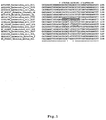
- rDNA 16S and 23S sequences available at the National Center for Biotechnology Information (NCBI) website http://www.ncbi.nlm.nih.gov/guide/ ), were chosen. This selection included six E. coli O157:H7, five E . coli non-O157:H7 serotypes and four other strains from related species belonging to the Enterobacteriaceae family ( Figure 1 ). Those sequences were aligned using the CluscalW software available at the European Bioinformatics Institute (EMBL-EBI, www.ebi.ac.uk/clustalw/ ).
- EBL-EBI European Bioinformatics Institute
- this invention also contemplates variations of the nucleotide sequences of the probes. Such variations may include deletions, insertions, among others. For example on of the following sequences:
- non-target sequences (this is, non- E . coli O157:H7 sequences) can have only one different nucleotide in comparison to the target sequences, and as such an increased level of discrimination is necessary to avoid non-specific hybridizations.
- hybridization and washing temperature were ranged between 53 and 61°C; fixation step using ethanol was ranged between 50 and 80% and finally different hybridization times were tested, 30, 45, 60 and 90 minutes.
- Trypticase Soy Broth is the most often used enrichment medium.
- Antibiotics such as novobiocin (most common), cefixime, cefsulodin or vancomycin and other selective compounds (such as bile salts - inhibit non- Enterobacteriaceae strains); are often added to this medium to promote a selective enrichment. These broths are then incubated for a period that usually varies between 16 and 24 hours (overnight growth) at 35 to 42°C.
- the data concerning the efficiency of enrichment protocols are very few and differ from study to study.
- E. coli 0157:H7 strains generally have an average optimal temperature around 40°C.
- ISO recommended for detection of E. coli 0157 in food samples ISO 16654:2001 - Microbiology of food and animal feeding stuffs - Horizontal method for the detection of Escherichia coli O157
- the good performance of mTSB may be related to the selective nature of the broth, that includes novobiocin and bile salts, which partially inhibit the growth of existing microflora in food.
- the composition of the broth (and not the selective factors) is the determining factor for bacterial growth.
- Growth rates of ⁇ 0.5 h -1 (41.5°C) and 0.4 h -1 (37°C) were observed for the strains cultivated in mTSB, while those grown in BPW had values of 0.07 h -1 for both temperatures.
- Another important feature to consider when optimizing FISH protocols is that some dietary components may present a strong autofluorescence signal, and thus interfere with the detection of the bacterium. In order to eliminate/reduce this phenomenon, ark additional step can be implemented before the hybridization procedure. Two different approaches were tested: one centrifugation step (to remove food particles) and using a detergent (Triton X-100, 1%) to emulsify the lipid compounds. Both steps have decreased autofluorescence signal, but the detergent showed a more significant reduction and also seemed to improve the fluorescence signal. This could arise from the detergent ability to help in the cellular permeabilization.
- the PNA-FISH method implementation can save at least 2 days in the detection of E . coli 0157:H7, when compared with the traditional protocol.
- Concentration Ground beef Unpasteurized milk (CFU/25g or ml) 37°C 41,5°C 37°C 41,5°C mTSB+N BPW mTBS+N BPW mTBS+N mTBS+N 100 + (6/6) a + (6/6) a + (6/6) a + (6/6) a + (6/6) a 10 + (6/6) a + (4/6) a + (6/6) a + (2/6) a +(6/6) a + (6/6) a 1 + (5/6) a - (0/6) a + (5/6) a - (0/6) a + (6/6) a + (6/6) a + (6/6) a 0.1 - (6/6) b - (6/6) b - (6/6) b (6/6) b - (6/6) b - (6/6) b 0.01 - (6/6) b - (6/6) b - (6/6) b - (6/6) b -
- This step can be performed in any epifluorescence microscope with a filter sensitive to fluorophore used.
- the kit of the present invention comprises a PNA probe at least 86% identical to SEQ ID No. 1 and another reagents or compositions that are selected to perform the test.
- the food, clinical or environmental samples were previously subjected to an enrichment step before application of the PNA probe. This happens because usually E. coli 0157:H7 presents low contamination levels.
- This enrichment step can be performed using the medium recommended for the conventional analysis method used for each sample. In the case of food, animal feed, feces or water, mTSB was used. Samples were then incubated for 18 - 22 hours at 37°C, at 120 rpm. Following the enrichment step, 15 ⁇ l of the culture medium were mixed with 15 ⁇ l of Triton X-100 (1%) directly in the microscope slide suitable for fluorescence. The slide was placed for approximately 5 minutes in an incubator oven at 59°C or left to air dry.
- the samples were immersed in a solution of 4% paraformaldehyde (wt/vol) and 50% ethanol (vol/vol) for 10 minutes each.
- hybridization solution containing: 10% (wt/vol) dextran sulfate (Sigma); 10 mM NaCl (Sigma); 30% (vol/vol) formamide (Sigma); 0.1% (wt/vol) sodium pyrophosphate (Sigma); 0.2% (wt/vol) polyvinylpirrolidone (Sigma); 0.2% (wt/vol) Ficol (Sigma); 5 mM disodium EDTA (Sigma); 0.1% (vol/vol) Triton X-100 (Sigma); 50 mM Tris-HCl (pH 7.5; Sigma); and 200nM of PNA probe.
- the samples were covered with coverslips (to assure an homogeneous spreading of the probe) placed in small wet boxes (to prevent the evaporation of the hybridization solution) protected from light and incubated for 45 minutes at 59°C.
- the coverslips were removed and the slides were immersed in a wash solution pre-warmed at 59°C containing 5 mM Tris Base (pH 10), 15 mM NaCl and 1% (vol/vol) Triton X-100. The samples were then placed in an oven at the hybridization temperature for 30 minutes. Subsequently, the slides were removed from the wash solution and dried at 59°C, in the same incubator, for approximately 5 minutes. Before the microscope visualization, a drop of non-fluorescent immersion oil (Merck) was placed and covered with a coverslip. The slides were kept in the dark for a maximum period of 24 hours before microscopy.
- a wash solution pre-warmed at 59°C containing 5 mM Tris Base (pH 10), 15 mM NaCl and 1% (vol/vol) Triton X-100.
- the samples were then placed in an oven at the hybridization temperature for 30 minutes. Subsequently, the slides were removed from the wash solution and dried at 59°C
- EXEMPLE 2 Salmonella and E . coli O157:H7 detection in feces.
- 25g of feces were mixture with 225 ml of mTSB. Different amount of sample can be used, but maintaining the 1/10 (wt/ vol) ratio. Samples were then incubated for 18 - 22 hours at 37°C, at 120 rpm. Following the enrichment step, 15 ⁇ l of the culture medium were mixed directly in a microscope slide suitable for fluorescence with 15 ⁇ l of 1% (vol/vol) Triton X-100 to minimize the interference from autofluorescent particles. The slide was placed for approximately 5 minutes in an incubator oven at 59°C or left to air dry.
- Hybridization was performed as previously described in Example 1 with a small change.
- the hybridization solution contained two probes: PNA probe for the detection of Salmonella and PNA probe for the detection of E. coli 0157:H7, each at a concentration of 200nM.
Landscapes
- Chemical & Material Sciences (AREA)
- Life Sciences & Earth Sciences (AREA)
- Analytical Chemistry (AREA)
- Proteomics, Peptides & Aminoacids (AREA)
- Organic Chemistry (AREA)
- Zoology (AREA)
- Wood Science & Technology (AREA)
- Health & Medical Sciences (AREA)
- Engineering & Computer Science (AREA)
- Microbiology (AREA)
- Immunology (AREA)
- Molecular Biology (AREA)
- Biotechnology (AREA)
- Biophysics (AREA)
- Physics & Mathematics (AREA)
- Biochemistry (AREA)
- Bioinformatics & Cheminformatics (AREA)
- General Engineering & Computer Science (AREA)
- General Health & Medical Sciences (AREA)
- Genetics & Genomics (AREA)
- Measuring Or Testing Involving Enzymes Or Micro-Organisms (AREA)
Abstract
PNA is a synthetic molecule analogue to DNA that, due to its physicochemical properties, allows a faster analysis with higher sensitivity and specificity than the DNA probes. These probes are combined with fluorescence in situ hybridization (FISH), a molecular biology technique that allows the direct visualization of the microorganisms in the sample. The combination of these two technologies rendered the FISH procedure to be faster, simpler and more efficient. This method can be applied to a great variety of samples such as food, blood, biopsies, feces, water and other clinical, environmental or agriculture and food industry samples. The present invention also includes the development of the kit of detection and respective procedure for the specific identification of Escherichia coli O157:H7 using the above referred sample types.
Description
- This invention relates to a process for the detection of microorganisms relevant for clinical and food safety. For that purpose, a PNA probe for the detection of Escherichia coli 0157:H7 serotype was developed.
- In addition to the probe, the present invention included the PNA-FISH procedure and its application to a kit for the detection and/or quantification of Escherichia coli 0157:H7, which can be used in the food and clinical fields.
- Escherichia coli species includes a genetically heterogeneous group of bacteria which are part of the normal microflora of the intestinal tract of humans and animals (Gyles, 2007). These bacteria are typically nonpathogens, but a considerable number of strains are pathogenic. Pathogenic strains of E. coli are classified according to their specific virulence factors, their pathogenicity and their specific strain. There are 6 classes of pathogenic E. coli, enterotoxigenic E. coli (ETEC), enteropathogenic E. coli (EPEC), enteroaggregative E. coli (EAEC), enteroinvasive E. coli (EIEC), the diffusely adherent E. coli (DAEC) and the enterohemorrhagic E. coli (EHEC) (Sheibani, 2007). Among pathogenic E. coli, enterohemorrhagic E. coli (EHEC) are perhaps the most important because of its virulence and its association with life-threatening complications (Sheibani, 2007). Within EHEC group, E. coli serotype 0157:H7 (the serotype is based on the 0 (Ohne) antigen, determined by the polysaccharide portion of cell wall lipopolysaccharide (LPS) and the H (Haunch) antigen due to flagella protein) is the most commonly isolated (Gyles, 2007).
- The infectious dose of E. coli O157:H7 is reported to be very low - around 1-100 CFU/mL - lower than most enteric pathogens (Robinson and McKillip, 2010). The virulence of this microorganism is due to 3 major factors. The first and by far the most critical, is the production of one or both phage-encoded Shiga toxins (Stxs), called Stx1 and Stx2. These Stxs are among the most potent cytotoxins currently known to affect eukaryotic cells (Robinson and McKillip, 2010). Although the type of Stx produced directly influences the severity of disease, the production of Stxs alone is insufficient for E. coli 0157:H7 became pathogenic. Instead, the assistance of the locus of enterocyte effacement (a bacterial pathogenicity island, that encodes a battery of specialized virulence factors) and the pO157 plasmid (encodes as type II secretion system, an enterohemolysin, a serine-protease, a catalase-peroxidase, a lymphocyte inhibitor, and potential adhesions, among others) is required (Robinson and McKillip, 2010).
- The effects of an EHEC infection are quite diverse, ranging from asymptomatic to lethal. The disease is self-limiting and the symptoms resolve itself after one week. In severe cases the patients can develop Hemolytic-Uremic Syndrome (HUS), Thrombocytopenic Thrombotic Purpura (TTP) or even die (Robinson and McKillip, 2010). HUS targets renal endothelial cells and can le ad to acute renal failure (Robinson and McKillip, 2010). HUS have considerably high mortality rate and in case of survival, patients are left with permanent renal consequences. In addition to typical HUS symptoms, TTP have associated neurological symptoms like severe headaches, convulsions, lethargy and encephalopathy (Robinson and McKillip, 2010). For the E. coli O157:H7 outbreaks reported in the USA, 25% of affected persons were hospitalized, 5 - 10% developed HUS or TTP and 1% died (Pennington, 2010).
- Regarding environmental reservoirs, cattle serve as the primary and natural reservoir of E. coli 0157:H7 and it is estimated that 10 - 80% of all cattle are colonized (Yoon and Hovde, 2008). Other animals such as goats, sheep and pigs may be carriers as well. Results from a study of 90 outbreaks confirmed microbiologically (between 1982 and 2006) showed that the source of transmission to humans was 42.2% associated to food, 12.2% to dairy products, 7.8% to animal contact, 6 - 7% to water, 2.2% to environmental and unknown in 28.9% of the outbreaks (Pennington, 2010).
- Before food products are shipped to consumer markets, they must be inspected for contamination during quality control measures. Because of the importance of quality control, the methods used to detect bacterial contaminants must be rapid, sensitive and reliable as well as versatile in order to accommodate the dynamic needs of the food processing plant and that can substantially cut down the time, labor and overall cost of detection process.
- Testing for sorbitol fermentation has been suggested as simple manner to screen E. coli 0157:H7, because it lacks the β-glucorunidase enzyme. Most existing culture methods were developed based on this feature. The bacteria inability to ferment rhamanose and tolerance to tellurite, have been also explored (Raji et al., 2003). These traditional plating techniques remain an integral aspect of quality control during food processing because they are both cost-effective and technically simple, with high level of accuracy and sensitivity. These plating techniques however, are very time consuming, laborious and fail to detect E. coli 0157:H7 which ferment sorbitol and are susceptible to tellurite (Raji et al., 2003). They also fail to detect pathogens in low numbers (< 200 CFU/g sample). Moreover, culture methods such as ISO 16654, usually include an agglutination assay (detecting 0157 or H7 antigen) that are not specific, since the 0157 and H7 antigen is present in other Escherichia coli species. These antibodies can also cross-react with other E. coli serotypes, Escherichia species and other members of the Enterobacteriaceae family (Raji et al., 2003).
- FISH is a molecular assay widely applied in identification of microorganisms. This method is based on the specific binding of small oligonucleotides (probes) to particular rRNA regions due to its high cellular abundance, universal distribution and use as a phylogenetic marker. The probe is coupled to a fluorochrome and after the hybridization a fluorescence signal can be detected due to the high rRNA copy number within the cell. More recently, peptide nucleic acid probes (PNA) have been developed for microbial detection (Cerqueira et al., 2008). These molecules mimic DNA and are capable of hybridizing specifically with complementary nucleic acids obeying to the Watson-Crick rules. The establish bond is stronger since in the PNA molecule, have a neutral repeated N-(2-aminoethil) glycin unit instead of the negative charged sugar-phosphate backbone. The adequate use of this molecule in FISH technology has become the procedure more robust, quicker and more efficient, which allowed the development of several PNA-FISH methods for the detection of pathogenic organisms (Cerqueira et al., 2008).
- In here, we have developed a new PNA FISH based method for the specific detection of E. coli 0157:H7 in food and or clinical samples. This is the first PNA-FISH method developed for detecting a specific serotype.
- The present invention refers to the development of a Peptide Nucleic Acid (PNA) probe and method thereof for the detection of Escherichia coli 0157:H7 serotype (that is, identification and quantification).
- The probe described in the present invention recognizes the 23S rRNA of the microorganism in question or the genomic sequences corresponding to the rRNA mentioned. The PNA probes have physiochemical characteristics that are inherent to its structure and when they are applied to a FISH-based method, allow a faster, more robust and more specific analysis than using a DNA probes.
- One of the advantages of this method is that the probe works robustly in a wide variety of biological samples, which usually does not happen with the other detection molecular methods.
- Another relevant aspect is the time required for detection. The method here developed matches the best times reported for the remaining molecular methods, even when the type of sample requires an enrichment step prior to the analysis. The rapidity and the reliability of the method can determine the appropriate and timely treatment of a contamination and/or infection for both clinical or food security perspectives.
- Another aspect of the present invention is related to the development of a kit based on the application of this blocker probe to fluorescence in situ hybridization (FISH), allowing the specific detection of E. coli 0157:H7 in a broad range of biological samples, in a prompt and simple way.
- In a preferred embodiment of the present invention, the PNA probe here described allow the detection of the target sequence in rRNA, in rDNA or in complementary sequences of the rRNA of E. coli 0157.
- One of the embodiments of the present invention is the description of a PNA probe used to the detection and/or quantification of E. coli 0157 characterized in that it has at least 86% of similarity to the sequence SEG ID No. 1 -5'- CAA CAC ACA GTG TC -3', preferably 87%, 88%, 89%, 90%, 91%, 92%, 93%, 94%, 95%, 96%, 97%, 98%, 99%, 100% of similarity to the sequence SEG ID No. 1 -5'- CAA CAC ACA GTG TC -3.
- In a more preferable embodiment of the present invention, the previously described sequence is linked to at least one type of detectable fraction. The type of detectable fraction to be used may be selected from one of the following groups: a conjugate, a branched detection system, a chromophore, a fluorophore, radioisotope, an enzyme, a hapten or a luminescent compound, among others.
- In an even more preferable embodiment, the fluorophore group can be at least one of the following: Alexa series fluorophores, cyanines, 5- (and -6) Carboxy-2', 7'-dichlorofluorescein, 5-ROX (5 -carboxy-X-rhodamine, triethylammonium salt), among others.
- It is still the subject of the present invention a kit for the specific detection of the presence or absence and/or quantification of E. coli 0157 in biological samples.
- In a more preferable embodiment of the present invention, the kit may additionally present at least one of the following solutions: a fixation solution, a hybridization solution and a washing solution.
- In yet another preferred embodiment of the present invention, the fixation solution can comprise paraformaldehyde and ethanol, namely 2-8% (weight/vol) of paraformaldehyde and 25-90% (vol/vol) of ethanol and/or hybridization solution may comprise formamide.
- It is still the object of the present invention the description of a method for the specific detection of E. coli 0157 or for the detection of E. coli 0157 in biological samples, which uses the PNA probe mentioned earlier and which comprises the following steps:
- contact of the PNA probe with biological samples;
- hybridization of the detecting probe with the target sequence of the microorganisms present within the biological samples;
- detection of the hybridization as an indication of the mentioned detection and quantification in the biological samples, the hybridization may be preferably carried on by fluorescence.
- The biological samples can be taken from food, feces, blood, air, water or biopsies among others.
- It is still the object of the present invention the use of the PNA probe described earlier, the use of the kit described earlier and the methodology to be applied in a methodology of detection of E. coli 0157, or detection of E. coli in biological samples.
- The present invention comprises the PNA probe, reagents, methods and a kit intended for the detection or quantification of E. coli 0157 strains.
- The PNA probe herewith described allows the specific detection of the E. coli 0157 through the bonding to rRNA, genomic sequences corresponding to rRNA (r), or their complementary sequences.
- The higher specificity of the PNA probes (in relation to DNA probes) allows a better discrimination between related nucleotide sequences. This has particular relevance to this probe since there are some phylogenetically related microorganisms to E. coli 0157 that present only one mismatch (nucleotide at position 8 of the probe described in this invention) within the selected target region. Some examples of this situation are the Escherichia coli non-O157:H7 strains.
- The PNA probe described in this invention has 14 nucleotides with the following nucleotidic sequence:
- SEQ ID No. 1 - 5'- CAA CAC ACA GTG TC -3'.
- This probe is applied to the analysis by fluorescence in situ hybridization (FISH), which, in the case of E. coli 0157 positive samples, results in the emission of detectable fluorescent signal either through fluorescence microscopy or through flow cytometry.
- The development of the new PNA-FISH probe was carried out empirically using specific software. The selection of the probe sequence was initially performed by aligning rDNA sequences of the target microorganism, with sequences of related microorganisms. This allowed the identification of potentially useful regions, which will be then evaluated based on other parameters such as specificity, hybridization temperature, percentage of guanine/cytosine, bond free energy and secondary structure.
- After the probe design and synthesis, the three steps of FISH procedure, fixation/permeabilization, hybridization and wash, have to be developed and optimized for the selected probe. This process usually involves the following parameters: temperature, concentration of formamide and ethanol, and hybridization and washing times. It is important to notice that due to the process complexity and the large number of variables, is not always possible to develop a method for each sequence and as such, several alternatives and sequences are often tested.
- A well-succeeded hybridization afterwards allows inferring about the presence/absence and even the concentration of a microorganism by fluorescence microscopy, flow cytometry or real time PCR. The detected fluorescent signal is generally the result of the specific binding of the small probes to tens or hundreds of rRNA copies present in the bacteria cytoplasm. That detectable fraction of the probe, which reports the existence of a stable complex formed by the probe and the target, is selected from one of the following groups: a conjugate, a branched detection system, a chromophore, a fluorophore, radioisotope, an enzyme, a hapten or a luminescent compound.
- The method described in the present invention comprises contact of a sample with at least one PNA probe with a similar sequence to that previously described. Consequently, the analysis is based on a single test with a definitive result in opposition to the conventional methods for the detection of E. coli 0157 that are based on phenotypic features and require several days to yield the result.
- It is still the object of the present invention a kit suitable for carrying out the test to detect specifically, this is, to find, identify or measure the E. coli 0157 present in biological samples. The kit comprises the PNA probe and other selected reagents or compounds needed to perform in situ hybridization tests.
- In a more preferable realization, the kit suitable for performing the assay for the detection, identification or quantification of E. coli O157 comprises additionally a fixation, hybridization and wash solution.
- Preferably, the method intends to be a diagnostic adjuvant for therapeutic decision and quality control. Thus, the implementation of this method for E. coli 0157 identification will allow the adequate clinical treatment to the bacterium and the early identification of the contamination source.
- The PNA probes can be applied directly on the sample prepared on a slide, since the application of these probes doesn't involve the use of reagents or enzymes for the permeabilization of the cellular membranes before the hybridization. However, some of the compounds that are frequently used in hybridization are required. Therefore, the probes are normally included in more user-friendly kits. If the desired approach involves the PNA-FISH analysis by flow cytometry, the probe could be applied to the sample in suspension, using the same hybridization compounds.
-
- a) As used herein, the term "nucleotide" includes natural and artificial molecules known generally by those who use technology related with nucleic acids, to thereby generate polymers that bind specifically to nucleic acids;
- b) When used the term "nucleotide sequence" is the same as referring to a segment of a polymer containing subunits, in this case the nucleotides;
- c) The term "target sequence" refers to a nucleotide sequence of E. coli 0157 that is intended to be detected in the test, where the portion of nucleotides of the probe is designed to hybridize;
- d) The term "PNA probe" refers to a polymer of subunits of PNA which has a nucleotide sequence and is specific to hybridize with a target sequence of the microorganism of interest. PNA molecules are DNA mimics in which the negatively charged sugar-phosphate backbone structure is replaced by an achiral and electrically neutral formed by repeated N - (2-aminoethyl) glycine units;
- e) When using the term "detectable fraction", it refers to molecules that can be connected to the probe, to thereby render the probe detectable by an instrument or method;
- f) The term "sample" refers to any biological sample that may contain the microorganism or target sequence for detection. The samples could be clinical (e.g. blood, urine, feces, etc.), food (e.g. meat, eggs, infant formulae, milk, etc.) or environmental (e.g. water).
-
Figure 1 presents the partial alignment of the 23S rDNA sequences for probe selection. The antisense complementary sequence of the EcoPNA1169 probe is shown above the alignment and the polymorphic positions are marked as well. - To identify potentially useful oligonucleotides sequences to use as probe, rDNA 16S and 23S sequences available at the National Center for Biotechnology Information (NCBI) website (http://www.ncbi.nlm.nih.gov/guide/), were chosen. This selection included six E. coli O157:H7, five E. coli non-O157:H7 serotypes and four other strains from related species belonging to the Enterobacteriaceae family (
Figure 1 ). Those sequences were aligned using the CluscalW software available at the European Bioinformatics Institute (EMBL-EBI, www.ebi.ac.uk/clustalw/). A conserved region in the 23S rRNA of all 0157:H7 strains of E. coli have been identified (figure 1 ). The criteria for the selection of the final sequence of the probe included: Guanine/Cytosine percentage; the type of secondary structures and hybridization temperature. After a evaluation of those parameters the following sequence was chosen: 5'-CAA CAC ACA GTG TC-3'. This sequence hybridizes between positions 1169 and 1183 of strain TW14359 E. coli 0157:H7 (accession number CP_001368). The probe was named EcoPNA1169 due to the initial position of the target sequence. Subsequently, the chosen sequence was synthesized and the oligonucleotide was attached, at the N terminus, to the fluorochrome Alexa Fluor594. - After the design of the probe, its performance was evaluated by determine the theoretical values for sensitivity and specificity. These parameters were evaluated with the above mentioned software ProbeCheck available in the ARB Silva database for rRNA. For this theoretical estimation only the good-quality sequences with at least 1900 bp were considered, as well as strains of E. coli with assigned serotype. The probe was aligned with a total of 180 344 sequences present in the database for the large RNA subunit (LSU, 23S/28S). The probe was also tested against the database for the small subunit (SSU, 16S/18S) to access the existence of possible cross-hybridization with the 16S rRNA sequences. Specificity was calculated as nECs/(TnECs) x 100, were nECs stands for the number of non-E. coli 0157:H7 strains not detected by the probe and TnECs is the total number of non-E. coli 0157:H7 strains examined. Sensitivity was calculated as ECs/(TECs) x 100, where ECs stands for the number of E. coli 0157:H7 strains detected by the probe and TECs is the total of number of E. coli 0157:H7 present in the database. Through ProbeCheck program, was possible to found that the EcoPNA1169 probe detect all 80 sequences of E. coli 0157:H7 existing in the database, and also 11 other sequences, performing a total of 91 sequences detected (last accessed, July 2012). Therefore, a theoretical specificity and sensitivity of 88 and 100% were obtained, respectively. The 11 non-E. coli O157:H7 strains detected by the EcoPNA1169 probe, included 3 non-O157 E. coli, 7 Salmonella spp. and 1 Cronobacter spp. strain.
- The PNA probe of this invention comprises preferably 14 nucleotides and may be at least 86% identical to the sequence SEQ ID No. 1 - 5'- CAA CAC ACA GTG TC -3', preferably 87%, 88%, 89%, 90%, 91%, 92%, 93%, 94%, 95%, 96%, 97%, 98%, 99%, 100% of similarity to SEQ ID No. 1 - 5'- CAA CAC ACA GTG TC -3'.
- Alternatively, this invention also contemplates variations of the nucleotide sequences of the probes. Such variations may include deletions, insertions, among others. For example on of the following sequences:
- SEQ ID No. 2 - 5'- AAC AAC ACA CAG TG -3';
- SEQ ID No. 3 - 5'- AAC ACA CAG TGT CG -3';
- SEQ ID No. 4 - 5'- AAC AAC ACA CAG TGT C -3';
- SEQ ID No. 5 - 5'- CAA CAC ATA GTG TC -3'.
- Not limited to the following examples, the detectable fraction of PNA probe can include various types of molecules such as dextran conjugates, chromophores, fluorophores, radioisotopes, enzymes, haptens, chemiluminescent compound, among others.
- As an example, among the fluorophores class those that are preferable for use are (but not limited to): Alexa series fluorophores, Alexa Fluor series, cyanines, 5 - (and -6) Carboxy-2',7'-dichlorofluorescein, 5-ROX (5-carboxy-X-rhodamine, triethylammonium salt).
- The present invention presents a method for detecting the presence of E. coli O157 using a nucleotide sequence with at least 86% of homology with the region of 14 nucleotides here described - SEQ ID No.1.
- The method can include the contact of a sample with the PNA probe described herein with the bacterium target sequence in appropriate hybridization conditions or appropriate in situ hybridization conditions (as shown in EXAMPLE 1).
- The method can be divided into: sample preparation (which includes the enrichment step, when necessary), fixation, hybridization, wash and visualization of the results (see EXAMPLE 1).
- The method can be performed on adhered or suspended cells.
- PNA-FISH methodology involves 4 steps, fixation and permeabilization of the sample; hybridization of the probe, wash of non-hybridized probe and observation on a fluorescence microscope. The following steps are a possible optimization of the hybridization conditions, without being a limitation of this invention:
- There are several factors that influence the hybridization of the PNA probes and the target sequence. These include the percentage of formamide (or other denaturing chemical reagent), salt concentration and consequently the ionic strength, ethanol percentage, temperature of hybridization and washing, detergent concentration, pH and others.
- To determine the optimal hybridization conditions, with high stringent conditions, it may be necessary to fix the different factors and change each factor individually until a desirable discriminatory degree is achieved.
- The closer a target sequence is from another non-target in the sample, the greater the stringency degree needed to define the various factors that influence the hybridization.
- In this invention non-target sequences (this is, non-E. coli O157:H7 sequences) can have only one different nucleotide in comparison to the target sequences, and as such an increased level of discrimination is necessary to avoid non-specific hybridizations.
- In order to understand the behavior of EcoPNA1169 probe and infer about the best hybridizations conditions, hybridization and washing temperature were ranged between 53 and 61°C; fixation step using ethanol was ranged between 50 and 80% and finally different hybridization times were tested, 30, 45, 60 and 90 minutes.
- After the optimization of all the parameters referred above, the procedure that was found to result in a stronger fluorescent signal was as follows:
- Smears of each bacterial culture (about 20 µl) in microscope slides appropriate for fluorescence visualization were prepared. The smears were immerse in 4% (wt/vol) para-formaldehyde (Sigma) followed by 50% (vol/vol) ethanol for 10 min each and allowed air dry. The smears were then covered with 20 µL of hybridization solution containing 10% (wt/vol) dextran sulfate (Sigma), 10 mM NaCl (Sigma), 30% (vol/vol) formamide (Sigma), 0.1% (wt/vol) sodium pyrophosphate (Sigma), 0.2% (wt/vol) polyvinylpyrrolidone (Sigma), 0.2% (wt/vol) Ficoll (Sigma), 5 mM disodium EDTA (Sigma), 0.1% (vol/vol) Triton X-100 (Sigma), 50 mM Tris-HCl (pH 7.5; Sigma) and 200 nM of PNA probe. Samples were covered with coverslipes, placed in moist chambers and incubated for 45 min at 59°C. Subsequently, the coverslipes were removed and the slides were submerged in a prewarmed (59°C) washing solution for 30 minutes, containing 15 mM NaCl (Sigma), 0,1% (vol/vol) Triton X-100 (Sigma) and 5 mM Tris base (pH 10; Sigma). Afterward, the slides were removed from the wash solution and dried at 59 °C in the same incubator for approximately 5 minutes. Before the microscope observation, a drop of non-fluorescent immersion oil (Merck) was placed and covered with a cover slip. The slides were stored in the dark for a maximum of 24h before microscopy.
- In order to evaluate the experimental specificity and sensitivity of the PNA probe, the above described protocol was applied to 53 strains. Those included 18 E. coli 0157:H7 and 25 non-E. coli 0157:H7. The two E. coli 0157 strains (CCC-18-12 and CCC-26-12) with uncharacterized "H" antigen, were excluded from the calculations. Additionally were included 8 taxonomically related stains belonging to the same genus and family (Escherichia, Salmonella, Enterobacter, Shigella and Klebsiella). The results show that hybridization occurs only with strains of E. coli 0157:H7 and therefore the sensitivity and specificity values obtained were both 100%.
Table 1 - Results of the EcoPNA1169 probe specificity and sensitivity test. Strain Serotype Isolation origin Verotox in production PNA FISH outcome E. coli CECT 4267 0157:H7 Human stool from outbreak of hemorrhagic colitis stx1, stx2 + E. coli CECT 4782 0157:H7 Human stool from outbreak of hemorrhagic colitis stx1, stx2 + E. coli CECT 4783 0157:H7 Raw hamburger meat implicated in hemorrhagic colitis outbreak stx1, stx2 + E. coli CECT 5947 0157:H7 - Gene stx2 has been replaced + E. a coli NCTC 12900 0157:H7 - NT + E. coli CCC-1-12* O157:H7 Faecal swab stx2 + E. coli CCC-5-12* 0157:H7 Faecal swab stx2 + E. coli CCC-7-12* 0157:H7 Faecal swab stx2 + E. coli CCC-10-12* 0157:H7 Faecal swab stx2 + E. coli CCC-11-12* O157:H7 Milk filter stx2 + E. coli CCC-12-12* 0157:H7 Milk filter stx2 + E. coli CCC-13-12* 0157:H7 Milk filter stx2 + E. coli CCC-14-12* 0157:H7 Bovine milk filter stx2 + E. coli CCC-15-12* 0157:H7 Bovine milk filter stx2 + E. coli CCC-16-12* 0157:H7 Caprine milk filter stx2 + E. coli CCC-18-12* 0157 Bovine milk filter stx2 + E. coli CCC-23-12* 0157:H7 Milk filter NT + E. coli CCC-24-12* 0157:H7 Milk filter NT + E. coli CCC-25-12* 0157:H7 Milk filter NT + E. coli CCC-26-12* 0157 Milk filter NT + E. coli CECT 352 0127a:K63(B8):H - - EPEC - E. coli CECT 504 0141:K85(B):H4 Swine oedema, ND - E. coli CECT 515T O1:K1(L1):H7 Human urine - cystitis ND - E. coli CECT 533 0103:K-:H- - ND - E. coli CECT 727 0111:K58(B4):H- Infantile gastroenteritis EPEC - E. coli CECT 730 O55:K59(B5):H- - ND - E. coli CECT 736 O28a,28c:K73 (B1 8):H- Faeces ND - E. coli CECT 740 O125a,125b:K70 ( B15):H19 Gastroenteritis ND - E. coli CECT 744 0158:K-:h23 Faeces of infant with diarrhoea ND - E. coli CECT 832 O111:K58 (B4) : H- Infantile gastroenteritis ND - E. coli .CECT 4537 O10 : K5 (L5) : H4 Human peritonitis ND - E. coli CECT 4555 097:K-:H- - ND - E. coli CCC-2-12* 0103 Faecal swab NT - E. coli CCC-3-12* 026 Faecal swab stx1, stx2 - E. coli CCC-4-12* 026 Faecal swab NT - E. coli CCC-8-12* O26 Faecal swab NT - E. coli CCC-9-12* O26 Faecal swab NT - E. coli CCC-19-12* O26 Caprine milk filter stx1 - E. coli CCC-20-12* O26 Caprine milk filter stx1 - E. coli CCC-21-12* O26 Bovine milk filter stx1 - E. coli CCC-22-12* O26 Bovine milk filter stx1 - Escherichi a coli CECT 434 O6 Clinical Isolate ND - Escherichi a coli N9* ND Porcine faeces ND - Escherichi a coli N5* ND Bovine faeces ND - Escherichi a coli ATCC 29425 (K12) OR:H48:K- - ND - Escherichi a hermanii ATCC 33650 NR Human isolate - - Escherichi a vulneris ATCC 29943 NR Human wound - - Shigella boydii ATCC 9207 NR - ND - Salmonella enterica enterica s orovar Typhimurium NCTC 12416 NR - - - Salmonella enterica enterica s orovar Typhi SGSC 3036 NR - - - Salmonella entritidis SGSC 2476 NR - - - Enterobact NR Child's - - er sakasaki CECT 858 throat Klebsiella pneumoniae ATCC 11296 NR - - - NT - non-toxigeninc E. coli; EPEC - enteropathogenic E. coli (epidemiologically implicated as pathogens, but virulence mechanism is not related to the excretion of enterotoxins); ND - Not determined; NR - non-relevant information for the present study; * - Isolates; SGSC - Salmonella Genetic Stock Centre; ATCC - American Type Culture Collection; NCTC - National Collection of Type Cultures; CECT - Spanish Type Culture Collection. - The samples to be analyzed can be obtained from food, water, feces, blood among others.
- The samples containing E. coli 0157:H7 usually present low contamination levels. Because of this, an enrichment step that facilitates the detection process is recommended. This enrichment step can be performed using several types of culture media, from complex rich media (such buffered peptone water and trypticase soy broth) to selective media, such as: GN (Gram Negative) broth, R & F® Enrichment Broth (R&F-EB) or E. coli (EC) broth (Vimont et al., 2006).
- Trypticase Soy Broth (TSB) is the most often used enrichment medium. Antibiotics such as novobiocin (most common), cefixime, cefsulodin or vancomycin and other selective compounds (such as bile salts - inhibit non-Enterobacteriaceae strains); are often added to this medium to promote a selective enrichment. These broths are then incubated for a period that usually varies between 16 and 24 hours (overnight growth) at 35 to 42°C. However, the data concerning the efficiency of enrichment protocols are very few and differ from study to study.
- The incubation temperature does not seem to be related to the serotype sought (Vimont et al., 2006.), but some authors have demonstrated that E. coli 0157:H7 strains generally have an average optimal temperature around 40°C. Actually the ISO recommended for detection of E. coli 0157 in food samples (ISO 16654:2001 - Microbiology of food and animal feeding stuffs - Horizontal method for the detection of Escherichia coli O157) includes a pre-enrichment in the broth mTSB at 41.5°C.
- In order to evaluate the influence of the enrichment medium and incubation temperature on the detection limit of the PNA-FISH method, two different broths at 37 and 41.5°C were tested. For this test the chosen broths were the selective medium mTSB with novobiocin (mTSB + N), that is currently recommended by the International Organization for Standardization (ISO 16654:2001) and the BPW (peptonated buffered water), a non-selective broth widely used in enrichment protocols of several pathogens, which has also been applied to the detection of E. coli O157: H7.
- The PNA probe described in this invention was tested in two different types of samples: ground beef and unpasteurized milk (two commonly associated matrices with infections by E. coli O157:H7) artificially contaminated with concentrations ranging from 0.01 to 100 CFU/g or 25 ml of food sample. Two strains of E. coli 0157:H7 were used (CCC-5-12 and CECT 4267) for inoculation and the samples were simultaneously analyzed by ISO 16654:2001. As seen in Table 2, the mTSB gave the best detection limit, while the use of higher temperatures (41.5°C) do not seem to improve the detection rate of E. coli 0157. The good performance of mTSB may be related to the selective nature of the broth, that includes novobiocin and bile salts, which partially inhibit the growth of existing microflora in food. However, after determining the growth rate of both strains of E. coli in BPW and mTSB, was observed that the composition of the broth (and not the selective factors) is the determining factor for bacterial growth. Growth rates of ≈ 0.5 h-1 (41.5°C) and 0.4 h-1 (37°C) were observed for the strains cultivated in mTSB, while those grown in BPW had values of 0.07 h-1 for both temperatures. To better quantify the performance of each enrichment method, the sensitivity and specificity for both media, mTSB and BPW at 37°C were determined based on the results presented in Table 3. The obtained specificity values were 100% for both broths (confidence interval [CI] 95% from 69.87 to 100), while the sensitivity values were 94.44% (95% CI, 70.63 to 99.71) to mTSB and 55.55% (95% CI, 31.35 to 77.59) for BPW. Although the best performance has been found to mTSB + N, it shall be possible to standardize the enrichment step to allow the simultaneous detection of different food pathogens.
- Another important feature to consider when optimizing FISH protocols is that some dietary components may present a strong autofluorescence signal, and thus interfere with the detection of the bacterium. In order to eliminate/reduce this phenomenon, ark additional step can be implemented before the hybridization procedure. Two different approaches were tested: one centrifugation step (to remove food particles) and using a detergent (Triton X-100, 1%) to emulsify the lipid compounds. Both steps have decreased autofluorescence signal, but the detergent showed a more significant reduction and also seemed to improve the fluorescence signal. This could arise from the detergent ability to help in the cellular permeabilization.
- Regarding the detection time of the assay, the PNA-FISH method implementation can save at least 2 days in the detection of E. coli 0157:H7, when compared with the traditional protocol.
- In conclusion, we found that the here described PNA-WISH method has a detection limit of 1 CFU per 25 g of food after an overnight enrichment step on mTSB. The comparison with the traditional culture method showed a value of 100% specificity and 94% sensitivity. Finally, it was observed that the use of selective compounds and higher temperature lead to a narrow improvement regarding the detection limit of the method.
Table 2 - PNA FISH results obtained in the detection of E. coli 0157:H7 in different food matrices inoculated with bacterial concentrations between 0.01 and 25 g or 100 CFU per ml of food. The results include 3 independent experiments. Concentration Ground beef Unpasteurized milk (CFU/25g or ml) 37°C 41,5°C 37°C 41,5°C mTSB+N BPW mTBS+N BPW mTBS+N mTBS+N 100 + (6/6)a + (6/6)a + (6/6)a + (6/6)a + (6/6)a + (6/6)a 10 + (6/6)a + (4/6)a + (6/6)a + (2/6)a +(6/6) a + (6/6)a 1 + (5/6)a - (0/6)a + (5/6)a - (0/6)a + (6/6) a + (6/6)a 0.1 - (6/6)b - (6/6)b - (6/6)b (6/6)b - (6/6)b - (6/6)b 0.01 - (6/6)b - (6/6)b - (6/6)b - (6/6)b - (6/6)b - (6/6)b a - Samples that tested positive by PNA FISH/Total positive samples determined by culture method; b - samples that tested negative by PNA-FISH/Total negative samples determined by culture. 
- This step can be performed in any epifluorescence microscope with a filter sensitive to fluorophore used. Other filters present in the microscope, which are not able to detect the fluorescent signal of the probe, were used to confirm the absence of autofluorescence.
- The present invention also refers to a kit that allows testing for the presence of E. coli O157:H7.
- The kit of the present invention comprises a PNA probe at least 86% identical to SEQ ID No. 1 and another reagents or compositions that are selected to perform the test.
- The PNA probe, its characteristics, the methods and kit of this invention are suitable for the analysis of nucleic acids sequences present, or not, internally in the target organism. As such, this invention can be used for both, the organism or nucleic acids extracted or derived from the organism of interest, implying that the source of the target sequence is not a limitation on this invention.
- The following examples illustrate different situations and several steps for implementing the invention, are preferred embodiments of the present invention, without intending to limit any of them:
- SEQ ID No. 1 - 5'- CAA CAC ACA GTG TC -3 (coupled to Alexa Fluor 594).
- The food, clinical or environmental samples were previously subjected to an enrichment step before application of the PNA probe. This happens because usually E. coli 0157:H7 presents low contamination levels. This enrichment step can be performed using the medium recommended for the conventional analysis method used for each sample. In the case of food, animal feed, feces or water, mTSB was used. Samples were then incubated for 18 - 22 hours at 37°C, at 120 rpm. Following the enrichment step, 15 µl of the culture medium were mixed with 15 µl of Triton X-100 (1%) directly in the microscope slide suitable for fluorescence. The slide was placed for approximately 5 minutes in an incubator oven at 59°C or left to air dry.
- For preventing the loss of 23S rRNA during the hybridization process, the samples were immersed in a solution of 4% paraformaldehyde (wt/vol) and 50% ethanol (vol/vol) for 10 minutes each.
- After fixation, samples were then covered with a drop of hybridization solution containing: 10% (wt/vol) dextran sulfate (Sigma); 10 mM NaCl (Sigma); 30% (vol/vol) formamide (Sigma); 0.1% (wt/vol) sodium pyrophosphate (Sigma); 0.2% (wt/vol) polyvinylpirrolidone (Sigma); 0.2% (wt/vol) Ficol (Sigma); 5 mM disodium EDTA (Sigma); 0.1% (vol/vol) Triton X-100 (Sigma); 50 mM Tris-HCl (pH 7.5; Sigma); and 200nM of PNA probe. The samples were covered with coverslips (to assure an homogeneous spreading of the probe) placed in small wet boxes (to prevent the evaporation of the hybridization solution) protected from light and incubated for 45 minutes at 59°C.
- After the hybridization time, the coverslips were removed and the slides were immersed in a wash solution pre-warmed at 59°C containing 5 mM Tris Base (pH 10), 15 mM NaCl and 1% (vol/vol) Triton X-100. The samples were then placed in an oven at the hybridization temperature for 30 minutes. Subsequently, the slides were removed from the wash solution and dried at 59°C, in the same incubator, for approximately 5 minutes. Before the microscope visualization, a drop of non-fluorescent immersion oil (Merck) was placed and covered with a coverslip. The slides were kept in the dark for a maximum period of 24 hours before microscopy.
- The results were obtained through the observation in a fluorescence microscope with a filter capable of detecting the fluorochrome Alexa Fluor 594 bonded to the PNA detecting probe.
- This example illustrates the possibility of using the probe for E. coli 0157:H7 together with the probe for Salmonella spp. previous developed in Almeida et al., 2010. Both these two bacteria are a common cause of gastroenteritis and as such, a fast detection in feces is of the most importance. The use of a "multiplex" approach (simultaneous use of multiple probes) can simplify and expedite the detection procedure. These two probes present very similar hybridization temperatures and can be used easily in a multiplex assay.
- SEQ ID No. 1 - 5'- CAA CAC ACA GTG TC -3' (coupled to Alexa Fluor 594).
- Sequence from Almeida et al., 2010 paper: 5'- AGG AGC TTC GCT TGC -3' (coupled to Alexa Fluor 448).
- 25g of feces were mixture with 225 ml of mTSB. Different amount of sample can be used, but maintaining the 1/10 (wt/ vol) ratio. Samples were then incubated for 18 - 22 hours at 37°C, at 120 rpm. Following the enrichment step, 15 µl of the culture medium were mixed directly in a microscope slide suitable for fluorescence with 15 µl of 1% (vol/vol) Triton X-100 to minimize the interference from autofluorescent particles. The slide was placed for approximately 5 minutes in an incubator oven at 59°C or left to air dry.
- Hybridization was performed as previously described in Example 1 with a small change. The hybridization solution contained two probes: PNA probe for the detection of Salmonella and PNA probe for the detection of E. coli 0157:H7, each at a concentration of 200nM.
- Washing was carried out according to the procedure described in Example 1.
- The results were obtained by observation under a fluorescence microscope with the appropriate filters for the detection of Alexa Fluor 594 and 488 fluorochrome connected to the PNA probes.
-
- Gyles CL: Shiga toxin-producing Escherichia coli: an overview. J Anim Sci 2007, 85(13 Suppl):E45-62.
- Pennington H: Escherichia coli 0157. Lancet 2010, 376(9750):1428-1435.
- Robinson AL, McKillip J: Biology of Escherichia coli O157:H7 in human health and food safety with emphasis on sublethal injury and detection. In: Current Research, Technology and Education Topics in Applied Microbiology and Microbial Biotechnology. Edited by Mendez-Vilas A: Formatex; 2010: 1096-1105.
- Raji MA, Jiwa SF, Minga MU, Gwakisa PS: Escherichia coli 0157: H7 reservoir, transmission, diagnosis and the African situation: a review. East Afr Med J 2003, 80(5):271-276.
- Cerqueira L, Azevedo NF, Almeida C, Jardim T, Keevil CW, Vieira MJ: DNA mimics for the rapid identification of microorganisms by fluorescence in situ hybridization (FISH). Int J Mol Sci 2008, 9(10):1944-1960.
- Almeida C, Azevedo NF, Fernandes RM, Keevil CW, Vieira MJ: Fluorescence in situ hybridization method using a peptide nucleic acid probe for identification of Salmonella spp. in a broad spectrum of samples. Appl Environ Microbiol 2010, 76(13):4476-4485.
- Vimont A, Vernozy-Rozand C, Delignette-Muller ML: Isolation of E. coli 0157:H7 and non-O157 STEC in different matrices: review of the most commonly used enrichment protocols. Lett Appl Microbiol 2006, 42(2):102-108.
Claims (16)
- PNA probe for the detection and/or quantification of E. coli 0157:H7 serotype characterized in that it comprises at least one sequence with at least 86% similarity to SEQ ID No. 1 5'- CAA CAC ACA GTG TC -3'.
- PNA probe, according to claim 1, characterized in that it comprises at least one sequence SEG ID No. 1 and considering variations in the nucleotide sequences of the probes for instance in the following sequences SEQ ID No. 2, SEQ ID No. 3, SEQ ID No. 4, SEQ ID No. 5.
- PNA probe, according to claim 1, characterized in that it is capable of detecting the target sequence in rRNA, rDNA or the sequences complementary to E. coli 0157:H7 rRNA.
- PNA probe, according to claim 1, characterized in that it additionally comprises one sequence with at least 86% similarity to SEQ ID No 5.
- PNA probe, according to any of the claims 1 - 4, characterized in that it is connected to at least one type of detectable fraction.
- PNA probe, according to claim 5, characterized in that the type of detectable fraction of the probe is selected from one of the following groups: a conjugate, a branched detection system, a chromophore, a fluorophore, radioisotope, an enzyme, a hapten or a luminescent compound.
- PNA probe, according to claim 6, characterized in that the fluorophore group is at least one of the following: fluorophores of the Alexa series, Alexa Fluor series, cyanines, 5-(and -6) Carboxy-2',7'-dichlorofluorescein, the 5-ROX (5-carboxy-X-rhodamine, triethylammonium salt).
- Kit for detecting E. coli 0157:H7, characterized in that it comprises at least one of the probes described in any of the claims 1 to 7.
- Kit, according to claim 8, characterized in that it further comprises at least one of the following solutions: one fixation solution, one hybridization solution and one washing solution.
- Kit, according to claim 9, characterized in that the fixation solution comprises paraformaldehyde and ethanol, namely 2-8% (wt/vol) of paraformaldehyde and 25-90% (vol/vol) of ethanol.
- Kit, according to claim 9, characterized in that the hybridization solution comprises formamide.
- A method for the specific detection of E. coli 0157:H7, characterized in that it uses the PNA probes described in claims 1 to 7 and it comprises the following steps:a. PNA probe contact with the biological samples;b. PNA probe hybridization with the target sequence of the microorganisms present in the referred samples;c. Hybridization detection as indication of the referred detection and quantification of the referred samples.
- Method, according to claim 12, characterized in that the biological sample is derived from food, feces, blood, air, water or biopsies.
- Method, according to claim 12, characterized in that the hybridization occurs by fluorescence.
- Use of PNA probes, as described in any one of claims 1 to 7, characterized in that it is applied in a methodology for detecting E. coli 0157:H7 in biological samples.
- Use of the kit, as described in any of claims 8 to 11, characterized in that it is applied in the detection of E. coli O157:H7 in biological samples.
Applications Claiming Priority (2)
| Application Number | Priority Date | Filing Date | Title |
|---|---|---|---|
| PT2013691613 | 2013-04-30 | ||
| PCT/PT2014/000023 WO2014178741A2 (en) | 2013-04-30 | 2014-04-30 | Peptide nucleic acid probe, kit and method for detecting and/or quantifying escherichia coli o157:h7 and respective uses |
Publications (1)
| Publication Number | Publication Date |
|---|---|
| EP2993237A2 true EP2993237A2 (en) | 2016-03-09 |
Family
ID=55229441
Family Applications (1)
| Application Number | Title | Priority Date | Filing Date |
|---|---|---|---|
| EP14733740.6A Withdrawn EP2993237A2 (en) | 2013-04-30 | 2014-04-30 | Peptide nucleic acid (pna) probe, kit and method for detecting and/or quantifying escherichia coli 0157 : h7 |
Country Status (1)
| Country | Link |
|---|---|
| EP (1) | EP2993237A2 (en) |
Cited By (2)
| Publication number | Priority date | Publication date | Assignee | Title |
|---|---|---|---|---|
| CN111575391A (en) * | 2020-05-12 | 2020-08-25 | 浙江省检验检疫科学技术研究院 | Identification method, kit and probe for detecting Escherichia coli O157: H7 by liquid phase in situ fluorescence hybridization |
| WO2025046313A1 (en) * | 2023-08-30 | 2025-03-06 | Foss Analytical A/S | Combined pre-treatment and hybridization of nucleic acids |
-
2014
- 2014-04-30 EP EP14733740.6A patent/EP2993237A2/en not_active Withdrawn
Non-Patent Citations (1)
| Title |
|---|
| See references of WO2014178741A2 * |
Cited By (2)
| Publication number | Priority date | Publication date | Assignee | Title |
|---|---|---|---|---|
| CN111575391A (en) * | 2020-05-12 | 2020-08-25 | 浙江省检验检疫科学技术研究院 | Identification method, kit and probe for detecting Escherichia coli O157: H7 by liquid phase in situ fluorescence hybridization |
| WO2025046313A1 (en) * | 2023-08-30 | 2025-03-06 | Foss Analytical A/S | Combined pre-treatment and hybridization of nucleic acids |
Similar Documents
| Publication | Publication Date | Title |
|---|---|---|
| Almeida et al. | Detection of Escherichia coli O157 by peptide nucleic acid fluorescence in situ hybridization (PNA-FISH) and comparison to a standard culture method | |
| US8883441B2 (en) | Method for detecting and counting micro-organisms in a sample | |
| EP0669399A2 (en) | Oligonucleotides and method for detecting bacteria | |
| JP2008283895A (en) | Diarrheagenic Escherichia coli detection method | |
| CN101363061B (en) | Fluorescent quantitative PCR method by using taqman probe for detecting salmonella in food | |
| CN102367475B (en) | M-PCR (multiplex-polymerase chain reaction) detection kit for different serotype vibrio cholerae and detection method thereof | |
| Fach et al. | Comparison of different PCR tests for detecting Shiga toxin-producing Escherichia coli O157 and development of an ELISA-PCR assay for specific identification of the bacteria | |
| US20160273025A1 (en) | Peptide nucleic acid probe, kit and method for the detection and/or quantification of esherichia coli o157:h7 and applications thereof | |
| Cano et al. | Detection of salmonellas by DNA hybridization with a fluorescent alkaline phosphatase substrate | |
| Yousef | Detection of bacterial pathogens in different matrices: current practices and challenges | |
| EP2993237A2 (en) | Peptide nucleic acid (pna) probe, kit and method for detecting and/or quantifying escherichia coli 0157 : h7 | |
| US20050064444A1 (en) | Method for specific fast detection of relevant bacteria in drinking water | |
| JP2005515756A6 (en) | Method for specific and rapid detection of related bacteria in drinking water | |
| JP2011212030A (en) | Genetic probe for quickly and specifically counting alive food-poisoning bacteria and hygiene index bacteria by in situ hybridization method combined with cultivation, and method thereof (vibrio parahaemolyticus) | |
| JP2009535052A (en) | Microbial detection and counting method | |
| EP2553120B1 (en) | Peptide nucleicacid probe, kit and method for detection and/or quantification of salmonella spp. and applications thereof | |
| Kumar | Modern trends to investigate foodborne Listeriosis | |
| JP2006166912A (en) | Gene probe for rapid and specific counting of live food poisoning bacteria and hygiene indicator bacteria by in situ hybridization combined with culture and its method | |
| Ashbolt | Methods to identify and enumerate frank and opportunistic bacterial | |
| Foroohi et al. | Evaluation and optimization of a loop-mediated isothermal amplification (LAMP) assay for the detection of Salmonella enterica serovar Typhi isolated from food samples | |
| Sutherland et al. | Cultural, serological, and genetic methods for identification of bacteria | |
| Thomson-Carter | General recovery, characterisation and typing protocols for VTEC | |
| JP2001136969A (en) | Oligonucleotide for detecting intestinal bacteria and method for detecting intestinal bacteria | |
| Guan | Quantitative detection of Escherichia coli O157: H7 in ground beef by the polymerase chain reaction incorporating immunomagnetic separation | |
| KR20080028633A (en) | Oligonucleotide primers for the detection of Salmonella and Campillar bacterium, early diagnostic kits including the same, and detection methods using the same |
Legal Events
| Date | Code | Title | Description |
|---|---|---|---|
| PUAI | Public reference made under article 153(3) epc to a published international application that has entered the european phase |
Free format text: ORIGINAL CODE: 0009012 |
|
| 17P | Request for examination filed |
Effective date: 20151106 |
|
| AK | Designated contracting states |
Kind code of ref document: A2 Designated state(s): AL AT BE BG CH CY CZ DE DK EE ES FI FR GB GR HR HU IE IS IT LI LT LU LV MC MK MT NL NO PL PT RO RS SE SI SK SM TR |
|
| AX | Request for extension of the european patent |
Extension state: BA ME |
|
| DAX | Request for extension of the european patent (deleted) | ||
| STAA | Information on the status of an ep patent application or granted ep patent |
Free format text: STATUS: THE APPLICATION IS DEEMED TO BE WITHDRAWN |
|
| 18D | Application deemed to be withdrawn |
Effective date: 20160702 |