EP3305918B1 - Verfahren für epigenetische sequenzierung - Google Patents
Verfahren für epigenetische sequenzierung Download PDFInfo
- Publication number
- EP3305918B1 EP3305918B1 EP17201280.9A EP17201280A EP3305918B1 EP 3305918 B1 EP3305918 B1 EP 3305918B1 EP 17201280 A EP17201280 A EP 17201280A EP 3305918 B1 EP3305918 B1 EP 3305918B1
- Authority
- EP
- European Patent Office
- Prior art keywords
- cells
- dna
- droplets
- cell
- adapters
- Prior art date
- Legal status (The legal status is an assumption and is not a legal conclusion. Google has not performed a legal analysis and makes no representation as to the accuracy of the status listed.)
- Active
Links
Images








Classifications
-
- C—CHEMISTRY; METALLURGY
- C12—BIOCHEMISTRY; BEER; SPIRITS; WINE; VINEGAR; MICROBIOLOGY; ENZYMOLOGY; MUTATION OR GENETIC ENGINEERING
- C12Q—MEASURING OR TESTING PROCESSES INVOLVING ENZYMES, NUCLEIC ACIDS OR MICROORGANISMS; COMPOSITIONS OR TEST PAPERS THEREFOR; PROCESSES OF PREPARING SUCH COMPOSITIONS; CONDITION-RESPONSIVE CONTROL IN MICROBIOLOGICAL OR ENZYMOLOGICAL PROCESSES
- C12Q1/00—Measuring or testing processes involving enzymes, nucleic acids or microorganisms; Compositions therefor; Processes of preparing such compositions
- C12Q1/68—Measuring or testing processes involving enzymes, nucleic acids or microorganisms; Compositions therefor; Processes of preparing such compositions involving nucleic acids
- C12Q1/6869—Methods for sequencing
-
- B—PERFORMING OPERATIONS; TRANSPORTING
- B01—PHYSICAL OR CHEMICAL PROCESSES OR APPARATUS IN GENERAL
- B01F—MIXING, e.g. DISSOLVING, EMULSIFYING OR DISPERSING
- B01F33/00—Other mixers; Mixing plants; Combinations of mixers
- B01F33/30—Micromixers
- B01F33/302—Micromixers the materials to be mixed flowing in the form of droplets
- B01F33/3021—Micromixers the materials to be mixed flowing in the form of droplets the components to be mixed being combined in a single independent droplet, e.g. these droplets being divided by a non-miscible fluid or consisting of independent droplets
-
- B—PERFORMING OPERATIONS; TRANSPORTING
- B01—PHYSICAL OR CHEMICAL PROCESSES OR APPARATUS IN GENERAL
- B01L—CHEMICAL OR PHYSICAL LABORATORY APPARATUS FOR GENERAL USE
- B01L3/00—Containers or dishes for laboratory use, e.g. laboratory glassware; Droppers
- B01L3/50—Containers for the purpose of retaining a material to be analysed, e.g. test tubes
- B01L3/502—Containers for the purpose of retaining a material to be analysed, e.g. test tubes with fluid transport, e.g. in multi-compartment structures
- B01L3/5027—Containers for the purpose of retaining a material to be analysed, e.g. test tubes with fluid transport, e.g. in multi-compartment structures by integrated microfluidic structures, i.e. dimensions of channels and chambers are such that surface tension forces are important, e.g. lab-on-a-chip
- B01L3/502769—Containers for the purpose of retaining a material to be analysed, e.g. test tubes with fluid transport, e.g. in multi-compartment structures by integrated microfluidic structures, i.e. dimensions of channels and chambers are such that surface tension forces are important, e.g. lab-on-a-chip characterised by multiphase flow arrangements
- B01L3/502784—Containers for the purpose of retaining a material to be analysed, e.g. test tubes with fluid transport, e.g. in multi-compartment structures by integrated microfluidic structures, i.e. dimensions of channels and chambers are such that surface tension forces are important, e.g. lab-on-a-chip characterised by multiphase flow arrangements specially adapted for droplet or plug flow, e.g. digital microfluidics
Definitions
- the present invention generally relates to microfluidics and epigenetic sequencing.
- Epigenetics is the study of the transmission of genetic information by mechanisms other than the DNA sequence of nucleotides.
- epigenetic information may be transmitted via methylation of nucleotides within DNA (e.g., cytosine to 5-methylcytosine), or by histone modifications such as histone acetylation or deacetylation, methylation, ubiquitylation, phosphorylation, sumoylation, etc.
- histone modifications such as histone acetylation or deacetylation, methylation, ubiquitylation, phosphorylation, sumoylation, etc.
- Such epigenetic modifications may affect the structure of chromatin, which is a higher-order structure of protein, DNA and RNA within cells. Chromatin structure is known to play an important role in regulating genome function and in particular, its varied structure across cell types helps ensure that the correct genes are expressed in the correct cell types.
- ChIP-seq advantages and challenges of a maturing technology.
- the present invention generally relates to microfluidics and epigenetic sequencing.
- the subject matter of the present description involves, in some cases, interrelated products, alternative solutions to a particular problem, and/or a plurality of different uses of one or more systems and/or articles.
- the present description is generally directed to a method comprising acts of providing a plurality of cells contained within a plurality of microfluidic droplets, lysing the cells contained within the microfluidic droplets to produce cell lysates therein, exposing at least some of the cell lysates contained within the microfluidic droplets to a non-nucleosome-cleaving nuclease to produce a plurality of nucleosome sequences within the microfluidic droplets, ligating adapters to at least some of the nucleosome sequences, the adapters comprising an identification sequence and a restriction site, and sequencing the nucleosome sequences containing ligated adapters.
- the present description is generally directed to a method of providing a solution comprising a plurality of nucleosome sequences originating from a plurality of cells, at least some of the nucleosome sequences being ligated to an adapter, the adapter comprising an identification sequence and a restriction site, wherein nucleosome sequences originating from the same cell contain identical identification sequences, and nucleosome sequences originating from different cells contain different identification sequences, and sequencing at least some of the nucleosome sequences.
- the present description in yet another set of embodiments, is generally directed to a composition
- a composition comprising a plurality of droplets, at least some of which each contain a nucleosome sequence ligated to an adapter, the adapter comprising an identification sequence and a restriction site, wherein nucleosome sequences originating from the same cell contain identical identification sequences, and nucleosome sequences originating from different cells contain different identification sequences.
- the present description is generally directed to a composition
- a composition comprising a nucleic acid sequence comprising an identification sequence, a restriction site, an inversion of the restriction site, and an inversion of the identification sequence.
- the present description is generally directed to a composition comprising a plurality of palindromic or substantially palindromic nucleic acid sequence comprising a plurality of different identification sequences each having the same length, and a substantially identical restriction site.
- present description encompasses methods of making one or more of the embodiments described herein. In still another aspect, the present description encompasses methods of using one or more of the embodiments described herein.
- the present invention generally relates to microfluidics and epigenetic sequencing.
- cells contained within a plurality of microfluidic droplets are lysed and the DNA (e.g., from nucleosomes) within the droplets are labeled, e.g., with adapters containing an identification sequence.
- the adapters may also contain other sequences, e.g., restriction sites, primer sites, etc., to assist with later analysis.
- the DNA from the different cells may be combined and analyzed, e.g., to determine epigenetic information about the cells.
- the DNA may be separated on the basis of certain modifications (e.g., methylation) using techniques such as chromatin immunoprecipitation ("ChIP"), and the DNA from the separated nucleosomes may be sequenced and aligned with genomes, e.g., to determine which portions of the genome were epigenetically modified, e.g., via methylation.
- ChIP chromatin immunoprecipitation
- various aspects of the present description are generally directed through DNA sequencing to determine epigenetic information.
- cells contained within microfluidic droplets are lysed and their DNA is exposed to certain enzymes, such as non-nucleosome-cleaving nucleases or other enzymes that are able to cleave the DNA for later analysis without destroying certain types of epigenetic information, e.g., the interaction of the DNA with the histones, the methylation patterns within DNA, etc.
- the DNA (still contained within the microfluidic droplets) is then barcoded or tagged using a ligation step with specific "adapters" that can be used to later identify the source, down to a single cell level in some cases, of the DNA.
- the DNA may then be sequenced.
- droplets containing the lysate of different cells may be combined together prior to analysis. Due to the presence of the ligated adapters, the DNA originating from the same cell may contain identical identification sequences, while DNA originating from different cells may contain different identification sequences, thereby allowing the DNA of each cell within a sample to be separately identified and determined.
- the identification sequences may be selected such that a certain number of cells taken from a plurality of cells can be readily identified. For example, an identification sequence of n nucleotides may allow for up to 4 n distinct cells to be studied. Thus, even relatively small identification sequences (e.g., 4, 5, 6, 7, 8, 9, etc. nucleotides long) may allow for relatively large populations of cells to be separately determined, e.g., at a single-cell level.
- a cell (containing chromatin) is contained within a microfluidic droplet.
- the cell is lysed and exposed to an enzyme such as MNase, which cleaves the DNA released from the lysed cell into smaller fragments without substantially affecting those portions of the DNA that interact with the histones within the nucleosome structures.
- An enzyme such as MNase, which cleaves the DNA released from the lysed cell into smaller fragments without substantially affecting those portions of the DNA that interact with the histones within the nucleosome structures.
- Barcodes or other adapters which may be formed from certain types of sequences as is discussed herein, are added to the droplet and ligated together.
- ChIP analysis is performed and the DNA sequenced.
- a plurality of cells are contained within droplets using a microfluidic droplet maker, e.g., within an aqueous environment contained within an oil.
- the cells are encapsulated within droplets at a density such that on average, each droplet contains one cell (or less).
- a cell may be lysed, then exposed to an enzyme such as MNase, which cleaves the DNA released from the lysed cell into smaller fragments without substantially affecting those portions of the DNA that interact with the histones within the nucleosome structures.
- Droplets containing an adapter (as discussed below), a ligase, and/or other components such as buffers or the like are also created separately, as shown in Fig. 1A , part 2. Similar to Fig. 1A , part 1, the adapters may be contained in a droplet within an aqueous environment, contained within an oil.
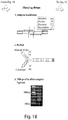
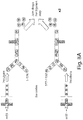
- FIG. 1B A schematic diagram of an example of an adapter is shown in Fig. 1B , part 1, and Fig. 3 .
- the adapter in this example comprises a sequence recognizable by a primer (e.g., a PCR primer), a "barcode" or other identification sequence, and a restriction site that can be cleaved by a suitable restriction endonuclease.
- the identification site typically has 4-15 nucleotides, and for a population of adapters to be used to identify a population of cells, the identification sequence of the adapter may differ while the rest of the adapter is substantially constant. Accordingly, because each cell is exposed to an adapter containing a different identification sequence, the nucleic acids arising from these cells may be subsequently distinguished.
- the adapter is palindromic or at least substantially palindromic, so that the adapter further contains inverses of these, e.g., as part of a double-stranded structure.
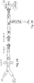
- Fig. 3 shows the structure of a typical adapter attached to a length of DNA 5, including an identification region 10, a restriction site (including a recognition sequence 22 and a cleavage sequence 24), and a primer sequence 30.
- DNA 5 in this example actually has two adapters, one on either side, having the same structure. Due to the generally palindromic nature of these adapters, the adapters cannot be added to the DNA incorrectly, as either orientation would be correct. In addition, in some embodiments, additional adapters could potentially be ligated onto the ends of the adapters. However, due to subsequent cleavage by a suitable restriction endonuclease at the cleavage site, any unwanted or extra adapters can be readily separated from the DNA itself, leaving just the identification sequence remaining on one or both ends, for subsequent determination or analysis.
- the droplets containing the cells may be merged with the droplets containing the adapters, as is shown in Fig. 1B , part 2, as well as Fig. 2B (where the tag library is formed from the adapters).
- Various techniques may be used to fuse the droplets together, and typically, the droplets are fused in a 1:1 ratio such that a single cell (contained within a single droplet) is exposed to a unique adapter (i.e., containing a unique identification sequence).
- the adapters are ligated to the DNA released from the lysed cells.
- each droplet typically contains a unique adapter, the DNA in each droplet is uniquely identified by a unique identification sequence.
- a plurality of DNA molecules is typically found within each droplet, some or all of which are thus labeled by the same adapter.
- the DNA within the droplets may be sequenced or analyzed, as is shown in Fig. 1C , part 1.
- DNA from different droplets may be combined together prior to analysis.
- the presence of unique identification sequences on the adapters on the DNA may allow the DNA from each of the droplets to be analyzed and distinguished. Accordingly, for example, the DNA (and the epigenetic profile) from a first cell can be distinguished from the DNA from a second cell.
- ChIP chromatin immunoprecipitation
- the DNA may be amplified (e.g., using PCR via the primer sequences), cleaved (e.g., using a restriction endonuclease that cleaves the adapter site, for instance, BciVI as is shown in this example), and/or ligated (e.g. using Illumina) such that the DNA is sequenced.
- a restriction endonuclease e.g., using a restriction endonuclease that cleaves the adapter site, for instance, BciVI as is shown in this example
- ligated e.g. using Illumina
- a plurality of cells may each be uniquely identified (for instance, upon selection of a suitable of nucleotides within the identification sequence), e.g., even for populations of 100 cells, 1 million cells, etc.
- a suitable of nucleotides within the identification sequence e.g., even for populations of 100 cells, 1 million cells, etc.
- other techniques could also be used for sequencing.
- microfluidic droplets are used, for example, to contain cells.
- Microfluidic droplets may be used to keep the cells of a plurality of cells separate and identifiable, e.g., such that epigenetic or genetic differences between the different cells may be identified.
- a plurality or a population of different cells may be studied for epigenetic differences, but there is no ability to determine those epigenetic differences on the level of an individual cell; instead, only average epigenetic profiles of those cells can be determined.
- a plurality of cells, some or all of which may contain individual epigenetic differences may be studied, at resolutions down to the single-cell level.
- the cells may arise from a human, or from a non-human animal, for example, an invertebrate cell (e.g., a cell from a fruit fly), a fish cell (e.g., a zebrafish cell), an amphibian cell (e.g., a frog cell), a reptile cell, a bird cell, or a mammal cell, such as a monkey, ape, cow, sheep, goat, horse, donkey, camel, llama, alpaca, rabbit, pig, mouse, rat, guinea pig, hamster, dog, cat, etc.
- the cell may be from any part of the organism.
- a tissue may be studied.
- a tissue from an organism may be processed to produce cells (e.g., through tissue homogenization or by laser-capturing the cells from the tissue), such that the epigenetic differences within the tissue may be determined, as discussed herein.
- the cells or tissues may arise from a healthy organism, or one that is diseased or suspected of being diseased.
- blood cells from an organism may be removed and studied to determine epigenetic differences or changes in the epigenetic profile of those cells, e.g., to determine if the animal is healthy or has a disease, for example, if the animal has cancer (e.g., by determining cancer cells within the blood).
- a tumor may be studied (e.g., using a biopsy), and the epigenetic profile of the tumor may be determined.
- the cells may be studied to determine if any of the cells are cancer stem cells.
- the cells may also be determined using other techniques, in addition to the ones discussed herein, which may assist in determining the epigenetic profile of the cells.
- the cells may be studied using flow cytometry, microscopy, the cells may be cultured, etc., to determine whether the epigenetic profile (or changes in the epigenetic profile) correlate to other changes in the cell, for example, expression levels of a protein, changes in morphology, ability to reproduce or differentiate, etc.
- a plurality of cells is contained within a plurality of droplets.
- the encapsulation rate may be kept low, for example, such that the average density is about 1 cell/droplet, or less.
- the average density may be less than about 0.95 cells/droplet, less than about 0.9 cells/droplet, less than about 0.8 cells/droplet, less than about 0.7 cells/droplet, less than about 0.6 cells/droplet, less than about 0.5 cells/droplet, less than about 0.4 cells/droplet, less than about 0.3 cells/droplet, or less than about 0.2 cells/droplet.
- the cells are contained such that no more than about 25%, no more than about 15%, no more than about 10% no more than about 5%, no more than about 3%, or no more than about 1% of the droplets contains more than one cell therein.
- Such relatively low densities may be useful, e.g., to avoid confusion of having more than one cell labeled with the same identification sequence (e.g., such that the DNA of the cell is ligated to an adapter), as discussed below.
- the droplets may be contained in a microfluidic channel.
- the droplets may have an average dimension or diameter of less than about 1 mm, less than about 500 micrometers, less than about 300 micrometers, less than about 200 micrometers, less than about 100 micrometers, less than about 75 micrometers, less than about 50 micrometers, less than about 30 micrometers, less than about 25 micrometers, less than about 10 micrometers, less than about 5 micrometers, less than about 3 micrometers, or less than about 1 micrometer in some cases.
- the average diameter may also be at least about 1 micrometer, at least about 2 micrometers, at least about 3 micrometers, at least about 5 micrometers, at least about 10 micrometers, at least about 15 micrometers, or at least about 20 micrometers in certain instances.
- the droplets may be spherical or non-spherical.
- the average diameter or dimension of a droplet, if the droplet is non-spherical, may be taken as the diameter of a perfect sphere having the same volume as the non-spherical droplet.
- the droplets may be produced using any suitable technique.
- a junction of channels may be used to create the droplets.
- the junction may be, for instance, a T-junction, a Y-junction, a channel-within-a-channel junction (e.g., in a coaxial arrangement, or comprising an inner channel and an outer channel surrounding at least a portion of the inner channel), a cross (or "X") junction, a flow-focus junction, or any other suitable junction for creating droplets.
- X cross
- a flow-focus junction or any other suitable junction for creating droplets.
- the junction may be configured and arranged to produce substantially monodisperse droplets.
- the cells may be encapsulated within the droplets at a relatively high rate.
- the rate of cell encapsulation in droplets may be at least about 10 cells/s, at least about 30 cells/s, at least about 100 cells/s, at least about 300 cells/s, at least about 1,000 cells/s, at least about 3,000 cells/s, at least about 10,000 cells/s, at least about 30,000 cells/s, at least about 100,000 cells/s, at least about 300,000 cells/s, or at least about 10 6 cells/s.
- the droplets may be substantially monodisperse in some embodiments, or the droplets may have a homogenous distribution of diameters, e.g., the droplets may have a distribution of diameters such that no more than about 10%, no more than about 5%, no more than about 3%, no more than about 2%, or no more than about 1% of the droplets have a diameter less than about 90% (or less than about 95%, less than about 97%, or less than about 99%) and/or greater than about 110% (or greater than about 101%, greater than about 103%, or greater than about 105%) of the overall average diameter of the plurality of droplets.
- the plurality of droplets has an overall average diameter and a distribution of diameters such that the coefficient of variation of the cross-sectional diameters of the droplets is less than about 10%, less than about 5%, less than about 2%, between about 1% and about 10%, between about 1% and about 5%, or between about 1% and about 2%.
- the coefficient of variation may be defined as the standard deviation divided by the mean, and can be determined by those of ordinary skill in the art.
- the fluid forming the droplets is substantially immiscible with the carrying fluid surrounding the droplets.
- the fluid may be hydrophilic or aqueous, while the carrying fluid may be hydrophobic or an "oil,” or vice versa.
- a “hydrophilic” fluid is one that is miscible with pure water
- a “hydrophobic” fluid is a fluid that is not miscible with pure water.
- oil as used herein, merely refers to a fluid that is hydrophobic and not miscible in water.
- the oil may be a hydrocarbon in some embodiments, but in other embodiments, the oil may be (or include) other hydrophobic fluids (for example, octanol).
- the hydrophilic or aqueous fluid need not be pure water.
- the hydrophilic fluid may be an aqueous solution, for example, a buffer solution, a solution containing a dissolved salt, or the like.
- a hydrophilic fluid may also be, or include, for example, ethanol or other liquids that are miscible in water, e.g., instead of or in addition to water.
- the cells may be lysed or otherwise processed to release the DNA within the cells, e.g., as a plurality of nucleosome sequences.
- the nucleosome sequences are those regions of the DNA that interact with the histones.
- the nucleosome sequence of the DNA typically winds around one or more histones to produce the basic nucleosome structure, which is subsequently packaged within the chromatin of the cell.
- the cells may be lysed within the droplets by sonication (exposure to ultrasound), temperature or osmotic changes, exposure to certain types of enzymes or chemicals (for example, detergents such as Triton, e.g., Triton X-100), or the like.
- sonication exposure to ultrasound
- temperature or osmotic changes exposure to certain types of enzymes or chemicals (for example, detergents such as Triton, e.g., Triton X-100), or the like.
- detergents such as Triton, e.g., Triton X-100
- the cells are lysed within the droplets without breaking down the droplets themselves, e.g., such that the cell lysate that is subsequently produced remains within the droplets.
- the DNA may also be exposed to enzymes which are able to process the DNA without substantially affecting the epigenetic information of interest, e.g., without substantially altering the methylation of nucleotides within the DNA, without altering any histone modifications that might be present, etc.
- the DNA may be exposed to a non-nucleosome-cleaving nuclease able to cleave the DNA at regions other than where the DNA is contained within a nucleosome.
- nuclease may accordingly be able to cleave the DNA into smaller fragments that can be subsequently analyzed (e.g., as discussed herein), without substantially affecting those portions of the DNA that interact with the histones within the nucleosome structures (i.e., the nucleosome sequences within the DNA). Accordingly, epigenetic information contained within the nucleosome structures may be preserved for subsequent determination.
- a suitable enzyme is MNase (S7 nuclease or micrococcal nuclease), which is available commercially.
- a restriction enzyme that targets a specific sequence may be used to digest genomic regions having particular sequence contents, such as GC-rich euchromatic loci.
- one or more adapters may be ligated or otherwise bonded onto the DNA (or RNA, in some embodiments).
- the adapter may be formed from DNA and/or RNA.
- the adapter may be single-stranded or double-stranded, and in some cases, the adapters may be palindromic or substantially palindromic, or in some cases the adapter can be single stranded.
- the adapter may include an identification sequence and a restriction site (and/or a portion of a restriction site), and optionally a primer sequence. If the adapter is at least substantially palindromic, the adapter may also contain inversions of these, e.g., the adapter may contain an identification sequence, a restriction site, an inversion of the restriction site, and an inversion of the identification sequence.
- adapter 70 includes a portion of a restriction site 71 ("site"), a first "barcode” or identification sequence 72, a second complete restriction site 73, a second "barcode” or identification sequence 74, and a second portion of a restriction site 75 ("restriction").
- Adapter 70 also includes a region 76 that can be recognized by a primer.
- the adapter is joined to a stretch of nucleic acid, such as DNA 77, to be studied (e.g., containing a nucleosome 80 as is shown in Fig. 7 , although this is just for explanatory purposes).
- the first and second portions of the restriction site have the same sequence, e.g., for restriction sites that are palindromic in nature.
- a restriction site is a site that is recognized by a restriction endonuclease.
- the restriction endonuclease may cleave the adapter within the restriction site.
- the restriction endonuclease may cleave the restriction site to leave behind blunt ends or "sticky" ends (e.g., leaving an overhang with one or more nucleotides lacking a complement).
- the restriction site in some cases, includes a recognition sequence (a specific sequence of nucleotides, e.g., 4, 5, 6, 7, or 8 nucleotides long) and a cleavage sequence that may be part of, or be separate from, the recognition sequence.
- the restriction site includes a recognition sequence (which is 6 nucleotides in length as is shown in Fig. 3 ), where the restriction endonuclease recognizes the adapter, and a separate cleavage sequence where the restriction endonuclease actually cleaves the adapter (indicated by the jagged lines in Fig. 3 ).
- a recognition sequence which is 6 nucleotides in length as is shown in Fig. 3
- the restriction endonuclease recognizes the adapter
- a separate cleavage sequence where the restriction endonuclease actually cleaves the adapter
- Non-limiting examples include BamHI, BsrI, NotI, XmaI, PspAI, DpnI, MboI, MnlI, Eco57I, Ksp632I, DraIII, AhaII, SmaI, MluI, HpaI, ApaI, BclI, BstEII, TaqI, EcoRI, SacI, HindII, HaeII, DraII, Tsp509I, Sau3AI, PacI, etc.
- an adaptor may contain a portion of a restriction site, e.g., first portions and second portions such that, when the first portion and the second portion are ligated or otherwise joined together, a complete restriction site.
- a restriction site e.g., first portions and second portions
- This may be useful, for example, in cases where an adapter is ligated to another adapter; the joined adapters, having a completed restriction site, may be exposed to a suitable corresponding restriction endonuclease that is able to cleave at the restriction site, thereby removing the extraneous adapters.
- a non-limiting example is shown schematically in Fig. 7 . In Fig.
- a first portion of the restriction site is labeled “site” and a second portion is labeled “restriction.”
- site a first portion of the restriction site
- second portion is labeled “restriction.”
- a corresponding restriction endonuclease will not recognize either portion, unless the portions are joined together to from a complete restriction site that can be recognized by the restriction endonuclease (i.e., forming the phrase "restriction site” in Fig. 7B ).
- the adapter is substantially or completely palindromic in nature.
- at least 30%, at least 35%, at least 40%, at least 45%, at least 50%, at least 55% , at least 60%, at least 65%, at least 70%, at least 75%, at least 80%, at least 85%, at least 90%, or at least 95% of the adapter may be palindromic.
- the adapter cannot be added in an incorrect orientation to the nucleic acid.
- the two adapters will form a complete restriction site that can be cleaved using a suitable restriction endonuclease, especially if the restriction site portions are also palindromic.
- the adapter is not fully palindromic, and there may be "bubble" regions that are not palindromic.
- identification sequences 72 and/or 74 within the adapter may be chosen to not be palindromic.
- an adapter is the following sequence: TTAA GGGCTTTC GTATCC GGGGG ACCTTAATTAAGGT GGGGG GGATAC CTTTCGGG TTAA (SEQ ID NO: 1) It should be noted that this sequence is not fully palindromic, as certain regions (such as the underlined portions) are not palindromic. In this example, the two outer underlined regions may be used as identification sequences within the adapter. It should be noted that these regions are mirror images of each other, e.g., for ease of identification, rather than palindromes of each other (although this is not necessarily a requirement of the adapter).
- sequences may also be selected to be nonpalindromic, e.g., so that the adapter does not readily form stem-loop structures.
- primer TAAGGTGGGGGGGATAC SEQ ID NO: 2 may be used with this adapter.
- the identification sequence may comprise any suitable number of nucleotides (for example, 4, 5, 6, 7, 8, 9, 10, 11, 12, 13, 14, 15, or more nucleotides).
- a plurality of adapters is prepared, containing identical (or substantially similar) restriction sites but different identification sequences.
- the different adapters may all thus be of the same length, in some embodiments.
- a number of unique adapter sequences can be created. For example, if the identification sequence is 4 nucleotides long, then up to 4 4 unique adapters may be created; if the identification sequence is 5 nucleotides long, then up to 4 5 unique adapters may be created; etc. (e.g., up to 4 n unique sequences where n is the number of nucleotides in the identification sequence).
- the adapter may also contain a suitable primer sequence, e.g., such that the adapter may be recognized by an enzyme used in PCR ("Polymerase Chain Reaction").
- a suitable primer sequence e.g., such that the adapter may be recognized by an enzyme used in PCR ("Polymerase Chain Reaction").
- the length of primer sequence is not more than about 30 nucleotides.
- Such a primer may be used, for example, to amplify the DNA (or RNA, in some cases) that the adapter is bound to.
- microwell plates containing wells containing the members of the library could be fabricated, e.g., using automated techniques, then encapsulated into droplets.
- a randomized oligomer population could be encapsulated no more than one in a drop and amplified within each droplet.
- Any suitable ligase may be used to ligate the adapter to DNA or RNA.
- Many such ligases are commercially available, e.g., Epicentre®.
- enzymes such as End-ItTM may be used, e.g., to repair DNA ends that were subjected to MNase degradation.
- the cell lysate is exposed to a solution containing adapters (e.g., with ligases) to cause the adapters to bind to the DNA.
- the action of the ligases may cause the adapters to be added randomly to the DNA.
- an adapter may be ligated to one or both ends of a DNA strand, more than one adapter may be ligated to one end of a strand (e.g., ligated to each other), one or more adapters can be ligated to one or both ends of the DNA and to each other to produce a circular DNA fragment, and in some cases, adapters may be ligated directly to each other without any DNA in between.
- the DNA may be exposed to a restriction endonuclease that is able to cleave the adapters at a cleavage site. Doing so effectively causes the only half of the adapter to stay behind on the DNA (containing the identification sequence), while the rest of the adapters (and any other adapters that may have been connected to the first adapter) will be cleaved away. Accordingly, at the end of this step, at least some of the DNA will have an end labeled with a unique identification sequence at one or both ends, and thus, the DNA can be determined using the identification sequence, even if later mixed with DNA similarly processed but containing different identification sequences.
- Shorter fragments e.g., the cleaved ends of the adapters, remains of adapters bound to each other that also been cleaved into small fragments, etc.
- Shorter fragments can be subsequently removed through regular DNA purification methods (size cut off), or left in solution but ignored.
- size cut off regular DNA purification methods
- the cell lysate (contained within droplets) are fused or coalesced with other droplets containing adapters.
- substantially each of the droplets contain unique adapters (i.e., the adapters may be substantially similar, but contain different identification sequences). Accordingly, the cell lysate of each droplet can be uniquely identified by determining the identification sequences.
- any suitable technique may be used to fuse a first droplet and a second droplet together to create a combined droplet.
- the first and second droplets each be given opposite electric charges (i.e., positive and negative charges, not necessarily of the same magnitude), which may increase the electrical interaction of the two droplets such that fusion or coalescence of the droplets can occur due to their opposite electric charges, e.g., to produce the combined droplet.
- an electric field may be applied to the droplets, the droplets may be passed through a capacitor, a chemical reaction may cause the droplets to become charged, etc.
- the separate droplets may not necessarily be given opposite electric charges (and, in some cases, may not be given any electric charge), and the droplets may instead be fused through the use of dipoles induced in the fluidic droplets that causes the fluidic droplets to coalesce.
- the dipoles may be induced using an electric field which may be an AC field, a DC field, etc., and the electric field may be created, for instance, using one or more electrodes.
- the induced dipoles in the fluidic droplets may cause the fluidic droplets to become electrically attracted towards each other due to their local opposite charges, thus causing the droplets to fuse.
- the various droplets containing DNA may be combined together in some aspects of the description e.g., to produce a common solution containing the DNA.
- the DNA may have arisen from different cells, due to the presence of the ligated adapters (e.g., containing unique identification sequences), the DNA is now distinguishable.
- the droplets may be combined by removing surfactant, removing the continuous fluid containing the droplets, or any other suitable technique.
- the DNA may be processed or sequenced using any suitable technique, in accordance with certain aspects of the description.
- techniques such as Chromatin Immunoprecipitation ("ChIP"), ChIP-Sequencing, ChIP-on-chip, fluorescent in situ hybridization, methylation-sensitive restriction enzymes, DNA adenine methyltransferase identification (DamID), or bisulfite sequencing may be used to analyze the labeled DNA.
- the DNA may be amplified, e.g., using PCR techniques known to those of ordinary skill in the art.
- some of the DNA may be analyzed or sequenced to determine a certain feature or characteristic.
- some of the DNA, still attached to nucleosomal structure may be immunoprecipitated by exposing the fragments to an antibody, for example, a histone-recognizing antibody such as H3-lysine-4-methyl, or the naked DNA to a methylcytosine antibody, or a hydroxymethylcytosine antibody.
- DNA having a certain feature e.g., methylated histones or deacylated histones
- histone modifications that may be studied include acetylation, methylation, ubiquitylation, phosphorylation and sumoylation.
- the DNA that is sequenced may be aligned with a genome (e.g., a known genome, such as a human genome) to determine locations of the DNA within the genome (e.g., of a particular cell) that exhibit such features (e.g., methylated histones or deacylated histones).
- a genome e.g., a known genome, such as a human genome
- features e.g., methylated histones or deacylated histones.
- RNA molecules e.g., from individual cells, could be "barcoded” or otherwise ligated with an identification sequence in droplets using single stranded indexed adaptors.
- the adaptors may be coupled to the RNA molecules, for example, by direct ligation, by poly-T or random primer-based reverse transcription methods, or by other methods known to those of ordinary skill in the art.
- selected RNA sequences could be interrogated by introducing a collection of single-stranded adaptors each comprising a barcode or other identification sequence (e.g., indexed to a single cell) and known sequences complementary to the RNA species of interest, followed by reverse transcription in single-cell-containing droplets. Template switching may be used in some embodiments. Accordingly, it should be understood that in the embodiments and examples discussed herein using DNA, this is by way of example only, and that in other embodiments, RNA could be used instead of and/or in addition to DNA.
- the various materials selected lend themselves to various methods.
- various components of the description can be formed from solid materials, in which the channels can be formed via micromachining, film deposition processes such as spin coating and chemical vapor deposition, laser fabrication, photolithographic techniques, etching methods including wet chemical or plasma processes, and the like. See, for example, Scientific American, 248:44-55, 1983 (Angell , et al ).
- at least a portion of the fluidic system is formed of silicon by etching features in a silicon chip. Technologies for precise and efficient fabrication of various fluidic systems and devices of the description from silicon are known.
- various components of the systems and devices of the description can be formed of a polymer, for example, an elastomeric polymer such as polydimethylsiloxane (“PDMS”), polytetrafluoroethylene (“PTFE” or Teflon®), or the like.
- PDMS polydimethylsiloxane
- PTFE polytetrafluoroethylene
- Teflon® Teflon®
- a base portion including a bottom wall and side walls can be fabricated from an opaque material such as silicon or PDMS, and a top portion can be fabricated from a transparent or at least partially transparent material, such as glass or a transparent polymer, for observation and/or control of the fluidic process.
- Components can be coated so as to expose a desired chemical functionality to fluids that contact interior channel walls, where the base supporting material does not have a precise, desired functionality.
- components can be fabricated as illustrated, with interior channel walls coated with another material.
- Material used to fabricate various components of the systems and devices of the description may desirably be selected from among those materials that will not adversely affect or be affected by fluid flowing through the fluidic system, e.g., material(s) that is chemically inert in the presence of fluids to be used within the device.
- various components of the description are fabricated from polymeric and/or flexible and/or elastomeric materials, and can be conveniently formed of a hardenable fluid, facilitating fabrication via molding (e.g. replica molding, injection molding, cast molding, etc.).
- the hardenable fluid can be essentially any fluid that can be induced to solidify, or that spontaneously solidifies, into a solid capable of containing and/or transporting fluids contemplated for use in and with the fluidic network.
- the hardenable fluid comprises a polymeric liquid or a liquid polymeric precursor (i.e. a "prepolymer").
- Suitable polymeric liquids can include, for example, thermoplastic polymers, thermoset polymers, or mixture of such polymers heated above their melting point.
- a suitable polymeric liquid may include a solution of one or more polymers in a suitable solvent, which solution forms a solid polymeric material upon removal of the solvent, for example, by evaporation.
- a suitable solvent such polymeric materials, which can be solidified from, for example, a melt state or by solvent evaporation, are well known to those of ordinary skill in the art.
- a variety of polymeric materials, many of which are elastomeric, are suitable, and are also suitable for forming molds or mold masters, for embodiments where one or both of the mold masters is composed of an elastomeric material.
- a non-limiting list of examples of such polymers includes polymers of the general classes of silicone polymers, epoxy polymers, and acrylate polymers.
- Epoxy polymers are characterized by the presence of a three-membered cyclic ether group commonly referred to as an epoxy group, 1,2-epoxide, or oxirane.
- diglycidyl ethers of bisphenol A can be used, in addition to compounds based on aromatic amine, triazine, and cycloaliphatic backbones.
- Another example includes the well-known Novolac polymers.
- Non-limiting examples of silicone elastomers suitable for use according to the description include those formed from precursors including the chlorosilanes such as methylchlorosilanes, ethylchlorosilanes, phenylchlorosilanes, etc.
- Silicone polymers are preferred in one set of embodiments, for example, the silicone elastomer polydimethylsiloxane.
- Non-limiting examples of PDMS polymers include those sold under the trademark Sylgard by Dow Chemical Co., Midland, MI, and particularly Sylgard 182, Sylgard 184, and Sylgard 186.
- Silicone polymers including PDMS have several beneficial properties simplifying fabrication of the microfluidic structures of the description. For instance, such materials are inexpensive, readily available, and can be solidified from a prepolymeric liquid via curing with heat.
- PDMSs are typically curable by exposure of the prepolymeric liquid to temperatures of about, for example, about 65 °C to about 75 °C for exposure times of, for example, about an hour.
- silicone polymers such as PDMS
- PDMS polymethyl methacrylate copolymer
- flexible (e.g., elastomeric) molds or masters can be advantageous in this regard.
- One advantage of forming structures such as microfluidic structures of the description from silicone polymers, such as PDMS, is the ability of such polymers to be oxidized, for example by exposure to an oxygen-containing plasma such as an air plasma, so that the oxidized structures contain, at their surface, chemical groups capable of cross-linking to other oxidized silicone polymer surfaces or to the oxidized surfaces of a variety of other polymeric and non-polymeric materials.
- an oxygen-containing plasma such as an air plasma
- oxidized silicone such as oxidized PDMS can also be sealed irreversibly to a range of oxidized materials other than itself including, for example, glass, silicon, silicon oxide, quartz, silicon nitride, polyethylene, polystyrene, glassy carbon, and epoxy polymers, which have been oxidized in a similar fashion to the PDMS surface (for example, via exposure to an oxygen-containing plasma).
- Oxidation and sealing methods useful in the context of the present invention, as well as overall molding techniques, are described in the art, for example, in an article entitled " Rapid Prototyping of Microfluidic Systems and Polydimethylsiloxane," Anal. Chem., 70:474-480, 1998 (Duffy , et al .),.
- certain microfluidic structures of the description may be formed from certain oxidized silicone polymers. Such surfaces may be more hydrophilic than the surface of an elastomeric polymer. Such hydrophilic channel surfaces can thus be more easily filled and wetted with aqueous solutions.
- a bottom wall of a microfluidic device of the description is formed of a material different from one or more side walls or a top wall, or other components.
- the interior surface of a bottom wall can comprise the surface of a silicon wafer or microchip, or other substrate.
- Other components can, as described above, be sealed to such alternative substrates. Where it is desired to seal a component comprising a silicone polymer (e.g.
- the substrate may be selected from the group of materials to which oxidized silicone polymer is able to irreversibly seal (e.g., glass, silicon, silicon oxide, quartz, silicon nitride, polyethylene, polystyrene, epoxy polymers, and glassy carbon surfaces which have been oxidized).
- materials to which oxidized silicone polymer is able to irreversibly seal e.g., glass, silicon, silicon oxide, quartz, silicon nitride, polyethylene, polystyrene, epoxy polymers, and glassy carbon surfaces which have been oxidized.
- other sealing techniques can be used, as would be apparent to those of ordinary skill in the art, including, but not limited to, the use of separate adhesives, thermal bonding, solvent bonding, ultrasonic welding, etc.
- the systems and methods described herein may include one or more microfluidic components, for example, one or more microfluidic channels.
- the "cross-sectional dimension" of a microfluidic channel is measured perpendicular to the direction of fluid flow within the channel.
- some or all of the microfluidic channels may have a largest cross-sectional dimension less than 2 mm, and in certain cases, less than 1 mm.
- the maximum cross-sectional dimension of a microfluidic channel is less than about 500 micrometers, less than about 300 micrometers, less than about 200 micrometers, less than about 100 micrometers, less than about 50 micrometers, less than about 30 micrometers, less than about 10 micrometers, less than about 5 micrometers, less than about 3 micrometers, or less than about 1 micrometer.
- the microfluidic channels may be formed in part by a single component (e.g. an etched substrate or molded unit). Of course, larger channels, tubes, chambers, reservoirs, etc. can also be used to store fluids and/or deliver fluids to various components or systems in other embodiments of the invention.
- a microfluidic channel can have any cross-sectional shape (circular, oval, triangular, irregular, square or rectangular, or the like) and can be covered or uncovered. In embodiments where it is completely covered, at least one portion of the channel can have a cross-section that is completely enclosed, or the entire channel may be completely enclosed along its entire length with the exception of its inlet(s) and/or outlet(s).
- a channel may also have an aspect ratio (length to average cross sectional dimension) of at least 2:1, more typically at least 3:1, 5:1, 10:1, 15:1, 20:1, or more.
- At least a portion of one or more of the channels may be hydrophobic, or treated to render at least a portion hydrophobic.
- one non-limiting method for making a channel surface hydrophobic comprises contacting the channel surface with an agent that confers hydrophobicity to the channel surface.
- a channel surface may be contacted (e.g., flushed) with Aquapel® (a commercial auto glass treatment) (PPG Industries, Pittsburgh, PA).
- Aquapel® a commercial auto glass treatment
- a channel surface contacted with an agent that confers hydrophobicity may be subsequently purged with air.
- the channel may be heated (e.g., baked) to evaporate solvent that contains the agent that confers hydrophobicity.
- a surface of a microfluidic channel may be modified, e.g., by coating a sol-gel onto at least a portion of a microfluidic channel.
- the sol-gel coating may be made more hydrophobic by incorporating a hydrophobic polymer in the sol-gel.
- the sol-gel may contain one or more silanes, for example, a fluorosilane (i.e., a silane containing at least one fluorine atom) such as heptadecafluorosilane, or other silanes such as methyltriethoxy silane (MTES) or a silane containing one or more lipid chains, such as octadecylsilane or other CH 3 (CH 2 ) n -silanes, where n can be any suitable integer. For instance, n may be greater than 1, 5, or 10, and less than about 20, 25, or 30.
- a fluorosilane i.e., a silane containing at least one fluorine atom
- MTES methyltriethoxy silane
- n can be any suitable integer.
- n may be greater than 1, 5, or 10, and less than about 20, 25, or 30.
- the silanes may also optionally include other groups, such as alkoxide groups, for instance, octadecyltrimethoxysilane.
- groups such as alkoxide groups, for instance, octadecyltrimethoxysilane.
- silanes can be used in the sol-gel, with the particular silane being chosen on the basis of desired properties such as hydrophobicity.
- Other silanes e.g., having shorter or longer chain lengths
- the silanes may contain other groups, for example, groups such as amines, which would make the sol-gel more hydrophilic.
- Non-limiting examples include diamine silane, triamine silane, or N -[3-(trimethoxysilyl)propyl] ethylene diamine silane.
- the silanes may be reacted to form oligomers or polymers within the sol-gel, and the degree of polymerization (e.g., the lengths of the oligomers or polymers) may be controlled by controlling the reaction conditions, for example by controlling the temperature, amount of acid present, or the like.
- more than one silane may be present in the sol-gel.
- the sol-gel may include fluorosilanes to cause the resulting sol-gel to exhibit greater hydrophobicity, and/or other silanes (or other compounds) that facilitate the production of polymers.
- the sol-gel is not limited to containing only silanes, and other materials may be present in addition to, or in place of, the silanes.
- the coating may include one or more metal oxides, such as SiO 2 , vanadia (V 2 O 5 ), titania (TiO 2 ), and/or alumina (Al 2 O 3 ).
- the microfluidic channel is constructed from a material suitable to receive the sol-gel, for example, glass, metal oxides, or polymers such as polydimethylsiloxane (PDMS) and other siloxane polymers.
- the microfluidic channel may be one in which contains silicon atoms, and in certain instances, the microfluidic channel may be chosen such that it contains silanol (Si-OH) groups, or can be modified to have silanol groups.
- the microfluidic channel may be exposed to an oxygen plasma, an oxidant, or a strong acid cause the formation of silanol groups on the microfluidic channel.
- This example illustrates certain systems and methods for profiling epigenomes of single cells within populations using droplet based microfluidics.
- Chromatin structure plays an important role in regulating genome function and in particular its varied structure across cell types helps ensure that the correct genes are expressed in the correct cell types. Chromatin structure is regulated by histone modifications and DNA methylation. Thus, genomewide maps of histone modifications or DNA methylation in a given cell type are a valuable research tool.
- epigenomic profiles These maps are collectively referred to as epigenomic profiles or "epigenomes.”
- epigenomic profiles have clinical relevance as they can identify defects in genome regulation in cancer or other diseases, propose therapeutic strategies or serve as diagnostic or early detection biomarkers.
- This example describes an epigenomic profiling method that can be used to map histone modifications in hundreds, thousands or more individual cells in a population.
- This example uses microfluidic devices to capture single cells in single droplets. The cells are then lysed and the genome is fragmented by enzymatic digestion in the droplets. Finally, DNA oligonucleotides with unique "barcodes" or identification sequences are added to each droplet and ligated to the fragmented genomic DNA sequences, thus providing a unique identifier for each individual cell.
- the materials now includes DNA fragments wrapped around histones and "bar coded" according to the cell from which they originated. The materials can now be combined (e.g., "combined and indexed chromatin matter") and subjected to epigenomic profiling.
- profiling can be performed by using chromatin immunoprecipitation using an antibody against a modified histone (e.g., histone H3 lysine 4 trimethyl).
- a modified histone e.g., histone H3 lysine 4 trimethyl
- the DNA is isolated and bar-coded fragments are selectively introduced into a sequencing library.
- the DNA is then sequenced, for example, using next-generation sequencing instruments (e.g., Illumina HiSeq).
- data may be processed in the following succession: (i) each read is assigned to an original cell based on its bar code; (ii) each read is aligned to the genome based on the sequence attached to the bar code; (iii) genomewide profiles are generated for each cell, based on the union of reads with the same bar code - specifically, the profiles reflect the density of reads as a function of genomic position. Furthermore, (iv) clustering algorithms can be applied to the individual profiles and used to identify dominant patterns characteristic of different cellular states in a heterogeneous population.
- cells may be encapsulated in drops at a density of at most one cell per drop.
- the cells are lysed and chromatin is fragmented by MNase enzymatic digestion into its single units, called nucleosomes (special buffer was optimized, including Triton, MNase and CaCl 2 to complement MNase requirements).
- Each nucleosome included a segment of DNA wound around a histone protein core.
- each droplet containing cells is fused with a droplet containing a cocktail of enzymes, including EndIt (Epicentre: repairs DNA ends that were subjected to MNase digestion), ligase (Epicentre), modified buffer including EGTA that stops the MNase digestion, and double-stranded, barcoded oligonucleotide adapters.
- EndIt EndIt
- ligase Epicentre
- modified buffer including EGTA that stops the MNase digestion
- double-stranded, barcoded oligonucleotide adapters double-stranded, barcoded oligonucleotide adapters.
- the oligonucleotide adapter comprised a barcode that is unique, specific to each droplet (or individual cell). It also contains a universal PCR primer sequence and a restriction site (i.e., the oligonucelotides vary in terms of their barcodes, but are constant in terms of the primer sequence and restriction site).
- the enzyme cocktail effectively ligates barcoded adapters to the ends of the DNA fragments in the droplet.
- each piece includes a fragment of genome flanked by barcoded adapters and wrapped around histones.
- dilution buffer is supplemented, including both EGTA and EDTA in concentrations that will stop any enzymatic reaction and maintain detergent levels.
- these complexes are "bar-coded" with identification sequences by their cell of origin, they may now be combined for epigenomic profiling. Specifically, the droplets are pooled together and broken down to form one aqueous volume. This combined and indexed chromatin matter is then subjected to immunoprecipitation or "ChIP" using an antibody, e.g., an antibody against a histone modification (e.g., H3 lysine 4 trimethyl or H3 lysine 4 methyl). This enriches for fragments associated with histones having this modification. The enriched DNA is then isolated using any suitable technique.
- an antibody e.g., an antibody against a histone modification (e.g., H3 lysine 4 trimethyl or H3 lysine 4 methyl).
- H3 lysine 4 trimethyl or H3 lysine 4 methyl e.g., H3 lysine 4 trimethyl or H3 lysine 4 methyl
- Fragments within the enriched DNA sample that have "bar-coded” adapters attached can be selected by amplification followed by restriction, using the universal primer and the restriction sites on the adapters.
- the restriction event leaves an end that is compatible with a next round of ligation to sequencing adapters.
- the result is a sequencing library that contains "bar-coded” sequences from the epigenomic enrichment assay.
- the "bar-codes” may serve as indexes that allow DNA fragments to be assigned to individual single cells. The fragments can also be aligned to a genome.
- a computational pipeline design including demultiplexing of the sequenced DNA can be aligned to a genome using known techniques such as a Bowtie algorithm, a Peak-calling using Scripture algorithm, and/or clustering to elucidate different population profiles.
- the cells may be encapsulated in drops at rates of thousands per second, or millions per hour.
- a library of unique barcodes the contents of a micro-titer well plate containing oligonucleotides (e.g., an oligonucleotide library) may be sequenced.
- a randomized oligomer population can be encapsulated at no more than one in a drop and amplified inside each drop to create a homogenized oligomer drop catalog.
- K562 erythrocytic leukaemia cells (ATCC CCL-243) were grown according to standard protocols in RPMI 1640 media (Invitrogen, 22400105) supplemented with 10% fetal bovine serum (FBS, Atlas Biologicals, F-0500-A) and 10% penicillin/streptomycin (Invitrogen, 15140122).
- the cells were lysed and chromatin was fragmented in 1% Triton, 0.1% sodium deoxycholate, 50 mM Tris-HCl pH 7.5, 150 mM NaCl supplemented with 10 units/ml of MNase (Thermo scientific, 88216), 1mM CaCl 2 and EDTA-free protease inhibitor (Roche, 13015000).
- the cells were incubated for 10 min at 4 °C, 15 min at 37 °C, and put back at 4 °C until the next step.
- Adapter ligation in droplets Each nucleosome was formed from a segment of DNA wound around a histone protein core.
- the droplets typically containing a single cell
- a droplet contains unique barcoded adaptor in a final concentration of 500 micromolar. Additional buffer was pico-injected into the fused droplets at the same time.
- This buffer had a total volume of 104 microliters and contained 8 microliters End-ItTM (Epicentre, ER81050) that repairs DNA ends that were subjected to MNase digestion, 20 microliters End-ItTM buffer, 8 Fast link ligase (Epicentre, LK6201H), 20 microliters fast link ligation buffer, 20 microliters dNTPs, 12 microliters from 10 mM ATP, and 8 microliters EGTA to a final concentration of 40 mM that stopped the MNase digestion.
- 8 microliters End-ItTM Epicentre, ER81050
- 8 Fast link ligase Epicentre, LK6201H
- 20 microliters fast link ligation buffer 20 microliters dNTPs
- 12 microliters from 10 mM ATP 12 microliters from 10 mM ATP
- 8 microliters EGTA to a final concentration of 40 mM that stopped the MNase digestion.
- the cells may be encapsulated in droplets at rates of thousands per second, or millions per hour.
- a library of unique barcodes the contents of a micro-titer well plate containing the oligo-library could be encapsulated in droplets.
- a randomized oligomer population could be encapsulated no more than one in a droplet and amplified inside each droplet to create a homogenized oligomer droplet catalog.
- the oligomer adapters used in this example comprised an 8-mer identification sequence (or "barcode") that was unique to each droplet (individual cell).
- the adapters also contained a universal PCR primer sequence (forward: ACACGCAGTATCCCTTCG (SEQ ID NO: 3), reverse: ACTGCGTGTATCCGACTC (SEQ ID NO: 4)) and a restriction site for BciVI (NEB, R0596S) that cuts at a 3' overhang.
- the oligomers varied in terms of their identification sequences, but were constant in terms of the primer sequence and restriction site.
- the enzyme cocktail effectively ligated blunt ended barcoded adaptors to the ends of the repaired DNA fragments in the droplet.
- the nucleic acids typically included a fragment of genome flanked by barcoded adaptors and wrapped about histones.
- Breaking droplets Since the nucleic acids were uniquely labeled with the adapters by their cell of origin, they could subsequently be combined for epigenomic profiling.
- the droplets were pooled together and broken into one aqueous volume. Before breaking the droplets into one aqueous volume, dilution buffer (50 mM Tris-HCl pH 7.5, 150 mM NaCl, 1% Triton, 20 mM EGTA, and 20 mM EDTA) was added. The concentrations of EGTA, EDTA, and detergent were thus maintained (i.e., at 20 mM, 10 mM, and 1%, respectively). 1H,1H,2H,2H-perfluoro-1-octanol, 97% (Sigma, 370533-25G) was used to break the droplets.
- H3K4me3 antibody Next 5 to 10 micrograms of H3K4me3 antibody (Millipore, 17-614) were pre-bound by incubating with a mix of Protein-A and Protein-G Dynabeads (Invitrogen, 100-02D and 100-07D, respectively) in blocking buffer (PBS supplemented with 0.5% TWEEN and 0.5% BSA) for 2 hours. Washed beads were added to the chromatin lysate (aqueous phase of broken droplets) for overnight incubation.
- the samples were washed 6 times with R1PA buffer, twice with RIPA buffer supplemented with 500 mM NaCl, twice with LiCl buffer (10 mM TE, 250 mM LiCl, 0.5% NP-40, 0.5% DOC), twice with TE (10 mM Tris-HCl pH 8.0, 1 mM EDTA), and then eluted with 0.5% SDS, 300 mM NaCl, 5 mM EDTA, 10 mM Tris-HCl pH 8.0 at 65 °C.
- the eluant was incubated in 65 °C for 1 hour, then treated sequentially with RNaseA (Roche, 11119915001) for 30 min and Proteinase K (NEB, P8102S) for two hours.
- the DNA was purified using Agencourt AMPure XP beads (Beckmangenomics, A63881).
- Sequencing library preparation Fragments within the enriched DNA sample that had ligated adapters were selected by amplification followed by restriction using the universal primer and the restriction sites on the adapters as described herein. The restriction event leaves a 3' A overhang that was compatible with a next round of Ilumina adapter ligation.
- the result was a sequencing library that was enriched with nucleic acids from the epigenomic enrichment assay. The identification sequences or "barcodes" within the nucleic acids then served as indexes that allowed the DNA fragments to be assigned to individual single cells.
- the first step included de-multiplexing of the sequenced reads, first, according to the Ilumina indexes and then for the 8-mer indexes implemented on each read and hold cell origin information for each fragment.
- the Bowtie algorithm was used to align the reads to the genome and a peak-caller (Scripture algorithm) to find regions with significant signal to noise ratios.
- panning was performed by clustering single cell epigenome profiles to elucidate the heterogeneity in cell population, detect different cell types in mixed population, and/or extract other information from the cells.
- Populations of cells have substantial heterogeneity that is important for their function and understanding. This variability is reflected in cell to cell variations of epigenetic features such as DNA methylation, chromatin organization, mRNA levels, and protein expression.
- epigenetic features such as DNA methylation, chromatin organization, mRNA levels, and protein expression.
- populations can be sorted by phenotype prior to characterization but ideally, cells could be characterized one-by-one. The problem of averaging over multiple cells is exacerbated when a small number of cells differ from the majority of the population. An example is the case of rare variants that are increasingly realized to underlie tumor biology and therapeutic resistance.
- this example illustrates scalable and flexible microfluidics methodology capable of profiling chromatin state and RNA expression in thousands of single cells, and thereby capturing the nature of population heterogeneity at unprecedented scale.
- Characterizing the genetic and epigenetic states of single cells is a challenge because the effective concentration of the contents of a single cell is a million times smaller than that of typical samples that pool many cells, and hence the rate of reactions becomes impractically slow.
- the content of the cell may be amplified prior to its characterization, as was previously used to measure single cell genetic variations or single cell RNA expression levels.
- amplifying the contents of single cells in wells is time consuming, expensive and thus not scalable to large numbers of cells; moreover, this solution is not relevant for assays involving proteins, which are denatured during amplification. Instead, this example restores the effective concentration in single cell assays by drastically decreasing the reaction volume using drop-based microfluidics.
- Droplet-based microfluidics use drops of water immersed in an inert carrier fluid as minute reaction vessels that can be precisely controlled by microfluidic devices.
- the droplets may be roughly 10 micrometers in diameter, each containing about 1pL of fluid surrounded by a surfactant that both stabilizes the droplet to prevent coalescence with other droplets, and protects its interface to prevent loss of reagents through surface adsorption.
- the reagents within the droplets never touch the walls of the microfluidic device and fluidic control may be achieved with the inert carrier fluid, totally independent of the droplets.
- the droplets can be formed, refilled, thermo-cycled, merged, split, sorted, etc. at rates of up to millions per hour with exquisite control over individual droplets.
- droplets can be used to compartmentalize millions of single cells per hour at high concentrations, allowing measurements of millions of single cells.
- This general method can be used, for example, to study epigenetic heterogeneity.
- Gene regulation in eukaryotes relies on the functional packaging of DNA into chromatin, a higher-order structure composed of DNA, RNA, histones and associated proteins. Chromatin structure and function is regulated by post-translational modifications of the histones, including acetylation, methylation and ubiquitinylation.
- Histone modifications (HM) can be mapped genome-wide, revealing type-specific regulation states of cells that reflect lineage-specific gene expression, developmental programs or disease processes. Given the central role of HM in stem and cancer cells, it is likely that epigenetic states differ between tumor cells and underlie their functional heterogeneity.
- mapping HM in single cells is not currently possible in other systems due to a low signal to noise ratio when performing ChIP on genomic material from less than 10,000 cells.
- cells are encapsulated one in a drop together with lysis buffer and Micrococcal Nuclease enzyme (MNase) that digests inter-nucleosomal DNA.
- MNase Micrococcal Nuclease enzyme
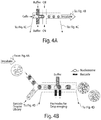
- each droplet containing fragmentized nucleosomes from a single cell is merged with a drop containing billions of copies of a unique DNA adapter and a ligation buffer as shown in Fig. 4 .
- the merging transpires by applying an electric field through an electrode positioned within the device. To allow adequate sequencing coverage for the information obtained from the barcoded cells, only 100 merged droplets are collected per ChIP-Seq experiment.
- the barcode library contains at least 10 times more unique barcodes than the number of cells collected. Thus when collecting 100 cells, a library containing 1152 different barcodes is used, ensuring that the probability of barcoding two different cells with the same barcode is lower than 5%.
- Barcoded nucleosomes can be merged with additional non-barcoded nucleosomes used as a biological buffer to ensure high signal to noise ratios during the Immuno-Precipitation step.
- the adapters may be designed with additional DNA sequences that are used as specific priming regions for amplification and as restriction sites for selection during the preparation of the library for Illumina sequencing. Thus, although initially the barcoded fragments make a negligible fraction of the DNA in the sample, the majority of sequenced reads are barcoded on both ends, as shown in Fig. 5A .
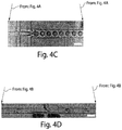
- Fig. 4 shows microfluidics of a single cell ChIP-seq.
- Fig. 4A shows cells are encapsulated in drops together with lysis buffer and digestive enzyme.
- Fig. 4B shows that, after incubation, droplets are re-injected into another microfluidic device where they are fused with drops containing barcodes.
- Fig. 4C is an image of single cells being encapsulated in a microfluidic droplet maker.
- Fig. 4D shows droplets containing barcodes are fused with drops containing cell lysate. Scale bars are 100 micrometers.
- each of the chosen barcodes After aligning and filtering the reads, each of the chosen barcodes typically tagged 2-5,000 distinct DNA fragments from each single cell, representing a sparse binary vector spanning the murine genome with enrichment for positive entries in genomic regions that were wrapped around H3K4me3 marked nucleosomes.
- the complete set of chosen barcodes was represented as a sparse binary matrix, in which only 15,000 out of the 1 million genomic bins have reads from more than one cell and can therefore be used to compare between them.
- sparseness of the data when aggregating all mES cells and all mEF cells the known profile measured in many cells in bulk was restored as shown in Fig.
- Fig. 3A shows the number of unique reads per barcode is presented for a sample of 50 mES cells and 50 mEF cells after ChIP-Seq with H3K4me3. Lighter shades indicate reads with the same barcode on both sides, while darker shades indicates reads with non-matching barcodes on both sides.
- Fig. 3B shows representative reads from 8 different mES cells and the aggregated data of 50 mESs at the top.
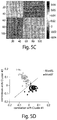
- Fig. 3C is a correlation matrix between genomic bins of 114 cells (50 mES , 64 mEF). The mES were the first 50 vectors while mEF were last 64, and their separation into two blocks of correlation was observed. After just 3 iterations, an unsupervised algorithm based on the correlations between each cell and two aggregates of cells could separate the two populations, as is shown in Fig. 3D .
- mouse embryonic stem cells were compared with mouse embryonic fibroblasts (mEF), using an embodiment of the invention.
- mESs were encoded with identification sequences 1-576 ("barcodes") while mEFs were encoded with identification sequences 577-1152.
- barcodes identification sequences 1-576
- mEFs were encoded with identification sequences 577-1152.
- the identification sequences were arbitrarily chosen and numbered 1-1152. After ligation, the populations of droplets were combined together.
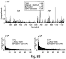
- the droplets were then analyzed using ChIP-sequencing, as is shown in Fig. 8B .
- the H3K4me3 histone was studied in this example. 4.6 million cells were studied, with 1 million distinct reads after alignment and filtering. It was found that each of the relevant barcodes typically tagged 2,000-5,000distinct DNA fragments from a single cell, representing a sparse binary vector spanning the mouse genome. Of these, about 70% contained adapters on both ends of the DNA. About 10-20% included "cross talk,” i.e., the DNA was incorrectly labeled. For each barcode, there were about 3,000-10,000 cells or "reads" that were identified.
- the separate reads could be pooled together to restore the epigenomic profile that was measured in a population of cells using more conventional techniques, as is shown in Fig. 8C , where aggregates of 50 single-cell profiles for both mESs and mEFs were compared to traditional protocols for detecting histones.
- a reference to "A and/or B", when used in conjunction with open-ended language such as “comprising” can refer, in one embodiment, to A only (optionally including elements other than B); in another embodiment, to B only (optionally including elements other than A); in yet another embodiment, to both A and B (optionally including other elements); etc.
- the phrase "at least one,” in reference to a list of one or more elements, should be understood to mean at least one element selected from any one or more of the elements in the list of elements, but not necessarily including at least one of each and every element specifically listed within the list of elements and not excluding any combinations of elements in the list of elements.
- This definition also allows that elements may optionally be present other than the elements specifically identified within the list of elements to which the phrase "at least one" refers, whether related or unrelated to those elements specifically identified.
- At least one of A and B can refer, in one embodiment, to at least one, optionally including more than one, A, with no B present (and optionally including elements other than B); in another embodiment, to at least one, optionally including more than one, B, with no A present (and optionally including elements other than A); in yet another embodiment, to at least one, optionally including more than one, A, and at least one, optionally including more than one, B (and optionally including other elements); etc.
Landscapes
- Chemical & Material Sciences (AREA)
- Life Sciences & Earth Sciences (AREA)
- Organic Chemistry (AREA)
- Proteomics, Peptides & Aminoacids (AREA)
- Health & Medical Sciences (AREA)
- Zoology (AREA)
- Wood Science & Technology (AREA)
- Engineering & Computer Science (AREA)
- Molecular Biology (AREA)
- Immunology (AREA)
- Biotechnology (AREA)
- Microbiology (AREA)
- Biochemistry (AREA)
- Bioinformatics & Cheminformatics (AREA)
- General Engineering & Computer Science (AREA)
- General Health & Medical Sciences (AREA)
- Genetics & Genomics (AREA)
- Biophysics (AREA)
- Analytical Chemistry (AREA)
- Physics & Mathematics (AREA)
- Chemical Kinetics & Catalysis (AREA)
- Measuring Or Testing Involving Enzymes Or Micro-Organisms (AREA)
Claims (3)
- Verfahren, umfassend:Bereitstellen einer Vielzahl von Zellen, die in einer Vielzahl von mikrofluidischen Tröpfchen enthalten sind,wobei die Vielzahl von Zellen in der Vielzahl von mikrofluidischen Tröpfchen mit einer Dichte von weniger als ungefähr 1 Zelle/Tröpfchen enthalten sind;Lysieren der in den mikrofluidischen Tröpfchen enthaltenen Zellen, um darin Zelllysate zu erzeugen um eine Vielzahl von Nukleinsäuren innerhalb der mikrofluidischen Tröpfchen zu produzieren, wobei die Nukleinsäure DNA ist;Aussetzen der Zelllysate einem Enzym, um fragmentierte DNA-Sequenzen zu erzeugen, wobei das Enzym eine Nicht-Nukleosom-spaltende Nuklease oder ein Enzym ist, das in der Lage ist, DNA zur Analyse zu schneiden, ohne die mit der DNA verbundene epigenetische Information zu zerstören;Aussetzen von Adaptern an mindestens einige der Nukleinsäuresequenzen, wobei die Adapter eine Identifizierungssequenz umfassen, wobei eindeutige Barcodes oder Identifizierungssequenzen zu jedem Tröpfchen hinzugefügt und der fragmentierten DNA hinzugefügt werden, wodurch ein eindeutiger Identifikator für jede einzelne Zelle bereitgestellt wird, und wobei die die Zellen enthaltenden Tröpfchen mit den die Adapter enthaltenden Tröpfchen fusioniert werden; undKombination der mikrofluidischen Tröpfchen zur Herstellung einer gemeinsamen Lösung; und Sequenzieren der DNA-Sequenzen, die angehängte Adapter enthalten, wobei die Sequenzierung der DNA-Sequenzen durch Chromatin-Immunpräzipitation (chromatin immunoprecipitation; ChiP) durchgeführt wird.
- Verfahren gemäß Anspruch 1, wobei die Adapter eine Restriktionsschnittstelle und eine Primerstelle umfassen.
- Verfahren gemäß Anspruch 2, wobei die Primerstelle eine universelle Primersequenz umfasst.
Applications Claiming Priority (3)
| Application Number | Priority Date | Filing Date | Title |
|---|---|---|---|
| US201261634744P | 2012-03-05 | 2012-03-05 | |
| PCT/US2013/029123 WO2013134261A1 (en) | 2012-03-05 | 2013-03-05 | Systems and methods for epigenetic sequencing |
| EP13758373.8A EP2823064B1 (de) | 2012-03-05 | 2013-03-05 | Verfahren für epigenetische sequenzierung |
Related Parent Applications (2)
| Application Number | Title | Priority Date | Filing Date |
|---|---|---|---|
| EP13758373.8A Division EP2823064B1 (de) | 2012-03-05 | 2013-03-05 | Verfahren für epigenetische sequenzierung |
| EP13758373.8A Division-Into EP2823064B1 (de) | 2012-03-05 | 2013-03-05 | Verfahren für epigenetische sequenzierung |
Publications (3)
| Publication Number | Publication Date |
|---|---|
| EP3305918A2 EP3305918A2 (de) | 2018-04-11 |
| EP3305918A3 EP3305918A3 (de) | 2018-06-20 |
| EP3305918B1 true EP3305918B1 (de) | 2020-06-03 |
Family
ID=49117256
Family Applications (3)
| Application Number | Title | Priority Date | Filing Date |
|---|---|---|---|
| EP17201280.9A Active EP3305918B1 (de) | 2012-03-05 | 2013-03-05 | Verfahren für epigenetische sequenzierung |
| EP18215320.5A Pending EP3495503A1 (de) | 2012-03-05 | 2013-03-05 | Systeme und verfahren zur epigenetischen sequenzierung |
| EP13758373.8A Active EP2823064B1 (de) | 2012-03-05 | 2013-03-05 | Verfahren für epigenetische sequenzierung |
Family Applications After (2)
| Application Number | Title | Priority Date | Filing Date |
|---|---|---|---|
| EP18215320.5A Pending EP3495503A1 (de) | 2012-03-05 | 2013-03-05 | Systeme und verfahren zur epigenetischen sequenzierung |
| EP13758373.8A Active EP2823064B1 (de) | 2012-03-05 | 2013-03-05 | Verfahren für epigenetische sequenzierung |
Country Status (4)
| Country | Link |
|---|---|
| US (6) | US11001883B2 (de) |
| EP (3) | EP3305918B1 (de) |
| LT (1) | LT3305918T (de) |
| WO (1) | WO2013134261A1 (de) |
Families Citing this family (125)
| Publication number | Priority date | Publication date | Assignee | Title |
|---|---|---|---|---|
| EP3305918B1 (de) | 2012-03-05 | 2020-06-03 | President and Fellows of Harvard College | Verfahren für epigenetische sequenzierung |
| GB2539836B (en) | 2012-08-13 | 2017-03-29 | Univ California | Methods for detecting target nucleic acids in sample lysate droplets |
| US10752949B2 (en) | 2012-08-14 | 2020-08-25 | 10X Genomics, Inc. | Methods and systems for processing polynucleotides |
| US10323279B2 (en) | 2012-08-14 | 2019-06-18 | 10X Genomics, Inc. | Methods and systems for processing polynucleotides |
| US11591637B2 (en) | 2012-08-14 | 2023-02-28 | 10X Genomics, Inc. | Compositions and methods for sample processing |
| US10584381B2 (en) | 2012-08-14 | 2020-03-10 | 10X Genomics, Inc. | Methods and systems for processing polynucleotides |
| US10273541B2 (en) | 2012-08-14 | 2019-04-30 | 10X Genomics, Inc. | Methods and systems for processing polynucleotides |
| US9951386B2 (en) | 2014-06-26 | 2018-04-24 | 10X Genomics, Inc. | Methods and systems for processing polynucleotides |
| US10221442B2 (en) | 2012-08-14 | 2019-03-05 | 10X Genomics, Inc. | Compositions and methods for sample processing |
| JP6367196B2 (ja) | 2012-08-14 | 2018-08-01 | テンエックス・ジェノミクス・インコーポレイテッド | マイクロカプセル組成物および方法 |
| US9701998B2 (en) | 2012-12-14 | 2017-07-11 | 10X Genomics, Inc. | Methods and systems for processing polynucleotides |
| EP2898071A4 (de) * | 2012-09-21 | 2016-07-20 | Broad Inst Inc | Zusammensetzungen und verfahren für bibliotheken mit langem einsatz und gepaartem ende von nukleinsäuren in emulsionstropfen |
| EP2898096B1 (de) | 2012-09-21 | 2024-02-14 | The Broad Institute, Inc. | Verfahren zur markierung von rns |
| EP3567116A1 (de) | 2012-12-14 | 2019-11-13 | 10X Genomics, Inc. | Verfahren und systeme zur verarbeitung von polynukleotiden |
| US10533221B2 (en) | 2012-12-14 | 2020-01-14 | 10X Genomics, Inc. | Methods and systems for processing polynucleotides |
| KR102190198B1 (ko) | 2013-02-08 | 2020-12-14 | 10엑스 제노믹스, 인크. | 폴리뉴클레오티드 바코드 생성 |
| WO2014143158A1 (en) | 2013-03-13 | 2014-09-18 | The Broad Institute, Inc. | Compositions and methods for labeling of agents |
| SG11201508193TA (en) | 2013-04-17 | 2015-11-27 | Agency Science Tech & Res | Method for generating extended sequence reads |
| US10395758B2 (en) | 2013-08-30 | 2019-08-27 | 10X Genomics, Inc. | Sequencing methods |
| US9824068B2 (en) | 2013-12-16 | 2017-11-21 | 10X Genomics, Inc. | Methods and apparatus for sorting data |
| EP3089822B1 (de) * | 2013-12-30 | 2022-04-06 | Atreca, Inc. | Analyse von mit einzelzellen assoziierten nukleinsäuren mithilfe von nukleinsäure-strichcodes |
| US10597698B2 (en) * | 2014-03-03 | 2020-03-24 | The Board Of Trustees Of The University Of Illinois | Chromatin immunocapture devices and methods of use |
| CA2943624A1 (en) | 2014-04-10 | 2015-10-15 | 10X Genomics, Inc. | Fluidic devices, systems, and methods for encapsulating and partitioning reagents, and applications of same |
| AU2015247462A1 (en) * | 2014-04-17 | 2016-11-03 | President And Fellows Of Harvard College | Methods and systems for droplet tagging and amplification |
| CA2945794A1 (en) * | 2014-04-17 | 2015-10-22 | President And Fellows Of Harvard College | Systems and methods for droplet tagging |
| ES2764504T3 (es) | 2014-04-17 | 2020-06-03 | Yeda Res & Dev | Métodos y equipos para analizar restos de unión al ADN fijados al ADN |
| LT3134536T (lt) | 2014-04-21 | 2020-05-11 | President And Fellows Of Harvard College | Sistemos ir būdai, skirti nukleorūgščių brūkšniniam kodavimui |
| EP4574974A3 (de) | 2014-06-26 | 2025-10-08 | 10x Genomics, Inc. | Verfahren zur analyse von nukleinsäuren aus einzelnen zellen oder zellpopulationen |
| US12312640B2 (en) | 2014-06-26 | 2025-05-27 | 10X Genomics, Inc. | Analysis of nucleic acid sequences |
| WO2015200869A1 (en) | 2014-06-26 | 2015-12-30 | 10X Genomics, Inc. | Analysis of nucleic acid sequences |
| WO2015200717A2 (en) | 2014-06-27 | 2015-12-30 | The Regents Of The University Of California | Pcr-activated sorting (pas) |
| KR102541849B1 (ko) * | 2014-09-15 | 2023-06-09 | 에이비비트로, 엘엘씨 | 고-처리량 뉴클레오티드 라이브러리 서열결정 |
| WO2016065056A1 (en) | 2014-10-22 | 2016-04-28 | The Regents Of The University Of California | High definition microdroplet printer |
| WO2016069939A1 (en) | 2014-10-29 | 2016-05-06 | 10X Genomics, Inc. | Methods and compositions for targeted nucleic acid sequencing |
| US9975122B2 (en) | 2014-11-05 | 2018-05-22 | 10X Genomics, Inc. | Instrument systems for integrated sample processing |
| US10233490B2 (en) | 2014-11-21 | 2019-03-19 | Metabiotech Corporation | Methods for assembling and reading nucleic acid sequences from mixed populations |
| CN107427808B (zh) | 2015-01-12 | 2020-10-23 | 10X基因组学有限公司 | 用于制备核酸测序文库的方法和系统以及用其制备的文库 |
| AU2016215304B2 (en) | 2015-02-04 | 2022-01-27 | The Regents Of The University Of California | Sequencing of nucleic acids via barcoding in discrete entities |
| CN107532202A (zh) | 2015-02-24 | 2018-01-02 | 10X 基因组学有限公司 | 用于靶向核酸序列覆盖的方法 |
| EP3262407B1 (de) | 2015-02-24 | 2023-08-30 | 10X Genomics, Inc. | Partitionsverarbeitungsverfahren und -systeme |
| WO2016149096A1 (en) * | 2015-03-13 | 2016-09-22 | President And Fellows Of Harvard College | Determination of cells using amplification |
| EP3283629A4 (de) * | 2015-04-17 | 2018-08-29 | President and Fellows of Harvard College | Barcodierungssysteme und verfahren für die gensequenzierung und andere anwendungen |
| US10712356B2 (en) | 2015-04-21 | 2020-07-14 | General Automation Lab Technologies Inc. | Apparatus and method for picking biological sample |
| SG11201708579WA (en) | 2015-04-21 | 2017-11-29 | Gen Automation Lab Tech Inc | High resolution systems, kits, apparatus, and methods for high throughput microbiology applications |
| ES2918336T3 (es) | 2015-04-30 | 2022-07-15 | European Molecular Biology Laboratory | Detección y clasificación de gotas microfluídicas |
| JP6962563B2 (ja) | 2015-05-12 | 2021-11-05 | 株式会社オンチップ・バイオテクノロジーズ | 単一粒子解析方法およびその解析のためのシステム |
| CA2986036C (en) | 2015-05-18 | 2022-07-26 | Karius, Inc. | Compositions and methods for enriching populations of nucleic acids |
| US11479805B2 (en) * | 2015-08-21 | 2022-10-25 | The General Hospital Corporation | Combinatorial single molecule analysis of chromatin |
| US11371094B2 (en) | 2015-11-19 | 2022-06-28 | 10X Genomics, Inc. | Systems and methods for nucleic acid processing using degenerate nucleotides |
| SG10202108763UA (en) | 2015-12-04 | 2021-09-29 | 10X Genomics Inc | Methods and compositions for nucleic acid analysis |
| CN108474026A (zh) * | 2016-01-29 | 2018-08-31 | 豪夫迈·罗氏有限公司 | 用于核酸测序的新的衔接子和使用方法 |
| WO2017184707A1 (en) | 2016-04-19 | 2017-10-26 | President And Fellows Of Harvard College | Immobilization-based systems and methods for genetic analysis and other applications |
| WO2017197343A2 (en) | 2016-05-12 | 2017-11-16 | 10X Genomics, Inc. | Microfluidic on-chip filters |
| WO2017197338A1 (en) | 2016-05-13 | 2017-11-16 | 10X Genomics, Inc. | Microfluidic systems and methods of use |
| WO2018017794A1 (en) * | 2016-07-21 | 2018-01-25 | President And Fellows Of Harvard College | Microfluidic sequencing techniques |
| US20190203270A1 (en) | 2016-07-24 | 2019-07-04 | Yeda Research And Development Co., Ltd. | Methods and kits for analyzing dna binding moieties attached to dna |
| US11142791B2 (en) | 2016-08-10 | 2021-10-12 | The Regents Of The University Of California | Combined multiple-displacement amplification and PCR in an emulsion microdroplet |
| PL418683A1 (pl) * | 2016-09-12 | 2018-03-26 | Uniwersytet Gdański | Sposób wykorzystujący spektrometrię mas MALDI-TOF oraz iPLEX do detekcji wariantów genetycznych kodów kreskowych oraz jej zastosowanie |
| CN110462053A (zh) | 2016-12-21 | 2019-11-15 | 加利福尼亚大学董事会 | 使用基于水凝胶的液滴进行单细胞基因组测序 |
| US10550429B2 (en) | 2016-12-22 | 2020-02-04 | 10X Genomics, Inc. | Methods and systems for processing polynucleotides |
| US10815525B2 (en) | 2016-12-22 | 2020-10-27 | 10X Genomics, Inc. | Methods and systems for processing polynucleotides |
| US10011872B1 (en) | 2016-12-22 | 2018-07-03 | 10X Genomics, Inc. | Methods and systems for processing polynucleotides |
| US12264411B2 (en) | 2017-01-30 | 2025-04-01 | 10X Genomics, Inc. | Methods and systems for analysis |
| EP4310183B1 (de) | 2017-01-30 | 2025-07-09 | 10X Genomics, Inc. | Verfahren und systeme zur tröpfchenbasierten einzelzellenbarcodierung |
| US10995333B2 (en) | 2017-02-06 | 2021-05-04 | 10X Genomics, Inc. | Systems and methods for nucleic acid preparation |
| EP3375889B1 (de) * | 2017-03-17 | 2019-12-11 | HiFiBiO SAS | Einzelzellenanalyse |
| CN109526228B (zh) | 2017-05-26 | 2022-11-25 | 10X基因组学有限公司 | 转座酶可接近性染色质的单细胞分析 |
| US10844372B2 (en) | 2017-05-26 | 2020-11-24 | 10X Genomics, Inc. | Single cell analysis of transposase accessible chromatin |
| US20210032693A1 (en) * | 2017-08-10 | 2021-02-04 | Rootpath Genomics, Inc. | Improved Method to Analyze Nucleic Acid Contents from Multiple Biological Particles |
| US10837047B2 (en) | 2017-10-04 | 2020-11-17 | 10X Genomics, Inc. | Compositions, methods, and systems for bead formation using improved polymers |
| US10501739B2 (en) | 2017-10-18 | 2019-12-10 | Mission Bio, Inc. | Method, systems and apparatus for single cell analysis |
| WO2019084043A1 (en) | 2017-10-26 | 2019-05-02 | 10X Genomics, Inc. | METHODS AND SYSTEMS FOR NUCLEIC ACID PREPARATION AND CHROMATIN ANALYSIS |
| WO2019084328A1 (en) * | 2017-10-26 | 2019-05-02 | 10X Genomics, Inc. | METHODS FOR PREPARING NUCLEIC ACID MOLECULES |
| CN111479631B (zh) | 2017-10-27 | 2022-02-22 | 10X基因组学有限公司 | 用于样品制备和分析的方法和系统 |
| CN111051523B (zh) | 2017-11-15 | 2024-03-19 | 10X基因组学有限公司 | 功能化凝胶珠 |
| US10829815B2 (en) | 2017-11-17 | 2020-11-10 | 10X Genomics, Inc. | Methods and systems for associating physical and genetic properties of biological particles |
| CN111699388B (zh) | 2017-12-12 | 2024-08-02 | 10X基因组学有限公司 | 用于单细胞处理的系统和方法 |
| CN111712579B (zh) | 2017-12-22 | 2024-10-15 | 10X基因组学有限公司 | 用于处理来自一个或多个细胞的核酸分子的系统和方法 |
| SG11202007686VA (en) | 2018-02-12 | 2020-09-29 | 10X Genomics Inc | Methods characterizing multiple analytes from individual cells or cell populations |
| US11639928B2 (en) | 2018-02-22 | 2023-05-02 | 10X Genomics, Inc. | Methods and systems for characterizing analytes from individual cells or cell populations |
| WO2019169028A1 (en) | 2018-02-28 | 2019-09-06 | 10X Genomics, Inc. | Transcriptome sequencing through random ligation |
| WO2019195166A1 (en) | 2018-04-06 | 2019-10-10 | 10X Genomics, Inc. | Systems and methods for quality control in single cell processing |
| US12275936B2 (en) | 2018-04-18 | 2025-04-15 | The Regents Of The University Of California | Method to connect chromatin accessibility and transcriptome |
| EP3556466A1 (de) | 2018-04-18 | 2019-10-23 | HiFiBiO SAS | Mikrofluidisches verfahren zur einzelzellanalyse |
| WO2019217758A1 (en) | 2018-05-10 | 2019-11-14 | 10X Genomics, Inc. | Methods and systems for molecular library generation |
| US11932899B2 (en) | 2018-06-07 | 2024-03-19 | 10X Genomics, Inc. | Methods and systems for characterizing nucleic acid molecules |
| US11703427B2 (en) | 2018-06-25 | 2023-07-18 | 10X Genomics, Inc. | Methods and systems for cell and bead processing |
| EP3594364A1 (de) * | 2018-07-13 | 2020-01-15 | HiFiBio SAS High Fidelity Biology | Verfahren zum testen von nukleinsäure in mikrofluidischen tröpfchen |
| WO2020012004A1 (en) * | 2018-07-13 | 2020-01-16 | Hifibio Sas | Use of droplet single cell epigenome profiling for patient stratification |
| EP3730625A1 (de) * | 2019-04-24 | 2020-10-28 | HiFiBiO SAS | Verwendung von tröpfchen-einzelzellen-epigenomprofilierung zur patientenstratifizierung |
| EP3730626A1 (de) * | 2019-04-24 | 2020-10-28 | HiFiBiO SAS | Verwendung der tröpfchen-einzelzellen-epigenom-profilierung zur patientenstratifizierung |
| US20220362771A1 (en) * | 2018-07-13 | 2022-11-17 | Hifibio Sas | Use of droplet single cell epigenome profiling for patient stratification |
| US12188014B1 (en) | 2018-07-25 | 2025-01-07 | 10X Genomics, Inc. | Compositions and methods for nucleic acid processing using blocking agents |
| US20200032335A1 (en) | 2018-07-27 | 2020-01-30 | 10X Genomics, Inc. | Systems and methods for metabolome analysis |
| SG11202101164TA (en) | 2018-08-03 | 2021-03-30 | 10X Genomics Inc | Methods and systems to minimize barcode exchange |
| WO2020041148A1 (en) | 2018-08-20 | 2020-02-27 | 10X Genomics, Inc. | Methods and systems for detection of protein-dna interactions using proximity ligation |
| US12065688B2 (en) | 2018-08-20 | 2024-08-20 | 10X Genomics, Inc. | Compositions and methods for cellular processing |
| US11459607B1 (en) | 2018-12-10 | 2022-10-04 | 10X Genomics, Inc. | Systems and methods for processing-nucleic acid molecules from a single cell using sequential co-partitioning and composite barcodes |
| US12169198B2 (en) | 2019-01-08 | 2024-12-17 | 10X Genomics, Inc. | Systems and methods for sample analysis |
| US11845983B1 (en) | 2019-01-09 | 2023-12-19 | 10X Genomics, Inc. | Methods and systems for multiplexing of droplet based assays |
| WO2020167862A1 (en) | 2019-02-12 | 2020-08-20 | 10X Genomics, Inc. | Systems and methods for transfer of reagents between droplets |
| WO2020167866A1 (en) | 2019-02-12 | 2020-08-20 | 10X Genomics, Inc. | Systems and methods for transposon loading |
| EP3924505B1 (de) | 2019-02-12 | 2025-12-17 | 10X Genomics, Inc. | Verfahren zur verarbeitung von nukleinsäuremolekülen |
| US11467153B2 (en) | 2019-02-12 | 2022-10-11 | 10X Genomics, Inc. | Methods for processing nucleic acid molecules |
| US12305239B2 (en) | 2019-02-12 | 2025-05-20 | 10X Genomics, Inc. | Analysis of nucleic acid sequences |
| US12275993B2 (en) | 2019-02-12 | 2025-04-15 | 10X Genomics, Inc. | Analysis of nucleic acid sequences |
| US11851683B1 (en) | 2019-02-12 | 2023-12-26 | 10X Genomics, Inc. | Methods and systems for selective analysis of cellular samples |
| US11655499B1 (en) | 2019-02-25 | 2023-05-23 | 10X Genomics, Inc. | Detection of sequence elements in nucleic acid molecules |
| US11920183B2 (en) | 2019-03-11 | 2024-03-05 | 10X Genomics, Inc. | Systems and methods for processing optically tagged beads |
| EP3947722A1 (de) | 2019-03-27 | 2022-02-09 | 10X Genomics, Inc. | Systeme und verfahren zur verarbeitung von rna aus zellen |
| WO2020216898A1 (en) | 2019-04-24 | 2020-10-29 | Institut Curie | Biomarkers for predicting resistance to cancer drugs |
| EP3973074A4 (de) | 2019-05-22 | 2023-09-06 | Mission Bio, Inc. | Verfahren und vorrichtung zur simultanen gezielten sequenzierung von dna, rna und protein |
| EP3983540A1 (de) | 2019-06-16 | 2022-04-20 | Yeda Research and Development Co. Ltd | Verfahren zur stabilisierung intrazellulärer rna |
| US11667954B2 (en) | 2019-07-01 | 2023-06-06 | Mission Bio, Inc. | Method and apparatus to normalize quantitative readouts in single-cell experiments |
| US12235262B1 (en) | 2019-09-09 | 2025-02-25 | 10X Genomics, Inc. | Methods and systems for single cell protein analysis |
| AU2020361681A1 (en) | 2019-10-10 | 2022-05-05 | 1859, Inc. | Methods and systems for microfluidic screening |
| IL295297A (en) | 2020-02-14 | 2022-10-01 | Univ Johns Hopkins | Methods and materials for assessing nucleic acids |
| AU2021246531A1 (en) * | 2020-04-02 | 2022-11-24 | Altius Institute For Biomedical Sciences | Methods, compositions, and kits for identifying regions of genomic DNA bound to a protein |
| US11851700B1 (en) | 2020-05-13 | 2023-12-26 | 10X Genomics, Inc. | Methods, kits, and compositions for processing extracellular molecules |
| US12480158B1 (en) | 2020-11-05 | 2025-11-25 | 10X Genomics, Inc. | Methods and systems for processing polynucleotides |
| US12084715B1 (en) | 2020-11-05 | 2024-09-10 | 10X Genomics, Inc. | Methods and systems for reducing artifactual antisense products |
| US12398262B1 (en) | 2021-01-22 | 2025-08-26 | 10X Genomics, Inc. | Triblock copolymer-based cell stabilization and fixation system and methods of use thereof |
| CN120158497A (zh) | 2021-02-23 | 2025-06-17 | 10X基因组学有限公司 | 基于探针的核酸和蛋白质分析 |
| EP4384637A4 (de) * | 2021-08-12 | 2025-06-18 | The Johns Hopkins University | Verfahren zur gleichzeitigen mutationsdetektion und methylierungsanalyse |
| CN114350757B (zh) * | 2021-12-03 | 2023-08-15 | 西安交通大学 | 一种基于dna邻近组合编码扩增的细胞内成对染色质修饰成像方法 |
Family Cites Families (123)
| Publication number | Priority date | Publication date | Assignee | Title |
|---|---|---|---|---|
| US1000158A (en) * | 1909-08-06 | 1911-08-08 | Willard E Dow | Electric switch. |
| US6013445A (en) * | 1996-06-06 | 2000-01-11 | Lynx Therapeutics, Inc. | Massively parallel signature sequencing by ligation of encoded adaptors |
| US5750346A (en) | 1995-08-07 | 1998-05-12 | The Perkin-Elmer Corporation | Host organism capture |
| US6825047B1 (en) * | 1996-04-03 | 2004-11-30 | Applera Corporation | Device and method for multiple analyte detection |
| US6750016B2 (en) | 1996-07-29 | 2004-06-15 | Nanosphere, Inc. | Nanoparticles having oligonucleotides attached thereto and uses therefor |
| US6054278A (en) | 1997-05-05 | 2000-04-25 | The Perkin-Elmer Corporation | Ribosomal RNA gene polymorphism based microorganism identification |
| US6974669B2 (en) | 2000-03-28 | 2005-12-13 | Nanosphere, Inc. | Bio-barcodes based on oligonucleotide-modified nanoparticles |
| US6777210B1 (en) * | 1998-09-24 | 2004-08-17 | Ambion, Inc. | Method and reagents for inactivating ribonucleases RNase A, RNase I and RNase T1 |
| DE60032259D1 (de) * | 1999-05-06 | 2007-01-18 | Sinai School Medicine | Steganographie auf DNA basis |
| EP2360270B1 (de) | 1999-05-20 | 2016-11-09 | Illumina, Inc. | Kombinatorische Entschlüsselung von Zufallsnukleinsäurearrays |
| US6696022B1 (en) | 1999-08-13 | 2004-02-24 | U.S. Genomics, Inc. | Methods and apparatuses for stretching polymers |
| JP2001149081A (ja) | 1999-11-29 | 2001-06-05 | Cyclex Co Ltd | 脱アセチル化酵素の活性測定方法、並びにこれら酵素の阻害剤もしくは促進剤のスクリーニング方法 |
| US8557804B2 (en) * | 2002-03-25 | 2013-10-15 | Universite Laval | Nicotinic receptor agonists for the treatment of inflammatory diseases |
| SE0201738D0 (sv) | 2002-06-07 | 2002-06-07 | Aamic Ab | Micro-fluid structures |
| JP2006507921A (ja) | 2002-06-28 | 2006-03-09 | プレジデント・アンド・フェロウズ・オブ・ハーバード・カレッジ | 流体分散のための方法および装置 |
| CA2500392C (en) | 2002-09-27 | 2012-11-27 | The General Hospital Corporation | Microfluidic device for cell separation and uses thereof |
| GB0307403D0 (en) | 2003-03-31 | 2003-05-07 | Medical Res Council | Selection by compartmentalised screening |
| EP3023140B1 (de) | 2003-04-10 | 2019-10-09 | President and Fellows of Harvard College | Erzeugung und kontrolle von fluiden |
| CN104069784B (zh) | 2003-08-27 | 2017-01-11 | 哈佛大学 | 流体物种的电子控制 |
| WO2005049787A2 (en) | 2003-11-24 | 2005-06-02 | Yeda Research And Development Co.Ltd. | Compositions and methods for in vitro sorting of molecular and cellular libraries |
| WO2005062982A2 (en) | 2003-12-23 | 2005-07-14 | Xing-Xiang Li | Signal amplification methods for analyte detection |
| US20100216151A1 (en) | 2004-02-27 | 2010-08-26 | Helicos Biosciences Corporation | Methods for detecting fetal nucleic acids and diagnosing fetal abnormalities |
| CA2591706A1 (en) | 2004-12-22 | 2006-06-29 | Schering Corporation | Pharmaceutical formulations |
| US7682817B2 (en) | 2004-12-23 | 2010-03-23 | Kimberly-Clark Worldwide, Inc. | Microfluidic assay devices |
| US9039273B2 (en) | 2005-03-04 | 2015-05-26 | President And Fellows Of Harvard College | Method and apparatus for forming multiple emulsions |
| JP2006349060A (ja) | 2005-06-16 | 2006-12-28 | Ntn Corp | ボールねじ |
| US20070031865A1 (en) | 2005-07-07 | 2007-02-08 | David Willoughby | Novel Process for Construction of a DNA Library |
| US7556776B2 (en) | 2005-09-08 | 2009-07-07 | President And Fellows Of Harvard College | Microfluidic manipulation of fluids and reactions |
| EP1931943A2 (de) | 2005-09-16 | 2008-06-18 | The Regents of the University of California | Kolorimetrisches bio-barcode-verstärkungsassay zur erkennung von analyten |
| WO2007089541A2 (en) | 2006-01-27 | 2007-08-09 | President And Fellows Of Harvard College | Fluidic droplet coalescence |
| US20090181864A1 (en) | 2006-03-31 | 2009-07-16 | Nam Trung Nguyen | Active control for droplet-based microfluidics |
| WO2008000090A1 (en) | 2006-06-30 | 2008-01-03 | University Of Guelph | Dna barcode sequence classification |
| CA2679586A1 (en) | 2007-02-27 | 2008-10-23 | Northwestern University | Molecule attachment to nanoparticles |
| US9029085B2 (en) | 2007-03-07 | 2015-05-12 | President And Fellows Of Harvard College | Assays and other reactions involving droplets |
| WO2008150432A1 (en) * | 2007-06-01 | 2008-12-11 | 454 Life Sciences Corporation | System and meth0d for identification of individual samples from a multiplex mixture |
| WO2009011808A1 (en) | 2007-07-13 | 2009-01-22 | President And Fellows Of Harvard College | Droplet-based selection |
| US8454906B2 (en) | 2007-07-24 | 2013-06-04 | The Regents Of The University Of California | Microfabricated droplet generator for single molecule/cell genetic analysis in engineered monodispersed emulsions |
| US8268564B2 (en) | 2007-09-26 | 2012-09-18 | President And Fellows Of Harvard College | Methods and applications for stitched DNA barcodes |
| US8518640B2 (en) * | 2007-10-29 | 2013-08-27 | Complete Genomics, Inc. | Nucleic acid sequencing and process |
| US8592150B2 (en) | 2007-12-05 | 2013-11-26 | Complete Genomics, Inc. | Methods and compositions for long fragment read sequencing |
| EP2217925A4 (de) | 2007-12-13 | 2011-01-19 | Beckman Coulter Inc | Vorrichtung und verfahren für den nachweis einer zielzelle |
| EP2235210B1 (de) | 2007-12-21 | 2015-03-25 | President and Fellows of Harvard College | Verfahren zur nukleinsäuresequenzierung |
| WO2012044847A1 (en) * | 2010-10-01 | 2012-04-05 | Life Technologies Corporation | Nucleic acid adaptors and uses thereof |
| US20090181375A1 (en) | 2008-01-11 | 2009-07-16 | Peter Brian J | Method for detection of nucleic acid barcodes |
| US8367370B2 (en) | 2008-02-11 | 2013-02-05 | Wheeler Aaron R | Droplet-based cell culture and cell assays using digital microfluidics |
| US20100120097A1 (en) | 2008-05-30 | 2010-05-13 | Board Of Regents, The University Of Texas System | Methods and compositions for nucleic acid sequencing |
| WO2010005593A1 (en) | 2008-07-11 | 2010-01-14 | President And Fellows Of Harvard College | Systems and methods of droplet-based selection |
| WO2010009365A1 (en) | 2008-07-18 | 2010-01-21 | Raindance Technologies, Inc. | Droplet libraries |
| US8808986B2 (en) | 2008-08-27 | 2014-08-19 | Gen9, Inc. | Methods and devices for high fidelity polynucleotide synthesis |
| US20110218123A1 (en) | 2008-09-19 | 2011-09-08 | President And Fellows Of Harvard College | Creation of libraries of droplets and related species |
| US20140141436A1 (en) | 2008-10-03 | 2014-05-22 | Roche Molecular Systems, Inc. | Methods and Compositions for Very High Resolution Genotyping of HLA |
| EP3587594B1 (de) | 2008-12-19 | 2022-04-13 | President and Fellows of Harvard College | Partikelunterstützte nukleinsäuresequenzierung |
| WO2010086622A1 (en) * | 2009-01-30 | 2010-08-05 | Oxford Nanopore Technologies Limited | Adaptors for nucleic acid constructs in transmembrane sequencing |
| JP5457222B2 (ja) | 2009-02-25 | 2014-04-02 | エフ.ホフマン−ラ ロシュ アーゲー | 小型化ハイスループット核酸分析 |
| EP3002337B1 (de) * | 2009-03-30 | 2018-10-24 | Illumina, Inc. | Genexpressionsanalyse in einzelzellen |
| DK2414547T3 (da) | 2009-04-02 | 2014-06-16 | Fluidigm Corp | Multiprimer-amplifikationsmetode til stregkodning af målnukleinsyrer |
| WO2010117461A2 (en) | 2009-04-08 | 2010-10-14 | Applied Biosystems, Llc | System and method for preparing and using bulk emulsion |
| SG184592A1 (en) | 2011-03-18 | 2012-10-30 | Univ Singapore | Isolating target cells from a biological fluid |
| EP2443457A4 (de) * | 2009-06-18 | 2012-10-31 | Penn State Res Found | Verfahren, systeme und kits zum nachweis von proteinnukleinsäure-interaktionen |
| EP4019977B1 (de) | 2009-06-26 | 2024-11-20 | President and Fellows of Harvard College | Flüssigkeitseinspritzung |
| WO2011028764A2 (en) | 2009-09-02 | 2011-03-10 | President And Fellows Of Harvard College | Multiple emulsions created using jetting and other techniques |
| EP3842150A1 (de) | 2009-10-27 | 2021-06-30 | President and Fellows of Harvard College | Tröpfchenbildungsverfahren |
| US9023650B2 (en) | 2009-11-02 | 2015-05-05 | Lawrence Livermore National Security, Llc | DNA tagged microparticles |
| US8293535B2 (en) | 2009-11-02 | 2012-10-23 | Lawrence Livermore National Security, Llc | Bio-threat microparticle simulants |
| US9023769B2 (en) * | 2009-11-30 | 2015-05-05 | Complete Genomics, Inc. | cDNA library for nucleic acid sequencing |
| US9255291B2 (en) * | 2010-05-06 | 2016-02-09 | Bioo Scientific Corporation | Oligonucleotide ligation methods for improving data quality and throughput using massively parallel sequencing |
| CA2802000C (en) | 2010-06-18 | 2016-08-16 | F. Hoffmann-La Roche Ag | Dna polymerases with increased 3'-mismatch discrimination |
| US8586301B2 (en) | 2010-06-30 | 2013-11-19 | Stratos Genomics, Inc. | Multiplexed identification of nucleic acid sequences |
| US9422517B2 (en) | 2010-07-30 | 2016-08-23 | The General Hospital Corporation | Microscale and nanoscale structures for manipulating particles |
| US9044458B2 (en) * | 2010-08-09 | 2015-06-02 | The Johns Hopkins University | Inhibition of tat activating regulatory DNA-binding protein 43 |
| WO2012019765A1 (en) | 2010-08-10 | 2012-02-16 | European Molecular Biology Laboratory (Embl) | Methods and systems for tracking samples and sample combinations |
| RU2565550C2 (ru) | 2010-09-24 | 2015-10-20 | Те Борд Оф Трастиз Оф Те Лилэнд Стэнфорд Джуниор Юниверсити | Прямой захват, амплификация и секвенирование днк-мишени с использованием иммобилизированных праймеров |
| WO2012045012A2 (en) | 2010-09-30 | 2012-04-05 | Raindance Technologies, Inc. | Sandwich assays in droplets |
| EP2625320B1 (de) | 2010-10-08 | 2019-03-27 | President and Fellows of Harvard College | Einzelzellen-barcoding mit hohem durchsatz |
| DK2625295T3 (da) | 2010-10-08 | 2019-06-11 | Harvard College | High-throughput-immunsekvensering |
| EP2633080B1 (de) | 2010-10-29 | 2018-12-05 | President and Fellows of Harvard College | Verfahren zur detektion von zielmolekülen durch fluoreszensmarkierte sonden bestehend aus nukleinsäure-nanotuben |
| US8829171B2 (en) | 2011-02-10 | 2014-09-09 | Illumina, Inc. | Linking sequence reads using paired code tags |
| ES2615733T3 (es) | 2010-12-16 | 2017-06-08 | Gigagen, Inc. | Métodos para el análisis masivo paralelo de ácidos nucleicos en células individuales |
| CN103889556A (zh) | 2011-01-06 | 2014-06-25 | Gpb科学有限责任公司 | 在引入亲和性和大小两者的微流体芯片上的循环肿瘤细胞捕获 |
| US9115353B2 (en) | 2011-01-31 | 2015-08-25 | Illumina, Inc. | Method for nucleotide detection |
| EP2675819B1 (de) | 2011-02-18 | 2020-04-08 | Bio-Rad Laboratories, Inc. | Zusammensetzungen und verfahren für molekulare etikettierung |
| WO2012149042A2 (en) | 2011-04-25 | 2012-11-01 | Bio-Rad Laboratories, Inc. | Methods and compositions for nucleic acid analysis |
| US20160194694A1 (en) * | 2011-04-28 | 2016-07-07 | Life Technologies Corporation | Multiplex transcriptome analysis |
| US20150344938A1 (en) * | 2011-04-28 | 2015-12-03 | Life Technologies Corporation | Multiplex transcriptome analysis |
| WO2012158603A2 (en) | 2011-05-13 | 2012-11-22 | Bioo Scientific Corporation | Pooled adapter strategy for reducing bias in small rna characterization |
| US9074204B2 (en) | 2011-05-20 | 2015-07-07 | Fluidigm Corporation | Nucleic acid encoding reactions |
| US8841071B2 (en) | 2011-06-02 | 2014-09-23 | Raindance Technologies, Inc. | Sample multiplexing |
| CN103703146A (zh) | 2011-07-19 | 2014-04-02 | 株式会社日立高新技术 | 核酸分析方法和核酸分析装置 |
| US9469874B2 (en) | 2011-10-18 | 2016-10-18 | The Regents Of The University Of California | Long-range barcode labeling-sequencing |
| US10837879B2 (en) * | 2011-11-02 | 2020-11-17 | Complete Genomics, Inc. | Treatment for stabilizing nucleic acid arrays |
| US10222391B2 (en) | 2011-12-07 | 2019-03-05 | The Johns Hopkins University | System and method for screening a library of samples |
| US9701959B2 (en) | 2012-02-02 | 2017-07-11 | Invenra Inc. | High throughput screen for biologically active polypeptides |
| WO2013119753A1 (en) | 2012-02-08 | 2013-08-15 | President And Fellows Of Harvard College | Droplet formation using fluid breakup |
| US10202628B2 (en) | 2012-02-17 | 2019-02-12 | President And Fellows Of Harvard College | Assembly of nucleic acid sequences in emulsions |
| EP2817418B1 (de) | 2012-02-24 | 2017-10-11 | Raindance Technologies, Inc. | Markierung und probenvorbereitung zur sequenzierung |
| EP3305918B1 (de) | 2012-03-05 | 2020-06-03 | President and Fellows of Harvard College | Verfahren für epigenetische sequenzierung |
| EP2861760B1 (de) | 2012-06-15 | 2020-01-01 | The Board of Regents of The University of Texas System | Sequenzierung von mehreren transkripten aus einer einzelnen zelle mit hohem durchsatz |
| EP2882868B1 (de) * | 2012-08-08 | 2019-07-31 | H. Hoffnabb-La Roche Ag | Dynamikbereichssteigerung zur identifikation mehrerer epitope in zellen |
| JP6367196B2 (ja) | 2012-08-14 | 2018-08-01 | テンエックス・ジェノミクス・インコーポレイテッド | マイクロカプセル組成物および方法 |
| US20140378345A1 (en) | 2012-08-14 | 2014-12-25 | 10X Technologies, Inc. | Compositions and methods for sample processing |
| US10323279B2 (en) | 2012-08-14 | 2019-06-18 | 10X Genomics, Inc. | Methods and systems for processing polynucleotides |
| US9701998B2 (en) | 2012-12-14 | 2017-07-11 | 10X Genomics, Inc. | Methods and systems for processing polynucleotides |
| EP2898096B1 (de) | 2012-09-21 | 2024-02-14 | The Broad Institute, Inc. | Verfahren zur markierung von rns |
| EP3567116A1 (de) | 2012-12-14 | 2019-11-13 | 10X Genomics, Inc. | Verfahren und systeme zur verarbeitung von polynukleotiden |
| US9683230B2 (en) | 2013-01-09 | 2017-06-20 | Illumina Cambridge Limited | Sample preparation on a solid support |
| KR102190198B1 (ko) | 2013-02-08 | 2020-12-14 | 10엑스 제노믹스, 인크. | 폴리뉴클레오티드 바코드 생성 |
| US8778609B1 (en) | 2013-03-14 | 2014-07-15 | Good Start Genetics, Inc. | Methods for analyzing nucleic acids |
| WO2014145555A1 (en) | 2013-03-15 | 2014-09-18 | Lariat Biosciences, Inc. | Microfluidic methods for manipulating dna |
| US20140274729A1 (en) * | 2013-03-15 | 2014-09-18 | Nugen Technologies, Inc. | Methods, compositions and kits for generation of stranded rna or dna libraries |
| KR102366116B1 (ko) | 2013-06-27 | 2022-02-23 | 10엑스 제노믹스, 인크. | 샘플 처리를 위한 조성물 및 방법 |
| US20150051117A1 (en) | 2013-08-16 | 2015-02-19 | President And Fellows Of Harvard College | Assembly of Nucleic Acid Sequences in Emulsions |
| US9567645B2 (en) | 2013-08-28 | 2017-02-14 | Cellular Research, Inc. | Massively parallel single cell analysis |
| AU2014362322B2 (en) * | 2013-12-11 | 2021-05-27 | The Regents For Of The University Of California | Methods for labeling DNA fragments to recontruct physical linkage and phase |
| EP3089822B1 (de) | 2013-12-30 | 2022-04-06 | Atreca, Inc. | Analyse von mit einzelzellen assoziierten nukleinsäuren mithilfe von nukleinsäure-strichcodes |
| CA2943624A1 (en) | 2014-04-10 | 2015-10-15 | 10X Genomics, Inc. | Fluidic devices, systems, and methods for encapsulating and partitioning reagents, and applications of same |
| WO2015160919A1 (en) | 2014-04-16 | 2015-10-22 | President And Fellows Of Harvard College | Systems and methods for producing droplet emulsions with relatively thin shells |
| AU2015247462A1 (en) | 2014-04-17 | 2016-11-03 | President And Fellows Of Harvard College | Methods and systems for droplet tagging and amplification |
| CA2945794A1 (en) | 2014-04-17 | 2015-10-22 | President And Fellows Of Harvard College | Systems and methods for droplet tagging |
| LT3134536T (lt) | 2014-04-21 | 2020-05-11 | President And Fellows Of Harvard College | Sistemos ir būdai, skirti nukleorūgščių brūkšniniam kodavimui |
| WO2015200541A1 (en) | 2014-06-24 | 2015-12-30 | Bio-Rad Laboratories, Inc. | Digital pcr barcoding |
| CN107873054B (zh) | 2014-09-09 | 2022-07-12 | 博德研究所 | 用于复合单细胞核酸分析的基于微滴的方法和设备 |
| EP3283629A4 (de) | 2015-04-17 | 2018-08-29 | President and Fellows of Harvard College | Barcodierungssysteme und verfahren für die gensequenzierung und andere anwendungen |
| SG11202007686VA (en) * | 2018-02-12 | 2020-09-29 | 10X Genomics Inc | Methods characterizing multiple analytes from individual cells or cell populations |
-
2013
- 2013-03-05 EP EP17201280.9A patent/EP3305918B1/de active Active
- 2013-03-05 WO PCT/US2013/029123 patent/WO2013134261A1/en not_active Ceased
- 2013-03-05 EP EP18215320.5A patent/EP3495503A1/de active Pending
- 2013-03-05 LT LTEP17201280.9T patent/LT3305918T/lt unknown
- 2013-03-05 EP EP13758373.8A patent/EP2823064B1/de active Active
-
2014
- 2014-09-05 US US14/478,672 patent/US11001883B2/en active Active
-
2017
- 2017-08-07 US US15/670,929 patent/US11047003B2/en active Active
- 2017-12-08 US US15/836,479 patent/US20180155777A1/en not_active Abandoned
- 2017-12-08 US US15/836,520 patent/US20180155778A1/en not_active Abandoned
-
2018
- 2018-04-27 US US15/965,452 patent/US20180265922A1/en not_active Abandoned
-
2021
- 2021-05-25 US US17/330,316 patent/US20210355535A1/en active Pending
Non-Patent Citations (1)
| Title |
|---|
| None * |
Also Published As
| Publication number | Publication date |
|---|---|
| EP2823064A1 (de) | 2015-01-14 |
| US20180265922A1 (en) | 2018-09-20 |
| US20180155778A1 (en) | 2018-06-07 |
| US11001883B2 (en) | 2021-05-11 |
| US20180155777A1 (en) | 2018-06-07 |
| US11047003B2 (en) | 2021-06-29 |
| EP2823064B1 (de) | 2019-02-06 |
| EP3495503A1 (de) | 2019-06-12 |
| EP2823064A4 (de) | 2015-12-23 |
| US20180023133A1 (en) | 2018-01-25 |
| US20150057163A1 (en) | 2015-02-26 |
| WO2013134261A1 (en) | 2013-09-12 |
| LT3305918T (lt) | 2020-09-25 |
| EP3305918A3 (de) | 2018-06-20 |
| EP3305918A2 (de) | 2018-04-11 |
| HK1253227A1 (en) | 2019-06-14 |
| US20210355535A1 (en) | 2021-11-18 |
Similar Documents
| Publication | Publication Date | Title |
|---|---|---|
| US11047003B2 (en) | Systems and methods for epigenetic sequencing | |
| US20240301401A1 (en) | Sample Preparation on a Solid Support | |
| US12551860B2 (en) | Systems and methods for barcoding nucleic acids | |
| RU2761432C2 (ru) | Способ и композиция для анализа клеточных компонентов | |
| EP2817418B1 (de) | Markierung und probenvorbereitung zur sequenzierung | |
| IL281552B1 (en) | Methods and means for preparing a library for sequencing | |
| AU2020314224B2 (en) | Compositions and methods for preparing nucleic acid sequencing libraries using CRISPR/Cas9 immobilized on a solid support | |
| HK40009999A (en) | Systems and methods for epigenetic sequencing | |
| HK1253227B (en) | Methods for epigenetic sequencing | |
| RU2815513C2 (ru) | Способы и средства получения библиотеки для секвенирования | |
| HK40049881B (zh) | 核酸编码反应 |
Legal Events
| Date | Code | Title | Description |
|---|---|---|---|
| PUAI | Public reference made under article 153(3) epc to a published international application that has entered the european phase |
Free format text: ORIGINAL CODE: 0009012 |
|
| STAA | Information on the status of an ep patent application or granted ep patent |
Free format text: STATUS: REQUEST FOR EXAMINATION WAS MADE |
|
| 17P | Request for examination filed |
Effective date: 20171113 |
|
| AC | Divisional application: reference to earlier application |
Ref document number: 2823064 Country of ref document: EP Kind code of ref document: P |
|
| AK | Designated contracting states |
Kind code of ref document: A2 Designated state(s): AL AT BE BG CH CY CZ DE DK EE ES FI FR GB GR HR HU IE IS IT LI LT LU LV MC MK MT NL NO PL PT RO RS SE SI SK SM TR |
|
| PUAL | Search report despatched |
Free format text: ORIGINAL CODE: 0009013 |
|
| AK | Designated contracting states |
Kind code of ref document: A3 Designated state(s): AL AT BE BG CH CY CZ DE DK EE ES FI FR GB GR HR HU IE IS IT LI LT LU LV MC MK MT NL NO PL PT RO RS SE SI SK SM TR |
|
| RIC1 | Information provided on ipc code assigned before grant |
Ipc: B01L 3/00 20060101ALI20180516BHEP Ipc: B01F 13/00 20060101ALI20180516BHEP Ipc: C12Q 1/6806 20180101ALI20180516BHEP Ipc: C12Q 1/6869 20180101AFI20180516BHEP |
|
| STAA | Information on the status of an ep patent application or granted ep patent |
Free format text: STATUS: EXAMINATION IS IN PROGRESS |
|
| 17Q | First examination report despatched |
Effective date: 20190404 |
|
| REG | Reference to a national code |
Ref country code: HK Ref legal event code: DE Ref document number: 1253227 Country of ref document: HK |
|
| GRAP | Despatch of communication of intention to grant a patent |
Free format text: ORIGINAL CODE: EPIDOSNIGR1 |
|
| RIC1 | Information provided on ipc code assigned before grant |
Ipc: B01F 13/00 20060101ALI20191218BHEP Ipc: B01L 3/00 20060101ALI20191218BHEP Ipc: C12Q 1/6869 20180101AFI20191218BHEP |
|
| STAA | Information on the status of an ep patent application or granted ep patent |
Free format text: STATUS: GRANT OF PATENT IS INTENDED |
|
| INTG | Intention to grant announced |
Effective date: 20200123 |
|
| GRAS | Grant fee paid |
Free format text: ORIGINAL CODE: EPIDOSNIGR3 |
|
| GRAA | (expected) grant |
Free format text: ORIGINAL CODE: 0009210 |
|
| STAA | Information on the status of an ep patent application or granted ep patent |
Free format text: STATUS: THE PATENT HAS BEEN GRANTED |
|
| AC | Divisional application: reference to earlier application |
Ref document number: 2823064 Country of ref document: EP Kind code of ref document: P |
|
| AK | Designated contracting states |
Kind code of ref document: B1 Designated state(s): AL AT BE BG CH CY CZ DE DK EE ES FI FR GB GR HR HU IE IS IT LI LT LU LV MC MK MT NL NO PL PT RO RS SE SI SK SM TR |
|
| REG | Reference to a national code |
Ref country code: GB Ref legal event code: FG4D |
|
| REG | Reference to a national code |
Ref country code: CH Ref legal event code: EP Ref country code: AT Ref legal event code: REF Ref document number: 1277070 Country of ref document: AT Kind code of ref document: T Effective date: 20200615 |
|
| REG | Reference to a national code |
Ref country code: DE Ref legal event code: R096 Ref document number: 602013069716 Country of ref document: DE |
|
| REG | Reference to a national code |
Ref country code: SE Ref legal event code: TRGR |
|
| REG | Reference to a national code |
Ref country code: NL Ref legal event code: FP |
|
| REG | Reference to a national code |
Ref country code: CH Ref legal event code: NV Representative=s name: VALIPAT S.A. C/O BOVARD SA NEUCHATEL, CH |
|
| PG25 | Lapsed in a contracting state [announced via postgrant information from national office to epo] |
Ref country code: NO Free format text: LAPSE BECAUSE OF FAILURE TO SUBMIT A TRANSLATION OF THE DESCRIPTION OR TO PAY THE FEE WITHIN THE PRESCRIBED TIME-LIMIT Effective date: 20200903 Ref country code: GR Free format text: LAPSE BECAUSE OF FAILURE TO SUBMIT A TRANSLATION OF THE DESCRIPTION OR TO PAY THE FEE WITHIN THE PRESCRIBED TIME-LIMIT Effective date: 20200904 Ref country code: FI Free format text: LAPSE BECAUSE OF FAILURE TO SUBMIT A TRANSLATION OF THE DESCRIPTION OR TO PAY THE FEE WITHIN THE PRESCRIBED TIME-LIMIT Effective date: 20200603 |
|
| PG25 | Lapsed in a contracting state [announced via postgrant information from national office to epo] |
Ref country code: LV Free format text: LAPSE BECAUSE OF FAILURE TO SUBMIT A TRANSLATION OF THE DESCRIPTION OR TO PAY THE FEE WITHIN THE PRESCRIBED TIME-LIMIT Effective date: 20200603 Ref country code: HR Free format text: LAPSE BECAUSE OF FAILURE TO SUBMIT A TRANSLATION OF THE DESCRIPTION OR TO PAY THE FEE WITHIN THE PRESCRIBED TIME-LIMIT Effective date: 20200603 Ref country code: RS Free format text: LAPSE BECAUSE OF FAILURE TO SUBMIT A TRANSLATION OF THE DESCRIPTION OR TO PAY THE FEE WITHIN THE PRESCRIBED TIME-LIMIT Effective date: 20200603 Ref country code: BG Free format text: LAPSE BECAUSE OF FAILURE TO SUBMIT A TRANSLATION OF THE DESCRIPTION OR TO PAY THE FEE WITHIN THE PRESCRIBED TIME-LIMIT Effective date: 20200903 |
|
| REG | Reference to a national code |
Ref country code: AT Ref legal event code: MK05 Ref document number: 1277070 Country of ref document: AT Kind code of ref document: T Effective date: 20200603 |
|
| PG25 | Lapsed in a contracting state [announced via postgrant information from national office to epo] |
Ref country code: AL Free format text: LAPSE BECAUSE OF FAILURE TO SUBMIT A TRANSLATION OF THE DESCRIPTION OR TO PAY THE FEE WITHIN THE PRESCRIBED TIME-LIMIT Effective date: 20200603 |
|
| PG25 | Lapsed in a contracting state [announced via postgrant information from national office to epo] |
Ref country code: CZ Free format text: LAPSE BECAUSE OF FAILURE TO SUBMIT A TRANSLATION OF THE DESCRIPTION OR TO PAY THE FEE WITHIN THE PRESCRIBED TIME-LIMIT Effective date: 20200603 Ref country code: RO Free format text: LAPSE BECAUSE OF FAILURE TO SUBMIT A TRANSLATION OF THE DESCRIPTION OR TO PAY THE FEE WITHIN THE PRESCRIBED TIME-LIMIT Effective date: 20200603 Ref country code: IT Free format text: LAPSE BECAUSE OF FAILURE TO SUBMIT A TRANSLATION OF THE DESCRIPTION OR TO PAY THE FEE WITHIN THE PRESCRIBED TIME-LIMIT Effective date: 20200603 Ref country code: SM Free format text: LAPSE BECAUSE OF FAILURE TO SUBMIT A TRANSLATION OF THE DESCRIPTION OR TO PAY THE FEE WITHIN THE PRESCRIBED TIME-LIMIT Effective date: 20200603 Ref country code: PT Free format text: LAPSE BECAUSE OF FAILURE TO SUBMIT A TRANSLATION OF THE DESCRIPTION OR TO PAY THE FEE WITHIN THE PRESCRIBED TIME-LIMIT Effective date: 20201006 Ref country code: AT Free format text: LAPSE BECAUSE OF FAILURE TO SUBMIT A TRANSLATION OF THE DESCRIPTION OR TO PAY THE FEE WITHIN THE PRESCRIBED TIME-LIMIT Effective date: 20200603 Ref country code: EE Free format text: LAPSE BECAUSE OF FAILURE TO SUBMIT A TRANSLATION OF THE DESCRIPTION OR TO PAY THE FEE WITHIN THE PRESCRIBED TIME-LIMIT Effective date: 20200603 Ref country code: ES Free format text: LAPSE BECAUSE OF FAILURE TO SUBMIT A TRANSLATION OF THE DESCRIPTION OR TO PAY THE FEE WITHIN THE PRESCRIBED TIME-LIMIT Effective date: 20200603 |
|
| PG25 | Lapsed in a contracting state [announced via postgrant information from national office to epo] |
Ref country code: SK Free format text: LAPSE BECAUSE OF FAILURE TO SUBMIT A TRANSLATION OF THE DESCRIPTION OR TO PAY THE FEE WITHIN THE PRESCRIBED TIME-LIMIT Effective date: 20200603 Ref country code: PL Free format text: LAPSE BECAUSE OF FAILURE TO SUBMIT A TRANSLATION OF THE DESCRIPTION OR TO PAY THE FEE WITHIN THE PRESCRIBED TIME-LIMIT Effective date: 20200603 Ref country code: IS Free format text: LAPSE BECAUSE OF FAILURE TO SUBMIT A TRANSLATION OF THE DESCRIPTION OR TO PAY THE FEE WITHIN THE PRESCRIBED TIME-LIMIT Effective date: 20201003 |
|
| REG | Reference to a national code |
Ref country code: DE Ref legal event code: R097 Ref document number: 602013069716 Country of ref document: DE |
|
| PLBE | No opposition filed within time limit |
Free format text: ORIGINAL CODE: 0009261 |
|
| STAA | Information on the status of an ep patent application or granted ep patent |
Free format text: STATUS: NO OPPOSITION FILED WITHIN TIME LIMIT |
|
| PG25 | Lapsed in a contracting state [announced via postgrant information from national office to epo] |
Ref country code: DK Free format text: LAPSE BECAUSE OF FAILURE TO SUBMIT A TRANSLATION OF THE DESCRIPTION OR TO PAY THE FEE WITHIN THE PRESCRIBED TIME-LIMIT Effective date: 20200603 |
|
| 26N | No opposition filed |
Effective date: 20210304 |
|
| PG25 | Lapsed in a contracting state [announced via postgrant information from national office to epo] |
Ref country code: SI Free format text: LAPSE BECAUSE OF FAILURE TO SUBMIT A TRANSLATION OF THE DESCRIPTION OR TO PAY THE FEE WITHIN THE PRESCRIBED TIME-LIMIT Effective date: 20200603 |
|
| PG25 | Lapsed in a contracting state [announced via postgrant information from national office to epo] |
Ref country code: MC Free format text: LAPSE BECAUSE OF FAILURE TO SUBMIT A TRANSLATION OF THE DESCRIPTION OR TO PAY THE FEE WITHIN THE PRESCRIBED TIME-LIMIT Effective date: 20200603 |
|
| PG25 | Lapsed in a contracting state [announced via postgrant information from national office to epo] |
Ref country code: LU Free format text: LAPSE BECAUSE OF NON-PAYMENT OF DUE FEES Effective date: 20210305 |
|
| P01 | Opt-out of the competence of the unified patent court (upc) registered |
Effective date: 20230505 |
|
| PG25 | Lapsed in a contracting state [announced via postgrant information from national office to epo] |
Ref country code: CY Free format text: LAPSE BECAUSE OF FAILURE TO SUBMIT A TRANSLATION OF THE DESCRIPTION OR TO PAY THE FEE WITHIN THE PRESCRIBED TIME-LIMIT Effective date: 20200603 |
|
| PG25 | Lapsed in a contracting state [announced via postgrant information from national office to epo] |
Ref country code: HU Free format text: LAPSE BECAUSE OF FAILURE TO SUBMIT A TRANSLATION OF THE DESCRIPTION OR TO PAY THE FEE WITHIN THE PRESCRIBED TIME-LIMIT; INVALID AB INITIO Effective date: 20130305 |
|
| PG25 | Lapsed in a contracting state [announced via postgrant information from national office to epo] |
Ref country code: MK Free format text: LAPSE BECAUSE OF FAILURE TO SUBMIT A TRANSLATION OF THE DESCRIPTION OR TO PAY THE FEE WITHIN THE PRESCRIBED TIME-LIMIT Effective date: 20200603 |
|
| PG25 | Lapsed in a contracting state [announced via postgrant information from national office to epo] |
Ref country code: TR Free format text: LAPSE BECAUSE OF FAILURE TO SUBMIT A TRANSLATION OF THE DESCRIPTION OR TO PAY THE FEE WITHIN THE PRESCRIBED TIME-LIMIT Effective date: 20200603 |
|
| PG25 | Lapsed in a contracting state [announced via postgrant information from national office to epo] |
Ref country code: MT Free format text: LAPSE BECAUSE OF FAILURE TO SUBMIT A TRANSLATION OF THE DESCRIPTION OR TO PAY THE FEE WITHIN THE PRESCRIBED TIME-LIMIT Effective date: 20200603 |
|
| PGFP | Annual fee paid to national office [announced via postgrant information from national office to epo] |
Ref country code: CH Payment date: 20250401 Year of fee payment: 13 |
|
| REG | Reference to a national code |
Ref country code: CH Ref legal event code: U11 Free format text: ST27 STATUS EVENT CODE: U-0-0-U10-U11 (AS PROVIDED BY THE NATIONAL OFFICE) Effective date: 20260401 |
|
| PGFP | Annual fee paid to national office [announced via postgrant information from national office to epo] |
Ref country code: SE Payment date: 20260327 Year of fee payment: 14 |
|
| PGFP | Annual fee paid to national office [announced via postgrant information from national office to epo] |
Ref country code: GB Payment date: 20260327 Year of fee payment: 14 Ref country code: LT Payment date: 20260218 Year of fee payment: 14 |
|
| PGFP | Annual fee paid to national office [announced via postgrant information from national office to epo] |
Ref country code: DE Payment date: 20260327 Year of fee payment: 14 Ref country code: IE Payment date: 20260327 Year of fee payment: 14 |
|
| PGFP | Annual fee paid to national office [announced via postgrant information from national office to epo] |
Ref country code: BE Payment date: 20260327 Year of fee payment: 14 |
|
| PGFP | Annual fee paid to national office [announced via postgrant information from national office to epo] |
Ref country code: NL Payment date: 20260326 Year of fee payment: 14 |
|
| PGFP | Annual fee paid to national office [announced via postgrant information from national office to epo] |
Ref country code: FR Payment date: 20260325 Year of fee payment: 14 |