EP3322338B1 - Lentille réflectrice anamorphosante micro-moulée pour cathéters à usage diagnostique/thérapeutique guidés par imagerie - Google Patents
Lentille réflectrice anamorphosante micro-moulée pour cathéters à usage diagnostique/thérapeutique guidés par imagerie Download PDFInfo
- Publication number
- EP3322338B1 EP3322338B1 EP16825136.1A EP16825136A EP3322338B1 EP 3322338 B1 EP3322338 B1 EP 3322338B1 EP 16825136 A EP16825136 A EP 16825136A EP 3322338 B1 EP3322338 B1 EP 3322338B1
- Authority
- EP
- European Patent Office
- Prior art keywords
- catheter
- optical fiber
- lens
- optical
- distal end
- Prior art date
- Legal status (The legal status is an assumption and is not a legal conclusion. Google has not performed a legal analysis and makes no representation as to the accuracy of the status listed.)
- Active
Links
Images




Classifications
-
- A—HUMAN NECESSITIES
- A61—MEDICAL OR VETERINARY SCIENCE; HYGIENE
- A61B—DIAGNOSIS; SURGERY; IDENTIFICATION
- A61B5/00—Measuring for diagnostic purposes; Identification of persons
- A61B5/0059—Measuring for diagnostic purposes; Identification of persons using light, e.g. diagnosis by transillumination, diascopy, fluorescence
- A61B5/0082—Measuring for diagnostic purposes; Identification of persons using light, e.g. diagnosis by transillumination, diascopy, fluorescence adapted for particular medical purposes
- A61B5/0084—Measuring for diagnostic purposes; Identification of persons using light, e.g. diagnosis by transillumination, diascopy, fluorescence adapted for particular medical purposes for introduction into the body, e.g. by catheters
-
- A—HUMAN NECESSITIES
- A61—MEDICAL OR VETERINARY SCIENCE; HYGIENE
- A61B—DIAGNOSIS; SURGERY; IDENTIFICATION
- A61B5/00—Measuring for diagnostic purposes; Identification of persons
- A61B5/0059—Measuring for diagnostic purposes; Identification of persons using light, e.g. diagnosis by transillumination, diascopy, fluorescence
- A61B5/0062—Arrangements for scanning
- A61B5/0066—Optical coherence imaging
-
- G—PHYSICS
- G01—MEASURING; TESTING
- G01B—MEASURING LENGTH, THICKNESS OR SIMILAR LINEAR DIMENSIONS; MEASURING ANGLES; MEASURING AREAS; MEASURING IRREGULARITIES OF SURFACES OR CONTOURS
- G01B9/00—Measuring instruments characterised by the use of optical techniques
- G01B9/02—Interferometers
- G01B9/0209—Low-coherence interferometers
- G01B9/02091—Tomographic interferometers, e.g. based on optical coherence
-
- G—PHYSICS
- G02—OPTICS
- G02B—OPTICAL ELEMENTS, SYSTEMS OR APPARATUS
- G02B17/00—Systems with reflecting surfaces, with or without refracting elements
- G02B17/02—Catoptric systems, e.g. image erecting and reversing system
- G02B17/06—Catoptric systems, e.g. image erecting and reversing system using mirrors only, i.e. having only one curved mirror
-
- G—PHYSICS
- G02—OPTICS
- G02B—OPTICAL ELEMENTS, SYSTEMS OR APPARATUS
- G02B27/00—Optical systems or apparatus not provided for by any of the groups G02B1/00 - G02B26/00, G02B30/00
- G02B27/09—Beam shaping, e.g. changing the cross-sectional area, not otherwise provided for
- G02B27/0911—Anamorphotic systems
-
- A—HUMAN NECESSITIES
- A61—MEDICAL OR VETERINARY SCIENCE; HYGIENE
- A61B—DIAGNOSIS; SURGERY; IDENTIFICATION
- A61B90/00—Instruments, implements or accessories specially adapted for surgery or diagnosis and not covered by any of the groups A61B1/00 - A61B50/00, e.g. for luxation treatment or for protecting wound edges
- A61B90/36—Image-producing devices or illumination devices not otherwise provided for
- A61B90/361—Image-producing devices, e.g. surgical cameras
- A61B2090/3614—Image-producing devices, e.g. surgical cameras using optical fibre
-
- A—HUMAN NECESSITIES
- A61—MEDICAL OR VETERINARY SCIENCE; HYGIENE
- A61B—DIAGNOSIS; SURGERY; IDENTIFICATION
- A61B2562/00—Details of sensors; Constructional details of sensor housings or probes; Accessories for sensors
- A61B2562/02—Details of sensors specially adapted for in-vivo measurements
- A61B2562/0233—Special features of optical sensors or probes classified in A61B5/00
-
- A—HUMAN NECESSITIES
- A61—MEDICAL OR VETERINARY SCIENCE; HYGIENE
- A61B—DIAGNOSIS; SURGERY; IDENTIFICATION
- A61B5/00—Measuring for diagnostic purposes; Identification of persons
- A61B5/02—Detecting, measuring or recording for evaluating the cardiovascular system, e.g. pulse, heart rate, blood pressure or blood flow
- A61B5/02007—Evaluating blood vessel condition, e.g. elasticity, compliance
-
- G—PHYSICS
- G02—OPTICS
- G02B—OPTICAL ELEMENTS, SYSTEMS OR APPARATUS
- G02B17/00—Systems with reflecting surfaces, with or without refracting elements
- G02B17/02—Catoptric systems, e.g. image erecting and reversing system
- G02B17/06—Catoptric systems, e.g. image erecting and reversing system using mirrors only, i.e. having only one curved mirror
- G02B17/0668—Catoptric systems, e.g. image erecting and reversing system using mirrors only, i.e. having only one curved mirror having non-imaging properties
-
- G—PHYSICS
- G02—OPTICS
- G02B—OPTICAL ELEMENTS, SYSTEMS OR APPARATUS
- G02B27/00—Optical systems or apparatus not provided for by any of the groups G02B1/00 - G02B26/00, G02B30/00
- G02B27/09—Beam shaping, e.g. changing the cross-sectional area, not otherwise provided for
- G02B27/0938—Using specific optical elements
- G02B27/0977—Reflective elements
- G02B27/0983—Reflective elements being curved
-
- G—PHYSICS
- G02—OPTICS
- G02B—OPTICAL ELEMENTS, SYSTEMS OR APPARATUS
- G02B5/00—Optical elements other than lenses
- G02B5/04—Prisms
Definitions
- Described herein are imaging devices and systems for use in biological probes.
- catheter-based imaging systems using Optical Coherence Tomography (OCT) having a molded lens adapted to minimize parasitic back-reflections and provide superior distance imaging and beam diameter characteristics.
- OCT Optical Coherence Tomography
- a number of vascular diseases are caused by the build-up of atherosclerotic deposits (plaque) in the arteries, which limit blood flow to the tissues that are supplied by that particular artery.
- disorders caused by occluded body vessels including coronary artery disease (CAD) and peripheral artery disease (PAD), may be debilitating and life-threatening.
- Chronic Total Occlusion (CTO) can result in limb gangrene, requiring amputation, and may lead to other complications and eventually death.
- treatment of such blockages may include interventional procedures in which a guidewire is inserted through a catheter into the diseased artery and threaded to the blocked region.
- the blockage may be either expanded into a more open position, for example, by pressure from an inflated catheter balloon (e.g., balloon angioplasty) and/or the blocked region may be held open by a stent.
- Treatment of such blockages can also include using a catheter to surgically remove the plaque from the inside of the artery (e.g., an atherectomy).
- OCT Optical Coherence Tomography
- OCT Optical Coherence Tomography
- a typical OCT system requires one or more interferometers to distinguish the signal from the applied light.
- most known OCT systems when applied to catheters, include a fiber that is rotated (often at high rates) within the catheter in order to scan the lumen and a second, large reference arm.
- a typical OCT device includes a target arm and a reference arm to generate a reference signal.
- the OCT device will split an illuminating light signal from the source in two equal or unequal parts, send part of the illuminating light to the target of interest through one target optical "target arm” and send the other part of the illuminating light down a separate reference arm.
- Light from the separate reference arm reflects off of a mirror, and then returns and interferes with the scattered light that is returning from the target optical arm after bouncing off of the target.
- the reference arm length is engineered to be exactly the same length as the target arm so that the interference effect is maximized.
- a typical OCT device can further include a focusing lens in the target arm, such as a graded index (GRIN) lens, configured to focus the light coming out of the optical fiber into the tissue.
- GRIN graded index
- a common path OCT system i.e., a system without a separate reference arm, is one way to eliminate the cost and size of such an imaging catheter.
- a common path OCT system requires that the reference reflection be formed within the same optical conduit as the target reflection. This reference reflection must be finely tuned to avoid noise in the system, requiring that the path from the light source to the reflection interface be free of unnecessary components, such as focusing elements that could interfere with the reference reflection.
- common path OCT it is desirable to have the reference reflection as close to the tissue as possible to maintain the imaging range within the coherence length of the source and avoid data processing burden, as data processed for the distance between the reference and the start of the imaging is not useful. Accordingly, a common path OCT system that solves some of these problems is desired.
- the distal imaging tip of a common-path OCT catheter should perform two main functions: (1) direct the beam towards the imaging object and (2) focus the beam on the imaging object for improved image quality.
- the geometry and properties of the distal imaging assembly should be such that it introduces only one primary source of back-reflection (reference reflection) and avoid any other parasitic reflection which could causes artifacts in the images.
- FIG. 1 illustrates a prior-art device 100, such as a catheter, having housing 109 holding a graded index (GRIN) fiber 103 at the end of an optical fiber 110, forming a grins lens assembly.
- GRIN graded index
- deflection and focusing of the beam is accomplished using two separate components.
- a mirror 101 is mounted at 45 degrees to the axis of the optical fiber 110 to deflect the beam perpendicular to axis of the catheter, as shown by beam 113.
- the graded index fiber 103 is spliced to the optical fiber 110, which is a single mode fiber (SMF) assembly, in order to focus the beam.
- the GRIN fiber 103 is spliced in front of the SMF fiber 110 and then cleaved to precise length and angle to meet the focusing and reference reflection requirements in conjunction with the epoxy 102 used to secure the GRIN fiber 103 at the distal tip.
- the precision splicing and cleaving requirements for this SMF-GRIN assembly makes it an expensive component for the device 100.
- the manufacturing processes required to make these SMF-GRIN assemblies is often difficult and requires precision alignment.
- the SMF fiber in order to splice the GRIN fiber 103 in front of the SMF fiber 110, the SMF fiber must be stripped to certain length (see stripped section 105 in FIG. 1 ). Stripping of the fiber SMF fiber, especially polyimide fibers, typically renders a long fragile portion 132 of fiber.
- the total length of the fragile portion 132 of the SMF-GRIN assembly may be greater than 2.5 mm. This fragile portion 132 often must be encapsulated inside a solid hypo tube, to prevent the distal portion from breaking while subjected to stress/strain while imaging in tortuous anatomy.
- the presence of the long (e.g., > 2.5 mm) hypo tube in the device 100 reduces the flexibility of the distal tip.
- this reduced flexibility can make it difficult to access the tortuous section of the anatomy.
- focusing length may be sacrificed by using a GRIN fiber 103 and separate mirror 101.
- the focusing capability of the GRIN fiber 103 is a function of the diameter of the fiber.
- significant portion of the Rayleigh range of the beam could be lying inside the catheter. This reduces the imaging depth within which the imaging catheter is able to maintain high resolution.
- Described herein are apparatuses, including tip assemblies for OCT (common path) imaging systems, and methods of making an using them, that may address the issues raised above.
- OCT common path imaging systems
- EP 0 845 692 discloses a decentered optical system having at least one rotationally asymmetric surface having no axis of rotational symmetry in or out of the surface.
- the rotationally asymmetric surface may be a transmitting lens surface or a reflecting surface.
- US 2013/0223801 discloses a stub lens assembly for use in optical coherence tomography.
- the stub lens assembly has an optical fiber having an end and a stub lens element having a stub section with a proximal end that resides adjacent to the optical fiber end.
- the stub section is formed integral with a lens, which has a lens surface.
- US 2006/109478 discloses a device for performing coherence range imaging using a common path interferometer.
- US2014/249407 discloses a further lens assembly for side-facing optical coherence tomography.
- OCT apparatuses including devices and systems
- a lens assembly that is positioned at distal tip of an optical fiber to provide common-path OCT imaging.
- the lens assembly is typically an anamorphic lens assembly.
- a catheter for optical coherence tomography comprising: an elongate catheter body extending distally to proximally, the elongate catheter body comprising a side-facing hole, an optical fiber in the elongate catheter body, and a lens body positioned within the side-facing hole and attached to and optically coupled with a distal end of the optical fiber by an interface medium.
- the lens body has a concave proximal surface that is opposite from and at an angle relative to the distal end of the optical fiber.
- the proximal surface has a compound radius having a first radius along a first axis that is different from a second radius along a second axis.
- the proximal surface includes a reflective material configured so that at least 90% of light passing from the distal end of the optical fiber through the lens body reflects off of the proximal surface.
- the optical fiber and the lens body are together configured to provide a common path for optical radiation reflected from a target and from a reference interface between the distal end of the optical fiber and the proximal surface.
- the interface medium can be an epoxy.
- a tangent of the proximal surface can be at an angle of between about 40 degrees and 50 degrees relative to a longitudinal axis of the fiber.
- the reflective material can include a reflective coating on the proximal surface.
- the reflective coating can be gold.
- the reflective material can be dielectric.
- the lens body can include a polycarbonate material.
- a secondary reflection of optical radiation from an interface between the optical fiber and the lens assembly can be less than -60dB.
- the reference interface can provide a reference reflection of between -28 and -42dB.
- the reflective coating can have an optical density of greater than 3.0.
- a radius of curvature of the proximal surface can be between 0.2mm and 1.5mm.
- the catheter system can further include a source of optical radiation configured to provide the light, wherein a reflective coating on the proximal surface can have a thickness of at least 1/6 of an excitation wavelength of the source of optical radiation.
- the catheter system can further include a source of optical radiation, receiving electronics configured to receive optical radiation reflected from the reference interface and the target, and a processor to generate an image of the target based upon the optical radiation received by the receiving electronics.
- the anamorphic lens assembly has a lens body, and a first surface coupled to the optical fiber through which light from the optical fiber may pass into the lens body so that it can be reflected from a second, reflective, surface at an angle, to direct light out of the lens assembly and into a tissue (e.g., at a 45 degree angle).
- the second, reflective, surface may have a compound radius so that the beam profile of the light beam from the optical fiber is made approximately circular as it exits the lens assembly (e.g., out of a third, flat, surface that is perpendicular to the direction of the beam) reflected from the second surface).
- the anamorphic lens assembly has a concave proximal surface that is reflective and includes a compound surface to reflect light therefrom.
- the lens assemblies described herein are used as part of a catheter capable of OCT imaging.
- the lens assembly may be an anamorphic lens assembly, refractive or reflective, e.g., having non-uniform or different magnifications or radii of curvatures along the two axes perpendicular to each other (e.g., the axis perpendicular to the path of light traveling through the lens body).
- the anamorphic lens may refer specifically to the compound radii of curvature of the mirror portion.
- the anamorphic lens may refer to a mirror having continuously changing or different radii of curvature between the two perpendicular axes.
- the lens assembly (e.g., the first surface of the lens assembly) may be connected to the distal end of the optical fiber by a layer of epoxy.
- the first surface (the surface immediately opposite the distal end of the optical fiber) may be at an angle relative to the distal end (out of which the light passes) of between about 2 degrees and 40 degrees relative to the distal end of the optical fiber, e.g., the angle may be between about 5 degrees and 13 degrees, between about 2 degrees and 20 degrees, between 6 degrees and 10 degrees, about 8 degrees, etc.).
- the second (reflective, or mirrored) surface is angled relative to the distal end of the optical fiber, so that light traveling to/from the distal end of the optical fiber, after passing the first surface of the lens body, is reflected by the second surface and directed laterally out of the lens.
- the second surface may be at an angle of between about 10 degrees and about 60 degrees (e.g., 20 degrees and 50 degrees, 30 degrees and 50 degrees, 40 degrees and 50 degrees, about 45 degrees, etc.) relative to the distal end of the optical fiber.
- the reflective proximal surface is angled relative to the distal end of the optical fiber, e.g. so that light traveling to/from the distal end of the optical fiber and impinging on the proximal surface is reflected and directed laterally out of the lens.
- the proximal surface may be at an angle of between about 10 degrees and about 60 degrees (e.g., 20 degrees and 50 degrees, 30 degrees and 50 degrees, 40 degrees and 50 degrees, about 45 degrees, etc.) relative to the distal end of the optical fiber
- the reflective second surface may be formed by coating (and/or attaching) a reflective material to the outer face of the second surface.
- the reflective material coating the second surface may be coated at a thickness that is configured to reflect more than 90% (e.g., more than 95%, etc.) of light.
- Any appropriate reflective material may be used.
- the reflective material may comprise gold, silver, platinum, etc.
- the reflective surfaces comprises a compound radius, e.g., a compound radius having a different radius in an x and a y axis.
- the compound radius may be configured so that light (e.g., a beam of light) reflecting off of the second surface has a beam profile that is more circular when it exits the lens body than when it entered the lens body.
- the lens may be formed of any material, particularly materials that may be molded.
- the lens body may be formed of a polycarbonate material having a refractive index that is mismatched relative to the refractive index of the optical fiber.
- a secondary reflection of optical radiation from an interface between the optical fiber and the lens assembly may be less than -60dB.
- the reference interface may provide a reference reflection of between about -28 and about -42dB.
- a system for optical coherence tomography may include: a source of optical radiation, an optical fiber extending distally to proximally, and a lens assembly optically coupled with a distal end of the optical fiber, wherein the optical fiber and the lens assembly are together configured to provide a common path for optical radiation reflected from a reference interface between the distal end of the optical fiber and the first surface, and from a target.
- the system can further include receiving electronics configured to receive the optical radiation reflected from the reference interface and the target and a processor to generate an image of the target based upon the optical radiation received by the receiving electronics.
- the method can include creating minimal (e.g., less than -55 dB) secondary reflection results.
- the method may also include reflecting the light (beam) from the reflective surface at an angle of between about 30 degrees and 60 degrees (e.g., approximately 45 degrees) so that it projects laterally out of the device and into the tissue, then collecting light returning from the tissue and passing it back through the lens body and into the fiber optic, where it can be collected and processed (e.g., using a receiver and/or processor) to form an optical coherence tomographic image.
- a catheter system for optical coherence tomography includes an elongate catheter body extending distally to proximally, an optical fiber in the elongate catheter body, and a lens assembly optically coupled with a distal end of the optical fiber.
- the lens assembly includes a lens body having a first surface that is opposite from and at an angle relative to the distal end of the optical fiber and a second surface distal to the first surface.
- the second surface is coated with a reflective material so that light passing from the distal end of the optical fiber, through the first surface, and into the lens body reflects off of the second surface and out of the lens body.
- the optical fiber and the lens assembly are together configured to provide a common path for optical radiation reflected from a target and from a reference interface between the distal end of the optical fiber and the first surface.
- a catheter system for optical coherence tomography includes an elongate catheter body extending distally to proximally, an optical fiber in the elongate catheter body, and a lens assembly optically coupled with a distal end of the optical fiber.
- the lens assembly includes a lens body having a first surface that is opposite from and at an angle relative to the distal end of the optical fiber and a second surface distal to the first surface.
- the second surface has a compound radius having a first radius along a first axis that is different from a second radius along a second axis. That light passing from the distal end of the optical fiber, through the first surface, and into the lens body reflects off of the second surface and out of the lens body.
- the optical fiber and the lens assembly are together configured to provide a common path for optical radiation reflected from a target and from a reference interface between the distal end of the optical fiber and the first surface.
- a catheter system for optical coherence tomography includes an elongate catheter body extending distally to proximally, an optical fiber in the elongate catheter body, and a lens assembly having a first refractive index optically coupled with a distal end of the optical fiber by an interface medium.
- the interface medium has a second refractive index.
- the first refractive index and the second refractive index differ by 0.02 or less.
- the lens assembly includes a lens body having a first surface that is opposite from the distal end of the optical fiber and a second surface distal to the first surface. Light passing from the distal end of the optical fiber through the first surface and into the lens body reflects off of the second surface and out of the lens body.
- the optical fiber and the lens assembly are together configured to provide a common path for optical radiation reflected from a target and from a reference interface between the distal end of the optical fiber and the interface medium.
- the lens assembly can be an anamorphic lens assembly.
- the catheter system can further include an interface medium connecting the distal end of the optical fiber and the first surface.
- the interface medium can be an epoxy.
- the lens assembly can have a first refractive index and the interface medium can have a second refractive index, wherein the first refractive index and the second refractive index can differ by 0.02 or less.
- the first surface can be at an angle of 8 degrees or more relative to the distal end of the optical fiber.
- a tangent of the second surface can be at an angle of between about 40 degrees and 50 degrees relative to a longitudinal axis of the fiber.
- the catheter system can further include a reflective coating on the second surface.
- the reflective coating can have a thickness that is configured to reflect more than 80% of light.
- the reflective coating can be gold.
- the reflective material can be dielectric.
- the second surface can include a compound radius.
- the second surface can include a compound radius having a first radius along a first axis that is different than a second radius along a second axis.
- the lens body can include a polycarbonate material.
- a secondary reflection of optical radiation from an interface between the optical fiber and the lens assembly can be less than -60dB.
- the reference interface can provide a reference reflection of between -28 and -42dB.
- the second surface can be concave relative to the light passing from the distal end of the optical fiber.
- An exit surface of the lens assembly can be angled by 8 degrees or more relative to a central longitudinal axis of the optical fiber.
- the reflective coating can have an optical density of greater than or equal to 3.0.
- a radius of curvature of the second surface can be between 0.2mm and 1.5mm.
- the catheter system can further include a source of optical radiation configured to provide the light, and a reflective coating on the second surface can have a thickness of at least 1/6 of an excitation wavelength of the source of optical radiation.
- the catheter system can further include a source of optical radiation, receiving electronics configured to receive optical radiation reflected from the reference interface and the target, and a processor to generate an image of the target based upon the optical radiation received by the receiving electronics.
- a catheter for optical coherence tomography includes an elongate catheter body extending distally to proximally, an optical fiber in the elongate catheter body, and a lens assembly optically coupled with a distal end of the optical fiber.
- the lens assembly includes a lens body having a first surface that is opposite from and at an angle relative to the distal end of the optical fiber and a second surface distal to the first surface. The second surface is uncoated such that light passing from the distal end of the optical fiber, through the first surface, and into the lens body reflects off of the second surface by total internal reflection and out of the lens body.
- the optical fiber and the lens assembly are together configured to provide a common path for optical radiation reflected from a target and from a reference interface between the distal end of the optical fiber and the first surface.
- a catheter for optical coherence tomography includes an elongate catheter body extending distally to proximally, an optical fiber in the elongate catheter body, and a lens assembly optically coupled with a distal end of the optical fiber by an interface medium.
- the lens assembly includes a lens body having a concave proximal surface that is opposite from and at an angle relative to the distal end of the optical fiber.
- the proximal surface has a compound radius having first radius along a first axis that is different from a second radius along a second axis.
- the proximal surface is uncoated such that light passing from the distal end of the optical fiber, through the first surface, and into the lens body reflects off of the second surface by total internal reflection and out of the lens body.
- the optical fiber and the lens assembly are together configured to provide a common path for optical radiation reflected from a target and from a reference interface between the distal end of the optical fiber and the proximal surface.
- lens assemblies for use with an imaging device.
- Any of the lens assemblies described herein may be anamorphic lens assemblies (i.e., lens assemblies that are circularly nonsymmetric and have or produce unequal magnifications along two different axes, i.e., two different axes that are perpendicular to one another).
- the anamorphic lens for example, can include two different radii along two different axes.
- the lens assemblies described herein may be used as part of any optical coherence tomography (OCT) device, and particularly as part of a common path OCT device, which may be included as part of a catheter or other device.
- OCT optical coherence tomography
- the lens assembly may be, for example, placed in and/or on a distal tip of a catheter to: (1) direct the beam towards the imaging object and (2) focus the beam on the imaging object for improved image quality.
- the lens assemblies described herein may be formed, e.g., using a mold. Thus, any of the lens assemblies may also be referred to as "molds" or “mold assemblies.” In some embodiments, the lens assemblies described herein may be made from polycarbonate material, such as Makrolon 2558. In some embodiments, the refractive index of the lens assembly can be close to the refractive index of the interface medium (e.g., epoxy) used to connect the lens assembly to the distal end of the imaging fiber optic.
- the interface medium e.g., epoxy
- an imaging device 200 can include an optical fiber 215, e.g., an SMF fiber, used as part of a common path OCT system.
- the device 200 further includes an anamorphic lens 210 attached to the distal end 212 of the optical fiber 215 with an interface medium 202 (e.g., an adhesive or epoxy).
- the index of refraction of the core of the optical fiber 215 and the index of refraction of the interface medium 202 can be mismatched as described in U.S. Patent Application No. 14/400,140, filed on 11/10/2014 (titled "OPTICAL COHERENCE TOMOGRAPHY WITH GRADED INDEX FIBER FOR BIOLOGICAL IMAGING").
- the index of refraction of the lens 210 and the interface medium 202 can be closely matched (e.g., within .02, such as within .01 or within .001).
- the distal surface 221 of the lens can have a compound radius (e.g., a different radius along the x-axis than along the y-axis). Further, the outer portion of the distal surface 221 can be convex (i.e., such that light hits the concave inner portion). The distal surface 221 can have a radius of curvature, for example, of between 0.2mm and 1.5mm.
- the distal surface 221 can include a mirror 201 attached or coated thereon.
- the mirror 201 can include a coating and/or attached layer on the distal surface 221 of the lens 210.
- the mirror can have a thickness that is configured to reflect more than 70% (e.g., more than 75%, more than 80%, more than 85%, more than 90%, more than 95%, etc.) of light.
- the mirror or coating can have a thickness of greater than or equal to 100nm, such as greater than or equal to 200nm or greater than or equal to 250nm in thickness.
- the mirror or coating can have an optical density of 3.0 or greater, such as 3.5 or greater. Any appropriate reflective material may be used for the mirror or coating.
- the reflective material may include gold, silver, or platinum.
- the mirror 201 can be angled at approximately 45 degrees, where the angle is defined as the angle off the intersection of the fiber optical axis the second surface (i.e., the angle between the optical axis and the tangent to the compound second surface at the intersection with the second surface).
- the proximal surface 211 of the lens 210 can be oriented substantially parallel to the distal surface 212 of the optical fiber. Further, the outer surface 217 of the lens 210 can be substantially parallel to the axis of the fiber 215 (and therefore substantially perpendicular to the proximal surface 211 of the lens and the distal surface 212 of the optical fiber). In one arrangement, surfaces 211, 217 can be substantially flat. Alternatively, the surfaces can be slightly curved with a radius of curvature of 2.54 mm (0.1 inches) or more.
- light from the light source can pass through the optical fiber 215.
- some of the light will be reflected back so as to create the reference reflection.
- the rest of the light can pass from the distal end 212 of the optical fiber through the interface medium 202, and through the proximal surface 211 of the lens. Because the indices of refraction of the fiber and the interface medium are closely matched, only a minimal amount (e.g., less than -55dB) of light will be reflected back from the surface 211.
- the light can thus travel through the lens 210 and impinge on the mirror 201.
- the light can be reflected perpendicular to the longitudinal axis of the fiber 315 with a beam profile 213 that is close to circular when it exits the outer surface 217 of the lens 210.
- the fiber 215 can include a stripped section 205 (i.e., a section that does not include a coating, such as a polyimide coating).
- the device 200 can include a much smaller fragile section that is made only of the stripped section 205 (i.e., it does not include a GRIN lens).
- the device 200 can include a combined lens and reflecting element (i.e., both can be built into lens 210).
- FIGS. 3A-3D illustrate another device 300 including an anamorphic lens 310.
- the lens 310 is similar to lens 210 except that the proximal surface 311 is lens is angled 2-20 degrees, such as greater than or equal to 8 degrees, relative to the distal end 311 of the optical fiber 315.
- the relative angle can be obtained by cleaving the distal end of the fiber at an angle and/or by cutting the proximal surface 311 at an angle. Because the surface 311 is angled relative to the distal end 311 of the optical fiber, the refractive indices of the interface medium 302 and the lens 310 can be chosen to be further apart from one another (i.e., can be greater than 0.01 or .02).
- the angle between the distal end of the optical fiber and the proximal surface 311 can ensure that only a minimal amount (e.g., less than -55dB) of light will be reflected back from the surface 311 while allowing flexibility in the choice of interface medium (e.g., adhesive or epoxy) used to attach the lens 310 to the optical fiber 315.
- interface medium e.g., adhesive or epoxy
- a first radius (R1) of the anamorphic lens can be 1.1 mm (0.0433 inches) while a second radius (R2) of the anamorphic lens can be 0.58 mm (0.0229 inches).
- the angle of the proximal surface 311 can be 8 degrees relative to a distal end of the fiber while an angle of the outer surface 317 can be 8 degrees relative to an axis that is parallel with the longitudinal axis of the fiber.
- R1 can be 0.70 mm (0.0276 inches)
- R2 can be 0.68 mm (0.0269 inches), which can result in focusing the beam closer within the target.
- the distal surface 321 can be angled at approximately 45 degrees relative to the longitudinal axis of the optical fiber (i.e.,the angle between the optical axis and the tangent to the compound second surface at the intersection with the second surface).
- FIG. 4 illustrates yet another device 400 including an anamorphic lens 410.
- the lens 410 is similar to lens 310 except that the outer surface 417 is angled relative to the longitudinal axis of the fiber. That is, the outer surface 417 can have an angle of between 2-20 degrees, such as 8 degrees or more, relative to an axis that is parallel to the axis of the fiber 417. Angling the surface 417 by 8 degrees or more advantageously ensures that a minimal secondary reflection is created by the surface 417 during imaging. In some devicess, the surface 417 can be angled so as to make a 90 degree angle with the proximal surface 411.
- the surface 417 can be angled in the opposite direction and/or can be angled by a different amount than the proximal surface 411 so as to prevent the formation of a 90 degree angle (thereby helping to prevent back-reflection into the optical fiber 415).
- FIGS. 5A-5B show an anamorphic lens 510 similar to lens 410.
- a circle 599a, 599b is drawn on each surface 521, 511, to show a focal area of each of the surfaces 521, 511.
- the circles 599a,b thus indicate the desired position of the beam when it hits the surfaces 521, 511.
- the beam hits within circle 599b at an intersection of both optical axes (e.g., along R1 and R2).
- the circles 599a,b can have a larger diameter than the diameter of the fiber to provide tolerance for placement of the fiber relative to the lens 510.
- the circles 599a,b can have a diameter of 10-100 microns.
- the optical density and/or thickness of the coating can be configured so as to provide the desired reflections regardless of where the light beam is positioned within circle 599b. Accordingly, the placement of the optical fiber relative to the lens assembly 510 can be within a set tolerance, such as to allow movement within 50 microns of center of the circle 599b.
- FIGS. 6A-6C illustrate another device 600 including an anamorphic lens 610.
- the lens 610 includes a proximal surface 611 that is both coated with a reflective coating, such as gold (as described above) and that has compound radii (i.e., is anamorphic). During use of this embodiment, the light thus does not travel through the lens, but instead immediately reflects off of the surface 611.
- the surface 611 is concave and can have radii and/or a radius of curvature that are approximately the same as described above with respect to the distal surface 321.
- the anamorphic lenses described herein can be made of polycarbonate.
- the lens assemblies described herein can be molded. Molding a material, such as a polycarbonate, can be relatively inexpensive and easy to make, simplifying the manufacturing process and lowering the cost for making imaging assemblies.
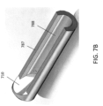
- the formed lens assembly can be dropped into a crevice 777 or hole in the device housing 709, and the hole can then be filled with the interface medium.
- the formed lens assembly can be integral with a hypotube 787 having an elongate channel 788 extending therethrough. The optical fiber can then be placed within the channel 788 and the interface medium used to attach the fiber to the lens 710.
- the interface mediums described herein can be, for example, an epoxy, such as a UV-curing epoxy.
- the use of the anamorphic lens assemblies described herein may shorten the length of the distal tip of the devices in which the assemblies are used.
- the length of the stripped section of the fiber may be much smaller, so the distal imaging and therapeutic housing can be made much smaller.
- the lens assemblies described herein can have significantly better focusing capability suited to the geometry of a catheter.
- the radius of curvature of the anamorphic lens structures described herein may be such that the focus is a preferred (further) distance away from the housing.
- FIG. 8 shows examples of a range of beam diameters relative to the distance from the imaging tip for an OCT catheter without a lens 801, with a GRIN fiber lens 803, and with the anamorphic lens apparatuses described herein 805.
- the beam diameter with 0 being the edge of the housing.
- the lens assemblies described herein can themselves cause the reflection of the light beam into the tissue. That is, the reflective surfaces can be uncoated, and the reflection can be caused by a total internal reflection resulting from the mismatch of the refractive indices between the lens material and the interface medium and/or air surrounding the lens.
- the refractive index of the lens material can be high, such as 1.0 or greater, 1.2 or greater, 1.5 or greater, or 1.7 or greater.
- the distal surface of the lens can be bordered by air to ensure total internal reflection.
- the reference reflection can be made by a surface other than the optical fiber / interface medium surface.
- the reference reflection can be made by the proximal or distal surfaces of the lens assembly and/or by the outer surface of the lens assembly.
- the indices of refraction of the interface medium and the core of the optical fiber can be closely matched (e.g., within 0.02) in order to provide only minimal secondary reflection at that interface.
- the lens assemblies described herein can be used with a variety of different imaging catheters.
- the lens assemblies can be used with: U.S. Patent Application No. 12/829,277, filed July 1, 2010 , titled “ATHERECTOMY CATHETER WITH LATERALLY-DISPLACEABLE TIP,” now U.S. Patent Application Publication No. 2011/0004107 ; U.S. Patent Application No. 12/829,267, filed July 1, 2010 , titled "CATHETER-BASED OFF-AXIS OPTICAL COHERENCE TOMOGRAPHY IMAGING SYSTEM," now U.S. Patent No. 9,125,562 ; U.S. Patent Application No.
- the system 1100 can therefore include a source of optical radiation 1122, a common path optical fiber 1115 (e.g., extending through a catheter elongate body), the lens assembly 1110, the interface medium or epoxy 1102, and a detector 1130 configured to receive the optical radiation reflected from the reference interface and the target 1114.
- the system 1100 can further include a processor to generate an image of the target based upon the optical radiation received by the receiving electronics.
- a Faraday isolation device 1112 such as a Faraday Effect optical circulator, can be used to separate the paths of the outgoing light source signal and the target and reference signals returning from the distal end of the fiber.
- Exemplary OCT systems with which the lens assembly can be used are further described in U.S. Patent Application No. 14/400,140, filed on 11/10/2014 (titled “OPTICAL COHERENCE TOMOGRAPHY WITH GRADED INDEX FIBER FOR BIOLOGICAL IMAGING").
- a secondary reflection of optical radiation from an interface between the optical fiber and the lens assembly is less than -60dB.
- the reference interface provides a reference reflection of between -28 and -42dB.
- carbon dioxide supply cartridges that can be used, for example, to inflate a balloon of a balloon catheter.
- FIG. 9 shows a single-use carbon dioxide supply cartridge or device 900.
- the device 900 includes a cylinder 901 filled with carbon dioxide and a valve assembly 903.
- the cylinder 901 and the valve assembly 903 can be twisted relative to one another in order to open the cylinder and pressure the valve assembly 903.
- the cylinder 901 can have a threaded male portion 905 while the valve assembly 903 can include a threaded female portion 907.
- the valve assembly 903 can further include a piercing element, such as a needle, configured to break a seal on the cylinder 901 when the threaded portions 905, 907 are connected together.
- the valve assembly 903 can further include a rotational valve 919 configured to control or restrict flow of the gas therethrough.
- a check valve 911 can be configured to control the maximum inflation pressure.
- the check valve 911 can limit the inflation pressure to 15psi.
- a catheter connection 909 can mechanically connect to the catheter and provide a flow path for the carbon dioxide from the device 900 to the balloon.
- Figure 10 shows another single-use carbon dioxide supply device 1000 with similar features as device 900.
- the carbon dioxide supply devices 900, 1000 can be used with a variety of different balloon catheters.
- the devices 900, 1000 can be used with balloon atherectomy catheters, such as those described in International Patent Application No. PCT/US2013/032494, filed March 15, 2013 , titled “BALOON ATHERECTOMY CATHETERS WITH IMAGING," and International Patent Application No. PCT/US2015/014613, filed February 5, 2015 , titled "ATHERECTOMY CATHETERS AND OCCLUSION CROSSING DEVICES".
- the carbon dioxide supply device 100 can further be used, for example, with balloon angioplasty catheters.
- the carbon dioxide supply devices 900,1000 can be a single-use sterile product.
- references to a structure or feature that is disposed "adjacent" another feature may have portions that overlap or underlie the adjacent feature.
- the device may be otherwise oriented (rotated 90 degrees or at other orientations) and the spatially relative descriptors used herein interpreted accordingly.
- the terms “upwardly”, “downwardly”, “vertical”, “horizontal” and the like are used herein for the purpose of explanation only unless specifically indicated otherwise.
- first and second may be used herein to describe various features/elements, these features/elements should not be limited by these terms, unless the context indicates otherwise. These terms may be used to distinguish one feature/element from another feature/element. Thus, a first feature/element discussed below could be termed a second feature/element, and similarly, a second feature/element discussed below could be termed a first feature/element.
- a numeric value may have a value that is +/- 0.1% of the stated value (or range of values), +/- 1% of the stated value (or range of values), +/- 2% of the stated value (or range of values), +/- 5% of the stated value (or range of values), +/- 10% of the stated value (or range of values), etc. Any numerical range recited herein is intended to include all sub-ranges subsumed therein.
Landscapes
- Health & Medical Sciences (AREA)
- Physics & Mathematics (AREA)
- Life Sciences & Earth Sciences (AREA)
- General Health & Medical Sciences (AREA)
- General Physics & Mathematics (AREA)
- Medical Informatics (AREA)
- Veterinary Medicine (AREA)
- Engineering & Computer Science (AREA)
- Biomedical Technology (AREA)
- Heart & Thoracic Surgery (AREA)
- Biophysics (AREA)
- Molecular Biology (AREA)
- Surgery (AREA)
- Animal Behavior & Ethology (AREA)
- Pathology (AREA)
- Public Health (AREA)
- Optics & Photonics (AREA)
- Nuclear Medicine, Radiotherapy & Molecular Imaging (AREA)
- Radiology & Medical Imaging (AREA)
- Endoscopes (AREA)
- Investigating Or Analysing Materials By Optical Means (AREA)
- Instruments For Viewing The Inside Of Hollow Bodies (AREA)
Claims (15)
- Cathéter (600) pour une tomographie par cohérence optique (OCT), comprenant :un corps de cathéter allongé s'étendant d'une façon distale à une façon proximale, le corps de cathéter allongé comprenant un trou dirigé latéralement (777) ;une fibre optique dans le corps de cathéter allongé ; etun corps de lentille (610) positionné à l'intérieur du trou dirigé latéralement (777) et fixé et optiquement couplé à une extrémité distale de la fibre optique par un support d'interface, le corps de lentille (610) ayant une surface proximale concave (611) qui est opposée et forme un angle par rapport à l'extrémité distale de la fibre optique ;dans lequel la surface proximale présente un rayon composé ayant un premier rayon le long d'un premier axe qui est différent d'un second rayon le long d'un second axe ;dans lequel la surface proximale comprend un matériau réfléchissant configuré de sorte qu'au moins 90 % de la lumière passant de l'extrémité distale de la fibre optique au corps de lentille soient réfléchis à partir de la surface proximale ; etdans lequel la fibre optique et le corps de lentille sont ensemble configurés pour fournir un trajet commun pour un rayonnement optique réfléchi à partir d'une cible et à partir d'une interface de référence entre l'extrémité distale de la fibre optique et la surface proximale.
- Cathéter selon la revendication 1, dans lequel le support d'interface est une résine époxy.
- Cathéter selon la revendication 1, dans lequel une tangente de la surface proximale (611) forme un angle compris entre 40 degrés et 50 degrés par rapport à un axe longitudinal de la fibre optique.
- Cathéter selon la revendication 1, dans lequel le matériau réfléchissant comprend un revêtement réfléchissant sur la surface proximale (611).
- Cathéter selon la revendication 4, dans lequel le revêtement réfléchissant comprend de l'or.
- Cathéter selon la revendication 1, dans lequel le matériau réfléchissant comprend un diélectrique.
- Cathéter selon la revendication 1, dans lequel le corps de lentille (610) comprend un matériau de polycarbonate.
- Cathéter selon la revendication 1, dans lequel une réflexion secondaire de rayonnement optique à partir d'une interface entre la fibre optique et le corps de lentille est inférieure à - 60 dB.
- Cathéter selon la revendication 1, dans lequel l'interface de référence fournit une réflexion de référence entre - 28 et - 42 dB.
- Cathéter selon la revendication 4, dans lequel le revêtement réfléchissant présente une densité optique supérieure à 3,0.
- Cathéter selon la revendication 1, dans lequel un rayon de courbure de la surface proximale (611) est compris entre 0,2 mm et 1,5 mm.
- Cathéter selon la revendication 1, comprenant en outre une source de rayonnement optique (1122) configurée pour fournir la lumière, dans lequel un revêtement réfléchissant sur la surface proximale (611) présente une épaisseur d'au moins 1/6 d'une longueur d'onde d'excitation de la source de rayonnement optique.
- Cathéter selon la revendication 1, comprenant en outre une source de rayonnement optique (1122), recevant des composants électroniques configurés pour recevoir un rayonnement optique réfléchi à partir de l'interface de référence et de la cible, et un processeur pour générer une image de la cible sur la base du rayonnement optique reçu par l'électronique de réception.
- Cathéter selon la revendication 1, dans lequel la lumière passant de l'extrémité distale de la fibre optique au corps de lentille ne voyage pas à travers la lentille.
- Cathéter selon la revendication 1, dans lequel le support d'interface raccorde le corps de lentille à l'extrémité distale de la fibre optique.
Applications Claiming Priority (3)
| Application Number | Priority Date | Filing Date | Title |
|---|---|---|---|
| US201562191956P | 2015-07-13 | 2015-07-13 | |
| US201562191986P | 2015-07-13 | 2015-07-13 | |
| PCT/US2016/042152 WO2017011587A1 (fr) | 2015-07-13 | 2016-07-13 | Lentille réflectrice anamorphosante micro-moulée pour cathéters à usage diagnostique/thérapeutique guidés par imagerie |
Publications (3)
| Publication Number | Publication Date |
|---|---|
| EP3322338A1 EP3322338A1 (fr) | 2018-05-23 |
| EP3322338A4 EP3322338A4 (fr) | 2019-03-13 |
| EP3322338B1 true EP3322338B1 (fr) | 2025-02-12 |
Family
ID=57757651
Family Applications (1)
| Application Number | Title | Priority Date | Filing Date |
|---|---|---|---|
| EP16825136.1A Active EP3322338B1 (fr) | 2015-07-13 | 2016-07-13 | Lentille réflectrice anamorphosante micro-moulée pour cathéters à usage diagnostique/thérapeutique guidés par imagerie |
Country Status (6)
| Country | Link |
|---|---|
| US (4) | US10568520B2 (fr) |
| EP (1) | EP3322338B1 (fr) |
| JP (2) | JP6896699B2 (fr) |
| CN (1) | CN107920780B (fr) |
| CA (1) | CA2992272A1 (fr) |
| WO (1) | WO2017011587A1 (fr) |
Families Citing this family (38)
| Publication number | Priority date | Publication date | Assignee | Title |
|---|---|---|---|---|
| US9125562B2 (en) | 2009-07-01 | 2015-09-08 | Avinger, Inc. | Catheter-based off-axis optical coherence tomography imaging system |
| WO2010129075A1 (fr) | 2009-04-28 | 2010-11-11 | Avinger, Inc. | Cathéter de support de fil-guide |
| WO2010138927A2 (fr) | 2009-05-28 | 2010-12-02 | Avinger, Inc. | Tomographie par cohérence optique (oct) pour imagerie biologique |
| WO2011003006A2 (fr) | 2009-07-01 | 2011-01-06 | Avinger, Inc. | Cathéter d'athérectomie pourvu d'une pointe déplaçable de manière latérale |
| US11382653B2 (en) | 2010-07-01 | 2022-07-12 | Avinger, Inc. | Atherectomy catheter |
| US9949754B2 (en) | 2011-03-28 | 2018-04-24 | Avinger, Inc. | Occlusion-crossing devices |
| EP2691038B1 (fr) | 2011-03-28 | 2016-07-20 | Avinger, Inc. | Dispositifs traversant une occlusion, imagerie et dispositifs d'athérectomie |
| EP3653151A1 (fr) | 2011-10-17 | 2020-05-20 | Avinger, Inc. | Cathéters d'athérectomie et mécanisme d'actionnement sans contact pour cathéters |
| US9345406B2 (en) | 2011-11-11 | 2016-05-24 | Avinger, Inc. | Occlusion-crossing devices, atherectomy devices, and imaging |
| WO2013172970A1 (fr) | 2012-05-14 | 2013-11-21 | Avinger, Inc. | Cathéters d'athérectomie à imagerie |
| WO2013172974A1 (fr) | 2012-05-14 | 2013-11-21 | Avinger, Inc. | Ensembles de pilotage de cathéter d'athérectomie |
| WO2013172972A1 (fr) | 2012-05-14 | 2013-11-21 | Avinger, Inc. | Tomographie à cohérence optique ayant une fibre à gradient d'indice pour imagerie biologique |
| EP2892448B1 (fr) | 2012-09-06 | 2020-07-15 | Avinger, Inc. | Cathéters d'athérectomie à ballon et à système d'imagerie |
| US11284916B2 (en) | 2012-09-06 | 2022-03-29 | Avinger, Inc. | Atherectomy catheters and occlusion crossing devices |
| US10932670B2 (en) | 2013-03-15 | 2021-03-02 | Avinger, Inc. | Optical pressure sensor assembly |
| US11096717B2 (en) | 2013-03-15 | 2021-08-24 | Avinger, Inc. | Tissue collection device for catheter |
| WO2014143064A1 (fr) | 2013-03-15 | 2014-09-18 | Avinger, Inc. | Dispositifs de traversée d'occlusion totale chronique à l'aide d'imagerie |
| JP6517198B2 (ja) | 2013-07-08 | 2019-05-22 | アビンガー・インコーポレイテッドAvinger, Inc. | 介入療法を案内する弾性層の識別 |
| MX2017000303A (es) | 2014-07-08 | 2017-07-10 | Avinger Inc | Dispositivos para oclusion transversal cronica total de alta velocidad. |
| EP3322338B1 (fr) * | 2015-07-13 | 2025-02-12 | Avinger, Inc. | Lentille réflectrice anamorphosante micro-moulée pour cathéters à usage diagnostique/thérapeutique guidés par imagerie |
| JP6981967B2 (ja) | 2015-08-31 | 2021-12-17 | ジェンテュイティ・リミテッド・ライアビリティ・カンパニーGentuity, LLC | 撮像プローブおよびデリバリデバイスを含む撮像システム |
| CN108882857A (zh) | 2016-01-25 | 2018-11-23 | 阿维格公司 | 具有滞后修正的oct成像导管 |
| JP6959255B2 (ja) | 2016-04-01 | 2021-11-02 | アビンガー・インコーポレイテッドAvinger, Inc. | 粥腫切除用カテーテルデバイス |
| US11344327B2 (en) | 2016-06-03 | 2022-05-31 | Avinger, Inc. | Catheter device with detachable distal end |
| US11224459B2 (en) | 2016-06-30 | 2022-01-18 | Avinger, Inc. | Atherectomy catheter with shapeable distal tip |
| US11519712B2 (en) * | 2016-07-05 | 2022-12-06 | The General Hospital Corporation | Systems and methods for an actively controlled optical imaging device |
| WO2018031462A1 (fr) * | 2016-08-12 | 2018-02-15 | Canon U.S.A. Inc. | Imagerie à plage de cohérence utilisant une interférence à trajet commun |
| WO2019108598A1 (fr) | 2017-11-28 | 2019-06-06 | Gentuity, Llc | Système d'imagerie |
| US12167867B2 (en) | 2018-04-19 | 2024-12-17 | Avinger, Inc. | Occlusion-crossing devices |
| US12262872B2 (en) | 2018-09-17 | 2025-04-01 | Gentuity, Llc | Imaging system with optical pathway |
| US12420069B2 (en) | 2018-12-19 | 2025-09-23 | Covidien Lp | Internal carotid artery thrombectomy devices and methods |
| WO2020158572A1 (fr) * | 2019-01-30 | 2020-08-06 | 日本ゼオン株式会社 | Cathéter à ballonnet intra-aortique |
| EP3962346A4 (fr) | 2019-04-30 | 2023-04-19 | Gentuity LLC | Sonde d'imagerie dotée d'un élément de mise sous pression de fluide |
| WO2020237024A1 (fr) | 2019-05-21 | 2020-11-26 | Gentuity, Llc | Systèmes et procédés pour un traitement de patients basé sur l'oct |
| JP2022553223A (ja) | 2019-10-18 | 2022-12-22 | アビンガー・インコーポレイテッド | 閉塞横断装置 |
| CN113180820A (zh) * | 2021-04-30 | 2021-07-30 | 广州迪光医学科技有限公司 | 径向激光消融导管 |
| CN113855228B (zh) * | 2021-09-30 | 2023-12-01 | 杭州佳量医疗科技有限公司 | 一种用于消融手术光纤的护套结构 |
| KR102511471B1 (ko) * | 2022-06-15 | 2023-03-17 | (주)옵토마인드 | 광송수신기 |
Citations (1)
| Publication number | Priority date | Publication date | Assignee | Title |
|---|---|---|---|---|
| US20140249407A1 (en) * | 2007-11-12 | 2014-09-04 | Lightlab Imaging, Inc. | Miniature Optical Elements for Fiber-Optic Beam Shaping |
Family Cites Families (604)
| Publication number | Priority date | Publication date | Assignee | Title |
|---|---|---|---|---|
| US3367727A (en) | 1965-10-22 | 1968-02-06 | Abraham W. Ward | Oral surgery tool with interchangeable blades |
| US3908637A (en) | 1974-04-22 | 1975-09-30 | Louis W Doroshow | Rigid urethral instrument |
| DE2733019C3 (de) | 1977-07-21 | 1981-01-15 | Boris Samoilovitsch Gechman | Vorrichtung zum Zerkleinem von Konkrementen im Harnleiter |
| US4527553A (en) | 1980-04-28 | 1985-07-09 | Upsher Michael S | Laryngoscope with improved light source |
| US4578061A (en) | 1980-10-28 | 1986-03-25 | Lemelson Jerome H | Injection catheter and method |
| US5435805A (en) | 1992-08-12 | 1995-07-25 | Vidamed, Inc. | Medical probe device with optical viewing capability |
| US4621353A (en) | 1982-09-09 | 1986-11-04 | Burroughs Corporation | Optical memory system providing improved focusing control and improved beam combining and separating apparatus |
| US4487206A (en) | 1982-10-13 | 1984-12-11 | Honeywell Inc. | Fiber optic pressure sensor with temperature compensation and reference |
| FR2541784B1 (fr) | 1983-02-25 | 1986-05-16 | Thomson Csf | Dispositif de deflexion statique d'un faisceau infra-rouge |
| US4611600A (en) | 1983-11-21 | 1986-09-16 | Cordis Corporation | Optical fiber pressure transducer |
| US4598710A (en) | 1984-01-20 | 1986-07-08 | Urban Engineering Company, Inc. | Surgical instrument and method of making same |
| US5178153A (en) | 1984-03-08 | 1993-01-12 | Einzig Robert E | Fluid flow sensing apparatus for in vivo and industrial applications employing novel differential optical fiber pressure sensors |
| US5041082A (en) | 1986-06-16 | 1991-08-20 | Samuel Shiber | Mechanical atherectomy system and method |
| US4926858A (en) | 1984-05-30 | 1990-05-22 | Devices For Vascular Intervention, Inc. | Atherectomy device for severe occlusions |
| US4781186A (en) | 1984-05-30 | 1988-11-01 | Devices For Vascular Intervention, Inc. | Atherectomy device having a flexible housing |
| US4552554A (en) | 1984-06-25 | 1985-11-12 | Medi-Tech Incorporated | Introducing catheter |
| US4651753A (en) | 1984-10-12 | 1987-03-24 | Jayco Pharmaceuticals | Endoscopic multiple biopsy instrument |
| US4686982A (en) | 1985-06-19 | 1987-08-18 | John Nash | Spiral wire bearing for rotating wire drive catheter |
| US4681106A (en) | 1985-08-12 | 1987-07-21 | Intravascular Surgical Instruments, Inc. | Catheter based surgical methods and apparatus therefor |
| US4654024A (en) | 1985-09-04 | 1987-03-31 | C.R. Bard, Inc. | Thermorecanalization catheter and method for use |
| US5182291A (en) | 1986-02-14 | 1993-01-26 | Sanofi | Pyrozala-pyridyl aminoabkoxyphenol compounds |
| US4771774A (en) | 1986-02-28 | 1988-09-20 | Devices For Vascular Intervention, Inc. | Motor drive unit |
| US5000185A (en) | 1986-02-28 | 1991-03-19 | Cardiovascular Imaging Systems, Inc. | Method for intravascular two-dimensional ultrasonography and recanalization |
| US4691708A (en) | 1986-03-10 | 1987-09-08 | Cordis Corporation | Optical pressure sensor for measuring blood pressure |
| US4842578A (en) | 1986-03-12 | 1989-06-27 | Dyonics, Inc. | Surgical abrading instrument |
| JPH0732758B2 (ja) | 1986-05-21 | 1995-04-12 | オリンパス光学工業株式会社 | 内視鏡 |
| US4729763A (en) | 1986-06-06 | 1988-03-08 | Henrie Rodney A | Catheter for removing occlusive material |
| SE453561B (sv) | 1986-06-25 | 1988-02-15 | Radisensor Ab | Miniatyriserad sensor for fysiologiska tryckmetningar |
| US4841977A (en) | 1987-05-26 | 1989-06-27 | Inter Therapy, Inc. | Ultra-thin acoustic transducer and balloon catheter using same in imaging array subassembly |
| US4808163A (en) | 1987-07-29 | 1989-02-28 | Laub Glenn W | Percutaneous venous cannula for cardiopulmonary bypass |
| US4850354A (en) | 1987-08-13 | 1989-07-25 | Baxter Travenol Laboratories, Inc. | Surgical cutting instrument |
| US4857046A (en) | 1987-10-21 | 1989-08-15 | Cordis Corporation | Drive catheter having helical pump drive shaft |
| US5529580A (en) | 1987-10-30 | 1996-06-25 | Olympus Optical Co., Ltd. | Surgical resecting tool |
| US5047040A (en) | 1987-11-05 | 1991-09-10 | Devices For Vascular Intervention, Inc. | Atherectomy device and method |
| US4920961A (en) | 1988-06-02 | 1990-05-01 | Circon Corporation | System for disconnetably mounting an endoscope sheath with an endoscope tool |
| EP0347098B1 (fr) | 1988-06-13 | 1996-02-28 | Samuel Shiber | Système d'athérectomie avec guide métallique |
| SE460396B (sv) | 1988-07-29 | 1989-10-09 | Radisensor Ab | Miniatyriserad givaranordning foer maetning av fysiologiska tryck in vivo |
| US5099850A (en) | 1989-01-17 | 1992-03-31 | Olympus Optical Co., Ltd. | Ultrasonic diagnostic apparatus |
| US5431673A (en) | 1989-02-17 | 1995-07-11 | American Biomed, Inc. | Distal atherectomy catheter |
| US4911148A (en) | 1989-03-14 | 1990-03-27 | Intramed Laboratories, Inc. | Deflectable-end endoscope with detachable flexible shaft assembly |
| US5002560A (en) | 1989-09-08 | 1991-03-26 | Advanced Cardiovascular Systems, Inc. | Expandable cage catheter with a rotatable guide |
| US5226909A (en) | 1989-09-12 | 1993-07-13 | Devices For Vascular Intervention, Inc. | Atherectomy device having helical blade and blade guide |
| US5085662A (en) | 1989-11-13 | 1992-02-04 | Scimed Life Systems, Inc. | Atherectomy catheter and related components |
| US5054501A (en) | 1990-05-16 | 1991-10-08 | Brigham & Women's Hospital | Steerable guide wire for cannulation of tubular or vascular organs |
| US5674232A (en) | 1990-06-05 | 1997-10-07 | Halliburton; Alexander George | Catheter and method of use thereof |
| BE1003189A5 (fr) | 1990-07-27 | 1992-01-07 | B A Cosurvey Optics S P R L B | Capteur de pression. |
| JP3467268B2 (ja) | 1990-08-06 | 2003-11-17 | アキュレイズ,インコーポレイテッド | ファイバー光学によるレーザーカテーテル |
| US5449372A (en) | 1990-10-09 | 1995-09-12 | Scimed Lifesystems, Inc. | Temporary stent and methods for use and manufacture |
| US5217479A (en) | 1991-02-14 | 1993-06-08 | Linvatec Corporation | Surgical cutting instrument |
| US5142155A (en) | 1991-03-11 | 1992-08-25 | Hewlett-Packard Company | Catheter tip fluorescence-quenching fiber optic pressure sensor |
| US6501551B1 (en) | 1991-04-29 | 2002-12-31 | Massachusetts Institute Of Technology | Fiber optic imaging endoscope interferometer with at least one faraday rotator |
| US6564087B1 (en) | 1991-04-29 | 2003-05-13 | Massachusetts Institute Of Technology | Fiber optic needle probes for optical coherence tomography imaging |
| US5321501A (en) | 1991-04-29 | 1994-06-14 | Massachusetts Institute Of Technology | Method and apparatus for optical imaging with means for controlling the longitudinal range of the sample |
| US5465147A (en) | 1991-04-29 | 1995-11-07 | Massachusetts Institute Of Technology | Method and apparatus for acquiring images using a ccd detector array and no transverse scanner |
| US5956355A (en) | 1991-04-29 | 1999-09-21 | Massachusetts Institute Of Technology | Method and apparatus for performing optical measurements using a rapidly frequency-tuned laser |
| US6485413B1 (en) | 1991-04-29 | 2002-11-26 | The General Hospital Corporation | Methods and apparatus for forward-directed optical scanning instruments |
| US6134003A (en) | 1991-04-29 | 2000-10-17 | Massachusetts Institute Of Technology | Method and apparatus for performing optical measurements using a fiber optic imaging guidewire, catheter or endoscope |
| US7074231B2 (en) | 1991-06-13 | 2006-07-11 | Advanced Cardiovascular Systems, Inc. | Convertible mode vascular catheter system |
| US5190050A (en) | 1991-11-08 | 1993-03-02 | Electro-Catheter Corporation | Tip deflectable steerable catheter |
| US5192291A (en) | 1992-01-13 | 1993-03-09 | Interventional Technologies, Inc. | Rotationally expandable atherectomy cutter assembly |
| JP2549822B2 (ja) | 1992-01-13 | 1996-10-30 | シュナイダー・(ユーエスエイ)・インコーポレーテッド | 外科用カッティングツール |
| GB9207532D0 (en) | 1992-04-07 | 1992-05-20 | Innovata Biomed Ltd | Medical connection system |
| JPH0627343A (ja) | 1992-07-06 | 1994-02-04 | Nippon Telegr & Teleph Corp <Ntt> | 光ファイバ増幅器用光ファイバ接続部 |
| US5312415A (en) | 1992-09-22 | 1994-05-17 | Target Therapeutics, Inc. | Assembly for placement of embolic coils using frictional placement |
| US5425371A (en) | 1992-10-05 | 1995-06-20 | Metatech Corporation | Fiberoptic pressure transducer |
| US5383460A (en) | 1992-10-05 | 1995-01-24 | Cardiovascular Imaging Systems, Inc. | Method and apparatus for ultrasound imaging and atherectomy |
| US5333142A (en) | 1992-10-26 | 1994-07-26 | The United States Of America As Represented By The Secretary Of The Navy | Technique for intracavity sum frequency generation |
| US5643297A (en) | 1992-11-09 | 1997-07-01 | Endovascular Instruments, Inc. | Intra-artery obstruction clearing apparatus and methods |
| US5383467A (en) | 1992-11-18 | 1995-01-24 | Spectrascience, Inc. | Guidewire catheter and apparatus for diagnostic imaging |
| US5368564A (en) | 1992-12-23 | 1994-11-29 | Angeion Corporation | Steerable catheter |
| US5460168A (en) | 1992-12-25 | 1995-10-24 | Olympus Optical Co., Ltd. | Endoscope cover assembly and cover-system endoscope |
| US5372601A (en) | 1993-03-30 | 1994-12-13 | Lary; Banning G. | Longitudinal reciprocating incisor |
| US5429136A (en) | 1993-04-21 | 1995-07-04 | Devices For Vascular Intervention, Inc. | Imaging atherectomy apparatus |
| ATE169483T1 (de) | 1993-04-28 | 1998-08-15 | Focal Inc | Vorrichtung, produkt und verwendung betreffend die intraluminale photothermoformgebung |
| US5868778A (en) | 1995-10-27 | 1999-02-09 | Vascular Solutions, Inc. | Vascular sealing apparatus and method |
| US5383896A (en) | 1993-05-25 | 1995-01-24 | Gershony; Gary | Vascular sealing device |
| US6017359A (en) | 1993-05-25 | 2000-01-25 | Vascular Solutions, Inc. | Vascular sealing apparatus |
| US5951583A (en) | 1993-05-25 | 1999-09-14 | Vascular Solutions, Inc. | Thrombin and collagen procoagulant and process for making the same |
| US5579767A (en) | 1993-06-07 | 1996-12-03 | Prince; Martin R. | Method for imaging abdominal aorta and aortic aneurysms |
| US5366464A (en) | 1993-07-22 | 1994-11-22 | Belknap John C | Atherectomy catheter device |
| CH687228A5 (de) | 1993-09-15 | 1996-10-31 | Synthes Ag | Markraumbohrkopf. |
| CA2173482A1 (fr) | 1993-10-07 | 1995-04-13 | Erik Andersen | Catheter de dilatation |
| US5507760A (en) | 1993-11-09 | 1996-04-16 | Devices For Vascular Intervention, Inc. | Cutter device |
| US5372560A (en) | 1993-11-24 | 1994-12-13 | Chang; John | Multi-functional sporting equipment |
| JPH07184888A (ja) | 1993-12-27 | 1995-07-25 | Toshiba Corp | 超音波診断装置 |
| US5437284A (en) | 1993-12-30 | 1995-08-01 | Camino Laboratories, Inc. | System and method for in vivo calibration of a sensor |
| US5517998A (en) | 1994-01-24 | 1996-05-21 | Medamicus, Inc. | Closed loop pressure determination system and method for fiber optic pressure transducer system |
| JP2869020B2 (ja) | 1994-03-23 | 1999-03-10 | 康男 橋本 | 癌治療器 |
| DE69514262T2 (de) | 1994-03-23 | 2001-10-11 | Hamamatsu Photonics K.K., Hamamatsu | Katheter mit Lichtleitfaser |
| US5507795A (en) | 1994-04-29 | 1996-04-16 | Devices For Vascular Intervention, Inc. | Catheter with perfusion system |
| US6032673A (en) | 1994-10-13 | 2000-03-07 | Femrx, Inc. | Methods and devices for tissue removal |
| US5836957A (en) | 1994-12-22 | 1998-11-17 | Devices For Vascular Intervention, Inc. | Large volume atherectomy device |
| US5632754A (en) | 1994-12-23 | 1997-05-27 | Devices For Vascular Intervention | Universal catheter with interchangeable work element |
| DE19504261A1 (de) | 1995-02-09 | 1996-09-12 | Krieg Gunther | Angioplastie-Katheter zum Erweitern und/oder Eröffnen von Blutgefäßen |
| US5613981A (en) | 1995-04-21 | 1997-03-25 | Medtronic, Inc. | Bidirectional dual sinusoidal helix stent |
| US5681336A (en) | 1995-09-07 | 1997-10-28 | Boston Scientific Corporation | Therapeutic device for treating vien graft lesions |
| US6615071B1 (en) | 1995-09-20 | 2003-09-02 | Board Of Regents, The University Of Texas System | Method and apparatus for detecting vulnerable atherosclerotic plaque |
| WO1997010748A1 (fr) | 1995-09-20 | 1997-03-27 | Texas Heart Institute | Detection d'ecarts thermiques dans les parois des vaisseaux |
| US6375615B1 (en) | 1995-10-13 | 2002-04-23 | Transvascular, Inc. | Tissue penetrating catheters having integral imaging transducers and their methods of use |
| US5556405A (en) | 1995-10-13 | 1996-09-17 | Interventional Technologies Inc. | Universal dilator with reciprocal incisor |
| RU2185859C2 (ru) | 1995-10-20 | 2002-07-27 | Надим М. Закка | Устройство для удаления стенозов и поддержки стенки сосуда (варианты) |
| US5907425A (en) | 1995-12-19 | 1999-05-25 | The Board Of Trustees Of The Leland Stanford Junior University | Miniature scanning confocal microscope |
| SE9601541D0 (sv) | 1995-11-08 | 1996-04-23 | Pacesetter Ab | Styrtrådsenhet |
| US5843050A (en) | 1995-11-13 | 1998-12-01 | Micro Therapeutics, Inc. | Microcatheter |
| SE9504334D0 (sv) | 1995-12-04 | 1995-12-04 | Pacesetter Ab | Styrtrådsenhet |
| US5676012A (en) | 1995-12-05 | 1997-10-14 | Spectrum Manufacturing, Inc. | Process for forming endoscopic shaver blade from elongate tube |
| US6373573B1 (en) | 2000-03-13 | 2002-04-16 | Lj Laboratories L.L.C. | Apparatus for measuring optical characteristics of a substrate and pigments applied thereto |
| US5733296A (en) | 1996-02-06 | 1998-03-31 | Devices For Vascular Intervention | Composite atherectomy cutter |
| US5935139A (en) | 1996-05-03 | 1999-08-10 | Boston Scientific Corporation | System for immobilizing or manipulating an object in a tract |
| NL1003172C2 (nl) | 1996-05-20 | 1997-11-21 | Cordis Europ | Katheter-invoerschede met occlusieballon. |
| ATE252345T1 (de) | 1996-06-11 | 2003-11-15 | Roke Manor Research | Katheterverfolgungssystem |
| US5795295A (en) | 1996-06-25 | 1998-08-18 | Carl Zeiss, Inc. | OCT-assisted surgical microscope with multi-coordinate manipulator |
| US6830577B2 (en) | 1996-07-26 | 2004-12-14 | Kensey Nash Corporation | System and method of use for treating occluded vessels and diseased tissue |
| US5779721A (en) | 1996-07-26 | 1998-07-14 | Kensey Nash Corporation | System and method of use for revascularizing stenotic bypass grafts and other blood vessels |
| US6080170A (en) | 1996-07-26 | 2000-06-27 | Kensey Nash Corporation | System and method of use for revascularizing stenotic bypass grafts and other occluded blood vessels |
| US6929481B1 (en) | 1996-09-04 | 2005-08-16 | Immersion Medical, Inc. | Interface device and method for interfacing instruments to medical procedure simulation systems |
| US5830145A (en) | 1996-09-20 | 1998-11-03 | Cardiovascular Imaging Systems, Inc. | Enhanced accuracy of three-dimensional intraluminal ultrasound (ILUS) image reconstruction |
| US5904651A (en) | 1996-10-28 | 1999-05-18 | Ep Technologies, Inc. | Systems and methods for visualizing tissue during diagnostic or therapeutic procedures |
| US5722403A (en) | 1996-10-28 | 1998-03-03 | Ep Technologies, Inc. | Systems and methods using a porous electrode for ablating and visualizing interior tissue regions |
| US5872879A (en) | 1996-11-25 | 1999-02-16 | Boston Scientific Corporation | Rotatable connecting optical fibers |
| JPH10161018A (ja) | 1996-11-27 | 1998-06-19 | Olympus Optical Co Ltd | 光学系 |
| US5899915A (en) | 1996-12-02 | 1999-05-04 | Angiotrax, Inc. | Apparatus and method for intraoperatively performing surgery |
| US6508825B1 (en) | 1997-02-28 | 2003-01-21 | Lumend, Inc. | Apparatus for treating vascular occlusions |
| US6010449A (en) | 1997-02-28 | 2000-01-04 | Lumend, Inc. | Intravascular catheter system for treating a vascular occlusion |
| US6120516A (en) | 1997-02-28 | 2000-09-19 | Lumend, Inc. | Method for treating vascular occlusion |
| US6183487B1 (en) | 1997-03-06 | 2001-02-06 | Scimed Life Systems, Inc. | Ablation device for reducing damage to vessels and/or in-vivo stents |
| US5843103A (en) | 1997-03-06 | 1998-12-01 | Scimed Life Systems, Inc. | Shaped wire rotational atherectomy device |
| US6758854B1 (en) | 1997-05-09 | 2004-07-06 | St. Jude Medical | Splittable occlusion balloon sheath and process of use |
| US5851212A (en) | 1997-06-11 | 1998-12-22 | Endius Incorporated | Surgical instrument |
| US6013072A (en) | 1997-07-09 | 2000-01-11 | Intraluminal Therapeutics, Inc. | Systems and methods for steering a catheter through body tissue |
| US6048349A (en) | 1997-07-09 | 2000-04-11 | Intraluminal Therapeutics, Inc. | Systems and methods for guiding a medical instrument through a body |
| US5987995A (en) | 1997-07-17 | 1999-11-23 | Sentec Corporation | Fiber optic pressure catheter |
| WO1999004700A1 (fr) | 1997-07-24 | 1999-02-04 | Mcguckin James F Jr | Catheter urinaire |
| GB9717580D0 (en) | 1997-08-19 | 1997-10-22 | Curry Paul | Device for opening blocked tubes |
| US6402719B1 (en) | 1997-09-05 | 2002-06-11 | Cordis Webster, Inc. | Steerable DMR catheter with infusion tube |
| AU712738B2 (en) | 1997-09-24 | 1999-11-18 | Eclipse Surgical Technologies, Inc. | Steerable catheter |
| US6193676B1 (en) | 1997-10-03 | 2001-02-27 | Intraluminal Therapeutics, Inc. | Guide wire assembly |
| US5951482A (en) | 1997-10-03 | 1999-09-14 | Intraluminal Therapeutics, Inc. | Assemblies and methods for advancing a guide wire through body tissue |
| JP2001522631A (ja) | 1997-11-07 | 2001-11-20 | プロリフィックス メディカル, インコーポレイテッド | 身体管腔内の閉塞を治療する方法およびシステム |
| US6183432B1 (en) | 1997-11-13 | 2001-02-06 | Lumend, Inc. | Guidewire and catheter with rotating and reciprocating symmetrical or asymmetrical distal tip |
| US5938671A (en) | 1997-11-14 | 1999-08-17 | Reflow, Inc. | Recanalization apparatus and devices for use therein and method |
| US6110164A (en) | 1997-12-05 | 2000-08-29 | Intratherapeutics, Inc. | Guideless catheter segment |
| US6027514A (en) | 1997-12-17 | 2000-02-22 | Fox Hollow Technologies, Inc. | Apparatus and method for removing occluding material from body lumens |
| US20050171478A1 (en) | 1998-01-13 | 2005-08-04 | Selmon Matthew R. | Catheter system for crossing total occlusions in vasculature |
| US6231546B1 (en) | 1998-01-13 | 2001-05-15 | Lumend, Inc. | Methods and apparatus for crossing total occlusions in blood vessels |
| US6416527B1 (en) | 1998-01-28 | 2002-07-09 | St. Jude Medical Cardiovascular Group, Inc. | Vessel cutting device |
| US6824550B1 (en) | 2000-04-06 | 2004-11-30 | Norbon Medical, Inc. | Guidewire for crossing occlusions or stenosis |
| US6175669B1 (en) | 1998-03-30 | 2001-01-16 | The Regents Of The Universtiy Of California | Optical coherence domain reflectometry guidewire |
| EP1067874B1 (fr) | 1998-03-31 | 2008-04-23 | Medtronic Vascular, Inc. | Catheters et systemes de creation de passages pour derivation arterio-veineuse in-situ percutanee |
| US6482217B1 (en) | 1998-04-10 | 2002-11-19 | Endicor Medical, Inc. | Neuro thrombectomy catheter |
| US6666874B2 (en) | 1998-04-10 | 2003-12-23 | Endicor Medical, Inc. | Rotational atherectomy system with serrated cutting tip |
| US6001112A (en) | 1998-04-10 | 1999-12-14 | Endicor Medical, Inc. | Rotational atherectomy device |
| US6290668B1 (en) | 1998-04-30 | 2001-09-18 | Kenton W. Gregory | Light delivery catheter and methods for the use thereof |
| US6285903B1 (en) | 1998-06-30 | 2001-09-04 | Eclipse Surgical Technologies, Inc. | Intracorporeal device with radiopaque marker |
| CA2277409C (fr) | 1998-07-10 | 2003-02-11 | Nippon Telegraph And Telephone Corporation | Systeme de transmission a fibres optiques |
| US6290689B1 (en) | 1999-10-22 | 2001-09-18 | Corazón Technologies, Inc. | Catheter devices and methods for their use in the treatment of calcified vascular occlusions |
| US6440124B1 (en) | 1998-07-22 | 2002-08-27 | Endovasix, Inc. | Flexible flow apparatus and method for the disruption of occlusions |
| US6319227B1 (en) | 1998-08-05 | 2001-11-20 | Scimed Life Systems, Inc. | Automatic/manual longitudinal position translator and rotary drive system for catheters |
| US6106515A (en) | 1998-08-13 | 2000-08-22 | Intraluminal Therapeutics, Inc. | Expandable laser catheter |
| US6241744B1 (en) | 1998-08-14 | 2001-06-05 | Fox Hollow Technologies, Inc. | Apparatus for deploying a guidewire across a complex lesion |
| AU6139499A (en) | 1998-09-11 | 2000-04-03 | Joseph A. Izatt | Interferometers for optical coherence domain reflectometry and optical coherencetomography using nonreciprocal optical elements |
| US6398755B1 (en) | 1998-10-06 | 2002-06-04 | Scimed Life Systems, Inc. | Driveable catheter system |
| US6228076B1 (en) | 1999-01-09 | 2001-05-08 | Intraluminal Therapeutics, Inc. | System and method for controlling tissue ablation |
| US6134002A (en) | 1999-01-14 | 2000-10-17 | Duke University | Apparatus and method for the rapid spectral resolution of confocal images |
| US6445944B1 (en) | 1999-02-01 | 2002-09-03 | Scimed Life Systems | Medical scanning system and related method of scanning |
| US6855123B2 (en) | 2002-08-02 | 2005-02-15 | Flow Cardia, Inc. | Therapeutic ultrasound system |
| AU3741400A (en) | 1999-03-15 | 2000-10-04 | Prolifix Medical, Inc. | Shielded atherectomy device |
| US6911026B1 (en) | 1999-07-12 | 2005-06-28 | Stereotaxis, Inc. | Magnetically guided atherectomy |
| US20040044350A1 (en) | 1999-04-09 | 2004-03-04 | Evalve, Inc. | Steerable access sheath and methods of use |
| US6645217B1 (en) | 1999-05-15 | 2003-11-11 | Advanced Cardiovascular Systems, Inc. | Over-the-wire atherectomy catheter |
| US6719769B2 (en) | 1999-11-15 | 2004-04-13 | Cardica, Inc. | Integrated anastomosis tool with graft vessel attachment device and cutting device |
| US6294775B1 (en) | 1999-06-08 | 2001-09-25 | University Of Washington | Miniature image acquistion system using a scanning resonant waveguide |
| US6563105B2 (en) | 1999-06-08 | 2003-05-13 | University Of Washington | Image acquisition with depth enhancement |
| US6890329B2 (en) | 1999-06-15 | 2005-05-10 | Cryocath Technologies Inc. | Defined deflection structure |
| US6546272B1 (en) | 1999-06-24 | 2003-04-08 | Mackinnon Nicholas B. | Apparatus for in vivo imaging of the respiratory tract and other internal organs |
| US6445939B1 (en) | 1999-08-09 | 2002-09-03 | Lightlab Imaging, Llc | Ultra-small optical probes, imaging optics, and methods for using same |
| US6447525B2 (en) | 1999-08-19 | 2002-09-10 | Fox Hollow Technologies, Inc. | Apparatus and methods for removing material from a body lumen |
| US7713279B2 (en) | 2000-12-20 | 2010-05-11 | Fox Hollow Technologies, Inc. | Method and devices for cutting tissue |
| US6638233B2 (en) | 1999-08-19 | 2003-10-28 | Fox Hollow Technologies, Inc. | Apparatus and methods for material capture and removal |
| US7708749B2 (en) | 2000-12-20 | 2010-05-04 | Fox Hollow Technologies, Inc. | Debulking catheters and methods |
| US6299622B1 (en) | 1999-08-19 | 2001-10-09 | Fox Hollow Technologies, Inc. | Atherectomy catheter with aligned imager |
| US20030120295A1 (en) | 2000-12-20 | 2003-06-26 | Fox Hollow Technologies, Inc. | Debulking catheters and methods |
| US20030125757A1 (en) | 2000-12-20 | 2003-07-03 | Fox Hollow Technologies, Inc. | Debulking catheters and methods |
| US8328829B2 (en) | 1999-08-19 | 2012-12-11 | Covidien Lp | High capacity debulking catheter with razor edge cutting window |
| US7887556B2 (en) | 2000-12-20 | 2011-02-15 | Fox Hollow Technologies, Inc. | Debulking catheters and methods |
| WO2001015609A1 (fr) | 1999-08-31 | 2001-03-08 | Fox Hollow Technologies | Catheter d'atherectomie a couteau telescopique et rotatif |
| US6687010B1 (en) | 1999-09-09 | 2004-02-03 | Olympus Corporation | Rapid depth scanning optical imaging device |
| US6673042B1 (en) | 1999-11-22 | 2004-01-06 | Wilfred J. Samson | Expandable venous cannula and method of use |
| DK176336B1 (da) | 1999-12-22 | 2007-08-20 | Asahi Optical Co Ltd | Endoskopisk vævsindsamlingsinstrument |
| US6929633B2 (en) | 2000-01-25 | 2005-08-16 | Bacchus Vascular, Inc. | Apparatus and methods for clot dissolution |
| AU2884401A (en) | 2000-01-28 | 2001-08-07 | Asahi Kasei Kabushiki Kaisha | Photothermic transducing spectroscopic analyzer |
| US6629953B1 (en) | 2000-02-18 | 2003-10-07 | Fox Hollow Technologies, Inc. | Methods and devices for removing material from a vascular site |
| US6579298B1 (en) | 2000-02-29 | 2003-06-17 | Scimed Life Systems, Inc. | Method and apparatus for treating vein graft lesions |
| US6565588B1 (en) | 2000-04-05 | 2003-05-20 | Pathway Medical Technologies, Inc. | Intralumenal material removal using an expandable cutting device |
| US7344546B2 (en) | 2000-04-05 | 2008-03-18 | Pathway Medical Technologies | Intralumenal material removal using a cutting device for differential cutting |
| US8475484B2 (en) | 2000-04-05 | 2013-07-02 | Medrad, Inc. | Liquid seal assembly for a rotating torque tube |
| DE60121556T2 (de) | 2000-04-05 | 2007-06-28 | Pathway Medical Technologies, Inc., Redmond | System und Verfahren zum Entfernen von intraluminalem Material |
| US20040243162A1 (en) | 2000-04-05 | 2004-12-02 | Pathway Medical Technologies, Inc. | Interventional catheter assemblies and control systems |
| US10092313B2 (en) | 2000-04-05 | 2018-10-09 | Boston Scientific Limited | Medical sealed tubular structures |
| US6454717B1 (en) | 2000-04-13 | 2002-09-24 | Scimed Life Systems, Inc. | Concentric catheter drive shaft clutch |
| US6517528B1 (en) | 2000-04-13 | 2003-02-11 | Scimed Life Systems, Inc. | Magnetic catheter drive shaft clutch |
| US6975898B2 (en) | 2000-06-19 | 2005-12-13 | University Of Washington | Medical imaging, diagnosis, and therapy using a scanning single optical fiber system |
| US7555333B2 (en) | 2000-06-19 | 2009-06-30 | University Of Washington | Integrated optical scanning image acquisition and display |
| US6511474B1 (en) | 2000-07-12 | 2003-01-28 | Corpak, Inc. | Bolus for non-occluding high flow enteral feeding tube |
| US6575995B1 (en) | 2000-07-14 | 2003-06-10 | Advanced Cardiovascular Systems, Inc. | Expandable cage embolic material filter system and method |
| US6572643B1 (en) | 2000-07-19 | 2003-06-03 | Vascular Architects, Inc. | Endoprosthesis delivery catheter assembly and method |
| WO2002021074A2 (fr) | 2000-09-04 | 2002-03-14 | Forskningscenter Risø | Amplification optique en reflectometrie de coherence |
| US6451009B1 (en) | 2000-09-12 | 2002-09-17 | The Regents Of The University Of California | OCDR guided laser ablation device |
| US6845190B1 (en) | 2000-11-27 | 2005-01-18 | University Of Washington | Control of an optical fiber scanner |
| US6856712B2 (en) | 2000-11-27 | 2005-02-15 | University Of Washington | Micro-fabricated optical waveguide for use in scanning fiber displays and scanned fiber image acquisition |
| US20020072706A1 (en) | 2000-12-11 | 2002-06-13 | Thomas Hiblar | Transluminal drug delivery catheter |
| US20100121360A9 (en) | 2000-12-20 | 2010-05-13 | Fox Hollow Technologies, Inc | Testing a patient population having a cardiovascular condition for drug efficacy |
| ATE499054T1 (de) | 2000-12-20 | 2011-03-15 | Fox Hollow Technologies Inc | Verkleinerungskatheter |
| US20050222519A1 (en) | 2000-12-20 | 2005-10-06 | Fox Hollow Technologies, Inc. | Debulking catheters and methods |
| US7699790B2 (en) | 2000-12-20 | 2010-04-20 | Ev3, Inc. | Debulking catheters and methods |
| US20060235366A1 (en) | 2000-12-20 | 2006-10-19 | Fox Hollow Technologies, Inc. | Method of evaluating a treatment for vascular disease |
| US7927784B2 (en) | 2000-12-20 | 2011-04-19 | Ev3 | Vascular lumen debulking catheters and methods |
| US20050154407A1 (en) | 2000-12-20 | 2005-07-14 | Fox Hollow Technologies, Inc. | Method of evaluating drug efficacy for treating atherosclerosis |
| US20060032508A1 (en) | 2000-12-20 | 2006-02-16 | Fox Hollow Technologies, Inc. | Method of evaluating a treatment for vascular disease |
| US20040167554A1 (en) | 2000-12-20 | 2004-08-26 | Fox Hollow Technologies, Inc. | Methods and devices for reentering a true lumen from a subintimal space |
| IL140780A0 (en) | 2001-01-08 | 2002-02-10 | Gaber Benny | Deflectable guiding apparatus |
| US6503261B1 (en) | 2001-01-17 | 2003-01-07 | Scimed Life Systems, Inc. | Bi-directional atherectomy burr |
| US6497649B2 (en) | 2001-01-21 | 2002-12-24 | University Of Washington | Alleviating motion, simulator, and virtual environmental sickness by presenting visual scene components matched to inner ear vestibular sensations |
| US6542665B2 (en) | 2001-02-17 | 2003-04-01 | Lucent Technologies Inc. | GRIN fiber lenses |
| US6760112B2 (en) | 2001-02-17 | 2004-07-06 | Lucent Technologies Inc. | Grin-fiber lens based optical endoscopes |
| US20020115931A1 (en) | 2001-02-21 | 2002-08-22 | Strauss H. William | Localizing intravascular lesions on anatomic images |
| US20020138091A1 (en) | 2001-03-23 | 2002-09-26 | Devonrex, Inc. | Micro-invasive nucleotomy device and method |
| US6616676B2 (en) | 2001-04-10 | 2003-09-09 | Scimed Life Systems, Inc. | Devices and methods for removing occlusions in vessels |
| WO2002083003A1 (fr) | 2001-04-11 | 2002-10-24 | Clarke Dana S | Identification de structures tissulaires lors de la progression d'un instrument |
| US6522452B2 (en) | 2001-04-26 | 2003-02-18 | Jds Uniphase Corporation | Latchable microelectromechanical structures using non-newtonian fluids, and methods of operating same |
| WO2002088705A2 (fr) | 2001-05-01 | 2002-11-07 | The General Hospital Corporation | Procede et appareil pour determiner un type de plaque d'atherosclerose par mesure de proprietes optiques de tissu |
| US7616986B2 (en) | 2001-05-07 | 2009-11-10 | University Of Washington | Optical fiber scanner for performing multimodal optical imaging |
| US7329223B1 (en) | 2001-05-31 | 2008-02-12 | Abbott Cardiovascular Systems Inc. | Catheter with optical fiber sensor |
| US6879851B2 (en) | 2001-06-07 | 2005-04-12 | Lightlab Imaging, Llc | Fiber optic endoscopic gastrointestinal probe |
| US20030032880A1 (en) | 2001-06-13 | 2003-02-13 | Pauliina Moore | Apparatus and method for ultrasonically identifying vulnerable plaque |
| US6702744B2 (en) | 2001-06-20 | 2004-03-09 | Advanced Cardiovascular Systems, Inc. | Agents that stimulate therapeutic angiogenesis and techniques and devices that enable their delivery |
| US6728571B1 (en) | 2001-07-16 | 2004-04-27 | Scimed Life Systems, Inc. | Electronically scanned optical coherence tomography with frequency modulated signals |
| AU2002322520A1 (en) | 2001-07-17 | 2003-03-03 | Kerberos Proximal Solutions | Fluid exchange system for controlled and localized irrigation and aspiration |
| US6599296B1 (en) | 2001-07-27 | 2003-07-29 | Advanced Cardiovascular Systems, Inc. | Ratcheting handle for intraluminal catheter systems |
| US20030045835A1 (en) | 2001-08-30 | 2003-03-06 | Vascular Solutions, Inc. | Method and apparatus for coagulation and closure of pseudoaneurysms |
| US6961123B1 (en) | 2001-09-28 | 2005-11-01 | The Texas A&M University System | Method and apparatus for obtaining information from polarization-sensitive optical coherence tomography |
| US7507245B2 (en) | 2001-10-19 | 2009-03-24 | Cardiovascular Systems, Inc. | Rotational angioplasty device with abrasive crown |
| US6749344B2 (en) | 2001-10-24 | 2004-06-15 | Scimed Life Systems, Inc. | Connection apparatus for optical coherence tomography catheters |
| US20050021075A1 (en) | 2002-12-30 | 2005-01-27 | Bonnette Michael J. | Guidewire having deployable sheathless protective filter |
| FR2832505B1 (fr) | 2001-11-16 | 2008-07-04 | Inst Francais Du Petrole | Refractometre a fibre optique |
| FR2832516B1 (fr) | 2001-11-19 | 2004-01-23 | Tokendo Sarl | Endoscopes rotatifs a visee distale deviee |
| US7557929B2 (en) | 2001-12-18 | 2009-07-07 | Massachusetts Institute Of Technology | Systems and methods for phase measurements |
| US6947787B2 (en) | 2001-12-21 | 2005-09-20 | Advanced Cardiovascular Systems, Inc. | System and methods for imaging within a body lumen |
| US7729742B2 (en) | 2001-12-21 | 2010-06-01 | Biosense, Inc. | Wireless position sensor |
| US7493156B2 (en) | 2002-01-07 | 2009-02-17 | Cardiac Pacemakers, Inc. | Steerable guide catheter with pre-shaped rotatable shaft |
| US7428001B2 (en) | 2002-03-15 | 2008-09-23 | University Of Washington | Materials and methods for simulating focal shifts in viewers using large depth of focus displays |
| US6869414B2 (en) | 2002-03-22 | 2005-03-22 | Cardiac Pacemakers, Inc. | Pre-shaped catheter with proximal articulation and pre-formed distal end |
| RU2218191C2 (ru) | 2002-04-11 | 2003-12-10 | Научно-исследовательский институт радиоэлектроники и лазерной техники Московского государственного технического университета им. Н.Э.Баумана | Эндовазальный мини-робот |
| US20050085708A1 (en) | 2002-04-19 | 2005-04-21 | University Of Washington | System and method for preparation of cells for 3D image acquisition |
| US7738945B2 (en) | 2002-04-19 | 2010-06-15 | University Of Washington | Method and apparatus for pseudo-projection formation for optical tomography |
| US7811825B2 (en) | 2002-04-19 | 2010-10-12 | University Of Washington | System and method for processing specimens and images for optical tomography |
| US20110201924A1 (en) | 2002-04-30 | 2011-08-18 | The General Hospital Corporation | Method and Apparatus for Improving Image Clarity and Sensitivity in Optical Tomography Using Dynamic Feedback to Control Focal Properties and Coherence Gating |
| US6852109B2 (en) | 2002-06-11 | 2005-02-08 | Intraluminal Therapeutics, Inc. | Radio frequency guide wire assembly with optical coherence reflectometry guidance |
| US6891984B2 (en) | 2002-07-25 | 2005-05-10 | Lightlab Imaging, Llc | Scanning miniature optical probes with optical distortion correction and rotational control |
| US20040039371A1 (en) | 2002-08-23 | 2004-02-26 | Bruce Tockman | Coronary vein navigator |
| US7359554B2 (en) | 2002-08-26 | 2008-04-15 | Cleveland Clinic Foundation | System and method for identifying a vascular border |
| US7076135B2 (en) | 2002-09-20 | 2006-07-11 | Nippon Telegraph And Telephone Corporation | Optical module and manufacturing method therefor |
| US7087064B1 (en) | 2002-10-15 | 2006-08-08 | Advanced Cardiovascular Systems, Inc. | Apparatuses and methods for heart valve repair |
| JP2006502784A (ja) | 2002-10-18 | 2006-01-26 | シア アリー | 撮像案内ワイヤを備えるアテローム切除システム |
| US20040147934A1 (en) | 2002-10-18 | 2004-07-29 | Kiester P. Douglas | Oscillating, steerable, surgical burring tool and method of using the same |
| US7493154B2 (en) | 2002-10-23 | 2009-02-17 | Medtronic, Inc. | Methods and apparatus for locating body vessels and occlusions in body vessels |
| US6849064B2 (en) | 2002-10-25 | 2005-02-01 | James S. Hamada | Minimal access lumbar diskectomy instrumentation and method |
| US6867753B2 (en) | 2002-10-28 | 2005-03-15 | University Of Washington | Virtual image registration in augmented display field |
| EP1592992B1 (fr) | 2003-01-24 | 2012-05-30 | University of Washington | Systeme de balayage a faisceau optique destine a l'affichage ou l'acquisition d'images compact |
| WO2004073501A2 (fr) | 2003-02-20 | 2004-09-02 | Gutin Mikhail | Tomographie par coherence optique avec balayage par coherence 3d |
| CA2517906A1 (fr) | 2003-03-03 | 2004-12-29 | Montana State University-Bozeman | Dispositif optique confocal miniaturise, systeme et procede |
| US7715896B2 (en) | 2003-03-21 | 2010-05-11 | Boston Scientific Scimed, Inc. | Systems and methods for internal tissue penetration |
| US20040254599A1 (en) | 2003-03-25 | 2004-12-16 | Lipoma Michael V. | Method and apparatus for pre-lancing stimulation of puncture site |
| US7001369B2 (en) | 2003-03-27 | 2006-02-21 | Scimed Life Systems, Inc. | Medical device |
| US6934444B2 (en) | 2003-04-10 | 2005-08-23 | Sioptical, Inc. | Beam shaping and practical methods of reducing loss associated with mating external sources and optics to thin silicon waveguides |
| US8246640B2 (en) | 2003-04-22 | 2012-08-21 | Tyco Healthcare Group Lp | Methods and devices for cutting tissue at a vascular location |
| DE10323217A1 (de) | 2003-05-22 | 2004-12-16 | Siemens Ag | Optisches Kohärenztomographiesystem zur Untersuchung des menschlichen oder tierischen Gewebes oder von Organen |
| US7488340B2 (en) | 2003-06-02 | 2009-02-10 | Vascular Solutions, Inc. | Vascular access closure system |
| USD489973S1 (en) | 2003-06-02 | 2004-05-18 | Vascular Solutions, Inc. | Medical device package |
| US6943881B2 (en) | 2003-06-04 | 2005-09-13 | Tomophase Corporation | Measurements of optical inhomogeneity and other properties in substances using propagation modes of light |
| US7150749B2 (en) | 2003-06-13 | 2006-12-19 | Sherwood Services Ag | Vessel sealer and divider having elongated knife stroke and safety cutting mechanism |
| US7311723B2 (en) | 2003-07-11 | 2007-12-25 | University Of Washington | Scanning laser device and methods of use |
| CN1929781A (zh) | 2003-08-21 | 2007-03-14 | 依斯克姆公司 | 用于脉管斑块检测和分析的自动化方法和系统 |
| US7608048B2 (en) | 2003-08-28 | 2009-10-27 | Goldenberg Alec S | Rotating soft tissue biopsy needle |
| US7758625B2 (en) | 2003-09-12 | 2010-07-20 | Abbott Vascular Solutions Inc. | Delivery system for medical devices |
| ES2564694T3 (es) | 2003-09-12 | 2016-03-28 | Vessix Vascular, Inc. | Sistema de remodelación y / o ablación excéntrica seleccionable de material ateroesclerótico |
| AU2004279388B2 (en) | 2003-10-03 | 2010-10-14 | Boston Scientific Limited | Systems for a delivering a medical implant to an anatomical location in a patient |
| WO2005032642A2 (fr) | 2003-10-03 | 2005-04-14 | Acumen Medical, Inc. | Gaine expansible de guidage et appareil, et leurs methodes de fabrication |
| JP2005114473A (ja) | 2003-10-06 | 2005-04-28 | Hitachi Medical Corp | 光検出方法及び生体光計測装置 |
| EP1684645A4 (fr) | 2003-10-07 | 2010-05-05 | Ford Henry Health System | Catheter pour embolectomie |
| EP1684842B1 (fr) | 2003-10-27 | 2013-03-06 | Petrus Antonius Besselink | Dispositif endoluminal auto-actionneur |
| EP2278287B1 (fr) | 2003-10-27 | 2016-09-07 | The General Hospital Corporation | Procédé et appareil de l'imagerie optique avec de l'interférométrie dans le domaine de fréquence |
| EP1691666B1 (fr) | 2003-12-12 | 2012-05-30 | University of Washington | Systeme de guidage et d'interface en 3d pour catheterscope |
| US20050149096A1 (en) | 2003-12-23 | 2005-07-07 | Hilal Said S. | Catheter with conduit traversing tip |
| US20050141843A1 (en) | 2003-12-31 | 2005-06-30 | Invitrogen Corporation | Waveguide comprising scattered light detectable particles |
| DE102004001498B4 (de) | 2004-01-09 | 2008-01-10 | Siemens Ag | Katheter zur Einführung in ein Gefäß |
| US20050159731A1 (en) | 2004-01-16 | 2005-07-21 | Lee Don W. | Intravascular catheter |
| US7706863B2 (en) | 2004-01-21 | 2010-04-27 | University Of Washington | Methods for assessing a physiological state of a mammalian retina |
| US20050197623A1 (en) | 2004-02-17 | 2005-09-08 | Leeflang Stephen A. | Variable steerable catheters and methods for using them |
| DE102004008371B4 (de) | 2004-02-20 | 2006-05-24 | Siemens Ag | Atherektomiekatheter |
| DE102004008370B4 (de) | 2004-02-20 | 2006-06-01 | Siemens Ag | Katheter zur Durchführung und Überwachung von Rotablation |
| MX2009004020A (es) | 2004-03-04 | 2009-04-27 | Cabezal de trabajo de un cateter para aspirar, fragmentar y extraer material extirpable de vasos sanguineos. | |
| JP2005249704A (ja) | 2004-03-08 | 2005-09-15 | Fujinon Corp | 断層映像装置 |
| US20050203425A1 (en) | 2004-03-10 | 2005-09-15 | Phil Langston | Coaxial dual lumen pigtail catheter |
| US7261687B2 (en) | 2004-03-23 | 2007-08-28 | California Institute Of Technology | Forward scanning imaging optical fiber probe |
| US7126693B2 (en) | 2004-03-29 | 2006-10-24 | Carl Zeiss Meditec, Inc. | Simple high efficiency optical coherence domain reflectometer design |
| DE102004015640B4 (de) | 2004-03-31 | 2007-05-03 | Siemens Ag | Vorrichtung zum Durchführen einer "Cutting-Balloon"-Intervention mit OCT-Überwachung |
| US20050228428A1 (en) | 2004-04-07 | 2005-10-13 | Afsar Ali | Balloon catheters and methods for manufacturing balloons for balloon catheters |
| US20050251116A1 (en) | 2004-05-05 | 2005-11-10 | Minnow Medical, Llc | Imaging and eccentric atherosclerotic material laser remodeling and/or ablation catheter |
| US7242480B2 (en) | 2004-05-14 | 2007-07-10 | Medeikon Corporation | Low coherence interferometry for detecting and characterizing plaques |
| US8018598B2 (en) | 2004-05-29 | 2011-09-13 | The General Hospital Corporation | Process, system and software arrangement for a chromatic dispersion compensation using reflective layers in optical coherence tomography (OCT) imaging |
| US7196318B2 (en) | 2004-07-16 | 2007-03-27 | Kin-Man Yip | Fiber-optic sensing system |
| US20060049587A1 (en) | 2004-09-09 | 2006-03-09 | Cornwell Webster R | Tool connector |
| US20060064009A1 (en) | 2004-09-21 | 2006-03-23 | Webler William E | Vessel imaging devices and methods |
| EP1804638B1 (fr) | 2004-09-29 | 2012-12-19 | The General Hospital Corporation | Systeme et procede d'imagerie a coherence optique |
| EP1807722B1 (fr) | 2004-11-02 | 2022-08-10 | The General Hospital Corporation | Dispositif rotationnel a fibres optiques, systeme optique pour imager un echantillon |
| US8409191B2 (en) | 2004-11-04 | 2013-04-02 | Boston Scientific Scimed, Inc. | Preshaped ablation catheter for ablating pulmonary vein ostia within the heart |
| DE102005045071A1 (de) | 2005-09-21 | 2007-04-12 | Siemens Ag | Kathetervorrichtung mit einem Positionssensorsystem zur Behandlung eines teilweisen und/oder vollständigen Gefäßverschlusses unter Bildüberwachung |
| EP2278266A3 (fr) * | 2004-11-24 | 2011-06-29 | The General Hospital Corporation | Interféromètre à chemin commun pour OCT endoscopique |
| JP5219518B2 (ja) | 2004-12-09 | 2013-06-26 | ザ ファウンドリー, エルエルシー | 大動脈弁修復 |
| US7402151B2 (en) | 2004-12-17 | 2008-07-22 | Biocardia, Inc. | Steerable guide catheters and methods for their use |
| US8983582B2 (en) | 2004-12-20 | 2015-03-17 | Advanced Cardiovascular Systems, Inc. | Methods and apparatuses for positioning within an internal channel |
| US7455649B2 (en) | 2005-01-05 | 2008-11-25 | Vascular Solutions, Inc. | Abdominal tissue support for femoral puncture procedures |
| US7450244B2 (en) | 2005-01-12 | 2008-11-11 | University Of Florida Research Foundation, Inc. | Full circumferential scanning OCT intravascular imaging probe based on scanning MEMS mirror |
| US7854755B2 (en) | 2005-02-01 | 2010-12-21 | Boston Scientific Scimed, Inc. | Vascular catheter, system, and method |
| AU2006210494B2 (en) | 2005-02-04 | 2011-01-06 | Ams Research Corporation | Needle design for male transobturator sling |
| EP1850735A2 (fr) | 2005-02-10 | 2007-11-07 | Lightlab Imaging, Inc. | Appareil de tomographie a coherence optique et procedes associes |
| US20060190024A1 (en) | 2005-02-24 | 2006-08-24 | Bei Nianjiong | Recovery catheter apparatus and method |
| US7530948B2 (en) | 2005-02-28 | 2009-05-12 | University Of Washington | Tethered capsule endoscope for Barrett's Esophagus screening |
| US20060252993A1 (en) | 2005-03-23 | 2006-11-09 | Freed David I | Medical devices and systems |
| US20060229646A1 (en) | 2005-04-12 | 2006-10-12 | Sparks Kurt D | Forward-directed atherectomy catheter |
| JP2006288775A (ja) | 2005-04-12 | 2006-10-26 | Hitachi Medical Corp | 内視鏡手術支援システム |
| US7794413B2 (en) | 2005-04-19 | 2010-09-14 | Ev3, Inc. | Libraries and data structures of materials removed by debulking catheters |
| US20060264907A1 (en) | 2005-05-02 | 2006-11-23 | Pulsar Vascular, Inc. | Catheters having stiffening mechanisms |
| DE102005021061B4 (de) | 2005-05-06 | 2011-12-15 | Siemens Ag | Verfahren zur tomographischen Darstellung eines Hohlraumes durch Optische-Kohärenz-Tomographie (OCT) und eine OCT-Vorrichtung zur Durchführung des Verfahrens |
| JP4643361B2 (ja) | 2005-05-30 | 2011-03-02 | オリンパス株式会社 | 内視鏡用処置具及び内視鏡用処置具システム |
| CA2509590A1 (fr) | 2005-06-06 | 2006-12-06 | Solar International Products Inc. | Appareil d'imagerie portable |
| EP1887946A2 (fr) | 2005-06-06 | 2008-02-20 | The Board Of Regents, The University Of Texas System | Tomographie a coherence optique utilisant une largeur de bande a resolution spectrale |
| WO2007002685A2 (fr) | 2005-06-24 | 2007-01-04 | Volcano Corporation | Co-registration de donnees d'images graphiques representant des elements vasculaires tridimensionnels |
| DE102005029897A1 (de) | 2005-06-27 | 2007-01-04 | Siemens Ag | Verbessertes OCT-basiertes Bildgebungsverfahren |
| US20080228033A1 (en) | 2005-07-04 | 2008-09-18 | Medizinische Universität Wien | Optical Coherence Tomography Probe Device |
| WO2007008788A2 (fr) | 2005-07-08 | 2007-01-18 | Imalux Corporation | Reflectometre optique coherent dans le domaine des frequences a chemin commun et tomographe optique coherent dans le domaine des frequences a chemin commun |
| US7428053B2 (en) | 2005-07-08 | 2008-09-23 | Imalux Corporation | Common path frequency domain optical coherence reflectometry/tomography device |
| DE102005032961A1 (de) | 2005-07-14 | 2007-01-18 | Siemens Ag | Verfahren und Vorrichtung zur Erzeugung eines Bildes mittels optischer Kohärenztomographie |
| US20080132929A1 (en) | 2005-07-19 | 2008-06-05 | O'sullivan Denis F | Surgical bur with anti-chatter flute geometry |
| US20070038173A1 (en) | 2005-07-27 | 2007-02-15 | Fox Hollow Technologies, Inc. | Methods affecting markers in patients having vascular disease |
| US7844348B2 (en) | 2005-08-09 | 2010-11-30 | Greatbatch Ltd. | Fiber optic assisted medical lead |
| US20070088230A1 (en) | 2005-09-06 | 2007-04-19 | Fmd Co., Ltd | Medical instrument and medical equipment for treatment, and rotational handle device |
| DE102005045088B4 (de) | 2005-09-21 | 2007-05-16 | Siemens Ag | Optisches Kohärenztomographie-System |
| DE102005045373A1 (de) | 2005-09-22 | 2007-04-05 | Siemens Ag | Kathetervorrichtung |
| DE602006018032D1 (de) | 2005-09-29 | 2010-12-16 | Bioptigen Inc | Tragbare optische kohärenz-tomographie-vorrichtungen und verwandte systeme |
| US20070078500A1 (en) | 2005-09-30 | 2007-04-05 | Cornova, Inc. | Systems and methods for analysis and treatment of a body lumen |
| JP4545696B2 (ja) | 2005-09-30 | 2010-09-15 | 富士フイルム株式会社 | 光プローブ |
| JP2007101249A (ja) | 2005-09-30 | 2007-04-19 | Fujifilm Corp | 光断層画像化方法および装置 |
| WO2007044612A2 (fr) | 2005-10-07 | 2007-04-19 | Bioptigen, Inc. | Systemes d'imagerie utilisant des lumieres non polarisees, et procedes et controleurs associes |
| JP4850495B2 (ja) | 2005-10-12 | 2012-01-11 | 株式会社トプコン | 眼底観察装置及び眼底観察プログラム |
| US8047996B2 (en) | 2005-10-31 | 2011-11-01 | Volcano Corporation | System and method for reducing angular geometric distortion in an imaging device |
| US20070106147A1 (en) | 2005-11-01 | 2007-05-10 | Altmann Andres C | Controlling direction of ultrasound imaging catheter |
| US7728985B2 (en) | 2005-11-14 | 2010-06-01 | Imalux Corporation | Polarization-sensitive common path optical coherence reflectometry/tomography device |
| JP2007135947A (ja) | 2005-11-21 | 2007-06-07 | Fujifilm Corp | 光プローブおよび光断層画像化装置 |
| JP2009516568A (ja) | 2005-11-23 | 2009-04-23 | ユニヴァーシティ オブ ワシントン | 中断される走査共振を使用する可変順次フレーミングを用いたビームの走査 |
| US7801343B2 (en) | 2005-11-29 | 2010-09-21 | Siemens Medical Solutions Usa, Inc. | Method and apparatus for inner wall extraction and stent strut detection using intravascular optical coherence tomography imaging |
| DE102005059262B4 (de) | 2005-12-12 | 2008-02-07 | Siemens Ag | Kathetervorrichtung |
| WO2007084915A2 (fr) | 2006-01-17 | 2007-07-26 | University Of Washington | Imagerie optique non linéaire à balayage par fibre optique et endoscope de spectroscopie |
| US9186066B2 (en) | 2006-02-01 | 2015-11-17 | The General Hospital Corporation | Apparatus for applying a plurality of electro-magnetic radiations to a sample |
| US7989207B2 (en) | 2006-02-17 | 2011-08-02 | Tyco Healthcare Group Lp | Testing lumenectomy samples for markers of non-vascular diseases |
| JP4454030B2 (ja) | 2006-02-21 | 2010-04-21 | 国立大学法人 筑波大学 | 3次元光断層画像の画像処理方法 |
| JP2009528128A (ja) | 2006-03-03 | 2009-08-06 | ユニヴァーシティ オブ ワシントン | 多クラッド光ファイバ走査器 |
| US7785286B2 (en) | 2006-03-30 | 2010-08-31 | Volcano Corporation | Method and system for imaging, diagnosing, and/or treating an area of interest in a patient's body |
| US7846175B2 (en) | 2006-04-03 | 2010-12-07 | Medrad, Inc. | Guidewire and collapsable filter system |
| DE102006016957B4 (de) | 2006-04-11 | 2010-04-22 | Vimecon Gmbh | Laserapplikator |
| MY144980A (en) | 2006-04-17 | 2011-12-15 | Univ Sains Malaysia | Method for rapid detection of lymphatic filariasis |
| US8808310B2 (en) | 2006-04-20 | 2014-08-19 | Integrated Vascular Systems, Inc. | Resettable clip applier and reset tools |
| EP2019619A4 (fr) | 2006-05-04 | 2010-07-21 | Centocor Ortho Biotech Inc | Procédés, systèmes appareil de suivi d'évolution, diagnostic et traitement de condition inflammatoire, et utilisations |
| US20070270647A1 (en) | 2006-05-19 | 2007-11-22 | Ams Research Corporation | Handle for Multifunction Endoscope |
| US20070276419A1 (en) | 2006-05-26 | 2007-11-29 | Fox Hollow Technologies, Inc. | Methods and devices for rotating an active element and an energy emitter on a catheter |
| US20070288036A1 (en) | 2006-06-09 | 2007-12-13 | Niranjan Seshadri | Assembly for crossing a chronic total occlusion and method therefor |
| US20080004645A1 (en) | 2006-06-30 | 2008-01-03 | Atheromed, Inc. | Atherectomy devices and methods |
| US8628549B2 (en) | 2006-06-30 | 2014-01-14 | Atheromed, Inc. | Atherectomy devices, systems, and methods |
| US20090018566A1 (en) | 2006-06-30 | 2009-01-15 | Artheromed, Inc. | Atherectomy devices, systems, and methods |
| US9492192B2 (en) | 2006-06-30 | 2016-11-15 | Atheromed, Inc. | Atherectomy devices, systems, and methods |
| US9314263B2 (en) | 2006-06-30 | 2016-04-19 | Atheromed, Inc. | Atherectomy devices, systems, and methods |
| US20080045986A1 (en) | 2006-06-30 | 2008-02-21 | Atheromed, Inc. | Atherectomy devices and methods |
| US8007506B2 (en) | 2006-06-30 | 2011-08-30 | Atheromed, Inc. | Atherectomy devices and methods |
| WO2008005888A2 (fr) | 2006-06-30 | 2008-01-10 | Atheromed, Inc. | Dispositifs et procédés d'athérectomie |
| IL176889A0 (en) | 2006-07-16 | 2006-10-31 | Medigus Ltd | Devices and methods for treating morbid obesity |
| JP4984706B2 (ja) | 2006-07-19 | 2012-07-25 | 株式会社デンソー | マイクロ構造体の製造方法 |
| US20080033396A1 (en) | 2006-08-01 | 2008-02-07 | Percutaneous Systems, Inc. | Vascular sheaths and methods for their deployment |
| US7674253B2 (en) | 2006-08-18 | 2010-03-09 | Kensey Nash Corporation | Catheter for conducting a procedure within a lumen, duct or organ of a living being |
| US20080058629A1 (en) | 2006-08-21 | 2008-03-06 | University Of Washington | Optical fiber scope with both non-resonant illumination and resonant collection/imaging for multiple modes of operation |
| US7538886B2 (en) | 2006-08-22 | 2009-05-26 | Imalux Corporation | Common path time domain optical coherence reflectometry/tomography device |
| EP1892501A3 (fr) | 2006-08-23 | 2009-10-07 | Heliotis AG | Microscopie tridimensionnelle colorimétrique |
| JP5139298B2 (ja) | 2006-08-24 | 2013-02-06 | テルモ株式会社 | 接続装置および補助装置ならびにアダプタ固定部材 |
| JPWO2008029506A1 (ja) | 2006-09-04 | 2010-01-21 | 学校法人北里研究所 | オプティカル・コヒーレンス・トモグラフィー装置 |
| US7821643B2 (en) | 2006-09-06 | 2010-10-26 | Imalux Corporation | Common path systems and methods for frequency domain and time domain optical coherence tomography using non-specular reference reflection and a delivering device for optical radiation with a partially optically transparent non-specular reference reflector |
| US20080065205A1 (en) | 2006-09-11 | 2008-03-13 | Duy Nguyen | Retrievable implant and method for treatment of mitral regurgitation |
| US7840283B1 (en) | 2006-09-21 | 2010-11-23 | Pacesetter, Inc. | Bipolar screw-in lead |
| US8068921B2 (en) | 2006-09-29 | 2011-11-29 | Vivant Medical, Inc. | Microwave antenna assembly and method of using the same |
| US7824089B2 (en) | 2006-10-03 | 2010-11-02 | Alcon, Inc. | Gradient index surgical illuminator |
| EP2462881B1 (fr) | 2006-10-04 | 2016-08-24 | Boston Scientific Limited | Cathéters chirurgicaux |
| US8394078B2 (en) | 2006-10-04 | 2013-03-12 | Medrad, Inc. | Interventional catheters incorporating an active aspiration system |
| US8126239B2 (en) | 2006-10-20 | 2012-02-28 | Siemens Aktiengesellschaft | Registering 2D and 3D data using 3D ultrasound data |
| EP2091442B1 (fr) | 2006-10-26 | 2011-05-18 | Wilson-Cook Medical Inc. | Dispositif de collecte de biopsie |
| JP4869877B2 (ja) | 2006-11-17 | 2012-02-08 | 富士フイルム株式会社 | 光断層画像化装置 |
| US8731264B2 (en) | 2006-11-27 | 2014-05-20 | Koninklijke Philips N.V. | System and method for fusing real-time ultrasound images with pre-acquired medical images |
| US20080146942A1 (en) | 2006-12-13 | 2008-06-19 | Ep Medsystems, Inc. | Catheter Position Tracking Methods Using Fluoroscopy and Rotational Sensors |
| US7879004B2 (en) | 2006-12-13 | 2011-02-01 | University Of Washington | Catheter tip displacement mechanism |
| JP2008145376A (ja) | 2006-12-13 | 2008-06-26 | Fujifilm Corp | 光断層画像化システム |
| DE202006018883U1 (de) | 2006-12-14 | 2007-02-15 | Aesculap Ag & Co. Kg | Chirurgischer Obturator |
| US8961551B2 (en) | 2006-12-22 | 2015-02-24 | The Spectranetics Corporation | Retractable separating systems and methods |
| US9028520B2 (en) | 2006-12-22 | 2015-05-12 | The Spectranetics Corporation | Tissue separating systems and methods |
| JP4389032B2 (ja) | 2007-01-18 | 2009-12-24 | 国立大学法人 筑波大学 | 光コヒーレンストモグラフィーの画像処理装置 |
| CN101686827B (zh) * | 2007-01-19 | 2014-08-13 | 桑尼布鲁克健康科学中心 | 具有组合的超声和光学成像装置的成像探头 |
| EP2122299A2 (fr) | 2007-01-20 | 2009-11-25 | Ecole Polytechnique Fédérale de Lausanne (EPFL) | Tomographie par cohérence optique dans le domaine de fourier hétérodyne à deux faisceaux |
| JP2008183208A (ja) | 2007-01-30 | 2008-08-14 | Hoya Corp | Octプローブおよびoctシステム。 |
| WO2009134686A1 (fr) | 2008-04-27 | 2009-11-05 | Loma Vista Medical, Inc. | Dispositif de navigation biologique |
| US7946997B2 (en) | 2007-02-16 | 2011-05-24 | Radi Medical Systems Ab | Measurement system to measure a physiological condition in a body |
| US20080221388A1 (en) | 2007-03-09 | 2008-09-11 | University Of Washington | Side viewing optical fiber endoscope |
| US8840566B2 (en) | 2007-04-02 | 2014-09-23 | University Of Washington | Catheter with imaging capability acts as guidewire for cannula tools |
| US20080243030A1 (en) | 2007-04-02 | 2008-10-02 | University Of Washington | Multifunction cannula tools |
| JP2008253492A (ja) | 2007-04-04 | 2008-10-23 | Fujifilm Corp | 断層画像処理方法および装置ならびにプログラム |
| US7583872B2 (en) | 2007-04-05 | 2009-09-01 | University Of Washington | Compact scanning fiber device |
| US7813538B2 (en) | 2007-04-17 | 2010-10-12 | University Of Washington | Shadowing pipe mosaicing algorithms with application to esophageal endoscopy |
| US7923801B2 (en) | 2007-04-18 | 2011-04-12 | Invisage Technologies, Inc. | Materials, systems and methods for optoelectronic devices |
| US7952718B2 (en) | 2007-05-03 | 2011-05-31 | University Of Washington | High resolution optical coherence tomography based imaging for intraluminal and interstitial use implemented with a reduced form factor |
| WO2008151155A2 (fr) | 2007-05-31 | 2008-12-11 | Board Of Regents, The University Of Texas System | Interférométrie spectrale sensible à la polarisation |
| CA2691449A1 (fr) | 2007-06-28 | 2009-01-08 | Gore Enterprise Holdings, Inc. | Catheter perfectionne |
| US8852112B2 (en) | 2007-06-28 | 2014-10-07 | W. L. Gore & Associates, Inc. | Catheter with deflectable imaging device and bendable electrical conductor |
| EP2164409B1 (fr) | 2007-06-29 | 2018-08-08 | Atheromed, Inc. | Système d'athérectomie |
| JP5291304B2 (ja) | 2007-06-29 | 2013-09-18 | テルモ株式会社 | 光ケーブルおよびこれを使用した光干渉画像診断装置 |
| US7972299B2 (en) | 2007-07-09 | 2011-07-05 | Cook Medical Technologies Llc | Balloon catheter with deflation mechanism |
| US10219780B2 (en) * | 2007-07-12 | 2019-03-05 | Volcano Corporation | OCT-IVUS catheter for concurrent luminal imaging |
| WO2009009799A1 (fr) | 2007-07-12 | 2009-01-15 | Volcano Corporation | Cathéter pour imagerie in-vivo |
| US20090024084A1 (en) | 2007-07-16 | 2009-01-22 | Peritec Biosciences Ltd. | Multi-lumen catheter assembly and method of providing relative motion thereto |
| US20090028507A1 (en) | 2007-07-27 | 2009-01-29 | Ashley Wesley Jones | Fiber optic adapter with integrated shutter |
| EP2191227A4 (fr) | 2007-08-10 | 2017-04-19 | Board of Regents, The University of Texas System | Systèmes et sonde de tomographie de cohérence optique (oct) à imagerie avant |
| DE102007039556B3 (de) | 2007-08-22 | 2009-01-22 | Carl Mahr Holding Gmbh | Optische Mikrosonde |
| JP5154868B2 (ja) | 2007-09-10 | 2013-02-27 | テルモ株式会社 | 画像診断装置及びその作動方法 |
| JP5022841B2 (ja) | 2007-09-14 | 2012-09-12 | Hoya株式会社 | 内視鏡用高周波処置具 |
| US20090076447A1 (en) | 2007-09-19 | 2009-03-19 | Jack Casas | Method and apparatus for conducting peripheral vascular disease procedures using a novel anchor balloon catheter |
| US8489190B2 (en) | 2007-10-08 | 2013-07-16 | Ais Gmbh Aachen Innovative Solutions | Catheter device |
| US8070762B2 (en) | 2007-10-22 | 2011-12-06 | Atheromed Inc. | Atherectomy devices and methods |
| US8236016B2 (en) | 2007-10-22 | 2012-08-07 | Atheromed, Inc. | Atherectomy devices and methods |
| WO2009062105A2 (fr) | 2007-11-08 | 2009-05-14 | Angiodynamics, Inc. | Dispositif et procédé de fourniture d'énergie à des éléments d'éclairage pour une utilisation comme indicateur visuel dans une sonde médicale |
| US7813609B2 (en) | 2007-11-12 | 2010-10-12 | Lightlab Imaging, Inc. | Imaging catheter with integrated reference reflector |
| WO2009065078A1 (fr) | 2007-11-14 | 2009-05-22 | Pathway Medical Technologies, Inc. | Acheminement et administration de compositions à l'aide de cathéters d'intervention |
| US7791009B2 (en) | 2007-11-27 | 2010-09-07 | University Of Washington | Eliminating illumination crosstalk while using multiple imaging devices with plural scanning devices, each coupled to an optical fiber |
| US20090137893A1 (en) | 2007-11-27 | 2009-05-28 | University Of Washington | Adding imaging capability to distal tips of medical tools, catheters, and conduits |
| US8059274B2 (en) | 2007-12-07 | 2011-11-15 | The Spectranetics Corporation | Low-loss polarized light diversion |
| JP5129562B2 (ja) | 2007-12-27 | 2013-01-30 | 富士フイルム株式会社 | 光断層画像化方法およびシステム |
| US8983580B2 (en) | 2008-01-18 | 2015-03-17 | The Board Of Trustees Of The University Of Illinois | Low-coherence interferometry and optical coherence tomography for image-guided surgical treatment of solid tumors |
| US20110009741A1 (en) | 2008-01-21 | 2011-01-13 | The Regents Of The University Of California | Endovascular Optical Coherence Tomography Device |
| WO2009094214A1 (fr) | 2008-01-22 | 2009-07-30 | Volk Donald A | Lentille d'examen ophtalmologique à formation d'image réelle qui utilise deux surfaces réfléchissantes |
| JP2009201969A (ja) | 2008-02-01 | 2009-09-10 | Fujifilm Corp | Oct用光プローブおよび光断層画像化装置 |
| US20090206131A1 (en) | 2008-02-15 | 2009-08-20 | Ethicon Endo-Surgery, Inc. | End effector coupling arrangements for a surgical cutting and stapling instrument |
| US20090208143A1 (en) | 2008-02-19 | 2009-08-20 | University Of Washington | Efficient automated urothelial imaging using an endoscope with tip bending |
| US8784440B2 (en) | 2008-02-25 | 2014-07-22 | Covidien Lp | Methods and devices for cutting tissue |
| DE102008013854A1 (de) | 2008-03-12 | 2009-09-24 | Siemens Aktiengesellschaft | Katheter und zugehörige medizinische Untersuchungs- und Behandlungseinrichtung |
| US8348429B2 (en) | 2008-03-27 | 2013-01-08 | Doheny Eye Institute | Optical coherence tomography device, method, and system |
| US8125645B2 (en) | 2008-03-31 | 2012-02-28 | Fujifilm Corporation | Optical tomographic imaging system, tomographic image acquiring method, and optical tomographic image forming method |
| US8374680B2 (en) | 2008-04-21 | 2013-02-12 | Medtronic Vascular, Inc. | Needleless catheters and methods for true lumen re-entry in treatment of chronic total occlusions and other disorders |
| US20100125253A1 (en) | 2008-11-17 | 2010-05-20 | Avinger | Dual-tip Catheter System for Boring through Blocked Vascular Passages |
| US9125562B2 (en) * | 2009-07-01 | 2015-09-08 | Avinger, Inc. | Catheter-based off-axis optical coherence tomography imaging system |
| US7992999B2 (en) | 2008-04-23 | 2011-08-09 | University Of Pittsburgh - Of The Commonwealth System Of Higher Education | Automated assessment of optic nerve head with spectral domain optical coherence tomography |
| US8062316B2 (en) | 2008-04-23 | 2011-11-22 | Avinger, Inc. | Catheter system and method for boring through blocked vascular passages |
| US20090270838A1 (en) | 2008-04-24 | 2009-10-29 | Medtronic Vascular, Inc. | Catheter Flushing Mandrel |
| US20090275966A1 (en) | 2008-05-05 | 2009-11-05 | Miroslav Mitusina | Flexible inner members having flexible regions comprising a plurality of intertwined helical cuts |
| EP2315999B1 (fr) | 2008-05-15 | 2013-11-20 | Axsun Technologies, Inc. | Tomographies par cohérence optique (tco) combinant des sondes et des systèmes intégrés |
| US8757812B2 (en) | 2008-05-19 | 2014-06-24 | University of Washington UW TechTransfer—Invention Licensing | Scanning laser projection display devices and methods for projecting one or more images onto a surface with a light-scanning optical fiber |
| EP2293714B1 (fr) | 2008-06-02 | 2014-08-13 | Lightlab Imaging, Inc. | Procédés quantitatifs pour obtenir des caractéristiques de tissu à partir d images de tomographie de cohérence optique |
| WO2009148317A1 (fr) | 2008-06-05 | 2009-12-10 | Technische Universiteit Delft | Systeme automatique de positionnement de catheter |
| JP5340648B2 (ja) | 2008-06-12 | 2013-11-13 | オリンパスメディカルシステムズ株式会社 | 被検体情報算出装置及び被検体情報算出方法 |
| EP2290416B1 (fr) | 2008-06-20 | 2020-03-18 | Terumo Kabushiki Kaisha | Dispositif de connexion et dispositif d'imagerie optique |
| JP2010011958A (ja) | 2008-07-02 | 2010-01-21 | Fujifilm Corp | 光断層画像化用プローブ及びこれを用いた光断層画像化装置 |
| US8313493B2 (en) | 2008-07-10 | 2012-11-20 | Cook Medical Technologies Llc | Hydraulic guidewire advancement system |
| US20100023033A1 (en) | 2008-07-25 | 2010-01-28 | Medtronic Vescular, Inc. | Hydrodynamic Thrombectomy Catheter |
| DE102008035240B4 (de) * | 2008-07-29 | 2017-07-06 | Ivoclar Vivadent Ag | Vorrichtung zur Erwärmung von Formteilen, insbesondere dentalkeramischen Formteilen |
| JP2010042182A (ja) | 2008-08-18 | 2010-02-25 | Fujifilm Corp | レーザ治療装置 |
| US8711471B2 (en) | 2008-08-21 | 2014-04-29 | Nlight Photonics Corporation | High power fiber amplifier with stable output |
| JP2010081957A (ja) | 2008-09-29 | 2010-04-15 | Olympus Corp | 光源装置 |
| US20100082000A1 (en) | 2008-09-30 | 2010-04-01 | Medtronic, Inc. | Catheter system with reinforced member |
| US20100081873A1 (en) | 2008-09-30 | 2010-04-01 | AiHeart Medical Technologies, Inc. | Systems and methods for optical viewing and therapeutic intervention in blood vessels |
| JP5555242B2 (ja) | 2008-10-13 | 2014-07-23 | コヴィディエン リミテッド パートナーシップ | カテーテルシャフトを操作するデバイスおよび方法 |
| WO2010056771A1 (fr) | 2008-11-11 | 2010-05-20 | Shifamed Llc | Ensemble d’électrodes à faible profil |
| US8864654B2 (en) | 2010-04-20 | 2014-10-21 | Jeffrey B. Kleiner | Method and apparatus for performing retro peritoneal dissection |
| FR2939281B1 (fr) | 2008-12-10 | 2014-07-04 | Affflex Europ | Dispositif de prelevement d'un echantillon de tissu d'un animal |
| US20100198081A1 (en) | 2009-02-02 | 2010-08-05 | John Harold Hanlin | Scanning light imager |
| US8403916B2 (en) | 2009-02-26 | 2013-03-26 | Enteroptyx | Surgical instrument having a magnetically driven detachable tool assembly |
| DE102009014489B4 (de) | 2009-03-23 | 2011-03-10 | Siemens Aktiengesellschaft | Katheter und medizinische Vorrichtung |
| WO2010129075A1 (fr) | 2009-04-28 | 2010-11-11 | Avinger, Inc. | Cathéter de support de fil-guide |
| AU2010241801B2 (en) | 2009-04-29 | 2013-04-11 | Covidien Lp | Methods and devices for cutting and abrading tissue |
| US8632557B2 (en) | 2009-05-12 | 2014-01-21 | Cardiovascular Systems, Inc. | Rotational atherectomy device and method to improve abrading efficiency |
| BRPI1010595A2 (pt) | 2009-05-14 | 2017-05-16 | Tyco Healthcare | cateteres de aterectomia facilmente limpaveis e metodos para uso |
| DE102009021580B3 (de) | 2009-05-15 | 2010-11-25 | Medizinisches Laserzentrum Lübeck GmbH | Vorwärtsscannendes OCT-Endoskop |
| WO2010138927A2 (fr) | 2009-05-28 | 2010-12-02 | Avinger, Inc. | Tomographie par cohérence optique (oct) pour imagerie biologique |
| US8226566B2 (en) | 2009-06-12 | 2012-07-24 | Flowcardia, Inc. | Device and method for vascular re-entry |
| WO2011003006A2 (fr) | 2009-07-01 | 2011-01-06 | Avinger, Inc. | Cathéter d'athérectomie pourvu d'une pointe déplaçable de manière latérale |
| BR112012001042A2 (pt) | 2009-07-14 | 2016-11-22 | Gen Hospital Corp | equipamento e método de medição do fluxo de fluído dentro de estrutura anatômica. |
| US8523867B2 (en) | 2009-07-31 | 2013-09-03 | Zimmer Gmbh | Orthopaedic reamer |
| US8151648B2 (en) | 2009-08-03 | 2012-04-10 | University Of Maryland | Ultra-miniature fiber-optic pressure sensor system and method of fabrication |
| US8435228B2 (en) | 2009-08-12 | 2013-05-07 | Medrad, Inc. | Interventional catheter assemblies incorporating guide wire brake and management systems |
| US8388582B2 (en) | 2009-08-12 | 2013-03-05 | Medrad, Inc. | Systems and methods for operating interventional catheters using a common operating console and adaptive interface components |
| US20110071401A1 (en) | 2009-09-24 | 2011-03-24 | Boston Scientific Scimed, Inc. | Systems and methods for making and using a stepper motor for an intravascular ultrasound imaging system |
| EP2485638B1 (fr) | 2009-10-07 | 2016-11-30 | Endophys Holdings, LLC | Dispositif médical de détection de pression |
| JP5704516B2 (ja) | 2009-11-17 | 2015-04-22 | コニカミノルタ株式会社 | 光断層画像測定装置のプローブ及びプローブの調整方法 |
| WO2011068932A1 (fr) | 2009-12-02 | 2011-06-09 | Fox Hollow Technologies, Inc. | Procédés et dispositifs de coupe de tissu |
| EP2509498B1 (fr) | 2009-12-08 | 2020-09-16 | Avinger, Inc. | Dispositifs destinés à prédire et à prévenir la resténose |
| RU2520801C2 (ru) | 2009-12-11 | 2014-06-27 | ТАЙКО ХЕЛСКЕА ГРУП эЛПи | Устройство для съема материала с улучшенной эффективностью захвата и способы применения |
| US8478384B2 (en) | 2010-01-19 | 2013-07-02 | Lightlab Imaging, Inc. | Intravascular optical coherence tomography system with pressure monitoring interface and accessories |
| US20110208222A1 (en) | 2010-02-25 | 2011-08-25 | Boris Ljahnicky | System and Method for the Treatment of Occluded Vessels |
| EP4183441A1 (fr) | 2010-02-26 | 2023-05-24 | The Board of Trustees of the Leland Stanford Junior University | Systèmes de création de valvule endoluminale |
| US8738151B2 (en) | 2010-04-28 | 2014-05-27 | Medtronic, Inc. | Body portal anchors and systems |
| US8764779B2 (en) | 2010-05-13 | 2014-07-01 | Rex Medical, L.P. | Rotational thrombectomy wire |
| WO2011153434A2 (fr) | 2010-06-03 | 2011-12-08 | The General Hospital Corporation | Appareil et procédé pour dispositifs de structures d'imagerie, dans ou sur un ou plusieurs organes luminaux |
| CN102946815B (zh) | 2010-06-14 | 2015-07-15 | 科维蒂恩有限合伙公司 | 材料移除装置 |
| US20110319905A1 (en) | 2010-06-23 | 2011-12-29 | Palme Robert A | Multiple function vascular device |
| EP2588012B1 (fr) | 2010-07-01 | 2016-08-17 | Avinger, Inc. | Cathéters d'athérectomie muni de tiges de commande pouvant être déplacées longitudinalement |
| WO2014039096A1 (fr) | 2012-09-06 | 2014-03-13 | Avinger, Inc. | Stylet de réintroduction destiné à un cathéter |
| US11382653B2 (en) | 2010-07-01 | 2022-07-12 | Avinger, Inc. | Atherectomy catheter |
| AU2011312049B2 (en) | 2010-10-06 | 2015-06-18 | Smith & Nephew, Inc. | A system for use in tissue repair |
| US9084610B2 (en) | 2010-10-21 | 2015-07-21 | Medtronic Ardian Luxembourg S.A.R.L. | Catheter apparatuses, systems, and methods for renal neuromodulation |
| EP2632338B1 (fr) | 2010-10-28 | 2015-07-01 | Boston Scientific Scimed, Inc. | Systèmes et procédés de réduction de distorsion de rotation non uniforme dans images ultrasonores |
| CA2808202C (fr) | 2010-11-09 | 2013-11-05 | Opsens Inc. | Fil-guide avec capteur de pression interne |
| KR101518151B1 (ko) | 2010-11-11 | 2015-05-06 | 코비디엔 엘피 | 촬영용 가요성 디벌킹 카테터와, 사용 및 제조 방법 |
| US9232957B2 (en) | 2011-02-04 | 2016-01-12 | Athrex, Inc. | Curved bur |
| GB2518340A (en) | 2011-03-15 | 2015-03-25 | Barts & London Nhs Trust | Steerable element for use in surgery |
| US9949754B2 (en) | 2011-03-28 | 2018-04-24 | Avinger, Inc. | Occlusion-crossing devices |
| EP2691038B1 (fr) | 2011-03-28 | 2016-07-20 | Avinger, Inc. | Dispositifs traversant une occlusion, imagerie et dispositifs d'athérectomie |
| US8801713B2 (en) | 2011-04-07 | 2014-08-12 | DePuy Synthes Products, LLC | Surgical drill instrument with motor and locking mechanism to receive an attachment and a cutting burr |
| JP2012229976A (ja) * | 2011-04-26 | 2012-11-22 | Hoya Corp | 光走査型プローブ |
| CN103764216B (zh) | 2011-05-03 | 2016-08-17 | 施菲姆德控股有限责任公司 | 可转向输送护套 |
| US20120289971A1 (en) | 2011-05-11 | 2012-11-15 | Merit Medical Systems, Inc. | Multiple lumen retrieval device and method of using |
| CA2837577C (fr) | 2011-05-27 | 2018-05-29 | Lightlab Imaging, Inc. | Systemes et procedes a base de tomographie par coherence optique et de pression |
| US9814862B2 (en) | 2011-06-30 | 2017-11-14 | The Spectranetics Corporation | Reentry catheter and method thereof |
| JP5989312B2 (ja) | 2011-08-18 | 2016-09-07 | 東芝メディカルシステムズ株式会社 | 画像処理表示装置及び画像処理表示プログラム |
| US9360630B2 (en) | 2011-08-31 | 2016-06-07 | Volcano Corporation | Optical-electrical rotary joint and methods of use |
| WO2013033490A1 (fr) | 2011-08-31 | 2013-03-07 | Volcano Corporation | Systèmes d'imagerie rotatifs à stabilisateurs |
| EP2755591B1 (fr) | 2011-09-16 | 2020-11-18 | Auris Health, Inc. | Système d'affichage d'un image de l'anatomie d'un patient sur un dispositif d'affichage mobile |
| US20130085514A1 (en) | 2011-09-30 | 2013-04-04 | Tyco Healthcare Group Lp | Rotating occlusion treatment system |
| US9345511B2 (en) | 2011-10-13 | 2016-05-24 | Atheromed, Inc. | Atherectomy apparatus, systems and methods |
| EP3653151A1 (fr) | 2011-10-17 | 2020-05-20 | Avinger, Inc. | Cathéters d'athérectomie et mécanisme d'actionnement sans contact pour cathéters |
| US9345406B2 (en) | 2011-11-11 | 2016-05-24 | Avinger, Inc. | Occlusion-crossing devices, atherectomy devices, and imaging |
| US9351757B2 (en) | 2012-01-17 | 2016-05-31 | Covidien Lp | Material removal device and method of use |
| US20130211221A1 (en) * | 2012-02-08 | 2013-08-15 | Imricor Medical Systems, Inc. | System and method for identifying and communicating with an interventional medical device |
| US8967885B2 (en) * | 2012-02-23 | 2015-03-03 | Corning Incorporated | Stub lens assemblies for use in optical coherence tomography systems |
| GB201203994D0 (en) | 2012-03-07 | 2012-04-18 | Everingham John S | medical applicator |
| JP5579218B2 (ja) | 2012-03-28 | 2014-08-27 | 株式会社東芝 | 圧力検知素子の製造方法 |
| US9036966B2 (en) * | 2012-03-28 | 2015-05-19 | Corning Incorporated | Monolithic beam-shaping optical systems and methods for an OCT probe |
| WO2013172974A1 (fr) | 2012-05-14 | 2013-11-21 | Avinger, Inc. | Ensembles de pilotage de cathéter d'athérectomie |
| WO2013172970A1 (fr) | 2012-05-14 | 2013-11-21 | Avinger, Inc. | Cathéters d'athérectomie à imagerie |
| WO2013172972A1 (fr) | 2012-05-14 | 2013-11-21 | Avinger, Inc. | Tomographie à cohérence optique ayant une fibre à gradient d'indice pour imagerie biologique |
| US9888994B2 (en) | 2012-05-15 | 2018-02-13 | Transverse Medical, Inc. | Catheter-based apparatuses and methods |
| US20130317519A1 (en) | 2012-05-25 | 2013-11-28 | Hansen Medical, Inc. | Low friction instrument driver interface for robotic systems |
| US9381062B2 (en) | 2012-05-31 | 2016-07-05 | Covidien Lp | Electro-mechanical intravascular device |
| US9498247B2 (en) | 2014-02-06 | 2016-11-22 | Avinger, Inc. | Atherectomy catheters and occlusion crossing devices |
| US11284916B2 (en) | 2012-09-06 | 2022-03-29 | Avinger, Inc. | Atherectomy catheters and occlusion crossing devices |
| EP2892448B1 (fr) | 2012-09-06 | 2020-07-15 | Avinger, Inc. | Cathéters d'athérectomie à ballon et à système d'imagerie |
| US9532844B2 (en) | 2012-09-13 | 2017-01-03 | Covidien Lp | Cleaning device for medical instrument and method of use |
| US9943329B2 (en) | 2012-11-08 | 2018-04-17 | Covidien Lp | Tissue-removing catheter with rotatable cutter |
| WO2014074764A1 (fr) | 2012-11-08 | 2014-05-15 | Covidien Lp | Cathéter de retrait de tissu comprenant un mécanisme de commande fonctionnelle |
| EP2919658B1 (fr) * | 2012-11-19 | 2024-03-20 | Lightlab Imaging, Inc. | Dispositifs d'interface, systèmes et procédés pour sondes multimodales |
| WO2014087245A2 (fr) | 2012-12-05 | 2014-06-12 | Rapid Medical Ltd. | Dispositifs et procédés d'assistance de traitements médicaux |
| JP6110509B2 (ja) | 2012-12-12 | 2017-04-05 | コヴィディエン リミテッド パートナーシップ | 押し付け機構を含む組織除去カテーテル |
| EP2931152B1 (fr) | 2012-12-12 | 2019-09-18 | Covidien LP | Cathéter de retrait de tissu pour lumière corporelle |
| EP2934324B1 (fr) | 2012-12-21 | 2020-05-06 | Volcano Corporation | Commande d'affichage pour un dispositif médical à détecteurs multiples |
| US20140188440A1 (en) | 2012-12-31 | 2014-07-03 | Intuitive Surgical Operations, Inc. | Systems And Methods For Interventional Procedure Planning |
| WO2014106847A1 (fr) | 2013-01-07 | 2014-07-10 | Taryag Medical Ltd. | Dispositif d'athérectomie extensible |
| US20140275996A1 (en) | 2013-03-12 | 2014-09-18 | Volcano Corporation | Systems and methods for constructing an image of a body structure |
| WO2014143064A1 (fr) | 2013-03-15 | 2014-09-18 | Avinger, Inc. | Dispositifs de traversée d'occlusion totale chronique à l'aide d'imagerie |
| US11096717B2 (en) | 2013-03-15 | 2021-08-24 | Avinger, Inc. | Tissue collection device for catheter |
| US10932670B2 (en) | 2013-03-15 | 2021-03-02 | Avinger, Inc. | Optical pressure sensor assembly |
| US9649484B2 (en) | 2013-03-28 | 2017-05-16 | Covidien Lp | Snap connection for two tubes |
| EP3042150B1 (fr) | 2013-05-17 | 2018-08-29 | NinePoint Medical, Inc. | Procédé d'affichage des images de tomographie par cohérence optique d'une lumière |
| US20160144155A1 (en) | 2013-07-01 | 2016-05-26 | Avinger, Inc. | Occlusion sheath for imaging catheter |
| JP6517198B2 (ja) | 2013-07-08 | 2019-05-22 | アビンガー・インコーポレイテッドAvinger, Inc. | 介入療法を案内する弾性層の識別 |
| US9605942B2 (en) * | 2013-07-31 | 2017-03-28 | Corning Incorporated | OCT probes and OCT optical probe component for use therein |
| JP2017500993A (ja) | 2013-11-18 | 2017-01-12 | ボルケーノ コーポレイション | 腔内カテーテルのトラッキング |
| US20150146211A1 (en) | 2013-11-27 | 2015-05-28 | Corning Incorporated | Optical coherence tomography probe |
| FR3015883B1 (fr) | 2013-12-31 | 2021-01-15 | Inria Inst Nat Rech Informatique & Automatique | Systeme et procede de suivi du deplacement d'un instrument medical dans le corps d'un sujet |
| JP6539669B2 (ja) | 2014-02-06 | 2019-07-03 | アビンガー・インコーポレイテッドAvinger, Inc. | 粥腫切除カテーテル及び閉塞横断装置 |
| EP3113701B1 (fr) | 2014-03-03 | 2020-07-22 | The Spectranetics Corporation | Dispositif de coupe chirurgical à configurations multiples |
| CN106456126B (zh) | 2014-04-29 | 2020-06-30 | 皇家飞利浦有限公司 | 用于确定导管的具体位置的设备 |
| US10175127B2 (en) | 2014-05-05 | 2019-01-08 | Covidien Lp | End-effector force measurement drive circuit |
| WO2015200702A1 (fr) | 2014-06-27 | 2015-12-30 | Covidien Lp | Dispositif de nettoyage pour cathéter, et cathéter le comprenant |
| MX2017000303A (es) | 2014-07-08 | 2017-07-10 | Avinger Inc | Dispositivos para oclusion transversal cronica total de alta velocidad. |
| TWM518835U (zh) | 2014-11-17 | 2016-03-11 | 宣德科技股份有限公司 | 電子連接器結構 |
| US10314667B2 (en) | 2015-03-25 | 2019-06-11 | Covidien Lp | Cleaning device for cleaning medical instrument |
| US9937322B2 (en) | 2015-04-23 | 2018-04-10 | Medtronic, Inc. | Assemblies and methods for deflectable shaft catheters |
| US10905459B2 (en) | 2015-06-08 | 2021-02-02 | Covidien Lp | Tissue-removing catheter, tissue-removing element, and method of making same |
| US10905458B2 (en) | 2015-06-08 | 2021-02-02 | Covidien Lp | Tissue-removing catheter, tissue-removing element, and method of making same |
| US20180364024A1 (en) * | 2015-06-17 | 2018-12-20 | Corning Incorporated | Beam-shaping elements for optical coherence tomography probes |
| EP3319680A4 (fr) | 2015-07-06 | 2018-12-19 | Avinger, Inc. | Mécanisme d'auto-alignement pour cathéter d'imagerie et ensemble d'entraînement |
| EP3322338B1 (fr) * | 2015-07-13 | 2025-02-12 | Avinger, Inc. | Lentille réflectrice anamorphosante micro-moulée pour cathéters à usage diagnostique/thérapeutique guidés par imagerie |
| US10231789B2 (en) | 2015-12-18 | 2019-03-19 | Biosense Webster (Israel) Ltd. | Using force sensor to give angle of ultrasound beam |
| CN108882857A (zh) | 2016-01-25 | 2018-11-23 | 阿维格公司 | 具有滞后修正的oct成像导管 |
| WO2017161166A1 (fr) | 2016-03-16 | 2017-09-21 | Avinger, Inc. | Cathéters d'athérectomie et dispositifs traversant une occlusion |
| JP6959255B2 (ja) | 2016-04-01 | 2021-11-02 | アビンガー・インコーポレイテッドAvinger, Inc. | 粥腫切除用カテーテルデバイス |
| EP3249541B1 (fr) | 2016-05-27 | 2020-07-08 | NXP USA, Inc. | Processeur de données |
| US11344327B2 (en) | 2016-06-03 | 2022-05-31 | Avinger, Inc. | Catheter device with detachable distal end |
| US11224459B2 (en) | 2016-06-30 | 2022-01-18 | Avinger, Inc. | Atherectomy catheter with shapeable distal tip |
| US20190313941A1 (en) | 2016-11-16 | 2019-10-17 | Avinger, Inc. | Methods, systems and apparatuses for displaying real-time catheter position |
| US12167867B2 (en) | 2018-04-19 | 2024-12-17 | Avinger, Inc. | Occlusion-crossing devices |
| CN114760941B (zh) | 2019-10-18 | 2025-09-05 | 阿维格公司 | 具有可成型远侧末端的粥样斑块切除导管 |
| JP2022553223A (ja) | 2019-10-18 | 2022-12-22 | アビンガー・インコーポレイテッド | 閉塞横断装置 |
-
2016
- 2016-07-13 EP EP16825136.1A patent/EP3322338B1/fr active Active
- 2016-07-13 CN CN201680052063.9A patent/CN107920780B/zh active Active
- 2016-07-13 CA CA2992272A patent/CA2992272A1/fr not_active Abandoned
- 2016-07-13 US US15/741,928 patent/US10568520B2/en active Active
- 2016-07-13 JP JP2018501221A patent/JP6896699B2/ja active Active
- 2016-07-13 WO PCT/US2016/042152 patent/WO2017011587A1/fr not_active Ceased
-
2020
- 2020-02-25 US US16/801,047 patent/US11033190B2/en active Active
-
2021
- 2021-03-23 JP JP2021049084A patent/JP7269272B2/ja active Active
- 2021-06-14 US US17/347,419 patent/US11627881B2/en active Active
-
2023
- 2023-03-14 US US18/183,432 patent/US11974830B2/en active Active
Patent Citations (1)
| Publication number | Priority date | Publication date | Assignee | Title |
|---|---|---|---|---|
| US20140249407A1 (en) * | 2007-11-12 | 2014-09-04 | Lightlab Imaging, Inc. | Miniature Optical Elements for Fiber-Optic Beam Shaping |
Also Published As
| Publication number | Publication date |
|---|---|
| US11627881B2 (en) | 2023-04-18 |
| CN107920780B (zh) | 2022-01-11 |
| US11974830B2 (en) | 2024-05-07 |
| JP7269272B2 (ja) | 2023-05-08 |
| US20230225616A1 (en) | 2023-07-20 |
| US11033190B2 (en) | 2021-06-15 |
| JP2021118850A (ja) | 2021-08-12 |
| WO2017011587A1 (fr) | 2017-01-19 |
| US20180192880A1 (en) | 2018-07-12 |
| US10568520B2 (en) | 2020-02-25 |
| EP3322338A4 (fr) | 2019-03-13 |
| US20220031168A1 (en) | 2022-02-03 |
| CA2992272A1 (fr) | 2017-01-19 |
| JP6896699B2 (ja) | 2021-06-30 |
| CN107920780A (zh) | 2018-04-17 |
| EP3322338A1 (fr) | 2018-05-23 |
| US20210015367A1 (en) | 2021-01-21 |
| JP2018529079A (ja) | 2018-10-04 |
Similar Documents
| Publication | Publication Date | Title |
|---|---|---|
| US11974830B2 (en) | Micro-molded anamorphic reflector lens for image guided therapeutic/diagnostic catheters | |
| US11647905B2 (en) | Optical coherence tomography with graded index fiber for biological imaging | |
| EP4145111B1 (fr) | Tomographie par cohérence optique pour imagerie biologique | |
| US20070078500A1 (en) | Systems and methods for analysis and treatment of a body lumen | |
| US20110009741A1 (en) | Endovascular Optical Coherence Tomography Device | |
| US20110144502A1 (en) | Imaging guidewire | |
| US20070270717A1 (en) | Multi-faceted optical reflector | |
| US20030114732A1 (en) | Sheath for guiding imaging instruments | |
| CN105228514A (zh) | 光学压力传感器组件 | |
| US20240108210A1 (en) | Imaging Guidewire Having Telescopically Movable Imaging Core | |
| JP2009531151A (ja) | 低反射の側方出力ファイバープローブ | |
| US20090175576A1 (en) | Shaped fiber ends and methods of making same | |
| US20210267442A1 (en) | Imaging system with optical pathway | |
| CN115996660A (zh) | 具有低折射率光学材料的oct导管 | |
| CN113907718B (zh) | 一种用于神经介入的微型oct成像导管 | |
| JP7193299B2 (ja) | 画像診断用カテーテル | |
| EP2309924A2 (fr) | Extrémités de fibre façonnées et leurs procédés de fabrication |
Legal Events
| Date | Code | Title | Description |
|---|---|---|---|
| STAA | Information on the status of an ep patent application or granted ep patent |
Free format text: STATUS: THE INTERNATIONAL PUBLICATION HAS BEEN MADE |
|
| PUAI | Public reference made under article 153(3) epc to a published international application that has entered the european phase |
Free format text: ORIGINAL CODE: 0009012 |
|
| STAA | Information on the status of an ep patent application or granted ep patent |
Free format text: STATUS: REQUEST FOR EXAMINATION WAS MADE |
|
| 17P | Request for examination filed |
Effective date: 20180212 |
|
| AK | Designated contracting states |
Kind code of ref document: A1 Designated state(s): AL AT BE BG CH CY CZ DE DK EE ES FI FR GB GR HR HU IE IS IT LI LT LU LV MC MK MT NL NO PL PT RO RS SE SI SK SM TR |
|
| AX | Request for extension of the european patent |
Extension state: BA ME |
|
| DAV | Request for validation of the european patent (deleted) | ||
| DAX | Request for extension of the european patent (deleted) | ||
| A4 | Supplementary search report drawn up and despatched |
Effective date: 20190211 |
|
| RIC1 | Information provided on ipc code assigned before grant |
Ipc: A61B 5/027 20060101ALI20190205BHEP Ipc: A61B 1/00 20060101ALI20190205BHEP Ipc: G02B 17/08 20060101ALI20190205BHEP Ipc: A61B 5/107 20060101AFI20190205BHEP Ipc: A61B 5/0215 20060101ALI20190205BHEP |
|
| STAA | Information on the status of an ep patent application or granted ep patent |
Free format text: STATUS: EXAMINATION IS IN PROGRESS |
|
| 17Q | First examination report despatched |
Effective date: 20220628 |
|
| GRAP | Despatch of communication of intention to grant a patent |
Free format text: ORIGINAL CODE: EPIDOSNIGR1 |
|
| STAA | Information on the status of an ep patent application or granted ep patent |
Free format text: STATUS: GRANT OF PATENT IS INTENDED |
|
| INTG | Intention to grant announced |
Effective date: 20240829 |
|
| GRAS | Grant fee paid |
Free format text: ORIGINAL CODE: EPIDOSNIGR3 |
|
| GRAA | (expected) grant |
Free format text: ORIGINAL CODE: 0009210 |
|
| STAA | Information on the status of an ep patent application or granted ep patent |
Free format text: STATUS: THE PATENT HAS BEEN GRANTED |
|
| AK | Designated contracting states |
Kind code of ref document: B1 Designated state(s): AL AT BE BG CH CY CZ DE DK EE ES FI FR GB GR HR HU IE IS IT LI LT LU LV MC MK MT NL NO PL PT RO RS SE SI SK SM TR |
|
| P01 | Opt-out of the competence of the unified patent court (upc) registered |
Free format text: CASE NUMBER: APP_469/2025 Effective date: 20250106 |
|
| REG | Reference to a national code |
Ref country code: GB Ref legal event code: FG4D |
|
| REG | Reference to a national code |
Ref country code: CH Ref legal event code: EP |
|
| REG | Reference to a national code |
Ref country code: DE Ref legal event code: R096 Ref document number: 602016091215 Country of ref document: DE |
|
| REG | Reference to a national code |
Ref country code: IE Ref legal event code: FG4D |
|
| REG | Reference to a national code |
Ref country code: NL Ref legal event code: MP Effective date: 20250212 |
|
| PG25 | Lapsed in a contracting state [announced via postgrant information from national office to epo] |
Ref country code: RS Free format text: LAPSE BECAUSE OF FAILURE TO SUBMIT A TRANSLATION OF THE DESCRIPTION OR TO PAY THE FEE WITHIN THE PRESCRIBED TIME-LIMIT Effective date: 20250512 |
|
| PG25 | Lapsed in a contracting state [announced via postgrant information from national office to epo] |
Ref country code: FI Free format text: LAPSE BECAUSE OF FAILURE TO SUBMIT A TRANSLATION OF THE DESCRIPTION OR TO PAY THE FEE WITHIN THE PRESCRIBED TIME-LIMIT Effective date: 20250212 |
|
| PG25 | Lapsed in a contracting state [announced via postgrant information from national office to epo] |
Ref country code: PL Free format text: LAPSE BECAUSE OF FAILURE TO SUBMIT A TRANSLATION OF THE DESCRIPTION OR TO PAY THE FEE WITHIN THE PRESCRIBED TIME-LIMIT Effective date: 20250212 |
|
| PG25 | Lapsed in a contracting state [announced via postgrant information from national office to epo] |
Ref country code: ES Free format text: LAPSE BECAUSE OF FAILURE TO SUBMIT A TRANSLATION OF THE DESCRIPTION OR TO PAY THE FEE WITHIN THE PRESCRIBED TIME-LIMIT Effective date: 20250212 |
|
| REG | Reference to a national code |
Ref country code: LT Ref legal event code: MG9D |
|
| PG25 | Lapsed in a contracting state [announced via postgrant information from national office to epo] |
Ref country code: NO Free format text: LAPSE BECAUSE OF FAILURE TO SUBMIT A TRANSLATION OF THE DESCRIPTION OR TO PAY THE FEE WITHIN THE PRESCRIBED TIME-LIMIT Effective date: 20250512 Ref country code: IS Free format text: LAPSE BECAUSE OF FAILURE TO SUBMIT A TRANSLATION OF THE DESCRIPTION OR TO PAY THE FEE WITHIN THE PRESCRIBED TIME-LIMIT Effective date: 20250612 |
|
| PG25 | Lapsed in a contracting state [announced via postgrant information from national office to epo] |
Ref country code: NL Free format text: LAPSE BECAUSE OF FAILURE TO SUBMIT A TRANSLATION OF THE DESCRIPTION OR TO PAY THE FEE WITHIN THE PRESCRIBED TIME-LIMIT Effective date: 20250212 |
|
| PG25 | Lapsed in a contracting state [announced via postgrant information from national office to epo] |
Ref country code: HR Free format text: LAPSE BECAUSE OF FAILURE TO SUBMIT A TRANSLATION OF THE DESCRIPTION OR TO PAY THE FEE WITHIN THE PRESCRIBED TIME-LIMIT Effective date: 20250212 |
|
| PG25 | Lapsed in a contracting state [announced via postgrant information from national office to epo] |
Ref country code: PT Free format text: LAPSE BECAUSE OF FAILURE TO SUBMIT A TRANSLATION OF THE DESCRIPTION OR TO PAY THE FEE WITHIN THE PRESCRIBED TIME-LIMIT Effective date: 20250612 Ref country code: LV Free format text: LAPSE BECAUSE OF FAILURE TO SUBMIT A TRANSLATION OF THE DESCRIPTION OR TO PAY THE FEE WITHIN THE PRESCRIBED TIME-LIMIT Effective date: 20250212 |
|
| PG25 | Lapsed in a contracting state [announced via postgrant information from national office to epo] |
Ref country code: BG Free format text: LAPSE BECAUSE OF FAILURE TO SUBMIT A TRANSLATION OF THE DESCRIPTION OR TO PAY THE FEE WITHIN THE PRESCRIBED TIME-LIMIT Effective date: 20250212 Ref country code: GR Free format text: LAPSE BECAUSE OF FAILURE TO SUBMIT A TRANSLATION OF THE DESCRIPTION OR TO PAY THE FEE WITHIN THE PRESCRIBED TIME-LIMIT Effective date: 20250513 |
|
| REG | Reference to a national code |
Ref country code: AT Ref legal event code: MK05 Ref document number: 1765263 Country of ref document: AT Kind code of ref document: T Effective date: 20250212 |
|
| PG25 | Lapsed in a contracting state [announced via postgrant information from national office to epo] |
Ref country code: SE Free format text: LAPSE BECAUSE OF FAILURE TO SUBMIT A TRANSLATION OF THE DESCRIPTION OR TO PAY THE FEE WITHIN THE PRESCRIBED TIME-LIMIT Effective date: 20250212 |
|
| PG25 | Lapsed in a contracting state [announced via postgrant information from national office to epo] |
Ref country code: SM Free format text: LAPSE BECAUSE OF FAILURE TO SUBMIT A TRANSLATION OF THE DESCRIPTION OR TO PAY THE FEE WITHIN THE PRESCRIBED TIME-LIMIT Effective date: 20250212 |
|
| PG25 | Lapsed in a contracting state [announced via postgrant information from national office to epo] |
Ref country code: DK Free format text: LAPSE BECAUSE OF FAILURE TO SUBMIT A TRANSLATION OF THE DESCRIPTION OR TO PAY THE FEE WITHIN THE PRESCRIBED TIME-LIMIT Effective date: 20250212 |
|
| PG25 | Lapsed in a contracting state [announced via postgrant information from national office to epo] |
Ref country code: IT Free format text: LAPSE BECAUSE OF FAILURE TO SUBMIT A TRANSLATION OF THE DESCRIPTION OR TO PAY THE FEE WITHIN THE PRESCRIBED TIME-LIMIT Effective date: 20250212 |
|
| PG25 | Lapsed in a contracting state [announced via postgrant information from national office to epo] |
Ref country code: AT Free format text: LAPSE BECAUSE OF FAILURE TO SUBMIT A TRANSLATION OF THE DESCRIPTION OR TO PAY THE FEE WITHIN THE PRESCRIBED TIME-LIMIT Effective date: 20250212 |
|
| PG25 | Lapsed in a contracting state [announced via postgrant information from national office to epo] |
Ref country code: EE Free format text: LAPSE BECAUSE OF FAILURE TO SUBMIT A TRANSLATION OF THE DESCRIPTION OR TO PAY THE FEE WITHIN THE PRESCRIBED TIME-LIMIT Effective date: 20250212 Ref country code: CZ Free format text: LAPSE BECAUSE OF FAILURE TO SUBMIT A TRANSLATION OF THE DESCRIPTION OR TO PAY THE FEE WITHIN THE PRESCRIBED TIME-LIMIT Effective date: 20250212 |
|
| PG25 | Lapsed in a contracting state [announced via postgrant information from national office to epo] |
Ref country code: RO Free format text: LAPSE BECAUSE OF FAILURE TO SUBMIT A TRANSLATION OF THE DESCRIPTION OR TO PAY THE FEE WITHIN THE PRESCRIBED TIME-LIMIT Effective date: 20250212 |
|
| PG25 | Lapsed in a contracting state [announced via postgrant information from national office to epo] |
Ref country code: SK Free format text: LAPSE BECAUSE OF FAILURE TO SUBMIT A TRANSLATION OF THE DESCRIPTION OR TO PAY THE FEE WITHIN THE PRESCRIBED TIME-LIMIT Effective date: 20250212 |
|
| REG | Reference to a national code |
Ref country code: DE Ref legal event code: R097 Ref document number: 602016091215 Country of ref document: DE |
|
| REG | Reference to a national code |
Ref country code: CH Ref legal event code: U11 Free format text: ST27 STATUS EVENT CODE: U-0-0-U10-U11 (AS PROVIDED BY THE NATIONAL OFFICE) Effective date: 20251215 |
|
| PLBE | No opposition filed within time limit |
Free format text: ORIGINAL CODE: 0009261 |
|
| STAA | Information on the status of an ep patent application or granted ep patent |
Free format text: STATUS: NO OPPOSITION FILED WITHIN TIME LIMIT |
|
| PGFP | Annual fee paid to national office [announced via postgrant information from national office to epo] |
Ref country code: GB Payment date: 20251215 Year of fee payment: 10 |
|
| PGFP | Annual fee paid to national office [announced via postgrant information from national office to epo] |
Ref country code: CH Payment date: 20251215 Year of fee payment: 10 |
|
| PGFP | Annual fee paid to national office [announced via postgrant information from national office to epo] |
Ref country code: IE Payment date: 20251211 Year of fee payment: 10 |
|
| 26N | No opposition filed |
Effective date: 20251113 |
|
| PG25 | Lapsed in a contracting state [announced via postgrant information from national office to epo] |
Ref country code: LU Free format text: LAPSE BECAUSE OF NON-PAYMENT OF DUE FEES Effective date: 20250713 |
|
| REG | Reference to a national code |
Ref country code: BE Ref legal event code: MM Effective date: 20250731 |
|
| PGFP | Annual fee paid to national office [announced via postgrant information from national office to epo] |
Ref country code: DE Payment date: 20251222 Year of fee payment: 10 |
|
| PG25 | Lapsed in a contracting state [announced via postgrant information from national office to epo] |
Ref country code: BE Free format text: LAPSE BECAUSE OF NON-PAYMENT OF DUE FEES Effective date: 20250731 |
|
| PG25 | Lapsed in a contracting state [announced via postgrant information from national office to epo] |
Ref country code: FR Free format text: LAPSE BECAUSE OF NON-PAYMENT OF DUE FEES Effective date: 20250731 |