EP3490616B1 - Imagerie fluorescente hautement multiplexée - Google Patents
Imagerie fluorescente hautement multiplexée Download PDFInfo
- Publication number
- EP3490616B1 EP3490616B1 EP17835238.1A EP17835238A EP3490616B1 EP 3490616 B1 EP3490616 B1 EP 3490616B1 EP 17835238 A EP17835238 A EP 17835238A EP 3490616 B1 EP3490616 B1 EP 3490616B1
- Authority
- EP
- European Patent Office
- Prior art keywords
- nucleic acid
- sample
- labeled
- dna
- probes
- Prior art date
- Legal status (The legal status is an assumption and is not a legal conclusion. Google has not performed a legal analysis and makes no representation as to the accuracy of the status listed.)
- Active
Links
Images






Classifications
-
- C—CHEMISTRY; METALLURGY
- C12—BIOCHEMISTRY; BEER; SPIRITS; WINE; VINEGAR; MICROBIOLOGY; ENZYMOLOGY; MUTATION OR GENETIC ENGINEERING
- C12Q—MEASURING OR TESTING PROCESSES INVOLVING ENZYMES, NUCLEIC ACIDS OR MICROORGANISMS; COMPOSITIONS OR TEST PAPERS THEREFOR; PROCESSES OF PREPARING SUCH COMPOSITIONS; CONDITION-RESPONSIVE CONTROL IN MICROBIOLOGICAL OR ENZYMOLOGICAL PROCESSES
- C12Q1/00—Measuring or testing processes involving enzymes, nucleic acids or microorganisms; Compositions therefor; Processes of preparing such compositions
- C12Q1/68—Measuring or testing processes involving enzymes, nucleic acids or microorganisms; Compositions therefor; Processes of preparing such compositions involving nucleic acids
- C12Q1/6804—Nucleic acid analysis using immunogens
-
- C—CHEMISTRY; METALLURGY
- C12—BIOCHEMISTRY; BEER; SPIRITS; WINE; VINEGAR; MICROBIOLOGY; ENZYMOLOGY; MUTATION OR GENETIC ENGINEERING
- C12Q—MEASURING OR TESTING PROCESSES INVOLVING ENZYMES, NUCLEIC ACIDS OR MICROORGANISMS; COMPOSITIONS OR TEST PAPERS THEREFOR; PROCESSES OF PREPARING SUCH COMPOSITIONS; CONDITION-RESPONSIVE CONTROL IN MICROBIOLOGICAL OR ENZYMOLOGICAL PROCESSES
- C12Q1/00—Measuring or testing processes involving enzymes, nucleic acids or microorganisms; Compositions therefor; Processes of preparing such compositions
- C12Q1/68—Measuring or testing processes involving enzymes, nucleic acids or microorganisms; Compositions therefor; Processes of preparing such compositions involving nucleic acids
- C12Q1/6813—Hybridisation assays
-
- G—PHYSICS
- G01—MEASURING; TESTING
- G01N—INVESTIGATING OR ANALYSING MATERIALS BY DETERMINING THEIR CHEMICAL OR PHYSICAL PROPERTIES
- G01N1/00—Sampling; Preparing specimens for investigation
- G01N1/28—Preparing specimens for investigation including physical details of (bio-)chemical methods covered elsewhere, e.g. G01N33/50, C12Q
- G01N1/30—Staining; Impregnating ; Fixation; Dehydration; Multistep processes for preparing samples of tissue, cell or nucleic acid material and the like for analysis
-
- G—PHYSICS
- G01—MEASURING; TESTING
- G01N—INVESTIGATING OR ANALYSING MATERIALS BY DETERMINING THEIR CHEMICAL OR PHYSICAL PROPERTIES
- G01N21/00—Investigating or analysing materials by the use of optical means, i.e. using sub-millimetre waves, infrared, visible or ultraviolet light
- G01N21/62—Systems in which the material investigated is excited whereby it emits light or causes a change in wavelength of the incident light
- G01N21/63—Systems in which the material investigated is excited whereby it emits light or causes a change in wavelength of the incident light optically excited
- G01N21/64—Fluorescence; Phosphorescence
- G01N21/6428—Measuring fluorescence of fluorescent products of reactions or of fluorochrome labelled reactive substances, e.g. measuring quenching effects, using measuring "optrodes"
-
- G—PHYSICS
- G01—MEASURING; TESTING
- G01N—INVESTIGATING OR ANALYSING MATERIALS BY DETERMINING THEIR CHEMICAL OR PHYSICAL PROPERTIES
- G01N21/00—Investigating or analysing materials by the use of optical means, i.e. using sub-millimetre waves, infrared, visible or ultraviolet light
- G01N21/62—Systems in which the material investigated is excited whereby it emits light or causes a change in wavelength of the incident light
- G01N21/63—Systems in which the material investigated is excited whereby it emits light or causes a change in wavelength of the incident light optically excited
- G01N21/64—Fluorescence; Phosphorescence
- G01N21/645—Specially adapted constructive features of fluorimeters
- G01N21/6456—Spatial resolved fluorescence measurements; Imaging
-
- G—PHYSICS
- G01—MEASURING; TESTING
- G01N—INVESTIGATING OR ANALYSING MATERIALS BY DETERMINING THEIR CHEMICAL OR PHYSICAL PROPERTIES
- G01N33/00—Investigating or analysing materials by specific methods not covered by groups G01N1/00 - G01N31/00
- G01N33/48—Biological material, e.g. blood, urine; Haemocytometers
- G01N33/50—Chemical analysis of biological material, e.g. blood, urine; Testing involving biospecific ligand binding methods; Immunological testing
- G01N33/53—Immunoassay; Biospecific binding assay; Materials therefor
- G01N33/5308—Immunoassay; Biospecific binding assay; Materials therefor for analytes not provided for elsewhere, e.g. nucleic acids, uric acid, worms, mites
-
- G—PHYSICS
- G01—MEASURING; TESTING
- G01N—INVESTIGATING OR ANALYSING MATERIALS BY DETERMINING THEIR CHEMICAL OR PHYSICAL PROPERTIES
- G01N33/00—Investigating or analysing materials by specific methods not covered by groups G01N1/00 - G01N31/00
- G01N33/48—Biological material, e.g. blood, urine; Haemocytometers
- G01N33/50—Chemical analysis of biological material, e.g. blood, urine; Testing involving biospecific ligand binding methods; Immunological testing
- G01N33/53—Immunoassay; Biospecific binding assay; Materials therefor
- G01N33/543—Immunoassay; Biospecific binding assay; Materials therefor with an insoluble carrier for immobilising immunochemicals
- G01N33/54306—Solid-phase reaction mechanisms
-
- G—PHYSICS
- G01—MEASURING; TESTING
- G01N—INVESTIGATING OR ANALYSING MATERIALS BY DETERMINING THEIR CHEMICAL OR PHYSICAL PROPERTIES
- G01N33/00—Investigating or analysing materials by specific methods not covered by groups G01N1/00 - G01N31/00
- G01N33/48—Biological material, e.g. blood, urine; Haemocytometers
- G01N33/50—Chemical analysis of biological material, e.g. blood, urine; Testing involving biospecific ligand binding methods; Immunological testing
- G01N33/53—Immunoassay; Biospecific binding assay; Materials therefor
- G01N33/543—Immunoassay; Biospecific binding assay; Materials therefor with an insoluble carrier for immobilising immunochemicals
- G01N33/554—Immunoassay; Biospecific binding assay; Materials therefor with an insoluble carrier for immobilising immunochemicals the carrier being a biological cell or cell fragment, e.g. bacteria, yeast cells
-
- G—PHYSICS
- G01—MEASURING; TESTING
- G01N—INVESTIGATING OR ANALYSING MATERIALS BY DETERMINING THEIR CHEMICAL OR PHYSICAL PROPERTIES
- G01N1/00—Sampling; Preparing specimens for investigation
- G01N1/28—Preparing specimens for investigation including physical details of (bio-)chemical methods covered elsewhere, e.g. G01N33/50, C12Q
- G01N1/30—Staining; Impregnating ; Fixation; Dehydration; Multistep processes for preparing samples of tissue, cell or nucleic acid material and the like for analysis
- G01N2001/302—Stain compositions
-
- G—PHYSICS
- G01—MEASURING; TESTING
- G01N—INVESTIGATING OR ANALYSING MATERIALS BY DETERMINING THEIR CHEMICAL OR PHYSICAL PROPERTIES
- G01N21/00—Investigating or analysing materials by the use of optical means, i.e. using sub-millimetre waves, infrared, visible or ultraviolet light
- G01N21/62—Systems in which the material investigated is excited whereby it emits light or causes a change in wavelength of the incident light
- G01N21/63—Systems in which the material investigated is excited whereby it emits light or causes a change in wavelength of the incident light optically excited
- G01N21/64—Fluorescence; Phosphorescence
- G01N21/6428—Measuring fluorescence of fluorescent products of reactions or of fluorochrome labelled reactive substances, e.g. measuring quenching effects, using measuring "optrodes"
- G01N2021/6439—Measuring fluorescence of fluorescent products of reactions or of fluorochrome labelled reactive substances, e.g. measuring quenching effects, using measuring "optrodes" with indicators, stains, dyes, tags, labels, marks
-
- G—PHYSICS
- G01—MEASURING; TESTING
- G01N—INVESTIGATING OR ANALYSING MATERIALS BY DETERMINING THEIR CHEMICAL OR PHYSICAL PROPERTIES
- G01N2458/00—Labels used in chemical analysis of biological material
- G01N2458/10—Oligonucleotides as tagging agents for labelling antibodies
Definitions
- Antibodies were first employed in tissue section analysis in 1942 to visualize pneumococcal antigens in organ biopsies from mice infused with live bacteria. Since that time, immunohistochemistry has become a mainstay of clinical diagnostics and basic research.
- the present method is automatable and allows for a highly multiplexed analysis. As such, the method is believed to meet some of the deficiencies of conventional immunohistochemistry methods.
- the method makes use of a plurality of capture agents that are each linked to a different oligonucleotide and a corresponding plurality of labeled nucleic acid probes, wherein each of the labeled nucleic acid probes specifically hybridizes with only one of the oligonucleotides.
- the sample is labeled with the capture agents en masse , and sub-sets of the capture agents are detected using iterative hybridization/label removal or inactivation cycles using corresponding subsets of the labeled nucleic acid probes.
- the capture agents are not stripped from the sample between hybridization/de-hybridization cycles.
- the method can be used to detect more than 40 epitopes in a sample without needing to strip the capture agents from the sample.
- nucleic acids are written left to right in 5' to 3' orientation; amino acid sequences are written left to right in amino to carboxy orientation, respectively.
- biological feature of interest refers to any part of a cell that can be indicated by binding to a capture agent.
- exemplary biological features of interest include cell walls, nuclei, cytoplasm, membrane, keratin, muscle fibers, collagen, bone, proteins, nucleic acid (e.g., mRNA or genomic DNA, etc),, fat, etc.
- a biological feature of interest can also be indicated by immunohistological methods, e.g., a capture agent that is linked to an oligonucleotide. In these embodiments, the capture agent binds to a site, e.g., a protein epitope, in the sample.
- Exemplary epitopes include, but are not limited to, carcinoembryonic antigen (for identification of adenocarcinomas), cytokeratins (for identification of carcinomas but may also be expressed in some sarcomas) CD15 and CD30 (for Hodgkin's disease), alpha fetoprotein (for yolk sac tumors and hepatocellular carcinoma), CD117 (for gastrointestinal stromal tumors), CD10 (for renal cell carcinoma and acute lymphoblastic leukemia), prostate specific antigen (for prostate cancer), estrogens and progesterone (for tumour identification), CD20 (for identification of B-cell lymphomas), CD3 (for identification of T-cell lymphomas).
- carcinoembryonic antigen for identification of adenocarcinomas
- cytokeratins for identification of carcinomas but may also be expressed in some sarcomas
- CD15 and CD30 for Hodgkin's disease
- alpha fetoprotein for yolk sac tumors and hepatocellular carcinoma
- multiplexing refers to using more than one label for the simultaneous or sequential detection and measurement of biologically active material.
- antibody and “immunoglobulin” are used interchangeably herein and are well understood by those in the field. Those terms refer to a protein consisting of one or more polypeptides that specifically binds an antigen.
- One form of antibody constitutes the basic structural unit of an antibody. This form is a tetramer and consists of two identical pairs of antibody chains, each pair having one light and one heavy chain. In each pair, the light and heavy chain variable regions are together responsible for binding to an antigen, and the constant regions are responsible for the antibody effector functions.
- the recognized immunoglobulin polypeptides include the kappa and lambda light chains and the alpha, gamma (IgG 1 , IgG 2 , IgG 3 , IgG 4 ), delta, epsilon and mu heavy chains or equivalents in other species.
- Full-length immunoglobulin "light chains" (of about 25 kDa or about 214 amino acids) comprise a variable region of about 110 amino acids at the NH 2 -terminus and a kappa or lambda constant region at the COOH-terminus.
- Full-length immunoglobulin "heavy chains” (of about 50 kDa or about 446 amino acids), similarly comprise a variable region (of about 116 amino acids) and one of the aforementioned heavy chain constant regions, e.g., gamma (of about 330 amino acids).
- antibodies and immunoglobulin include antibodies or immunoglobulins of any isotype, fragments of antibodies which retain specific binding to antigen, including, but not limited to, Fab, Fv, scFv, and Fd fragments, chimeric antibodies, humanized antibodies, minibodies, single-chain antibodies, and fusion proteins comprising an antigen-binding portion of an antibody and a non-antibody protein. Also encompassed by the term are Fab', Fv, F(ab') 2 , and or other antibody fragments that retain specific binding to antigen, and monoclonal antibodies.
- Antibodies may exist in a variety of other forms including, for example, Fv, Fab, and (Fab') 2 , as well as bi-functional (i.e. bi-specific) hybrid antibodies (e.g., Lanzavecchia et al., Eur. J. Immunol. 17, 105 (1987 )) and in single chains (e. g., Huston et al., Proc. Natl. Acad. Sci. U.S.A., 85, 5879-5883 (1988 ) and Bird et al., Science, 242, 423-426 (1988 )).
- Hood et al. "Immunology", Benjamin, N.Y., 2nd ed. (1984 ), and Hunkapiller and Hood, Nature, 323, 15-16 (1986 )).
- binding refers to the ability of a binding reagent to preferentially bind to a particular analyte that is present in a homogeneous mixture of different analytes. In certain embodiments, a specific binding interaction will discriminate between desirable and undesirable analytes in a sample, in some embodiments more than about 10 to 100-fold or more (e.g., more than about 1000- or 10,000-fold).
- the affinity between a binding reagent and analyte when they are specifically bound in a capture agent/analyte complex is characterized by a K D (dissociation constant) of less than 10 -6 M, less than 10 -7 M, less than 10 -8 M, less than 10 -9 M, less than 10 -9 M, less than 10 -11 M, or less than about 10 -12 M or less.
- K D dissociation constant
- a "plurality” contains at least 2 members. In certain cases, a plurality may have at least 2, at least 5, at least 10, at least 100, at least 1000, at least 10,000, at least 100,000, at least 10 6 , at least 10 7 , at least 10 8 or at least 10 9 or more members.
- labeling refers to attaching a capture agent to specific sites in a sample (e.g., sites containing an epitope for the antibody being used, for example) such that the presence and/or abundance of the sites can be determined by evaluating the presence and/or abundance of the capture agent.
- labeling refers to a method for producing a labeled sample in which any necessary steps are performed in any convenient order, as long as the required labeled sample is produced.
- the capture agent may be linked to an oligonucleotide prior to binding of the antibody to the sample, in which case a sample can be labeled using relatively few steps.
- planar sample refers to a substantially planar, i.e., two dimensional, material (e.g. glass, metal, ceramics, organic polymer surface or gel) that contains cells or any combination of biomolecules derived from cells, such as proteins, nucleic acids, lipids, oligo/polysachharides, biomolecule complexes, cellular organelles, cellular debris or excretions (exosomes, microvesicles).
- a substantially planar i.e., two dimensional, material (e.g. glass, metal, ceramics, organic polymer surface or gel) that contains cells or any combination of biomolecules derived from cells, such as proteins, nucleic acids, lipids, oligo/polysachharides, biomolecule complexes, cellular organelles, cellular debris or excretions (exosomes, microvesicles).
- a planar cellular sample can be made by, e.g., growing cells on a planar surface, depositing cells on a planar surface, e.g., by centrifugation, by cutting a three dimensional object that contains cells into sections and mounting the sections onto a planar surface, i.e., producing a tissue section, absorbing the cellular components onto the surface that is functionalized with affinity agents (e.g. antibodies, haptens, nucleic acid probes), introducing the biomolecules into a polymer gel or transferring them onto a polymer surface electrophoretically or by other means.
- affinity agents e.g. antibodies, haptens, nucleic acid probes
- the cells or biomolecules may be fixed using any number of reagents including formalin, methanol, paraformaldehyde, methanol:acetic acid, glutaraldehyde, bifunctional crosslinkers such as bis(succinimidyl)suberate, bis(succinimidyl)polyethyleneglycole etc.
- This definition is intended to cover cellular samples (e.g., tissue sections, etc.), electrophoresis gels and blots thereof, Western blots, dot-blots, ELISAs, antibody microarrays, nucleic acid microarrays, etc.
- tissue section refers to a piece of tissue that has been obtained from a subject, fixed, sectioned, and mounted on a planar surface, e.g., a microscope slide.
- FFPE formalin-fixed paraffin embedded
- non-planar sample refers to a sample that is not substantially flat, e.g., a whole or part organ mount (e.g., of a lymph node, brain, liver, etc.), that has been made transparent by means of a refractive index matching technique such as Clear Lipid-exchanged Acrylamide-hybridized Rigid Imaging-compatible Tissue-hydrogel (CLARITY).
- a refractive index matching technique such as Clear Lipid-exchanged Acrylamide-hybridized Rigid Imaging-compatible Tissue-hydrogel (CLARITY).
- Clearing agents such as BenzylAlcohol/Benzyl Benzoate (BABB) or Benzyl-ether may be used to render a specimen transparent.
- BABB BenzylAlcohol/Benzyl Benzoate
- BABB Benzyl-ether
- spatialally-addressable measurements refers to a set of values that are each associated with a specific position on a surface. Spatially-addressable measurements can be mapped to a position in a sample and can be used to reconstruct an image, e.g., a two- or three-dimensional image of the sample.
- a “diagnostic marker” is a specific biochemical in the body which has a particular molecular feature that makes it useful for detecting a disease, measuring the progress of disease or the effects of treatment, or for measuring a process of interest.
- a “pathoindicative” cell is a cell which, when present in a tissue, indicates that the animal in which the tissue is located (or from which the tissue was obtained) is afflicted with a disease or disorder.
- a disease or disorder By way of example, the presence of one or more breast cells in a lung tissue of an animal is an indication that the animal is afflicted with metastatic breast cancer.
- complementary site is used to refer to an epitope for an antibody or aptamer. Specifically, if the capture agent is an antibody or aptamer, then the complementary site for the capture agent is the epitope in the sample to which the antibody binds.
- epitope is defined as a small chemical group on the antigen molecule that is bound to by an antibody or aptamer.
- An antigen can have one or more epitopes. In many cases, an epitope is roughly five amino acids or sugars in size.
- an epitope is roughly five amino acids or sugars in size.
- One skilled in the art understands that generally the overall three-dimensional structure or the specific linear sequence of the molecule can be the main criterion of antigenic specificity.
- a "subject" of diagnosis or treatment is a plant or animal, including a human.
- Non-human animals subject to diagnosis or treatment include, for example, livestock and pets.
- the term "incubating” refers to maintaining a sample and capture agent under conditions (which conditions include a period of time, a temperature, an appropriate binding buffer and a wash) that are suitable for specific binding of the capture agent to molecules (e.g., epitopes or complementary nucleic acid) in the sample.
- capture agent refers to an agent that can specifically bind to complementary sites in a sample.
- exemplary capture agents include antibodies and aptamers. If antibodies or aptamers are used, in many cases they may bind to protein epitopes.
- the term "capture agent that is linked to a oligonucleotide” refers to a capture agent, e.g., an antibody or aptamer, that is non-covalently (e.g., via a streptavidin/biotin interaction) or covalently (e.g., via a click reaction or the like) linked to a single-stranded oligonucleotide in a way that the capture agent can still bind to its binding site.
- the nucleic acid and the capture agent may be linked via a number of different methods, including those that use maleimide or halogen-containing group, which are cysteine-reactive.
- the capture agent and the oligonucleotide may be linked proximal to or at the 5' end of the oligonucleotide, proximal to or at the 3' end of the oligonucleotide, or anywhere in-between.
- the term "removing", in the context of removing the labels and/or the probes that are associated with, i.e., hybridized to, a sample, refers to any method for physically separating the labels and/or probes from a sample.
- the labels and/or the probes can be removed from the sample by denaturation or by cleaving a linkage in the probe or a linker that attaches the label to the probe, for example, where the removal method used leaves the unhybridized oligonucleotides that are attached to the other antibodies intact and free to hybridize to the labeled probes used in the next cycle.
- inactivating in the context of inactivating a label, refers to chemically modifying a label so that it no longer produces a detectable signal. Photobleaching is one way to inactivate a label, although other ways are known.
- nucleic acid and “polynucleotide” are used interchangeably herein to describe a polymer of any length, e.g., greater than about 2 bases, greater than about 10 bases, greater than about 100 bases, greater than about 500 bases, greater than 1000 bases, up to about 10,000 or more bases composed of nucleotides, e.g., deoxyribonucleotides, ribonucleotides or a combination thereof, and may be produced enzymatically or synthetically (e.g., PNA as described in U.S. Patent No.
- Naturally-occurring nucleotides include guanine, cytosine, adenine, thymine, uracil (G, C, A, T and U respectively).
- DNA and RNA have a deoxyribose and ribose sugar backbone, respectively, whereas PNA's backbone is composed of repeating N-(2-aminoethyl)-glycine units linked by peptide bonds.
- LNA locked nucleic acid
- a locked nucleic acid is a modified RNA nucleotide.
- the ribose moiety of an LNA nucleotide is modified with an extra bridge connecting the 2' oxygen and 4' carbon. The bridge "locks" the ribose in the 3'-endo (North) conformation, which is often found in the A-form duplexes.
- LNA nucleotides can be mixed with DNA or RNA residues in the oligonucleotide whenever desired.
- unstructured nucleic acid is a nucleic acid containing non-natural nucleotides that bind to each other with reduced stability.
- an unstructured nucleic acid may contain a G' residue and a C' residue, where these residues correspond to non-naturally occurring forms, i.e., analogs, of G and C that base pair with each other with reduced stability, but retain an ability to base pair with naturally occurring C and G residues, respectively.
- Unstructured nucleic acid is described in US20050233340 .
- oligonucleotide refers to a multimer of at least 10, e.g., at least 15 or at least 30 nucleotides. In some embodiments, an oligonucleotide may be in the range of 15-200 nucleotides in length, or more. Any oligonucleotide used herein may be composed of G, A, T and C, or bases that are capable of base pairing reliably with a complementary nucleotide.
- an oligonucleotide may be an LNA, a PNA, a UNA, or an morpholino oligomer, for example.
- the oligonucleotides used herein may contain natural or non-natural nucleotides or linkages.
- reading in the context of reading a fluorescent signal, refers to obtaining an image by scanning or by microscopy, where the image shows the pattern of fluorescence as well as the intensity of fluorescence in a field of view.
- the method may be implemented using a molecular inversion probe with a donor fluor at one end and an acceptor fluor at the other for fluorescence energy transfer. When the probe is free in solution the two fluors are far apart. When they are hybridized to the oligo on the antibody, the two fluors are immediately adjacent to each other.
- a method for analyzing a sample e.g., a planar sample.
- the method comprises obtaining: i. a plurality of capture agents that are each linked to a different oligonucleotide and ii. a corresponding plurality of labeled nucleic acid probes (where the term "corresponding" is intended to mean that the number of labeled nucleic acid probes is the same as the number of capture agents used), where each of the labeled nucleic acid probes is complementary to and specifically hybridizes with only one of the oligonucleotides.
- each labeled nucleic acid probe is complementary to and specifically hybridizes with only one of the oligonucleotides.
- the number of capture agents and labeled nucleic acid probes used in the method may vary. In some embodiments, the method may be performed using: i. at least 10 or at least 20 and up to 50 or 100 or more capture agents, each linked to a different oligonucleotide, and ii. a corresponding number of labeled nucleic acid probes.
- the method comprises labeling the sample with the plurality of capture agents.
- This step involves contacting the sample (e.g., an FFPE section mounted on a planar surface such as a microscope slide) with all of the capture agents, en masse under conditions by which the capture agents bind to complementary sites in (e.g., protein epitopes) in the sample. Methods for binding antibodies and aptamers to sites in the sample are well known.
- the capture agents may be cross-linked to the sample, thereby preventing the capture agent from disassociating during subsequent steps.
- This crosslinking step may be done using any amine-to-amine crosslinker (e.g. formaldehyde, disuccinimiyllutarate or another reagent of similar action) although a variety of other chemistries can be used to cross-link the capture agent to the sample if desired.
- the method involves specifically hybridizing a first sub-set of the labeled nucleic acid probes with the sample, wherein the probes in the first sub-set are distinguishably labeled, to produce labeled probe/oligonucleotide duplexes.
- sub-set is meant at least two, e.g., two, three or four and the term "distinguishably labeled” means that the labels can be separately detected, even if they are at the same location.
- the method may involve specifically hybridizing two, three or four of the labeled nucleic acid probes with the sample, thereby producing labeled probe/oligonucleotide duplexes that are linked to antibodies that are bound to sites in the sample.
- the label may be a pro-fluorophore, a secondary activatible fluorophore, a fluorescent protein, a visible stain, a polychromatic barcode, a mass tag (e.g., an isotope or a polymer of a defined size), a structural tags for label-free detection, a radio sensitive tag (activated by THz camera) a radioactive tag or an absorbance tag that only absorbs light at a specific frequency for example.
- an oligonucleotide may deliver an enzyme that delivers a fluorophore or there may be an enzymatic amplification of signal.
- the signal detected may be generated by fluorescence resonance energy transfer (FRET) and in other embodiments the detection may be done by raman spectroscopy, infrared detection, or magnetic/electrical detection.
- the detecting step may involve a secondary nucleic acid amplification step, including, but not limited, to hybridization chain reaction, branched DNA (bDNA) amplification, etc.
- Suitable distinguishable fluorescent label pairs useful in the subject methods include Cy-3 and Cy-5 (Amersham Inc., Piscataway, NJ), Quasar 570 and Quasar 670 (Biosearch Technology, Novato CA), Alexafluor555 and Alexafluor647 (Molecular Probes, Eugene, OR), BODIPY V-1002 and BODIPY V1005 (Molecular Probes, Eugene, OR), POPO-3 and TOTO-3 (Molecular Probes, Eugene, OR), and POPRO3 and TOPRO3 (Molecular Probes, Eugene, OR). Further suitable distinguishable detectable labels may be found in Kricka et al. (Ann Clin Biochem. 39:114-29, 2002 ), Ried et al. (Proc. Natl. Acad. Sci.
- fluorescent dyes of interest include: xanthene dyes, e.g., fluorescein and rhodamine dyes, such as fluorescein isothiocyanate (FITC), 6-carboxyfluorescein (commonly known by the abbreviations FAM and F), 6-carboxy-2',4',7',4,7-hexachlorofluorescein (HEX), 6-carboxy-4', 5'-dichloro-2', 7'-dimethoxyfluorescein (JOE or J), N,N,N',N'-tetramethyl-6-carboxyrhodamine (TAMRA or T), 6-carboxy-X-rhodamine (ROX or R), 5-carboxyrhodamine-6G (FITC), 6-carboxyfluorescein (commonly known by the abbreviations FAM and F), 6-carboxy-2',4',7',4,7-hexachlorofluorescein (HEX
- phenanthridine dyes e.g., Texas Red
- ethidium dyes e.g., acridine dyes
- carbazole dyes e.g., phenoxazine dyes
- porphyrin dyes e.g., polymethine dyes, e.g., BODIPY dyes and quinoline dyes.
- fluorophores of interest include: Pyrene, Coumarin, Diethylaminocoumarin, FAM, Fluorescein Chlorotriazinyl, Fluorescein, R110, Eosin, JOE, R6G, Tetramethylrhodamine, TAMRA, Lissamine, Napthofluorescein, Texas Red, Cy3, and Cy5, etc.
- the fluorophores may be chosen so that they are distinguishable, i.e., independently detectable, from one another, meaning that the labels can be independently detected and measured, even when the labels are mixed.
- the amounts of label present e.g., the amount of fluorescence
- the labels are co-located (e.g., in the same tube or in the same area of the section).
- the method comprises reading the sample to obtain an image showing the binding pattern for each of the sub-set of probes hybridized in the prior step.
- This step may be done using any convenient reading method and, in some embodiments, e.g., hybridization of the different probes can be separately read using a fluorescence microscope equipped with an appropriate filter for each fluorophore, or by using dual or triple band-pass filter sets to observe multiple fluorophores (see, e.g., U.S. Pat. No. 5,776,688 ).
- the method comprises inactivating or removing the labels that are associated with (i.e., hybridized to) the sample, leaving the plurality of capture agents and their associated oligonucleotides (i.e., the unhybridized oligonucleotides) still bound to the sample.
- the labels that are associated with the sample are removed or inactivated by denaturation (in which case the label and the probe in its entirety may be released and can be washed away). In this removal method, the unhybridized oligonucleotides that are attached to the other antibodies are intact and free to hybridize to the set of labeled probes used in the next cycle.
- the removing step is done by inactivating or removing the hybridized probes from the sample by denaturation, leaving the other capture agents (i.e., the capture agents that are not hybridized to a probe) and their associated oligonucleotides still bound to the sample.
- the method comprises (e) removing the probes hybridized in step (c) from the sample by denaturation (i.e., by un-annealing the labeled probes from the oligonucleotides and washing them away), leaving the capture agents of (b) and their associated oligonucleotides still bound to the sample.
- This step is done using a suitable chemical denaturant, e.g., formamide, DMSO, urea, or a chaotropic agent (e.g., guanidinium chloride or the like), optionally using a toehold release strategy (see, e.g., Kennedy-Darling, Chembiochem.
- the probes may by removed by incubating the sample in 70% to 90% formamide (e.g., 75% to 85% formamide) for a period of at least 1 minute (e.g., 1 to 5 mins), followed by a wash. This denaturation step may be repeated, if necessary, so that all of the hybridized probes have been removed.
- this step is not implemented enzymatically, i.e., does not use a nuclease such as a DNAse or a restriction enzyme, and does not result in cleavage of any covalent bonds, e.g., in any of the probes or oligonucleotides or removal of any of the capture agents from the sample.
- the strands of the probe/oligonucleotide duplexes are separated from one another (i.e., denatured), and the separated probes, which are now free in solution, are washed away, leaving the capture agents and their associated oligonucleotides intact and in place.
- the sample may be hybridized with a different sub-set of the labeled probes (e.g., a second sub-set of two to four of the labeled probes, where the probes are distinguishably labeled), and the sample may be re-read to produce an image showing the binding pattern for each of the most recently hybridized sub-set of probes.
- the probes may be removed from the sample by denaturation, and the hybridization and reading steps may be repeated with a different sub-set of distinguishably labeled probes.
- the method may comprise repeating the hybridization, label removal or inactivation and reading steps multiple times with a different sub-set of two to four of the labeled nucleic acid probes, where the probes in each sub-set are distinguishably labeled and each repeat is followed by removal of the probes, by denaturation(except for the final repeat) to produce a plurality of images of the sample, where each image corresponds to a sub-set of labeled nucleic acid probes.
- the hybridization/reading/label removal or inactivation steps can be repeated until all of the probes have been analyzed.
- the DNA sequences used may be selected in order to minimize background staining, either from non-specific adsorption or through binding to endogenous genomic sequences (RNA or DNA).
- the hybridization and washing buffers may be designed to minimize background staining either from non-specific adsorption or through binding to endogenous genomic sequences (RNA or DNA) or through binding to other reporter sequences
- the method comprises: specifically hybridizing a first sub-set of the labeled nucleic acid probes with the sample, wherein the probes in the first sub-set are distinguishably labeled, to produce labeled probe/oligonucleotide duplexes; reading the sample to obtain an image showing the binding pattern for each of the probes hybridized in the prior step; removing the probes hybridized in the prior step from the sample, by chemical denaturation as described elsewhere herein, leaving the plurality of capture agents and their associated oligonucleotides still bound to the sample; specifically hybridizing a second sub-set of the labeled nucleic acid probes with the sample, wherein the probes in the second sub-set are distinguishably labeled, to produce labeled probe/oligonucleotide duplexes; reading the sample to obtain an image showing the binding pattern for each of the probes in the second sub-set of probes; removing the probes in the probes in the second sub
- the hybridization/reading/label removal or inactivation cycle can then be repeated for a third, fourth and fifth or more sub-set of probes until all of the probes have been hybridized and read, with the exception that in the final cycle the probes do not need to be removed from the sample.
- the hybridization/reading steps may be repeated 2 to 20 or more times, with a chemical denaturation step as described elsewhere herein after each reading except for the last repeat.
- the labeled nucleic acid probes are 8 to 20 nucleotides in length, e.g., 10 to 18 nucleotides or 11 to 17 nucleotides in length although, in some embodiments, the probe may be as short as 5 nucleotides in length to as long as a 150 nucleotides in length (e.g., 6 nucleotides in length to 100 nucleotides in length).
- a probe may have a calculated Tm in the range of 15 °C to 70 °C (e.g., 20°C-60 °C or 35°C-50°C) such that the duplexes of the hybridization step have a Tm in the same range.
- the Tm may be calculated using the IDT oligoanalyzer program (available at IDT's website and described in Owczarzy et al., Nucleic Acids Res. 2008 36: W163-9) using default settings of 50mM Na+, 250nM oligonucleotide.
- the sequence of the probes can be any sequence although, in some embodiments, each labeled nucleic acid probe may have a sequence selected from SEQ ID NOS: 1-47, or a complement thereof.
- the probes are T m -matched, where the term "T m -matched" refers to sequences that have melting temperatures that are within a defined range, e.g., less than 15°C, less than 10 °C or less than 5 °C of a defined temperature.
- the probes may be labeled at the 5' end, the 3' end or anywhere in between.
- the probes may be specifically cleavable, e.g., may contain a cleavable linker (e.g., a photo- or chemically-cleavable linker).
- the oligonucleotides may be at least 5 nucleotides in length, e.g., at least 10, at least 15 or at least 20 such as 30-40 nucleotides in length.
- the sequences of the oligonucleotides to which the capture agents are linked are the same length and are perfectly complementary to the labeled probes.
- the oligonucleotides may be linked to the capture agents by a linker that spaces the oligonucleotide from the capture agents.
- the sequences of the oligonucleotides to which the capture agents are linked are: i. longer than the sequences of the labeled nucleic acid probes and otherwise identical to one other except for a sub-sequence that is complementary to a single labeled nucleic acid probe.
- the extra sequence acts as a linker to space the oligonucleotides from the capture agents.
- the oligonucleotides that are linked to the capture agents are from 38 to 40nt in length. Oligonucleotides may be linked to capture agents using any convenient method (see, e.g., Gong et al., Bioconjugate Chem. 2016 27: 217-225 and Kazane et al. Proc Natl Acad Sci 2012 109: 3731-3736 ). A variety of labeling methods are available. For example, the unique oligonucleotides may be linked to the capture agents directly using any suitable chemical moiety on the capture agent (e.g., a cysteine residue or via an engineered site).
- any suitable chemical moiety on the capture agent e.g., a cysteine residue or via an engineered site.
- a common oligonucleotide may be conjugated directly to all of the capture agent using any suitable chemistry, and the unique oligonucleotides may be linked to the common oligonucleotides enzymatically, e.g., by ligation.
- the unique oligonucleotides may be linked to the capture agents directly or indirectly via a non-covalent interaction, e.g., via a biotin/streptavidin or an equivalent thereof, via an aptamer or secondary antibody, or via a protein-protein interaction such as a leucine-zipper tag interaction or the like.
- the oligonucleotides and probes of the present method can be substituted for other entities that can bind to one another in a specific manner, e.g., leucine zipper pairs or antigen/antibody pairs.
- Each reading step produces an image of the sample showing the pattern of binding of a sub-set of probes.
- the method may further comprise analyzing, comparing or overlaying, at least two of the images.
- the method may further comprise overlaying all of the images to produce an image showing the pattern of binding of all of the capture agents to the sample.
- the image analysis module used may transform the signals from each fluorophore to produce a plurality of false color images.
- the image analysis module may overlay the plurality of false color images (e.g., superimpose the false colors at each pixel) to obtain a multiplexed false color image.
- Multiple images may be transformed into a single false color, e.g., so as to represent a biological feature of interest characterized by the binding of specific capture agent.
- False colors may be assigned to specific capture agents or combinations of capture agents, based on manual input from the user.
- the image may comprise false colors relating only to the intensities of labels associated with a feature of interest, such as in the nuclear compartment.
- the image analysis module may further be configured to adjust (e.g., normalize) the intensity and/or contrast of signal intensities or false colors, to perform a convolution operation (such as blurring or sharpening of the intensities or false colors), or perform any other suitable operations to enhance the image.
- the image analysis module may perform any of the above operations to align pixels obtained from successive images and/or to blur or smooth intensities or false colors across pixels obtained from successive images.
- images of the sample may be taken at different focal planes, in the z direction. These optical sections can be used to reconstruct a three dimensional image of the sample. Optical sections may be taken using confocal microscopy, although other methods are known.
- the image analysis method may be implemented on a computer.
- a general-purpose computer can be configured to a functional arrangement for the methods and programs disclosed herein.
- the hardware architecture of such a computer is well known by a person skilled in the art, and can comprise hardware components including one or more processors (CPU), a random-access memory (RAM), a read-only memory (ROM), an internal or external data storage medium (e.g., hard disk drive).
- a computer system can also comprise one or more graphic boards for processing and outputting graphical information to display means.
- the above components can be suitably interconnected via a bus inside the computer.
- the computer can further comprise suitable interfaces for communicating with general-purpose external components such as a monitor, keyboard, mouse, network, etc.
- the computer can be capable of parallel processing or can be part of a network configured for parallel or distributive computing to increase the processing power for the present methods and programs.
- the program code read out from the storage medium can be written into a memory provided in an expanded board inserted in the computer, or an expanded unit connected to the computer, and a CPU or the like provided in the expanded board or expanded unit can actually perform a part or all of the operations according to the instructions of the program code, so as to accomplish the functions described below.
- the method can be performed using a cloud computing system.
- the data files and the programming can be exported to a cloud computer, which runs the program, and returns an output to the user.
- the sample may be stained using a cytological stain, either before or after performing the method described above.
- the stain may be, for example, phalloidin, gadodiamide, acridine orange, bismarck brown, barmine, Coomassie blue, bresyl violet, brystal violet, DAPI, hematoxylin, eosin, ethidium bromide, acid fuchsine, haematoxylin, hoechst stains, iodine, malachite green, methyl green, methylene blue, neutral red, Nile blue, Nile red, osmium tetroxide (formal name: osmium tetraoxide), rhodamine, safranin, phosphotungstic acid, osmium tetroxide, ruthenium tetroxide, ammonium molybdate, cadmium iodide,
- the stain may be specific for any feature of interest, such as a protein or class of proteins, phospholipids, DNA (e.g., dsDNA, ssDNA), RNA, an organelle (e.g., cell membrane, mitochondria, endoplasmic recticulum, golgi body, nuclear envelope, and so forth), or a compartment of the cell (e.g., cytosol, nuclear fraction, and so forth).
- the stain may enhance contrast or imaging of intracellular or extracellular structures.
- the sample may be stained with haematoxylin and eosin (H&E).
- the methods described herein find general use in a wide variety of applications for analysis of any sample (e.g., in the analysis of tissue sections, sheets of cells, spun-down cells, blots of electrophoresis gels, Western blots, dot-blots, ELISAs, antibody microarrays, nucleic acid microarrays, whole tissues or parts thereof, etc.).
- the method may be used to analyze any tissue, including tissue that has been clarified, e.g., through lipid elimination, for example.
- the sample may be prepared using expansion microscopy methods (see, e.g., Chozinski et al.
- the method can be used to analyze spreads of cells, exosomes, extracellular structures, biomolecules deposited on a solid support or in a gel (Elisa, western blot, dot blot), whole organism, individual organs, tissues, cells, extracellular components, organelles, cellular components, chromatin and epigenetic markers, biomolecules and biomolecular complexes, for example.
- the capture agents may bind to any type of molecule, including proteins, lipids, polysaccharides, proteoglycans, metabolites, or artificial small molecules or the like.
- the method may have many biomedical applications in high throughput screening and drug discovery and the like. Further, the method has a variety of clinical applications, including, but not limited to, diagnostics, prognostics, disease stratification, personalized medicine, clinical trials and drug accompanying tests.
- the sample may be a section of a tissue biopsy obtained from a patient.
- Biopsies of interest include both tumor and non-neoplastic biopsies of skin (melanomas, carcinomas, etc.), soft tissue, bone, breast, colon, liver, kidney, adrenal, gastrointestinal, pancreatic, gall bladder, salivary gland, cervical, ovary, uterus, testis, prostate, lung, thymus, thyroid, parathyroid, pituitary (adenomas, etc.), brain, spinal cord, ocular, nerve, and skeletal muscle, etc.
- capture agents specifically bind to biomarkers, including cancer biomarkers, that may be proteinaceous.
- cancer biomarkers include, but are not limited to carcinoembryonic antigen (for identification of adenocarcinomas), cytokeratins (for identification of carcinomas but may also be expressed in some sarcomas), CD15 and CD30 (for Hodgkin's disease), alpha fetoprotein (for yolk sac tumors and hepatocellular carcinoma), CD117 (for gastrointestinal stromal tumors), CD 10 (for renal cell carcinoma and acute lymphoblastic leukemia), prostate specific antigen (for prostate cancer), estrogens and progesterone (for tumour identification), CD20 (for identification of B-cell lymphomas) and CD3 (for identification of T-cell lymphomas).
- carcinoembryonic antigen for identification of adenocarcinomas
- cytokeratins for identification of carcinomas but may also be expressed in some sarcomas
- CD15 and CD30 for Hodgkin'
- the above-described method can be used to analyze cells from a subject to determine, for example, whether the cell is normal or not or to determine whether the cells are responding to a treatment.
- the method may be employed to determine the degree of dysplasia in cancer cells.
- the cells may be a sample from a multicellular organism.
- a biological sample may be isolated from an individual, e.g., from a soft tissue.
- the method may be used to distinguish different types of cancer cells in FFPE samples.
- the method described above finds particular utility in examining samples using a plurality of antibodies, each antibody recognizing a different marker. Examples of cancers, and biomarkers that can be used to identify those cancers, are shown below. In these embodiments, one does not need to examine all of the markers listed below in order to make a diagnosis.
- the method may involve obtaining an image as described above (an electronic form of which may have been forwarded from a remote location), and the image may be analyzed by a doctor or other medical professional to determine whether a patient has abnormal cells (e.g., cancerous cells) or which type of abnormal cells are present.
- the image may be used as a diagnostic to determine whether the subject has a disease or condition, e.g., a cancer.
- the method may be used to determine the stage of a cancer, to identify metastasized cells, or to monitor a patient's response to a treatment, for example.
- the method described herein can be used to diagnose a patient with a disease.
- the presence or absence of a biomarker in the patient's sample can indicate that the patient has a particular disease (e.g., cancer).
- a patient can be diagnosed with a disease by comparing a sample from the patient with a sample from a healthy control.
- a level of a biomarker, relative to the control can be measured.
- a difference in the level of a biomarker in the patient's sample relative to the control can be indicative of disease.
- one or more biomarkers are analyzed in order to diagnose a patient with a disease.
- the methods of the disclosure are particularly suited for identifying the presence or absence of, or determining expression levels, of a plurality of biomarkers in a sample.
- the method herein can be used to determine a treatment plan for a patient.
- the presence or absence of a biomarker may indicate that a patient is responsive to or refractory to a particular therapy.
- a presence or absence of one or more biomarkers may indicate that a disease is refractory to a specific therapy, and an alternative therapy can be administered.
- a patient is currently receiving the therapy and the presence or absence of one or more biomarkers may indicate that the therapy is no longer effective.
- data can be forwarded to a "remote location", where "remote location,” means a location other than the location at which the image is examined.
- a remote location could be another location (e.g., office, lab, etc.) in the same city, another location in a different city, another location in a different state, another location in a different country, etc.
- office, lab, etc. another location in the same city
- another location in a different city e.g., another location in a different city
- another location in a different state e.g., another location in a different state
- another location in a different country etc.
- the two items can be in the same room but separated, or at least in different rooms or different buildings, and can be at least one mile, ten miles, or at least one hundred miles apart.
- Communication information refers to transmitting the data representing that information as electrical signals over a suitable communication channel (e.g., a private or public network).
- a suitable communication channel e.g., a private or public network.
- "Forwarding" an item refers to any means of getting that item from one location to the next, whether by physically transporting that item or otherwise (where that is possible) and includes, at least in the case of data, physically transporting a medium carrying the data or communicating the data. Examples of communicating media include radio or infra-red transmission channels as well as a network connection to another computer or networked device, and the internet or including email transmissions and information recorded on websites and the like.
- the image may be analyzed by an MD or other qualified medical professional, and a report based on the results of the analysis of the image may be forwarded to the patient from which the sample was obtained.
- the method may be employed in a variety of diagnostic, drug discovery, and research applications that include, but are not limited to, diagnosis or monitoring of a disease or condition (where the image identifies a marker for the disease or condition), discovery of drug targets (where the a marker in the image may be targeted for drug therapy), drug screening (where the effects of a drug are monitored by a marker shown in the image), determining drug susceptibility (where drug susceptibility is associated with a marker) and basic research (where is it desirable to measure the differences between cells in a sample).
- diagnosis or monitoring of a disease or condition where the image identifies a marker for the disease or condition
- discovery of drug targets where the a marker in the image may be targeted for drug therapy
- drug screening where the effects of a drug are monitored by a marker shown in the image
- determining drug susceptibility where drug susceptibility is associated with a marker
- basic research where is it desirable to measure the differences between cells in a sample).
- two different samples may be compared using the above methods.
- the different samples may be composed of an "experimental" sample, i.e., a sample of interest, and a "control" sample to which the experimental sample may be compared.
- the different samples are pairs of cell types or fractions thereof, one cell type being a cell type of interest, e.g., an abnormal cell, and the other a control, e.g., normal, cell. If two fractions of cells are compared, the fractions are usually the same fraction from each of the two cells. In certain embodiments, however, two fractions of the same cell may be compared.
- Exemplary cell type pairs include, for example, cells isolated from a tissue biopsy (e.g., from a tissue having a disease such as colon, breast, prostate, lung, skin cancer, or infected with a pathogen, etc.) and normal cells from the same tissue, usually from the same patient; cells grown in tissue culture that are immortal (e.g., cells with a proliferative mutation or an immortalizing transgene), infected with a pathogen, or treated (e.g., with environmental or chemical agents such as peptides, hormones, altered temperature, growth condition, physical stress, cellular transformation, etc.), and a normal cell (e.g., a cell that is otherwise identical to the experimental cell except that it is not immortal, infected, or treated, etc.); a cell isolated from a mammal with a cancer, a disease, a geriatric mammal, or a mammal exposed to a condition, and a cell from a mammal of the same species, preferably from the same family, that is
- cells of different types e.g., neuronal and non-neuronal cells, or cells of different status (e.g., before and after a stimulus on the cells) may be employed.
- the experimental material contains cells that are susceptible to infection by a pathogen such as a virus, e.g., human immunodeficiency virus (HIV), etc.
- the control material contains cells that are resistant to infection by the pathogen.
- the sample pair is represented by undifferentiated cells, e.g., stem cells, and differentiated cells.
- the images produced by the method may be viewed side-by-side or, in some embodiments, the images may be superimposed or combined.
- the images may be in color, where the colors used in the images may correspond to the labels used.
- Cells from any organism e.g., from bacteria, yeast, plants and animals, such as fish, birds, reptiles, amphibians and mammals may be used in the subject methods.
- mammalian cells i.e., cells from mice, rabbits, primates, or humans, or cultured derivatives thereof, may be used.
- the present invention concerns a method for analyzing a sample, comprising the following steps, performed in order:
- the sample of said method may be a planar sample, particularly a planar cellular sample.
- the oligonucleotides of step (a)(i) of said method may be at least 15 nucleotides in length.
- the oligonucleotides of step (a)(i) of said method may be 30-40 nucleotides in length.
- the labeled nucleic acid probes of step (a)(ii) of said method may be 8 to 20 nucleotides in length.
- sequences of the oligonucleotides to which the capture agents of step (a)(i) of said method are linked may be: i. longer than the sequences of the labeled nucleic acid probes of (a)(ii) and ii. otherwise identical to one other except for a sub-sequence that is complementary to a single labeled nucleic acid probe of (a)(ii).
- the duplexes of step (c) of said method may have a T m of at least 15 °C.
- the duplexes of step (c) of said method may also have a T m in the range of the 35°C-75°C.
- step (c) of said method may be for a period of time of about 2 minutes.
- Each labeled nucleic acid probe may have a sequence selected from SEQ ID NOS: 1-47, or a complement thereof.
- Each oligonucleotide may be linked to a capture agent selected from SEQ ID NOS: 48-94, or a complement thereof.
- the plurality of capture agents of said method may be at least 10 capture agents.
- Each of the subsets of said nucleic acid probes may independently be 2 to 4 labeled nucleic acid probes.
- the probes of said method may be removed in step (e) by incubating the sample in 70% to 90% formamide for a period of at least 1 minute, followed by a wash.
- Step (f) of said method may comprise repeating steps (c) and (d) 2 to 20 times.
- Said method may comprise analyzing at least two of the images.
- Said analyzing may comprise comparing or overlaying at least two of the images.
- Said method may further comprise overlaying all of the images to produce an image showing the pattern of binding of all of the capture agents to the sample.
- the labeled nucleic acid probes may be fluorescently labeled.
- the reading may be done by fluorescence microscopy.
- the capture agent of said method may be an antibody or aptamer.
- the sample may be a formalin-fixed, paraffin-embedded (FFPE) section or a cell spread.
- FFPE paraffin-embedded
- Each antibody is conjugated to a unique oligonucleotide, which hybridizes to a shorter complementary oligonucleotide conjugated to a dye molecule. All antibodies are combined, and a target tissue (or cell spread) is stained using this cocktail. The tissue is attached to a flow cell and iterative cycles of oligonucleotide annealing and removal are performed using an auto-sampler to deliver the library of dye-oligonucleotides in sets of three for standard four color microscopes. After each hybridization step, the tissue or cell spread is imaged on a fluorescent microscope. The dye-oligonucleotides are removed using formamide solution in between each cycle. Fluorescent images from each cycle are overlaid and single-cell resolution information is extracted across all cycles and fluorescent channels.
- Each probe set contains an oligonucleotide sequence (oligo a) that is between 30-40nt that is conjugated to an antibody and a complementary oligonucleotide, shorter in length (10-20nt) that is conjugated to a fluorescent dye (oligo b). 2)
- Each oligo b has a melting temperature (Tm) between 35°C-50°C. If the Tm is below 35°C, oligo b does not hybridize to oligo a for the duration of imaging. If the Tm is above 75°C, the dye-oligonucleotide (b) cannot be removed during the formamide incubation step.
- the probe set library was screened for sequence overlap. The following sequences were found to have no sequence similarity. pair dye-labeled oligonucleotide (5'-3') (b) SEQ ID NO. antibody conjugated oligonucleotide (5'-3') (a) SEQ ID NO.
- This process is fully automated using a pump system controlled by a custom built electronic board/python program in combination with a 96-well plate compatible auto-sampler.
- Dye-oligonucleotide can be hybridized to antibody-DNA conjugate and removed over repeated cycles
- the basis of this technology is the ability to anneal and remove dye-labeled oligonucleotides to/from a DNA-conjugated antibody.
- a human fresh-frozen lymph node tissue was stained with a DNA-conjugated CD3 antibody (clone UCHT1).
- a directly dye-labeled (Alexa647) antibody against CD19 was used as a counterstain. Iterative cycles of annealing/de-hybridization were performed. The same region of tissue was visualized on a Keyence microscope after both the hybridization/formamide steps.
- the utility in this technology is improved with decreased time per cycle.
- a fresh-frozen human lymph node tissue was stained with a DNA-conjugated CD3 antibody and an Alexa647 conjugated CD19 antibody.
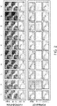
- the complementary FITC-labeled oligonucleotide was added to the tissue (1 ⁇ M) for different incubation times and the cell staining intensity was measured. Each hybridization incubation was followed by a formamide incubation (30%) to remove all hybridized oligonucleotide. The same tissue region was imaged for all tested incubation times for direct fluorescence intensity comparison. Within two minutes of hybridization, the fluorescence intensity was maximized ( Fig. 4 ).
- Each cycle involves both a hybridization step and removal step using formamide.
- the minimum amount of time to remove all hybridized dye-labeled oligonucleotides was determined.
- the same tissue that was used to test the hybridization kinetics was used to test the formamide removal kinetics.
- the complementary dye-labeled oligonucleotide (1 ⁇ M) was hybridized for five minutes.
- Formamide solution (30%) was incubated for different time periods after which the solution was washed away to halt the removal ( Fig. 5 ). Between each time point tested, additional dye-labeled oligonucleotide was added. After one minute, the dye-labeled oligonucleotide was completely removed.
- Each oligonucleotide pair consists of an oligonucleotide conjugated to an antibody and a complementary sequence with a dye modification.
- the oligonueclodtide bound to antibody is longer in length than the dye-labeled oligonucleotide to allow a tether sequence so that the hybridization does not need to take place right next to the antibody.
- a library of 30 oligonucleotide pairs was designed and synthesized. To screen for cross-hybridization, each maleimide oligonucleotide was conjugated to a mouse CD45 antibody. Aliquots of mouse spleen cells were stained with a single oligonucleotide labeled CD45 antibody.
- the cells were combined and placed on a coverslip. Iterative cycles of hybridization of sets of three dye-labeled oligonucleotides were performed. Removal of dye-labeled oligonucleotides was performed in between each hybridization using formamide. Fluorescence intensities across cells corresponding to each dye-labeled oligonucleotide was measured and compared with fluorescence intensities corresponding to all other dye-labeled oligonucleotides. The fluorescence intensity values are plotted in Fig. 8 . A representative trace of the fluorescence intensity profile for each cell population is given in Fig. 9 .
- oligonucleotide pairs show cross-hybridization activity (T9 and T10, ee.g.). Based on the fluorescence intensity data given in Fig. 8 , a minimum set of oligonucleotide pairs were removed from the library of 30 to create a sequence-orthogonal library set. The resultant library of 24 oligonucleotide pairs is shown in Fig. 10 .
- Each cycle involves delivery of three types of solution to the sample well: 1) oligonucleotide mix, 2) wash solution and 3) formamide solution.
- the fluidics was fully automated.
- An autosampler was programmed in line with a series of pumps controlling each solution.
- the corresponding set of three oligonucleotides is withdrawn from a designated well within a 96-well plate.
- the solution is pumped to the sample and incubated.
- the entire set of commands to complete a single cycle is fully automated and controlled by a python program.
- pairs of dye-labeled oligonucleotides were added to the first eight positions in the 96-well plate.
- Each odd cycle well contains dye-oligonucleotides Tll-cy5 and T18-cy3, while each even cycle contains T24-Cy5 and T26-Cy3.
- Populations of mouse spleen cells stained with CD45 antibodies conjugated to different oligonucleotides were imaged using this platform. Images of stained cells from each cycle are shown in Fig. 11 as well as representative cell traces across five cells from each population. As shown, the fluorescence intensity is equivalent across all odd and even cycles, indicating the autosampler delivers the dye-labeled oligonucleotide solution to the sample without any carryover.
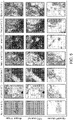
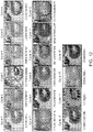
- Human tissues were stained with a cocktail of antibodies conjugated to one of the maleimide oligonucleotides. Iterative cycles of hybridization/removal were performed with imaging occurring after each hybridization step. A human tonsil ( Fig. 12 ) and human lymph node ( Fig. 13 ) were imaged using this platform. Expected staining occurred in nearly every cycle.
Landscapes
- Health & Medical Sciences (AREA)
- Life Sciences & Earth Sciences (AREA)
- Chemical & Material Sciences (AREA)
- Immunology (AREA)
- Engineering & Computer Science (AREA)
- Molecular Biology (AREA)
- Organic Chemistry (AREA)
- Analytical Chemistry (AREA)
- Physics & Mathematics (AREA)
- General Health & Medical Sciences (AREA)
- Biochemistry (AREA)
- Biomedical Technology (AREA)
- Zoology (AREA)
- Wood Science & Technology (AREA)
- Pathology (AREA)
- Proteomics, Peptides & Aminoacids (AREA)
- Microbiology (AREA)
- Biotechnology (AREA)
- General Physics & Mathematics (AREA)
- Hematology (AREA)
- Urology & Nephrology (AREA)
- General Engineering & Computer Science (AREA)
- Bioinformatics & Cheminformatics (AREA)
- Genetics & Genomics (AREA)
- Biophysics (AREA)
- Cell Biology (AREA)
- Food Science & Technology (AREA)
- Medicinal Chemistry (AREA)
- Chemical Kinetics & Catalysis (AREA)
- Tropical Medicine & Parasitology (AREA)
- Nuclear Medicine, Radiotherapy & Molecular Imaging (AREA)
- Mycology (AREA)
- Optics & Photonics (AREA)
- Measuring Or Testing Involving Enzymes Or Micro-Organisms (AREA)
- Investigating Or Analysing Biological Materials (AREA)
- Investigating, Analyzing Materials By Fluorescence Or Luminescence (AREA)
- Apparatus Associated With Microorganisms And Enzymes (AREA)
Claims (13)
- Procédé d'analyse d'un échantillon, comprenant les étapes suivantes, réalisées dans l'ordre :(a) obtention :i. d'une pluralité d'agents de capture qui sont chacun liés à un oligonucléotide différent ; etii. d'une pluralité correspondante de sondes d'acide nucléique marquées, dans lequel chacune des sondes d'acide nucléique marquées s'hybride spécifiquement à un seul des oligonucléotides de (a)(i) ;(b) marquage d'un échantillon avec la pluralité d'agents de capture de (a)(i) ;(c) hybridation spécifique d'un premier sous-ensemble des sondes d'acide nucléique marquées de (a)(ii) avec l'échantillon, dans lequel les sondes dans le premier sous-ensemble sont marquées de manière distinctive, pour produire des duplex sonde marquée/oligonucléotide ;(d) lecture de l'échantillon pour obtenir une image présentant le motif de liaison pour chacune des sondes hybridées à l'étape (c) ;(e) inactivation ou retrait des marqueurs de a(ii) qui sont associés à l'échantillon à l'étape (c) en utilisant du formamide, du DMSO, de l'urée, ou un agent chaotropique, laissant la pluralité d'agents de capture de (b) et leurs oligonucléotides associés toujours liés à l'échantillon ; et(f) répétition des étapes (c) et (d) de multiples fois avec un sous-ensemble différent des sondes d'acide nucléique marquées de (a)(ii), chaque répétition suivie par l'étape (e) à l'exception de la répétition finale, pour produire une pluralité d'images de l'échantillon, chaque image correspondant à un sous-ensemble de sondes d'acide nucléique marquées utilisé en (c).
- Procédé selon la revendication 1, dans lequel l'échantillon est un échantillon cellulaire planaire.
- Procédé selon une quelconque revendication précédente, dans lequel les oligonucléotides de (a)(i) sont d'une longueur d'au moins 15 nucléotides.
- Procédé selon une quelconque revendication précédente, dans lequel les sondes d'acide nucléique marquées de (a)(ii) sont d'une longueur de 8 à 20 nucléotides.
- Procédé selon une quelconque revendication précédente, dans lequel les séquences des oligonucléotides auxquelles les agents de capture de (a)(i) sont liés sont :i. plus longues que les séquences des sondes d'acide nucléique marquées de (a)(ii) etii. sinon identiques les unes aux autres à l'exception d'une sous-séquence qui est complémentaire d'une sonde d'acide nucléique marquée unique de (a)(ii).
- Procédé selon une quelconque revendication précédente, dans lequel chaque sonde d'acide nucléique marquée a une séquence sélectionnée parmi SEQ ID NO : 1 à 47 et les compléments de SEQ ID NO : 1 à 47.
- Procédé selon une quelconque revendication précédente, dans lequel chaque oligonucléotide lié à un agent de capture est sélectionné parmi SEQ ID NO : 48 à 94 et les compléments de SEQ ID NO : 48 à 94.
- Procédé selon une quelconque revendication précédente, dans lequel la pluralité d'agents de capture est d'au moins 10 agents de capture.
- Procédé selon une quelconque revendication précédente, dans lequel l'étape (f) comprend la répétition des étapes (c) et (d) 2 à 20 fois.
- Procédé selon une quelconque revendication précédente, comprenant en outre l'analyse d'au moins deux des images.
- Procédé selon une quelconque revendication précédente, dans lequel les sondes d'acide nucléique marquées sont marquées par fluorescence.
- Procédé selon une quelconque revendication précédente, dans lequel l'agent de capture est un anticorps ou un aptamère.
- Procédé selon une quelconque revendication précédente, dans lequel l'échantillon est une section fixée au formol incluse dans la paraffine (FFPE) ou une propagation cellulaire.
Priority Applications (2)
| Application Number | Priority Date | Filing Date | Title |
|---|---|---|---|
| EP25186852.7A EP4641166A3 (fr) | 2016-07-27 | 2017-07-26 | Imagerie fluorescente hautement multiplexée |
| EP22197655.8A EP4180533B1 (fr) | 2016-07-27 | 2017-07-26 | Imagerie fluorescente hautement multiplexée |
Applications Claiming Priority (2)
| Application Number | Priority Date | Filing Date | Title |
|---|---|---|---|
| US201662367530P | 2016-07-27 | 2016-07-27 | |
| PCT/US2017/044039 WO2018022809A1 (fr) | 2016-07-27 | 2017-07-26 | Imagerie fluorescente hautement multiplexée |
Related Child Applications (2)
| Application Number | Title | Priority Date | Filing Date |
|---|---|---|---|
| EP25186852.7A Division EP4641166A3 (fr) | 2016-07-27 | 2017-07-26 | Imagerie fluorescente hautement multiplexée |
| EP22197655.8A Division EP4180533B1 (fr) | 2016-07-27 | 2017-07-26 | Imagerie fluorescente hautement multiplexée |
Publications (3)
| Publication Number | Publication Date |
|---|---|
| EP3490616A1 EP3490616A1 (fr) | 2019-06-05 |
| EP3490616A4 EP3490616A4 (fr) | 2020-03-04 |
| EP3490616B1 true EP3490616B1 (fr) | 2022-09-28 |
Family
ID=61009327
Family Applications (3)
| Application Number | Title | Priority Date | Filing Date |
|---|---|---|---|
| EP17835238.1A Active EP3490616B1 (fr) | 2016-07-27 | 2017-07-26 | Imagerie fluorescente hautement multiplexée |
| EP22197655.8A Active EP4180533B1 (fr) | 2016-07-27 | 2017-07-26 | Imagerie fluorescente hautement multiplexée |
| EP25186852.7A Pending EP4641166A3 (fr) | 2016-07-27 | 2017-07-26 | Imagerie fluorescente hautement multiplexée |
Family Applications After (2)
| Application Number | Title | Priority Date | Filing Date |
|---|---|---|---|
| EP22197655.8A Active EP4180533B1 (fr) | 2016-07-27 | 2017-07-26 | Imagerie fluorescente hautement multiplexée |
| EP25186852.7A Pending EP4641166A3 (fr) | 2016-07-27 | 2017-07-26 | Imagerie fluorescente hautement multiplexée |
Country Status (7)
| Country | Link |
|---|---|
| US (3) | US10370698B2 (fr) |
| EP (3) | EP3490616B1 (fr) |
| JP (2) | JP7017554B2 (fr) |
| CN (2) | CN116064727A (fr) |
| AU (1) | AU2017302300B2 (fr) |
| CA (1) | CA3031586A1 (fr) |
| WO (1) | WO2018022809A1 (fr) |
Families Citing this family (226)
| Publication number | Priority date | Publication date | Assignee | Title |
|---|---|---|---|---|
| US8835358B2 (en) | 2009-12-15 | 2014-09-16 | Cellular Research, Inc. | Digital counting of individual molecules by stochastic attachment of diverse labels |
| EP3604555B1 (fr) | 2011-10-14 | 2024-12-25 | President and Fellows of Harvard College | Séquençage par assemblage de structure |
| ES2991004T3 (es) | 2011-12-22 | 2024-12-02 | Harvard College | Métodos para la detección de analitos |
| US9914967B2 (en) | 2012-06-05 | 2018-03-13 | President And Fellows Of Harvard College | Spatial sequencing of nucleic acids using DNA origami probes |
| EP4592400A3 (fr) | 2012-10-17 | 2025-10-29 | 10x Genomics Sweden AB | Procédés et produits pour optimiser la detection localisée ou spatiale de l'expression génique dans un échantillon de tissu |
| EP2971184B1 (fr) | 2013-03-12 | 2019-04-17 | President and Fellows of Harvard College | Procédé de génération d'une matrice tridimensionnelle contenant des acides nucléiques |
| US9909167B2 (en) | 2014-06-23 | 2018-03-06 | The Board Of Trustees Of The Leland Stanford Junior University | On-slide staining by primer extension |
| WO2016007839A1 (fr) | 2014-07-11 | 2016-01-14 | President And Fellows Of Harvard College | Méthodes de marquage et de détection à haut rendement de caractéristiques biologiques in situ à l'aide de microscopie |
| EP3901282B1 (fr) | 2015-04-10 | 2023-06-28 | Spatial Transcriptomics AB | Analyse de plusieurs acides nucléiques spatialement différenciés de spécimens biologiques |
| AU2016349288A1 (en) | 2015-11-03 | 2018-05-31 | President And Fellows Of Harvard College | Method and apparatus for volumetric imaging of a three-dimensional nucleic acid containing matrix |
| EP4613756A3 (fr) * | 2016-04-25 | 2025-11-12 | President And Fellows Of Harvard College | Procedes de reaction en chaine d'hybridation pour detection moleculaire in situ |
| US10202641B2 (en) | 2016-05-31 | 2019-02-12 | Cellular Research, Inc. | Error correction in amplification of samples |
| US11352667B2 (en) | 2016-06-21 | 2022-06-07 | 10X Genomics, Inc. | Nucleic acid sequencing |
| AU2017302300B2 (en) * | 2016-07-27 | 2023-08-17 | The Board Of Trustees Of The Leland Stanford Junior University | Highly-multiplexed fluorescent imaging |
| CN118853848A (zh) | 2016-08-31 | 2024-10-29 | 哈佛学院董事及会员团体 | 将生物分子的检测组合到使用荧光原位测序的单个试验的方法 |
| CN109791157B (zh) * | 2016-09-26 | 2022-06-07 | 贝克顿迪金森公司 | 使用具有条形码化的寡核苷酸序列的试剂测量蛋白质表达 |
| CN110719959B (zh) | 2017-06-05 | 2021-08-06 | 贝克顿迪金森公司 | 针对单细胞的样品索引 |
| US11181482B1 (en) * | 2017-10-02 | 2021-11-23 | University Of South Florida | Mobile colorimetric assay device |
| CN120210336A (zh) | 2017-10-06 | 2025-06-27 | 10X基因组学有限公司 | Rna模板化连接 |
| US12091706B2 (en) * | 2018-01-30 | 2024-09-17 | Rush University Medical Center | Sequential staining for multiplex analyses of tissues and cells |
| CN112272710A (zh) | 2018-05-03 | 2021-01-26 | 贝克顿迪金森公司 | 高通量多组学样品分析 |
| US11530435B2 (en) * | 2018-05-15 | 2022-12-20 | Yale University | Compositions and methods for improved RNA capture |
| CN119264211A (zh) | 2018-08-27 | 2025-01-07 | 瑞泽恩制药公司 | 拉曼光谱在下游纯化中的应用 |
| US11519033B2 (en) | 2018-08-28 | 2022-12-06 | 10X Genomics, Inc. | Method for transposase-mediated spatial tagging and analyzing genomic DNA in a biological sample |
| WO2020047005A1 (fr) | 2018-08-28 | 2020-03-05 | 10X Genomics, Inc. | Résolution de réseaux spatiaux |
| US20210332424A1 (en) | 2018-08-28 | 2021-10-28 | 10X Genomics, Inc. | Methods of generating an array |
| WO2020076976A1 (fr) | 2018-10-10 | 2020-04-16 | Readcoor, Inc. | Indexation moléculaire spatiale tridimensionnelle |
| US12529094B2 (en) | 2018-12-10 | 2026-01-20 | 10X Genomics, Inc. | Imaging system hardware |
| WO2020123316A2 (fr) | 2018-12-10 | 2020-06-18 | 10X Genomics, Inc. | Procédés de détermination d'un emplacement d'un analyte biologique dans un échantillon biologique |
| US11926867B2 (en) | 2019-01-06 | 2024-03-12 | 10X Genomics, Inc. | Generating capture probes for spatial analysis |
| US11649485B2 (en) | 2019-01-06 | 2023-05-16 | 10X Genomics, Inc. | Generating capture probes for spatial analysis |
| WO2020163397A2 (fr) * | 2019-02-04 | 2020-08-13 | Akoya Biosciences, Inc. | Détection d'analytes par marquage sélectif d'échantillons biologiques |
| WO2020167920A1 (fr) | 2019-02-14 | 2020-08-20 | Cellular Research, Inc. | Amplification de transcriptome entier et ciblé hybride |
| SG11202108618VA (en) * | 2019-02-15 | 2021-09-29 | Advanced Cell Diagnostics Inc | Methods for multiplex detection of nucleic acids by in situ hybridization |
| US20220186296A1 (en) * | 2019-02-28 | 2022-06-16 | The Board Of Trustees Of The Leland Stanford Junior University | Multiplexed signal amplification methods using enzymatic based chemical deposition |
| CN114174531A (zh) | 2019-02-28 | 2022-03-11 | 10X基因组学有限公司 | 用空间条码化寡核苷酸阵列对生物分析物进行概况分析 |
| EP3938538A1 (fr) | 2019-03-15 | 2022-01-19 | 10X Genomics, Inc. | Méthodes d'utilisation de matrices spatiales pour le séquençage d'organismes unicellulaires |
| EP3887542A1 (fr) | 2019-03-22 | 2021-10-06 | 10X Genomics, Inc. | Analyse spatiale tridimensionnelle |
| WO2020243579A1 (fr) | 2019-05-30 | 2020-12-03 | 10X Genomics, Inc. | Procédés de détection de l'hétérogénéité spatiale d'un échantillon biologique |
| CN119220641A (zh) | 2019-05-31 | 2024-12-31 | 10X基因组学有限公司 | 检测目标核酸分子的方法 |
| WO2020252488A2 (fr) | 2019-06-14 | 2020-12-17 | Akoya Biosciences, Inc. | Imagerie tissulaire multiplexée |
| CN114729391A (zh) | 2019-09-30 | 2022-07-08 | 阿科亚生物科学股份有限公司 | 用酶介导的放大的多路复用成像 |
| WO2021067726A1 (fr) * | 2019-10-03 | 2021-04-08 | The Regents Of The University Of California | Imagerie en fond clair imitant la fluorescence |
| WO2021081466A1 (fr) | 2019-10-25 | 2021-04-29 | Phitonex, Inc. | Bioconjugués lié à un polynucléotide et procédés de préparation et d'utilisation |
| US12157124B2 (en) | 2019-11-06 | 2024-12-03 | 10X Genomics, Inc. | Imaging system hardware |
| WO2021091611A1 (fr) | 2019-11-08 | 2021-05-14 | 10X Genomics, Inc. | Agents de capture d'analytes marqués spatialement pour le multiplexage d'analytes |
| WO2021092433A2 (fr) | 2019-11-08 | 2021-05-14 | 10X Genomics, Inc. | Amélioration de la spécificité de la liaison d'un analyte |
| ES3024383T3 (en) | 2019-11-13 | 2025-06-04 | 10X Genomics Inc | Generating capture probes for spatial analysis |
| WO2021102003A1 (fr) | 2019-11-18 | 2021-05-27 | 10X Genomics, Inc. | Systèmes et procédés de classification de tissu |
| AU2020388573A1 (en) | 2019-11-21 | 2022-06-23 | 10X Genomics, Inc, | Spatial analysis of analytes |
| EP4062372B1 (fr) | 2019-11-22 | 2024-05-08 | 10X Genomics, Inc. | Systèmes et procédés d'analyse spatiale d'analytes à l'aide d'un alignement de repères |
| GB201919032D0 (en) | 2019-12-20 | 2020-02-05 | Cartana Ab | Method of detecting an analyte |
| ES2982420T3 (es) | 2019-12-23 | 2024-10-16 | 10X Genomics Inc | Métodos para el análisis espacial mediante el uso de la ligazón con plantilla de ARN |
| EP4081656A1 (fr) | 2019-12-23 | 2022-11-02 | 10X Genomics, Inc. | Compositions et méthodes d'utilisation d'échantillons biologiques fixés dans des dosages basés sur des compartiments |
| CN115135984B (zh) | 2019-12-23 | 2025-12-23 | 10X基因组学有限公司 | 可逆固定试剂及其使用方法 |
| WO2021142233A1 (fr) | 2020-01-10 | 2021-07-15 | 10X Genomics, Inc. | Procédés de détermination d'un emplacement d'un acide nucléique cible dans un échantillon biologique |
| US12365942B2 (en) | 2020-01-13 | 2025-07-22 | 10X Genomics, Inc. | Methods of decreasing background on a spatial array |
| WO2021146219A1 (fr) | 2020-01-13 | 2021-07-22 | Becton, Dickinson And Company | Capture de cellules à l'aide d'oligonucléotides contenant de la du |
| US12405264B2 (en) | 2020-01-17 | 2025-09-02 | 10X Genomics, Inc. | Electrophoretic system and method for analyte capture |
| US11702693B2 (en) | 2020-01-21 | 2023-07-18 | 10X Genomics, Inc. | Methods for printing cells and generating arrays of barcoded cells |
| US11732299B2 (en) | 2020-01-21 | 2023-08-22 | 10X Genomics, Inc. | Spatial assays with perturbed cells |
| US20210230681A1 (en) | 2020-01-24 | 2021-07-29 | 10X Genomics, Inc. | Methods for spatial analysis using proximity ligation |
| US11821035B1 (en) | 2020-01-29 | 2023-11-21 | 10X Genomics, Inc. | Compositions and methods of making gene expression libraries |
| EP4097251A1 (fr) | 2020-01-29 | 2022-12-07 | 10X Genomics, Inc. | Compositions et procédés pour la détection d'analytes |
| WO2021155057A1 (fr) | 2020-01-29 | 2021-08-05 | Becton, Dickinson And Company | Puits à code-barres pour la cartographie spatiale de cellules individuelles par séquençage |
| US12076701B2 (en) | 2020-01-31 | 2024-09-03 | 10X Genomics, Inc. | Capturing oligonucleotides in spatial transcriptomics |
| US12110541B2 (en) | 2020-02-03 | 2024-10-08 | 10X Genomics, Inc. | Methods for preparing high-resolution spatial arrays |
| US11898205B2 (en) | 2020-02-03 | 2024-02-13 | 10X Genomics, Inc. | Increasing capture efficiency of spatial assays |
| US12110548B2 (en) | 2020-02-03 | 2024-10-08 | 10X Genomics, Inc. | Bi-directional in situ analysis |
| US12112833B2 (en) | 2020-02-04 | 2024-10-08 | 10X Genomics, Inc. | Systems and methods for index hopping filtering |
| US11732300B2 (en) | 2020-02-05 | 2023-08-22 | 10X Genomics, Inc. | Increasing efficiency of spatial analysis in a biological sample |
| WO2021158925A1 (fr) | 2020-02-07 | 2021-08-12 | 10X Genomics, Inc. | Évaluation quantitative et automatisée de la performance de perméabilisation pour la transcriptomique spatiale |
| US11835462B2 (en) | 2020-02-11 | 2023-12-05 | 10X Genomics, Inc. | Methods and compositions for partitioning a biological sample |
| US12399123B1 (en) | 2020-02-14 | 2025-08-26 | 10X Genomics, Inc. | Spatial targeting of analytes |
| US12281357B1 (en) | 2020-02-14 | 2025-04-22 | 10X Genomics, Inc. | In situ spatial barcoding |
| CN115485391A (zh) | 2020-02-17 | 2022-12-16 | 10X基因组学有限公司 | 染色质相互作用的原位分析 |
| WO2021168278A1 (fr) | 2020-02-20 | 2021-08-26 | 10X Genomics, Inc. | Procédés pour combiner les synthèses d'adnc des premier et de deuxième brins pour l'analyse spatiale |
| EP4484571A3 (fr) | 2020-02-21 | 2025-03-19 | 10X Genomics, Inc. | Procédés et compositions pour un dosage spatial in situ intégré |
| AU2021224760A1 (en) | 2020-02-21 | 2022-09-15 | 10X Genomics, Inc. | Capturing genetic targets using a hybridization approach |
| US11891654B2 (en) | 2020-02-24 | 2024-02-06 | 10X Genomics, Inc. | Methods of making gene expression libraries |
| WO2021173719A1 (fr) | 2020-02-25 | 2021-09-02 | Becton, Dickinson And Company | Sondes bi-spécifiques permettant l'utilisation d'échantillons monocellulaires en tant que contrôle de compensation de couleur unique |
| US11768175B1 (en) | 2020-03-04 | 2023-09-26 | 10X Genomics, Inc. | Electrophoretic methods for spatial analysis |
| US12188085B2 (en) | 2020-03-05 | 2025-01-07 | 10X Genomics, Inc. | Three-dimensional spatial transcriptomics with sequencing readout |
| EP4139485B1 (fr) | 2020-04-22 | 2023-09-06 | 10X Genomics, Inc. | Procédés d'analyse spatiale utilisant un appauvrissement d'arn ciblée |
| CN115803462A (zh) * | 2020-04-22 | 2023-03-14 | 日华化学株式会社 | 核酸的检测方法及寡核苷酸探针 |
| WO2021225900A1 (fr) | 2020-05-04 | 2021-11-11 | 10X Genomics, Inc. | Modes de transfert transcriptomique spatial |
| US11661625B2 (en) | 2020-05-14 | 2023-05-30 | Becton, Dickinson And Company | Primers for immune repertoire profiling |
| US12416603B2 (en) | 2020-05-19 | 2025-09-16 | 10X Genomics, Inc. | Electrophoresis cassettes and instrumentation |
| EP4153776B1 (fr) | 2020-05-22 | 2025-03-05 | 10X Genomics, Inc. | Analyse spatiale pour détecter des variants de séquence |
| WO2021237056A1 (fr) | 2020-05-22 | 2021-11-25 | 10X Genomics, Inc. | Analyse d'intégrité d'arn dans un échantillon biologique |
| EP4414459B1 (fr) | 2020-05-22 | 2025-09-03 | 10X Genomics, Inc. | Mesure spatio-temporelle simultanée de l'expression génique et de l'activité cellulaire |
| WO2021242834A1 (fr) | 2020-05-26 | 2021-12-02 | 10X Genomics, Inc. | Procédé de réinitialisation d'un réseau |
| EP4600376A3 (fr) | 2020-06-02 | 2025-10-22 | 10X Genomics, Inc. | Transcriptome spatial pour récepteurs d'antigènes |
| AU2021283174A1 (en) | 2020-06-02 | 2023-01-05 | 10X Genomics, Inc. | Nucleic acid library methods |
| US12157913B2 (en) | 2020-06-02 | 2024-12-03 | Becton, Dickinson And Company | Oligonucleotides and beads for 5 prime gene expression assay |
| US12031177B1 (en) | 2020-06-04 | 2024-07-09 | 10X Genomics, Inc. | Methods of enhancing spatial resolution of transcripts |
| EP4421186B1 (fr) | 2020-06-08 | 2025-08-13 | 10X Genomics, Inc. | Procédés de détermination d'une marge chirurgicale et procédés d'utilisation de ceux-ci |
| US20230279474A1 (en) | 2020-06-10 | 2023-09-07 | 10X Genomics, Inc. | Methods for spatial analysis using blocker oligonucleotides |
| WO2021252747A1 (fr) | 2020-06-10 | 2021-12-16 | 1Ox Genomics, Inc. | Procédés de distribution de fluide |
| EP4165207B1 (fr) | 2020-06-10 | 2024-09-25 | 10X Genomics, Inc. | Methode pour determiner la position d'un analyte dans un echantillon biologique |
| US12435363B1 (en) | 2020-06-10 | 2025-10-07 | 10X Genomics, Inc. | Materials and methods for spatial transcriptomics |
| US12209273B2 (en) | 2020-06-12 | 2025-01-28 | 10X Genomics, Inc. | Nucleic acid assays using click chemistry bioconjugation |
| ES2994976T3 (en) | 2020-06-25 | 2025-02-05 | 10X Genomics Inc | Spatial analysis of dna methylation |
| US12168801B1 (en) | 2020-07-02 | 2024-12-17 | 10X Genomics, Inc. | Hybrid/capture probe designs for full-length cDNA |
| US11761038B1 (en) | 2020-07-06 | 2023-09-19 | 10X Genomics, Inc. | Methods for identifying a location of an RNA in a biological sample |
| US11981960B1 (en) | 2020-07-06 | 2024-05-14 | 10X Genomics, Inc. | Spatial analysis utilizing degradable hydrogels |
| US12209280B1 (en) | 2020-07-06 | 2025-01-28 | 10X Genomics, Inc. | Methods of identifying abundance and location of an analyte in a biological sample using second strand synthesis |
| EP4182676A1 (fr) * | 2020-07-20 | 2023-05-24 | Akoya Biosciences, Inc. | Traitement et imagerie d'échantillons de tissu |
| CN116323968A (zh) | 2020-07-31 | 2023-06-23 | 10X基因组学有限公司 | 用于空间分析的去交联化合物和使用方法 |
| US20240295549A1 (en) * | 2020-08-07 | 2024-09-05 | Vital Biosciences Inc. | Multiplexed analyte detection |
| US12553898B1 (en) | 2020-08-10 | 2026-02-17 | 10X Genomics, Inc. | Fluorescent hybridization of antibody-oligonucleotide for multiplexing and signal amplification |
| US20220049303A1 (en) | 2020-08-17 | 2022-02-17 | Readcoor, Llc | Methods and systems for spatial mapping of genetic variants |
| US12297499B2 (en) | 2020-08-17 | 2025-05-13 | 10X Genomics, Inc. | Multicomponent nucleic acid probes for sample analysis |
| US11981958B1 (en) | 2020-08-20 | 2024-05-14 | 10X Genomics, Inc. | Methods for spatial analysis using DNA capture |
| US12580044B1 (en) | 2020-09-02 | 2026-03-17 | 10X Genomics, Inc. | Systems and methods for identifying cells that are antigen-specific for an immunogenic feature |
| EP4530358A3 (fr) | 2020-09-15 | 2025-06-18 | 10x Genomics, Inc. | Procédés de libération d'une sonde de capture étendue à partir d'un substrat et leurs utilisations |
| CN116507739A (zh) | 2020-09-16 | 2023-07-28 | 10X基因组学有限公司 | 使用多个孔确定生物样品中分析物位置的方法 |
| CA3192943A1 (fr) * | 2020-09-16 | 2022-03-24 | Nanostring Technologies, Inc. | Compositions chimiques et leurs procedes d'utilisation |
| WO2022061150A2 (fr) | 2020-09-18 | 2022-03-24 | 10X Geonomics, Inc. | Appareil de manipulation d'échantillons et procédés d'enregistrement d'images |
| EP4213993A2 (fr) | 2020-09-18 | 2023-07-26 | 10X Genomics, Inc. | Appareil de manipulation d'échantillon et procédés système de distribution de fluide |
| US11926822B1 (en) | 2020-09-23 | 2024-03-12 | 10X Genomics, Inc. | Three-dimensional spatial analysis |
| WO2022081643A2 (fr) | 2020-10-13 | 2022-04-21 | 10X Genomics, Inc. | Compositions et procédés pour générer des molécules de liaison à un antigène recombinant à partir de cellules uniques |
| US20240018572A1 (en) | 2020-10-22 | 2024-01-18 | 10X Genomics, Inc. | Methods for spatial analysis using rolling circle amplification |
| US12071667B2 (en) | 2020-11-04 | 2024-08-27 | 10X Genomics, Inc. | Sequence analysis using meta-stable nucleic acid molecules |
| CN116829733A (zh) | 2020-11-06 | 2023-09-29 | 10X基因组学有限公司 | 用于将分析物结合至捕获探针的组合物和方法 |
| AU2021385065A1 (en) | 2020-11-18 | 2023-06-15 | 10X Genomics, Inc. | Methods and compositions for analyzing immune infiltration in cancer stroma to predict clinical outcome |
| WO2022109196A1 (fr) | 2020-11-18 | 2022-05-27 | Akoya Biosciences, Inc. | Imagerie multiplexée avec des sondes nanocorps |
| US11827935B1 (en) | 2020-11-19 | 2023-11-28 | 10X Genomics, Inc. | Methods for spatial analysis using rolling circle amplification and detection probes |
| US11333588B1 (en) | 2020-12-07 | 2022-05-17 | Nebulum Technologies Co., Ltd. | Matrix-assisted methods and compositions to prepare biological samples for super-resolution imaging |
| US12509717B2 (en) | 2020-12-11 | 2025-12-30 | 10X Genomics, Inc. | Methods and compositions for multimodal in situ analysis |
| EP4729631A2 (fr) | 2020-12-21 | 2026-04-22 | 10X Genomics, Inc. | Procédés, compositions et systèmes de capture de sondes et/ou de codes à barres |
| WO2022147296A1 (fr) | 2020-12-30 | 2022-07-07 | 10X Genomics, Inc. | Clivage de sondes de capture pour analyse spatiale |
| WO2022147005A1 (fr) | 2020-12-30 | 2022-07-07 | 10X Genomics, Inc. | Procédés de détermination de capture d'analytes |
| US12060603B2 (en) | 2021-01-19 | 2024-08-13 | 10X Genomics, Inc. | Methods for internally controlled in situ assays using padlock probes |
| US12398262B1 (en) | 2021-01-22 | 2025-08-26 | 10X Genomics, Inc. | Triblock copolymer-based cell stabilization and fixation system and methods of use thereof |
| AU2022212231A1 (en) | 2021-01-29 | 2023-08-03 | 10X Genomics, Inc. | Method for transposase mediated spatial tagging and analyzing genomic dna in a biological sample |
| WO2022182903A1 (fr) * | 2021-02-24 | 2022-09-01 | California Institute Of Technology | Multiplexage de conditions expérimentales et d'échantillons en génomique spatiale |
| US12275984B2 (en) | 2021-03-02 | 2025-04-15 | 10X Genomics, Inc. | Sequential hybridization and quenching |
| AU2022238446A1 (en) | 2021-03-18 | 2023-09-07 | 10X Genomics, Inc. | Multiplex capture of gene and protein expression from a biological sample |
| EP4319792B1 (fr) | 2021-04-05 | 2025-09-03 | 10X Genomics, Inc. | Composition de ligase recombinante et ses utilisations |
| EP4305196B1 (fr) | 2021-04-14 | 2025-04-02 | 10X Genomics, Inc. | Procédés de mesure d'une mauvaise localisation d'un analyte |
| WO2022226057A1 (fr) | 2021-04-20 | 2022-10-27 | 10X Genomics, Inc. | Procédés d'évaluation de la qualité d'un échantillon avant une analyse spatiale à l'aide d'une ligature modélisée |
| EP4320271B1 (fr) | 2021-05-06 | 2025-03-19 | 10X Genomics, Inc. | Procédés pour augmenter la résolution d'une analyse spatiale |
| WO2022256503A1 (fr) | 2021-06-03 | 2022-12-08 | 10X Genomics, Inc. | Procédés, compositions, kits et systèmes pour améliorer la capture d'analytes pour une analyse spatiale |
| EP4109333B1 (fr) * | 2021-06-18 | 2025-03-12 | Leica Microsystems CMS GmbH | Procédé d'analyse d'échantillons biologiques |
| WO2022271820A1 (fr) | 2021-06-22 | 2022-12-29 | 10X Genomics, Inc. | Détection spatiale de sars-cov-2 à l'aide d'une ligature à matrice |
| WO2023288225A1 (fr) | 2021-07-13 | 2023-01-19 | 10X Genomics, Inc. | Procédés de préparation d'une matrice polymérisée à épaisseur contrôlable |
| WO2023287765A1 (fr) | 2021-07-13 | 2023-01-19 | 10X Genomics, Inc. | Procédés d'analyse spatiale utilisant un silençage de sonde ciblée |
| AU2022317053B2 (en) | 2021-07-30 | 2025-01-02 | 10X Genomics, Inc. | Methods and compositions for synchronizing reactions in situ |
| US12139751B2 (en) | 2021-07-30 | 2024-11-12 | 10X Genomics, Inc. | Circularizable probes for in situ analysis |
| US12553805B2 (en) | 2021-08-02 | 2026-02-17 | 10X Genomics, Inc. | Methods of preserving a biological sample |
| US12391984B2 (en) | 2021-08-03 | 2025-08-19 | 10X Genomics, Inc. | Compositions and methods for rolling circle amplification |
| US12460251B2 (en) | 2021-08-03 | 2025-11-04 | 10X Genomics, Inc. | Stabilization and/or compaction of nucleic acid molecules |
| US12529096B2 (en) | 2021-08-03 | 2026-01-20 | 10X Genomics, Inc. | Stabilization and/or compaction of nucleic acid structures |
| US12553079B2 (en) | 2021-08-03 | 2026-02-17 | 10X Genomics, Inc. | Nucleic acid concatemers and methods for stabilizing and/or compacting the same |
| EP4370675B1 (fr) | 2021-08-12 | 2025-10-01 | 10X Genomics, Inc. | Procedes, compositions et systemes d'identification de molecules de liaison a l'antigene |
| EP4446426B1 (fr) | 2021-08-16 | 2026-03-04 | 10X Genomics, Inc. | Sondes comprenant une région de code-barres divisée et procédés d'utilisation |
| WO2023034489A1 (fr) | 2021-09-01 | 2023-03-09 | 10X Genomics, Inc. | Procédés, compositions et kits pour bloquer une sonde de capture sur un réseau spatial |
| US20240378734A1 (en) | 2021-09-17 | 2024-11-14 | 10X Genomics, Inc. | Systems and methods for image registration or alignment |
| WO2023076345A1 (fr) | 2021-10-26 | 2023-05-04 | 10X Genomics, Inc. | Procédés d'analyse spatiale utilisant la capture ciblée d'arn |
| EP4430398B1 (fr) | 2021-11-10 | 2025-12-31 | 10X Genomics, Inc. | Procédés d'identification de molécules de liaison à l'antigène |
| WO2023086880A1 (fr) | 2021-11-10 | 2023-05-19 | 10X Genomics, Inc. | Procédés, compositions et kits pour déterminer l'emplacement d'un analyte dans un échantillon biologique |
| WO2023102313A1 (fr) | 2021-11-30 | 2023-06-08 | 10X Genomics, Inc. | Systèmes et procédés d'identification de régions d'aneuploïdie dans un tissu |
| WO2023102118A2 (fr) | 2021-12-01 | 2023-06-08 | 10X Genomics, Inc. | Procédés, compositions et systèmes pour la détection améliorée d'analytes in situ et analyse spatiale |
| WO2023122033A1 (fr) | 2021-12-20 | 2023-06-29 | 10X Genomics, Inc. | Auto-test pour dispositif d'imagerie |
| EP4733410A2 (fr) | 2022-01-21 | 2026-04-29 | 10X Genomics, Inc. | Signaux de lecture multiples pour analyser un échantillon |
| WO2023150163A1 (fr) | 2022-02-01 | 2023-08-10 | 10X Genomics, Inc. | Procédés, compositions et systèmes de capture d'analytes à partir de tissu lymphatique |
| WO2023150171A1 (fr) | 2022-02-01 | 2023-08-10 | 10X Genomics, Inc. | Procédés, compositions et systèmes de capture d'analytes à partir d'échantillons de glioblastome |
| WO2023150098A1 (fr) | 2022-02-01 | 2023-08-10 | 10X Genomics, Inc. | Procédés, kits, compositions et systèmes d'analyse spatiale |
| WO2023159028A1 (fr) | 2022-02-15 | 2023-08-24 | 10X Genomics, Inc. | Systèmes et procédés d'analyse spatiale d'analytes à l'aide d'un alignement de repères |
| CN118974836A (zh) | 2022-03-08 | 2024-11-15 | 10X基因组学有限公司 | 将光学拥挤最小化的原位代码设计方法 |
| WO2023172670A2 (fr) | 2022-03-11 | 2023-09-14 | 10X Genomics, Inc. | Appareil de manipulation d'échantillon et procédés système de distribution de fluide |
| EP4505177A1 (fr) | 2022-04-01 | 2025-02-12 | 10x Genomics, Inc. | Compositions et procédés de masquage ciblé d'autofluorescence |
| EP4508238A2 (fr) | 2022-04-12 | 2025-02-19 | 10X Genomics, Inc. | Compositions et procédés de génération et de caractérisation de molécules de liaison à un antigène recombinant |
| WO2023215603A1 (fr) | 2022-05-06 | 2023-11-09 | 10X Genomics, Inc. | Procédés et compositions pour l'analyse in situ de séquences v(d)j |
| EP4519674A1 (fr) | 2022-05-06 | 2025-03-12 | 10X Genomics, Inc. | Analyse d'un antigène et des interactions antigène-récepteur |
| WO2023215552A1 (fr) | 2022-05-06 | 2023-11-09 | 10X Genomics, Inc. | Lecteurs de codes-barres moléculaires pour détection d'analytes |
| US20240084378A1 (en) | 2022-05-11 | 2024-03-14 | 10X Genomics, Inc. | Compositions and methods for in situ sequencing |
| WO2023225519A1 (fr) | 2022-05-17 | 2023-11-23 | 10X Genomics, Inc. | Transposons modifiés, compositions et utilisations de ceux-ci |
| WO2023229988A1 (fr) | 2022-05-23 | 2023-11-30 | 10X Genomics, Inc. | Moule pour échantillon de tissu |
| WO2023229982A2 (fr) | 2022-05-24 | 2023-11-30 | 10X Genomics, Inc. | Confinement de structure poreuse pour suppression de convection |
| EP4540607A1 (fr) | 2022-06-17 | 2025-04-23 | 10X Genomics, Inc. | Dé-réticulation catalytique d'échantillons pour analyse in situ |
| WO2023250077A1 (fr) | 2022-06-22 | 2023-12-28 | 10X Genomics, Inc. | Procédés, compositions et systèmes pour la capture de sondes et/ou de code-barres |
| WO2024015862A1 (fr) | 2022-07-13 | 2024-01-18 | 10X Genomics, Inc. | Procédés de caractérisation de molécules de liaison à l'antigène à partir d'échantillons biologiques |
| WO2024015578A1 (fr) | 2022-07-15 | 2024-01-18 | 10X Genomics, Inc. | Procédés pour déterminer l'emplacement d'un acide nucléique cible dans un échantillon biologique |
| EP4565870A1 (fr) | 2022-08-05 | 2025-06-11 | 10x Genomics, Inc. | Systèmes et procédés de quantification d'immunofluorescence |
| WO2024036191A1 (fr) | 2022-08-10 | 2024-02-15 | 10X Genomics, Inc. | Systèmes et procédés de colocalisation |
| WO2024035844A1 (fr) | 2022-08-12 | 2024-02-15 | 10X Genomics, Inc. | Procédés de réduction de la capture d'analytes |
| WO2024040060A1 (fr) | 2022-08-16 | 2024-02-22 | 10X Genomics, Inc. | Ap50 polymérases et leurs utilisations |
| US20260055395A1 (en) | 2022-08-24 | 2026-02-26 | 10X Genomics, Inc. | Compositions and methods for antigenic epitope mapping in biological samples |
| WO2024081212A1 (fr) | 2022-10-10 | 2024-04-18 | 10X Genomics, Inc. | Transcription in vitro d'acides nucléiques capturés spatialement |
| WO2024081869A1 (fr) | 2022-10-14 | 2024-04-18 | 10X Genomics, Inc. | Procédés d'analyse d'échantillons biologiques |
| WO2024086167A2 (fr) | 2022-10-17 | 2024-04-25 | 10X Genomics, Inc. | Procédés, compositions et kits pour déterminer l'emplacement d'un analyte dans un échantillon biologique |
| EP4482979B1 (fr) | 2022-11-09 | 2025-10-01 | 10X Genomics, Inc. | Procédés, compositions et kits pour déterminer l'emplacement de plusieurs analytes dans un échantillon biologique |
| EP4638788A1 (fr) | 2022-12-21 | 2025-10-29 | 10X Genomics, Inc. | Analyse d'analytes et d'expression génique spatiale |
| WO2024145441A1 (fr) | 2022-12-29 | 2024-07-04 | 10X Genomics, Inc. | Procédés, compositions et kits pour déterminer un emplacement d'un acide nucléique cible dans un échantillon biologique fixe |
| WO2024145224A1 (fr) | 2022-12-29 | 2024-07-04 | 10X Genomics, Inc. | Compositions, procédés et systèmes d'analyse spatiale à haute résolution |
| WO2024145445A1 (fr) | 2022-12-30 | 2024-07-04 | 10X Genomics, Inc. | Procédés de capture d'analytes cibles |
| EP4624590A3 (fr) | 2022-12-30 | 2025-12-24 | 10X Genomics, Inc. | Procédés, compositions et kits pour le codage à barres multiples et/ou le codage à barres spatial haute densité |
| WO2024206603A1 (fr) | 2023-03-28 | 2024-10-03 | 10X Genomics, Inc. | Procédés, compositions et kits pour réduire une mauvaise localisation d'analytes |
| EP4450971A1 (fr) * | 2023-04-21 | 2024-10-23 | Leica Microsystems CMS GmbH | Marqueur pour l'analyse d'un échantillon biologique |
| EP4698644A1 (fr) | 2023-04-21 | 2026-02-25 | 10X Genomics, Inc. | Procédés et compositions pour détecter des acides nucléiques dans un échantillon biologique fixe |
| WO2024238625A1 (fr) | 2023-05-15 | 2024-11-21 | 10X Genomics, Inc. | Normalisation de données d'anticorps spatial |
| WO2024238900A1 (fr) | 2023-05-17 | 2024-11-21 | 10X Genomics, Inc. | Procédés pour réduire la mauvaise localisation d'un analyte |
| WO2024238992A1 (fr) | 2023-05-18 | 2024-11-21 | 10X Genomics, Inc. | Polymérases de la famille b sans déplacement de brin modifiées pour des applications de transcription inverse et de remplissage d'espace |
| WO2024254316A1 (fr) | 2023-06-07 | 2024-12-12 | 10X Genomics, Inc. | Compositions et procédés pour identifier les caillots sanguins dans les échantillons biologiques |
| WO2024258911A1 (fr) | 2023-06-13 | 2024-12-19 | 10X Genomics, Inc. | Protéines de liaison à l'adn spécifiques d'une séquence |
| WO2025029605A1 (fr) | 2023-07-28 | 2025-02-06 | 10X Genomics, Inc. | Méthodes et compositions de détection de variants génétiques en utilisant des sondes de capture et une extension d'amorce |
| WO2025029627A1 (fr) | 2023-07-31 | 2025-02-06 | 10X Genomics, Inc. | Procédés, compositions et kits de détection spatiale de variants génétiques |
| WO2025029831A1 (fr) | 2023-07-31 | 2025-02-06 | 10X Genomics, Inc. | Procédés et systèmes de clivage d'arn ciblé et d'amplification de cercle roulant amorcé par arn cible |
| WO2025043076A1 (fr) | 2023-08-24 | 2025-02-27 | 10X Genomics, Inc. | Procédés, kits et compositions aux fins d'une détection spatiale de variants génétiques |
| WO2025072119A1 (fr) | 2023-09-26 | 2025-04-03 | 10X Genomics, Inc. | Procédés, compositions et kits pour détecter des interactions chromosomiques spatiales |
| WO2025090912A1 (fr) | 2023-10-26 | 2025-05-01 | 10X Genomics, Inc. | Réseaux spatiaux contenant des sondes maîtresses réutilisables |
| WO2025096581A1 (fr) | 2023-10-31 | 2025-05-08 | 10X Genomics, Inc. | Procédés d'analyse spatiale et de séquençage de noyaux uniques |
| WO2025101864A1 (fr) | 2023-11-09 | 2025-05-15 | 10X Genomics, Inc. | Procédés, compositions et kits pour réduire la mauvaise localisation des analytes dans les dosages d'analyse spatiale |
| WO2025101377A1 (fr) | 2023-11-09 | 2025-05-15 | 10X Genomics, Inc. | Analyse spatiale assistée par matrice d'échantillons biologiques |
| EP4560319A1 (fr) | 2023-11-24 | 2025-05-28 | Pierre Fabre Medicament | Expression de cellules immunitaires et tumorales de vista dans un panel d'indications cancéreuses |
| US20250207180A1 (en) | 2023-12-21 | 2025-06-26 | Akoya Biosciences, Inc. | Spatially resolved cyclic multiplexed detection of nucleic acids and proteins in tissue |
| WO2025178607A1 (fr) | 2024-02-19 | 2025-08-28 | Leica Biosystems Richmond, Inc. | Procédés et systèmes de multiplexage chromogène |
| WO2025179146A1 (fr) * | 2024-02-23 | 2025-08-28 | Sanford Burnham Prebys Medical Discovery Institute | Compositions et procédés de visualisation de glycosylation |
| WO2025189030A1 (fr) | 2024-03-07 | 2025-09-12 | Stellaromics, Inc. | Procédés et systèmes d'imagerie volumétrique |
| US20250321232A1 (en) | 2024-04-15 | 2025-10-16 | Leica Biosystems Newcastle Ltd. | Chromogenic multiplexing methods and systems for identifying a cancer of unknown primary origin |
| WO2025255457A1 (fr) | 2024-06-07 | 2025-12-11 | 10X Genomics, Inc. | Compositions et procédés de profilage spatial sur cible amélioré |
| WO2026024934A1 (fr) | 2024-07-25 | 2026-01-29 | 10X Genomics, Inc. | Procédés, compositions et kits d'analyse spatiale par tagmentation |
| WO2026043885A1 (fr) | 2024-08-20 | 2026-02-26 | 10X Genomics, Inc. | Procédés et systèmes de préparation d'une bibliothèque d'acides nucléiques |
| CN119033971B (zh) * | 2024-09-01 | 2025-11-04 | 山西医科大学 | 一种复合碳点载体及携载VEGF siRNA碳点复合体 |
Family Cites Families (103)
| Publication number | Priority date | Publication date | Assignee | Title |
|---|---|---|---|---|
| US1000796A (en) * | 1910-11-08 | 1911-08-15 | Jacob Whitfield Graham | Spring-tire. |
| US5068178A (en) | 1988-12-06 | 1991-11-26 | Genetic Systems Corporation | Method of enhancing direct immunofluorescence staining of cells |
| US5491224A (en) | 1990-09-20 | 1996-02-13 | Bittner; Michael L. | Direct label transaminated DNA probe compositions for chromosome identification and methods for their manufacture |
| US5985548A (en) | 1993-02-04 | 1999-11-16 | E. I. Du Pont De Nemours And Company | Amplification of assay reporters by nucleic acid replication |
| US5854033A (en) * | 1995-11-21 | 1998-12-29 | Yale University | Rolling circle replication reporter systems |
| US6027890A (en) | 1996-01-23 | 2000-02-22 | Rapigene, Inc. | Methods and compositions for enhancing sensitivity in the analysis of biological-based assays |
| US20100081134A1 (en) * | 1997-07-21 | 2010-04-01 | Mirkin Chad A | Bio-barcode based detection of target analytes |
| US5948902A (en) | 1997-11-20 | 1999-09-07 | South Alabama Medical Science Foundation | Antisense oligonucleotides to human serine/threonine protein phosphatase genes |
| AU3463099A (en) * | 1998-03-31 | 1999-10-18 | Tularik Inc. | High throughput assay for detection of mrna in cells |
| US8131053B2 (en) * | 1999-01-25 | 2012-03-06 | Amnis Corporation | Detection of circulating tumor cells using imaging flow cytometry |
| US6316230B1 (en) | 1999-08-13 | 2001-11-13 | Applera Corporation | Polymerase extension at 3′ terminus of PNA-DNA chimera |
| US6531283B1 (en) | 2000-06-20 | 2003-03-11 | Molecular Staging, Inc. | Protein expression profiling |
| US6743592B1 (en) | 2000-07-25 | 2004-06-01 | The Trustees Of The University Of Pennsylvania | Methods, systems and kits for immuno-detection of epitopes expressed on molecules |
| US20020072053A1 (en) | 2000-12-08 | 2002-06-13 | Mcnally Alan J. | Immunoassay based on DNA replication using labeled primer |
| US7846746B2 (en) | 2001-04-10 | 2010-12-07 | Children's Medical Center Corporation | Methods of analysis and labeling of protein-protein interactions |
| EP1270738A1 (fr) | 2001-06-18 | 2003-01-02 | chimera biotec GmbH | Méthode pour détecter des substances dans des liquides |
| AU2002305478A1 (en) * | 2001-06-20 | 2003-01-08 | Molecular Staging, Inc. | Conjugates of reduced antibodies and biomolecules |
| US7341831B2 (en) | 2001-07-18 | 2008-03-11 | The Trustees Of The University Of Pennsylvania | Method for immuno-detection of epitopes |
| US20040091857A1 (en) | 2001-07-20 | 2004-05-13 | Nallur Girish N. | Gene expression profiling |
| WO2003062444A2 (fr) | 2001-11-13 | 2003-07-31 | Emory University | Systemes matriciels et procedes |
| AU2003222178A1 (en) | 2002-03-29 | 2003-10-13 | Nugen Technologies, Inc. | Single primer isothermal nucleic acid amplification-enhanced analyte detection and quantification |
| ATE432365T1 (de) | 2003-02-21 | 2009-06-15 | Geneform Technologies Ltd | Verfahren, kits und reagenzien zur nukleinsäuresequenzierung |
| US20040185453A1 (en) | 2003-03-21 | 2004-09-23 | Joel Myerson | Affinity based methods for separating homologous parental genetic material and uses thereof |
| CN101410530B (zh) | 2003-04-18 | 2013-03-27 | 贝克顿·迪金森公司 | 免疫-扩增 |
| US7198900B2 (en) * | 2003-08-29 | 2007-04-03 | Applera Corporation | Multiplex detection compositions, methods, and kits |
| WO2005054514A1 (fr) | 2003-12-02 | 2005-06-16 | Agency For Science, Technology And Research | Dosage d'amplification immunologique d'arn |
| KR100695123B1 (ko) * | 2003-12-03 | 2007-03-14 | 삼성전자주식회사 | Tm에 따라 기판상에 고정화된 프로브 폴리뉴클레오티드군을 2이상 포함하는 폴리뉴클레오티드 어레이 및 그를이용하는 표적 핵산 검출 방법 |
| US20050147976A1 (en) * | 2003-12-29 | 2005-07-07 | Xing Su | Methods for determining nucleotide sequence information |
| US20050233340A1 (en) | 2004-04-20 | 2005-10-20 | Barrett Michael T | Methods and compositions for assessing CpG methylation |
| DK1842226T3 (da) | 2004-11-03 | 2010-10-18 | Iris Molecular Diagnostics Inc | Homogen analytpåvisning |
| CA2598508A1 (fr) | 2005-02-25 | 2006-08-31 | Commissariat A L'energie Atomique | Procede et dispositif pour separer des cibles moleculaires dans un melange complexe |
| US20070020650A1 (en) | 2005-04-01 | 2007-01-25 | Avak Kahvejian | Methods for detecting proteins |
| WO2006128010A2 (fr) | 2005-05-26 | 2006-11-30 | The Trustees Of Boston University | Quantification d'acides nucleiques et de proteines au moyen d'etiquettes de masse d'oligonucleotides |
| US20070026430A1 (en) | 2005-06-30 | 2007-02-01 | Applera Corporation | Proximity probing of target proteins comprising restriction and/or extension |
| WO2007117700A2 (fr) * | 2006-04-07 | 2007-10-18 | The Trustees Of The University Of Pennsylvania | Clonage d'échantillons de tissu pathologique in situ |
| CA2656525A1 (fr) | 2006-06-30 | 2008-01-10 | Applera Corporation | Nucleotides de sequences de terminaison reversibles et procedes d'utilisation |
| WO2008052774A2 (fr) | 2006-10-31 | 2008-05-08 | Noxxon Pharma Ag | Procédé de détection d'un acide nucléique simple brin ou double brin |
| US9201063B2 (en) | 2006-11-16 | 2015-12-01 | General Electric Company | Sequential analysis of biological samples |
| US8305579B2 (en) | 2006-11-16 | 2012-11-06 | Thomas Pirrie Treynor | Sequential analysis of biological samples |
| SE531135C2 (sv) | 2006-12-01 | 2008-12-23 | Alf Maansson | Känslig detektion av gifter, patogener och sjukdomsmarkörer genom aggregation av cytoskeletala filament |
| WO2008076375A2 (fr) | 2006-12-13 | 2008-06-26 | Autogenomics, Inc. | Analyse simultanée d'échantillons de patients multiples par essai multiplex adressable en phase solide à rapport signal sur bruit élevé |
| CN101210270A (zh) * | 2006-12-31 | 2008-07-02 | 陕西北美基因股份有限公司 | 病毒病原高通量快速排查检测方法 |
| GB0703996D0 (en) | 2007-03-01 | 2007-04-11 | Oxitec Ltd | Nucleic acid detection |
| WO2009012220A2 (fr) | 2007-07-17 | 2009-01-22 | Agilent Technologies Inc. | Dosages de détection d'analytes |
| WO2009064933A2 (fr) | 2007-11-13 | 2009-05-22 | Ikonisys, Inc. | Détection de cellules tumorales circulantes dans du sang périphérique à l'aide d'un microscope à fluorescence et à balayage automatisé |
| WO2009126678A2 (fr) | 2008-04-08 | 2009-10-15 | Cornell University | Détection de séquences d'acide nucléique cibles à l'aide d'un transfert d'énergie par résonance de fluorescence |
| JP2012515931A (ja) * | 2008-04-25 | 2012-07-12 | ウィンケルマン、ジェイムズ | 全血球数及び白血球百分率を決定するシステム及び方法 |
| US20100021904A1 (en) * | 2008-05-21 | 2010-01-28 | Pierce Niles A | Shielded cross-linking probes |
| US8530156B2 (en) | 2008-08-08 | 2013-09-10 | President And Fellows Of Harvard College | Chemically cleavable phosphoramidite linkers for sequencing by ligation |
| US8685937B2 (en) * | 2008-08-09 | 2014-04-01 | University Of Iowa Research Foundation | Nucleic acid aptamers |
| US9156010B2 (en) | 2008-09-23 | 2015-10-13 | Bio-Rad Laboratories, Inc. | Droplet-based assay system |
| US8309306B2 (en) | 2008-11-12 | 2012-11-13 | Nodality, Inc. | Detection composition |
| US8993245B2 (en) * | 2008-11-21 | 2015-03-31 | Mediomics, Llc | Biosensor for detecting multiple epitopes on a target |
| US20110294116A1 (en) * | 2008-12-11 | 2011-12-01 | The Regents of the Unicersity of California | Methods and systems for direct sequencing of single dna molecules |
| WO2010115089A2 (fr) | 2009-04-03 | 2010-10-07 | University Of Virginia Patent Foundation | Colocalisation en série de plusieurs antigènes dans un tissu enrobé de paraffine |
| US9284559B2 (en) | 2009-04-14 | 2016-03-15 | Wake Forest University Health Sciences | Multivalent aptamer complexes |
| CN102459298A (zh) * | 2009-05-05 | 2012-05-16 | 阿尔特姆恩科技责任有限公司 | 化学可编程免疫 |
| US9677125B2 (en) | 2009-10-21 | 2017-06-13 | General Electric Company | Detection of plurality of targets in biological samples |
| US20130059741A1 (en) | 2010-05-13 | 2013-03-07 | Illumina, Inc. | Binding assays for markers |
| US8658780B2 (en) | 2010-05-18 | 2014-02-25 | California Institute Of Technology | Triggered covalent probes for imaging and silencing genetic expression |
| WO2012018397A2 (fr) * | 2010-08-02 | 2012-02-09 | The Regents Of The University Of California | Sous-typage moléculaire de carcinomes squameux buccaux pour distinguer un sous-type qui est peu susceptible de se métastaser |
| US20120034603A1 (en) | 2010-08-06 | 2012-02-09 | Tandem Diagnostics, Inc. | Ligation-based detection of genetic variants |
| WO2012058638A2 (fr) | 2010-10-29 | 2012-05-03 | President And Fellows Of Harvard College | Sondes code à barres à nanostructure d'acide nucléique |
| WO2012057689A1 (fr) | 2010-10-29 | 2012-05-03 | Ge Healthcare Bio-Sciences Ab | Technologie de ligature de proximité pour des applications de western blot |
| EP3574990B1 (fr) * | 2010-11-01 | 2022-04-06 | Bio-Rad Laboratories, Inc. | Système de formation d'émulsions |
| US20120258870A1 (en) | 2010-11-22 | 2012-10-11 | The University Of Chicago | Methods, Systems, and/or Use of Oligonucleotide Conjugates to Develop Panels for Use in Assays and Detections |
| GB201101621D0 (en) | 2011-01-31 | 2011-03-16 | Olink Ab | Method and product |
| US20120252682A1 (en) | 2011-04-01 | 2012-10-04 | Maples Corporate Services Limited | Methods and systems for sequencing nucleic acids |
| US8946389B2 (en) * | 2011-04-25 | 2015-02-03 | University Of Washington | Compositions and methods for multiplex biomarker profiling |
| GB201108678D0 (en) | 2011-05-24 | 2011-07-06 | Olink Ab | Multiplexed proximity ligation assay |
| US9938524B2 (en) | 2011-11-22 | 2018-04-10 | Active Motif, Inc. | Multiplex isolation of protein-associated nucleic acids |
| US10689643B2 (en) | 2011-11-22 | 2020-06-23 | Active Motif, Inc. | Targeted transposition for use in epigenetic studies |
| EP4249605B1 (fr) | 2011-12-22 | 2024-08-28 | President And Fellows Of Harvard College | Procédés de détection d'analyte |
| GB201201547D0 (en) | 2012-01-30 | 2012-03-14 | Olink Ab | Method and product |
| US9267168B2 (en) * | 2012-06-12 | 2016-02-23 | Pacific Biosciences Of California, Inc. | Methods and compositions for isolating template nucleic acids |
| WO2013188756A1 (fr) * | 2012-06-15 | 2013-12-19 | The University Of Chicago | Immuno-essais multiplexés quantitatifs faisant intervenir des oligonucléotides |
| US9707562B2 (en) * | 2013-03-13 | 2017-07-18 | Denovo Sciences, Inc. | System for capturing and analyzing cells |
| EP2971141B1 (fr) * | 2013-03-15 | 2018-11-28 | Genapsys, Inc. | Systèmes pour l'analyse biologique |
| CA2907493A1 (fr) * | 2013-04-30 | 2014-11-13 | California Institute Of Technology | Marquage multiplex de molecules par marquage par code-barres a hybridation sequentielle |
| US10510435B2 (en) * | 2013-04-30 | 2019-12-17 | California Institute Of Technology | Error correction of multiplex imaging analysis by sequential hybridization |
| US20160369329A1 (en) * | 2013-04-30 | 2016-12-22 | California Institute Of Technology | Multiplex labeling of molecules by sequential hybridization barcoding using probes with cleavable linkers |
| WO2014182283A1 (fr) * | 2013-05-07 | 2014-11-13 | Empire Technology Development Llc | Étiquettes à fréquence dans la gamme des térahertz et procédés pour leur préparation et leur utilisation |
| ES2754177T3 (es) | 2013-06-12 | 2020-04-16 | Massachusetts Gen Hospital | Métodos para la detección multiplexada de moléculas diana y sus usos |
| CA2919833A1 (fr) | 2013-07-30 | 2015-02-05 | President And Fellows Of Harvard College | Imagerie a tres haute resolution et imagerie reposant sur l'adn quantitatif |
| DK3063291T3 (da) | 2013-10-11 | 2019-05-06 | Ventana Med Syst Inc | Multiplex her2 og østrogenreceptor co-farvnings-assays til påvisning af tumorheterogenitet |
| US9834814B2 (en) * | 2013-11-22 | 2017-12-05 | Agilent Technologies, Inc. | Spatial molecular barcoding of in situ nucleic acids |
| GB201401885D0 (en) | 2014-02-04 | 2014-03-19 | Olink Ab | Proximity assay with detection based on hybridisation chain reaction (HCR) |
| US10294510B2 (en) | 2014-03-11 | 2019-05-21 | President And Fellows Of Harvard College | High-throughput and highly multiplexed imaging with programmable nucleic acid probes |
| AU2015239720B2 (en) * | 2014-03-31 | 2019-07-11 | Ventana Medical Systems, Inc. | Automated specimen processing systems and multistep processing of microscope slides |
| US9753036B2 (en) | 2014-04-29 | 2017-09-05 | Edp Biotech Corporation | Methods and compositions for screening and detecting biomarkers |
| WO2015191666A2 (fr) * | 2014-06-10 | 2015-12-17 | Blueprint Medicines Corporation | Fusions de raf1 |
| WO2015188839A2 (fr) | 2014-06-13 | 2015-12-17 | Immudex Aps | Détection générale et isolement de cellules spécifiques par liaison de molécules marquées |
| US9909167B2 (en) * | 2014-06-23 | 2018-03-06 | The Board Of Trustees Of The Leland Stanford Junior University | On-slide staining by primer extension |
| RU2715038C2 (ru) * | 2014-07-11 | 2020-02-21 | Дженентек, Инк. | Антитела анти-pd-l1 и способы их диагностического применения |
| US9625387B2 (en) | 2014-09-16 | 2017-04-18 | Lawrence Livermore National Security, Llc | System and method for controlling depth of imaging in tissues using fluorescence microscopy under ultraviolet excitation following staining with fluorescing agents |
| US10071121B2 (en) * | 2014-11-14 | 2018-09-11 | San Diego State University (Sdsu) Foundation | Cardiac, mesenchymal and endothelial progenitor cell (CPC) chimeras and methods for making and using them |
| US10731202B2 (en) | 2014-11-24 | 2020-08-04 | Battelle Memorial Institute | Chromatin activity precipitation method and system |
| US10006917B2 (en) | 2014-12-15 | 2018-06-26 | President And Fellows Of Harvard College | Methods and compositions relating to super-resolution imaging and modification |
| US20180095067A1 (en) * | 2015-04-03 | 2018-04-05 | Abbott Laboratories | Devices and methods for sample analysis |
| KR102557419B1 (ko) | 2015-08-07 | 2023-07-19 | 프레지던트 앤드 펠로우즈 오브 하바드 칼리지 | 단백질-단백질 상호작용의 초해상도 영상화 |
| US20170349949A1 (en) * | 2016-06-03 | 2017-12-07 | Partikula Llc | Methods of selecting subjects for treatment with metabolic modulators |
| AU2017302300B2 (en) * | 2016-07-27 | 2023-08-17 | The Board Of Trustees Of The Leland Stanford Junior University | Highly-multiplexed fluorescent imaging |
| AU2019371382A1 (en) * | 2018-10-31 | 2021-05-20 | The Board Of Trustees Of The Leland Standford Junior University | Methods and kits for detecting cells using oligonucleotide conjugated antibodies |
-
2017
- 2017-07-26 AU AU2017302300A patent/AU2017302300B2/en active Active
- 2017-07-26 EP EP17835238.1A patent/EP3490616B1/fr active Active
- 2017-07-26 EP EP22197655.8A patent/EP4180533B1/fr active Active
- 2017-07-26 CN CN202211182188.7A patent/CN116064727A/zh active Pending
- 2017-07-26 CA CA3031586A patent/CA3031586A1/fr active Pending
- 2017-07-26 WO PCT/US2017/044039 patent/WO2018022809A1/fr not_active Ceased
- 2017-07-26 EP EP25186852.7A patent/EP4641166A3/fr active Pending
- 2017-07-26 JP JP2019503242A patent/JP7017554B2/ja active Active
- 2017-07-26 US US15/660,846 patent/US10370698B2/en active Active
- 2017-07-26 CN CN201780052323.7A patent/CN109789228B/zh active Active
-
2019
- 2019-06-14 US US16/442,349 patent/US11168350B2/en active Active
-
2021
- 2021-10-11 US US17/498,627 patent/US20220267827A1/en active Pending
-
2022
- 2022-01-27 JP JP2022010647A patent/JP2022062117A/ja active Pending
Also Published As
| Publication number | Publication date |
|---|---|
| US20180030504A1 (en) | 2018-02-01 |
| EP4641166A2 (fr) | 2025-10-29 |
| WO2018022809A1 (fr) | 2018-02-01 |
| US20220267827A1 (en) | 2022-08-25 |
| JP2019528052A (ja) | 2019-10-10 |
| CN116064727A (zh) | 2023-05-05 |
| JP2022062117A (ja) | 2022-04-19 |
| EP4180533A1 (fr) | 2023-05-17 |
| AU2017302300B2 (en) | 2023-08-17 |
| CN109789228B (zh) | 2022-10-21 |
| EP3490616A1 (fr) | 2019-06-05 |
| EP4180533B1 (fr) | 2025-08-06 |
| US10370698B2 (en) | 2019-08-06 |
| AU2017302300A1 (en) | 2019-02-07 |
| CA3031586A1 (fr) | 2018-02-01 |
| US11168350B2 (en) | 2021-11-09 |
| EP4641166A3 (fr) | 2025-11-19 |
| JP7017554B2 (ja) | 2022-02-08 |
| EP3490616A4 (fr) | 2020-03-04 |
| CN109789228A (zh) | 2019-05-21 |
| US20200024641A1 (en) | 2020-01-23 |
Similar Documents
| Publication | Publication Date | Title |
|---|---|---|
| EP3490616B1 (fr) | Imagerie fluorescente hautement multiplexée | |
| US12331344B2 (en) | On-slide staining by primer extension | |
| CN113557309A (zh) | 用于使用寡核苷酸缀合抗体检测细胞的方法和试剂盒 | |
| WO2020176534A1 (fr) | Procédés d'amplification de signal multiplexé utilisant un dépôt chimique à base enzymatique | |
| HK40009147A (en) | Highly-multiplexed fluorescent imaging | |
| HK40009147B (en) | Highly-multiplexed fluorescent imaging | |
| HK40057000A (en) | On-slide staining by primer extension |
Legal Events
| Date | Code | Title | Description |
|---|---|---|---|
| STAA | Information on the status of an ep patent application or granted ep patent |
Free format text: STATUS: THE INTERNATIONAL PUBLICATION HAS BEEN MADE |
|
| PUAI | Public reference made under article 153(3) epc to a published international application that has entered the european phase |
Free format text: ORIGINAL CODE: 0009012 |
|
| STAA | Information on the status of an ep patent application or granted ep patent |
Free format text: STATUS: REQUEST FOR EXAMINATION WAS MADE |
|
| 17P | Request for examination filed |
Effective date: 20190129 |
|
| AK | Designated contracting states |
Kind code of ref document: A1 Designated state(s): AL AT BE BG CH CY CZ DE DK EE ES FI FR GB GR HR HU IE IS IT LI LT LU LV MC MK MT NL NO PL PT RO RS SE SI SK SM TR |
|
| AX | Request for extension of the european patent |
Extension state: BA ME |
|
| RAP1 | Party data changed (applicant data changed or rights of an application transferred) |
Owner name: THE BOARD OF TRUSTEES OF THE LELAND STANFORD JUNIO |
|
| DAV | Request for validation of the european patent (deleted) | ||
| DAX | Request for extension of the european patent (deleted) | ||
| A4 | Supplementary search report drawn up and despatched |
Effective date: 20200204 |
|
| RIC1 | Information provided on ipc code assigned before grant |
Ipc: C12Q 1/6804 20180101AFI20200129BHEP Ipc: A61K 51/10 20060101ALI20200129BHEP Ipc: G01N 33/53 20060101ALI20200129BHEP Ipc: C12Q 1/6813 20180101ALI20200129BHEP |
|
| REG | Reference to a national code |
Ref country code: HK Ref legal event code: DE Ref document number: 40009147 Country of ref document: HK |
|
| STAA | Information on the status of an ep patent application or granted ep patent |
Free format text: STATUS: EXAMINATION IS IN PROGRESS |
|
| 17Q | First examination report despatched |
Effective date: 20201002 |
|
| REG | Reference to a national code |
Ref country code: DE Ref legal event code: R079 Ref document number: 602017062224 Country of ref document: DE Free format text: PREVIOUS MAIN CLASS: A61K0051100000 Ipc: C12Q0001680400 |
|
| GRAP | Despatch of communication of intention to grant a patent |
Free format text: ORIGINAL CODE: EPIDOSNIGR1 |
|
| STAA | Information on the status of an ep patent application or granted ep patent |
Free format text: STATUS: GRANT OF PATENT IS INTENDED |
|
| RIC1 | Information provided on ipc code assigned before grant |
Ipc: A61K 51/10 20060101ALI20220329BHEP Ipc: G01N 33/53 20060101ALI20220329BHEP Ipc: G01N 1/30 20060101ALI20220329BHEP Ipc: C12Q 1/6813 20180101ALI20220329BHEP Ipc: C12Q 1/6804 20180101AFI20220329BHEP |
|
| INTG | Intention to grant announced |
Effective date: 20220425 |
|
| GRAS | Grant fee paid |
Free format text: ORIGINAL CODE: EPIDOSNIGR3 |
|
| GRAA | (expected) grant |
Free format text: ORIGINAL CODE: 0009210 |
|
| STAA | Information on the status of an ep patent application or granted ep patent |
Free format text: STATUS: THE PATENT HAS BEEN GRANTED |
|
| AK | Designated contracting states |
Kind code of ref document: B1 Designated state(s): AL AT BE BG CH CY CZ DE DK EE ES FI FR GB GR HR HU IE IS IT LI LT LU LV MC MK MT NL NO PL PT RO RS SE SI SK SM TR |
|
| REG | Reference to a national code |
Ref country code: GB Ref legal event code: FG4D |
|
| REG | Reference to a national code |
Ref country code: CH Ref legal event code: EP |
|
| REG | Reference to a national code |
Ref country code: AT Ref legal event code: REF Ref document number: 1521271 Country of ref document: AT Kind code of ref document: T Effective date: 20221015 |
|
| REG | Reference to a national code |
Ref country code: DE Ref legal event code: R096 Ref document number: 602017062224 Country of ref document: DE |
|
| REG | Reference to a national code |
Ref country code: IE Ref legal event code: FG4D |
|
| REG | Reference to a national code |
Ref country code: SE Ref legal event code: TRGR Ref country code: LT Ref legal event code: MG9D |
|
| PG25 | Lapsed in a contracting state [announced via postgrant information from national office to epo] |
Ref country code: RS Free format text: LAPSE BECAUSE OF FAILURE TO SUBMIT A TRANSLATION OF THE DESCRIPTION OR TO PAY THE FEE WITHIN THE PRESCRIBED TIME-LIMIT Effective date: 20220928 Ref country code: NO Free format text: LAPSE BECAUSE OF FAILURE TO SUBMIT A TRANSLATION OF THE DESCRIPTION OR TO PAY THE FEE WITHIN THE PRESCRIBED TIME-LIMIT Effective date: 20221228 Ref country code: LV Free format text: LAPSE BECAUSE OF FAILURE TO SUBMIT A TRANSLATION OF THE DESCRIPTION OR TO PAY THE FEE WITHIN THE PRESCRIBED TIME-LIMIT Effective date: 20220928 Ref country code: LT Free format text: LAPSE BECAUSE OF FAILURE TO SUBMIT A TRANSLATION OF THE DESCRIPTION OR TO PAY THE FEE WITHIN THE PRESCRIBED TIME-LIMIT Effective date: 20220928 Ref country code: FI Free format text: LAPSE BECAUSE OF FAILURE TO SUBMIT A TRANSLATION OF THE DESCRIPTION OR TO PAY THE FEE WITHIN THE PRESCRIBED TIME-LIMIT Effective date: 20220928 |
|
| REG | Reference to a national code |
Ref country code: NL Ref legal event code: MP Effective date: 20220928 |
|
| REG | Reference to a national code |
Ref country code: AT Ref legal event code: MK05 Ref document number: 1521271 Country of ref document: AT Kind code of ref document: T Effective date: 20220928 |
|
| PG25 | Lapsed in a contracting state [announced via postgrant information from national office to epo] |
Ref country code: HR Free format text: LAPSE BECAUSE OF FAILURE TO SUBMIT A TRANSLATION OF THE DESCRIPTION OR TO PAY THE FEE WITHIN THE PRESCRIBED TIME-LIMIT Effective date: 20220928 Ref country code: GR Free format text: LAPSE BECAUSE OF FAILURE TO SUBMIT A TRANSLATION OF THE DESCRIPTION OR TO PAY THE FEE WITHIN THE PRESCRIBED TIME-LIMIT Effective date: 20221229 |
|
| PG25 | Lapsed in a contracting state [announced via postgrant information from national office to epo] |
Ref country code: SM Free format text: LAPSE BECAUSE OF FAILURE TO SUBMIT A TRANSLATION OF THE DESCRIPTION OR TO PAY THE FEE WITHIN THE PRESCRIBED TIME-LIMIT Effective date: 20220928 Ref country code: RO Free format text: LAPSE BECAUSE OF FAILURE TO SUBMIT A TRANSLATION OF THE DESCRIPTION OR TO PAY THE FEE WITHIN THE PRESCRIBED TIME-LIMIT Effective date: 20220928 Ref country code: PT Free format text: LAPSE BECAUSE OF FAILURE TO SUBMIT A TRANSLATION OF THE DESCRIPTION OR TO PAY THE FEE WITHIN THE PRESCRIBED TIME-LIMIT Effective date: 20230130 Ref country code: ES Free format text: LAPSE BECAUSE OF FAILURE TO SUBMIT A TRANSLATION OF THE DESCRIPTION OR TO PAY THE FEE WITHIN THE PRESCRIBED TIME-LIMIT Effective date: 20220928 Ref country code: CZ Free format text: LAPSE BECAUSE OF FAILURE TO SUBMIT A TRANSLATION OF THE DESCRIPTION OR TO PAY THE FEE WITHIN THE PRESCRIBED TIME-LIMIT Effective date: 20220928 Ref country code: AT Free format text: LAPSE BECAUSE OF FAILURE TO SUBMIT A TRANSLATION OF THE DESCRIPTION OR TO PAY THE FEE WITHIN THE PRESCRIBED TIME-LIMIT Effective date: 20220928 |
|
| PG25 | Lapsed in a contracting state [announced via postgrant information from national office to epo] |
Ref country code: SK Free format text: LAPSE BECAUSE OF FAILURE TO SUBMIT A TRANSLATION OF THE DESCRIPTION OR TO PAY THE FEE WITHIN THE PRESCRIBED TIME-LIMIT Effective date: 20220928 Ref country code: PL Free format text: LAPSE BECAUSE OF FAILURE TO SUBMIT A TRANSLATION OF THE DESCRIPTION OR TO PAY THE FEE WITHIN THE PRESCRIBED TIME-LIMIT Effective date: 20220928 Ref country code: IS Free format text: LAPSE BECAUSE OF FAILURE TO SUBMIT A TRANSLATION OF THE DESCRIPTION OR TO PAY THE FEE WITHIN THE PRESCRIBED TIME-LIMIT Effective date: 20230128 Ref country code: EE Free format text: LAPSE BECAUSE OF FAILURE TO SUBMIT A TRANSLATION OF THE DESCRIPTION OR TO PAY THE FEE WITHIN THE PRESCRIBED TIME-LIMIT Effective date: 20220928 |
|
| REG | Reference to a national code |
Ref country code: DE Ref legal event code: R097 Ref document number: 602017062224 Country of ref document: DE |
|
| PG25 | Lapsed in a contracting state [announced via postgrant information from national office to epo] |
Ref country code: NL Free format text: LAPSE BECAUSE OF FAILURE TO SUBMIT A TRANSLATION OF THE DESCRIPTION OR TO PAY THE FEE WITHIN THE PRESCRIBED TIME-LIMIT Effective date: 20220928 Ref country code: AL Free format text: LAPSE BECAUSE OF FAILURE TO SUBMIT A TRANSLATION OF THE DESCRIPTION OR TO PAY THE FEE WITHIN THE PRESCRIBED TIME-LIMIT Effective date: 20220928 |
|
| PG25 | Lapsed in a contracting state [announced via postgrant information from national office to epo] |
Ref country code: DK Free format text: LAPSE BECAUSE OF FAILURE TO SUBMIT A TRANSLATION OF THE DESCRIPTION OR TO PAY THE FEE WITHIN THE PRESCRIBED TIME-LIMIT Effective date: 20220928 |
|
| PLBE | No opposition filed within time limit |
Free format text: ORIGINAL CODE: 0009261 |
|
| STAA | Information on the status of an ep patent application or granted ep patent |
Free format text: STATUS: NO OPPOSITION FILED WITHIN TIME LIMIT |
|
| 26N | No opposition filed |
Effective date: 20230629 |
|
| PG25 | Lapsed in a contracting state [announced via postgrant information from national office to epo] |
Ref country code: SI Free format text: LAPSE BECAUSE OF FAILURE TO SUBMIT A TRANSLATION OF THE DESCRIPTION OR TO PAY THE FEE WITHIN THE PRESCRIBED TIME-LIMIT Effective date: 20220928 |
|
| PG25 | Lapsed in a contracting state [announced via postgrant information from national office to epo] |
Ref country code: MC Free format text: LAPSE BECAUSE OF FAILURE TO SUBMIT A TRANSLATION OF THE DESCRIPTION OR TO PAY THE FEE WITHIN THE PRESCRIBED TIME-LIMIT Effective date: 20220928 |
|
| PG25 | Lapsed in a contracting state [announced via postgrant information from national office to epo] |
Ref country code: MC Free format text: LAPSE BECAUSE OF FAILURE TO SUBMIT A TRANSLATION OF THE DESCRIPTION OR TO PAY THE FEE WITHIN THE PRESCRIBED TIME-LIMIT Effective date: 20220928 |
|
| REG | Reference to a national code |
Ref country code: CH Ref legal event code: PL |
|
| REG | Reference to a national code |
Ref country code: BE Ref legal event code: MM Effective date: 20230731 |
|
| PG25 | Lapsed in a contracting state [announced via postgrant information from national office to epo] |
Ref country code: LU Free format text: LAPSE BECAUSE OF NON-PAYMENT OF DUE FEES Effective date: 20230726 |
|
| PG25 | Lapsed in a contracting state [announced via postgrant information from national office to epo] |
Ref country code: LU Free format text: LAPSE BECAUSE OF NON-PAYMENT OF DUE FEES Effective date: 20230726 |
|
| REG | Reference to a national code |
Ref country code: IE Ref legal event code: MM4A |
|
| PG25 | Lapsed in a contracting state [announced via postgrant information from national office to epo] |
Ref country code: CH Free format text: LAPSE BECAUSE OF NON-PAYMENT OF DUE FEES Effective date: 20230731 |
|
| PG25 | Lapsed in a contracting state [announced via postgrant information from national office to epo] |
Ref country code: IT Free format text: LAPSE BECAUSE OF FAILURE TO SUBMIT A TRANSLATION OF THE DESCRIPTION OR TO PAY THE FEE WITHIN THE PRESCRIBED TIME-LIMIT Effective date: 20220928 Ref country code: BE Free format text: LAPSE BECAUSE OF NON-PAYMENT OF DUE FEES Effective date: 20230731 |
|
| PG25 | Lapsed in a contracting state [announced via postgrant information from national office to epo] |
Ref country code: IE Free format text: LAPSE BECAUSE OF NON-PAYMENT OF DUE FEES Effective date: 20230726 |
|
| PG25 | Lapsed in a contracting state [announced via postgrant information from national office to epo] |
Ref country code: IE Free format text: LAPSE BECAUSE OF NON-PAYMENT OF DUE FEES Effective date: 20230726 |
|
| PG25 | Lapsed in a contracting state [announced via postgrant information from national office to epo] |
Ref country code: BG Free format text: LAPSE BECAUSE OF FAILURE TO SUBMIT A TRANSLATION OF THE DESCRIPTION OR TO PAY THE FEE WITHIN THE PRESCRIBED TIME-LIMIT Effective date: 20220928 |
|
| PG25 | Lapsed in a contracting state [announced via postgrant information from national office to epo] |
Ref country code: BG Free format text: LAPSE BECAUSE OF FAILURE TO SUBMIT A TRANSLATION OF THE DESCRIPTION OR TO PAY THE FEE WITHIN THE PRESCRIBED TIME-LIMIT Effective date: 20220928 |
|
| PGFP | Annual fee paid to national office [announced via postgrant information from national office to epo] |
Ref country code: GB Payment date: 20250626 Year of fee payment: 9 |
|
| PGFP | Annual fee paid to national office [announced via postgrant information from national office to epo] |
Ref country code: FR Payment date: 20250623 Year of fee payment: 9 |
|
| PG25 | Lapsed in a contracting state [announced via postgrant information from national office to epo] |
Ref country code: CY Free format text: LAPSE BECAUSE OF FAILURE TO SUBMIT A TRANSLATION OF THE DESCRIPTION OR TO PAY THE FEE WITHIN THE PRESCRIBED TIME-LIMIT; INVALID AB INITIO Effective date: 20170726 |
|
| PGFP | Annual fee paid to national office [announced via postgrant information from national office to epo] |
Ref country code: SE Payment date: 20250626 Year of fee payment: 9 |
|
| PG25 | Lapsed in a contracting state [announced via postgrant information from national office to epo] |
Ref country code: HU Free format text: LAPSE BECAUSE OF FAILURE TO SUBMIT A TRANSLATION OF THE DESCRIPTION OR TO PAY THE FEE WITHIN THE PRESCRIBED TIME-LIMIT; INVALID AB INITIO Effective date: 20170726 |
|
| PGFP | Annual fee paid to national office [announced via postgrant information from national office to epo] |
Ref country code: DE Payment date: 20250624 Year of fee payment: 9 |
|
| PG25 | Lapsed in a contracting state [announced via postgrant information from national office to epo] |
Ref country code: TR Free format text: LAPSE BECAUSE OF FAILURE TO SUBMIT A TRANSLATION OF THE DESCRIPTION OR TO PAY THE FEE WITHIN THE PRESCRIBED TIME-LIMIT Effective date: 20220928 |