EP3578666A1 - Procédé de génération d'une matrice contenant un acide nucléique tridimensionnel - Google Patents
Procédé de génération d'une matrice contenant un acide nucléique tridimensionnel Download PDFInfo
- Publication number
- EP3578666A1 EP3578666A1 EP19169540.2A EP19169540A EP3578666A1 EP 3578666 A1 EP3578666 A1 EP 3578666A1 EP 19169540 A EP19169540 A EP 19169540A EP 3578666 A1 EP3578666 A1 EP 3578666A1
- Authority
- EP
- European Patent Office
- Prior art keywords
- matrix
- nucleic acids
- amplicons
- dna
- dimensional
- Prior art date
- Legal status (The legal status is an assumption and is not a legal conclusion. Google has not performed a legal analysis and makes no representation as to the accuracy of the status listed.)
- Pending
Links
Images




Classifications
-
- C—CHEMISTRY; METALLURGY
- C12—BIOCHEMISTRY; BEER; SPIRITS; WINE; VINEGAR; MICROBIOLOGY; ENZYMOLOGY; MUTATION OR GENETIC ENGINEERING
- C12Q—MEASURING OR TESTING PROCESSES INVOLVING ENZYMES, NUCLEIC ACIDS OR MICROORGANISMS; COMPOSITIONS OR TEST PAPERS THEREFOR; PROCESSES OF PREPARING SUCH COMPOSITIONS; CONDITION-RESPONSIVE CONTROL IN MICROBIOLOGICAL OR ENZYMOLOGICAL PROCESSES
- C12Q1/00—Measuring or testing processes involving enzymes, nucleic acids or microorganisms; Compositions therefor; Processes of preparing such compositions
- C12Q1/68—Measuring or testing processes involving enzymes, nucleic acids or microorganisms; Compositions therefor; Processes of preparing such compositions involving nucleic acids
- C12Q1/6806—Preparing nucleic acids for analysis, e.g. for polymerase chain reaction [PCR] assay
-
- C—CHEMISTRY; METALLURGY
- C12—BIOCHEMISTRY; BEER; SPIRITS; WINE; VINEGAR; MICROBIOLOGY; ENZYMOLOGY; MUTATION OR GENETIC ENGINEERING
- C12P—FERMENTATION OR ENZYME-USING PROCESSES TO SYNTHESISE A DESIRED CHEMICAL COMPOUND OR COMPOSITION OR TO SEPARATE OPTICAL ISOMERS FROM A RACEMIC MIXTURE
- C12P19/00—Preparation of compounds containing saccharide radicals
- C12P19/26—Preparation of nitrogen-containing carbohydrates
- C12P19/28—N-glycosides
- C12P19/30—Nucleotides
- C12P19/34—Polynucleotides, e.g. nucleic acids, oligoribonucleotides
-
- C—CHEMISTRY; METALLURGY
- C12—BIOCHEMISTRY; BEER; SPIRITS; WINE; VINEGAR; MICROBIOLOGY; ENZYMOLOGY; MUTATION OR GENETIC ENGINEERING
- C12Q—MEASURING OR TESTING PROCESSES INVOLVING ENZYMES, NUCLEIC ACIDS OR MICROORGANISMS; COMPOSITIONS OR TEST PAPERS THEREFOR; PROCESSES OF PREPARING SUCH COMPOSITIONS; CONDITION-RESPONSIVE CONTROL IN MICROBIOLOGICAL OR ENZYMOLOGICAL PROCESSES
- C12Q1/00—Measuring or testing processes involving enzymes, nucleic acids or microorganisms; Compositions therefor; Processes of preparing such compositions
- C12Q1/68—Measuring or testing processes involving enzymes, nucleic acids or microorganisms; Compositions therefor; Processes of preparing such compositions involving nucleic acids
- C12Q1/6844—Nucleic acid amplification reactions
-
- C—CHEMISTRY; METALLURGY
- C12—BIOCHEMISTRY; BEER; SPIRITS; WINE; VINEGAR; MICROBIOLOGY; ENZYMOLOGY; MUTATION OR GENETIC ENGINEERING
- C12Q—MEASURING OR TESTING PROCESSES INVOLVING ENZYMES, NUCLEIC ACIDS OR MICROORGANISMS; COMPOSITIONS OR TEST PAPERS THEREFOR; PROCESSES OF PREPARING SUCH COMPOSITIONS; CONDITION-RESPONSIVE CONTROL IN MICROBIOLOGICAL OR ENZYMOLOGICAL PROCESSES
- C12Q1/00—Measuring or testing processes involving enzymes, nucleic acids or microorganisms; Compositions therefor; Processes of preparing such compositions
- C12Q1/68—Measuring or testing processes involving enzymes, nucleic acids or microorganisms; Compositions therefor; Processes of preparing such compositions involving nucleic acids
- C12Q1/6869—Methods for sequencing
-
- C—CHEMISTRY; METALLURGY
- C12—BIOCHEMISTRY; BEER; SPIRITS; WINE; VINEGAR; MICROBIOLOGY; ENZYMOLOGY; MUTATION OR GENETIC ENGINEERING
- C12Q—MEASURING OR TESTING PROCESSES INVOLVING ENZYMES, NUCLEIC ACIDS OR MICROORGANISMS; COMPOSITIONS OR TEST PAPERS THEREFOR; PROCESSES OF PREPARING SUCH COMPOSITIONS; CONDITION-RESPONSIVE CONTROL IN MICROBIOLOGICAL OR ENZYMOLOGICAL PROCESSES
- C12Q1/00—Measuring or testing processes involving enzymes, nucleic acids or microorganisms; Compositions therefor; Processes of preparing such compositions
- C12Q1/68—Measuring or testing processes involving enzymes, nucleic acids or microorganisms; Compositions therefor; Processes of preparing such compositions involving nucleic acids
- C12Q1/6869—Methods for sequencing
- C12Q1/6874—Methods for sequencing involving nucleic acid arrays, e.g. sequencing by hybridisation
Definitions
- the present invention relates to methods of making a three-dimensional matrix of nucleic acids and amplifying, detecting and sequencing such nucleic acids within the matrix.
- Embodiments of the present invention are directed to methods of making a three-dimensional matrix of nucleic acids.
- Embodiments of the present invention are directed to methods of making a three-dimensional matrix including nucleic acids covalently bound into a matrix or into or to a matrix material.
- the nucleic acids may be co-polymerized with the matrix material or cross-linked to the matrix material or both.
- a plurality of nucleic acid sequences of certain length, such as DNA or RNA sequences are part of a three-dimensional copolymer.
- the nucleic acids may then be amplified and sequenced in situ, i.e. within the matrix.
- the three-dimensional matrix of nucleic acids provides, in a certain aspect, an information storage medium where the nucleic acids, i.e. a sequence of one or more nucleotides, represent stored information which can be read within the three-dimensional matrix.
- nucleic acids such as DNA or RNA sequences of given length are covalently attached to a matrix material to preserve their spatial orientation in the x, y and z axes within the matrix.
- the three-dimensional matrix may include a matrix material and that the term copolymer, matrix and matrix material may be used interchangeably.
- methods described herein are directed to immobilizing naturally occurring nucleic acids within their native environment, such as within a cell or within a tissue sample.
- the three-dimensional nucleic acid matrix can be generated in situ in a cell or tissue sample to preserve the naturally occurring nucleic acid sequence diversity (such as DNA and RNA) and spatial orientation in cells, tissues or any other complex biomaterial.
- the location of nucleic acids and their relative position is identified as a three-dimensional structure, such as within subcellular compartments, within cells, within tissues, as three-dimensional nucleic acid assemblies, as three-dimensional nucleic acid material, etc.
- the nucleic acids can be amplified and sequenced, if desired, in situ thereby providing positional information of the nucleic acids within the cell or tissue.
- nucleic acids of interest can be present within a three-dimensional matrix material and covalently attached to the three-dimensional matrix material such that the relative position of each nucleic acid is fixed, i.e. immobilized, within the three-dimensional matrix material.
- a three-dimensional matrix of covalently bound nucleic acids of any desired sequence is provided.
- Each nucleic acid has its own three-dimensional coordinates within the matrix material and each nucleic acid represents information.
- a large amount of information can be stored in a three-dimensional matrix.
- Individual information-encoding nucleic acids, such as DNA or RNA can be amplified and sequenced in situ, i.e., within the matrix, thereby enabling a large amount of information to be stored and read in a suitable three-dimensional material.
- the nucleic acids can be amplified to produce amplicons within the three-dimensional matrix material.
- the amplicons can then be covalently attached to the matrix, for example, by copolymerization or cross-linking. This results in a structurally stable and chemically stable three-dimensional matrix of nucleic acids.
- the three-dimensional matrix of nucleic acids allows for prolonged information storage and read-out cycles.
- the nucleic acid / amplicon matrix allows for high throughput sequencing of a wide ranging array of biological and non-biological samples in three dimensions.
- a three-dimensional nucleic acid matrix where a plurality of nucleic acid molecules, such as DNA or RNA, amplicons or nucleic acid structural units are immobilized, such as by covalent bonding to the matrix, in a three-dimensional space relative to one another.
- the nucleic acid molecules are rigidly fixed to the extent that they maintain their coordinate position within the matrix. It is to be understood that even though a nucleic acid molecule may be covalently attached to the three-dimensional matrix material, the nucleic acid molecule itself may be capable of movement though bound to the matrix, such as for example, when a nucleic acid sequence is bound to the matrix at a single location on the nucleic acid.
- the three-dimensional matrix including nucleic acids is porous. According to one aspect, the three-dimensional matrix including nucleic acids is porous to the extent that reagents typically used in amplification methods can diffuse or otherwise move through the matrix to contact nucleic acids and thereby amplify nucleic acids under suitable conditions.
- the three-dimensional matrix material is chemically inert and thermally stable to allow for various reaction conditions and reaction temperatures. According to this aspect, the three-dimensional matrix material is chemically inert and thermally stable to conditions used in amplification and sequencing methods known to those of skill in the art.
- the three-dimensional matrix material is optically transparent. According to one aspect, the three-dimensional matrix material is optically transparent to allow for three-dimensional imaging techniques known to those of skill in the art.
- the nucleic acids are amplified to an extent to produce sufficient levels of amplicons for three-dimensional imaging.
- the nucleic acids are amplified and include a label sufficient for a high level of fluorescence compatible with three-dimensional imaging.
- the material used to form the matrix is compatible with a wide range of biological and non-biological specimens in situ so as to avoid extracting the nucleic acid molecules away from their native environment.
- the matrix material may be a semi-solid medium that can be made from polyacrylamide, cellulose, alginate, polyamide, cross-linked agarose, cross-linked dextran or cross-linked polyethylene glycol.
- the semi-solid medium has x, y and z axes, and the nucleic acids are present randomly or non-randomly within the three-dimensional matrix.
- the matrix material is porous. Porosity can result from polymerization and/or crosslinking of molecules used to make the matrix material.
- the diffusion property within the gel matrix is largely a function of the pore size.
- the molecular sieve size is chosen to allow for rapid diffusion of enzymes, oligonucleotides, formamide and other buffers used for amplification and sequencing (>50-nm).
- the molecular sieve size is also chosen so that large DNA or RNA amplicons do not readily diffuse within the matrix ( ⁇ 500-nm).
- the porosity is controlled by changing the cross-linking density, the chain lengths and the percentage of co-polymerized branching monomers according to methods known to those of skill in the art.
- the semi-solid medium can be attached to a solid support such as a microscope slide or a flow cell.
- the solid support can be attached to the bottom surface of the semi-solid medium.
- the present invention provides:
- the present invention provides a three-dimensional matrix of a plurality of nucleic acids.
- the present invention provides a three-dimensional matrix including a plurality of nucleic acids bound thereto.
- the matrix is a three-dimensional nucleic acid-containing polymer.
- the nucleic acids may be naturally occurring nucleic acids or non-naturally occurring nucleic acids, such as nucleic acids that have been made using synthetic methods.
- the nucleic acids in the three-dimensional matrix may be ordered or unordered.
- the nucleic acids in the three-dimensional matrix may be present in their natural spatial relationship within a cell, tissue or organism.
- the nucleic acids in the three-dimensional matrix may be present in rows and columns within the three-dimensional matrix.
- the nucleic acids are modified to incorporate a functional moiety for attachment to the matrix.
- the functional moiety can be covalently cross-linked, copolymerize with or otherwise non-covalently bound to the matrix.
- the functional moiety can react with a cross-linker.
- the functional moiety can be part of a ligand-ligand binding pair.
- dNTP or dUTP can be modified with the functional group, so that the function moiety is introduced into the DNA during amplification.
- a suitable exemplary functional moiety includes an amine, acrydite, alkyne, biotin, azide, and thiol. In the case of crosslinking, the functional moiety is cross-linked to modified dNTP or dUTP or both.
- Suitable exemplary cross-linker reactive groups include imidoester (DMP), succinimide ester (NHS), maleimide (Sulfo-SMCC), carbodiimide (DCC, EDC) and phenyl azide.
- Cross-linkers within the scope of the present disclosure may include a spacer moiety. Such spacer moieties may be functionalized. Such spacer moieties may be chemically stable. Such spacer moieties may be of sufficient length to allow amplification of the nucleic acid bound to the matrix. Suitable exemplary spacer moieties include polyethylene glycol, carbon spacers, photo-cleavable spacers and other spacers known to those of skill in the art and the like.
- a matrix-forming material is contacted to a plurality of nucleic acids spatially arrange in three-dimensions relative to one another.
- Matrix forming materials include polyacrylamide, cellulose, alginate, polyamide, cross-linked agarose, cross-linked dextran or cross-linked polyethylene glycol.
- the matrix forming materials can form a matrix by polymerization and/or crosslinking of the matrix forming materials using methods specific for the matrix forming materials and methods, reagents and conditions known to those of skill in the art.
- a matrix-forming material can be introduced into a cell.
- the cells are fixed with formaldehyde and then immersed in ethanol to disrupt the lipid membrane.
- the matrix forming reagents are added to the sample and are allowed to permeate throughout the cell.
- a polymerization inducing catalyst, UV or functional cross-linkers are then added to allow the formation of a gel matrix.
- the un-incorporated material is washed out and any remaining functionally reactive group is quenched.
- Exemplary cells include any cell, human or otherwise, including diseased cells or healthy cells. Certain cells include human cells, non-human cells, human stem cells, mouse stem cells, primary cell lines, immortalized cell lines, primary and immortalized fibroblasts, HeLa cells and neurons.
- a matrix-forming material can be used to encapsulate a biological sample, such as a tissue sample.
- a biological sample such as a tissue sample.
- the formalin-fixed embedded tissues on glass slides are incubated with xylene and washed using ethanol to remove the embedding wax. They are then treated with Proteinase K to permeabilized the tissue.
- a polymerization inducing catalyst, UV or functional cross-linkers are then added to allow the formation of a gel matrix.
- the un-incorporated material is washed out and any remaining functionally reactive group is quenched.
- Exemplary tissue samples include any tissue samples of interest whether human or non-human. Such tissue samples include those from skin tissue, muscle tissue, bone tissue, organ tissue and the like.
- Exemplary tissues include human and mouse brain tissue sections, embryo sections, tissue array sections, and whole insect and worm embryos.
- the matrix-forming material forms a three-dimensional matrix including the plurality of nucleic acids.
- the matrix-forming material forms a three-dimensional matrix including the plurality of nucleic acids while maintaining the spatial relationship of the nucleic acids.
- the plurality of nucleic acids is immobilized within the matrix material.
- the plurality of nucleic acids may be immobilized within the matrix material by co-polymerization of the nucleic acids with the matrix-forming material.
- the plurality of nucleic acids may also be immobilized within the matrix material by crosslinking of the nucleic acids to the matrix material or otherwise cross-linking with the matrix-forming material.
- the plurality of nucleic acids may also be immobilized within the matrix by covalent attachment or through ligand-protein interaction to the matrix.
- the matrix is porous thereby allowing the introduction of reagents into the matrix at the site of a nucleic acid for amplification of the nucleic acid.
- a porous matrix may be made according to methods known to those of skill in the art.
- a polyacrylamide gel matrix is co-polymerized with acrydite-modified streptavidin monomers and biotinylated DNA molecules, using a suitable acrylamide:bis-acrylamide ratio to control the cross-linking density. Additional control over the molecular sieve size and density is achieved by adding additional cross-linkers such as functionalized polyethylene glycols.
- the nucleic acids which may represent individual bits of information, are readily accessed by oligonucleotides, such as labeled oligonucleotide probes, primers, enzymes and other reagents with rapid kinetics.
- oligonucleotides such as labeled oligonucleotide probes, primers, enzymes and other reagents with rapid kinetics.
- the matrix is sufficiently optically transparent or otherwise has optical properties suitable for standard Next Generation sequencing chemistries and deep three-dimensional imaging for high throughput information readout.
- the Next Generation sequencing chemistries that utilize fluorescence imaging include ABI SoLiD (Life Technologies), in which a sequencing primer on a template is ligated to a library of fluorescently labeled nonamers with a cleavable terminator. After ligation, the beads are then imaged using four color channels (FITC, Cy3, Texas Red and Cy5). The terminator is then cleaved off leaving a free-end to engage in the next ligation-extension cycle.
- the images are mapped to the color code space to determine the specific base calls per template.
- the workflow is achieved using an automated fluidics and imaging device (i.e. SoLiD 5500 W Genome Analyzer, ABI Life Technologies).
- Another sequencing platform uses sequencing by synthesis, in which a pool of single nucleotide with a cleavable terminator is incorporated using DNA polymerase. After imaging, the terminator is cleaved and the cycle is repeated. The fluorescence images are then analyzed to call bases for each DNA amplicons within the flow cell (HiSeq, Illumia).
- the plurality of nucleic acids may be amplified to produce amplicons by methods known to those of skill in the art.
- the amplicons may be immobilized within the matrix generally at the location of the nucleic acid being amplified, thereby creating a localized colony of amplicons.
- the amplicons may be immobilized within the matrix by steric factors.
- the amplicons may also be immobilized within the matrix by covalent or noncovalent bonding. In this manner, the amplicons may be considered to be attached to the matrix.
- the amplicons are resistant to movement or unraveling under mechanical stress.
- the amplicons such as DNA amplicons
- the amplicons are then copolymerized and/or covalently attached to the surrounding matrix thereby preserving their spatial relationship and any information inherent thereto.
- the amplicons are those generated from DNA or RNA within a cell embedded in the matrix, the amplicons can also be functionalized to form covalent attachment to the matrix preserving their spatial information within the cell thereby providing a subcellular localization distribution pattern.
- a covalent interaction is a chemical linkage between two atoms or radicals formed by the sharing of a pair of electrons (i.e., a single bond), two pairs of electrons (i.e., a double bond) or three pairs of electrons (i.e., a triple bond).
- Covalent interactions are also known in the art as electron pair interactions or electron pair bonds.
- Noncovalent interactions include, but are not limited to, van der Waals interactions, hydrogen bonds, weak chemical bonds (i.e., via short-range noncovalent forces), hydrophobic interactions, ionic bonds and the like.
- nucleic acid includes the term “oligonucleotide” or “polynucleotide” which includes a plurality of nucleotides.
- nucleic acid is intended to include naturally occurring nucleic acids and synthetic nucleic acids.
- nucleic acid is intended to include single stranded nucleic acids and double stranded nucleic acids.
- nucleic acid is intended to include DNA and RNA, whether single stranded or double stranded.
- Nucleotides of the present invention will typically be the naturally-occurring nucleotides such as nucleotides derived from adenosine, guanosine, uridine, cytidine and thymidine.
- double-stranded it is understood by those of skill in the art that a pair of oligonucleotides exists in a hydrogen-bonded, helical array typically associated with, for example, DNA.
- double-stranded as used herein is also meant to include those form which include such structural features as bulges and loops (see Stryer, Biochemistry, Third Ed.
- polynucleotide refers to a strand of nucleic acids that can be a variety of different sizes. Polynucleotides may be the same size as an oligonucleotide, or may be two-times, three-times, four-times, five-times, ten-times, or greater than the size of an oligonucleotide.
- Oligonucleotides and/or polynucleotides may be isolated from natural sources or purchased from commercial sources. Oligonucleotide and/or polynucleotide sequences may be prepared by any suitable method, e.g., the phosphoramidite method described by Beaucage and Carruthers ((1981) Tetrahedron Lett. 22: 1859 ) or the triester method according to Matteucci et al. (1981) J. Am. Chem. Soc. 103:3185 ), or by other chemical methods using either a commercial automated oligonucleotide synthesizer or high-throughput, high-density array methods described herein and known in the art (see U.S. Patent Nos.
- Pre-synthesized oligonucleotides may also be obtained commercially from a variety of vendors.
- oligonucleotides and/or polynucleotides may be prepared using a variety of microarray technologies known in the art. Pre-synthesized oligonucleotide and/or polynucleotide sequences may be attached to a support or synthesized in situ using light-directed methods, flow channel and spotting methods, inkjet methods, pin-based methods and bead-based methods set forth in the following references: McGall et al. (1996) Proc. Natl. Acad. Sci. U.S.A. 93:13555 ; Synthetic DNA Arrays In Genetic Engineering, Vol. 20:111, Plenum Press (1998 ); Duggan et al. (1999) Nat. Genet.
- WO 04/031399 WO 04/031351 , WO 04/029586 , WO 03/100012 , WO 03/066212 , WO 03/065038 , WO 03/064699 , WO 03/064027 , WO 03/064026 , WO 03/046223 , WO 03/040410 and WO 02/24597s .
- Nucleic acids may be obtained from libraries, e.g., genomic libraries, cDNA libraries and the like. Examples of methods for the synthesis of molecular libraries can be found in the art, for example in: DeWitt et al. (1993) Proc. Natl. Acad. Sci. USA 90:6909 ; Erb et al. (1994) Proc. Natl. Acad. Sci. USA 91:11422 ; Zuckermann et al. (1994) J. Med. Chem. 37:2678 ; Cho et al. (1993) Science 261:1303 ; Carrell et al. (1994) Angew. Chem. Int. Ed. Engl. 33:2059 ; Carell et al. (1994) Angew. Chem. Int. Ed. Engl. 33:2061 ; and in Gallop et al. (1994) J. Med. Chem. 37:1233 .
- nucleic acids are those found naturally in a biological sample, such as a cell or tissue.
- a matrix is used in conjunction with a solid support.
- the matrix can be polymerized in such a way that one surface of the matrix is attached to a solid support (e.g., a glass surface), while the other surface of the matrix is exposed or sandwiched between two solid supports.
- the matrix can be contained within a container.
- Solid supports of the invention may be fashioned into a variety of shapes.
- the solid support is substantially planar.
- Examples of solid supports include plates such as slides, microtitre plates, flow cells, coverslips, microchips, and the like, containers such as microfuge tubes, test tubes and the like, tubing, sheets, pads, films and the like.
- the solid supports may be, for example, biological, nonbiological, organic, inorganic, or a combination thereof.
- Embodiments of the present invention are further directed to the amplification of nucleic acid sequences within the matrix, i.e. in situ, within the matrix.
- Methods of amplifying nucleic acids include rolling circle amplification in situ.
- methods of amplifying nucleic acids involves the use of PCR, such as anchor PCR or RACE PCR, or, alternatively, in a ligation chain reaction (LCR) (see, e.g., Landegran et al. (1988) Science 241:1077-1080 ; and Nakazawa et al. (1994) Proc. Natl. Acad. Sci. U.S.A. 91:360-364 ).
- LCR ligation chain reaction
- Alternative amplification methods include: self-sustained sequence replication ( Guatelli et al. (1990) Proc. Natl. Acad. Sci. USA 87:1874 ), transcriptional amplification system ( Kwoh et al. (1989) Proc. Natl. Acad. Sci. US. 86:1173 ), Q-Beta Replicase ( Lizardi et al. (1988) BioTechnology 6:1197 ), recursive PCR ( Jaffe et al. (2000) J. Biol. Chem. 275:2619 ; and Williams et al. (2002) J. Biol. Chem.
- Embodiments of the present invention are directed to methods of amplifying nucleic acids in situ within the matrix by contacting the nucleic acids within the matrix with reagents and under suitable reaction conditions sufficient to amplify the nucleic acids.
- the matrix is porous to allow migration of reagents into the matrix to contact the nucleic acids.
- oligonucleotides are amplified by selectively hybridizing an amplification primer to an amplification site at the 3' end of an oligonucleotide using conventional methods.
- Amplification primers are 6 to 100, and even up to 1,000, nucleotides in length, but typically from 10 to 40 nucleotides, although oligonucleotides of different length are of use.
- Amplification primers may be present in solution to be added to the matrix or they may be added during formation of the matrix to be present therein sufficiently adjacent to nucleic acids to allow for hybridization and amplification.
- selective hybridization occurs when two nucleic acid sequences are substantially complementary, i.e., at least about 65% 75%, 80%, 85%, 90%, 91%, 92%, 93%, 94%, 95%, 96%, 97%, 98% 99%, 99.1%, 99.2%, 99.3%, 99.4%, 99.5%, 99.6%, 99.7%, 99.8%, 99.9% or 100% complementary over a stretch of at least 14 to 25 nucleotides. See Kanehisa, M., 1984, Nucleic Acids Res. 12: 203 .
- Primer sequences with a high G-C content or that comprise palindromic sequences tend to self-hybridize, as do their intended target sites, since unimolecular, rather than bimolecular, hybridization kinetics are generally favored in solution; at the same time, it is important to design a primer containing sufficient numbers of G-C nucleotide pairings to bind the target sequence tightly, since each such pair is bound by three hydrogen bonds, rather than the two that are found when A and T bases pair.
- Hybridization temperature varies inversely with primer annealing efficiency, as does the concentration of organic solvents, e.g., formamide, that might be included in a hybridization mixture, while increases in salt concentration facilitate binding.
- Stringent hybridization conditions typically include salt concentrations of less than about 1M, more usually less than about 500 mM and preferably less than about 200 mM.
- Hybridization temperatures range from as low as 0 °C to greater than 22 °C, greater than about 30 °C, and (most often) in excess of about 37 °C. Longer fragments may require higher hybridization temperatures for specific hybridization. As several factors affect the stringency of hybridization, the combination of parameters is more important than the absolute measure of any one alone. Hybridization conditions are known to those skilled in the art and can be found in Current Protocols in Molecular Biology, John Wiley & Sons, N.Y. (1989), 6.3.1-6.3.6 .
- Primers are designed with the above first four considerations in mind. While estimates of the relative merits of numerous sequences are made mentally, computer programs have been designed to assist in the evaluation of these several parameters and the optimization of primer sequences (see, e.g., Hoover et al. (2002) Nucleic Acids Res. 30:e43 , and Rouillard et al. (2004) Nucleic Acids Res. 32:W176 ).
- methods of sequencing nucleic acid in situ within a matrix are provided.
- General sequencing methods known in the art such as sequencing by extension with reversible terminators, fluorescent in situ sequencing (FISSEQ), pyrosequencing, massively parallel signature sequencing (MPSS) and the like (described in Shendure et al. (2004) Nat. Rev. 5:335 ), are suitable for use with the matrix in which the nucleic acids are present.
- Reversible termination methods use step-wise sequencing-by-synthesis biochemistry that coupled with reversible termination and removable fluorescence (Shendure et al. supra and U.S. Patent Nos. 5,750,341 and 6,306,597 ).
- FISSEQ is a method whereby DNA is extended by adding a single type of fluorescently-labelled nucleotide triphosphate to the reaction, washing away unincorporated nucleotide, detecting incorporation of the nucleotide by measuring fluorescence, and repeating the cycle. At each cycle, the fluorescence from previous cycles is bleached or digitally subtracted or the fluorophore is cleaved from the nucleotide and washed away.
- FISSEQ is described further in Mitra et al. (2003) Anal. Biochem. 320:55 , incorporated herein by reference in its entirety for all purposes.
- Pyrosequencing is a method in which the pyrophosphate (PPi) released during each nucleotide incorporation event (i.e., when a nucleotide is added to a growing polynucleotide sequence).
- the PPi released in the DNA polymerase-catalyzed reaction is detected by ATP sulfurylase and luciferase in a coupled reaction which can be visibly detected.
- the added nucleotides are continuously degraded by a nucleotide-degrading enzyme. After the first added nucleotide has been degraded, the next nucleotide can be added. As this procedure is repeated, longer stretches of the template sequence are deduced. Pyrosequencing is described further in Ronaghi et al.
- MPSS utilizes ligation-based DNA sequencing simultaneously on microbeads. A mixture of labelled adaptors comprising all possible overhangs is annealed to a target sequence of four nucleotides. The label is detected upon successful ligation of an adaptor. A restriction enzyme is then used to cleave the DNA template to expose the next four bases. MPSS is described further in Brenner et al. (2000) Nat. Biotech. 18:630 .
- the nucleic acids within the matrix can be interrogated using methods known to those of skill in the art including fluorescently labeled oligonucleotide/DNA/RNA hybridization, primer extension with labeled ddNTP, sequencing by ligation and sequencing by synthesis.
- Ligated circular padlock probes described in Larsson, et al., (2004), Nat. Methods 1:227-232 can be used to detect multiple sequence targets in parallel, followed by either sequencing-by-ligation, -synthesis or -hybridization of the barcode sequences in the padlock probe to identify individual targets.
- methods described herein produce a three-dimensional nucleic acid amplicon matrix which is stable, long-lasting and resistant, substantially resistant or partially resistant to enzymatic or chemical degradation.
- the three-dimensional nucleic acid amplicon matrix can be repeatedly interrogated using standard probe hybridization and/or fluorescence-based sequencing.
- the three-dimensional nucleic acid amplicon matrix can be repeatedly interrogated with little or no signal degradation, such as after more than 50 cycles, and with little position shift, such as less than 1 ⁇ m per amplicon.
- a plurality of circular DNA molecules is covalently linked to one another.
- the circular DNA molecules are then amplified using methods known to those of skill in the art, such as isothermal enzymatic amplification one example of which is rolling circle amplification.
- the amplicons are localized near the circular DNA.
- the amplicons form a shell around the circular DNA or otherwise assemble around the circular DNA.
- Each circular DNA may have more than 1000 amplicons surrounding or otherwise associated therewith.
- the amplicons surrounding a particular circular DNA provide a high signal intensity, due in part to the number of amplicons and/or detectable labels associated with the amplicons.
- the amplicons may be functionalized and cross-linked or otherwise covalently bound together around their associate circular DNA to form a series or network of tightly bound DNA amplicon shells around each circular DNA.
- the series or network of tightly bound DNA amplicon shells around each circular DNA may be assembled onto a three-dimensional support.
- the series or network of tightly bound DNA amplicon shells around each circular DNA may be assembled onto a three-dimensional support producing a three-dimensional DNA polymer with defined overall shape, size and amplicon position.
- amplicons are covalently linked without the need for separate cross-linkers, such as bis-N-succinimidyl-(nonaethylene glycol) ester.
- An acrydite moiety such as a catalyst activated acrydite moiety is introduced at the end of a long carbon spacer (i.e., about C6 to about C12) at position 5 of a uracil base a representative formula of which is shown below.
- R represents the acrydite spacer moiety attached to the 5 position of the uracil base.
- the spacer can be a carbon chain of between about 2 carbons to about 200 carbons.
- the spacer can be polyethylene glycol.
- the length of the spacer can vary from about 30 angstroms to about 100 angstroms and can be of various molecular weights.
- the spacer can be permanent or reversible, such as by using UV light, enzymes, chemical cleavage, etc.
- a three-dimensional matrix such as a polyacrylamide gel matrix, can be used to embed a variety of biological structures containing enzymatically or chemically modified DNA or RNA molecules containing an acrydite functional moiety or moieties.
- the non-nucleic acid component is selectively dissolved using detergents, proteases, organic solvents or denaturants to create a three-dimensional matrix that preserves individual DNA or RNA molecules and their relative spatial location. Examples include embedding cells, healthy and diseased tissues and tissue sections, small model organisms such as worms and insects, bacterial colonies or biofilm, environmental samples containing other DNA or RNA containing materials or organisms.
- Human iPS cells or human primary fibroblasts are grown on a 1.5 cover slip. They are fixed using 4% formaldehyde in PBS for 15 min, followed by three washes of 70% ethanol.
- the reverse transcription mixture containing 1 uM random hexamer or 0.1 uM polydT(18)V primer with additional adapter sequences (TCTCGGGAACGCTGAAGA), 250 uM dNTP, 40 uM aminoallyl dUTP (Anaspec), 20U RNase inhibitor and 100 U MMuLV reverse transcriptase (Enzymatics) are then added to the fixed cells and incubated overnight at 37°C.
- the sample is then washed using PBS, and cross-linked using 100 uM BS(PEG)9 (Thermo-Fisher Scientific) in PBS for 1 hour, followed by 1M Tris treatment for 15 min.
- the circularization mixture containing 25U CircLigase (Epicentre), 1 mM MnCl and 1 M Betain is added, and the sample is incubated at 60°C for 2 hours.
- the residual RNA is degraded using a mixture of RNase cocktail (Roche) and RNase H (Enzymatics) at 37°C for 1 hour.
- the RCA primer is then hybridized to the sample at 60°C for 15 min and washed.
- Imaging is done using Leica SP5 scanning confocal microscope using 10x, 20x or 63x objectives in four color channels (FITC, Cy3, Texas Red and Cy5). The image stacks containing up to 50 optical sections are then visualized using Imaris Bitplane software for three-dimensional reconstruction of the DNA amplicons within the sample matrix.
- Methods described herein allow one to immobilize, amplify and image single DNA/RNA molecules in a three-dimensional space without perturbing the structure.
- single cells were grown in tissue culture.
- DNA/RNA was amplified in situ.
- the DNA/RNA was co-polymerized into a matrix material in situ, and individual amplicons were interrogated/hybridized with fluorescent oligonucleotides and imaged.
- individual amplicons can be imaged using confocal microscopy. This allows one to find out where different DNA/RNA molecules reside, how they are compartmentalized among different cell types and morphologies and how their representation changes over time in developing tissues.
- the similar concept can be used for many other specimens in both natural and synthetic materials, as long as they can be co-polymerized and/or encapsulated by the DNA amplicons.
- 20 to 500K mRNA molecules are distributed throughput the cytoplasm (Islam et al., 2011).
- cells are fixed and permeabilized.
- Cellular RNA is then converted into cDNA molecules using dUTP in place or in addition to dTTP.
- the cDNA molecules containing modified dUMP residues are then cross-linked to each other and circularized, forming a three-dimensional pseudo-polymer of circular cDNA molecules inside individual cells.
- rolling circle amplification is used to amplify the cDNA network into a DNA amplicon network.
- This cell-based DNA amplicon network then stores information about each transcript's identity, location, variation/mutations, etc.
- the cell-based DNA amplicon matrix can be read using sequencing by ligation (i.e. ABI SoLiD), sequencing by synthesis (i.e. Illumina), or any other proprietary or open sequencing chemistries (see Drmanac et al., 2010; Shendure et al., 2005).
- sequencing by ligation i.e. ABI SoLiD
- sequencing by synthesis i.e. Illumina
- any other proprietary or open sequencing chemistries see Drmanac et al., 2010; Shendure et al., 2005.
- Given the three-dimensional nature of the DNA amplicon network one can use confocal or multi-photon microscopy to sequencing individual amplicons throughout the whole thickness of the amplicon network, enabling one to visualize the cDNA distribution of transcripts between the apical side and the basal side of the cells as shown in Fig. 2 .
- Given the tight packing density one can selectively read different subpopulations sequentially, reducing the density of
- Drosophila embryos are fixed using 4% formaldehyde in PBS, followed by multiple washes of 70% ethanol. The embryos are then mounted on a cover glass using an optically transparent adhesive.
- the reverse transcription mixture containing 1 uM random hexamer or 0.1 uM polydT(18)V primer with additional adapter sequences (TCTCGGGAACGCTGAAGA), 250 uM dNTP, 40 uM aminoallyl dUTP (Anaspec), 20U RNase inhibitor and 100 U MMuLV reverse transcriptase (Enzymatics) are then added to the fixed cells and incubated overnight at 37°C.
- the sample is then washed using PBS, and cross-linked using 100 uM BS(PEG)9 (Thermo-Fisher Scientific) in PBS for 1 hour, followed by 1M Tris treatment for 15 min.
- the circularization mixture containing 25U CircLigase (Epicentre), 1 mM MnCl and 1 M Betain is added, and the sample is incubated at 60°C for 2 hours.
- the residual RNA is degraded using a mixture of RNase cocktail (Roche) and RNase H (Enzymatics) at 37°C for 1 hour.
- the RCA primer is then hybridized to the sample at 60°C for 15 min and washed.
- Imaging is done using Leica SP5 scanning confocal microscope using 10x, 20x or 63x objectives in four color channels (FITC, Cy3, Texas Red and Cy5). The image stacks are then visualized using Imaris Bitplane software for three-dimensional reconstruction of the DNA amplicons within the sample matrix.
- fly embryos were obtained and DNA/RNA was amplified in situ.
- the DNA/RNA was copolymerized into a matrix material in situ, and individual amplicons were interrogated/hybridized with fluorescent oligonucleotides and imaged.
- individual amplicons can be imaged using confocal microscopy even in these thick biological specimens. This allows one to find out where different DNA/RNA molecules reside, how they are compartmentalized among different cell types and morphologies and how their representation changes over time in developing tissues.
- the similar concept can be used for many other specimens in both natural and synthetic materials, as long as they can be co-polymerized and/or encapsulated by the DNA amplicons.
- a fresh frozen adult mouse brain sections (20-um cryosections) are fixed using 4% formaldehyde in PBS. It is then treated with 0.4 ug/ml Proteinase K for 30 min at room temperature and thoroughly washed using 70%, 95% and 100% ethanol.
- the reverse transcription mixture containing 1 uM random hexamer or 0.1 uM polydT(18)V primer with additional adapter sequences (TCTCGGGAACGCTGAAGA), 250 uM dNTP, 40 uM aminoallyl dUTP (Anaspec), 20U RNase inhibitor and 100 U MMuLV reverse transcriptase (Enzymatics) are then added to the fixed cells and incubated overnight at 37°C.
- the sample is then washed using PBS, and cross-linked using 100 uM BS(PEG)9 (Thermo-Fisher Scientific) in PBS for 1 hour, followed by 1M Tris treatment for 15 min.
- the circularization mixture containing 25U CircLigase (Epicentre), 1 mM MnCl and 1 M Betain is added, and the sample is incubated at 60°C for 2 hours.
- the residual RNA is degraded using a mixture of RNase cocktail (Roche) and RNase H (Enzymatics) at 37°C for 1 hour.
- the RCA primer is then hybridized to the sample at 60°C for 15 min and washed.
- Imaging is done using Leica epifluorescence microscope using a 10x objective in four color channels (FITC, Cy3, Texas Red and Cy5) in a tiled scan mode (20 by 15 separate images). The images are then stitched together during the image acquisition and visualized using Imaris Bitplane software.
- mouse brain sections were obtained and DNA/RNA was amplified in situ.
- the DNA/RNA was copolymerized into a matrix material in situ, and individual amplicons were interrogated/hybridized with fluorescent oligonucleotides and imaged.
- individual amplicons can be imaged using confocal microscopy even in these thick biological specimens. This allows one to find out where different DNA/RNA molecules reside, how they are compartmentalized among different cell types and morphologies and how their representation changes over time in developing tissues.
- the similar concept can be used for many other specimens in both natural and synthetic materials, as long as they can be co-polymerized and/or encapsulated by the DNA amplicons.
- a 50-base oligonucleotide is phosphorylated at the 5' end using polynucleotide kinase in the T4 ligase buffer for 15 min.
- the reaction mixture is incubated with CircLigase mixture at 60°C for 1 hour to generate circular templates for testing.
- the RCA primer (18 bases) is then hybridized to the circular template in solution and a diluted template:primer mixture is used for rolling circle amplification.
- the RCA reaction solution contained 0, 0.1 uM, 1 uM or 10 uM aminoallyl dUTP in addition to the normal dNTP.
- the reaction mixture was then loaded onto an 1% agarose gel and visualized using SYBR safe dyes.
- aminoallyl dUTP that is later cross-linked to each other and to the amine exposing substrate still allows for reverse transcription using M-MuLV reverse transcriptase and rolling circle amplification using Phi29 DNA polymerase, albeit at a reduced rate in a concentration dependent manner.
- Increasing amounts of aminoallyl dUTP were added as a competitor to dTTP present in the amplification mixture in solution.
- the rolling circle amplicons are single stranded DNA which are highly folded. As shown in Figure 3A , these structures run as a single large band around ⁇ 10-15-kb on an 1% agarose gel.
- a 50-base oligonucleotide is phosphorylated at the 5' end using polynucleotide kinase in the T4 ligase buffer for 15 min.
- the reaction mixture is incubated with CircLigase mixture at 60°C for 1 hour to generate circular templates for testing.
- the RCA primer (18 bases) is then hybridized to the circular template in solution and a diluted template:primer mixture is used for rolling circle amplification.
- the RCA reaction solution contained aminoallyl dUTP and normal dNTP at varying ratios (1:50 to 1:50,000).
- the reaction mixture was diluted in PBS and bound to amino-silane treated coverglass.
- the bound RCA amplicons were then visualized by staining it with SYBR safe and imaging it using an epifluorescence microscope (63x objective). The images were then processed using Imaris Bitplane to identify individual amplicons and measure the average diameter of each spot.
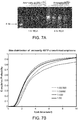
- the incorporation of aminoallyl dUTP leads to slightly smaller diameter of the average DNA amplicon size.
- the circular cDNA template was used for rolling circle amplification, during which a range of aminoallyl dUTP was added.
- the amplicon mixture in solution was then arrayed on a glass surface and hybridized to a common fluorescent probe sequence. Since aminoallyl dUTP has a single positive charge, the increasing incorporation of aminoallyl dUTP led to a reduction in the overall negative charge, making each DNA amplicon slightly more compact.
- the ratio shown in the graph legend represents the molar ratio of aminoallyl dUTP to dTTP during the amplification step.
- a 50-base oligonucleotide is phosphorylated at the 5' end using polynucleotide kinase in the T4 ligase buffer for 15 min.
- the reaction mixture is incubated with CircLigase mixture at 60°C for 1 hour to generate circular templates for testing.
- the RCA primer (18 bases) is then hybridized to the circular template in solution and a diluted template:primer mixture is used for rolling circle amplification with or without aminoallyl dUTP.
- the reaction mixture was diluted in PBS and bound to amino-silane treated coverglass.
- the bound RCA amplicons were then cross-linked with BS(PEG)9. They were then washed using a continuous stream of 2x SSC wash solution for 1 min, stained with SYBR safe and imaged using an epifluorescence microscope (63x objective).
- the DNA amplicons generated in solution are arrayed on a glass surface and cross-linked via the aminoallyl moiety. They were then exposed to a constant flow of distilled water running across its surface with and with the cross-linker chemistry for 5 min at room temperature and then imaged after SYBR Gold staining. As shown in Figure 3C , the DNA amplicons that were not cross-linked stretched out as a result of high shear stress for about 5 minutes. The DNA amplicons cross-linked with aminoallyl dUTP were morphologically preserved after high shear stress for 5 minutes.
- Human primary fibroblasts are grown on a 1.5 cover slip. They are fixed using 4% formaldehyde in PBS for 15 min, followed by three washes of 70% ethanol.
- the reverse transcription mixture containing 1 uM random hexamer or 0.1 uM polydT(18)V primer with additional adapter sequences, 250 uM dNTP, 40 uM aminoallyl dUTP, 20U RNase inhibitor and 100 U M-MuLV reverse transcriptase are then added to the fixed cells and incubated overnight at 37°C. The sample is then washed using PBS, and cross-linked using 100 uM BS(PEG)9 in PBS for 1 hour, followed by 1M Tris treatment for 15 min.
- the circularization mixture containing 25 U CircLigase, 1 mM MnCl and 1 M Betain is added, and the sample is incubated at 60°C.
- the residual RNA is degraded using a mixture of RNase cocktail and RNase H.
- the RCA primer is then hybridized to the sample at 60°C for 15 min.
- 100 U phi29 DNA polymerase, 250 uM dNTP and 40 uM aminoallyl dNTP are added to the sample and incubated at 30°C overnight.
- the sample is then washed using PBS, and cross-linked using 100 uM BS(PEG)9 in PBS for 1 hour, followed by 1M Tris treatment for 15 min.
- 1 uM fluorescently labeled oligonucleotides are diluted in 2x SSC and hybridized to the matrix containing the DNA amplicons at 60°C and washed. Imaging is done using Leica SP5 scanning confocal microscope using a 63x objectives. The fluorescent oligonucleotides are stripped off using 80% formamide heated to 80°C. The sample is then dried and stored at 4°C from July 2011 to March 2012. The sample was rehydrated in PBS, and rehybridized to the fluorescently labeled oligonucleotides and imaged. The second image was obtained using an epifluorescence microscope.
- the DNA amplicons are structurally and chemically stable over a long period of time once cross-linked.
- the DNA amplicons preserved as a three-dimensional matrix in human fibroblasts can be interrogated using fluorescent primers and stored in phosphate buffered solution for up to a year and re-interrogated without losing their structural or sequence information.
- the different image quality here reflects the difference between confocal microscopy vs. epifluorescence microscopy, not the sample quality.
- a DNA Amplicon Matrix Within a Cell is Structurally and Chemically Stable
- Human primary fibroblasts are grown on a 1.5 cover slip. They are fixed using 4% formaldehyde in PBS for 15 min, followed by three washes of 70% ethanol.
- the reverse transcription mixture containing 1 uM random hexamer or 0.1 uM polydT(18)V primer with additional adapter sequences, 250 uM dNTP, 40 uM aminoallyl dUTP, 20U RNase inhibitor and 100 U M-MuLV reverse transcriptase are then added to the fixed cells and incubated overnight at 37°C. The sample is then washed using PBS, and cross-linked using 100 uM BS(PEG)9 in PBS for 1 hour, followed by 1M Tris treatment for 15 min.
- the circularization mixture containing 25 U CircLigase, 1 mM MnCl and 1 M Betain is added, and the sample is incubated at 60°C.
- the residual RNA is degraded using a mixture of RNase cocktail and RNase H.
- the RCA primer is then hybridized to the sample at 60°C for 15 min.
- 100 U phi29 DNA polymerase, 250 uM dNTP and 40 uM aminoallyl dNTP are added to the sample and incubated at 30°C overnight.
- the sample is then washed using PBS, and cross-linked using 100 uM BS(PEG)9 in PBS for 1 hour, followed by 1M Tris treatment for 15 min.
- 1 uM fluorescently labeled oligonucleotides are diluted in 2x SSC and hybridized to the matrix containing the DNA amplicons at 60°C and washed. Imaging is done using Leica SP5 scanning confocal microscope using a 63x objectives. The fluorescent oligonucleotides are stripped off using 80% formamide heated to 80°C. The sample is then washed with distilled water and rehybridized to the fluorescently labeled oligonucleotides and imaged.
- the DNA amplicon matrix inside the cell can be stripped using harsh chemical agents (i.e. 0.1N NaOH, 80% formamide) and heated up to 95°C for a prolonged period of time without losing their structural integrity or definition.
- harsh chemical agents i.e. 0.1N NaOH, 80% formamide
- a DNA Amplicon Matrix Within a Cell is Structurally and Chemically Stable
- Human iPS cells are grown on a 1.5 cover slip. They are fixed using 4% formaldehyde in PBS for 15 min, followed by three washes of 70% ethanol.
- the reverse transcription mixture containing 1 uM random hexamer or 0.1 uM polydT(18)V primer with additional adapter sequences, 250 uM dNTP, 40 uM aminoallyl dUTP, 20U RNase inhibitor and 100 U M-MuLV reverse transcriptase are then added to the fixed cells and incubated overnight at 37°C. The sample is then washed using PBS, and cross-linked using 100 uM BS(PEG)9 in PBS for 1 hour, followed by 1M Tris treatment for 15 min.
- the circularization mixture containing 25 U CircLigase, 1 mM MnCl and 1 M Betain is added, and the sample is incubated at 60°C.
- the residual RNA is degraded using a mixture of RNase cocktail and RNase H.
- the RCA primer is then hybridized to the sample at 60°C for 15 min.
- 100 U phi29 DNA polymerase, 250 uM dNTP and 40 uM aminoallyl dNTP are added to the sample and incubated at 30°C overnight.
- the sample is then washed using PBS, and cross-linked using 100 uM BS(PEG)9 in PBS for 1 hour, followed by 1M Tris treatment for 15 min.
- 1 uM fluorescently labeled oligonucleotides are diluted in 2x SSC and hybridized to the matrix containing the DNA amplicons at 60°C and washed three times using 2x SSC. After imaging, the fluorescent oligonucleotides are stripped off using 80% formamide heated to 80°C. The sample is then washed with distilled water and rehybridized to the fluorescently labeled oligonucleotides. This cycle is repeated sixty times (5 minutes per cycle). The multiple images were then aligned and processed using MatLab to identify a region of interest.
- the sample can be cycled through more than 50 heating, cooling, enzymatic and chemical reactions without any changes in the signal to noise ratio.
- the high absolute signal intensity here was due to insufficient probe washing in the initial cycles.
- the individual DNA amplicons in the matrix was imaged in three dimensions using confocal microscopy and tracked over 12 cycles, one measure the relative displacement of each amplicon over time.
- the mean displacement of each amplicon was ⁇ 500-nm in both lateral and axial dimensions, which was about the diameter of each amplicon.
- An example image of the analysis is shown in the right panel, in which the line representing the displacement is shown in different colors according to their cycle number.
- Human iPS cells are grown on a 1.5 cover slip. They are fixed using 4% formaldehyde in PBS for 15 min, followed by three washes of 70% ethanol.
- the reverse transcription mixture containing 1 uM random hexamer or 0.1 uM polydT(18)V primer with additional adapter sequences, 250 uM dNTP, 40 uM aminoallyl dUTP, 20U RNase inhibitor and 100 U M-MuLV reverse transcriptase are then added to the fixed cells and incubated overnight at 37°C. The sample is then washed using PBS, and cross-linked using 100 uM BS(PEG)9 in PBS for 1 hour, followed by 1M Tris treatment for 15 min.
- the circularization mixture containing 25 U CircLigase, 1 mM MnCl and 1 M Betain is added, and the sample is incubated at 60°C.
- the residual RNA is degraded using a mixture of RNase cocktail and RNase H.
- the RCA primer is then hybridized to the sample at 60°C for 15 min.
- 100 U phi29 DNA polymerase, 250 uM dNTP and 40 uM aminoallyl dNTP are added to the sample and incubated at 30°C overnight.
- the sample is then washed using PBS, and cross-linked using 100 uM BS(PEG)9 in PBS for 1 hour, followed by 1M Tris treatment for 15 min.
- 1 uM fluorescently labeled oligonucleotides are diluted in 2x SSC and hybridized to the matrix containing the DNA amplicons at 60°C and washed three times using 2x SSC.
- Leica SP5 scanning confocal microscope with 63x objective is used and scanning optical zoom of 5x is used. The line scan was repeated three times and averaged to generate a high quality image.
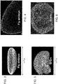
- Figure 5A is an image of the DNA amplicons (derived from reverse transcription of the cytoplasmic and the nuclear RNA) embedded within the cross-linked matrix inside human induced pluripotent stem cells. Individual amplicons are too tightly packed to visualize discrete amplicons, given the optical diffraction limitation in microscopy. But various subcellular compartments where the RNA is not expected to be present (i.e. nm: nuclear membrane, pm: plasma membrane) show dark staining, whereas the nucleus (Nu) and the cytoplasm (Cy) show a high density of the amplicons.
- the distribution of the cellular RNA shows unique patterns from cell to cell (1st panel vs. 2nd panel) and from one cell cycle phase to another (1st panel vs. 3rd panel). These results show that the DNA amplicons can be immobilized, amplified and interrogated in a manner to reflect their original spatial information.
- Human iPS cells are grown on a 1.5 cover slip. They are fixed using 4% formaldehyde in PBS for 15 min, followed by three washes of 70% ethanol.
- the reverse transcription mixture containing 1 uM random hexamer or 0.1 uM polydT(18)V primer with additional adapter sequences, 250 uM dNTP, 40 uM aminoallyl dUTP, 20U RNase inhibitor and 100 U M-MuLV reverse transcriptase are then added to the fixed cells and incubated overnight at 37°C. The sample is then washed using PBS, and cross-linked using 100 uM BS(PEG)9 in PBS for 1 hour, followed by 1M Tris treatment for 15 min.
- the circularization mixture containing 25 U CircLigase, 1 mM MnCl and 1 M Betain is added, and the sample is incubated at 60°C.
- the residual RNA is degraded using a mixture of RNase cocktail and RNase H.
- the RCA primer is then hybridized to the sample at 60°C for 15 min.
- 100 U phi29 DNA polymerase, 250 uM dNTP and 40 uM aminoallyl dNTP are added to the sample and incubated at 30°C overnight.
- the sample is then washed using PBS, and cross-linked using 100 uM BS(PEG)9 in PBS for 1 hour, followed by 1M Tris treatment for 15 min.
- the sequencing primer is designed with different 3' ends that that each primer can detect only 1/4th of the amplicons. If different dinucleotides are added to the 3' ends of the primer, each primer can detect only 1/16th of the amplicons.
- a chosen sequencing primer in 2x SSC is hybridized to the sample at 60°C for 15 minutes and washed.
- a ligation mixture containing 10 U T4 DNA ligase, ligation buffer and 1 uM fluorescently labeled nonamers (a pool containing A, G, C or T at fixed positions and labeled with FITC, Cy3, Texas Red or Cy5, respectively) is added and incubated for 50 min at room temperature.
- the cell After washing three times with 2x SSC, the cell is imaged on Leica SP5 scanning confocal microscope using four color channels. After imaging, the probe complex is stripped using 80% formamide and washed with distilled water. The sequencing by ligation step is repeated using a different nonamer set interrogating the next sequence.
- the left panel shows a subset of randomly primed cDNA amplicons being sequenced on a confocal microscope.
- the right panel shows GAPDH cDNA amplicons being sequenced over time using confocal microscopy. Only a single optical section is shown here.
- the axial dimension represents time or sequencing cycle steps.
- Circular DNA is Cross-linked or Co-polymerized Into A Matrix and Amplified
- circular DNA including cDNA
- a given cross-linker chemistry i.e. aminoallyl, thiol, biotin
- modified dUTP that competes with natural dTTP.
- the circular DNA is then cross-linked and/or co-polymerized within a three-dimensional container (i.e. cell), conforming the shape and the size of the container. Uncrosslinked molecules are then washed away, and one then performs rolling circle amplification, followed by imaging (i.e. sequencing).
- the density, the size and the signal strength can be controlled by varying the template size, the amplification time and the detection primer sequence.
- the DNA amplicons can be made into an ordered 3D matrix in a suitable scaffold material with addressable primers that can serve as amplification primers.
Landscapes
- Chemical & Material Sciences (AREA)
- Life Sciences & Earth Sciences (AREA)
- Organic Chemistry (AREA)
- Proteomics, Peptides & Aminoacids (AREA)
- Health & Medical Sciences (AREA)
- Engineering & Computer Science (AREA)
- Zoology (AREA)
- Wood Science & Technology (AREA)
- Molecular Biology (AREA)
- Analytical Chemistry (AREA)
- General Engineering & Computer Science (AREA)
- Genetics & Genomics (AREA)
- Microbiology (AREA)
- General Health & Medical Sciences (AREA)
- Biotechnology (AREA)
- Bioinformatics & Cheminformatics (AREA)
- Biochemistry (AREA)
- Immunology (AREA)
- Physics & Mathematics (AREA)
- Biophysics (AREA)
- Chemical Kinetics & Catalysis (AREA)
- General Chemical & Material Sciences (AREA)
- Measuring Or Testing Involving Enzymes Or Micro-Organisms (AREA)
- Cell Biology (AREA)
Applications Claiming Priority (3)
| Application Number | Priority Date | Filing Date | Title |
|---|---|---|---|
| US201361777383P | 2013-03-12 | 2013-03-12 | |
| PCT/US2014/018580 WO2014163886A1 (fr) | 2013-03-12 | 2014-02-26 | Procédé de génération d'une matrice tridimensionnelle contenant des acides nucléiques |
| EP14779940.7A EP2971184B1 (fr) | 2013-03-12 | 2014-02-26 | Procédé de génération d'une matrice tridimensionnelle contenant des acides nucléiques |
Related Parent Applications (1)
| Application Number | Title | Priority Date | Filing Date |
|---|---|---|---|
| EP14779940.7A Division EP2971184B1 (fr) | 2013-03-12 | 2014-02-26 | Procédé de génération d'une matrice tridimensionnelle contenant des acides nucléiques |
Publications (1)
| Publication Number | Publication Date |
|---|---|
| EP3578666A1 true EP3578666A1 (fr) | 2019-12-11 |
Family
ID=51658791
Family Applications (2)
| Application Number | Title | Priority Date | Filing Date |
|---|---|---|---|
| EP19169540.2A Pending EP3578666A1 (fr) | 2013-03-12 | 2014-02-26 | Procédé de génération d'une matrice contenant un acide nucléique tridimensionnel |
| EP14779940.7A Active EP2971184B1 (fr) | 2013-03-12 | 2014-02-26 | Procédé de génération d'une matrice tridimensionnelle contenant des acides nucléiques |
Family Applications After (1)
| Application Number | Title | Priority Date | Filing Date |
|---|---|---|---|
| EP14779940.7A Active EP2971184B1 (fr) | 2013-03-12 | 2014-02-26 | Procédé de génération d'une matrice tridimensionnelle contenant des acides nucléiques |
Country Status (3)
| Country | Link |
|---|---|
| US (8) | US10138509B2 (fr) |
| EP (2) | EP3578666A1 (fr) |
| WO (1) | WO2014163886A1 (fr) |
Cited By (4)
| Publication number | Priority date | Publication date | Assignee | Title |
|---|---|---|---|---|
| US12098425B2 (en) | 2018-10-10 | 2024-09-24 | Readcoor, Llc | Three-dimensional spatial molecular indexing |
| US12203136B2 (en) | 2020-08-17 | 2025-01-21 | Readcoor, Llc | Methods and systems for spatial mapping of genetic variants |
| US12359253B2 (en) | 2018-04-09 | 2025-07-15 | The Board Of Trustees Of The Leland Stanford Junior University | Method of in situ gene sequencing |
| US12559790B2 (en) | 2020-01-29 | 2026-02-24 | 10X Genomics, Inc. | Compositions and methods for analyte detection |
Families Citing this family (244)
| Publication number | Priority date | Publication date | Assignee | Title |
|---|---|---|---|---|
| US10787701B2 (en) | 2010-04-05 | 2020-09-29 | Prognosys Biosciences, Inc. | Spatially encoded biological assays |
| HRP20151294T1 (hr) | 2010-04-05 | 2016-03-11 | Prognosys Biosciences, Inc. | Biološka testiranja sa prostornim kodiranjem |
| GB201106254D0 (en) | 2011-04-13 | 2011-05-25 | Frisen Jonas | Method and product |
| WO2013055995A2 (fr) | 2011-10-14 | 2013-04-18 | President And Fellows Of Harvard College | Séquençage par assemblage structurel |
| ES2991004T3 (es) | 2011-12-22 | 2024-12-02 | Harvard College | Métodos para la detección de analitos |
| EP4697020A2 (fr) | 2011-12-22 | 2026-02-18 | President And Fellows Of Harvard College | Compositions et procédés de détection d'analyte |
| US9914967B2 (en) | 2012-06-05 | 2018-03-13 | President And Fellows Of Harvard College | Spatial sequencing of nucleic acids using DNA origami probes |
| WO2014060483A1 (fr) | 2012-10-17 | 2014-04-24 | Spatial Transcriptomics Ab | Procédés et produit d'optimisation de la détection localisée ou spatiale de l'expression génique dans un échantillon de tissu |
| EP3578666A1 (fr) | 2013-03-12 | 2019-12-11 | President and Fellows of Harvard College | Procédé de génération d'une matrice contenant un acide nucléique tridimensionnel |
| EP4596565A3 (fr) | 2013-06-04 | 2025-11-05 | President And Fellows Of Harvard College | Régulation transcriptionnelle guidée par arn |
| EP3013983B1 (fr) | 2013-06-25 | 2023-02-15 | Prognosys Biosciences, Inc. | Essais biologiques à codage spatial faisant appel à un dispositif microfluidique |
| WO2016007839A1 (fr) | 2014-07-11 | 2016-01-14 | President And Fellows Of Harvard College | Méthodes de marquage et de détection à haut rendement de caractéristiques biologiques in situ à l'aide de microscopie |
| CN112029826B (zh) | 2014-07-30 | 2025-03-14 | 哈佛学院院长及董事 | 用于测定核酸的系统和方法 |
| US10774374B2 (en) | 2015-04-10 | 2020-09-15 | Spatial Transcriptomics AB and Illumina, Inc. | Spatially distinguished, multiplex nucleic acid analysis of biological specimens |
| JP6882282B2 (ja) * | 2015-11-03 | 2021-06-02 | プレジデント アンド フェローズ オブ ハーバード カレッジ | 三次元核酸含有マトリックスの立体撮像のための方法と装置 |
| WO2017079382A1 (fr) | 2015-11-03 | 2017-05-11 | President And Fellows Of Harvard College | Systèmes et procédés de traitement de données de séquence liées spatialement et reçues d'un dispositif de séquençage |
| CN109415761B (zh) | 2016-04-25 | 2022-09-20 | 哈佛学院董事及会员团体 | 用于原位分子检测的杂交链反应方法 |
| EP4050112A1 (fr) | 2016-06-21 | 2022-08-31 | 10X Genomics, Inc. | Séquençage d'acide nucléique |
| GB2570412A (en) | 2016-08-31 | 2019-07-24 | Harvard College | Methods of generating libraries of nucleic acid sequences for detection via fluorescent in situ sequencing |
| CN109923216B (zh) * | 2016-08-31 | 2024-08-02 | 哈佛学院董事及会员团体 | 将生物分子的检测组合到使用荧光原位测序的单个试验的方法 |
| KR102313431B1 (ko) | 2016-11-21 | 2021-10-18 | 나노스트링 테크놀로지스, 인크. | 화학적 조성물 및 이것을 사용하는 방법 |
| JP2020504600A (ja) | 2016-12-09 | 2020-02-13 | アルティヴュー, インク. | 標識化核酸イメージング剤を使用した多重イメージングのための改善された方法 |
| US11185568B2 (en) | 2017-04-14 | 2021-11-30 | President And Fellows Of Harvard College | Methods for generation of cell-derived microfilament network |
| WO2018218150A1 (fr) | 2017-05-26 | 2018-11-29 | President And Fellows Of Harvard College | Systèmes et méthodes de criblage à base d'image à haut débit |
| WO2018226930A1 (fr) * | 2017-06-07 | 2018-12-13 | President And Fellows Of Harvard College | Séquençage fluorescence in situ d'expansion ciblée |
| CA3078158A1 (fr) | 2017-10-06 | 2019-04-11 | Cartana Ab | Ligature a matrice d'arn |
| US11629342B2 (en) | 2017-10-17 | 2023-04-18 | President And Fellows Of Harvard College | Cas9-based transcription modulation systems |
| AU2019271028B2 (en) | 2018-05-14 | 2025-09-18 | Bruker Spatial Biology, Inc. | Chemical compositions and methods of using same |
| WO2019236841A1 (fr) | 2018-06-08 | 2019-12-12 | Ultivue, Inc. | Dépôt de rapporteur catalysé multiplexé |
| CN112770776B (zh) | 2018-07-30 | 2025-08-19 | 瑞德库尔有限责任公司 | 用于样品处理或分析的方法和系统 |
| EP3830289B1 (fr) | 2018-08-03 | 2026-02-11 | 10X Genomics, Inc. | Procédés pour réduire au minimum un échange de codes à barres |
| EP3844308A1 (fr) | 2018-08-28 | 2021-07-07 | 10X Genomics, Inc. | Résolution de réseaux spatiaux |
| US11519033B2 (en) | 2018-08-28 | 2022-12-06 | 10X Genomics, Inc. | Method for transposase-mediated spatial tagging and analyzing genomic DNA in a biological sample |
| EP3844304B1 (fr) | 2018-08-28 | 2024-10-02 | 10X Genomics, Inc. | Procédés de génération de matrices à codes-barres spatiaux |
| WO2020076979A1 (fr) | 2018-10-10 | 2020-04-16 | Readcoor, Inc. | Capture de surface de cibles |
| US12529094B2 (en) | 2018-12-10 | 2026-01-20 | 10X Genomics, Inc. | Imaging system hardware |
| EP3894590B1 (fr) | 2018-12-10 | 2025-10-22 | 10X Genomics, Inc. | Procédés d'utilisation de réseaux maître/copie pour la détection spatiale |
| CA3122569A1 (fr) | 2018-12-13 | 2020-06-18 | President And Fellows Of Harvard College | Systemes et procedes d'amplification pour merfish et d'autres applications |
| US11926867B2 (en) | 2019-01-06 | 2024-03-12 | 10X Genomics, Inc. | Generating capture probes for spatial analysis |
| US11649485B2 (en) | 2019-01-06 | 2023-05-16 | 10X Genomics, Inc. | Generating capture probes for spatial analysis |
| EP3921418A4 (fr) | 2019-02-06 | 2023-02-08 | Singular Genomics Systems, Inc. | Compositions et procédés de séquençage d'acide nucléique |
| CN114174531A (zh) | 2019-02-28 | 2022-03-11 | 10X基因组学有限公司 | 用空间条码化寡核苷酸阵列对生物分析物进行概况分析 |
| WO2020190509A1 (fr) | 2019-03-15 | 2020-09-24 | 10X Genomics, Inc. | Méthodes d'utilisation de matrices spatiales pour le séquençage d'organismes unicellulaires |
| WO2020198071A1 (fr) | 2019-03-22 | 2020-10-01 | 10X Genomics, Inc. | Analyse spatiale tridimensionnelle |
| EP3976820A1 (fr) | 2019-05-30 | 2022-04-06 | 10X Genomics, Inc. | Procédés de détection de l'hétérogénéité spatiale d'un échantillon biologique |
| WO2020240025A1 (fr) | 2019-05-31 | 2020-12-03 | Cartana Ab | Procédé de détection de molécules d'acide nucléique cible |
| EP4038546B1 (fr) | 2019-10-01 | 2024-08-21 | 10X Genomics, Inc. | Les systèmes et méthodes d'identification des caractéristiques morphologiques dans les échantillons de tissus |
| US12157124B2 (en) | 2019-11-06 | 2024-12-03 | 10X Genomics, Inc. | Imaging system hardware |
| WO2021091611A1 (fr) | 2019-11-08 | 2021-05-14 | 10X Genomics, Inc. | Agents de capture d'analytes marqués spatialement pour le multiplexage d'analytes |
| WO2021092433A2 (fr) | 2019-11-08 | 2021-05-14 | 10X Genomics, Inc. | Amélioration de la spécificité de la liaison d'un analyte |
| WO2021097255A1 (fr) | 2019-11-13 | 2021-05-20 | 10X Genomics, Inc. | Génération de sondes de capture pour analyse spatiale |
| AU2020388047A1 (en) | 2019-11-18 | 2022-06-23 | 10X Genomics, Inc. | Systems and methods for tissue classification |
| WO2021102039A1 (fr) | 2019-11-21 | 2021-05-27 | 10X Genomics, Inc, | Analyse spatiale d'analytes |
| EP4435718A3 (fr) | 2019-11-22 | 2024-12-18 | 10X Genomics, Inc. | Systèmes et procédés d'analyse spatiale d'analytes à l'aide d'un alignement de repères |
| GB201919032D0 (en) | 2019-12-20 | 2020-02-05 | Cartana Ab | Method of detecting an analyte |
| WO2021133849A1 (fr) | 2019-12-23 | 2021-07-01 | 10X Genomics, Inc. | Procédés d'analyse spatiale utilisant une ligature à matrice d'arn |
| CN115135984B (zh) | 2019-12-23 | 2025-12-23 | 10X基因组学有限公司 | 可逆固定试剂及其使用方法 |
| EP4081656A1 (fr) | 2019-12-23 | 2022-11-02 | 10X Genomics, Inc. | Compositions et méthodes d'utilisation d'échantillons biologiques fixés dans des dosages basés sur des compartiments |
| CN115715329A (zh) | 2020-01-10 | 2023-02-24 | 10X基因组学有限公司 | 确定生物样品中靶核酸位置的方法 |
| US12365942B2 (en) | 2020-01-13 | 2025-07-22 | 10X Genomics, Inc. | Methods of decreasing background on a spatial array |
| US12405264B2 (en) | 2020-01-17 | 2025-09-02 | 10X Genomics, Inc. | Electrophoretic system and method for analyte capture |
| US11702693B2 (en) | 2020-01-21 | 2023-07-18 | 10X Genomics, Inc. | Methods for printing cells and generating arrays of barcoded cells |
| US11732299B2 (en) | 2020-01-21 | 2023-08-22 | 10X Genomics, Inc. | Spatial assays with perturbed cells |
| US20210230681A1 (en) | 2020-01-24 | 2021-07-29 | 10X Genomics, Inc. | Methods for spatial analysis using proximity ligation |
| US11821035B1 (en) | 2020-01-29 | 2023-11-21 | 10X Genomics, Inc. | Compositions and methods of making gene expression libraries |
| US12076701B2 (en) | 2020-01-31 | 2024-09-03 | 10X Genomics, Inc. | Capturing oligonucleotides in spatial transcriptomics |
| US11898205B2 (en) | 2020-02-03 | 2024-02-13 | 10X Genomics, Inc. | Increasing capture efficiency of spatial assays |
| US12110541B2 (en) | 2020-02-03 | 2024-10-08 | 10X Genomics, Inc. | Methods for preparing high-resolution spatial arrays |
| US12110548B2 (en) | 2020-02-03 | 2024-10-08 | 10X Genomics, Inc. | Bi-directional in situ analysis |
| US12112833B2 (en) | 2020-02-04 | 2024-10-08 | 10X Genomics, Inc. | Systems and methods for index hopping filtering |
| US11732300B2 (en) | 2020-02-05 | 2023-08-22 | 10X Genomics, Inc. | Increasing efficiency of spatial analysis in a biological sample |
| WO2021158925A1 (fr) | 2020-02-07 | 2021-08-12 | 10X Genomics, Inc. | Évaluation quantitative et automatisée de la performance de perméabilisation pour la transcriptomique spatiale |
| US11835462B2 (en) | 2020-02-11 | 2023-12-05 | 10X Genomics, Inc. | Methods and compositions for partitioning a biological sample |
| CN115428088A (zh) | 2020-02-13 | 2022-12-02 | 10X基因组学有限公司 | 用于基因表达和dna染色质可及性的联合交互式可视化的系统和方法 |
| US12399123B1 (en) | 2020-02-14 | 2025-08-26 | 10X Genomics, Inc. | Spatial targeting of analytes |
| US12281357B1 (en) | 2020-02-14 | 2025-04-22 | 10X Genomics, Inc. | In situ spatial barcoding |
| EP4107284A1 (fr) | 2020-02-17 | 2022-12-28 | 10X Genomics, Inc. | Analyse in situ de l'interaction chromatinienne |
| US20230081381A1 (en) | 2020-02-20 | 2023-03-16 | 10X Genomics, Inc. | METHODS TO COMBINE FIRST AND SECOND STRAND cDNA SYNTHESIS FOR SPATIAL ANALYSIS |
| EP4484571A3 (fr) | 2020-02-21 | 2025-03-19 | 10X Genomics, Inc. | Procédés et compositions pour un dosage spatial in situ intégré |
| AU2021224860A1 (en) * | 2020-02-21 | 2022-09-15 | 10X Genomics, Inc. | Compositions, methods and systems for sample processing |
| CN116157533A (zh) | 2020-02-21 | 2023-05-23 | 10X基因组学有限公司 | 使用杂交方法捕获遗传靶标 |
| US11891654B2 (en) | 2020-02-24 | 2024-02-06 | 10X Genomics, Inc. | Methods of making gene expression libraries |
| US11768175B1 (en) | 2020-03-04 | 2023-09-26 | 10X Genomics, Inc. | Electrophoretic methods for spatial analysis |
| US12188085B2 (en) | 2020-03-05 | 2025-01-07 | 10X Genomics, Inc. | Three-dimensional spatial transcriptomics with sequencing readout |
| ES2965354T3 (es) | 2020-04-22 | 2024-04-12 | 10X Genomics Inc | Métodos para análisis espacial que usan eliminación de ARN elegido como diana |
| WO2021225900A1 (fr) | 2020-05-04 | 2021-11-11 | 10X Genomics, Inc. | Modes de transfert transcriptomique spatial |
| CN116134308B (zh) | 2020-05-19 | 2025-05-27 | 10X基因组学有限公司 | 电泳盒和仪器 |
| EP4153775B1 (fr) | 2020-05-22 | 2024-07-24 | 10X Genomics, Inc. | Mesure spatio-temporelle simultanée de l'expression génique et de l'activité cellulaire |
| WO2021237056A1 (fr) | 2020-05-22 | 2021-11-25 | 10X Genomics, Inc. | Analyse d'intégrité d'arn dans un échantillon biologique |
| EP4553168A3 (fr) | 2020-05-22 | 2025-08-27 | 10x Genomics, Inc. | Analyse spatiale pour détecter des variantes de séquence |
| WO2021242834A1 (fr) | 2020-05-26 | 2021-12-02 | 10X Genomics, Inc. | Procédé de réinitialisation d'un réseau |
| EP4158054B1 (fr) | 2020-06-02 | 2025-04-16 | 10X Genomics, Inc. | Trancriptomique spatiale pour les récepteurs d'antigènes |
| EP4025692A2 (fr) | 2020-06-02 | 2022-07-13 | 10X Genomics, Inc. | Procédés de banques d'acides nucléiques |
| US12031177B1 (en) | 2020-06-04 | 2024-07-09 | 10X Genomics, Inc. | Methods of enhancing spatial resolution of transcripts |
| WO2021252499A1 (fr) | 2020-06-08 | 2021-12-16 | 10X Genomics, Inc. | Méthodes de détermination de marge chirurgicale et méthodes d'utilisation associées |
| WO2021252747A1 (fr) | 2020-06-10 | 2021-12-16 | 1Ox Genomics, Inc. | Procédés de distribution de fluide |
| EP4165207B1 (fr) | 2020-06-10 | 2024-09-25 | 10X Genomics, Inc. | Methode pour determiner la position d'un analyte dans un echantillon biologique |
| US12435363B1 (en) | 2020-06-10 | 2025-10-07 | 10X Genomics, Inc. | Materials and methods for spatial transcriptomics |
| US20230279474A1 (en) | 2020-06-10 | 2023-09-07 | 10X Genomics, Inc. | Methods for spatial analysis using blocker oligonucleotides |
| US12209273B2 (en) | 2020-06-12 | 2025-01-28 | 10X Genomics, Inc. | Nucleic acid assays using click chemistry bioconjugation |
| EP4172362B1 (fr) | 2020-06-25 | 2024-09-18 | 10X Genomics, Inc. | Analyse spatiale de la méthylation de l'adn |
| US12168801B1 (en) | 2020-07-02 | 2024-12-17 | 10X Genomics, Inc. | Hybrid/capture probe designs for full-length cDNA |
| US11981960B1 (en) | 2020-07-06 | 2024-05-14 | 10X Genomics, Inc. | Spatial analysis utilizing degradable hydrogels |
| US12209280B1 (en) | 2020-07-06 | 2025-01-28 | 10X Genomics, Inc. | Methods of identifying abundance and location of an analyte in a biological sample using second strand synthesis |
| US11761038B1 (en) | 2020-07-06 | 2023-09-19 | 10X Genomics, Inc. | Methods for identifying a location of an RNA in a biological sample |
| US20230287475A1 (en) | 2020-07-31 | 2023-09-14 | 10X Genomics, Inc. | De-crosslinking compounds and methods of use for spatial analysis |
| WO2022032195A2 (fr) | 2020-08-06 | 2022-02-10 | Singular Genomics Systems, Inc. | Séquençage spatial |
| EP4121561A4 (fr) | 2020-08-06 | 2024-04-17 | Singular Genomics Systems, Inc. | Méthodes pour la transcriptomique et la protéomique in situ |
| US12553898B1 (en) | 2020-08-10 | 2026-02-17 | 10X Genomics, Inc. | Fluorescent hybridization of antibody-oligonucleotide for multiplexing and signal amplification |
| US12297499B2 (en) | 2020-08-17 | 2025-05-13 | 10X Genomics, Inc. | Multicomponent nucleic acid probes for sample analysis |
| US11981958B1 (en) | 2020-08-20 | 2024-05-14 | 10X Genomics, Inc. | Methods for spatial analysis using DNA capture |
| US12580044B1 (en) | 2020-09-02 | 2026-03-17 | 10X Genomics, Inc. | Systems and methods for identifying cells that are antigen-specific for an immunogenic feature |
| EP4530358A3 (fr) | 2020-09-15 | 2025-06-18 | 10x Genomics, Inc. | Procédés de libération d'une sonde de capture étendue à partir d'un substrat et leurs utilisations |
| AU2021345133A1 (en) | 2020-09-16 | 2023-03-30 | 10X Genomics, Inc. | Methods of determining the location of an analyte in a biological sample using a plurality of wells |
| US20240033743A1 (en) | 2020-09-18 | 2024-02-01 | 10X Genomics, Inc. | Sample handling apparatus and fluid delivery methods |
| EP4491742B1 (fr) | 2020-09-18 | 2026-03-04 | 10X Genomics, Inc. | Appareil de manipulation d'échantillons et procédés d'enregistrement d'images |
| US11926822B1 (en) | 2020-09-23 | 2024-03-12 | 10X Genomics, Inc. | Three-dimensional spatial analysis |
| WO2022081643A2 (fr) | 2020-10-13 | 2022-04-21 | 10X Genomics, Inc. | Compositions et procédés pour générer des molécules de liaison à un antigène recombinant à partir de cellules uniques |
| AU2021366701A1 (en) | 2020-10-22 | 2023-05-04 | 10X Genomics, Inc. | Methods for spatial analysis using rolling circle amplification |
| US12071667B2 (en) | 2020-11-04 | 2024-08-27 | 10X Genomics, Inc. | Sequence analysis using meta-stable nucleic acid molecules |
| AU2021376399A1 (en) | 2020-11-06 | 2023-06-15 | 10X Genomics, Inc. | Compositions and methods for binding an analyte to a capture probe |
| AU2021385065A1 (en) | 2020-11-18 | 2023-06-15 | 10X Genomics, Inc. | Methods and compositions for analyzing immune infiltration in cancer stroma to predict clinical outcome |
| US11827935B1 (en) | 2020-11-19 | 2023-11-28 | 10X Genomics, Inc. | Methods for spatial analysis using rolling circle amplification and detection probes |
| EP4012046A1 (fr) | 2020-12-11 | 2022-06-15 | 10X Genomics, Inc. | Procédés et compositions pour l'analyse multimodale in situ |
| EP4729631A2 (fr) | 2020-12-21 | 2026-04-22 | 10X Genomics, Inc. | Procédés, compositions et systèmes de capture de sondes et/ou de codes à barres |
| CN116848263A (zh) | 2020-12-23 | 2023-10-03 | 10X基因组学有限公司 | 用于分析物检测的方法和组合物 |
| WO2022147296A1 (fr) | 2020-12-30 | 2022-07-07 | 10X Genomics, Inc. | Clivage de sondes de capture pour analyse spatiale |
| WO2022147005A1 (fr) | 2020-12-30 | 2022-07-07 | 10X Genomics, Inc. | Procédés de détermination de capture d'analytes |
| US12060603B2 (en) | 2021-01-19 | 2024-08-13 | 10X Genomics, Inc. | Methods for internally controlled in situ assays using padlock probes |
| US12398262B1 (en) | 2021-01-22 | 2025-08-26 | 10X Genomics, Inc. | Triblock copolymer-based cell stabilization and fixation system and methods of use thereof |
| EP4284945B1 (fr) | 2021-01-26 | 2024-10-23 | 10X Genomics, Inc. | Sondes analogiques d'acide nucléique pour analyse in situ |
| US20240093290A1 (en) | 2021-01-29 | 2024-03-21 | 10X Genomics, Inc. | Method for transposase mediated spatial tagging and analyzing genomic dna in a biological sample |
| WO2022182903A1 (fr) * | 2021-02-24 | 2022-09-01 | California Institute Of Technology | Multiplexage de conditions expérimentales et d'échantillons en génomique spatiale |
| US12275984B2 (en) | 2021-03-02 | 2025-04-15 | 10X Genomics, Inc. | Sequential hybridization and quenching |
| US20220282316A1 (en) * | 2021-03-03 | 2022-09-08 | 10X Genomics, Inc. | Methods and compositions for modifying primary probes in situ |
| WO2022187366A1 (fr) | 2021-03-03 | 2022-09-09 | 10X Genomics, Inc. | Détection d'analytes in situ à l'aide d'origami d'acide nucléique |
| ES3008686T3 (en) | 2021-03-18 | 2025-03-24 | 10X Genomics Inc | Multiplex capture of gene and protein expression from a biological sample |
| EP4620481A3 (fr) | 2021-04-05 | 2025-12-03 | 10X Genomics, Inc. | Composition de ligase recombinante et ses utilisations |
| EP4428246B1 (fr) | 2021-04-14 | 2025-12-24 | 10X Genomics, Inc. | Procédés de mesure de la localisation erronée d'un analyte |
| WO2022226057A1 (fr) | 2021-04-20 | 2022-10-27 | 10X Genomics, Inc. | Procédés d'évaluation de la qualité d'un échantillon avant une analyse spatiale à l'aide d'une ligature modélisée |
| WO2022235764A1 (fr) | 2021-05-05 | 2022-11-10 | Singular Genomics Systems, Inc. | Dispositif d'analyse multiomique et ses procédés d'utilisation |
| EP4320271B1 (fr) | 2021-05-06 | 2025-03-19 | 10X Genomics, Inc. | Procédés pour augmenter la résolution d'une analyse spatiale |
| WO2022241227A1 (fr) | 2021-05-14 | 2022-11-17 | Akoya Biosciences, Inc. | Amplification de signaux de détection d'arn dans des échantillons biologiques |
| IL308570A (en) * | 2021-05-28 | 2024-01-01 | Broad Inst Inc | Co-mapping of transcriptional states and protein histology |
| US20220380838A1 (en) | 2021-06-01 | 2022-12-01 | 10X Genomics, Inc. | Methods and compositions for analyte detection and probe resolution |
| WO2022256422A1 (fr) | 2021-06-02 | 2022-12-08 | 10X Genomics, Inc. | Analyse d'échantillon à l'aide de sondes circularisables asymétriques |
| EP4582555A3 (fr) | 2021-06-03 | 2025-10-22 | 10X Genomics, Inc. | Procédés, compositions, kits et systèmes pour améliorer la capture d'analytes pour une analyse spatiale |
| US20240368711A1 (en) | 2021-06-22 | 2024-11-07 | 10X Genomics, Inc. | Spatial detection of sars-cov-2 using templated ligation |
| WO2023283442A1 (fr) | 2021-07-09 | 2023-01-12 | 10X Genomics, Inc. | Procédés de détection d'analytes à l'aide d'un marquage épars |
| CN117651855A (zh) | 2021-07-13 | 2024-03-05 | 10X基因组学有限公司 | 用于制备具有可控厚度的聚合基质的方法 |
| EP4352252B1 (fr) | 2021-07-13 | 2026-01-28 | 10X Genomics, Inc. | Procédés d'analyse spatiale utilisant un silençage de sonde ciblée |
| CA3224093A1 (fr) | 2021-07-30 | 2023-02-02 | Jorge Ivan Hernandez Neuta | Procedes et compositions pour la synchronisation de reactions in situ |
| US12139751B2 (en) | 2021-07-30 | 2024-11-12 | 10X Genomics, Inc. | Circularizable probes for in situ analysis |
| US12553805B2 (en) | 2021-08-02 | 2026-02-17 | 10X Genomics, Inc. | Methods of preserving a biological sample |
| US12391984B2 (en) | 2021-08-03 | 2025-08-19 | 10X Genomics, Inc. | Compositions and methods for rolling circle amplification |
| US12460251B2 (en) | 2021-08-03 | 2025-11-04 | 10X Genomics, Inc. | Stabilization and/or compaction of nucleic acid molecules |
| US12529096B2 (en) | 2021-08-03 | 2026-01-20 | 10X Genomics, Inc. | Stabilization and/or compaction of nucleic acid structures |
| WO2023014825A1 (fr) | 2021-08-03 | 2023-02-09 | Akoya Biosciences, Inc. | Détection d'arn par marquage et amplification sélectifs |
| EP4381095A1 (fr) | 2021-08-03 | 2024-06-12 | 10X Genomics, Inc. | Concatémères d'acide nucléique et procédés de stabilisation et/ou de compactage de ceux-ci |
| WO2023018799A1 (fr) | 2021-08-12 | 2023-02-16 | 10X Genomics, Inc. | Procédés, compositions et systèmes pour identifier des molécules de liaison à l'antigène |
| US12435364B2 (en) | 2021-08-16 | 2025-10-07 | 10X Genomics, Inc. | Probes comprising a split barcode region and methods of use |
| ES3011462T3 (en) | 2021-09-01 | 2025-04-07 | 10X Genomics Inc | Methods for blocking a capture probe on a spatial array |
| US20240378734A1 (en) | 2021-09-17 | 2024-11-14 | 10X Genomics, Inc. | Systems and methods for image registration or alignment |
| WO2023076345A1 (fr) | 2021-10-26 | 2023-05-04 | 10X Genomics, Inc. | Procédés d'analyse spatiale utilisant la capture ciblée d'arn |
| WO2023086824A1 (fr) | 2021-11-10 | 2023-05-19 | 10X Genomics, Inc. | Procédés d'identification de molécules de liaison à l'antigène |
| WO2023086880A1 (fr) | 2021-11-10 | 2023-05-19 | 10X Genomics, Inc. | Procédés, compositions et kits pour déterminer l'emplacement d'un analyte dans un échantillon biologique |
| WO2023102313A1 (fr) | 2021-11-30 | 2023-06-08 | 10X Genomics, Inc. | Systèmes et procédés d'identification de régions d'aneuploïdie dans un tissu |
| EP4305195A2 (fr) | 2021-12-01 | 2024-01-17 | 10X Genomics, Inc. | Procédés, compositions et systèmes pour la détection améliorée d'analytes in situ et analyse spatiale |
| EP4441711A1 (fr) | 2021-12-20 | 2024-10-09 | 10X Genomics, Inc. | Auto-test pour dispositif d'imagerie |
| WO2023129898A2 (fr) | 2021-12-27 | 2023-07-06 | 10X Genomics, Inc. | Procédés et compositions pour l'amplification par cercle roulant |
| EP4733410A2 (fr) | 2022-01-21 | 2026-04-29 | 10X Genomics, Inc. | Signaux de lecture multiples pour analyser un échantillon |
| WO2023150171A1 (fr) | 2022-02-01 | 2023-08-10 | 10X Genomics, Inc. | Procédés, compositions et systèmes de capture d'analytes à partir d'échantillons de glioblastome |
| WO2023150098A1 (fr) | 2022-02-01 | 2023-08-10 | 10X Genomics, Inc. | Procédés, kits, compositions et systèmes d'analyse spatiale |
| WO2023150163A1 (fr) | 2022-02-01 | 2023-08-10 | 10X Genomics, Inc. | Procédés, compositions et systèmes de capture d'analytes à partir de tissu lymphatique |
| EP4479933A1 (fr) | 2022-02-15 | 2024-12-25 | 10x Genomics, Inc. | Systèmes et procédés d'analyse spatiale d'analytes à l'aide d'un alignement de repères |
| WO2023164570A1 (fr) | 2022-02-23 | 2023-08-31 | Insitro, Inc. | Criblage optique groupé et mesures transcriptionnelles de cellules comprenant des perturbations génétiques à code-barres |
| EP4490737A1 (fr) | 2022-03-08 | 2025-01-15 | 10X Genomics, Inc. | Procédés de conception de code in situ pour réduire à un minimum un chevauchement optique |
| WO2023172670A2 (fr) | 2022-03-11 | 2023-09-14 | 10X Genomics, Inc. | Appareil de manipulation d'échantillon et procédés système de distribution de fluide |
| AU2023243205A1 (en) | 2022-04-01 | 2024-09-19 | 10X Genomics, Inc. | Compositions and methods for targeted masking of autofluorescence |
| CN119234045A (zh) | 2022-04-06 | 2024-12-31 | 10X基因组学有限公司 | 用于多重细胞分析的方法 |
| US20250251403A1 (en) | 2022-04-12 | 2025-08-07 | 10X Genomics, Inc. | Compositions and methods for generating and characterizing recombinant antigen binding molecules |
| WO2023215552A1 (fr) | 2022-05-06 | 2023-11-09 | 10X Genomics, Inc. | Lecteurs de codes-barres moléculaires pour détection d'analytes |
| EP4519453A1 (fr) | 2022-05-06 | 2025-03-12 | 10X Genomics, Inc. | Procédés et compositions pour l'analyse in situ de séquences v(d)j |
| WO2023215612A1 (fr) | 2022-05-06 | 2023-11-09 | 10X Genomics, Inc. | Analyse d'un antigène et des interactions antigène-récepteur |
| CN119213143A (zh) | 2022-05-11 | 2024-12-27 | 10X基因组学有限公司 | 用于原位测序的组合物和方法 |
| WO2023225519A1 (fr) | 2022-05-17 | 2023-11-23 | 10X Genomics, Inc. | Transposons modifiés, compositions et utilisations de ceux-ci |
| WO2023229988A1 (fr) | 2022-05-23 | 2023-11-30 | 10X Genomics, Inc. | Moule pour échantillon de tissu |
| WO2023229982A2 (fr) | 2022-05-24 | 2023-11-30 | 10X Genomics, Inc. | Confinement de structure poreuse pour suppression de convection |
| WO2023245190A1 (fr) | 2022-06-17 | 2023-12-21 | 10X Genomics, Inc. | DÉ-RÉTICULATION CATALYTIQUE D'ÉCHANTILLONS POUR ANALYSE IN SITU <i /> |
| WO2023250077A1 (fr) | 2022-06-22 | 2023-12-28 | 10X Genomics, Inc. | Procédés, compositions et systèmes pour la capture de sondes et/ou de code-barres |
| EP4554973A1 (fr) | 2022-07-13 | 2025-05-21 | 10X Genomics, Inc. | Procédés de caractérisation de molécules de liaison à l'antigène à partir d'échantillons biologiques |
| WO2024015578A1 (fr) | 2022-07-15 | 2024-01-18 | 10X Genomics, Inc. | Procédés pour déterminer l'emplacement d'un acide nucléique cible dans un échantillon biologique |
| EP4565870A1 (fr) | 2022-08-05 | 2025-06-11 | 10x Genomics, Inc. | Systèmes et procédés de quantification d'immunofluorescence |
| WO2024036191A1 (fr) | 2022-08-10 | 2024-02-15 | 10X Genomics, Inc. | Systèmes et procédés de colocalisation |
| WO2024035844A1 (fr) | 2022-08-12 | 2024-02-15 | 10X Genomics, Inc. | Procédés de réduction de la capture d'analytes |
| CN119677856A (zh) | 2022-08-12 | 2025-03-21 | 10X基因组学有限公司 | Puma1聚合酶及其用途 |
| AU2023325064A1 (en) | 2022-08-15 | 2025-03-06 | Element Biosciences, Inc. | Spatially resolved surface capture of nucleic acids |
| WO2024040060A1 (fr) | 2022-08-16 | 2024-02-22 | 10X Genomics, Inc. | Ap50 polymérases et leurs utilisations |
| EP4577670A1 (fr) | 2022-08-24 | 2025-07-02 | 10X Genomics, Inc. | Compositions et procédés de cartographie d'épitopes antigéniques dans des échantillons biologiques |
| IL320056A (en) * | 2022-10-06 | 2025-06-01 | Element Biosciences Inc | 3D base reading in next-generation paving analysis |
| EP4573208A1 (fr) | 2022-10-10 | 2025-06-25 | 10X Genomics, Inc. | Transcription in vitro d'acides nucléiques capturés spatialement |
| EP4602344A1 (fr) | 2022-10-14 | 2025-08-20 | 10X Genomics, Inc. | Procédés d'analyse d'échantillons biologiques |
| WO2024086167A2 (fr) | 2022-10-17 | 2024-04-25 | 10X Genomics, Inc. | Procédés, compositions et kits pour déterminer l'emplacement d'un analyte dans un échantillon biologique |
| WO2024102736A1 (fr) | 2022-11-08 | 2024-05-16 | 10X Genomics, Inc. | Procédés et compositions d'immobilisation pour détection in situ |
| WO2024102809A1 (fr) | 2022-11-09 | 2024-05-16 | 10X Genomics, Inc. | Procédés, compositions et kits pour déterminer l'emplacement de plusieurs analytes dans un échantillon biologique |
| US20240158852A1 (en) | 2022-11-16 | 2024-05-16 | 10X Genomics, Inc. | Methods and compositions for assessing performance of in situ assays |
| US12372771B2 (en) | 2022-11-18 | 2025-07-29 | 10X Genomics, Inc. | Systems and methods for actively mitigating vibrations |
| US12581050B2 (en) | 2022-11-23 | 2026-03-17 | 10X Genomics, Inc. | Optical assemblies for machine vision calibration |
| WO2024130203A1 (fr) | 2022-12-16 | 2024-06-20 | 10X Genomics, Inc. | Procédés et compositions d'évaluation de performances |
| WO2024137860A1 (fr) | 2022-12-21 | 2024-06-27 | 10X Genomics, Inc. | Procédés d'accroche d'acides ribonucléiques dans des échantillons biologiques |
| EP4638788A1 (fr) | 2022-12-21 | 2025-10-29 | 10X Genomics, Inc. | Analyse d'analytes et d'expression génique spatiale |
| WO2024145441A1 (fr) | 2022-12-29 | 2024-07-04 | 10X Genomics, Inc. | Procédés, compositions et kits pour déterminer un emplacement d'un acide nucléique cible dans un échantillon biologique fixe |
| WO2024145224A1 (fr) | 2022-12-29 | 2024-07-04 | 10X Genomics, Inc. | Compositions, procédés et systèmes d'analyse spatiale à haute résolution |
| EP4486913B1 (fr) | 2022-12-30 | 2025-07-16 | 10x Genomics, Inc. | Procédés, compositions et kits pour le codage à barres multiple et/ou le codage à barres spatial à haute densité |
| WO2024145445A1 (fr) | 2022-12-30 | 2024-07-04 | 10X Genomics, Inc. | Procédés de capture d'analytes cibles |
| EP4689174A1 (fr) | 2023-03-28 | 2026-02-11 | 10X Genomics, Inc. | Procédés, compositions et kits pour réduire une mauvaise localisation d'analytes |
| US20250257393A1 (en) | 2023-04-21 | 2025-08-14 | 10X Genomics, Inc. | Methods and compositions for detecting nucleic acids in a fixed biological sample |
| US20240376521A1 (en) | 2023-05-10 | 2024-11-14 | 10X Genomics, Inc. | Boronic acid compositions and methods for tethering ribonucleic acids in biological samples |
| WO2024238625A1 (fr) | 2023-05-15 | 2024-11-21 | 10X Genomics, Inc. | Normalisation de données d'anticorps spatial |
| WO2024238900A1 (fr) | 2023-05-17 | 2024-11-21 | 10X Genomics, Inc. | Procédés pour réduire la mauvaise localisation d'un analyte |
| WO2024238992A1 (fr) | 2023-05-18 | 2024-11-21 | 10X Genomics, Inc. | Polymérases de la famille b sans déplacement de brin modifiées pour des applications de transcription inverse et de remplissage d'espace |
| CN120981456A (zh) | 2023-05-24 | 2025-11-18 | 金橘生物科技公司 | 杂环化合物及其用途 |
| EP4720332A1 (fr) | 2023-06-01 | 2026-04-08 | Singular Genomics Systems, Inc. | Procédés et sondes pour détecter des séquences polynucléotidiques dans des cellules et des tissus |
| WO2024254316A1 (fr) | 2023-06-07 | 2024-12-12 | 10X Genomics, Inc. | Compositions et procédés pour identifier les caillots sanguins dans les échantillons biologiques |
| EP4728062A1 (fr) | 2023-06-13 | 2026-04-22 | 10X Genomics, Inc. | Protéines de liaison à l'adn spécifiques d'une séquence |
| WO2025029605A1 (fr) | 2023-07-28 | 2025-02-06 | 10X Genomics, Inc. | Méthodes et compositions de détection de variants génétiques en utilisant des sondes de capture et une extension d'amorce |
| WO2025029831A1 (fr) | 2023-07-31 | 2025-02-06 | 10X Genomics, Inc. | Procédés et systèmes de clivage d'arn ciblé et d'amplification de cercle roulant amorcé par arn cible |
| WO2025029627A1 (fr) | 2023-07-31 | 2025-02-06 | 10X Genomics, Inc. | Procédés, compositions et kits de détection spatiale de variants génétiques |
| EP4581160A1 (fr) | 2023-08-24 | 2025-07-09 | 10X Genomics, Inc. | Procédés, kits et compositions aux fins d'une détection spatiale de variants génétiques |
| WO2025072119A1 (fr) | 2023-09-26 | 2025-04-03 | 10X Genomics, Inc. | Procédés, compositions et kits pour détecter des interactions chromosomiques spatiales |
| WO2025090912A1 (fr) | 2023-10-26 | 2025-05-01 | 10X Genomics, Inc. | Réseaux spatiaux contenant des sondes maîtresses réutilisables |
| WO2025096581A1 (fr) | 2023-10-31 | 2025-05-08 | 10X Genomics, Inc. | Procédés d'analyse spatiale et de séquençage de noyaux uniques |
| WO2025101377A1 (fr) | 2023-11-09 | 2025-05-15 | 10X Genomics, Inc. | Analyse spatiale assistée par matrice d'échantillons biologiques |
| WO2025101864A1 (fr) | 2023-11-09 | 2025-05-15 | 10X Genomics, Inc. | Procédés, compositions et kits pour réduire la mauvaise localisation des analytes dans les dosages d'analyse spatiale |
| US20250163502A1 (en) | 2023-11-22 | 2025-05-22 | 10X Genomics, Inc. | Methods for processing ribonucleic acids in biological samples |
| US20250207203A1 (en) | 2023-12-20 | 2025-06-26 | 10X Genomics, Inc. | In situ detection of copy number variations in biological samples |
| WO2025189030A1 (fr) | 2024-03-07 | 2025-09-12 | Stellaromics, Inc. | Procédés et systèmes d'imagerie volumétrique |
| US20250288988A1 (en) | 2024-03-15 | 2025-09-18 | 10X Genomics, Inc. | Systems and methods for covering and sealing an open well |
| WO2025239936A1 (fr) | 2024-05-13 | 2025-11-20 | Element Biosciences, Inc. | Sondes multivalentes et procédés d'utilisation |
| WO2025255457A1 (fr) | 2024-06-07 | 2025-12-11 | 10X Genomics, Inc. | Compositions et procédés de profilage spatial sur cible amélioré |
| WO2025264702A1 (fr) | 2024-06-18 | 2025-12-26 | 10X Genomics, Inc. | Hydrogels 3d pour le séquençage d'acides nucléiques |
| WO2026024934A1 (fr) | 2024-07-25 | 2026-01-29 | 10X Genomics, Inc. | Procédés, compositions et kits d'analyse spatiale par tagmentation |
| WO2026043885A1 (fr) | 2024-08-20 | 2026-02-26 | 10X Genomics, Inc. | Procédés et systèmes de préparation d'une bibliothèque d'acides nucléiques |
| WO2026055487A1 (fr) | 2024-09-06 | 2026-03-12 | 10X Genomics, Inc. | Nucléotides modifiés avec espaceurs oligomères |
Citations (40)
| Publication number | Priority date | Publication date | Assignee | Title |
|---|---|---|---|---|
| US4659774A (en) | 1985-11-01 | 1987-04-21 | American Hoechst Corporation | Support for solid-phase oligonucleotide synthesis |
| US4861571A (en) | 1985-09-20 | 1989-08-29 | Toyo Soda Manufacturing Co., Ltd. | Process for preparation of synthetic mordenite molded body |
| US5141813A (en) | 1989-08-28 | 1992-08-25 | Clontech Laboratories, Inc. | Multifunctional controlled pore glass reagent for solid phase oligonucleotide synthesis |
| US5264566A (en) | 1985-10-15 | 1993-11-23 | Genentech, Inc. | Method for in vitro oligonucleotide synthesis using H-phosphonates |
| US5428148A (en) | 1992-04-24 | 1995-06-27 | Beckman Instruments, Inc. | N4 - acylated cytidinyl compounds useful in oligonucleotide synthesis |
| US5554744A (en) | 1994-09-23 | 1996-09-10 | Hybridon, Inc. | Method for loading solid supports for nucleic acid synthesis |
| US5574146A (en) | 1994-08-30 | 1996-11-12 | Beckman Instruments, Inc. | Oligonucleotide synthesis with substituted aryl carboxylic acids as activators |
| US5602244A (en) | 1988-05-26 | 1997-02-11 | Competitive Technologies, Inc. | Polynucleotide phosphorodithioate compounds |
| US5612199A (en) | 1991-10-11 | 1997-03-18 | Behringwerke Ag | Method for producing a polynucleotide for use in single primer amplification |
| US5700637A (en) | 1988-05-03 | 1997-12-23 | Isis Innovation Limited | Apparatus and method for analyzing polynucleotide sequences and method of generating oligonucleotide arrays |
| US5750341A (en) | 1995-04-17 | 1998-05-12 | Lynx Therapeutics, Inc. | DNA sequencing by parallel oligonucleotide extensions |
| US5959463A (en) | 1997-03-10 | 1999-09-28 | Mitsubishi Denki Kabushiki Kaisha | Semiconductor test apparatus for measuring power supply current of semiconductor device |
| US6124090A (en) | 1989-01-19 | 2000-09-26 | Behringwerke Ag | Nucleic acid amplification using single primer |
| US6261797B1 (en) | 1996-01-29 | 2001-07-17 | Stratagene | Primer-mediated polynucleotide synthesis and manipulation techniques |
| US6294323B1 (en) | 1993-04-14 | 2001-09-25 | Behringwerke Ag | Self initiating single primer amplification of nucleic acids |
| WO2002024597A2 (fr) | 2000-09-20 | 2002-03-28 | Goodrich Corporation | Compositions et composites pour matrice minerale et leur procede de fabrication |
| US6365375B1 (en) | 1998-03-26 | 2002-04-02 | Wolfgang Dietmaier | Method of primer-extension preamplification PCR |
| US6375903B1 (en) | 1998-02-23 | 2002-04-23 | Wisconsin Alumni Research Foundation | Method and apparatus for synthesis of arrays of DNA probes |
| US6391544B1 (en) | 1998-05-15 | 2002-05-21 | Abbott Laboratories | Method for using unequal primer concentrations for generating nucleic acid amplification products |
| US20020081582A1 (en) | 1998-02-11 | 2002-06-27 | Xiaolian Gao | Method and apparatus for chemical and biochemical reactions using photo-generated reagents |
| US20030068633A1 (en) | 2001-05-18 | 2003-04-10 | Belshaw Peter J. | Method for the synthesis of DNA sequences |
| WO2003040410A1 (fr) | 2001-11-02 | 2003-05-15 | Nimblegen Systems, Inc. | Detection d'une hybridation sur un microreseau d'oligonucleotides par marquage covalent d'une sonde de microreseau |
| WO2003046223A1 (fr) | 2001-11-26 | 2003-06-05 | Nimblegen Systems, Inc. | Jeux ordonnes de micro-echantillons et detection de modele visible |
| WO2003065038A2 (fr) | 2002-01-31 | 2003-08-07 | Nimblegen Systems Llc | Procede et appareil de synthese de series de sondes d'adn |
| WO2003064027A2 (fr) | 2002-01-31 | 2003-08-07 | Nimblegen Systems Llc | Correction de non uniformite d'eclairage pendant la synthese d'ensembles oligomeriques |
| WO2003064699A2 (fr) | 2002-01-31 | 2003-08-07 | Nimblegen Systems Llc | Procede et appareil de synthese de reseaux de sondes d'adn |
| WO2003064026A1 (fr) | 2002-01-31 | 2003-08-07 | Nimblegen Systems Llc | Substrat predessine, dispositif et procede pour la synthese optique de sondes d'adn |
| WO2003066212A2 (fr) | 2002-02-01 | 2003-08-14 | Nimblegen Systems Llc | Procede et appareil de synthese de microreseaux |
| WO2003100012A2 (fr) | 2002-05-24 | 2003-12-04 | Nimblegen Systems, Inc. | Jeux ordonnes de microechantillons et procede permettant de declencher une reaction d'hybridation pour plusieurs echantillons sur un seul jeu ordonne |
| WO2004029586A1 (fr) | 2002-09-27 | 2004-04-08 | Nimblegen Systems, Inc. | Microreseau a barrieres hydrophobes |
| WO2004031351A2 (fr) | 2002-10-01 | 2004-04-15 | Nimblegen Systems, Inc. | Microreseaux comportant de multiples oligonucleotides dans les zones caracteristiques d'un reseau |
| WO2004031399A2 (fr) | 2002-09-30 | 2004-04-15 | Nimblegen Systems, Inc. | Chargement en parallele de reseaux |
| US6800439B1 (en) | 2000-01-06 | 2004-10-05 | Affymetrix, Inc. | Methods for improved array preparation |
| US6824866B1 (en) | 1999-04-08 | 2004-11-30 | Affymetrix, Inc. | Porous silica substrates for polymer synthesis and assays |
| US6830890B2 (en) | 1994-10-24 | 2004-12-14 | Affymetrix, Inc. | Nucleic acid probe libraries |
| US6833450B1 (en) | 2000-03-17 | 2004-12-21 | Affymetrix, Inc. | Phosphite ester oxidation in nucleic acid array preparation |
| US20060024711A1 (en) * | 2004-07-02 | 2006-02-02 | Helicos Biosciences Corporation | Methods for nucleic acid amplification and sequence determination |
| WO2007001986A2 (fr) * | 2005-06-20 | 2007-01-04 | Yuling Luo | Methodes de detection des acides nucleiques dans des cellules individuelles et d'identification de cellules rares a partir de grandes populations cellulaires heterogenes |
| WO2012110899A2 (fr) * | 2011-02-15 | 2012-08-23 | Bernitz Mats Nilsson | Procédé de détection d'arnm in situ |
| WO2015127183A2 (fr) * | 2014-02-21 | 2015-08-27 | Massachusetts Institute Of Technology | Microscopie d'expansion |
Family Cites Families (267)
| Publication number | Priority date | Publication date | Assignee | Title |
|---|---|---|---|---|
| US4123610A (en) | 1977-03-09 | 1978-10-31 | The United States Government | Nucleic acid crosslinking agent and affinity inactivation of nucleic acids therewith |
| US5525464A (en) | 1987-04-01 | 1996-06-11 | Hyseq, Inc. | Method of sequencing by hybridization of oligonucleotide probes |
| US4981985A (en) | 1987-10-20 | 1991-01-01 | Trustees Of The University Of Pennsylvania | Synthesis of photolabile chelators for multivalent cations |
| US4886741A (en) | 1987-12-09 | 1989-12-12 | Microprobe Corporation | Use of volume exclusion agents for the enhancement of in situ hybridization |
| US4844617A (en) | 1988-01-20 | 1989-07-04 | Tencor Instruments | Confocal measuring microscope with automatic focusing |
| US5151189A (en) | 1990-09-17 | 1992-09-29 | Gelman Sciences, Inc. | Cationic charge modified microporous membrane |
| JPH04268359A (ja) | 1991-02-22 | 1992-09-24 | Taiho Yakuhin Kogyo Kk | 刺激応答性ヒドロゲル及びその製造方法 |
| US5424413A (en) | 1992-01-22 | 1995-06-13 | Gen-Probe Incorporated | Branched nucleic acid probes |
| AU3664693A (en) | 1992-02-13 | 1993-09-03 | Bio-Metric Systems, Inc. | Immobilization of chemical species in crosslinked matrices |
| US5594235A (en) | 1993-06-17 | 1997-01-14 | Ultrapointe Corporation | Automated surface acquisition for a confocal microscope |
| US5681697A (en) | 1993-12-08 | 1997-10-28 | Chiron Corporation | Solution phase nucleic acid sandwich assays having reduced background noise and kits therefor |
| US5578832A (en) | 1994-09-02 | 1996-11-26 | Affymetrix, Inc. | Method and apparatus for imaging a sample on a device |
| SE9400522D0 (sv) | 1994-02-16 | 1994-02-16 | Ulf Landegren | Method and reagent for detecting specific nucleotide sequences |
| US5830708A (en) | 1995-06-06 | 1998-11-03 | Advanced Tissue Sciences, Inc. | Methods for production of a naturally secreted extracellular matrix |
| US6284284B1 (en) | 1995-06-06 | 2001-09-04 | Advanced Tissue Sciences, Inc. | Compositions and methods for production and use of an injectable naturally secreted extracellular matrix |
| CA2256700A1 (fr) | 1996-06-06 | 1997-12-11 | Lynx Therapeutics, Inc. | Signatures par ligature d'adaptateurs codes |
| JP4532608B2 (ja) | 1997-02-07 | 2010-08-25 | オリンパス株式会社 | 走査型プローブ顕微鏡を用いた核酸検出方法 |
| US5948617A (en) | 1997-06-13 | 1999-09-07 | Biospeparations, Inc. | Methods of in situ hybridization |
| US6083726A (en) | 1998-02-03 | 2000-07-04 | Lucent Technologies, Inc. | Methods for polynucleotide synthesis and articles for polynucleotide hybridization |
| US6068979A (en) | 1998-04-22 | 2000-05-30 | Lumigen, Inc. | Simplified sequential chemiluminescent detection |
| JP3662850B2 (ja) | 1998-06-24 | 2005-06-22 | イルミナ インコーポレイテッド | 微小球を有するアレイセンサーのデコード |
| JP2002531056A (ja) | 1998-08-07 | 2002-09-24 | セレイ, エルエルシー | 遺伝子分析におけるゲルマイクロドロップ |
| US6232067B1 (en) | 1998-08-17 | 2001-05-15 | The Perkin-Elmer Corporation | Adapter directed expression analysis |
| DE60045917D1 (de) | 1999-02-23 | 2011-06-16 | Caliper Life Sciences Inc | Sequenzierung durch inkorporation |
| EP1171636A2 (fr) | 1999-04-22 | 2002-01-16 | Albert Einstein College Of Medicine Of Yeshiva University | Recherche de compositions d'expression genique par la methode multi-fluor fish |
| US6969615B2 (en) | 1999-07-26 | 2005-11-29 | 20/20 Genesystems, Inc. | Methods, devices, arrays and kits for detecting and analyzing biomolecules |
| US20020015952A1 (en) | 1999-07-30 | 2002-02-07 | Anderson Norman G. | Microarrays and their manufacture by slicing |
| CA2386151A1 (fr) | 1999-10-12 | 2001-04-19 | Marc Madou | Valve polymere reactive, dispositifs de distribution et procedes d'utilisation de ces derniers |
| AU1774501A (en) | 1999-11-17 | 2001-05-30 | Research Foundation Of State University Of New York, The | Three dimensional data storage device and method for reading |
| US7115738B2 (en) | 2000-03-14 | 2006-10-03 | Active Motif | Hydroxyproline/phosphono oligonucleotide analogues, methods of synthesis and methods of use |
| US7033758B2 (en) | 2000-06-02 | 2006-04-25 | Bayer Corporation | Highly sensitive gene detection and localization using in situ branched-DNA hybridization |
| US8548745B2 (en) | 2000-06-08 | 2013-10-01 | The Regents Of The University Of California | Visual-servoing optical microscopy |
| US7613571B2 (en) | 2000-07-28 | 2009-11-03 | Doyle Michael D | Method and system for the multidimensional morphological reconstruction of genome expression activity |
| US6797152B2 (en) | 2000-07-31 | 2004-09-28 | California Institute Of Technology | Sensors and sensing methods for detecting analytes based on changes in pKa of a sensing polymer |
| EP1368369A4 (fr) | 2000-11-15 | 2006-02-22 | Hoffmann La Roche | Methodes et reactifs permettant d'identifier des cellules embryonnaires rares dans le systeme circulatoire maternel |
| AU3090102A (en) | 2000-12-14 | 2002-06-24 | Gen Probe Inc | Method and kit for enhancing the association rates of polynucleotides |
| EP2801624B1 (fr) | 2001-03-16 | 2019-03-06 | Singular Bio, Inc | Puces et procédés d'utilisation |
| US7473767B2 (en) | 2001-07-03 | 2009-01-06 | The Institute For Systems Biology | Methods for detection and quantification of analytes in complex mixtures |
| US20060248349A1 (en) | 2001-09-24 | 2006-11-02 | Rathjen Alicemarie G | Method for preparing and using personal and genetic profiles |
| US20030148335A1 (en) | 2001-10-10 | 2003-08-07 | Li Shen | Detecting targets by unique identifier nucleotide tags |
| GB2382137A (en) | 2001-11-20 | 2003-05-21 | Mats Gullberg | Nucleic acid enrichment |
| US8013134B2 (en) | 2001-11-23 | 2011-09-06 | Olink Ab | Kit for proximity probing with multivalent proximity probes |
| US20090246879A1 (en) | 2002-12-20 | 2009-10-01 | Callida Genomics | Materials and Methods Relating to Nano-Tags and Nano-Brands |
| WO2005003375A2 (fr) | 2003-01-29 | 2005-01-13 | 454 Corporation | Procede d'amplification et de sequençage d'acides nucleiques |
| EP2365095A1 (fr) | 2003-02-26 | 2011-09-14 | Callida Genomics, Inc. | Analyse de réseau d'ADN aléatoire par hybridation |
| US7385043B1 (en) | 2003-04-30 | 2008-06-10 | The Public Health Research Institute Of The City Of New York, Inc. | Homogeneous multiplex screening assays and kits |
| CN102135661B (zh) | 2003-05-20 | 2014-06-04 | 卢西德有限公司 | 用于对患者身体所选位置进行成像的共焦显微镜 |
| US7255994B2 (en) | 2003-06-10 | 2007-08-14 | Applera Corporation | Ligation assay |
| CN1580283A (zh) | 2003-08-13 | 2005-02-16 | 清华大学 | 一种检测核酸分子的方法 |
| US7193054B2 (en) | 2003-08-26 | 2007-03-20 | University Of Rochester | Nanofabrication using actin filaments |
| US20050064435A1 (en) | 2003-09-24 | 2005-03-24 | Xing Su | Programmable molecular barcodes |
| WO2005049851A2 (fr) | 2003-11-14 | 2005-06-02 | University Of Utah Research Foundation | Procedes, articles et compositions destines a l'identification d'oligonucleotides |
| US7381529B2 (en) | 2003-12-31 | 2008-06-03 | Intel Corporation | Methods and compositions for detecting nucleic acids using scanning probe microscopy and nanocodes |
| US20050191687A1 (en) | 2004-02-27 | 2005-09-01 | Tianxin Wang | Method for multiplexed analyte detection |
| JP2007526772A (ja) | 2004-02-27 | 2007-09-20 | プレジデント・アンド・フェロウズ・オブ・ハーバード・カレッジ | インサイチュー配列決定用ポロニー蛍光ビーズ |
| WO2006076017A2 (fr) | 2004-04-30 | 2006-07-20 | Applera Corporation | Procedes et kits d'identification de nucleotides cibles dans des populations melangees |
| US7253947B2 (en) | 2004-10-07 | 2007-08-07 | New York University | Portable automated confocal microscope |
| CN101916359B (zh) | 2005-01-27 | 2012-06-20 | 剑桥研究和仪器设备股份有限公司 | 把样本的不同区域分类到相应类别的方法和设备 |
| WO2006084132A2 (fr) | 2005-02-01 | 2006-08-10 | Agencourt Bioscience Corp. | Reactifs, methodes et bibliotheques pour sequençage fonde sur des billes |
| US7393665B2 (en) | 2005-02-10 | 2008-07-01 | Population Genetics Technologies Ltd | Methods and compositions for tagging and identifying polynucleotides |
| ITRM20050068A1 (it) | 2005-02-17 | 2006-08-18 | Istituto Naz Per Le Malattie I | Metodo per la rivelazione di acidi nucleici di agenti patogeni batterici o di parassiti nelle urine. |
| US20060234261A1 (en) | 2005-03-08 | 2006-10-19 | Pierce Niles A | Colorimetric readout of hybridization chain reaction |
| US7727721B2 (en) | 2005-03-08 | 2010-06-01 | California Institute Of Technology | Hybridization chain reaction amplification for in situ imaging |
| US8252754B2 (en) | 2005-03-21 | 2012-08-28 | The Trustees Of Columbia University In The City Of New York | Neuronal pain pathway |
| US20070020650A1 (en) | 2005-04-01 | 2007-01-25 | Avak Kahvejian | Methods for detecting proteins |
| WO2007086900A2 (fr) | 2005-04-08 | 2007-08-02 | Mycosol, Inc. | Analyses de recherche de stilbazium |
| EP1871896B1 (fr) | 2005-04-12 | 2015-06-03 | Olink AB | Sondes circulaires pour l'identifcation des biomolécules |
| US11561951B2 (en) | 2005-05-16 | 2023-01-24 | Panvia Future Technologies, Inc. | Multidimensional associative memory and data searching |
| US7601499B2 (en) | 2005-06-06 | 2009-10-13 | 454 Life Sciences Corporation | Paired end sequencing |
| US7842793B2 (en) | 2005-06-14 | 2010-11-30 | The California Institute Of Technology | Methods of making nucleic acid nanostructures |
| JP5331476B2 (ja) | 2005-06-15 | 2013-10-30 | カリダ・ジェノミックス・インコーポレイテッド | 遺伝子解析および化学解析用の単分子アレイ |
| EP1920072A2 (fr) | 2005-07-01 | 2008-05-14 | Dako Denmark A/S | Procede de detection d'hybridation in situ |
| US8486621B2 (en) | 2005-08-11 | 2013-07-16 | Cornell Research Foundation, Inc. | Nucleic acid-based matrixes |
| CN1959384A (zh) | 2005-11-01 | 2007-05-09 | 河南省生物工程技术研究中心 | 一种三维网格检测技术 |
| US20070190543A1 (en) | 2005-11-14 | 2007-08-16 | Applera Corporation | Coded Molecules for Detecting Target Analytes |
| US8986926B2 (en) | 2005-12-23 | 2015-03-24 | Nanostring Technologies, Inc. | Compositions comprising oriented, immobilized macromolecules and methods for their preparation |
| EP1963531B1 (fr) | 2005-12-23 | 2011-09-21 | Nanostring Technologies, Inc. | Nanorapporteurs et procedes de production et d'utilisation de ceux-ci |
| WO2007092538A2 (fr) * | 2006-02-07 | 2007-08-16 | President And Fellows Of Harvard College | Procédés de confection de sondes nucléotidiques pour séquençage et synthèse |
| US7864996B2 (en) | 2006-02-17 | 2011-01-04 | Lucid, Inc. | System for macroscopic and confocal imaging of tissue |
| WO2007106402A2 (fr) | 2006-03-10 | 2007-09-20 | President And Fellows Of Harvard College | Procedes et appareil pour irradiation en champ proche |
| GB0605584D0 (en) | 2006-03-20 | 2006-04-26 | Olink Ab | Method for analyte detection using proximity probes |
| US20070231823A1 (en) | 2006-03-23 | 2007-10-04 | Mckernan Kevin J | Directed enrichment of genomic DNA for high-throughput sequencing |
| RU2394915C2 (ru) | 2006-03-24 | 2010-07-20 | Александр Борисович Четверин | Бесконтактные способы обнаружения молекулярных колоний, наборы реагентов и устройство для их осуществления |
| US8975216B2 (en) | 2006-03-30 | 2015-03-10 | Pacific Biosciences Of California | Articles having localized molecules disposed thereon and methods of producing same |
| EP3722409A1 (fr) | 2006-03-31 | 2020-10-14 | Illumina, Inc. | Systèmes et procédés pour analyse de séquençage par synthèse |
| CN101495654A (zh) | 2006-04-19 | 2009-07-29 | 阿普里拉股份有限公司 | 无凝胶珠基测序的试剂、方法和文库 |
| JP5081232B2 (ja) | 2006-05-22 | 2012-11-28 | ナノストリング テクノロジーズ, インコーポレイテッド | ナノレポーターを分析するためのシステムおよび方法 |
| US7964347B2 (en) | 2006-06-15 | 2011-06-21 | Krassen Dimitrov | Labels for electronic detection of individual molecules and methods for their detection |
| US20080038163A1 (en) | 2006-06-23 | 2008-02-14 | Applera Corporation | Systems and Methods for Cooling in Biological Analysis Instruments |
| US7745129B1 (en) | 2006-07-12 | 2010-06-29 | Kenneth David Schatz | Methods for sequencing of a necleic acid |
| JP2010500617A (ja) | 2006-08-04 | 2010-01-07 | イコニシス インコーポレーテッド | 動的走査を行う自動式顕微鏡および方法 |
| WO2008033848A2 (fr) | 2006-09-11 | 2008-03-20 | The Arizona Board Of Regents, A Body Corporate Of The State Of Arizona Acting For And On Behalf Of Arizona State University | Nanomatrices de codage combinatoire auto-assemblées destinées à une biodétection mutliplexe |
| US20090208965A1 (en) | 2006-10-25 | 2009-08-20 | Ikonisys, Inc. | Automated method for detecting cancers and high grade hyperplasias |
| EP2061588B1 (fr) | 2006-11-06 | 2016-07-13 | Clondiag GmbH | Installation et procédé pour tests de liaison |
| US9201063B2 (en) | 2006-11-16 | 2015-12-01 | General Electric Company | Sequential analysis of biological samples |
| US7655898B2 (en) | 2006-11-30 | 2010-02-02 | Cambridge Research & Instrumentation, Inc. | Optical filter assembly with selectable bandwidth and rejection |
| WO2008069973A2 (fr) | 2006-12-01 | 2008-06-12 | The Trustees Of Columbia University In The City Of New York | Séquençage en quatre couleurs de l'adn par synthèse utilisant des terminateurs nucléotidiques réversibles, fluorescents et clivables |
| US20080269068A1 (en) | 2007-02-06 | 2008-10-30 | President And Fellows Of Harvard College | Multiplex decoding of sequence tags in barcodes |
| AU2008223544B2 (en) | 2007-03-02 | 2014-06-05 | Dupont Nutrition Biosciences Aps | Cultures with improved phage resistance |
| US8145677B2 (en) | 2007-03-27 | 2012-03-27 | Faleh Jassem Al-Shameri | Automated generation of metadata for mining image and text data |
| US8415102B2 (en) | 2007-04-10 | 2013-04-09 | Nanostring Technologies, Inc. | Methods and computer systems for identifying target-specific sequences for use in nanoreporters |
| WO2008144562A1 (fr) | 2007-05-16 | 2008-11-27 | California Institute Of Technology | Motif polyvalent d'acide nucléique en épingle à cheveux pour la programmation de voies d'auto-assemblage biomoléculaire |
| ES2959127T3 (es) | 2007-06-19 | 2024-02-20 | Hoffmann La Roche | Secuenciación de ácidos nucleicos de alto rendimiento por expansión |
| US7534991B2 (en) | 2007-07-10 | 2009-05-19 | Cambridge Research & Instrumentation, Inc. | Athermalized birefringent filter apparatus and method |
| US20110294135A1 (en) | 2007-08-02 | 2011-12-01 | BioDesic, LLC | Compositions and methods for analyte detection and quantitation |
| JP2010539982A (ja) | 2007-10-01 | 2010-12-24 | アプライド バイオシステムズ, エルエルシー | チェイスライゲーション配列決定法 |
| AU2008308457A1 (en) | 2007-10-04 | 2009-04-09 | Halcyon Molecular | Sequencing nucleic acid polymers with electron microscopy |
| US20090139311A1 (en) | 2007-10-05 | 2009-06-04 | Applied Biosystems Inc. | Biological Analysis Systems, Devices, and Methods |
| EP2051051B1 (fr) | 2007-10-16 | 2020-06-03 | Cambridge Research & Instrumentation, Inc. | Système d'imagerie spectrale avec correction optique dynamique |
| US20110020291A1 (en) | 2007-11-17 | 2011-01-27 | Debrabrata Banerjee | Use of stem cells for wound healing |
| CA2713712A1 (fr) | 2008-05-06 | 2009-11-12 | Kollmorgen Corporation | Sequenceur genetique incorporant une microscopie a fluorescence |
| CN101586150B (zh) | 2008-05-23 | 2016-09-28 | 陕西佰美基因股份有限公司 | 检测探针、通用寡核苷酸芯片及核酸检测方法及其用途 |
| US8199999B2 (en) | 2008-06-17 | 2012-06-12 | Cambridge Research & Instrumentation, Inc. | Image classifier training |
| WO2010011961A2 (fr) | 2008-07-25 | 2010-01-28 | University Of Georgia Research Foundation, Inc. | Système procaryote arni-like et ses procédés d'utilisation |
| US20120040397A1 (en) * | 2008-08-05 | 2012-02-16 | Dan Luo | Photo-Crosslinked Nucleic Acid Hydrogels |
| US8519115B2 (en) | 2008-08-14 | 2013-08-27 | Nanostring Technologies, Inc. | Stable nanoreporters |
| WO2010030818A2 (fr) | 2008-09-10 | 2010-03-18 | University Of Medicine And Dentistry Of New Jersey | Imagerie de molécules d’arnm individuelles utilisant des sondes multiples à marquage unique |
| US20100076057A1 (en) | 2008-09-23 | 2010-03-25 | Northwestern University | TARGET DNA INTERFERENCE WITH crRNA |
| US9156010B2 (en) | 2008-09-23 | 2015-10-13 | Bio-Rad Laboratories, Inc. | Droplet-based assay system |
| US20100087325A1 (en) | 2008-10-07 | 2010-04-08 | Illumina, Inc. | Biological sample temperature control system and method |
| RU2529804C2 (ru) | 2008-10-23 | 2014-09-27 | Конинклейке Филипс Электроникс, Н.В. | Молекулярная визуализация |
| WO2010054108A2 (fr) | 2008-11-06 | 2010-05-14 | University Of Georgia Research Foundation, Inc. | Polypeptides cas6 et procédés d'utilisation |
| US8309306B2 (en) | 2008-11-12 | 2012-11-13 | Nodality, Inc. | Detection composition |
| WO2010080134A1 (fr) | 2008-12-19 | 2010-07-15 | President And Fellows Of Harvard College | Séquençage d'acide nucléique assisté par particules |
| JP4528345B2 (ja) | 2009-01-28 | 2010-08-18 | シャープ株式会社 | 動画再生装置、動画再生方法、動画再生方法をコンピュータで実現するためのプログラム及びそのプログラムを記録した記録媒体 |
| JP5277082B2 (ja) | 2009-06-15 | 2013-08-28 | 株式会社日立ハイテクノロジーズ | 蛍光分析方法 |
| FR2948475A1 (fr) | 2009-07-24 | 2011-01-28 | Bionext | Procede de caracterisation d'objets tridimensionnels |
| US8431151B2 (en) | 2009-08-06 | 2013-04-30 | Syracuse University | Antimicrobial nanostructured hydrogel web containing silver |
| WO2011047087A2 (fr) | 2009-10-13 | 2011-04-21 | Nanostring Technologies, Inc. | Détection de protéine par l'intermédiaire de nanorapporteurs |
| US20110104693A1 (en) | 2009-11-03 | 2011-05-05 | High Throughput Genomics, Inc. | QUANTITATIVE NUCLEASE PROTECTION SEQUENCING (qNPS) |
| EP2531220B1 (fr) | 2009-12-18 | 2018-05-02 | President and Fellows of Harvard College | Échafaudages actifs pour administration de médicament et de cellule à la demande |
| US8632976B2 (en) | 2010-01-19 | 2014-01-21 | Agdia | Amplification of TRP1 for specific detection of Phytophthora ramorum |
| EP2516681B1 (fr) | 2010-02-11 | 2017-10-18 | Nanostring Technologies, Inc | Compositions et procédés de détection de préalablement petits arn par hybridisation à pont et ligation |
| WO2011100617A2 (fr) | 2010-02-12 | 2011-08-18 | Life Technologies Corporation | Codes d'identification d'acides nucléiques, de biomolécules et de polymères |
| US10267808B2 (en) | 2010-03-08 | 2019-04-23 | California Institute Of Technology | Molecular indicia of cellular constituents and resolving the same by super-resolution technologies in single cells |
| US10087431B2 (en) | 2010-03-10 | 2018-10-02 | The Regents Of The University Of California | Methods of generating nucleic acid fragments |
| GB201004292D0 (en) | 2010-03-15 | 2010-04-28 | Olink Ab | Assay for localised detection of analytes |
| HRP20151294T1 (hr) | 2010-04-05 | 2016-03-11 | Prognosys Biosciences, Inc. | Biološka testiranja sa prostornim kodiranjem |
| US8462981B2 (en) | 2010-04-07 | 2013-06-11 | Cambridge Research & Instrumentation, Inc. | Spectral unmixing for visualization of samples |
| SG185481A1 (en) | 2010-05-10 | 2012-12-28 | Univ California | Endoribonuclease compositions and methods of use thereof |
| WO2011143583A1 (fr) | 2010-05-13 | 2011-11-17 | Illumina, Inc. | Dosage par liaison de marqueurs |
| WO2012005595A2 (fr) | 2010-07-09 | 2012-01-12 | Wouter Leonard De Laat | Stratégies de séquençage de la région génomique d'intérêt v3-d |
| US9541504B2 (en) | 2010-08-05 | 2017-01-10 | Cambridge Research & Instrumentation, Inc. | Enhancing visual assessment of samples |
| EP2627781B1 (fr) | 2010-10-15 | 2017-02-22 | Olink Bioscience AB | Methodes de gamme dynamique |
| CN104849472A (zh) | 2010-10-21 | 2015-08-19 | 领先细胞医疗诊断有限公司 | 用于原位检测核酸的超灵敏方法 |
| WO2012056440A1 (fr) | 2010-10-28 | 2012-05-03 | Nanodoc Ltd. | COMPOSITIONS ET PROCÉDÉS D'ACTIVATION DE L'EXPRESSION PAR UN miARN ENDOGÈNE SPÉCIFIQUE |
| EP2633081B1 (fr) | 2010-10-29 | 2017-01-11 | Olink Bioscience AB | Technologie de ligature de proximité pour des applications de western blot |
| EP2633080B1 (fr) | 2010-10-29 | 2018-12-05 | President and Fellows of Harvard College | Procédé pour la detection des molecules cibles utilisant des sondes à nanotubes d'acide nucléique marquées de façon fluorescente |
| WO2012067911A1 (fr) | 2010-11-16 | 2012-05-24 | Nabsys, Inc. | Procédés de séquençage d'une biomolécule par détection de positions relatives de sondes hybridées |
| JP5687514B2 (ja) | 2011-02-17 | 2015-03-18 | 株式会社日立ハイテクノロジーズ | 核酸配列解析装置 |
| EP2500436B1 (fr) | 2011-03-17 | 2016-05-25 | Institut Pasteur | Procédé, sonde et kit pour l'hybridation d'ADN in situ et son utilisation |
| US20120252686A1 (en) | 2011-03-31 | 2012-10-04 | Good Start Genetics | Methods for maintaining the integrity and identification of a nucleic acid template in a multiplex sequencing reaction |
| GB201106254D0 (en) | 2011-04-13 | 2011-05-25 | Frisen Jonas | Method and product |
| US8946389B2 (en) | 2011-04-25 | 2015-02-03 | University Of Washington | Compositions and methods for multiplex biomarker profiling |
| EP2705161A1 (fr) | 2011-05-04 | 2014-03-12 | Genovoxx GmbH | Conjugués nucléoside-triphosphate et procédés d'utilisation de ceux-ci |
| GB201107863D0 (en) | 2011-05-11 | 2011-06-22 | Olink Ab | Method and product |
| GB201108678D0 (en) | 2011-05-24 | 2011-07-06 | Olink Ab | Multiplexed proximity ligation assay |
| WO2012166425A2 (fr) | 2011-05-27 | 2012-12-06 | President And Fellows Of Harvard College | Procédés d'amplification du génome total d'une cellule isolée |
| WO2012164565A1 (fr) | 2011-06-01 | 2012-12-06 | Yeda Research And Development Co. Ltd. | Compositions et procédés pour la régulation à la baisse de gènes procaryotes |
| PL222511B1 (pl) | 2011-06-08 | 2016-08-31 | Międzynarodowy Inst Biologii Molekularnej I Komórkowej W Warszawie | Endorybonukleazy dsRNA |
| WO2013055995A2 (fr) | 2011-10-14 | 2013-04-18 | President And Fellows Of Harvard College | Séquençage par assemblage structurel |
| ES2991004T3 (es) | 2011-12-22 | 2024-12-02 | Harvard College | Métodos para la detección de analitos |
| EP4697020A2 (fr) | 2011-12-22 | 2026-02-18 | President And Fellows Of Harvard College | Compositions et procédés de détection d'analyte |
| GB201122458D0 (en) | 2011-12-30 | 2012-02-08 | Univ Wageningen | Modified cascade ribonucleoproteins and uses thereof |
| WO2013119827A1 (fr) | 2012-02-07 | 2013-08-15 | Pathogenica, Inc. | Capture directe d'arn par des sondes d'inversion moléculaire |
| RU2650811C2 (ru) | 2012-02-24 | 2018-04-17 | Фред Хатчинсон Кэнсер Рисерч Сентер | Композиции и способы лечения гемоглобинопатии |
| US10941396B2 (en) | 2012-02-27 | 2021-03-09 | Becton, Dickinson And Company | Compositions and kits for molecular counting |
| CN108285491B (zh) | 2012-02-29 | 2021-08-10 | 桑格摩生物科学股份有限公司 | 治疗亨廷顿氏病的方法和组合物 |
| WO2013141680A1 (fr) | 2012-03-20 | 2013-09-26 | Vilnius University | Clivage d'adn dirigé par arn par le complexe cas9-arncr |
| US9637739B2 (en) | 2012-03-20 | 2017-05-02 | Vilnius University | RNA-directed DNA cleavage by the Cas9-crRNA complex |
| EP3241902B1 (fr) | 2012-05-25 | 2018-02-28 | The Regents of The University of California | Procédés et compositions permettant la modification de l'adn cible dirigée par l'arn et la modulation de la transcription dirigée par l'arn |
| US9082418B2 (en) | 2012-07-16 | 2015-07-14 | Marvell International Ltd. | Methods for reading data from a storage medium using a reader and storage devices |
| JP6019120B2 (ja) | 2012-07-20 | 2016-11-02 | 株式会社Lsiメディエンス | 光応答性核酸類を含むプローブを用いた光連結方法 |
| HK1207111A1 (en) | 2012-08-03 | 2016-01-22 | 加利福尼亚大学董事会 | Methods and compositions for controlling gene expression by rna processing |
| WO2014025392A1 (fr) | 2012-08-09 | 2014-02-13 | The Board Of Trustees Of The Leland Stanford Junior University | Procédés et compositions pour préparer des échantillons biologiques pour une analyse microscopique |
| WO2014028538A2 (fr) | 2012-08-13 | 2014-02-20 | William Marsh Rice University | Analyses moléculaires in situ en multiplexe et sondes moléculaires programmables pour une amplification de signal régulée |
| EP2885670B1 (fr) | 2012-08-15 | 2021-01-27 | Lucid, Inc. | Systèmes et procédés permettant d'imager un tissu |
| EP2888576B1 (fr) | 2012-08-21 | 2023-10-04 | Akoya Biosciences, Inc. | Visualisation et mesure de compartiments cellulaires |
| CN102908119A (zh) | 2012-09-26 | 2013-02-06 | 温州医学院眼视光研究院 | 一种共焦扫描成像系统及其像差控制方法 |
| US9783841B2 (en) | 2012-10-04 | 2017-10-10 | The Board Of Trustees Of The Leland Stanford Junior University | Detection of target nucleic acids in a cellular sample |
| EP4357457B1 (fr) | 2012-10-23 | 2024-10-16 | Toolgen Incorporated | Composition pour le clivage d'un adn cible comprenant un arn guide spécifique de l'adn cible et un acide nucléique codant pour la protéine cas ou la protéine cas, et leur utilisation |
| WO2014076209A1 (fr) | 2012-11-14 | 2014-05-22 | Olink Ab | Procédé d'amplification par rca localisée |
| EP3363902B1 (fr) | 2012-12-06 | 2019-11-27 | Sigma Aldrich Co. LLC | Modification et régulation de génome à base de crispr |
| DK2931897T3 (en) | 2012-12-12 | 2018-02-05 | Broad Inst Inc | CONSTRUCTION, MODIFICATION AND OPTIMIZATION OF SYSTEMS, PROCEDURES AND COMPOSITIONS FOR SEQUENCE MANIPULATION AND THERAPEUTICAL APPLICATIONS |
| AU2013359262C1 (en) | 2012-12-12 | 2021-05-13 | Massachusetts Institute Of Technology | CRISPR-Cas component systems, methods and compositions for sequence manipulation |
| DK2931898T3 (en) | 2012-12-12 | 2016-06-20 | Massachusetts Inst Technology | CONSTRUCTION AND OPTIMIZATION OF SYSTEMS, PROCEDURES AND COMPOSITIONS FOR SEQUENCE MANIPULATION WITH FUNCTIONAL DOMAINS |
| WO2014093694A1 (fr) | 2012-12-12 | 2014-06-19 | The Broad Institute, Inc. | Systèmes, procédés et compositions de crispr-nickase cas pour la manipulation de séquences dans les eucaryotes |
| CN105121648B (zh) | 2012-12-12 | 2021-05-07 | 布罗德研究所有限公司 | 用于序列操纵的系统、方法和优化的指导组合物的工程化 |
| US8697359B1 (en) | 2012-12-12 | 2014-04-15 | The Broad Institute, Inc. | CRISPR-Cas systems and methods for altering expression of gene products |
| CA2895155C (fr) | 2012-12-17 | 2021-07-06 | President And Fellows Of Harvard College | Manipulation du genome humain guidee par l'arn |
| CN105103190B (zh) | 2013-01-10 | 2018-07-17 | 加利珀生命科学股份有限公司 | 全视野载玻片多光谱成像系统和方法 |
| WO2014110027A1 (fr) | 2013-01-10 | 2014-07-17 | Caliper Life Sciences, Inc. | Système et procédés d'imagerie multispectrale plein champ |
| EP3919505B1 (fr) | 2013-01-16 | 2023-08-30 | Emory University | Utilisations des complexes d'acide nucléique cas9 |
| US9411930B2 (en) | 2013-02-01 | 2016-08-09 | The Regents Of The University Of California | Methods for genome assembly and haplotype phasing |
| EP3578666A1 (fr) | 2013-03-12 | 2019-12-11 | President and Fellows of Harvard College | Procédé de génération d'une matrice contenant un acide nucléique tridimensionnel |
| US9273349B2 (en) | 2013-03-14 | 2016-03-01 | Affymetrix, Inc. | Detection of nucleic acids |
| MX374090B (es) | 2013-03-14 | 2025-03-05 | Caribou Biosciences Inc | Composiciones y métodos de ácidos nucleicos dirigidos al ácido nucleico. |
| AU2014227653B2 (en) | 2013-03-15 | 2017-04-20 | The General Hospital Corporation | Using RNA-guided foki nucleases (RFNs) to increase specificity for RNA-guided genome editing |
| US9330295B2 (en) | 2013-03-15 | 2016-05-03 | Brown University | Spatial sequencing/gene expression camera |
| US20140273230A1 (en) | 2013-03-15 | 2014-09-18 | Sigma-Aldrich Co., Llc | Crispr-based genome modification and regulation |
| US9234213B2 (en) | 2013-03-15 | 2016-01-12 | System Biosciences, Llc | Compositions and methods directed to CRISPR/Cas genomic engineering systems |
| CA2907493A1 (fr) | 2013-04-30 | 2014-11-13 | California Institute Of Technology | Marquage multiplex de molecules par marquage par code-barres a hybridation sequentielle |
| US10510435B2 (en) | 2013-04-30 | 2019-12-17 | California Institute Of Technology | Error correction of multiplex imaging analysis by sequential hybridization |
| US20160122774A1 (en) | 2013-05-29 | 2016-05-05 | Cellectis | A method for producing precise dna cleavage using cas9 nickase activity |
| EP4596565A3 (fr) | 2013-06-04 | 2025-11-05 | President And Fellows Of Harvard College | Régulation transcriptionnelle guidée par arn |
| US20140356956A1 (en) | 2013-06-04 | 2014-12-04 | President And Fellows Of Harvard College | RNA-Guided Transcriptional Regulation |
| EP3011030B1 (fr) | 2013-06-17 | 2023-11-08 | The Broad Institute, Inc. | Systèmes, procédés et compositions à double nickase crispr-cas optimisés, pour la manipulation de séquences |
| EP3013983B1 (fr) | 2013-06-25 | 2023-02-15 | Prognosys Biosciences, Inc. | Essais biologiques à codage spatial faisant appel à un dispositif microfluidique |
| US10794802B2 (en) | 2013-09-20 | 2020-10-06 | California Institute Of Technology | Whole-body tissue stabilization and selective extractions via tissue-hydrogel hybrids for high resolution intact circuit mapping and phenotyping |
| JP2016538569A (ja) | 2013-09-20 | 2016-12-08 | カリフォルニア インスティチュート オブ テクノロジー | 無傷全組織の表現型分類のための方法 |
| US10539772B2 (en) | 2013-10-09 | 2020-01-21 | Howard Hughes Medical Institute | Multiview light-sheet microscopy |
| EP3058091B1 (fr) | 2013-10-18 | 2020-03-25 | The Broad Institute, Inc. | Cartographie spatiale et cellulaire de biomolécules in situ par séquençage à haut débit |
| JP6286183B2 (ja) | 2013-11-07 | 2018-02-28 | 株式会社日立ハイテクノロジーズ | 分析装置 |
| EP3080259B1 (fr) | 2013-12-12 | 2023-02-01 | The Broad Institute, Inc. | Ingénierie de systèmes, procédés et compositions guides optimisées avec de nouvelles architectures pour la manipulation de séquences |
| GB201401885D0 (en) | 2014-02-04 | 2014-03-19 | Olink Ab | Proximity assay with detection based on hybridisation chain reaction (HCR) |
| CN105960572B (zh) | 2014-02-24 | 2019-01-22 | 索尼公司 | 用于定制的触觉反馈的智能可穿戴设备和方法 |
| JP2017517761A (ja) | 2014-05-30 | 2017-06-29 | ザ ボード オブ トラスティーズ オブ ザ レランド スタンフォード ジュニア ユニバーシティー | 大きなインタクトな組織試料を撮像するための方法及び装置 |
| US9909167B2 (en) | 2014-06-23 | 2018-03-06 | The Board Of Trustees Of The Leland Stanford Junior University | On-slide staining by primer extension |
| WO2016007739A1 (fr) | 2014-07-09 | 2016-01-14 | Caliper Life Sciences, Inc. | Extraction de spectres purs d'échantillons biologiques |
| WO2016007839A1 (fr) * | 2014-07-11 | 2016-01-14 | President And Fellows Of Harvard College | Méthodes de marquage et de détection à haut rendement de caractéristiques biologiques in situ à l'aide de microscopie |
| US10872679B2 (en) | 2014-07-17 | 2020-12-22 | California Institute Of Technology | Multiplex analysis of molecules in single cells by image correlation |
| CN112029826B (zh) | 2014-07-30 | 2025-03-14 | 哈佛学院院长及董事 | 用于测定核酸的系统和方法 |
| US11091810B2 (en) | 2015-01-27 | 2021-08-17 | BioSpyder Technologies, Inc. | Focal gene expression profiling of stained FFPE tissues with spatial correlation to morphology |
| US20160108458A1 (en) | 2014-10-06 | 2016-04-21 | The Board Of Trustees Of The Leland Stanford Junior University | Multiplexed detection and quantification of nucleic acids in single-cells |
| CA2968376C (fr) | 2014-11-21 | 2020-06-23 | Nanostring Technologies, Inc. | Sequencage sans enzyme ni amplification |
| JP7144142B2 (ja) | 2014-11-24 | 2022-09-29 | ナノストリング テクノロジーズ,インコーポレイティド | 遺伝子精製および画像化のための方法および装置 |
| CN107209934B (zh) | 2014-12-03 | 2021-03-30 | 文塔纳医疗系统公司 | 用于定量分析异质生物标志物分布的方法、系统和装置 |
| WO2016138496A1 (fr) | 2015-02-27 | 2016-09-01 | Cellular Research, Inc. | Codage à barres moléculaire à adressage spatial |
| WO2016160844A2 (fr) | 2015-03-30 | 2016-10-06 | Cellular Research, Inc. | Procédés et compositions pour codage à barres combinatoire |
| US10774374B2 (en) | 2015-04-10 | 2020-09-15 | Spatial Transcriptomics AB and Illumina, Inc. | Spatially distinguished, multiplex nucleic acid analysis of biological specimens |
| US10526649B2 (en) | 2015-04-14 | 2020-01-07 | Massachusetts Institute Of Technology | Augmenting in situ nucleic acid sequencing of expanded biological samples with in vitro sequence information |
| US11408890B2 (en) | 2015-04-14 | 2022-08-09 | Massachusetts Institute Of Technology | Iterative expansion microscopy |
| US9607375B2 (en) | 2015-06-03 | 2017-03-28 | Eileen B. Gallagher | Biological data annotation and visualization |
| WO2017015099A1 (fr) | 2015-07-17 | 2017-01-26 | Nanostring Technologies, Inc. | Quantification simultanée de l'expression génique dans une région définie par l'utilisateur d'un tissu en coupe transversale |
| EP3325649B1 (fr) | 2015-07-17 | 2024-01-03 | Nanostring Technologies, Inc. | Quantification simultanée d'une pluralité de protéines dans une région définie par l'utilisateur d'un tissu en coupe transversale |
| WO2017019481A1 (fr) | 2015-07-24 | 2017-02-02 | The Johns Hopkins University | Compositions et procédés d'analyse d'arn |
| US11929149B2 (en) | 2015-08-06 | 2024-03-12 | Arc Bio, Llc | Systems and methods for genomic analysis |
| EP3332258B1 (fr) | 2015-08-07 | 2020-01-01 | Massachusetts Institute of Technology | Microscopie d'expansion avec rétension de protéine |
| CA2994958C (fr) | 2015-08-07 | 2024-02-13 | Massachusetts Institute Of Technology | Imagerie a l'echelle nanometrique de proteines et d'acides nucleiques par microscopie d'expansion |
| US11214661B2 (en) | 2015-09-17 | 2022-01-04 | Massachusetts Institute Of Technology | Three-dimensional nanofabrication by patterning of hydrogels |
| US10802262B2 (en) | 2015-10-29 | 2020-10-13 | The Board Of Trustees Of The Leland Stanford Junior University | Methods and systems for imaging a biological sample |
| JP6882282B2 (ja) | 2015-11-03 | 2021-06-02 | プレジデント アンド フェローズ オブ ハーバード カレッジ | 三次元核酸含有マトリックスの立体撮像のための方法と装置 |
| WO2017079382A1 (fr) | 2015-11-03 | 2017-05-11 | President And Fellows Of Harvard College | Systèmes et procédés de traitement de données de séquence liées spatialement et reçues d'un dispositif de séquençage |
| CN108700460B (zh) | 2015-12-21 | 2020-11-03 | 威里利生命科学有限责任公司 | 成像系统和成像方法 |
| US11254974B2 (en) | 2016-02-10 | 2022-02-22 | The Board Of Trustees Of The Leland Stanford Junior University | RNA fixation and detection in clarity-based hydrogel tissue |
| US20210207131A1 (en) | 2016-02-18 | 2021-07-08 | President And Fellows Of Harvard College | Multiplex Alteration of Cells Using a Pooled Nucleic Acid Library and Analysis Thereof |
| US11360025B2 (en) | 2016-02-22 | 2022-06-14 | Miltenyi Biotec B.V. & Co. KG | Automated analysis tool for biological specimens |
| US20170253918A1 (en) | 2016-03-01 | 2017-09-07 | Expansion Technologies | Combining protein barcoding with expansion microscopy for in-situ, spatially-resolved proteomics |
| WO2017161251A1 (fr) | 2016-03-17 | 2017-09-21 | President And Fellows Of Harvard College | Procédés de détection et d'identification d'acides nucléiques génomiques |
| CN109415761B (zh) | 2016-04-25 | 2022-09-20 | 哈佛学院董事及会员团体 | 用于原位分子检测的杂交链反应方法 |
| US20180052081A1 (en) | 2016-05-11 | 2018-02-22 | Expansion Technologies | Combining modified antibodies with expansion microscopy for in-situ, spatially-resolved proteomics |
| EP4674976A3 (fr) | 2016-05-16 | 2026-03-25 | Bruker Spatial Biology, Inc. | Procédés de détection d'acides nucléiques cibles dans un échantillon |
| EP3252452A1 (fr) | 2016-05-25 | 2017-12-06 | The Board of Trustees of the Leland Stanford Junior University | Procédé d'imagerie et d'analyse d'un échantillon biologique |
| IL324487A (en) | 2016-07-05 | 2026-01-01 | California Inst Of Techn | Hybridization chain reaction based on a minimal initiator |
| CN109789228B (zh) | 2016-07-27 | 2022-10-21 | 斯坦福大学托管董事会 | 高度复用荧光成像 |
| US12421540B2 (en) | 2016-08-01 | 2025-09-23 | California Institute Of Technology | Sequential probing of molecular targets based on pseudo-color barcodes with embedded error correction mechanism |
| CA3034617A1 (fr) | 2016-08-30 | 2018-03-08 | California Institute Of Technology | Immunohistochimie par reaction en chaine d'hybridation |
| CN116732143A (zh) | 2016-11-08 | 2023-09-12 | 哈佛学院院长及董事 | 基质印迹和清除 |
| US20190276881A1 (en) | 2016-11-08 | 2019-09-12 | President And Fellows Of Harvard College | Multiplexed imaging using merfish, expansion microscopy, and related technologies |
| KR102313431B1 (ko) | 2016-11-21 | 2021-10-18 | 나노스트링 테크놀로지스, 인크. | 화학적 조성물 및 이것을 사용하는 방법 |
| CN117187346A (zh) | 2017-03-31 | 2023-12-08 | 乌尔蒂维尤股份有限公司 | Dna-抗原交换和扩增 |
| US11180804B2 (en) | 2017-07-25 | 2021-11-23 | Massachusetts Institute Of Technology | In situ ATAC sequencing |
| US11753676B2 (en) | 2017-10-11 | 2023-09-12 | Expansion Technologies | Multiplexed in situ hybridization of tissue sections for spatially resolved transcriptomics with expansion microscopy |
| WO2019103996A1 (fr) | 2017-11-21 | 2019-05-31 | Expansion Technologies | Hybridation in situ compatible et multiplexée par microscopie à expansion de sections tissulaires enrobées dans de la paraffine et fixées dans la formaline en vue d'une transcriptomique à résolution spatiale |
| US20210115504A1 (en) | 2017-12-08 | 2021-04-22 | California Institute Of Technology | Multiplex labeling of molecules by sequential hybridization barcoding with rapid switching and rehybridization of probes |
| CN112770776B (zh) * | 2018-07-30 | 2025-08-19 | 瑞德库尔有限责任公司 | 用于样品处理或分析的方法和系统 |
| WO2021155063A1 (fr) * | 2020-01-29 | 2021-08-05 | Readcoor, Llc | Compositions et procédés pour la détection d'analytes |
-
2014
- 2014-02-26 EP EP19169540.2A patent/EP3578666A1/fr active Pending
- 2014-02-26 WO PCT/US2014/018580 patent/WO2014163886A1/fr not_active Ceased
- 2014-02-26 EP EP14779940.7A patent/EP2971184B1/fr active Active
- 2014-02-26 US US14/774,282 patent/US10138509B2/en active Active
-
2017
- 2017-10-18 US US15/786,921 patent/US10494662B2/en active Active
-
2018
- 2018-10-11 US US16/157,243 patent/US11078520B2/en active Active
-
2021
- 2021-06-30 US US17/363,097 patent/US11299767B2/en active Active
-
2022
- 2022-03-30 US US17/708,050 patent/US20220228196A1/en active Pending
-
2024
- 2024-05-02 US US18/652,896 patent/US20240301468A1/en active Pending
- 2024-05-02 US US18/652,923 patent/US12264358B2/en active Active
-
2025
- 2025-02-24 US US19/061,321 patent/US20250179557A1/en active Pending
Patent Citations (41)
| Publication number | Priority date | Publication date | Assignee | Title |
|---|---|---|---|---|
| US4861571A (en) | 1985-09-20 | 1989-08-29 | Toyo Soda Manufacturing Co., Ltd. | Process for preparation of synthetic mordenite molded body |
| US5264566A (en) | 1985-10-15 | 1993-11-23 | Genentech, Inc. | Method for in vitro oligonucleotide synthesis using H-phosphonates |
| US4659774A (en) | 1985-11-01 | 1987-04-21 | American Hoechst Corporation | Support for solid-phase oligonucleotide synthesis |
| US5700637A (en) | 1988-05-03 | 1997-12-23 | Isis Innovation Limited | Apparatus and method for analyzing polynucleotide sequences and method of generating oligonucleotide arrays |
| US5602244A (en) | 1988-05-26 | 1997-02-11 | Competitive Technologies, Inc. | Polynucleotide phosphorodithioate compounds |
| US6124090A (en) | 1989-01-19 | 2000-09-26 | Behringwerke Ag | Nucleic acid amplification using single primer |
| US5141813A (en) | 1989-08-28 | 1992-08-25 | Clontech Laboratories, Inc. | Multifunctional controlled pore glass reagent for solid phase oligonucleotide synthesis |
| US5612199A (en) | 1991-10-11 | 1997-03-18 | Behringwerke Ag | Method for producing a polynucleotide for use in single primer amplification |
| US5428148A (en) | 1992-04-24 | 1995-06-27 | Beckman Instruments, Inc. | N4 - acylated cytidinyl compounds useful in oligonucleotide synthesis |
| US6294323B1 (en) | 1993-04-14 | 2001-09-25 | Behringwerke Ag | Self initiating single primer amplification of nucleic acids |
| US5574146A (en) | 1994-08-30 | 1996-11-12 | Beckman Instruments, Inc. | Oligonucleotide synthesis with substituted aryl carboxylic acids as activators |
| US5554744A (en) | 1994-09-23 | 1996-09-10 | Hybridon, Inc. | Method for loading solid supports for nucleic acid synthesis |
| US6830890B2 (en) | 1994-10-24 | 2004-12-14 | Affymetrix, Inc. | Nucleic acid probe libraries |
| US5750341A (en) | 1995-04-17 | 1998-05-12 | Lynx Therapeutics, Inc. | DNA sequencing by parallel oligonucleotide extensions |
| US6306597B1 (en) | 1995-04-17 | 2001-10-23 | Lynx Therapeutics, Inc. | DNA sequencing by parallel oligonucleotide extensions |
| US6261797B1 (en) | 1996-01-29 | 2001-07-17 | Stratagene | Primer-mediated polynucleotide synthesis and manipulation techniques |
| US5959463A (en) | 1997-03-10 | 1999-09-28 | Mitsubishi Denki Kabushiki Kaisha | Semiconductor test apparatus for measuring power supply current of semiconductor device |
| US20020081582A1 (en) | 1998-02-11 | 2002-06-27 | Xiaolian Gao | Method and apparatus for chemical and biochemical reactions using photo-generated reagents |
| US6375903B1 (en) | 1998-02-23 | 2002-04-23 | Wisconsin Alumni Research Foundation | Method and apparatus for synthesis of arrays of DNA probes |
| US6365375B1 (en) | 1998-03-26 | 2002-04-02 | Wolfgang Dietmaier | Method of primer-extension preamplification PCR |
| US6391544B1 (en) | 1998-05-15 | 2002-05-21 | Abbott Laboratories | Method for using unequal primer concentrations for generating nucleic acid amplification products |
| US6824866B1 (en) | 1999-04-08 | 2004-11-30 | Affymetrix, Inc. | Porous silica substrates for polymer synthesis and assays |
| US6800439B1 (en) | 2000-01-06 | 2004-10-05 | Affymetrix, Inc. | Methods for improved array preparation |
| US6833450B1 (en) | 2000-03-17 | 2004-12-21 | Affymetrix, Inc. | Phosphite ester oxidation in nucleic acid array preparation |
| WO2002024597A2 (fr) | 2000-09-20 | 2002-03-28 | Goodrich Corporation | Compositions et composites pour matrice minerale et leur procede de fabrication |
| US20030068633A1 (en) | 2001-05-18 | 2003-04-10 | Belshaw Peter J. | Method for the synthesis of DNA sequences |
| WO2003040410A1 (fr) | 2001-11-02 | 2003-05-15 | Nimblegen Systems, Inc. | Detection d'une hybridation sur un microreseau d'oligonucleotides par marquage covalent d'une sonde de microreseau |
| WO2003046223A1 (fr) | 2001-11-26 | 2003-06-05 | Nimblegen Systems, Inc. | Jeux ordonnes de micro-echantillons et detection de modele visible |
| WO2003065038A2 (fr) | 2002-01-31 | 2003-08-07 | Nimblegen Systems Llc | Procede et appareil de synthese de series de sondes d'adn |
| WO2003064026A1 (fr) | 2002-01-31 | 2003-08-07 | Nimblegen Systems Llc | Substrat predessine, dispositif et procede pour la synthese optique de sondes d'adn |
| WO2003064699A2 (fr) | 2002-01-31 | 2003-08-07 | Nimblegen Systems Llc | Procede et appareil de synthese de reseaux de sondes d'adn |
| WO2003064027A2 (fr) | 2002-01-31 | 2003-08-07 | Nimblegen Systems Llc | Correction de non uniformite d'eclairage pendant la synthese d'ensembles oligomeriques |
| WO2003066212A2 (fr) | 2002-02-01 | 2003-08-14 | Nimblegen Systems Llc | Procede et appareil de synthese de microreseaux |
| WO2003100012A2 (fr) | 2002-05-24 | 2003-12-04 | Nimblegen Systems, Inc. | Jeux ordonnes de microechantillons et procede permettant de declencher une reaction d'hybridation pour plusieurs echantillons sur un seul jeu ordonne |
| WO2004029586A1 (fr) | 2002-09-27 | 2004-04-08 | Nimblegen Systems, Inc. | Microreseau a barrieres hydrophobes |
| WO2004031399A2 (fr) | 2002-09-30 | 2004-04-15 | Nimblegen Systems, Inc. | Chargement en parallele de reseaux |
| WO2004031351A2 (fr) | 2002-10-01 | 2004-04-15 | Nimblegen Systems, Inc. | Microreseaux comportant de multiples oligonucleotides dans les zones caracteristiques d'un reseau |
| US20060024711A1 (en) * | 2004-07-02 | 2006-02-02 | Helicos Biosciences Corporation | Methods for nucleic acid amplification and sequence determination |
| WO2007001986A2 (fr) * | 2005-06-20 | 2007-01-04 | Yuling Luo | Methodes de detection des acides nucleiques dans des cellules individuelles et d'identification de cellules rares a partir de grandes populations cellulaires heterogenes |
| WO2012110899A2 (fr) * | 2011-02-15 | 2012-08-23 | Bernitz Mats Nilsson | Procédé de détection d'arnm in situ |
| WO2015127183A2 (fr) * | 2014-02-21 | 2015-08-27 | Massachusetts Institute Of Technology | Microscopie d'expansion |
Non-Patent Citations (41)
| Title |
|---|
| "Current Protocols in Molecular Biology", 1989, JOHN WILEY & SONS, pages: 6.3.1 - 6.3.6 |
| "Microarrays: Making Them and Using Them In Microarray Bioinformatics", 2003, CAMBRIDGE UNIVERSITY PRESS |
| "Synthetic DNA Arrays In Genetic Engineering", vol. 20, 1998, PLENUM PRESS, pages: 111 |
| ALBERTS ET AL.: "Molecular Biology of the Cell", 1994, GARLAND PUBLISHING |
| BRENNER ET AL., NAT. BIOTECH., vol. 18, 2000, pages 630 |
| CARELL ET AL., ANGEW. CHEM. INT. ED. ENGL., vol. 33, 1994, pages 2061 |
| CARL-MAGNUS CLAUSSON ET AL: "Compaction of rolling circle amplification products increases signal integrity and signal-to-noise ratio", SCIENTIFIC REPORTS, vol. 5, 23 July 2015 (2015-07-23), pages 12317, XP055305777, DOI: 10.1038/srep12317 * |
| CARRELL ET AL., ANGEW. CHEM. INT. ED. ENGL., vol. 33, 1994, pages 2059 |
| CHO ET AL., SCIENCE, vol. 261, 1993, pages 1303 |
| DEWITT ET AL., PROC. NATL. ACAD. SCI. USA, vol. 90, 1993, pages 6909 |
| DRMANAC, R.; SPARKS, A.B.; CALLOW, M.J.; HALPERN, A.L.; BURNS, N.L.; KERMANI, B.G.; CARNEVALI, P.; NAZARENKO, I.; NILSEN, G.B; YEU: "Human genome sequencing using unchained base reads on self-assembling DNA nanoarrays", SCIENCE, vol. 327, 2010, pages 78 - 81, XP055367869, DOI: doi:10.1126/science.1181498 |
| DUGGAN ET AL., NAT. GENET., vol. S21, 1999, pages 10 |
| ERB ET AL., PROC. NATL. ACAD. SCI. USA, vol. 91, 1994, pages 11422 |
| G. J. NUOVO: "Co-labeling Using In Situ PCR: A Review", JOURNAL OF HISTOCHEMISTRY & CYTOCHEMISTRY, vol. 49, no. 11, 1 November 2001 (2001-11-01), pages 1329 - 1339, XP055164942, ISSN: 0022-1554, DOI: 10.1177/002215540104901101 * |
| GALLOP ET AL., J. MED. CHEM., vol. 37, 1994, pages 1233 |
| GUATELLI ET AL., PROC. NATL. ACAD. SCI. USA, vol. 87, 1990, pages 1874 |
| HOOVER ET AL., NUCLEIC ACIDS RES., vol. 30, 2002, pages e43 |
| IRENE WEIBRECHT ET AL: "Simultaneous Visualization of Both Signaling Cascade Activity and End-Point Gene Expression in Single Cells", PLOS ONE, vol. 6, no. 5, 25 May 2011 (2011-05-25), pages e20148, XP055158565, DOI: 10.1371/journal.pone.0020148 * |
| ISLAM, S.; KJALLQUIST, U.; MOLINER, A.; ZAJAC, P.; FAN, J.B.; LONNERBERG, P.; LINNARSSON, S.: "Characterization of the single-cell transcriptional landscape by highly multiplex RNA-seq", GENOME RES, vol. 21, 2011, pages 1160 - 1167, XP002682367, DOI: doi:10.1101/GR.110882.110 |
| J. H. LEE ET AL: "Highly Multiplexed Subcellular RNA Sequencing in Situ", SCIENCE, vol. 343, no. 6177, 27 February 2014 (2014-02-27), US, pages 1360 - 1363, XP055305772, ISSN: 0036-8075, DOI: 10.1126/science.1250212 * |
| JAFFE ET AL., J. BIOL. CHEM., vol. 275, 2000, pages 2619 |
| KANEHISA, M., NUCLEIC ACIDS RES., vol. 12, 1984, pages 203 |
| KWOH ET AL., PROC. NATL. ACAD. SCI. US., vol. 86, 1989, pages 1173 |
| LANDEGRAN ET AL., SCIENCE, vol. 241, 1988, pages 1077 - 1080 |
| LARSSON ET AL., NAT. METHODS, vol. 1, 2004, pages 227 - 232 |
| LARSSON, C.; GRUNDBERG, I.; SODERBERG, O.; NILSSON, M.: "In situ detection and genotyping of individual mRNA molecules", NATURE METHODS, vol. 7, 2010, pages 395 - 397, XP055378898, DOI: doi:10.1038/nmeth.1448 |
| LARSSON, C.; KOCH, J.; NYGREN, A.; JANSSEN, G.; RAAP, A.K.; LANDEGREN, U.; NILSSON, M.: "In situ genotyping individual DNA molecules by target-primed rolling-circle amplification of padlock probes", NATURE METHODS, vol. 1, 2004, pages 227 - 232, XP008154567, DOI: doi:10.1038/nmeth723 |
| LARSSON, CHATARINA; GRUNDBERG, IDA; SÖDERBERG, OLA; NILSSON, MATS: "In situ detection and genotyping of individual mRNA molecules", NATURE METHODS, vol. 7, no. 5, 11 April 2010 (2010-04-11), pages 395 - 397, XP055035168, Retrieved from the Internet <URL:http://www.nature.com/nmeth/journal/v7/n5/pdf/nmeth.1448.pdf> [retrieved on 20120809], DOI: 10.1038/nmeth.1448 * |
| LIZARDI ET AL., BIOTECHNOLOGY, vol. 6, 1988, pages 1197 |
| MCGALL ET AL., PROC. NATL. ACAD. SCI. U.S.A., vol. 93, 1996, pages 13555 |
| MITRA ET AL., ANAL. BIOCHEM., vol. 320, 2003, pages 55 |
| MITRA R D ET AL: "In situ localized amplification and contact replication of many individual DNA molecules", NUCLEIC ACIDS RESEARCH, INFORMATION RETRIEVAL LTD, GB, vol. 27, no. 24, 15 December 1999 (1999-12-15), pages e34, XP002292358, ISSN: 0305-1048, DOI: 10.1093/NAR/27.24.E34 * |
| NAKAZAWA ET AL., PROC. NATL. ACAD. SCI. U.S.A., vol. 91, 1994, pages 360 - 364 |
| RONAGHI ET AL., SCIENCE, vol. 281, 1998, pages 363 |
| RONGQIN KE ET AL: "In situ sequencing for RNA analysis in preserved tissue and cells", NATURE METHODS, vol. 10, no. 9, 14 July 2013 (2013-07-14), pages 857 - 860, XP055163946, ISSN: 1548-7091, DOI: 10.1038/nmeth.2563 * |
| ROUILLARD ET AL., NUCLEIC ACIDS RES., vol. 32, 2004, pages W176 |
| SHENDURE ET AL., NAT. REV., vol. 5, 2004, pages 335 |
| SHENDURE, J.; PORRECA, G.J.; REPPAS, N.B.; LIN, X.; MCCUTCHEON, J.P.; ROSENBAUM, A.M.; WANG, M.D.; ZHANG, K.; MITRA, R.D.; CHURCH,: "Accurate multiplex polony sequencing of an evolved bacterial genome", SCIENCE, vol. 309, 2005, pages 1728 - 1732, XP002427180, DOI: doi:10.1126/science.1117389 |
| STRYER, BIOCHEMISTRY, 1988 |
| WILLIAMS ET AL., J. BIOL. CHEM., vol. 277, 2002, pages 7790 |
| ZUCKERMANN ET AL., J. MED. CHEM., vol. 37, 1994, pages 2678 |
Cited By (4)
| Publication number | Priority date | Publication date | Assignee | Title |
|---|---|---|---|---|
| US12359253B2 (en) | 2018-04-09 | 2025-07-15 | The Board Of Trustees Of The Leland Stanford Junior University | Method of in situ gene sequencing |
| US12098425B2 (en) | 2018-10-10 | 2024-09-24 | Readcoor, Llc | Three-dimensional spatial molecular indexing |
| US12559790B2 (en) | 2020-01-29 | 2026-02-24 | 10X Genomics, Inc. | Compositions and methods for analyte detection |
| US12203136B2 (en) | 2020-08-17 | 2025-01-21 | Readcoor, Llc | Methods and systems for spatial mapping of genetic variants |
Also Published As
| Publication number | Publication date |
|---|---|
| US20160024555A1 (en) | 2016-01-28 |
| EP2971184A4 (fr) | 2016-11-09 |
| US11299767B2 (en) | 2022-04-12 |
| US20180051322A1 (en) | 2018-02-22 |
| US20240301468A1 (en) | 2024-09-12 |
| HK1220494A1 (en) | 2017-05-05 |
| US20240287582A1 (en) | 2024-08-29 |
| US11078520B2 (en) | 2021-08-03 |
| US12264358B2 (en) | 2025-04-01 |
| US20190024144A1 (en) | 2019-01-24 |
| WO2014163886A1 (fr) | 2014-10-09 |
| US10494662B2 (en) | 2019-12-03 |
| EP2971184A1 (fr) | 2016-01-20 |
| US10138509B2 (en) | 2018-11-27 |
| US20210324450A1 (en) | 2021-10-21 |
| EP2971184B1 (fr) | 2019-04-17 |
| US20220228196A1 (en) | 2022-07-21 |
| US20250179557A1 (en) | 2025-06-05 |
Similar Documents
| Publication | Publication Date | Title |
|---|---|---|
| US12264358B2 (en) | Method of selectively sequencing amplicons in a biological sample | |
| US7425431B2 (en) | Polony fluorescent in situ sequencing beads | |
| US8202691B2 (en) | Uniform fragmentation of DNA using binding proteins | |
| US20100062494A1 (en) | Enzymatic oligonucleotide pre-adenylation | |
| RU2742955C2 (ru) | Ортогональное деблокирование нуклеотидов | |
| WO2012040387A1 (fr) | Capture directe, amplification et séquençage d'adn cible à l'aide d'amorces immobilisées | |
| JP2011509095A (ja) | 核酸配列決定のための対をなすタグのライブラリーを製造する方法 | |
| US20130344540A1 (en) | Methods for minimizing sequence specific bias | |
| US20210254145A1 (en) | Methods for Attaching Cellular Constituents to a Matrix | |
| US10501779B2 (en) | Oligonucleotide trapping | |
| HK1220494B (en) | Method of generating a three-dimensional nucleic acid containing matrix | |
| KR20220169916A (ko) | 병렬적 다중 전위 증폭 방법 | |
| Keller et al. | Application of High Throughput Microcapsule Culturing to Develop a Novel Genomics Technology Platform |
Legal Events
| Date | Code | Title | Description |
|---|---|---|---|
| PUAI | Public reference made under article 153(3) epc to a published international application that has entered the european phase |
Free format text: ORIGINAL CODE: 0009012 |
|
| STAA | Information on the status of an ep patent application or granted ep patent |
Free format text: STATUS: THE APPLICATION HAS BEEN PUBLISHED |
|
| AC | Divisional application: reference to earlier application |
Ref document number: 2971184 Country of ref document: EP Kind code of ref document: P |
|
| AK | Designated contracting states |
Kind code of ref document: A1 Designated state(s): AL AT BE BG CH CY CZ DE DK EE ES FI FR GB GR HR HU IE IS IT LI LT LU LV MC MK MT NL NO PL PT RO RS SE SI SK SM TR |
|
| STAA | Information on the status of an ep patent application or granted ep patent |
Free format text: STATUS: REQUEST FOR EXAMINATION WAS MADE |
|
| 17P | Request for examination filed |
Effective date: 20200611 |
|
| RBV | Designated contracting states (corrected) |
Designated state(s): AL AT BE BG CH CY CZ DE DK EE ES FI FR GB GR HR HU IE IS IT LI LT LU LV MC MK MT NL NO PL PT RO RS SE SI SK SM TR |
|
| STAA | Information on the status of an ep patent application or granted ep patent |
Free format text: STATUS: EXAMINATION IS IN PROGRESS |
|
| 17Q | First examination report despatched |
Effective date: 20210422 |