EP3722409A1 - Systèmes et procédés pour analyse de séquençage par synthèse - Google Patents
Systèmes et procédés pour analyse de séquençage par synthèse Download PDFInfo
- Publication number
- EP3722409A1 EP3722409A1 EP20158102.2A EP20158102A EP3722409A1 EP 3722409 A1 EP3722409 A1 EP 3722409A1 EP 20158102 A EP20158102 A EP 20158102A EP 3722409 A1 EP3722409 A1 EP 3722409A1
- Authority
- EP
- European Patent Office
- Prior art keywords
- flowcell
- holder
- stage
- optionally
- laser
- Prior art date
- Legal status (The legal status is an assumption and is not a legal conclusion. Google has not performed a legal analysis and makes no representation as to the accuracy of the status listed.)
- Ceased
Links
- 230000015572 biosynthetic process Effects 0.000 title description 12
- 238000004458 analytical method Methods 0.000 title description 11
- 238000003786 synthesis reaction Methods 0.000 title description 8
- 238000012163 sequencing technique Methods 0.000 claims abstract description 74
- 150000007523 nucleic acids Chemical class 0.000 claims abstract description 55
- 102000039446 nucleic acids Human genes 0.000 claims abstract description 49
- 108020004707 nucleic acids Proteins 0.000 claims abstract description 49
- 238000005286 illumination Methods 0.000 claims description 89
- 239000003153 chemical reaction reagent Substances 0.000 claims description 75
- 239000000758 substrate Substances 0.000 claims description 68
- 238000003384 imaging method Methods 0.000 claims description 65
- 238000000034 method Methods 0.000 claims description 56
- 238000010438 heat treatment Methods 0.000 claims description 49
- 125000003729 nucleotide group Chemical group 0.000 claims description 44
- 239000012530 fluid Substances 0.000 claims description 43
- 239000002773 nucleotide Substances 0.000 claims description 40
- 238000001816 cooling Methods 0.000 claims description 38
- 238000010348 incorporation Methods 0.000 claims description 36
- 230000005284 excitation Effects 0.000 claims description 34
- 238000006243 chemical reaction Methods 0.000 claims description 25
- 230000033001 locomotion Effects 0.000 claims description 17
- 239000011324 bead Substances 0.000 claims description 16
- 108091034117 Oligonucleotide Proteins 0.000 claims description 10
- XUIMIQQOPSSXEZ-UHFFFAOYSA-N Silicon Chemical compound [Si] XUIMIQQOPSSXEZ-UHFFFAOYSA-N 0.000 claims description 7
- 239000010703 silicon Substances 0.000 claims description 7
- 229910052710 silicon Inorganic materials 0.000 claims description 7
- 230000002441 reversible effect Effects 0.000 claims description 5
- 239000005388 borosilicate glass Substances 0.000 claims description 3
- 108010014303 DNA-directed DNA polymerase Proteins 0.000 claims description 2
- 102000016928 DNA-directed DNA polymerase Human genes 0.000 claims description 2
- 238000003491 array Methods 0.000 abstract description 7
- 108091028043 Nucleic acid sequence Proteins 0.000 abstract description 6
- 239000000835 fiber Substances 0.000 description 101
- 239000000872 buffer Substances 0.000 description 34
- 239000000203 mixture Substances 0.000 description 32
- 108020004414 DNA Proteins 0.000 description 28
- 102000053602 DNA Human genes 0.000 description 28
- 102000040430 polynucleotide Human genes 0.000 description 27
- 108091033319 polynucleotide Proteins 0.000 description 27
- 239000002157 polynucleotide Substances 0.000 description 27
- 230000003287 optical effect Effects 0.000 description 23
- 239000011521 glass Substances 0.000 description 22
- 239000000523 sample Substances 0.000 description 21
- 239000007787 solid Substances 0.000 description 20
- 239000000463 material Substances 0.000 description 16
- YMWUJEATGCHHMB-UHFFFAOYSA-N Dichloromethane Chemical compound ClCCl YMWUJEATGCHHMB-UHFFFAOYSA-N 0.000 description 15
- 230000008859 change Effects 0.000 description 15
- 238000003776 cleavage reaction Methods 0.000 description 15
- 230000007017 scission Effects 0.000 description 15
- 239000000243 solution Substances 0.000 description 15
- 239000013307 optical fiber Substances 0.000 description 14
- 230000008569 process Effects 0.000 description 14
- 238000005086 pumping Methods 0.000 description 14
- 230000010287 polarization Effects 0.000 description 13
- OKKJLVBELUTLKV-UHFFFAOYSA-N Methanol Chemical compound OC OKKJLVBELUTLKV-UHFFFAOYSA-N 0.000 description 12
- HEMHJVSKTPXQMS-UHFFFAOYSA-M Sodium hydroxide Chemical compound [OH-].[Na+] HEMHJVSKTPXQMS-UHFFFAOYSA-M 0.000 description 12
- 238000013461 design Methods 0.000 description 12
- 230000000670 limiting effect Effects 0.000 description 12
- 102000004190 Enzymes Human genes 0.000 description 11
- 108090000790 Enzymes Proteins 0.000 description 11
- 230000000903 blocking effect Effects 0.000 description 11
- 239000006090 Foturan Substances 0.000 description 10
- 239000000975 dye Substances 0.000 description 10
- 239000004973 liquid crystal related substance Substances 0.000 description 10
- 239000002699 waste material Substances 0.000 description 10
- XEKOWRVHYACXOJ-UHFFFAOYSA-N Ethyl acetate Chemical compound CCOC(C)=O XEKOWRVHYACXOJ-UHFFFAOYSA-N 0.000 description 9
- 239000003086 colorant Substances 0.000 description 9
- 238000010586 diagram Methods 0.000 description 9
- 230000000694 effects Effects 0.000 description 9
- 238000005530 etching Methods 0.000 description 9
- 239000000047 product Substances 0.000 description 9
- 238000001514 detection method Methods 0.000 description 8
- 230000010354 integration Effects 0.000 description 8
- 238000002156 mixing Methods 0.000 description 8
- 230000037452 priming Effects 0.000 description 8
- JLCPHMBAVCMARE-UHFFFAOYSA-N [3-[[3-[[3-[[3-[[3-[[3-[[3-[[3-[[3-[[3-[[3-[[5-(2-amino-6-oxo-1H-purin-9-yl)-3-[[3-[[3-[[3-[[3-[[3-[[5-(2-amino-6-oxo-1H-purin-9-yl)-3-[[5-(2-amino-6-oxo-1H-purin-9-yl)-3-hydroxyoxolan-2-yl]methoxy-hydroxyphosphoryl]oxyoxolan-2-yl]methoxy-hydroxyphosphoryl]oxy-5-(5-methyl-2,4-dioxopyrimidin-1-yl)oxolan-2-yl]methoxy-hydroxyphosphoryl]oxy-5-(6-aminopurin-9-yl)oxolan-2-yl]methoxy-hydroxyphosphoryl]oxy-5-(6-aminopurin-9-yl)oxolan-2-yl]methoxy-hydroxyphosphoryl]oxy-5-(6-aminopurin-9-yl)oxolan-2-yl]methoxy-hydroxyphosphoryl]oxy-5-(6-aminopurin-9-yl)oxolan-2-yl]methoxy-hydroxyphosphoryl]oxyoxolan-2-yl]methoxy-hydroxyphosphoryl]oxy-5-(5-methyl-2,4-dioxopyrimidin-1-yl)oxolan-2-yl]methoxy-hydroxyphosphoryl]oxy-5-(4-amino-2-oxopyrimidin-1-yl)oxolan-2-yl]methoxy-hydroxyphosphoryl]oxy-5-(5-methyl-2,4-dioxopyrimidin-1-yl)oxolan-2-yl]methoxy-hydroxyphosphoryl]oxy-5-(5-methyl-2,4-dioxopyrimidin-1-yl)oxolan-2-yl]methoxy-hydroxyphosphoryl]oxy-5-(6-aminopurin-9-yl)oxolan-2-yl]methoxy-hydroxyphosphoryl]oxy-5-(6-aminopurin-9-yl)oxolan-2-yl]methoxy-hydroxyphosphoryl]oxy-5-(4-amino-2-oxopyrimidin-1-yl)oxolan-2-yl]methoxy-hydroxyphosphoryl]oxy-5-(4-amino-2-oxopyrimidin-1-yl)oxolan-2-yl]methoxy-hydroxyphosphoryl]oxy-5-(4-amino-2-oxopyrimidin-1-yl)oxolan-2-yl]methoxy-hydroxyphosphoryl]oxy-5-(6-aminopurin-9-yl)oxolan-2-yl]methoxy-hydroxyphosphoryl]oxy-5-(4-amino-2-oxopyrimidin-1-yl)oxolan-2-yl]methyl [5-(6-aminopurin-9-yl)-2-(hydroxymethyl)oxolan-3-yl] hydrogen phosphate Polymers Cc1cn(C2CC(OP(O)(=O)OCC3OC(CC3OP(O)(=O)OCC3OC(CC3O)n3cnc4c3nc(N)[nH]c4=O)n3cnc4c3nc(N)[nH]c4=O)C(COP(O)(=O)OC3CC(OC3COP(O)(=O)OC3CC(OC3COP(O)(=O)OC3CC(OC3COP(O)(=O)OC3CC(OC3COP(O)(=O)OC3CC(OC3COP(O)(=O)OC3CC(OC3COP(O)(=O)OC3CC(OC3COP(O)(=O)OC3CC(OC3COP(O)(=O)OC3CC(OC3COP(O)(=O)OC3CC(OC3COP(O)(=O)OC3CC(OC3COP(O)(=O)OC3CC(OC3COP(O)(=O)OC3CC(OC3COP(O)(=O)OC3CC(OC3COP(O)(=O)OC3CC(OC3COP(O)(=O)OC3CC(OC3COP(O)(=O)OC3CC(OC3CO)n3cnc4c(N)ncnc34)n3ccc(N)nc3=O)n3cnc4c(N)ncnc34)n3ccc(N)nc3=O)n3ccc(N)nc3=O)n3ccc(N)nc3=O)n3cnc4c(N)ncnc34)n3cnc4c(N)ncnc34)n3cc(C)c(=O)[nH]c3=O)n3cc(C)c(=O)[nH]c3=O)n3ccc(N)nc3=O)n3cc(C)c(=O)[nH]c3=O)n3cnc4c3nc(N)[nH]c4=O)n3cnc4c(N)ncnc34)n3cnc4c(N)ncnc34)n3cnc4c(N)ncnc34)n3cnc4c(N)ncnc34)O2)c(=O)[nH]c1=O JLCPHMBAVCMARE-UHFFFAOYSA-N 0.000 description 7
- 230000003321 amplification Effects 0.000 description 7
- 238000013459 approach Methods 0.000 description 7
- 230000006870 function Effects 0.000 description 7
- 238000003199 nucleic acid amplification method Methods 0.000 description 7
- 229920000136 polysorbate Polymers 0.000 description 7
- RTZKZFJDLAIYFH-UHFFFAOYSA-N Diethyl ether Chemical compound CCOCC RTZKZFJDLAIYFH-UHFFFAOYSA-N 0.000 description 6
- 229920001213 Polysorbate 20 Polymers 0.000 description 6
- FAPWRFPIFSIZLT-UHFFFAOYSA-M Sodium chloride Chemical compound [Na+].[Cl-] FAPWRFPIFSIZLT-UHFFFAOYSA-M 0.000 description 6
- ZMANZCXQSJIPKH-UHFFFAOYSA-N Triethylamine Chemical compound CCN(CC)CC ZMANZCXQSJIPKH-UHFFFAOYSA-N 0.000 description 6
- 238000004422 calculation algorithm Methods 0.000 description 6
- 230000009977 dual effect Effects 0.000 description 6
- 239000004033 plastic Substances 0.000 description 6
- 229920003023 plastic Polymers 0.000 description 6
- 239000000256 polyoxyethylene sorbitan monolaurate Substances 0.000 description 6
- 235000010486 polyoxyethylene sorbitan monolaurate Nutrition 0.000 description 6
- 238000002360 preparation method Methods 0.000 description 6
- XLYOFNOQVPJJNP-UHFFFAOYSA-N water Substances O XLYOFNOQVPJJNP-UHFFFAOYSA-N 0.000 description 6
- 238000012935 Averaging Methods 0.000 description 5
- 238000001712 DNA sequencing Methods 0.000 description 5
- 230000000295 complement effect Effects 0.000 description 5
- 238000010276 construction Methods 0.000 description 5
- 238000009396 hybridization Methods 0.000 description 5
- 238000012544 monitoring process Methods 0.000 description 5
- 239000011541 reaction mixture Substances 0.000 description 5
- 230000002829 reductive effect Effects 0.000 description 5
- 230000001105 regulatory effect Effects 0.000 description 5
- 229920002477 rna polymer Polymers 0.000 description 5
- 238000007493 shaping process Methods 0.000 description 5
- 238000012549 training Methods 0.000 description 5
- HRPVXLWXLXDGHG-UHFFFAOYSA-N Acrylamide Chemical compound NC(=O)C=C HRPVXLWXLXDGHG-UHFFFAOYSA-N 0.000 description 4
- 241001270131 Agaricus moelleri Species 0.000 description 4
- 239000007983 Tris buffer Substances 0.000 description 4
- 210000004027 cell Anatomy 0.000 description 4
- 150000002009 diols Chemical class 0.000 description 4
- 238000002474 experimental method Methods 0.000 description 4
- 239000012634 fragment Substances 0.000 description 4
- 238000012417 linear regression Methods 0.000 description 4
- 239000011159 matrix material Substances 0.000 description 4
- 230000002572 peristaltic effect Effects 0.000 description 4
- 239000000126 substance Substances 0.000 description 4
- LENZDBCJOHFCAS-UHFFFAOYSA-N tris Chemical compound OCC(N)(CO)CO LENZDBCJOHFCAS-UHFFFAOYSA-N 0.000 description 4
- 238000005160 1H NMR spectroscopy Methods 0.000 description 3
- IAZDPXIOMUYVGZ-UHFFFAOYSA-N Dimethylsulphoxide Chemical compound CS(C)=O IAZDPXIOMUYVGZ-UHFFFAOYSA-N 0.000 description 3
- KCXVZYZYPLLWCC-UHFFFAOYSA-N EDTA Chemical compound OC(=O)CN(CC(O)=O)CCN(CC(O)=O)CC(O)=O KCXVZYZYPLLWCC-UHFFFAOYSA-N 0.000 description 3
- YXFVVABEGXRONW-UHFFFAOYSA-N Toluene Chemical compound CC1=CC=CC=C1 YXFVVABEGXRONW-UHFFFAOYSA-N 0.000 description 3
- 230000004075 alteration Effects 0.000 description 3
- 230000008901 benefit Effects 0.000 description 3
- 230000033228 biological regulation Effects 0.000 description 3
- 238000011109 contamination Methods 0.000 description 3
- 230000001276 controlling effect Effects 0.000 description 3
- 239000013078 crystal Substances 0.000 description 3
- 238000006073 displacement reaction Methods 0.000 description 3
- 239000012145 high-salt buffer Substances 0.000 description 3
- 230000003993 interaction Effects 0.000 description 3
- 238000002955 isolation Methods 0.000 description 3
- 238000013507 mapping Methods 0.000 description 3
- 238000005259 measurement Methods 0.000 description 3
- 239000002609 medium Substances 0.000 description 3
- 238000013450 outlier detection Methods 0.000 description 3
- 230000036961 partial effect Effects 0.000 description 3
- 239000003208 petroleum Substances 0.000 description 3
- 238000005498 polishing Methods 0.000 description 3
- 229920002401 polyacrylamide Polymers 0.000 description 3
- -1 polypropylene Polymers 0.000 description 3
- 238000000513 principal component analysis Methods 0.000 description 3
- 238000012545 processing Methods 0.000 description 3
- 230000035945 sensitivity Effects 0.000 description 3
- 239000011780 sodium chloride Substances 0.000 description 3
- 238000000204 total internal reflection microscopy Methods 0.000 description 3
- 239000001226 triphosphate Substances 0.000 description 3
- 235000011178 triphosphate Nutrition 0.000 description 3
- HZAXFHJVJLSVMW-UHFFFAOYSA-N 2-Aminoethan-1-ol Chemical compound NCCO HZAXFHJVJLSVMW-UHFFFAOYSA-N 0.000 description 2
- SYZRZLUNWVNNNV-UHFFFAOYSA-N 2-bromoacetyl chloride Chemical compound ClC(=O)CBr SYZRZLUNWVNNNV-UHFFFAOYSA-N 0.000 description 2
- XKRFYHLGVUSROY-UHFFFAOYSA-N Argon Chemical compound [Ar] XKRFYHLGVUSROY-UHFFFAOYSA-N 0.000 description 2
- IXHRWOJWNSJMSO-UHFFFAOYSA-N C(C1=CC=CC=C1)S(=O)(=O)O.C(C)(C)(C)OC(=O)NCCCCCN Chemical compound C(C1=CC=CC=C1)S(=O)(=O)O.C(C)(C)(C)OC(=O)NCCCCCN IXHRWOJWNSJMSO-UHFFFAOYSA-N 0.000 description 2
- 108010008286 DNA nucleotidylexotransferase Proteins 0.000 description 2
- 102100029764 DNA-directed DNA/RNA polymerase mu Human genes 0.000 description 2
- IAZDPXIOMUYVGZ-WFGJKAKNSA-N Dimethyl sulfoxide Chemical compound [2H]C([2H])([2H])S(=O)C([2H])([2H])[2H] IAZDPXIOMUYVGZ-WFGJKAKNSA-N 0.000 description 2
- KRHYYFGTRYWZRS-UHFFFAOYSA-N Fluorane Chemical compound F KRHYYFGTRYWZRS-UHFFFAOYSA-N 0.000 description 2
- KFZMGEQAYNKOFK-UHFFFAOYSA-N Isopropanol Chemical compound CC(C)O KFZMGEQAYNKOFK-UHFFFAOYSA-N 0.000 description 2
- TWRXJAOTZQYOKJ-UHFFFAOYSA-L Magnesium chloride Chemical compound [Mg+2].[Cl-].[Cl-] TWRXJAOTZQYOKJ-UHFFFAOYSA-L 0.000 description 2
- CSNNHWWHGAXBCP-UHFFFAOYSA-L Magnesium sulfate Chemical compound [Mg+2].[O-][S+2]([O-])([O-])[O-] CSNNHWWHGAXBCP-UHFFFAOYSA-L 0.000 description 2
- KWYHDKDOAIKMQN-UHFFFAOYSA-N N,N,N',N'-tetramethylethylenediamine Chemical compound CN(C)CCN(C)C KWYHDKDOAIKMQN-UHFFFAOYSA-N 0.000 description 2
- PXHVJJICTQNCMI-UHFFFAOYSA-N Nickel Chemical compound [Ni] PXHVJJICTQNCMI-UHFFFAOYSA-N 0.000 description 2
- 238000012300 Sequence Analysis Methods 0.000 description 2
- VYPSYNLAJGMNEJ-UHFFFAOYSA-N Silicium dioxide Chemical compound O=[Si]=O VYPSYNLAJGMNEJ-UHFFFAOYSA-N 0.000 description 2
- BQCADISMDOOEFD-UHFFFAOYSA-N Silver Chemical compound [Ag] BQCADISMDOOEFD-UHFFFAOYSA-N 0.000 description 2
- DTQVDTLACAAQTR-UHFFFAOYSA-N Trifluoroacetic acid Chemical compound OC(=O)C(F)(F)F DTQVDTLACAAQTR-UHFFFAOYSA-N 0.000 description 2
- HFBMWMNUJJDEQZ-UHFFFAOYSA-N acryloyl chloride Chemical compound ClC(=O)C=C HFBMWMNUJJDEQZ-UHFFFAOYSA-N 0.000 description 2
- 239000011248 coating agent Substances 0.000 description 2
- 238000000576 coating method Methods 0.000 description 2
- 238000013480 data collection Methods 0.000 description 2
- 238000004925 denaturation Methods 0.000 description 2
- 230000036425 denaturation Effects 0.000 description 2
- 230000001419 dependent effect Effects 0.000 description 2
- 238000010511 deprotection reaction Methods 0.000 description 2
- 238000005516 engineering process Methods 0.000 description 2
- 238000006911 enzymatic reaction Methods 0.000 description 2
- 238000000605 extraction Methods 0.000 description 2
- 238000003818 flash chromatography Methods 0.000 description 2
- 238000001506 fluorescence spectroscopy Methods 0.000 description 2
- 239000007850 fluorescent dye Substances 0.000 description 2
- 239000000499 gel Substances 0.000 description 2
- 230000014509 gene expression Effects 0.000 description 2
- KWIUHFFTVRNATP-UHFFFAOYSA-N glycine betaine Chemical compound C[N+](C)(C)CC([O-])=O KWIUHFFTVRNATP-UHFFFAOYSA-N 0.000 description 2
- 238000009499 grossing Methods 0.000 description 2
- 230000003760 hair shine Effects 0.000 description 2
- 239000000017 hydrogel Substances 0.000 description 2
- 238000011534 incubation Methods 0.000 description 2
- 230000007246 mechanism Effects 0.000 description 2
- 238000002493 microarray Methods 0.000 description 2
- KVLNTIPUCYZQHA-UHFFFAOYSA-N n-[5-[(2-bromoacetyl)amino]pentyl]prop-2-enamide Chemical compound BrCC(=O)NCCCCCNC(=O)C=C KVLNTIPUCYZQHA-UHFFFAOYSA-N 0.000 description 2
- 239000002777 nucleoside Substances 0.000 description 2
- 239000003921 oil Substances 0.000 description 2
- 239000012071 phase Substances 0.000 description 2
- 239000006089 photosensitive glass Substances 0.000 description 2
- 229920000642 polymer Polymers 0.000 description 2
- 238000006116 polymerization reaction Methods 0.000 description 2
- SCVFZCLFOSHCOH-UHFFFAOYSA-M potassium acetate Chemical compound [K+].CC([O-])=O SCVFZCLFOSHCOH-UHFFFAOYSA-M 0.000 description 2
- USHAGKDGDHPEEY-UHFFFAOYSA-L potassium persulfate Chemical compound [K+].[K+].[O-]S(=O)(=O)OOS([O-])(=O)=O USHAGKDGDHPEEY-UHFFFAOYSA-L 0.000 description 2
- 239000000843 powder Substances 0.000 description 2
- 230000001902 propagating effect Effects 0.000 description 2
- 238000011160 research Methods 0.000 description 2
- 238000005316 response function Methods 0.000 description 2
- 230000000979 retarding effect Effects 0.000 description 2
- 150000003839 salts Chemical class 0.000 description 2
- 238000013515 script Methods 0.000 description 2
- JQWHASGSAFIOCM-UHFFFAOYSA-M sodium periodate Chemical compound [Na+].[O-]I(=O)(=O)=O JQWHASGSAFIOCM-UHFFFAOYSA-M 0.000 description 2
- 238000001228 spectrum Methods 0.000 description 2
- 230000003068 static effect Effects 0.000 description 2
- 239000000725 suspension Substances 0.000 description 2
- 238000012546 transfer Methods 0.000 description 2
- 230000009466 transformation Effects 0.000 description 2
- 238000000411 transmission spectrum Methods 0.000 description 2
- 230000032258 transport Effects 0.000 description 2
- 125000002264 triphosphate group Chemical class [H]OP(=O)(O[H])OP(=O)(O[H])OP(=O)(O[H])O* 0.000 description 2
- WKBPZYKAUNRMKP-UHFFFAOYSA-N 1-[2-(2,4-dichlorophenyl)pentyl]1,2,4-triazole Chemical compound C=1C=C(Cl)C=C(Cl)C=1C(CCC)CN1C=NC=N1 WKBPZYKAUNRMKP-UHFFFAOYSA-N 0.000 description 1
- QKNYBSVHEMOAJP-UHFFFAOYSA-N 2-amino-2-(hydroxymethyl)propane-1,3-diol;hydron;chloride Chemical compound Cl.OCC(N)(CO)CO QKNYBSVHEMOAJP-UHFFFAOYSA-N 0.000 description 1
- 108091023037 Aptamer Proteins 0.000 description 1
- 101000822425 Arthrobacter sp. (strain KUJ 8602) Guanidinobutyrase Proteins 0.000 description 1
- CURLTUGMZLYLDI-UHFFFAOYSA-N Carbon dioxide Chemical compound O=C=O CURLTUGMZLYLDI-UHFFFAOYSA-N 0.000 description 1
- VYZAMTAEIAYCRO-UHFFFAOYSA-N Chromium Chemical compound [Cr] VYZAMTAEIAYCRO-UHFFFAOYSA-N 0.000 description 1
- 208000035970 Chromosome Breakpoints Diseases 0.000 description 1
- 229910021580 Cobalt(II) chloride Inorganic materials 0.000 description 1
- 108091026890 Coding region Proteins 0.000 description 1
- RYGMFSIKBFXOCR-UHFFFAOYSA-N Copper Chemical compound [Cu] RYGMFSIKBFXOCR-UHFFFAOYSA-N 0.000 description 1
- 229920000089 Cyclic olefin copolymer Polymers 0.000 description 1
- 239000004713 Cyclic olefin copolymer Substances 0.000 description 1
- 230000006820 DNA synthesis Effects 0.000 description 1
- 229920002307 Dextran Polymers 0.000 description 1
- 108700028146 Genetic Enhancer Elements Proteins 0.000 description 1
- 241000511976 Hoya Species 0.000 description 1
- 206010020751 Hypersensitivity Diseases 0.000 description 1
- 102100034343 Integrase Human genes 0.000 description 1
- 125000000652 N(6),N(6)-dimethyl-L-lysine group Chemical group 0.000 description 1
- 229920005439 Perspex® Polymers 0.000 description 1
- 239000004743 Polypropylene Substances 0.000 description 1
- 239000004793 Polystyrene Substances 0.000 description 1
- 108010092799 RNA-directed DNA polymerase Proteins 0.000 description 1
- 241000566604 Sturnella Species 0.000 description 1
- PZBFGYYEXUXCOF-UHFFFAOYSA-N TCEP Chemical compound OC(=O)CCP(CCC(O)=O)CCC(O)=O PZBFGYYEXUXCOF-UHFFFAOYSA-N 0.000 description 1
- 108010006785 Taq Polymerase Proteins 0.000 description 1
- 238000002679 ablation Methods 0.000 description 1
- 230000009471 action Effects 0.000 description 1
- 230000006978 adaptation Effects 0.000 description 1
- 239000000853 adhesive Substances 0.000 description 1
- 230000001070 adhesive effect Effects 0.000 description 1
- 239000002390 adhesive tape Substances 0.000 description 1
- 230000002411 adverse Effects 0.000 description 1
- WYTGDNHDOZPMIW-RCBQFDQVSA-N alstonine Natural products C1=CC2=C3C=CC=CC3=NC2=C2N1C[C@H]1[C@H](C)OC=C(C(=O)OC)[C@H]1C2 WYTGDNHDOZPMIW-RCBQFDQVSA-N 0.000 description 1
- 238000000137 annealing Methods 0.000 description 1
- 230000000692 anti-sense effect Effects 0.000 description 1
- 239000007864 aqueous solution Substances 0.000 description 1
- 229910052786 argon Inorganic materials 0.000 description 1
- 238000000149 argon plasma sintering Methods 0.000 description 1
- 238000003556 assay Methods 0.000 description 1
- 230000002238 attenuated effect Effects 0.000 description 1
- 229960003237 betaine Drugs 0.000 description 1
- 230000005540 biological transmission Effects 0.000 description 1
- 238000011088 calibration curve Methods 0.000 description 1
- 238000012512 characterization method Methods 0.000 description 1
- 238000002487 chromatin immunoprecipitation Methods 0.000 description 1
- 238000004587 chromatography analysis Methods 0.000 description 1
- 230000000052 comparative effect Effects 0.000 description 1
- 239000002299 complementary DNA Substances 0.000 description 1
- 238000009833 condensation Methods 0.000 description 1
- 230000005494 condensation Effects 0.000 description 1
- 229910052802 copper Inorganic materials 0.000 description 1
- 239000010949 copper Substances 0.000 description 1
- 230000008878 coupling Effects 0.000 description 1
- 238000010168 coupling process Methods 0.000 description 1
- 238000005859 coupling reaction Methods 0.000 description 1
- 238000007405 data analysis Methods 0.000 description 1
- 230000003247 decreasing effect Effects 0.000 description 1
- 238000000708 deep reactive-ion etching Methods 0.000 description 1
- 230000001066 destructive effect Effects 0.000 description 1
- 239000005546 dideoxynucleotide Substances 0.000 description 1
- 239000003085 diluting agent Substances 0.000 description 1
- 201000010099 disease Diseases 0.000 description 1
- 208000037265 diseases, disorders, signs and symptoms Diseases 0.000 description 1
- VHJLVAABSRFDPM-QWWZWVQMSA-N dithiothreitol Chemical compound SC[C@@H](O)[C@H](O)CS VHJLVAABSRFDPM-QWWZWVQMSA-N 0.000 description 1
- 239000000839 emulsion Substances 0.000 description 1
- 238000001317 epifluorescence microscopy Methods 0.000 description 1
- 238000010195 expression analysis Methods 0.000 description 1
- 239000000706 filtrate Substances 0.000 description 1
- 238000002073 fluorescence micrograph Methods 0.000 description 1
- 238000000799 fluorescence microscopy Methods 0.000 description 1
- 238000012632 fluorescent imaging Methods 0.000 description 1
- 239000005350 fused silica glass Substances 0.000 description 1
- 238000007429 general method Methods 0.000 description 1
- 230000007614 genetic variation Effects 0.000 description 1
- 238000003205 genotyping method Methods 0.000 description 1
- PCHJSUWPFVWCPO-UHFFFAOYSA-N gold Chemical compound [Au] PCHJSUWPFVWCPO-UHFFFAOYSA-N 0.000 description 1
- 239000010931 gold Substances 0.000 description 1
- 229910052737 gold Inorganic materials 0.000 description 1
- 229910052736 halogen Inorganic materials 0.000 description 1
- 150000002367 halogens Chemical class 0.000 description 1
- 230000020169 heat generation Effects 0.000 description 1
- 229920001519 homopolymer Polymers 0.000 description 1
- 125000002887 hydroxy group Chemical group [H]O* 0.000 description 1
- 238000010191 image analysis Methods 0.000 description 1
- 238000007654 immersion Methods 0.000 description 1
- 230000006872 improvement Effects 0.000 description 1
- 238000002347 injection Methods 0.000 description 1
- 239000007924 injection Substances 0.000 description 1
- 238000002372 labelling Methods 0.000 description 1
- 239000004816 latex Substances 0.000 description 1
- 229920000126 latex Polymers 0.000 description 1
- UEGPKNKPLBYCNK-UHFFFAOYSA-L magnesium acetate Chemical compound [Mg+2].CC([O-])=O.CC([O-])=O UEGPKNKPLBYCNK-UHFFFAOYSA-L 0.000 description 1
- 239000011654 magnesium acetate Substances 0.000 description 1
- 235000011285 magnesium acetate Nutrition 0.000 description 1
- 229940069446 magnesium acetate Drugs 0.000 description 1
- 229910001629 magnesium chloride Inorganic materials 0.000 description 1
- 229910052943 magnesium sulfate Inorganic materials 0.000 description 1
- 235000019341 magnesium sulphate Nutrition 0.000 description 1
- 238000012423 maintenance Methods 0.000 description 1
- 230000000873 masking effect Effects 0.000 description 1
- 230000013011 mating Effects 0.000 description 1
- 238000010297 mechanical methods and process Methods 0.000 description 1
- 229910052751 metal Inorganic materials 0.000 description 1
- 239000002184 metal Substances 0.000 description 1
- 230000011987 methylation Effects 0.000 description 1
- 238000007069 methylation reaction Methods 0.000 description 1
- 108091070501 miRNA Proteins 0.000 description 1
- 239000002679 microRNA Substances 0.000 description 1
- 238000012986 modification Methods 0.000 description 1
- 230000004048 modification Effects 0.000 description 1
- 239000003068 molecular probe Substances 0.000 description 1
- 230000035772 mutation Effects 0.000 description 1
- 229910052759 nickel Inorganic materials 0.000 description 1
- 238000003499 nucleic acid array Methods 0.000 description 1
- 125000003835 nucleoside group Chemical group 0.000 description 1
- 230000005257 nucleotidylation Effects 0.000 description 1
- 238000012634 optical imaging Methods 0.000 description 1
- 238000000399 optical microscopy Methods 0.000 description 1
- 238000012856 packing Methods 0.000 description 1
- 238000000059 patterning Methods 0.000 description 1
- KHIWWQKSHDUIBK-UHFFFAOYSA-N periodic acid Chemical compound OI(=O)(=O)=O KHIWWQKSHDUIBK-UHFFFAOYSA-N 0.000 description 1
- 230000002093 peripheral effect Effects 0.000 description 1
- 238000002823 phage display Methods 0.000 description 1
- 239000008363 phosphate buffer Substances 0.000 description 1
- 230000000704 physical effect Effects 0.000 description 1
- 230000010399 physical interaction Effects 0.000 description 1
- 238000003752 polymerase chain reaction Methods 0.000 description 1
- 239000004926 polymethyl methacrylate Substances 0.000 description 1
- 229920001155 polypropylene Polymers 0.000 description 1
- 229920002223 polystyrene Polymers 0.000 description 1
- 239000004810 polytetrafluoroethylene Substances 0.000 description 1
- 229920001343 polytetrafluoroethylene Polymers 0.000 description 1
- 235000011056 potassium acetate Nutrition 0.000 description 1
- 239000008057 potassium phosphate buffer Substances 0.000 description 1
- 239000010453 quartz Substances 0.000 description 1
- 238000001953 recrystallisation Methods 0.000 description 1
- 230000000717 retained effect Effects 0.000 description 1
- 239000004065 semiconductor Substances 0.000 description 1
- 238000000926 separation method Methods 0.000 description 1
- 238000002864 sequence alignment Methods 0.000 description 1
- 238000007841 sequencing by ligation Methods 0.000 description 1
- 229910052709 silver Inorganic materials 0.000 description 1
- 239000004332 silver Substances 0.000 description 1
- 235000010378 sodium ascorbate Nutrition 0.000 description 1
- PPASLZSBLFJQEF-RKJRWTFHSA-M sodium ascorbate Substances [Na+].OC[C@@H](O)[C@H]1OC(=O)C(O)=C1[O-] PPASLZSBLFJQEF-RKJRWTFHSA-M 0.000 description 1
- 229960005055 sodium ascorbate Drugs 0.000 description 1
- PPASLZSBLFJQEF-RXSVEWSESA-M sodium-L-ascorbate Chemical compound [Na+].OC[C@H](O)[C@H]1OC(=O)C(O)=C1[O-] PPASLZSBLFJQEF-RXSVEWSESA-M 0.000 description 1
- 239000007790 solid phase Substances 0.000 description 1
- 238000000638 solvent extraction Methods 0.000 description 1
- 241000894007 species Species 0.000 description 1
- 238000010561 standard procedure Methods 0.000 description 1
- 230000001360 synchronised effect Effects 0.000 description 1
- 210000001519 tissue Anatomy 0.000 description 1
- 238000000844 transformation Methods 0.000 description 1
- 239000006163 transport media Substances 0.000 description 1
- 230000001960 triggered effect Effects 0.000 description 1
- PIEPQKCYPFFYMG-UHFFFAOYSA-N tris acetate Chemical compound CC(O)=O.OCC(N)(CO)CO PIEPQKCYPFFYMG-UHFFFAOYSA-N 0.000 description 1
- 238000013024 troubleshooting Methods 0.000 description 1
- 235000012431 wafers Nutrition 0.000 description 1
- 238000003631 wet chemical etching Methods 0.000 description 1
- 238000001039 wet etching Methods 0.000 description 1
- 229910052724 xenon Inorganic materials 0.000 description 1
- FHNFHKCVQCLJFQ-UHFFFAOYSA-N xenon atom Chemical compound [Xe] FHNFHKCVQCLJFQ-UHFFFAOYSA-N 0.000 description 1
Images


Classifications
-
- C—CHEMISTRY; METALLURGY
- C12—BIOCHEMISTRY; BEER; SPIRITS; WINE; VINEGAR; MICROBIOLOGY; ENZYMOLOGY; MUTATION OR GENETIC ENGINEERING
- C12Q—MEASURING OR TESTING PROCESSES INVOLVING ENZYMES, NUCLEIC ACIDS OR MICROORGANISMS; COMPOSITIONS OR TEST PAPERS THEREFOR; PROCESSES OF PREPARING SUCH COMPOSITIONS; CONDITION-RESPONSIVE CONTROL IN MICROBIOLOGICAL OR ENZYMOLOGICAL PROCESSES
- C12Q1/00—Measuring or testing processes involving enzymes, nucleic acids or microorganisms; Compositions therefor; Processes of preparing such compositions
- C12Q1/68—Measuring or testing processes involving enzymes, nucleic acids or microorganisms; Compositions therefor; Processes of preparing such compositions involving nucleic acids
- C12Q1/6869—Methods for sequencing
-
- B—PERFORMING OPERATIONS; TRANSPORTING
- B01—PHYSICAL OR CHEMICAL PROCESSES OR APPARATUS IN GENERAL
- B01L—CHEMICAL OR PHYSICAL LABORATORY APPARATUS FOR GENERAL USE
- B01L3/00—Containers or dishes for laboratory use, e.g. laboratory glassware; Droppers
- B01L3/50—Containers for the purpose of retaining a material to be analysed, e.g. test tubes
- B01L3/502—Containers for the purpose of retaining a material to be analysed, e.g. test tubes with fluid transport, e.g. in multi-compartment structures
- B01L3/5027—Containers for the purpose of retaining a material to be analysed, e.g. test tubes with fluid transport, e.g. in multi-compartment structures by integrated microfluidic structures, i.e. dimensions of channels and chambers are such that surface tension forces are important, e.g. lab-on-a-chip
- B01L3/502715—Containers for the purpose of retaining a material to be analysed, e.g. test tubes with fluid transport, e.g. in multi-compartment structures by integrated microfluidic structures, i.e. dimensions of channels and chambers are such that surface tension forces are important, e.g. lab-on-a-chip characterised by interfacing components, e.g. fluidic, electrical, optical or mechanical interfaces
-
- B—PERFORMING OPERATIONS; TRANSPORTING
- B01—PHYSICAL OR CHEMICAL PROCESSES OR APPARATUS IN GENERAL
- B01L—CHEMICAL OR PHYSICAL LABORATORY APPARATUS FOR GENERAL USE
- B01L9/00—Supporting devices; Holding devices
- B01L9/52—Supports specially adapted for flat sample carriers, e.g. for plates, slides, chips
- B01L9/527—Supports specially adapted for flat sample carriers, e.g. for plates, slides, chips for microfluidic devices, e.g. used for lab-on-a-chip
-
- G—PHYSICS
- G01—MEASURING; TESTING
- G01N—INVESTIGATING OR ANALYSING MATERIALS BY DETERMINING THEIR CHEMICAL OR PHYSICAL PROPERTIES
- G01N21/00—Investigating or analysing materials by the use of optical means, i.e. using sub-millimetre waves, infrared, visible or ultraviolet light
- G01N21/62—Systems in which the material investigated is excited whereby it emits light or causes a change in wavelength of the incident light
- G01N21/63—Systems in which the material investigated is excited whereby it emits light or causes a change in wavelength of the incident light optically excited
- G01N21/64—Fluorescence; Phosphorescence
- G01N21/645—Specially adapted constructive features of fluorimeters
- G01N21/6456—Spatial resolved fluorescence measurements; Imaging
-
- B—PERFORMING OPERATIONS; TRANSPORTING
- B01—PHYSICAL OR CHEMICAL PROCESSES OR APPARATUS IN GENERAL
- B01L—CHEMICAL OR PHYSICAL LABORATORY APPARATUS FOR GENERAL USE
- B01L2300/00—Additional constructional details
- B01L2300/08—Geometry, shape and general structure
- B01L2300/0861—Configuration of multiple channels and/or chambers in a single devices
-
- C—CHEMISTRY; METALLURGY
- C12—BIOCHEMISTRY; BEER; SPIRITS; WINE; VINEGAR; MICROBIOLOGY; ENZYMOLOGY; MUTATION OR GENETIC ENGINEERING
- C12Q—MEASURING OR TESTING PROCESSES INVOLVING ENZYMES, NUCLEIC ACIDS OR MICROORGANISMS; COMPOSITIONS OR TEST PAPERS THEREFOR; PROCESSES OF PREPARING SUCH COMPOSITIONS; CONDITION-RESPONSIVE CONTROL IN MICROBIOLOGICAL OR ENZYMOLOGICAL PROCESSES
- C12Q2535/00—Reactions characterised by the assay type for determining the identity of a nucleotide base or a sequence of oligonucleotides
- C12Q2535/122—Massive parallel sequencing
-
- C—CHEMISTRY; METALLURGY
- C12—BIOCHEMISTRY; BEER; SPIRITS; WINE; VINEGAR; MICROBIOLOGY; ENZYMOLOGY; MUTATION OR GENETIC ENGINEERING
- C12Q—MEASURING OR TESTING PROCESSES INVOLVING ENZYMES, NUCLEIC ACIDS OR MICROORGANISMS; COMPOSITIONS OR TEST PAPERS THEREFOR; PROCESSES OF PREPARING SUCH COMPOSITIONS; CONDITION-RESPONSIVE CONTROL IN MICROBIOLOGICAL OR ENZYMOLOGICAL PROCESSES
- C12Q2565/00—Nucleic acid analysis characterised by mode or means of detection
- C12Q2565/60—Detection means characterised by use of a special device
Definitions
- the fluorophore can be attached to the blocking moiety, which can be located at the 3' position of the sugar, or can be attached through the nucleotide base through a linker that can optionally be cleaved using the same conditions as removal of the blocking moiety.
- the linker and blocking moiety may be cleaved using the same reagents.
- the present invention comprises systems and devices to analyze a large number of different nucleic acid sequences from, e.g., clonally amplified single-molecule DNA arrays in flowcells, or from an array of immobilized beads.
- the systems herein are optionally useful in, e.g., sequencing for comparative genomics (such as for genotyping, SNP discovery, BAC-end sequencing, chromosome breakpoint mapping, and whole genome sequence assembly), tracking gene expression, micro RNA sequence analysis, epigenomics (e.g., with methylation mapping DNAsel hypersensitive site mapping or chromatin immunoprecipitation), and aptamer and phage display library characterization.
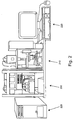
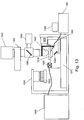
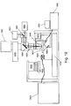
- fluid delivery module or device 100 directs the flow of reagents (e.g., fluorescent nucleotides, buffers, enzymes, cleavage reagents, etc.) to (and through) flowcell 110 and waste valve 120.
- the flowcell comprises clusters of nucleic acid sequences (e.g., of about 200-1000 bases in length) to be sequenced which are optionally attached to the substrate of the flowcell, as well as optionally other components.
- the flowcell can also comprise an array of beads, where each bead optionally contains multiple copies of a single sequence. The preparation of such beads can be performed according to a variety of techniques, for example as described in USPN 6,172,218 or WO04069849 (Bead emulsion nucleic acid amplification).
- the system also comprises temperature station actuator 130 and heater/cooler 135, which can optionally regulate the temperature of conditions of the fluids within the flowcell.
- various embodiments can comprise different configurations of the heating/cooling components.
- the flowcell is monitored, and sequencing is tracked, by camera system 140 (e.g., a CCD camera) which can interact with various filters within filter switching assembly 145, lens objective 142, and focusing laser/focusing laser assembly 150.
- Laser device 160 e.g., an excitation laser within an assembly optionally comprising multiple lasers
- polynucleotide or “nucleic acids” refer to deoxyribonucleic acid (DNA), but where appropriate the skilled artisan will recognize that the systems and devices herein can also be utilized with ribonucleic acid (RNA).
- RNA ribonucleic acid
- the terms should be understood to include, as equivalents, analogs of either DNA or RNA made from nucleotide analogs.
- the terms as used herein also encompasses cDNA, that is complementary, or copy, DNA produced from an RNA template, for example by the action of reverse transcriptase.
- the present invention comprises novel systems and devices for sequencing nucleic acids.
- references herein to a particular nucleic acid sequence may, depending on the context, also refer to nucleic acid molecules which comprise such nucleic acid sequence.
- Sequencing of a target fragment means that a read of the chronological order of bases is established. The bases that are read do not need to be contiguous, although this is preferred, nor does every base on the entire fragment have to be sequenced during the sequencing.
- the current invention utilizes sequencing-by-synthesis (SBS).
- SBS sequencing-by-synthesis
- four fluorescently labeled modified nucleotides are used to sequence dense clusters of amplified DNA (possibly millions of clusters) present on the surface of a substrate (e.g., a flowcell).
- a substrate e.g., a flowcell.
- the flowcells containing the nucleic acid samples for sequencing are placed within the appropriate flowcell holder of the present invention (various embodiments of which are described herein).
- the samples for sequencing can take the form of single molecules, amplified single molecules in the form of clusters, or beads comprising molecules of nucleic acid.
- the nucleic acids are prepared such that they comprise an oligonucleotide primer adjacent to an unknown target sequence.
- one or more differently labeled nucleotides, and DNA polymerase, etc. are flowed into/through the flowcell by the fluid flow subsystem (various embodiments of which are described herein).
- Either a single nucleotide can be added at a time, or the nucleotides used in the sequencing procedure can be specially designed to possess a reversible termination property, thus allowing each cycle of the sequencing reaction to occur simultaneously in the presence of all four labeled nucleotides (A, C, T, G).
- the polymerase is able to select the correct base to incorporate and each sequence is extended by a single base.
- the natural competition between all four alternatives leads to higher accuracy than wherein only one nucleotide is present in the reaction mixture (where most of the sequences are therefore not exposed to the correct nucleotide).
- Sequences where a particular base is repeated one after another e.g., homopolymers
- the heating/cooling components of the system regulate the reaction conditions within the flowcell channels and reagent storage areas/containers (and optionally the camera, optics, and/or other components), while the fluid flow components allow the substrate surface to be exposed to suitable reagents for incorporation (e.g., the appropriate fluorescently labeled nucleotides to be incorporated) while unincorporated reagents are rinsed away.
- suitable reagents for incorporation e.g., the appropriate fluorescently labeled nucleotides to be incorporated
- An optional movable stage upon which the flowcell is placed allows the flowcell to be brought into proper orientation for laser (or other light) excitation of the substrate and optionally moved in relation to a lens objective to allow reading of different areas of the substrate.
- the systems herein can use two excitation lasers coupled through a fiberoptic device to ensure that they illuminate the same area (i.e. that the illuminated areas, or footprints, of the lasers overlap).
- the current invention can contain a shaking, squeezed, or waveplate modulated fiber (mode scrambler) such that the optical intensity from a multimode beam is made uniform over the whole illumination footprint.
- the shape of the fiber may be adjusted, for example to be square or rectangular, such that the shape of the illumination can be matched to the shape of the data collection device (e.g., a CCD with square pixels).
- a single laser excites two fluorophores, one with a narrow emission filter near the wavelength, and one with a wider band emission filter at longer wavelength.
- Such arrangement normalizes the relative intensities of the two dyes (with the same bandwidth filters, the dye further from the laser wavelength would be much weaker).
- the embodiments herein also can comprise a moving stage such that the chemistry (which requires heating and cooling) can happen on the same instrument, but out of the optical train.
- the systems herein also often contain an autofocus system to allow automated imaging of many tiles, and contain a fluidics system for performing on-line fluidic changes.
- the individual components of the system/device can optionally each have its own power source or supply or can optionally all be powered via one source.
- the components herein are often described in isolation or in relation to only one or two other components, that the various components in the embodiments are typically operably and/or functionally connected and work together in the systems/devices herein.
- the systems herein comprise one or more substrates upon which the nucleic acids to be sequenced are bound, attached or associated. See , e.g., WO 9844151 or WO0246456 .
- the substrate is within a channel or other area as part of a "flowcell.”
- the flowcells used in the various embodiments of the invention can comprise millions of individual nucleic acid clusters, e.g., about 2-8 million clusters per channel. Each of such clusters can give read lengths of at least 25 bases for DNA sequencing and 20 bases for gene expression analysis.
- the systems herein can generate a gigabase (one billion bases) of sequence per run (e.g., 5 million nucleic acid clusters per channel, 8 channels per flowcell, 25 bases per polynucleotide).
- the channeled layer can optionally be constructed using standard photolithographic methods, with which those of skill in the art will be familiar.
- One such method which can be used in the current invention involves exposing a 100 ⁇ m layer of silicon and etching away the exposed channel using Deep Reactive Ion Etching or wet etching.
- various flowcells herein can comprise different numbers of channels (e.g., 1 channel, 2 or more channels, 4 or more channels, or 6, 8, 10, 16 or more channels, etc.
- various flowcells can comprise channels of different depths and/or widths (different both between channels in different flowcells and different between channels within the same flowcell).
- the channels formed in the cell in Figure 4B are 100 ⁇ m deep, other embodiments can optionally comprise channels of greater depth (e.g., 500 ⁇ m) or lesser depth (e.g., 50 ⁇ m).
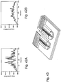
- FIG. 4C and 4D Additional exemplary flowcell designs are shown in Figures 4C and 4D (e.g., a flowcell with "wide" channels, such as channels 440 in Figure 4C , having two channels with 8 inlet and outlet ports (ports 445 - 8 inlet and 8 outlet) to maintain flow uniformity and a center wall, such as wall 450, for added structural support; or a flowcell with offset channels, such as the 16 offset channels (channels 480), etc.).
- the flowcells can be designed to maximize the collection of fluorescence from the illuminated surface and obtain diffraction limited imaging.
- the light comes into the channel through 1000 ⁇ m thick bottom layer 460, which can be made of borosilicate glass, fused silica or other material as described herein, and the emitted light travels through 100 ⁇ m depth of aqueous solution within the channel and 300 ⁇ m depth of "top" layer material 470.
- the thickness of the "top" layer may be less than 300 ⁇ m to prevent spherical aberrations and to image a diffraction limited spot.
- the thickness of the top layer can be around 170 ⁇ m for use with a standard diffraction limited optical system.
- the objective can optionally be custom designed, e.g., as described herein.
- the flowcells can be created from/with a number of possible materials.
- the flowcells can comprise photosensitive glass(es) such as Foturan® (Mikroglas, Mainz, Germany) or Fotoform® (Hoya, Tokyo, Japan) that can be formed and manipulated as necessary.
- Other possible materials can include plastics such as cyclic olefin copolymers (e.g., Topas® (Ticona, Florence, KY) or Zeonor® (Zeon Chemicals, Louisville, KY)) which have excellent optical properties and can withstand elevated temperatures if need be (e.g., up to 100°C).
- the flowcells can comprise a number of different materials within the same cell.
- the base layer, the walls of the channels, and the top/cover layer can optionally be of different materials.
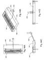
- FIG. 4B shows a flowcell comprised of 3 layers
- other embodiments can comprise 2 layers, e.g., a base layer having channels etched/ablated/formed within it and a top cover layer, etc.
- other embodiments can comprise flowcells having only one layer which comprises the flow channel etched/ablated/otherwise formed within it.
- FIG. 5A gives a schematic diagram of one possible way of patterning a flowcell (e.g., one comprising Foturan®).
- a flowcell e.g., one comprising Foturan®.
- the glass is exposed to UV light at a wavelength between 290 and 330 nm. It can be possible to illuminate material thicknesses of up to 2 mm. An energy density of approximately 20 J/cm 2 is typically sufficient to structurize a 1 mm thick Foturan® plate.
- silver or other doped atoms are coalesced in the illuminated areas (areas 520).
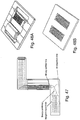
- FIG. 6 panels A through E show an exemplary etching process to construct a sample flowcell as used herein.
- channels 600 (seen in an end view) and through-holes 605 (seen in an end view) are exposed/etched into layer 630.
- Layer 630 is the "top" layer of a two layer flowcell as can be seen in Figure 6E (mated with bottom layer 620).
- the through-holes (where reagents/fluids enter into the flowcell channels) and channels can be etched into layer 630 through a 3-D process such as those available from Invenios (Santa Barbara, CA).
- Top layer 630 can comprise Foturan which, as described, can be UV etched.
- FIG. 6B layer 630 has been masked and light exposed to produce darkened areas 610 within the layer (similar to the masking in Figure 5A , but without the further etching). Such optically opaque areas can be helpful in blocking misdirected light, light scatter, or other nondesirable reflections that could otherwise negatively affect the quality of sequence reading herein.
- a thin (e.g., 100-500 nm) layer of metal such as chrome or nickel is optionally deposited between the layers of the flowcell (e.g., between the top and bottom layers in Figure 6E ) to help block unwanted light scattering.
- Figures 6C and 6D display the mating of bottom layer 620 with channel layer 630 and Figure 6E shows a cut away view of the same.
- the layers of the flowcells are attached to one another in any of a number of different ways.
- the layers can be attached via adhesives, bonding (e.g., heat, chemical, etc.), and/or mechanical methods.
- bonding e.g., heat, chemical, etc.
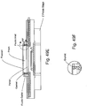
- the reagents, buffers, etc. used in the sequencing of the nucleic acids are regulated and dispensed via a fluid flow subsystem or aspect.
- Figures 7A-C present generalized diagrams of exemplary fluid flow arrangements of the invention, set up in one way push, eight way pull, and one way pull configurations respectively.
- the fluid flow subsystem transports the appropriate reagents (e.g., enzymes, buffers, dyes, nucleotides, etc.) at the appropriate rate and optionally at the appropriate temperature, from reagent storage areas (e.g., bottles, or other storage containers) through the flowcell and optionally to a waste receiving area.
- reagents e.g., enzymes, buffers, dyes, nucleotides, etc.
- various solutions are optionally mixed prior to flow through the flowcell (e.g., a concentrated buffer mixed with a diluent, appropriate nucleotides, etc.).
- a concentrated buffer mixed with a diluent, appropriate nucleotides, etc. Such mixing and regulation is also optionally controlled by the fluid flow aspect of the invention. It is advantageous if the distance between the mixed fluids and the flowcell is minimized in many embodiments. Therefore the pump can be placed after the flowcell and used to pull the reagents into the flowcell ( Figure 7B and 7C ) as opposed to having the pump push the reagents into the flowcell (as in Figure 7A ). Such pull configurations mean that any materials trapped in dead volumes within the pump do not contaminate the flowcell.
- the pump can be a syringe type pump, and can be configured to have one syringe per flow channel to ensure even flow through each channel of the flowcell.
- the pump can be an 8 way pump, if it is desired to use an 8 way flowcell, such as for example a Kloehn 8 way syringe pump (Kloehn, Las Vegas, NV).
- a fluidics diagram of an 8 way pull configuration is shown in figure 7B .
- fluidic reagents are stored in reagent containers 700 (e.g., buffers at room temperature, 5X SSC buffer, enzymology buffer, water, cleavage buffer, etc.) and 710 (e.g., cooled containers for enzymes, enzyme mixes, water, scanning mix, etc.).
- Pump 730 moves the fluids from the reagent containers through reagent valve 740, priming/waste valve 770 and into/through flowcell 760.
- fluidic reagents are stored in reagent containers 702 (e.g., buffers at room temperature similar to those listed above) and 703 (e.g., cooled containers for enzymes, etc. similar to those listed above), linked through reagent valve 701.
- reagent valve 705 e.g., buffers at room temperature similar to those listed above
- 703 e.g., cooled containers for enzymes, etc. similar to those listed above
- reagent valve 701 e.g., buffers at room temperature similar to those listed above
- the reagent valve is linked into flowcell 705 via an optional priming valve (or waste valve) 704, connected to optional priming pump 706.
- the priming pump can optionally draw reagents from the containers up through the tubing so that the reagents are "ready to go" into the flowcell.
- the fluidic configurations can comprise "sipper" tubes or the like that extend into the various reagent containers in order to extract the reagents from the containers.
- Figure 7C shows a single channel pump rather than an 8 channel pump.
- Single channel pump 726 can also act as the optional priming pump, and thus optional priming pump or waste valve 723 can be connected directly to pump 726 through bypass 725.
- the arrangement of components is similar in this embodiment as to that of Figure 7B . Thus it comprises reagent containers 721 and 722, multi-way selector valve 720, flowcell 724, etc.
- the fluid flow itself is optionally driven by any of a number of pump types, (e.g., positive/negative displacement, vacuum, peristaltic, etc.) such as an Encynova® 2-1 Pump (Encynova, Greeley, CO) or a Kloehn® V3 Syringe Pump (Kloehn, Las Vegas, NV).
- pump types e.g., positive/negative displacement, vacuum, peristaltic, etc.
- Encynova® 2-1 Pump Encynova, Greeley, CO
- a Kloehn® V3 Syringe Pump Keloehn, Las Vegas, NV.
- the fluid delivery rate is from about 50 ⁇ L to about 500 ⁇ L/min (e.g., controlled +/- 2 ⁇ L) for the 8 channels.
- the flow can be between 10-100 ⁇ l/min/channel, depending on the process.
- the maximum volume of nucleotide reagents required for sequencing a polynucleotide of 25 bases is about 12 mL.
- the reagents are optionally transported from their storage areas to the flowcell through tubing.
- tubing such as PTFE
- the diameter of the tubing can vary between embodiments (and/or optionally between different reagent storage areas), but can be chosen based on, e.g., the desire to decrease "dead volume" or the amount of fluid left in the lines;
- the size of the tubing can optionally vary from one area of a flow path to another.
- the tube size from a reagent storage area can be of a different diameter than the size of the tube from the pump to the flowcell, etc.
- the fluid flow subsystem of the invention also can control the flow rate of the reagents involved.
- the flow rate is optionally adjustable for each flow path (e.g., some flow paths can proceed at higher flow rates than others; flow rates can optionally be reversed; different channels can receive different reagent flows or different timings of reagent flows, etc.).
- the flow rate can be set in conjunction with the tube diameter for each flow path in order to have the proper volume of reagent, etc in the flowcell at a given time.
- the tubing through which the reagents flow is 0.3 mm ID, 0.5 mm, or 1.0 mm while the flow rate is 480 ⁇ L/min or 120 ⁇ L/min.
- the speed of flow is optionally balanced to optimize the reactions of interest.
- High flow can cause efficient clearing of the lines and minimize the time spent in changing the reagents in a given flowcell volume, but can also cause a higher level of shear flow at the substrate surface and can cause a greater problem with leaks or bubbles.
- a typical flow rate for the introduction of reagents can be 15 ⁇ l/min/channel in some embodiments.
- the system can be further equipped with pressure sensors that automatically detect and report features of the fluidic performance of the system, such as leaks, blockages and flow volumes. Such pressure or flow sensors can be useful in instrument maintenance and troubleshooting.
- the fluidic system can be controlled by the one or more computer component, e.g., as described below. It will be appreciated that the fluid flow configurations in the various embodiments of the invention can vary, e.g., in terms of number of reagent containers, tubing length/diameter/composition, types of selector valves and pumps, etc.
- the systems herein comprise a heating/cooling control component having heating/cooling capabilities, e.g., through Peltier devices, etc.
- the various components herein e.g., the flowcell and its contents
- Such heating/cooling component(s) can control the temperature of the flowcells (and the fluids within them) during the various reactions required in sequencing-by-synthesis.
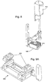
- An exemplary flowcell temperature control system is shown in Figure 8 (in isolation from the other components of the system).
- Peltier fan 800 is shown in relationship to heat sink 810 and Peltier heater 820.
- the flowcell heating/cooling component is optionally positionable and/or movable in relation to the other components of the system (e.g., the flowcell and flowcell holder, etc.).
- the heating/cooling component can be moved into place when needed (e.g., to raise the temperature of the reagents in the flowcell to allow for enzyme activity, etc.) and moved away when not needed.
- the flowcell and flowcell holder can optionally be moved in relation to the heating/cooling component. See Figure 10A and 10B below.
- the temperature control elements control the flowcell temperature, e.g., from about 20°C to about 60°C or any other temperature/temperature ranges as required by the reactions to be done within the systems/devices.
- the temperature of the heating element can be adjusted to control the temperature of the flowcell and the reagents therein. As the flowcell is exposed to a flow of cooled reagents, the temperature of the heating element may be higher than the temperature desired at the surface of the flowcell. For example the heating element may be set to 55°C to obtain a flowcell temperature of 45°C.
- Peltier devices used for temperature control (which can optionally be used in the systems herein). Again, it will be appreciated that while certain heating/cooling devices are recited herein, such should not be construed as necessarily limiting. Thus, in certain embodiments heating/cooling devices other than Peltier devices are optionally comprised within the present invention. In typical embodiments, notwithstanding the type of device, the heating/cooling component is optionally controlled (e.g., in terms of temperature, time at particular temperatures, movement of the component, and/or movement of other devices such as the flowcell holder to the heating/cooling component) by the computer component ( see below ).
- additional heating/cooling elements can optionally regulate the temperature of other components in addition to or alternate to the flowcell.
- heating/cooling components can optionally regulate the temperature of the camera, the reagent reservoirs, which can be cooled, for example to 4°C to prolong the storage life of the reagents during long sequencing runs, the temperature of the atmosphere inside the instrument etc.
- the systems/devices can comprise additional approaches to flowcell configuration, TIR illumination, heating/cooling configurations of the flowcell(s), and in how the flowcells are held/stabilized within the device. While such approaches can optionally be utilized together in certain embodiments, it will be appreciated that they each can be used in any combination, e.g., with each other, with any of the other approaches described herein, etc.
- the flowcells herein can be "bottom flow” flowcells.
- some flowcells can comprise configurations that allow fluid flow that enters from the bottom of the flowcell.
- Such bottom flowcells can be similar in construction and composition as "top flow” flowcells.
- bottom flow flowcells can comprise less fluidic dead volume (and use more of the whole channel length than top flow flowcells, e.g., since the ends of the flowcells are not covered by clamps/manifolds, etc.). See , e.g., Figure 44-49 .
- Bottom flow flowcells can optionally be held to the flowcell holder through vacuum chucking rather than clamps.
- a vacuum can hold the flowcell into the correct position within the device so that proper illumination and imaging can take place.
- some embodiments herein also comprise one or more vacuum creation device to create a vacuum (or partial vacuum, etc.) to hold the flowcell and/or prism to the flowcell holder, XY stage, etc.
- FIG 46 Various examples of flowcell holder manifolds are shown in Figure 46 that can be used with bottom flow flowcells. As can be seen, the fluids flowed into/through/out of the flowcell are directed through various branching tubes within the manifolds to/ from specific channels within the flowcell. Again, such embodiments can optionally not obstruct any (or not substantially any) of the top surface of the flowcell which might interfere with illumination/imaging of the full length of the channels.
- Figure 47 displays an exemplary fluidic valve. Such valve has no moving parts or vibrations and a low dead volume. In such arrangements, each reagent bottle/container can have an open/close valve. After drawing a fluid, air can be injected before closing the reservoir valve thereby forcing an air gap valve between reagents. Cooled reagents can be returned to their reservoirs and all reagents in case of a system shut down. Also, an air injection pump can be added to the push/pull pump (e.g., a kloeh
- top down illumination can be useful when used in conjunction with vacuum chucking (and bottom temperature control below). It can optionally be problematic to illuminate from the bottom (e.g., as in Figure 1 , etc.) in configurations with vacuum chucking and bottom temperature control since such embodiments often utilize the space below the flowcell.
- top down or side illumination comes from above into prism 4401 upon which flowcell 4402 rests (and is optionally held down by vacuum). Such arrangement can also help prevent bowing of the flowcell which presentation can aid in autofocusing and flat field imaging and can aid in configuration with multiple flowcells having simultaneous reading, etc.
- Laser illumination 4400 is also shown entering into the prism in Figure 44 as is mirror 4405 and manifold/fluidic connector 4404.
- Figure 45 shows another approach to thermodynamic control of a flowcell (and the reagents and reactions within it).
- Figure 45 shows an exemplary embodiment of a bottom temperature controlled device.
- the aspect can comprise a water cooled bench that can help assure dimensional stability during read cycles and controlled scan buffer temperature.
- a thermal plate can extend past the prism and flowcell and under the manifolds to optionally help in uniform temperature control. Fluids can optionally be preheated when passing through the inlet manifold.
- RTD temperature feedback can be imbedded in top of the prism to assure that the flowcell is at the desired set temperature and that thermal resistant effects of the prism are minimized.
- FIG. 48 Configurations having multiple flowcells within a flowcell holder are shown in Figure 48 .
- up to four flowcells can be loaded into the holder in Figure 48A (or two double wide flowcells, e.g., having 18-20 channels each).
- Peltiers or other similar devices can be beneath the flowcells and can optionally be water cooled through the holder bench aspect (which can be kept at room temperature optionally).
- Placement and movement of the flowcell is controlled and secured by, e.g., a movable stage upon which the flowcell and flowcell holder (or other substrate) are located.
- a movable stage can optionally allow movement of the flowcell in relation to the laser illumination and lens objective to read the sequencing reactions within the channels.
- the scanning stage or other components can be actively cooled during the scanning cycle to control the temperature of the substrate during the imaging cycles.
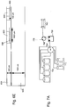
- Figure 9 panels A through D, displays schematic diagrams of an exemplary flowcell holder of the current system.
- Figure 9A shows flowcell holder 900 before a flowcell is placed upon it.
- the holder comprises adjustable clamps 910 (optionally spring loaded) to securely fasten the flowcell to the holder and optionally one or more manifolds (e.g., optionally comprised within the clamps) to fluidically connect the flowcell channels to the rest of the fluidic system.
- a manifold can individually connect each of the channels in parallel.
- a manifold can connect the channels such that they are connected via a single inlet line that is split to flow in parallel to each channel, or can be configured as a "serpentine" configuration to make a single fluid flow.
- Such a manifold can be configured to contain a single 1-8 split, or can comprise a binary splitter wherein each fluid channel is only split into 2, to obtain a split from 1-2-4-8, in order to give a more uniform flow along each of the 8 channels.
- the "exit" manifold from the flowcell can comprise 8 individual ports, each connected to a barrel of an 8 way syringe pump, whilst the "inlet” manifold can contain a single entry tube to reduce the length of tubing needed to fill the flowcell.
- the inlet manifold can contain a 1-8 splitter or a binary 1-2-4-8 splitter for partitioning the flow evenly down each of the 8 channels.
- Figure 9B also shows the presence of adjustable prism 920 that optionally can be raised/lowered to come into contact with the underside of the flowcell.
- the prism is used in conjunction with the lasers in the TIRF activity.
- oil e.g., immersion oil such as that available from Cargille, catalog #19570 or the like
- Figure 9C shows placement of flowcell 930 upon the holder and prism
- Figure 9D shows the flowcell clamped to the flowcell holder with handle/clamp 940 being lowered to help secure the clamps and flowcell.
- the flowcell and flowcell holder can be situated upon a movable stage or platform.
- stage optionally is adjustable along, X, Y, and Z axes. This allows fine scale height and placement adjustment of the flowcell in relation to the lasers, camera, lens optics, etc, and allows the surface of the flowcell to be kept in focus relative to the imaging device.
- the movable stage can optionally allow the flowcell to be moved back and forth between the heating/cooling component and the optic/laser components (i.e., to allow enzymatic reactions when heated and to quantify the outcome of such reactions with the camera/laser components).
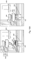
- Figure 10 shows photographs depicting movement of flowcell 1020 and flowcell holder 1010 between the heating/cooling element (left picture) and the camera/laser elements (right picture).
- the x and y components can allow the flowcell to be moved laterally (e.g., by 10s of centimeters), whilst the height can be adjusted (e.g., by 10s of nanometers) vertically to allow focusing of the images.
- the stage also can be simply an XY stage with no vertical setting, and the lens objective can be adjustable in the Z plane to ensure focus is maintained.
- the heating/cooling elements are optionally movable as well, e.g., in order to come into closer proximity with the flowcell, etc.
- Figure 10 left picture heating/cooling device raised
- Figure 10 right picture heating/cooling device lowered onto flowcell.
- FIG 10A shows a photograph of the instrument before and during the heating step.
- Peltier device 1000 (comprised of fan 1001, heat sink 1002 and heater unit 1003) moves in the vertical direction to come into contact with the flowcell 1020 and flowcell holder 1010 mounted on XY stage 1050. Reagents are introduced into the flowcell via tube 1040. The flowcell can move to a position located under camera 1030 for imaging.
- a schematic representation of the device in the imaging location is shown in figure 10B , where the Peltier device 1070 is in the raised position (with fan 1071, heatsink 1072, and heater 1073), flowcell 1085 and stage 1086 are sited next to the fiber optic mount 1090 and below lens objective 1080.
- the fiber optic mount is connected to the Z stage 1075, which also controls the height of lens objective 1080.
- the flowcell is clamped in place onto the flowcell holder by the manifold lever/handle 1095.
- the various components herein e.g., the laser components, heating/cooling components, etc.
- the particular configuration of such framework and/or housing can optionally vary in different embodiments based upon, e.g., the particular components, their size, etc.
- the framework keeps the various components secure and in the proper location and orientation while also optionally aiding in the movement of the components when necessary.
- the framework should be rigid enough to prevent vibrations within the instrument and the various components.
- the mode scrambler can be motion damped and vibrationally isolated from the stage to prevent shaking of the flowcell during imaging.
- Figure 11A shows a schematic displaying an exemplary framework holding the camera (1100), heating/cooling components 1110, ( cf. , Figure 8 ) flowcell and flowcell holder, and movable stage 1120. Additional aspects of framework and mounting that aid in tying together the various components and aspects of the device/system include various alignment and mounting pins/locations can be seen in Figure 11B which shows the bearing slide for laser piece vertical adjustment 1165 and flowcell leveling adjustment component 1175. Other frameworks and housing, including external covers (skins) for the housing can be seen in Figure 12 along with computer monitor 1201.
- the incorporation of specific nucleic acid bases with their accompanying specific fluorescences is tracked via laser excitation and camera observation.
- the illumination is performed using Total Internal Reflection (TIR) comprising a laser component.
- TIR Total Internal Reflection
- TIRF laser Total Internal Reflection Fluorescence
- TIRF laser system Total Internal Reflection Fluorescence
- xenon arc lamps all of which are also included in the current description of TIRF, TIRF laser, TIRF laser system, etc. herein).
- a "TIRF laser” is a laser used with a TIRF system
- a “TIRF laser system” is a TIRF system using a laser, etc.
- the TIRF systems herein should also be understood to include those TIRF systems/instruments comprising non-laser based excitation sources.
- the camera component comprises a CCD camera.
- the laser comprises dual individually modulated 50 mW to 500 mW solid state and/or semiconductor lasers coupled to a TIRF prism, optionally with excitation wavelengths of 532 nm and 660 nm.
- the coupling of the laser into the instrument can be via an optical fiber to help ensure that the footprints of the two lasers are focused on the same area of the substrate (i.e., overlap).
- the area wherein the laser(s) or other excitation source(s) illuminate the sample is typically desired to be spatially flat and uniform.
- the devices/systems herein take advantage of properties of multimode fibers that allow propagation of all optical modes through their cores with near equal amplitude to produce a flat or top-hat profile illumination footprint from the laser on the illuminated substrate surface (e.g., the surface of a flowcell), etc.
- the finite number of modes present in such fibers can constructively and destructively interfere with each other and produce local minima and maxima in the intensity profile of the laser (or other light).
- some embodiments herein produce a substantially uniform footprint by use of dynamic mode scrambling by constantly changing the index of refraction within the illumination beam, e.g. , by modulating the beam with a waveplate, or by shaking, squeezing or compressing one or more areas of a fiber carrying the illumination beam.
- some embodiments of the current invention produce a substantially uniform flat-top output (i.e. , a substantially uniform illumination/excitation footprint from a laser or light source) by dynamically scrambling the modes in an illuminating beam, e.g. , by squeezing/compressing a fiber carrying the beam in one or more area over its length.
- Figures 33 and 34 summarize various embodiments of mode scrambling as described herein. See below.
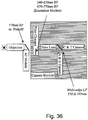
- the devices herein comprise component(s) to produce a "top-hat” illumination, e.g., a uniform or substantially uniform illumination over a particular illumination footprint, as seen in Figure 35 .
- Such embodiments comprise one or more aspects that dynamically change the index of refraction within the medium transmitting the illumination (e.g., a fiber) at one or more nodes.
- a fiber can be squeezed at various locations along its length to induce a continuously changing index of refraction.
- Such squeezing of the fiber e.g., a Step Index Fiber, can be used to spatially/temporally scramble the modes in the fiber to cause sufficient overlap over a desired integration time of the output illumination.
- the fiber can also be shaken, rotated, vibrated or physically deformed in other ways to change the optical path through the fiber.
- the dynamic scrambling of the modes in the fibers allows achievement of spatially uniform illumination over a minimum user defined integration time. This thus prevents interference of propagating modes of monochromatic light in multimode fibers which would produce light and dark patterns in the resulting beam. It is optionally sufficient that these modes disappear over the minimum integration time.
- the relative path lengths of these modes within the illumination beam are rapidly varied by introducing time variable curvature and index variations into the fiber, e.g., by mechanical means.
- dynamic mode scrambling comprises one or more aspects/components used to dynamically change the index of refraction of an illumination beam in order to average out an end illumination footprint. While many existing refractive optical concepts require an input Gaussian beam and existing diffractive optical concepts are often wavelength dependent, the present embodiment does not require a Gaussian beam input and is wavelength independent.
- the devices/systems herein desire a uniformly illuminated field for excitation/measurement of the sequencing reactions, etc.
- the uneven light/dark patterns that result from interference of propagating modes of monochromatic light in a multimode fiber is typically undesirable.
- Averaging of the light output over an illumination footprint (over a period of observation time such as the time captured by a camera during an imaging) to allow integration of the light means that the light/dark patterns "disappear" or are averaged out, and thus the excitation intensity seen by each fluorophore on the surface should be uniform.
- Underlying dynamic mode scrambling is the constant varying of the index of refraction at a point or node of the light beam over time (e.g. , by physically squeezing a fiber over time) which causes the light to be scrambled and take different paths and thus averages out the light output in the illumination footprint.
- the position of interference minima and maxima changes as the index of refraction of the input beam is changed. If the index of refraction is changed at a frequency that is faster than the image acquisition time, then a spatially uniform image can be produced in the timescale of the observation.
- mode scramble which most often refers to randomization of an input mode or modes relative to the output.
- the desired function of the current embodiment is to temporally as well as spatially randomize modes, i.e., producing dynamic scrambling.
- the dynamic mode scrambling of the current embodiment can also be used in conjunction with fibers comprising cores of particular shapes to achieve a beam shape with uniform illumination. For example, squeezing a fiber with a square core will result in a uniformly illuminated square beam.
- the beam can be shaped along a particular axis to make a rectangle, or oval shape, which beam is imaged as square or circular when it hits upon the imaging surface. See Figures 17-18 .
- rectangular beams can be generated from optical fibers, as shown in Figure 34 .
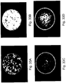
- Figure 35 shows the optical output from a variety of different lasers, fibers, and mode scrambling aspects, etc. During device operation, the ends of the fibers were re-imaged onto a beam profiler.
- Figure 35 shows the effect of dynamic modescrambling (i.e., by manipulation of the fibers at one or more nodes with, e.g., piezo-electric actuators) by comparing the images from different wavelength lasers (e.g., 532 nm and 550 nm) and laser times (solid state and diode) in conjunction with different beam shapers (two versions of rectangles and a circle) by showing the output when the dynamic modescrambling is "on” versus the light output when the modescrambling is "off' for each laser type, etc.
- different wavelength lasers e.g., 532 nm and 550 nm
- laser times solid state and diode
- one embodiment of the device can therefore comprise a dynamic mode scrambler as opposed to static mode scrambler. It is the dynamic variation of index of refraction that causes the modes to overlap over the desired integration time.
- the index of refraction is constantly changed at one or more location (node).
- a fiber transmitting the illumination is constantly squeezed at a point with a changing degree of intensity (e.g., from no squeezing to maximum squeezing and back again).
- the fiber can be temporarily deformed by such squeezing so that its shape changes from a circle to an ellipse to a circle, etc. which, in turn, keeps changing the index of refraction.
- the mode scrambling stops.
- Efficiency of averaging of the illumination output in a footprint depends on length of image capture, the degree of change in index of refraction, the type/strength of the light source, etc. Thus, it is a user controllable variable and should not necessarily be taken as limiting.
- the user can optionally control the degree of scrambling to fine tune the averaging of light output in a footprint.
- the time period over which light output averaging is measured is variable, e.g., it can be the period during which an image is captured of the area illuminated by the light output (e.g., tiles (specific image capture areas) upon the flowcells in certain sequencing embodiments herein).
- the time period of scrambling efficiency is equivalent to or substantially equivalent to the expose period for each image captured by a camera (e.g., the CCD camera in particular sequencing embodiments herein).
- exposure times can vary from embodiment to embodiment, e.g., from less than 1 millisecond to over 1 hour or more depending upon the particular requirements of the embodiment (e.g., at least 1, 5, 10, 25, 50, 100, 250, 500 or more microseconds; at least 1, 5, 10, 25, 50, 100, 250, 500 or more milliseconds; at least 1, 5, 10, 25, 50, 100, 250, 500 or more seconds, etc.).
- the imaging time may be of the order of 50-500 milliseconds per exposure.
- the current dynamic mode scrambler can, no matter the overall system with which it is used, be used with different light sources/types, different beam media, different ways of changing the index of refraction, different numbers of nodes where the index of refraction is changed, etc.
- Dynamic mode scrambling is not limited by the particular light/illumination used.
- lasers of particular wavelength e.g., 532 and/or 660 nm
- other embodiments can use illumination of entirely different wavelength.
- the lasers used with dynamic mode scrambling can be, e.g., visible light lasers, IR lasers, narrow alignment lasers, broad linewidth lasers, etc.
- particular laser wavelengths are mentioned herein, such recitation should not necessarily be taken as limiting.
- other parameters are optionally adjusted to achieve substantially uniform illumination. For example, the number of nodes where the index of refraction is changed and/or the rate of change of the index at such nodes is optionally different for different light sources to achieve the same degree of uniformity of the footprint.
- dynamic mode scrambling is also optionally used with light transmitted through glass, plastic, non-fiber optic lines, air, vacuum, etc.
- dynamic mode scrambling is not limited by the medium in which the light is transmitted.
- differences in the transmission medium can optionally also match with a difference in other aspects of the mode scrambler needed to achieve substantially uniform output.
- the index of refraction is optionally changed/varied by changes in temperature rather than any mechanical change in the transport medium.
- the index of refraction can optionally be varied through a number of ways. For example, as mentioned above, when the light is not transmitted through a cable/fiber, but rather traverses air/vacuum, the index of refraction of the light beam can be varied by changes in temperature. Thus, one or more heaters/coolers can be used to vary the temperature of one or more node of the light beam to change the index of refraction. For beams that travel through a fiber/cable, the physical properties of the fiber can be changed in order to vary the index of refraction. For example, the fiber can be physically bent, shaken, twisted, squeezed, compressed, pulled, or heated/cooled at one or more nodes to change the index of refraction at those points.
- the physical interaction with the fiber can be through actual mechanical manipulation (e.g., through rollers, pinchers, etc. and/or through piezo-electric actuators that squeeze the fiber (e.g., similar to those available from General Photonics (Chino, CA)), etc.).
- any way of varying the index of refraction can be used.
- dynamic mode scrambling can comprise one or more node (i.e., area where the index is varied) on an illumination beam, which node can be fixed/static or movable along the light beam. In a general, but not limiting sense, the greater the number of nodes, the more scrambling occurs. Similarly, for multiple nodes it is typically preferred that the changes in refraction not be synchronized with one another (i.e., it is preferred that the variation in index of refraction be random).
- Figures 33-35 show examples of mode scrambling with various fiber shapes and various light sources. As can be seen from the images, substantially uniform "top hat” illumination is achieved when the dynamic mode scramble is performed using a vibrating or squeezed fiber. The figures also illustrate that images can be shaped through use of shaped-core fibers.
- Figure 33 shows a nonscrambled beam output (A) compared with beam outputs wherein the fiber was shaken, e.g., through use of a MKIII MS from Point Source (Hamble, UK) (B), vibrated, e.g., with an MKIV MS from Point Source (C), or squeezed, e.g., through use of one or more piezo-electric squeezer/compressors (e.g., squeezed over 6 nodes at about 500-600 Htz per node) (D).
- the results shown in Figure 33 were all performed with the same fiber and laser types (e.g., 15 micron step index fiber and a 532 nm solid state laser).
- the panels correspond to: 660 um wavelength diode laser in a rectangular core fiber with no mode scrambling (A) and the same fiber with dynamic mode scrambling (B); a 532 um wavelength solid state laser with no mode scrambling (C) and the same fiber with dynamic mode scrambling (D); a 660 um wavelength diode laser (a second rectangular) with no mode scrambling (E) and the same fiber with dynamic mode scrambling (F); a 532 solid state second rectangular fiber with no scrambling (G) and with dynamic mode scrambling (H); a 660 um diode laser (round) with no mode scrambling (I) and with dynamic mode scrambling (J); a 532 um solid state laser with no mode scrambling (K) and with dynamic mode scrambling (L).
- specific beam shapes such as a square or rectangular laser beams are optionally used.
- Such shaped illumination allows for efficient exposure and tiling over a surface, e.g., comprising a nucleic acid sample, which can result in higher throughput in various devices herein. This can be advantageous in cases where the imaging is performed using a CCD device with square pixels, as the illumination footprint and imaging area can be tiled to prevent illumination, and photobleaching of areas outside the image capture area.
- the laser is coupled into a square or rectangular (or other shaped) core fiber.
- a square or rectangular (or other shaped) core fiber the laser is coupled into a square or rectangular (or other shaped) core fiber.
- all the available laser power is efficiently used for illumination.
- Propagation down a sufficient length of such shaped fiber fills the core efficiently to produce the desired illumination shape.
- the end of this fiber can then be re-imaged onto a sample, e.g., a flowcell substrate.
- such re-imaging of the illumination from the fiber is typically desired to not substantially disturb the top-hat profile and/or beam shape achieved from scrambling and/or beam shaping (or even to distort the beam when it has not been beam shaped or scrambled).
- re-imaging aspects e.g., lens(es), etc.
- Re-imaging can also be chosen to be achromatic (i.e., to be able to function with any wavelength light).
- re-imaging components can also be "pistoned" by slightly moving the re-imaging components to have the illumination land properly on particular areas of the flowcell.
- Illumination uniformity in such embodiments can optionally be controlled by the condition of the beam launched into the shaped fiber coupled with the length of the fiber. Illumination uniformity optionally can be enhanced by dynamically scrambling the modes within the shaped fiber. For example utilizing a device that continuously squeezes the shaped core fiber at various locations. See above.
- the delivered beam dimensions at the sample surface optionally can be manipulated by imaging lenses.
- Figures 34 and 35 show the results of use of a rectangular core optical fiber.
- the end of the fiber was re-imaged onto a beam profiler.
- the image from the beam profile illustrates the desired rectangular beam with uniform illumination in the vertical and horizontal dimensions.
- the dynamic mode scrambling and/or beam shaping systems comprise components to generate and deliver a substantially uniform and wavelength-switchable evanescent beam to the lower surface of a flowcell channel (or other substrate) in an SBS reader instrument.
- these components interface with several other modules/components in the overall SBS system (e.g., the various optics components described above, etc.), and can be controlled/directed through one or more computer component.
- dynamic mode scrambling and beam shaping embodiments include, and are described throughout in terms of their interaction with, nucleic acid sequencing systems (e.g., various sequencing by synthesis configurations as described herein), it will be appreciated by those of skill in the art that such embodiments are also applicable to a wide range of other uses/systems.
- dynamic mode scrambling can be included in myriad systems comprising one or more aspects to dynamically vary the index of refraction of an illumination beam to mix the optic modes of a multimode optical fiber in order to produce a substantially uniform image or output in a desired timeframe (e.g., such as during the image capture time for a camera or the like).
- Dynamic mode scrambling can optionally be utilized with systems such as those tracking fluorescence on a plate or microarray or the like, i.e., uses that do not comprise tracking of sequencing reactions.
- the invention comprises a system for mixing optic modes in a multimode optic fiber through use of waveplates.
- waveplates comprise a light source (e.g. , a laser) which sends light through a multimode optic fiber and also optionally through at least one waveplate and then optionally through a re-imaging lens(es), prism, and onto a substrate (flowcell).
- the waveplates in such systems can comprise "rotating" waveplates. In some embodiments the waveplates actually physically rotate at various rpms, while in other embodiments, such as with liquid crystal waveplates, the plate "rotates” and alters the polarization of the light passing through it by varying voltage across the liquid crystal.
- the waveplate comprises two or more sections of oriented retarders each of which rotates polarization in different directions.
- the light output from the fiber comprises a substantially uniform light output on a surface over a defined time period.
- the light output on the surfaces in various embodiments herein comprises reduced intensity minima and reduced intensity maxima in comparison to the output from a multimode optic fiber that does not comprise one or more rotating waveplates.
- the invention comprises methods for equalizing light output from a multimode optic fiber over a surface in a defined time period by sending light from a light source (e.g ., a laser) through a multimode optic fiber and through one or more rotating waveplates.
- the output on the surface comprises reduced intensity minima and reduced intensity maxima as compared to the output from a multimode optic fiber that does not comprise one or more rotating waveplate.
- the waveplates actually physically rotate at various rpms, while in other embodiments, such as with liquid crystal waveplates, the plate "rotates" and alters the polarization of the light passing through it by varying the voltage across the liquid crystal.
- the waveplate comprises two or more sections of oriented retarders each of which rotates polarization in different directions.
- a "waveplate” refers to an optical device that alters velocity of light rays as they pass through it, thus, creating a phase difference.
- Waveplates are typically comprised of a birefringent crystal.
- Some embodiments can comprise a liquid crystal waveplate.
- the illumination of the sample (the area of which illumination is referred to as the "footprint") is spatially flat and uniform.
- the optic instruments herein exploit the properties of multimode fibers that allow propagation of all optical modes through their core with near equal amplitude which produces a flat or top-hat profile of the footprint.
- the finite number of modes present in such fibers can constructively and destructively interfere with each other, thus producing local minima and maxima in the intensity profile of the laser (or other light).
- Some embodiments produce a uniform footprint by physically shaking the fiber at a timescale shorter than the exposure time of the camera capturing the images, which averages the intensity minima and maxima and produces a uniform flat top footprint.
- This shaking can require an off balance DC motor that rotates and shakes the fiber, which in some instances can cause undesired noise and vibrations that need to be damped to avoid causing imaging problems.
- the shaking can also adversely affect reliability since off balance DC motors have a shorter mean time between failure than balanced motors, and may increase physical wear on the fiber. Because of these factors, mode mixing in a multimode optical fiber without mechanical vibrations and, in some instances without moving parts, by using waveplates can be advantageous in some instances.
- One embodiment of the current invention produces a substantially uniform flat-top beam (i.e., illumination/excitation area or footprint) by mixing the modes of the multimode optical fiber using a rotating ⁇ /2 waveplate (retarding plate).
- the spatial content of the modes depends on the state of polarization of the input light. As polarization is changed, the spatial content is changed. Thus, the position of interference minima and maxima changes as the polarization of the input beam is changed.
- the waveplate is rotated at an angular frequency that is faster than image acquisition time, then a spatially uniform image can be produced in the timescale of the observation.
- the waveplate completes one or more rotation during a certain time period.
- the time period is, e.g., one during which an image is captured of the area illuminated by the light output (e.g., substrate areas of the flowcells in certain sequencing embodiments herein).
- the time period is equivalent to or substantially equivalent to the expose period for each image captured by a camera (e.g., a CCD camera in particular sequencing embodiments herein).
- exposure times can vary from embodiment to embodiment, e.g., from less than 1 msec to over 1 hour or more depending upon the particular requirements of the embodiment (e.g., at least 1, 5, 10, 25, 50, 100, 250, 500 or more ⁇ sec; at least 1, 5, 10, 25, 50, 100, 250, 500 or more msec; at least 1, 5, 10, 25, 50, 100, 250, 500 or more seconds, etc.).
- the exposure time may be 50-500 milliseconds.
- the waveplates can rotate less than or more than a full rotation during the time period, thus, in some embodiments, aliasing can also be included.
- a ⁇ /2 waveplate see waveplate 3800 in Figure 38
- a suitable DC motor or the like operating at a speed fast enough so that a spatially substantially uniform image is produced during the appropriate image capture time.
- Other embodiments comprise modified waveplate(s) which consist of several sections of oriented waveplates or smaller pieces, with the fast axis oriented in different directions ( see waveplate 3900 in Figure 39 ). Since sections rotate the polarization in different ways/amounts, a much faster mixing of the modes can occur and the DC motor optionally does not rotate as fast as in the embodiments with several sections.
- liquid crystals such as, but not limited to, those manufactured by Meadowlark Optics (Frederick, CO)
- the polarization can be rotated by varying the voltage across the device.
- Figure 40 shows a schematic diagram representing an exemplary arrangement of an embodiment of the invention.
- linearly polarized light 4100 (200mW, 532nm) from diode pumped solid state laser (4200) is attenuated by use of several OD filters.
- the intensity of the beam is further controlled by ⁇ /2 waveplate 4900 (e.g., Casix, Fuzhou, Fujian, China) and polarizing beamsplitter cube 4300 (e.g., Thorlabs, Newton, NJ).
- ⁇ /2 waveplate 4900 e.g., Casix, Fuzhou, Fujian, China
- polarizing beamsplitter cube 4300 e.g., Thorlabs, Newton, NJ.
- the entire laser intensity is not needed, thus, in the example described only about 0.1 ⁇ W is used.
- Rotation of the waveplate 4900 allows a precise control of the input laser power while keeping the polarization fixed.
- the beam is then passed through second ⁇ /2 retarding waveplate 4400 and steered by two mirrors 4500 and 4600 into microscope objective 4700 (e.g., Nikon, 20x NA 0.3).
- the microscope objective reduces the beam to the required size to be accepted by multimode optical fiber, 4800 200 ⁇ m core, 0.22NA (e.g., OZ optics, Ottawa, Canada).
- the output end of the fiber in the embodiment shown is placed directly on the chip of CCD camera 4905 (e.g., Cascade 512, Photometrics, Arlington, AZ).
- the camera was operated in frame transfer mode and exposures of 100 ms were adequate to capture the beam profile as explained herein.
- the current invention can comprise different waveplates (e.g. , different in terms of type, placement, arrangement, construction, etc.), different mirrors and beam splitters (e.g. , different in terms of type, location, angle, etc.).
- different embodiments can comprise, e.g. , ⁇ /2 waveplates, ⁇ /4 ⁇ waveplates (e.g., when the input polarization is circular), ⁇ /n waveplates of other specific retardation, etc., and can comprise at least 1 waveplate, at least 2 waveplates, at least 3 waveplates, or at least 5 or more waveplates in various arrangements.
- the waveplates of the invention are not necessarily limited by their construction.
- solid crystal e.g., crystal quartz, or any other appropriate substance
- liquid crystal waveplates are included herein.
- the embodiments can include systems comprising one or more waveplate (typically rotating) that mixes the optic modes of a multimode optical fiber in order to produce a spatially substantially uniform image or output in a desired timeframe (e.g., such as during the image capture time for a camera or the like).
- the current waveplate aspects can optionally be utilized with systems, such as those tracking fluorescence on a plate or microarray or the like, that is not a sequencing reaction.
- the waveplate aspects can also include methods to create a substantially uniform image or output from a multimode optic fiber in a desired timeframe by passing the optic modes of the fiber through one or more waveplate (typically rotating and typically rotating at a speed faster than the image capture time or desired timeframe).
- Figure 41 Various images obtained from exposure of a camera from such exemplary embodiments are shown in Figure 41 . From a single exposure, the prominent regions of bright and dark pixels are evident in Figure 41A . The bright/dark images result from constructive and destructive interference of various modes that are present in the multimode optical fiber. Rotation of the waveplates results in spatial redistribution of the dark and bright regions as shown in the series of images in Figure 41B . In such Figures, each image was taken at a different waveplate setting. As mentioned previously, if the waveplate is rotated faster than the image acquisition time, then the spatial profile is averaged and uniform smoothing of the image results. Such uniform smoothing is comparable to obtaining a large number of images and averaging them.
- Figures 41C and 41D show a single image with its line profile (41C) and an average of 54 images with associated line profile (41D).
- Figure 42 shows the substantial uniformity of the footprint produced by use of the waveplate(s).
- the diffuser can be a holographic diffuser that would superimpose light waves originating at the end of the fiber (if fiber coupled) or at the laser (if no fiber were present) in such a way that the waves superimpose and produce the required beam shape.
- the various mode scrambling aspects herein can optionally be controlled/manipulated through the one or more computer component and are typically coordinated/synched with the light illumination and light detection components (also typically by the computer aspects herein).
- the detector component can comprise a 1 mega pixel CCD-based optical imaging system such as a 1024 x 1024 back thinned CCD camera with 8 ⁇ m pixels, which at 40x magnification can optionally image an area of 0.33 x 0.33 mm per tile using a laser spot size of 0.5 x 0.5 mm (e.g., a square spot, or a circle of 0.5 mm diameter, or an elliptical spot, etc.).
- the cameras can optionally have more or less than 1 million pixels, for example a 4 mega pixel camera can be used.
- the readout rate of the camera should be as fast as possible, for example the transfer rate can be 10 MHz or higher, for example 20 or 30 MHz. More pixels generally mean that a larger area of surface, and therefore more sequencing reactions, can be imaged simultaneously for a single exposure. This has the advantage of requiring fewer stage moves and filter wheel changes, and helps to speed up imaging.
- the CCD camera/TIRF lasers herein are capable of collecting about 6400 images to interrogate 1600 tiles (since images are optionally done in 4 different colors with optionally different filters in place) per cycle.
- certain images optionally can contain between about 5,000 to 50,000 randomly spaced unique nucleic acid clusters (i.e., images upon the flowcell surface).
- the theoretical density of resolvable clusters per unit area (or image) is dependant of the size of the clusters, as shown in Figure 29 which shows a 1Mpix image of the number of detected clusters as a function of total cluster number and minimum cluster area.
- Figure 29 shows a 1Mpix image of the number of detected clusters as a function of total cluster number and minimum cluster area.
- the systems herein can optionally quantify about 45 million features per hour. At a faster imaging rate, and higher cluster density, the imaging rate can be significantly improved.
- the readout can be 1 million clusters per second.
- the instrument can be configured to have more than a single camera.
- the light can be split to simultaneously image two colors onto two cameras, or even four colors onto four cameras. If four cameras are used in parallel, it is thus possible to sequence 1 million bases per second, or 86.4 billion bases per day.
- the excitation blocker e.g., as shown in Figure 36 can comprise a dual notch filter (e.g., 532 and 660 nm).
- a picture of an instrument with two detection cameras 3700 and two fluidics systems 3701 (and two flowcells 3702) is shown in Figure 37 .
- a "tile” herein is functionally equivalent to the image size mapped onto the substrate surface.
- Tiles can be, e.g., 0.33 mm 2 , 0.5 mm 2 , 1 mm 2 , 2 mm 2 etc, although the size of the tile will depend to a large extent on the number and size of pixels on the camera and the desired level of magnification. Also, it will be appreciated that the tile does not have to equal the same size or shape as the illumination footprint from the laser (or other light source), although this can be advantageous if the minimization of photobleaching is desired.
- the camera/laser systems collect fluorescence from 4 different fluorescent dyes (i.e., one for each nucleotide base type added to the flowcell).
- SBS sequencing can be found in applicants' co-pending applications, for example WO04018497 , WO04018493 and US7057026 (nucleotides), WO05024010 and WO06120433 (polymerases), WO05065814 (surface attachment techniques), and WO 9844151 , WO06064199 and WO07010251 (cluster preparation and sequencing).
- Figures 1 and 13-16 show various possible configurations of the cameras and lasers of the present invention, including a backlight design, a TIRF Imaging configuration, a laser focusing configuration, a white-light viewing configuration, and an alternative laser focusing design.
- the white light excitation source is optional, and can be used as well as, or instead of, the excitation lasers.
- Figure 1 shows the backlight design system whilst recording an image in the TIRF imaging configuration.
- the configuration in Figure 1 for the TIRF imaging is optionally a configuration of the backlight design setup shown in Figure 13 .
- one of the two lasers (in laser assembly 160) is used to illuminate the sample (in flowcell 110), and a single one of the four emission filters (in filter switching assembly 145) is selected to record a single emission wavelength and to cut out any stray laser light.
- both focus laser (150) and optional white light lamp (165) do not illuminate the sample as they are either blocked with a shutter or switched off.
- Laser illumination 101 and illumination from the flowcell up through the lens objective and camera 102 are also shown.
- Figure 13 shows all the components of the system in the backlight design but without the specific TIRF imaging configuration. Cf. Figures 1 and 13 .
- Figure 13 shows: fluid delivery module 1300, flowcell 1310, waste valve 1320, temperature actuator 1330, heating/cooling component (e.g., Peltier) 1335, camera (e.g., CCD camera) 1340, lens objective 1342, filter switching assembly 1345, focusing laser assembly 1350, excitation lasers assembly 1360, low watt lamp 1365, precision XY stage 1370, focus (z-axis) device 1375, mirror 1380, "reverse" dichroic 1385, and laser fiber optic 1390.
- heating/cooling component e.g., Peltier
- camera e.g., CCD camera
- Figure 14 shows a similar system as that in Figure 1 , but in the laser focusing configuration where the excitation lasers (in laser assembly 1460) and optional white light 1465 are switched off.
- Focusing laser 1450 is on and shines into the system, hits beam splitter 1485 (e.g., a pick-off mirror 1% beam splitter) which direct a faint beam 1402 down the objective to hit a small spot on the sample (in flowcell 1410).
- the scattered light from the sample returns up objective (1442) through an empty slot in filter wheel switching assembly 1445 and is imaged by CCD camera 1440.
- the position of the spot on the camera is used to ensure the sample is at the right distance from the objective, and therefore the image will be in focus.
- Figure 15 shows the optional white light viewing configuration, where focus laser 1550 and illumination lasers 1560 are off.
- the white light from low watt lamp 1565 goes into the system as beam 1503 and is imaged directly on the camera.
- the numbering of elements, except for beam 1503, etc. follows that of Figures 13 and 14 .
- Figure 16 shows an alternative focus configuration where the system contains second focusing camera 1641, which can be a quadrant detector, PSD, or similar detector to measure the location of the scattered beam reflected from the surface. This configuration allows for focus control concurrent with data collection.
- the focus laser wavelength is optionally longer than the reddest dye emission filter.
- Figures 17-19 show various schematics for beam shape and dimensions for TIRF assays carried out with use of the current systems herein, while Figure 20 displays an optional embodiment of a TIRF prism for use with the systems herein.
- shape e.g., round, square, etc.
- Figure 17 shows the dimensions and geometries of the beam as it emerges from the fiber. In order to illuminate a circle on the substrate, the beam must be projected from the fiber as an ellipse since the beam hits the substrate surface at an angle to the normal.
- Edge view 1700 of circle projected by ellipse 1730 (e.g., an elliptical beam shape required at fiber exit (looking down fiber centerline at 22°).
- the prism face partial outline 1710 is not to scale and the edge view 1720 of ellipse 1730 is shown on the minor axis.
- the beam must be projected onto the surface as a rectangle, as shown in Figure 18 .
- rectangle 1830 is shown.
- a rec-elliptical beam shape is required at fiber exit (looking down fiber centerline at 22°).
- Edge view 1800 of the square projected by rectangle 1830 is also indicated as is prism face partial outline 1830 (not to scale) and edge view 1820 of rec-ellipse 1830 (minor axis).
- the prism is designed to allow the imaging beam to hit the substrate surface at approximately 68° (relative to the normal) to achieve a total internal reflection and generate an evanescent beam that excites the fluorophores on the surface.
- the prism may have a geometry where the angle of the prism to the surface is also 68°, thereby ensuring that the light always hits the prism at 90°.
- the desired geometry of the prism is more fully described in application WO03062897 , and the exemplary size and geometry is shown in Figure 20 , etc.
- Beam shape of the lasers herein is optionally controlled by polishing the multimode fiber output end in order to create, e.g., a square beam. See, e.g., Figures 21.
- Figure 21 shows imaged beam results from such polishing.
- the beam is optionally round.
- the beam properties may be a Gaussian profile with the following properties: a nominal image size of radius 0.17 mm, a maximal spot size of 0.25 mm radius, and 0.32 mm as the point after which there is effectively no laser intensity.
- the beam intensity is greater than 90% maximum intensity at all positions within the nominal image size; 80% maximum intensity at all positions within the maximal spot size, and no greater than 1% of maximal intensity outside of 0.32 radius.
- the intensity at any point does not vary by more than 5% RMS within the timescale of 1s-1h and the variation in the total (integrated) laser power is not more than 3% RMS measured over 24 hours.
- the illumination systems can comprise lamps and/or lasers.
- the systems can contain one or more illumination lasers of different wavelengths.
- the systems herein may contain two lasers of 532 nm and 660 nm, although lasers with other wavelengths may also be used.
- the lasers in the systems herein are actively temperature controlled to 0.1C, have TTL modulation for the 660 nm laser diode with rise time less than 100 ms; have integrated manual shutters for fast modulation of the 532 nm laser, have integrated beam shaping optics to ensure the optimum beam aspect ratio is maintained at the instrument interface to maximize signal to noise ratio, have integrated mode scrambler to reduce ripple on the output of the multi-mode fiber, and have minimal heat generation.
- the shutters and TTL modulation are used to ensure that the illumination is only on the sample surface whilst the camera is recording images. Illumination of fluorophores can cause photobleaching, and therefore exposure of substrates to the laser when not needed is generally minimized, especially before the images are recorded.
- Figures 22A and B give various filter wheel arrangements of certain embodiments for use with various optic configurations.
- the use of a two laser excitation system to detect four fluorophores means that two of the fluorophores are excited away from their maximum absorbtion wavelength, as shown in figure 22B . If the emission filters used in all four channels were the same band width, then the two fluorophores nearest the 532 and 660 nm lasers would be significantly brighter than the two fluorophores excited further from the lasers. However, this factor can be negated by changing the bandwidth of the filters. For example, as shown in figure 22B , in the case of the 532 nm laser, dyes that absorb at for example 530 and 560 nm can both be excited by the 532 laser.
- the use of a narrow filter close to the laser for example a 560/20 that lets light through from 550-570 nm only allows the light from the 532 nm dye through.
- the use of a wider bandpass filter further away from the laser for example a 610/75 that allows light through from 572 nm to 647 nm lets through the light from both fluorophores.
- the intensity of the 532 fluorophore through the 560/20 filter is similar to the intensity of the 560 fluorophore through the 610/75 filter.
- the two fluorophores can be clearly distinguished using a single laser and two emission filters.
- the effect is not wavelength specific, and can be performed using any excitation wavelength. The same effect can therefore be achieved using the red laser.
- Two fluorophores that absorb at 650 nm and 680 nm can be distinguished using a narrow filter close to the laser (for example a 682/22), and a broader filter further away, for example a 700 long pass. Again the intensities of the two dyes through their respective filters is similar, whilst the signal from the 680 dye in the 682/22 filter is much reduced. Both dyes emit into the 700 long pass channel, but the signals can clearly be determined due to the different level of emission in the narrow filter.
- the adaptation of laser wavelengths, fluorophore selections and filter bandwidths can be used to obtain a set of four fluorophores using any number of wavelengths, and the intensities of the emission through each channel can be normalized using the bandwidth of the filters to control how much light is transmitted.
- Figure 23 shows a nominal design for an embodiment of the 30x system ray trace
- Figure 24 shows the 30x imaging performance.
- the imaging performance of the system is dependent on the magnification of the objective lens, and the other lenses in the system.
- a smaller magnification will allow a larger area of the substrate to be imaged, but at a cost of resolution of closely packed clusters and the brightness of each cluster.
- a preferred magnification is optionally between 10X-40X, for example 20X or 30X.
- the objective can be custom designed to allow diffraction limited imaging to be retained when viewing fluorescence objects through a non-standard geometry (for example thicker glass substrates) and hence removing the otherwise present spherical aberration.
- the objective lens may be connected to the detector via a further tube lens.
- the systems herein can comprise components to aid in proper focusing of imaging clusters.
- an autofocus laser beam shines down to a sample through an objective lens, reflects from the flowcell surface, goes back to the lens and then to the camera, thus creating a spot on the image.
- the spot centroids align around a straight line on the image (calibration curve). Displacement "dr" along this calibration line is proportional to the change "d(z-zf)" in the distance between the objective and the focal plane.
- the software establishes the orientation of the calibration line (its slope) and the "sensitivity": dz/dr (nm/pixel). This is accomplished by taking 21 images with the step of 1000 nm in z-direction around focus position which is established visually.
- the software also can require the x, y pixel coordinates of the spot when the sample is in focus: x f , y f . this is determined from the first (central) focus image from the set of the 21 calibration images.
- the devices herein optionally comprise an auto focus function objective achieving 100 nm resolution mounted with up to 50 mm Z axis motion.
- the objective lens can optionally move vertically in relation to the substrate, and the illumination laser can be coupled to the Z axis motion such that the illumination inputs also move in relation to the substrate.
- an autofocus beam is optionally sent along the edge of the microscope objective lens (optionally as far off-axis as possible in order to correspond to maximum sensitivity).
- the autofocus beam can come from the illumination lasers, or from a separate source that is optionally a different wavelength than the illumination laser, for example 488 nm, 630 nm or an infra-red laser of 700 nm or redder.
- the reflected beam is then optionally monitored by either a quad cell or by leakage through a dichroic beam splitter onto the fluorescence imaging camera.
- the lens and camera are optionally the same as that used in the instrument (e.g., 20X lens). Similar autofocus systems which are optionally included within the current systems and devices have been previously described, for example in WO03060589 .
- the reflected monitoring beam also optionally moves laterally (i.e. dotted line is the in focus plane while the solid line represents an out of focus plane which gives rise to a lateral shift in the detected beam in Figure 25 ).
- the dichroic mirror chosen in such embodiments is usually one that reflects the autofocus beam.
- the small leakage that is actually transmitted (c.a. ⁇ 5%) is more than adequate to observe on a CCD camera with no emission filter in the emission path.
- Figure 26 shows sample photographs of both out of focus and in focus images where the spot is seen on the imaging camera. The lower images show the detected autofocus spot on the imaging camera. The spot can also be seen on a separate detector, as shown in Figure 27 .
- the computer component optionally comprises an autofocus algorithm.
- Such algorithms optionally aid in the determination of the correct focus (e.g., by monitoring the above measurements and adjusting accordingly).
- the autofocus spot can be made to move in a 1D manner e.g. in just the y-direction rather than x & y, thus simplifying the procedure.
- the focus position of the objective lens is assumed to move in the z-direction.
- the first step in some embodiments of the autofocus analysis is a "Setup Response function" wherein positions of the autofocus spot (y 1 , y 2 ....y n ) are measured on the imaging camera for several positions of the objective lens (z 1 ,z 2 ...z n ). Typically 5 positions are adequate. Shown in Figure 27 is an analysis with just 3 positions. The movement of the reflected spot is shown as imaged on a fluorescence camera in the lower panels. For each Z plane there is an associated y-position of the reflected spot (centroid) on the camera.
- the next step in such embodiments of the autofocus analysis comprises "Calculate newC (for out of focus position)" wherein for each position c is constant, i.e. doesn't change for an out of focus or in focus position. However, it does change for different positions.
- Another aspect in auto focus components of the invention comprises laser pointing stability requirements.
- the pixel size is roughly 0.3 ⁇ m.
- the focal length of that lens is 10 mm.
- the error angle that corresponds to 1 pixel is approximately 30 ⁇ rad.
- Some embodiments of the system have their auto focus set for a sensitivity of about 4 pixel shift of the auto focus laser spot per micron of z motion.
- the first step in some embodiments of the focus tracking procedure is to obtain the image of the autofocus laser spot on an imaging device, which may be the imaging camera. Data from this primary spot is extracted in two passes-a first coarse pass that determines the approximate position and size of the spot, followed by a second fine pass that determines the spot boundaries correctly before determining the COL (Center of Light) and other spot features.
- the first pass analysis can be performed in 5 steps: (1) the 16-bit image is converted to an 8-bit image, with maximum of the image set to 255, minimum of image set to 0, and all other grayscale values linearly in between; (2)
- the Picture Quality of the image is computed. Picture quality is defined as the average of the normalized autocorrelation of the image with itself with shifts of unit pixel to the left and unit pixel down.
- this image is thresholded at 128. Anything above this value will be regarded as foreground, while anything below this level will be seen as background.
- region-grow to find all 8-connected foreground components; (4) Of all these candidate foreground components, the component with the highest average brightness is chosen as "the" component specifying the position and approximate size of the primary spot; and (5) The bounding box of this component is computed.
- the second (fine) pass analysis is performed in three steps: (1) The subimage corresponding to twice the area of the bounding box is cut out from the 8-bit grayscale image. This makes the population of foreground and background pixels approximately equal, thereby making it easier for standard image histogram based thresholding techniques to work reliably; (2) The histogram of the subimage is computed and the "best" grayscale threshold that separates foreground from background is determined. The threshold used is called “Otsu's Threshold” ( see IEEE Trans. Systems, Man, and Cybernetics, vol. 9, pp. 62-66, 1979 , or Computer and Robot Vision, volume 1. Addison-Wesley, 1992 .); and (3) The image is thresholded at the Otsu threshold and the 8-connected foreground component is determined. This is the primary spot blob on which every subsequent feature extraction is carried out.
- An additional pass can be carried out to extract the position of the secondary spot.
- Four steps can be done to carry out such additional pass: (1) The 8-bit image (from the First Pass) is thresholded at a low threshold of 16; (2) It should be noted that at this lower threshold, the number of pixels (area) of the primary spot component increases. This area of the primary spot is recorded and is used to determine how tight or diffuse it is; (3) The component (of sufficient size) closest in distance to the primary spot component is identified as the secondary spot; (4) The geometric centroid of the secondary spot is recorded.
- the list of features calculated for the primary spot blob includes Area which is a measure of how diffuse (non-tight) the primary blob is. This is set to the area (count in pixels) of the primary blob at the low threshold divided by its area at the Otsu threshold.
- Other features calculated include Volume (the average brightness, above threshold ⁇ Area of the primary blob); Average Brightness, which is the sum of the gray values of the pixels in the blob divided by its area; and Maximum Brightness: Maximum gray value.
- the extracted data can then be used to calibrate the Z focus, and thereby determine how much to move the objective in order for the image to be in focus.
- the overview of the calibration procedure is as follows: (1) Calibration for the Z focus is done (with user help) at the beginning of every run; (2) At the beginning of calibration the user makes sure that the image is in focus, i.e. at the focal plane. He/she thus sets the Z focus point z F ; (3) This embodiment of autofocus relies on user input and the coordinates of the autofocus laser spot on an image; (4) As z is changed during the Calibration process, the spot moves in linear proportion to the change of z along a line.
- the calibration algorithmic procedure begins with a sequence of Center of Lights ( x , y ) extracted from the sequence of autofocus spot images acquired as z is changed. Ideally these points when graphed should all fall perfectly on a straight line, shown below.
- Input A sequence of Center of Lights ( x , y ) extracted from the sequence of autofocus spot images acquired as z is changed.
- Output The PCA Regression Coefficients, i.e. transformation to the (R, Q) coordinate space. Description: Perform Principal Component Analysis and save the coefficients. Also, given the centroid position of the secondary spot, determine whether it is to the left or the right of the primary spot along the principal axis.
- An outlier detection scheme is used to warn the presence of a bubble or to flag a filter wheel problem.
- Input The q coordinates obtained from the PCA Regression, along with the spot features for every spot in the calibration sequence are used to train a classifier to detect outlier spots.
- Output Internal settings of the Outlier Detector. Description: For every feature ⁇ , the mean ⁇ ⁇ and standard deviation ⁇ ⁇ is calculated from the spots used during calibration. The general idea is that a spot would be declared an outlier spot if the feature ⁇ is outside the bounds of ⁇ ⁇ ⁇ 3 ⁇ ⁇ . However in the current implementation only the Volume and Area features are used in this way-in fact, only the upper bounds are currently used for these two features. Special bounds are set to override these values for both the picture quality and the q residue feature. For the picture quality, a lower bound is used. For the residue, an upper bound is used.
- a run starts from an image of the autofocus laser spot and uses the coefficients of the transformations learned during the calibration process to make the best estimate for the z displacement required to move to focus.
- Input An image of the autofocus laser spot.
- Output (z - z F ) to move to focus. Also provides recommendation of whether or not to move based upon outlier detection. Description: Derives best estimate of (z - z F ) using: 1. The spot extraction algorithm. , 2. The PCA based XY Regression coefficients. 3. The Linear RZ Regression coefficients.
- the outlier detection scheme allows a move only in the case of a large q residual. In case of low picture quality, high volume, or high area, it recommends non-movement.
- the various components of the present system are coupled to an appropriately programmed processor or computer that functions to instruct the operation of these instruments in accordance with preprogrammed or user input instructions, receive data and information from these instruments, and interpret, manipulate and report this information to the user.
- the computer is typically appropriately coupled to these instruments/components (e.g., including an analog to digital or digital to analog converter as needed).
- the computer optionally includes appropriate software for receiving user instructions, either in the form of user input into set parameter fields, e.g., in a GUI, or in the form of preprogrammed instructions, e.g., preprogrammed for a variety of different specific operations (e.g., auto focusing, SBS sequencing, etc.).
- the software then converts these instructions to appropriate language for instructing the correct operation to carry out the desired operation (e.g., of fluid direction and transport, autofocusing, etc.).
- the computer is optionally used to direct a fluid flow component to control fluid flow, e.g., through a variety of tubing.
- the fluid flow component optionally directs the movement of the appropriate buffers, nucleotides, enzymes, etc., into and through the flowcell.
- the computer also optionally receives the data from the one or more sensors/detectors included within the system, and interprets the data, either provides it in a user understood format, or uses that data to initiate further controller instructions, in accordance with the programming, e.g., such as in monitoring and control of flow rates, temperatures, and the like.
- the computer typically includes software for the monitoring and control of materials in the flowcells. Additionally the software is optionally used to control excitation of the fluorescent labels and monitoring of the resulting emissions.
- the computer also typically provides instructions, e.g., to the heating/cooling component and autofocus system, etc.
- Any controller or computer optionally includes a monitor, which is often a cathode ray tube ("CRT") display, a flat panel display (e.g., active matrix liquid crystal display, liquid crystal display), or the like.
- a monitor which is often a cathode ray tube ("CRT") display, a flat panel display (e.g., active matrix liquid crystal display, liquid crystal display), or the like.
- Data produced from the current systems, e.g., nucleic acid sequence results is optionally displayed in electronic form on the monitor.
- the data e.g., light emission profiles from the nucleic acid arrays, or other data, gathered from the system can be outputted in printed form.
- the data whether in printed form or electronic form (e.g., as displayed on a monitor), can be in various or multiple formats, e.g., curves, histograms, numeric series, tables, graphs and the like.
- Computer circuitry is often placed in a box which includes, e.g., numerous integrated circuit chips, such as a microprocessor, memory, interface circuits.
- the box also optionally includes a hard disk drive, a floppy disk drive, a high capacity removable drive such as a writeable CD-ROM, and other common peripheral elements.
- Inputting devices such as a keyboard or mouse optionally provide for input from a user and for user selection of sequences to be compared or otherwise manipulated in the relevant computer system.
- the SBS systems herein comprise CCD/TIRF laser based excitation and imaging subsystems which can image millions of nucleic acid clusters per sample (typically within a flowcell) and which can detect each of four fluorescent dyes (one for each of the four bases).
- the SBS chemistry components e.g., nucleotides, WO04018497 , WO04018493 and US7057026 , polymerases WO05024010 and WO06120433 , surface attachment techniques, WO05065814 , cluster preparation and sequencing, WO 9844151 , WO06064199 and WO07010251 , are compatible with the channeled flowcell components herein, etc.
- the computer or data analysis system aspects of the system are optionally capable of processing thousands of images per hour into sequence information
- genomic DNA is randomly fragmented, end capped with known sequences, and covalently attached to a substrate (such as the channel in a flowcell), e.g., by hybridization to a covalent primer.
- a substrate such as the channel in a flowcell
- SBS analysis e.g., using the systems and devices herein
- SBS analysis can generate a series of images of the clusters, which can then be processed to read the sequence of the nucleic acids in each cluster which can then be aligned against a reference sequence to determine sequence differences, a larger overall sequence, or the like.
- Algorithms for the alignment of short reads of nucleic acids are described in WO05068089 .
- each sequencing cycle will include a round of incorporation onto the growing nucleic acid chain. Such cycle is typically done by an addition of all four dNTPs, each modified so that each base is identifiable by a unique fluorophore. Additionally, the triphosphates are modified at the 3' position so that extension is controlled and not more than a single base can be added to each molecule in each cycle.
- the generic concept of performing clusters amplified from a single template molecule on a random array, and the subsequent sequencing of said array is shown in Figures 30-32 , which schematically illustrate various aspects of sequencing procedures and methods carried out by the systems herein.
- Figure 30 shows the basic outlines of nucleic acid cluster creation and sequencing while Figure 31 compares nucleic acid density between an array (on left) and on a nucleic acid cluster substrate such as those capable of use with the systems/devices of the invention (on right).
- Figure 32 gives a cartoon outlining the SBS sequencing procedure, e.g., as done by embodiments of the invention.
- the channels of the flowcell are washed out by the fluid flow subsystem in order to remove any unincorporated nucleosides and enzyme.
- a read step is performed by the system, whereby the identity of the individual labels (read as a group in each cluster) incorporated in the incorporation step is recorded using optical microscopy and the corresponding base incorporated is noted.
- the sequencing system can read the four different fluorophores using two lasers at distinct wavelengths via total internal reflection microscopy (TIRF) and four distinct emission filters at different parts of the spectrum. The images are recorded onto a CCD camera and reported into the attached computer module.
- TIRF total internal reflection microscopy
- a deprotection step removes the labeling moiety and block from the surface bound DNA.
- the deprotection allows repetition of the above incorporation and reading steps until sufficient cycles of information are obtained to uniquely place the sequence of each nucleic acid cluster (present on the flowcell) in its genomic context. For example, in the case of the human genome this will be >16 cycles, e.g., about 25-50 cycles.
- the images can be stored off line, or processed in real time such that the individual bases are read during the sequencing process. Processing the images provides a database of a sequence read from every cluster, where each cluster is derived from a random position somewhere in entire sample (e.g., a genome).
- a database of millions of sequence reads covering every part of the genome is typically constructed.
- Such database can be, e.g., compared with a database of every sequence derived from a reference sequence, etc.
- image analysis, sequence determination, and/or sequence alignment are optionally performed "off-line" after the fluorescent images are captured.
- Such procedures are also optionally performed by a computer separate from the one present in the current systems.
- the components can comprise: illumination lasers (used to excite the fluorophores in the sequencing reactions) of 532 and 660 nm each with 75 mW power (or optionally greater) that project as 0.5 mm circle on the bottom of the channel in a flowcell; a TIR prism of glass (68 deg or 71 deg); a glass flowcell with channels of 1x61 mm area having 8 channels that are 100 ⁇ m deep (39x1 mm usable or accessible for viewing); an objective lens in the camera component comprising a Nikon Plan Apo 20x, 0.75NA (corrected for glass thickness); emission filters comprising Bandpass filters of 557 ⁇ 11 nm, 615 ⁇ 40 nm, 684 ⁇ 11 nm, and 740 ⁇ 50nm (or optionally filters as shown, or similar to those shown, in Figure 22 );
- Such 1 Megapixel embodiments can illuminate a 0.5 mm circle and detect a 0.35 mm square inside it.
- the flowcell in such embodiments can have a total of 156 non-overlapping tiles in a channel or higher.
- the clusters can be on the order of 1 ⁇ m.
- the NA of the microscope can optionally give a PSF of approximately 0.6 ⁇ m at 700 nm.
- a "typical" cluster gets an apparent diameter of approximately 1.2 ⁇ m.
- 1 pixel represents approximately 0.35 ⁇ m, so a typical cluster would have about 3.5 pixels diameter.
- the area of a cluster is the about 9.25 pixels. Poisson distribution of 10 area pixel objects on 1 Mpixel CCD shows maximum of about 38,000 objects will be non-overlapping as shown in Figure 29.
- Figure 29 gives an example of information throughput from an exemplary configuration of a system of the invention.
- the number of detected clusters is a function of the total cluster number and the minimum cluster area.
- the resolution limit (using Rayleigh criterion) is about 0.6 ⁇ m and clusters are about 1 ⁇ m for an apparent size of about 1.2 ⁇ m. Pixels map to about 0.35 ⁇ m in image plane so a cluster is about 3.5 pixels across and about 10 pixels in area. For randomly distributed clusters, the maximum number of unconfused clusters in the 1 MPix camera will be about 38,000 in a 0.35 mm square tile.
- "b" flowcells accommodate 150 non-overlapping illumination tiles per channel for a total of 1200 tiles per flowcell. This is 45.6 M Bases per cycle and about 1 GBase in a 25 cycle run. Overlapping the illumination and closely packing the tiles means that 200 tiles can be imaged per channel, and therefore 1600 per flowcell.
- the laser wavelengths are: green laser wavelength 532 nm; green laser power optionally 75 mW; red laser wavelength 660 nm; red laser power optionally 75 mW; projected TIRF beam diameter 0.5 mm; and allowed variation across beam 20%.
- the system of the invention can comprise: illumination lasers of 532 and 660 nm, each with 500 mW power (ideally projected as 0.5 mm square), a TIR prism of glass (68 deg); a glass flowcell having 8 channels 100 ⁇ m deep and of 1 x 61 mm in area with a 50 mm usable; an objective lens comprising a Nikon Plan Fluor 40x, 0.6 NA adjustable collar, or custom 40x, 0.75 NA corrected for an SBS flowcell; emission filters comprising Bandpass filters of 557 ⁇ 11 nm, 615 ⁇ 40 nm, 684 ⁇ 11 nm, and 740 ⁇ 50 nm; image optics comprising an 150 mm achromatic doublet for system magnification of 30x; and a digital CCD camera comprising a Photometrics CoolSNAP K4, 2048 by 2048 pixels, 4Mpix camera, 7.4 ⁇ m pixel size, 20 MHz readout.
- illumination lasers of 532 and 660 nm each with 500 mW power (
- a 0.5 mm square is uniformly illuminated and that the same 0.5 mm square is detected (2048 x 7.4 / 30000).
- the clusters on the flowcells herein can be as small as 0.5 ⁇ m.
- PSF at 700 nm is approximately 0.7 ⁇ m.
- Clusters thus appear as 0.86 ⁇ m where 1 pixel represents 0.25 ⁇ m.
- a typical cluster therefore is 3.5 pixels and the area of a cluster is 9.25 pixels.
- 4 Mpixel CCD gives a maximum of about 135,000 detectable non-overlapping clusters per tile.
- the illumination footprint is four times larger, meaning a 4 time increase in laser powers is needed to obtain the same level of signal in the same exposure time. To minimize exposure times, the laser power can be increased further.
- the instrument can be configured to perform multiple chemistry steps with multiple fluidics systems coupled to a single optical system. In the single chemistry system, the optical system is not imaging whilst the chemistry steps are occurring. If the chemistry and imaging parts of the cycle take similar lengths, then for 50% of the time, the instrument is not recording data.
- TDI Time Delay Integration
- the system as described is shown with the illumination from underneath, and the objective on top, the system as shown can be inverted to illuminate from the top, and have the detection system underneath. See above.
- the heating and illumination can be carried out from either face of the substrate, so that bottom side heating and top side illumination are also within the scope of the invention.
- the operation of systems within the scope of the inventions are further described in the following general methods.
- the solid supports used for attachment of nucleic acid to be sequenced are optionally 8-channel glass chips such as those provided by Silex Microsystems (Sweden). However, the experimental conditions and procedures are readily applicable to other solid supports as well. In some embodiments chips were washed as follows: neat Decon for 30 min, milliQ H2O for 30 min, NaOH IN for 15 min, milliQ H2O for 30 min, HCl 0.1N for 15 min, milliQ H2O for 30 min. The Polymer solution preparation entailed: For 10 ml of 2% polymerization mix.
- the 10 ml solution of acrylamide was first degassed with argon for 15 min.
- the solutions of BRAPA, TEMED and potassium persulfate were successively added to the acrylamide solution.
- the mixture was then quickly vortexed and immediately used. Polymerization was then carried out for 1h 30 at RT. Afterwards the channels were washed with milliQ H2O for 30 min and filled with 0.1 M potassium phosphate buffer for storage until required.
- N-Boc-1,5-diaminopentane toluene sulfonic acid was obtained from Novabiochem.
- the bromoacetyl chloride and acryloyl chloride were obtained from Fluka. All other reagents were Aldrich products.
- a peristaltic pump Ismatec IPC equipped with tubing Ismatec Ref 070534-051 (orange/yellow, 0.51 mm internal diameter) is optionally used.
- the pump is run in the forward direction (pulling fluids).
- a waste dish is installed to collect used solution at the outlet of the peristaltic pump tubing.
- the different solutions used are dispensed into 8 tube microtube strips, using 1 tube per chip inlet tubing, in order to monitor the correct pumping of the solutions in each channel. The volume required per channel is specified for each step.
- the Silex chip is mounted on top of an MJ-Research thermocycler.
- the chip sits on top of a custom made copper block, which is attached to the flat heating block of the thermocycler.
- the chip is covered with a small Perspex block and is held in place by adhesive tape. Both pump and thermocycler are controlled by computer run scripts, which prompt the user to change solutions between each step.
- An SFA coated chip is placed onto a modified MJ-Research thermocycler and attached to a peristaltic pump as described above. Grafting mix consisting of 0.5 ⁇ M of a forward primer and 0.5 ⁇ M of a reverse primer in 10 mM phosphate buffer (pH 7.0) is pumped into the channels of the chip at a flow rate of 60 ⁇ l/min for 75 s at 20 °C. The thermocycler is then heated up to 51.6 °C, and the chip is incubated at this temperature for 1 hour.
- the grafting mix undergoes 18 cycles of pumping: grafting mix is pumped in at 15 ⁇ l/min for 20 s, then the solution is pumped back and forth (5 s forward at 15 ⁇ l/min, then 5 s backward at 15 ⁇ l/min) for 180 s. After 18 cycles of pumping, the chip is washed by pumping in 5xSSC/5mM EDTA at 15 ⁇ l/min for 300 s at 51.6 °C. The thermocycler is then cooled to 20 °C.
- the DNA templates to be hybridized to the grafted chip are diluted to the required concentration (currently 0.5-2pM) in 5xSSC/0.1% Tween.
- the diluted DNA is heated on a heating block at 100 °C for 5 min to denature the double stranded DNA into single strands suitable for hybridization.
- the DNA is then immediately snap-chilled in an ice/water bath for 3 min.
- the tubes containing the DNA are briefly spun in a centrifuge to collect any condensation, and then transferred to a pre-chilled 8-tube strip and used immediately.
- the hybridized template molecules are amplified by a bridging polymerase chain reaction using the grafted primers and a thermostable polymerase.
- Amplification buffer consisting of 10 mM Tris (pH 9.0), 50 mM KCl, 1.5 mM MgCl2, 1 M betaine and 1.3% DMSO is pumped into the chip at 15 ⁇ l/min for 200 s at 40.2 °C.
- amplification mix of the above buffer supplemented with 200 ⁇ M dNTPs and 25 U/ml Taq polymerase is pumped in at 60 ⁇ l/min for 75 s at 40.2 °C.
- the thermocycler is then heated to 74 °C and held at this temperature for 90 s.
- thermocycler then carries out 50 cycles of amplification by heating to 98.5 °C for 45 s (denaturation of bridged strands), 58 °C for 90 s (annealing of strands to surface primers) and 74 °C for 90 s (primer extension).
- 98.5 °C fresh PCR mix is pumped into the channels of the chip at 15 ⁇ l/min for 10 s.
- this step also removes DNA strands and primers which have become detached from the surface and which could lead to contamination between clusters.
- thermocycling the chip is cooled to 20 °C.
- the chip is then washed by pumping in 0.3xSSC/0.1% Tween at 15 ⁇ l/min for 300 s at 74 °C.
- the thermocycler is then cooled to 20 °C.
- Linearization mix consisting of 0.1 M sodium periodate and 0.1 M ethanolamine is pumped into the chip at 15 ⁇ l/min for 1 hr at 20 °C. The chip is then washed by pumping in water at 15 ⁇ l/min for 300 s at 20 °C.
- This step uses Terminal Transferase to incorporate a dideoxynucleotide onto the free 3' OH ends of DNA strands (both grafted primers and amplified cluster molecules).
- Blocking buffer consisting of 50 mM potassium acetate, 20 mM Tris-acetate, 10 mM magnesium acetate, 1 mM dithiothreitol (pH 7.9) and 250 ⁇ M CoCl2 is pumped into the chip at 15 ⁇ l/min for 200 s at 20 °C.
- Blocking Mix of the above buffer supplemented with 2.4 ⁇ M ddNTPs and 250 U/ml Terminal transferase is pumped in at 15 ⁇ l/min for 300 s at 37.7 °C.
- the thermocycler is held at 37.7 °C for 30 min, during which time Blocking Mix is pumped into the chip at 15 ⁇ l/min for 20 s every 3 min.
- the chip is then washed by pumping in 0.3xSSC/0.1 % Tween at 15 ⁇ l/min for 300 s at 20 °C.
- This step uses NaOH to denature and wash away one of the strands of the amplified, linearized and blocked clusters. After a wash to remove the NaOH, the sequencing primer is then hybridized onto the single strands left on the surface.
- the double stranded DNA in the clusters is denatured by pumping in 0.1N NaOH at 15 ⁇ l/min for 300 s at 20 °C.
- the chip is then washed by pumping in TE (10 mM Tris pH 8.0, 1 mM EDTA) at 15 ⁇ l/min for 300 s at 20 °C.
- Sequencing primer is diluted to 0.5 ⁇ M in 5xSSC/0.1% Tween, and pumped into the channels at 15 ⁇ l/min for 300 s at 20 °C.
- the thermocycler is then heated up to 60 °C and held at this temperature for 15 min.
- the thermocycler is then cooled to 40.2 °C and the chip is washed by pumping in 0.3xSSC/0.1% Tween at 15 ⁇ l/min for 300 s.
- the clusters are now ready for 1st cycle sequencing enzymology, e.g., with the systems and devices of the current invention.
- the DNA sequence used in this process was a single monotemplate sequence of 400 bases, with ends complimentary to the grafted primers.
- the duplex DNA was denatured as described above.
- the primers are typically 5'-phosphorothioate oligonucleotides incorporating any specific sequences or modifications required for cleavage. Their sequences and suppliers vary according to the experiment they are to be used for, and in this case were complementary to the 5'-ends of the template duplex.
- the amplified clusters contained a diol linkage in one of the grafted primers.
- Diol linkages can be introduced by including a suitable linkage into one of the primers used for solid-phase amplification.
- Suitable primers including any desired template-specific sequence can be manufactured by standard automated DNA synthesis techniques using components available from commercial suppliers (e.g. Fidelity Systems Inc., ATD).
- a cleavable diol-containing primer would typically have the following structure: 5' -phosphorothioate-arm 26-diol22A-sequence-3'OH.
- sequence represents a sequence of nucleotides capable of hybridizing to the template to be amplified.
- the structures of the arm26 and diol22A components are as follows: Products containing such diol linkages can be cleaved using periodate as described above, and the resulting single stranded polynucleotides hybridized as described above.
- a mutant 9°N polymerase enzyme (an exo- variant including the triple mutation L408Y/Y409A/P410V and C223S) was used for the nucleotide incorporation steps.
- Incorporation mix Incorporation buffer (50 mM Tris-HCl pH 8.0, 6 mM MgSO4, 1 mM EDTA, 0.05% (v/v) Tween -20, 50mM NaCl) plus 110nM YAV exo-C223S, and 1 ⁇ M each of the four labeled modified nucleotides, was applied to the clustered templates, and heated to 45°C.
- Incorporation buffer 50 mM Tris-HCl pH 8.0, 6 mM MgSO4, 1 mM EDTA, 0.05% (v/v) Tween -20, 50mM NaCl
- 110nM YAV exo-C223S 1 ⁇ M each of the four labeled modified nucleotides
- Templates were maintained at 45°C for 30min, cooled to 20°C and washed with Incorporation buffer, then with 5x SSC/0.05% Tween 20. Templates were then exposed to Imaging buffer(100mM Tris pH7.0, 30mM NaCl, 0.05% Tween 20, 50mM sodium ascorbate, freshly dissolved). Templates were scanned in 4 colors at RT.
- the flowcell can be sequenced in a fully automated way, with the first incorporation being performed on this instrument, as described below:
- the templates can be exposed to the sequencing cycles described below : first base incorporation, imaging then alternating cleavage, imaging and incorporation, imaging steps for as many sequencing cycles as required.
- Each tile of each the chip for the non-fully automated process above was recorded in each of the four colors corresponding to the labeled nucleotides.
- the images were analyzed to pick the brightest color for each cluster, and this image intensity analysis was used to call the base for each cluster at each cycle. Images from each cycle were co-localized to obtain the sequence corresponding to each cluster.
- the error rates i.e. clusters not called as the correct sequence
- the error rates were less than 1% for the first 20 cycles of the experiment, meaning the known sequence of the monotemplate was correctly called.
Landscapes
- Chemical & Material Sciences (AREA)
- Health & Medical Sciences (AREA)
- Life Sciences & Earth Sciences (AREA)
- Organic Chemistry (AREA)
- Proteomics, Peptides & Aminoacids (AREA)
- Analytical Chemistry (AREA)
- General Health & Medical Sciences (AREA)
- Zoology (AREA)
- Physics & Mathematics (AREA)
- Immunology (AREA)
- Biochemistry (AREA)
- Wood Science & Technology (AREA)
- Engineering & Computer Science (AREA)
- General Physics & Mathematics (AREA)
- Dispersion Chemistry (AREA)
- Microbiology (AREA)
- Bioinformatics & Cheminformatics (AREA)
- General Engineering & Computer Science (AREA)
- Biotechnology (AREA)
- Genetics & Genomics (AREA)
- Nuclear Medicine, Radiotherapy & Molecular Imaging (AREA)
- Biophysics (AREA)
- Molecular Biology (AREA)
- Pathology (AREA)
- Clinical Laboratory Science (AREA)
- Chemical Kinetics & Catalysis (AREA)
- Hematology (AREA)
- Apparatus Associated With Microorganisms And Enzymes (AREA)
- Investigating, Analyzing Materials By Fluorescence Or Luminescence (AREA)
- Measuring Or Testing Involving Enzymes Or Micro-Organisms (AREA)
- Investigating Or Analysing Materials By The Use Of Chemical Reactions (AREA)
Priority Applications (1)
| Application Number | Priority Date | Filing Date | Title |
|---|---|---|---|
| EP22177074.6A EP4105644A3 (fr) | 2006-03-31 | 2007-03-30 | Systèmes et procédés pour analyse de séquençage par synthèse |
Applications Claiming Priority (5)
| Application Number | Priority Date | Filing Date | Title |
|---|---|---|---|
| US78824806P | 2006-03-31 | 2006-03-31 | |
| US79536806P | 2006-04-26 | 2006-04-26 | |
| EP07754505.1A EP2018622B1 (fr) | 2006-03-31 | 2007-03-30 | Systèmes pour analyse de séquençage par synthèse |
| EP18168962.1A EP3373174A1 (fr) | 2006-03-31 | 2007-03-30 | Systèmes et procédés pour analyse de séquençage par synthèse |
| PCT/US2007/007991 WO2007123744A2 (fr) | 2006-03-31 | 2007-03-30 | Systèmes et procédés pour analyse de séquençage par synthèse |
Related Parent Applications (2)
| Application Number | Title | Priority Date | Filing Date |
|---|---|---|---|
| EP07754505.1A Division EP2018622B1 (fr) | 2006-03-31 | 2007-03-30 | Systèmes pour analyse de séquençage par synthèse |
| EP18168962.1A Division EP3373174A1 (fr) | 2006-03-31 | 2007-03-30 | Systèmes et procédés pour analyse de séquençage par synthèse |
Related Child Applications (1)
| Application Number | Title | Priority Date | Filing Date |
|---|---|---|---|
| EP22177074.6A Division EP4105644A3 (fr) | 2006-03-31 | 2007-03-30 | Systèmes et procédés pour analyse de séquençage par synthèse |
Publications (1)
| Publication Number | Publication Date |
|---|---|
| EP3722409A1 true EP3722409A1 (fr) | 2020-10-14 |
Family
ID=38625484
Family Applications (4)
| Application Number | Title | Priority Date | Filing Date |
|---|---|---|---|
| EP20158102.2A Ceased EP3722409A1 (fr) | 2006-03-31 | 2007-03-30 | Systèmes et procédés pour analyse de séquençage par synthèse |
| EP22177074.6A Withdrawn EP4105644A3 (fr) | 2006-03-31 | 2007-03-30 | Systèmes et procédés pour analyse de séquençage par synthèse |
| EP07754505.1A Not-in-force EP2018622B1 (fr) | 2006-03-31 | 2007-03-30 | Systèmes pour analyse de séquençage par synthèse |
| EP18168962.1A Withdrawn EP3373174A1 (fr) | 2006-03-31 | 2007-03-30 | Systèmes et procédés pour analyse de séquençage par synthèse |
Family Applications After (3)
| Application Number | Title | Priority Date | Filing Date |
|---|---|---|---|
| EP22177074.6A Withdrawn EP4105644A3 (fr) | 2006-03-31 | 2007-03-30 | Systèmes et procédés pour analyse de séquençage par synthèse |
| EP07754505.1A Not-in-force EP2018622B1 (fr) | 2006-03-31 | 2007-03-30 | Systèmes pour analyse de séquençage par synthèse |
| EP18168962.1A Withdrawn EP3373174A1 (fr) | 2006-03-31 | 2007-03-30 | Systèmes et procédés pour analyse de séquençage par synthèse |
Country Status (8)
| Country | Link |
|---|---|
| US (1) | US8241573B2 (fr) |
| EP (4) | EP3722409A1 (fr) |
| JP (1) | JP5122555B2 (fr) |
| CN (1) | CN101460953B (fr) |
| CA (1) | CA2648149A1 (fr) |
| ES (1) | ES2675047T3 (fr) |
| SG (1) | SG170802A1 (fr) |
| WO (1) | WO2007123744A2 (fr) |
Families Citing this family (809)
| Publication number | Priority date | Publication date | Assignee | Title |
|---|---|---|---|---|
| US20040224325A1 (en) | 2002-12-20 | 2004-11-11 | Caliper Life Sciences, Inc. | Single molecule amplification and detection of DNA |
| GB0307403D0 (en) | 2003-03-31 | 2003-05-07 | Medical Res Council | Selection by compartmentalised screening |
| GB0307428D0 (en) | 2003-03-31 | 2003-05-07 | Medical Res Council | Compartmentalised combinatorial chemistry |
| US20060078893A1 (en) | 2004-10-12 | 2006-04-13 | Medical Research Council | Compartmentalised combinatorial chemistry by microfluidic control |
| US20050221339A1 (en) | 2004-03-31 | 2005-10-06 | Medical Research Council Harvard University | Compartmentalised screening by microfluidic control |
| US7968287B2 (en) | 2004-10-08 | 2011-06-28 | Medical Research Council Harvard University | In vitro evolution in microfluidic systems |
| JP2009536313A (ja) | 2006-01-11 | 2009-10-08 | レインダンス テクノロジーズ, インコーポレイテッド | ナノリアクターの形成および制御において使用するマイクロ流体デバイスおよび方法 |
| US9562837B2 (en) | 2006-05-11 | 2017-02-07 | Raindance Technologies, Inc. | Systems for handling microfludic droplets |
| ATE540750T1 (de) | 2006-05-11 | 2012-01-15 | Raindance Technologies Inc | Mikrofluidische vorrichtung und verfahren |
| WO2008002502A2 (fr) | 2006-06-23 | 2008-01-03 | Illumina, Inc. | Dispositifs et systèmes destinés à la création d'amas ordonnés d'adn |
| EP3536396B1 (fr) | 2006-08-07 | 2022-03-30 | The President and Fellows of Harvard College | Tensioactifs fluorocarbonés stabilisateurs d'émulsions |
| US7835871B2 (en) | 2007-01-26 | 2010-11-16 | Illumina, Inc. | Nucleic acid sequencing system and method |
| US8315817B2 (en) | 2007-01-26 | 2012-11-20 | Illumina, Inc. | Independently removable nucleic acid sequencing system and method |
| US12497656B2 (en) | 2007-01-26 | 2025-12-16 | Illumina, Inc. | Independently removable nucleic acid sequencing system and method |
| US11940413B2 (en) * | 2007-02-05 | 2024-03-26 | IsoPlexis Corporation | Methods and devices for sequencing nucleic acids in smaller batches |
| US8772046B2 (en) | 2007-02-06 | 2014-07-08 | Brandeis University | Manipulation of fluids and reactions in microfluidic systems |
| JP4843543B2 (ja) * | 2007-03-28 | 2011-12-21 | 株式会社日立ハイテクノロジーズ | 蛍光検出装置及び方法 |
| US8592221B2 (en) | 2007-04-19 | 2013-11-26 | Brandeis University | Manipulation of fluids, fluid components and reactions in microfluidic systems |
| WO2009042862A1 (fr) | 2007-09-28 | 2009-04-02 | Illumina, Inc | Excitation de fluorescence, et procédé et système de détection |
| JP5766948B2 (ja) | 2007-10-12 | 2015-08-19 | カリス ライフ サイエンシズ スウィッツァーランド ホールディングスゲーエムベーハー | プロテオミクスにおけるアプタマーの使用 |
| US20170022558A1 (en) * | 2007-10-30 | 2017-01-26 | Complete Genomics, Inc. | Integrated system for nucleic acid sequence and analysis |
| US8854595B2 (en) | 2008-03-03 | 2014-10-07 | Manufacturing Resources International, Inc. | Constricted convection cooling system for an electronic display |
| US12185512B2 (en) | 2007-11-16 | 2024-12-31 | Manufacturing Resources International, Inc. | Electronic display assembly with thermal management |
| US8263367B2 (en) | 2008-01-25 | 2012-09-11 | Agency For Science, Technology And Research | Nucleic acid interaction analysis |
| WO2009097368A2 (fr) | 2008-01-28 | 2009-08-06 | Complete Genomics, Inc. | Procédés et compositions pour un appel de base efficace dans des réactions de séquençage |
| US8173080B2 (en) | 2008-02-14 | 2012-05-08 | Illumina, Inc. | Flow cells and manifolds having an electroosmotic pump |
| US8497972B2 (en) * | 2009-11-13 | 2013-07-30 | Manufacturing Resources International, Inc. | Thermal plate with optional cooling loop in electronic display |
| US8773633B2 (en) | 2008-03-03 | 2014-07-08 | Manufacturing Resources International, Inc. | Expanded heat sink for electronic displays |
| US8654302B2 (en) | 2008-03-03 | 2014-02-18 | Manufacturing Resources International, Inc. | Heat exchanger for an electronic display |
| US9173325B2 (en) | 2008-03-26 | 2015-10-27 | Manufacturing Resources International, Inc. | Heat exchanger for back to back electronic displays |
| US9017973B2 (en) | 2008-03-19 | 2015-04-28 | Intelligent Biosystems, Inc. | Methods and compositions for incorporating nucleotides |
| US8367976B2 (en) * | 2008-03-21 | 2013-02-05 | Lawrence Livermore National Security, Llc | Laser heating of aqueous samples on a micro-optical-electro-mechanical system |
| US8693185B2 (en) | 2008-03-26 | 2014-04-08 | Manufacturing Resources International, Inc. | System and method for maintaining a consistent temperature gradient across an electronic display |
| JP5072688B2 (ja) * | 2008-04-02 | 2012-11-14 | キヤノン株式会社 | 走査型撮像装置 |
| US8975572B2 (en) * | 2008-04-04 | 2015-03-10 | Cvi Laser, Llc | Compact, thermally stable fiber-optic array mountable to flow cell |
| US7903706B2 (en) * | 2008-04-04 | 2011-03-08 | O'shaughnessy John | Compact, thermally stable multi-laser engine |
| US10114213B2 (en) | 2008-04-04 | 2018-10-30 | Cvi Laser, Llc | Laser systems and optical devices for manipulating laser beams |
| US9413130B2 (en) | 2012-12-12 | 2016-08-09 | Cvi Laser, Llc | Optical systems |
| US8039817B2 (en) | 2008-05-05 | 2011-10-18 | Illumina, Inc. | Compensator for multiple surface imaging |
| CN102016665A (zh) * | 2008-05-06 | 2011-04-13 | 惠普发展公司,有限责任合伙企业 | 薄膜分束器的制造 |
| US8198028B2 (en) | 2008-07-02 | 2012-06-12 | Illumina Cambridge Limited | Using populations of beads for the fabrication of arrays on surfaces |
| EP2315629B1 (fr) | 2008-07-18 | 2021-12-15 | Bio-Rad Laboratories, Inc. | Bibliothèque de gouttelettes |
| US12038438B2 (en) | 2008-07-18 | 2024-07-16 | Bio-Rad Laboratories, Inc. | Enzyme quantification |
| JP2010022315A (ja) * | 2008-07-23 | 2010-02-04 | Sony Corp | 核酸量検出装置及び核酸量検出方法 |
| US8736751B2 (en) * | 2008-08-26 | 2014-05-27 | Empire Technology Development Llc | Digital presenter for displaying image captured by camera with illumination system |
| US8383345B2 (en) | 2008-09-12 | 2013-02-26 | University Of Washington | Sequence tag directed subassembly of short sequencing reads into long sequencing reads |
| WO2010038042A1 (fr) | 2008-10-02 | 2010-04-08 | Illumina Cambridge Ltd. | Enrichissement d'échantillons d'acide nucléique pour des applications de séquençage |
| WO2010048337A2 (fr) | 2008-10-22 | 2010-04-29 | Illumina, Inc. | Préservation d'informations liées à une méthylation d'adn génomique |
| CA2742298C (fr) | 2008-11-03 | 2019-09-10 | The Regents Of The University Of California | Procedes de detection d'acides nucleiques resistant a une modification |
| US8252250B2 (en) | 2008-11-26 | 2012-08-28 | Illumina, Inc. | Electroosmotic pump with improved gas management |
| US20100157086A1 (en) | 2008-12-15 | 2010-06-24 | Illumina, Inc | Dynamic autofocus method and system for assay imager |
| US8749749B2 (en) | 2008-12-18 | 2014-06-10 | Manufacturing Resources International, Inc. | System for cooling an electronic image assembly with manifolds and ambient gas |
| US10827656B2 (en) | 2008-12-18 | 2020-11-03 | Manufacturing Resources International, Inc. | System for cooling an electronic image assembly with circulating gas and ambient gas |
| WO2010075188A2 (fr) | 2008-12-23 | 2010-07-01 | Illumina Inc. | Libération multibase pour lectures longues dans le séquençage par des protocoles de synthèse |
| EP3415235B1 (fr) | 2009-03-23 | 2025-11-12 | Bio-Rad Laboratories, Inc. | Manipulation de gouttelettes microfluidiques |
| EP2425240A4 (fr) | 2009-04-30 | 2012-12-12 | Good Start Genetics Inc | Procédés et compositions d'évaluation de marqueurs génétiques |
| US12129514B2 (en) | 2009-04-30 | 2024-10-29 | Molecular Loop Biosolutions, Llc | Methods and compositions for evaluating genetic markers |
| EP2427572B1 (fr) * | 2009-05-01 | 2013-08-28 | Illumina, Inc. | Procédés de séquençage |
| WO2010137543A1 (fr) * | 2009-05-27 | 2010-12-02 | 株式会社日立ハイテクノロジーズ | Dispositif d'analyse des acides nucléiques, appareil d'analyse des acides nucléiques, et procédé d'analyse des acides nucléiques |
| WO2010141131A1 (fr) | 2009-06-04 | 2010-12-09 | Lockheed Martin Corporation | Puce microfluidique a echantillons multiples pour l'analyse d'adn |
| WO2011011175A2 (fr) | 2009-07-24 | 2011-01-27 | Illumina, Inc. | Procédé de séquençage d'une matrice polynucléotidique |
| CA2770389C (fr) | 2009-08-25 | 2015-12-29 | Illumina, Inc. | Procedes de selection et d?amplification de polynucleotides |
| US8182994B2 (en) | 2009-09-15 | 2012-05-22 | Illumina Cambridge Limited | Centroid markers for image analysis of high denisty clusters in complex polynucleotide sequencing |
| WO2011042564A1 (fr) | 2009-10-09 | 2011-04-14 | Universite De Strasbourg | Nanomatériau marqué à base de silice à propriétés améliorées et ses utilisations |
| EP2517025B1 (fr) | 2009-12-23 | 2019-11-27 | Bio-Rad Laboratories, Inc. | Procédés pour réduire l'échange de molécules entre des gouttelettes |
| EP3151052B1 (fr) | 2010-02-01 | 2025-03-26 | Illumina, Inc. | Procédés de focalisation et systèmes optiques et ensembles les utilisant |
| US9366632B2 (en) | 2010-02-12 | 2016-06-14 | Raindance Technologies, Inc. | Digital analyte analysis |
| US9399797B2 (en) | 2010-02-12 | 2016-07-26 | Raindance Technologies, Inc. | Digital analyte analysis |
| US10351905B2 (en) | 2010-02-12 | 2019-07-16 | Bio-Rad Laboratories, Inc. | Digital analyte analysis |
| CA2789425C (fr) | 2010-02-12 | 2020-04-28 | Raindance Technologies, Inc. | Analyse numerique d'analyte comportant la correction d'erreur de polymerase |
| WO2011103467A2 (fr) | 2010-02-19 | 2011-08-25 | Life Technologies Corporation | Procédés et systèmes de validation, étalonnage et normalisation d'un séquençage d'acides nucléiques |
| DE202011003570U1 (de) | 2010-03-06 | 2012-01-30 | Illumina, Inc. | Systeme und Vorrichtungen zum Detektieren optischer Signale aus einer Probe |
| HRP20151294T1 (hr) | 2010-04-05 | 2016-03-11 | Prognosys Biosciences, Inc. | Biološka testiranja sa prostornim kodiranjem |
| US10787701B2 (en) | 2010-04-05 | 2020-09-29 | Prognosys Biosciences, Inc. | Spatially encoded biological assays |
| US20190300945A1 (en) | 2010-04-05 | 2019-10-03 | Prognosys Biosciences, Inc. | Spatially Encoded Biological Assays |
| WO2011159942A1 (fr) | 2010-06-18 | 2011-12-22 | Illumina, Inc. | Sondes conformationnelles et procédés de séquençage d'acides nucléiques |
| US8465922B2 (en) * | 2010-08-26 | 2013-06-18 | Pacific Biosciences Of California, Inc. | Methods and systems for monitoring reactions |
| US9029103B2 (en) | 2010-08-27 | 2015-05-12 | Illumina Cambridge Limited | Methods for sequencing polynucleotides |
| WO2012050920A1 (fr) * | 2010-09-29 | 2012-04-19 | Illumina, Inc. | Compositions et procédés de séquençage d'acides nucléiques |
| WO2012045012A2 (fr) | 2010-09-30 | 2012-04-05 | Raindance Technologies, Inc. | Dosages sandwich dans des gouttelettes |
| GB2497501A (en) | 2010-10-15 | 2013-06-12 | Lockheed Corp | Micro fluidic optic design |
| EP2633069B1 (fr) * | 2010-10-26 | 2015-07-01 | Illumina, Inc. | Procédés de séquençage |
| US8575071B2 (en) | 2010-11-03 | 2013-11-05 | Illumina, Inc. | Reducing adapter dimer formation |
| CA2821299C (fr) | 2010-11-05 | 2019-02-12 | Frank J. Steemers | Liaison entre des lectures de sequences a l'aide de codes marqueurs apparies |
| US9074251B2 (en) | 2011-02-10 | 2015-07-07 | Illumina, Inc. | Linking sequence reads using paired code tags |
| WO2012074855A2 (fr) | 2010-11-22 | 2012-06-07 | The Regents Of The University Of California | Procédés d'identification d'un transcrit cellulaire naissant d'arn |
| US9163281B2 (en) | 2010-12-23 | 2015-10-20 | Good Start Genetics, Inc. | Methods for maintaining the integrity and identification of a nucleic acid template in a multiplex sequencing reaction |
| US8951781B2 (en) * | 2011-01-10 | 2015-02-10 | Illumina, Inc. | Systems, methods, and apparatuses to image a sample for biological or chemical analysis |
| EP2670864B1 (fr) | 2011-01-31 | 2017-03-08 | Illumina, Inc. | Procédés pour réduire les dommages occasionnés aux acides nucléiques |
| US10457936B2 (en) | 2011-02-02 | 2019-10-29 | University Of Washington Through Its Center For Commercialization | Massively parallel contiguity mapping |
| EP3859011A1 (fr) | 2011-02-11 | 2021-08-04 | Bio-Rad Laboratories, Inc. | Procédés permettant de former des gouttelettes mélangées |
| EP3736281A1 (fr) | 2011-02-18 | 2020-11-11 | Bio-Rad Laboratories, Inc. | Compositions et méthodes de marquage moléculaire |
| CN102250751B (zh) * | 2011-03-22 | 2013-05-22 | 博奥生物有限公司 | 一种用于生物芯片的接口装置 |
| GB201106254D0 (en) | 2011-04-13 | 2011-05-25 | Frisen Jonas | Method and product |
| CA2822076C (fr) * | 2011-05-25 | 2014-01-28 | Obzerv Technologies Inc. | Dispositif d'imagerie actif comportant un champ de vue et un champ d'eclairage avec rapports d'aspect rectangulaire correspondants |
| EP3709018A1 (fr) | 2011-06-02 | 2020-09-16 | Bio-Rad Laboratories, Inc. | Appareil microfluidique pour l'identification de composants d'une reaction chimique |
| US8841071B2 (en) | 2011-06-02 | 2014-09-23 | Raindance Technologies, Inc. | Sample multiplexing |
| EP2718465B1 (fr) | 2011-06-09 | 2022-04-13 | Illumina, Inc. | Procede de fabrication d'un reseau d'analytes |
| WO2012175245A2 (fr) | 2011-06-21 | 2012-12-27 | Illumina Cambridge Limited | Procédés et systèmes pour analyse de données |
| US8658430B2 (en) | 2011-07-20 | 2014-02-25 | Raindance Technologies, Inc. | Manipulating droplet size |
| MX342195B (es) * | 2011-09-13 | 2016-09-20 | Lasergen Inc | Nucleótidos de terminación de rápida fotodescomposición 5-metoxi, 3' -oh no bloqueados y métodos para secuenciación de ácido nucleico. |
| CA2859660C (fr) | 2011-09-23 | 2021-02-09 | Illumina, Inc. | Procedes et compositions de sequencage d'acides nucleiques |
| WO2013049135A1 (fr) | 2011-09-26 | 2013-04-04 | Gen-Probe Incorporated | Algorithmes de détermination de séquences |
| US10378051B2 (en) | 2011-09-29 | 2019-08-13 | Illumina Cambridge Limited | Continuous extension and deblocking in reactions for nucleic acids synthesis and sequencing |
| WO2013055995A2 (fr) | 2011-10-14 | 2013-04-18 | President And Fellows Of Harvard College | Séquençage par assemblage structurel |
| CA2852665A1 (fr) | 2011-10-17 | 2013-04-25 | Good Start Genetics, Inc. | Methodes d'identification de mutations associees a des maladies |
| CA2856163C (fr) | 2011-10-28 | 2019-05-07 | Illumina, Inc. | Systeme et procede de fabrication de micropuces |
| WO2013070627A2 (fr) | 2011-11-07 | 2013-05-16 | Illumina, Inc. | Appareils de séquençage intégré et procédés d'utilisation |
| DK2788499T3 (en) | 2011-12-09 | 2016-03-21 | Illumina Inc | Enhanced root for polymer tags |
| WO2013096692A1 (fr) | 2011-12-21 | 2013-06-27 | Illumina, Inc. | Appareil et procédés pour l'analyse cinétique et la détermination de séquences d'acide nucléique |
| EP4697020A2 (fr) | 2011-12-22 | 2026-02-18 | President And Fellows Of Harvard College | Compositions et procédés de détection d'analyte |
| ES2991004T3 (es) | 2011-12-22 | 2024-12-02 | Harvard College | Métodos para la detección de analitos |
| WO2013117595A2 (fr) | 2012-02-07 | 2013-08-15 | Illumina Cambridge Limited | Enrichissement et amplification ciblés d'acides nucléiques sur un support |
| EP2817418B1 (fr) | 2012-02-24 | 2017-10-11 | Raindance Technologies, Inc. | Marquage et préparation d'échantillon pour le séquençage |
| NO2694769T3 (fr) | 2012-03-06 | 2018-03-03 | ||
| CN102591100B (zh) * | 2012-03-16 | 2015-09-02 | 盛司潼 | 一种测序采图设备的自动聚焦系统及其方法 |
| US20130261984A1 (en) | 2012-03-30 | 2013-10-03 | Illumina, Inc. | Methods and systems for determining fetal chromosomal abnormalities |
| EP2834622B1 (fr) | 2012-04-03 | 2023-04-12 | Illumina, Inc. | Tête de lecture optoélectronique intégrée et cartouche fluidique utile pour le séquençage d'acides nucléiques |
| US8209130B1 (en) | 2012-04-04 | 2012-06-26 | Good Start Genetics, Inc. | Sequence assembly |
| US8812422B2 (en) | 2012-04-09 | 2014-08-19 | Good Start Genetics, Inc. | Variant database |
| US9444880B2 (en) | 2012-04-11 | 2016-09-13 | Illumina, Inc. | Cloud computing environment for biological data |
| US20130274148A1 (en) | 2012-04-11 | 2013-10-17 | Illumina, Inc. | Portable genetic detection and analysis system and method |
| US10227635B2 (en) | 2012-04-16 | 2019-03-12 | Molecular Loop Biosolutions, Llc | Capture reactions |
| CN102703310B (zh) * | 2012-05-24 | 2013-10-16 | 中国科学院北京基因组研究所 | 与测序芯片直接耦合的ccd相机 |
| US20140163900A1 (en) * | 2012-06-02 | 2014-06-12 | Whitehead Institute For Biomedical Research | Analyzing short tandem repeats from high throughput sequencing data for genetic applications |
| US9914967B2 (en) | 2012-06-05 | 2018-03-13 | President And Fellows Of Harvard College | Spatial sequencing of nucleic acids using DNA origami probes |
| US9012022B2 (en) | 2012-06-08 | 2015-04-21 | Illumina, Inc. | Polymer coatings |
| US8895249B2 (en) | 2012-06-15 | 2014-11-25 | Illumina, Inc. | Kinetic exclusion amplification of nucleic acid libraries |
| CA2878291A1 (fr) | 2012-07-03 | 2014-01-09 | Sloan Kettering Institute For Cancer Research | Evaluation quantitative de la reconstitution du repertoire des cellules t chez l'homme apres une greffe allogenique de cellules souches hematopoietiques |
| AU2013292610B2 (en) | 2012-07-17 | 2018-05-17 | Myriad Women's Health, Inc. | System and methods for detecting genetic variation |
| US9092401B2 (en) | 2012-10-31 | 2015-07-28 | Counsyl, Inc. | System and methods for detecting genetic variation |
| US9977861B2 (en) | 2012-07-18 | 2018-05-22 | Illumina Cambridge Limited | Methods and systems for determining haplotypes and phasing of haplotypes |
| NL2017959B1 (en) | 2016-12-08 | 2018-06-19 | Illumina Inc | Cartridge assembly |
| EP3699577B1 (fr) | 2012-08-20 | 2023-11-08 | Illumina, Inc. | Système de séquençage basé sur la durée de vie de la fluorescence |
| WO2014034275A1 (fr) * | 2012-08-30 | 2014-03-06 | 株式会社 日立ハイテクノロジーズ | Dispositif d'analyse d'acides nucléiques |
| KR101868077B1 (ko) | 2012-10-16 | 2018-06-18 | 매뉴팩처링 리소시스 인터내셔널 인코포레이티드 | 전자 디스플레이용 후방 팬 냉각 어셈블리 |
| WO2014060483A1 (fr) | 2012-10-17 | 2014-04-24 | Spatial Transcriptomics Ab | Procédés et produit d'optimisation de la détection localisée ou spatiale de l'expression génique dans un échantillon de tissu |
| US10942184B2 (en) | 2012-10-23 | 2021-03-09 | Caris Science, Inc. | Aptamers and uses thereof |
| AU2013340414B2 (en) | 2012-10-23 | 2019-05-02 | Caris Science, Inc. | Aptamers and uses thereof |
| US9181583B2 (en) | 2012-10-23 | 2015-11-10 | Illumina, Inc. | HLA typing using selective amplification and sequencing |
| EP2935628B1 (fr) | 2012-12-19 | 2018-03-21 | Caris Life Sciences Switzerland Holdings GmbH | Compositions et procédés pour le criblage d'aptamères |
| US9683230B2 (en) | 2013-01-09 | 2017-06-20 | Illumina Cambridge Limited | Sample preparation on a solid support |
| US9805407B2 (en) | 2013-01-25 | 2017-10-31 | Illumina, Inc. | Methods and systems for using a cloud computing environment to configure and sell a biological sample preparation cartridge and share related data |
| US9512422B2 (en) | 2013-02-26 | 2016-12-06 | Illumina, Inc. | Gel patterned surfaces |
| WO2014138119A2 (fr) | 2013-03-04 | 2014-09-12 | Fry Laboratories, LLC | Procédé et trousse pour la caractérisation de micro-organismes |
| EP3578666A1 (fr) | 2013-03-12 | 2019-12-11 | President and Fellows of Harvard College | Procédé de génération d'une matrice contenant un acide nucléique tridimensionnel |
| WO2014142850A1 (fr) | 2013-03-13 | 2014-09-18 | Illumina, Inc. | Procédés et compositions pour le séquençage d'acide nucléique |
| DK2969479T3 (da) * | 2013-03-13 | 2021-08-02 | Illumina Inc | Væskeanordninger i flere lag og fremgangsmåder til deres fremstilling |
| WO2014143158A1 (fr) | 2013-03-13 | 2014-09-18 | The Broad Institute, Inc. | Compositions et procédés pour le marquage d'agents |
| US10421996B2 (en) | 2013-03-14 | 2019-09-24 | Illumina, Inc. | Modified polymerases for improved incorporation of nucleotide analogues |
| US9146248B2 (en) | 2013-03-14 | 2015-09-29 | Intelligent Bio-Systems, Inc. | Apparatus and methods for purging flow cells in nucleic acid sequencing instruments |
| WO2014152421A1 (fr) | 2013-03-14 | 2014-09-25 | Good Start Genetics, Inc. | Procédés d'analyse d'acides nucléiques |
| BR112015022448B1 (pt) | 2013-03-15 | 2020-12-08 | Illumina Cambridge Limited | molécula de nucleotídeo ou nucleosídeo modificado, métodos para preparar o crescimento de polinucleotídeo complementar a polinucleotídeo alvo de fita simples em reação de sequenciação e para determinar a sequência de polinucleotídeo alvo de fita simples e kit |
| US9591268B2 (en) | 2013-03-15 | 2017-03-07 | Qiagen Waltham, Inc. | Flow cell alignment methods and systems |
| US9279750B2 (en) | 2013-03-15 | 2016-03-08 | Iris International, Inc. | Method and composition for staining and sample processing |
| US9193998B2 (en) | 2013-03-15 | 2015-11-24 | Illumina, Inc. | Super resolution imaging |
| US10767221B2 (en) | 2013-03-15 | 2020-09-08 | Illumina, Inc. | Enzyme-linked nucleotides |
| US20140274747A1 (en) | 2013-03-15 | 2014-09-18 | Illumina, Inc. | Super resolution imaging |
| US10524384B2 (en) | 2013-03-15 | 2019-12-31 | Manufacturing Resources International, Inc. | Cooling assembly for an electronic display |
| US9648790B2 (en) | 2013-03-15 | 2017-05-09 | Manufacturing Resources International, Inc. | Heat exchanger assembly for an electronic display |
| US8847799B1 (en) | 2013-06-03 | 2014-09-30 | Good Start Genetics, Inc. | Methods and systems for storing sequence read data |
| EP4596565A3 (fr) | 2013-06-04 | 2025-11-05 | President And Fellows Of Harvard College | Régulation transcriptionnelle guidée par arn |
| EP3013983B1 (fr) | 2013-06-25 | 2023-02-15 | Prognosys Biosciences, Inc. | Essais biologiques à codage spatial faisant appel à un dispositif microfluidique |
| WO2015002813A1 (fr) | 2013-07-01 | 2015-01-08 | Illumina, Inc. | Greffage de polymère et fonctionnalisation de surface sans catalyseur |
| CA2914248C (fr) | 2013-07-03 | 2021-09-07 | Illumina, Inc. | Sequencage par synthese orthogonale |
| EP3020260A4 (fr) | 2013-07-08 | 2017-03-15 | Manufacturing Resources International, INC. | Système de refroidissement en boucle fermée formant le chiffre huit pour un affichage électronique |
| US10112195B2 (en) * | 2013-07-31 | 2018-10-30 | Hitachi High-Technologies Corporation | Flow cell for nucleic acid analysis and nucleic acid analyzer |
| TWI707038B (zh) | 2013-08-05 | 2020-10-11 | 美商扭轉生物科技有限公司 | 重新合成之基因庫 |
| WO2015021228A1 (fr) | 2013-08-08 | 2015-02-12 | Illumina, Inc. | Système fluidique pour l'apport de réactif à une cuve à circulation |
| WO2015031385A1 (fr) | 2013-08-26 | 2015-03-05 | The Translational Genomics Research Institute | Analyse de lecture à chevauchement monomoléculaire pour la détection de mutation variante mineure dans des échantillons de pathogènes |
| CN206281782U (zh) | 2013-08-28 | 2017-06-27 | 伊鲁米那股份有限公司 | 检查设备及光学检测装置 |
| KR20160081896A (ko) | 2013-08-30 | 2016-07-08 | 일루미나, 인코포레이티드 | 친수성 또는 다양한-친수성 표면 상에서의 액적의 조작 |
| US9352315B2 (en) | 2013-09-27 | 2016-05-31 | Taiwan Semiconductor Manufacturing Company, Ltd. | Method to produce chemical pattern in micro-fluidic structure |
| US11901041B2 (en) | 2013-10-04 | 2024-02-13 | Bio-Rad Laboratories, Inc. | Digital analysis of nucleic acid modification |
| US10851414B2 (en) | 2013-10-18 | 2020-12-01 | Good Start Genetics, Inc. | Methods for determining carrier status |
| US11041203B2 (en) | 2013-10-18 | 2021-06-22 | Molecular Loop Biosolutions, Inc. | Methods for assessing a genomic region of a subject |
| US9990723B2 (en) * | 2013-10-18 | 2018-06-05 | Koninklijke Philips N.V. | Image based ROI tracking for MDX |
| CN104570813A (zh) * | 2013-10-25 | 2015-04-29 | 仲兆准 | 喷雾干燥仪集成控制器 |
| GB201318919D0 (en) * | 2013-10-25 | 2013-12-11 | Isis Innovation | Compact microscope |
| WO2015064757A1 (fr) * | 2013-10-31 | 2015-05-07 | コニカミノルタ株式会社 | Dispositif de détection, procédé de détection utilisant ledit dispositif de détection, et puce de détection utilisée dans ledit dispositif de détection |
| US10540783B2 (en) * | 2013-11-01 | 2020-01-21 | Illumina, Inc. | Image analysis useful for patterned objects |
| RU2675502C1 (ru) * | 2013-11-29 | 2018-12-19 | Конинклейке Филипс Н.В. | Оптический контроль химической реакции |
| RS60736B1 (sr) | 2013-12-03 | 2020-09-30 | Illumina Inc | Postupci i sistemi za analizu podataka sa slika |
| KR102102934B1 (ko) | 2013-12-10 | 2020-04-21 | 일루미나, 인코포레이티드 | 생물학적 또는 화학적 분석을 위한 바이오센서들 및 이를 제조하기 위한 방법 |
| US9944977B2 (en) | 2013-12-12 | 2018-04-17 | Raindance Technologies, Inc. | Distinguishing rare variations in a nucleic acid sequence from a sample |
| EP3572875A1 (fr) | 2013-12-19 | 2019-11-27 | Illumina, Inc. | Procédé rouleau à rouleau de préparation d'un substrat à motifs et substrat à motifs préparé par ce procédé |
| CN106414765A (zh) | 2013-12-20 | 2017-02-15 | Illumina公司 | 在片段化的基因组dna样品中保留基因组连接信息 |
| CN106062212A (zh) | 2013-12-23 | 2016-10-26 | 伊鲁米那股份有限公司 | 用于改进光发射检测的结构化基底和关于其的方法 |
| WO2015103225A1 (fr) | 2013-12-31 | 2015-07-09 | Illumina, Inc. | Cuve à circulation adressable utilisant des électrodes à motif |
| US11193176B2 (en) | 2013-12-31 | 2021-12-07 | Bio-Rad Laboratories, Inc. | Method for detecting and quantifying latent retroviral RNA species |
| WO2015105993A1 (fr) | 2014-01-09 | 2015-07-16 | AgBiome, Inc. | Recherche à haut débit de nouveaux gènes à partir de mélanges complexes de microbes de l'environnement |
| US9677132B2 (en) | 2014-01-16 | 2017-06-13 | Illumina, Inc. | Polynucleotide modification on solid support |
| HK1225758A1 (zh) | 2014-01-16 | 2017-09-15 | Illumina, Inc. | 固体支持物上的扩增子制备和测序 |
| JP6430703B2 (ja) * | 2014-01-31 | 2018-11-28 | 三菱電線工業株式会社 | 光ファイバデバイス |
| ES2866044T3 (es) | 2014-02-18 | 2021-10-19 | Illumina Inc | Métodos y composiciones para la elaboración de perfiles de ADN |
| CN106459869B (zh) | 2014-03-11 | 2020-01-21 | 伊鲁米那股份有限公司 | 一次性的集成微流体盒及其制备和使用的方法 |
| AU2015229457B2 (en) | 2014-03-11 | 2018-11-22 | Manufacturing Resources International, Inc. | Hybrid Rear Cover and Mounting Bracket for Electronic Display |
| SG11201609053YA (en) | 2014-04-29 | 2016-11-29 | Illumina Inc | Multiplexed single cell gene expression analysis using template switch and tagmentation |
| AU2015253128B2 (en) | 2014-04-30 | 2017-12-14 | Manufacturing Resources International, Inc. | Back to back electronic display assembly |
| SG11201609055RA (en) * | 2014-05-08 | 2016-11-29 | Fluidigm Corp | Integrated single cell sequencing |
| US11053548B2 (en) | 2014-05-12 | 2021-07-06 | Good Start Genetics, Inc. | Methods for detecting aneuploidy |
| US10570447B2 (en) | 2014-05-16 | 2020-02-25 | Illumina, Inc. | Nucleic acid synthesis techniques |
| CN110038653A (zh) | 2014-05-27 | 2019-07-23 | 伊鲁米那股份有限公司 | 包括基本仪器和可拆卸盒的用于生物化学分析的系统和方法 |
| GB201409777D0 (en) | 2014-06-02 | 2014-07-16 | Illumina Cambridge Ltd | Methods of reducing density-dependent GC bias in isothermal amplification |
| CN106687574B (zh) | 2014-06-03 | 2021-06-29 | 亿明达股份有限公司 | 使用对纳米颗粒或纳米颗粒附近锚定的系链检测事件的组合物,系统和方法 |
| US20150353989A1 (en) | 2014-06-09 | 2015-12-10 | Illumina Cambridge Limited | Sample preparation for nucleic acid amplification |
| SG11201610357UA (en) | 2014-06-13 | 2017-01-27 | Illumina Cambridge Ltd | Methods and compositions for preparing sequencing libraries |
| WO2015195831A1 (fr) * | 2014-06-17 | 2015-12-23 | Life Technologies Corporation | Dispositif de séquençage |
| US10829814B2 (en) | 2014-06-19 | 2020-11-10 | Illumina, Inc. | Methods and compositions for single cell genomics |
| US10017759B2 (en) | 2014-06-26 | 2018-07-10 | Illumina, Inc. | Library preparation of tagged nucleic acid |
| EP3702471B1 (fr) | 2014-06-27 | 2025-12-24 | Illumina, Inc. | Polymérases modifiées pour l'incorporation améliorée d'analogues nucléotidiques |
| WO2016003814A1 (fr) | 2014-06-30 | 2016-01-07 | Illumina, Inc. | Méthodes et compositions utilisant une transposition unilatérale |
| WO2016007839A1 (fr) | 2014-07-11 | 2016-01-14 | President And Fellows Of Harvard College | Méthodes de marquage et de détection à haut rendement de caractéristiques biologiques in situ à l'aide de microscopie |
| EP3460075B1 (fr) | 2014-07-15 | 2020-11-04 | Illumina, Inc. | Dispositif électronique à activation biochimique |
| AU2015294354B2 (en) | 2014-07-21 | 2021-10-28 | Illumina, Inc. | Polynucleotide enrichment using CRISPR-Cas systems |
| GB201414098D0 (en) | 2014-08-08 | 2014-09-24 | Illumina Cambridge Ltd | Modified nucleotide linkers |
| US20160048608A1 (en) | 2014-08-15 | 2016-02-18 | Good Start Genetics, Inc. | Systems and methods for genetic analysis |
| US9982250B2 (en) | 2014-08-21 | 2018-05-29 | Illumina Cambridge Limited | Reversible surface functionalization |
| US11408024B2 (en) | 2014-09-10 | 2022-08-09 | Molecular Loop Biosciences, Inc. | Methods for selectively suppressing non-target sequences |
| WO2016040602A1 (fr) | 2014-09-11 | 2016-03-17 | Epicentre Technologies Corporation | Séquence au bisulfite à représentation réduite utilisant de l'uracile n-glycosylase (ung) et de l'endonucléase iv |
| EP3191606B1 (fr) | 2014-09-12 | 2020-05-27 | Illumina, Inc. | Procédés de détection de la présence de sous-motifs polymères par chimioluminescence |
| EP3194627B1 (fr) | 2014-09-18 | 2023-08-16 | Illumina, Inc. | Procédés et systèmes pour analyser des données de séquençage d'acide nucléique |
| US10429399B2 (en) | 2014-09-24 | 2019-10-01 | Good Start Genetics, Inc. | Process control for increased robustness of genetic assays |
| WO2016054096A1 (fr) | 2014-09-30 | 2016-04-07 | Illumina, Inc. | Polymérases modifiées pour l'incorporation améliorée d'analogues de nucléotides |
| US10118173B2 (en) | 2014-10-09 | 2018-11-06 | Illumina, Inc. | Method and device for separating immiscible liquids to effectively isolate at least one of the liquids |
| US9897791B2 (en) | 2014-10-16 | 2018-02-20 | Illumina, Inc. | Optical scanning systems for in situ genetic analysis |
| US11873480B2 (en) | 2014-10-17 | 2024-01-16 | Illumina Cambridge Limited | Contiguity preserving transposition |
| WO2016069943A1 (fr) * | 2014-10-29 | 2016-05-06 | Lumencor, Inc. | Système de balayage à fluorescence intégré |
| EP3970849B1 (fr) | 2014-10-31 | 2025-12-17 | Illumina Cambridge Limited | Polymères et revêtements de copolymères d'adn |
| US10181190B2 (en) * | 2014-11-04 | 2019-01-15 | Olympus Corporation | Microscope and microscope image acquisition method |
| US10000799B2 (en) | 2014-11-04 | 2018-06-19 | Boreal Genomics, Inc. | Methods of sequencing with linked fragments |
| EP3215616B1 (fr) | 2014-11-05 | 2019-12-18 | Illumina Cambridge Limited | Réduction des dommages à l'adn pendant la préparation de l'échantillon et le séquençage en utilisant chélateurs sidérophores |
| GB201419731D0 (en) | 2014-11-05 | 2014-12-17 | Illumina Cambridge Ltd | Sequencing from multiple primers to increase data rate and density |
| DK3218511T3 (da) | 2014-11-11 | 2020-07-20 | Illumina Cambridge Ltd | Fremgangsmåder og arrays til frembringelse og sekventering af monoklonale klynger af nukleinsyre |
| WO2016077350A1 (fr) | 2014-11-11 | 2016-05-19 | Illumina, Inc. | Enrichissement polynucléotidique à l'aide de systèmes crispr-cas |
| EP3234187B1 (fr) | 2014-12-15 | 2021-02-17 | Illumina, Inc. | Procédé de positionnement de molécules individuelles sur un substrat |
| CN107735496B (zh) * | 2014-12-26 | 2021-05-07 | 株式会社日立高新技术 | 核酸分析装置 |
| EP4095261B1 (fr) | 2015-01-06 | 2025-05-28 | Molecular Loop Biosciences, Inc. | Criblage de variantes structurales |
| US10472599B2 (en) | 2015-01-07 | 2019-11-12 | Dana-Farber Cancer Institute, Inc. | Microfluidic cell culture of patient-derived tumor cell spheroids |
| WO2016126882A1 (fr) | 2015-02-04 | 2016-08-11 | Twist Bioscience Corporation | Procédés et dispositifs pour assemblage de novo d'acide oligonucléique |
| JP6635052B2 (ja) * | 2015-02-05 | 2020-01-22 | 株式会社ニコン | 構造化照明顕微鏡、及び観察方法 |
| EP3254057A4 (fr) * | 2015-02-06 | 2019-01-16 | Intelligent Imaging Innovations, Inc. | Illuminateur amélioré pour l'imagerie confocale à foyers multiples et le remplissage optimisé d'un modulateur spatial de lumière destiné à la microscopie |
| SG10202104816QA (en) | 2015-02-10 | 2021-06-29 | Illumina Inc | Methods and compositions for analyzing cellular components |
| US9723765B2 (en) | 2015-02-17 | 2017-08-01 | Manufacturing Resources International, Inc. | Perimeter ventilation system for electronic display |
| WO2016154038A1 (fr) | 2015-03-20 | 2016-09-29 | Illumina, Inc. | Cartouche fluidique pour une utilisation dans la position verticale ou sensiblement verticale |
| EP4089398B1 (fr) | 2015-03-24 | 2024-09-04 | Illumina, Inc. | Ensembles et systèmes de support d'échantillons d'imagerie pour analyse chimique ou biologique |
| CN107532205B (zh) | 2015-03-31 | 2021-06-08 | 伊卢米纳剑桥有限公司 | 模板的表面连环化 |
| US10774374B2 (en) | 2015-04-10 | 2020-09-15 | Spatial Transcriptomics AB and Illumina, Inc. | Spatially distinguished, multiplex nucleic acid analysis of biological specimens |
| EP3696536A1 (fr) | 2015-04-14 | 2020-08-19 | Illumina, Inc. | Procédé de fabrication d'un substrat et procédé d'analyse de biomolécules aptes à générer des émissions de lumière |
| US9981239B2 (en) | 2015-04-21 | 2018-05-29 | Twist Bioscience Corporation | Devices and methods for oligonucleic acid library synthesis |
| GB201507021D0 (en) | 2015-04-24 | 2015-06-10 | Isis Innovation | Compact microscope |
| US10844428B2 (en) | 2015-04-28 | 2020-11-24 | Illumina, Inc. | Error suppression in sequenced DNA fragments using redundant reads with unique molecular indices (UMIS) |
| US10989661B2 (en) * | 2015-05-01 | 2021-04-27 | The Board Of Regents Of The University Of Texas System | Uniform and scalable light-sheets generated by extended focusing |
| ES2938072T3 (es) | 2015-05-11 | 2023-04-04 | Illumina Inc | Plataforma para el descubrimiento y análisis de agentes terapéuticos |
| US10787704B2 (en) | 2015-05-12 | 2020-09-29 | Illumina, Inc. | Field-effect apparatus and methods for sequencing nucleic acids |
| ES2789349T3 (es) | 2015-05-29 | 2020-10-26 | Illumina Cambridge Ltd | Utilización mejorada de cebadores de superficie en grupos |
| WO2016196210A2 (fr) | 2015-05-29 | 2016-12-08 | Illumina, Inc. | Porte-échantillon et système de dosage permettant de provoquer des réactions indiquées |
| WO2016196358A1 (fr) | 2015-05-29 | 2016-12-08 | Epicentre Technologies Corporation | Méthodes d'analyse d'acides nucléiques |
| CN114703264B (zh) | 2015-06-03 | 2024-09-20 | 亿明达股份有限公司 | 使用锚定至邻近纳米孔的聚合酶的系链对多核苷酸测序的组合物、系统和方法 |
| US10889861B2 (en) | 2015-06-17 | 2021-01-12 | The Translational Genomics Research Institute | Systems and methods for obtaining biological molecules from a sample |
| US10379048B2 (en) * | 2015-06-26 | 2019-08-13 | Li-Cor, Inc. | Fluorescence biopsy specimen imager and methods |
| CN107922966B (zh) | 2015-07-06 | 2022-06-28 | 伊卢米纳剑桥有限公司 | 用于核酸扩增的样品制备 |
| US10857537B2 (en) | 2015-07-06 | 2020-12-08 | Illumina, Inc. | Balanced AC modulation for driving droplet operations electrodes |
| HK1253950A1 (zh) | 2015-07-07 | 2019-07-05 | Illumina, Inc. | 经由纳米压印的选择性表面图案化 |
| ES2945607T3 (es) | 2015-07-17 | 2023-07-04 | Illumina Inc | Láminas de polímero para aplicaciones de secuenciación |
| ES2888626T3 (es) | 2015-07-27 | 2022-01-05 | Illumina Inc | Cartografiado espacial de información de secuencia de ácidos nucleicos |
| CN114634976B (zh) | 2015-07-30 | 2025-02-21 | 亿明达股份有限公司 | 核苷酸的正交去封闭 |
| SG10202106413SA (en) | 2015-08-14 | 2021-07-29 | Illumina Inc | Systems and methods using magnetically-responsive sensors for determining a genetic characteristic |
| EP3338096B1 (fr) | 2015-08-24 | 2024-03-06 | Illumina, Inc. | Accumulateur de pression en ligne et système de commande d'écoulement pour dosages biologiques ou chimiques |
| EP4368715A3 (fr) | 2015-08-28 | 2024-07-24 | Illumina, Inc. | Analyse de sequences d'acides nucleiques a partir de cellules uniques |
| EP3344389B1 (fr) | 2015-09-02 | 2020-06-10 | Illumina Cambridge Limited | Procédé de réparation de défauts dans la surface hydrophobe d'un actionneur de gouttelettes |
| US10647981B1 (en) | 2015-09-08 | 2020-05-12 | Bio-Rad Laboratories, Inc. | Nucleic acid library generation methods and compositions |
| US10450598B2 (en) | 2015-09-11 | 2019-10-22 | Illumina, Inc. | Systems and methods for obtaining a droplet having a designated concentration of a substance-of-interest |
| KR20180050411A (ko) | 2015-09-18 | 2018-05-14 | 트위스트 바이오사이언스 코포레이션 | 올리고핵산 변이체 라이브러리 및 그의 합성 |
| KR102794025B1 (ko) | 2015-09-22 | 2025-04-09 | 트위스트 바이오사이언스 코포레이션 | 핵산 합성을 위한 가요성 기판 |
| JP6882282B2 (ja) | 2015-11-03 | 2021-06-02 | プレジデント アンド フェローズ オブ ハーバード カレッジ | 三次元核酸含有マトリックスの立体撮像のための方法と装置 |
| WO2017087724A1 (fr) | 2015-11-17 | 2017-05-26 | Omniome, Inc. | Procédés permettant de déterminer des profils de séquences |
| US10675627B2 (en) * | 2015-11-17 | 2020-06-09 | The Regents Of The University Of California | Ferromagnet infused microstructure array |
| US10377538B2 (en) | 2015-12-01 | 2019-08-13 | Illumina, Inc. | Liquid storage and delivery mechanisms and methods |
| DK3384046T3 (da) | 2015-12-01 | 2021-07-12 | Illumina Inc | Digitalt mikrofluidisk system til enkeltcelleisolering og karakterisering af analytter |
| EP3390657B1 (fr) | 2015-12-17 | 2020-09-16 | Illumina, Inc. | Procédé permettant de différencier des taux de méthylation dans des échantillons biologiques complexes |
| KR20180108578A (ko) | 2016-01-11 | 2018-10-04 | 일루미나, 인코포레이티드 | 마이크로 형광 측정기, 유체 시스템, 및 플로우 셀 래치 클램프 모듈을 구비하는 검출 장치 |
| CN108700739A (zh) | 2016-03-04 | 2018-10-23 | 制造资源国际公司 | 一种用于双侧显示组件的冷却系统 |
| US10509215B2 (en) * | 2016-03-14 | 2019-12-17 | Olympus Corporation | Light-field microscope |
| US10059992B2 (en) | 2016-03-24 | 2018-08-28 | Illumina, Inc. | Photonic superlattice-based devices and compositions for use in luminescent imaging, and methods of using the same |
| WO2017168331A1 (fr) | 2016-03-28 | 2017-10-05 | Boreal Genomics, Inc. | Séquençage de fragment duplex lié |
| US20170274374A1 (en) | 2016-03-28 | 2017-09-28 | Ilumina, Inc. | Multi-plane microarrays |
| US10961573B2 (en) | 2016-03-28 | 2021-03-30 | Boreal Genomics, Inc. | Linked duplex target capture |
| CN105861293B (zh) * | 2016-04-06 | 2017-11-07 | 深圳市瀚海基因生物科技有限公司 | 单分子基因测序仪 |
| US11326206B2 (en) | 2016-04-07 | 2022-05-10 | Pacific Biosciences Of California, Inc. | Methods of quantifying target nucleic acids and identifying sequence variants |
| AU2017246899B2 (en) | 2016-04-07 | 2020-04-09 | Illumina, Inc. | Methods and systems for construction of normalized nucleic acid libraries |
| WO2017180680A1 (fr) | 2016-04-12 | 2017-10-19 | The Board Of Regents Of The University Of Texas System | Microscope à feuille de lumière avec acquisition d'images 3d parallélisées |
| KR102254451B1 (ko) | 2016-04-22 | 2021-05-20 | 옴니옴 인코포레이티드 | 뉴클레오타이드-특이적 삼원 복합체 형성의 향상된 검출을 사용하는 핵산 서열 결정 방법 및 시스템 |
| JP6953430B2 (ja) | 2016-04-22 | 2021-10-27 | イラミーナ インコーポレーテッド | ピクセル内における複数部位の発光性造影に使用するためのフォトニック構造をベースとしたデバイス及び組成物、並びにその使用方法 |
| CN109415761B (zh) | 2016-04-25 | 2022-09-20 | 哈佛学院董事及会员团体 | 用于原位分子检测的杂交链反应方法 |
| EP3449012B1 (fr) | 2016-04-29 | 2020-12-23 | Omniome, Inc. | Procédé de séquençage faisant appel à la déstabilisation d'un complexe ternaire pour identifier des nucléotides apparentés |
| US10597643B2 (en) | 2016-04-29 | 2020-03-24 | Omniome, Inc. | Polymerases engineered to reduce nucleotide-independent DNA binding |
| CA3022431C (fr) | 2016-04-29 | 2023-03-21 | Omniome, Inc. | Procede de determination d'une sequence d'acides nucleiques |
| WO2017197027A1 (fr) | 2016-05-11 | 2017-11-16 | Illumina, Inc. | Enrichissement et amplification de polynucléotides à l'aide de systèmes d'argonaute |
| JP6650342B2 (ja) * | 2016-05-16 | 2020-02-19 | オリンパス株式会社 | 顕微鏡システム |
| US10900076B2 (en) | 2016-05-18 | 2021-01-26 | Illumina, Inc. | Self assembled patterning using patterned hydrophobic surfaces |
| US20220076779A1 (en) | 2016-06-16 | 2022-03-10 | The Johns Hopkins University | Methods and system for epigenetic analysis |
| US10278586B2 (en) | 2016-06-23 | 2019-05-07 | Li-Cor, Inc. | Complementary color flashing for multichannel image presentation |
| CN117822128A (zh) | 2016-07-22 | 2024-04-05 | 俄勒冈健康与科学大学 | 单细胞全基因组文库及制备其的组合索引方法 |
| US10428378B2 (en) | 2016-08-15 | 2019-10-01 | Omniome, Inc. | Sequencing method for rapid identification and processing of cognate nucleotide pairs |
| KR102230444B1 (ko) | 2016-08-15 | 2021-03-23 | 옴니옴 인코포레이티드 | 핵산을 시퀀싱하기 위한 방법 및 시스템 |
| CN109923216B (zh) | 2016-08-31 | 2024-08-02 | 哈佛学院董事及会员团体 | 将生物分子的检测组合到使用荧光原位测序的单个试验的方法 |
| GB2570412A (en) | 2016-08-31 | 2019-07-24 | Harvard College | Methods of generating libraries of nucleic acid sequences for detection via fluorescent in situ sequencing |
| US10417457B2 (en) | 2016-09-21 | 2019-09-17 | Twist Bioscience Corporation | Nucleic acid based data storage |
| WO2018064116A1 (fr) | 2016-09-28 | 2018-04-05 | Illumina, Inc. | Procédés et systèmes de traitement de données |
| US11530352B2 (en) | 2016-10-03 | 2022-12-20 | Illumina, Inc. | Fluorescent detection of amines and hydrazines and assaying methods thereof |
| MY194951A (en) | 2016-10-14 | 2022-12-28 | Illumina Inc | Cartridge assembly |
| US20180127816A1 (en) | 2016-10-19 | 2018-05-10 | Illumina, Inc. | Methods for chemical ligation of nucleic acids |
| JP7113838B2 (ja) | 2016-11-16 | 2022-08-05 | イルミナ インコーポレイテッド | 配列バリアントコールのための有効化方法およびシステム |
| GB201619458D0 (en) | 2016-11-17 | 2017-01-04 | Spatial Transcriptomics Ab | Method for spatial tagging and analysing nucleic acids in a biological specimen |
| WO2018098162A1 (fr) | 2016-11-23 | 2018-05-31 | Li-Cor, Inc. | Procédé d'imagerie interactive adaptatif au mouvement |
| JP7048609B2 (ja) | 2016-12-09 | 2022-04-05 | ボリアル ジェノミクス, インコーポレイテッド | 連結型ライゲーション |
| CA3049961A1 (fr) | 2016-12-09 | 2018-06-14 | The Broad Institute, Inc. | Diagnostics bases sur un systeme effecteur crispr |
| US20180195115A1 (en) | 2016-12-22 | 2018-07-12 | Illumina, Inc. | Arrays with quality control tracers |
| MX2019006616A (es) | 2016-12-22 | 2019-10-24 | Illumina Inc | Arreglo que incluye cebador secuenciador y entidad no secuenciadora. |
| CN110248725B (zh) | 2016-12-22 | 2022-08-02 | 伊鲁米那股份有限公司 | 包括树脂膜和图案化的聚合物层的阵列 |
| CN108265004A (zh) * | 2016-12-30 | 2018-07-10 | 广州康昕瑞基因健康科技有限公司 | 多通道测序反应小室的转接座 |
| CN108265112B (zh) | 2016-12-30 | 2023-04-25 | 深圳市真迈生物科技有限公司 | 对序列测定反应进行控制的方法、序列测定系统和控制装置 |
| CN106676654A (zh) * | 2016-12-30 | 2017-05-17 | 广东工业大学 | 一种3d打印精度控制系统 |
| CN108265003A (zh) * | 2016-12-30 | 2018-07-10 | 广州康昕瑞基因健康科技有限公司 | 多通道基因测序反应小室及多通道基因测序反应装置 |
| US11248254B2 (en) | 2016-12-30 | 2022-02-15 | Omniome, Inc. | Method and system employing distinguishable polymerases for detecting ternary complexes and identifying cognate nucleotides |
| GB201704768D0 (en) * | 2017-01-05 | 2017-05-10 | Illumina Inc | Flow cell liquid degassing systema and method |
| GB201704766D0 (en) * | 2017-01-05 | 2017-05-10 | Illumia Inc | System and methods for selective effluent collection |
| GB201704772D0 (en) | 2017-01-05 | 2017-05-10 | Illumina Inc | Automated volumetric reagent delivery testing |
| GB201704761D0 (en) * | 2017-01-05 | 2017-05-10 | Illumina Inc | Common line selector valve for a system |
| GB201704763D0 (fr) | 2017-01-05 | 2017-05-10 | Illumina Inc | |
| GB201704747D0 (en) * | 2017-01-05 | 2017-05-10 | Illumina Inc | Reagent mixing system and methods |
| GB201704758D0 (en) | 2017-01-05 | 2017-05-10 | Illumina Inc | Reagent channel mixing systema and method |
| GB201704754D0 (en) | 2017-01-05 | 2017-05-10 | Illumina Inc | Kinetic exclusion amplification of nucleic acid libraries |
| CN116612818A (zh) | 2017-01-06 | 2023-08-18 | 伊鲁米那股份有限公司 | 移相校正 |
| WO2018128544A1 (fr) | 2017-01-06 | 2018-07-12 | Agendia N.V. | Biomarqueurs pour la sélection de groupes de patients, et leurs utilisations |
| WO2018132389A1 (fr) | 2017-01-10 | 2018-07-19 | Omniome, Inc. | Polymérases modifiées pour réduire la liaison d'adn indépendante des nucléotides |
| WO2018136416A1 (fr) | 2017-01-17 | 2018-07-26 | Illumina, Inc. | Détermination de variant d'épissage oncogène |
| US10844429B2 (en) | 2017-01-18 | 2020-11-24 | Illumina, Inc. | Methods and systems for generation and error-correction of unique molecular index sets with heterogeneous molecular lengths |
| CN110249059B (zh) | 2017-01-20 | 2021-04-13 | 欧姆尼欧美公司 | 核酸的等位基因特异性捕获 |
| SG11201906569XA (en) | 2017-01-20 | 2019-08-27 | Omniome Inc | Genotyping by polymerase binding |
| US10975427B2 (en) | 2017-01-20 | 2021-04-13 | Omniome, Inc. | Process for cognate nucleotide detection in a nucleic acid sequencing workflow |
| GB201701686D0 (en) | 2017-02-01 | 2017-03-15 | Illunina Inc | System & method with fiducials having offset layouts |
| GB201701688D0 (en) * | 2017-02-01 | 2017-03-15 | Illumia Inc | System and method with fiducials in non-recliner layouts |
| US11896944B2 (en) | 2017-02-01 | 2024-02-13 | Illumina, Inc. | System and method with fiducials responding to multiple excitation frequencies |
| GB201701689D0 (en) | 2017-02-01 | 2017-03-15 | Illumia Inc | System and method with fiducials of non-closed shapes |
| GB201701691D0 (en) | 2017-02-01 | 2017-03-15 | Illumina Inc | System and method with reflective fiducials |
| US11492666B2 (en) | 2017-02-15 | 2022-11-08 | Pacific Biosciences Of California, Inc. | Distinguishing sequences by detecting polymerase dissociation |
| WO2018151601A1 (fr) | 2017-02-17 | 2018-08-23 | Stichting Vumc | Diagnostic et sélection de thérapie améliorés par l'intelligence en essaim pour le cancer à l'aide de plaquettes éduquées contre les tumeurs |
| KR20250143875A (ko) | 2017-02-21 | 2025-10-02 | 일루미나, 인코포레이티드 | 링커를 갖는 고정된 트랜스포좀을 사용한 태그먼트화 |
| CN110892485B (zh) | 2017-02-22 | 2024-03-22 | 特韦斯特生物科学公司 | 基于核酸的数据存储 |
| US11021740B2 (en) | 2017-03-15 | 2021-06-01 | The Broad Institute, Inc. | Devices for CRISPR effector system based diagnostics |
| US11104937B2 (en) | 2017-03-15 | 2021-08-31 | The Broad Institute, Inc. | CRISPR effector system based diagnostics |
| CN111108220B (zh) | 2017-03-15 | 2024-11-19 | 博德研究所 | 用于病毒检测的基于crispr效应系统的诊断 |
| US11174515B2 (en) | 2017-03-15 | 2021-11-16 | The Broad Institute, Inc. | CRISPR effector system based diagnostics |
| JP7062681B2 (ja) | 2017-03-20 | 2022-05-06 | イルミナ インコーポレイテッド | 核酸ライブラリを調製するための方法および組成物 |
| EP4053294A1 (fr) | 2017-03-24 | 2022-09-07 | Life Technologies Corporation | Adaptateurs polynucléotidiques et procédés d'utilisation de ces derniers |
| WO2018183745A1 (fr) | 2017-03-30 | 2018-10-04 | Illumina, Inc. | Système et procédé d'analyse de données génomiques |
| CA3055767A1 (fr) | 2017-03-31 | 2018-10-04 | Dana-Farber Cancer Institute, Inc. | Procedes d'evaluation de spheroides de cellules tumorales a l'aide d'un dispositif de culture cellulaire microfluidique 3d |
| US10737267B2 (en) | 2017-04-04 | 2020-08-11 | Omniome, Inc. | Fluidic apparatus and methods useful for chemical and biological reactions |
| SG11201908360QA (en) * | 2017-04-06 | 2019-10-30 | Illumina Inc | In-situ fluidic inspection |
| SG10202110538VA (en) | 2017-04-06 | 2021-11-29 | Illumina Inc | Method and apparatus to identify fluidic path positions |
| CA3059840C (fr) | 2017-04-23 | 2022-04-26 | Illumina Cambridge Limited | Compositions et procedes pour ameliorer l'identification d'echantillons dans des bibliotheques d'acides nucleiques indexees |
| FI3842545T3 (fi) | 2017-04-23 | 2023-01-31 | Koostumuksia ja menetelmiä näytteiden tunnistuksen parantamiseksi indeksoiduissa nukleiinihappokirjastoissa | |
| EP3872187B1 (fr) | 2017-04-23 | 2022-09-21 | Illumina Cambridge Limited | Compositions et procédés d'amélioration de l'identification d'échantillons dans des bibliothèques indexées d'acides nucléiques |
| EP3615671B1 (fr) | 2017-04-23 | 2021-07-21 | Illumina Cambridge Limited | Compositions et procédés permettant d'améliorer l'identification d'échantillons dans des bibliothèques d'acides nucléiques indexés |
| US9951385B1 (en) | 2017-04-25 | 2018-04-24 | Omniome, Inc. | Methods and apparatus that increase sequencing-by-binding efficiency |
| US10386301B2 (en) | 2017-04-25 | 2019-08-20 | Li-Cor, Inc. | Top-down and rotational side view biopsy specimen imager and methods |
| US10161003B2 (en) | 2017-04-25 | 2018-12-25 | Omniome, Inc. | Methods and apparatus that increase sequencing-by-binding efficiency |
| US10485113B2 (en) | 2017-04-27 | 2019-11-19 | Manufacturing Resources International, Inc. | Field serviceable and replaceable display |
| AU2018258497B2 (en) | 2017-04-27 | 2020-10-15 | Manufacturing Resources International, Inc. | System and method for preventing display bowing |
| US11028435B2 (en) | 2017-05-01 | 2021-06-08 | Illumina, Inc. | Optimal index sequences for multiplex massively parallel sequencing |
| ES2989056T3 (es) | 2017-05-08 | 2024-11-25 | Illumina Inc | Método de secuenciación usando adaptadores cortos universales para la indexación de muestras de polinucleótidos |
| FI3981884T3 (fi) | 2017-06-07 | 2023-09-21 | Univ Oregon Health & Science | Yksittäissolujen kokogenomikirjastoja metylaatiosekvensointia varten |
| US10696965B2 (en) | 2017-06-12 | 2020-06-30 | Twist Bioscience Corporation | Methods for seamless nucleic acid assembly |
| WO2018231864A1 (fr) | 2017-06-12 | 2018-12-20 | Twist Bioscience Corporation | Méthodes d'assemblage d'acides nucléiques continus |
| EP3642362B1 (fr) | 2017-06-20 | 2025-10-15 | Illumina, Inc. | Procédés pour résoudre les inefficacités dans des réactions d'amplification |
| AU2018302034B2 (en) | 2017-07-18 | 2021-05-20 | Pacific Biosciences Of California, Inc. | Method of chemically modifying plastic surfaces |
| EP3658681A4 (fr) * | 2017-07-26 | 2021-04-21 | Mohamed Mohamed Adel Elsokkary | Machine de séquençage d'adn basée sur la concentration |
| WO2019027767A1 (fr) | 2017-07-31 | 2019-02-07 | Illumina Inc. | Système de séquençage comportant une agrégation d'échantillons biologiques multiplexés |
| CN118086480A (zh) | 2017-08-01 | 2024-05-28 | 深圳华大智造科技有限公司 | 核酸测序方法 |
| WO2019028166A1 (fr) | 2017-08-01 | 2019-02-07 | Illumina, Inc. | Billes d'hydrogel pour le séquençage nucléotidique |
| FI3662083T3 (fi) | 2017-08-01 | 2024-11-20 | Illumina Inc | Geneettisen materiaalin ja kirjastovalmisteen spatiaalinen indeksointi hydrogeelihelmiä ja virtauskennoja käyttämällä |
| WO2019023924A1 (fr) | 2017-08-01 | 2019-02-07 | Helitec Limited | Procédés d'enrichissement et de détermination de séquences nucléotidiques cibles |
| WO2019035897A1 (fr) | 2017-08-15 | 2019-02-21 | Omniome, Inc. | Appareil de balayage et procédés utiles pour la détection d'analytes chimiques ou biologiques |
| CN111566261B (zh) | 2017-08-18 | 2025-03-25 | 诺迪勒思附属公司 | 选择结合试剂的方法 |
| CN111566125A (zh) | 2017-09-11 | 2020-08-21 | 特韦斯特生物科学公司 | Gpcr结合蛋白及其合成 |
| US11447818B2 (en) | 2017-09-15 | 2022-09-20 | Illumina, Inc. | Universal short adapters with variable length non-random unique molecular identifiers |
| US10559965B2 (en) | 2017-09-21 | 2020-02-11 | Manufacturing Resources International, Inc. | Display assembly having multiple charging ports |
| WO2019079182A1 (fr) | 2017-10-16 | 2019-04-25 | Illumina, Inc. | Apprentissage semi-supervisé pour l'apprentissage d'un ensemble de réseaux neuronaux à convolution profonde |
| SG11201912745WA (en) | 2017-10-16 | 2020-01-30 | Illumina Inc | Deep learning-based splice site classification |
| WO2019079593A1 (fr) | 2017-10-19 | 2019-04-25 | Omniome, Inc. | Réduction du bruit de fond et stabilisation de complexes simultanées dans des flux de travaux de dosage par liaison |
| CN111565834B (zh) | 2017-10-20 | 2022-08-26 | 特韦斯特生物科学公司 | 用于多核苷酸合成的加热的纳米孔 |
| WO2019099306A1 (fr) | 2017-11-14 | 2019-05-23 | Illumina, Inc. | Distribution de gouttelettes |
| KR20250152680A (ko) | 2017-12-29 | 2025-10-23 | 노틸러스 서브시디어리, 인크. | 단백질 식별을 위한 디코딩 접근법 |
| KR102804057B1 (ko) | 2018-01-04 | 2025-05-07 | 트위스트 바이오사이언스 코포레이션 | Dna 기반 디지털 정보 저장 |
| US11378544B2 (en) | 2018-01-08 | 2022-07-05 | Illumina, Inc. | High-throughput sequencing with semiconductor-based detection |
| NL2020621B1 (en) | 2018-01-08 | 2019-07-15 | Illumina Inc | Multiplexing of an active sensor detector using structured illumination |
| KR102369895B1 (ko) | 2018-01-08 | 2022-03-03 | 일루미나, 인코포레이티드 | 칩-기반 서열분석기를 위한 고-처리율 염기 콜링 |
| NL2020618B1 (en) * | 2018-01-12 | 2019-07-18 | Illumina Inc | Real time controller switching |
| IL283427B2 (en) | 2018-01-15 | 2023-10-01 | Illumina Inc | Identifying variants using Empiric ranking of variants |
| BR112020015093A2 (pt) | 2018-01-29 | 2020-12-08 | The Broad Institute, Inc. | Diagnóstico baseado no sistema de efetor crispr |
| KR20200139671A (ko) | 2018-02-06 | 2020-12-14 | 옴니옴 인코포레이티드 | 핵산 프라이머 연장을 위한 조성물 및 기술 |
| US10976533B2 (en) | 2018-02-12 | 2021-04-13 | Intelligent Imaging Innovations, Inc. | Tiling light sheet selective plane illumination microscopy using discontinuous light sheets |
| EP4083225A1 (fr) | 2018-02-13 | 2022-11-02 | Illumina, Inc. | Séquençage d'adn à l'aide de billes d'hydrogel |
| AU2019221691B2 (en) | 2018-02-16 | 2022-04-28 | Illumina Singapore Pte. Ltd. | Charge-tagged nucleotides and methods of use thereof |
| EP3775847B1 (fr) | 2018-03-29 | 2026-04-29 | Illumina, Inc. | Éclairage pour imagerie par fluorescence à l'aide d'une lentille d'objectif |
| US12188012B2 (en) | 2018-04-02 | 2025-01-07 | Illumina, Inc. | Compositions and methods for making controls for sequence-based genetic testing |
| US20210172008A1 (en) | 2018-04-04 | 2021-06-10 | Lifeedit, Inc. | Methods and compositions to identify novel crispr systems |
| US20190318806A1 (en) | 2018-04-12 | 2019-10-17 | Illumina, Inc. | Variant Classifier Based on Deep Neural Networks |
| NL2020861B1 (en) | 2018-04-12 | 2019-10-22 | Illumina Inc | Variant classifier based on deep neural networks |
| CA3097583A1 (fr) | 2018-04-19 | 2019-10-24 | Omniome, Inc. | Amelioration de la precision d'appels de base dans des procedes de sequencage d'acide nucleique |
| KR20200026250A (ko) | 2018-04-20 | 2020-03-10 | 일루미나, 인코포레이티드 | 단일 세포를 캡슐화하는 방법, 캡슐화된 세포 및 이의 용도 |
| US10400272B1 (en) | 2018-04-26 | 2019-09-03 | Omniome, Inc. | Methods and compositions for stabilizing nucleic acid-nucleotide-polymerase complexes |
| WO2019209776A1 (fr) | 2018-04-27 | 2019-10-31 | Corning Incorporated | Dispositifs microfluidiques et procédés de fabrication de dispositifs microfluidiques |
| NL2021147B1 (en) * | 2018-05-15 | 2019-11-21 | Illumina Inc | Flow cell with flexible connection |
| FI3794012T3 (fi) | 2018-05-15 | 2024-01-08 | Illumina Inc | Koostumuksia ja menetelmiä pintaan sitoutuneiden oligonukleotidien kemiallista pilkkomista ja suojauksen poistoa varten |
| WO2019221913A1 (fr) * | 2018-05-15 | 2019-11-21 | Illumina, Inc. | Cuve à circulation à raccord flexible |
| IL271454B2 (en) | 2018-05-17 | 2025-04-01 | Illumina Inc | Rapid single-cell genetic sequencing with low Aggregation bias |
| CA3100739A1 (fr) | 2018-05-18 | 2019-11-21 | Twist Bioscience Corporation | Polynucleotides, reactifs, et procedes d'hybridation d'acides nucleiques |
| WO2019227015A1 (fr) | 2018-05-25 | 2019-11-28 | Illumina, Inc. | Signatures d'arn circulants spécifiques de la prééclampsie |
| WO2019231568A1 (fr) | 2018-05-31 | 2019-12-05 | Omniome, Inc. | Augmentation du rapport signal/bruit lors du séquençage d'acides nucléiques |
| US11180794B2 (en) | 2018-05-31 | 2021-11-23 | Omniome, Inc. | Methods and compositions for capping nucleic acids |
| CN111247248A (zh) | 2018-06-04 | 2020-06-05 | 伊鲁米纳公司 | 高通量单细胞转录组文库及制备和使用方法 |
| US10690486B2 (en) | 2018-07-03 | 2020-06-23 | Amo Development, Llc | Water-immersed high precision laser focus spot size measurement apparatus |
| NL2021377B1 (en) | 2018-07-03 | 2020-01-08 | Illumina Inc | Interposer with first and second adhesive layers |
| US12073922B2 (en) | 2018-07-11 | 2024-08-27 | Illumina, Inc. | Deep learning-based framework for identifying sequence patterns that cause sequence-specific errors (SSEs) |
| US11378808B2 (en) | 2018-07-18 | 2022-07-05 | Idex Health & Science Llc | Laser systems and optical devices for laser beam shaping |
| CN112567048B (zh) | 2018-07-24 | 2026-01-23 | 加利福尼亚太平洋生物科学股份有限公司 | 三元复合物种类的连续形成 |
| WO2020022891A2 (fr) | 2018-07-26 | 2020-01-30 | Stichting Vumc | Biomarqueurs pour fibrillation auriculaire |
| CN112770776B (zh) | 2018-07-30 | 2025-08-19 | 瑞德库尔有限责任公司 | 用于样品处理或分析的方法和系统 |
| US10602626B2 (en) | 2018-07-30 | 2020-03-24 | Manufacturing Resources International, Inc. | Housing assembly for an integrated display unit |
| CN113286884A (zh) | 2018-08-07 | 2021-08-20 | 博德研究所 | 新型cas12b酶和系统 |
| AU2019320771B2 (en) | 2018-08-15 | 2025-12-18 | Illumina Cambridge Limited | Compositions and methods for improving library enrichment |
| WO2020039261A1 (fr) | 2018-08-23 | 2020-02-27 | Boreal Genomics, Inc. | Capture et ligature de cible liée |
| EP3853358A1 (fr) | 2018-09-17 | 2021-07-28 | Omniome, Inc. | Polymérases modifiées pour un séquençage amélioré |
| WO2020072816A1 (fr) | 2018-10-03 | 2020-04-09 | The Broad Institute, Inc. | Diagnostics basés sur un système effecteur crispr pour la détection de fièvres hémorragiques |
| WO2020076976A1 (fr) | 2018-10-10 | 2020-04-16 | Readcoor, Inc. | Indexation moléculaire spatiale tridimensionnelle |
| JP6888123B2 (ja) | 2018-10-15 | 2021-06-16 | イルミナ インコーポレイテッド | 深層畳み込みニューラルネットワークを事前訓練するための深層学習ベースの技術 |
| MY202795A (en) | 2018-10-26 | 2024-05-22 | Illumina Inc | Modulating polymer beads for dna processing |
| US11104888B2 (en) | 2018-10-31 | 2021-08-31 | Illumina, Inc. | Polymerases, compositions, and methods of use |
| JP2022043362A (ja) * | 2018-11-12 | 2022-03-16 | 株式会社日立ハイテク | 核酸分析装置 |
| US20240011086A1 (en) | 2018-11-15 | 2024-01-11 | Omniome, Inc. | Electronic detection of nucleic acid structure |
| EP3884048A4 (fr) | 2018-11-20 | 2022-08-17 | Nautilus Biotechnology, Inc. | Conception et sélection de réactifs d'affinité |
| NL2022043B1 (en) | 2018-11-21 | 2020-06-03 | Akershus Univ Hf | Tagmentation-Associated Multiplex PCR Enrichment Sequencing |
| EP4477758B1 (fr) | 2018-11-30 | 2026-04-22 | Illumina, Inc. | Analyse d'analytes multiples à l'aide d'un dosage unique |
| CN211311477U (zh) | 2018-12-04 | 2020-08-21 | 欧姆尼欧美公司 | 测序系统 |
| SG11202012762WA (en) | 2018-12-05 | 2021-01-28 | Illumina Cambridge Ltd | Methods and compositions for cluster generation by bridge amplification |
| EP3891275A2 (fr) | 2018-12-05 | 2021-10-13 | Illumina, Inc. | Polymérases, compositions et procédés d'utilisation |
| GB201820341D0 (en) | 2018-12-13 | 2019-01-30 | 10X Genomics Inc | Method for transposase-mediated spatial tagging and analysing genomic DNA in a biological specimen |
| GB201820300D0 (en) | 2018-12-13 | 2019-01-30 | 10X Genomics Inc | Method for spatial tagging and analysing genomic DNA in a biological specimen |
| JP7542444B2 (ja) | 2018-12-14 | 2024-08-30 | イルミナ ケンブリッジ リミテッド | 配列決定の間のアンラベル化ヌクレオチドによるフェージング減少 |
| ES3032918T3 (en) | 2018-12-17 | 2025-07-28 | Illumina Cambridge Ltd | Method of polynucleotide sequencing |
| CN112654714B (zh) | 2018-12-17 | 2024-10-11 | 伊卢米纳剑桥有限公司 | 用于测序的引物寡核苷酸 |
| WO2020126602A1 (fr) | 2018-12-18 | 2020-06-25 | Illumina Cambridge Limited | Procédés et compositions pour séquençage d'extrémités appariées à l'aide d'une amorce de surface unique |
| EP3899037B1 (fr) | 2018-12-19 | 2023-10-18 | Illumina, Inc. | Procédés pour améliorer la priorité de clonalité de clusters polynucléotidiques |
| WO2020132350A2 (fr) | 2018-12-20 | 2020-06-25 | Omniome, Inc. | Régulation de température pour l'analyse d'acides nucléiques et d'autres analytes |
| BR112020026394A2 (pt) * | 2018-12-21 | 2021-03-23 | Illumina, Inc. | método, sistema e aparelho de detecção |
| US11293061B2 (en) | 2018-12-26 | 2022-04-05 | Illumina Cambridge Limited | Sequencing methods using nucleotides with 3′ AOM blocking group |
| US12357959B2 (en) | 2018-12-26 | 2025-07-15 | Twist Bioscience Corporation | Highly accurate de novo polynucleotide synthesis |
| WO2020141464A1 (fr) | 2019-01-03 | 2020-07-09 | Boreal Genomics, Inc. | Capture de cible liée |
| MY204975A (en) | 2019-01-11 | 2024-09-25 | Illumina Cambridge Ltd | Complex surface-bound transposome complexes |
| CN113767315B (zh) * | 2019-01-18 | 2024-05-24 | 上海宜晟生物科技有限公司 | 多模式照明系统 |
| TWI857001B (zh) | 2019-01-29 | 2024-10-01 | 美商伊路米納有限公司 | 定序套組 |
| TWI857000B (zh) | 2019-01-29 | 2024-10-01 | 美商伊路米納有限公司 | 流體槽以及將複合物引入至流體槽之方法 |
| EP3921418A4 (fr) | 2019-02-06 | 2023-02-08 | Singular Genomics Systems, Inc. | Compositions et procédés de séquençage d'acide nucléique |
| US11499189B2 (en) | 2019-02-14 | 2022-11-15 | Pacific Biosciences Of California, Inc. | Mitigating adverse impacts of detection systems on nucleic acids and other biological analytes |
| WO2020172444A1 (fr) * | 2019-02-20 | 2020-08-27 | Omniome, Inc. | Appareil de balayage et procédés permettant de détecter des analytes chimiques ou biologiques |
| US11096317B2 (en) | 2019-02-26 | 2021-08-17 | Manufacturing Resources International, Inc. | Display assembly with loopback cooling |
| CA3131691A1 (fr) | 2019-02-26 | 2020-09-03 | Twist Bioscience Corporation | Banques d'acides nucleiques variants pour l'optimisation d'anticorps |
| CN113766930B (zh) | 2019-02-26 | 2025-07-22 | 特韦斯特生物科学公司 | Glp1受体的变异核酸文库 |
| CA3113841A1 (fr) | 2019-03-01 | 2020-09-10 | Illumina, Inc. | Banques de noyaux uniques et a cellule unique a haut rendement et leurs procedes de production et d'utilisation |
| WO2020191390A2 (fr) | 2019-03-21 | 2020-09-24 | Illumina, Inc. | Notation de qualité faisant appel à l'intelligence artificielle |
| US11783917B2 (en) | 2019-03-21 | 2023-10-10 | Illumina, Inc. | Artificial intelligence-based base calling |
| NL2023316B1 (en) | 2019-03-21 | 2020-09-28 | Illumina Inc | Artificial intelligence-based sequencing |
| NL2023311B9 (en) | 2019-03-21 | 2021-03-12 | Illumina Inc | Artificial intelligence-based generation of sequencing metadata |
| NL2023314B1 (en) | 2019-03-21 | 2020-09-28 | Illumina Inc | Artificial intelligence-based quality scoring |
| US11210554B2 (en) | 2019-03-21 | 2021-12-28 | Illumina, Inc. | Artificial intelligence-based generation of sequencing metadata |
| NL2023312B1 (en) | 2019-03-21 | 2020-09-28 | Illumina Inc | Artificial intelligence-based base calling |
| NL2023310B1 (en) | 2019-03-21 | 2020-09-28 | Illumina Inc | Training data generation for artificial intelligence-based sequencing |
| SG10201902905UA (en) * | 2019-04-01 | 2020-11-27 | Delta Electronics Int’L Singapore Pte Ltd | Thermal cycling system |
| US10795413B1 (en) | 2019-04-03 | 2020-10-06 | Manufacturing Resources International, Inc. | Electronic display assembly with a channel for ambient air in an access panel |
| US11738336B2 (en) | 2019-04-12 | 2023-08-29 | Western Digital Technologies, Inc. | Spin torque oscillator (STO) sensors used in nucleic acid sequencing arrays and detection schemes for nucleic acid sequencing |
| EP3941625A1 (fr) | 2019-04-12 | 2022-01-26 | Western Digital Technologies Inc. | Séquençage d'acide nucléique par synthèse à l'aide de réseaux de capteurs magnétiques |
| US11609208B2 (en) | 2019-04-12 | 2023-03-21 | Western Digital Technologies, Inc. | Devices and methods for molecule detection based on thermal stabilities of magnetic nanoparticles |
| US11327073B2 (en) | 2019-04-12 | 2022-05-10 | Western Digital Technologies, Inc. | Thermal sensor array for molecule detection and related detection schemes |
| US11579217B2 (en) | 2019-04-12 | 2023-02-14 | Western Digital Technologies, Inc. | Devices and methods for frequency- and phase-based detection of magnetically-labeled molecules using spin torque oscillator (STO) sensors |
| US11112468B2 (en) | 2019-04-12 | 2021-09-07 | Western Digital Technologies, Inc. | Magnetoresistive sensor array for molecule detection and related detection schemes |
| CA3135206A1 (fr) | 2019-04-29 | 2020-11-05 | Pierre Indermuhle | Procedes et systemes de detection de molecule unique integree sur puce |
| US11423306B2 (en) | 2019-05-16 | 2022-08-23 | Illumina, Inc. | Systems and devices for characterization and performance analysis of pixel-based sequencing |
| US11593649B2 (en) | 2019-05-16 | 2023-02-28 | Illumina, Inc. | Base calling using convolutions |
| NL2023516B1 (en) | 2019-05-28 | 2020-12-08 | Illumina Inc | Manufacturing a flowcell with a planar waveguide |
| WO2020252186A1 (fr) | 2019-06-11 | 2020-12-17 | Omniome, Inc. | Détection de mise au point étalonnée |
| TWI860366B (zh) * | 2019-06-19 | 2024-11-01 | 美商伊路米納有限公司 | 儀器中的試劑交換 |
| AU2020298294A1 (en) | 2019-06-21 | 2022-02-17 | Twist Bioscience Corporation | Barcode-based nucleic acid sequence assembly |
| JP7667739B2 (ja) | 2019-07-12 | 2025-04-23 | イルミナ ケンブリッジ リミテッド | 固体支持体上で固定化されたcrispr/cas9を使用する核酸シークエンシングライブラリを調製するための組成物及び方法 |
| MX2021007774A (es) | 2019-07-12 | 2021-08-05 | Illumina Cambridge Ltd | Preparacion de genoteca de acido nucleico usando electroforesis. |
| US11377655B2 (en) | 2019-07-16 | 2022-07-05 | Pacific Biosciences Of California, Inc. | Synthetic nucleic acids having non-natural structures |
| US10656368B1 (en) | 2019-07-24 | 2020-05-19 | Omniome, Inc. | Method and system for biological imaging using a wide field objective lens |
| EP4028407B1 (fr) | 2019-09-10 | 2023-08-16 | Pacific Biosciences of California, Inc. | Modification reversible de nucleotides |
| US20220290245A1 (en) | 2019-09-11 | 2022-09-15 | The United States Of America, As Represented By The Secretary, Department Of Health And Human Servic | Cancer detection and classification |
| US11208682B2 (en) * | 2019-09-13 | 2021-12-28 | Western Digital Technologies, Inc. | Enhanced optical detection for nucleic acid sequencing using thermally-dependent fluorophore tags |
| JP2022548309A (ja) | 2019-09-23 | 2022-11-17 | ツイスト バイオサイエンス コーポレーション | Crth2のバリアント核酸ライブラリー |
| WO2021061842A1 (fr) | 2019-09-23 | 2021-04-01 | Twist Bioscience Corporation | Bibliothèques d'acides nucléiques variants pour des anticorps à domaine unique |
| US12117396B2 (en) | 2019-09-24 | 2024-10-15 | Mgi Tech Co., Ltd. | Biochemical substance analysis system, method, and device |
| EP4045683B1 (fr) | 2019-10-18 | 2025-02-19 | Pacific Biosciences of California, Inc. | Procédés pour le coiffage d'acides nucléiques |
| WO2021080811A1 (fr) * | 2019-10-23 | 2021-04-29 | Corning Incorporated | Articles en verre comprenant des canaux d'écoulement et leurs procédés de fabrication |
| US12157124B2 (en) | 2019-11-06 | 2024-12-03 | 10X Genomics, Inc. | Imaging system hardware |
| WO2021092431A1 (fr) | 2019-11-08 | 2021-05-14 | Omniome, Inc. | Polymérases modifiées pour un séquençage amélioré par liaison |
| CN113677807A (zh) | 2019-11-22 | 2021-11-19 | 因美纳有限公司 | 先兆子痫特异性的循环rna标记 |
| US11747329B2 (en) | 2019-11-22 | 2023-09-05 | Western Digital Technologies, Inc. | Magnetic gradient concentrator/reluctance detector for molecule detection |
| FI3931355T3 (fi) * | 2019-12-02 | 2023-04-26 | Illumina Cambridge Ltd | Genomisen dna:n amplifioitujen säilyneen vierekkäisyyden kirjastofragmenttien aikaperusteinen klusterikuvaus |
| DE202019106695U1 (de) | 2019-12-02 | 2020-03-19 | Omniome, Inc. | System zur Sequenzierung von Nukleinsäuren in Fluidschaum |
| DE202019106694U1 (de) | 2019-12-02 | 2020-03-19 | Omniome, Inc. | System zur Sequenzierung von Nukleinsäuren in Fluidschaum |
| EP4010489A1 (fr) | 2019-12-04 | 2022-06-15 | Illumina, Inc. | Préparation de bibliothèques de séquençage d'adn pour la détection d'agents pathogènes d'adn dans le plasma |
| IL293670A (en) | 2019-12-09 | 2022-08-01 | Twist Bioscience Corp | Variable nucleic acid libraries for adenosine receptors |
| WO2021118349A1 (fr) | 2019-12-10 | 2021-06-17 | Prinses Máxima Centrum Voor Kinderoncologie B.V. | Procédés de typage de tumeurs de cellules germinales |
| KR20220111315A (ko) | 2019-12-18 | 2022-08-09 | 에프. 호프만-라 로슈 아게 | 연속 표지화 방식을 사용하는 합성에 의한 시퀀싱 방법 |
| EP3927824A2 (fr) | 2019-12-19 | 2021-12-29 | Illumina, Inc. | Banques de cellules uniques à haut débit et leurs procédés de production et d'utilisation |
| US20210187503A1 (en) * | 2019-12-19 | 2021-06-24 | Personal Genomics Taiwan, Inc. | Apparatus and system for single-molecule nucleic acids detection |
| US11498078B2 (en) | 2019-12-23 | 2022-11-15 | Singular Genomics Systems, Inc. | Flow cell receiver and methods of use |
| US12448648B2 (en) | 2019-12-23 | 2025-10-21 | Mgi Tech Co., Ltd. | Controlled strand-displacement for paired end sequencing |
| US11747262B2 (en) | 2019-12-23 | 2023-09-05 | Singular Genomics Systems, Inc. | Flow cell carrier and methods of use |
| US12405264B2 (en) | 2020-01-17 | 2025-09-02 | 10X Genomics, Inc. | Electrophoretic system and method for analyte capture |
| WO2021155063A1 (fr) | 2020-01-29 | 2021-08-05 | Readcoor, Llc | Compositions et procédés pour la détection d'analytes |
| US12110548B2 (en) | 2020-02-03 | 2024-10-08 | 10X Genomics, Inc. | Bi-directional in situ analysis |
| WO2021158511A1 (fr) | 2020-02-04 | 2021-08-12 | Omniome, Inc. | Cuves à circulation et procédés pour leur fabrication et leur utilisation |
| US12592298B2 (en) | 2020-02-20 | 2026-03-31 | Illumina, Inc. | Hardware execution and acceleration of artificial intelligence-based base caller |
| CA3168435A1 (fr) | 2020-02-20 | 2021-08-26 | Illumina Inc. | Appel de base de plusieurs a plusieurs base sur l'intelligence artificielle |
| US12354008B2 (en) | 2020-02-20 | 2025-07-08 | Illumina, Inc. | Knowledge distillation and gradient pruning-based compression of artificial intelligence-based base caller |
| US12591780B2 (en) | 2020-02-20 | 2026-03-31 | Illumina, Inc. | Data compression for artificial intelligence-based base calling |
| AU2021230282A1 (en) | 2020-03-03 | 2022-09-22 | Pacific Biosciences Of California, Inc. | Methods and compositions for sequencing double stranded nucleic acids |
| US20210299668A1 (en) * | 2020-03-30 | 2021-09-30 | Singular Genomics Systems, Inc. | Sequencing system with preheating |
| US20230330223A1 (en) | 2020-04-27 | 2023-10-19 | Agendia N.V. | Treatment of her2 negative, mammaprint high risk 2 breast cancer |
| MX2022013499A (es) | 2020-04-27 | 2023-01-16 | Twist Bioscience Corp | Bibliotecas de ácidos nucleicos variantes para coronavirus. |
| US20230178183A1 (en) * | 2020-04-30 | 2023-06-08 | Genemind Biosciences Co., Ltd. | Sequencing method, analysis method therefor and analysis system thereof, computer-readable storage medium, and electronic device |
| CN115836135A (zh) | 2020-05-05 | 2023-03-21 | 加利福尼亚太平洋生物科学股份有限公司 | 用于修饰聚合酶-核酸复合物的组合物和方法 |
| US11188778B1 (en) | 2020-05-05 | 2021-11-30 | Illumina, Inc. | Equalization-based image processing and spatial crosstalk attenuator |
| WO2021224677A1 (fr) | 2020-05-05 | 2021-11-11 | Akershus Universitetssykehus Hf | Compositions et méthodes de caractérisation du cancer de l'intestin |
| KR20230035237A (ko) | 2020-05-12 | 2023-03-13 | 일루미나, 인코포레이티드 | 재조합 말단 데옥시뉴클레오티딜 트랜스퍼라제를 사용하여 변형된 염기를 갖는 핵산의 생성 |
| CN116134308B (zh) | 2020-05-19 | 2025-05-27 | 10X基因组学有限公司 | 电泳盒和仪器 |
| EP4153775B1 (fr) | 2020-05-22 | 2024-07-24 | 10X Genomics, Inc. | Mesure spatio-temporelle simultanée de l'expression génique et de l'activité cellulaire |
| WO2021245859A1 (fr) * | 2020-06-03 | 2021-12-09 | 株式会社日立ハイテク | Analyseur d'acide nucléique |
| US12031177B1 (en) | 2020-06-04 | 2024-07-09 | 10X Genomics, Inc. | Methods of enhancing spatial resolution of transcripts |
| EP4162075A4 (fr) | 2020-06-08 | 2024-06-26 | The Broad Institute, Inc. | Indexation combinatoire à cellule unique, à partir d'acides nucléiques amplifiés |
| WO2021252617A1 (fr) | 2020-06-09 | 2021-12-16 | Illumina, Inc. | Procédés pour augmenter le rendement de bibliothèques de séquençage |
| WO2021252747A1 (fr) | 2020-06-10 | 2021-12-16 | 1Ox Genomics, Inc. | Procédés de distribution de fluide |
| AU2021288692A1 (en) | 2020-06-11 | 2023-02-02 | Nautilus Subsidiary, Inc. | Methods and systems for computational decoding of biological, chemical, and physical entities |
| EP4578954B1 (fr) * | 2020-06-16 | 2026-04-15 | DNA Script | Procede de synthese enzymatique de polynucleotides |
| US12522877B2 (en) | 2020-06-17 | 2026-01-13 | The Translational Genomics Research Institute | Early detection of drug-resistant mycobacterium tuberculosis |
| IL316161A (en) | 2020-06-22 | 2024-12-01 | Illumina Cambridge Ltd | Nucleotides and nucleosides with a 3' masking group |
| AU2021299216A1 (en) | 2020-06-30 | 2022-12-08 | Illumina, Inc. | Catalytically controlled sequencing by synthesis to produce scarless DNA |
| US20230242967A1 (en) | 2020-07-02 | 2023-08-03 | Illumina, Inc. | A method to calibrate nucleic acid library seeding efficiency in flowcells |
| KR20230038145A (ko) | 2020-07-08 | 2023-03-17 | 일루미나, 인코포레이티드 | 트랜스포좀 담체로서의 비드 |
| US12292566B2 (en) | 2020-07-31 | 2025-05-06 | Idex Health & Science Llc | Laser systems and optical devices for laser beam shaping |
| JP7839145B2 (ja) | 2020-08-06 | 2026-04-01 | イルミナ インコーポレイテッド | ビーズ連結トランスポソームを使用するrna及びdna配列決定ライブラリの調製 |
| US20220049303A1 (en) | 2020-08-17 | 2022-02-17 | Readcoor, Llc | Methods and systems for spatial mapping of genetic variants |
| CN116323971A (zh) | 2020-08-18 | 2023-06-23 | Illumina公司 | 核酸的序列特异性靶向转座和选择以及分选 |
| GB202013141D0 (en) | 2020-08-21 | 2020-10-07 | Univ College Cardiff Consultants Ltd | A screening method for the detection of DNA damage |
| US12391762B2 (en) | 2020-08-26 | 2025-08-19 | Twist Bioscience Corporation | Methods and compositions relating to GLP1R variants |
| CA3190917A1 (fr) | 2020-08-28 | 2022-03-03 | Andres Fernandez | Dispositifs et procedes de synthese |
| US20220067489A1 (en) | 2020-08-28 | 2022-03-03 | Illumina, Inc. | Detecting and Filtering Clusters Based on Artificial Intelligence-Predicted Base Calls |
| MX2023001400A (es) | 2020-09-11 | 2023-04-25 | Illumina Cambridge Ltd | Metodos para enriquecer una secuencia diana a partir de una genoteca de secuenciación usando adaptadores de horquilla. |
| EP4491742B1 (fr) | 2020-09-18 | 2026-03-04 | 10X Genomics, Inc. | Appareil de manipulation d'échantillons et procédés d'enregistrement d'images |
| US11477923B2 (en) | 2020-10-02 | 2022-10-18 | Manufacturing Resources International, Inc. | Field customizable airflow system for a communications box |
| CN115109694B (zh) * | 2021-03-19 | 2025-11-21 | 深圳市真迈生物科技有限公司 | 测序系统和承载装置 |
| WO2022086866A1 (fr) | 2020-10-19 | 2022-04-28 | Twist Bioscience Corporation | Procédés de synthèse d'oligonucléotides à l'aide de nucléotides attachés |
| IL302207A (en) | 2020-10-21 | 2023-06-01 | Illumina Inc | Sequencing templates comprising multiple inserts and compositions and methods for improving sequencing throughput |
| US11778757B2 (en) | 2020-10-23 | 2023-10-03 | Manufacturing Resources International, Inc. | Display assemblies incorporating electric vehicle charging equipment |
| US11470749B2 (en) | 2020-10-23 | 2022-10-11 | Manufacturing Resources International, Inc. | Forced air cooling for display assemblies using centrifugal fans |
| EP4200444A4 (fr) | 2020-10-30 | 2024-09-25 | Singular Genomics Systems, Inc. | Procédés et compositions pour réduire la teneur en impuretés nucléotidiques |
| WO2022098553A1 (fr) | 2020-11-06 | 2022-05-12 | Illumina, Inc. | Appareil et procédé d'obtention d'une image d'un échantillon en mouvement |
| CA3196729A1 (fr) | 2020-11-11 | 2022-05-19 | Tural AKSEL | Reactifs d'affinite ayant des caracteristiques de liaison et de detection ameliorees |
| WO2022108790A1 (fr) | 2020-11-17 | 2022-05-27 | Singular Genomics Systems, Inc. | Raccords de déconnexion multiports |
| US12099178B2 (en) | 2020-12-16 | 2024-09-24 | Singular Genomics Systems, Inc. | Kinematic imaging system |
| WO2022140291A1 (fr) | 2020-12-21 | 2022-06-30 | Singular Genomics Systems, Inc. | Systèmes et procédés d'imagerie multicolore |
| AU2022208683A1 (en) | 2021-01-13 | 2023-08-03 | Pacific Biosciences Of California, Inc. | Surface structuring with colloidal assembly |
| EP4281775B1 (fr) | 2021-01-21 | 2026-04-29 | Nautilus Subsidiary, Inc. | Systèmes et procédés de préparation de biomolécules |
| WO2022159620A1 (fr) | 2021-01-21 | 2022-07-28 | Twist Bioscience Corporation | Méthodes et compositions se rapportant à des récepteurs d'adénosine |
| EP4284944A1 (fr) | 2021-01-29 | 2023-12-06 | Illumina, Inc. | Procédés, compositions et kits pour améliorer l'efficacité d'ensemencement de cellules d'écoulement avec des polynucléotides |
| KR20230137394A (ko) | 2021-02-04 | 2023-10-04 | 일루미나, 인코포레이티드 | 트랜스포좀 결합된 비드 상의 긴 인덱싱된-연결된 판독 생성 |
| WO2022174054A1 (fr) | 2021-02-13 | 2022-08-18 | The General Hospital Corporation | Procédés et compositions pour la détection de macromolécules in situ et utilisations correspondantes |
| WO2022178267A2 (fr) | 2021-02-19 | 2022-08-25 | 10X Genomics, Inc. | Dispositifs de support de dosage modulaires |
| IL305336B2 (en) | 2021-03-11 | 2025-09-01 | Nautilus Subsidiary Inc | Systems and methods for preserving biomolecules |
| US11884977B2 (en) | 2021-03-12 | 2024-01-30 | Singular Genomics Systems, Inc. | Nanoarrays and methods of use thereof |
| EP4263868A4 (fr) | 2021-03-12 | 2024-11-27 | Singular Genomics Systems, Inc. | Nanoréseaux et leurs procédés d'utilisation |
| US20220301657A1 (en) | 2021-03-16 | 2022-09-22 | Illumina, Inc. | Tile location and/or cycle based weight set selection for base calling |
| WO2022197754A1 (fr) | 2021-03-16 | 2022-09-22 | Illumina Software, Inc. | Quantification de paramètres de réseau neuronal pour appel de base |
| CA3210451A1 (fr) | 2021-03-22 | 2022-09-29 | Illumina Cambridge Limited | Procedes d'amelioration de la clonalite de groupes d'acides nucleiques |
| EP4314075A4 (fr) | 2021-03-24 | 2025-04-09 | Twist Bioscience Corporation | Banques d'acides nucléiques variants pour cd3 |
| KR20230163434A (ko) | 2021-03-29 | 2023-11-30 | 일루미나, 인코포레이티드 | 라이브러리에서 dna 손상을 평가하고 앰플리콘 크기 바이어스를 정규화하기 위한 조성물 및 방법 |
| JP2024511766A (ja) | 2021-03-29 | 2024-03-15 | イルミナ インコーポレイテッド | 改善されたライブラリ調製方法 |
| WO2022212330A1 (fr) | 2021-03-30 | 2022-10-06 | Illumina, Inc. | Méthodes améliorées d'adn complémentaire isotherme et préparation de bibliothèque |
| AU2022248999A1 (en) | 2021-03-31 | 2023-02-02 | Illumina, Inc. | Artificial intelligence-based base caller with contextual awareness |
| EP4314283A1 (fr) | 2021-03-31 | 2024-02-07 | Illumina, Inc. | Procédés de préparation de banques de séquençage par marquage directionnel utilisant une technologie basée sur les transposons avec des identificateurs moléculaires uniques pour la correction d' erreurs |
| JP2024519547A (ja) | 2021-04-01 | 2024-05-16 | イルミナ インコーポレイテッド | シーケンシング機器内のサブコンポーネント障害を識別するためのシステム及び方法 |
| CN117043867B (zh) | 2021-04-02 | 2025-11-28 | 因美纳有限公司 | 用于检测用于测序的核苷酸样品玻片内的气泡的机器学习模型 |
| US20220336057A1 (en) | 2021-04-15 | 2022-10-20 | Illumina, Inc. | Efficient voxelization for deep learning |
| US12217829B2 (en) | 2021-04-15 | 2025-02-04 | Illumina, Inc. | Artificial intelligence-based analysis of protein three-dimensional (3D) structures |
| FR3121936A1 (fr) | 2021-04-20 | 2022-10-21 | Naos Institute Of Life Science | Methode de criblage d’ingredients et de compositions topiques ecobiologiques, et son utilisation pour realiser un soin cosmetique de restauration du microbiote cutane |
| US12070744B2 (en) | 2021-04-22 | 2024-08-27 | Illumina, Inc. | Valve assemblies and related systems |
| WO2022235584A1 (fr) | 2021-05-03 | 2022-11-10 | Twist Bioscience Corporation | Banques de variants d'acides nucléiques pour glycanes |
| WO2022235764A1 (fr) | 2021-05-05 | 2022-11-10 | Singular Genomics Systems, Inc. | Dispositif d'analyse multiomique et ses procédés d'utilisation |
| US20250011873A1 (en) | 2021-05-07 | 2025-01-09 | Agendia N.V. | Endocrine treatment of hormone receptor positive breast cancer typed as having a low risk of recurrence |
| WO2022240764A1 (fr) | 2021-05-10 | 2022-11-17 | Pacific Biosciences Of California, Inc. | Ensemencement et amplification monomoléculaire sur une surface |
| EP4337789A1 (fr) | 2021-05-10 | 2024-03-20 | Pacific Biosciences of California, Inc. | Régénération de tampon d'amplification d'adn pendant l'amplification par cercle roulant |
| BR112023024130A2 (pt) | 2021-05-20 | 2024-01-30 | Illumina Cambridge Ltd | Composições e métodos para sequenciamento por síntese |
| WO2022265994A1 (fr) | 2021-06-15 | 2022-12-22 | Illumina, Inc. | Fonctionnalisation de surface exempte d'hydrogel pour séquençage |
| WO2022271884A2 (fr) | 2021-06-22 | 2022-12-29 | Twist Bioscience Corporation | Méthodes et compositions se rapportant à des épitopes d'anticorps contre le covid |
| EP4359555A1 (fr) | 2021-06-23 | 2024-05-01 | Illumina, Inc. | Compositions, procédés, kits, cartouches et systèmes de séquençage de réactifs |
| WO2022271983A1 (fr) | 2021-06-24 | 2022-12-29 | Nautilus Biotechnology, Inc. | Procédés et systèmes d'amélioration de dosage |
| WO2022269033A1 (fr) | 2021-06-25 | 2022-12-29 | Illumina Cambridge Limited | Repère de fourier linéaire |
| WO2022271689A1 (fr) | 2021-06-25 | 2022-12-29 | Illumina, Inc. | Repères destinés à être utilisés dans l'enregistrement d'une surface à motifs |
| IL309308A (en) | 2021-06-29 | 2024-02-01 | Illumina Inc | A signal-to-noise ratio measure for determining nucleotide base reads and base call quality |
| WO2023278966A1 (fr) | 2021-06-29 | 2023-01-05 | Illumina, Inc. | Modèle d'apprentissage automatique pour générer des classifications de confiance pour des coordonnées génomiques |
| WO2023278184A1 (fr) | 2021-06-29 | 2023-01-05 | Illumina, Inc. | Procédés et systèmes pour corriger une diaphonie dans un éclairage émis par des sites de réaction |
| JP7809733B2 (ja) | 2021-06-29 | 2026-02-02 | イルミナ インコーポレイテッド | オリゴ配列を使用して訓練された自己学習ベースコーラ |
| US12530882B2 (en) | 2021-07-01 | 2026-01-20 | Illumina, Inc. | Efficient artificial intelligence-based base calling of index sequences |
| US12423815B2 (en) | 2021-07-13 | 2025-09-23 | Illumina, Inc. | Methods and systems for real time extraction of crosstalk in illumination emitted from reaction sites |
| US11455487B1 (en) | 2021-10-26 | 2022-09-27 | Illumina Software, Inc. | Intensity extraction and crosstalk attenuation using interpolation and adaptation for base calling |
| KR20240031968A (ko) | 2021-07-19 | 2024-03-08 | 일루미나, 인코포레이티드 | 염기 호출에 대한 보간 및 적응을 갖는 강도 추출 |
| KR20240063107A (ko) | 2021-07-21 | 2024-05-10 | 엘리먼트 바이오사이언스, 인크. | 핵산 시퀀싱을 위한 광학 시스템 및 이의 방법 |
| CN117813391A (zh) | 2021-07-23 | 2024-04-02 | 因美纳有限公司 | 制备用于dna测序的基底表面的方法 |
| US20230021577A1 (en) | 2021-07-23 | 2023-01-26 | Illumina Software, Inc. | Machine-learning model for recalibrating nucleotide-base calls |
| US11966263B2 (en) | 2021-07-28 | 2024-04-23 | Manufacturing Resources International, Inc. | Display assemblies for providing compressive forces at electronic display layers |
| EP4377960A1 (fr) | 2021-07-28 | 2024-06-05 | Illumina, Inc. | Étalonnage de score de qualité de systèmes d'appel de bases |
| US12408312B2 (en) | 2021-07-28 | 2025-09-02 | Manufacturing Resources International, Inc. | Display assemblies with vents |
| US12553805B2 (en) | 2021-08-02 | 2026-02-17 | 10X Genomics, Inc. | Methods of preserving a biological sample |
| WO2023014741A1 (fr) | 2021-08-03 | 2023-02-09 | Illumina Software, Inc. | Assignation de bases utilisant de multiples modèles de systèmes d'assignation de bases |
| US12077789B2 (en) | 2021-08-14 | 2024-09-03 | Illumina, Inc. | Polymerases, compositions, and methods of use |
| WO2023023500A1 (fr) | 2021-08-17 | 2023-02-23 | Illumina, Inc. | Méthodes et compositions pour identifier des cytosines méthylées |
| EP4388118A4 (fr) | 2021-08-19 | 2025-05-21 | Twist Bioscience Corporation | Procédés et compositions se rapportant à des acides nucléiques fermés de manière covalente |
| US11762231B2 (en) | 2021-08-23 | 2023-09-19 | Manufacturing Resources International, Inc. | Display assemblies inducing turbulent flow |
| US11919393B2 (en) | 2021-08-23 | 2024-03-05 | Manufacturing Resources International, Inc. | Display assemblies inducing relatively turbulent flow and integrating electric vehicle charging equipment |
| US11744054B2 (en) | 2021-08-23 | 2023-08-29 | Manufacturing Resources International, Inc. | Fan unit for providing improved airflow within display assemblies |
| EP4344443A1 (fr) | 2021-08-31 | 2024-04-03 | Illumina, Inc. | Cellule d'écoulement à résolution d'imagerie de puits améliorée |
| CA3227872A1 (fr) | 2021-09-09 | 2023-03-16 | James Henry JOLY | Caracterisation et localisation de modifications de proteines |
| US12566125B2 (en) | 2021-09-10 | 2026-03-03 | Illumina, Inc. | Sequencer focus quality metrics and focus tracking for periodically patterned surfaces |
| GB202112907D0 (en) | 2021-09-10 | 2021-10-27 | Res & Innovation Uk | Methods of biomolecule display |
| JP2024535663A (ja) | 2021-09-17 | 2024-10-02 | イルミナ インコーポレイテッド | ヌクレオチド配列決定における塩基コールエラーパターンからの障害ソースの自動的特定 |
| USD1064308S1 (en) | 2021-09-17 | 2025-02-25 | 10X Genomics, Inc. | Sample handling device |
| US20230095961A1 (en) | 2021-09-21 | 2023-03-30 | Illumina, Inc. | Graph reference genome and base-calling approach using imputed haplotypes |
| CA3227592A1 (fr) | 2021-09-22 | 2023-03-30 | Gregory KAPP | Procedes et systemes pour determiner des interactions polypeptidiques |
| WO2023049215A1 (fr) | 2021-09-22 | 2023-03-30 | Illumina, Inc. | Appel de base basé sur l'état compressé |
| US12480157B2 (en) | 2021-09-30 | 2025-11-25 | Illumina, Inc. | Polynucleotide sequencing |
| WO2023056328A2 (fr) | 2021-09-30 | 2023-04-06 | Illumina, Inc. | Supports solides et procédés d'appauvrissement et/ou d'enrichissement de fragments de bibliothèque préparés à partir de bioéchantillons |
| AU2022367166A1 (en) | 2021-10-11 | 2024-04-04 | Nautilus Subsidiary, Inc. | Highly multiplexable analysis of proteins and proteomes |
| CA3234961A1 (fr) | 2021-10-20 | 2023-04-27 | Illumina, Inc. | Procedes de capture d'adn de banque pour le sequencage |
| EP4422792A4 (fr) | 2021-10-26 | 2025-12-17 | Singular Genomics Systems Inc | Amplification ciblée multiplexée de polynucléotides |
| US20230149883A1 (en) | 2021-11-03 | 2023-05-18 | Nautilus Biotechnology, Inc. | Systems and methods for surface structuring |
| WO2023081485A1 (fr) | 2021-11-08 | 2023-05-11 | Pacific Biosciences Of California, Inc. | Séquençage par étapes d'un polynucléotide à l'aide d'un mélange réactionnel homogène |
| WO2023085932A1 (fr) | 2021-11-10 | 2023-05-19 | Omnigen B.V. | Prédiction de la réponse au traitement par folfirinox chez les patients atteints de cancer |
| US11968813B2 (en) | 2021-11-23 | 2024-04-23 | Manufacturing Resources International, Inc. | Display assembly with divided interior space |
| US20230343415A1 (en) | 2021-12-02 | 2023-10-26 | Illumina Software, Inc. | Generating cluster-specific-signal corrections for determining nucleotide-base calls |
| US20230183799A1 (en) | 2021-12-10 | 2023-06-15 | Illumina, Inc. | Parallel sample and index sequencing |
| EP4441711A1 (fr) | 2021-12-20 | 2024-10-09 | 10X Genomics, Inc. | Auto-test pour dispositif d'imagerie |
| EP4453250A1 (fr) | 2021-12-21 | 2024-10-30 | Illumina, Inc. | Compositions matricielles de cire-microsphère et leurs procédés de fabrication et d'utilisation |
| WO2023122363A1 (fr) | 2021-12-23 | 2023-06-29 | Illumina Software, Inc. | Résumés dynamiques d'état graphique pour séquençage de nucléotides |
| CN117597569A (zh) | 2021-12-23 | 2024-02-23 | 伊鲁米那有限公司 | 系统和相关的温度校准方法 |
| US20230215515A1 (en) | 2021-12-23 | 2023-07-06 | Illumina Software, Inc. | Facilitating secure execution of external workflows for genomic sequencing diagnostics |
| US20230207050A1 (en) | 2021-12-28 | 2023-06-29 | Illumina Software, Inc. | Machine learning model for recalibrating nucleotide base calls corresponding to target variants |
| CA3242624A1 (fr) | 2021-12-29 | 2023-07-06 | Shannon Whitmore | Permutation automatique des versions de modele d'analyse de variants pour les applications d'analyse genomique |
| US12325739B2 (en) | 2022-01-03 | 2025-06-10 | Twist Bioscience Corporation | Bispecific SARS-CoV-2 antibodies and methods of use |
| CA3223362A1 (fr) | 2022-01-20 | 2023-07-27 | Xiaolin Wu | Procedes de detection de methylcytosine et d'hydroxymethylcytosine par sequencage |
| CA3224595A1 (fr) | 2022-02-25 | 2023-08-31 | Steven Norberg | Modeles d'apprentissage automatique destines a detecter et ajuster des valeurs pour des niveaux de methylation de nucleotides |
| EP4483372B1 (fr) | 2022-02-25 | 2026-04-01 | Illumina, Inc. | Séquences d'étalonnage à des fins de séquençage de nucléotide |
| US20250188521A1 (en) | 2022-03-10 | 2025-06-12 | Singular Genomics Systems, Inc. | Nucleic acid delivery scaffolds |
| EP4494151A1 (fr) | 2022-03-15 | 2025-01-22 | Illumina, Inc. | Procédés de détermination d'informations de séquence |
| EP4496658A1 (fr) | 2022-03-22 | 2025-01-29 | Illumina, Inc. | Substrat à nanodomaines orthogonalement fonctionnels |
| US20230343414A1 (en) | 2022-03-25 | 2023-10-26 | Illumina, Inc. | Sequence-to-sequence base calling |
| US12092578B2 (en) | 2022-03-29 | 2024-09-17 | Nautilus Subsidiary, Inc. | Integrated arrays for single-analyte processes |
| CN119053701A (zh) | 2022-04-07 | 2024-11-29 | 因美纳有限公司 | 经改变的胞苷脱氨酶和使用方法 |
| WO2023212490A1 (fr) | 2022-04-25 | 2023-11-02 | Nautilus Subsidiary, Inc. | Systèmes et procédés d'évaluation et d'amélioration de la qualité de dosages moléculaires multiplex |
| EP4515547A1 (fr) | 2022-04-26 | 2025-03-05 | Illumina, Inc. | Modèles d'apprentissage automatique pour sélectionner des sondes oligonucléotidiques pour des technologies de réseau |
| US12270703B2 (en) | 2022-04-28 | 2025-04-08 | Idex Health & Science Llc | Illumination systems and optical devices for laser beam shaping |
| US20230348967A1 (en) | 2022-04-29 | 2023-11-02 | Illumina Cambridge Limited | Methods and systems for encapsulating lyophilised microspheres |
| US20230368866A1 (en) | 2022-05-10 | 2023-11-16 | Illumina Software, Inc. | Adaptive neural network for nucelotide sequencing |
| US20260049359A1 (en) | 2022-05-19 | 2026-02-19 | Agendia N.V. | Prediction of response to immune therapy in breast cancer patients |
| WO2023224488A1 (fr) | 2022-05-19 | 2023-11-23 | Agendia N.V. | Signature de réparation d'adn et prédiction de réponse après une cancérothérapie |
| EP4532769A2 (fr) | 2022-06-03 | 2025-04-09 | Illumina, Inc. | Biomarqueurs d'arn en circulation pour prééclampsie |
| US20230407386A1 (en) | 2022-06-09 | 2023-12-21 | Illumina, Inc. | Dependence of base calling on flow cell tilt |
| EP4544077A1 (fr) | 2022-06-21 | 2025-04-30 | Nautilus Subsidiary, Inc. | Procédé pour la détection d'analytes sur des sites dont la distance n'est pas optiquement résolvable |
| GB202209189D0 (en) | 2022-06-22 | 2022-08-10 | Broken String Biosciences Ltd | Methods and compositions for nucleic acid sequencing |
| EP4544558A1 (fr) | 2022-06-24 | 2025-04-30 | Illumina, Inc. | Amélioration d'alignement de lecture de division en identifiant et en évaluant de manière intelligente des groupes de divisions candidats |
| WO2024006769A1 (fr) | 2022-06-27 | 2024-01-04 | Illumina Software, Inc. | Génération et mise en œuvre d'un génome à graphe de variation structurelle |
| CN119422199A (zh) | 2022-06-27 | 2025-02-11 | 因美纳有限公司 | 用于基因型推算模型的加速器 |
| WO2024006705A1 (fr) | 2022-06-27 | 2024-01-04 | Illumina Software, Inc. | Génotypage amélioré d'antigène leucocytaire humain (hla) |
| EP4548144A1 (fr) * | 2022-07-01 | 2025-05-07 | Illumina, Inc. | Balayage multibande et faisceau de fibres pour permettre une intensité de source de lumière réduite et une qualité d'imagerie améliorée |
| WO2024015962A1 (fr) | 2022-07-15 | 2024-01-18 | Pacific Biosciences Of California, Inc. | Adaptateurs en épingle à cheveux asymétriques bloqués |
| US12072561B2 (en) | 2022-07-22 | 2024-08-27 | Manufacturing Resources International, Inc. | Self-contained electronic display assembly, mounting structure and methods for the same |
| US12010813B2 (en) | 2022-07-22 | 2024-06-11 | Manufacturing Resources International, Inc. | Self-contained electronic display assembly, mounting structure and methods for the same |
| US12035486B1 (en) | 2022-07-25 | 2024-07-09 | Manufacturing Resources International, Inc. | Electronic display assembly with fabric panel communications box |
| US20240038327A1 (en) | 2022-07-26 | 2024-02-01 | Illumina Software, Inc. | Rapid single-cell multiomics processing using an executable file |
| US20240033738A1 (en) | 2022-07-27 | 2024-02-01 | Illumina, Inc. | Flow cell based motion system calibration and control methods |
| US20250361558A1 (en) | 2022-08-19 | 2025-11-27 | Illumina, Inc. | Third dna base pair site-specific dna detection |
| WO2024059655A1 (fr) | 2022-09-15 | 2024-03-21 | Nautilus Subsidiary, Inc. | Caractérisation de l'accessibilité de structures macromoléculaires |
| EP4594012A1 (fr) | 2022-09-26 | 2025-08-06 | Illumina, Inc. | Procédés d'étalonnage et de commande de système de mouvement basé sur des cuves à circulation |
| CN118974830A (zh) | 2022-09-29 | 2024-11-15 | 因美纳有限公司 | 用于推算靶变体的靶变体参考组 |
| WO2024073599A1 (fr) | 2022-09-29 | 2024-04-04 | Nautilus Subsidiary, Inc. | Préparation de surfaces de réseaux pour des procédés à analyte unique |
| CA3250930A1 (fr) * | 2022-09-29 | 2024-04-04 | Illumina, Inc. | Étalonnage dynamique de système optique |
| WO2024073043A1 (fr) | 2022-09-30 | 2024-04-04 | Illumina, Inc. | Procédés d'utilisation de protéines de liaison cpg dans la cartographie de nucléotides cytosine modifiés |
| JP2025534192A (ja) | 2022-09-30 | 2025-10-15 | イルミナ インコーポレイテッド | 構造バリアントコールを精緻化するための機械学習モデル |
| EP4594481A1 (fr) | 2022-09-30 | 2025-08-06 | Illumina, Inc. | Complexes hélicase-cytidine désaminase et procédés d'utilisation |
| WO2024073047A1 (fr) | 2022-09-30 | 2024-04-04 | Illumina, Inc. | Cytidine désaminases et procédés d'utilisation dans la cartographie de nucléotides cytosine modifiés |
| US20240141427A1 (en) | 2022-09-30 | 2024-05-02 | Illumina, Inc. | Polymerases, compositions, and methods of use |
| EP4599449A1 (fr) | 2022-10-05 | 2025-08-13 | Illumina, Inc. | Intégration d'appels de variants à partir de multiples pipelines de séquençage à l'aide d'une architecture d'apprentissage automatique |
| WO2024077202A2 (fr) | 2022-10-06 | 2024-04-11 | Illumina, Inc. | Sondes pour améliorer la surveillance d'échantillons environnementaux |
| WO2024077152A1 (fr) | 2022-10-06 | 2024-04-11 | Illumina, Inc. | Sondes servant à appauvrir un petit arn non codant abondant |
| EP4482987A2 (fr) | 2022-10-06 | 2025-01-01 | Illumina, Inc. | Sondes pour améliorer la surveillance d'échantillons de coronavirus |
| WO2024081649A1 (fr) | 2022-10-11 | 2024-04-18 | Illumina, Inc. | Détection et correction de valeurs de méthylation à partir de dosages de séquençage de méthylation |
| WO2024113148A1 (fr) * | 2022-11-29 | 2024-06-06 | 深圳华大智造科技股份有限公司 | Système de fluide, analyse biochimique et plateforme de test, et procédé de fonctionnement de fluide |
| WO2024118791A1 (fr) | 2022-11-30 | 2024-06-06 | Illumina, Inc. | Prédiction précise de variants à partir de données de séquençage de méthylation |
| WO2024118903A1 (fr) | 2022-11-30 | 2024-06-06 | Illumina, Inc. | Correction chimio-enzymatique de transformations d'uracile faux positives |
| US20240219835A1 (en) | 2022-12-07 | 2024-07-04 | Illumina, Inc. | Etch-free photoresist patterning in multi-depth nanowells |
| EP4630577A1 (fr) | 2022-12-07 | 2025-10-15 | Illumina, Inc. | Regroupement monoclonal par exclusion stérique d'adn double brin avec ensemencement à motifs |
| WO2024124073A1 (fr) | 2022-12-09 | 2024-06-13 | Nautilus Subsidiary, Inc. | Procédé comprenant la mise en oeuvre sur un réseau d'analytes uniques d'au moins 50 cycles d'un processus |
| WO2024129672A1 (fr) | 2022-12-12 | 2024-06-20 | The Broad Institute, Inc. | Arn en circulation pour évaluer la connectivité cellule-cellule et la neuroanatomie |
| EP4634400A1 (fr) | 2022-12-14 | 2025-10-22 | Illumina, Inc. | Systèmes et procédés de capture et d'enrichissement de billes en grappe sur des substrats de cuve à circulation |
| US20240201182A1 (en) | 2022-12-15 | 2024-06-20 | Nautilus Subsidiary, Inc. | Inhibition of photon phenomena on single molecule arrays |
| EP4634403A1 (fr) | 2022-12-16 | 2025-10-22 | Illumina, Inc. | Boranes sur supports solides |
| EP4638795A1 (fr) | 2022-12-22 | 2025-10-29 | Illumina, Inc. | Compositions de catalyseur de métal de transition et procédés de séquençage par synthèse |
| CA3246545A1 (fr) | 2022-12-22 | 2024-06-27 | Illumina, Inc. | Compositions de catalyseur au palladium et procédés de séquençage par synthèse |
| CN119095984A (zh) | 2022-12-27 | 2024-12-06 | 伊路米纳有限公司 | 使用3'烯丙基封端的核苷酸的测序方法 |
| CN119301271A (zh) | 2023-01-06 | 2025-01-10 | 因美纳有限公司 | 通过聚合酶减少尿嘧啶 |
| US20240241110A1 (en) | 2023-01-12 | 2024-07-18 | Nautilus Subsidiary, Inc. | Characterization of glycans and glycoconjugates |
| IL318086A (en) | 2023-01-27 | 2025-02-01 | Illumina Inc | Image analysis for flicker correction |
| WO2024167954A1 (fr) | 2023-02-06 | 2024-08-15 | Illumina, Inc. | Détermination et élimination d'interférence de lumière inter-groupes |
| US20250361562A1 (en) | 2023-02-17 | 2025-11-27 | Illumina, Inc. | Cell-free dna signals as biomarkers of preeclampsia |
| EP4689173A1 (fr) | 2023-03-24 | 2026-02-11 | Nautilus Subsidiary, Inc. | Amélioration du transfert des nanoparticules sur les surfaces des réseaux |
| WO2024206848A1 (fr) | 2023-03-30 | 2024-10-03 | Illumina, Inc. | Génotypage à répétition en tandem |
| CN119452101A (zh) | 2023-03-30 | 2025-02-14 | 因美纳公司 | 用于核酸测序的组合物和方法 |
| CN119744419A (zh) | 2023-05-03 | 2025-04-01 | 因美纳有限公司 | 用于重新校准来自现有测序数据文件的基因型检出的机器学习模型 |
| US20240394902A1 (en) | 2023-05-24 | 2024-11-28 | Nautilus Subsidiary, Inc. | Systems and methods for machine vision |
| WO2024249200A1 (fr) | 2023-05-26 | 2024-12-05 | Illumina, Inc. | Procédés de conservation de l'état de méthylation pendant le regroupement |
| WO2024249940A1 (fr) | 2023-05-31 | 2024-12-05 | Illumina, Inc. | Amélioration d'un alignement de variants structuraux et appel de variant à l'aide d'un génome de référence de variant structural |
| WO2024249973A2 (fr) | 2023-06-02 | 2024-12-05 | Illumina, Inc. | Liaison de gènes humains à des phénotypes cliniques à l'aide de réseaux neuronaux graphiques |
| WO2024254003A1 (fr) | 2023-06-05 | 2024-12-12 | Illumina, Inc. | Identification et cartographie de sites de méthylation |
| CN121666455A (zh) | 2023-06-26 | 2026-03-13 | 加利福尼亚太平洋生物科学股份有限公司 | 用于核酸延伸的组合物和方法 |
| CN121359206A (zh) | 2023-06-27 | 2026-01-16 | 因美纳有限公司 | 使用甲基化水平估计的变体检出 |
| WO2025006431A1 (fr) | 2023-06-28 | 2025-01-02 | Illumina, Inc. | Formation de motifs de résine photosensible dans des nanopuits à profondeurs multiples |
| AU2024311047A1 (en) | 2023-06-30 | 2025-12-11 | Illumina, Inc. | Using flowcell spatial coordinates to link reads for improved genome analysis |
| WO2025006874A1 (fr) | 2023-06-30 | 2025-01-02 | Illumina, Inc. | Modèle d'apprentissage automatique pour réétalonner des appels de génotype correspondant à des variants de lignée germinale et variants de mosaïque somatique |
| WO2025010160A1 (fr) | 2023-07-06 | 2025-01-09 | Pacific Biosciences Of California, Inc. | Procédés et compositions pour stabiliser des concatémères |
| GB202312147D0 (en) | 2023-08-08 | 2023-09-20 | Syndex Bio Ltd | Methylation method |
| US20250066841A1 (en) | 2023-08-24 | 2025-02-27 | Nautilus Subsidiary, Inc. | Compositions and methods for detecting binding interactions under equilibrium or non-equilibrium conditions |
| WO2025054389A1 (fr) | 2023-09-07 | 2025-03-13 | Illumina, Inc. | Identification de la cytosine méthylée par l'utilisation de points de repère |
| WO2025058517A1 (fr) | 2023-09-12 | 2025-03-20 | Levels Diagnostics Holding B.V. | Biomarqueurs pour typer un échantillon d'un individu à la recherche d'un carcinome hépatocellulaire |
| CN121368802A (zh) | 2023-09-12 | 2026-01-20 | 因美纳有限公司 | 用于确定流动池上的序列读段的连锁的系统和方法 |
| WO2025059533A1 (fr) | 2023-09-13 | 2025-03-20 | The Broad Institute, Inc. | Enzymes crispr et systèmes |
| WO2025061922A1 (fr) | 2023-09-20 | 2025-03-27 | Illumina, Inc. | Procédés de séquençage |
| WO2025061942A1 (fr) | 2023-09-20 | 2025-03-27 | Illumina, Inc. | Identification et correction d'erreur de séquençage |
| WO2025072793A1 (fr) | 2023-09-28 | 2025-04-03 | Illumina, Inc. | Cytidine désaminases modifiées et procédés d'utilisation |
| WO2025072368A1 (fr) | 2023-09-28 | 2025-04-03 | Illumina, Inc. | Capture et libération sélective de matériau biologique |
| US20250111899A1 (en) | 2023-09-29 | 2025-04-03 | Illumina, Inc. | Predicting insert lengths using primary analysis metrics |
| WO2025072870A1 (fr) | 2023-09-29 | 2025-04-03 | Illumina, Inc. | Suivi et modification d'emplacement de cluster sur des lames d'échantillon de nucléotides en temps réel |
| WO2025081064A2 (fr) | 2023-10-11 | 2025-04-17 | Illumina, Inc. | Désaminase thermophile et procédés d'identification de cytosine modifiée |
| WO2025090883A1 (fr) | 2023-10-27 | 2025-05-01 | Illumina, Inc. | Détection de variants dans des séquences nucléotidiques sur la base d'une diversité d'haplotype |
| WO2025106629A1 (fr) | 2023-11-17 | 2025-05-22 | Illumina, Inc. | Détection de variante structurale à l'aide de lectures spatialement liées |
| WO2025106431A1 (fr) | 2023-11-17 | 2025-05-22 | Illumina, Inc. | Détermination de variantes structurelles |
| WO2025117738A1 (fr) | 2023-11-28 | 2025-06-05 | Illumina, Inc. | Procédés d'amélioration d'efficacité de ligature d'indice moléculaire unique |
| EP4567128A1 (fr) | 2023-12-07 | 2025-06-11 | Max-Delbrück-Centrum für Molekulare Medizin in der Helmholtz-Gemeinschaft | Procede et moyens ameliores de detection spatiale d'acides nucleiques in situ |
| US20250189519A1 (en) | 2023-12-11 | 2025-06-12 | Nautilus Subsidiary, Inc. | Methods and compositions of particle-based arrays |
| WO2025129074A2 (fr) | 2023-12-14 | 2025-06-19 | Illumina, Inc. | Techniques d'indexation pour bibliothèques d'adn tagmentés |
| US20250201342A1 (en) | 2023-12-15 | 2025-06-19 | Illumina, Inc. | Minimal residual disease (mrd) models for determining likelihoods or probabilities of a subject comprising cancer |
| US20250201346A1 (en) | 2023-12-18 | 2025-06-19 | Illumina, Inc. | Using machine learning models for detecting minimum residual disease (mrd) in a subject |
| WO2025137222A1 (fr) | 2023-12-19 | 2025-06-26 | Illumina, Inc. | Dosage de détection de méthylation |
| WO2025137341A1 (fr) | 2023-12-20 | 2025-06-26 | Illumina, Inc. | Détermination directe de mesures de rapport signal sur bruit pour une convergence accélérée lors de la détermination d'appels de base nucléotidique et d'une qualité d'appel de base |
| WO2025136105A1 (fr) | 2023-12-20 | 2025-06-26 | Stichting Amsterdam UMC | Signatures microbiennes adhérentes au tissu intestinal prédictives de la réponse à l'anti-tnf-alpha dans la maladie de crohn |
| WO2025137166A1 (fr) | 2023-12-20 | 2025-06-26 | Nautilus Subsidiary, Inc. | Dispositifs fluidiques pour dosages d'analyte unique |
| WO2025137268A1 (fr) | 2023-12-20 | 2025-06-26 | Pacific Biosciences Of California, Inc. | Procédés et compositions pour pour réduire la dépendance à la cg |
| US20250210141A1 (en) | 2023-12-21 | 2025-06-26 | Illumina, Inc. | Enhanced mapping and alignment of nucleotide reads utilizing an improved haplotype data structure with allele-variant differences |
| GB202319988D0 (en) | 2023-12-22 | 2024-02-07 | Broken String Biosciences Ltd | Methods of detecting dsbs and mutations |
| WO2025136717A1 (fr) | 2023-12-22 | 2025-06-26 | Illumina, Inc. | Amélioration de la résolution de mappage à l'aide d'informations spatiales de lectures séquencées |
| US20250215491A1 (en) | 2023-12-28 | 2025-07-03 | Illumina, Inc. | Sequencing with single-stranded binding proteins |
| US20250215493A1 (en) | 2023-12-28 | 2025-07-03 | Illumina, Inc. | Nucleotides with enzymatically cleavable 3'-o-glycoside blocking groups for sequencing |
| US12598727B2 (en) | 2024-01-09 | 2026-04-07 | Manufacturing Resources International, Inc. | Double sided display assembly with bi-directional flow through a common, partitioned heat exchanger |
| WO2025160089A1 (fr) | 2024-01-26 | 2025-07-31 | Illumina, Inc. | Construction de référence multigénome personnalisée pour une analyse de séquençage améliorée d'échantillons génomiques |
| WO2025174774A1 (fr) | 2024-02-12 | 2025-08-21 | Illumina, Inc. | Détermination de corrections hors ligne pour des erreurs spécifiques de séquence provoquées par des séquences nucléotidiques de faible complexité |
| WO2025174708A1 (fr) | 2024-02-13 | 2025-08-21 | Illumina, Inc. | Conception et procédé de compatibilité inter-plateformes de séquençage de cuves à circulation |
| WO2025184226A1 (fr) | 2024-02-28 | 2025-09-04 | Illumina, Inc. | Nucléotides à coiffage terminal de phosphate |
| CN121359209A (zh) | 2024-02-28 | 2026-01-16 | 因美纳有限公司 | 用于改进核苷酸读段的映射和比对以及改进基因型检出的个性化单倍型数据库 |
| WO2025188906A1 (fr) | 2024-03-08 | 2025-09-12 | Illumina, Inc. | Nucléotides d'adénosine modifiés |
| WO2025189105A1 (fr) | 2024-03-08 | 2025-09-12 | Illumina, Inc. | Seuillage par taille de fragments d'adn |
| WO2025193747A1 (fr) | 2024-03-12 | 2025-09-18 | Illumina, Inc. | Modèles d'apprentissage automatique pour ordonner et accélérer les tâches de séquençage ou les lames d'échantillons de nucléotides correspondantes |
| WO2025198469A1 (fr) | 2024-03-18 | 2025-09-25 | Agendia N.V. | Prédiction de réponse à une thérapie immunitaire chez des patients atteints d'un cancer du sein triple négatif |
| US20250298016A1 (en) | 2024-03-22 | 2025-09-25 | Nautilus Subsidiary, Inc. | Plasmonic particle systems for single-analyte assays |
| WO2025207886A1 (fr) | 2024-03-28 | 2025-10-02 | Illumina, Inc. | Kits et procédés de préparation de bibliothèque de cuves à circulation et de détection de méthylation |
| WO2025212338A1 (fr) | 2024-04-01 | 2025-10-09 | Nautilus Subsidiary, Inc. | Nanostructures pour la modulation de conformation d'analytes |
| WO2025230914A1 (fr) | 2024-04-29 | 2025-11-06 | Illumina, Inc. | Nucléotides avec lieurs auto-immolables déclenchés par des enzymes pour le séquençage par synthèse |
| WO2025240241A1 (fr) | 2024-05-13 | 2025-11-20 | Illumina, Inc. | Modification de cycles de séquençage pendant une analyse de séquençage pour satisfaire des estimations de couverture personnalisées pour une région génomique cible |
| WO2025240905A1 (fr) | 2024-05-17 | 2025-11-20 | The Broad Institute, Inc. | Reconstruction d'abondance de macromolécules spatiales à haute résolution sans imagerie |
| WO2025240924A1 (fr) | 2024-05-17 | 2025-11-20 | Illumina, Inc. | Systèmes d'égalisation aveugle pour applications d'appel de base |
| WO2025250996A2 (fr) | 2024-05-31 | 2025-12-04 | Illumina, Inc. | Modèles de génération et de réétalonnage d'appel pour mettre en œuvre des haplotypes de référence diploïdes personnalisés dans un appel de génotype |
| WO2025264836A1 (fr) | 2024-06-18 | 2025-12-26 | Illumina, Inc. | Procédés pour augmenter la qualité de séquençage de régions riches en gc |
| WO2025264831A1 (fr) | 2024-06-18 | 2025-12-26 | Illumina, Inc. | Procédés pour augmenter la qualité de séquençage de régions riches en gc |
| WO2026006314A1 (fr) | 2024-06-28 | 2026-01-02 | Illumina, Inc. | Marquage de régions cibles avant le séquençage de nucléotides |
| WO2026006771A1 (fr) | 2024-06-28 | 2026-01-02 | Illumina, Inc. | Scores de filtrage de grappe |
| WO2026006774A1 (fr) | 2024-06-28 | 2026-01-02 | Illumina, Inc. | Cytidine désaminases modifiées et procédés d'utilisation |
| WO2026030370A1 (fr) | 2024-07-30 | 2026-02-05 | Illumina, Inc. | Génération de limites de décision de valeur d'intensité adaptative pour séquençage de nucléotides |
| WO2026072725A1 (fr) | 2024-09-25 | 2026-04-02 | Illumina, Inc. | Ajustement de l'emplacement d'un groupe d'oligonucléotides dans des images en temps réel à l'aide de régions spectrales de bord de bande |
| WO2026072263A1 (fr) | 2024-09-27 | 2026-04-02 | Illumina, Inc. | Méthodes et systèmes pour détecter des variants de nombre de copies |
| WO2026072986A1 (fr) | 2024-09-27 | 2026-04-02 | Illumina, Inc. | Détermination de corrections hors ligne pour des erreurs spécifiques de séquence provoquées par des séquences de faible complexité à l'aide d'un modèle d'apprentissage automatique |
| WO2026072464A1 (fr) | 2024-09-27 | 2026-04-02 | Illumina, Inc. | Mappage de lecture amélioré par des informations spatiales |
| WO2026072262A1 (fr) | 2024-09-27 | 2026-04-02 | Illumina, Inc. | Procédés et systèmes de mise en phase, de mise en correspondance et d'assemblage de lectures de séquence et de détection de variants structuraux |
| WO2026073213A1 (fr) | 2024-09-30 | 2026-04-02 | Illumina, Inc. | Techniques de capture de protéines avec aptamères liés en surface |
| WO2026073084A2 (fr) | 2024-09-30 | 2026-04-02 | Illumina, Inc. | Procédés et kits d'enrichissement d'adn |
| WO2026080196A1 (fr) | 2024-10-09 | 2026-04-16 | Illumina, Inc. | Procédés et systèmes d'apprentissage automatique permettant de détecter des variants structuraux |
| WO2026080827A1 (fr) | 2024-10-11 | 2026-04-16 | Illumina, Inc. | Kits et procédés de tagmentation |
| CN119959115B (zh) * | 2025-02-21 | 2025-11-14 | 北京化工大学 | 一种用于原位捕获成核反应器中纳米粒子动态演变行为的时间分辨检测系统 |
Citations (21)
| Publication number | Priority date | Publication date | Assignee | Title |
|---|---|---|---|---|
| WO1996041011A1 (fr) | 1995-06-07 | 1996-12-19 | Lynx Therapeutics, Inc. | Marqueurs oligonuclotidiques servant a trier et a identifier |
| WO1998014773A1 (fr) * | 1996-10-04 | 1998-04-09 | Spectrumedix Corporation | Support de tubes capillaires pour applications electrophoretiques |
| WO1998044141A2 (fr) | 1997-03-27 | 1998-10-08 | The University Of British Columbia | Vecteurs d'expression d'insectes |
| WO1998044151A1 (fr) | 1997-04-01 | 1998-10-08 | Glaxo Group Limited | Methode d'amplification d'acide nucleique |
| WO2000006770A1 (fr) | 1998-07-30 | 2000-02-10 | Solexa Ltd. | Biomolecules en rangees et leur utilisation dans une procedure de sequençage |
| US6172218B1 (en) | 1994-10-13 | 2001-01-09 | Lynx Therapeutics, Inc. | Oligonucleotide tags for sorting and identification |
| WO2002046456A1 (fr) | 2000-12-08 | 2002-06-13 | Applied Research Systems Ars Holding N.V. | Amplification isothermique d'acides nucleiques sur un support solide |
| WO2002072892A1 (fr) | 2001-03-12 | 2002-09-19 | California Institute Of Technology | Procedes et appareil d'analyse de sequences de polynucleotide par extension de base asynchrone |
| WO2003060589A1 (fr) | 2002-01-15 | 2003-07-24 | Solexa Ltd. | Dispositif et procede de mise au point automatique |
| WO2003062897A1 (fr) | 2002-01-16 | 2003-07-31 | Solexa Ltd. | Conception de prismes pour applications de balayage optique |
| WO2004018497A2 (fr) | 2002-08-23 | 2004-03-04 | Solexa Limited | Nucleotides modifies |
| WO2004018493A1 (fr) | 2002-08-23 | 2004-03-04 | Solexa Limited | Nucleotides marques |
| WO2004069849A2 (fr) | 2003-01-29 | 2004-08-19 | 454 Corporation | Amplification d'acides nucleiques par emulsion de billes |
| WO2005024010A1 (fr) | 2003-09-11 | 2005-03-17 | Solexa Limited | Polymerases modifies pour l'incorporation amelioree d'analogues nucleotidiques |
| US20050136685A1 (en) * | 2003-12-19 | 2005-06-23 | Kei Takenaka | Chips, and apparatus and method for reaction analysis |
| WO2005065814A1 (fr) | 2004-01-07 | 2005-07-21 | Solexa Limited | Arrangements moleculaires modifies |
| WO2005068089A2 (fr) | 2004-01-16 | 2005-07-28 | Solexa Limited | Concordance inexacte multiple |
| US7057026B2 (en) | 2001-12-04 | 2006-06-06 | Solexa Limited | Labelled nucleotides |
| WO2006064199A1 (fr) | 2004-12-13 | 2006-06-22 | Solexa Limited | Procede ameliore de detection de nucleotides |
| WO2006120433A1 (fr) | 2005-05-10 | 2006-11-16 | Solexa Limited | Polymerases ameliorees |
| WO2007010251A2 (fr) | 2005-07-20 | 2007-01-25 | Solexa Limited | Preparation de matrices pour sequencage d'acides nucleiques |
Family Cites Families (17)
| Publication number | Priority date | Publication date | Assignee | Title |
|---|---|---|---|---|
| JPS58145912A (ja) * | 1982-02-25 | 1983-08-31 | Nobuhiro Satou | レ−ザ光照明装置 |
| WO1998033939A1 (fr) * | 1997-01-31 | 1998-08-06 | Hitachi, Ltd. | Procede pour determiner une sequence de base d'acide nucleique et appareil correspondant |
| AU3174600A (en) | 1999-03-10 | 2000-09-28 | Asm Scientific, Inc. | A method for direct nucleic acid sequencing |
| EE04249B1 (et) * | 1999-04-21 | 2004-02-16 | Asper O� | Meetod biopolümeermaatriksi lugemiseks ja fluorestsentsdetektor |
| US6548796B1 (en) * | 1999-06-23 | 2003-04-15 | Regents Of The University Of Minnesota | Confocal macroscope |
| GB9923644D0 (en) * | 1999-10-06 | 1999-12-08 | Medical Biosystems Ltd | DNA sequencing |
| GB0002389D0 (en) * | 2000-02-02 | 2000-03-22 | Solexa Ltd | Molecular arrays |
| US6661937B2 (en) * | 2000-06-07 | 2003-12-09 | Corning, Incorporated | Fiber squeezing device |
| FI114399B (fi) * | 2001-06-06 | 2004-10-15 | Biohit Oyj | Menetelmä ekstensiotuotteiden määrittämiseksi |
| US6744955B2 (en) * | 2001-06-29 | 2004-06-01 | Alcatel | Buffer tube having a high fiber count ribbon stack packaging configuration and corner cushions |
| US7122384B2 (en) * | 2002-11-06 | 2006-10-17 | E. I. Du Pont De Nemours And Company | Resonant light scattering microparticle methods |
| WO2004072294A2 (fr) | 2003-02-12 | 2004-08-26 | Genizon Svenska Ab | Procedes et moyen de sequençage de sequences nucleotidiques |
| EP2365095A1 (fr) * | 2003-02-26 | 2011-09-14 | Callida Genomics, Inc. | Analyse de réseau d'ADN aléatoire par hybridation |
| US7276720B2 (en) * | 2004-07-19 | 2007-10-02 | Helicos Biosciences Corporation | Apparatus and methods for analyzing samples |
| US20060110756A1 (en) * | 2004-10-25 | 2006-05-25 | Tom Tang | Large-scale parallelized DNA sequencing |
| EP1910688A4 (fr) * | 2005-08-04 | 2010-03-03 | Helicos Biosciences Corp | Cellules a circulation multicanaux |
| WO2007038260A2 (fr) * | 2005-09-23 | 2007-04-05 | Massachusetts Institute Of Technology | Systemes et procedes de microscopie a force atomique et fluorescence |
-
2007
- 2007-03-30 EP EP20158102.2A patent/EP3722409A1/fr not_active Ceased
- 2007-03-30 EP EP22177074.6A patent/EP4105644A3/fr not_active Withdrawn
- 2007-03-30 US US12/295,337 patent/US8241573B2/en active Active
- 2007-03-30 EP EP07754505.1A patent/EP2018622B1/fr not_active Not-in-force
- 2007-03-30 ES ES07754505.1T patent/ES2675047T3/es active Active
- 2007-03-30 WO PCT/US2007/007991 patent/WO2007123744A2/fr not_active Ceased
- 2007-03-30 CN CN2007800201225A patent/CN101460953B/zh not_active Expired - Fee Related
- 2007-03-30 JP JP2009503048A patent/JP5122555B2/ja not_active Expired - Fee Related
- 2007-03-30 EP EP18168962.1A patent/EP3373174A1/fr not_active Withdrawn
- 2007-03-30 CA CA002648149A patent/CA2648149A1/fr not_active Abandoned
- 2007-03-30 SG SG201102250-6A patent/SG170802A1/en unknown
Patent Citations (21)
| Publication number | Priority date | Publication date | Assignee | Title |
|---|---|---|---|---|
| US6172218B1 (en) | 1994-10-13 | 2001-01-09 | Lynx Therapeutics, Inc. | Oligonucleotide tags for sorting and identification |
| WO1996041011A1 (fr) | 1995-06-07 | 1996-12-19 | Lynx Therapeutics, Inc. | Marqueurs oligonuclotidiques servant a trier et a identifier |
| WO1998014773A1 (fr) * | 1996-10-04 | 1998-04-09 | Spectrumedix Corporation | Support de tubes capillaires pour applications electrophoretiques |
| WO1998044141A2 (fr) | 1997-03-27 | 1998-10-08 | The University Of British Columbia | Vecteurs d'expression d'insectes |
| WO1998044151A1 (fr) | 1997-04-01 | 1998-10-08 | Glaxo Group Limited | Methode d'amplification d'acide nucleique |
| WO2000006770A1 (fr) | 1998-07-30 | 2000-02-10 | Solexa Ltd. | Biomolecules en rangees et leur utilisation dans une procedure de sequençage |
| WO2002046456A1 (fr) | 2000-12-08 | 2002-06-13 | Applied Research Systems Ars Holding N.V. | Amplification isothermique d'acides nucleiques sur un support solide |
| WO2002072892A1 (fr) | 2001-03-12 | 2002-09-19 | California Institute Of Technology | Procedes et appareil d'analyse de sequences de polynucleotide par extension de base asynchrone |
| US7057026B2 (en) | 2001-12-04 | 2006-06-06 | Solexa Limited | Labelled nucleotides |
| WO2003060589A1 (fr) | 2002-01-15 | 2003-07-24 | Solexa Ltd. | Dispositif et procede de mise au point automatique |
| WO2003062897A1 (fr) | 2002-01-16 | 2003-07-31 | Solexa Ltd. | Conception de prismes pour applications de balayage optique |
| WO2004018497A2 (fr) | 2002-08-23 | 2004-03-04 | Solexa Limited | Nucleotides modifies |
| WO2004018493A1 (fr) | 2002-08-23 | 2004-03-04 | Solexa Limited | Nucleotides marques |
| WO2004069849A2 (fr) | 2003-01-29 | 2004-08-19 | 454 Corporation | Amplification d'acides nucleiques par emulsion de billes |
| WO2005024010A1 (fr) | 2003-09-11 | 2005-03-17 | Solexa Limited | Polymerases modifies pour l'incorporation amelioree d'analogues nucleotidiques |
| US20050136685A1 (en) * | 2003-12-19 | 2005-06-23 | Kei Takenaka | Chips, and apparatus and method for reaction analysis |
| WO2005065814A1 (fr) | 2004-01-07 | 2005-07-21 | Solexa Limited | Arrangements moleculaires modifies |
| WO2005068089A2 (fr) | 2004-01-16 | 2005-07-28 | Solexa Limited | Concordance inexacte multiple |
| WO2006064199A1 (fr) | 2004-12-13 | 2006-06-22 | Solexa Limited | Procede ameliore de detection de nucleotides |
| WO2006120433A1 (fr) | 2005-05-10 | 2006-11-16 | Solexa Limited | Polymerases ameliorees |
| WO2007010251A2 (fr) | 2005-07-20 | 2007-01-25 | Solexa Limited | Preparation de matrices pour sequencage d'acides nucleiques |
Non-Patent Citations (13)
| Title |
|---|
| "Computer and Robot Vision", vol. 1, 1992, ADDISON-WESLEY |
| BRADSHAW J T ET AL: "BROADBAND COUPLING INTO A SINGLE-MODE, ELECTROACTIVE INTEGRATED OPTICAL WAVEGUIDE FOR SPECTROELECTROCHEMICAL ANALYSIS OF SURFACE-CONFINED REDOX COUPLES", ANALYTICAL CHEMISTRY, AMERICAN CHEMICAL SOCIETY, US, vol. 75, no. 5, 1 March 2003 (2003-03-01), pages 1080 - 1088, XP001170267, ISSN: 0003-2700, DOI: 10.1021/AC026086R * |
| I.T. JOLLIFFE: "Springer Series in Statistics", 2002, article "Principal Component Analysis" |
| IEEE TRANS. SYSTEMS, MAN, AND CYBERNETICS, vol. 9, 1979, pages 62 - 66 |
| NAT BIOTECHNOL., vol. 6, 2000, pages 630 - 6344 |
| NATURE, vol. 437, 2005, pages 376 - 380 |
| O'BRIEN M J ET AL: "A surface plasmon resonance array biosensor based on spectroscopic imaging", BIOSENSORS AND BIOELECTRONICS, ELSEVIER SCIENCE LTD, UK, AMSTERDAM , NL, vol. 16, no. 1-2, 1 January 2001 (2001-01-01), pages 97 - 108, XP027368918, ISSN: 0956-5663, [retrieved on 20010101] * |
| PILIARIK M ET AL: "A new surface plasmon resonance sensor for high-throughput screening applications", BIOSENSORS AND BIOELECTRONICS, ELSEVIER SCIENCE LTD. UK, AMSTERDAM, NL, vol. 20, no. 10, 15 April 2005 (2005-04-15), pages 2104 - 2110, XP004769147, ISSN: 0956-5663, DOI: 10.1016/J.BIOS.2004.09.025 * |
| PROCEEDINGS OF THE NATIONAL ACADEMY OF SCIENCES U.S.A, vol. 100, 2003, pages 3960 - 3964 |
| SCIENCE, vol. 309, no. 5741, 2005, pages 1728 - 1732 |
| SJOLANDER S ET AL: "INTEGRATED FLUID HANDLING SYSTEM FOR BIOMOLECULAR INTERACTION ANALYSIS", ANALYTICAL CHEMISTRY, AMERICAN CHEMICAL SOCIETY, vol. 63, no. 20, 15 October 1991 (1991-10-15), pages 2338 - 2345, XP000275479, ISSN: 0003-2700, DOI: 10.1021/AC00020A025 * |
| T M CHINOWSKY: "Optical multisensors based on surface plasmon resonance", DISSERTATION, 1 January 2000 (2000-01-01), XP055409405, Retrieved from the Internet <URL:https://digital.lib.washington.edu/researchworks/handle/1773/5857> [retrieved on 20170925] * |
| T.E PLOWMAN ET AL: "Femtomolar sensitivity using a channel-etched thin film waveguide fluoroimmunosensor", BIOSENSORS AND BIOELECTRONICS, vol. 11, no. 1-2, 1 January 1996 (1996-01-01), pages 149 - 160, XP055120495, ISSN: 0956-5663, DOI: 10.1016/0956-5663(96)83722-5 * |
Also Published As
| Publication number | Publication date |
|---|---|
| SG170802A1 (en) | 2011-05-30 |
| JP2009532031A (ja) | 2009-09-10 |
| CA2648149A1 (fr) | 2007-11-01 |
| US8241573B2 (en) | 2012-08-14 |
| ES2675047T3 (es) | 2018-07-06 |
| CN101460953B (zh) | 2012-05-30 |
| WO2007123744A3 (fr) | 2008-11-27 |
| EP2018622A4 (fr) | 2013-03-20 |
| JP5122555B2 (ja) | 2013-01-16 |
| WO2007123744A2 (fr) | 2007-11-01 |
| EP4105644A3 (fr) | 2022-12-28 |
| EP2018622B1 (fr) | 2018-04-25 |
| EP3373174A1 (fr) | 2018-09-12 |
| CN101460953A (zh) | 2009-06-17 |
| EP2018622A2 (fr) | 2009-01-28 |
| US20100111768A1 (en) | 2010-05-06 |
| EP4105644A2 (fr) | 2022-12-21 |
Similar Documents
| Publication | Publication Date | Title |
|---|---|---|
| EP2018622B1 (fr) | Systèmes pour analyse de séquençage par synthèse | |
| CN115369157B (zh) | 用于基因组测试测定的高性能荧光成像模块 | |
| US10017815B2 (en) | Method for high throughput screening of nucleic acids | |
| TWI689719B (zh) | 檢測來自樣本之光信號的方法 | |
| EP2531880B1 (fr) | Procédés de focalisation et systèmes optiques et ensembles les utilisant | |
| CN119061125A (zh) | 用于核酸测序的方法 | |
| US20140038274A1 (en) | Nucleic acid analysis device, nucleic acid analysis apparatus, and nucleic acid analysis method | |
| WO2016196942A1 (fr) | Système intégré pour séquence et analyse d'acide nucléique | |
| JP2024501232A (ja) | 多色撮像のためのシステム及び方法 | |
| US20250207191A1 (en) | Flow cell devices and optical systems for nucleic acid sequencing | |
| EP4345444A1 (fr) | Étalonnage de système optique dynamique | |
| HK1260787A1 (en) | Systems and devices for sequence by synthesis analysis | |
| CN117980503A (zh) | 用于核酸测序的光学系统及其方法 | |
| JP5171668B2 (ja) | 基板の位置ずれを補正する方法 | |
| HK40116203A (zh) | 用於核酸测序的光学系统 | |
| HK40116195A (zh) | 用於核酸测序的方法 | |
| HK1216764B (zh) | 用於核酸高通量测序的装置 |
Legal Events
| Date | Code | Title | Description |
|---|---|---|---|
| PUAI | Public reference made under article 153(3) epc to a published international application that has entered the european phase |
Free format text: ORIGINAL CODE: 0009012 |
|
| STAA | Information on the status of an ep patent application or granted ep patent |
Free format text: STATUS: THE APPLICATION HAS BEEN PUBLISHED |
|
| AC | Divisional application: reference to earlier application |
Ref document number: 3373174 Country of ref document: EP Kind code of ref document: P Ref document number: 2018622 Country of ref document: EP Kind code of ref document: P |
|
| AK | Designated contracting states |
Kind code of ref document: A1 Designated state(s): AT BE BG CH CY CZ DE DK EE ES FI FR GB GR HU IE IS IT LI LT LU LV MC MT NL PL PT RO SE SI SK TR |
|
| STAA | Information on the status of an ep patent application or granted ep patent |
Free format text: STATUS: REQUEST FOR EXAMINATION WAS MADE |
|
| 17P | Request for examination filed |
Effective date: 20210414 |
|
| RBV | Designated contracting states (corrected) |
Designated state(s): AT BE BG CH CY CZ DE DK EE ES FI FR GB GR HU IE IS IT LI LT LU LV MC MT NL PL PT RO SE SI SK TR |
|
| STAA | Information on the status of an ep patent application or granted ep patent |
Free format text: STATUS: EXAMINATION IS IN PROGRESS |
|
| 17Q | First examination report despatched |
Effective date: 20210622 |
|
| STAA | Information on the status of an ep patent application or granted ep patent |
Free format text: STATUS: THE APPLICATION HAS BEEN REFUSED |
|
| 18R | Application refused |
Effective date: 20220714 |