EP3741843A1 - Verfahren zur gewinnung von immunstimulatorischen dendritischen zellen - Google Patents
Verfahren zur gewinnung von immunstimulatorischen dendritischen zellen Download PDFInfo
- Publication number
- EP3741843A1 EP3741843A1 EP20160435.2A EP20160435A EP3741843A1 EP 3741843 A1 EP3741843 A1 EP 3741843A1 EP 20160435 A EP20160435 A EP 20160435A EP 3741843 A1 EP3741843 A1 EP 3741843A1
- Authority
- EP
- European Patent Office
- Prior art keywords
- platelets
- monocytes
- flow chamber
- mammalian subject
- immuno
- Prior art date
- Legal status (The legal status is an assumption and is not a legal conclusion. Google has not performed a legal analysis and makes no representation as to the accuracy of the status listed.)
- Granted
Links
Images








Classifications
-
- C—CHEMISTRY; METALLURGY
- C12—BIOCHEMISTRY; BEER; SPIRITS; WINE; VINEGAR; MICROBIOLOGY; ENZYMOLOGY; MUTATION OR GENETIC ENGINEERING
- C12N—MICROORGANISMS OR ENZYMES; COMPOSITIONS THEREOF; PROPAGATING, PRESERVING, OR MAINTAINING MICROORGANISMS; MUTATION OR GENETIC ENGINEERING; CULTURE MEDIA
- C12N5/00—Undifferentiated human, animal or plant cells, e.g. cell lines; Tissues; Cultivation or maintenance thereof; Culture media therefor
- C12N5/06—Animal cells or tissues; Human cells or tissues
- C12N5/0602—Vertebrate cells
- C12N5/0634—Cells from the blood or the immune system
- C12N5/0639—Dendritic cells, e.g. Langherhans cells in the epidermis
-
- A—HUMAN NECESSITIES
- A61—MEDICAL OR VETERINARY SCIENCE; HYGIENE
- A61K—PREPARATIONS FOR MEDICAL, DENTAL OR TOILETRY PURPOSES
- A61K35/00—Medicinal preparations containing materials or reaction products thereof with undetermined constitution
- A61K35/12—Materials from mammals; Compositions comprising non-specified tissues or cells; Compositions comprising non-embryonic stem cells; Genetically modified cells
- A61K35/14—Blood; Artificial blood
- A61K35/15—Cells of the myeloid line, e.g. granulocytes, basophils, eosinophils, neutrophils, leucocytes, monocytes, macrophages or mast cells; Myeloid precursor cells; Antigen-presenting cells, e.g. dendritic cells
-
- A—HUMAN NECESSITIES
- A61—MEDICAL OR VETERINARY SCIENCE; HYGIENE
- A61K—PREPARATIONS FOR MEDICAL, DENTAL OR TOILETRY PURPOSES
- A61K40/00—Cellular immunotherapy
- A61K40/10—Cellular immunotherapy characterised by the cell type used
- A61K40/19—Dendritic cells
-
- A—HUMAN NECESSITIES
- A61—MEDICAL OR VETERINARY SCIENCE; HYGIENE
- A61K—PREPARATIONS FOR MEDICAL, DENTAL OR TOILETRY PURPOSES
- A61K40/00—Cellular immunotherapy
- A61K40/40—Cellular immunotherapy characterised by antigens that are targeted or presented by cells of the immune system
- A61K40/41—Vertebrate antigens
- A61K40/42—Cancer antigens
- A61K40/428—Undefined tumor antigens, e.g. tumor lysate or antigens targeted by cells isolated from tumor
-
- A—HUMAN NECESSITIES
- A61—MEDICAL OR VETERINARY SCIENCE; HYGIENE
- A61P—SPECIFIC THERAPEUTIC ACTIVITY OF CHEMICAL COMPOUNDS OR MEDICINAL PREPARATIONS
- A61P35/00—Antineoplastic agents
-
- A—HUMAN NECESSITIES
- A61—MEDICAL OR VETERINARY SCIENCE; HYGIENE
- A61K—PREPARATIONS FOR MEDICAL, DENTAL OR TOILETRY PURPOSES
- A61K35/00—Medicinal preparations containing materials or reaction products thereof with undetermined constitution
- A61K35/12—Materials from mammals; Compositions comprising non-specified tissues or cells; Compositions comprising non-embryonic stem cells; Genetically modified cells
- A61K2035/124—Materials from mammals; Compositions comprising non-specified tissues or cells; Compositions comprising non-embryonic stem cells; Genetically modified cells the cells being hematopoietic, bone marrow derived or blood cells
-
- A—HUMAN NECESSITIES
- A61—MEDICAL OR VETERINARY SCIENCE; HYGIENE
- A61K—PREPARATIONS FOR MEDICAL, DENTAL OR TOILETRY PURPOSES
- A61K2121/00—Preparations for use in therapy
-
- A—HUMAN NECESSITIES
- A61—MEDICAL OR VETERINARY SCIENCE; HYGIENE
- A61K—PREPARATIONS FOR MEDICAL, DENTAL OR TOILETRY PURPOSES
- A61K2300/00—Mixtures or combinations of active ingredients, wherein at least one active ingredient is fully defined in groups A61K31/00 - A61K41/00
-
- C—CHEMISTRY; METALLURGY
- C12—BIOCHEMISTRY; BEER; SPIRITS; WINE; VINEGAR; MICROBIOLOGY; ENZYMOLOGY; MUTATION OR GENETIC ENGINEERING
- C12N—MICROORGANISMS OR ENZYMES; COMPOSITIONS THEREOF; PROPAGATING, PRESERVING, OR MAINTAINING MICROORGANISMS; MUTATION OR GENETIC ENGINEERING; CULTURE MEDIA
- C12N2502/00—Coculture with; Conditioned medium produced by
- C12N2502/11—Coculture with; Conditioned medium produced by blood or immune system cells
- C12N2502/115—Platelets, megakaryocytes
-
- C—CHEMISTRY; METALLURGY
- C12—BIOCHEMISTRY; BEER; SPIRITS; WINE; VINEGAR; MICROBIOLOGY; ENZYMOLOGY; MUTATION OR GENETIC ENGINEERING
- C12N—MICROORGANISMS OR ENZYMES; COMPOSITIONS THEREOF; PROPAGATING, PRESERVING, OR MAINTAINING MICROORGANISMS; MUTATION OR GENETIC ENGINEERING; CULTURE MEDIA
- C12N2506/00—Differentiation of animal cells from one lineage to another; Differentiation of pluripotent cells
- C12N2506/11—Differentiation of animal cells from one lineage to another; Differentiation of pluripotent cells from blood or immune system cells
- C12N2506/115—Differentiation of animal cells from one lineage to another; Differentiation of pluripotent cells from blood or immune system cells from monocytes, from macrophages
-
- C—CHEMISTRY; METALLURGY
- C12—BIOCHEMISTRY; BEER; SPIRITS; WINE; VINEGAR; MICROBIOLOGY; ENZYMOLOGY; MUTATION OR GENETIC ENGINEERING
- C12N—MICROORGANISMS OR ENZYMES; COMPOSITIONS THEREOF; PROPAGATING, PRESERVING, OR MAINTAINING MICROORGANISMS; MUTATION OR GENETIC ENGINEERING; CULTURE MEDIA
- C12N2521/00—Culture process characterised by the use of hydrostatic pressure, flow or shear forces
-
- C—CHEMISTRY; METALLURGY
- C12—BIOCHEMISTRY; BEER; SPIRITS; WINE; VINEGAR; MICROBIOLOGY; ENZYMOLOGY; MUTATION OR GENETIC ENGINEERING
- C12N—MICROORGANISMS OR ENZYMES; COMPOSITIONS THEREOF; PROPAGATING, PRESERVING, OR MAINTAINING MICROORGANISMS; MUTATION OR GENETIC ENGINEERING; CULTURE MEDIA
- C12N2529/00—Culture process characterised by the use of electromagnetic stimulation
- C12N2529/10—Stimulation by light
Definitions
- the present invention relates to methods for producing immuno-stimulatory dendritic cells.
- the present invention further relates to the use of such cells for treating patients suffering from hyper-proliferative disease such as cancer.
- the present invention in particular relates to a method of preferentially producing immuno-stimulatory dendritic cells relative to immuno-suppressive dendritic cells
- DC Dendritic cells
- DC Dendritic cells
- DC can either be immuno-stimulatory or immuno-suppressive, depending on which set of their potential properties they express at the moment of interaction with responsive specific clones of T cells, they are considered profoundly important pivotal players in T cell-mediated immune reactions.
- immature DC are more "tolerogenic” than their more mature counterparts, while mature DC are thought to be more "immunogenic” than their immature precursors.
- the capacity of DC, generated ex vivo from monocytes and armed with specific antigen, to function effectively in either immunologic direction, is dependent on their viability and vigor after being returned to the patient. It is logically concluded that the balance between counteractive immunostimulatory and immunosuppressive DC will be a major determinant of both the direction and potency of DC-dependent therapeutic immune responses.
- the purpose of this invention is to facilitate production of DC populations particularly conducive to the generation of powerful and clinically relevant immune responses.
- DC-based therapy such as efforts to enhance anti-cancer immunity, clinical results have generally been disappointing.
- Provenge the recently first FDA-approved immunotherapy for a solid tumor, adapting the conventional method of ex vivo production of DC from blood monocytes, has yielded merely a four-month improvement in survival of patients with advanced prostate cancer.
- That conventional method of inducing DC, as well as Extracorporeal Photopheresis (ECP), an FDA-approved therapy for the "liquid" malignancy Cutaneous T Cell Lymphoma (CTCL), is encumbered by production of relatively heterogeneous DC populations under conditions which handicap the in vivo vigor, and viability, of the resulting DC.
- ECP Extracorporeal Photopheresis
- the present invention enables production of more maturationally synchronized DC, whose survival and vigor are not inhibited by factors inherent to the method by which they are produced.
- this method is applicable to both human and animal leukocytes.
- DC prime both CD8 + cytotoxic T-cell (CTL) and CD4 + T helper (Th1) responses.
- CTL cytotoxic T-cell
- Th1 T helper
- DC are capable of capturing and processing antigens and migrating to the regional lymph nodes to present the captured antigens and induce T-cell responses.
- DC are a relatively rare component of peripheral blood ( ⁇ 1 % of leukocytes).
- large quantities of DC can be differentiated by laboratory procedures from CD34 + precursors or blood monocytes.
- DC have been identified as important cellular agents for eliciting effective anti-tumor immune responses.
- the idea is to generate DC, which present tumor-specific antigens on their MHC Class I and MHC Class II complex and can be (re)introduced into a patient to thereby launch an immune-attack against the tumor.
- generation of such immuno-stimulatory DC usually requires differentiation of CD34 + precursors or blood monocytes using complex and rather expensive cytokine cocktails.
- the cytokines are employed at concentrations very much higher (often by orders of magnitude) than those encountered in vivo under physiological conditions.
- DC produced by the common cytokine method may not function effectively at the far lower cytokine concentrations actually in patients.
- DC produced ex vivo at markedly supra-physiologic concentrations of growth factors, such as cytokines, are selected to be dependent on conditions which are reproduced in the in vivo environment in patients.
- ECP Classical Extracorporeal Photopheresis
- CTCL cutaneous T-cell lymphoma
- 8-MOP the photoactivatable compound 8-methoxypsoralen
- patients are then leukapheresed to obtain buffy coats and these buffy coats are passed through a continuous closed circuit ultraviolet exposure device to irradiate the leukapheresed buffy coats and thereby lethally damage exposed lymphocytes.
- 8-MOP is induced to covalently crosslink base pairs of DNA.
- ECP ECP-induced DC
- 8-MOP UV irradiation
- reintroduction of these loaded dendritic cells to the patient account for at least part of the success of ECP when treating CTLC.
- ECP-like processes in which neither 8-MOP nor UV light irradiation are used, but in which extracorporeal blood sample comprising monocytes are passed under shear stress through an ECP device have also been assumed to initiate monocyte differentiation into DC.
- the present ECP and ECP-like processes are thus conceived to lead to complex mixtures of immuno-stimulatory and immuno-suppressive DC.
- the classical ECP process is, in principle an in vivo method as the obtained dendritic cell mixtures are reinfused into the patient. It would, however, be desirable to have methods available that allow preferential production of immuno-stimulatory over immuno-suppressive DC and vice versa outside the human or animal body.
- One objective of the present invention is to provide methods for producing immuno-stimulatory autologous dendritic cells.
- Another objective of the present invention is to provide methods for producing immuno-stimulatory autologous antigen-presenting cells, preferably immuno-stimulatory autologous dendritic cells.
- Another objective of the present invention is to provide methods for producing immuno-stimulatory autologous dendritic cells or immuno-stimulatory autologous antigen-presenting cells, preferably immuno-stimulatory autologous dendritic cells in an extracorporeal amount of blood obtained from a patient.
- a further objective of the present invention is to provide methods for producing immuno-stimulatory autologous antigen-presenting cells, preferably immuno-stimulatory autologous dendritic cells in an extracorporeal amount of blood obtained from a patient without the need for cytokine cocktails.
- Yet another objective of the present invention is to provide methods for preferentially producing immuno-stimulatory autologous dendritic cells or immuno-stimulatory autologous antigen-presenting cells, preferably in an extracorporeal amount of blood obtained from an individual such as a patient over immuno-suppressive dendritic cells.
- Yet another objective of the present invention relates to the use of such immuno-stimulatory autologous dendritic cells or immuno-stimulatory autologous antigen-presenting cells for treating patients suffering from hyper-proliferative disease such as cancer.
- the present invention is based to some extent on data presented hereinafter, which for a miniaturized and scalable device allowed (i) to mimic some aspects of the classical ECP procedure, (ii) to elucidate the cellular, molecular mechanism and biophysical conditions of induction of differentiation of monocytes into immuno-stimulatory autologous dendritic cells in an extracorporeal amount of blood.
- This data shows that the activation of platelets and binding of monocytes to such activated platelets under conditions of shear force is essential for obtaining immuno-stimulatory autologous dendritic cells.
- these immuno-stimulatory autologous dendritic cells can be characterized by expression of molecular markers indicative of immuno-stimulatory autologous dendritic cells.
- the data also shows that conditions that lead to an increased expression of Glucocorticoid-induced Leucine Zipper (GILZ) will favorably allow monocytes to differentiate into immuno-suppressive autologous dendritic cells.
- GILZ Glucocorticoid-induced Leucine Zipper
- the findings of the present invention allow to preferentially produce immuno-stimulatory dendritic cells over immuno-suppressive dendritic cells and thus overcome the limitations of the classical ECP procedure because, in the classical ECP, procedure the lack of understanding what type of dendritic cells are produced and how their production can be manipulated to some extent prevents the extension of using this method for other than the authorized applications (see Girardi et al. (2002), Transfusion and Apheresis Science, 26, 181-190 ). Moreover, other than for the classical ECP procedure as used in the device obtainable from Therakos, the present invention allows to obtain such immuno-stimulatory dendritic cells in an experimental setting, where the extracorporeal amount of blood is not in a continuous connection with the body.
- a global monocyte activation step e.g. dendritic cell
- the invention relates to a method for inducing differentiation of monocytes contained in an extracorporeal quantity of a mammalian subject's blood sample into immuno-stimulatory autologous dendritic cells, said method comprising at least the steps of:
- Suitable molecular markers are described hereinafter and may be taken from e.g. Table 1. These molecular markers may be grouped according to their know function as e.g. molecular markers of antigen-presenting cells, molecular markers of cellulara adhesion etc. Preferred molecular markers the expression of which is considered indicative of immuno-stimulatory dendritic cells include PLAUR, NEU1, CD80, CCR7, LOX1, CD83, ADAM Decysin, FPRL2, GPNMB, ICAM-1, HLA-DR, and/or CD86. Markers like HLA-DR, PLAUR and ICAM-1 may be considered to be indicative of global monocyte activation while increased expression of e.g. CD83 and ADAM-Decysin seems indicative of monocyte to dendritic cell differentiation.
- activation of monocytes is inter alia achieved in that said extracorporeal quantity of said mammalian subject's blood sample is subjected to a physical force by passing or cycling said extracorporeal quantity of said mammalian subject's blood sample through a flow chamber of a device, which allows adjustment of the flow rate of said extracorporeal quantity of said mammalian subject's blood sample through said flow chamber of said device such that a shear force is applied to said monocytes contained within said mammalian subject's blood sample.
- activation of monocytes and induction of differentiation into immuno-stimulatory autologous dendritic cells can be achieved and influenced by varying the flow forces of the extracorporeal quantity of the mammalian subject's blood sample through the flow chamber of such a device, by varying the geometry of the flow path of the flow chamber, by varying the dimensions of the flow chamber, by varying the temperature of the flow chamber and thus of the extracorporeal quantity of the mammalian subject's blood sample, by changing the biophysical and geometric surface properties of the flow path, by allowing the exposure of the extracorporeal quantity of the mammalian subject's blood sample in the flow chamber to visible or UV light, etc.
- activation of monocytes and induction of differentiation into immuno-stimulatory autologous dendritic cells may be optimized dependent on interaction of monocytes with activated platelets and/or specific plasma components in a situation where the monocytes experience physical force which may be provided by a device as described hereinafter.
- the present invention thus relates to activation of monocytes, which experience a physical force and which interact with activated platelets and/or plasma components such as fibrinogen or fibronectin.
- Activation may be a process of subsequent steps including the steps of (i) immobilizing plasma components such as fibrinogen or fibronectin either as isolated components or as part of the extracorporeal quantity of the mammalian subject's blood sample in the flow chamber of said device (ii) passing platelets, which may be obtained as a purified fraction from the extracorporeal quantity of the mammalian subject's blood sample or as part of the extracorporeal quantity of the mammalian subject's blood sample, through the flow chamber such that the platelets can interact with and become activated by the plasma components and (iii) passing monocytes, which may be obtained as a purified fraction from the extracorporeal quantity of the mammalian subject's blood sample or as part of the extracorporeal quantity of the mammalian subject's blood sample
- activation of monocytes and induction of differentiation into immuno-stimulatory autologous dendritic cells can be achieved and influenced by varying the nature, purity and concentrations of the plasma components, the nature, purity and concentration of the platelets, the order of steps by which the plasma components and/or the platelets are passed through and/or disposed on the flow chamber, the density by which the flow chamber is coated with the plasma components and/or the platelets, the flow forces of the extracorporeal quantity of the mammalian subject's blood sample and in particular the platelets and/or the monocytes are passed through the flow chamber of such a device, the temperature and/or time at which the extracorporeal quantity of the mammalian subject's blood sample and in particular the platelets and/or the monocytes are passed through the flow chamber of such a device, etc., the nature, purity and concentrations of additional factors such as 8-MOP and/or
- the invention In order to effect monocyte activation and differentiation into immuno-stimulatory antigen-presenting cells such as dendritic cells the invention thus contemplates as a mimimal requirement the application of physical forces. In order to let this process proceed as uninfluenced as possible, the invention as a preferred embodiment always considers to not apply molecular cocktails to achieve maturation and differentiation of monocytes into immuno-stimulatory autologous dendritic cells and to avoid conditions that lead to e.g. increased expression of GILZ such as co-application of photoactivatable agents and UV-A.
- GILZ such as co-application of photoactivatable agents and UV-A.
- the extracorporeal quantity of the mammalian subject's blood sample and in particular the monocytes thus may or may not be obtained by apheresis such as leukaphereses.
- the invention also relates to such methods which are conducted under conditions which avoid an increased expression of GILZ and/or an increased number of CD4 + CD25 + Foxp3 + cells and/or a down-regulations of CD80, CD86 and CD83.
- the invention thus relates to e.g. methods, which are conducted in the absence of a photoactivatable agent such as 8-MOP and without exposure to light such as UV-A.
- Another embodiment relates to methods as described hereinafter for obtaining autologous immuno-stimulatory antigen-presenting cells, which can be used e.g. in immunization against cancer antigens, viral antigens, bacterial antigens or fungal antigens.
- Another embodiment relates to methods as described hereinafter for producing immune-stimulatory antigen presenting cells and preferably immuno-stimulatory dendritic cells which may be mammalian cells. Even though some of the preferred embodiments of the invention relate to producing human immuno-stimulatory antigen presenting cells and preferably human immuno-stimulatory allogeneic dendritic cells, the present invention considers also to use the methods for producing animal immuno-stimulatory antigen presenting cells and preferably animal immuno-stimulatory dendritic cells such as for mice, rats, etc. These embodiments of the invention provide useful animal models and thus scalability of the methods and results described herein from e.g. mice to man.
- animal immuno-stimulatory antigen presenting cells and preferably animal immuno-stimulatory dendritic cells such as mice immuno-stimulatory antigen presenting cells and preferably mice immuno-stimulatory dendritic cells may be introduced either in the individual from which the extracorporeal amount of blood sample was taken and thus be autologous in the strict sense or be introduced in a genetically identical individual. This will allow e.g. testing for any unexpected effects of these cells.
- immune-stimulatory cells which due to their molecular markers seem to be related to if not correspond to cells that are commonly named immune-stimulatory dendritic cells.
- immune-stimulatory dendritic cells have been named immune-stimulatory dendritic cells.
- dendritic cells are representatives of a broader class of cells, which may be designated as antigen-presenting cells.
- the methods as described hereinafter generally refer to the production of immune-stimulatory antigen-presenting cells with immune-stimulatory dendritic cells being preferred.
- a second aspect of the invention relates to autologous immuno-stimulatory dendritic cells or autologous immuno-stimulatory antigen-presenting cells obtainable by a method as described hereinafter for use in immunization against cancer antigens, viral antigens, bacterial antigens or fungal antigens.
- the term "obtained” is considered to be a preferred embodiment of the term “obtainable”. If hereinafter e.g. an antibody is defined to be obtainable from a specific source, this is also to be understood to disclose an antibody, which is obtained from this source.
- the present invention is based to some extent on data presented hereinafter, which for a miniaturized device allowed (i) to mimic some aspects of the classical ECP procedure and (ii) to elucidate the cellular and molecular mechanism of induction of differentiation of monocytes into immuno-stimulatory dendritic cells in an extracorporeal amount of blood.
- these immuno-stimulatory autologous dendritic cells can be characterized by expression of molecular markers indicative of immuno-stimulatory autologous dendritic cells.
- the data also shows that conditions that lead to an increased expression of Glucocorticoid-induced Leucine Zipper (GILZ) will favorably allow monocytes to differentiate into immuno-suppressive autologous dendritic cells.
- immuno-suppressive autologous dendritic cells are also designated as immuno-inhibiting autologous dendritic cells, tolerogenic autologous dendritic cells or truncated autologous dendritic cells.
- This data shows that the sequential activation of platelets and binding of monocytes to such activated platelets under conditions of shear force is essential for obtaining immuno-stimulatory dendritic cells. Further, these findings immediately allow for a rationalized approach to obtain immuno-stimulatory dendritic cells.
- the method as described hereinafter may be performed without the need of molecular cocktails to achieve maturation and differentiation of monocytes into immuno-stimulatory autologous dendritic cells.
- the differentiation process is not limited to the molecular events which can be triggered by typical cytokine cocktails. Rather, dendritic cells as obtainable with the methods described hereinafter seem to have more complex molecular, albeit synchronized patterns, which seem representative of a broader functionality of these dendritic cells.
- the invention thus relates to a method for inducing differentiation of monocytes contained in an extracorporeal quantity of a mammalian subject's blood sample into immuno-stimulatory dendritic cells, said method comprising at least the steps of:
- this first aspect of the invention as well as all the embodiments described hereinafter can preferably be used to provide immuno-stimulatory autologous dendritic cells, namely if the obtained immuno-stimulatory dendritic cells are later re-introduced into the same donor. This can be done in a continous or dis-continous fashion where the dendritic cells are cultivated fro extended periods of time before they are re-introduced into the donor. As this will be the preferred application, all of the embodiments discussed hereinafter refer to immuno-stimulatory autologous dendritic cells.
- the methods described hereinafter have been shown to produce immune-stimulatory and immune-suppressive cells, which due to their molecular markers seem to be related to if not correspond to cells that are commonly named dendritic cells.
- the immune-stimulatory cells according to the invention have been named immune-stimulatory dendritic cells.
- dendritic cells are representatives of a broader class of cells, which may be designated as antigen-presenting cells.
- the methods as described hereinafter generally refer to the production of immune-stimulatory antigen-presenting cells with immune-stimulatory dendritic cells being preferred.
- immuno-stimulatory autologous dendritic cells thus refers to cells derivable from monocytes by treating the monocytes contained in an extracorporeal quantity of said mammalian subject's blood sample as it is described herein and identifiable by molecular markers as described in the following. These molecular markers have been discussed in the literature for dendritic cells which can present antigens by way of MHC I and MHC II. It is to be understood that the immuno-stimulatory autologous dendritic cells as obtainable by the methods described herein and identifiable by the molecular markers described herein may be considered as dendritic cells which have already differentiated enough and internalized and even display e.g.
- tumor-specific antigens from apoptotic cells such as cytotoxic T-cells, which are contained in the extracorporeal quantity of a respective mammalian subject's blood sample, or e.g. viral or bacterial antigens, which are contained in the extracorporeal quantity of a respective mammalian subject's blood sample, such that they can be considered o be immuno-stimulatory autologous antigen-presenting dendritic cells.
- the process can also be conducted in a way such that the dendritic cells express molecular markers indicative of immuno-stimulatory dendritic cells, which have not yet internalized and display antigens.
- immuno-stimulatory autologous dendritic cells in one embodiment thus encompasses immuno-stimulatory autologous antigen-presenting dendritic cells. It needs to be understood that where immuno-stimulatory antigen-presenting cells such as dendritic cells are mentioned herein, this refers to immuno-stimulatory antigen-presenting cells such as dendritic cells which have the capacity of displaying e.g. disease-specific antigens in their surfaces after these cells have been contacted with such antigens.
- the present invention allows preferential production of immuno-stimulatory DC relative to immuno-suppressive DC.
- the preferential production of immuno-stimulatory dendritic cells over immuno-suppressive dendritic cells means that starting from an extracorporeal amount of blood sample, more immuno-stimulatory dendritic cells than immuno-suppressive DC can be selectively obtained compared to a situation where e.g. the same extracorporeal amount of blood sample was subjected to a classical ECP procedure. Even though production of immuno-stimulatory DC will be produced preferentially over produce immuno-suppressive DC, immuno-suppressive DC may be still present after the methods in accordance with the invention have been performed.
- the present invention provides the parameters and variables that can be manipulated to skew production of one dendritic cell population over the other.
- preferential production of immuno-stimulatory dendritic cells may be achieved by not using 8-MOP and UVA and by culturing the obtained immuno-stimulatory dendritic cells for extended periods of time such as 1, 2, 3, or 4 days.
- formation of immuno-suppressive dendritic cells may be reduced to a clinically acceptable level.
- the eventually remaining immuno-suppressive dendritic cells may be removed by e.g. affinity purification against molecular markers that are indicative of immuno-suppressive dendritic cells.
- molecular markers which are indicative of immuno-stimulatory autologous dendritic cells obtainable by the methods described herein were identified by subjecting monocytes contained in the extracorporeal quantity of mammalian subjects' blood samples derived either from healthy volunteers to the process using a miniaturized device (see markers 88 to 99 of Table 1). Further, as is also described in the example, molecular markers, which are indicative of immuno-stimulatory autologous dendritic cells, were identified by subjecting monocytes contained in the extracorporeal quantity of mammalian subjects' blood samples derived either from healthy volunteers or from patients suffering from CTCL or from GvH disease (GvHD) to an ECP process (see markers 1 to 87 of Table 1).
- the dendritic cells were then isolated and up-regulated expression of molecular markers, which are known or suspected to play a role in immuno-stimulatory dendritic cells, was analyzed. Some of the markers identified for the ECP process, which is assumed to lead to a complex mixture of immune-stimulatory and immune-suppressive dendritic cells, are the same as they were observed for the dendritic cells obtained by the process with the miniaturized device, which should lead to immune-stimulatory dendritic cells only.
- Immuno-stimulatory autologous dendritic cells are thus identifiable by determining expression of at least one molecular marker for the immuno-stimulatory autologous dendritic cells obtainable by the methods described herein and by comparing its expression for monocytes contained within the extracorporeal quantity of a mammalian subject's blood sample. If an increased expression for immuno-stimulatory autologous dendritic cells vs. monocytes is observed, this is indicative of the differentiation of monocytes to immuno-stimulatory autologous dendritic cells.
- immuno-stimulatory autologous dendritic cells are identifiable by determining expression for at least 2, 3, 4, 5, 6, 7, 8, 9, 10, 15, 20, 25, 30, 35, 40, 45, 50, or more molecular markers selectable from Table 1.
- the most preferred markers, which are considered indicative of immuno-stimulatory autologous dendritic cells are PLAUR, NEU1, CD80, CD83, and/or CD86.
- the data and conclusions presented herein suggest that the process of obtaining immuno-stimulatory dendritic cells seems to include a global monocyte activation step and a monocyte to immuno-stimulatory antigen-presenting cell (e.g. dendritic cell) differentiation step. These different steps seem to be traceable by molecular markers as described above and by Forward Scattering/Side Scattering Complexity (FSC/SSC Complexity) which is determinable by FACS analysis.
- the molecular markers may moreover be be be grouped according to their know function as e.g. molecular markers of antigen-presentation, molecular markers of cellular adhesion etc..
- HLA-DR, CD86, and CD 80 may be considered to representative of antigen-presentation.
- PLAUR, and ICAM-1 may be considered to representative of cell adhesion. Markers like HLA-DR, PLAUR and ICAM-1 as well as FSC/SSC complexity may be moreover considered to be indicative of global monocyte activation while increased expression of e.g. CD83, ADAM-Decysin, CD40, CD80, LAMP-3, and CCR7 seems indicative of monocyte to dendritic cell differentiation.
- GILZ SEQ ID No.: 100
- IDO Indoleamine
- KMO kynurenine 3-hydroxylase
- TGF ⁇ transforming growth factor-beta
- IL-10 Interleukin 10
- monocytes contained within the extracorporeal quantity of a mammalian subject's blood sample will not differentiate into immuno-stimulatory autologous dendritic cells, but rather into immature, so-called truncated or immuno-suppressive dendritic cells.
- immuno-stimulatory autologous dendritic cells are identifiable not only by determining expression of the afore-mentioned molecular markers, but also by determining that expression of GILZ, IDO, KMO, TGF ⁇ , and/or IL-10 is not increased for immuno-stimulatory autologous dendritic cells vs. monocytes. If increased GILZ, IDO, KMO, TGF ⁇ and/or IL-10 expression was determined, this would be considered indicative of at least some for immuno-suppressive dendritic cells having formed.
- the preferred molecular marker, which is considered indicative for immune-suppressive dendritic cells is currently GILZ.
- the method as described hereinafter may be performed without the need of molecular cocktails to achieve maturation and differentiation of monocytes into immuno-stimulatory autologous dendritic cells.
- Such cocktails may comprise factors such as e.g. IL-4, GM-CSF, LPS, IFN- ⁇ , IL-1 ⁇ and TNF- ⁇ .
- it is considered to add such maturation cocktails to the immuno-stimulatory autologous dendritic cells as they are obtainable by methods in accordance with the invention, e.g. to push the differentiation towards a certain dendritic cell profile that can be achieved with such cocktails.
- the immuno-stimulatory autologous dendritic cells as they are obtainable by the methods described herein can thus not only be positively identified by molecular markers, which are indicative of immune-stimulatory dendritic cells such as PLAUR, CD80 and CD83, but also by the absence of up-regulation of molecular markers, which are indicative of immune-suppressive dendritic cells such as GILZ. Further both of these cell types, i.e.
- immune-stimulatory and immune-suppressive dendritic cells can be distinguished from the monocytes, which are subjected to a physical force to induce the differentiation process thereof, by determining the expression of molecular markers which are considered indicative of monocytes such as CD33 , CD36, and/or FCGR1a (Receptor for IgGFc fragment 1A). If it is found that the expression of these factors is down-regulated compared to expression of the monocytes, before they have been subjected to a physical force and process as described herein, then this is considered indicative that the monocytes have entered the maturation and differentiation pathway towards immune-stimulatory and/or immuno-suppressive dendritic cells. The distinction between these later two dendritic cell population can then be made by determining expression of molecular markers such as PLAUR, ICAM-1, CD80, CD83 and GILZ.
- molecular markers such as PLAUR, ICAM-1, CD80, CD83 and GILZ.
- an extracorporeal quantity of a mammalian subject's blood sample is passed through a flow chamber of a device, such that a shear force is applied to said monocytes contained within said mammalian subject's blood sample.
- Alterations of the design of the device and the flow chamber which have an influence on the differentiation of monocytes into immuno-stimulatory autologous dendritic cells include variation of flow forces, variation of the geometry of the flow path of the flow chamber, variation of the dimensions of the flow chamber, the possibility to adjust temperature, the possibility of exposure of the extracorporeal quantity of the mammalian subject's blood sample in the flow chamber to visible or UV light, etc..
- Application of a physical force may not only be achieved by e.g.
- activation of monocytes and induction of differentiation into immuno-stimulatory autologous dendritic cells is dependent on interaction of monocytes with activated platelets and/or specific plasma components in a situation where the monocytes experience physical force, which may be provided by a device as described hereinafter.
- Variation of process parameters thus include varying the nature, purity and concentrations of plasma components; the nature, purity and concentration of platelets; the order of steps by which plasma components and/or platelets are passed through and/or disposed on the flow chamber; the density by which the flow chamber is coated with plasma components and/or platelets, the flow forces of the extracorporeal quantity of the mammalian subject's blood sample and in particular the platelets and/or the monocytes are passed through the flow chamber of such a flow chamber, the temperature and/or time at which the extracorporeal quantity of the mammalian subject's blood sample and in particular the platelets and/or the monocytes are passed through the flow chamber of such a device, etc., the nature, purity and concentrations of additional factors such as 8-MOP and/or cytokines are added to the extracorporeal quantity of the mammalian subject's blood sample and in particular to the monocytes, etc.
- additional factors such as 8-MOP and/or cytokines are added to the extracorporeal quantity
- monocytes that are contained in an extracorporeal quantity of a mammalian subject's blood sample are subjected to a physical force such as shear stress in order to allow them to differentiate into immuno-stimulatory autologous dendritic cells, e.g. upon interaction with activated platelets and/or plasma components.
- the invention relates to a method of inducing differentiation of monocytes contained in an extracorporeal quantity of a mammalian subject's blood sample into immuno-stimulatory autologous dendritic cells, wherein said extracorporeal quantity of said mammalian subject's blood sample is subjected to a physical force by passing said extracorporeal quantity of said mammalian subject's blood sample through a flow chamber of a device, which allows adjustment of the flow rate of said extracorporeal quantity of said mammalian subject's blood sample through said flow chamber of said device such that a shear force is applied to said monocytes contained within said mammalian subject's blood sample.
- the invention in another embodiment of the first aspect, relates to a method of inducing differentiation of monocytes contained in an extracorporeal quantity of a mammalian subject's blood sample into immuno-stimulatory autologous dendritic cells, wherein said extracorporeal quantity of said mammalian subject's blood sample is subjected to a physical force by passing said extracorporeal quantity of said mammalian subject's blood sample through a flow chamber of a device, which allows adjustment of the flow rate of said extracorporeal quantity of said mammalian subject's blood sample through said flow chamber of said device such that a shear force is applied to said monocytes contained within said mammalian subject's blood sample, and wherein said flow chamber of said device has a design allowing to apply a shear force to said monocytes contained within said mammalian subject's blood sample.
- the invention in another embodiment, relates to a method of inducing differentiation of monocytes contained in an extracorporeal quantity of a mammalian subject's blood sample into immuno-stimulatory autologous dendritic cells, wherein said extracorporeal quantity of said mammalian subject's blood sample is subjected to a physical force by passing said extracorporeal quantity of said mammalian subject's blood sample through a flow chamber of a device, which allows adjustment of the flow rate of said extracorporeal quantity of said mammalian subject's blood sample through said flow chamber of said device such that a shear force is applied to said monocytes contained within said mammalian subject's blood sample, and wherein said device additionally allows for adjustment of at least one parameter selected from the group comprising temperature, and light exposure.
- the invention in another embodiment, relates to a method of inducing differentiation of monocytes contained in an extracorporeal quantity of a mammalian subject's blood sample into immuno-stimulatory autologous dendritic cells, wherein said extracorporeal quantity of said mammalian subject's blood sample is subjected to a physical force by passing said extracorporeal quantity of said mammalian subject's blood sample through a flow chamber of a device as mentioned before and wherein said monocytes are activated and induced to differentiate into immuno-stimulatory autologous dendritic cells through interaction with activated platelets and/or plasma components.
- the invention relates to a method of inducing differentiation of monocytes contained in an extracorporeal quantity of a mammalian subject's blood sample into immuno-stimulatory autologous dendritic cells, wherein said method comprises at least the steps of:
- the invention in another embodiment, relates to a method of inducing differentiation of monocytes contained in an extracorporeal quantity of a mammalian subject's blood sample into immuno-stimulatory autologous dendritic cells, wherein said method comprises at least the steps of:
- the invention relates to a method of inducing differentiation of monocytes contained in an extracorporeal quantity of a mammalian subject's blood sample into immuno-stimulatory autologous dendritic cells, wherein said method comprises at least the steps of:
- the invention relates to a method of inducing differentiation of monocytes contained in an extracorporeal quantity of a mammalian subject's blood sample into immuno-stimulatory autologous dendritic cells, wherein said method comprises at least the steps of:
- the method for inducing differentiation of monocytes contained in an extracorporeal quantity of a mammalian subject's blood into immuno-stimulatory autologous dendritic cells works optimal, if platelets which are comprised within said extracorporeal quantity of said mammalian subject's blood are activated and if the extracorporeal quantity of said mammalian subject's blood comprising at least monocytes in said device is treated by applying a physical force to the monocytes contained within said extracorporeal quantity of said mammalian subject's blood such that said monocytes are activated and induced to differentiate into immuno-stimulatory autologous dendritic cells by binding to said activated platelets.
- activation of monocytes may also be achieved by direct interaction with plasma components, i.e. without interaction with activated platelets.
- plasma components including proteins like fibrinogen or fibronectin, or fragments thereof like the gamma component of fibrinogen may be provided either as fractions obtained from the extracorporeal amount of blood sample or in purified form from other resources e.g. in the form of recombinantly expressed proteins.
- plasma proteins such as fibrinogen and fibronectin
- plasma fractions which are obtained from the extracorporeal amount of blood sample and comprise these proteins have a more complex composition and may comprise all plasma components, which provide for an optimal activation of platelets.
- Plasma protein fractions, plasma proteins or fragments thereof may be passed through the flow chamber, which may be made of plastic or non-plastic materials such as glass in order to adhere to the walls of the channels or channel-like structures. There is no requirement that the plasma fractions or plasma proteins are passed through the flow chamber at a specific physical force such as e.g. a specific pressure. However, in order to streamline the process, it is envisaged to pass the plasma fractions or plasma proteins through the flow chamber at a shear stress, which is comparable if not identical to the shear stress required for monocyte activation being described in more detail below. In general, the plasma fractions or plasma proteins are first pumped through the flow chamber to coat the surfaces thereof with plasma proteins, including fibronectin and fibrinogen.
- the flow rate of the plasma protein fractions, plasma proteins or fragments thereof through the flow chamber is controlled to obtain a desired level of protein adherence to the plastic surfaces. If desired, the flow can be stopped for a period of time and the plasma component can "soak" the surfaces of the flow chamber. By controlling the speed and timing of the pump that propels the plasma components through the flow chamber, the degree of coating of can be controlled. In one approach, the plasma fractions or plasma proteins are exposed to the surfaces of the flow chamber structures for a period between about 1 to 60 min, between about 1 to about 30 min, between about 1 to about 20 min, or between about 1 to about 10 min.
- the flow may be temporarily discontinued (for up to about 60 min), before resumption, or the flow rate may be slowed from the filling rate (up to 100 ml/minute) to as low as 5 ml/minute, during this phase of the procedure.
- a device with a flow chamber is used for which the surfaces of the flow chamber have been pre-coated with e.g. platelets-rich plasma.
- the platelet fraction is passed by e.g. pumping into and through the channels or channel-like structures.
- the flow rate and residence time of the platelets within the channels or channel-like structures is selected to allow the platelets to bind to the plasma components or proteins or fragments thereof which have adhered before to the surfaces of the channels or channel-like structures and to thereby activated.
- plasma protein fragments and peptides may allow for efficient activation of platelets and at the same time for an optimal control of the coating process of the surfaces of the channels or channel-like structures.
- all of these components are present if a plasma fraction obtained from the extracorporeal amount of blood is used for coating and activation.
- the flow rate may be adjusted upward or downward compared to the coating step of the plasma components, or flow may be stopped for a period of time, to obtain the desired level of platelets bound to the plasma components.
- the flow rates for plasma activation will typically be in the range of about 5 ml/min to about 200 ml/min, of about 10 ml/min to about 150 ml/min, of about 10 ml/min to about 100 ml/min, or of about 5 ml/min to about 50 ml/min.
- a shear force of about 0.1 to about 20.0 dynes/cm 2 , of about 0.2 to about 15.0 dynes/cm 2 , of about 0.3 to about 10.0 dynes/cm 2 such as from about 0.2 to about 0.4, to about 0.5, to about 0.6, to about 0.7, to about 0.8, to about 0.9, to about 1, to about 2, to about 3, to about 4, to about 5, or to about 6 dynes/cm 2 .
- Typical flow rates of the platelets-containing fraction may be in the range of about 5 ml/min to about 200 ml/min, of about 10 ml/min to about 150 ml/min, of about 10 ml/min to about 100 ml/min, or of about 5 ml/min to about 50 ml/min.
- the flow rates will depend to some extent on the size and geometry of the flow chamber and can particularly be used if flow chamber of the below-mentioned dimensions are used. In general, one will select flow rates to achieve the afore-mentioned shear stress values.
- the platelets-containing fraction through the channels or channel-like structures with a flow rate of about 10 ml/minute to about 200 ml/minute to produce a shear force of about 0.1 to about 10.0 dynes/cm 2 .
- the monocytes-containing fraction e.g. the extracorporeal quantity of said mammalian subject's blood sample or the below-mentioned leukocyte or buffy coat fraction, which have been obtained from the extracorporeal amount of blood sample
- the monocytes-containing fraction e.g. the extracorporeal quantity of said mammalian subject's blood sample or the below-mentioned leukocyte or buffy coat fraction, which have been obtained from the extracorporeal amount of blood sample.
- factors derived from platelets may be used, which are sufficient to activate monocytes. These factors include e.g. fibronectin and may also include factors such as P-selectin, Integrin ⁇ 5 ⁇ 1 the C-type lectin receptor, CD61, CD36, CD47 and complement inhibitors such as CD55 and CD59, or TREM-like transcipt-1.
- Such platelet-derived factors may also be disposed directly on the surfaces of the flow chamber either as e.g. mixtures of purified components or mixtures of components obtained by e.g. lysis of platelets contained within the extracorporeal quantity of a mammalian subject's blood sample. In this case, the need for e.g. coating the surfaces of the flow chamber with plasma components may be bypassed.
- a physical force for moving the monocytes-containing fraction through the flow chamber preferably may mean that a monocytes-containing fraction such as the extracorporeal quantity of a mammalian subject's blood sample is moved through the flow chamber under shear stress.
- monocytes-containing fraction may be passed through the flow chamber under a shear force of about 0.1 to about 20.0 dynes/cm 2 , of about 0.2 to about 10.0 dynes/cm 2 , such as from about 0.2 to about 0.3, to about 0.4, to about 0.5, to about 0.6, to about 0.7, to about 0.8, to about 0.9, to about 1, to about 1.5, or to about 2 dynes/cm 2 .
- Typical flow rates of the monocytes-containing fraction may be in the range of about 5 ml/min to about 200 ml/min, of about 10 ml/min to about 150 ml/min, of about 10 ml/min to about 100 ml/min, or of about 5 ml/min to about 50 ml/min.
- the flow rates will depend to some extent on the size and geometry of the flow chamber and can particularly be used if channels or channel-like structures of the below-mentioned dimensions are used. In general, one will select flow rates to achieve the afore-mentioned shear stress values.
- the monocytes-containing fraction through the channels or channel-like structures with a flow rate of about 10 ml/minute to about 200 ml/minute to produce a shear force of about 0.1 to about 0.5 dynes/cm 2 .
- a shear force is generated that allows binding of monocytes to activated platelets and differentiation of such activated monocytes into immuno-stimulatory DC.
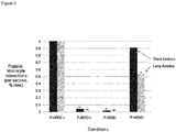
- monocyte-platelet interaction can be divided into short-acting interactions which are arbitrarily defined as contact occurring for less than 3 seconds by detection with a light microscope and long-acting interactions, defined as contact longer than 3 seconds by detection with a light microscope. It seems that the initial short-acting interactions are mediated by P-selectin which is expressed on activated platelets. These initial contacts can then subsequently trigger long-acting interactions mediated by RGD-containing proteins expressed by the activated platelets.
- the activation of monocytes and differentiation into immuno-stimulatory DC may be positively influenced by allowing the monocytes to establish long-acting contacts with platelets, e.g. by giving the monocytes and platelets enough time to interact. As the monocytes flow through the channels or channel-like structures, they alternately bind to and disadhere from the platelets by the shearing force induced by the flow through the channels or channel-like structures.
- the residence time of the monocyte/platelet interaction may be controlled by varying the flow rate, e.g. by controlling the speed of the pump.
- the pump may initially be operated at a slow speed/low flow rate to enhance monocyte/platelet interaction, and the speed/flow rate may then be increased to facilitate disadherence and collection of the treated monocytes from the treatment device. It seems that adherence of the monocytes to the platelets may be best accomplished at about 0.1 to about 2 dynes/cm 2 , at about 0.1 to about 1 dynes/cm 2 , and preferably at about 0.1 to about 0.5 dynes/cm 2 , while disadherence and collection of the monocytes may be best accomplished at increased shear levels.
- activation of monocytes leads to immobilization, e.g. by interacting with activated platelets, platelets-derived factors or plasma components.
- Temperature is another factor to influence activation of monocytes and their differentiation into immuno-stimulatory autologous dendritic cells.
- the methods in accordance with the invention may be performed in a range of about 18°C to about 42°C, preferably in a range of about 22°C to about 41°C and more preferably in a range of about 37°C to about 41°C.
- One parameter that can also be varied to tune activation of monocytes is the density by which the flow chamber is coated with plasma components and thus with platelets that bind to the plasma components.
- activated platelets are activated by binding to plasma components.
- activated platelets in accordance with the invention is used to refer to platelets which show an increased expression of P-selectin, ⁇ IIb- ⁇ 3 integrin and/or RGD-containing proteins such as fibronectin, fibrinogen or vitronectin as a consequence of binding of platelets to plasma components such as fibronectin and/or fibrinogen. Expression may be determined by conventional methods such as RT-PCR, Western-Blotting or FACS analysis.
- unactivated platelets in accordance with the invention is used to refer to platelets for which binding to plasma proteins such as fibronectin or fibrinogen cannot be reduced by pre-incubating platelets with the gamma component of fibrinogen.
- monocytes are activated and start to differentiate into immuno-stimulatory autologous dendritic cells by binding to activated platelets under shear stress conditions.
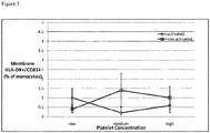
- activated monocytes in accordance with the invention is used to refer to monocytes which upon binding to activated platelets under shear stress conditions express increased levels of the open confirmation of ⁇ 1-integrin and start expressing markers of maturing DC such as HLA-DR + /CD83 + .
- HLA-DR + /CD83 + after binding of monocytes to activated platelets under shear stress condition either in the absence of anti-P-selectin antibodies (activation) or presence of anti-P-selectin antibodies (control).
- Expression may be determined by conventional methods such as RT-PCR, Western-Blotting or FACS analysis.
- immuno-stimulatory autologous dendritic cells After monocytes have been activated by a method in accordance with the invention, they start differentiating into immuno-stimulatory autologous dendritic cells.
- immuno-stimulatory autologous dendritic cells in accordance with the invention is used as mentioned above. These immuno-stimulatory autologous dendritic cells can be identified by expression of markers described above. Immuno-stimulatory autologous dendritic cells can be further distinguished from immuno-suppressive or so-called truncated autologous dendritic cells in that no change in expression of GILZ is observed when obtaining autologous dendritic cells by a method in accordance with the invention.
- the methods in accordance with the invention may require substantial lower amount of blood samples thus bypassing the need of apheresis such as leukaphereses or other processes, which are a considerable burden to patients.
- the present invention can be performed without the need for apheresis such as leukaphereses and the whole process of obtaining such immuno-stimulatory autologous dendritic cells may be performed in a handheld device.
- Said extracorporeal quantity of said mammalian subject's blood may be between about 5 ml to about 500 ml, between about 10 ml to about 450 ml, between about 20 ml to about 400 ml, between about 30 ml to about 350 ml, between about 40 ml to about 300 ml, or between about 50 ml to about 200 ml or between about 50 ml to about 100 ml of extracorporeal blood of said mammalian subject to give a final volume between about 1 ml to about 100 ml, between about 1 ml to about 50 ml, between about 1 ml to about 40 ml, or between about 1 ml to about 30 ml an extracorporeal amount of a mammalian's blood sample.
- the quantity of extracorporeal blood withdrawn and applied to the device may be whole blood.
- said extracorporeal quantity of said mammalian subject's blood may be obtained by isolating leukocytes from between about 5 ml to about 500 ml, between about 10 ml to about 450 ml, between about 20 ml to about 400 ml, between about 30 ml to about 350 ml, between about 40 ml to about 300 ml, or between about 50 ml to about 200 ml or between about 50 ml to about 100 ml of extracorporeal whole blood of said mammalian subject.
- Said extracorporeal quantity of said mammalian subject's blood may also be obtained by isolating buffy coats from between about 5 ml to about 500 ml, between about 10 ml to about 450 ml, between about 20 ml to about 400 ml, between about 30 ml to about 350 ml, between about 40 ml to about 300 ml, or between about 50 ml to about 200 ml or between about 50 ml to about 100 ml of extracorporeal whole blood of said mammalian subject.
- said extracorporeal amount of blood will typically comprise between about 1x10 4 to about 1x10 8 such as about 5x10 6 mononuclear cells/ml.
- a mononuclear leukocyte fraction For obtaining a mononuclear leukocyte fraction, one may obtain a whole blood sample as described and then layer such a sample on e.g. Ficoll-Hypaque. Subsequently a centrifugation step is performed at e.g. about 100g to about 200 g such as 180 g and the mononuclear leukocyte fraction can then be collected from the interface and washed with common buffers such as HBSS. The washed mononuclear leukocyte fraction can then be resuspended in serum-free cell culture medium such as RPMI-1640 medium (GIBCO). Other methods for obtaining mononuclear leukocyte fractions include elutriation, filtration, density centrifugation, etc..
- a platelet-rich plasma containing fraction which may be obtained by centrifuging a whole blood sample which has been obtained as described above at about 100 g to about 180 g such as about 150 g for about 10 min to about 20 min such as about 15 min to separate the debris of the whole blood sample.
- the platelet-rich plasma layer is then collected and recentrifuged at about 700 g to about 1000 g such as about 900 g for about 3 min to about 10 min such as about 5 min.
- the resultant pellet is then resuspended in serum-free cell culture medium.
- a leukocyte fraction comprising a monocytes- or buffy-coat fraction comprising monocytes, which does not comprise plasma components and which does not comprise platelets.
- Such plasma- and platelet-free monocytes-containing fractions may be obtained as is described in the art. If leukocyte or buffy-coat fractions are obtained as described above, they will be sufficiently free of plasma or platelets for the purposes of the invention. For this approach, it may also be desirable to have platelet- and/or plasma-fractions.
- the invention contemplate to use platelets which have been separated from the extracorporeal quantity of said mammalian subject's blood before said extracorporeal quantity of said mammalian subject's blood is applied to said device. These platelets may then be passed through the flow chamber, which has been coated with plasma components such as fibronectin.
- the invention considers to use plasma components, which have been separated from the extracorporeal quantity of said mammalian subject's blood before said extracorporeal quantity of said mammalian subject's blood is applied to said device. These plasma components may then be passed through flow chamber so that they can adhere.
- plasma components which have been obtained from the extracorporeal amount of blood
- plasma components which have been isolated from other sources such as e.g. by recombinant protein expression.
- plasma components include fibrinogen, fibronectin, P-selectin, and fragments thereof such as the gamma component of fibrinogen.
- Apheresis such as leukaphereses may be performed as is known in the art.
- an extracorporeal quantity of blood such as 2.5 L to 61 may be obtained from a subject and treated by conventional leukaphereses to obtain three fractions, namely the plasma, the platelets and the buffy coats.
- the plasma which contains proteins such as fibronectin and fibrinogen, is the lightest blood fraction, and therefore is the first portion of the blood selectively removed from the centrifuge and passaged through channels or channel-like structures.
- the second lightest component in the leukaphereses centrifuge, the platelet fraction is pumped into and through the channels or channel-like structures.
- the third lightest fraction to be eluted from the leukaphereses centrifuge is the buffy coat, which contains the white blood cells, including the blood monocytes.
- the buffy coat including the monocytes is then pumped through the channels or channel-like structures.
- Blood sample may be obtained using the Therakos device, the Spectra cell separator (see Andreu et al., (1994), Transf. Sci., 15(4), 443-454 ), or the Theraflex device from Macopharma.
- the invention in one embodiment the invention considers to use platelets which have been separated from the extracorporeal quantity of said mammalian subject's blood obtained by apheresis such as leukaphereses before said extracorporeal quantity of said mammalian subject's blood comprising monocytes is applied to said device.
- the invention considers to use plasma components, which have been separated from the extracorporeal quantity of said mammalian subject's blood obtained by apheresis such as leukaphereses before said extracorporeal quantity of said mammalian subject's blood comprising monocytes and/or platelets is applied to said device.
- plasma components which have been obtained from the extracorporeal amount of blood may use also either plasma components which have been isolated from other sources such as e.g. by recombinant protein expression.
- plasma components include fibrinogen, fibronectin, or P-selectin.
- plasma fractions which at least, if not predominantly comprise fibronectin.
- plasma fractions which at least, if not predominantly comprise fibronectin.
- the extracorporeal amount of blood is obtained or not obtained by apheresis such as leukaphereses
- it may be considered to pass all three fractions, namely plasma components, platelets and the monocytes-containing fraction at once, e.g. even in the form of a whole blood sample or by using only pre-purified fractions of whole blood, through the flow chamber even though the afore-described sequential passing of these fractions through the flow chamber may provide for better control over the process.
- Pre-purified fractions of whole blood may be obtained by e.g. centrifuging a blood bag and squeezing out the supernatant, which would be enriched in white blood cells and platelets.
- the flow rate through flow chamber and thus the resulting shear stress will effect the differentiation of the monocytes into immuno-stimulatory autologous dendritic cells.
- the design and the dimensions of the flow chamber may be varied to manipulate and even improve the application of a physical force to the monocytes.
- a device having a flow chamber with channels or channel-like structures may be suitable.
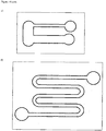
- Such a flow chamber having the general architecture, albeit at smaller dimensions, of a device, which is used for the classical ECP procedure is depicted in Fig. 17 .
- a device having a multiplicity of flow chambers may be suitable.
- Such a flow chamber having the general architecture, albeit at smaller dimensions, of a device, which is used for the classical ECP procedure is depicted in Fig. 17 .
- a flow gradient will be created in the flow chamber such as channels as the monocytes-containing fraction is passed through.
- the monocytes will alternately bind to and disengage from the platelets and/or plasma components. Maturation of monocytes into immuno-stimulatory autologous dendritic cells is greatly enhanced by this interaction, with increased exposure to the platelets and/or plasma components thereby providing increased signaling of this maturational process.
- the flow chamber such as channels may in principle have any cross-sectional shape suitable for the above-described purposes. They thus may have a rectangular, round, elliptical, or other cross-sectional form. Even though the dimensions of such flow chamber will be discussed in the following mainly with respect to a rectangular cross-section, it can be preferred that flow chamber such as channels with an elliptical or round cross-section are used as such cross-sections should allow for e.g. more homogenous coating with plasma components and/or more continuous flow properties with less turbulences.
- flow chamber such as channels may have dimensions of about 5 ⁇ m to up to about 500 ⁇ m of height and of about 5 ⁇ m to up to about 500 ⁇ m of width.
- the channels or channel-like structures may also have dimensions of about 10 ⁇ m to up to and including about 400 ⁇ m of height and of about 10 ⁇ m to up to and including about 400 ⁇ m of width, of about 10 ⁇ m to up to and including about 300 ⁇ m of height and of about 10 ⁇ m to up to and including about 300 ⁇ m of width, of about 10 ⁇ m to up to and including about 250 ⁇ m of height and of about 10 ⁇ m to up to and including about 250 ⁇ m of width, of about 10 ⁇ m to up to and including about 100 ⁇ m of height and of about 10 ⁇ m to up to and including about 100 ⁇ m of width, or of about 10 ⁇ m to up to and including about 50 ⁇ m of height and of about 10 ⁇ m to up to and including about 50 ⁇ m of width.
- flow chambers such as channels of elliptical cross-section are used, the aforementioned dimensions of height and width would have to be adapted correspondingly to allow for a comparable volume.
- the diameter may typically be in the range of about 5 ⁇ m to up to and including about 500 ⁇ m, of about 10 ⁇ m to up to and including about 400 ⁇ m, of about 10 ⁇ m to up to and including about 300 ⁇ m, of about 10 ⁇ m to up to and including about 250 ⁇ m, of about 10 ⁇ m to up to and including about 100 ⁇ m, or of about 10 ⁇ m to up to and including about 50 ⁇ m.
- the length of the flow chamber such as channels channel-like structures is usually selected such that the flow chamber allows for passage of the volume of extracorporeal blood.
- the flow chamber and the device may be configured to allow for passing of an overall volume of between about 1 ml to about 50 ml, between about 1 ml to about 40 ml, or between about 1 ml to about 30 ml.
- the flow chamber may have internal sub structures to increase the surface area or to make the flow conditions less heterogeneous.
- the flow chamber may be filled with particles to increase the surface area or to make the flow conditions less heterogeneous.
- the material of the flow chamber may be plastic or non-plastic.
- the surface of the chamber may be coated covalently or via adsorption.
- Materials for auxiliary tubing, chambers, valves etc. may be selected to for having reduced interactions with blood components.
- auxiliary tubing, chambers, valves etc. may be treated/coated for having reduced interactions with blood components.
- the flow chamber is made from an acrylic plastic.
- the flow chamber may be made of a material that provides a degree of transparency such that the sample within the flow chamber such as the monocytes-containing fractions can be irradiated with visible or UV light, preferably with UV-A.
- UV-A and 8-MOP leads to increased expression of GILZ and thus to activation and differentiation of monocytes into immuno-suppressive autologous dendritic cells.
- exposure to light such as UV-A and DNA-cross linking agents such as 8-MOP should be generally avoided when producing immuno-stimulatory autologous dendritic cells.
- immuno-stimulatory autologous dendritic cells have formed as can be determined by the molecular markers mentioned above, one can envisage to administer DNA-cross linking agents such as 8-MOP and to expose the immuno-stimulatory autologous dendritic cells to e.g. UV-A to render other cells in the extracorporeal blood sample apoptotic.
- DNA-cross linking agents such as 8-MOP
- Such cells may be cytotoxic T-cells, virally infected cells or bacterial cells. Apoptosis of such cells may lead to antigen shedding.
- the immuno-stimulatory autologous dendritic cells can then taken up and process these antigens so that immuno-stimulatory autologous antigen-presenting cells are formed.
- immuno-stimulatory autologous antigen-presenting cells can then e.g. be re-introduced into the respective individual to elicit an immune response against the respective tumor, viral or bacterial antigens.
- immuno-stimulatory autologous dendritic cells Once immuno-stimulatory autologous dendritic cells have formed, one may also separately introduce tumor cells, bacterial cells or virally infected cells of such an individual into the flow chamber and render these cells apoptotic by e.g.
- DNA-cross linking agents such as 8-MOP and irradiate the mixture of immuno-stimulatory autologous dendritic cells and tumor cells, bacterial cells or virally infected cells to render the tumor cells, bacterial cells or virally infected cells apoptotic such that immuno-stimulatory autologous antigen-presenting cells can form. It is for these embodiments, that a design of the flow chamber allowing exposure to light such as UV-A is contemplated.
- a typical flow chamber may have the geometry depicted in Fig. 19A ).
- the flow path has dimensions of 20 mm by 80 mm.
- the chamber is made of polystyrene, PET (polyethylenteherephtalate), PMMA (poly (methyl mathacrylate)) and silicon.
- a blood sample may be spun at low speed through a Ficoll gradient to obtain e.g. 8 ml of sample with a concentration of white blood cells of e.g. 10 10 cells/ml.
- the chamber may be pre-coated with platelets-rich plasma.
- the sample may be passed through the chamber at about 0.028 Pa for about... min.
- the chamber may then be washed with about 3 ml RPMI at 0.028 Pa.
- a second wash with 30-55 ml RPMI may be performed at about 1.2 Pa.
- the collected activated monocytes will then be combined and used for further analysis.
- immuno-stimulatory autologous dendritic cells Once immuno-stimulatory autologous dendritic cells have been obtained by methods in accordance with the invention, they can be generally further processed for specific purposes. These newly formed immuno-stimulatory dendritic cells can for example be incubated under standard conditions to allow completion of their maturation. Culturing of these immuno-stimulatory dendritic cells can be performed under standard conditions, e.g. at 37° C and 5% CO 2 in standard mediums for culturing of human cells such as in RPMI-1640 medium (obtainable e.g. from GIBCO), supplemented with 15% AB serum (obtainable from e.g. Gemini Bio-Products).
- immuno-stimulatory dendritic cells can be obtained without the need for rather expensive cocktails of cytokines for induction of monocyte to DC differentiation. Even though not necessary, it can be considered to cultivate such immuno-stimulatory dendritic cells in a buffered culture medium with one or more cytokines, such as GM-CSF and IL-4, during the incubation period. Maturation cocktails (typically consisting of combinations of ligands such as CD40L, cytokines such as interferon gamma, TNF alpha, interleukin 1 or prostaglandin E2 or the factors mentioned above) may be added as well.
- Maturation cocktails typically consisting of combinations of ligands such as CD40L, cytokines such as interferon gamma, TNF alpha, interleukin 1 or prostaglandin E2 or the factors mentioned above
- one preferentially produce immuno-stimulatory dendritic cells over immuno-suppressive dendritic cells by cultivating the dendritic cells over extended periods of time such as e.g. at least 1, at least 2, at least 3, at least 4, or at least 5 days. This may help the initially formed immuno-stimulatory dendritic cells to further embark on their maturation pathway.
- immuno-stimulatory antigen-presenting cells such as dendritic cells, which are obtained without cocktails of cytokines for induction of monocyte to DC differentiation are a particularly preferred embodiment of the present invention.
- the immuno-stimulatory antigen-presenting cells such as dendritic cells in accordance with the present invention can be tested for the functionality in assays as described herein. For example, one can adapt the assay described in Bioley et al., The Journal of Immunology 2006), 177:6769-6779 .
- immuno-stimulatory antigen-presenting cells such as dendritic cells, which are obtained by the methods disclosed herein e.g. by passing white blood cells as described above through a device depicted in Fig.19 , are co-incubated with CD4 + and CD8 + cells of the same donor and the Melan-A/MART-1 26-35 Peptide, described by Bioley et al. Detection of Melan-A/MART-1 26-35 positive CD4 + cells and CD8 + cells allows confirmation of functional immuno-stimulatory antigen-presenting cells such as dendritic cells which are obtained in accordance with the present invention.
- immuno-stimulatory dendritic cells can then be manipulated ex vivo, prior to re-administration to the subject, in order to tailor them for the desired therapeutic purpose.
- such immuno-stimulatory dendritic cells can e.g. be processed ex vivo, such as by loading them with immunogenic antigens, e.g. those expressed on apoptotic tumor cells or pathogenic infectious agents, or enhancing their maturation in order to increasing their efficiency in cancer immunotherapy.
- immunogenic antigens e.g. those expressed on apoptotic tumor cells or pathogenic infectious agents
- This will lead to immuno-stimulatory antigen-presenting cells displaying the antigen on their surfaces.
- One of the most preferred embodiments of the present invention contemplates to separate as much as possible the generation of immuno-stimulatory antigen-presenting cells such as dendritic cells as described herein, the generation of disease-antigens and the loading of these antigen-presenting cells with the disease-antigens.
- immuno-stimulatory antigen-presenting cells such as dendritic cells are made from monocytes by applying physical forces in the absence of e.g. 8-MOP and UVA and/or cytokine coctails.
- These immuno-stimulatory antigen-presenting cells such as dendritic cells may then be co-incubated with disease-antigens which were obtained separately to effect efficient loading of the immuno-stimulatory antigen-presenting cells such as dendritic cells and display of the antigens on their surface.
- the immuno-stimulatory dendritic cells may in particular be loaded with disease effector agents to produce antigen presenting dendritic cells, which upon reintroduction into the subject can launch an immune response against the disease effector genes.
- disease effector agents refers to agents that are central to the causation of a disease state in a subject.
- immuno-stimulatory dendritic cells may be loaded with antigens, which are known being expressed in cancerous tissue.
- antigens may be expressed in immuno-stimulatory dendritic cells such that they are displayed on the MHC class 1 and MHC class 2 molecules.
- the antigen-loaded, reintroduced immuno-stimulatory antigen-presenting cells may launch an immune response against the cancer.
- these disease effector agents are disease-causing cells which may be circulating in the bloodstream, thereby making them readily accessible to extracorporeal manipulations and treatments.
- diseases causing cells include e.g. malignant T-cells, malignant B cells, and virally or bacterially infected white or red blood cells which may harbor or express microbial (e.g. viral, bacterial, fungal, mycobacterial, protozoal) peptides or proteins or other pathogen-associated molecules.
- Exemplary disease categories giving rise to disease-causing cells include lymphoproliferative disorders such as leukemia, lymphoma, and myeloma, as well as infections including malaria, human-immunodeficiency virus (HIV), Epstein-Barr virus (EBV), cytomegalovirus (CMV), hepatitis B virus (HBV), hepatitis C virus (HCV), Lyme disease, leprosy, tuberculosis, and other blood borne pathogens.
- HIV human-immunodeficiency virus
- EBV Epstein-Barr virus
- CMV cytomegalovirus
- HBV hepatitis B virus
- HCV hepatitis C virus
- Lyme disease leprosy
- tuberculosis tuberculosis
- disease-causing cells include those isolated from surgically excised specimens from solid tumors, such as lung, colon, brain, kidney or skin cancers. These cells can be manipulated extracorporeally in analogous fashion to blood leukocytes, after they are brought into suspension or propagated in tissue culture. Alternatively, in some instances, it has been shown that the circulating blood of patients with solid tumors can contain malignant cells that have broken off from the tumors and entered the circulation. These circulating tumor cells can provide an easily accessible source of cancer cells, which may be isolated, rendered apoptotic and engulfed by the dendritic cells in accordance with the method described and claimed herein.
- Disease effector agents may also be obtained from such disease-causing cells, which have been rendered apoptotic by e.g. cytotoxic agents. It is to be understood that apoptotic cells may send different signals through effector agents depending on whether they are derived from normal or abnormal cells, such as healthy or malignant cells. Combining and cultivating immuno-stimulatory dendritic cells with such apoptotic cells may also be used to load immuno-stimulatory dendritic cells with disease-causing antigens and generate immuno-stimulatory antigen-presenting cells.
- disease effector agents falling within the scope of the invention further include microbes such as bacteria, fungi and viruses, which express disease-associated antigens. It should be understood that viruses can be engineered to be “incomplete”, i.e., produce distinguishing disease-causing antigens without being able to function as an actual infectious agent, and that such "incomplete” viruses fall within the meaning of the term “disease effector agents" as used herein.
- Immunogenic antigens may also be obtained by treating cancer specimens or cancer cells with agents which are know to induce immunogenic antigens such as Bortezomib.
- Cancers which may be in particular treatable by the above described approaches include CTLC, Melanoma, Prostate cancer, of HNSCC as e.g. disease-antigens are know for some of these disease or as e.g. animal models of some of these diseases can be used initially.
- immune-stimulatory autologous dendritic cells obtainable by the methods described herein with antigens allows producing immuno-stimulatory autologous antigen-presenting cells.
- NPs polymeric nanoparticles
- Such NPs may be further modified with targeting moieties for DEC-205 such as an DEC-205 antibody to improve receptor-mediated endocytosis and antigen presentation.
- the invention contemplates to use encapsulation of antigens in polymeric nanoparticles to optionally further enhance formation of immuno-stimulatory autologous antigen-presenting cells.
- the immuno-stimulatory dendritic cells obtained in accordance with the invention and disease effector agents are incubated for a period of time sufficient to maximize the number of functional antigen presenting dendritic cells in the incubated cell population.
- the treated blood cell concentrate and disease effector agents are incubated for a period of from about 1 to about 24 hours, with the preferred incubation time extending over a period of from about 12 to about 24 hours. Additional incubation time may be necessary to fully mature the loaded immuno-stimulatory antigen-presenting cells prior to reintroduction to the subject.
- the blood cell concentrate and disease effector agents are incubated at a temperature of between 35° C and 40° C. In a particularly preferred embodiment, the incubation is performed at about 37°C.
- Inducing monocyte differentiation provides immuno-stimulatory dendritic cells in numbers which equal or exceed the numbers of dendritic cells that are obtained by expensive and laborious culture of leukocytes in the presence of cytokines such as GM-CSF and IL-4 for a couple of days.
- the large numbers of functional dendritic cells generated by the method described above provide a ready means of presenting selected material, such as, for example, apoptotic cells, disease agents, antigens, plasmids, DNA or a combination thereof, and are thereby conducive to efficient immunotherapy.
- Antigen preparations selected to elicit a particular immune response may be derived from, for example, tumors, disease-causing non-malignant cells, or microbes such as bacteria, viruses and fungi.
- the antigen-loaded dendritic cells can be used as immunogens by reinfusing the DC into the subject or by otherwise administering the cells in accordance with methods known to elicit an immune response, such as subcutaneous, intradermal or intramuscular injection.
- immuno-stimulatory dendritic cells can be obtained by a method in accordance with the invention in the absence of a photoactivatable agent and without exposure to light such as visible and preferably UV-A.
- the present invention thus aims at obtaining individual-specific functionally and maturationally synchronized autologous immuno-stimulatory dendritic cells.
- the present invention relates to autologous immuno-stimulatory dendritic cells obtainable by a method described herein, preferably for use in immunization against cancer antigens, viral antigens, bacterial antigens or fungal antigens.
- the interface containing the mononuclear leukocyte fraction was collected and washed twice in HBSS, then resuspended in RPMI-1640 medium (GIBCO) to a final concentration of 5 x 10 6 mononuclear cells/ml. Cells were utilized within one hour of being acquired.
- Vena8 biochips (Cellix Ltd, Dublin, Ireland) were used to generate laminar flow.
- the volumetric flow path for a channel of the Vena8 biochips measured 20000 x 400 x 100 microns (length x width x height). Protein coating of these chips is described in the appropriate section below.
- the parallel-plate flow chamber was mounted on the stage of a phase contrast optical microscope (CK40, Olympus, Japan) with a 10x objective. All runs were performed at room temperature. A uniform laminar flow field was simulated by use of a syringe pump (KD Scientific, New Hope, PA) capable of generating near-constant volumetric flow rates. The components of the configuration were devised to minimize tubing. Prior to infusing cell suspensions through the plates, the system was washed with 5 ml of RPMI at a flow rate producing a wall shear stress of approximately 1 dyne/cm 2 . Cell suspensions of interest were then passed through the chamber at a fixed flow rate and wall shear stress.
- CK40 phase contrast optical microscope
- Monoclonal antibodies for immunophenotyping included CD 14 (LPS receptor; monocytes), CD11c (integrin subunit; monocytes and DC), HLA- DR (class II MHC molecule), CD83 (DC marker), CD62p (P-selectin; activated platelets), and CD61 (integrin subunit; platelets).
- Antibodies were obtained from Beckman Coulter (CD14, CD11c, HLADR, CD83) or Sigma (CD62p, CD61) and used at their pre-determined optimal dilutions. Background staining was established with appropriate isotype controls, and immunofluorescence was analyzed using a FC500 flow cytometer (Beckman Coulter).
- Two-color membrane staining was performed by adding the pre-determined optimal concentrations of both antibodies directly conjugated to FITC or PE and incubating for 20 min at 4°C, followed by washing to remove unbound antibodies.
- Combined membrane and cytoplasmic staining was performed following manufacturer's instructions for cell fixation and permeabilization (Intraprep kit, Beckman Coulter).
- Reverse transcription was carried out in a 96-well thermocycler (MJ Research PTC-200) in the following conditions: 25°C, 10 minutes, 37°C, 120 minutes, 85°C, 5 seconds.
- TaqMan real-time PCR was used to detect transcripts of DC-LAMP, CD40, ADAM Decysin, Lox1, CCR7, CD80, CD83, CD86, FPRL2, and GPNMB. Primers and probes for each sequence were obtained as inventoried Taqman Gene Expression Assays (Applied Biosystems). HPRT1 was used as a reference gene.
- Platelet adhesion experiments were performed using the Vena8 flow chamber described above. Fibrinogen and fibronectin (Sigma) were dissolved in PBS to a final concentration of 200 mcg/ml. Channels of the Vena8 chips were incubated at room temperature in a humidified chamber for 2 hours with the protein solution, autologous plasma, or PBS alone. The channels were washed with 5x the volume RPMI. Platelet-rich-plasma was then perfused through the protein-coated channel at the indicated shear-stress, held constant. For each channel, still images were acquired exactly 90 seconds into the experiment at 4 pre-defined low power fields located along the flow path (fields were centered at 2500, 7500, 12500, and 17500 microns from the start point of infusion).
- Small RGD peptides containing the amino-acid sequence Arg-Gly-Asp-Ser; DRG peptides, contain the amino-acid sequence Ser-Asp- Gly-Arg; or fragment 400-411 of fibrinogen, containing the amino-acid sequence His- His-Leu-Gly-Gly-Ala-Lys-Gln-Ala-Gly-Asp-Val, were incubated at a concentration of 2mM with PRP for 20 minutes at room temperature. The PRP was then perfused through the channels as previously described.
- Platelet-coated Vena8 channels were pre-treated with either 40 ⁇ g/ml anti-P-selectin (R&D Systems) or 40 ⁇ g/ml of an isotype control for 30 minutes at room temperature, then washed with 5x the volume RPMI.
- Mononuclear cell suspensions were pre-treated with either RGD or DGR peptides at a concentration of 2.5 mM.
- Video samples lasting 400 frames (26.3 seconds) were recorded 60 seconds after commencement of flow using a lower power field of view spanning 400 microns and centered at 7500 microns from the flow start point.
- ⁇ -1 integrin conformation was assessed using the Glycotech flow chamber. 15mm platelet-coated petri dishes (described above) were pre-treated with 40 ⁇ g/ml anti-P-selectin or an isotype control for 20 minutes at room temperature, then washed with 5x the volume RPMI. Immediately following perfusion through the platelets, cells were immunophenotyped with anti-CD29 HUTS-21 (BD Biosciences), an antibody that specifically binds to the active (open) conformation of ⁇ 1 integrins.
- ECP was initially developed as a means to enable extracorporeal chemotherapeutic exposure of pathogenic leukocytes to ultraviolet A (UVA)-activated 8-methoxypsoralen (8-MOP), a DNA-cross-linking drug. Therefore, ECP involves the flow of leukapheresed blood between large transparent plastic parallel-plates separated by 1 mm. To permit detailed analysis of the flow dynamics involved during ECP, independent of UVA/8-MOP exposure, the flow conditions of ECP were reproduced using miniature parallel plates with surface area of only 0.8 mm 2 , separated by 100 microns. This model permitted visualization using digital microscopy. Studies using the model revealed the following sequence (determined by video analysis): initial adherence of platelets from the flow stream to the plate, followed by transient binding of passaged monocytes to the immobilized platelets.
- UVA ultraviolet A
- 8-MOP 8-methoxypsoralen
- monocytes were hypothesized to induce monocyte-to-DC differentiation under conditions of flow.
- monocytes were passed between parallel plates pre-coated with autologous platelets at low (10 ⁇ 5/low power field [lpf]), medium (44 ⁇ 20/lpf), and high (102 ⁇ 32/lpf) densities.
- Cells were passed through the plates at a flow rate producing a wall shear stress of 0.5 dyne/cm 2 , analogous to the wall shear stress in post-capillary venules.
- the number of monocyte-platelet interactions per unit time increased in proportion to augmented density of platelets (determined by video analysis).
- RT-PCR was performed to assess for changes in gene expression.
- Monocytes were passed through parallel plates coated with high or low densities of platelets as described in the previous section. Following overnight incubation, RNA was extracted and RT-PCR performed to determine level of expression for 10 genes associated with DC ( Figure 2 ).
- CD40 a costimulatory molecule with known expression on mature DC (Cella et al., 1996, see reference list), was found to be upregulated by over 567% in monocytes exposed to high densities of platelets relative to monocytes exposed to low levels.
- LAMP3 a marker specific to DC differentiation (de Saint-Vis at al., 1998, see reference list), was upregulated by 398%.
- CD80 is a costimulatory molecule known to be upregulated upon APC activation (Slavik et al., 1999, see reference list), upregulated by 220% in monocytes exposed to high levels of platelets.
- CCR7 a chemokine receptor known to play a role in DC migration to lymphoid organs, was upregulated by 376%.
- LOX1, CD83, CCR7, and ADAM Decysin, all genes associated with DC (Berger et al., 2010, see reference list), were also upregulated in the monocytes exposed to high levels of platelets.
- FPRL2, GPNMB, and CD86 were all downregulated in monocytes exposed to high levels of platelets.
- FPRL2 is a receptor that when activated is known to inhibit DC maturation (Kang et al., 2005, see reference list)
- GPNMB is a protein involved in decreasing cytokine production (Ripoll et al., 2007, see reference list);
- CD86 is a costimulatory molecule expressed by APCs.
- Platelets could potentially influence monocytes through direct receptor-ligand interaction, or via cytokines and other secreted mediators.
- Platelets suspended in flow bind to serum proteins adsorbed onto the plate
- Both fibrinogen and fibronectin contain segments with the amino acid sequence arginine(R)-gylcine(G)-aspartate(D), RGD.
- RGD segments are well-known to interact with many integrin receptors, particularly the I/A domain of beta subunits, which are exposed when the integrins are in the active conformation (Xiong et al., 2002, see references).
- Platelets are activated by adhesion to the plate
- Platelets physiologically circulate in an inactive state, with an array of proteins stored in intracellular granules. Upon encountering stimuli such as damaged endothelium or thrombin, platelets become activated and almost instantaneously translocate these intracellular proteins to the plasma membrane (Kaplan et al., 1979, see references). It was postulated that platelet adhesion to the plastic plate/absorbed proteins caused platelet activation similar to that caused by well-known stimuli. To test this hypothesis, surface expression of P-selectin, a well-known marker of platelet activation, was assessed before and after adhesion.
- P-selectin was further assessed at 30, 60, and 90 minutes following platelet adhesion; P-selectin expression remained stable at all time points, with 72 ⁇ 11% of platelets P-selectin positive 90 minutes after adhesion, indicating that platelets remain in an active state for the duration of the procedure. Similar trends were found in assessment of ⁇ IIb- ⁇ 3, a fibrinogen-binding integrin, with surface expression of this protein increasing from 4 ⁇ 3% prior to adhesion, to 49 ⁇ 18% post-adhesion.
- the monocyte-platelet interactions observed on video were divided into two categories: (1) short-acting, arbitrarily defined as contact occurring for less than 3 seconds (46 frames), and (2) long-acting, defined as contact longer than 3 seconds, including stable binding. Since it had been previously determined that the platelets in the ECP system were in an activated state, and that activated platelets express an array of proteins including P- selectin and RGD containing proteins (e.g. fibronectin, fibrinogen, and vitronectin), it was sought to determine the involvement, if any, of these proteins in either short or long- duration interactions.
- P- selectin and RGD containing proteins e.g. fibronectin, fibrinogen, and vitronectin
- Plates were pre-coated with platelets, and four conditions tested: (1) platelets pre-treated with an irrelevant isotype control, and monocytes untreated (P+RGD+); (2) platelets pre-treated with an irrelevant isotype control, and monocytes pre-incubated with RGD peptides (P+RGD-); (3) platelets pre-treated with anti-P-selectin, and monocytes untreated (P-RGD+); (4) platelets pre-treated with anti- P-selectin, and monocytes pre-treated with RGD peptides (P-RGD-).
- Blocking with anti-P-selectin alone resulted in a decrease of both short and long monocyte- platelet interactions to almost zero (p ⁇ 0.01; Figure 6 , also confirmed by video analysis).
- blocking RGD alone did not significantly alter the number of short-duration interactions, but decreased the long-duration monocyte-platelet interactions by 44% (p ⁇ 0.05; Figure 6 ).
- Blocking both P-selectin and RGD simultaneously resulted in a pattern similar to that seen when only P-selectin was blocked, with both long and short duration interactions reduced to near zero.
- Integrin receptors in their open conformation, are known to interact with RGD-containing ligands (Ruoslathi et al., 1996, see references). Using an antibody that recognizes an epitope exposed only when the ⁇ 1 integrin is in its open conformation, we assessed the conformation of monocyte integrins before and after flow through the model. Figure 7 shows that as the number of short-acting monocyte-platelet interactions increased, there was corresponding increase in the percentage of monocytes expressing integrins in their open conformation immediately post-flow.
- the black line shows that an average of 71% of monocytes which had received a high number of platelet-interactions (> 61 ⁇ 19/lpf x sec) expressed ⁇ 1 in the active form, compared to 9% of monocytes which had received a low number of platelet interactions ( ⁇ 5.1 + 2 /lpf x sec).
- PBMC peripheral blood specimens were acquired from healthy subjects under the guidelines of the Yale Human Investigational Review Board, and informed consent was provided according to the Declaration of Helsinki.
- PBMC peripheral blood specimens were isolated by centrifugation over a Ficoll-Hypaque gradient (Isolymph, CTL Scientific).
- Monocytes were enriched from freshly isolated PBMC by: 1) plastic adherence for dexamethasone dose-titration experiments (purity: 71.6 ⁇ 5.6% CD14 + ); 2) CD14 magnetic bead positive selection (Miltenyi Biotec) for PUVA dose-titration experiments (purity: 88.1 ⁇ 3.5% CD14 + ), and; 3) Monocyte Isolation Kit II (Miltenyi Biotec) for LPS stimulation experiments (purity: 83.8 ⁇ 3.8% CD14 + ).
- Monocytes were cultured at a density of 5 x 10 6 cells/mL in 6- and 12-well polystyrene tissue culture plates at 37°C and 5% CO 2 in RPMI-1640 (Gibco) supplemented with heat-inactivated 15% AB serum (Gemini) and 1% penicillin/streptomycin (now referred to as complete media). 800 IU/mL recombinant human GM-CSF (R&D Systems) and 1000 IU/mL recombinant human IL-4 (R&D Systems) were added to cultures for 36 hr to induce monocyte to DC differentiation as described.
- Non-adherent cells (purity: 66.0 ⁇ 4.5% CD3 + ) removed during plastic adherence will now be generally referred to as lymphocytes.
- Lymphocytes were treated with 8-MOP (100 ng/mL) and UVA (1 J/cm 2 ), washed with PBS, and co-cultured in complete media at 37°C and 5% CO 2 with either PUVA-treated or untreated-MoDC in a ratio of 5 or 10 lymphocytes to 1 MoDC.
- MoDC treated for 24 hr with 100 nM dexamethasone (Sigma) served as the positive control group. After 24 hr, cells were harvested and MoDC were re-purified.
- CD11c + MoDC were re-plated at 0.5-1.0 x 10 6 cells/mL in complete media and stimulated with 100 ng/mL LPS (Sigma). 24 hr after LPS stimulation, cells were harvested for RNA isolation and immunophenotyping, and supernatants were collected for cytokine quantification. As negative controls, parallel groups did not receive LPS.
- Silencer select pre-designed and validated GILZ siRNA (Invitrogen), with off-target prediction algorithms, was used to knockdown GILZ expression.
- Mo-DC were transfected using Lipofectamine RNAiMAX Reagent (Invitrogen). RNA i duplex and lipofectamine reagent were incubated together for 20 min, then added to MoDC cultures and incubated for 2 hr at 37°C and 5% CO 2 . Transfected MoDC were treated in an identical fashion as described for the MoDC/lymphocyte co-cultures. MoDC were also transfected with scramble siRNA.
- Monoclonal antibodies included HLA-DR, CD80, CD83, CD3, CD86, CD14, CD11c and GILZ. Antibodies were obtained from Beckman-Coulter and eBioscience and were used at their pre-determined optimal dilutions. Apoptosis was assessed using the Annexin-V Apoptosis Detection Kit (eBioscience), with Annexin-V recognizing phosphatidylserine (PS) on the surface of apoptotic cells. 7-AAD substituted for PI as the cell viability dye.
- Cells displaying an Annexin-V + /7-AAD - phenotype were classified as early apoptotic cells, and cells displaying an Annexin-V + /7-AAD + phenotype were classified as late apoptotic cells.
- Dual membrane and intracytoplasmic staining was performed using the IntraPrep fix and permeabilization kit (Beckman-Coulter). Background staining was established with appropriate isotype and fluorescence minus one controls. Immunofluorescence was analyzed using a FACSCalibur L (BD Biosciences) within 2 hr of fixation with 2% paraformaldehyde. A minimum of 10,000 events were collected for each group.
- SYBR green real-time PCR (Applied Biosystems) was used to detect transcripts of IL-12, IL-10, IL-6, TNF-alpha, and TGF- ⁇ . Primers were designed to span intron junctions using Primer3Plus. Primer melting curves were obtained to confirm a single product. HPRT-1 and GAPDH were used as reference genes. Samples were run in triplicate on a 7500 Real Time PCR System (Applied Biosystems). The delta-delta C(t) method was used to calculate the fold change.
- MoDC up-regulate GILZ after treatment with dexamethasone (dex) in a dose-dependent manner ( Figure 10C ).
- Treatment with 100 nM dex for 24 hr was selected as the positive control for inducing GILZ expression in MoDC (Dex-DC) ( Figure 10D ).
- Immature MoDC treated with the combination of 8-MOP and UVA light up-regulate GILZ and assume a tolerogenic, immuno-suppressive phenotype
- the number of photo-adducts formed per 10 6 base pairs is directly related to the product of the 8-MOP concentration and UVA dose, see Gasparro et al., references. As the product of 8-MOP and UVA reached 100, GILZ was up-regulated 3-fold, and as the product increased to 200 and 400, GILZ was up-regulated 4.8- and 8.6-fold respectively ( Figure 11B ).
- the percentage of early apoptotic CD11c + cells was minimally ( p > 0.05) higher at 2 J/cm 2 as compared to 1 J/cm 2 for all doses of 8-MOP tested ( Figure 11C ).
- the percentage of late apoptotic CD11c + cells remained less than 13% at 1 J/cm 2 , and less than 16% at 2 J/cm 2 for all doses of 8-MOP tested ( Figure 11D ).
- dot plots highlight the relative resistance of MoDC to the pro-apoptotic effect of escalating doses of PUVA ( Figure 11E ).
- the number of cells recovered from cultures did not statistically differ in any group treated with 1 or 2 J/cm 2 (data not shown), and greater than 90% CD11c + cells (range 91.0-97.5%) were harvested after treatment.
- lymphocytes display Annexin-V as early as 2 hr after treatment with 1 J/cm 2 and 100 ng/mL (data not shown).
- MoDC treated with 100 ng/mL and 1 J/cm 2 Figure 11F
- the percentage of early apoptotic lymphocytes increased from 6.6% in untreated MoDC to 44.3% in PUVA-DC
- the percentage of late apoptotic lymphocytes increased from 4.5% to 33.7% ( Figure 11G ).
- PUVA-treated lymphocytes are subsequently referred to as apoptotic lymphocytes (ApoL).
- the PUVA dose-dependent induction of GILZ correlated with a decrease in cell surface expression of CD80, CD86, and CD83 ( Figure 12A , 3B ). Down-regulation of these markers paralleled the induction of GILZ (see Figure 11B ), beginning at 8-MOP concentrations of 100 ng/mL for both 1 and 2 J/cm 2 . As the product of 8-MOP and UVA exceeded 100, CD83, CD80 and CD86 expression were reduced by 31%, 30% and 54% respectively, and HLA-DR expression increased by 38%.
- MoDC exposed to apoptotic lymphocytes up-regulate GILZ and are resistant to LPS-induced full maturation
- Induction of GILZ was not related to an increase in the number of early or late apoptotic CD11c + cells, as there were ⁇ 12% early apoptotic (range 3.8-11.4%) and late apoptotic (range 6.3-11.5%) CD11c + cells in all groups demonstrating up-regulation of GILZ.
- MoDC expressing GILZ greater than 2.5-fold above untreated MoDC were resistant to full maturation by LPS and exhibited a semi-mature, tolerogenic phenotype.
- LPS stimulation increased CD80 expression in MoDC up-regulating GILZ to only 50% of the levels seen after LPS stimulation in untreated MoDC ( Figure 13D , range 0.48-0.57%), and increased CD86 expression to only 45% of untreated MoDC ( Figure 13D , range 0.42-0.47%). Similar results were obtained for HLA-DR and CD83 ( Figure 14E , range 47-65% and 23-57% of untreated MoDC after LPS respectively).
- MoDC up-regulating GILZ expressed 6% of the CD80 mRNA of untreated MoDC (range 4.5-7.5%), and expressed 50% of the CD86 mRNA of untreated MoDC (range 12.4-85.1%), as assessed by qRT-PCR.
- MoDC expressing GILZ display a tolerogenic cytokine profile, and knockdown of GILZ reduces the IL-10 to IL-12p70 ratio
- PUVA-DC or untreated MoDC exposed to ApoL expressed GILZ at higher levels that PUVA-DC cultured alone (3.6- and 6.7-fold higher, respectively; see Figure 13B ).
- These two groups increased production of IL-10, and decreased production of all pro-inflammatory cytokines and chemokines tested. Cytokine levels were confirmed at the RNA level, with MoDC that up-regulated GILZ also demonstrating up-regulation of IL-10 mRNA 8-fold above untreated MoDC (range 5.5-11.8, p ⁇ 0.01). Reductions in IL-12, TNF- ⁇ , and IL-6 were also confirmed at the RNA level (data not shown). TGF- ⁇ was up-regulated 2.5-fold in MoDC up-regulating GILZ (data not shown). TGF- ⁇ was not included in the multiplex analysis and therefore was only analyzed at the mRNA level.
- the IL-10 to IL-12p70 ratio is a useful indicator of tolerogenicity, since tolerogenic DC are characterized by an increased IL-10 to IL-12p70 ratio, see Steinman et al., references).
- the ratio of IL-10 to IL-12p70 increased from 6.7 in untreated MoDC to 67.7 in Dex-DC.
- the IL-10 to IL-12p70 ratio increased to 38.7 in PUVA-DC, and to 89.4 and 114.9 in untreated MoDC and PUVA-DC exposed to ApoL, respectively ( p ⁇ 0.05).
- Treated MoDC up-regulating GILZ 2.5-fold higher than untreated MoDC produced higher levels of IL-10 ( Figure 15B ), and knockdown of GILZ reduced IL-10 production by 39% (range 34-48%, p ⁇ 0.05).
- Treated MoDC up-regulating GILZ 2.5-fold higher than untreated MoDC also produced lower amounts of EL-12p70 ( Figure 15C ), and knockdown of GILZ increased IL-12p70 production by 188% (range 149-214%, p ⁇ 0.05).
- Treatment with scramble siRNA had no appreciable effect on the production of IL-10 or IL-12p70. Knockdown of GILZ reduced the IL-10 to IL-12p70 ratio that had been elevated after GILZ induction.
- Dex-DC treated with GILZ siRNA demonstrated a reduction in the IL-10 to IL-12p70 ratio from 15.3 in non-transfected MoDC to 3.9 in transfected Dex-DC.
- the ratio decreased from 8.4 in non-transfected MoDC to 2.9 in PUVA-DC, and in untreated MoDC and PUVA-DC exposed to ApoL, reductions in the ratio from 18.1 to 7.8 and 28.4 to 8.3, respectively, were observed.
- GILZ is necessary for the polarization towards a tolerogenic cytokine profile, characterized by increased IL-10 production, and decreased pro-inflammatory cytokine and chemokine production, including IL-12p70.
- ECP ECP-induced monocytes from healthy subjects to convert to DC
- mononuclear leukocytes from normal subjects were examined in 2 ways.
- a desktop apparatus incorporating a UVA light source and a plastic exposure plate, enabled laboratory reproduction of the clinical ECP system and sample access for parallel RNA isolation, immunophenotyping, and functional studies.
- the standard 8-MOP concentrated solution (Therakos) was added directly to the clinical ECP apparatus and to the laboratory model system. That mode of introduction enabled precise 100-200 ng/mL concentrations throughout the clinical procedures and experimentation.
- ECP ECP-derived monocyte gene activation, maturation and function.
- the cells were resuspended in RPMI-1640 medium (Gibco), supplemented with 7.5% AB serum, 7.5% autologous serum (Gemini Bio-Products) and cultured (for patients) in 6-well polystyrene tissue-culture plates at a density of 5 ⁇ 10 6 cells/mL and in Baxter platelet storage bags (for normal subjects 37°C, 5% CO 2 ). After overnight culture (ECP Day 1), cells were harvested before undergoing monocyte enrichment. To generate DC for comparative phenotypic analysis, cells were cultured in RPMI 1640 15% serum in the presence of 1 mL of GMCSF and IL4 (25 ng/mL; R&D Systems) for 6 days.
- monocytes were enriched from the mononuclear cell pool by single passage through affinity columns. This negative selection method limited physical perturbation, whereas lymphocytes adherent to magnetic microbeads (Miltenyi Biotec), conjugated to relevant monoclonal antibodies (anti-CD4, CD8, CD19), were depleted.
- ECP-processed lymphocytes Due to their extreme sensitivity to UVA-activated 8-MOP, 99% of ECP-processed lymphocytes were apoptotic after overnight incubation (as determined by staining with APO2-PE, Trypan blue, and/or annexin-fluorescein isothiocynate FITC/ propidium idodide). Because ECP causes global lymphocyte apoptosis, 90%-95% of viable mononuclear leukocytes in the ECP day 1 fraction were monocytes. This phenomenon accounts for the observation that multiple step magnetic bead removal of apoptotic lymphocytes, performed as follows and yielding monocyte purity of greater than 95%, does not alter levels of observed gene expression in the studied cell populations.
- the magnetically labeled cells were instead separated from the unlabeled monocytes using the EasySep magnet (StemCell Technologies).
- Cells in 2 mL of buffer in a 5-mL polystyrene tube, were placed in the magnet for 10 minutes, and then the unlabeled cells were carefully poured off into a new tube. This procedure was repeated 2x, to maximally enhance monocyte purity.
- the purity was still insufficient, cells were relabeled with the Monocyte Isolation Kit II reagents and placed in the EasySep magnet for an additional 10 minutes, and the unlabeled monocytes were eluted.
- CD14 lipopolysaccharide [LPS] receptor, monocytes
- CD36 receptor for apoptotic cells, monocytes
- human leukocyte antigen DR-1 HLA-DR; class II major histocompatibility complex [MHC] molecule
- CD83 dendritic cell marker
- DC-LAMP cytoplasmic dendritic cell-lysosome-associated membrane protein
- CD80 and CD86 B7.1 and B7.2 costimulatory molecules.
- Antibodies were obtained from Beckman Coulter and used at their predetermined optimal dilutions.
- ECP-processed monocytes are functionally capable of stimulating MHC class I-restricted cytotoxicity by CD8 T cells.
- mononuclear leukocytes from 3 normal subjects were studied.
- One unit of anti-coagulated blood freshly procured from each of 3 HLA-A2-positive volunteers, served as sources of stimulator monocyte/dendritic cells, before and after being processed through the clinical ECP apparatus in a manner identical to the actual ECP procedure.
- Mononuclear fractions were isolated from the blood immediately prior to ECP processing (pre-ECP) and immediately after ECP (ECP D0).
- the Pre ECP fraction was serially diluted in RPMI 1640/15% autologous serum, and 100 ⁇ L containing from 25 000 to 250 cells was plated in round-bottom microtiter plate wells, in 5 replicates.
- the ECP D0 fraction was incubated for 18 hours in large well plates and harvested by scraping the wells to free adherent cells. The re-suspended cells were then serially diluted and plated as above.
- An A-2-negative normal donor served as the source of responder CD4 and CD8 T cells, purified by positive selection on Miltenyi magnetic bead columns (average purity 98%).
- Responder T cells (50 000/well in 100 ⁇ L) were then added to the wells containing either Pre-ECP or ECP-D0 stimulators, and the plates were cultured for 7 days at 37°C in a CO2 incubator.
- target cells the A-2-positive T-B hybridoma lymphoblast line, 174 x CWM.T1
- 51 Cr was labeled with 51 Cr and added to the MLR cultures at 10 4 cells/well. After 4-hour incubation, plates were centrifuged, and 100 ⁇ L of supernatant was removed from each well for counting in a gamma counter.
- Percent-specific lysis was defined as 100 times the following fraction:
- RNA yield and purity were measured using the NanoDrop ND-1000 Spectrophotometer and the Agilent 2100 Bioana- lyzer. Fragmented cRNAs were hybridized on Affymetrix HG U133 Plus 2.0 human chips, and screening for approximately 47 400 human genes and ESTs was performed by the Yale University W. M. Keck Resource Laboratory. The microarray results are available on Gene Expression Omnibus under accession number GSE23604.
- TaqMan real-time PCR was used to detect transcripts of DC-LAMP, CCR7, CD80, CD86, and CD14. Primers and probes for each sequence were obtained as inventoried Taqman Gene Expression Assays (Applied Biosystems). HPRT1 was used as a reference gene.
- the stimulation by ECP of individual gene activation in monocytes was expressed as the ratio of ECP Day 1 to pre-ECP expression for the relevant gene.
- a negative column purification method was used, whereby lymphocytes were retained, and monocytes were passively filtered. The results revealed that the ECP-processed monocytes from both patients and normal subjects remain sufficiently viable to reproducibly express a shared transcriptome signature.
- RNA transcripts were considered significantly up- or down-regulated by ECP if fold change was ⁇ 2 and significance was P ⁇ .05 compared with pre ECP.
- Levels of RNA transcripts from approximately 3000 genes were significantly changed in each patient group and in normal subjects (Table 2).
- 1129 genes were up- or down-regulated in common by ECP-processed monocytes from both CTCL and GVHD patients and from normal subjects, indicating commonality in ECP-induced gene activation.
- Table 2 Number of Monocyte Genes with Altered Expression after ECP.
- CD14 monocyte marker
- Table 5 ECP-Enhanced Expression of Immunosuppression-Associated Genes, Ratio* of Post-ECP/Pre-ECP Levels.
- ECP-induced dendritic cells transcriptome was performed to identify a subset of surface molecule gene products as markers and functional mediators of immuno-stimulatory dendritic cells.
- 466 genes upregulated in ECP-induced dendritic cells were cross referenced to approximately 2000 known or presumed full-length human transmembrane genes to identify 87 shared surface proteins.
- the interface containing the mononuclear leukocyte fraction was collected and washed twice in HBSS, then resuspended in RPMI-1640 medium (GIBCO) to a final concentration of 5 x 10 6 mononuclear cells/ml. Cells were utilized within one hour of being acquired.
- Plate passage was conducted using a Glycotech system (Glycotech, Rockville, MD). This system consisted of a volumetric flow path measuring 20000 x 10000 x 254 microns (length x width x height).
- the bottom plate in this system was composed of a 15mm petri dish (BD Biosciences, Durham, NC) separated by a gasket and vacuum-connected to an acrylic flow deck, which formed the upper plate.
- 20 drops of the desired concentration of PRP was placed in the center of the petri dish and platelets allowed to settle for 20 minutes at room temperature.
- the petri dish was washed twice with 2ml of RPMI, and the flow chamber then assembled.
- Monoclonal antibodies for immunophenotyping included CD14 (LPS receptor; monocytes), CD11c (integrin subunit; monocytes and DC), HLA-DR (class II MHC molecule), CD83 (DC marker), CD62p (P-selectin; activated platelets), and CD61 (integrin subunit; platelets).
- Antibodies were obtained from Beckman Coulter (CD14, CD11c, HLADR, CD83) or Sigma (CD62p, CD61) and used at their pre-determined optimal dilutions. Background staining was established with appropriate isotype controls, and immunofluorescence was analyzed using a FC500 flow cytometer (Beckman Coulter).
- Two-color membrane staining was performed by adding the pre-determined optimal concentrations of both antibodies directly conjugated to FITC or PE and incubating for 20 min at 4°C, followed by washing to remove unbound antibodies.
- Combined membrane and cytoplasmic staining was performed following manufacturer's instructions for cell fixation and permeabilization (Intraprep kit, Beckman Coulter).
- Plate-passed and/or PBMC D1 populations showed significant upregulation of analyzed surface expression of SIRPa, ICAM1, CXCL16, LIGHT, PLAUR (CD87, plasminogen activator, urokinase receptor), MSR1, Neul (sialidase), CD137L, and CATB (CTSB, cathepsin B).
- Monocytes were passed through a device depicted in Fig. 19 .
- a blood sample was spun at low speed through a Ficoll gradient to obtain e.g. 8 ml of sample with a concentration of peripheral blood mononuclear cells (PBMC) of e.g. 10 10 cells/ml.
- PBMC peripheral blood mononuclear cells
- the chamber was pre-coated with platelets.
- the sample was passed through the chamber at about 0.028 Pa.
- the chamber and then washed with about 3 ml RPMI at 0.028 Pa.
- a second wash with 30-55 ml RPMI was performed at about 1.2 Pa.
- the collected activated monocytes were combined, incubated for a day and used for further analysis (PP D1 PBMC).
- PBMCs were not passed through the device and incubated for a day (D1 PBMC).
- immature fast DC were obtained by directly cultivating PBMC in the presence of GM-CSF and IL-4 (immature Fast DC). Further, PBMC were analyzed directly after harvest through a Ficoll gradient (Fresh (Ficoll) PBMC).
- HLA-DR HLA-DR
- CD86 CD86
- ICAM-1 ICAM-1
- PLAUR PLAUR
- FSC/SSC complexity The results are depicted for HLA-DR in Figure 20 and for FSC/SSC complexity in Figure 21 and 22 .
- a summary is shown in Figure 23 .
- the dendritic cells obtained by methods in accordance with the invention in the absence of e.g. 8-MOP and UV-A moreover have a more complex and distinct pattern than immature Fast DC obtained with cytokine cocktails.
Landscapes
- Health & Medical Sciences (AREA)
- Life Sciences & Earth Sciences (AREA)
- Engineering & Computer Science (AREA)
- Biomedical Technology (AREA)
- Immunology (AREA)
- Chemical & Material Sciences (AREA)
- General Health & Medical Sciences (AREA)
- Zoology (AREA)
- Biotechnology (AREA)
- Hematology (AREA)
- Organic Chemistry (AREA)
- Wood Science & Technology (AREA)
- Genetics & Genomics (AREA)
- Bioinformatics & Cheminformatics (AREA)
- Veterinary Medicine (AREA)
- Public Health (AREA)
- Animal Behavior & Ethology (AREA)
- Cell Biology (AREA)
- Epidemiology (AREA)
- Pharmacology & Pharmacy (AREA)
- General Engineering & Computer Science (AREA)
- Medicinal Chemistry (AREA)
- Microbiology (AREA)
- Biochemistry (AREA)
- Developmental Biology & Embryology (AREA)
- Virology (AREA)
- General Chemical & Material Sciences (AREA)
- Nuclear Medicine, Radiotherapy & Molecular Imaging (AREA)
- Chemical Kinetics & Catalysis (AREA)
- Medicines Containing Material From Animals Or Micro-Organisms (AREA)
- Micro-Organisms Or Cultivation Processes Thereof (AREA)
- Measuring Or Testing Involving Enzymes Or Micro-Organisms (AREA)
- Apparatus Associated With Microorganisms And Enzymes (AREA)
Applications Claiming Priority (4)
| Application Number | Priority Date | Filing Date | Title |
|---|---|---|---|
| US201361748546P | 2013-01-03 | 2013-01-03 | |
| GBGB1300049.2A GB201300049D0 (en) | 2013-01-03 | 2013-01-03 | Method for obtaining immuno-stimulatory dendritic cells |
| EP14700025.1A EP2941483B1 (de) | 2013-01-03 | 2014-01-02 | Verfahren zur gewinnung immunstimulatorischer dendritischer zellen |
| PCT/EP2014/050010 WO2014106629A1 (en) | 2013-01-03 | 2014-01-02 | Method for obtaining immuno-stimulatory dendritic cells |
Related Parent Applications (1)
| Application Number | Title | Priority Date | Filing Date |
|---|---|---|---|
| EP14700025.1A Division EP2941483B1 (de) | 2013-01-03 | 2014-01-02 | Verfahren zur gewinnung immunstimulatorischer dendritischer zellen |
Publications (3)
| Publication Number | Publication Date |
|---|---|
| EP3741843A1 true EP3741843A1 (de) | 2020-11-25 |
| EP3741843C0 EP3741843C0 (de) | 2024-06-26 |
| EP3741843B1 EP3741843B1 (de) | 2024-06-26 |
Family
ID=47747911
Family Applications (2)
| Application Number | Title | Priority Date | Filing Date |
|---|---|---|---|
| EP20160435.2A Active EP3741843B1 (de) | 2013-01-03 | 2014-01-02 | Verfahren zur gewinnung von immunstimulatorischen dendritischen zellen |
| EP14700025.1A Active EP2941483B1 (de) | 2013-01-03 | 2014-01-02 | Verfahren zur gewinnung immunstimulatorischer dendritischer zellen |
Family Applications After (1)
| Application Number | Title | Priority Date | Filing Date |
|---|---|---|---|
| EP14700025.1A Active EP2941483B1 (de) | 2013-01-03 | 2014-01-02 | Verfahren zur gewinnung immunstimulatorischer dendritischer zellen |
Country Status (12)
| Country | Link |
|---|---|
| US (2) | US20160298082A1 (de) |
| EP (2) | EP3741843B1 (de) |
| JP (1) | JP6621665B2 (de) |
| KR (3) | KR20200043549A (de) |
| CN (2) | CN109161528A (de) |
| AU (1) | AU2014204344B2 (de) |
| CA (2) | CA2897109C (de) |
| DK (1) | DK2941483T3 (de) |
| ES (2) | ES2987346T3 (de) |
| GB (1) | GB201300049D0 (de) |
| PL (2) | PL3741843T3 (de) |
| WO (1) | WO2014106629A1 (de) |
Families Citing this family (26)
| Publication number | Priority date | Publication date | Assignee | Title |
|---|---|---|---|---|
| PT2768942T (pt) | 2011-10-17 | 2020-01-21 | Massachusetts Inst Technology | Entrega intracelular |
| KR102243597B1 (ko) | 2013-08-16 | 2021-04-26 | 메사추세츠 인스티튜트 오브 테크놀로지 | 세포로의 선택적 물질 전달 |
| EP3705570A1 (de) * | 2014-02-10 | 2020-09-09 | Institut Curie | Verwendung von mcoln-1-modulatoren zum regeln der zellmigration |
| GB201413665D0 (en) * | 2014-07-03 | 2014-09-17 | Transimmune Ag And Yale University | Method for obtaining globally activated monocytes |
| KR20170074235A (ko) | 2014-10-31 | 2017-06-29 | 메사추세츠 인스티튜트 오브 테크놀로지 | 면역 세포로의 생체분자의 전달 |
| CN107002089B (zh) | 2014-11-14 | 2021-09-07 | 麻省理工学院 | 化合物和组合物向细胞中的破坏和场实现的递送 |
| CN107250373A (zh) | 2015-01-12 | 2017-10-13 | 麻省理工学院 | 通过微流体递送实现的基因编辑 |
| GB201513278D0 (en) * | 2015-07-03 | 2015-09-09 | Transimmune Ag And Yale University | Device and method for obtaining immunostimulatory antigen-presenting cells |
| AU2016289530B2 (en) | 2015-07-09 | 2021-05-06 | Massachusetts Institute Of Technology | Delivery of materials to anucleate cells |
| CN105039256A (zh) * | 2015-07-13 | 2015-11-11 | 中政道和(北京)生物科技有限公司 | 一种支气管镜下获取肿瘤组织进行树突细胞培养扩增方法 |
| EP3344658B1 (de) | 2015-09-02 | 2022-05-11 | Yissum Research Development Company of The Hebrew University of Jerusalem Ltd. | Anti tigit (human t-cell immunoglobulin and itim domain) antikörper |
| JP2019528756A (ja) * | 2016-09-29 | 2019-10-17 | ハダシット メディカル リサーチ サービシズ アンド ディベロップメント リミテッド | 樹状細胞調製物、その組成物及びその使用方法 |
| KR102356510B1 (ko) * | 2017-09-05 | 2022-02-08 | 메디진 이뮤노테라피스 게엠바하 | 수지상 세포 효능 분석 |
| GB201715445D0 (en) * | 2017-09-25 | 2017-11-08 | Senzagen Ab | Novel cell line and uses thereof |
| US12331320B2 (en) | 2018-10-10 | 2025-06-17 | The Research Foundation For The State University Of New York | Genome edited cancer cell vaccines |
| US11098093B2 (en) | 2019-01-07 | 2021-08-24 | Shattuck Labs, Inc. | Heterodimeric proteins for modulating gamma delta T cells |
| PH12021551475A1 (en) | 2019-01-07 | 2022-05-02 | Shattuck Labs Inc | Heterodimeric proteins for modulating gamma delta t cells |
| CN112301057B (zh) * | 2019-07-30 | 2023-02-21 | 上海市第一人民医院 | 一种重组腺相关病毒载体及其制备方法与应用 |
| EP4446439A3 (de) * | 2019-11-13 | 2025-03-26 | The Regents Of The University Of Colorado, A Body Corporate | Identifizierung von wirts-rna-biomarkern einer infektion |
| WO2023006971A1 (en) | 2021-07-29 | 2023-02-02 | Transimmune Ag | Selective tolerization - methods of selectively generating tolerogenic dendritic cells |
| WO2023006982A1 (en) | 2021-07-29 | 2023-02-02 | Transimmune Ag | Methods for selectively reducing immunogenicity in a transplant |
| CN116381247A (zh) * | 2022-12-26 | 2023-07-04 | 上海生物芯片有限公司 | 一种同时检测il-6和il-8的试剂盒及方法 |
| CN121335708A (zh) | 2023-05-01 | 2026-01-13 | 耶鲁大学 | 用于疫苗接种的抗原特异性生理性树突状细胞 |
| WO2024227806A1 (en) | 2023-05-01 | 2024-11-07 | Yale University | Antigen-specific physiologic dendritic cells for therapy |
| WO2026074196A1 (en) | 2024-10-05 | 2026-04-09 | Yale University | Checkpoint inhibitor loaded activated dendritic cells |
| WO2026074195A1 (en) | 2024-10-05 | 2026-04-09 | Yale University | Cytokine loaded activated dendritic cells |
Citations (2)
| Publication number | Priority date | Publication date | Assignee | Title |
|---|---|---|---|---|
| US6524855B2 (en) | 1999-04-20 | 2003-02-25 | Richard Leslie Edelson | Methods for inducing the differentiation of monocytes into functional dendritic cells |
| WO2011137365A1 (en) * | 2010-04-30 | 2011-11-03 | Yale University | Methods for inducing the differentiation of blood monocytes into functional dendritic cells |
Family Cites Families (4)
| Publication number | Priority date | Publication date | Assignee | Title |
|---|---|---|---|---|
| US20050158856A1 (en) * | 1999-04-20 | 2005-07-21 | Edelson Richard L. | Methods for producing functional antigen presenting dendritic cells using biodegradable microparticles for delivery of antigenic materials |
| US7727523B2 (en) * | 2001-08-13 | 2010-06-01 | Yale University | Method for suppressing immune system response to transplanted tissue or cells |
| CN102600461B (zh) * | 2002-12-06 | 2017-07-14 | 西北生物治疗药物公司 | 给予体外部分成熟的树突细胞治疗肿瘤 |
| US9127268B2 (en) * | 2009-12-11 | 2015-09-08 | Atyr Pharma, Inc. | Aminoacyl tRNA synthetases for modulating inflammation |
-
2013
- 2013-01-03 GB GBGB1300049.2A patent/GB201300049D0/en not_active Ceased
-
2014
- 2014-01-02 PL PL20160435.2T patent/PL3741843T3/pl unknown
- 2014-01-02 AU AU2014204344A patent/AU2014204344B2/en active Active
- 2014-01-02 EP EP20160435.2A patent/EP3741843B1/de active Active
- 2014-01-02 EP EP14700025.1A patent/EP2941483B1/de active Active
- 2014-01-02 KR KR1020207011396A patent/KR20200043549A/ko not_active Ceased
- 2014-01-02 CN CN201810815275.9A patent/CN109161528A/zh active Pending
- 2014-01-02 DK DK14700025.1T patent/DK2941483T3/da active
- 2014-01-02 KR KR1020187034193A patent/KR20180128993A/ko not_active Ceased
- 2014-01-02 WO PCT/EP2014/050010 patent/WO2014106629A1/en not_active Ceased
- 2014-01-02 KR KR1020157020665A patent/KR20150122638A/ko not_active Ceased
- 2014-01-02 CN CN201480003950.8A patent/CN105008522A/zh active Pending
- 2014-01-02 CA CA2897109A patent/CA2897109C/en active Active
- 2014-01-02 ES ES20160435T patent/ES2987346T3/es active Active
- 2014-01-02 ES ES14700025T patent/ES2795926T3/es active Active
- 2014-01-02 PL PL14700025T patent/PL2941483T3/pl unknown
- 2014-01-02 JP JP2015551178A patent/JP6621665B2/ja active Active
- 2014-01-02 CA CA3187235A patent/CA3187235A1/en active Pending
- 2014-01-02 US US14/759,012 patent/US20160298082A1/en not_active Abandoned
-
2021
- 2021-10-26 US US17/510,981 patent/US20220112462A1/en active Pending
Patent Citations (2)
| Publication number | Priority date | Publication date | Assignee | Title |
|---|---|---|---|---|
| US6524855B2 (en) | 1999-04-20 | 2003-02-25 | Richard Leslie Edelson | Methods for inducing the differentiation of monocytes into functional dendritic cells |
| WO2011137365A1 (en) * | 2010-04-30 | 2011-11-03 | Yale University | Methods for inducing the differentiation of blood monocytes into functional dendritic cells |
Non-Patent Citations (30)
| Title |
|---|
| ANDREU ET AL., TRANS. SCI., vol. 15, no. 4, 1994, pages 443 - 454 |
| ANDREU ET AL., TRANSF. SCI., vol. 15, no. 4, 1994, pages 443 - 454 |
| BERGER CAROLE ET AL: "Rapid generation of maturationally synchronized human dendritic cells: contribution to the clinical efficacy of extracorporeal photochemotherapy.", BLOOD 2 DEC 2010, vol. 116, no. 23, 2 December 2010 (2010-12-02), pages 4838 - 4847, XP055112768, ISSN: 1528-0020 * |
| BERGER CHOFFMANN KVASQUEZ JGMANE SLEWIS JFILLER R ET AL.: "Rapid generation of maturationally synchronized human dendritic cells: contribution to the clinical efficacy of extracorporeal photochemotherapy", BLOOD, vol. 116, no. 23, 2010, pages 4838 - 4847, XP055112768, DOI: 10.1182/blood-2009-11-256040 |
| BIOLEY ET AL., THE JOURNAL OF IMMUNOLOGY, vol. 177, 2006, pages 6769 - 6779 |
| BRUIL ET AL., TRANSFUSION MEDICINE REVIEWS, vol. IX, no. 2, 1995, pages 145 - 166 |
| CELLA MSCHEIDEGGER DPALMERLEHMANN KLANE PLANZAVECCHIA AALBER G: "Ligation of CD40 on dendritic cells triggers production of high levels of interleukin-12 and enhances T cell stimulatory capacity: T-T help via APC activation", JOURNAL OF EXPERIMENTAL MEDICINE, vol. 184, no. 2, 1996, pages 747 - 752, XP001119677, DOI: 10.1084/jem.184.2.747 |
| CHEN SQSPRINGER TA: "Selectin receptor-ligand bonds: Formation limited by shear rate and dissociation governed by the Bell model", PROCEEDINGS OF THE NATIONAL ACADEMY OF SCIENCES OF THE UNITED STATES OF AMERICA, vol. 98, no. 3, 2001, pages 950 - 955 |
| DE SAINT-VIS BVINCENT JVANDENABEELE SVANBERVLIET BPIN JJAIT-YAHIA S ET AL.: "A novel lysosome-associated membrane glycoprotein, DC-LAMP, induced upon DC maturation, is transiently expressed in MHC class II compartment", IMMUNITY, vol. 9, no. 3, 1998, pages 325 - 336 |
| DURAZZO T S ET AL: "Platelet induction of monocyle-to-dendritic cell maturation", JOURNAL OF INVESTIGATIVE DERMATOLOGY, vol. 130, no. Suppl. 1, April 2010 (2010-04-01), & ANNUAL MEETING OF THE SOCIETY-FOR-INVESTIGATIVE-DERMATOLOGY; ATLANTA, GA, USA; MAY 05 -08, 2010, pages S121, XP055113123, ISSN: 0022-202X * |
| ENGBERG A K ET AL: "Generation of tumor-specific anti-melanoma T-cell responses by extracorporeal photochemotherapy-induced dendritic cells", JOURNAL OF INVESTIGATIVE DERMATOLOGY, vol. 131, no. Suppl. 1, April 2011 (2011-04-01), & ANNUAL MEETING OF THE SOCIETY-FOR-INVESTIGATIVE-DERMATOLOGY; PHOENIX, AZ, USA; MAY 04 -07, 2011, pages S94, XP055113416, ISSN: 0022-202X * |
| GASPARRO FPBEVILACQUA PMGOLDMINZ D ET AL.: "DNA Damage and Repair in Human Tissues", PLENUM PRESS, article "Repair of 8-MOP photoadducts in human lymphocyte" |
| GIRARDI ET AL., TRANSFUSION AND APHERESIS SCIENCE, vol. 26, 2002, pages 181 - 190 |
| GIRARDI ET AL., TRANSFUSION ANDAPHERESIS SCIENCE, vol. 26, 2002, pages 181 - 190 |
| HOFFMANN K E ET AL: "Induction of immunomodulatory dendritic cells by transimmunization", JOURNAL OF INVESTIGATIVE DERMATOLOGY, vol. 127, no. Suppl. 1, April 2007 (2007-04-01), & 68TH ANNUAL MEETING OF THE SOCIETY-FOR-INVESTIGATIVE-DERMATOLOGY; LOS ANGELES, CA, USA; MAY 09 -12, 2007, pages S117, XP055113432, ISSN: 0022-202X * |
| KANG HKLEE HYKIM MKPARK KSPARK YMKWAK JY ET AL.: "The synthetic peptide Trp- Lys-Tyr-Met-Val-D-Met inhibits human monocyte-derived dendritic cell maturation via formyl peptide receptor and formyl peptide receptor-like 2", JOURNAL OF IMMUNOLOGY, vol. 175, no. 2, 2005, pages 685 - 692, XP008161782, DOI: 10.4049/jimmunol.175.2.685 |
| KAPLAN KLBROEKMAN MJCHERNOFF ALESZNIK GRDRILLINGS M: "Platelet alpha-granule proteins - studies on release and subcellular-localization", BLOOD, vol. 53, no. 4, 1979, pages 604 - 618 |
| KHALIL D ET AL: "Conversion of monocytes to dendritic cells by platelet signaling", JOURNAL OF INVESTIGATIVE DERMATOLOGY, vol. 132, no. Suppl. 1, May 2012 (2012-05-01), & 75TH ANNUAL MEETING OF THE SOCIETY-FOR-INVESTIGATIVE-DERMATOLOGY; RALEIGH, NC, USA; MAY 09 -12, 2012, pages S101, XP055113116, ISSN: 0022-202X * |
| KLEIN E: "CD83 localization in a recycling compartment of immature human monocyte-derived dendritic cells", INTERNATIONAL IMMUNOLOGY, vol. 17, no. 4, 2005, pages 477 - 487 |
| RENZO MDRUBEGNI PPASQUI AL ET AL.: "Extracorporeal photopheresis affects interleukin (IL)-IO and IL-12 production by monocytes in patients with chronic graft-versus-host disease", BR J DERMATOL., vol. 153, no. 1, 2005, pages 59 - 65 |
| RIPOLL VMIRVINE KMRAVASI TSWEET MJHUME DA: "Gpnmb is induced in macrophages by IFN-gamma and lipopolysaccharide and acts as a feedback regulator of proinflammatory responses", JOURNAL OF IMMUNOLOGY, vol. 178, no. 10, 2007, pages 6557 - 6566, XP055194067, DOI: 10.4049/jimmunol.178.10.6557 |
| RUOSLAHTI E: "RGD and other recognition sequences for integrins", ANNUAL REVIEW OF CELL AND DEVELOPMENTAL BIOLOGY, vol. 12, 1996, pages 697 - 715, XP000944583, DOI: 10.1146/annurev.cellbio.12.1.697 |
| SALSKOV-IVERSEN MARIA ET AL: "Rapid construction of a dendritic cell vaccine through physical perturbation and apoptotic malignant T cell loading", JOURNAL OF IMMUNE BASED THERAPIES AND VACCINES, BIOMED CENTRAL LTD., LONDON, GB, vol. 3, no. 1, 19 July 2005 (2005-07-19), pages 4, XP021008598, ISSN: 1476-8518, DOI: 10.1186/1476-8518-3-4 * |
| SLAVIK JMHUTCHCROFT JEBIERER BE: "CD80 and CD86 are not equivalent in their ability to induce the tyrosine phosphorylation of CD28", JOURNAL OF BIOLOGICAL CHEMISTRY, vol. 274, no. 5, 1999, pages 3116 - 3124 |
| STEINMAN RMHAWIGER DNUSSENZWEIG MC: "Tolerogenic dendritic cells", ANNU. REV. IMMUNOL., vol. 21, 2003, pages 685 - 711, XP002433336, DOI: 10.1146/annurev.immunol.21.120601.141040 |
| THOMAS WE: "Understanding the counterintuitive phenomenon of catch bonds", CURRENT NANOSCIENCE, vol. 3, 2007, pages 63 - 83 |
| WAECKERLE-MEN ET AL., ADV DRUG DELIV REV, vol. 57, 2005, pages 475 - 82 |
| WEISEL JWNAGASWAMI CVILAIRE GBENNETT JS: "Examination of the Platelet Membrane Glycoprotein-IIB-IIIA Complex and its Interaction with Fibrinogen and Other Ligands by Electron-Microscopy", JOURNAL OF BIOLOGICAL CHEMISTRY, vol. 267, no. 23, 1992, pages 16637 - 16643 |
| XIONG JPSTEHLE TZHANG RGJOACHIMIAK AFRECH MGOODMAN SL ET AL.: "Crystal structure of the extracellular segment of integrin alpha V beta 3 in complex with an Arg-Gly-Asp ligand", SCIENCE, vol. 296, no. 5565, 2002, pages 151 - 155 |
| ZHOU LJTEDDER TF: "CD14+ blood monocytes can differentiate into functionally mature CD83+ dendritic cells", PROC. NATL. ACAD. SCI. U.S.A., vol. 93, no. 6, 1996, pages 2588 - 2592, XP002558300, DOI: 10.1073/pnas.93.6.2588 |
Also Published As
| Publication number | Publication date |
|---|---|
| GB201300049D0 (en) | 2013-02-20 |
| EP3741843C0 (de) | 2024-06-26 |
| CA3187235A1 (en) | 2014-07-10 |
| US20220112462A1 (en) | 2022-04-14 |
| EP3741843B1 (de) | 2024-06-26 |
| CA2897109A1 (en) | 2014-07-10 |
| CN109161528A (zh) | 2019-01-08 |
| CN105008522A (zh) | 2015-10-28 |
| EP2941483B1 (de) | 2020-03-04 |
| AU2014204344A1 (en) | 2015-07-02 |
| KR20200043549A (ko) | 2020-04-27 |
| KR20180128993A (ko) | 2018-12-04 |
| HK1210625A1 (en) | 2016-04-29 |
| ES2795926T3 (es) | 2020-11-25 |
| KR20150122638A (ko) | 2015-11-02 |
| PL2941483T3 (pl) | 2020-09-21 |
| DK2941483T3 (da) | 2020-06-02 |
| WO2014106629A1 (en) | 2014-07-10 |
| JP6621665B2 (ja) | 2019-12-18 |
| ES2987346T3 (es) | 2024-11-14 |
| JP2016504912A (ja) | 2016-02-18 |
| CA2897109C (en) | 2023-03-28 |
| AU2014204344B2 (en) | 2017-04-06 |
| US20160298082A1 (en) | 2016-10-13 |
| EP2941483A1 (de) | 2015-11-11 |
| PL3741843T3 (pl) | 2025-01-07 |
Similar Documents
| Publication | Publication Date | Title |
|---|---|---|
| US20220112462A1 (en) | Method for obtaining immuno-stimulatory dendritic cells | |
| US12364755B2 (en) | Method for obtaining globally activated monocytes | |
| US20220259556A1 (en) | Method for obtaining immuno-suppressive dendritic cells | |
| HK40042400A (en) | Method for obtaining immuno-stimulatory dendritic cells | |
| HK40042400B (en) | Method for obtaining immuno-stimulatory dendritic cells | |
| HK40038738A (en) | Method for obtaining immuno-suppressive dendritic cells | |
| HK40068512B (en) | Method for obtaining globally activated monocytes | |
| HK40068512A (en) | Method for obtaining globally activated monocytes | |
| CA2954090C (en) | Method for obtaining globally activated monocytes | |
| HK1210625B (en) | Method for obtaining immuno-stimulatory dendritic cells | |
| HK1210626B (en) | Method for obtaining immuno-suppressive dendritic cells | |
| HK1236835B (en) | Globally activated monocytes for use in treating cancer |
Legal Events
| Date | Code | Title | Description |
|---|---|---|---|
| PUAI | Public reference made under article 153(3) epc to a published international application that has entered the european phase |
Free format text: ORIGINAL CODE: 0009012 |
|
| STAA | Information on the status of an ep patent application or granted ep patent |
Free format text: STATUS: THE APPLICATION HAS BEEN PUBLISHED |
|
| AC | Divisional application: reference to earlier application |
Ref document number: 2941483 Country of ref document: EP Kind code of ref document: P |
|
| AK | Designated contracting states |
Kind code of ref document: A1 Designated state(s): AL AT BE BG CH CY CZ DE DK EE ES FI FR GB GR HR HU IE IS IT LI LT LU LV MC MK MT NL NO PL PT RO RS SE SI SK SM TR |
|
| STAA | Information on the status of an ep patent application or granted ep patent |
Free format text: STATUS: REQUEST FOR EXAMINATION WAS MADE |
|
| 17P | Request for examination filed |
Effective date: 20210525 |
|
| RBV | Designated contracting states (corrected) |
Designated state(s): AL AT BE BG CH CY CZ DE DK EE ES FI FR GB GR HR HU IE IS IT LI LT LU LV MC MK MT NL NO PL PT RO RS SE SI SK SM TR |
|
| REG | Reference to a national code |
Ref country code: HK Ref legal event code: DE Ref document number: 40042400 Country of ref document: HK |
|
| STAA | Information on the status of an ep patent application or granted ep patent |
Free format text: STATUS: EXAMINATION IS IN PROGRESS |
|
| 17Q | First examination report despatched |
Effective date: 20211005 |
|
| GRAP | Despatch of communication of intention to grant a patent |
Free format text: ORIGINAL CODE: EPIDOSNIGR1 |
|
| STAA | Information on the status of an ep patent application or granted ep patent |
Free format text: STATUS: GRANT OF PATENT IS INTENDED |
|
| RIC1 | Information provided on ipc code assigned before grant |
Ipc: A61K 39/00 20060101ALI20231221BHEP Ipc: C12N 5/0784 20100101AFI20231221BHEP |
|
| INTG | Intention to grant announced |
Effective date: 20240124 |
|
| RIN1 | Information on inventor provided before grant (corrected) |
Inventor name: GIRARDI, MICHAEL Inventor name: TIGELAAR, ROBERT Inventor name: EDELSON, RICHARD Inventor name: HAYDAY, ADRIAN Inventor name: DUCKWORTH, JUSTIN Inventor name: BAUER, GUENTER Inventor name: HENCO, KARSTEN |
|
| GRAS | Grant fee paid |
Free format text: ORIGINAL CODE: EPIDOSNIGR3 |
|
| GRAA | (expected) grant |
Free format text: ORIGINAL CODE: 0009210 |
|
| STAA | Information on the status of an ep patent application or granted ep patent |
Free format text: STATUS: THE PATENT HAS BEEN GRANTED |
|
| AC | Divisional application: reference to earlier application |
Ref document number: 2941483 Country of ref document: EP Kind code of ref document: P |
|
| AK | Designated contracting states |
Kind code of ref document: B1 Designated state(s): AL AT BE BG CH CY CZ DE DK EE ES FI FR GB GR HR HU IE IS IT LI LT LU LV MC MK MT NL NO PL PT RO RS SE SI SK SM TR |
|
| REG | Reference to a national code |
Ref country code: GB Ref legal event code: FG4D |
|
| REG | Reference to a national code |
Ref country code: CH Ref legal event code: EP |
|
| REG | Reference to a national code |
Ref country code: DE Ref legal event code: R096 Ref document number: 602014090424 Country of ref document: DE |
|
| U01 | Request for unitary effect filed |
Effective date: 20240723 |
|
| U07 | Unitary effect registered |
Designated state(s): AT BE BG DE DK EE FI FR IT LT LU LV MT NL PT SE SI Effective date: 20240801 |
|
| PG25 | Lapsed in a contracting state [announced via postgrant information from national office to epo] |
Ref country code: HR Free format text: LAPSE BECAUSE OF FAILURE TO SUBMIT A TRANSLATION OF THE DESCRIPTION OR TO PAY THE FEE WITHIN THE PRESCRIBED TIME-LIMIT Effective date: 20240626 |
|
| PG25 | Lapsed in a contracting state [announced via postgrant information from national office to epo] |
Ref country code: GR Free format text: LAPSE BECAUSE OF FAILURE TO SUBMIT A TRANSLATION OF THE DESCRIPTION OR TO PAY THE FEE WITHIN THE PRESCRIBED TIME-LIMIT Effective date: 20240927 |
|
| PG25 | Lapsed in a contracting state [announced via postgrant information from national office to epo] |
Ref country code: NO Free format text: LAPSE BECAUSE OF FAILURE TO SUBMIT A TRANSLATION OF THE DESCRIPTION OR TO PAY THE FEE WITHIN THE PRESCRIBED TIME-LIMIT Effective date: 20240926 Ref country code: HR Free format text: LAPSE BECAUSE OF FAILURE TO SUBMIT A TRANSLATION OF THE DESCRIPTION OR TO PAY THE FEE WITHIN THE PRESCRIBED TIME-LIMIT Effective date: 20240626 Ref country code: GR Free format text: LAPSE BECAUSE OF FAILURE TO SUBMIT A TRANSLATION OF THE DESCRIPTION OR TO PAY THE FEE WITHIN THE PRESCRIBED TIME-LIMIT Effective date: 20240927 Ref country code: RS Free format text: LAPSE BECAUSE OF FAILURE TO SUBMIT A TRANSLATION OF THE DESCRIPTION OR TO PAY THE FEE WITHIN THE PRESCRIBED TIME-LIMIT Effective date: 20240926 |
|
| REG | Reference to a national code |
Ref country code: ES Ref legal event code: FG2A Ref document number: 2987346 Country of ref document: ES Kind code of ref document: T3 Effective date: 20241114 |
|
| PG25 | Lapsed in a contracting state [announced via postgrant information from national office to epo] |
Ref country code: IS Free format text: LAPSE BECAUSE OF FAILURE TO SUBMIT A TRANSLATION OF THE DESCRIPTION OR TO PAY THE FEE WITHIN THE PRESCRIBED TIME-LIMIT Effective date: 20241026 |
|
| PG25 | Lapsed in a contracting state [announced via postgrant information from national office to epo] |
Ref country code: CZ Free format text: LAPSE BECAUSE OF FAILURE TO SUBMIT A TRANSLATION OF THE DESCRIPTION OR TO PAY THE FEE WITHIN THE PRESCRIBED TIME-LIMIT Effective date: 20240626 |
|
| PG25 | Lapsed in a contracting state [announced via postgrant information from national office to epo] |
Ref country code: RO Free format text: LAPSE BECAUSE OF FAILURE TO SUBMIT A TRANSLATION OF THE DESCRIPTION OR TO PAY THE FEE WITHIN THE PRESCRIBED TIME-LIMIT Effective date: 20240626 Ref country code: SK Free format text: LAPSE BECAUSE OF FAILURE TO SUBMIT A TRANSLATION OF THE DESCRIPTION OR TO PAY THE FEE WITHIN THE PRESCRIBED TIME-LIMIT Effective date: 20240626 |
|
| PG25 | Lapsed in a contracting state [announced via postgrant information from national office to epo] |
Ref country code: SM Free format text: LAPSE BECAUSE OF FAILURE TO SUBMIT A TRANSLATION OF THE DESCRIPTION OR TO PAY THE FEE WITHIN THE PRESCRIBED TIME-LIMIT Effective date: 20240626 |
|
| PG25 | Lapsed in a contracting state [announced via postgrant information from national office to epo] |
Ref country code: SM Free format text: LAPSE BECAUSE OF FAILURE TO SUBMIT A TRANSLATION OF THE DESCRIPTION OR TO PAY THE FEE WITHIN THE PRESCRIBED TIME-LIMIT Effective date: 20240626 Ref country code: SK Free format text: LAPSE BECAUSE OF FAILURE TO SUBMIT A TRANSLATION OF THE DESCRIPTION OR TO PAY THE FEE WITHIN THE PRESCRIBED TIME-LIMIT Effective date: 20240626 Ref country code: RO Free format text: LAPSE BECAUSE OF FAILURE TO SUBMIT A TRANSLATION OF THE DESCRIPTION OR TO PAY THE FEE WITHIN THE PRESCRIBED TIME-LIMIT Effective date: 20240626 Ref country code: IS Free format text: LAPSE BECAUSE OF FAILURE TO SUBMIT A TRANSLATION OF THE DESCRIPTION OR TO PAY THE FEE WITHIN THE PRESCRIBED TIME-LIMIT Effective date: 20241026 Ref country code: CZ Free format text: LAPSE BECAUSE OF FAILURE TO SUBMIT A TRANSLATION OF THE DESCRIPTION OR TO PAY THE FEE WITHIN THE PRESCRIBED TIME-LIMIT Effective date: 20240626 |
|
| U20 | Renewal fee for the european patent with unitary effect paid |
Year of fee payment: 12 Effective date: 20250127 |
|
| PLBE | No opposition filed within time limit |
Free format text: ORIGINAL CODE: 0009261 |
|
| STAA | Information on the status of an ep patent application or granted ep patent |
Free format text: STATUS: NO OPPOSITION FILED WITHIN TIME LIMIT |
|
| 26N | No opposition filed |
Effective date: 20250327 |
|
| PG25 | Lapsed in a contracting state [announced via postgrant information from national office to epo] |
Ref country code: MC Free format text: LAPSE BECAUSE OF FAILURE TO SUBMIT A TRANSLATION OF THE DESCRIPTION OR TO PAY THE FEE WITHIN THE PRESCRIBED TIME-LIMIT Effective date: 20240626 |
|
| PG25 | Lapsed in a contracting state [announced via postgrant information from national office to epo] |
Ref country code: IE Free format text: LAPSE BECAUSE OF NON-PAYMENT OF DUE FEES Effective date: 20250102 |
|
| PGFP | Annual fee paid to national office [announced via postgrant information from national office to epo] |
Ref country code: PL Payment date: 20251219 Year of fee payment: 13 |
|
| REG | Reference to a national code |
Ref country code: CH Ref legal event code: U11 Free format text: ST27 STATUS EVENT CODE: U-0-0-U10-U11 (AS PROVIDED BY THE NATIONAL OFFICE) Effective date: 20260201 |
|
| U20 | Renewal fee for the european patent with unitary effect paid |
Year of fee payment: 13 Effective date: 20260128 |
|
| PGFP | Annual fee paid to national office [announced via postgrant information from national office to epo] |
Ref country code: GB Payment date: 20260122 Year of fee payment: 13 |
|
| PGFP | Annual fee paid to national office [announced via postgrant information from national office to epo] |
Ref country code: ES Payment date: 20260217 Year of fee payment: 13 |
|
| PGFP | Annual fee paid to national office [announced via postgrant information from national office to epo] |
Ref country code: CH Payment date: 20260201 Year of fee payment: 13 |