EP3748359A1 - Test bactérien rapide - Google Patents
Test bactérien rapide Download PDFInfo
- Publication number
- EP3748359A1 EP3748359A1 EP20171507.5A EP20171507A EP3748359A1 EP 3748359 A1 EP3748359 A1 EP 3748359A1 EP 20171507 A EP20171507 A EP 20171507A EP 3748359 A1 EP3748359 A1 EP 3748359A1
- Authority
- EP
- European Patent Office
- Prior art keywords
- bacteria
- chamber
- labeling reagent
- reagent
- membrane
- Prior art date
- Legal status (The legal status is an assumption and is not a legal conclusion. Google has not performed a legal analysis and makes no representation as to the accuracy of the status listed.)
- Pending
Links
- 241000894006 Bacteria Species 0.000 title claims abstract description 98
- 238000012360 testing method Methods 0.000 title description 2
- 238000000034 method Methods 0.000 claims abstract description 31
- 239000003153 chemical reaction reagent Substances 0.000 claims description 76
- 239000000523 sample Substances 0.000 claims description 44
- 238000002372 labelling Methods 0.000 claims description 42
- 239000012528 membrane Substances 0.000 claims description 38
- 238000001069 Raman spectroscopy Methods 0.000 claims description 22
- 238000005259 measurement Methods 0.000 claims description 18
- 241000589248 Legionella Species 0.000 claims description 17
- 208000007764 Legionnaires' Disease Diseases 0.000 claims description 17
- 238000004020 luminiscence type Methods 0.000 claims description 16
- 230000005855 radiation Effects 0.000 claims description 16
- 241001515965 unidentified phage Species 0.000 claims description 16
- 238000011534 incubation Methods 0.000 claims description 12
- XLYOFNOQVPJJNP-UHFFFAOYSA-N water Substances O XLYOFNOQVPJJNP-UHFFFAOYSA-N 0.000 claims description 11
- 241000588724 Escherichia coli Species 0.000 claims description 8
- 238000011158 quantitative evaluation Methods 0.000 claims description 7
- 238000001514 detection method Methods 0.000 claims description 6
- 108020004711 Nucleic Acid Probes Proteins 0.000 claims description 4
- 239000007850 fluorescent dye Substances 0.000 claims description 4
- 239000002853 nucleic acid probe Substances 0.000 claims description 4
- 241000186781 Listeria Species 0.000 claims description 3
- 241000607142 Salmonella Species 0.000 claims description 3
- 206010041925 Staphylococcal infections Diseases 0.000 claims description 3
- 244000052616 bacterial pathogen Species 0.000 claims description 3
- 208000015688 methicillin-resistant staphylococcus aureus infectious disease Diseases 0.000 claims description 3
- 230000003287 optical effect Effects 0.000 claims description 3
- 238000003841 Raman measurement Methods 0.000 claims description 2
- 238000001215 fluorescent labelling Methods 0.000 claims description 2
- 239000011148 porous material Substances 0.000 claims description 2
- 210000004379 membrane Anatomy 0.000 description 34
- 210000004027 cell Anatomy 0.000 description 10
- 239000007788 liquid Substances 0.000 description 8
- 238000006243 chemical reaction Methods 0.000 description 7
- 230000001580 bacterial effect Effects 0.000 description 6
- 239000003651 drinking water Substances 0.000 description 5
- 235000020188 drinking water Nutrition 0.000 description 5
- 230000001332 colony forming effect Effects 0.000 description 4
- 230000005284 excitation Effects 0.000 description 4
- 239000002245 particle Substances 0.000 description 4
- 230000000875 corresponding effect Effects 0.000 description 3
- 235000018417 cysteine Nutrition 0.000 description 3
- XUJNEKJLAYXESH-UHFFFAOYSA-N cysteine Natural products SCC(N)C(O)=O XUJNEKJLAYXESH-UHFFFAOYSA-N 0.000 description 3
- 239000000975 dye Substances 0.000 description 3
- 239000003550 marker Substances 0.000 description 3
- 150000007523 nucleic acids Chemical group 0.000 description 3
- 238000004611 spectroscopical analysis Methods 0.000 description 3
- 238000004416 surface enhanced Raman spectroscopy Methods 0.000 description 3
- 229920001817 Agar Polymers 0.000 description 2
- BQCADISMDOOEFD-UHFFFAOYSA-N Silver Chemical compound [Ag] BQCADISMDOOEFD-UHFFFAOYSA-N 0.000 description 2
- 239000008272 agar Substances 0.000 description 2
- 238000004378 air conditioning Methods 0.000 description 2
- 238000004458 analytical method Methods 0.000 description 2
- 239000000427 antigen Substances 0.000 description 2
- 102000036639 antigens Human genes 0.000 description 2
- 108091007433 antigens Proteins 0.000 description 2
- 235000013305 food Nutrition 0.000 description 2
- PCHJSUWPFVWCPO-UHFFFAOYSA-N gold Chemical compound [Au] PCHJSUWPFVWCPO-UHFFFAOYSA-N 0.000 description 2
- 229910052737 gold Inorganic materials 0.000 description 2
- 239000010931 gold Substances 0.000 description 2
- 230000002906 microbiologic effect Effects 0.000 description 2
- 239000002105 nanoparticle Substances 0.000 description 2
- 108020004707 nucleic acids Proteins 0.000 description 2
- 102000039446 nucleic acids Human genes 0.000 description 2
- 229910052709 silver Inorganic materials 0.000 description 2
- 239000004332 silver Substances 0.000 description 2
- 230000003595 spectral effect Effects 0.000 description 2
- 238000001228 spectrum Methods 0.000 description 2
- OALHHIHQOFIMEF-UHFFFAOYSA-N 3',6'-dihydroxy-2',4',5',7'-tetraiodo-3h-spiro[2-benzofuran-1,9'-xanthene]-3-one Chemical compound O1C(=O)C2=CC=CC=C2C21C1=CC(I)=C(O)C(I)=C1OC1=C(I)C(O)=C(I)C=C21 OALHHIHQOFIMEF-UHFFFAOYSA-N 0.000 description 1
- 241000701959 Escherichia virus Lambda Species 0.000 description 1
- 102000008394 Immunoglobulin Fragments Human genes 0.000 description 1
- 108010021625 Immunoglobulin Fragments Proteins 0.000 description 1
- 208000004023 Legionellosis Diseases 0.000 description 1
- 108091028043 Nucleic acid sequence Proteins 0.000 description 1
- 239000004677 Nylon Substances 0.000 description 1
- 238000001237 Raman spectrum Methods 0.000 description 1
- 239000000443 aerosol Substances 0.000 description 1
- 235000001014 amino acid Nutrition 0.000 description 1
- 150000001413 amino acids Chemical class 0.000 description 1
- 238000013459 approach Methods 0.000 description 1
- 239000012472 biological sample Substances 0.000 description 1
- 210000004369 blood Anatomy 0.000 description 1
- 239000008280 blood Substances 0.000 description 1
- 210000001124 body fluid Anatomy 0.000 description 1
- 239000010839 body fluid Substances 0.000 description 1
- 238000005119 centrifugation Methods 0.000 description 1
- 230000000295 complement effect Effects 0.000 description 1
- 238000001816 cooling Methods 0.000 description 1
- 230000002596 correlated effect Effects 0.000 description 1
- 230000008878 coupling Effects 0.000 description 1
- 238000010168 coupling process Methods 0.000 description 1
- 238000005859 coupling reaction Methods 0.000 description 1
- 235000013365 dairy product Nutrition 0.000 description 1
- 239000005547 deoxyribonucleotide Substances 0.000 description 1
- 125000002637 deoxyribonucleotide group Chemical group 0.000 description 1
- 230000014670 detection of bacterium Effects 0.000 description 1
- 201000010099 disease Diseases 0.000 description 1
- 208000037265 diseases, disorders, signs and symptoms Diseases 0.000 description 1
- 235000013601 eggs Nutrition 0.000 description 1
- 238000011156 evaluation Methods 0.000 description 1
- 238000001914 filtration Methods 0.000 description 1
- 238000002060 fluorescence correlation spectroscopy Methods 0.000 description 1
- 238000001506 fluorescence spectroscopy Methods 0.000 description 1
- 238000001943 fluorescence-activated cell sorting Methods 0.000 description 1
- 102000034287 fluorescent proteins Human genes 0.000 description 1
- 108091006047 fluorescent proteins Proteins 0.000 description 1
- 239000003365 glass fiber Substances 0.000 description 1
- 238000010438 heat treatment Methods 0.000 description 1
- 238000009396 hybridization Methods 0.000 description 1
- 238000011065 in-situ storage Methods 0.000 description 1
- 208000015181 infectious disease Diseases 0.000 description 1
- 238000009434 installation Methods 0.000 description 1
- 230000003993 interaction Effects 0.000 description 1
- ZBKFYXZXZJPWNQ-UHFFFAOYSA-N isothiocyanate group Chemical group [N-]=C=S ZBKFYXZXZJPWNQ-UHFFFAOYSA-N 0.000 description 1
- 150000002540 isothiocyanates Chemical class 0.000 description 1
- 238000004519 manufacturing process Methods 0.000 description 1
- 235000013372 meat Nutrition 0.000 description 1
- 239000013528 metallic particle Substances 0.000 description 1
- 238000010369 molecular cloning Methods 0.000 description 1
- 229910000510 noble metal Inorganic materials 0.000 description 1
- 230000000474 nursing effect Effects 0.000 description 1
- 229920001778 nylon Polymers 0.000 description 1
- 150000004893 oxazines Chemical class 0.000 description 1
- 230000001717 pathogenic effect Effects 0.000 description 1
- 210000002381 plasma Anatomy 0.000 description 1
- 239000004033 plastic Substances 0.000 description 1
- 229920003023 plastic Polymers 0.000 description 1
- 239000002096 quantum dot Substances 0.000 description 1
- 108020004418 ribosomal RNA Proteins 0.000 description 1
- 210000002966 serum Anatomy 0.000 description 1
- 239000000126 substance Substances 0.000 description 1
- 229910052717 sulfur Inorganic materials 0.000 description 1
- 239000011593 sulfur Substances 0.000 description 1
- 125000004434 sulfur atom Chemical group 0.000 description 1
- 230000009182 swimming Effects 0.000 description 1
- 239000013306 transparent fiber Substances 0.000 description 1
- WFKWXMTUELFFGS-UHFFFAOYSA-N tungsten Chemical compound [W] WFKWXMTUELFFGS-UHFFFAOYSA-N 0.000 description 1
- 229910052721 tungsten Inorganic materials 0.000 description 1
- 239000010937 tungsten Substances 0.000 description 1
- 210000002700 urine Anatomy 0.000 description 1
- 238000005406 washing Methods 0.000 description 1
Images
Classifications
-
- G—PHYSICS
- G01—MEASURING; TESTING
- G01N—INVESTIGATING OR ANALYSING MATERIALS BY DETERMINING THEIR CHEMICAL OR PHYSICAL PROPERTIES
- G01N33/00—Investigating or analysing materials by specific methods not covered by groups G01N1/00 - G01N31/00
- G01N33/18—Water
- G01N33/1826—Organic contamination in water
-
- G—PHYSICS
- G01—MEASURING; TESTING
- G01N—INVESTIGATING OR ANALYSING MATERIALS BY DETERMINING THEIR CHEMICAL OR PHYSICAL PROPERTIES
- G01N33/00—Investigating or analysing materials by specific methods not covered by groups G01N1/00 - G01N31/00
- G01N33/48—Biological material, e.g. blood, urine; Haemocytometers
- G01N33/50—Chemical analysis of biological material, e.g. blood, urine; Testing involving biospecific ligand binding methods; Immunological testing
- G01N33/53—Immunoassay; Biospecific binding assay; Materials therefor
- G01N33/536—Immunoassay; Biospecific binding assay; Materials therefor with immune complex formed in liquid phase
- G01N33/537—Immunoassay; Biospecific binding assay; Materials therefor with immune complex formed in liquid phase with separation of immune complex from unbound antigen or antibody
-
- G—PHYSICS
- G01—MEASURING; TESTING
- G01N—INVESTIGATING OR ANALYSING MATERIALS BY DETERMINING THEIR CHEMICAL OR PHYSICAL PROPERTIES
- G01N33/00—Investigating or analysing materials by specific methods not covered by groups G01N1/00 - G01N31/00
- G01N33/48—Biological material, e.g. blood, urine; Haemocytometers
- G01N33/50—Chemical analysis of biological material, e.g. blood, urine; Testing involving biospecific ligand binding methods; Immunological testing
- G01N33/53—Immunoassay; Biospecific binding assay; Materials therefor
- G01N33/569—Immunoassay; Biospecific binding assay; Materials therefor for microorganisms, e.g. protozoa, bacteria, viruses
- G01N33/56911—Bacteria
-
- G—PHYSICS
- G01—MEASURING; TESTING
- G01N—INVESTIGATING OR ANALYSING MATERIALS BY DETERMINING THEIR CHEMICAL OR PHYSICAL PROPERTIES
- G01N33/00—Investigating or analysing materials by specific methods not covered by groups G01N1/00 - G01N31/00
- G01N33/48—Biological material, e.g. blood, urine; Haemocytometers
- G01N33/50—Chemical analysis of biological material, e.g. blood, urine; Testing involving biospecific ligand binding methods; Immunological testing
- G01N33/58—Chemical analysis of biological material, e.g. blood, urine; Testing involving biospecific ligand binding methods; Immunological testing involving labelled substances
- G01N33/582—Chemical analysis of biological material, e.g. blood, urine; Testing involving biospecific ligand binding methods; Immunological testing involving labelled substances with fluorescent label
-
- G—PHYSICS
- G01—MEASURING; TESTING
- G01N—INVESTIGATING OR ANALYSING MATERIALS BY DETERMINING THEIR CHEMICAL OR PHYSICAL PROPERTIES
- G01N21/00—Investigating or analysing materials by the use of optical means, i.e. using sub-millimetre waves, infrared, visible or ultraviolet light
- G01N21/01—Arrangements or apparatus for facilitating the optical investigation
- G01N21/03—Cuvette constructions
-
- G—PHYSICS
- G01—MEASURING; TESTING
- G01N—INVESTIGATING OR ANALYSING MATERIALS BY DETERMINING THEIR CHEMICAL OR PHYSICAL PROPERTIES
- G01N21/00—Investigating or analysing materials by the use of optical means, i.e. using sub-millimetre waves, infrared, visible or ultraviolet light
- G01N21/62—Systems in which the material investigated is excited whereby it emits light or causes a change in wavelength of the incident light
- G01N21/63—Systems in which the material investigated is excited whereby it emits light or causes a change in wavelength of the incident light optically excited
- G01N21/65—Raman scattering
-
- G—PHYSICS
- G01—MEASURING; TESTING
- G01N—INVESTIGATING OR ANALYSING MATERIALS BY DETERMINING THEIR CHEMICAL OR PHYSICAL PROPERTIES
- G01N2333/00—Assays involving biological materials from specific organisms or of a specific nature
- G01N2333/195—Assays involving biological materials from specific organisms or of a specific nature from bacteria
-
- Y—GENERAL TAGGING OF NEW TECHNOLOGICAL DEVELOPMENTS; GENERAL TAGGING OF CROSS-SECTIONAL TECHNOLOGIES SPANNING OVER SEVERAL SECTIONS OF THE IPC; TECHNICAL SUBJECTS COVERED BY FORMER USPC CROSS-REFERENCE ART COLLECTIONS [XRACs] AND DIGESTS
- Y02—TECHNOLOGIES OR APPLICATIONS FOR MITIGATION OR ADAPTATION AGAINST CLIMATE CHANGE
- Y02A—TECHNOLOGIES FOR ADAPTATION TO CLIMATE CHANGE
- Y02A50/00—TECHNOLOGIES FOR ADAPTATION TO CLIMATE CHANGE in human health protection, e.g. against extreme weather
- Y02A50/30—Against vector-borne diseases, e.g. mosquito-borne, fly-borne, tick-borne or waterborne diseases whose impact is exacerbated by climate change
Definitions
- the invention relates to methods and devices for the quantitative determination of bacteria.
- Legionella are a genus of gram-negative bacteria. They find optimal conditions for their reproduction in water in a temperature range of 25-50 ° C. They can occur in systems for hot water production and distribution, in swimming pools, air washers for air conditioning systems, cooling towers, water tanks and water pipes with long downtimes. Inhaling legionella-containing water as an aerosol, e.g. showering, air conditioning, lawn sprinklers or whirlpools can lead to an infection in humans, which can cause a dangerous and in many cases fatal disease, legionellosis.
- German Drinking Water Ordinance stipulates a regular obligation to test for Legionella, which affects owners of drinking water installations with large systems for drinking water heating, provided that water is released from them and it is used to nebulize it, e.g. in showers, comes.
- This regulation applies to hospitals, schools, kindergartens, hotels and nursing homes, among others. These facilities are obliged to carry out an examination for Legionella once a year. More recently, owners or landlords of apartment buildings, housing associations and property management companies have also been required to carry out corresponding examinations every three years.
- CFU colony-forming units
- the analysis within the scope of the examination obligation is carried out using classic microbiological detection methods.
- the drinking water samples to be examined are divided up in the laboratory and examined in two parallel approaches in accordance with ISO 11731 (1998) and DIN EN ISO 11731-2 (2008). Part of the original sample is applied directly and another part after filtration in Petri dishes with agar medium. The agar plates from both batches are incubated for ten days at a constant temperature of 36 ⁇ 2 ° C. in the incubator. Thereafter, colonies of Legionella that have grown during the incubation are counted and evaluated. The quantitative result is given in "colony-forming units" (CFU) based on 100 ml of sample.
- CFU colony-forming units
- a disadvantage of the above-mentioned analysis is that the microbiological detection method is costly and time-consuming. Another disadvantage of the method is that the results obtained with it have an inaccuracy of between approx. 15 and 35%.
- the object on which the present invention is based was thus to provide an improved method for the quantitative determination of Legionella and other bacteria, in which the disadvantages of the prior art are at least partially avoided can.
- the method should allow a considerably faster determination with high accuracy.
- the invention is based on a measurement of the marked bacteria, the measurement signal obtained being correlated with the total number of bacteria present in the sample.
- the bacteria can be detected, for example, using luminescence, fluorescence or Raman radiation.
- the method according to the invention allows rapid and quantitative determination of bacteria in a sample. It can be carried out directly after taking the sample and without prior cultivation of the bacteria to be determined.
- the incubation time required for the labeling reagent to bind to the bacteria is preferably around one second to a maximum of five minutes, particularly preferably up to a maximum of 2.5 minutes. As a result, a total measurement time of less than ten minutes, preferably less than five minutes, can be achieved.
- the method is preferably used for the quantitative determination of Legionella.
- bacteria pathogenic to humans such as Salmonella, Listeria, coliform bacteria such as E. coli, e.g. EHEC, or hospital germs such as MRSA can be determined.
- the sample in which the bacteria are determined can be a wipe or water sample, but also a biological sample, e.g. be a food sample made from meat, dairy or egg products, or a body fluid such as blood, plasma, serum, urine, etc.
- the bacteria are determined by the method according to the invention by using a labeling reagent capable of binding with the bacteria to be determined and measuring a signal which originates from the labeling reagent bound to the bacteria.
- the determination can for example be by luminescence measurement, fluorescence measurement and / or by measurement of Raman radiation, e.g. by means of surface-enhanced Raman spectroscopy (SERS).
- a marking reagent that is capable of binding to the bacteria to be determined preferably a fluorescent, luminescent or Raman-active marking reagent, is used which is selective for the bacteria to be determined, i.e. recognizes a structure on or in the bacteria to be determined that is not found in other components of the sample.
- the labeling reagent can be selected from labeled nucleic acid probes, antibodies, bacteriophages and combinations thereof.
- the labeling reagent is preferably a labeled bacteriophage.
- a fluorescent or luminescent Marking reagent can be used which emits fluorescence or luminescence radiation, for example in the range from 200 nm to 10 ⁇ m, in particular from 200 nm to 2 ⁇ m. Particularly preferred emission wavelengths are in the range of 1300-1500 nm.
- Fluorescence- or luminescence-labeled nucleic acid probes which are used for hybridization with complementary bacterial nucleic acid sequences are known in principle and are e.g. used for the FISH technique.
- the nucleic acid probes are usually probes based on deoxyribonucleotide and / or nucleic acid analogs, e.g. LNA building blocks. For example, they can be directed against ribosomal RNA and / or DNA sequences which are specific for the bacterium to be detected.
- Labeled antibodies e.g. fluorescence or luminescence labeled antibodies, i. polyclonal or monoclonal antibodies as well as antibody fragments or derivatives which are raised against an antigen specific for the bacterium to be detected, e.g. an antigen located on the bacterial surface, are known.
- the labeling reagent can be a bacteriophage which carries a label, for example a fluorescent or luminescent label, and specifically recognizes surface structures on the bacterium to be determined.
- Bacteriophages for Legionella are e.g. at Lammetyn ( Microb. Ecol. 56: 191-197 (2000) ) described.
- Bacteriophages for Salmonella are for example at Atterbury et al. (Appl. Environ. Microbiol. 73 (2007), 4543-4549 ) Wichard et al. (J. Food Prot 2: 178-340 (2003) ) or deLappe et al. (J. Med. Microbiol. 58 (2009 ) described.
- Listeria bacteriophages are described, for example, by Klumpp & Loessner (www. Ncbi. Nlm.nih.gov> PMC3827098).
- Bacteriophages for MRSA are for example at Sahin et al. (Mikrobiol. Bul. 47 (2013), 27-34 ) described.
- Bacteriophages for E. coli, e.g. T phages, Lambda phages or others are included Sambrook et al. (Molecular Cloning, A Laboratory Manual ) described.
- labeled bacteriophages for example fluorescence- or luminescence-labeled bacteriophages
- labeled bacteriophages are preferably used in excess with respect to the bacteria to be detected.
- About 10 7 -10 9 phages can be used per batch. It is usually found that under these conditions each bacterial cell is covered with about 50-400 phages, in particular with about 50-250 or 50-150 phages.
- An E. coli bacterial cell for example, can be covered with around 70 phages.
- a labeling reagent detectable by Raman spectroscopy can be used, e.g. a labeling reagent which contains a surface-enhanced ("surface enhanced") Raman spectroscopically active particle.
- This labeling reagent preferably comprises a metallic particle, e.g. a gold or silver particle and / or a Raman reporter molecule which is bound to a probe capable of binding to the bacteria to be detected.
- the Raman reporter molecule can e.g. can be selected from an isothiocyanate or a multi-sulfur fluorescent dye.
- the probe can be selected from nucleic acids, antibodies and bacteriophages which selectively bind to the bacteria to be determined.
- the fluorescent or luminescent groups of the marking reagent can be selected from any known organic or inorganic fluorescent or luminescent dyes which are preferably covalently bound to the marking reagent.
- Suitable fluorescent groups are fluorescent dyes, for example fluoresceins, rhodamines, oxazines or phycoerythrins, fluorescent proteins such as GFP or variants thereof and fluorescent ones Quantum dots.
- Suitable detection groups for surface-enhanced Raman spectroscopy (SERS) are substances which comprise one or more isothiocyanate groups and / or sulfur atoms.
- the bacteria to be detected are incubated with the marking reagent in a reaction chamber.
- the sample to be examined is introduced into the reaction chamber.
- the labeling reagent can be brought into contact with the sample before or after it is introduced into the first chamber.
- the first chamber preferably contains a depot with a predetermined amount of the marking reagent, which can be present there in liquid or in dry form.
- the labeling reagent is preferably in a dry form which can be reconstituted after contact with the sample liquid.
- the first chamber, in which the bacteria to be determined are incubated with the marking reagent is delimited on at least one side by a membrane.
- the sample mixed with the marking reagent can be passed from the incubation chamber into a further chamber which is delimited by a membrane.
- This membrane is impermeable for the bacteria to be determined, but for the labeling reagent, i. for a labeling reagent that is not bound to bacteria, permeable.
- This membrane has, for example, a pore diameter of about 0.5-3 ⁇ m, e.g. 1-2 ⁇ m.
- Suitable membranes, e.g. made of plastics such as nylon are known.
- a measuring cell is used in the determination method according to the invention which contains at least two chambers and a membrane arranged between the chambers which is impermeable to the bacteria to be determined and permeable to the marking reagent not bound to the bacteria to be determined.
- the measuring cell can comprise a first chamber for the incubation and a second chamber for receiving separated sample components and excess marking, which is covered by a membrane as above described in connection.
- the labeling reagent not bound to the bacteria is removed through the membrane and the bacteria to be determined are collected on the membrane.
- This step can be carried out by applying a negative pressure to the membrane, so that sample liquid in contact with the membrane and unbound labeling reagent pass through the membrane and the bacteria are collected on the membrane.
- the sample liquid and unbound marking reagent can also be removed by centrifugation.
- the sample liquid separated from the bacteria and the unbound marking reagent are preferably passed into a second chamber located behind the membrane.
- the labeling reagent and the sample to be examined can be homogeneously mixed.
- a vibration element can be provided, which is optionally integrated in the measuring device.
- the marked bacteria can optionally be washed one or more times.
- the first chamber provided for incubation is designed such that it can accommodate a sample volume in which there is an amount of bacteria sufficient for a quantitative determination.
- the reaction chamber is suitable for a sample volume in the range of 0.5 ml or larger, e.g. 1-100 ml, preferably 5-50 ml, provided. A volume in the range of approximately 10 ml is particularly preferred.
- the method according to the invention is based on a determination of the marking reagent bound to the collected bacteria by measuring radiation characteristic of the marking group, for example fluorescence, luminescence and / or Raman radiation.
- radiation characteristic of the marking group for example fluorescence, luminescence and / or Raman radiation.
- excitation light from a light source is applied to the collected bacteria irradiated and measured the fluorescence, luminescence and / or Raman radiation emitted by the marking reagent located on the bacteria.
- the measurement is carried out by examining the collected bacteria directly on the membrane after removing the sample liquid and the excess marking reagent and optionally one or more washing steps.
- a laser e.g. a quantum cascade laser is used.
- This laser is connected to suitable optics in order to enable irradiation of the collected bacteria, preferably areal irradiation of the collected bacteria, preferably areal irradiation of the bacteria collected on the membrane.
- the coupling of the laser system into the reaction chamber can be achieved by optically transparent fibers, e.g. Glass fibers.
- a multimode or singlemode laser diode in the UV range e.g. with an irradiation wavelength of 350-420 nm can be used.
- a laser with a suitable radiation wavelength in the UV, VIS or near-IR range can also be used to detect Raman radiation.
- the labeling reagent bound to the bacteria can be determined by fluorescence spectroscopy, preferably by fluorescence correlation spectroscopy and / or by Raman spectroscopy.
- the excitation light from the light source is focused into the sample by means of suitable confocal optics in order to generate an excitation volume in which the bacteria to be determined are located.
- the radiation emitted by the labeling reagent bound to the bacteria is measured with a photodetector.
- the photodetector can e.g. be an avalanche photodiode or an EMCCD camera.
- a spectrometer is preferably used as the photodetector, which enables a spectral resolution of the emitted light over a range of at least 50 nm, at least 100 nm and up to 200 or 250 nm.
- a photodetector e.g. uses a spectrometer with a measuring range between 200 and 2000 nm.
- the optical resolution of the spectrometer is 0.35-1 nm, preferably about 0.6-0.9 nm.
- the labeled bacteria are determined by surface-enhanced Raman spectroscopy (SERS).
- SERS surface-enhanced Raman spectroscopy
- the method is based on the excitation of a Raman reporter molecule by monochromatic laser light, whereby a specific Raman spectrum is emitted.
- the signal is amplified by the interaction of the Raman reporter molecule with a metallic nanoparticle to which it is bound.
- Preferred metallic nanoparticles consist of a noble metal such as silver or gold and can have different structures and sizes ( Wang et al., Chem. Rev. 2013, 113, 1391-1428 , EP 2 134 642 B1 ).
- the measurement signal received in the photodetector is evaluated quantitatively.
- the result obtained is the number of bacteria in the sample to be examined, which correlates with the measured fluorescence, luminescence and / or Raman radiation.
- the number of bacteria measured corresponds to a certain value of colony-forming units (CFU) per sample volume.
- the number of bacteria determined on the basis of the measurement signal can be classified into one or more categories based on limit values. These limit values correlate with one specific to the respective process Fluorescence intensity. If Legionella is detected, for example, a first limit value can be set that corresponds to a number of 100 CFU per 100 ml. If the number of certain bacteria exceeds this value, measures to combat Legionella in the relevant water system must be initiated. A number of 10,000 CFU per 100 ml can be specified as a further limit value. If the number of certain bacteria exceeds this value, emergency measures connected with an immediate shutdown of the corresponding water system are to be initiated.
- a calibration e.g. by using at least one reference signal which can be generated by a reference laser.
- calibration can also be carried out using internal standards, e.g. fluorescent, luminescent or Raman-active particles which carry a fluorescence, luminescence or Raman marking different from the optically detectable marking reagent.
- the measuring cell and device according to the invention can be used in a method for the quantitative determination of bacteria, in particular in the method according to the invention described above.
- the use for the quantitative determination of Legionella in a water sample is particularly preferred.
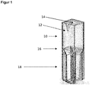
- illustration 1 shows a measuring cell that is constructed in the form of a measuring cuvette.
- the measuring cell contains a reaction chamber or first chamber (10) for receiving a sample volume of 10 ml, for example.
- a marker depot (12) with a predetermined amount of marker reagent, preferably in dry form.
- the first chamber (10) contains an opening (14) on one side for receiving the sample.
- the first chamber (10) is also connected to a second chamber (18) via a membrane (16).
- the membrane (16) is designed in such a way that it is impermeable to bacteria located in the first chamber (10), but permeable to labeling reagent that is not bound to bacteria.
- the liquid in the first chamber (10) is passed into the second chamber (18) after incubation with the marking reagent.
- Bacteria present in the sample with a marking reagent bound to it are deposited on the membrane (16). Unbound labeling reagent can penetrate the membrane (16) and enter the second chamber (18) with the liquid.
- the bacteria deposited on the membrane (16) can be measured in situ without any further measures.
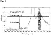
- Figure 2 shows the result of a spectral measurement on a sample that is positive for the bacteria to be determined, e.g. E. coli or Legionella.
- the measurement signal is recorded by a spectrometer, the measurement range of which is preferably between 350 and 550 nm.
- the optical resolution of the spectrometer is preferably 0.4-0.1 nm.
- the fluorescence signal from the bacteria to be determined is in the range between 510 and 520 nm (ROI).
- a reference laser ensures that the amplitude of the spectrum has a correct reference value.
- the evaluation includes the definition of two limit values for bacterial counts of 100 CFU per 100 ml and 10,000 CFU per 100 ml. In the figure, the intensity of the measurement signal exceeds the limit value of 10,000 CFU. As a result, the particular sample is extremely heavily contaminated.
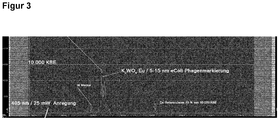
- Figure 3 shows the result of the determination of a sample with E. coli bacteria.
- E. coli phages coupled with the inorganic dye K 2 WO 4 : Eu (particle size 5-15 nm) were used as the labeling reagent.
- the bacteria contained in the sample were deposited on a membrane in a reaction chamber and determined there.
- the membrane was illuminated homogeneously with a laser (405 nm / 25 mW) and the membrane was illuminated at a wavelength of 610-615 nm generated emission radiation determined quantitatively.
- the quantitative conversion of the signal measured in corrected pulses with the number of colony-forming units (CFU) in the sample was possible by prior calibration using standard samples with a known number of E. coli bacteria, for example in the range between 10-10 9 cells.
- the spectrum also shows the peak of a Ca reference laser at 702 nm, which was irradiated with an intensity corresponding to 10% of the limit value of 10,000 CFU.
Landscapes
- Health & Medical Sciences (AREA)
- Life Sciences & Earth Sciences (AREA)
- Immunology (AREA)
- Engineering & Computer Science (AREA)
- Chemical & Material Sciences (AREA)
- Molecular Biology (AREA)
- Biomedical Technology (AREA)
- Hematology (AREA)
- Urology & Nephrology (AREA)
- Physics & Mathematics (AREA)
- General Health & Medical Sciences (AREA)
- Pathology (AREA)
- Food Science & Technology (AREA)
- Medicinal Chemistry (AREA)
- General Physics & Mathematics (AREA)
- Analytical Chemistry (AREA)
- Biochemistry (AREA)
- Biotechnology (AREA)
- Microbiology (AREA)
- Cell Biology (AREA)
- Tropical Medicine & Parasitology (AREA)
- Virology (AREA)
- Measuring Or Testing Involving Enzymes Or Micro-Organisms (AREA)
- Investigating, Analyzing Materials By Fluorescence Or Luminescence (AREA)
- Apparatus Associated With Microorganisms And Enzymes (AREA)
- Investigating Or Analysing Biological Materials (AREA)
- Optical Measuring Cells (AREA)
- Micro-Organisms Or Cultivation Processes Thereof (AREA)
Applications Claiming Priority (3)
| Application Number | Priority Date | Filing Date | Title |
|---|---|---|---|
| EP15181240 | 2015-08-17 | ||
| EP16753650.7A EP3338093B1 (fr) | 2015-08-17 | 2016-08-17 | Test rapide de bacteries |
| PCT/EP2016/069526 WO2017029332A1 (fr) | 2015-08-17 | 2016-08-17 | Test rapide pour déceler des bactéries |
Related Parent Applications (1)
| Application Number | Title | Priority Date | Filing Date |
|---|---|---|---|
| EP16753650.7A Division EP3338093B1 (fr) | 2015-08-17 | 2016-08-17 | Test rapide de bacteries |
Publications (1)
| Publication Number | Publication Date |
|---|---|
| EP3748359A1 true EP3748359A1 (fr) | 2020-12-09 |
Family
ID=53938144
Family Applications (2)
| Application Number | Title | Priority Date | Filing Date |
|---|---|---|---|
| EP16753650.7A Active EP3338093B1 (fr) | 2015-08-17 | 2016-08-17 | Test rapide de bacteries |
| EP20171507.5A Pending EP3748359A1 (fr) | 2015-08-17 | 2016-08-17 | Test bactérien rapide |
Family Applications Before (1)
| Application Number | Title | Priority Date | Filing Date |
|---|---|---|---|
| EP16753650.7A Active EP3338093B1 (fr) | 2015-08-17 | 2016-08-17 | Test rapide de bacteries |
Country Status (5)
| Country | Link |
|---|---|
| US (2) | US10823719B2 (fr) |
| EP (2) | EP3338093B1 (fr) |
| JP (1) | JP2018527567A (fr) |
| CN (1) | CN108431602B (fr) |
| WO (1) | WO2017029332A1 (fr) |
Families Citing this family (1)
| Publication number | Priority date | Publication date | Assignee | Title |
|---|---|---|---|---|
| EP4236766A1 (fr) * | 2020-11-02 | 2023-09-06 | Glucomat GmbH | Système de détection miniaturisé actif |
Citations (3)
| Publication number | Priority date | Publication date | Assignee | Title |
|---|---|---|---|---|
| EP0496345A1 (fr) * | 1991-01-22 | 1992-07-29 | NAGASE & COMPANY, LTD. | Méthode de détection et quantification de bactéries cariogénique |
| EP1277505A1 (fr) * | 2001-07-18 | 2003-01-22 | Fraunhofer-Gesellschaft Zur Förderung Der Angewandten Forschung E.V. | Appareil, procédé et système d'analyse de flux pour capter des particules immunogènes |
| EP2134642B1 (fr) | 2007-04-02 | 2016-02-24 | Emory University | Marqueurs de nanoparticules de SERS et procédée de détection spectroscopique de tumeurs |
Family Cites Families (5)
| Publication number | Priority date | Publication date | Assignee | Title |
|---|---|---|---|---|
| JP3270722B2 (ja) | 1996-09-27 | 2002-04-02 | オルガノ株式会社 | 細菌の検出方法及び検出装置 |
| US7880876B2 (en) * | 2004-10-21 | 2011-02-01 | University Of Georgia Research Foundation, Inc. | Methods of use for surface enhanced raman spectroscopy (SERS) systems for the detection of bacteria |
| US7608399B2 (en) * | 2006-06-26 | 2009-10-27 | Blood Cell Storage, Inc. | Device and method for extraction and analysis of nucleic acids from biological samples |
| GB201122158D0 (en) * | 2011-12-22 | 2012-02-01 | Ucl Business Plc | Fluorescent composition |
| AU2014281406A1 (en) | 2013-06-19 | 2016-01-21 | Sample6 Technologies, Inc. | Phage-based bacterial detection assay |
-
2016
- 2016-08-17 JP JP2018509821A patent/JP2018527567A/ja active Pending
- 2016-08-17 WO PCT/EP2016/069526 patent/WO2017029332A1/fr not_active Ceased
- 2016-08-17 US US15/753,303 patent/US10823719B2/en active Active
- 2016-08-17 EP EP16753650.7A patent/EP3338093B1/fr active Active
- 2016-08-17 CN CN201680058074.8A patent/CN108431602B/zh active Active
- 2016-08-17 EP EP20171507.5A patent/EP3748359A1/fr active Pending
-
2020
- 2020-09-28 US US17/034,800 patent/US11906497B2/en active Active
Patent Citations (3)
| Publication number | Priority date | Publication date | Assignee | Title |
|---|---|---|---|---|
| EP0496345A1 (fr) * | 1991-01-22 | 1992-07-29 | NAGASE & COMPANY, LTD. | Méthode de détection et quantification de bactéries cariogénique |
| EP1277505A1 (fr) * | 2001-07-18 | 2003-01-22 | Fraunhofer-Gesellschaft Zur Förderung Der Angewandten Forschung E.V. | Appareil, procédé et système d'analyse de flux pour capter des particules immunogènes |
| EP2134642B1 (fr) | 2007-04-02 | 2016-02-24 | Emory University | Marqueurs de nanoparticules de SERS et procédée de détection spectroscopique de tumeurs |
Non-Patent Citations (9)
| Title |
|---|
| "Mikrobiologischer Trinkwassertest in weniger als einer Stunde", 1 April 2009 (2009-04-01), XP055228170, Retrieved from the Internet <URL:http://www.wiley-vch.de/berlin/journals/op/09-01/OP0901_S28-S31.pdf> [retrieved on 20151112] * |
| ATTERBURY ET AL., APPL. ENVIRON. MICROBIOL., vol. 73, 2007, pages 4543 - 4549 |
| DELAPPE ET AL., J. MED. MICROBIOL., vol. 58, 2009 |
| LAMMETYN, MICROB. ECOL., vol. 56, 2000, pages 191 - 197 |
| ROMPRE A ET AL: "Detection and enumeration of coliforms in drinking water: Current methods and emerging approaches", JOURNAL OF MICROBIOLOGICAL METHODS, ELSEVIER, AMSTERDAM, NL, vol. 49, no. 1, 1 March 2002 (2002-03-01), pages 31 - 54, XP002359233, ISSN: 0167-7012, DOI: 10.1016/S0167-7012(01)00351-7 * |
| SAHIN ET AL., MIKROBIOL. BUL., vol. 47, 2013, pages 27 - 34 |
| ULRICH REIDT ET AL: "REPRODUCIBLE FILTRATION OF BACTERIA WITH MICROMECHANICAL FILTERS", JOURNAL OF RAPID METHODS AND AUTOMATION IN MICROBIOLOGY, vol. 16, no. 4, 1 December 2008 (2008-12-01), pages 337 - 350, XP055169725, ISSN: 1060-3999, DOI: 10.1111/j.1745-4581.2008.00140.x * |
| WANG ET AL., CHEM. REV., vol. 113, 2013, pages 1391 - 1428 |
| WICHARD ET AL., J. FOOD PROT, vol. 2, 2003, pages 178 - 340 |
Also Published As
| Publication number | Publication date |
|---|---|
| US20180238849A1 (en) | 2018-08-23 |
| CN108431602A (zh) | 2018-08-21 |
| WO2017029332A1 (fr) | 2017-02-23 |
| EP3338093B1 (fr) | 2020-04-29 |
| US20210018483A1 (en) | 2021-01-21 |
| CA2996038A1 (fr) | 2017-02-23 |
| US11906497B2 (en) | 2024-02-20 |
| EP3338093A1 (fr) | 2018-06-27 |
| CN108431602B (zh) | 2022-04-08 |
| US10823719B2 (en) | 2020-11-03 |
| JP2018527567A (ja) | 2018-09-20 |
Similar Documents
| Publication | Publication Date | Title |
|---|---|---|
| EP2152913B1 (fr) | Dispositif de détection permettant la détection de microparticules biologiques telles que des bactéries, des virus, des spores, du pollen ou des toxines biologiques, et procédé de détection | |
| Jakobsen et al. | FlowCAM: sizing cells and understanding the impact of size distributions on biovolume of planktonic community structure | |
| EP3380825B1 (fr) | Dispositif et procédé pour analyser des objets biologiques par spectroscopie raman | |
| EP3377902A1 (fr) | Procédé et dispositif de décèlement de bactéries | |
| EP3053646B1 (fr) | Dispositif et procede de detection de la resistance de bacteries par rapport a un agent actif a analyser a l'aide d'une puce microfluidique | |
| DE102014215735A1 (de) | Verfahren zum Betreiben einer raumlufttechnischen Anlage, Sensor und raum-lufttechnische Anlage | |
| EP1573051A1 (fr) | Procede et dispositif pour determiner des germes | |
| EP1846569B1 (fr) | Procede pour detecter des germes | |
| DE102020105123B3 (de) | Verfahren zum spektrometrischen Charakterisieren von Mikroorganismen | |
| EP3338093B1 (fr) | Test rapide de bacteries | |
| EP1664747B1 (fr) | Procede et dispositif pour identifier de tres petites quantites de particules | |
| WO2018197183A1 (fr) | Procédé et dispositif permettant d'obtenir une lignée cellulaire ayant une certaine expression d'une protéine cible souhaitée | |
| EP2583102A1 (fr) | Test de détermination des légionelles | |
| DE102009036562B4 (de) | Verfahren zur Bestimmung der Wasserqualität eines Gewässers | |
| DE19906047C2 (de) | Verfahren und Vorrichtung zur Detektion biotischer Kontaminationen auf einer Oberfläche | |
| EP1972928B1 (fr) | Dispositif et procédé de vérification non destructive des caractéristiques biostatiques ou biocides d'un revêtement de surface photocatalytique | |
| EP2613139A1 (fr) | Procédé destiné à l'analyse d'échantillons et système à cet effet | |
| EP2135072B1 (fr) | Dispositif et procédé pour le contrôle en ligne d'eau potable pour l'aptitude à la consommation humaine à l'intérieur d'un réseau de distribution d'eau potable | |
| DE10226731A1 (de) | Vorrichtung und Verfahren zur Online-Detektion biologischer Kontaminanten und deren Reste | |
| DE102017222366A1 (de) | Verfahren und Vorrichtung zum qualitativen und quantitativen Nachweis von in einem aquatischen System enthaltenen, biofilmbauenden Bakterien | |
| DE102016225009A1 (de) | Vorrichtung und Verfahren zur Bestimmung einer Ablagerung biologischen Materials | |
| WO2005033674A1 (fr) | Detection de nanoparticules | |
| DE102007032506A1 (de) | Vorrichtung und Verfahren zur zerstörungsfreien Prüfung der biostatischen bwz. bioziden Eigenschaften einer photokatalytischen Oberflächenbeschichtung |
Legal Events
| Date | Code | Title | Description |
|---|---|---|---|
| PUAI | Public reference made under article 153(3) epc to a published international application that has entered the european phase |
Free format text: ORIGINAL CODE: 0009012 |
|
| STAA | Information on the status of an ep patent application or granted ep patent |
Free format text: STATUS: THE APPLICATION HAS BEEN PUBLISHED |
|
| AC | Divisional application: reference to earlier application |
Ref document number: 3338093 Country of ref document: EP Kind code of ref document: P |
|
| AK | Designated contracting states |
Kind code of ref document: A1 Designated state(s): AL AT BE BG CH CY CZ DE DK EE ES FI FR GB GR HR HU IE IS IT LI LT LU LV MC MK MT NL NO PL PT RO RS SE SI SK SM TR |
|
| STAA | Information on the status of an ep patent application or granted ep patent |
Free format text: STATUS: REQUEST FOR EXAMINATION WAS MADE |
|
| 17P | Request for examination filed |
Effective date: 20210609 |
|
| RBV | Designated contracting states (corrected) |
Designated state(s): AL AT BE BG CH CY CZ DE DK EE ES FI FR GB GR HR HU IE IS IT LI LT LU LV MC MK MT NL NO PL PT RO RS SE SI SK SM TR |