EP4008730A1 - Anti-ctla4-anti-pd-bispezifischer antikörper und verwendungen davon - Google Patents
Anti-ctla4-anti-pd-bispezifischer antikörper und verwendungen davon Download PDFInfo
- Publication number
- EP4008730A1 EP4008730A1 EP20850855.6A EP20850855A EP4008730A1 EP 4008730 A1 EP4008730 A1 EP 4008730A1 EP 20850855 A EP20850855 A EP 20850855A EP 4008730 A1 EP4008730 A1 EP 4008730A1
- Authority
- EP
- European Patent Office
- Prior art keywords
- seq
- amino acid
- variable region
- chain variable
- set forth
- Prior art date
- Legal status (The legal status is an assumption and is not a legal conclusion. Google has not performed a legal analysis and makes no representation as to the accuracy of the status listed.)
- Pending
Links
Images


Classifications
-
- A—HUMAN NECESSITIES
- A61—MEDICAL OR VETERINARY SCIENCE; HYGIENE
- A61K—PREPARATIONS FOR MEDICAL, DENTAL OR TOILETRY PURPOSES
- A61K39/00—Medicinal preparations containing antigens or antibodies
- A61K39/395—Antibodies; Immunoglobulins; Immune serum, e.g. antilymphocytic serum
-
- C—CHEMISTRY; METALLURGY
- C07—ORGANIC CHEMISTRY
- C07K—PEPTIDES
- C07K16/00—Immunoglobulins [IG], e.g. monoclonal or polyclonal antibodies
- C07K16/18—Immunoglobulins [IG], e.g. monoclonal or polyclonal antibodies against material from animals or humans
- C07K16/28—Immunoglobulins [IG], e.g. monoclonal or polyclonal antibodies against material from animals or humans against receptors, cell surface antigens or cell surface determinants
- C07K16/2803—Immunoglobulins [IG], e.g. monoclonal or polyclonal antibodies against material from animals or humans against receptors, cell surface antigens or cell surface determinants against the immunoglobulin superfamily
- C07K16/2818—Immunoglobulins [IG], e.g. monoclonal or polyclonal antibodies against material from animals or humans against receptors, cell surface antigens or cell surface determinants against the immunoglobulin superfamily against CD28 or CD152
-
- A—HUMAN NECESSITIES
- A61—MEDICAL OR VETERINARY SCIENCE; HYGIENE
- A61K—PREPARATIONS FOR MEDICAL, DENTAL OR TOILETRY PURPOSES
- A61K31/00—Medicinal preparations containing organic active ingredients
- A61K31/33—Heterocyclic compounds
- A61K31/395—Heterocyclic compounds having nitrogen as a ring hetero atom, e.g. guanethidine or rifamycins
- A61K31/435—Heterocyclic compounds having nitrogen as a ring hetero atom, e.g. guanethidine or rifamycins having six-membered rings with one nitrogen as the only ring hetero atom
- A61K31/47—Quinolines; Isoquinolines
- A61K31/4709—Non-condensed quinolines and containing further heterocyclic rings
-
- A—HUMAN NECESSITIES
- A61—MEDICAL OR VETERINARY SCIENCE; HYGIENE
- A61K—PREPARATIONS FOR MEDICAL, DENTAL OR TOILETRY PURPOSES
- A61K31/00—Medicinal preparations containing organic active ingredients
- A61K31/33—Heterocyclic compounds
- A61K31/395—Heterocyclic compounds having nitrogen as a ring hetero atom, e.g. guanethidine or rifamycins
- A61K31/435—Heterocyclic compounds having nitrogen as a ring hetero atom, e.g. guanethidine or rifamycins having six-membered rings with one nitrogen as the only ring hetero atom
- A61K31/47—Quinolines; Isoquinolines
- A61K31/475—Quinolines; Isoquinolines having an indole ring, e.g. yohimbine, reserpine, strychnine, vinblastine
-
- A—HUMAN NECESSITIES
- A61—MEDICAL OR VETERINARY SCIENCE; HYGIENE
- A61K—PREPARATIONS FOR MEDICAL, DENTAL OR TOILETRY PURPOSES
- A61K39/00—Medicinal preparations containing antigens or antibodies
- A61K39/395—Antibodies; Immunoglobulins; Immune serum, e.g. antilymphocytic serum
- A61K39/39533—Antibodies; Immunoglobulins; Immune serum, e.g. antilymphocytic serum against materials from animals
- A61K39/3955—Antibodies; Immunoglobulins; Immune serum, e.g. antilymphocytic serum against materials from animals against proteinaceous materials, e.g. enzymes, hormones, lymphokines
-
- A—HUMAN NECESSITIES
- A61—MEDICAL OR VETERINARY SCIENCE; HYGIENE
- A61K—PREPARATIONS FOR MEDICAL, DENTAL OR TOILETRY PURPOSES
- A61K45/00—Medicinal preparations containing active ingredients not provided for in groups A61K31/00 - A61K41/00
-
- A—HUMAN NECESSITIES
- A61—MEDICAL OR VETERINARY SCIENCE; HYGIENE
- A61K—PREPARATIONS FOR MEDICAL, DENTAL OR TOILETRY PURPOSES
- A61K45/00—Medicinal preparations containing active ingredients not provided for in groups A61K31/00 - A61K41/00
- A61K45/05—Immunological preparations stimulating the reticulo-endothelial system, e.g. against cancer
-
- A—HUMAN NECESSITIES
- A61—MEDICAL OR VETERINARY SCIENCE; HYGIENE
- A61K—PREPARATIONS FOR MEDICAL, DENTAL OR TOILETRY PURPOSES
- A61K45/00—Medicinal preparations containing active ingredients not provided for in groups A61K31/00 - A61K41/00
- A61K45/06—Mixtures of active ingredients without chemical characterisation, e.g. antiphlogistics and cardiaca
-
- A—HUMAN NECESSITIES
- A61—MEDICAL OR VETERINARY SCIENCE; HYGIENE
- A61P—SPECIFIC THERAPEUTIC ACTIVITY OF CHEMICAL COMPOUNDS OR MEDICINAL PREPARATIONS
- A61P35/00—Antineoplastic agents
-
- A—HUMAN NECESSITIES
- A61—MEDICAL OR VETERINARY SCIENCE; HYGIENE
- A61P—SPECIFIC THERAPEUTIC ACTIVITY OF CHEMICAL COMPOUNDS OR MEDICINAL PREPARATIONS
- A61P35/00—Antineoplastic agents
- A61P35/02—Antineoplastic agents specific for leukemia
-
- A—HUMAN NECESSITIES
- A61—MEDICAL OR VETERINARY SCIENCE; HYGIENE
- A61P—SPECIFIC THERAPEUTIC ACTIVITY OF CHEMICAL COMPOUNDS OR MEDICINAL PREPARATIONS
- A61P7/00—Drugs for disorders of the blood or the extracellular fluid
- A61P7/06—Antianaemics
-
- C—CHEMISTRY; METALLURGY
- C07—ORGANIC CHEMISTRY
- C07K—PEPTIDES
- C07K16/00—Immunoglobulins [IG], e.g. monoclonal or polyclonal antibodies
- C07K16/18—Immunoglobulins [IG], e.g. monoclonal or polyclonal antibodies against material from animals or humans
- C07K16/28—Immunoglobulins [IG], e.g. monoclonal or polyclonal antibodies against material from animals or humans against receptors, cell surface antigens or cell surface determinants
-
- C—CHEMISTRY; METALLURGY
- C07—ORGANIC CHEMISTRY
- C07K—PEPTIDES
- C07K16/00—Immunoglobulins [IG], e.g. monoclonal or polyclonal antibodies
- C07K16/46—Hybrid immunoglobulins
-
- C—CHEMISTRY; METALLURGY
- C07—ORGANIC CHEMISTRY
- C07K—PEPTIDES
- C07K16/00—Immunoglobulins [IG], e.g. monoclonal or polyclonal antibodies
- C07K16/46—Hybrid immunoglobulins
- C07K16/468—Immunoglobulins having two or more different antigen binding sites, e.g. multifunctional antibodies
-
- C—CHEMISTRY; METALLURGY
- C12—BIOCHEMISTRY; BEER; SPIRITS; WINE; VINEGAR; MICROBIOLOGY; ENZYMOLOGY; MUTATION OR GENETIC ENGINEERING
- C12N—MICROORGANISMS OR ENZYMES; COMPOSITIONS THEREOF; PROPAGATING, PRESERVING, OR MAINTAINING MICROORGANISMS; MUTATION OR GENETIC ENGINEERING; CULTURE MEDIA
- C12N15/00—Mutation or genetic engineering; DNA or RNA concerning genetic engineering, vectors, e.g. plasmids, or their isolation, preparation or purification; Use of hosts therefor
- C12N15/09—Recombinant DNA-technology
- C12N15/11—DNA or RNA fragments; Modified forms thereof; Non-coding nucleic acids having a biological activity
- C12N15/62—DNA sequences coding for fusion proteins
-
- C—CHEMISTRY; METALLURGY
- C12—BIOCHEMISTRY; BEER; SPIRITS; WINE; VINEGAR; MICROBIOLOGY; ENZYMOLOGY; MUTATION OR GENETIC ENGINEERING
- C12N—MICROORGANISMS OR ENZYMES; COMPOSITIONS THEREOF; PROPAGATING, PRESERVING, OR MAINTAINING MICROORGANISMS; MUTATION OR GENETIC ENGINEERING; CULTURE MEDIA
- C12N15/00—Mutation or genetic engineering; DNA or RNA concerning genetic engineering, vectors, e.g. plasmids, or their isolation, preparation or purification; Use of hosts therefor
- C12N15/09—Recombinant DNA-technology
- C12N15/63—Introduction of foreign genetic material using vectors; Vectors; Use of hosts therefor; Regulation of expression
-
- G—PHYSICS
- G01—MEASURING; TESTING
- G01N—INVESTIGATING OR ANALYSING MATERIALS BY DETERMINING THEIR CHEMICAL OR PHYSICAL PROPERTIES
- G01N33/00—Investigating or analysing materials by specific methods not covered by groups G01N1/00 - G01N31/00
- G01N33/48—Biological material, e.g. blood, urine; Haemocytometers
- G01N33/50—Chemical analysis of biological material, e.g. blood, urine; Testing involving biospecific ligand binding methods; Immunological testing
- G01N33/53—Immunoassay; Biospecific binding assay; Materials therefor
- G01N33/575—Immunoassay; Biospecific binding assay; Materials therefor for cancer
- G01N33/5758—Immunoassay; Biospecific binding assay; Materials therefor for cancer involving compounds serving as markers for tumours, cancers or neoplasias, e.g. cellular determinants, receptors, heat shock/stress proteins, A-protein, oligosaccharides or metabolites
- G01N33/5759—Immunoassay; Biospecific binding assay; Materials therefor for cancer involving compounds serving as markers for tumours, cancers or neoplasias, e.g. cellular determinants, receptors, heat shock/stress proteins, A-protein, oligosaccharides or metabolites involving compounds localised on the membrane of tumour or cancer cells
-
- G—PHYSICS
- G01—MEASURING; TESTING
- G01N—INVESTIGATING OR ANALYSING MATERIALS BY DETERMINING THEIR CHEMICAL OR PHYSICAL PROPERTIES
- G01N33/00—Investigating or analysing materials by specific methods not covered by groups G01N1/00 - G01N31/00
- G01N33/48—Biological material, e.g. blood, urine; Haemocytometers
- G01N33/50—Chemical analysis of biological material, e.g. blood, urine; Testing involving biospecific ligand binding methods; Immunological testing
- G01N33/53—Immunoassay; Biospecific binding assay; Materials therefor
- G01N33/577—Immunoassay; Biospecific binding assay; Materials therefor involving monoclonal antibodies binding reaction mechanisms characterised by the use of monoclonal antibodies
-
- G—PHYSICS
- G01—MEASURING; TESTING
- G01N—INVESTIGATING OR ANALYSING MATERIALS BY DETERMINING THEIR CHEMICAL OR PHYSICAL PROPERTIES
- G01N33/00—Investigating or analysing materials by specific methods not covered by groups G01N1/00 - G01N31/00
- G01N33/48—Biological material, e.g. blood, urine; Haemocytometers
- G01N33/50—Chemical analysis of biological material, e.g. blood, urine; Testing involving biospecific ligand binding methods; Immunological testing
- G01N33/68—Chemical analysis of biological material, e.g. blood, urine; Testing involving biospecific ligand binding methods; Immunological testing involving proteins, peptides or amino acids
- G01N33/6872—Intracellular protein regulatory factors and their receptors, e.g. including ion channels
-
- G—PHYSICS
- G01—MEASURING; TESTING
- G01N—INVESTIGATING OR ANALYSING MATERIALS BY DETERMINING THEIR CHEMICAL OR PHYSICAL PROPERTIES
- G01N33/00—Investigating or analysing materials by specific methods not covered by groups G01N1/00 - G01N31/00
- G01N33/48—Biological material, e.g. blood, urine; Haemocytometers
- G01N33/50—Chemical analysis of biological material, e.g. blood, urine; Testing involving biospecific ligand binding methods; Immunological testing
- G01N33/68—Chemical analysis of biological material, e.g. blood, urine; Testing involving biospecific ligand binding methods; Immunological testing involving proteins, peptides or amino acids
- G01N33/6893—Chemical analysis of biological material, e.g. blood, urine; Testing involving biospecific ligand binding methods; Immunological testing involving proteins, peptides or amino acids related to diseases not provided for elsewhere
-
- A—HUMAN NECESSITIES
- A61—MEDICAL OR VETERINARY SCIENCE; HYGIENE
- A61K—PREPARATIONS FOR MEDICAL, DENTAL OR TOILETRY PURPOSES
- A61K39/00—Medicinal preparations containing antigens or antibodies
- A61K2039/505—Medicinal preparations containing antigens or antibodies comprising antibodies
-
- A—HUMAN NECESSITIES
- A61—MEDICAL OR VETERINARY SCIENCE; HYGIENE
- A61K—PREPARATIONS FOR MEDICAL, DENTAL OR TOILETRY PURPOSES
- A61K39/00—Medicinal preparations containing antigens or antibodies
- A61K2039/545—Medicinal preparations containing antigens or antibodies characterised by the dose, timing or administration schedule
-
- C—CHEMISTRY; METALLURGY
- C07—ORGANIC CHEMISTRY
- C07K—PEPTIDES
- C07K2317/00—Immunoglobulins specific features
- C07K2317/30—Immunoglobulins specific features characterized by aspects of specificity or valency
- C07K2317/31—Immunoglobulins specific features characterized by aspects of specificity or valency multispecific
-
- C—CHEMISTRY; METALLURGY
- C07—ORGANIC CHEMISTRY
- C07K—PEPTIDES
- C07K2317/00—Immunoglobulins specific features
- C07K2317/50—Immunoglobulins specific features characterized by immunoglobulin fragments
- C07K2317/52—Constant or Fc region; Isotype
-
- C—CHEMISTRY; METALLURGY
- C07—ORGANIC CHEMISTRY
- C07K—PEPTIDES
- C07K2317/00—Immunoglobulins specific features
- C07K2317/50—Immunoglobulins specific features characterized by immunoglobulin fragments
- C07K2317/52—Constant or Fc region; Isotype
- C07K2317/524—CH2 domain
-
- C—CHEMISTRY; METALLURGY
- C07—ORGANIC CHEMISTRY
- C07K—PEPTIDES
- C07K2317/00—Immunoglobulins specific features
- C07K2317/50—Immunoglobulins specific features characterized by immunoglobulin fragments
- C07K2317/56—Immunoglobulins specific features characterized by immunoglobulin fragments variable (Fv) region, i.e. VH and/or VL
- C07K2317/565—Complementarity determining region [CDR]
-
- C—CHEMISTRY; METALLURGY
- C07—ORGANIC CHEMISTRY
- C07K—PEPTIDES
- C07K2317/00—Immunoglobulins specific features
- C07K2317/60—Immunoglobulins specific features characterized by non-natural combinations of immunoglobulin fragments
- C07K2317/62—Immunoglobulins specific features characterized by non-natural combinations of immunoglobulin fragments comprising only variable region components
- C07K2317/622—Single chain antibody (scFv)
-
- C—CHEMISTRY; METALLURGY
- C07—ORGANIC CHEMISTRY
- C07K—PEPTIDES
- C07K2317/00—Immunoglobulins specific features
- C07K2317/70—Immunoglobulins specific features characterized by effect upon binding to a cell or to an antigen
- C07K2317/71—Decreased effector function due to an Fc-modification
-
- C—CHEMISTRY; METALLURGY
- C07—ORGANIC CHEMISTRY
- C07K—PEPTIDES
- C07K2317/00—Immunoglobulins specific features
- C07K2317/70—Immunoglobulins specific features characterized by effect upon binding to a cell or to an antigen
- C07K2317/73—Inducing cell death, e.g. apoptosis, necrosis or inhibition of cell proliferation
- C07K2317/732—Antibody-dependent cellular cytotoxicity [ADCC]
-
- C—CHEMISTRY; METALLURGY
- C07—ORGANIC CHEMISTRY
- C07K—PEPTIDES
- C07K2317/00—Immunoglobulins specific features
- C07K2317/90—Immunoglobulins specific features characterized by (pharmaco)kinetic aspects or by stability of the immunoglobulin
- C07K2317/92—Affinity (KD), association rate (Ka), dissociation rate (Kd) or EC50 value
-
- C—CHEMISTRY; METALLURGY
- C07—ORGANIC CHEMISTRY
- C07K—PEPTIDES
- C07K2319/00—Fusion polypeptide
-
- C—CHEMISTRY; METALLURGY
- C07—ORGANIC CHEMISTRY
- C07K—PEPTIDES
- C07K2319/00—Fusion polypeptide
- C07K2319/30—Non-immunoglobulin-derived peptide or protein having an immunoglobulin constant or Fc region, or a fragment thereof, attached thereto
-
- G—PHYSICS
- G01—MEASURING; TESTING
- G01N—INVESTIGATING OR ANALYSING MATERIALS BY DETERMINING THEIR CHEMICAL OR PHYSICAL PROPERTIES
- G01N2333/00—Assays involving biological materials from specific organisms or of a specific nature
- G01N2333/435—Assays involving biological materials from specific organisms or of a specific nature from animals; from humans
- G01N2333/705—Assays involving receptors, cell surface antigens or cell surface determinants
- G01N2333/70503—Immunoglobulin superfamily, e.g. VCAMs, PECAM, LFA-3
- G01N2333/70521—CD28, CD152
-
- G—PHYSICS
- G01—MEASURING; TESTING
- G01N—INVESTIGATING OR ANALYSING MATERIALS BY DETERMINING THEIR CHEMICAL OR PHYSICAL PROPERTIES
- G01N2800/00—Detection or diagnosis of diseases
- G01N2800/02—Nutritional disorders
-
- G—PHYSICS
- G01—MEASURING; TESTING
- G01N—INVESTIGATING OR ANALYSING MATERIALS BY DETERMINING THEIR CHEMICAL OR PHYSICAL PROPERTIES
- G01N2800/00—Detection or diagnosis of diseases
- G01N2800/22—Haematology
Definitions
- the present invention relates to the field of tumor treatment and molecular immunology, and particularly, to an anti-CTLA4/anti-PD-1 bispecific antibody and use thereof. More particularly, the present invention relates to mutant anti-CTLA4/anti-PD-1 bispecific antibodies.
- the transmembrane receptor PD-1 (programmed cell death protein 1) is a member of the CD28 family, and is expressed in activated T cells, B cells and myeloid cells. Both ligands of PD-1, PDL1 (programmed cell death 1 ligand 1, or PDL-1) and PDL2 (programmed cell death 1 ligand 2, or PDL-2), are members of the B7 superfamily. PDL1 is expressed in a variety of cells including T cells, B cells, endothelial cells and epithelial cells, and PDL2 is expressed only in antigen presenting cells such as dendritic cells and macrophages.
- PDL1 is expressed in a variety of cells including T cells, B cells, endothelial cells and epithelial cells
- PDL2 is expressed only in antigen presenting cells such as dendritic cells and macrophages.
- the PD-1/PDL1 signaling pathway plays an important role in regulating immune tolerance, microbial infection and tumor immune escape.
- PD-1 is mainly expressed in immune cells such as T cells, and the ligand PDL1 of PD-1 is highly expressed in a plurality of human tumor tissues.
- Blocking the PD-1/PDL1 signaling pathway may activate inhibited T cells, which thus attack cancer cells. Blocking the PD-1/PDL1 signaling can promote the proliferation of tumor antigen-specific T cells, activate tumor cell killing process and further inhibit local tumor growth ( Julie R et al., 2012, N Engl J Med., 366:2455-2465 ).
- tumors with high PDL1 expression are associated with cancers that are difficult to detect ( Hamanishi et al., 2007, Proc. Natl. Acad. Sci. USA, 104:3360-5 ).
- An effective method is administering an anti-PD-1 antibody to modulate the expression of PD-1. Due to the broad anti-tumor prospects and surprising efficacy of PD-1 antibodies, it is widely accepted in the industry that antibodies targeting the PD-1 pathway will bring about breakthroughs in the treatment of various tumors, for example, non-small cell lung cancer, renal cell carcinoma, ovarian cancer, melanoma ( Homet M.
- Cytotoxic T lymphocyte-associated antigen 4 and CD28 molecules are very similar in aspects of gene structure, chromosome location, sequence homology and gene expression. Both molecules are receptors of co-stimulatory molecule B7, and mainly expressed on the surface of activated T cells. Binding of CTLA4 to B7 inhibits the activation of mouse and human T cells, and plays a negative regulatory role in T cell activation.
- CTLA4 antibodies or anti-CTLA4 monoclonal antibodies
- CTLA4 ligands can prevent CTLA4 from binding to its natural ligands, thereby blocking the transmission of negative regulatory signals by CTLA4 to T cells and enhancing the reactivity of T cells to various antigens.
- CTLA4 monoclonal antibodies in clinical trials or approved for treating prostate cancer, bladder cancer, colorectal cancer, gastrointestinal cancer, liver cancer, malignant melanoma, etc. ( Grosso JF., Jure-Kunkel MN., 2013, Cancer Immun., 13:5 ).
- Interleukin 2 is produced by T cells. It is a growth factor that regulates T cell subgroups, and an important factor in regulating immune responses. It promotes the proliferation of activated B cells, and participates in antibody responses, hematopoiesis and tumor surveillance. Recombinant human IL-2 has been approved by the U.S. FDA for treating malignancies, including melanoma, renal tumor, etc., while a clinical study is currently ongoing for treating chronic viral infections ( Chavez, A.R., et al., 2009, Ann. N. Y. Acad. Sci., 1182:p.14-27 ). CTLA4 and CTLA4 antibodies are important influencing factors of T cell functions and interfere with the immune microenvironment in the body. In-vitro and in-vivo studies demonstrated that CTLA4 antibodies can specifically relieve the immunosuppression of CTLA4, activate T cells, and induce IL-2 generation, and is promising in wide applications in gene therapy against diseases such as tumors and parasite infections.
- CTLA4 antibodies can produce specific therapeutic effect on diseases and remarkable efficacy, and may be used for supplementing traditional medicines and for exploring new means of gene therapy.
- Bispecific antibodies also known as bifunctional antibodies, are specific drugs that target two different antigens simultaneously, and can be produced by immunomagnetic separation. Alternatively, they can be obtained by genetic engineering. The genetic engineering has flexibility in aspects of binding site optimization, synthetic form, yield and the like, thus having certain advantages. Currently, over 45 forms have been demonstrated ( Dafne Muller, Kontermann R E., 2010, BioDrugs, 24(2):89-98 ).
- bispecific antibodies are in the form of IgG-scFv, i.e., the Morrison format ( Coloma MJ, Morrison SL., 1997, Nat Biotechnol., 15:159-163 ), which has been demonstrated to be one of the ideal forms for the bispecific antibodies because of its similarity to the natually existing IgG forms and advantages in antibody engineering, expression and purification ( Miller BR, Demarest SJ, et al., 2010, Protein Eng Des Sel, 23:549-57 ; Fitzgerald J, Lugovskoy A., 2011, MAbs, 3:299-309 ).
- ADCC antibody-dependent cell-mediated cytotoxicity refers to killing of a target cell by a killer cell (NK cell, macrophage, etc.) that is mediated by binding of the Fab fragment of an antibody to an epitope of a virus-infected cell or a tumor cell and binding of the Fc fragment of the antibody to an Fc receptor (FcR) on the surface of the killer cell.
- NK cell killer cell
- FcR Fc receptor
- CDC complement dependent cytotoxicity refers to that the specific binding of an antibody to a corresponding antigen on a cell membrane surface forms a complex and activates the complement system, which further forms an MAC on the surface of the target cell resulting in subsequent target cell lysis. Complements may cause lysis of various bacteria and other pathogenic organisms, and are an important defense mechanism against pathogenic organism infections.
- Fc receptors belong to an immunoglobulin family that are expressed on the surface of specific immune cells to recognize antibody Fc regions and mediate immune responses. After the Fab region recognizes an antigen, the Fc region of the antibody binds to the Fc receptor on the immune cell (e.g., a killer cell) to initiate the response function of the immune cell, such as phagocytosis and ADCC.
- the immune cell e.g., a killer cell
- Fc receptors are mainly classified into three types, FcyR, FcaR and Fc ⁇ R.
- FcyR can be further classified into four subtypes, FcyRI (CD64), FcyRII (CD32), FcyRIII (CD16) and FcRn (neonatal Fc receptor).
- FcyRI, FcyRII and FcyRIII are closely associated with ADCC effect.
- FcyRIII is the most predominant molecule mediating ADCC, with two highly homologous subtypes, FcyRIIIa and FcyRIIIb, in different cell types.
- Fc ⁇ RIIIa populations two subtypes distinguished by sites of single nucleotide polymorphism (SNP), Fc ⁇ RIIIa_V158 with high affinity and Fc ⁇ RIIIa_F158 with low affinity, are present.
- FcyRI has higher affinity for the Fc region of IgG and participates in ADCC process;
- FcyRII comprises three subtypes, FcyRIIa, FcyRIIb and FcyRIIc (also referred to as CD32a, CD32b and CD32c, respectively), among which FcyRIIa has ADCC activity; for FcyRIIa, two subtypes, Fc ⁇ RIIa_H131 and Fc ⁇ RIIa_R131, are present in humans due to single nucleotide mutation;
- FcyRIIb is an inhibitory receptor, and is a typical inhibitory FcyR that inhibits nearby ITAM pathways.
- the Fc fragment binds to FcyRIIb on the same cell, negatively regulating B cell activation and decreasing secretion of antibodies and cytokines ( Hogarth PM, Pietersz GA., 2012, NATURE REVIEWS DRUG DISCOVERY, 11(4):311-331 ).
- the IgG family comprises four members, IgG1, IgG2, IgG3 and IgG4, which differ in amino acids in the fragment crystallizable (Fc) region of the heavy chain constant region, resulting in their varying affinities for FcyRs.
- IgG1 is the most abundant subtype in humans and is also the most common subtype used in monoclonal antibody medication. IgG1 is capable of binding various FcyRs and is able to induce ADCC and CDC effects.
- IgG2 has the lowest affinity for FcyRs, but is still able to induce monocyte-mediated ADCC by binding to FcyRIIa.
- IgG3 features the highest binding capacity to FcyRs, and can induce ADCC and a greater CDC effect than IgG1.
- IgG4 molecules demonstrate a weak binding to FcyRs other than FcyRI, having a lower probability of causing CDC and NK cell-mediated ADCC.
- antibodies of the IgG4 subtype can mediate ADCP effects through binding to FcyRI, and the ADCP effects, present in antibody therapies targeting immune cells, may cause damage to immune cells, posing pharmacological adverse effects.
- there is still a need for developing a novel anti-CTLA4/anti-PD-1 bispecific antibody to reduce or eliminate the damage caused by antibody-mediated ADCC, ADCP and/or CDC activity on immune cells to which the anti-CTLA4/anti-PD-1 bispecific antibody binds, and to improve the efficacy of the antibody therapy.
- Chemotherapies are currently mainly classified into the following nine classes ( He Jie, et al., Clinical Oncology, Beijing, People's Medical Publishing House, 2016:230-237 ).
- the first class are drugs that directly bind to DNA and prevent DNA replication, including various alkylating agents, mitomycin, bleomycin, dacarbazine, platinum-based drugs (e.g., cisplatin and carboplatin), camptothecins, and derivatives thereof.
- the second class are drugs for preventing nucleic acid biosynthesis, which mainly affect the enzyme system of tumor cells and block the synthesis of precursors of DNA and RNA, thereby inhibiting the formation of DNA or RNA, including methotrexate, fluorouracil, 6-mercaptopurine, hydroxyurea and cytarabine; such drugs mainly act on cells in S phase, and are antimetabolite chemotherapeutic drugs and cell cycle-specific anticancer drugs.
- the third class are chemotherapeutic drugs which affect transcription through the pharmacological mechanism that the drugs are inserted into the DNA double helix to form non-covalent binding with the DNA double helix, interfering with the transcription of genetic information on DNA to the DNA-dependent mRNA and causing compromised template function and hindered transcription.
- the fourth class are those affecting tubulin and mitosis, including vinca alkaloids, podophyllotoxins and taxanes.
- the fifth class are drugs affecting the function of ribosomes and blocking protein synthesis; representatives of such drugs are harringtonines, which inhibit the initiation of protein synthesis, decompose the ribosome and release new peptide chain, but do not block the binding of mRNA and tRNA to ribosomes; such drugs cause the reduction of nuclear DNA and cytoplasmic RNA and depolymerization of polysomes, and inhibit mitosis.
- the sixth class are drugs that affect the tumor cell membrane such as concanavalin (Con-A) and phytohemagglutinin (PHA); they can bind to glycoprotein receptors on the cell membrane, thereby affecting DNA synthesis in tumor cells and preventing tumor cell from dividing.
- Con-A concanavalin
- PHA phytohemagglutinin
- the seventh class are drugs that induce apoptosis, such as arsenic trioxide.
- the eighth class are hormones that treat tumors by regulating the endocrine system, including estrogens, antiestrogens, progestogens, androgens, antiandrogens, corticosteroids, and anticorticosteroids (including dichlorodiphenyldichloroethane and aminoglutethimide).
- the ninth class is anticancer targeted therapy, including monoclonal antibodies, epidermal growth factor signaling inhibitors (e.g., targeted drugs against receptor tyrosine kinase pathway), ubiquitin-proteasome inhibitors, and angiogenesis inhibitors.
- Anlotinib is a quinoline derivative tyrosine kinase inhibitor.
- TKI multi-target tyrosine kinase inhibitor
- the major targets include: receptor tyrosine kinases vascular endothelial growth factor receptors (VEGFRs) 1 to 3, epidermal growth factor receptor (EGFR), fibroblast growth factor receptors (FGFRs) 1 to 4, platelet-derived growth factor receptors (PDGFRs) ⁇ and ⁇ , and stem cell factor receptors (SCFRs) 7, 8 and 9.
- Example 24 of Patent No. WO2008112407 discloses a quinoline-derived tyrosine kinase inhibitor 1-[[[4-(4-fluoro-2-methyl-1 H -indol-5-yl)oxy-6-methoxyquinolin-7-yl]oxy]methyl] cyclopropylamine and a method for preparing the same.
- the structural formula of the quinoline-derived tyrosine kinase inhibitor is shown in formula I.
- Anlotinib hydrochloride is the hydrochloride salt of the compound of formula I.
- Lenvatinib an oral multiple tyrosine kinase inhibitor developed by Eisai (Japan), is a multi-target receptor tyrosine kinase inhibitor that inhibits the kinase activity of VEGFR1 (FLT1), VEGFR2 (KDR) and VEGFR3 (FLT4).
- lenvatinib also inhibits other receptor tyrosine kinases involved in pathogenic angiogenesis, tumor growth and cancer progression, including fibroblast growth factor (FGF) receptors FGFR1, FGFR2, FGFR3 and FGFR4, "rearranged during transfection" (RET) receptor, KIT and platelet-derived growth factor receptor ⁇ (PDGFR ⁇ ).
- FGF fibroblast growth factor
- RET platelet-derived growth factor receptor ⁇
- Lenvatinib also exhibits antiproliferative activity in hepatocellular carcinoma cell lines, which is dependent on activated FGFR signaling and simultaneous inhibition of phosphorylation of FGF receptor substrate
- lenvatinib 4-(3-chloro-4(cyclopropylaminocarbonyl)aminophenoxy)-7-methoxy-6-quinolinecarboxamide, is disclosed in Example 368 of U.S. Patent No. 7,612,208 .
- 7,253,286 discloses the mesylate salt form of lenvatinib (i.e., lenvatinib mesylate), named 4-[3-chloro-4-(cyclopropylureido)phenoxy]-7-methoxyquinoline-6-carboxamide mesylate, the chemical structure of which is provided below (formula II):
- lenvatinib mesylate i.e., lenvatinib mesylate
- the disease is still uncontrollable for a long term after chemotherapy, and the 5-year survival rate is still very low. Therefore, developing a medication or combination therapy with lower toxicity and higher efficacy is of great meaning.
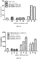
- the inventor correspondingly modified the Fc fragment of the anti-CTLA4/anti-PD-1 antibody structure to reduce the binding capacity of the Fc region to Fc receptors, thereby reducing ADCC, ADCP and/or CDC effects on immune cells and increasing the efficacy of the anti-CTLA4/anti-PD-1 antibody.
- bispecific antibody comprising:
- the affinity constant of the bispecific antibody to FcyRIIIa, FcyRI, Fc ⁇ RIIa_H131, Fc ⁇ RIIIa_V158 and/or FcyRIIb is reduced as compared to that before the mutation; preferably, the affinity constant is measured by a Fortebio Octet system.
- the heavy chain constant region of the immunoglobulin has the following mutations at positions 234, 235 and/or 237:
- letters before the position number represent amino acids before mutation
- letters after the position number represent amino acids after mutation, unless otherwise specified.
- the present invention further relates to a bispecific antibody, comprising:
- the heavy chain constant region of the immunoglobulin has one or more mutations selected from: N297A, D265A, D270A, P238D, L328E, E233D, H268D, P271G, A330R, C226S, C229S, E233P, P331S, S267E, L328F, A330L, M252Y, S254T, T256E, N297Q, P238S, P238A, A327Q, A327G, P329A, K322A, T394D, G236R, G236A, L328R, A330S, P331S, H268A, E318A and K320A.
- the bispecific antibody is in the form of IgG-scFv, i.e., the Morrison format.
- the bispecific antibody is selected from any one of the following (1)-(20):
- the amino acid sequence of the heavy chain of the immunoglobulin is set forth in SEQ ID NO: 40, and the amino acid sequence of the light chain of the immunoglobulin is set forth in SEQ ID NO: 24.
- the immunoglobulin or the antigen-binding fragment thereof binds to Fc ⁇ RIIIa_F158, FcyRI, Fc ⁇ RIIa_H131, Fc ⁇ RIIIa_V158 and/or FcyRIIb with an affinity constant greater than about 10 -7 M, for example, greater than about 10 -6 M, 10 -5 M, 10 -4 M, or 10 -3 M or greater; preferably, the affinity constant is measured by a Fortebio Octet system; preferably, the immunoglobulin or the antigen binding fragment thereof has no binding signal or a binding signal of less than 0.1 nm to Fc ⁇ RIIIa_F158, FcyRI, Fc ⁇ RIIa_H131, Fc ⁇ RIIIa_V158 and/or FcyRIIb; preferably, the binding signal refers to a response measured by a Fortebio Octet system.
- the immunoglobulin or the antigen-binding fragment thereof binds to C1q with an affinity constant greater than about 10 -9 M, for example, greater than about 10 -8 M, 10 -7 M, 10 -6 M, or 10 -5 M or greater; preferably, the affinity constant is measured by a Fortebio Octet system; preferably, the immunoglobulin or the antigen binding fragment thereof has no binding signal or a binding signal of less than 0.1 nm to C1q; preferably, the binding signal refers to a response measured by a Fortebio Octet system.
- the first protein functional region is linked to the second protein functional region either directly or via a linker fragment; and/or the heavy chain variable region of the single chain antibody is linked to the light chain variable region of the single chain antibody either directly or via a linker fragment.
- the linker fragment is (GGGGS)n, n being a positive integer; preferably, n is 1, 2, 3, 4, 5 or 6.
- the numbers of the first protein functional region and the second protein functional region are each independently 1, 2 or more.
- the number of the first protein functional region is 1 and the number of the second protein functional region is 2.
- the single chain antibody is linked to the C terminus of the heavy chain of the immunoglobulin. Since an immunoglobulin has two heavy chains, two single chain antibody molecules are linked to one immunoglobulin molecule. Preferably, the two single chain antibody molecules are identical. Preferably, the single chain antibody is linked to the C terminus of the heavy chain of the immunoglobulin by forming an amide bond via the aforementioned linker fragment.
- the constant regions of the immunoglobulin are humanized.
- the heavy chain constant region is Ig gamma-1 chain C region, ACCESSION: P01857
- the light chain constant region is Ig kappa chain C region, ACCESSION: P01834.
- the bispecific antibody binds to CTLA4 protein and/or PD-1 protein with a K D less than about 10 -5 M, e.g., less than about 10 -6 M, 10 -7 M, 10 -8 M, 10 -9 M or 10 -10 M or less.
- the bispecific antibody is a monoclonal antibody.
- the bispecific antibody is a humanized antibody.
- the bispecific antibody comprises:
- Another aspect of the present invention relates to an isolated nucleic acid molecule encoding the bispecific antibody according to any embodiment of the present invention.
- the present invention also relates to a vector comprising the isolated nucleic acid molecule of the present invention.
- the present invention also relates to a host cell comprising the isolated nucleic acid molecule of the present invention or the vector of the present invention.
- Another aspect of the present invention relates to a conjugate comprising an antibody or antigen-binding fragment thereof, and a conjugated moiety, wherein the immunoglobulin is the bispecific antibody according to any embodiment of the present invention, and the conjugated moiety is a detectable label; preferably, the conjugated moiety is a radioisotope, a fluorescent substance, a luminescent substance, a colored substance, or an enzyme.
- kits comprising the bispecific antibody according to any embodiment of the present invention or comprising the conjugate of the present invention; wherein preferably, the kit further comprises a second antibody capable of specifically recognizing the immunoglobulin or the antigen binding fragment thereof; optionally, the second antibody further comprises a detectable label, for example, a radioisotope, a fluorescent substance, a luminescent substance, a colored substance, or an enzyme.
- a detectable label for example, a radioisotope, a fluorescent substance, a luminescent substance, a colored substance, or an enzyme.
- Another aspect of the present invention relates to use of the bispecific antibody or the conjugate according to any embodiment of the present invention in preparing a kit for detecting the presence or level of PD-1 and/or CTLA4 in a sample.
- Another aspect of the present invention relates to a pharmaceutical composition
- a pharmaceutical composition comprising the bispecific antibody or the conjugate according to any embodiment of the present invention; optionally, the pharmaceutical composition further comprises a pharmaceutically acceptable carrier and/or excipient.
- the pharmaceutical composition further comprises one or more anti-tumor chemotherapeutics; preferably, the anti-tumor chemotherapeutic is a tyrosine kinase inhibitor; more preferably, the anti-tumor chemotherapeutic is anlotinib or a pharmaceutically acceptable salt thereof (e.g., hydrochloride salt), or lenvatinib or a pharmaceutically acceptable salt thereof (e.g., mesylate salt).
- the anti-tumor chemotherapeutic is a tyrosine kinase inhibitor
- the anti-tumor chemotherapeutic is anlotinib or a pharmaceutically acceptable salt thereof (e.g., hydrochloride salt), or lenvatinib or a pharmaceutically acceptable salt thereof (e.g., mesylate salt).
- the unit dose of the pharmaceutical composition is 100-1000 mg, 200-800 mg, 200-500 mg, 300-600 mg, 400-500 mg, or 450 mg, based on the mass of the bispecific antibody.
- Another aspect of the present invention relates to a combination product comprising a first product and a second product in separate packages,
- the unit dose of the first product is 100-1000 mg, 200-800 mg, 200-500 mg, 300-600 mg, 400-500 mg, or 450 mg, based on the mass of the bispecific antibody.
- the unit dose of the second product is 0.1-100 mg, 0.5-50 mg, 0.5-10 mg, 1-10 mg, 2-8 mg, or 1-5 mg, based on the mass of the active ingredient.
- the unit dose of the second product is 1-20 mg, 2-15 mg, 4-12 mg, or 8-12 mg, based on the mass of the active ingredient.
- Another aspect of the present invention relates to use of the bispecific antibody, the conjugate, the pharmaceutical composition or the combination product according to any embodiment of the present invention in preparing a medicament for treating and/or preventing a tumor or anemia, or in preparing a medicament for diagnosing a tumor or anemia;
- the tumor is selected from one or more of melanoma, renal cancer, prostate cancer, bladder cancer, colon cancer, rectal cancer, gastric cancer, liver cancer, lung cancer, ovarian cancer, leukemia, breast cancer, mesothelioma, cervical cancer, endometrial cancer, lymphoma and nasopharyngeal cancer;
- MSI refers to microsatellite instability. Microsatellites are short tandem repeats throughout the human genome, including 10-50 repeats of one, two or more nucleotides. Microsatellites in certain abnormal cells, such as tumors, are altered in length by insertion or deletion of repeat units as compared to normal cells. Such alteration is referred to as MSI. Based on instability and extent, MSI can be classified as microsatellite instability-high (MSI-H), microsatellite instability-low (MSI-L) and microsatellite stable (MSS). The major cause of MSI is DNA mismatch repair (MMR). Human mismatch repair genes (MMR genes) can express corresponding mismatch repair proteins through transcription and translation.
- MMR DNA mismatch repair
- MSI-H and dMMR represent the results of two different assays and are biologically consistent, called MSI-H/dMMR or MSI-high/dMMR, while MSI-L and MSS are phenotypes of proficient MMR (pMMR).
- the detection of dMMR is to carry out immunohistochemistry of protein expression for four mismatch genes of MSH2, MLH1, MSH6 and PMS2 based on tumor specimens (including surgical specimens and aspiration specimens). Absence of any of the four proteins confirms the dMMR; positive results of all the four proteins indicate pMMR, i.e., a complete mismatch repair function.
- MSI The detection of MSI is to match the length of the repeated DNA sequences (microsatellite sequences) in tumor cells and somatic cells, and to compare the lengths.
- MSI-H 5 standard loci are detected using PCR based on the American NCI standard
- inconsistencies in two or more loci indicate instability, defined as MSI-H
- one inconsistent locus indicates MSI-L
- 5 consistent loci indicate MSS.
- High-throughput sequencing also referred to as next-generation sequencing, or NGS
- NGS next-generation sequencing
- inconsistency in ⁇ 30% loci is defined as MSI-H
- consistency in all loci is defined as MSS
- inconsistency between 0 and 30% is defined as MSI-L.
- Another aspect of the present invention relates to a method for the prevention and/or
- the administration is before or after a surgical treatment and/or before or after a radiotherapy.
- the administration of the anti-CTLA4/anti-PD-1 bispecific antibody is performed in cycles of 2 weeks (14 days) or 3 weeks (21 days), and preferably, the anti-CTLA4/anti-PD-1 bispecific antibody is administered intravenously on the first day (D1) of each cycle.
- the anti-CTLA4/anti-PD-1 bispecific antibody is administered once every two weeks (q2w) or three weeks (q3w).
- the bispecific antibody, the conjugate, the pharmaceutical composition or the combination product according to any embodiment of the present invention for use in the prevention and/or treatment and/or adjuvant treatment and/or diagnosis of a tumor; preferably, the tumor is selected from one or more of melanoma, renal cancer, prostate cancer, bladder cancer, colon cancer, rectal cancer, gastric cancer, liver cancer, lung cancer, ovarian cancer, leukemia, breast cancer, mesothelioma, cervical cancer, endometrial cancer, lymphoma and nasopharyngeal cancer;
- Antibody drugs especially monoclonal antibodies (mAbs) have achieved good efficacy in the treatment of various diseases.
- Traditional experimental methods for acquiring these therapeutic antibodies are to immunize animals with the antigen and acquire antibodies targeting the antigen in the immunized animals, or to improve those antibodies with lower affinity for the antigen by affinity maturation.
- variable regions of the light chain and the heavy chain determine the binding of the antigen; the variable region of each chain comprises three hypervariable regions, i.e., complementarity determining regions (CDRs)
- CDRs of the heavy chain (H) include HCDR1, HCDR2, HCDR3, and the CDRs of the light chain (L) include LCDR1, LCDR2, LCDR3; defined by Kabat et al., see Sequences of Proteins of Immunological Interest, Fifth Edition (1991), Volumes 1-3, NIH Publication 91-3242, Bethesda Md ).
- amino acid sequence of the heavy chain variable region is set forth in SEQ ID NO: 14, and the amino acid sequence of the light chain variable region is set forth in SEQ ID NO: 16.
- amino acid sequences of the 3 CDR regions of the heavy chain variable region are as follows:
- amino acid sequences of the 3 CDR regions of the light chain variable region are as follows:
- amino acid sequence of the heavy chain variable region is set forth in SEQ ID NO: 18, and the amino acid sequence of the light chain variable region is set forth in SEQ ID NO: 20.
- amino acid sequences of the 3 CDR regions of the heavy chain variable region are the same as 14C12.
- amino acid sequences of the 3 CDR regions of the light chain variable region are the same as 14C12.
- amino acid sequence of the heavy chain variable region is set forth in SEQ ID NO: 2
- amino acid sequence of the light chain variable region is set forth in SEQ ID NO: 4.
- amino acid sequences of the 3 CDR regions of the heavy chain variable region are as follows:
- amino acid sequences of the 3 CDR regions of the light chain variable region are as follows:
- amino acid sequence of the heavy chain variable region is set forth in SEQ ID NO: 6, and the amino acid sequence of the light chain variable region is set forth in SEQ ID NO: 8;
- amino acid sequences of the 3 CDR regions of the heavy chain variable region are the same as 4G10.
- amino acid sequences of the 3 CDR regions of the light chain variable region are the same as 4G10.
- amino acid sequence of the heavy chain variable region is set forth in SEQ ID NO: 10
- amino acid sequence of the light chain variable region is set forth in SEQ ID NO: 12.
- amino acid sequences of the 3 CDR regions of the heavy chain variable region are the same as 4G10.
- amino acid sequences of the 3 CDR regions of the light chain variable region are the same as 4G10.
- amino acid sequences of the 9 CDR regions associated with the heavy chain variable region are as follows:
- amino acid sequences of the 3 CDR regions associated with the light chain variable region are as follows:
- amino acid sequences of the 9 CDR regions associated with the heavy chain variable region are the same as BiAb001(M).
- amino acid sequences of the 3 CDR regions associated with the light chain variable region are the same as BiAb001(M).
- amino acid sequences of the 9 CDR regions associated with the heavy chain variable region are the same as BiAb001(M).
- amino acid sequences of the 3 CDR regions associated with the light chain variable region are the same as BiAb001(M).
- amino acid sequences of the 9 CDR regions associated with the heavy chain variable region are the same as BiAb001(M).
- amino acid sequences of the 3 CDR regions associated with the light chain variable region are the same as BiAb001(M).
- BiAb004(hG1TM) of the present invention amino acid mutations are introduced into the non-variable region of BiAb004(M). According to the EU numbering system, amino acid mutations are introduced at positions 234, 235 and 237: BiAb004(hG1TM) is obtained by introducing a leucine-to-alanine point mutation at position 234 (L234A), a leucine-to-alanine point mutation at position 235 (L235A), and a glycine-to-alanine point mutation at position 237 (G237A) in the hinge region of the heavy chain.
- L234A leucine-to-alanine point mutation at position 234
- L235A leucine-to-alanine point mutation at position 235
- G237A glycine-to-alanine point mutation at position 237
- CTLA4 protein cytotoxic T-lymphocyte antigen 4
- it includes the full length of CTLA4 protein, or the extracellular fragment CTLA4ECD of CTLA4 or a fragment comprising CTLA4ECD; also included are fusion proteins of CTLA4ECD, such as a fragment fused to an Fc protein fragment of a mouse or human IgG (mFc or hFc).
- fusion proteins of CTLA4ECD such as a fragment fused to an Fc protein fragment of a mouse or human IgG (mFc or hFc).
- CTLA4 protein should include all such sequences, including their natural or artificial variants.
- sequence fragment of the CTLA4 protein it also includes the corresponding sequence fragments in its natural or artificial variants.
- PD-1 protein when referring to the amino acid sequence of PD-1 protein (NCBI GenBank: NM_005018), it includes the full length of the PD-1 protein, or the extracellular fragment PD-1ECD of PD-1 or a fragment comprising PD-1ECD; also included are fusion proteins of PD-1ECD, such as a fragment fused to an Fc protein fragment of a mouse or human IgG (mFc or hFc).
- mFc or hFc Fc protein fragment of a mouse or human IgG
- PD-1 protein should include all such sequences, including their natural or artificial variants.
- sequence fragment of the PD-1 protein when describing the sequence fragment of the PD-1 protein, it also includes the corresponding sequence fragments in its natural or artificial variants.
- the B7 is B7-1 and/or B7-2; specific sequences thereof are those known in the prior art, and reference may be made to sequences disclosed in the existing literature or GenBank.
- B7-1 CD80, NCBI Gene ID: 941
- B7-2 CD86, NCBI Gene ID: 942
- EC 50 refers to the concentration for 50% of maximal effect, i.e., the concentration that can cause 50% of the maximal effect.
- the term "antibody” refers to an immunoglobulin molecule that generally consists of two pairs of polypeptide chains (each pair with one "light” (L) chain and one "heavy” (H) chain).
- the heavy chain can be interpreted as a polypeptide chain with a larger molecular weight in an antibody
- the light chain refers to a polypeptide chain with a smaller molecular weight in an antibody.
- Light chains are classified as ⁇ and ⁇ light chains.
- Heavy chains are generally classified as ⁇ , ⁇ , ⁇ , ⁇ , or ⁇ , and isotypes of antibodies are defined as IgM, IgD, IgG, IgA, and IgE, respectively.
- variable region and constant region are linked by a "J" region of about 12 or more amino acids, and the heavy chain also comprises a "D" region of about 3 or more amino acids.
- Each heavy chain consists of a heavy chain variable region (V H ) and a heavy chain constant region (C H ).
- the heavy chain constant region consists of 3 domains (C H1 , C H2 , and C H3 ).
- Each light chain consists of a light chain variable region (V L ) and a light chain constant region (C L ).
- the light chain constant region consists of one domain C L .
- the constant region of the antibody can mediate the binding of immunoglobulins to host tissues or factors, including the binding of various cells of the immune system (e.g., effector cells) to the first component (C1q) of classical complement system.
- the V H and V L regions can be further subdivided into highly variable regions (called Complementarity Determining Regions (CDRs)), between which conservative regions called framework regions (FRs) are distributed.
- CDRs Complementarity Determining Regions
- FRs framework regions
- Each V H and V L consists of 3 CDRs and 4 FRs arranged from amino terminus to carboxyl terminus in the following order: FR1, CDR1, FR2, CDR2, FR3, CDR3, FR4.
- the variable regions (V H and V L ) of each heavy chain/light chain pair form antibody binding sites, respectively.
- the heavy chain may also comprise more than 3 CDRs, such as 6, 9, or 12.
- the heavy chain may be an scFv with the C terminus of the heavy chain of IgG antibody linked to another antibody, and in this case, the heavy chain comprises 9 CDRs.
- antibody is not limited by any specific method for producing antibody.
- the antibody includes, in particular, a recombinant antibody, a monoclonal antibody, and a polyclonal antibody.
- Antibodies can be different isotypes, such as IgG (e.g., subtype IgG1, IgG2, IgG3 or IgG4), IgA1, IgA2, IgD, IgE or IgM.
- Antigen binding fragments e.g., the above mentioned antibody fragments
- antibodies can be obtained from given antibodies by using conventional techniques known to those skilled in the art (e.g., DNA recombination, or enzymatic or chemical cleavage), and the antigen binding fragments of the antibodies are screened for specificity in the same way as for intact antibodies.
- antibody As used herein, unless otherwise clearly defined in the context, when referring to the term “antibody”, it includes not only intact antibodies but also antigen binding fragments of antibodies.
- mAb and “monoclonal antibody” refer to an antibody or a fragment thereof that is derived from a group of highly homologous antibodies, i.e. from a group of identical antibody molecules, except for natural mutations that may occur spontaneously.
- the monoclonal antibody is highly specific for a single epitope on an antigen.
- the polyclonal antibody, relative to the monoclonal antibody generally comprises at least two or more different antibodies which generally identify different epitopes on an antigen.
- Monoclonal antibodies can generally be obtained using hybridoma technique first reported by Kohler et al. ( Kohler et al., Nature, 256:495, 1975 ), but can also be obtained using DNA recombination (see, e.g., U.S. Patent No. 4,816,567 ).
- humanized antibody refers to an antibody or antibody fragment obtained when all or a part of CDR regions of a human immunoglobulin (receptor antibody) are replaced by the CDR regions of a non-human antibody (donor antibody), wherein the donor antibody may be a non-human (e.g., mouse, rat or rabbit) antibody having expected specificity, affinity or reactivity.
- donor antibody may be a non-human (e.g., mouse, rat or rabbit) antibody having expected specificity, affinity or reactivity.
- some amino acid residues in the framework regions (FRs) of the receptor antibody can also be replaced by the amino acid residues of corresponding non-human antibodies or by the amino acid residues of other antibodies to further improve or optimize the performance of the antibody.
- epitope refers to a site on the antigen that an immunoglobulin or antibody specifically binds to. "Epitope” is also called in the field as an "antigenic determinant".
- the epitope or antigenic determinant generally consists of chemically active surface groups of molecules such as amino acids, carbohydrates or sugar side chains, and usually has specific three-dimensional structural characteristics and specific charge characteristics.
- the epitope generally comprises at least 3, 4, 5, 6, 7, 8, 9, 10, 11, 12, 13, 14, or 15 consecutive or non-consecutive amino acids in a unique spatial conformation, which can be "linear” or “conformational”. See, e.g., Epitope Mapping Protocols in Methods in Molecular Biology, Vol. 66, G. E.
- isolated refers to obtained by artificial means from natural state. If a certain "isolated” substance or component appears in nature, it may be the case that change occurs in its natural environment, or that it is isolated from the natural environment, or both. For example, a certain non-isolated polynucleotide or polypeptide naturally exists in a certain living animal, and the same polynucleotide or polypeptide with a high purity isolated from such a natural state is called isolated polynucleotide or polypeptide.
- isolated does not exclude the existence of artificial or synthetic substances or other impurities that do not affect the activity of the substance.
- E. coli expression system refers to an expression system consisting of E. coli (strain) and a vector, wherein the E. coli (strain) is derived from a commercially available strain, such as but not limited to GI698, ER2566, BL21 (DE3), B834 (DE3), and BLR (DE3).
- vector refers to a nucleic acid vehicle into which a polynucleotide can be inserted.
- a vector allows for the expression of the protein encoded by the inserted polynucleotide
- the vector is called an expression vector.
- a vector can be introduced into a host cell by transformation, transduction, or transfection so that the genetic substance elements carried by the vector can be expressed in the host cell.
- Vectors are well known to those skilled in the art, including but not limited to: plasmids; phagemids; cosmids; artificial chromosomes, such as yeast artificial chromosome (YAC), bacterial artificial chromosome (BAC), or P1-derived artificial chromosome (PAC); phages such as lambda phages or M13 phages; and animal viruses.
- artificial chromosomes such as yeast artificial chromosome (YAC), bacterial artificial chromosome (BAC), or P1-derived artificial chromosome (PAC)
- phages such as lambda phages or M13 phages
- animal viruses include but not limited to: plasmids; phagemids; cosmids; artificial chromosomes, such as yeast artificial chromosome (YAC), bacterial artificial chromosome (BAC), or P1-derived artificial chromosome (PAC); phages such as lambda phages or M13 phag
- Animal viruses that can be used as vectors include, but are not limited to retroviruses (including lentiviruses), adenoviruses, adeno-associated viruses, herpes viruses (such as herpes simplex virus), poxviruses, baculoviruses, papillomaviruses, and papovaviruses (such as SV40).
- retroviruses including lentiviruses
- adenoviruses such as lentiviruses
- adeno-associated viruses such as herpes simplex virus
- poxviruses such as herpes simplex virus
- baculoviruses such as baculoviruses
- papillomaviruses papillomaviruses
- papovaviruses such as SV40.
- a vector may comprise a variety of elements that control expression, including, but not limited to promoter sequences, transcription initiation sequences, enhancer sequences, selection elements, and reporter genes
- the term "host cell” refers to cells to which vectors can be introduced, including, but not limited to, prokaryotic cells such as E. coli or bacillus subtilis, fungal cells such as yeast cells or aspergillus, insect cells such as S2 drosophila cells or Sf9, or animal cells such as fibroblasts, CHO cells, COS cells, NSO cells, HeLa cells, BHK cells, HEK 293 cells, or human cells.
- prokaryotic cells such as E. coli or bacillus subtilis
- fungal cells such as yeast cells or aspergillus
- insect cells such as S2 drosophila cells or Sf9
- animal cells such as fibroblasts
- CHO cells COS cells, NSO cells, HeLa cells, BHK cells, HEK 293 cells, or human cells.
- the term “specifically bind” refers to a non-random binding reaction between two molecules, such as a reaction between an antibody and an antigen it targets.
- an antibody that specifically binds to an antigen refers to that the antibody binds to the antigen with an affinity (K D ) of less than about 10 -5 M, such as less than about 10 -6 M, 10 -7 M, 10 -8 M, 10 -9 M or 10 -10 M or less.
- affinity K D
- target refers to specific binding.
- K D refers to a dissociation equilibrium constant for a specific antibody-antigen interaction, which is used to describe the binding affinity between the antibody and the antigen.
- an antibody binds to an antigen with a dissociation equilibrium constant (K D ) of less than about 10 -5 M, such as less than about 10 -6 M, 10 -7 M, 10 -8 M, 10 -9 M or 10 -10 M or less, for example, as measured by a BIACORE surface plasmon resonance (SPR) instrument or a Fortebio Octet system.
- SPR surface plasmon resonance
- the terms “monoclonal antibody” and “mAb” have the same meaning and can be used interchangeably; the terms “polyclonal antibody” and “pAb” have the same meaning and can be used interchangeably; the terms “polypeptide” and “protein” have the same meaning and can be used interchangeably.
- amino acids are generally represented by single-letter and three-letter abbreviations known in the art.
- alanine can be represented by A or Ala.
- the term "pharmaceutically acceptable carrier and/or excipient” refers to a carrier and/or excipient that is pharmacologically and/or physiologically compatible with the subject and the active ingredient, which is well known in the art (see, e.g., Remington's Pharmaceutical Sciences, edited by Gennaro AR, 19th ed., Pennsylvania, Mack Publishing Company, 1995 ), including but not limited to: pH regulators, surfactants, adjuvants, and ionic strength enhancers.
- the pH regulators include, but are not limited to, phosphate buffer;
- the surfactants include, but are not limited to, cationic, anionic, or non-ionic surfactants, such as Tween-80;
- the ionic strength enhancers include, but are not limited to, sodium chloride.
- adjuvant refers to a non-specific immune enhancer, which can enhance the immune response of an organism to antigens or change the type of immune response when delivered into the organism together with the antigens or delivered into the organism in advance.
- adjuvants including, but not limited to, aluminum adjuvant (e.g., aluminum hydroxide), Freund's adjuvant (e.g., complete Freund's adjuvant and incomplete Freund's adjuvant), Corynebacterium parvum, lipopolysaccharide, cytokine, etc.
- the Freund's adjuvant is the most commonly used adjuvant in animal experiments.
- the aluminum hydroxide adjuvant is used more in clinical trials.
- the term "effective amount" refers to an amount sufficient to obtain or at least partially obtain desired effect.
- a prophylactically effective amount e.g., for a disease associated with binding of CTLA4 to B7 or CTLA4 overactivity, such as a tumor
- a therapeutically effective amount is an amount sufficient to cure or at least partially stop a disease and its complications in a patient suffering from the disease. It is undoubtedly within the ability of those skilled in the art to determine such an effective amount.
- the amount effective for therapeutic purpose will depend on the severity of the disease to be treated, the overall state of the patient's own immune system, the general condition of the patient such as age, weight and gender, the route of administration, and other treatments given concurrently, etc.
- a “recurrent” cancer is one that regenerates at the original site or a distant site after response to a previous treatment (e.g., surgery).
- a “locally recurrent” cancer is one that occurs at the same site after treatment as the previously treated cancer.
- a “metastatic” cancer refers to one that spreads from one part of the body (e.g., the lungs) to another.
- the term "completely eliminated” refers to the absence of binding signal or an extremely weak binding signal as detected by existing instrumentation (e.g., a Fortebio Octet system).
- the absence of binding signal or the extremely weak binding signal refers to a binding signal (i.e., response) below 0.1 nm.
- first e.g., first protein functional region, first linker fragment or first product
- second e.g., second protein functional region, second linker fragment or second product
- “about” or “approximately” refers to that the value or physical quantity defined fluctuates within a range of 10%, 20% or 30%, unless otherwise specified.
- about 100 minutes or approximately 100 minutes may be 90 minutes to 110 minutes, 80 minutes to 120 minutes or 70 minutes to 130 minutes.
- the present invention achieves one or more of the following technical effects (1) to (3):
- BALB/c mice were purchased from Guangdong Medical Laboratory Animal Center.
- Raji-PDL1 is a cell expressing human PD-L1 constructed by Akeso Biopharma on the basis of human B cells Raji via transfection.
- Ficoll-Paque TM PLUS (or Ficoll-Paque PLUS) was purchased from GE Healthcare.
- Human IL-2 ELISA kit was purchased from Dakewe Biotech Co., Ltd.
- RPMI 1640 medium, DMEM medium, Trypsin-EDTA (0.25%) phenol red and Blastidin were all purchased from Gibco.
- Staphylococcus aureus enterotoxin B (SEB) was purchased from Dianotech.
- FBS was purchased from Excell bio.
- Mitomycin C was purchased from Stressmarq.
- the sequence of the isotype control, human anti-hen egg lysozyme IgG is derived from the variable region sequence of the Fab F10.6.6 sequence in the study reported by Acierno et al., entitled “Affinity maturation increases the stability and plasticity of the Fv domain of anti-protein antibodies” (Acierno et al., J Mol Biol., 2007; 374(1):130-146 ).
- Anlotinib used in the examples is hydrochloride salt of anlotinib under the brand name Fukewei ® and generic name anlodinib hydrochloride, and was purchased from CTTQ Pharma.
- Lenvatinib used in the examples is lenvatinib mesylate under the brand name Lenvima ® , and was purchased from Eisai (China).
- amino acid sequences and encoding nucleotide sequences of the heavy and light chains of the anti-CTLA4 antibody 4G10 and its humanized antibodies 4G10H1L1 and 4G10H3L3 are identical to those of 4G10, 4G10H1L1 and 4G10H3L3 in Chinese Patent Publication No. CN106967172A , respectively.
- amino acid sequences and encoding nucleotide sequences of the heavy and light chains of anti-PD-1 antibody 14C12 and its humanized antibody 14C12H1L1 are identical to those of 14C12 and 14C12H1L1 in Chinese Patent Publication No. CN106967172A , respectively.
- bifunctional antibodies BiAb001(M), BiAb002(M), BiAb003(M) and BiAb004(M) are in the Morrison format (IgG-scFv), i.e., C termini of two heavy chains of one IgG antibody are separately linked to the scFv fragment of another antibody via linker fragments.
- the components for the heavy and light chain design are shown in Table A below.
- scFv fragments 4G10H1V(M), 4G10L1V(M), 4G10H3V(M) and 4G10L3V(M) of BiAb001(M), BiAb002(M), BiAb003(M) and BiAb004(M) antibodies comprised mutations in individual amino acids of framework regions on the basis of 4G10H1V, 4G10L1V, 4G10H3V and 4G10L3V, respectively, which effectively optimized the structure of the antibodies and improved their efficacy.
- BiAb004(M) is also referred to as BiAb004(hG1WT) in the this example.
- BiAb004(M) described above is the "wild-type", comprising an Ig gamma-1 chain C region (ACCESSION: P01857) as the heavy chain constant region and an Ig kappa chain C region (ACCESSION: P01834) as the light chain constant region.
- BiAb004(hG1WT) obtained in Preparation Example 3
- BiAb004(hG1TM) was obtained by introducing a leucine-to-alanine point mutation at position 234 (L234A), a leucine-to-alanine point mutation at position 235 (L235A), and a glycine-to-alanine point mutation at position 237 (G237A) in the heavy chain.
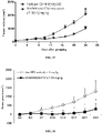
- the Fc receptor FcyRI also known as CD64, can bind to the Fc fragment of IgG antibodies and is involved in antibody-dependent cell-mediated cytotoxicity (ADCC).
- ADCC antibody-dependent cell-mediated cytotoxicity
- the method for determining the affinity constant of the antibodies to FcyRI by the Fortebio Octet system is briefly described as follows: the sample dilution buffer was a solution of PBS, 0.02% Tween-20 and 0.1% BSA, pH 7.4. A 1 ⁇ g/mL FcyRI solution (from Sinobio) was added to the HIS1K sensor to immobilize the FcyRI on the sensor surface for 50 s. The association and dissociation constants of the antibodies to FcyRI were both determined in the buffer with the antibody concentrations being 3.12-50 nM (serial two-fold dilution).
- the sensor with immobilized antigen was equilibrated in the buffer for 60 s, and then the binding of the immobilized FcyRI on the sensor to the antibodies was determined for 120 s; the dissociation of FcyRI from the antibodies was determined in 120 s.
- the temperature was 30 °C and the frequency was 0.3 Hz.
- the data were fitted and analyzed with a 1:1 model to obtain the affinity constants to FcyRI for the antibodies.
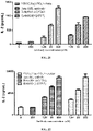
- the Fc receptor Fc ⁇ RIIIa_V158 (also known as CD16a_V158), can bind to the Fc fragment of IgG antibodies and mediate ADCC effects.
- the sample dilution buffer was a solution of PBS, 0.02% Tween-20 and 0.1% BSA, pH 7.4.
- 5 ⁇ g/mL Fc ⁇ RIIIa_V158 was immobilized on the HIS1K sensor for 120 s.
- the sensor was equilibrated in a buffer for 60 s, and the binding of the immobilized Fc ⁇ RIIIa_V158 on the sensor to the antibodies at concentrations of 31.25-500 nM (serial two-fold dilution) was determined for 60 s.
- the antibody was dissociated in the buffer for 60 s.
- the sensor was refreshed 4 times in 10 mM glycine pH 1.5, each for 5 s.
- the temperature was 30 °C and the frequency was 0.3 Hz.
- the data were analyzed by 1:1 model fitting to obtain affinity constants.
- the Fc receptor Fc ⁇ RIIIa_F158 (also known as CD16a_F158), can bind to the Fc fragment of IgG antibodies and mediate ADCC effects.
- the method for determining the affinity constant of BiAb004(hG1WT) and BiAb004(hG1TM) to Fc ⁇ RIIIa_F158 by the Fortebio Octet system is briefly described as follows: the sample dilution buffer was a solution of PBS, 0.02% Tween-20 and 0.1% BSA, pH 7.4. 5 ⁇ g/mL Fc ⁇ RIIIa_F158 was immobilized on the HIS1K sensor for 120 s. The sensor was equilibrated in a buffer for 60 s, and the binding of the immobilized Fc ⁇ RIIIa_F158 on the sensor to the antibodies at concentrations of 31.25-500 nM (serial two-fold dilution) was determined for 60 s.
- the sample dilution buffer was a solution of PBS, 0.02% Tween-20 and 0.1% BSA, pH 7.4. 5 ⁇ g/mL Fc ⁇ RIIIa_F158 was immobilized on the HIS1K sensor for 120 s
- the antibody was dissociated in the buffer for 60 s.
- the sensor was refreshed 4 times in 10 mM glycine pH 1.5, each for 5 s.
- the temperature was 30 °C and the frequency was 0.3 Hz.
- the data were analyzed by 1:1 model fitting to obtain affinity constants.
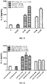
- the Fc receptor Fc ⁇ RIIa_H131 (also known as CD32a_H131), can bind to the Fc fragment of IgG antibodies and mediate ADCC effects.
- the immobilization dilution buffer was a solution of PBS, 0.02% Tween-20 and 0.1% BSA, pH 7.4
- the analyte dilution buffer was a solution of PBS, 0.02% Tween-20, 0.02% casein and 0.1% BSA, pH 7.4.
- 5 ⁇ g/mL Fc ⁇ RIIa_H131 was immobilized on the NTA sensor at an immobilization height of about 1.0 nm.
- the sensor was equilibrated in a buffer of PBS, 0.02% Tween-20, 0.02% casein and 0.1% BSA, pH 7.4 for 300 s of blocking, and the binding of the immobilized Fc ⁇ RIIa_H131 on the sensor to the antibodies at concentrations of 12.5-200 nM (serial two-fold dilution) was determined for 60 s.
- the antibody was dissociated in the buffer for 60 s.
- the sensor was refreshed in 10 mM glycine pH 1.7 and 10 nM nickel sulfate. The temperature was 30 °C and the frequency was 0.6 Hz. The data were analyzed by 1:1 model fitting to obtain affinity constants.
- the Fc receptor Fc ⁇ IIa_R131 (also known as CD32a_R131), can bind to the Fc fragment of IgG antibodies and mediate ADCC effects.
- the immobilization dilution buffer was a solution of PBS, 0.02% Tween-20 and 0.1% BSA, pH 7.4, and the analyte dilution buffer was a solution of PBS, 0.02% Tween-20, 0.02% casein and 0.1% BSA, pH 7.4.
- 5 ⁇ g/mL Fc ⁇ IIa_R131 was immobilized on the NTA sensor at an immobilization height of about 1.0 nm.
- the sensor was equilibrated in a buffer of PBS, 0.02% Tween-20, 0.02% casein and 0.1% BSA, pH 7.4 for 300 s of blocking, and the binding of the immobilized Fc ⁇ IIa_R131 on the sensor to the antibodies at concentrations of 12.5-200 nM (serial two-fold dilution) was determined for 60 s.
- the antibody was dissociated in the buffer for 60 s.
- the sensor was refreshed in 10 mM glycine pH 1.7 and 10 nM nickel sulfate. The temperature was 30 °C and the frequency was 0.6 Hz. The data were analyzed by 1:1 model fitting to obtain affinity constants.
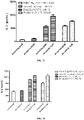
- the Fc receptor FcyRIIb (also known as CD32b), can bind to the Fc fragment of IgG antibodies, down-regulate functions of immune cells, inhibit the activation and proliferation of immune cells and inhibit the secretion of cytokines.
- the immobilization dilution buffer was a solution of PBS, 0.02% Tween-20 and 0.1% BSA, pH 7.4
- the analyte dilution buffer was a solution of PBS, 0.02% Tween-20, 0.02% casein and 0.1% BSA, pH 7.4.
- 5 ⁇ g/mL FcyRIIb was immobilized on the NTA sensor at an immobilization height of about 1.0 nm.
- the sensor was equilibrated in a buffer of PBS, 0.02% Tween-20, 0.02% casein and 0.1% BSA, pH 7.4 for 300 s of blocking, and the binding of the immobilized FcyRIIb on the sensor to the antibodies at concentrations of 12.5-200 nM (serial two-fold dilution) was determined for 60 s.
- the antibody was dissociated in the buffer for 60 s.
- the sensor was refreshed in 10 mM glycine pH 1.7 and 10 nM nickel sulfate. The temperature was 30 °C and the frequency was 0.6 Hz. The data were analyzed by 1:1 model fitting to obtain affinity constants.
- Serum complement C1q can bind to the Fc fragment of IgG antibodies and mediate CDC effects.
- the binding capacity of a therapeutic monoclonal antibody to C1q will influence the safety and efficacy of the antibody.
- the sample dilution buffer was a solution of PBS, 0.02% Tween-20 and 0.1% BSA, pH 7.4. 50 ⁇ g/mL antibody was immobilized on the FAB2G sensor at an immobilization height of about 2.0 nm.
- the sensor was equilibrated in a buffer for 60 s for blocking, and the binding of the immobilized antibody on the sensor to the antigen C1q at concentrations of 1.25-20 nM (serial two-fold dilution) was determined for 60 s.
- the antigen and antibody were dissociated in the buffer for 60 s.
- the sensor was refreshed 4 times in 10 mM glycine pH 1.7, each for 5 s.
- the shaking speed of the sample plate was 1000 rpm, the temperature was 30 °C and the frequency was 0.6 Hz.
- the data were analyzed by 1:1 model fitting to obtain affinity constants.
- the data acquisition software was Fortebio Data Acquisition 7.0, and the data analysis software was Fortebio Data Analysis 7.0.
- the ADCC effect refers to that effector immune cells with killing activity recognize Fc fragments of antibodies bound to antigens of target cells through Fc receptors (FcR) expressed on their surfaces, and directly kill the target cells.
- FcR Fc receptors
- the inventors constructed a co-culture system of 293T-CTLA4-PD1 cells expressing PD-1 and CTLA4 antigens and primary PBMCs to test the ADCC activity of the antibodies.
- the method for ADCC activity assay of BiAb004(hG1WT) and BiAb004(hG1TM) on 293T-CTLA4-PD1 cells expressing CTLA4 and PD-1 antigens is as follows: Normal human PBMCs were isolated according to the Ficoll peripheral blood mononuclear cell isolation instruction. The isolated PBMCs were resuspended in RPMI-1640 complete medium, stained with AO/PI, counted and frozen. One day before the experiment, PBMCs were thawed and counted, and the viability was determined. The cells were incubated overnight in a carbon dioxide incubator at 5% CO 2 and 37 °C.
- 293T-CTLA4-PD1 cells and PBMCs were collected, centrifuged at 800 rpm or 1200 rpm for 5 min, resuspended in RPMI-1640 (containing 1% FBS; hereinafter referred to as the medium) and washed twice. The cells were counted and viability was determined.
- the cell density was adjusted to a proper range using the medium.
- 100 ⁇ L of 293T-CTLA4-PD1 cells was added to a 96-well plate at a density of 3.0E+04 cells/well according to the experimental design.
- 50 ⁇ L of antibody was added, and the cells were pre-incubated for 1 h at room temperature; after 1 h, 50 ⁇ L of PBMCs were added at 9.0E+05 cells/well and the mixture was mixed well, incubated at 37 °C for 4 h in a carbon dioxide incubator at 5% CO 2 .
- the cells were centrifuged at 250x g for 5 min.
- the mouse MC38 cell line is a mouse colorectal cancer cell line. It has been demonstrated that the MC38 cells line is a useful model for studying human MSI-H/dMMR tumors ( Efremova M et al., Nat Commun., 2018; 9(1):32 ).
- MC-38 cells (purchased from Shanghai Ruilu Biotech Co., Ltd.) were collected, adjusted to a density of 5 million cells/mL, and subcutaneously grafted into PD-1 transgenic mice (purchased from Shanghai Model Organisms Center, Inc.) in a volume of 200 ⁇ L/mouse at the right flank. 12 mice were grafted, each bearing 1 ⁇ 10 6 cells.
- the tumor volume reached about 60-150 mm 3
- the mice were divided by tumor volume (with similar average volume of approximately 94 mm 3 ) into 2 groups, model group and BiAb004(hG1TM)/lenvatinib treatment group, each containing 6 mice.
- the day of grouping was set as D0.
- Table 8 Study design Group Number of animals Model establishment Regimen Model group 6 MC-38 cells were grafted subcutaneously at a dose of 1 million cells/200 ⁇ L/mouse Anti-HEL antibody (i.e., isotype control antibody) at 1 mg/kg was injected intraperitoneally twice weekly in a total of 6 doses BiAb004(hG1TM) + lenvatinib group 6 BiAb004(hG1TM) at 1.33 mg/kg was injected intraperitoneally twice weekly in a total of 6 doses; Lenvatinib at 5 mg/kg was administered by oral gavage once daily in 20 doses
- i.e., isotype control antibody i.e., isotype control antibody
- human anti-hen egg lysozyme IgG anti-HEL antibody, or human IgG, abbreviated as hIgG
- hIgG human anti-hen egg lysozyme IgG
- BiAb004(hG1TM) in combination with lenvatinib significantly inhibited the growth of MC38 tumors; BiAb004(hG1TM) in combination with lenvatinib demonstrated significantly efficacy against rectal and/or colon cancers of MSI-H/dMMR phenotype.
- mice Female C57BL/6-hPD1/hPDL1/hCD73 mice (purchased from Nanjing GemPharmatech Co., Ltd.) were divided into groups of 8 and grafted subcutaneously on the right forelimb with colon cancer MC38-hPDL1/hCD73 cells (purchased from Nanjing GemPharmatech Co., Ltd.) (2 ⁇ 10 6 cells/100 ⁇ L/mouse). The day of grafting was defined as D0. The dosing volume was adjusted according to the body weight: 10 ⁇ L/g mouse body weight (g).
- Table 9 Protocol and grouping Group Dose (mg/kg) Route Frequency Cycle Isotype Control 10 Intraperitoneal injection Twice weekly, for a total of three weeks 6 doses in total BiAb004 (hG1TM) 1
- the cell pellet was resuspended in 10 mL of DMEM complete medium and macrophage colony stimulating factor (M-CSF) were added at a working concentration of 100 ng/mL.
- M-CSF macrophage colony stimulating factor
- the cells were cultured for 7 days at 37 °C and 5% CO 2 in a cell culture chamber for induction. Half of the medium was exchanged and M-CSF was added on Days 3 and 5. The induction of cells was completed on day 7.
- the cells were digested with 0.25% trypsin. Macrophages were collected, and centrifuged at 170x g for 5 min. The supernatant was discarded and the cells were suspended in DMEM complete medium and counted. The cell was adjusted to a proper density and filled into sterile EP tubes for further use.
- CHO-K1-CTLA4-PD1 cells (a cell line based on CHO-K1 cells overexpressing both human CTLA4 and PD1 antigens, constructed by Akeso Biopharma, Inc.) at logarithmic phase were collected, centrifuged at 170x g for 5 min, washed with PBS, resuspended and counted, and the viability was determined.
- Carboxyfluorescein diacetate succinimidyl ester (CFSE) was diluted to 2.5 ⁇ M with PBS to resuspend the cells (staining density: 10 million cells/mL). The cells were incubated in a cell incubator for 20 min. 6 mL of DMEM complete medium was added to stop the staining.
- the cells were centrifuged at 170x g for 5 min, and the supernatant was discarded. 1 mL of DMEM complete medium was added for resuspension. The cells were incubated in an incubator for 10 min, and adjusted to the experiment density. The cells were coded as CHO-K1-PD1-CTLA4-CFSE. The test antibodies were diluted in DMEM complete medium. An isotype control antibody anti-HEL antibody and a medium were used as the isotype control group and a blank control group.
- the diluted antibody and CHO-K1-PD1-CTLA4-CFSE cells were added into 1.5 mL EP tubes containing macrophages (the final volume was 100 ⁇ L, the effector-to-target ratio was 50,000:150,000, the working concentrations of the antibody were 50, 5 and 0.5 nM).
- the mixture was resuspended, mixed evenly and incubated in an incubator at 37 °C for 2 h. 800 ⁇ L of PBS containing 1 % bovine serum albumin (BSA) was added at room temperature to each tube. The mixture was centrifuged at 1200x g for 5 min, and the supernatant was discarded. The cells were washed once with 800 ⁇ L of 1 % PBSA.
- BSA bovine serum albumin
- APC anti-mouse/human CD11b antibody (Biolegend, Cat. No.: 101212) was diluted 400-fold with 1 % PBSA and added to the corresponding samples at 100 ⁇ L/sample. The mixture was mixed well, incubated on ice for 30 min, washed once with 800 ⁇ L of 1% PBSA, and centrifuged at 1200x g for 5 min, and the supernatant was discarded. 200 ⁇ L of 1 % PBSA was added to each tube to resuspend the cells. The cells were transferred to loading tube and analyzed by BD FACSCalibur flow cytometer.
- Macrophages in the system were APC + positive, and macrophages involved in phagocytosis were APC and CFSE double positive.
- the phagocytosis rate was determined as the ratio of the number of double positive cells to the number of APC positive cells, and the antibody-mediated ADCP activity was evaluated.
- PBMCs were isolated from healthy human peripheral blood according to the Ficoll-Paque TM Plus reagent instruction, and the isolated PBMCs were counted and frozen.
- Raji-PDL1 and KATO III cells purchased from Chinese Academy of Sciences Shanghai Cell Bank) were cultured in DMEM + 10% FBS complete medium.
- PBMCs were thawed and activated with 0.5 ⁇ g/mL SEB for two days.
- Raji-PDL1 cells were treated with 2 ⁇ g/mL mitomycin C (MMC) for 1 h.
- MMC mitomycin C
- BiAb004(hG1TM) more significantly enhanced the immune response of immune cells to human gastric cancer cells KATO III characterized by significantly increased secretion level of IL-2 as compared to BiAb004(hG1WT), and thus BiAb004(hG1TM) is prospective for treating gastric cancer.
- PBMCs were isolated from healthy human peripheral blood according to the Ficoll-Paque TM Plus reagent instruction, and the isolated PBMCs were counted and frozen.
- Raji-PDL1 and Hela cells purchased from Chinese Academy of Sciences Cell Bank
- PBMCs were thawed and activated with 0.5 ⁇ g/mL SEB for two days.
- Raji-PDL1 cells were treated with 2 ⁇ g/mL MMC for 1 h.
- BiAb004(hG1TM) more significantly enhanced the immune response of immune cells to human cervical cancer Hela cells characterized by significantly increased secretion level of IL-2 as compared to BiAb004(hG1WT), and thus BiAb004(hG1TM) is prospective for treating cervical cancer.
- PBMCs were isolated from healthy human peripheral blood according to the Ficoll-Paque TM Plus reagent instruction, and the isolated PBMCs were counted and frozen.
- Raji-PDL1 and human T-cell lymphoma Jurkat cells purchased from Chinese Academy of Sciences Cell Bank
- PBMCs were thawed and activated with 0.5 ⁇ g/mL SEB for two days.
- Raji-PDL1 cells were treated with 2 ⁇ g/mL MMC for 1 h.
- BiAb004(hG1TM) significantly enhanced the immune response of immune cells to human T-cell lymphoma Jurkat cells characterized by significantly increased secretion level of IL-2 as compared to BiAb004(hG1WT), while eliminating the ADCC and CDC (i.e., ADCP); the antibody has an equivalent or higher pharmacological activity at doses of 1.34 nM and 20 nM as compared to BiAb004(hG1WT) and is prospective for treating cervical cancer.
- PBMCs were isolated from healthy human peripheral blood according to the Ficoll-Paque TM Plus reagent instruction, and the isolated PBMCs were counted and frozen.
- Raji-PDL1 and CNE-2Z cells purchased from GuangZhou Jennio Biotech Co., Ltd.
- PBMCs were thawed and activated with 0.5 ⁇ g/mL SEB for two days.
- Raji-PDL1 cells were treated with 2 ⁇ g/mL MMC for 1 h.
- BiAb004(hG1TM) equivalently and/or more significantly enhanced the immune response of immune cells to human nasopharyngeal cancer cells CNE-2Z characterized by significantly increased secretion level of IL-2 as compared to BiAb004(hG1WT), and that BiAb004(hG1TM) especially had significant superiority at 20 nM.
- BiAb004(hG1TM) had better or equivalent pharmacological activity relative to BiAb004(hG1WT) on the basis of effectively eliminating ADCC, CDC and ADCP effects, indicating the potential for treating human nasopharyngeal cancer.
- PBMCs were isolated from healthy human peripheral blood according to the Ficoll-Paque TM Plus reagent instruction, and the isolated PBMCs were counted and frozen.
- Raji-PDL1 and MDA-MB-231 cells purchased from GuangZhou Jennio Biotech Co., Ltd.
- PBMCs were thawed and activated with 0.5 ⁇ g/mL SEB for two days.
- Raji-PDL1 cells were treated with 2 ⁇ g/mL MMC for 1 h.
- BiAb004(hG1TM) equivalently or more significantly enhanced the immune response of immune cells to human breast cancer MDA-MB-231 cells characterized by significantly increased secretion level of IL-2 as compared to BiAb004(hG1WT).
- BiAb004(hG1TM) had better or equivalent pharmacological activity relative to BiAb004(hG1WT) on the basis of effectively eliminating ADCC, CDC and ADCP effects, indicating the potential for treating human breast cancer.
- PBMCs were isolated from healthy human peripheral blood according to the Ficoll-Paque TM Plus reagent instruction, and the isolated PBMCs were counted and frozen.
- Raji-PDL1 and NCI-H2452 cells purchased from Chinese Academy of Sciences Shanghai Cell Bank) were cultured in RPMI 1640 + 10% FBS complete medium.
- PBMCs were thawed and activated with 0.5 ⁇ g/mL SEB for two days. On the day of the experiment, Raji-PDL1 cells were treated with 2 ⁇ g/mL MMC for 1 h.
- BiAb004(hG1TM) more significantly enhanced the immune response of immune cells to human mesothelioma cells NCI-H2452 characterized by significantly increased secretion level of IL-2 as compared to BiAb004(hG1WT), and thus BiAb004(hG1TM) is prospective for treating mesothelioma.
- PBMCs were isolated from healthy human peripheral blood according to the Ficoll-Paque TM Plus reagent instruction, and the isolated PBMCs were counted and frozen.
- Raji-PDL1 cells were cultured in RPMI 1640 + 10% FBS complete medium and A549 cells were cultured in DMEM + 10% FBS complete medium.
- PBMCs were thawed and activated with 0.5 ⁇ g/mL SEB for two days. On the day of the experiment, Raji-PDL1 cells were treated with 2 ⁇ g/mL MMC for 1 h.
- BiAb004(hG1TM) in combination with anlotinib hydrochloride more significantly enhanced the immune response of immune cells to human non-small cell lung cancer (human lung adenocarcinoma) A549 cells characterized by significantly increased secretion level of IL-2 as compared to BiAb004(hG1TM) monotherapy, BiAb004(hG1WT) monotherapy and BiAb004(hG1WT) in combination with anlotinib hydrochloride, and thus BiAb004(hG1TM) in combination with anlotinib hydrochloride is prospective for treating human non-small cell lung cancer or human lung adenocarcinoma.
- PBMCs were isolated from healthy human peripheral blood according to the Ficoll-Paque TM Plus reagent instruction, and the isolated PBMCs were counted and frozen.
- Raji-PDL1 and NCI-H446 cells purchased from Chinese Academy of Sciences, Shanghai Institutes for Biological Sciences) were cultured in RPMI 1640 + 10% FBS complete medium.
- PBMCs were thawed and activated with 0.5 ⁇ g/mL SEB for two days. On the day of the experiment, Raji-PDL1 cells were treated with 2 ⁇ g/mL MMC for 1 h.
- BiAb004(hG1TM) in combination with anlotinib hydrochloride significantly enhanced the immune response of immune cells to human small cell lung cancer NCI-H446 cells characterized by significantly increased secretion level of IL-2 as compared to BiAb004(hG1MT) monotherapy, BiAb004(hG1WT) monotherapy and BiAb004(hG1WT) in combination with anlotinib hydrochloride, and thus BiAb004(hG1TM) in combination with anlotinib hydrochloride is prospective for treating human small cell lung cancer.
- PBMCs were isolated from healthy human peripheral blood according to the Ficoll-Paque TM Plus reagent instruction, and the isolated PBMCs were counted and frozen.
- Raji-PDL1 and NCI-H226 cells purchased from Chinese Academy of Sciences, Shanghai Institutes for Biological Sciences) were cultured in RPMI 1640 + 10% FBS complete medium.
- PBMCs were thawed and activated with 0.5 ⁇ g/mL SEB for two days. On the day of the experiment, Raji-PDL1 cells were treated with 2 ⁇ g/mL MMC for 1 h.
- BiAb004(hG1TM) monotherapy more effectively and significantly enhanced the immune response of immune cells to human squamous cell lung cancer NCI-H226 cells characterized by significantly increased secretion level of IL-2 as compared to BiAb004(hG1WT) monotherapy.
- BiAb004(hG1TM) in combination with anlotinib hydrochloride more significantly enhanced the immune response of immune cells to human squamous cell lung cancer NCI-H226 cells as compared to BiAb004(hG1WT) monotherapy and BiAb004(hG1TM) monotherapy, and demonstrated a comparable pharmacological activity with BiAb004(hG1WT) in combination with anlotinib.
- BiAb004(hG1TM) had better or equivalent pharmacological activity relative to BiAb004(hG1WT) monotherapy or BiAb004(hG1WT) in combination with anlotinib on the basis of effectively eliminating ADCC, CDC and ADCP effects, indicating a better efficacy for treating human squamous cell lung cancer.
- SW48 is a human colorectal cancer cell line and is identified with MSI-H/dMMR phenotype ( Branch P et al., (1995), Cancer Res, 55(11): 2304-2309 .). It was used for detecting the enhanced immune cell response to tumor of MSI-H/dMMR phenotype by BiAb004(hG1TM).
- PBMCs were isolated from healthy human peripheral blood according to the Ficoll-Paque TM Plus reagent instruction, and the isolated PBMCs were counted and frozen.
- Raji-PDL1 cells were cultured in RPMI 1640 + 10% FBS complete medium, and SW48 cells (purchased from GuangZhou Jennio Biotech Co., Ltd.) were cultured in DMEM + 10% FBS complete medium.
- PBMCs were thawed and activated with 0.5 ⁇ g/mL SEB for two days. On the day of the experiment, Raji-PDL1 cells were treated with 2 ⁇ g/mL MMC for 1 h.
- BiAb004(hG1WT) and BiAb004(hG1TM) significantly enhanced the immune response of immune cells to human colorectal cancer cells SW48 cells of MSI-H/dMMR phenotype characterized by significantly increased secretion level of IL-2 as compared to anti-HEL antibody.
- BiAb004(hG1TM) had better or equivalent pharmacological activity relative to BiAb004(hG1WT) on the basis of effectively eliminating ADCC, CDC and ADCP effects, indicating the potential for treating solid tumor of MSI-H/dMMR phenotype, particularly colon cancer and/or rectal cancer of MSI-H/dMMR phenotype.
- SW837 is a human colorectal cancer cell line of non-MSI-H/dMMR (i.e., MSS) phenotype ( Guo J et al., Cancer Res., 2011;71(8):2978-2987 .), and was used for detecting the enhanced immune cell response to tumor of non-MSI-H/dMMR phenotype by BiAb004(hG1TM) in this example.
- MSS non-MSI-H/dMMR
- PBMCs were isolated from healthy human peripheral blood according to the Ficoll-Paque TM Plus reagent instruction, and the isolated PBMCs were counted and frozen.
- Raji-PDL1 cells were cultured in RPMI 1640 + 10% FBS complete medium, and SW837 cells (purchased from Shanghai Honsun Biological Technology Co., Ltd) were cultured in 10% FBS + Leibovitz's L-15 (purchased from Gibco) complete medium.
- PBMCs were thawed and activated with 0.5 ⁇ g/mL SEB for two days. On the day of the experiment, Raji-PDL1 cells were treated with 2 ⁇ g/mL MMC for 1 h.
- the media in this experiment were all 10% FBS + RPMI 1640.
- BiAb004(hG1WT) and BiAb004(hG1TM) significantly enhanced the immune response of immune cells to human colorectal cancer cells SW837 cells of non-MSI-H/dMMR phenotype as compared to anti-HEL antibody.
- the pharmacological activity of BiAb004(hG1TM) in the high dose group was superior to that of BiAb004(hG1WT), characterized by significantly increased secretion level of IL-2.
Landscapes
- Health & Medical Sciences (AREA)
- Life Sciences & Earth Sciences (AREA)
- Chemical & Material Sciences (AREA)
- Immunology (AREA)
- Medicinal Chemistry (AREA)
- General Health & Medical Sciences (AREA)
- Engineering & Computer Science (AREA)
- Molecular Biology (AREA)
- Organic Chemistry (AREA)
- Hematology (AREA)
- Biomedical Technology (AREA)
- Animal Behavior & Ethology (AREA)
- Veterinary Medicine (AREA)
- Pharmacology & Pharmacy (AREA)
- Public Health (AREA)
- Biochemistry (AREA)
- Urology & Nephrology (AREA)
- Genetics & Genomics (AREA)
- Microbiology (AREA)
- Proteomics, Peptides & Aminoacids (AREA)
- Biotechnology (AREA)
- Chemical Kinetics & Catalysis (AREA)
- Physics & Mathematics (AREA)
- Cell Biology (AREA)
- Epidemiology (AREA)
- Biophysics (AREA)
- Nuclear Medicine, Radiotherapy & Molecular Imaging (AREA)
- General Chemical & Material Sciences (AREA)
- Bioinformatics & Cheminformatics (AREA)
- Food Science & Technology (AREA)
- Analytical Chemistry (AREA)
- Pathology (AREA)
- General Physics & Mathematics (AREA)
- Oncology (AREA)
- Wood Science & Technology (AREA)
- General Engineering & Computer Science (AREA)
- Zoology (AREA)
- Mycology (AREA)
- Diabetes (AREA)
- Endocrinology (AREA)
Applications Claiming Priority (3)
| Application Number | Priority Date | Filing Date | Title |
|---|---|---|---|
| CN201910711122 | 2019-08-02 | ||
| CN201911224135 | 2019-12-02 | ||
| PCT/CN2020/106309 WO2021023117A1 (zh) | 2019-08-02 | 2020-07-31 | 抗ctla4-抗pd-1双特异性抗体及其用途 |
Publications (2)
| Publication Number | Publication Date |
|---|---|
| EP4008730A1 true EP4008730A1 (de) | 2022-06-08 |
| EP4008730A4 EP4008730A4 (de) | 2023-09-06 |
Family
ID=74483591
Family Applications (1)
| Application Number | Title | Priority Date | Filing Date |
|---|---|---|---|
| EP20850855.6A Pending EP4008730A4 (de) | 2019-08-02 | 2020-07-31 | Anti-ctla4-anti-pd-bispezifischer antikörper und verwendungen davon |
Country Status (12)
| Country | Link |
|---|---|
| US (1) | US20220275089A1 (de) |
| EP (1) | EP4008730A4 (de) |
| JP (2) | JP7655896B2 (de) |
| KR (1) | KR20220053578A (de) |
| CN (1) | CN112300286A (de) |
| AU (1) | AU2020325350A1 (de) |
| BR (1) | BR112022002013A2 (de) |
| CA (1) | CA3146780A1 (de) |
| IL (2) | IL290255B2 (de) |
| MX (1) | MX2022001321A (de) |
| PH (1) | PH12022550243A1 (de) |
| WO (1) | WO2021023117A1 (de) |
Cited By (2)
| Publication number | Priority date | Publication date | Assignee | Title |
|---|---|---|---|---|
| EP4114464A4 (de) * | 2020-03-05 | 2024-05-01 | Merck Sharp & Dohme LLC | Verfahren zur behandlung von krebs mit einer kombination aus einem pd-1-antagonisten, einem ctla4-antagonisten und lenvatinib oder einem pharmazeutisch akzeptablen salz davon |
| EP4357367A4 (de) * | 2022-09-09 | 2025-02-26 | Akeso Biopharma, Inc. | Therapeutische kombination und verwendung davon |
Families Citing this family (21)
| Publication number | Priority date | Publication date | Assignee | Title |
|---|---|---|---|---|
| CN106967172B (zh) | 2016-08-23 | 2019-01-08 | 康方药业有限公司 | 抗ctla4-抗pd-1 双功能抗体、其药物组合物及其用途 |
| CN109053895B (zh) | 2018-08-30 | 2020-06-09 | 中山康方生物医药有限公司 | 抗pd-1-抗vegfa的双功能抗体、其药物组合物及其用途 |
| BR112022002028A2 (pt) * | 2019-08-02 | 2022-04-12 | Cttq Akeso Shanghai Biomed Tech Co Ltd | Anticorpo anti-pd-1 e uso farmacêutico do mesmo |
| EP4190819A4 (de) * | 2021-03-12 | 2024-08-14 | Akeso Biopharma, Inc. | Pharmazeutische kombination mit bispezifischem anti-pd-1-anti-vegfa-antikörper und verwendung davon |
| CN115073588B (zh) * | 2021-03-12 | 2024-06-18 | 中山康方生物医药有限公司 | 提高含有免疫球蛋白Fc片段的药物的安全性的方法 |
| CN115192718A (zh) * | 2021-04-14 | 2022-10-18 | 中山康方生物医药有限公司 | 抗cd47的单克隆抗体及其用途 |
| EP4324854A4 (de) * | 2021-04-14 | 2025-04-23 | Akeso Pharmaceuticals, Inc. | Verwendung von antikörpern in der antitumorbehandlung |
| IL310217A (en) * | 2021-07-23 | 2024-03-01 | Akeso Biopharma Inc | Pharmaceutical composition and use |
| WO2023160647A1 (zh) * | 2022-02-24 | 2023-08-31 | 康方药业有限公司 | 包含抗ctla4-抗pd-1双特异性抗体和西奥罗尼的药物组合 |
| CA3253514A1 (en) * | 2022-03-07 | 2023-09-14 | Medimmune Llc | TREATMENT METHODS USING CTLA-4 AND PD-1 BISPECIFIC ANTIBODIES |
| WO2023198089A1 (zh) * | 2022-04-13 | 2023-10-19 | 齐鲁制药有限公司 | 包含抗ctla4和抗pd1的混合抗体的药物组合物及其治疗用途 |
| CN117679505A (zh) | 2022-09-09 | 2024-03-12 | 中山康方生物医药有限公司 | 药物组合及用途 |
| KR20250122541A (ko) | 2022-10-25 | 2025-08-13 | 사이즈믹 테라퓨틱, 인코포레이티드. | 변이체 IgG FC 폴리펩티드 및 그 용도 |
| WO2024141014A1 (zh) * | 2022-12-30 | 2024-07-04 | 康方药业有限公司 | Cd74的医药用途 |
| JP2026507907A (ja) * | 2023-03-07 | 2026-03-06 | アケソ・ファーマ・カンパニー,リミテッド | 抗ctla4抗pd-1二重特異性抗体を含有する医薬組成物とその使用 |
| CN121219403A (zh) * | 2023-03-31 | 2025-12-26 | 上海细胞治疗集团股份有限公司 | 双特异性抗体及自分泌双特异性抗体的工程化免疫细胞 |
| CN118767127A (zh) * | 2023-04-10 | 2024-10-15 | 康方药业有限公司 | 抗ctla4-抗pd-1双特异性抗体的医药用途 |
| WO2025157267A1 (zh) * | 2024-01-25 | 2025-07-31 | 中山康方生物医药有限公司 | 抗pd-1-抗vegfa的双特异性抗体的医药用途 |
| WO2025201450A1 (zh) * | 2024-03-27 | 2025-10-02 | 康方汇科(上海)生物有限公司 | 药物组合及用途 |
| CN120919303A (zh) * | 2024-05-10 | 2025-11-11 | 康方汇科(上海)生物有限公司 | 药物组合及用途 |
| CN120965861B (zh) * | 2025-10-22 | 2026-02-13 | 杭州博茵生物技术有限公司 | 一种Fc效应消除的新型骨架流式抗体及其制备方法与应用 |
Family Cites Families (19)
| Publication number | Priority date | Publication date | Assignee | Title |
|---|---|---|---|---|
| US4816567A (en) | 1983-04-08 | 1989-03-28 | Genentech, Inc. | Recombinant immunoglobin preparations |
| AU9598601A (en) | 2000-10-20 | 2002-04-29 | Eisai Co Ltd | Nitrogenous aromatic ring compounds |
| KR100804566B1 (ko) | 2003-12-25 | 2008-02-20 | 에자이 알앤드디 매니지먼트 가부시키가이샤 | 4-(3-클로로-4-(시클로프로필아미노카르보닐)아미노페녹시)-7-메톡시-6-퀴놀린카르복사미드의 염 또는 그 용매화물의 결정 및 이들의 제조 방법 |
| EP2471813B1 (de) * | 2004-07-15 | 2014-12-31 | Xencor, Inc. | Optimierte Fc-Varianten |
| US8148532B2 (en) | 2007-03-14 | 2012-04-03 | Guoqing Paul Chen | Spiro substituted compounds as angiogenesis inhibitors |
| AU2009288730B2 (en) * | 2008-08-25 | 2013-06-20 | Amplimmune, Inc. | Compositions of PD-1 antagonists and methods of use |
| US8961967B2 (en) * | 2009-11-30 | 2015-02-24 | Janssen Biotech, Inc. | Antibody Fc mutants with ablated effector functions |
| CA2785907A1 (en) * | 2009-12-29 | 2011-07-28 | Emergent Product Development Seattle, Llc | Ron binding constructs and methods of use thereof |
| JP6180663B2 (ja) * | 2014-12-23 | 2017-08-16 | ブリストル−マイヤーズ スクイブ カンパニーBristol−Myers Squibb Company | Tigitに対する抗体 |
| KR20250057128A (ko) * | 2015-12-30 | 2025-04-28 | 코디악 사이언시스 인코포레이티드 | 항체 및 이의 접합체 |
| RU2693661C2 (ru) | 2016-03-04 | 2019-07-03 | Сычуань Келунь-Байотек Байофармасьютикал Ко., Лтд. | Антитело против pdl-1, его фармацевтическая композиция и применение |
| CN109862910A (zh) * | 2016-08-03 | 2019-06-07 | 小利兰·斯坦福大学托管委员会 | 破坏巨噬细胞上的Fc受体接合增强抗SIRPα抗体疗法的功效 |
| WO2018035710A1 (en) * | 2016-08-23 | 2018-03-01 | Akeso Biopharma, Inc. | Anti-ctla4 antibodies |
| CN106967172B (zh) * | 2016-08-23 | 2019-01-08 | 康方药业有限公司 | 抗ctla4-抗pd-1 双功能抗体、其药物组合物及其用途 |
| CN106977602B (zh) * | 2016-08-23 | 2018-09-25 | 中山康方生物医药有限公司 | 一种抗pd1单克隆抗体、其药物组合物及其用途 |
| AU2017371070B2 (en) * | 2016-12-09 | 2025-01-02 | Alector Llc | Anti-SIRP-alpha antibodies and methods of use thereof |
| CN106699891B (zh) * | 2017-01-25 | 2019-04-09 | 北京天广实生物技术股份有限公司 | 一种抗pd-l1抗体、其药物组合物及其用途 |
| EP3634485A4 (de) * | 2017-06-07 | 2021-07-21 | Silverback Therapeutics, Inc. | Antikörperkonjugate von immunmodulatorischen verbindungen und deren verwendungen |
| CN109762068A (zh) * | 2018-08-09 | 2019-05-17 | 源道隆(苏州)医学科技有限公司 | 一种可靶向ctla4和pd-1的单基因双特异性抗体及其应用 |
-
2020
- 2020-07-31 WO PCT/CN2020/106309 patent/WO2021023117A1/zh not_active Ceased
- 2020-07-31 MX MX2022001321A patent/MX2022001321A/es unknown
- 2020-07-31 AU AU2020325350A patent/AU2020325350A1/en active Pending
- 2020-07-31 JP JP2022506483A patent/JP7655896B2/ja active Active
- 2020-07-31 PH PH1/2022/550243A patent/PH12022550243A1/en unknown
- 2020-07-31 EP EP20850855.6A patent/EP4008730A4/de active Pending
- 2020-07-31 IL IL290255A patent/IL290255B2/en unknown
- 2020-07-31 BR BR112022002013A patent/BR112022002013A2/pt unknown
- 2020-07-31 KR KR1020227006972A patent/KR20220053578A/ko not_active Ceased
- 2020-07-31 IL IL321822A patent/IL321822A/en unknown
- 2020-07-31 US US17/630,467 patent/US20220275089A1/en active Pending
- 2020-07-31 CN CN202010757322.6A patent/CN112300286A/zh active Pending
- 2020-07-31 CA CA3146780A patent/CA3146780A1/en active Pending
-
2025
- 2025-03-21 JP JP2025047199A patent/JP2025102836A/ja active Pending
Cited By (2)
| Publication number | Priority date | Publication date | Assignee | Title |
|---|---|---|---|---|
| EP4114464A4 (de) * | 2020-03-05 | 2024-05-01 | Merck Sharp & Dohme LLC | Verfahren zur behandlung von krebs mit einer kombination aus einem pd-1-antagonisten, einem ctla4-antagonisten und lenvatinib oder einem pharmazeutisch akzeptablen salz davon |
| EP4357367A4 (de) * | 2022-09-09 | 2025-02-26 | Akeso Biopharma, Inc. | Therapeutische kombination und verwendung davon |
Also Published As
| Publication number | Publication date |
|---|---|
| IL290255B1 (en) | 2025-08-01 |
| KR20220053578A (ko) | 2022-04-29 |
| AU2020325350A1 (en) | 2022-03-03 |
| US20220275089A1 (en) | 2022-09-01 |
| PH12022550243A1 (en) | 2023-04-24 |
| MX2022001321A (es) | 2022-05-20 |
| BR112022002013A2 (pt) | 2022-03-29 |
| EP4008730A4 (de) | 2023-09-06 |
| IL290255B2 (en) | 2025-12-01 |
| JP7655896B2 (ja) | 2025-04-02 |
| IL290255A (en) | 2022-04-01 |
| JP2025102836A (ja) | 2025-07-08 |
| CN112300286A (zh) | 2021-02-02 |
| IL321822A (en) | 2025-08-01 |
| WO2021023117A1 (zh) | 2021-02-11 |
| JP2022542450A (ja) | 2022-10-03 |
| CA3146780A1 (en) | 2021-02-11 |
Similar Documents
| Publication | Publication Date | Title |
|---|---|---|
| JP7655896B2 (ja) | 抗ctla4/抗pd-1二重特異性抗体及びその使用 | |
| JP7753186B2 (ja) | 抗pd-1抗体およびその医学的使用 | |
| EP4067387B1 (de) | Bispezifischer anti-pd-1-anti-vegfa-antikörper, pharmazeutische zusammensetzung und verwendung davon | |
| EP4483877A1 (de) | Pharmazeutische zusammensetzung mit bispezifischem anti-ctla4-anti-pd-1-antikörper und chiauranib | |
| US20250114450A1 (en) | Pharmaceutical combination containing anti-pd-1-anti-vegfa bispecific antibody, and use thereof | |
| EP4321537A1 (de) | Pharmazeutische zusammensetzung und verwendung davon | |
| EP4357367A1 (de) | Therapeutische kombination und verwendung davon | |
| HK40075399A (en) | Anti-ctla4-anti-pd-1 bispecific antibody and uses thereof | |
| HK40117394A (en) | Pharmaceutical composition comprising anti-ctla4-anti-pd-1 bispecific antibody and chiauranib | |
| HK40075629A (en) | Anti-pd-1 antibody and medical use thereof | |
| EP4678186A1 (de) | Pharmazeutische zusammensetzung mit bispezifischem anti-ctla4-anti-pd-1-antikörper und verwendung davon | |
| EA049587B1 (ru) | Биспецифическое антитело против ctla4/против pd-1 и его применение | |
| EA050042B1 (ru) | Антитело против pd-1 и его фармацевтическое применение | |
| HK40091003A (en) | Pharmaceutical combination containing anti-pd-1-anti-vegfa bispecific antibody, and use thereof | |
| HK40080672A (en) | Anti-pd-1-anti-vegfa bispecific antibody, pharmaceutical composition and use thereof | |
| HK40096182A (zh) | 药物组合物及其用途 |
Legal Events
| Date | Code | Title | Description |
|---|---|---|---|
| STAA | Information on the status of an ep patent application or granted ep patent |
Free format text: STATUS: THE INTERNATIONAL PUBLICATION HAS BEEN MADE |
|
| PUAI | Public reference made under article 153(3) epc to a published international application that has entered the european phase |
Free format text: ORIGINAL CODE: 0009012 |
|
| STAA | Information on the status of an ep patent application or granted ep patent |
Free format text: STATUS: REQUEST FOR EXAMINATION WAS MADE |
|
| 17P | Request for examination filed |
Effective date: 20220301 |
|
| AK | Designated contracting states |
Kind code of ref document: A1 Designated state(s): AL AT BE BG CH CY CZ DE DK EE ES FI FR GB GR HR HU IE IS IT LI LT LU LV MC MK MT NL NO PL PT RO RS SE SI SK SM TR |
|
| DAV | Request for validation of the european patent (deleted) | ||
| DAX | Request for extension of the european patent (deleted) | ||
| REG | Reference to a national code |
Ref country code: HK Ref legal event code: DE Ref document number: 40075399 Country of ref document: HK |
|
| P01 | Opt-out of the competence of the unified patent court (upc) registered |
Effective date: 20230505 |
|
| A4 | Supplementary search report drawn up and despatched |
Effective date: 20230808 |
|
| RIC1 | Information provided on ipc code assigned before grant |
Ipc: A61P 35/02 20060101ALI20230802BHEP Ipc: A61P 35/00 20060101ALI20230802BHEP Ipc: A61P 7/06 20060101ALI20230802BHEP Ipc: G01N 33/68 20060101ALI20230802BHEP Ipc: G01N 33/577 20060101ALI20230802BHEP Ipc: A61K 39/395 20060101ALI20230802BHEP Ipc: C12N 15/63 20060101ALI20230802BHEP Ipc: C12N 15/62 20060101ALI20230802BHEP Ipc: C12N 15/13 20060101ALI20230802BHEP Ipc: C07K 16/28 20060101ALI20230802BHEP Ipc: C07K 16/46 20060101AFI20230802BHEP |
|
| STAA | Information on the status of an ep patent application or granted ep patent |
Free format text: STATUS: EXAMINATION IS IN PROGRESS |
|
| 17Q | First examination report despatched |
Effective date: 20260203 |