EP4105237A1 - Cldn18.2-antikörper und verwendung davon - Google Patents
Cldn18.2-antikörper und verwendung davon Download PDFInfo
- Publication number
- EP4105237A1 EP4105237A1 EP21753717.4A EP21753717A EP4105237A1 EP 4105237 A1 EP4105237 A1 EP 4105237A1 EP 21753717 A EP21753717 A EP 21753717A EP 4105237 A1 EP4105237 A1 EP 4105237A1
- Authority
- EP
- European Patent Office
- Prior art keywords
- antibody
- seq
- sequence
- cells
- cancer
- Prior art date
- Legal status (The legal status is an assumption and is not a legal conclusion. Google has not performed a legal analysis and makes no representation as to the accuracy of the status listed.)
- Pending
Links
Images


Classifications
-
- C—CHEMISTRY; METALLURGY
- C07—ORGANIC CHEMISTRY
- C07K—PEPTIDES
- C07K16/00—Immunoglobulins [IG], e.g. monoclonal or polyclonal antibodies
- C07K16/18—Immunoglobulins [IG], e.g. monoclonal or polyclonal antibodies against material from animals or humans
- C07K16/28—Immunoglobulins [IG], e.g. monoclonal or polyclonal antibodies against material from animals or humans against receptors, cell surface antigens or cell surface determinants
-
- A—HUMAN NECESSITIES
- A61—MEDICAL OR VETERINARY SCIENCE; HYGIENE
- A61P—SPECIFIC THERAPEUTIC ACTIVITY OF CHEMICAL COMPOUNDS OR MEDICINAL PREPARATIONS
- A61P35/00—Antineoplastic agents
-
- C—CHEMISTRY; METALLURGY
- C12—BIOCHEMISTRY; BEER; SPIRITS; WINE; VINEGAR; MICROBIOLOGY; ENZYMOLOGY; MUTATION OR GENETIC ENGINEERING
- C12N—MICROORGANISMS OR ENZYMES; COMPOSITIONS THEREOF; PROPAGATING, PRESERVING, OR MAINTAINING MICROORGANISMS; MUTATION OR GENETIC ENGINEERING; CULTURE MEDIA
- C12N15/00—Mutation or genetic engineering; DNA or RNA concerning genetic engineering, vectors, e.g. plasmids, or their isolation, preparation or purification; Use of hosts therefor
- C12N15/09—Recombinant DNA-technology
- C12N15/63—Introduction of foreign genetic material using vectors; Vectors; Use of hosts therefor; Regulation of expression
-
- G—PHYSICS
- G01—MEASURING; TESTING
- G01N—INVESTIGATING OR ANALYSING MATERIALS BY DETERMINING THEIR CHEMICAL OR PHYSICAL PROPERTIES
- G01N33/00—Investigating or analysing materials by specific methods not covered by groups G01N1/00 - G01N31/00
- G01N33/48—Biological material, e.g. blood, urine; Haemocytometers
- G01N33/50—Chemical analysis of biological material, e.g. blood, urine; Testing involving biospecific ligand binding methods; Immunological testing
- G01N33/53—Immunoassay; Biospecific binding assay; Materials therefor
- G01N33/575—Immunoassay; Biospecific binding assay; Materials therefor for cancer
- G01N33/5758—Immunoassay; Biospecific binding assay; Materials therefor for cancer involving compounds serving as markers for tumours, cancers or neoplasias, e.g. cellular determinants, receptors, heat shock/stress proteins, A-protein, oligosaccharides or metabolites
- G01N33/5759—Immunoassay; Biospecific binding assay; Materials therefor for cancer involving compounds serving as markers for tumours, cancers or neoplasias, e.g. cellular determinants, receptors, heat shock/stress proteins, A-protein, oligosaccharides or metabolites involving compounds localised on the membrane of tumour or cancer cells
-
- A—HUMAN NECESSITIES
- A61—MEDICAL OR VETERINARY SCIENCE; HYGIENE
- A61K—PREPARATIONS FOR MEDICAL, DENTAL OR TOILETRY PURPOSES
- A61K39/00—Medicinal preparations containing antigens or antibodies
- A61K2039/505—Medicinal preparations containing antigens or antibodies comprising antibodies
-
- C—CHEMISTRY; METALLURGY
- C07—ORGANIC CHEMISTRY
- C07K—PEPTIDES
- C07K2317/00—Immunoglobulins specific features
- C07K2317/20—Immunoglobulins specific features characterized by taxonomic origin
- C07K2317/24—Immunoglobulins specific features characterized by taxonomic origin containing regions, domains or residues from different species, e.g. chimeric, humanized or veneered
-
- C—CHEMISTRY; METALLURGY
- C07—ORGANIC CHEMISTRY
- C07K—PEPTIDES
- C07K2317/00—Immunoglobulins specific features
- C07K2317/30—Immunoglobulins specific features characterized by aspects of specificity or valency
- C07K2317/31—Immunoglobulins specific features characterized by aspects of specificity or valency multispecific
-
- C—CHEMISTRY; METALLURGY
- C07—ORGANIC CHEMISTRY
- C07K—PEPTIDES
- C07K2317/00—Immunoglobulins specific features
- C07K2317/50—Immunoglobulins specific features characterized by immunoglobulin fragments
- C07K2317/56—Immunoglobulins specific features characterized by immunoglobulin fragments variable (Fv) region, i.e. VH and/or VL
- C07K2317/565—Complementarity determining region [CDR]
-
- C—CHEMISTRY; METALLURGY
- C07—ORGANIC CHEMISTRY
- C07K—PEPTIDES
- C07K2317/00—Immunoglobulins specific features
- C07K2317/70—Immunoglobulins specific features characterized by effect upon binding to a cell or to an antigen
- C07K2317/73—Inducing cell death, e.g. apoptosis, necrosis or inhibition of cell proliferation
- C07K2317/732—Antibody-dependent cellular cytotoxicity [ADCC]
Definitions
- the present application relates to the technical field of antibodies, as well as the use and preparation method of antibodies. Specifically, it relates to a CLDN18.2 antibody and use thereof.
- Claudin is a class of transmembrane proteins with a molecular weight of 20 to 30 kDa. Its family has 24 members which are expressed in many tissues and are important molecules for the formation of tight junctions (TJ) between cells.
- TJ tight junctions
- Epithelial cells or endothelial cells form a highly ordered organization of binding through tight-binding molecules (including Claudin molecules) expressed on the cell surface, that is, tight junctions between cells, which control the flow of molecules in the epithelium space.
- TJ plays a crucial role in maintaining the stable state of epithelial cells or endothelial cells under normal physiological conditions and maintaining cell polarity.
- CLDN18 claudin 18
- CLDN18 splice variant 1 Claudin18.1, CLDN18.1
- CLDN18 splice variant 2 Claudin 18.2, CLDN 18.2
- CLDN18.2 (Genbank accession numbers NM_001002026, and NP_001002026) is a transmembrane protein with a molecular weight of 27.8 kDa, and both the N-terminal and C-terminal of the protein are located intracellularly.
- the main conformation of the protein comprises 4 transmembrane domains (TMD) and 2 extracellular loops (ECL).
- CLDN18.2 is highly conserved in a variety of mammals, with a full length of 261 amino acids, of which amino acids 1-6 are the N-terminal intracellular domain, amino acids 7-27 are the transmembrane domain 1 (TMD1), amino acids 28-78 are the extracellular loop 1 (ECL1), amino acids 79-99 are the transmembrane domain 2 (TMD2), amino acids 123-144 are the transmembrane domain 3 (TMD3), amino acids 100-122 are the intracellular region connecting TMD2 and TMD3, amino acids 145-167 are the extracellular loop 2 (ECL2), amino acids 168-190 are the transmembrane region 4 (TMD4), and amino acids 191-261 are the C-terminal intracellular region.
- CLDN18.1 and CLDN18.2 differ only in the first 26 amino acids of the N-terminal in the N-terminal-transmembrane region 1-extracellular loop 1 of the protein, and the rest of the sequences are the same.
- CLDN18.2 is highly expressed in tumor cells of stomach, pancreas, esophagus and other tissues, especially in adenocarcinoma subtype tumor cells.
- CLDN18.1 is selectively expressed in lung and gastric epithelial cells;
- CLDN18.2 is only expressed in short-lived gastric glandular epithelial mucosal tissue (differentiated glandular epithelial cells), but not in gastric epithelial stem cells, and differentiated glandular epithelial cells can be continuously replenished by gastric epithelial stem cells.
- CDx companion diagnostics
- patient tumor specimens are usually formalin fixed paraffin-embedded (FFPE) tissues, and tumor-specific antigens will change their natural conformation during FFPE processing.
- FFPE formalin fixed paraffin-embedded
- special epitopes that are different from the natural conformation will be exposed (Biotech Histochem, 2009, 84(5): 207-215), therefore diagnosis is typically performed by screening for antibodies that bind specifically to cancer-specific antigens on tumor FFPE tissue.
- the application relates to the preparation method, screening, sequence and use of an antibody that specifically recognizes CLDN18.2 antigen in tumor and its FFPE tissue.
- the present application provides a CLDN18.2 antibody for diagnosing cancer
- the antibody can recognize CLDN18.2 antigen molecules in fixed tissue samples to diagnose cancer lesions of that including gastric cancer, pancreatic cancer or esophageal cancer.
- the CLDN18.2 antibody provided in the present application can also effectively treat cancer lesions of that including gastric cancer, pancreatic cancer or esophageal cancer.
- the application relates to:
- antibody is used herein in a broad sense to encompass various antibody structural molecules that bind CLDN18.2 and comprise one or more of the CDR domains disclosed herein, including but not limited to monoclonal antibodies, polyclonal antibodies, multispecific antibodies (e.g., bispecific antibodies) as well as antibody fragments (e.g., Fv, Fab, Fab', Fab'-SH, and F(ab') 2 ), linear antibodies and single chain antibody molecules (e.g., scFv) etc. as long as it exhibits the desired binding activity to CLDN18.2.
- monoclonal antibodies polyclonal antibodies
- multispecific antibodies e.g., bispecific antibodies
- antibody fragments e.g., Fv, Fab, Fab', Fab'-SH, and F(ab') 2
- linear antibodies and single chain antibody molecules e.g., scFv
- Those skilled in the art can fuse one or more CDR domains disclosed in the present application with one or more other polypeptide sequences to prepare functional fusion proteins or polypeptide molecules that bind to CLDN18.2 molecules, such as vaccines, cell membrane receptor antagonists, signaling pathway modulators and chimeric antigen receptor molecules, etc.
- one or more CDR domains disclosed in the present application can be used to prepare a CLDN18.2 CAR-T (Chimeric Antigen Receptor T-Cell Immunotherapy) molecule.
- CLDN18.2 CAR-T Chimeric Antigen Receptor T-Cell Immunotherapy
- monoclonal antibody in the term “monoclonal antibody” as used herein means that the antibody is obtained from a substantially homogeneous population of antibodies, comprising only trace amounts of naturally occurring mutations or those that occur during the preparation of monoclonal antibodies. Compared to polyclonal antibody preparations that typically include different antibodies specific to different epitopes, each monoclonal antibody in a monoclonal antibody preparation is specific to a single epitope on an antigen. Monoclonal antibodies of the application can be made by a variety of techniques including, but not limited to, hybridoma methods, recombinant DNA methods, phage display methods, and methods using transgenic animals comprising all or part of the human immunoglobulin loci.
- full-length antibody and “intact antibody” refer to an antibody having a structure substantially similar to that of a natural antibody, and the terms are used interchangeably herein.
- the "class" of an antibody refers to the type of the constant domain or constant region possessed by its heavy chain.
- the heavy chain constant domains corresponding to the different classes of immunoglobulins are called ⁇ , ⁇ , ⁇ , ⁇ and ⁇ , respectively.
- a “chimeric antibody” is an antibody having at least a portion of a heavy chain variable region and at least a portion of a light chain variable region derived from one species and at least a portion of a constant region derived from another species.
- the chimeric antibody may comprise murine variable regions and human constant regions.
- a “humanized” antibody refers to a chimeric antibody comprising amino acid residues from a non-human HVR and amino acid residues from a human FR.
- the humanized antibody comprises at least one, usually two, or substantially an entire variable domain, wherein all or substantially all HVRs (e.g., CDRs) correspond to those of the non-human antibody, and all or substantially all FRs correspond to those of the human antibody.
- the humanized antibody may comprise at least a portion of an antibody constant region derived from the human antibody.
- a "humanized form" of the antibody (e.g., a non-human antibody) refers to an antibody that has undergone humanization.
- a "human common framework” is a framework that represents the amino acid residues most frequently found in the selection of human immunoglobulin VL or VH framework sequences.
- human immunoglobulin VL or VH sequences are selected from a subgroup of variable domain sequences.
- the subgroup of sequences is as in Kabat et al., Sequences of Proteins of Immunological Interest, 5th edition, NIH Publication 91-3242, Bethesda MD (1991), vols. 1-3.
- the subgroup is subgroup ⁇ I as described by Kabat et al. (supra).
- the subgroup is subgroup III as described by Kabat et al. (supra).
- a “human being antibody” which may also be referred to as a "human antibody,” “fully human source antibody,” or “fully human antibody” is an antibody whose amino acid sequence corresponds to that produced by humans or by human cells. This definition of human antibody specifically excludes humanized antibodies comprising non-human antigen-binding residues.
- Human antibodies can be prepared using a variety of techniques known in the art, including phage display library techniques, as described in Hoogenboom and Winter, J. Mol. Biol., 227: 381 (1991 ); Marks et al., J. Mol. Biol., 222: 581 (1991 ); Cole et al., Monoclonal Antibodies and Cancer Therapy, Alan R. Liss, p.
- Human antibodies can be prepared by administering antigen to transgenic animals (e.g., immunized xenogeneic mice) that have been modified to produce such antibodies in response to antigenic challenge, but whose endogenous loci have been disabled (for XENOMOUSE TM technology, see, e.g., US Pat. Nos. 6,075,181 and 6,150,584 ).
- transgenic animals e.g., immunized xenogeneic mice
- endogenous loci see, e.g., US Pat. Nos. 6,075,181 and 6,150,584 .
- human antibodies produced via human B cell hybridoma technology see also, e.g., Li et al., Proc. Natl. Acad. Sci. USA, 103: 3557-3562 (2006 ).
- hypervariable region refers to a region of the variable domain of an antibody having a sequence hypervariable region (also referred to as “complementarity determining region” or “CDR") and/or forming a structurally defined loop ("hypervariable loop") and/or comprising an antigen-contacting residue ("antigen contact”).
- CDR complementarity determining region
- the antibody comprises 6 HVRs (CDR regions): 3 in the VH (HI, H2, and H3) and 3 in the VL (L1, L2, and L3).
- Exemplary HVRs (CDR regions) herein include:
- HVR CDR region residues and other residues in the variable domain (e.g., FR residues) are numbered herein according to Kabat et al. (supra).
- variable region refers to the antibody heavy or light chain domain involved in binding an antibody to an antigen.
- the variable domains of the heavy and light chains (VH and VL, respectively) of natural antibodies generally have similar structures, with each domain comprising 4 conserved framework regions (FRs) and 3 complementarity determining regions (CDR regions). (See, e.g., Kindt et al., Kuby Immunology, 6th ed.) A single VH or VL domain may be sufficient to confer antigen-binding specificity.
- antibody fragment refers to a molecule other than an intact antibody that comprises a portion of an intact antibody that binds the antigen to which the intact antibody binds.
- antibody fragments include, but are not limited to, Fv, Fab, Fab', Fab'-SH, F(ab') 2 ; diabodies; linear antibodies; single-chain antibody molecules (e.g., scFv); and multispecific antibodies formed from antibody fragments.
- cytotoxic agent refers to a substance that inhibits or prevents cell function and/or causes cell death or destruction.
- Cytotoxic agents include, but are not limited to, radioisotopes (e.g., radioisotopes of At211, 1131, 1125, Y90, Re186, Re188, Sm153, Bi212, P32, Pb212, and Lu); chemotherapeutic agents or drugs (e.g., methotrexate, adriamicin, vinca alkaloids (vincristine, vinblastine, etoposide), doxorubicin, melphalan, mitomycin C, chlorambucil, daunorubicin or other intercalating agents); growth inhibitory agents; enzymes and fragments thereof, such as nucleolytic enzymes; antibiotics; toxins, such as small molecule toxins or enzymatically active toxins of bacterial, fungal, plant or animal origin, including fragments and/or variants thereof; various antit
- an “immunoconjugate” is a conjugate of an antibody and one or more heterologous molecules, including but not limited to cytotoxic agents.
- a stimulator of interferon genes (STING) receptor agonist is a molecule that activates the STING-dependent signaling pathway to promote the secretion of type I interferon and the expression of proteins related to anti-viral and anti-tumor immunity, block virus replication, and promote the immune response to cancer cells.
- STING agonists of structural classes such as cyclic dinucleotides, aminobenzimidazoles, xanthones, acridinones, benzothiophenes and benzodioxoles.
- a "subject” or “individual” is a mammal. Mammals include, but are not limited to, domesticated animals (e.g., cows, sheep, cats, dogs, and horses), primates (e.g., humans and non-human primates, such as monkeys), rabbits, and rodents (e.g., mice and rats). In certain embodiments, the subject or individual is a human.
- package insert is used to refer to instructions typically included in commercial packages of therapeutic products, which comprise information on indications, usage, dosage, administration, combination therapies, contraindications and/or warnings regarding the use of such therapeutic products.
- Bind refers to the strength of the sum of non-covalent interactions between a single binding site of a molecule (e.g., an antibody) and its binding partner (e.g., an antigen). Unless otherwise indicated, as used herein, "binding affinity” refers to intrinsic binding affinity, which reflects a 1:1 interaction between binding partner members (e.g., antibody and antigen).
- the affinity of a molecule X for its partner Y can generally be represented by the dissociation constant (Kd). Affinity can be measured by common methods known in the art, including those described herein. Specific illustrative and exemplary embodiments for measuring binding affinity are described below.
- Percent (%) amino acid sequence homology with respect to a reference polypeptide sequence is defined as the percentage of amino acid residues in a candidate sequence that are identical with the amino acid residues in the reference polypeptide sequence, after aligning the candidate sequence with the reference polypeptide sequence and introducing gaps if necessary to achieve maximum percent sequence homology and in the event that any conservative substitutions are not considered part of the sequence homology. Determination of percent amino acid sequence homology can be accomplished in a variety of ways in the art, for example, homology alignment can be performed using software such as BLAST, BLAST-2, ALIGN or Megalign (DNASTAR). Those skilled in the art can determine suitable parameters for aligning sequences, including any algorithms required to achieve maximal alignment over the full length of the sequences being compared.
- the % amino acid sequence homology of a given amino acid sequence A to, with, or relative to a given amino acid sequence B is calculated as follows: 100 ⁇ fraction X / Y where X is the number of amino acid residues scored as identical matches by the sequence alignment program ALIGN-2 when the program aligns A with B, and where Y is the total number of amino acid residues in B. It will be understood that where the length of amino acid sequence A is not equal to the length of amino acid sequence B, the % amino acid sequence homology of A to B will not equal to the % amino acid sequence homology of B to A. All % amino acid sequence homology values used herein are obtained using the ALIGN-2 computer program unless otherwise indicated.
- the present application relates to anti-CLDN18.2 antibodies.
- the present application provides an anti-CLDN18.2 antibody comprising at least 1, 2, 3, 4, 5 or 6 following hypervariable regions (HVRs) or binding domains referred to as complementarity determining regions (CDRs):
- HVRs hypervariable regions
- CDRs complementarity determining regions
- HVR-H1 comprising the amino acid sequence of the sequence SEQ ID NO:18 or SEQ ID NO:21, or an amino acid sequence having a homology of at least 90%, 95%, 96%, 97%, 98%, 99% to the sequence
- HVR-H2 comprising the amino acid sequence of sequence SEQ ID NO:19 or SEQ ID NO:22, or an amino acid sequence having a homology of at least 90%, 95%, 96%, 97%, 98%, 99% to the sequence
- HVR-H3 comprising the amino acid sequence of the sequence SEQ ID NO:20 or SEQ ID NO:23, or an amino acid sequence having a homology
- the heavy chain variable (VH) domain (region) of the anti-CLDN18.2 antibody may comprise an amino acid sequence having a homology of at least 90% (e.g., at least 91%, 92%, 93%, 94%, 95%, 96%, 97%, 98% or 99% sequence homology) to the sequence SEQ ID NO:36 or SEQ ID NO:37, or the amino acid sequence of SEQ ID NO:36 or SEQ ID NO:37, and/or its light chain variable (VL) domain (region) comprises an amino acid sequence having a homology of at least 90% (e.g., at least 91%, 92%, 93%, 94%, 95%, 96%, 97%, 98% or 99% sequence homology) to the sequence SEQ ID NO:38 or SEQ ID NO:39, or the amino acid sequence of SEQ ID NO:38 or SEQ ID NO:39.
- the anti-CLDN18.2 antibody comprises a heavy chain variable region and a light chain variable region, wherein the heavy chain variable region comprises the following amino acid sequence:
- the antibodies provided herein are antibody fragments.
- Antibody fragments include, but are not limited to, Fab, Fab', Fab'-SH, (Fab') 2 , Fv and scFv fragments and others described below.
- Fab fragment antigen binding protein
- Fab' fragment antigen binding protein
- Fv fragment antigen binding protein
- scFv fragments see e.g., WO 93/16185 .
- a bifunctional antibody is an antibody fragment with two antigen-binding sites, which can be bivalent or bispecific. See e.g., EP 404,097 ; WO1993/01161 . Trifunctional and tetrafunctional antibodies can be found in, e.g., Hudson et al., Nat. Med. 9:129-134 (2003 ).
- a single domain antibody is an antibody fragment comprising all or part of a heavy chain variable domain or all or part of a light chain variable domain of an antibody.
- the single domain antibody is a human single domain antibody (see, e.g., US Patent No. 6,248,516 B1 ).
- Antibody fragments can be produced by various techniques including, but not limited to, production by proteolytic digestion of intact antibodies and recombinant host cell (e.g., E. coli or phage).
- recombinant host cell e.g., E. coli or phage.
- the antibodies provided herein are chimeric antibodies.
- the preparation of chimeric antibodies can be found in, eg, US Patent No. 4,816,567 .
- the chimeric antibody is a humanized antibody.
- non-human antibodies are humanized to reduce immunogenicity to humans while retaining the specificity and affinity of the parental non-human antibodies.
- humanized antibodies comprise one or more variable domains in which all HVR (CDR) regions, or portions thereof, are derived from non-human antibodies, and FRs (or portions thereof) are derived from human antibody sequences.
- a humanized antibody optionally comprises at least a portion of a human constant region.
- some FR residues in a humanized antibody may be replaced with corresponding residues from a non-human antibody to repair or improve the affinity of the antibody.
- the antibodies provided herein are human antibodies.
- Human antibodies can be produced using various techniques known in the art.
- Whole human antibodies or whole antibodies with human variable regions can be prepared by administering an immunogen to a modified transgenic animal, followed by challenge with the antigen.
- Such animals typically comprise all or part of the human immunoglobulin loci which replace the endogenous immunoglobulin loci or are present extrachromosomally or randomly integrated into the animal's chromosomes. In such transgenic mice, the endogenous immunoglobulin loci are generally inactivated.
- human antibodies or whole antibodies with human variable regions can be prepared by administering an immunogen to a modified transgenic animal, followed by challenge with the antigen.
- Such animals typically comprise all or part of the human immunoglobulin loci which replace the endogenous immunoglobulin loci or are present extrachromosomally or randomly integrated into the animal's chromosomes. In such transgenic mice, the endogenous immunoglobulin loci are generally inactivated.
- Human variable regions derived from intact antibodies produced by such animals can be further modified, e.g., by combining them with different human constant
- Human antibodies can also be made by hybridoma-based methods.
- Human myeloma and mouse-human hybrid myeloma cell strains for the production of human monoclonal antibodies have been described, see e.g., Boerner et al., J. Immunol., 147:86 (1991 ).
- Human antibodies produced via human B cell hybridoma technology are also described in Li et al., Proc. Natl. Acad. Sci. USA, 103: 3557-3562 (2006 ).
- Other methods include, e.g., those described in US Patent No. 7,189,826 (describing production of monoclonal human IgM antibodies from hybridoma cell strains) and Ni, Xiandai Mianyixue, 26(4):265-268 (2006 ) (describing human-human hybridomas).
- Human antibodies can also be prepared by isolating Fv clone variable domain sequences selected from phage display libraries of human source. Such variable domain sequences can then be combined with the desired human constant domains.
- antibodies of the application with high affinity can be isolated by screening combinatorial libraries for antibodies with binding activity to CLDN18.2.
- various methods are known in the art for generating phage display libraries and screening such libraries for antibodies with desired binding characteristics. Such methods can be found, for example, in Hoogenboom et al., Methods in Molecular Biology 178:1-37 (O'Brien et al., eds., Human Press, Totowa, NJ, 2001 ), Marks and Bradbury, Methods in Molecular Biology 248:161-175 (Edited by Lo, Human Press, Totowa, NJ, 2003 ) and Lee et al., J. Immunol. Methods 284(1-2): 119-132 (2004 ).
- the VH and VL gene lineages are individually cloned by polymerase chain reaction (PCR) and randomly recombined in a phage library, followed by screening against antigen-binding phage.
- Phages typically present antibody fragments as single-chain Fv (scFv) fragments or Fab fragments.
- Patents describing human antibody phage libraries include, for example: US Patent No. 5,750,373 and US Patent Publication Nos. 2005/0079574 , 2005/0119455 , 2005/0266000 , 2007/0117126 , 2007/0237764 , 2007/0292936 and 2009/0002360 .
- Antibodies or antibody fragments isolated from human antibody libraries are considered herein to be human antibodies or human antibody fragments.
- the anti-CLDN18.2 antibodies provided herein are multispecific antibodies, e.g., bispecific antibodies.
- Multispecific antibodies are monoclonal antibodies that have binding specificities for at least two different sites.
- one binding specificity is for CLDN18.2 and the other binding specificity is for any other antigen (e.g., a second biomolecule, e.g., a cell surface antigen, e.g., a tumor antigen).
- bispecific anti-CLDN18.2 antibodies can have binding specificity for CLDN18.2 and tumor antigens such as CD3, CD20, FcRH5, HER2, LYPD1, LY6G6D, PMEL17, LY6E, CD19, CD33, CD22, CD79A, CD79B, EDAR, GFRA1, MRP4, RET, Steap1 or TenB2.
- Bispecific antibodies can be prepared as full-length antibodies or antibody fragments.
- Techniques for making multispecific antibodies include, but are not limited to, recombinant co-expression of two immunoglobulin heavy chain-light chain pairs with different specificities, see WO 93/08829 , WO2009/08025 , and WO 2009/089004A1 etc.
- the antibodies of the present application encompass amino acid sequence variants of the anti-CLDN18.2 antibodies of the present application.
- antibody variants prepared to further improve the binding affinity and/or other biological properties of the antibody may be desired.
- Amino acid sequence variants of an antibody can be prepared by introducing appropriate modifications into the nucleotide sequence encoding the antibody. Such modifications include, for example, deletions and/or insertions and/or substitutions of residues within the amino acid sequence of the antibody. Any combination of deletions, insertions and substitutions can be made to obtain the final construct, provided that the final construct has the desired characteristics, such as the binding property to the CLDN18.2 antigen.
- antibody variants with one or more amino acid substitutions are provided.
- Substitution mutants (including conservative substitution mutants or non-conservative substitution mutants) can be obtained by substitution at one or more sites in the HVR (CDR) region and/or FR region.
- Amino acids can be grouped according to common side chain properties:
- Antibody variants of the application can be obtained by introducing amino acid substitutions into the antibodies of the application and screening the product for the desired activity (e.g., retention/improvement of antigen binding or improvement of ADCC or CDC).
- the present application encompasses antibody variants obtained according to the antibodies disclosed herein that comprise non-conservative mutations and/or conservative mutations, so long as the variants still possess the desired CLDN18.2 binding activity.
- substitutional variants involves antibody variants that substitute one or more hypervariable region residues of a parent antibody (e.g., a humanized or human antibody).
- a parent antibody e.g., a humanized or human antibody
- the resulting variant selected for further study will be modified (e.g., improved) relative to the parent antibody with respect to certain biological properties (e.g., increased affinity) and/or will substantially retain certain biological properties of the parent antibody.
- Exemplary substitutional variants are affinity matured antibodies, which can be conveniently produced using, for example, phage display-based affinity maturation techniques such as those described herein. Briefly, one or more HVR (CDR) residues are mutated and the mutated antibody is displayed on phage, and the mutated antibody is screened for a particular biological activity (e.g., binding affinity).
- CDR HVR
- substitutions, insertions or deletions may occur within one or more of the HVRs (CDRs), so long as such changes do not substantially impair the ability of the antibody to bind CLDN18.2.
- CDRs HVRs
- conservative changes can be made in the HVR (CDR) that do not substantially reduce binding affinity.
- such changes may be outside the antigen-contacting residues in the HVR, e.g., conservative or non-conservative amino acid substitutions may occur at one 1, 2, 3, 4, 5 amino acid residues of the FR region.
- Anti-CLDN18.2 antibodies of the application can be prepared using recombinant methods, e.g., as described in US Patent No. 4,816,567 .
- an isolated nucleic acid encoding the anti-CLDN18.2 antibody described herein is provided.
- Such nucleic acids may encode the VL amino acid sequence and/or the VH amino acid sequence of the antibody.
- one or more vectors e.g., expression vectors
- host cells comprising such nucleic acids are provided.
- the host cell comprises (e.g., transformed to have): (1) a vector comprising a nucleic acid encoding an amino acid sequence comprising the VL of the antibody and an amino acid sequence comprising the VH of the antibody; or (2) a first vector comprising a nucleic acid encoding an amino acid sequence comprising the VL of the antibody and a second vector comprising a nucleic acid encoding an amino acid sequence comprising the VH of the antibody.
- the host cell is a eukaryotic cell, such as a Chinese hamster ovary (CHO) cell or a lymphoid cell (e.g., Y0, NSO, Sp20 cells).
- a method for making an anti-CLDN18.2 antibody comprises culturing a host cell comprising a nucleic acid encoding the antibody as provided above under conditions suitable for expression of the antibody, and optionally recovering the antibody from the host cell (or host cell culture medium).
- nucleic acids encoding the antibody are isolated (e.g., as described above) and inserted into one or more vectors for further cloning and/or expression in host cells.
- nucleic acids can be readily isolated and sequenced using conventional procedures (e.g., by using oligonucleotide probes capable of specifically binding to the genes encoding the heavy and light chains of the antibody).
- Suitable host cells for cloning or expressing antibody-encoding vectors include prokaryotic or eukaryotic cells as described herein.
- antibodies can be produced in bacteria, especially when glycosylation and Fc effector functions are not required.
- For expression of antibody fragments and polypeptides in bacteria see, e.g., US Patent Nos. 5,648,237 , 5,789,199 , and 5,840,523 .
- the antibody in the soluble fraction can be isolated from the bacterial cytoplasm and can be further purified.
- eukaryotic microorganisms such as filamentous fungi or yeast are also suitable cloning or expression hosts for antibody-encoding vectors, including fungal and yeast strains of which glycosylation pathways have been "humanized” to produce antibodies with partially or fully human glycosylation patterns. See Li et al., Nat. Biotech. 24: 210-215 (2006 ).
- Host cells suitable for expression of glycosylated antibodies can also be derived from multicellular organisms (invertebrates and vertebrates). Examples of invertebrate cells include plant and insect cells. A number of baculovirus strains have been identified to be used for binding to insect cells, especially for transfection of Spodoptera frugiperda cells.
- Plant cell cultures can also be used as hosts. See, e.g., US Patent Nos. 5,959,177 , 6,040,498 , 6,420,548 , 7,125,978 , and 6,417,429 (describing PLANTIBODIESTM technology for producing antibodies in transgenic plants).
- Vertebrate cells can also be used as hosts.
- mammalian cell lines suitable for growth in suspension may be suitable.
- suitable mammalian host cell strains are SV40-transformed monkey kidney CV1 cell strains (COS-7); human embryonic kidney cell strains (e.g., 293 cells); baby hamster kidney cells (BHK); mouse sertoli cells (e.g., TM4 cells); monkey kidney cells (CV1); African green monkey kidney cells (VERO-76); human cervical carcinoma cells (HELA); canine kidney cells (MDCK); buffalo rat hepatocytes (BRL3A); human lung cells (W138); human hepatocytes (Hep G2); mouse breast tumors (MMT 060562); TRI cells; MRC 5 cells; Chinese hamster ovary (CHO) cells, including DHFR-CHO cells; and myeloma cell strains such as Y0, NSO and Sp2/0.
- COS-7 SV40-transformed monkey kidney CV1 cell strains
- the present application also provides immunoconjugates comprising the anti-CLDN18.2 antibody herein in combination with one or more cytotoxic agents, such as chemotherapeutic or chemotherapeutic drugs, growth inhibitors, toxins (for example, protein toxins, enzymatically active toxins of bacterial, fungal, plant or animal origin or fragments thereof) or radioisotopes.
- cytotoxic agents such as chemotherapeutic or chemotherapeutic drugs, growth inhibitors, toxins (for example, protein toxins, enzymatically active toxins of bacterial, fungal, plant or animal origin or fragments thereof) or radioisotopes.
- the immunoconjugate is an antibody-drug conjugate (ADC), wherein the antibody is conjugated to one or more drugs, including but not limited to maytansine, orlistatin, dolastatin, methotrexate, vindesine, taxane, trichothecene and CC1065.
- ADC antibody-drug conjugate
- the immunoconjugate comprises a conjugate of the anti-CLDN18.2 antibody as described herein with an enzymatically active toxin, or a fragment thereof, including but not limited to diphtheria A chain, non-binding active fragments of diphtheria toxin, exotoxin A chain and trichothecene, etc.
- the immunoconjugate comprises a radio-conjugate formed by combining an anti-CLDN18.2 antibody as described herein with a radioactive atom.
- a radioactive atom e.g., a radioactive atom.
- radioisotopes can be used to generate radio-conjugates. Examples include At 211 , I 131 , I 125 , Y 90 , Re 186 , Re 188 , Sm 153 , Bi 212 , P 32 , Pb 212 , and radioisotopes of Lu.
- Conjugates of antibodies and cytotoxic agents can be made using a variety of bifunctional protein coupling agents, such as N-succinimidyl-3-(2-pyridyldithio)propionate (SPDP), succinimido-4-(N-maleimidomethyl)cyclohexane-1-carboxylate (SMCC), imidothiacyclopentane (IT), bifunctional derivatives of imidate ester (such as dimethyl adipate hydrochloride), active esters (such as disuccinimide octanedioate), aldehydes (such as glutaraldehyde), diazido compounds (such as bis (p-azidobenzoyl) hexanediamine), disazo derivatives (such as bis(p-diazobenzoyl) ethanediamine), diisocyanates (such as toluene 2,6-diisocyanate) and double active fluorine compounds (such as 1,5-di
- a pharmaceutical preparation of the anti-CLDN18.2 antibody herein is prepared by mixing the antibodies of the desired purity with one or more optional pharmaceutically acceptable carriers in the form of a lyophilized formulation or an aqueous solution.
- Pharmaceutically acceptable carriers are generally non-toxic to the acceptor at the dosages and concentrations employed, and include, but are not limited to: buffers, such as phosphates, citrates, and other organic acids; antioxidants, including ascorbic acid and methionine; preservatives (such as octadecyldimethylbenzyl ammonium chloride; hexahydrocarbon quaternary ammonium chloride; benzalkonium chloride; benzethonium chloride; phenol, butanol or benzyl alcohol; alkyl ethyl p-hydroxybenzoate such as methyl ethyl p-hydroxybenzoate or propyl ethyl p-hydroxybenzoate; catechol; resorcinol;
- Exemplary lyophilized antibody formulations are described in US Patent No. 6,267,958 .
- Aqueous antibody formulations include those described in US Patent No. 6,171,586 and WO2006/044908 .
- the preparation herein may also comprise more than one active ingredient as necessary for the particular indication being treated, preferably active ingredients having complementary activities that do not adversely affect each other.
- active ingredients having complementary activities that do not adversely affect each other.
- additional therapeutic agents e.g., chemotherapeutic agents, cytotoxic agents, growth inhibitors, and/or antihormone agents.
- Such active ingredients are suitably present in combination in amounts effective for the intended purpose.
- an article comprising the antibody or pharmaceutical composition of the application.
- the article comprises a container and a label or package insert on or associated with the container.
- Suitable containers include, for example, bottles, vials, syringes, IV solution bags, and the like. Such containers may be formed from various materials, such as glass or plastic.
- the container holds the composition of the present application itself or in combination with another composition, and may have a sterile access port (e.g., the container may be an intravenous solution bag or a vial having a stopper which can be pierced by a hypodermic needle).
- At least one active agent in the composition is the antibody of the application.
- the label or package insert indicates that the composition is used to treat the selected tumor.
- the article can comprise (a) a first container comprising a composition therein, wherein the composition comprises an antibody of the application; and (b) a second container comprising a composition therein, wherein the composition comprises another tumor treatment drug or another antibody.
- the article in this embodiment of the application may further comprise a package insert indicating that such compositions can be used to treat tumors.
- the article may further comprise a second (or third) container comprising a pharmaceutically acceptable buffer, such as bacteriostatic water for injection (BWFI), phosphate buffered saline, Ringer's solution and dextrose solution. It may further include other materials as may be desirable from a commercial and user standpoint, including other buffers, diluents, filters, needles and syringes.
- BWFI bacteriostatic water for injection
- the present application also provides cancer detection reagents comprising the anti-CLDN18.2 antibody or antigen-binding fragments thereof herein.
- Cancer detection reagents can be used to test tumor or blood tissue from actual patients. For example, testing can be performed using PCR, microarray or chip technology. Once cancer detection reagents are found to be present in a patient, those reagents can be selected for use in treating the patient or directing therapy.
- the anti-CLDN18.2 antibody or antigen-binding fragment thereof is chemically labeled, specifically, enzymatic labels such as alkaline phosphatase and glucose oxidase; fluorescent labels such as fluorescein and rhodamine; isotopes labels such as iodine (1251, 1311), carbon (14C), sulfur (35S), tritium (3H), indium (121In), and technetium (99mTc); or chemiluminescent labels such as luminol and fluorescein enzymes.
- enzymatic labels such as alkaline phosphatase and glucose oxidase
- fluorescent labels such as fluorescein and rhodamine
- isotopes labels such as iodine (1251, 1311), carbon (14C), sulfur (35S), tritium (3H), indium (121In), and technetium (99mTc)
- chemiluminescent labels such as luminol and fluorescein enzymes
- the present application also provides a cancer detection kit comprising the anti-CLDN18.2 antibody or antigen-binding fragment thereof herein.
- the kit may also comprise a labeled anti-CLDN18.2 antibody or antigen-binding fragment thereof.
- the cancer detection kit can be used to detect gastric cancer, pancreatic cancer or esophageal cancer.
- the hCLDN18.2 cDNA (SEQ ID NO:1) was cloned and fused with the T-cell epitope peptide at the C-terminal, resulting in hCLDN18.2-TCE.
- T-cell epitope peptides can make antigens break through immune tolerance and promote antibody production ( Percival-Alwyn J. et al, mAbs, 2015, 7(1), 129-137 ).
- the hCLDN18.2-TCE was cloned into a retroviral vector to obtain the hCLDN18.2-TCE retroviral expression plasmid.
- the hCLDN18.2-TCE retrovirus expression plasmid was transiently transfected into HEK293-T cells, and the expression of hCLDN18.2 on the surface of HEK293-T cells was analyzed by flow cytometry 72 hours after transfection.
- the primary antibody (1st Ab) was the home-made positive control (Benchmarker) chimeric antibody 163E12. Cells were first incubated with the primary antibody (1 ⁇ g/mL) at 4°C for 45 min, washed with PBS, and then incubated with Alexa Fluor 488-labeled goat anti-human Fc secondary antibody (Thermofisher, Cat. No.
- the hCLDN18.2-TCE retrovirus expression plasmid was mixed with the lentiviral packaging plasmid and transfected into 293-T cells to prepare hCLDN18.2-TCE lentiviral particles.
- the hCLDN18.2-TCE lentiviral particles were transfected into mouse tumor cell lines, and 72 hours later, flow cytometry was performed and the mouse tumor cells pool with high expression of hCLDN18.2-TCE were detected and sorted.
- the primary antibody (1st Ab) was the home-made positive control (Benchmarker) chimeric antibody 163E12.
- extracellular loop1 (ECL1) SEQ ID NO:2
- MIR major immunodominant region
- HBcAg Hepatitis B virus core antigen
- 6xHis tag was added to the C-terminal of the full-length fusion protein.
- the full-length gene of the fusion protein was synthesized, cloned into pET24a (+) plasmid, and then transfected into the BL21(DE3) E.
- HBV hCLDN18.2 extracellular loop 1 virus-like particles were prepared in renaturation buffer.
- the preparation of the extracellular loop 1 virus-like particles of hCLDN18.2 will be used in (3) virus-like particle multiple-point repetitive immunization-cellular immunization scheme and (4) virus-like particle tarsus joint immunization-cellular immunization scheme in Example 2.
- hCLDN18.2 Full length hCLDN18.2 (SEQ ID NO:4), hCLDN18.1 (SEQ ID NO:5), mouse mhCLDN18.2 (SEQ ID NO:6), and mCLDN18.1 (SEQ ID NO:7) genes were synthesized respectively, and cloned into pcDNA3.4 vector plasmid respectively. Plasmids were transfected into HEK293 cells and neomycin (G418) was added for pressure selection. The Stably transfected HEK293 cell strains with high expression of hCLDN18.2, hCLDN18.1, m hCLDN18.2 and mCLDN18.1 were selected by limited dilution method.
- test results of flow cytometry showed that the Stably transfected HEK293 cell strains (HEK293-hCLDN18.2, HEK293-mCLDN18.2) transfected with hCLDN18.2 and mCLDN18.2, both paraformaldehyde-fixed and non-fixed cells can be specifically recognized by home-made anti-CLDN18.2 positive control antibody 163E12 or 175D10 ( Figure 3 . A, C), and can also be recognized by the commercial broad spectrum anti-CLDN18 antibody 34H14L15 (Abcam catalog number ab203563) ( Figure 3 . A, C).
- Stably transfected HEK293 cell strains transfected with hCLDN18.1 and mCLDN18.1 (HEK293-hCLDN18.1, HEK293-mCLDN18.1), both paraformaldehyde-fixed and non-fixed cells cannot be specifically recognized by home-made anti-CLDN18.2 positive control antibody 163E12 or 175D10 ( Figure 3 . B, D), but can be recognized by the commercial broad spectrum anti-CLDN18 antibody (34H14L15) ( Figure 3 . B, D).
- the above Stably transfected HEK293 cell strains will be used for hybridoma screening in Example 3.
- Human-mouse chimeric positive control antibodies 43A11 heavy chain (SEQ ID NO:8) and light chain (SEQ ID NO:9), 175D10 heavy chain (SEQ ID NO:10) and light chain (SEQ ID NO:11), 163E12 heavy chain (SEQ ID NO:12) and light chain (SEQ ID NO:13) full-length genes were synthesized respectively, with signal peptides added at the N-terminal respectively, and then cloned into pcDNA3.4 eukaryotic expression vector.
- the 43A11, 175D10 and 163E12 heavy chain and light chain expression plasmids were mixed and co-transfected into CHOS cells for transient protein expression. The supernatant was collected, affinity purified with Protein A and detected by SDS-PAGE. The results were shown in Figure 4 .
- mice A total of 4 immunization schemes were adopted (Table 1), and at least two different strains of mice were used for each immunization scheme (Table 2). In each immunization scheme, some individual mice showed higher levels of serum immune titers. Mice individuals with high levels of immune titers in each immunization scheme were sacrificed and their spleens were removed. B lymphocytes were isolated, mixed, and then electrofused with cells from mouse myeloma cell lines to prepare hybridomas. A total of three batches of hybridoma preparation were prepared (Table 2).
- mice with serum reaction were boosted with HEK293 cells expressing high levels of human CLDN18.2 ( Sotoshi N, et al. J Immunol Method, 2003, 280, 59-72 ).
- flow cytometry was used to detect the immune titer of mouse serum. Mice with high immune titers were selected to be sacrificed and spleen B cells were fused to prepare hybridomas.
- mice were subcutaneously inoculated with mouse tumor cell lines expressing high levels of CLDN18.2-TCE, and the immune titer of mouse serum was detected by flow cytometry in the second and fourth weeks. The mice with serum reaction and insignificant tumor growth were boosted at week 5 with mouse tumor cell lines expressing CLDN18.2-TCE ( Sotoshi N, et al. J Immunol Method, 2003, 280, 59-72 ).
- mice with high immune titers were selected to be sacrificed and the spleen B cells were fused to prepare hybridomas.
- 3 Virus-like particle multiple-point repeat immunization-cellular immunization scheme Virus-like particles fused to express extracellular loop 1 of CLDN18.2 NZB/W(M1, M2) B6;129(M3, M4) Virus-like particles fused to express extracellular loop 1 of CLDN18.2 were first immunized by multiple-point repeat immunization method ( Edward AG, Antibodies: A laboratory manual (Second Edition), 2012, Chapter 6, Protocol 30 ), and the immune titer of mouse serum against virus-like particles was detected by ELISA method one week later.
- mice with high immune titer were selected for further immunization with the mouse tumor cell line with CLDN18.2-TCE, and the immune titer of the mouse serum was detected by flow cytometry at the fourth week after cell immunization.
- the mice with serum reaction were boosted with cells ( Sotoshi N, et al. J Immunol Method, 2003, 280, 59-72 ), and three days later, the mice were sacrificed and spleen B cells were fused to prepare hybridomas.
- Virus-like particle tarsus joint immunization-cellular immunization scheme Virus-like particles fused to express extracellular loop 1 of CLDN18.2 NZB/W(M1, M2) B6;129(M3, M4) SJL(M5, M6, M7) BALB/c(M8, M9, M10, M11) Virus-like particles fused to express extracellular loop 1 of CLDN18.2 were preliminarily immunized by tarsus joint immunization method ( Edward AG, Antibodies: A laboratory manual (Second Edition), 2012, Chapter 6, Protocol 18 ), and the immune titer of mouse serum against virus-like particles was detected by ELISA method one week later.
- mice with high immune titer were selected for further immunization with the mouse tumor cell line with CLDN18.2-TCE, and the immune titer of the mouse serum was detected by flow cytometry at the fourth week after cell immunization.
- the mice with serum reaction were boosted with cells ( Sotoshi N, et al. J Immunol Method, 2003, 280, 59-72 ), and three days later, the mice were sacrificed and spleen B cells were fused to prepare hybridomas.
- Virus-like particle multiple-point repeat immunization-cellular immunization scheme Cellular immunization scheme: NZB/W mouse M3; BALB/c mouse M3, NZB/W mouse M1, M2, B6; 129 mouse M3; Virus-like particle multiple-point repeat immunization-cellular immunization scheme: M4 Virus-like particle tarsus joint immunization-cellular immunization scheme: B6;129 mouse M4, SJL mouse M6, BALB/c mouse M9 B6; 129 mouse M4; Virus-like particle tarsus joint immunization-cellular immunization scheme: B6; 129 mouse M3
- the hybridomas from three batches were cloned and cultured by limited dilution method, and the supernatant of the clone was used for three rounds of binding or reverse binding screening by flow cytometry, antibody typing and antibody-dependent cytotoxicity (ADCC) assay.
- a total of 128 hybridoma clones with ADCC function were screened out, which can specifically bind strongly to CLDN18.2 but not or weakly bind to CLDN18.1.
- the screening steps and the hybridoma clones obtained by screening at each stage were summarized in Table 3.
- the saturated supernatants of 128 positive clones were selected for antibody-dependent cytotoxicity function detection.
- the effector cells were human peripheral blood mononuclear cells (PBMCs), which were derived from two donors (Nos. 45 and 46). Blood was drawn from the donors the day before the experiment. The collected blood was stored at room temperature, and the PBMCs were freshly isolated through the Ficoll gradient.
- the target cells were HEK293 cells expressing CLDN18.2 (HEK293-hCLDN18.2). The hybridoma expression products were uniformly diluted 4-fold. Freshly obtained PBMCs and target cells were incubated with each sample for 4 hours at an effect-to-target ratio of 25:1.
- ADCC effect of the antibody was characterized by measuring lactate dehydrogenase (LDH) associated with cytotoxicity.
- LDH lactate dehydrogenase
- the absorbance value detected when the target cells were lysed spontaneously was defined as 0%
- the absorbance value detected when the target cells were completely lysed was defined as 100%
- the ADCC effect was characterized by the relative percentage activity of each test sample.
- the ADCC effect results of some clones were shown in Figure 7 .
- the ADCC killing effect of the clone supernatant was higher than 20% on average, the ADCC effect of some clones (9D-1, 8-011) was higher than 50%, and the ADCC effect was less than 10% for mouse IgG1 subtypes 6-G11 and 4-O2.
- FFPE paraffin-embedded
- binding of the supernatant of 128 positive hybridoma on cell block paraffin-embedded (FFPE) tissue sections of 3 engineered cell lines was detected by immunohistology chemistry (IHC) method of PPFE tissue sections.
- IHC immunohistology chemistry
- IHC staining was performed on FFPE tissue sections of HEK293 cell blocks stably expressing CLDN18, including culturing HEK293-hCLDN18.2 cells, HEK293-hCLDN18.1 cells and HEK293 wild-type cells, collecting cells, centrifuging to form cell blocks, then performing dehydration, clearing, and paraffin-embedding steps followed by FFPE tissue sectioning and IHC staining.
- the supernatant of hybridoma clones that can be positively stained on HEK293-hCLDN18.2 cell block FFPE tissue sections and negatively stained on HEK293-hCLDN18.1 cells and HEK293 wild-type cell block FFPE tissue sections was screened.
- the obtained positive hybridoma supernatant was verified by IHC staining on FFPE tissue sections of human gastric cancer tissue.
- IHC staining was performed with the verified IHC-positive hybridoma supernatant on human normal stomach and gastric cancer FFPE tissue micro-array (TMA). The final IHC-positive colonies were determined according to the IHC staining intensity and staining ratio.
- Figure 8 showed the IHC staining results of the positive control commercial antibody EPR19202 on FFPE tissue sections of the HEK293 cell block, wherein the antibody can specifically stain FFPE tissue sections of the cell blocks of the HEK293-hCLDN18.2 stable cell line, and the positive staining was obvious at the location of the cell membrane ( Figure 8 . A, indicated by the arrow), which was consistent with the conclusion that CLDN18.2 was a membrane protein ( Figure 8A ).
- the antibody showed no obvious staining on the FFPE tissue sections of the cell blocks of the stable HEK293-hCLDN18.1 cell line ( Figure 8B ), and no obvious staining on the HEK293 wild cell line ( Figure 8C ).
- Hybridomas clones (5-15, 5-110, 5-G17 and 10-D3) were obtained by IHC staining of the supernatant of 120 hybridoma clones on FFPE tissue sections of cell blocks of the 3 cell lines (HEK293-hCLDN18.2 cells, HEK293-hCLDN18.1 cells and HEK293 wild-type cells).
- the supernatant of the clones shows positive IHC staining on FFPE tissue sections of the HEK293-hCLDN18.2 cell block, while showing no or weak staining on FFPE tissue sections of the HEK293-hCLDN18.1 and HEK293-WT cell blocks.
- the supernatant of 4 IHC-positive hybridoma clones screened using FFPE tissue sections of the HEK293-hCLDN18.2 cell block was verified by IHC staining on FFPE tissue sections derived from the human gastric cancer tissues, wherein the IHC staining results of the supernatant of hybridoma clone 5-15 on FFPE tissue sections of 3 cases of human gastric cancer tissues were shown in Figure 9 (A, B, C).
- the positive staining of the antibody only appears at the location of the glandular epithelial cells (indicated by the arrow), and there was no obvious positive chromogenic reaction or weak staining for stromal cells (indicated by triangular arrows).
- the incubation time of the supernatant of single hybridoma clone and positive control antibody was 30 minutes, and the incubation time of the supernatant mixture of 4 hybridomas was 120 minutes.
- Table 4 summarized the tissue sites and tissue types of membrane-positive staining and cytoplasmic-positive staining, and the corresponding positively stained clones. 5-110, 5-G17, 5-15, 10-D3 and the mixture of supernatant of the 4 clones showed cell membrane positive staining rates of 28.9%, 17.1%, 14.5%, 11.8% and 28.9% on all 76 tissue sites, respectively, and the cell membrane positive rate of the positive control antibody (EPR19202) was 23.7%.
- the positive staining rate of each hybridoma supernatant on the gastric cancer tissue varies greatly, the cell membrane positive staining rates on gastric cancer tissues were 23.5% for 5-110, 10% for 5-G17, 4.4% for 5-15, 2.9% for 10-D3 and 22% for the mixture of supernatant of the 4 hybridomas (Mix. Of 4), which may be related to the high heterogeneity of the gastric cancer tissue.
- the supernatant of 5-110 hybridoma clone had the highest positive staining rate in the gastric cancer tissue, reaching 23.5%, which was significantly higher than 14.7% of the positive control antibody EPR19202, and was substantially consistent with the positive rate of 16-23% reported by Zhu G et al ( Zhu G et al, Sci Rep., 2019, 9: 8420-8431 ).
- the positive staining rates of 5-110 and 5-G17 in the gastric cancer FFPE tissue was high, and the composite (5-110 + 5-G17) positive rate of the two reached up to 26%.
- the gastric cancer FFPE tissue sites with positive staining sites substantially include the positive sites appeared on 5-15 and 10-D3. Therefore, using recombinantly expressed and purified antibodies 5-110 and 5-G17 in a certain proportion to form an antibody mixture for companion diagnosis can maximize the selection of gastric cancer patients with high CLDN18.2 expression, which was significant for guiding the clinical administration of drugs targeting CLDN18.2.
- Example 5 Subcloning of hybridoma clones with hCLDN18.2 positive staining FFPE tissue sections and sequencing for the antibody molecules
- the limited dilution method was used for subcloning of the 4 hybridoma clones (5-110, 5-G17, 5-15 and 10-D3) with IHC-positive FFPE tissue sections and sequencing of the variable regions of the antibody molecules was performed.
- the thawed hybridoma clones were subjected to limited dilution and plated into 192-well plates at a density of 0.8 cell/well.
- Single clones were selected by microscopy, and the supernatant were collected after 14 days of culture.
- the specific binding of the subclone supernatant to HEK293-hCLDN18.2 cells was detected by flow cytometry analysis, and meanwhile the single clone was amplified and cultured.
- the top 5 monoclonal supernatant (shown in blue box in Figure 9 ) with the highest binding intensity to HEK293-hCLDN18.2 cells were selected from each hybridoma clone for IHC verification of FFPE tissue sections, and all monoclonal supernatant showed IHC positive staining.
- the 2 single clones with the highest IHC intensity on FFPE tissue sections were selected for each hybridoma (5-15-31 and 5-15-25, 10-D3-22 and 10-D3-21, 5-G17-30 and 5-G17-6, 5-110-33 and 5-110-18) for gene clone sequencing of the heavy and light chain variable regions.
- VH and VL DNA sequences of the hybridoma single clones were amplified using the RACE (rapid amplification of cDNA ends) method.
- RNA was extracted from amplified hybridoma monoclonal cells, which was reverse transcribed into cDNA and subjected to a 5'-RACE reaction using a combination of 5'-universal primers and 3'-H, L( ⁇ ) or L( ⁇ ) FR1 degenerate primers to perform PCR for the heavy chain or light chain V region, and the PCR-amplified fragments were ligated to a sequencing vector by the TOPO clone method for sequencing.
- the heavy and light chain V regions of 5-110 and 5-G17 were fused with mouse IgG2a-type heavy chain constant regions (Table 7, Mouse IGHC2A, SEQ ID NO:30) and mouse kappa-type light chain constant regions (Table 7, Mouse IGKC, SEQ ID NO:31), respectively, and a signal peptide was added to the N-terminal of the heavy and light chain sequences, respectively.
- the mouse heavy chain full-length genes (SEQ ID NOs: 32, 33) and light chain full-length genes (SEQ ID NOs: 34, 35) of 5-I10 and 5-G17 were synthesized and cloned into expression vectors, and verified by sequencing (Genewiz Biotechnology Co., Ltd., Suzhou).
- the constructed expression plasmid of each candidate molecule was amplified and extracted, verified by agarose gel electrophoresis, and used as a transfection material.
- HEK293 cells were inoculated into shake flasks for suspension seed culture using serum-free and chemically defined media.
- the amplified HEK293 cells were inoculated into new shake flasks and replaced with fresh medium.
- the heavy chain and light chain expression plasmids of 5-I10 and 5-G17 were co-transfected into HEK293 cells, cultured in shake flasks until the cell viability decreases, and the supernatant was collected for product purification. First, most of the cell debris and insoluble particles were removed by centrifugation and filtration of the supernatant, and then the feed liquid was loaded onto the Protein A column, where the antibody molecules bind to Protein A on the filler.
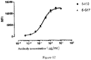
- Example 7 HEK293-hCLDN18.2 binding activity of the recombinant mouse monoclonal antibody
- HEK293-hCLDN18.2 The binding of purified monoclonal antibodies 5-110 and 5-G17 to HEK293-hCLDN18.2 was verified by flow cytometry.
- HEK293-hCLDN18.2 was plated in a 96-well U-shaped plate (1.5 ⁇ 10 5 cells/well), and the plate was washed once with PBS buffer containing 1% FBS.
- Purified monoclonal antibodies 5-I10 and 5-G17 were diluted with PBS (1% FBS) respectively, and a 3-fold gradient dilution was performed starting from the concentration of 10 ⁇ g/mL to obtain a total of 8 concentration points, they were then added to a 96-well U-plate at 100 ⁇ L/well and incubated at 4°C for 1 hour.
- Example 8 Positive staining of the recombinant mouse monoclonal antibody on gastric cancer FFPE tissue sections
- the CLDN18.2 IHC positive staining of purified monoclonal antibodies 5-110 and 5-G17 was verified on 6 cases of gastric cancer FFPE tissue sections, and EPR19202 was used as a positive control.
- the IHC staining procedure was the same as described in Example 4, wherein the working concentration of the positive control antibody EPR19202 in the primary antibody incubation step was 1:100 dilution, and the working concentration of purified antibodies 5-I10 and 5-G17 was 5 ⁇ g/mL.
- the staining results were shown in Figure 13 .
- the recombinant and purified mouse IgG2a type 5-110 and 5-G17 monoclonal antibodies can be positively stained on several cases of gastric cancer FFPE tissue sections.
- Gastric cancer cells form a gland-like structure on the S11-4725-1 tissue, but it was significantly different from the glandular structure on the aforementioned sections; the positive control SPR19202 had no obvious IHC staining, while 5-110 and 5-G17 had strong positive staining, which was in diffuse type, and both glandular epithelium and stromal cells were stained.
- Tumor cells on S09-15785-2FS and S07-7939-C sections had no obvious glandular structure formed, and positive control SPR19202, 5-110 and 5-G17 had no positive IHC staining on S09-15785-2FS. Only 5-G17 showed weak IHC positive staining on S07-7939-C.
Landscapes
- Health & Medical Sciences (AREA)
- Chemical & Material Sciences (AREA)
- Life Sciences & Earth Sciences (AREA)
- Immunology (AREA)
- Organic Chemistry (AREA)
- General Health & Medical Sciences (AREA)
- Engineering & Computer Science (AREA)
- Medicinal Chemistry (AREA)
- Molecular Biology (AREA)
- Genetics & Genomics (AREA)
- Biochemistry (AREA)
- Biomedical Technology (AREA)
- Biophysics (AREA)
- Biotechnology (AREA)
- Urology & Nephrology (AREA)
- Hematology (AREA)
- Nuclear Medicine, Radiotherapy & Molecular Imaging (AREA)
- Animal Behavior & Ethology (AREA)
- General Chemical & Material Sciences (AREA)
- Chemical Kinetics & Catalysis (AREA)
- Veterinary Medicine (AREA)
- Proteomics, Peptides & Aminoacids (AREA)
- Public Health (AREA)
- Pharmacology & Pharmacy (AREA)
- Microbiology (AREA)
- Physics & Mathematics (AREA)
- Bioinformatics & Cheminformatics (AREA)
- General Engineering & Computer Science (AREA)
- Zoology (AREA)
- Wood Science & Technology (AREA)
- Cell Biology (AREA)
- Food Science & Technology (AREA)
- Pathology (AREA)
- Analytical Chemistry (AREA)
- General Physics & Mathematics (AREA)
- Plant Pathology (AREA)
- Peptides Or Proteins (AREA)
- Medicines Containing Antibodies Or Antigens For Use As Internal Diagnostic Agents (AREA)
- Preparation Of Compounds By Using Micro-Organisms (AREA)
- Micro-Organisms Or Cultivation Processes Thereof (AREA)
Applications Claiming Priority (2)
| Application Number | Priority Date | Filing Date | Title |
|---|---|---|---|
| CN202010084476 | 2020-02-10 | ||
| PCT/CN2021/076481 WO2021160154A1 (zh) | 2020-02-10 | 2021-02-10 | Cldn18.2抗体及其用途 |
Publications (2)
| Publication Number | Publication Date |
|---|---|
| EP4105237A1 true EP4105237A1 (de) | 2022-12-21 |
| EP4105237A4 EP4105237A4 (de) | 2024-03-27 |
Family
ID=77292052
Family Applications (1)
| Application Number | Title | Priority Date | Filing Date |
|---|---|---|---|
| EP21753717.4A Pending EP4105237A4 (de) | 2020-02-10 | 2021-02-10 | Cldn18.2-antikörper und verwendung davon |
Country Status (8)
| Country | Link |
|---|---|
| US (1) | US20230312704A1 (de) |
| EP (1) | EP4105237A4 (de) |
| JP (2) | JP7663883B2 (de) |
| KR (1) | KR102924134B1 (de) |
| CN (1) | CN115427453B (de) |
| AU (1) | AU2021220887B2 (de) |
| CA (1) | CA3167299A1 (de) |
| WO (1) | WO2021160154A1 (de) |
Cited By (1)
| Publication number | Priority date | Publication date | Assignee | Title |
|---|---|---|---|---|
| CN116333170A (zh) * | 2023-03-28 | 2023-06-27 | 陇东学院 | 一种用于免疫治疗胃癌的重组病毒样纳米颗粒及其应用 |
Families Citing this family (7)
| Publication number | Priority date | Publication date | Assignee | Title |
|---|---|---|---|---|
| JP7522482B2 (ja) * | 2020-02-10 | 2024-07-25 | 上海詩健生物科技有限公司 | クローディン18.2の抗体及びその使用 |
| CN117295524A (zh) | 2021-06-02 | 2023-12-26 | 百奥泰生物制药股份有限公司 | 药物偶联物及其用途 |
| AU2022371521A1 (en) * | 2021-10-19 | 2024-05-02 | Biosion Inc. | Antibodies binding cldn18.2 and uses thereof |
| WO2023196882A1 (en) * | 2022-04-06 | 2023-10-12 | Zai Lab (Us) Llc | Claudin 18.2 immunohistochemistry assay and use thereof |
| WO2024211459A1 (en) * | 2023-04-04 | 2024-10-10 | Zai Lab (Shanghai) Co., Ltd | Use of anti-claudin antibody for cancer treatment based on certain biomarkers |
| CN117327182B (zh) * | 2023-09-19 | 2024-06-04 | 上海交通大学医学院附属仁济医院 | Cldn18.2单域抗体探针的制备方法及应用 |
| CN117551199B (zh) * | 2023-11-16 | 2024-04-19 | 杭州荣谷生物科技有限公司 | 一种Claudin18.2纳米抗体的制备方法及其应用 |
Family Cites Families (43)
| Publication number | Priority date | Publication date | Assignee | Title |
|---|---|---|---|---|
| US4816567A (en) | 1983-04-08 | 1989-03-28 | Genentech, Inc. | Recombinant immunoglobin preparations |
| US6548640B1 (en) | 1986-03-27 | 2003-04-15 | Btg International Limited | Altered antibodies |
| EP0368684B2 (de) | 1988-11-11 | 2004-09-29 | Medical Research Council | Klonierung von Immunglobulin sequenzen aus den variabelen Domänen. |
| DE3920358A1 (de) | 1989-06-22 | 1991-01-17 | Behringwerke Ag | Bispezifische und oligospezifische, mono- und oligovalente antikoerperkonstrukte, ihre herstellung und verwendung |
| US5959177A (en) | 1989-10-27 | 1999-09-28 | The Scripps Research Institute | Transgenic plants expressing assembled secretory antibodies |
| US6150584A (en) | 1990-01-12 | 2000-11-21 | Abgenix, Inc. | Human antibodies derived from immunized xenomice |
| US6075181A (en) | 1990-01-12 | 2000-06-13 | Abgenix, Inc. | Human antibodies derived from immunized xenomice |
| US5770429A (en) | 1990-08-29 | 1998-06-23 | Genpharm International, Inc. | Transgenic non-human animals capable of producing heterologous antibodies |
| CA2095633C (en) | 1990-12-03 | 2003-02-04 | Lisa J. Garrard | Enrichment method for variant proteins with altered binding properties |
| EP1400536A1 (de) | 1991-06-14 | 2004-03-24 | Genentech Inc. | Verfahren zur Herstellung humanisierter Antikörper |
| GB9114948D0 (en) | 1991-07-11 | 1991-08-28 | Pfizer Ltd | Process for preparing sertraline intermediates |
| US7018809B1 (en) | 1991-09-19 | 2006-03-28 | Genentech, Inc. | Expression of functional antibody fragments |
| WO1993008829A1 (en) | 1991-11-04 | 1993-05-13 | The Regents Of The University Of California | Compositions that mediate killing of hiv-infected cells |
| CA2372813A1 (en) | 1992-02-06 | 1993-08-19 | L.L. Houston | Biosynthetic binding protein for cancer marker |
| US5789199A (en) | 1994-11-03 | 1998-08-04 | Genentech, Inc. | Process for bacterial production of polypeptides |
| US5840523A (en) | 1995-03-01 | 1998-11-24 | Genetech, Inc. | Methods and compositions for secretion of heterologous polypeptides |
| US6267958B1 (en) | 1995-07-27 | 2001-07-31 | Genentech, Inc. | Protein formulation |
| US6171586B1 (en) | 1997-06-13 | 2001-01-09 | Genentech, Inc. | Antibody formulation |
| US6040498A (en) | 1998-08-11 | 2000-03-21 | North Caroline State University | Genetically engineered duckweed |
| US6610833B1 (en) | 1997-11-24 | 2003-08-26 | The Institute For Human Genetics And Biochemistry | Monoclonal human natural antibodies |
| DK1034298T3 (da) | 1997-12-05 | 2012-01-30 | Scripps Research Inst | Humanisering af murint antistof |
| ES2248127T3 (es) | 1999-10-04 | 2006-03-16 | Medicago Inc. | Metodo para regular la transcripcion de genes foraneos en presencia de nigtrogeno. |
| US7125978B1 (en) | 1999-10-04 | 2006-10-24 | Medicago Inc. | Promoter for regulating expression of foreign genes |
| EP1240319A1 (de) | 1999-12-15 | 2002-09-18 | Genentech, Inc. | "shotgun scanning", eine methode zur kartierung von funktionellen protein epitopen |
| US6596541B2 (en) | 2000-10-31 | 2003-07-22 | Regeneron Pharmaceuticals, Inc. | Methods of modifying eukaryotic cells |
| CA2430013C (en) | 2000-11-30 | 2011-11-22 | Medarex, Inc. | Transgenic transchromosomal rodents for making human antibodies |
| CA2488441C (en) | 2002-06-03 | 2015-01-27 | Genentech, Inc. | Synthetic antibody phage libraries |
| WO2004065416A2 (en) | 2003-01-16 | 2004-08-05 | Genentech, Inc. | Synthetic antibody phage libraries |
| EP1740615B1 (de) | 2004-03-31 | 2014-11-05 | Genentech, Inc. | Humanisierte anti-tgf-beta-antikörper |
| US7785903B2 (en) | 2004-04-09 | 2010-08-31 | Genentech, Inc. | Variable domain library and uses |
| JO3000B1 (ar) | 2004-10-20 | 2016-09-05 | Genentech Inc | مركبات أجسام مضادة . |
| EP1973951A2 (de) | 2005-12-02 | 2008-10-01 | Genentech, Inc. | Bindende polypeptide mit eingeschränkten diversitätssequenzen |
| TW200812616A (en) | 2006-05-09 | 2008-03-16 | Genentech Inc | Binding polypeptides with optimized scaffolds |
| CN100592373C (zh) | 2007-05-25 | 2010-02-24 | 群康科技(深圳)有限公司 | 液晶显示面板驱动装置及其驱动方法 |
| ITCL20070039A1 (it) | 2007-07-10 | 2009-01-11 | Michele Neri | Bombolo a pressione costante con tubo estrusore a proboscide e valvola dosatrice, agganciato ad una struttura su cui scorre, azionato da una fonte di pressione controllata, utilizzabile in particolare per la estrusione-lavorazione di tutte le paste: |
| JP6157046B2 (ja) | 2008-01-07 | 2017-07-05 | アムジェン インコーポレイテッド | 静電的ステアリング(electrostaticsteering)効果を用いた抗体Fcヘテロ二量体分子を作製するための方法 |
| WO2013167153A1 (en) * | 2012-05-09 | 2013-11-14 | Ganymed Pharmaceuticals Ag | Antibodies useful in cancer diagnosis |
| WO2014127785A1 (en) * | 2013-02-20 | 2014-08-28 | Ganymed Pharmaceuticals Ag | Combination therapy involving antibodies against claudin 18.2 for treatment of cancer |
| SG11202007055QA (en) | 2018-03-08 | 2020-09-29 | Phanes Therapeutics Inc | Anti-claudin 18.2 antibodies and uses thereof |
| EP3765522A4 (de) * | 2018-03-14 | 2022-05-18 | Beijing Xuanyi Pharmasciences Co., Ltd. | Anti-claudin-18.2-antikörper |
| US11059887B2 (en) * | 2018-05-18 | 2021-07-13 | Lanova Medicines Limited Company | Anti-claudin 18.2 antibodies and uses thereof |
| CN110606891B (zh) * | 2018-06-17 | 2022-12-06 | 上海健信生物医药科技有限公司 | 一种针对人cldn18.2的抗体分子,抗原结合片段及其医药用途 |
| JP7513605B2 (ja) * | 2018-07-18 | 2024-07-09 | アスクジーン・ファーマ・インコーポレイテッド | 新規な抗体ならびにそれを調製および使用するための方法 |
-
2021
- 2021-02-10 AU AU2021220887A patent/AU2021220887B2/en active Active
- 2021-02-10 WO PCT/CN2021/076481 patent/WO2021160154A1/zh not_active Ceased
- 2021-02-10 JP JP2022573794A patent/JP7663883B2/ja active Active
- 2021-02-10 CA CA3167299A patent/CA3167299A1/en active Pending
- 2021-02-10 KR KR1020227030885A patent/KR102924134B1/ko active Active
- 2021-02-10 US US17/798,863 patent/US20230312704A1/en active Pending
- 2021-02-10 CN CN202180013663.5A patent/CN115427453B/zh active Active
- 2021-02-10 EP EP21753717.4A patent/EP4105237A4/de active Pending
-
2024
- 2024-09-30 JP JP2024171863A patent/JP2025011153A/ja not_active Withdrawn
Cited By (1)
| Publication number | Priority date | Publication date | Assignee | Title |
|---|---|---|---|---|
| CN116333170A (zh) * | 2023-03-28 | 2023-06-27 | 陇东学院 | 一种用于免疫治疗胃癌的重组病毒样纳米颗粒及其应用 |
Also Published As
| Publication number | Publication date |
|---|---|
| CN115427453B (zh) | 2024-07-12 |
| KR20220139357A (ko) | 2022-10-14 |
| AU2021220887A1 (en) | 2022-09-29 |
| AU2021220887B2 (en) | 2024-11-21 |
| US20230312704A1 (en) | 2023-10-05 |
| CN115427453A (zh) | 2022-12-02 |
| JP2023513400A (ja) | 2023-03-30 |
| KR102924134B1 (ko) | 2026-02-09 |
| EP4105237A4 (de) | 2024-03-27 |
| JP7663883B2 (ja) | 2025-04-17 |
| WO2021160154A1 (zh) | 2021-08-19 |
| JP2025011153A (ja) | 2025-01-23 |
| CA3167299A1 (en) | 2021-08-19 |
Similar Documents
| Publication | Publication Date | Title |
|---|---|---|
| EP4105237A1 (de) | Cldn18.2-antikörper und verwendung davon | |
| CN113321731B (zh) | 结合人程序性死亡配体1(pd-l1)的抗体 | |
| JP6629837B2 (ja) | 抗ptk7抗体−薬物コンジュゲート | |
| US12084510B2 (en) | Antibodies selective for cells presenting EGFR at high density | |
| JP6898925B2 (ja) | Asct2特異的結合分子及びその使用 | |
| AU2021218927B2 (en) | Claudin 18.2 antibody and use thereof | |
| JP7361980B2 (ja) | 脳への向上した薬物送達の方法 | |
| US20210228731A1 (en) | Antibody drug conjugates for ablating hematopoietic stem cells | |
| US11840568B2 (en) | Lymphocyte activation gene-3 (LAG-3) binding antibody and use thereof | |
| CN113150150B (zh) | Tim3结合分子及其应用 | |
| WO2025006898A1 (en) | Anti-idiotypic antibodies to cluster of differentiation 7-targeted binding domains and related compositions and methods | |
| TW202504637A (zh) | 與結合至nectin-4蛋白之抗體及抗體藥物結合物(adc)相關之方法及組合物 |
Legal Events
| Date | Code | Title | Description |
|---|---|---|---|
| STAA | Information on the status of an ep patent application or granted ep patent |
Free format text: STATUS: THE INTERNATIONAL PUBLICATION HAS BEEN MADE |
|
| PUAI | Public reference made under article 153(3) epc to a published international application that has entered the european phase |
Free format text: ORIGINAL CODE: 0009012 |
|
| STAA | Information on the status of an ep patent application or granted ep patent |
Free format text: STATUS: REQUEST FOR EXAMINATION WAS MADE |
|
| 17P | Request for examination filed |
Effective date: 20220907 |
|
| AK | Designated contracting states |
Kind code of ref document: A1 Designated state(s): AL AT BE BG CH CY CZ DE DK EE ES FI FR GB GR HR HU IE IS IT LI LT LU LV MC MK MT NL NO PL PT RO RS SE SI SK SM TR |
|
| DAV | Request for validation of the european patent (deleted) | ||
| DAX | Request for extension of the european patent (deleted) | ||
| A4 | Supplementary search report drawn up and despatched |
Effective date: 20240223 |
|
| RIC1 | Information provided on ipc code assigned before grant |
Ipc: A61P 35/00 20060101ALI20240219BHEP Ipc: A61K 39/00 20060101ALI20240219BHEP Ipc: A61K 39/395 20060101ALI20240219BHEP Ipc: C07K 16/30 20060101AFI20240219BHEP |
|
| STAA | Information on the status of an ep patent application or granted ep patent |
Free format text: STATUS: EXAMINATION IS IN PROGRESS |
|
| 17Q | First examination report despatched |
Effective date: 20251029 |