EP4177335A1 - Procédés de fabrication in vitro de tissu de fundus d'estomac et compositions associées à celui-ci - Google Patents
Procédés de fabrication in vitro de tissu de fundus d'estomac et compositions associées à celui-ci Download PDFInfo
- Publication number
- EP4177335A1 EP4177335A1 EP22189427.2A EP22189427A EP4177335A1 EP 4177335 A1 EP4177335 A1 EP 4177335A1 EP 22189427 A EP22189427 A EP 22189427A EP 4177335 A1 EP4177335 A1 EP 4177335A1
- Authority
- EP
- European Patent Office
- Prior art keywords
- cell
- period
- cells
- wnt
- gastric
- Prior art date
- Legal status (The legal status is an assumption and is not a legal conclusion. Google has not performed a legal analysis and makes no representation as to the accuracy of the status listed.)
- Withdrawn
Links
Images








Classifications
-
- C—CHEMISTRY; METALLURGY
- C12—BIOCHEMISTRY; BEER; SPIRITS; WINE; VINEGAR; MICROBIOLOGY; ENZYMOLOGY; MUTATION OR GENETIC ENGINEERING
- C12N—MICROORGANISMS OR ENZYMES; COMPOSITIONS THEREOF; PROPAGATING, PRESERVING, OR MAINTAINING MICROORGANISMS; MUTATION OR GENETIC ENGINEERING; CULTURE MEDIA
- C12N5/00—Undifferentiated human, animal or plant cells, e.g. cell lines; Tissues; Cultivation or maintenance thereof; Culture media therefor
- C12N5/06—Animal cells or tissues; Human cells or tissues
- C12N5/0602—Vertebrate cells
- C12N5/0679—Cells of the gastro-intestinal tract
-
- C—CHEMISTRY; METALLURGY
- C12—BIOCHEMISTRY; BEER; SPIRITS; WINE; VINEGAR; MICROBIOLOGY; ENZYMOLOGY; MUTATION OR GENETIC ENGINEERING
- C12N—MICROORGANISMS OR ENZYMES; COMPOSITIONS THEREOF; PROPAGATING, PRESERVING, OR MAINTAINING MICROORGANISMS; MUTATION OR GENETIC ENGINEERING; CULTURE MEDIA
- C12N5/00—Undifferentiated human, animal or plant cells, e.g. cell lines; Tissues; Cultivation or maintenance thereof; Culture media therefor
- C12N5/06—Animal cells or tissues; Human cells or tissues
- C12N5/0602—Vertebrate cells
- C12N5/0603—Embryonic cells ; Embryoid bodies
- C12N5/0606—Pluripotent embryonic cells, e.g. embryonic stem cells [ES]
-
- C—CHEMISTRY; METALLURGY
- C12—BIOCHEMISTRY; BEER; SPIRITS; WINE; VINEGAR; MICROBIOLOGY; ENZYMOLOGY; MUTATION OR GENETIC ENGINEERING
- C12N—MICROORGANISMS OR ENZYMES; COMPOSITIONS THEREOF; PROPAGATING, PRESERVING, OR MAINTAINING MICROORGANISMS; MUTATION OR GENETIC ENGINEERING; CULTURE MEDIA
- C12N5/00—Undifferentiated human, animal or plant cells, e.g. cell lines; Tissues; Cultivation or maintenance thereof; Culture media therefor
- C12N5/06—Animal cells or tissues; Human cells or tissues
- C12N5/0602—Vertebrate cells
- C12N5/0607—Non-embryonic pluripotent stem cells, e.g. MASC
-
- C—CHEMISTRY; METALLURGY
- C12—BIOCHEMISTRY; BEER; SPIRITS; WINE; VINEGAR; MICROBIOLOGY; ENZYMOLOGY; MUTATION OR GENETIC ENGINEERING
- C12N—MICROORGANISMS OR ENZYMES; COMPOSITIONS THEREOF; PROPAGATING, PRESERVING, OR MAINTAINING MICROORGANISMS; MUTATION OR GENETIC ENGINEERING; CULTURE MEDIA
- C12N5/00—Undifferentiated human, animal or plant cells, e.g. cell lines; Tissues; Cultivation or maintenance thereof; Culture media therefor
- C12N5/06—Animal cells or tissues; Human cells or tissues
- C12N5/0602—Vertebrate cells
- C12N5/0608—Germ cells
- C12N5/0609—Oocytes, oogonia
-
- C—CHEMISTRY; METALLURGY
- C12—BIOCHEMISTRY; BEER; SPIRITS; WINE; VINEGAR; MICROBIOLOGY; ENZYMOLOGY; MUTATION OR GENETIC ENGINEERING
- C12N—MICROORGANISMS OR ENZYMES; COMPOSITIONS THEREOF; PROPAGATING, PRESERVING, OR MAINTAINING MICROORGANISMS; MUTATION OR GENETIC ENGINEERING; CULTURE MEDIA
- C12N2501/00—Active agents used in cell culture processes, e.g. differentation
- C12N2501/10—Growth factors
- C12N2501/11—Epidermal growth factor [EGF]
-
- C—CHEMISTRY; METALLURGY
- C12—BIOCHEMISTRY; BEER; SPIRITS; WINE; VINEGAR; MICROBIOLOGY; ENZYMOLOGY; MUTATION OR GENETIC ENGINEERING
- C12N—MICROORGANISMS OR ENZYMES; COMPOSITIONS THEREOF; PROPAGATING, PRESERVING, OR MAINTAINING MICROORGANISMS; MUTATION OR GENETIC ENGINEERING; CULTURE MEDIA
- C12N2501/00—Active agents used in cell culture processes, e.g. differentation
- C12N2501/10—Growth factors
- C12N2501/115—Basic fibroblast growth factor (bFGF, FGF-2)
-
- C—CHEMISTRY; METALLURGY
- C12—BIOCHEMISTRY; BEER; SPIRITS; WINE; VINEGAR; MICROBIOLOGY; ENZYMOLOGY; MUTATION OR GENETIC ENGINEERING
- C12N—MICROORGANISMS OR ENZYMES; COMPOSITIONS THEREOF; PROPAGATING, PRESERVING, OR MAINTAINING MICROORGANISMS; MUTATION OR GENETIC ENGINEERING; CULTURE MEDIA
- C12N2501/00—Active agents used in cell culture processes, e.g. differentation
- C12N2501/10—Growth factors
- C12N2501/155—Bone morphogenic proteins [BMP]; Osteogenins; Osteogenic factor; Bone inducing factor
-
- C—CHEMISTRY; METALLURGY
- C12—BIOCHEMISTRY; BEER; SPIRITS; WINE; VINEGAR; MICROBIOLOGY; ENZYMOLOGY; MUTATION OR GENETIC ENGINEERING
- C12N—MICROORGANISMS OR ENZYMES; COMPOSITIONS THEREOF; PROPAGATING, PRESERVING, OR MAINTAINING MICROORGANISMS; MUTATION OR GENETIC ENGINEERING; CULTURE MEDIA
- C12N2501/00—Active agents used in cell culture processes, e.g. differentation
- C12N2501/30—Hormones
- C12N2501/38—Hormones with nuclear receptors
- C12N2501/385—Hormones with nuclear receptors of the family of the retinoic acid recptor, e.g. RAR, RXR; Peroxisome proliferator-activated receptor [PPAR]
-
- C—CHEMISTRY; METALLURGY
- C12—BIOCHEMISTRY; BEER; SPIRITS; WINE; VINEGAR; MICROBIOLOGY; ENZYMOLOGY; MUTATION OR GENETIC ENGINEERING
- C12N—MICROORGANISMS OR ENZYMES; COMPOSITIONS THEREOF; PROPAGATING, PRESERVING, OR MAINTAINING MICROORGANISMS; MUTATION OR GENETIC ENGINEERING; CULTURE MEDIA
- C12N2501/00—Active agents used in cell culture processes, e.g. differentation
- C12N2501/40—Regulators of development
- C12N2501/415—Wnt; Frizzeled
-
- C—CHEMISTRY; METALLURGY
- C12—BIOCHEMISTRY; BEER; SPIRITS; WINE; VINEGAR; MICROBIOLOGY; ENZYMOLOGY; MUTATION OR GENETIC ENGINEERING
- C12N—MICROORGANISMS OR ENZYMES; COMPOSITIONS THEREOF; PROPAGATING, PRESERVING, OR MAINTAINING MICROORGANISMS; MUTATION OR GENETIC ENGINEERING; CULTURE MEDIA
- C12N2506/00—Differentiation of animal cells from one lineage to another; Differentiation of pluripotent cells
- C12N2506/02—Differentiation of animal cells from one lineage to another; Differentiation of pluripotent cells from embryonic cells
- C12N2506/025—Differentiation of animal cells from one lineage to another; Differentiation of pluripotent cells from embryonic cells from extra-embryonic cells, e.g. trophoblast, placenta
-
- C—CHEMISTRY; METALLURGY
- C12—BIOCHEMISTRY; BEER; SPIRITS; WINE; VINEGAR; MICROBIOLOGY; ENZYMOLOGY; MUTATION OR GENETIC ENGINEERING
- C12N—MICROORGANISMS OR ENZYMES; COMPOSITIONS THEREOF; PROPAGATING, PRESERVING, OR MAINTAINING MICROORGANISMS; MUTATION OR GENETIC ENGINEERING; CULTURE MEDIA
- C12N2506/00—Differentiation of animal cells from one lineage to another; Differentiation of pluripotent cells
- C12N2506/03—Differentiation of animal cells from one lineage to another; Differentiation of pluripotent cells from non-embryonic pluripotent stem cells
-
- C—CHEMISTRY; METALLURGY
- C12—BIOCHEMISTRY; BEER; SPIRITS; WINE; VINEGAR; MICROBIOLOGY; ENZYMOLOGY; MUTATION OR GENETIC ENGINEERING
- C12N—MICROORGANISMS OR ENZYMES; COMPOSITIONS THEREOF; PROPAGATING, PRESERVING, OR MAINTAINING MICROORGANISMS; MUTATION OR GENETIC ENGINEERING; CULTURE MEDIA
- C12N2506/00—Differentiation of animal cells from one lineage to another; Differentiation of pluripotent cells
- C12N2506/45—Differentiation of animal cells from one lineage to another; Differentiation of pluripotent cells from artificially induced pluripotent stem cells
-
- C—CHEMISTRY; METALLURGY
- C12—BIOCHEMISTRY; BEER; SPIRITS; WINE; VINEGAR; MICROBIOLOGY; ENZYMOLOGY; MUTATION OR GENETIC ENGINEERING
- C12N—MICROORGANISMS OR ENZYMES; COMPOSITIONS THEREOF; PROPAGATING, PRESERVING, OR MAINTAINING MICROORGANISMS; MUTATION OR GENETIC ENGINEERING; CULTURE MEDIA
- C12N2513/00—3D culture
Definitions
- hFGOs human fundic-type gastric organoids
- the instant disclosure relates to methods for converting mammalian definitive endoderm (DE) cells into specific tissue(s) or organ(s) through directed differentiation.
- the disclosure relates to formation of gastric fundus tissue and/or organoids formed from differentiated definitive endoderm.
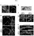
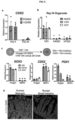
- FIG 1 Wnt/ ⁇ -catenin signaling is required for specification of the embryonic fundus in mice, a, Pdx1 and Sox2 were expressed in the antrum (a), whereas Pdx1 was absent in the fundus (f), identified by Atp4b-expressing parietal cells at E18.5. b, X-gal staining of an E10.5 foregut from an Axin2:LacZ reporter embryo showed that Wnt activity was restricted to the anterior domain of the stomach but excluded from the posterior stomach. c, Deletion of ⁇ -catenin in the gastric epithelium caused an anterior expansion of Pdx1 into the fundic region of the stomach.
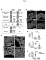
- FIG 2 ⁇ -catenin activation promotes fundus development from human foregut progenitor spheroids.
- a Schematized diagram of differentiation protocol for both fundic and antral hGOs.
- b c, At day 9, CHIR-treated organoids exhibited reduction in PDX1, increase in IRX2, IRX3, and IRX5, and no change in gastric markers SOX2 or GATA4.
- hFGOs grew comparably to hAGOs, but also exhibited glandular budding morphogenesis (white arrowheads).
- hGOs contained epithelium that expressed CDH1, KRT8, and CTNNB1, as well as gastric markers GATA4 and CLDN18.
- hAGOs exhibited nearly ubiquitous PDX1 expression while hFGOs did not.
- Scale bars 50 ⁇ m (c), 500 ⁇ m (d) and 100 ⁇ m (e). Error bars represent s.e.m.
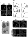
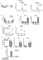
- FIG 3 Differentiation of mucous and endocrine cell lineages in hGOs.
- a Schematic of the shared and distinct lineages found in fundic and antral glands of the stomach.
- b Both antral and fundic hGOs contained MUC5AC-positive surface mucous cells and MUC6-positive mucous neck cells, c, d, hFGOs contained endocrine cells expressing the pan-endocrine marker SYP. Diverse hormone cell types were identified in hFGOs, including GHRL-, SST-, and histamine-expressing endocrine cells.
- FIG 4 Formation of chief cells in hFGOs.
- a hFGOs had a both MIST1 and Pepsinogen C (PGC) positive cells
- PPC Pepsinogen C
- b High magnification of boxed region in panel (a) showing a gland with a cluster of cells with apical PGC staining
- d Transmission electron micrograph of an hFGO cell containing dense zymogen granules, indicative of a chief cell.
- Scale bars 200 ⁇ m (a), 25 ⁇ m (b), and 10 ⁇ m (d). Error bars represent s.e.m.
- FIG 5 Identification of pathways that drive differentiation of functional parietal cells in hFGOs.
- b Stimulated differentiation of ATP4B-expressing parietal cells following treatment with PD03/BMP4.
- hFGO-derived parietal cells resembled those found in the maturing mouse fundic epithelium in vivo.
- d Transmission electron micrograph of an hFGO cell with canalicular structure reminiscent of parietal cells.
- e The epithelium of human fundic glands and hFGO epithelium were organized into MUC5AC-expressing cells in the surface epithelium and ATP4B-expressing parietal cells in the glandular units
- f Analysis of luminal pH in organoids in response to histamine by luminal injection of SNARF-5F. The luminal pH in hFGOs rapidly dropped, while hAGOs exhibited no response.
- n 9, 9, 7, and 4 biological replicates in hFGOs (histamine), hFGOs (histamine and famotidine), hFGOs (histamine and omeprazole), and hAGOs (histamine), respectively; data representative of three independent experiments.
- AO Histamine induced acridine orange
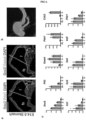
- FIG 6 Defining molecular domains in the developing stomach in vivo.
- a Analysis of Sox2, Pdx1, and Gata4 in the embryonic mouse stomach (E14.5) showed that the fundus (f) was Sox2+Gata4+Pdx1-, whereas the antrum (a) was Sox2+Gata4+Pdx1+.
- the forestomach (fs) expressed Sox2 but neither Gata4 nor Pdx1.
- Brightfield stereomicrograph showing dissected regions of the E14.5 mouse stomach that were analyzed by qPCR.
- fs forestomach
- f fundus
- a antrum
- d duodenum
- c Dissected regions in b were analyzed by qPCR for known regionally expressed markers (Sox2, P63, Gata4, Pdx1, and Cdx2) to validate the accuracy of micro-dissection.
- qPCR analysis of the dissected E14.5 stomach regions showed that putative fundus markers Irx1, Irx2, Irx3, Irx5, and Pitx1 were enriched in the fundus compared to the antrum.
- n 4 biological replicates per dissected region. Scale bar, 500 ⁇ m. Error bars represent s.d.
- FIG 8 Stable induction of fundic fate in hGOs and efficiency of protocol, a, Applicant investigated how long CHIR treatment was necessary to establish fundus identity. Brief CHIR treatment (d6-9) and subsequent growth of organoids in control growth medium until day 34 resulted in fundic organoids expressing the antral marker PDX1, suggesting that short CHIR treatment did not produce a stable fundic fate. Applicant then tested whether longer exposures to CHIR were required to retain fundic fate and found that only continuous treatment through at least day 29 could maintain low expression of the antral marker PDX1.
- FIG 9 BMP-dependence of Wnt/ ⁇ -catenin activation to induce intestinal fate from foregut progenitors, a, The intestine-specific transcription factor CDX2 was not significantly induced in CHIR-treated hGOs at either day 9 or day 20.
- c Anterior-posterior fate is coordinately controlled by WNT and BMP activity.
- FIG 10 hFGOs contain organized glands supported by associated mesenchymal layer, a, Transmission electron micrographs demonstrated that hFGO glands exhibited organized architecture with narrow apical membranes. b, Both hFGOs and hAGOs contained a supporting layer FOXF1+/VIM+ undifferentiated fibroblasts. Scale bars, 5 ⁇ m (a) and 100 ⁇ m (b).
- FIG 11 Region-specific cytodifferentiation in human gastric organoids.
- a Antral and fundic hGOs exhibited comparable expression of mucous cell markers MUC5AC and MUC6.
- b As shown in transmission electron micrograph, hFGOs contained abundant cells exhibiting granule pattern consistent with mucous neck cells, the precursors to differentiated chief cells,
- c Exogenous expression of NEUROG3 in hGOs derived from NEUROG3-deficient hESC line induced robust differentiation of SYP-positive endocrine cells. While both hAGOs and hFGOs formed GHRL- and SST-expressing endocrine cells, specification of GAST+ G-cells was observed only in hAGOs.
- FIG 12 Analysis of murine chief cell development.
- a Unlike parietal cells, which expressed functional markers ( Atp4b ) as early as late embryonic stages, chief cell gene products were not detectable until much later stages of development.
- E18.5 and juvenile (P12) stomach Gif and Pgc were not yet expressed, indicating that chief cells mature much later in development than other lineages in the gastric epithelium.
- Pgc the P12 mouse stomach did contain abundant glandular cells expressing nuclear Mist1, a chief cell-specific marker. Thus, chief cells were indeed specified earlier but took several weeks to develop robust expression of terminal differentiation markers. Scale bars, 100 ⁇ m (a) and 200 ⁇ m (b).
- FIG 13 Screen for pathways that promote differentiation of parietal cells in fundic hGOs.
- a To test for growth factors/small molecules capable of inducing parietal cell differentiation, hFGOs were exposed for two days (30-32) to the indicated agonist or antagonist and then analyzed at day 34. In a screening experiment of different pathways, only MEK inhibition with PD03 was found to robustly induce expression of ATP4A / B.
- b Reduction or removal of EGF from the culture medium was not sufficient to reproduce the effect of MEK inhibition
- c The ability of PD03/BMP4 to induce parietal cell development was exclusive to fundic hGOs, as antral hGOs did not express fundic markers in response to PD03/BMP4.
- FIG 14 Live in vitro pH monitoring in gastric organoids.
- a The dye SNAFR5F exhibits responsiveness over pH range of 5-8, which makes it well suited to detect physiologic changes in response to parietal cell-mediated acid secretion.
- b Media and luminal pH measurements recorded before (closed circles) and 60 minutes following addition of histamine (open circles). Antral hGOs did not respond, while the fundic hGO luminal pH decreased in response to histamine.
- the acidification was inhibited by pre-treatment of organoids with either famotidine or omeprazole. Further, omeprazole was sufficient to raise the pH in fundic organoids prior to histamine exposure, suggesting a baseline acid secretion in the fundic organoids.
- hFGOs contained parietal cell-dense glands in which acridine orange (AO) accumulated in nearly all of the cells lining the lumen of the gland.
- AO accumulation was observed in a canalicular-type pattern in parietal cells in hFGOs. Scale bars, 10 ⁇ m. Error bars represent s.d.
- FIG 15 Serial passaging of human gastric organoids.
- a Schematic representation of experiments to determine the presence of gastric stem cells in hGOs.
- b When fragments were grown in culture medium containing only EGF, they did not grow or expand to form new organoids. However, addition of CHIR and FGF10 to the culture medium was sufficient to support the growth of individual fragments into newly formed organoids.
- c Following two passages, hFGOs still expressed genes consistent with a gastric phenotype, including PGC, MUC6, MUC5AC, and GHRL.
- hFGOs contain cells with properties analogous to those of adult gastric stem cells, d, Although passaged hFGOs expressed markers associated with several differentiated gastric cell types, they did not express genes associated with parietal cells such as ATP4B. Further, differentiation of parietal cells could not be induced through MEK inhibition as they could prior to passaging. Error bars represent s.d.
- gastric fundus tissue means a fundic type of gastric epithelium found in the corpus that contains fundic cell types, including but not limited to acid-producing parietal cells and protease-producing chief cells.
- DE cell means one of the three primary germ layers produced by the process of gastrulation.
- wnt signalling pathway means the wnt/beta-catenin pathway and is a signal transduction pathway that is mediated by Wnt ligands and frizzled cell surface receptors that acts through the beta-catenin protein.
- activator with respect to a pathway, such as a "wnt pathway” means a substance that activates the Wnt/beta-catenin pathway such that Wnt/beta-catenin targets are increased.
- FGF signaling pathway activator means a substance that activates the FGF pathway such that FGF targets are increased.
- BMP signalling pathway inhibitor a substance that interferes with the BMP pathway and causes BMP targets to be decreased.
- growth factor means a substance capable of stimulating cellular processes including but not limited to growth, proliferation, morphogenesis or differentiation.
- fundic lineage means cell types found in fundic epithelium in the corpus stomach.
- SOX2+GATA+PDX1- epithelium means epithelium that expresses the listed proteins.
- stable expression of a marker means expression that does not change upon modification of the growth environment.
- totipotent stem cells are stem cells that can differentiate into embryonic and extra-embryonic cell types. Such cells can construct a complete, viable, organism. These cells are produced from the fusion of an egg and sperm cell. Cells produced by the first few divisions of the fertilized egg are also totipotent.
- pluripotent stem cells encompasses any cells that can differentiate into nearly all cells, i.e., cells derived from any of the three germ layers (germinal epithelium), including endoderm (interior stomach lining, gastrointestinal tract, the lungs), mesoderm (muscle, bone, blood, urogenital), and ectoderm (epidermal tissues and nervous system).
- PSCs can be the descendants of totipotent cells, derived from embryos (including embryonic germ cells) or obtained through induction of a non-pluripotent cell, such as an adult somatic cell, by forcing the expression of certain genes.
- iPSCs induced pluripotent stem cells
- iPS cells also commonly abbreviated as iPS cells
- iPS cells refers to a type of pluripotent stem cells artificially derived from a normally non-pluripotent cell, such as an adult somatic cell, by inducing a "forced" expression of certain genes.
- a precursor cell encompasses any cells that can be used in methods described herein, through which one or more precursor cells acquire the ability to renew itself or differentiate into one or more specialized cell types.
- a precursor cell is pluripotent or has the capacity to becoming pluripotent.
- the precursor cells are subjected to the treatment of external factors (e.g., growth factors) to acquire pluripotency.
- a precursor cell can be a totipotent stem cell; a pluripotent stem cell (induced or non-induced); a multipotent stem cell; and a unipotent stem cell.
- a precursor cell can be from an embryo, an infant, a child, or an adult.
- a precursor cell can be a somatic cell subject to treatment such that pluripotency is conferred via genetic manipulation or protein/peptide treatment.
- cellular differentiation is the process by which a less specialized cell becomes a more specialized cell type.
- directed differentiation describes a process through which a less specialized cell becomes a particular specialized target cell type.
- the particularity of the specialized target cell type can be determined by any applicable methods that can be used to define or alter the destiny of the initial cell. Exemplary methods include but are not limited to genetic manipulation, chemical treatment, protein treatment, and nucleic acid treatment.
- cellular constituents are individual genes, proteins, mRNA expressing genes, and/or any other variable cellular component or protein activities such as the degree of protein modification (e.g., phosphorylation), for example, that is typically measured in biological experiments (e.g., by microarray or immunohistochemistry) by those skilled in the art.
- Significant discoveries relating to the complex networks of biochemical processes underlying living systems, common human diseases, and gene discovery and structure determination can now be attributed to the application of cellular constituent abundance data as part of the research process.
- Cellular constituent abundance data can help to identify biomarkers, discriminate disease subtypes and identify mechanisms of toxicity.
- pluripotent stem cells are derived from embryonic stem cells, which are in turn derived from totipotent cells of the early mammalian embryo and are capable of unlimited, undifferentiated proliferation in vitro.
- Embryonic stem cells are pluripotent stem cells derived from the inner cell mass of the blastocyst, an early-stage embryo. Methods for deriving embryonic stem cells from blastocytes are well known in the art. Human embryonic stem cells H9 (H9-hESCs) are used in the exemplary embodiments described in the present application, but it would be understood by one of skill in the art that the methods and systems described herein are applicable to any stem cells.
- Additional stem cells that can be used in embodiments in accordance with the present invention include but are not limited to those provided by or described in the database hosted by the National Stem Cell Bank (NSCB), Human Embryonic Stem Cell Research Center at the University of California, San Francisco (UCSF); WISC cell Bank at the Wi Cell Research Institute; the University of Wisconsin Stem Cell and Regenerative Medicine Center (UW-SCRMC); Novocell, Inc. (San Diego, Calif.); Cellartis AB (Goteborg, Sweden); ES Cell International Pte Ltd (Singapore); Technion at the Israel Institute of Technology (Haifa, Israel); and the Stem Cell Database hosted by Princeton University and the University of Pennsylvania.
- NSCB National Stem Cell Bank
- UW-SCRMC University of Wisconsin Stem Cell and Regenerative Medicine Center
- UW-SCRMC University of Wisconsin Stem Cell and Regenerative Medicine Center
- Novocell, Inc. San Diego, Calif.
- Cellartis AB Goteborg, Sweden
- Exemplary embryonic stem cells that can be used in embodiments in accordance with the present invention include but are not limited to SA01 (SA001); SA02 (SA002); ES01 (HES-1); ES02 (HES-2); ES03 (HES-3); ES04 (HES-4); ES05 (HES-5); ES06 (HES-6); BG01 (BGN-01); BG02 (BGN-02); BG03 (BGN-03); TE03 (13); TE04 (14); TE06 (16); UC01 (HSF1); UC06 (HSF6); WA01 (H1); WA07 (H7); WA09 (H9); WA13 (H13); WA14 (H14).
- embryonic stem cells More details on embryonic stem cells can be found in, for example, Thomson et al., 1998, "Embryonic Stem Cell Lines Derived from Human Blastocysts," Science 282 (5391):1145-1147 ; Andrews et al., 2005, “Embryonic stem (ES) cells and embryonal carcinoma (EC) cells: opposite sides of the same coin,” Biochem Soc Trans 33:1526-1530 ; Martin 1980, “Teratocarcinomas and mammalian embryogenesis,”.
- ES Embryonic Stem Cell Lines Derived from Human Blastocysts
- EC embryonal carcinoma
- iPSCs Induced Pluripotent Stem Cells
- iPSCs are derived by transfection of certain stem cell-associated genes into non-pluripotent cells, such as adult fibroblasts. Transfection is typically achieved through viral vectors, such as retroviruses. Transfected genes include the master transcriptional regulators Oct-3/4 (Pouf51) and Sox2, although it is suggested that other genes enhance the efficiency of induction. After 3-4 weeks, small numbers of transfected cells begin to become morphologically and biochemically similar to pluripotent stem cells, and are typically isolated through morphological selection, doubling time, or through a reporter gene and antibiotic selection.
- iPSCs include but are not limited to first generation iPSCs, second generation iPSCs in mice, and human induced pluripotent stem cells.
- a retroviral system is used to transform human fibroblasts intopluripotent stem cells using four pivotal genes: Oct3/4, Sox2, Klf4, and c-Myc.
- a lentiviral system is used to transform somatic cells with OCT4, SOX2, NANOG, and LIN28.
- Genes whose expression are induced in iPSCs include but are not limited to Oct-3/4 (e.g., Pou5fl); certain members of the Sox gene family (e.g., Sox1, Sox2, Sox3, and Sox15); certain members of the Klf family (e.g., Klf1, Klf2, Klf4, and Klf5), certain members of the Myc family (e.g., C-myc, L-myc, and N-myc), Nanog, and LIN28.
- Oct-3/4 e.g., Pou5fl
- Sox gene family e.g., Sox1, Sox2, Sox3, and Sox15
- Klf family e.g., Klf1, Klf2, Klf4, and Klf5
- Myc family e.g., C-myc, L-myc, and N-myc
- Nanog LIN28.
- non-viral based technologies are employed to generate iPSCs.
- an adenovirus can be used to transport the requisite four genes into the DNA of skin and liver cells of mice, resulting in cells identical to embryonic stem cells. Since the adenovirus does not combine any of its own genes with the targeted host, the danger of creating tumors is eliminated.
- reprogramming can be accomplished via plasmid without any virus transfection system at all, although at very low efficiencies.
- direct delivery of proteins is used to generate iPSCs, thus eliminating the need for viruses or genetic modification.
- generation of mouse iPSCs is possible using a similar methodology: a repeated treatment of the cells with certain proteins channeled into the cells via poly-arginine anchors was sufficient to induce pluripotency.
- the expression of pluripotency induction genes can also be increased by treating somatic cellswith FGF2 under low oxygen conditions.
- embryonic stem cells More details on embryonic stem cells can be found in, for example, Kaji et al., 2009, "Virus free induction of pluripotency and subsequent excision of reprogramming factors," Nature 458:771-775 ; Woltjen et al., 2009, "piggyBac transposition reprograms fibroblasts to induced pluripotent stem cells," Nature 458:766-770 ; Okita et al., 2008, “Generation of Mouse Induced Pluripotent Stem Cells Without Viral Vectors," Science 322(5903):949-953 ; Stadtfeld et al., 2008, “Induced Pluripotent Stem Cells Generated without Viral Integration,” Science 322(5903):945-949 ; and Zhou et al., 2009, “Generation of Induced Pluripotent Stem Cells Using Recombinant Proteins," Cell Stem Cell 4(5):381-384 ; each of which is hereby
- exemplary iPS cell lines include but not limited to iPS-DF19-9; iPS-DF19-9; iPS-DF4-3; iPS-DF6-9; iPS(Foreskin); iPS(IMR90); and iPS(IMR90).
- pluripotent cells are derived from a morula.
- pluripotent stem cells are stem cells.
- Stem cells used in these methods can include, but are not limited to, embryonic stem cells.
- Embryonic stem cells can be derived from the embryonic inner cell mass or from the embryonic gonadal ridges.
- Embryonic stem cells or germ cells can originate from a variety of animal species including, but not limited to, various mammalian species including humans.
- human embryonic stem cells are used to produce definitive endoderm.
- human embryonic germ cells are used to produce definitive endoderm.
- iPSCs are used to produce definitive endoderm.
- hPSCs pluripotent stem cells
- Applicant first identified, and then recapitulated key events in embryonic fundus development to arrive at the claimed compositions.
- Applicant found that disruption of Wnt/ ⁇ -catenin signaling in mouse embryos led to conversion of fundic to antral epithelium, while ⁇ -catenin activation in hPSC-derived foregut progenitors promoted the development of human fundic-type gastric organoids (hFGOs).
- Applicant then used hFGOs to identify temporally distinct roles for multiple signaling pathways in epithelial morphogenesis and differentiation of fundic cell types, including chief cells and functional parietal cells. While hFGOs are a powerful new model for studying the development of the human fundus and its lineages, they also represent a critical new model system to study the molecular basis of human gastric physiology, pathophysiology, and drug discovery.
- an in vitro method of inducing formation of a gastric fundus tissue is disclosed.
- the method may comprise the steps of:
- step e) may further comprise the step of contacting the fundal hGOs with an activator of BMP4 signalling.
- step e may be carried out for a period of time sufficient to develop SOX2+GATA+PDX1- epithelium.
- the functional fundic cell type may be a parietal cell that expresses proton pump proteins and secretes acid. In one aspect, the functional fundic cell type may be a chief cell that secretes pepsinogen.
- step d and step e are carried out for a period of time sufficient to confer stable expression of lineage markers MUC5AC, MUC6, PGC, and GHRL.
- the definitive endoderm may be derived from a precursor cell selected from an embryonic stem cell, an embryonic germ cell, an induced pluripotent stem cell, a mesoderm cell, a definitive endoderm cell, a posterior endoderm cell, a posterior endoderm cell, and a hindgut cell, a definitive endoderm derived from a pluripotent stem cell, a definitive endoderm derived from a pluripotent stem cell selected from an embryonic stem cell, an adult stem cell, or an induced pluripotent stem cell.
- the definitive endoderm may be derived from contacting a pluripotent stem cell with one or more molecules selected from Activin, the BMP subgroups of the TGF-beta superfamily of growth factors; Nodal, Activin A, Activin B, BMP4, Wnt3a, and combinations thereof.
- Some existing wnt signalling pathway activators include but are not limited to:
- Wnt ligands including but not limited to Wnt1, Wnt2, Wnt2b, Wnt3, Wnt3a, Wnt8, et al; modifiers of Wnt ligand activity including but not limited to activated Wnt frizzled receptors, (LRP) co-receptors, R-spondin proteins, Dkk proteins, regulators of Wnt ligand secretion and trafficking (Wntless, Porcupine), inhibiting beta-catenin degredation APC and GSK3beta inhibition, activated beta-catenin, constitutively active TCF/Lef proteins.
- LRP activated Wnt frizzled receptors
- R-spondin proteins R-spondin proteins
- Dkk proteins regulators of Wnt ligand secretion and trafficking
- beta-catenin degredation APC and GSK3beta inhibition activated beta-catenin, constitutively active TCF/Lef proteins.
- Chemical activators there are over 28 known chemicals that either activate or inhibit Wnt/beta-catenin signaling. Some activators include but are not limited to GSK3-beta inhibitors CHIR99021, BIO, LY2090314, SB-216763, lithium, porcupine inhibitors IWP, LGK974, C59, SFRP inhibitor WAY-316606, beta-catenin activator DCA.
- the WNT pathway activator may be one or more molecules selected from Wnt1, Wnt2, Wnt2b, Wnt3, Wnt3a, Wnt4, Wnt5a, Wnt5b, Wnt6, Wnt7a, Wnt7b, Wnt8a, Wnt8b, Wnt9a, Wnt9b, WntlOa, Wnt10b, Wnt11, and Wnt16, for example, Wnt3a, or for example, Wnt3a at a concentration between about 50 to about 1500 ng/ml.
- Suitable FGF signalling pathway activators include: FGF ligands FGF2, 4, 5, 8, et al.. Activated forms of FGF receptors. Proteins and chemicals that stimulate the FGF receptor and signaling components downstream of the receptors including MAPK, MEK, ERK proteins and chemicals that modulate their activity. FGF signaling can be activated by inhibiting inhibitors of FGF signaling pathways including but not limited to Sprouty protein family members.
- the BMP signalling pathway inhibitor may be selected from Noggin, Dorsomorphin, LDN189, DMH-1, and combinations thereof, for example, wherein said precursor cell may be contacted with a BMP inhibitor at a concentration between about 50 to about 1500 ng/ml.
- the steps are conducted in vitro.
- a composition comprising gastric tissue produced according to the aforementioned method(s) is disclosed.
- the gastric tissue may be characterized, for example, by being free of innervation and/or blood vessels.
- an in vitro method of inducing formation of a gastric fundus tissue may comprise the steps of contacting a fundal hGO (hFGO) with a wnt pathway activating agent and an EGF signalling pathway activating agent for a first period, and a MEK inhibitor for a second period, (wherein said MEK inhibitor may be PD0325901), wherein said first and second periods are carried out for a period of time sufficient to form a functional fundic cell type;
- hFGO fundal hGO
- MEK inhibitor may be PD0325901
- hFGO are obtained by contacting a three-dimensional posterior foregut spheroid in a basement membrane matrix with a growth factor, a wnt pathway activating agent, an EGF signalling pathway activator, a BMP signalling pathway inhibitor, and retinoic acid for a period of time sufficient to convert said three-dimensional posterior foregut spheroid to said hFGO;
- said three-dimensional posterior foregut spheroids are obtained by contacting a mammalian definitive endoderm (DE) cells with a wnt pathway activating agent, an FGF signaling pathway activating agent, a BMP signalling pathway inhibitor, and retinoic acid.
- DE definitive endoderm
- Organoids have proven to be powerful experimental models that combine architectural complexity and cellular diversity with the tractability and scalability of traditional cell culture methods.
- Organoid generation through directed differentiation of pluripotent stem cells (PSCs; comprising both embryonic stem cells and induced PSCs) offers several advantages over other approaches including an unlimited source of starting material, no requirement for surgical acquisition of tissue, and ease of genetic manipulations. Further, PSC-based methods permit direct investigation of mechanisms underlying normal and aberrant human development 3 .
- PSCs pluripotent stem cells
- differentiating PSCs into specific organoid types depends on a robust molecular knowledge of normal organ development. For some organs, such as the stomach, there are large gaps in understanding of molecular pathways that drive embryonic development.
- the stomach is one of the most structurally diverse organs among mammals 4 .
- the gastric mucosa generally consists of two types of epithelial glands 5,6 .
- oxyntic glands Located in the more proximal anatomic domains - the corpus and fundus - of the stomach, oxyntic glands comprise acid-secreting parietal cells, protease-producing chief cells, mucus-producing cells, and endocrine cells.
- hPSCs human gastric organoids
- hAGOs antral hGOs
- hAGOs antral hGOs
- Noguchi et. al. successfully differentiated mouse ESCs into organoids comprising various types of mouse gastric tissue 8 .
- Embryonic organ development is guided by a series of instructive cues between neighboring tissues 10,11 , and differentiation of hPSCs into specific lineages has relied heavily on use of these signals to direct differentiation in vitro.
- Applicant previously identified a step-wise differentiation approach to generate hAGOs, whereby hPSCs were differentiated into definitive endoderm, patterned to posterior foregut, then specified into presumptive antral epithelium 7 .
- Applicant hypothesized that the fundus and antrum derive from a common population of posterior foregut progenitors, which could be directed toward the fundic lineage if provided with the appropriate signals.
- Applicant first had to identify signaling pathways that pattern the embryonic stomach along the proximal-distal axis.
- Applicant analyzed mouse embryos to identify molecular markers that could distinguish between presumptive fundus, antrum and forestomach.
- Sox2 was expressed in all foregut organ lineages while Gata4 was restricted to the glandular stomach epithelium.
- Pdx1 was specific to the presumptive antral region ( FIG 6, a ); thus, the embryonic fundus domain is believed to be Sox2+Gata+Pdx1-.
- Applicant analyzed published microarray datasets (GSM326648-GSM32665012 and GSM80809-GMS8081613) and dissected regions of the E14.5 foregut to demonstrate that expression of the transcription factors Irx2, Irx3, and Irx5 was greater than tenfold enriched in the embryonic fundus compared to antrum ( FIG 6 , b-c), indicating that their expression can further distinguish between regions of the glandular gastric epithelium.
- Ectopic Pdx1 was initially restricted to the ventral half of the fundic epithelium, consistent with previously reported recombination activity using this Shh-cre line 16 , but it then expanded over time to include a majority of the proximal stomach and greater curvature by E14.5 ( FIG 7, a ). Additionally, expression of the fundus markers Irx2, Irx3, and Irx5 were dramatically reduced in the cKO embryos ( FIG 7, b ).
- Parietal cells a fundic cell type marked by expression of Atp4b , were reduced in the CKO stomach ( FIG 1, d ) and completely absent in ⁇ -catenin deficient epithelium ( FIG 1, e ). In contrast, the parietal cells that did develop were only observed in ⁇ -catenin-expressing epithelium ( FIG 1 , e and FIG 7 , d-e). Taken together, these in vivo data support a model by which Wnt/p-catenin signaling induces fundus specification and inhibits antral identity. Further, disruption of this early patterning coincides with subsequent cell autonomous loss of parietal cells, suggesting that cytodifferentiation is impaired secondary to developmental patterning defects.
- Applicant next investigated the role of Wnt/ ⁇ -catenin signaling in establishing fundic-antral pattern of the developing human stomach.
- Applicant started with a previously described protocol for differentiating hPSCs into antrum-like gastric organoids, which recapitulates the normal stages of early gastric development with high fidelity 7 .
- Applicant then sought to determine whether CHIR-treated spheroids would further develop into more mature hGOs containing a fundus-like epithelium.
- a three-day pulse of CHIR from days 6-9 was not sufficient to irreversibly specify a fundic identity, as the hGOs ultimately reverted to a PDX1+ antral phenotype at later stages.
- continued Wnt stimulation via CHIR treatment through at least day 29 led to stable induction of fundic gene expression ( FIG 8, a ). This was consistent with the prolonged activity of Wnt/ ⁇ -catenin signaling during embryonic stomach development in vivo.
- the organoids maintained their respective gastric identities throughout their development ( FIG 8 , b-c).
- hFGOs and hAGOs comprised CDH1+CTNNB1+KRT8+ polarized, columnar epithelia that ubiquitously expressed the gastric-specific 17 claudin CLDN 18 ( FIG 2, e and FIG 9, d ), as well as comparable undifferentiated mesenchymal cells ( FIG 10, b ).
- hFGOs had a distinctive architecture with organized glands that bud from the organoid epithelium ( FIG 2 , d-e and FIG 10, a ), while hAGOs had complex folding and primitive gland-like organization but rarely glandular buds 7 .
- the novel Wnt/ ⁇ -catenin dependent mechanism of specifying fundus is conserved in humans and can be manipulated to generate three-dimensional hFGOs with a glandular epithelium that molecularly resembles the developing fundus.
- hFGOs Differentiated antral gastric cell types were first detected in hAGOs around day 27 and then increased by day 34 7 , analogous to the first few weeks of postnatal development in the mouse stomach 18 .
- hFGOs contained both MUC5AC+ surface mucous cells and MUC6+ mucous neck cells as expected, similar to the hAGOs ( FIG 3 , a-b and FIG 11, a ).
- hFGOs also formed a variety of endocrine cell types ( FIG 3, c ), but expression of the hormone GAST was specific to hAGOs while GHRL was enriched 10-fold in hFGOs ( FIG 3, d ), consistent with the normal gastroendocrine pattern 19 .
- hFGOs exhibited epithelial expression of the chief cell-specific 21 transcription factor MIST1 ( FIG 4, a ), had 100-1,000-fold increases in transcripts for the proenzymes PGA5 and PGC ( FIG 4, c ), and contained significantly increased pepsinogen content measured by ELISA ( FIG 4, e ).
- the transcript levels were less than 1% those found in the adult human stomach ( FIG 11, d ) and pepsinogen-positive cells were only rarely detectable by immunohistochemistry ( FIG 4 , b-c).
- zymogen granule-containing cells 22 were identified by TEM ( FIG 4, d ) but were rare. In contrast, cells with a more immature mucous granule pattern were abundant ( FIG 11, b ). Since chief cells in vivo do not exhibit robust pepsinogen expression for the first few weeks of life ( FIG 12 , a-b), Applicant concluded that the chief cells were present in hFGOs but were immature. hFGOs therefore represent a robust platform to dissect the intrinsic and extrinsic mechanisms that regulate chief cell maturation.
- hFGOs contained only a small number of parietal cells (PCs; FIG 5 , a-b), the defining cell type of fundic glands that acidify the gastric lumen via the proton pump (consisting of ATP4A and ATP4B subunits).
- PCs parietal cells
- FIG 5 , a-b the defining cell type of fundic glands that acidify the gastric lumen via the proton pump
- PSC-derived hFGOs as a platform to functionally screen candidate signaling pathways for a role in regulating PC differentiation.
- Applicant exposed day 30 hFGOs to signaling agonists or antagonists for two days and analyzed PC differentiation at day 34.
- hFGOs produced a swift and marked decrease in luminal pH in response to histamine that was blocked by either the H2 antagonist famotidine or the H+K+-ATPase antagonist omeprazole ( FIG 5, f and FIG 14, b ).
- AO acridine orange
- hFGOs Regrowth of organoids from passaged hFGOs was dependent on high Wnt and high FGF culture medium, similar to what is used to grow primary gastric tissue organoids 24,25 .
- hFGOs maintained expression of lineage markers MUC5AC, MUC6, PGC, and GHRL; however, they did not contain PCs and were refractory to PD03/BMP4-mediated induction of the parietal lineage ( FIG 15 , c-d). This finding was similar to what has been observed in adult stem cell-derived gastric organoids, which do not robustly produce PCs despite being derived from the bona fide oxyntic mucosa 20,26 . Thus it will be important to identify conditions that preserve PC competence in long-term cultures of hGOs and adult gastric organoids.
- Applicant has directly applied in vivo and in vitro discovery-based studies towards the differentiation of hPSCs into a new tissue type.
- Applicant has defined a novel function of Wnt/ ⁇ -catenin signaling in specifying the fundic domain during stomach development in mice, and used Wnt modulation as the mechanistic basis to direct differentiation of hPSCs into three-dimensional human fundic organoids.
- Wnt-mediated fundus specification was led to the subsequent formation of PCs.
- the mesenchymal role for Wnt/ ⁇ -catenin could modulate other signaling pathways such as BMP 27 , which our data show synergizes with Wnt to promote intestinal specification from early endoderm ( FIG 7 and FIG 9, c )
- BMP 27 signaling pathways
- the human gastric organoid systems might be useful, in combination with animal models, to dissect how these signaling pathways interact in the mesenchyme and epithelium to coordinate early embryonic gastrointestinal development.
- hGOs are a new and tractable human model system to identify and study signaling mechanisms involved in normal cellular homeostasis in the fundus and antrum.
- the following genetic mouse strains were obtained from The Jackson Laboratory, housed at Cincinnati Children's Hospital Research Foundation animal facility, and maintained according to IACUC protocol (0B09074): Axin2:LacZ (stock no. 009120), Shh:Cre (stock no. 005622), and ⁇ -cateninfloxed (stock no. 004152).
- Human embryonic stem cell line WA01 (H1; obtained from WiCell) was supplied by the Pluripotent Stem Cell Facility at Cincinnati Children's Hospital Medical Center. Cell identity was confirmed by short tandem repeat analysis (Microsatellite STR Analysis; Applied Biosystems), and cells were routinely tested for mycoplasma contamination (MycoAlert Mycoplasma Detection Kit; Lonza). Pluripotent cells were maintained in feeder-free conditions on HESC-qualified Matrigel (BD Biosciences) in mTesR1 media (Stem Cell Technologies). Colonies were passaged every four days using dispase (Invitrogen).
- hPSCs were dissociated into single cells using Accutase (Stem Cell Technologies) and plated into 24-well plates at a density of roughly 200,000 cells per well in mTesR1 with Y-27632 (10 ⁇ M; Stemgent). The following day, cells were differentiated into definitive endoderm (DE) by adding Activin A (100 ng/ml; Cell Guidance Systems) in RPMI 1640 media (Invitrogen) for three days.
- DE definitive endoderm
- Activin A 100 ng/ml
- RPMI 1640 media Invitrogen
- NEAA (1X; Gibco) and defined FBS (dFBS; Invitrogen) at 0%, 0.2%, and 2.0% on days 1, 2, and 3, respectively.
- BMP4 50 ng/ml; R&D Systems
- DE was differentiated to posterior foregut endoderm by exposing cells to CHIR99021 (2 ⁇ M; Stemgent), FGF4 (500 ng/ml; R&D Systems), and Noggin (200 ng/ml; R&D systems) for three days in RPMI 1640 supplemented with NEAA and 2.0% dFBS.
- Retinoic acid (2 ⁇ M; Sigma Aldrich) was added for the final day. Media was changed every day. This process resulted in the spontaneous formation of three-dimensional posterior foregut spheroids.
- Posterior foregut spheroids were collected and transferred to a three-dimensional culture system as previously described 36 . Briefly, spheroids were suspended in 50 ⁇ l Matrigel (BD Biosciences) and plated as a droplet into 24-well plates. The matrigel was allowed to solidify for 10 minutes in the tissue culture incubator, then overlayed with basic gut media (BGM) containing growth factors and/or small molecule agonsists.
- BGM basic gut media
- BGM consisted of Advanced DMEM/F12 media (Gibco) supplemented with N2 (1X; Invitrogen), B27 (1X; Invitrogen), HEPES (10 ⁇ M; Gibco), L-glutamine, penicillin/streptomycin, and EGF (100 ng/ml; R&D Systems).
- N2 (1X; Invitrogen
- B27 (1X; Invitrogen
- HEPES 10 ⁇ M
- L-glutamine penicillin/streptomycin
- EGF 100 ng/ml
- R&D Systems EGF
- FGF10 50 ng/ml; R&D Systems
- FGF10 50 ng/ml; R&D Systems
- CHIR glandular morphogenesis driven by CHIR (data not shown).
- organoids were collected and re-plated at a dilution of 1:10-1:20.
- hFGOs were grown to day 30, then exposed for two days to individual signaling pathway agonists and antagonists: DAPT (1 ⁇ M; Stemgent), SB431542 (10 ⁇ M; Stemgent), BMP4 (50 ng/ml; R&D Systems), PD0325901 (2 ⁇ M; Stemgent), Gastrin (10 nM; Sigma Aldrich), Dexamethasone (50 nM; Sigma Aldrich), and Wnt5a (50 ng/ml; R&D Systems). Following treatment, hFGOs were grown for two more days to day 34, then analyzed by qPCR.
- Tissues were fixed in 4% paraformaldehyde overnight at 4°C, then washed thoroughly in PBS.
- embryos were processed as previously described37. Briefly, they were permeabilized in Dent's Bleach (4:1:1 EtOH: DMSO: 30% H2O2) for two hours at room temperature and rehydrated through series of methanol washes. Embryos were then blocked for one hour, incubated in primary antibody overnight at 4°C, washed in PBS, incubated in primary antibody overnight at 4°C, and thoroughly washed.
- For paraffin embedding tissues were dehydrated through series of ethanol washes, washed in xylene, then embedded in paraffin.
- slides were deparaffinized and rehydrated. Antigen retrieval was performed in citrate buffer for 45 minutes in steamer. Primary antibodies were incubated overnight at 4°C. Following primary antibody, slides were washed in PBS then incubated with secondary antibody (at dilution of 1:500) for one hour at room temperature. Secondary antibodies (Jackson ImmunoResearch Laboratories) were made in donkey and conjugated to Alexa Fluor 488, 594, or 647.
- Antibodies used for immunofluorescent staining are listed with antigen, host species, manufacturer and catalogue number, and dilution used for staining.
- Atp4b rabbit, Santa Cruz sc84304, 1:500; Cdh1, goat, R&D Systems AF648, 1:500; Cdh1, mouse, BD Biosciences 610182, 1:500; Cdx2, mouse, Biogenex MU392A, 1:500, Cldnl8, rabbit, Sigma HPA018446, 1:200; Ctnnb1, rabbit, Santa Cruz sc7190, 1:100; FoxF1, goat, R&D Systems F4798, 1:500, Gastrin, rabbit, Dako A0568, 1:1,000; Gata4, goat, Santa Cruz sc1237, 1:200; Gif, rabbit, Sigma HPA040774, 1:100; Ghrl, goat, Santa Cruz sc10368, 1:200; Histamine, rabbit, Immunostar 22939, 1:1,000; K
- Confocal imaging was performed on Nikon A1Rsi inverted confocal microscope.
- embryos were dehydrated in methanol and cleared in Murray's clear (2:1 benzyl benzoate: benzyl alcohol) just prior to imaging. After staining, slides were mounted with Fluoromount G (SouthernBiotech), and air-dried overnight at room temperature.
- hGOs were processed as previously described7. Briefly, organoids were fixed in 3% glutaraldehyde, washed in 0.1 M sodium cacodylate buffer, and incubated for one hour 4% osmium tetroxide. They were subsequently washed then dehydrated in ethanol series, and finally embedded in propylene oxide/LX112. Tissue was then sectioned and stained with 2% uranyl acetate followed by lead citrate. Images were visualized on Hitachi transmission electron microscope.
- ELISA was performed using the Human Pepsinogen I (PGI) ELISA Kit (Thermo Scientific, EHPGI) according to manufacturer's instructions. Briefly, day 34 hGOs were collected and incubated in Cell Recovery Solution (Corning) for one hour at 4°C then washed in PBS. Organoids were lysed with RIPA buffer followed by vigorous vortexing at high velocity for 30 minutes at room temperature. Lysates were pelleted and supernatant was collected and stored at -80°C. For ELISA, the samples and standards were performed in technical replicates. The reactions were measured on ⁇ Quant microplate plate reader (Bio Tek). Absorbance at 450 nm was measured, and the 570 nm absorbance was subtracted.
- PGI Human Pepsinogen I
- Acid secretion assays were performed as previously described (Schumacher et al., 2015). hGOs were grown in the chambered coverglass (Thermo Scientific) and the chamber was placed on an inverted confocal microscope (Zeiss LSM 710), and experiments were performed under 5% CO2 and 37°C conditions (incubation chamber, PeCon, Erbach, Germany).
- Freshly isolated mouse gastric fundic glands or cultured hGO were incubated with acridine orange (10 ⁇ M), then acridine orange fluorescence was excited at 458 nm or 488 nm and images were collected at 600-650 nm (Red) or 500-550 nm (Green), respectively.
- the ratiometric pH sensitive dye, 5-(and-6)-carboxy SNARF-5F was microinjected (46-92 nl) into the lumen and monitored. Fluorescent dye also added into medium.
- Histamine 100 ⁇ M; Sigma was added to media, while famotidine (100 ⁇ M; Sigma) or omeprazole (100 ⁇ M; Sigma) were pre-incubated at least 30 min before histamine. Images were analyzed using MetaMorph software (Molecular Devices, Downingtown, PA). Background corrected 620-680/565-605 nm ratio values were converted to pH using a standard curve.
- a gastric fundus tissue is generated in vitro, comprising the following steps:
- Example 2 The method of Example 1, wherein said first period is three days ⁇ 24 hours and wherein said retinoic acid is added for the third day of said period ⁇ 24 hours
- Example 3 The method of any preceding example, wherein said second period is three days ⁇ 24 hours
- Example 4 The method of any preceding example, wherein said third period is 11 days ⁇ 24 hours
- Example 5 The method of any preceding example, wherein said fourth period is 10 days ⁇ 24 hours
- Example 6 The method of any preceding example, wherein said fifth period is a two day period ⁇ 24 hours
- Example 7 The method of any preceding example, wherein step e) further comprises the step of contacting said fundal hGOs with an activator of BMP4 signalling.
- Example 8 The method of any preceding example, wherein said functional fundic cell type is a parietal cell that expresses proton pump proteins and secretes acid.
- Example 9 The method of any preceding example, wherein said functional fundic cell type is a chief cell that secretes pepsinogen.
- Example 10 The method of any preceding example, wherein said step e is carried out for a period of time sufficient to develop SOX2+GATA+PDX1- epithelium.
- Example 11 The method of any preceding example, wherein said step d and step e are carried out for a period of time sufficient to confer stable expression of lineage markers MUC5AC, MUC6, PGC, and GHRL.
- Example 12 The method of any preceding example, wherein said definitive endoderm is derived from a precursor cell selected from an embryonic stem cell, an embryonic germ cell, an induced pluripotent stem cell, a mesoderm cell, a definitive endoderm cell, a posterior endoderm cell, a posterior endoderm cell, and a hindgut cell, a definitive endoderm derived from a pluripotent stem cell, a definitive endoderm derived from a pluripotent stem cell selected from an embryonic stem cell, an adult stem cell, or an induced pluripotent stem cell.
- a precursor cell selected from an embryonic stem cell, an embryonic germ cell, an induced pluripotent stem cell, a mesoderm cell, a definitive endoderm cell, a posterior endoderm cell, a posterior endoderm cell, and a hindgut cell, a definitive endoderm derived from a pluripotent stem cell, a definitive endoderm derived from a pluripotent stem cell selected
- Example 13 The method of any preceding example, wherein said definitive endoderm is derived from contacting a pluripotent stem cell with one or more molecules selected from Activin, the BMP subgroups of the TGF-beta superfamily of growth factors; Nodal, Activin A, Activin B, BMP4, Wnt3a, and combinations thereof.
- Activin the BMP subgroups of the TGF-beta superfamily of growth factors
- Example 14 The method of any preceding example, wherein said WNT pathway activator is one or more molecules selected from Wnt1, Wnt2, Wnt2b, Wnt3, Wnt3a, Wnt4, Wnt5a, Wnt5b, Wnt6, Wnt7a, Wnt7b, Wnt8a, Wnt8b, Wnt9a, Wnt9b, Wnt10a, Wnt10b, Wnt11, and Wnt16, for example, Wnt3a, or for example, Wnt3a at a concentration between about 50 to about 1500 ng/ml.
- Wnt3a or for example, Wnt3a at a concentration between about 50 to about 1500 ng/ml.
- Example 15 The method of any preceding example, wherein said BMP signalling pathway inhibitor is selected from Noggin, Dorsomorphin, LDN189, DMH-1, and combinations thereof, for example, wherein said precursor cell is contacted with a BMP inhibitor at a concentration between about 50 to about 1500 ng/ml.
- the BMP inhibitor may be a protein and/or chemical capable of inhibiting the BMP signalling pathway.
- Example 16 The method of any preceding example, wherein said steps are conducted in vitro.
- Example 17 A composition comprising gastric tissue is produced according to any preceding Example.
- the gastric tissue is characterized by being free of innervation and/or blood vessels.
- a gastric fundus tissue is formed via the following steps: contacting a fundal hGO (hFGO) with a wnt pathway activating agent and an EGF signalling pathway activating agent for a first period, and a MEK inhibitor for a second period, (wherein said MEK inhibitor may be, for example, PD0325901), wherein the first and second periods are carried out for a period of time sufficient to form a functional fundic cell type;
- hFGO are obtained by contacting a three-dimensional posterior foregut spheroid in a basement membrane matrix with a growth factor, a wnt pathway activating agent, an EGF signalling pathway activator, a BMP signalling pathway inhibitor, and retinoic acid for a period of time sufficient to convert said three-dimensional posterior foregut spheroid to said hFGO;
- said three-dimensional posterior foregut spheroids are obtained by contacting a mammalian definitive endoderm (DE) cells with a wnt pathway activating agent, an FGF signaling pathway activating agent, a BMP signalling pathway inhibitor, and retinoic acid.
- DE definitive endoderm
Landscapes
- Health & Medical Sciences (AREA)
- Engineering & Computer Science (AREA)
- Life Sciences & Earth Sciences (AREA)
- Biomedical Technology (AREA)
- Wood Science & Technology (AREA)
- Zoology (AREA)
- Organic Chemistry (AREA)
- Bioinformatics & Cheminformatics (AREA)
- Genetics & Genomics (AREA)
- Biotechnology (AREA)
- Chemical & Material Sciences (AREA)
- Biochemistry (AREA)
- Microbiology (AREA)
- General Engineering & Computer Science (AREA)
- General Health & Medical Sciences (AREA)
- Cell Biology (AREA)
- Developmental Biology & Embryology (AREA)
- Gynecology & Obstetrics (AREA)
- Reproductive Health (AREA)
- Gastroenterology & Hepatology (AREA)
- Micro-Organisms Or Cultivation Processes Thereof (AREA)
- Medicines Containing Material From Animals Or Micro-Organisms (AREA)
- Measuring Or Testing Involving Enzymes Or Micro-Organisms (AREA)
Applications Claiming Priority (3)
| Application Number | Priority Date | Filing Date | Title |
|---|---|---|---|
| US201662332194P | 2016-05-05 | 2016-05-05 | |
| EP17793451.0A EP3452578B1 (fr) | 2016-05-05 | 2017-05-05 | Procédés de fabrication in vitro de tissu de fundus d'estomac et compositions associées à celui-ci |
| PCT/US2017/031309 WO2017192997A1 (fr) | 2016-05-05 | 2017-05-05 | Procédés de fabrication in vitro de tissu de fundus d'estomac et compositions associées à celui-ci |
Related Parent Applications (1)
| Application Number | Title | Priority Date | Filing Date |
|---|---|---|---|
| EP17793451.0A Division EP3452578B1 (fr) | 2016-05-05 | 2017-05-05 | Procédés de fabrication in vitro de tissu de fundus d'estomac et compositions associées à celui-ci |
Publications (1)
| Publication Number | Publication Date |
|---|---|
| EP4177335A1 true EP4177335A1 (fr) | 2023-05-10 |
Family
ID=60203655
Family Applications (2)
| Application Number | Title | Priority Date | Filing Date |
|---|---|---|---|
| EP22189427.2A Withdrawn EP4177335A1 (fr) | 2016-05-05 | 2017-05-05 | Procédés de fabrication in vitro de tissu de fundus d'estomac et compositions associées à celui-ci |
| EP17793451.0A Active EP3452578B1 (fr) | 2016-05-05 | 2017-05-05 | Procédés de fabrication in vitro de tissu de fundus d'estomac et compositions associées à celui-ci |
Family Applications After (1)
| Application Number | Title | Priority Date | Filing Date |
|---|---|---|---|
| EP17793451.0A Active EP3452578B1 (fr) | 2016-05-05 | 2017-05-05 | Procédés de fabrication in vitro de tissu de fundus d'estomac et compositions associées à celui-ci |
Country Status (7)
| Country | Link |
|---|---|
| US (2) | US11066650B2 (fr) |
| EP (2) | EP4177335A1 (fr) |
| JP (3) | JP6963882B2 (fr) |
| CN (2) | CN116790476A (fr) |
| CA (1) | CA3016641A1 (fr) |
| ES (1) | ES2929758T3 (fr) |
| WO (1) | WO2017192997A1 (fr) |
Families Citing this family (15)
| Publication number | Priority date | Publication date | Assignee | Title |
|---|---|---|---|---|
| WO2011140441A2 (fr) | 2010-05-06 | 2011-11-10 | Children's Hospital Medical Center | Procédés et systèmes de conversion de cellules précurseurs en tissus intestinaux par différenciation dirigée |
| EP3712254A1 (fr) | 2014-05-28 | 2020-09-23 | Children's Hospital Medical Center | Procédés et systèmes de conversion de cellules précurseurs dans des tissus gastriques à travers la différenciation dirigée |
| CA2963704A1 (fr) | 2014-10-17 | 2016-04-21 | Children's Hospital Medical Center | Modele in vivo d'intestin grele humain faisant intervenir des cellules souches pluripotentes et ses procedes de fabrication et d'utilisation |
| EP4177335A1 (fr) | 2016-05-05 | 2023-05-10 | Children's Hospital Medical Center | Procédés de fabrication in vitro de tissu de fundus d'estomac et compositions associées à celui-ci |
| SG11201903697WA (en) | 2016-11-04 | 2019-05-30 | Childrens Hospital Med Ct | Liver organoid compositions and methods of making and using same |
| EP3548507A4 (fr) | 2016-12-05 | 2020-07-15 | Children's Hospital Medical Center | Organoïdes du côlon et leurs procédés de préparation et d'utilisation |
| EP3609997A4 (fr) | 2017-04-14 | 2021-03-03 | Children's Hospital Medical Center | Compositions de cellules souches issues de multiples cellules donneuses et leurs procédés de préparation |
| US12297457B2 (en) | 2017-10-10 | 2025-05-13 | Children's Hospital Medical Center | Esophageal tissue and/or organoid compositions and methods of making same |
| EP3727394A4 (fr) | 2017-12-21 | 2021-09-08 | Children's Hospital Medical Center | Organoïdes humains numérisés et méthodes d'utilisation de ceux-ci |
| US12421500B2 (en) | 2018-07-26 | 2025-09-23 | Children's Hospital Medical Center | Hepato-biliary-pancreatic tissues and methods of making same |
| JP2021535753A (ja) | 2018-09-12 | 2021-12-23 | チルドレンズ ホスピタル メディカル センター | 造血幹細胞およびその派生体の生成のためのオルガノイド組成物 |
| WO2020160371A1 (fr) | 2019-02-01 | 2020-08-06 | The University Of Hong Kong | Compositions d'organoïdes innervés et leurs procédés de fabrication |
| CN114174494A (zh) | 2019-05-31 | 2022-03-11 | 儿童医院医学中心 | 产生和扩增造血干细胞的方法 |
| CA3141814A1 (fr) | 2019-05-31 | 2020-12-03 | Children's Hospital Medical Center | Compositions organoides faconnees et leurs procedes de fabrication |
| WO2025072803A1 (fr) | 2023-09-29 | 2025-04-03 | Children's Hospital Medical Center | Lésion et réparation de capillaire alvéolaire médiées par signalisation ntrk2 |
Citations (3)
| Publication number | Priority date | Publication date | Assignee | Title |
|---|---|---|---|---|
| DK92456C (da) | 1957-08-22 | 1961-12-27 | Chemiewerk Homburg Zweignieder | Fremgangsmåde til fremstilling af koncentrerede og holdbare vandige opløsninger af digoxin. |
| WO2012178215A1 (fr) * | 2011-06-23 | 2012-12-27 | The Children's Hospital Of Philadelphia | Lignées de progéniteurs endodermiques à auto-renouvellement générées à partir de cellules souches pluripotentes humaines et leurs procédés d'utilisation |
| WO2015183920A2 (fr) * | 2014-05-28 | 2015-12-03 | Children's Hospital Medical Center | Procédés et systèmes de conversion de cellules précurseurs en tissus gastriques par différenciation dirigée |
Family Cites Families (286)
| Publication number | Priority date | Publication date | Assignee | Title |
|---|---|---|---|---|
| WO1992007615A1 (fr) | 1990-10-29 | 1992-05-14 | Regents Of The University Of Minnesota | Foie bio-artificiel |
| US5523226A (en) | 1993-05-14 | 1996-06-04 | Biotechnology Research And Development Corp. | Transgenic swine compositions and methods |
| US5912227A (en) | 1995-01-27 | 1999-06-15 | North Carolina State University | Method of enhancing nutrient uptake |
| WO1998021312A1 (fr) | 1996-11-08 | 1998-05-22 | Rpms Technology Limited | Hepatocytes humains sur systemes de support tridimensionnel |
| US7291626B1 (en) | 1998-04-09 | 2007-11-06 | John Hopkins University School Of Medicine | Inhibitors of hedgehog signaling pathways, compositions and uses related thereto |
| US20020010550A1 (en) | 1998-09-14 | 2002-01-24 | George M. Grass | Pharmacokinetic-based drug design tool and method |
| US7759113B2 (en) | 1999-04-30 | 2010-07-20 | The General Hospital Corporation | Fabrication of tissue lamina using microfabricated two-dimensional molds |
| US6607501B2 (en) | 2001-05-14 | 2003-08-19 | Reynolds G. Gorsuch | Process and apparatus for utilization of in vivo extracted plasma with tissue engineering devices, bioreactors, artificial organs, and cell therapy applications |
| AU2002367580A1 (en) | 2001-05-16 | 2003-09-22 | Tracy C. Grikscheit | Tissue-engineered organs |
| CN101240262A (zh) | 2001-12-07 | 2008-08-13 | 杰龙公司 | 源自人胚胎干细胞的胰岛细胞 |
| US20050170506A1 (en) | 2002-01-16 | 2005-08-04 | Primegen Biotech Llc | Therapeutic reprogramming, hybrid stem cells and maturation |
| US20030187515A1 (en) | 2002-03-26 | 2003-10-02 | Hariri Robert J. | Collagen biofabric and methods of preparing and using the collagen biofabric |
| US7160719B2 (en) | 2002-06-07 | 2007-01-09 | Mayo Foundation For Medical Education And Research | Bioartificial liver system |
| JP4187167B2 (ja) | 2002-08-28 | 2008-11-26 | 和守 船津 | 異形断面中空糸膜型細胞含有デバイス |
| TW571101B (en) | 2003-01-21 | 2004-01-11 | Ind Tech Res Inst | Fluid analysis apparatus |
| WO2004071573A2 (fr) | 2003-02-07 | 2004-08-26 | The Johns Hopkins University | Facteur mitogenique induit par hypoxie |
| CA2515717A1 (fr) | 2003-02-10 | 2004-08-19 | Banyu Pharmaceutical Co., Ltd. | Antagonistes des recepteurs de l'hormone concentrant la melanine contenant des derives de piperidine en tant que principe actif |
| DE10362002B4 (de) | 2003-06-23 | 2006-10-12 | Fraunhofer-Gesellschaft zur Förderung der angewandten Forschung e.V. | Adulte pluripotente Stammzellen |
| JP4819697B2 (ja) | 2003-12-23 | 2011-11-24 | ヴィアサイト,インコーポレイテッド | 胚体内胚葉 |
| US7625753B2 (en) | 2003-12-23 | 2009-12-01 | Cythera, Inc. | Expansion of definitive endoderm cells |
| US7541185B2 (en) | 2003-12-23 | 2009-06-02 | Cythera, Inc. | Methods for identifying factors for differentiating definitive endoderm |
| WO2005116073A2 (fr) | 2004-04-27 | 2005-12-08 | Cythera, Inc. | Endoderme exprimant pdx1 |
| US8586357B2 (en) | 2003-12-23 | 2013-11-19 | Viacyte, Inc. | Markers of definitive endoderm |
| US8647873B2 (en) | 2004-04-27 | 2014-02-11 | Viacyte, Inc. | PDX1 expressing endoderm |
| US7985585B2 (en) | 2004-07-09 | 2011-07-26 | Viacyte, Inc. | Preprimitive streak and mesendoderm cells |
| US20050266554A1 (en) | 2004-04-27 | 2005-12-01 | D Amour Kevin A | PDX1 expressing endoderm |
| US8241905B2 (en) | 2004-02-24 | 2012-08-14 | The Curators Of The University Of Missouri | Self-assembling cell aggregates and methods of making engineered tissue using the same |
| DE102004017476B4 (de) | 2004-04-08 | 2009-03-26 | Fraunhofer-Gesellschaft zur Förderung der angewandten Forschung e.V. | Verfahren zur Herstellung einer Epithelzellen enthaltenden Zellzusammensetzung |
| JP4650608B2 (ja) | 2004-05-18 | 2011-03-16 | 信越化学工業株式会社 | フォトマスクブランク及びフォトマスクの製造方法 |
| EP1747264B1 (fr) | 2004-05-21 | 2012-09-12 | Fraunhofer-Gesellschaft zur Förderung der angewandten Forschung e.V. | Systemes de culture tissulaire et organique multicellulaires |
| CA2897218A1 (fr) | 2004-06-17 | 2006-01-26 | Thrasos Innovation, Inc. | Composes associes au facteur de differenciation tissulaire et leurs analogues |
| EP1786896B1 (fr) | 2004-07-09 | 2018-01-10 | Viacyte, Inc. | Methodes d'identification de facteurs pour differencier l'endoderme definitif |
| NZ552558A (en) | 2004-07-12 | 2009-11-27 | Emisphere Tech Inc | Compositions for delivering peptide YY and PYY agonists |
| US7638299B2 (en) | 2004-07-21 | 2009-12-29 | Ambrx, Inc. | Biosynthetic polypeptides utilizing non-naturally encoded amino acids |
| JP5102030B2 (ja) | 2004-08-13 | 2012-12-19 | ユニバーシティ・オブ・ジョージア・リサーチ・ファウンデイション・インコーポレイテッド | ヒト胚性幹細胞における自己再生および分化のための組成物および方法 |
| US20060236415A1 (en) | 2005-03-09 | 2006-10-19 | Silversides David W | Neural crest cells specific promoters; isolated neural crest cells; and methods of isolating and of using same |
| US7604929B2 (en) | 2005-04-21 | 2009-10-20 | In Vitro Technologies, Inc. | Cellular compositions and methods for their preparation |
| WO2006126219A1 (fr) | 2005-05-26 | 2006-11-30 | Fresenius Medical Care Deutschland G.M.B.H. | Cellules progeniteurs hepatiques |
| GB0517382D0 (en) | 2005-08-26 | 2005-10-05 | Plasticell Ltd | Cell culture |
| US7776592B2 (en) | 2005-08-31 | 2010-08-17 | Stc.Unm | Human renal stem cells |
| EP3584311A1 (fr) | 2005-10-27 | 2019-12-25 | Viacyte, Inc. | Endoderme d'intestin antérieur dorsal et ventral d'expression pdx-1 |
| WO2007065095A2 (fr) | 2005-11-29 | 2007-06-07 | Spencer Rosero | Systeme et procede de support d'un dispositif a puce biologique |
| US20070239083A1 (en) | 2006-01-18 | 2007-10-11 | Axel Voss | Shock wave generators |
| US20070238169A1 (en) | 2006-04-11 | 2007-10-11 | The Board Of Trustees Of The Leland Stanford Junior University | Cell sorter and culture system |
| US8685730B2 (en) | 2006-05-02 | 2014-04-01 | Wisconsin Alumni Research Foundation | Methods and devices for differentiating pluripotent stem cells into cells of the pancreatic lineage |
| WO2007136707A2 (fr) | 2006-05-17 | 2007-11-29 | University Of Utah Research Foundation | Méthodes et compositions pour la régulation des éosinophiles |
| CN101522703B (zh) | 2006-06-27 | 2013-04-17 | 英特塞普特医药品公司 | 胆酸派生物及其在制备预防或治疗fxr介导的疾病或状况的药物中的应用 |
| WO2008011561A2 (fr) | 2006-07-21 | 2008-01-24 | Massachusetts Institute Of Technology | Poly(béta-amino esters) à extrémité modifiée et leurs utilisations |
| US8497240B2 (en) | 2006-08-17 | 2013-07-30 | Amylin Pharmaceuticals, Llc | DPP-IV resistant GIP hybrid polypeptides with selectable properties |
| WO2008075339A2 (fr) | 2006-12-18 | 2008-06-26 | Ben Gurion University Of The Negev | Échafaudage pour une régénération ou une réparation de tissu |
| FR2917425B1 (fr) | 2007-06-18 | 2010-11-19 | Univ Nancy 1 Henri Poincare | Procede de proliferation de cellules sur des multicouches de polyelectrolytes et son application, notamment a la preparation de biomateriaux cellularises |
| EP2022848A1 (fr) | 2007-08-10 | 2009-02-11 | Hubrecht Institut | Procédé d'identification, d'expansion, et de suppression de cellules souches adultes et cellules souches de cancer |
| US7695963B2 (en) | 2007-09-24 | 2010-04-13 | Cythera, Inc. | Methods for increasing definitive endoderm production |
| WO2009076529A1 (fr) | 2007-12-11 | 2009-06-18 | Research Development Foundation | Petites molécules pour différenciation neuronale de cellules souches embryonnaires |
| EP2235162B1 (fr) | 2008-01-08 | 2014-10-15 | The University Of Queensland | Procédé de production d'une population de cellules |
| WO2009149179A2 (fr) | 2008-06-03 | 2009-12-10 | Aethlon Medical Inc. | Procédés et dispositifs améliorés de thérapie antivirale |
| RU2517046C2 (ru) | 2008-06-04 | 2014-05-27 | Тиссюз Гмбх | Устройство "орган-на-чипе" |
| KR101711354B1 (ko) | 2008-06-24 | 2017-02-28 | 더 큐레이터스 오브 더 유니버시티 오브 미주리 | 자가-조립가능 다세포체 및 이들을 이용하여 3차원 생물학적 구조체를 생산하는 방법 |
| US20130115673A1 (en) | 2008-07-16 | 2013-05-09 | Biotime, Inc. | Methods of Screening Embryonic Progenitor Cell Lines |
| WO2011009106A2 (fr) | 2009-07-16 | 2011-01-20 | Biotime, Inc. | Procédés et compositions pour chondrogenèse in vitro et in vivo |
| US20110294735A1 (en) | 2008-11-05 | 2011-12-01 | Merck Sharp & Dohme Corp. | Mechanism of neuromedin u action and uses thereof |
| JP5351601B2 (ja) | 2008-12-26 | 2013-11-27 | 矢崎総業株式会社 | 絶縁キャップの製造方法及び絶縁キャップの製造装置 |
| CN102439135B (zh) | 2009-02-03 | 2014-04-30 | 荷兰皇家科学院 | 用于上皮干细胞和包含所述干细胞的类器官的培养基 |
| US9752124B2 (en) | 2009-02-03 | 2017-09-05 | Koninklijke Nederlandse Akademie Van Wetenschappen | Culture medium for epithelial stem cells and organoids comprising the stem cells |
| GB201111244D0 (en) | 2011-06-30 | 2011-08-17 | Konink Nl Akademie Van Wetenschappen Knaw | Culture media for stem cells |
| EP2412800A1 (fr) | 2010-07-29 | 2012-02-01 | Koninklijke Nederlandse Akademie van Wetenschappen | Organoïde du foie, ses utilisations et son procédé de culture pour l'obtenir |
| WO2010094694A1 (fr) | 2009-02-23 | 2010-08-26 | F. Hoffmann-La Roche Ag | Essais permettant de prédire la cardiotoxicité |
| CN102421467B (zh) | 2009-03-13 | 2015-04-22 | 梅约医学教育与研究基金会 | 生物人工肝 |
| RU2011139643A (ru) | 2009-03-17 | 2013-04-27 | Акскан Фарма, Инк. | Способ лечения неалкогольного стеатогепатита повышенными дозами урсодезоксихолевой кислоты |
| WO2010127399A1 (fr) | 2009-05-06 | 2010-11-11 | Walter And Eliza Hall Institute Of Medical Research | Profils d'expression génétique et leurs utilisations |
| US12371522B2 (en) | 2011-10-12 | 2025-07-29 | The Johns Hopkins University | Bioreducible poly (beta-amino ester)s for siRNA delivery |
| SG175880A1 (en) | 2009-05-20 | 2011-12-29 | Cardio3 Biosciences Sa | Parmaceutical composition for the treatment of heart diseases. |
| US20120135519A1 (en) | 2009-05-29 | 2012-05-31 | Cellartis Ab | INDUCED DERIVATION OF SPECIFIC ENDODERM FROM hPS CELL-DERIVED DEFINITIVE ENDODERM |
| WO2010143747A1 (fr) | 2009-06-10 | 2010-12-16 | 公立大学法人奈良県立医科大学 | Procédé de production d'un tractus intestinal artificiel |
| EP2724739B1 (fr) | 2009-07-30 | 2015-07-01 | Tandem Diabetes Care, Inc. | Système de pompe à perfusion ambulatoire |
| US8501476B2 (en) | 2009-10-07 | 2013-08-06 | Brown University | Assays and methods for fusing cell aggregates to form proto-tissues |
| JP2013511969A (ja) | 2009-11-25 | 2013-04-11 | アンスティチュ ナショナル ドゥ ラ サンテ エ ドゥ ラ ルシェルシュ メディカル | 確定的内胚葉細胞の肝分化のための方法 |
| MX339669B (es) | 2009-12-23 | 2016-06-02 | Janssen Biotech Inc | Diferenciacion de celulas madres embrionarias humanas. |
| WO2011116930A1 (fr) | 2010-03-22 | 2011-09-29 | Cellartis Ab | Différenciation et maturation de cellules pluripotentes en cellules de type hépatocytes par modulation de la voie de signalisation par wnt |
| EP2380920A1 (fr) | 2010-04-22 | 2011-10-26 | QGel SA | Formulation de précurseur d'hydrogel et son procédé de fabrication |
| WO2011139628A1 (fr) | 2010-04-25 | 2011-11-10 | Mount Sinai School Of Medicine | Génération d'endoderme de l'intestin antérieur à partir de cellules pluripotentes |
| WO2011140441A2 (fr) | 2010-05-06 | 2011-11-10 | Children's Hospital Medical Center | Procédés et systèmes de conversion de cellules précurseurs en tissus intestinaux par différenciation dirigée |
| SG10201914007YA (en) | 2010-08-24 | 2020-03-30 | Univ Minnesota | Non-static suspension culture of cell aggregates |
| WO2012030540A2 (fr) | 2010-08-31 | 2012-03-08 | Janssen Biotech, Inc. | Différenciation de cellules souches pluripotentes |
| WO2012068170A2 (fr) | 2010-11-15 | 2012-05-24 | Jau-Nan Lee | Génération de cellules souches neurales à partir de cellules souches trophoblastiques humaines |
| WO2012089669A1 (fr) | 2010-12-31 | 2012-07-05 | Universität Für Bodenkultur Wien | Procédé de génération de cellules souches pluripotentes induites et de cellules différenciées |
| US8951781B2 (en) | 2011-01-10 | 2015-02-10 | Illumina, Inc. | Systems, methods, and apparatuses to image a sample for biological or chemical analysis |
| US9200258B2 (en) | 2011-01-27 | 2015-12-01 | University Of Maryland, Baltimore | Multicellular organotypic model of human intestinal mucosa |
| EP2484750A1 (fr) | 2011-02-07 | 2012-08-08 | Ecole Polytechnique Fédérale de Lausanne (EPFL) | Système de surveillance pour culture cellulaire |
| EP2681306B1 (fr) | 2011-02-28 | 2019-01-09 | President and Fellows of Harvard College | Système de culture cellulaire |
| GB201106395D0 (en) | 2011-04-14 | 2011-06-01 | Hubrecht Inst | Compounds |
| US20140158233A1 (en) | 2011-05-09 | 2014-06-12 | President And Fellows Of Harvard College | Aerosol delivery to a microfluidic device |
| WO2012155110A1 (fr) | 2011-05-11 | 2012-11-15 | Massachusetts Institute Of Technology | Microgels et microtissus utilisés en ingénierie tissulaire |
| SG10201805199RA (en) | 2011-06-02 | 2018-07-30 | Harvard College | Methods and Uses for Ex Vivo Tissue Culture Systems |
| RU2623303C2 (ru) | 2011-09-12 | 2017-06-23 | Органово, Инк. | Платформа для инженерии имплантируемых тканей и органов и способы создания (биофабрикации) этих тканей и органов |
| JP2013066414A (ja) | 2011-09-22 | 2013-04-18 | National Institute Of Advanced Industrial Science & Technology | 胃前駆細胞の表面マーカー |
| WO2013063588A1 (fr) * | 2011-10-28 | 2013-05-02 | The Board Of Trustees Of The Leland Stanford Junior University | Culture ex vivo, prolifération et expansion d'organoïdes tissulaires primaires |
| ES2689265T3 (es) | 2011-11-04 | 2018-11-12 | Inregen | Cribado de fármacos y ensayos de potencia |
| WO2013086502A1 (fr) | 2011-12-09 | 2013-06-13 | President And Fellows Of Harvard College | Puces à organes et leurs utilisations |
| US9725687B2 (en) | 2011-12-09 | 2017-08-08 | President And Fellows Of Harvard College | Integrated human organ-on-chip microphysiological systems |
| ES2561604T3 (es) | 2011-12-19 | 2016-02-29 | Umc Utrecht Holding B.V. | Un ensayo cuantitativo rápido para medir la función de CFTR en un modelo de cultivo intestinal primario |
| US20150017140A1 (en) | 2011-12-23 | 2015-01-15 | Anthrogenesis Corporation | Organoids comprising decellularized and repopulated placental vascular scaffold |
| EP2802647B1 (fr) | 2012-01-13 | 2020-10-28 | The General Hospital Corporation | Cellules progénitrices de poumon humain isolé et leurs utilisations |
| EP2809269B1 (fr) | 2012-01-31 | 2020-05-13 | Wake Forest University Health Sciences | Innervation de structures synthétisées |
| US20150201588A1 (en) | 2012-02-22 | 2015-07-23 | Amgen Inc. | Autologous Mammalian Models Derived from Induced Pluripotent Stem Cells and Related Methods |
| EP2634251A1 (fr) | 2012-02-29 | 2013-09-04 | Technische Universität Berlin | Construction de cartilage-os bi-phasique in vitro 3D |
| AU2013246084B2 (en) | 2012-04-09 | 2018-03-29 | Thomas J. Goodwin | Alternating ionic magnetic resonance (AIMR) multiple-chambered culture apparatus and methods of use |
| EP2852661A1 (fr) | 2012-05-23 | 2015-04-01 | F. Hoffmann-La Roche AG | Compositions et procédés d'obtention et d'utilisation de cellules endodermiques et d'hépatocytes |
| US20140099709A1 (en) | 2012-06-19 | 2014-04-10 | Organovo, Inc. | Engineered three-dimensional connective tissue constructs and methods of making the same |
| PT3336097T (pt) | 2012-06-19 | 2020-10-29 | Intercept Pharmaceuticals Inc | Preparação da forma não cristalina de ácido obeticólico |
| DE102012105540A1 (de) | 2012-06-26 | 2014-04-24 | Karlsruher Institut für Technologie | Gefäßmodell, Verfahren zu seiner Herstellung und seine Verwendung |
| EP2875360B1 (fr) | 2012-07-20 | 2018-08-08 | Agency For Science, Technology And Research | Dosage in vitro permettant de prédire la cytotoxicité tubulaire proximale rénale |
| GB201216796D0 (en) | 2012-09-20 | 2012-11-07 | Cambridge Entpr Ltd | In vitro pancreatic differentiation |
| PT2712918E (pt) | 2012-09-28 | 2015-02-17 | Tissuse Gmbh | Chip-multi-órgãos com melhor tempo de vida e homeostase |
| EP2716298A1 (fr) | 2012-10-03 | 2014-04-09 | Institut Pasteur | Voie dépendant de nod2 de cytoprotection de cellules souches |
| WO2014082096A1 (fr) | 2012-11-26 | 2014-05-30 | The Trustees Of Columbia University In The City Of New York | Procédé pour la culture d'organoïdes de prostate humaine et murine et utilisations correspondantes |
| PL2735326T3 (pl) | 2012-11-26 | 2017-08-31 | Gambro Lundia Ab | Układ wspomagania wątroby |
| IL239317B (en) | 2012-12-12 | 2022-07-01 | Broad Inst Inc | Providing, engineering and optimizing systems, methods and compositions for sequence manipulation and therapeutic applications |
| EP2743345A1 (fr) | 2012-12-13 | 2014-06-18 | IMBA-Institut für Molekulare Biotechnologie GmbH | Culture de tissu différencié hétérogène tridimensionnel |
| US20150359849A1 (en) | 2013-01-31 | 2015-12-17 | President And Fellows Of Harvard College | Methods of increasing neuronal connectivity and/or treating a neurodegenerative condition |
| WO2014127170A1 (fr) | 2013-02-13 | 2014-08-21 | Wake Forest University Health Sciences | Constructions hépatiques biologiquement modifiées et méthodes associées |
| GB201304245D0 (en) | 2013-03-08 | 2013-04-24 | Inst Quimic De Sarria | Chemical compounds |
| WO2014165056A1 (fr) | 2013-03-12 | 2014-10-09 | Board Of Regents, The University Of Texas System | Système de génération de contrainte mécanique a rendement élevé pour des cultures cellulaires et ses applications |
| SG11201507275TA (en) | 2013-03-13 | 2015-10-29 | Wisconsin Alumni Res Found | Methods and materials for hematoendothelial differentiation of human pluripotent stem cells under defined conditions |
| CA2899507A1 (fr) | 2013-03-14 | 2014-09-25 | The Regents Of The University Of California | Production in vitro de cellules de precurseurs d'eminence ganglionnaire mediane |
| WO2014152906A2 (fr) | 2013-03-14 | 2014-09-25 | Research Institute At Nationwide Children's Hospital, Inc. | Intestin issu de l'ingénierie tissulaire |
| DK2970890T3 (da) * | 2013-03-14 | 2020-05-04 | Brigham & Womens Hospital Inc | Sammensætninger og fremgangsmåder til opformering og dyrkning af epitelstamceller |
| US9442105B2 (en) | 2013-03-15 | 2016-09-13 | Organovo, Inc. | Engineered liver tissues, arrays thereof, and methods of making the same |
| US20160237400A1 (en) | 2013-03-15 | 2016-08-18 | The Jackson Laboratory | Isolation of non-embryonic stem cells and uses thereof |
| WO2014153294A1 (fr) | 2013-03-17 | 2014-09-25 | The Regents Of The University Of California | Procédé d' expansion et de transduction de cellules souches humaines cultivées de l'intestin grêle et du gros intestin |
| US9677085B2 (en) | 2013-03-18 | 2017-06-13 | Massachusetts Institute Of Technology | Engineering a heterogeneous tissue from pluripotent stem cells |
| EP2796873A1 (fr) | 2013-04-25 | 2014-10-29 | QGel SA | Procédé pour un test de criblage de médicaments à base de cellules et son utilisation |
| US20160245653A1 (en) | 2013-04-30 | 2016-08-25 | Sangtae Park | Cylindrical resonator gyroscope |
| KR102349183B1 (ko) | 2013-05-08 | 2022-01-07 | 인리젠 | 단리된 신장 세포를 포함하는 오가노이드 및 이의 용도 |
| US10545133B2 (en) | 2013-05-13 | 2020-01-28 | The Johns Hopkins University | Molecular signatures of invasive cancer subpopulations |
| CA2913559C (fr) | 2013-06-10 | 2024-05-28 | Kuraray Co., Ltd. | Structure tissulaire et procede de fabrication associe |
| AU2014280843B2 (en) | 2013-06-14 | 2017-04-20 | The University Of Queensland | Renal progenitor cells |
| CN105378064B (zh) | 2013-07-23 | 2021-05-04 | 公立大学法人横滨市立大学 | 向生物学组织赋予血管系统的方法 |
| KR20160042039A (ko) | 2013-08-09 | 2016-04-18 | 알데릭스, 인코포레이티드 | 인산염 수송을 억제하기 위한 화합물 및 방법 |
| CN105658786B (zh) | 2013-08-28 | 2020-07-03 | 普罗米迪拉生物科学公司 | 用于生产成人肝脏祖细胞的方法 |
| GB201317869D0 (en) | 2013-10-09 | 2013-11-20 | Cambridge Entpr Ltd | In vitro production of foregut stem cells |
| EP3060650A4 (fr) | 2013-10-25 | 2017-04-19 | Agency For Science, Technology And Research | Mise en culture de cellules souches pluripotentes |
| EP3071686B1 (fr) | 2013-11-22 | 2020-07-22 | Cellectis SA | Procédé de production de lots de lymphocytes t allogéniques de puissance moyennee |
| US11198850B2 (en) | 2013-11-22 | 2021-12-14 | Riken | Method for manufacturing telencephalon or progenitor tissue thereof |
| EP2876441B1 (fr) | 2013-11-26 | 2017-10-25 | Bergen Teknologioverforing AS | Analyse quantitative de transfert intercellulaire en fonction du contact et transmission de maladies |
| GB2583047B (en) | 2013-12-20 | 2021-03-24 | Harvard College | Organomimetic devices and methods of use and manufacturing thereof |
| ES3056357T3 (en) | 2014-01-14 | 2026-02-19 | Univ Yale | Compositions and methods of preparing airway cells |
| US11648335B2 (en) | 2014-01-31 | 2023-05-16 | Wake Forest University Health Sciences | Organ/tissue decellularization, framework maintenance and recellularization |
| WO2015123183A1 (fr) | 2014-02-11 | 2015-08-20 | Anthrogenesis Corporation | Des procédés pour préparer et utiliser ledit additif sont en outre décrits. |
| US10369254B2 (en) | 2014-02-26 | 2019-08-06 | The Regents Of The University Of California | Method and apparatus for in vitro kidney organogenesis |
| JP6489484B2 (ja) | 2014-02-27 | 2019-03-27 | 公立大学法人横浜市立大学 | 自己組織化用細胞集合体の作製方法 |
| DE102014003465A1 (de) | 2014-03-11 | 2015-09-17 | NeuroProof GmbH | Gewinnung von Gehirnregion-spezifischen neuronalen Kulturen aus dreidimensionalen Gewebekulturen von Stammzellen |
| US11066649B2 (en) | 2014-03-19 | 2021-07-20 | Institut National De La Sante Et De La Recherche Medicale (Inserm) | Method for inducing human cholangiocyte differentiation |
| EP3922246A1 (fr) | 2014-03-21 | 2021-12-15 | Tobira Therapeutics, Inc. | Cénicriviroc pour le traitement de la fibrose |
| CA3177480A1 (fr) | 2014-04-04 | 2015-10-08 | Organovo, Inc. | Tissu mammaire tridimensionnel artificiel, tissu adipeux, et modele de maladies tumorales |
| WO2015156929A1 (fr) | 2014-04-07 | 2015-10-15 | The Trustees Of Columbia University In The City Of New York | Procédé de culture de lignées cellulaires et d'organoïdes de vessie humaine, et leurs utilisations |
| US10222370B2 (en) | 2014-04-07 | 2019-03-05 | Rush University Medical Center | Screening assay for choice of prebiotic to prevent/treat gastrointestinal and systemic diseases |
| SG11201608077PA (en) | 2014-04-11 | 2016-10-28 | Cymabay Therapeutics Inc | Treatment of nafld and nash |
| AU2015253425B2 (en) | 2014-04-27 | 2019-04-18 | The Research Foundation For The State University Of New York | Enamel products and methods of use |
| WO2015173425A1 (fr) | 2014-05-16 | 2015-11-19 | Koninklijke Nederlandse Akademie Van Wetenschappen | Procédé de culture amélioré pour organoïdes |
| WO2015184375A2 (fr) | 2014-05-29 | 2015-12-03 | Whitehead Institute For Biomedical Research | Compositions et procédés permettant de promouvoir la fonction des cellules souches et/ou des cellules progénitrices non-souches intestinales |
| US10683476B2 (en) | 2014-05-29 | 2020-06-16 | Icahn School Of Medicine At Mount Sinai | Method and apparatus to prepare cardiac organoids in a bioreactor system |
| EP3786285A1 (fr) | 2014-06-05 | 2021-03-03 | Cedars-Sinai Medical Center | Procédé nouveau et efficace pour reprogrammer des lignées cellulaires lymphoblastoïdes immortalisées en cellules souches pluripotentes induites |
| DK3152296T3 (da) | 2014-06-06 | 2021-11-08 | Univ Brussel Vrije | Human hepatisk 3d-samkulturmodel og anvendelser deraf |
| FR3022110B1 (fr) | 2014-06-13 | 2016-07-01 | Liphatech Inc | Appat rodonticide et procede de lutte contre des rongeurs cibles nuisibles mettant en œuvre un tel appat |
| CA2953122A1 (fr) | 2014-06-20 | 2015-12-23 | Rutgers, The State University Of New Jersey | Organoides derives d'une cellule unique |
| US10487314B2 (en) | 2014-06-26 | 2019-11-26 | The Trustees Of Columbia University In The City Of New York | Inhibition of serotonin expression in gut enteroendocrine cells results in conversion to insulin-positive cells |
| SG11201700303UA (en) | 2014-07-17 | 2017-02-27 | Celmatix Inc | Methods and systems for assessing infertility and related pathologies |
| CA3194562C (fr) | 2014-07-29 | 2026-03-17 | Shenzhen Hightide Biopharmaceutical, Ltd. | Sel de berberine-acide ursodesoxycholique, methode de preparation et application connexe |
| WO2016015158A1 (fr) | 2014-07-30 | 2016-02-04 | University Health Network | Organoïdes destinés au criblage de médicaments et à la médecine personnalisée |
| EP3183336B2 (fr) | 2014-08-22 | 2025-07-09 | Cambridge Enterprise Limited | Réinitialisation de cellules souches pluripotentes |
| PL3140393T3 (pl) | 2014-08-28 | 2023-11-06 | Cellaïon SA | Sposób wytwarzania komórek progenitorowych dorosłej wątroby |
| CA2955254A1 (fr) | 2014-08-29 | 2016-03-03 | Immunomedics, Inc. | Identification de genes cancereux par fusion in vivo de cellules cancereuses humaines et de cellules animales |
| JP2017526713A (ja) | 2014-09-12 | 2017-09-14 | トビラ セラピューティクス, インコーポレイテッド | 線維症を処置するためのセニクリビロック併用療法 |
| CN107002033B (zh) | 2014-10-06 | 2021-08-10 | 奥加诺沃公司 | 工程化肾组织、其阵列及其制备方法 |
| JP2017534269A (ja) | 2014-10-08 | 2017-11-24 | エージェンシー フォー サイエンス,テクノロジー アンド リサーチ | 幹細胞を肝臓細胞系譜に分化させる方法 |
| US10047341B2 (en) | 2014-10-14 | 2018-08-14 | FUJIFILM Cellular Dynamics, Inc. | Generation of keratinocytes from pluripotent stem cells and maintenance of keratinocyte cultures |
| CA2963704A1 (fr) | 2014-10-17 | 2016-04-21 | Children's Hospital Medical Center | Modele in vivo d'intestin grele humain faisant intervenir des cellules souches pluripotentes et ses procedes de fabrication et d'utilisation |
| US20160121023A1 (en) | 2014-10-30 | 2016-05-05 | Massachusetts Institute Of Technology | Materials and Methods for Rescue of Ischemic Tissue and Regeneration of Tissue Integrity During Resection, Engraftment and Transplantation |
| WO2016073989A2 (fr) | 2014-11-07 | 2016-05-12 | The Trustees Of Columbia University In The City Of New York | Cellules souches ostéochondroréticulaires pour la régénération des os et du cartilage |
| AU2015353853B2 (en) | 2014-11-25 | 2020-10-15 | President And Fellows Of Harvard College | Methods for generation of podocytes from pluripotent stem cells and cells produced by the same |
| US20170260501A1 (en) | 2014-11-25 | 2017-09-14 | International Stem Cell Corporation | Derivation of neural crest stem cells and uses thereof |
| GB201421094D0 (en) | 2014-11-27 | 2015-01-14 | Koninklijke Nederlandse Akademie Van Wetenschappen | Culture medium |
| GB201421092D0 (en) | 2014-11-27 | 2015-01-14 | Koninklijke Nederlandse Akademie Van Wetenschappen | Culture medium |
| AU2014277667B2 (en) | 2014-12-15 | 2022-07-14 | The University Of Queensland | Differentiation of pluripotent stem cells to form renal organoids |
| ES2858600T3 (es) | 2014-12-22 | 2021-09-30 | Ecole Polytechnique Fed Lausanne Epfl | Dispositivos para la agregación, de alto rendimiento, y la manipulación de células de mamífero |
| WO2016103269A1 (fr) | 2014-12-23 | 2016-06-30 | Ramot At Tel-Aviv University Ltd. | Populations de cellules progénitrices neuronales et leurs procédés de production et d'utilisation |
| US20180021341A1 (en) | 2015-01-09 | 2018-01-25 | Gilead Apollo, Llc | Acc inhibitor combination therapy for the treatment of non-alcoholic fatty liver disease |
| EP3252144A4 (fr) | 2015-01-28 | 2018-08-08 | Public University Corporation Yokohama City University | Procédé de préparation d'agrégat cellulaire de moelle osseuse |
| US20160256672A1 (en) | 2015-02-10 | 2016-09-08 | Cedars-Sinai Medical Center | Induced pluripotent stem cell-derived hepatocyte based bioartificial liver device |
| CN105985395A (zh) | 2015-02-13 | 2016-10-05 | 江苏奥赛康药业股份有限公司 | 一种奥贝胆酸化合物及含有该化合物的药物组合物 |
| WO2016140716A1 (fr) | 2015-03-02 | 2016-09-09 | The Trustees Of Columbia University In The City Of New York | Systèmes, dispositifs et procédés de micro-tissus injectables |
| EP3265104A4 (fr) | 2015-03-03 | 2018-11-07 | President and Fellows of Harvard College | Procédés de génération de tissu humain fonctionnel |
| US20160257937A1 (en) | 2015-03-06 | 2016-09-08 | University Of North Carolina At Chapel Hill | HUMAN FIBROLAMELLAR HEPATOCELLULAR CARCINOMAS (hFL-HCCS) |
| US10777397B2 (en) | 2015-03-06 | 2020-09-15 | Micromass Uk Limited | Inlet instrumentation for ion analyser coupled to rapid evaporative ionisation mass spectrometry (“REIMS”) device |
| HK1249847A1 (zh) | 2015-03-09 | 2018-11-16 | Intekrin Therapeutics, Inc. | 用於治疗非酒精性脂肪肝疾病和/或脂肪营养不良的方法 |
| US10023922B2 (en) | 2015-03-23 | 2018-07-17 | Whitehead Institute For Biomedical Research | Reporter of genomic methylation and uses thereof |
| US10894054B2 (en) | 2015-04-07 | 2021-01-19 | Intercept Pharmaceuticals, Inc. | FXR agonist compositions for combination therapy |
| US10087417B2 (en) | 2015-04-22 | 2018-10-02 | William J. Freed | Three-dimensional model of human cortex |
| US10557124B2 (en) | 2015-04-22 | 2020-02-11 | The Regents Of The University Of Michigan | Compositions and methods for obtaining stem cell derived lung tissue, and related uses thereof |
| CN104877964A (zh) | 2015-04-24 | 2015-09-02 | 赵振民 | 一种唾液腺类器官和类腺泡的体外构建方法 |
| PE20180690A1 (es) | 2015-04-27 | 2018-04-23 | Intercept Pharmaceuticals Inc | Composiciones de acido obeticolico y metodos de uso |
| WO2016174604A1 (fr) | 2015-04-30 | 2016-11-03 | Helmholtz Zentrum München - Deutsches Forschungszentrum für Gesundheit und Umwelt (GmbH) | Moyens et procédés de production de cellules souches de sein |
| WO2016183143A1 (fr) | 2015-05-11 | 2016-11-17 | The Trustees Of Columbia University Inthe City Of New York | Tissu cardiaque humain de type adulte issu de l'ingénierie tissulaire |
| EP3303566B1 (fr) | 2015-06-03 | 2020-10-14 | Takara Bio Europe AB | Maturation d'hépatocytes de mammifères |
| GB201510950D0 (en) | 2015-06-22 | 2015-08-05 | Cambridge Entpr Ltd | In vitro Production of Cholangiocytes |
| US11136548B2 (en) | 2015-06-24 | 2021-10-05 | Whitehead Institute For Biomedical Research | Culture medium for generating microglia from pluripotent stem cells and related methods |
| US20180171302A1 (en) | 2015-06-26 | 2018-06-21 | Domenico ACCILI | Genetically Modified IPS Cells That Carry a Marker to Report Expression of Neurogenin3, TPH2, FOXO1 and/or Insulin Genes |
| CA2991523A1 (fr) | 2015-07-10 | 2017-01-19 | Etablissement Francais Du Sang | Procede permettant d'obtenir des adipocytes humains bruns/beiges |
| US10449221B2 (en) | 2015-07-29 | 2019-10-22 | Trustees Of Boston University | Differentiation of stem cells into thyroid tissue |
| JP6914920B2 (ja) | 2015-09-03 | 2021-08-04 | ザ ブリガム アンド ウィメンズ ホスピタル インコーポレイテッドThe Brigham and Women’s Hospital, Inc. | ステージ特異的な上皮の生理機能、形態形成、および疾患をモデル化する、胚盤葉上層スフェロイドの腎臓オルガノイドへの三次元分化 |
| WO2017036533A1 (fr) | 2015-09-03 | 2017-03-09 | Ecole Polytechnique Federale De Lausanne (Epfl) | Hydrogels tridimensionnels pour la culture de cellules souches et d'organoïdes épithéliaux adultes |
| CN110582564A (zh) | 2015-09-15 | 2019-12-17 | 新加坡科技研究局 | 从人类多能干细胞衍生肝类器官 |
| CA2998509A1 (fr) | 2015-09-16 | 2017-03-23 | Tobira Therapeutics, Inc. | Polytherapie faisant appel au cenicriviroc pour le traitement de la fibrose |
| US20180258404A1 (en) | 2015-09-17 | 2018-09-13 | The Brigham And Women's Hospital, Inc. | Methods of generating nephrons from human pluripotent stem cells |
| JP7005018B2 (ja) | 2015-10-02 | 2022-02-04 | ウェイク・フォレスト・ユニヴァーシティ・ヘルス・サイエンシズ | 自発拍動心臓オルガノイド構築物およびそれを含む統合ボディ・オン・チップ装置 |
| LU92845B1 (en) | 2015-10-08 | 2017-05-02 | Univ Du Luxembourg Campus Belval | Means and methods for generating midbrain organoids |
| AU2016340819B2 (en) | 2015-10-15 | 2023-04-06 | Wake Forest University Health Sciences | Methods of producing in vitro liver constructs and uses thereof |
| US10993433B2 (en) | 2015-10-15 | 2021-05-04 | Wake Forest University Health Sciences | Method of producing in vitro testicular constructs and uses thereof |
| EP3362554B1 (fr) | 2015-10-16 | 2025-09-03 | Wake Forest University Health Sciences | Organoïdes multicouches des voies respiratoires et leurs procédés de fabrication et d'utilisation |
| WO2017066659A1 (fr) | 2015-10-16 | 2017-04-20 | The Trustees Of Columbia University In The City Of New York | Expression jag1 prédisant la réponse thérapeutique dans nash |
| US12209253B2 (en) | 2016-08-29 | 2025-01-28 | EMULATE, Inc. | Development of spinal cord on a microfluidic chip |
| GB2561312B (en) | 2015-10-19 | 2021-03-24 | Emulate Inc | Microfluidic model of the blood brain barrier |
| ES2862676T3 (es) | 2015-10-20 | 2021-10-07 | Fujifilm Cellular Dynamics Inc | Producción de células precursoras hematopoyéticas multilinaje mediante programación genética |
| CN108291197B (zh) | 2015-10-21 | 2022-09-16 | 印第安纳大学研究与技术公司 | 生成人内耳感觉上皮和感觉神经元的方法 |
| CN108367099B (zh) | 2015-10-21 | 2021-11-23 | 印第安纳大学研究与技术公司 | 来自多能干细胞的人皮肤类器官的衍生 |
| MX2018005274A (es) | 2015-10-30 | 2019-09-19 | The Regents Of The Universtiy Of California | Metodos para la generacion de celulas-t a partir de celulas madre y metodos inmunoterapeuticos que utilizan las celulas-t. |
| US11866409B2 (en) | 2015-11-02 | 2024-01-09 | Carmel-Haifa University Economic Corporation Ltd. | Apoptosis related protein in the tgf-beta signaling pathway (ARTS) mimetic compounds, compositions, methods and uses thereof in induction of differentiation and/or apoptosis of premalignant and malignant cells, thereby restoring their normal-like phenotype |
| WO2017079632A1 (fr) | 2015-11-04 | 2017-05-11 | Cedars-Sinai Medical Center | Modèles de xenogreffes de ctc dérivées d'un patient |
| EP3370713A4 (fr) | 2015-11-06 | 2019-10-23 | Gemphire Therapeutics Inc. | Traitement de la dyslipidémie mixte |
| US10265153B2 (en) | 2015-11-12 | 2019-04-23 | Biostage, Inc. | Systems and methods for producing gastrointestinal tissues |
| US10597623B2 (en) | 2015-11-13 | 2020-03-24 | The Johns Hopkins University | Multiwell cell culture system having rotating shafts for mixing culture media and method of use thereof |
| CA3013309A1 (fr) | 2015-12-04 | 2017-06-08 | President And Fellows Of Harvard College | Dispositifs de simulation d'une fonction d'un tissu hepatique et leurs procedes d'utilisation et de fabrication |
| AU2016364929B2 (en) | 2015-12-04 | 2020-04-30 | EMULATE, Inc. | Devices and methods for simulating a function of a liver tissue |
| JP7549442B2 (ja) | 2015-12-23 | 2024-09-11 | メモリアル スローン ケタリング キャンサー センター | ヒト腸神経堤系統由来多能性幹細胞によって可能にされるヒルシュスプルング病における細胞ベースの処置および薬物発見 |
| KR101733137B1 (ko) | 2015-12-30 | 2017-05-08 | (주)엑셀세라퓨틱스 | 연골조직 제조를 위한 3차원 오가노이드 블록 제작 방법 |
| WO2017117333A1 (fr) | 2015-12-30 | 2017-07-06 | Cellular Dynamics International, Inc. | Formation d'un microtissu à l'aide d'hépatocytes humains dérivés de cellules souches |
| WO2017117547A1 (fr) | 2015-12-31 | 2017-07-06 | President And Fellows Of Harvard College | Procédés de génération de tissu neural et leurs utilisations |
| WO2017117571A1 (fr) | 2015-12-31 | 2017-07-06 | President And Fellows Of Harvard College | Neurones et compositions et méthodes de production associées |
| JP2019506153A (ja) | 2016-01-08 | 2019-03-07 | マサチューセッツ インスティテュート オブ テクノロジー | 分化した腸内分泌細胞およびインスリン産生細胞の作製 |
| EP3190176A1 (fr) | 2016-01-11 | 2017-07-12 | IMBA-Institut für Molekulare Biotechnologie GmbH | Procédé de développement de culture tissulaire sur échafaudage et culture de tissus différenciés |
| JP2019502407A (ja) | 2016-01-14 | 2019-01-31 | オハイオ ステイト イノベイション ファウンデーション | 神経オルガノイド組成物および使用方法 |
| US20170205396A1 (en) | 2016-01-15 | 2017-07-20 | Salk Institute For Biological Studies | Systems and methods for culturing nephron progenitor cells |
| GB2564582B (en) | 2016-02-01 | 2021-09-22 | Emulate Inc | Systems and methods for growth of intestinal cells in microfluidic devices |
| AU2017217688B2 (en) | 2016-02-10 | 2023-01-19 | Wake Forest University Health Sciences | Model system of liver fibrosis and method of making and using the same |
| WO2017139638A1 (fr) | 2016-02-11 | 2017-08-17 | The Johns Hopkins University | Compositions et méthodes pour la neurogenèse |
| JP6947739B2 (ja) | 2016-02-16 | 2021-10-13 | ザ ボード オブ トラスティーズ オブ ザ レランド スタンフォード ジュニア ユニバーシティー | 既存のヒト中和抗体に対して耐性である新規組換えアデノ随伴ウイルスカプシド |
| CN109072179B (zh) | 2016-02-18 | 2022-04-01 | 学校法人庆应义塾 | 细胞培养基、培养方法以及类器官 |
| WO2017172086A1 (fr) | 2016-02-19 | 2017-10-05 | Leung Chuen Yan | Marqueurs génétiques pour la greffe de cellules progénitrices ventriculaires cardiaques humaines |
| US20170267970A1 (en) | 2016-02-29 | 2017-09-21 | Whitehead Institute For Biomedical Research | Three-Dimensional Hydrogels that Support Growth of Physiologically Relevant Tissue and Methods of Use Thereof |
| GB201603569D0 (en) | 2016-03-01 | 2016-04-13 | Koninklijke Nederlandse Akademie Van Wetenschappen | Improved differentiation method |
| US20190086391A1 (en) | 2016-03-08 | 2019-03-21 | Yissum Research Development Company Of The Hebrew University Of Jerusalem Ltd. | Method and system for continuous biosensing |
| WO2017160234A1 (fr) | 2016-03-14 | 2017-09-21 | Agency For Science, Technology And Research | Génération d'organoïdes spécifiques du mésencéphale à partir de cellules souches pluripotentes humaines |
| US10772863B2 (en) | 2016-03-15 | 2020-09-15 | Yissum Research Development Company Of The Hebrew University Of Jerusalem Ltd | Methods of inducing metabolic maturation of human pluripotent stem cells— derived hepatocytes |
| US20190076482A1 (en) | 2016-03-15 | 2019-03-14 | The Johns Hopkins University | Enhanced organoid formation and intestinal stem cell renewal |
| US20170285002A1 (en) | 2016-03-16 | 2017-10-05 | Public University Corporation Yokohama City University | Method for reconstituting tumor with microenvironment |
| WO2017172638A1 (fr) | 2016-03-29 | 2017-10-05 | Smsbiotech, Inc. | Compositions et procédés d'utilisation de petites cellules souches mobiles |
| EP3228306A1 (fr) | 2016-04-04 | 2017-10-11 | ratiopharm GmbH | Composé complexe comprenant un acide obéticholique et de la cyclodextrine et formulation pharmaceutique comprenant le composé complexe |
| US20210155895A1 (en) | 2016-04-04 | 2021-05-27 | Lineage Cell Therapeutics, Inc. | Pluripotent Stem Cell-Derived 3D Retinal Tissue and Uses Thereof |
| JP2019513418A (ja) | 2016-04-04 | 2019-05-30 | フメルティス | がん治療における患者特有の治療決定のための診断方法 |
| WO2017175876A1 (fr) | 2016-04-05 | 2017-10-12 | 学校法人自治医科大学 | Méthode de rétablissement de cellules souches |
| WO2017184586A1 (fr) | 2016-04-18 | 2017-10-26 | The Trustees Of Columbia University In The City Of New York | Cibles thérapeutiques impliquées dans la progression de la stéatose hépatique non alcoolique (nash) |
| CN109069503A (zh) | 2016-04-22 | 2018-12-21 | 景凯生物科技股份有限公司 | 纳美芬(nalmefene)、纳曲酮(naltrexone)或其衍生物在治疗(非)酒精性脂肪肝炎(nash)或非酒精性脂肪性肝病(nafld)的应用 |
| US20170321188A1 (en) | 2016-05-04 | 2017-11-09 | The Research Foundation For The State University Of New York | Methods of generating retinal progenitor cell preparations and uses thereof |
| EP4177335A1 (fr) | 2016-05-05 | 2023-05-10 | Children's Hospital Medical Center | Procédés de fabrication in vitro de tissu de fundus d'estomac et compositions associées à celui-ci |
| CA3025344A1 (fr) | 2016-05-25 | 2017-11-30 | Salk Institute For Biological Studies | Compositions et procedes pour la production d'organoides et la modelisation de maladies |
| US20170349659A1 (en) | 2016-06-03 | 2017-12-07 | The Board Of Trustees Of The Leland Stanford Junior University | Wnt signaling agonist molecules |
| WO2017218287A1 (fr) | 2016-06-15 | 2017-12-21 | Children's Medical Center Corporation | Procédés et compositions concernant la différenciation de cellules pulmonaires |
| GB201610748D0 (en) | 2016-06-20 | 2016-08-03 | Koninklijke Nederlandse Akademie Van Wetenschappen | Improved diffrentation method |
| GB201611982D0 (en) | 2016-07-11 | 2016-08-24 | Cellesce Ltd | Cell culture |
| EP3275997A1 (fr) | 2016-07-28 | 2018-01-31 | QGel SA | Formulation de précurseur d'hydrogel et son utilisation |
| KR20230170142A (ko) | 2016-08-02 | 2023-12-18 | 메모리얼 슬로안 케터링 캔서 센터 | 전이성 암의 치료 및 전이성 질환에 대한 모델 시스템 |
| EP4478049A3 (fr) | 2016-08-03 | 2024-12-25 | Wake Forest University Health Sciences | Plates-formes de modélisation du cancer et leurs procédés d'utilisation |
| EP3494213B1 (fr) | 2016-08-04 | 2026-03-25 | Wake Forest University Health Sciences | Modèle de barrière hémato-encéphalique et procédés de fabrication et d'utilisation de ce dernier |
| WO2018038042A1 (fr) | 2016-08-24 | 2018-03-01 | 学校法人慶應義塾 | Organoïde 2d pour l'infection et la culture de virus de la diarrhée humaine, et utilisation dudit organoïde 2d |
| AU2017314870B2 (en) | 2016-08-26 | 2023-11-30 | The Council Of The Queensland Institute Of Medical Research | Cardiomyocyte maturation |
| WO2018044685A1 (fr) | 2016-08-28 | 2018-03-08 | Baylor College Of Medicine | Nouveau modèle de métastase faisant appel à un œuf de poulet pour le cancer |
| US20210277102A1 (en) | 2016-08-30 | 2021-09-09 | Beth Israel Deaconess Medical Center, Inc. | Compositions and methods for treating a tumor suppressor deficient cancer |
| EP3507360A4 (fr) | 2016-08-30 | 2020-07-29 | Beth Israel Deaconess Medical Center Inc. | Compositions et méthodes de traitement d'un cancer déficient en suppresseur de tumeur |
| SG11201903697WA (en) | 2016-11-04 | 2019-05-30 | Childrens Hospital Med Ct | Liver organoid compositions and methods of making and using same |
| EP3545087A4 (fr) | 2016-11-23 | 2019-11-06 | Valorisation-HSJ, Limited Partnership | Tissu hépatique encapsulé |
| EP3548507A4 (fr) | 2016-12-05 | 2020-07-15 | Children's Hospital Medical Center | Organoïdes du côlon et leurs procédés de préparation et d'utilisation |
| GB201622222D0 (en) | 2016-12-23 | 2017-02-08 | Cs Genetics Ltd | Reagents and methods for molecular barcoding of nucleic acids of single cells |
| EP3609997A4 (fr) | 2017-04-14 | 2021-03-03 | Children's Hospital Medical Center | Compositions de cellules souches issues de multiples cellules donneuses et leurs procédés de préparation |
| EP3395942A1 (fr) | 2017-04-25 | 2018-10-31 | IMBA-Institut für Molekulare Biotechnologie GmbH | Organoïde bi ou multi-différencié |
| US12297457B2 (en) | 2017-10-10 | 2025-05-13 | Children's Hospital Medical Center | Esophageal tissue and/or organoid compositions and methods of making same |
-
2017
- 2017-05-05 EP EP22189427.2A patent/EP4177335A1/fr not_active Withdrawn
- 2017-05-05 CA CA3016641A patent/CA3016641A1/fr active Pending
- 2017-05-05 CN CN202310699246.1A patent/CN116790476A/zh active Pending
- 2017-05-05 CN CN201780024903.5A patent/CN109415685B/zh not_active Expired - Fee Related
- 2017-05-05 US US16/084,599 patent/US11066650B2/en active Active
- 2017-05-05 JP JP2018550724A patent/JP6963882B2/ja active Active
- 2017-05-05 ES ES17793451T patent/ES2929758T3/es active Active
- 2017-05-05 WO PCT/US2017/031309 patent/WO2017192997A1/fr not_active Ceased
- 2017-05-05 EP EP17793451.0A patent/EP3452578B1/fr active Active
-
2021
- 2021-07-14 US US17/375,293 patent/US20220064602A1/en not_active Abandoned
- 2021-10-15 JP JP2021169192A patent/JP7463326B2/ja active Active
-
2024
- 2024-03-27 JP JP2024050953A patent/JP2024095710A/ja active Pending
Patent Citations (3)
| Publication number | Priority date | Publication date | Assignee | Title |
|---|---|---|---|---|
| DK92456C (da) | 1957-08-22 | 1961-12-27 | Chemiewerk Homburg Zweignieder | Fremgangsmåde til fremstilling af koncentrerede og holdbare vandige opløsninger af digoxin. |
| WO2012178215A1 (fr) * | 2011-06-23 | 2012-12-27 | The Children's Hospital Of Philadelphia | Lignées de progéniteurs endodermiques à auto-renouvellement générées à partir de cellules souches pluripotentes humaines et leurs procédés d'utilisation |
| WO2015183920A2 (fr) * | 2014-05-28 | 2015-12-03 | Children's Hospital Medical Center | Procédés et systèmes de conversion de cellules précurseurs en tissus gastriques par différenciation dirigée |
Non-Patent Citations (55)
| Title |
|---|
| AHNFELT-RONNE, J ET AL.: "An improved method for three-dimensional reconstruction of protein expression patterns in intact mouse and chicken embryos and organs", J. HISTOCHEM. CYTOCHEM., vol. 55, 2007, pages 925 - 930 |
| ANDREWS ET AL.: "Embryonic stem (ES) cells and embryonal carcinoma (EC) cells: opposite sides of the same coin", BIOCHEM SOC TRANS, vol. 33, 2005, pages 1526 - 1530, XP009082531, DOI: 10.1042/BST20051526 |
| BARKER, N. ET AL.: "Lgr5(+ve) stem cells drive self-renewal in the stomach and build long-lived gastric units in vitro", CELL STEM CELL, vol. 6, 2010, pages 25 - 36, XP055210573, DOI: 10.1016/j.stem.2009.11.013 |
| BARTFELD, S. ET AL.: "In vitro expansion of human gastric epithelial stem cells and their responses to bacterial infection", GASTROENTEROLOGY, vol. 148, 2015, pages 126 - 136 |
| BRODA TAYLOR R ET AL: "Generation of human antral and fundic gastric organoids from pluripotent stem cells", NATURE PROTOCOLS, NATURE PUBLISHING GROUP, GB, vol. 14, no. 1, 23 November 2018 (2018-11-23), pages 28 - 50, XP036660403, ISSN: 1754-2189, [retrieved on 20181123], DOI: 10.1038/S41596-018-0080-Z * |
| CHOI, E. ET AL.: "Cell lineage distribution atlas of the human stomach reveals heterogeneous gland populations in the gastric antrum", GUT, vol. 63, 2014, pages 1711 - 1720 |
| CHOI, E.HENDLEY, A. M.BAILEY, J. M.LEACH, S. D.GOLDENRING, J. R.: "Expression of Activated Ras in Gastric Chief Cells of Mice Leads to the Full Spectrum of Metaplastic Lineage Transitions", GASTROENTEROLOGY, 2015 |
| DE SANTA BARBARA ET AL.: "Development and differentiation of the intestinal epithelium", CELL MOL LIFE SCI, vol. 60, no. 7, 2003, pages 1322 - 1332 |
| DESSIMOZ ET AL.: "FGF signaling is necessary for establishing gut tube domains along the anterior-posterior axis in vivo", MECH DEV, vol. 123, 2006, pages 42 - 55 |
| EVANSKAUFMAN: "Establishment in culture of pluripotent cells from mouse embryos", NATURE, vol. 292, no. 5819, 1981, pages 154 - 156, XP000941713, DOI: 10.1038/292154a0 |
| GOLDENRING, J. R. ET AL.: "Overexpression of transforming growth factor-alpha alters differentiation of gastric cell lineages", DIG. DIS. SCI., vol. 41, 1996, pages 773 - 784 |
| GOLDENRING, J. R., NOMURA, S.: " Differentiation of the gastric mucosa III. Animal models of oxyntic atrophy and metaplasia", GASTROINTESTINAL AND LIVER PHYSIOLOGY, vol. 291, 2006, pages G999 - 1004 |
| HOFFMANN, W.: "Current Status on Stem Cells and Cancers of the Gastric Epithelium", IJMS, vol. 16, 2015, pages 19153 - 19169 |
| HUCH, M.KOO, B.-K.: "Modeling mouse and human development using organoid cultures", DEVELOPMENT, vol. 142, 2015, pages 3113 - 3125, XP009188081, DOI: 10.1242/dev.118570 |
| HUH, W. J.COFFEY, R. J.WASHINGTON, M. K.: "Menetrier's Disease: Its Mimickers and Pathogenesis", J PATHOL TRANSL MED, vol. 50, 2016, pages 10 - 16 |
| KAJI ET AL.: "Virus free induction of pluripotency and subsequent excision of reprogramming factors", NATURE, vol. 458, 2009, pages 771 - 775, XP002568858, DOI: 10.1038/nature07864 |
| KEELEY, T. M.SAMUELSON, L. C.: "Cytodifferentiation of the postnatal mouse stomach in normal and Huntingtin-interacting protein 1-related-deficient mice", AM. J. PHYSIOL. GASTROINTEST. LIVER PHYSIOL., vol. 299, 2010, pages G1241 - 51 |
| KIM, B.-M.BUCHNER, G.MILETICH, I.SHARPE, P. T.SHIVDASANI, R. A.: "The stomach mesenchymal transcription factor Barxl specifies gastric epithelial identity through inhibition of transient Wnt signaling", DEVELOPMENTAL CELL, vol. 8, 2005, pages 611 - 622 |
| KIM, B.-M.WOO, J.KANELLOPOULOU, C.SHIVDASANI, R. A.: "Regulation of mouse stomach development and Barxl expression by specific microRNAs", DEVELOPMENT, vol. 138, 2011, pages 1081 - 1086 |
| KIM, T.-H.SHIVDASANI, R. A.: "Stomach development, stem cells and disease", DEVELOPMENT, vol. 143, 2016, pages 554 - 565 |
| KLIMANSKAYA ET AL.: "Human embryonic stem cells derived without feeder cells", LANCET, vol. 365, no. 9471, 2005, pages 1636 - 1641, XP025276910, DOI: 10.1016/S0140-6736(05)66473-2 |
| KRAUS, M. R.-C.GRAPIN-BOTTON, A.: "Patterning and shaping the endoderm in vivo and in culture", CURR. OPIN. GENET. DEV., vol. 22, 2012, pages 347 - 353 |
| KYLE W. MCCRACKEN ET AL: "Modelling human development and disease in pluripotent stem-cell-derived gastric organoids", NATURE, vol. 516, no. 7531, 29 October 2014 (2014-10-29), pages 400 - 404, XP055210509, ISSN: 0028-0836, DOI: 10.1038/nature13863 * |
| LAMBRECHT, N. W. G.YAKUBOV, I.SCOTT, D.SACHS, G.: "Identification of the K efflux channel coupled to the gastric H-K-ATPase during acid secretion", PHYSIOLOGICAL GENOMICS, vol. 21, 2005, pages 81 - 91 |
| LAMERIS, A. L. ET AL.: "Expression profiling of claudins in the human gastrointestinal tract in health and during inflammatory bowel disease", SCAND. J. GASTROENTEROL., vol. 48, 2013, pages 58 - 69 |
| LANCASTER, M. A., KNOBLICH, J. A.: "Organogenesis in a dish: modeling development and disease using organoid technologies", SCIENCE, vol. 345, 2014, pages 1247125 - 1247125 |
| LENNERZ, J. K. M. ET AL.: "The transcription factor MIST1 is a novel human gastric chief cell marker whose expression is lost in metaplasia, dysplasia, and carcinoma", AM. J. PATHOL., vol. 177, 2010, pages 1514 - 1533 |
| MARTIN: "Teratocarcinomas and mammalian embryogenesis", SCIENCE, vol. 209, no. 4458, 1980, pages 768 - 776 |
| MCCRACKEN ET AL: "Mechanisms of endoderm patterning and directed differentiation of human stem cells into foregut tissues", 9 June 2014 (2014-06-09), pages 1 - 184, XP009516541, Retrieved from the Internet <URL:https://etd.ohiolink.edu/pg_10?0::NO:10:P10_ACCESSION_NUM:ucin1406819241#abstract-files> [retrieved on 20191008] * |
| MCCRACKEN, K. W. ET AL.: "Modelling human development and disease in pluripotent stem-cell-derived gastric organoids", NATURE, vol. 516, 2014, pages 400 - 404, XP055210509, DOI: 10.1038/nature13863 |
| MCCRACKEN, KWHOWELL JCWELLS, JMSPENCE, JR: "Generating human intestinal tissue from pluripotent stem cells in vitro", NAT. PROTOCOLS, vol. 6, 2011, pages 1920 - 1928, XP055210541, DOI: 10.1038/nprot.2011.410 |
| MCLIN ET AL.: "Repression of Wnt/p-catenin signaling in the anterior endoderm is essential for liver and pancreas development", DEVELOPMENT, vol. 134, 2007, pages 2207 - 2217, XP002505149 |
| MILLS, J. C.SHIVDASANI, R. A.: "Gastric epithelial stem cells", GASTROENTEROLOGY, vol. 140, 2011, pages 412 - 424, XP028177878, DOI: 10.1053/j.gastro.2010.12.001 |
| NIELSEN, C.MURTAUGH, L. C.CHYUNG, J. C.LASSAR, A.ROBERTS, D. J.: "Gizzard Formation and the Role of Bapxl", DEVELOPMENTAL BIOLOGY, vol. 231, 2001, pages 164 - 174 |
| NOGUCHI, T.-A. K. ET AL.: "Generation of stomach tissue from mouse embryonic stem cells", NATURE CELL BIOLOGY, vol. 17, 2015, pages 984 - 993, XP055225165, DOI: 10.1038/ncb3200 |
| NOMURA, S. ET AL.: "Evidence for Repatterning of the Gastric Fundic Epithelium Associated With Menetrier's Disease and TGFa Overexpression", GASTROENTEROLOGY, vol. 128, 2005, pages 1292 - 1305 |
| OKITA ET AL.: "Generation of Mouse Induced Pluripotent Stem Cells Without Viral Vectors", SCIENCE, vol. 322, no. 5903, 2008, pages 949 - 953, XP002571322, DOI: 10.1126/science.1164270 |
| PARK, Y. H.KIM, N.: "Review of atrophic gastritis and intestinal metaplasia as a premalignant lesion of gastric cancer", J CANCER PREV, vol. 20, 2015, pages 25 - 40 |
| PEEK, R. M.: "Helicobacter pylori infection and disease: from humans to animal models", DISEASE MODELS & MECHANISMS, vol. 1, 2008, pages 50 - 55 |
| RAMSEY, V. G. ET AL.: "The maturation of mucus-secreting gastric epithelial progenitors into digestive-enzyme secreting zymogenic cells requires Mistl", DEVELOPMENT, vol. 134, 2007, pages 211 - 222 |
| RODRIGUEZ, P. ET AL.: "BMP signaling in the development of the mouse esophagus and forestomach", DEVELOPMENT, vol. 137, 2010, pages 4171 - 4176 |
| ROTH, R. B. ET AL.: "Gene expression analyses reveal molecular relationships among 20 regions of the human CNS", NEUROGENETICS, vol. 7, 2006, pages 67 - 80, XP019385847, DOI: 10.1007/s10048-006-0032-6 |
| SCHUMACHER, M. A. ET AL.: "The use of murine-derived fundic organoids in studies of gastric physiology", J. PHYSIOL. (LOND., vol. 593, 2015, pages 1809 - 1827, XP055577918, DOI: 10.1113/jphysiol.2014.283028 |
| SHERWOOD, R. I.CHEN, T.-Y. A.MELTON, D. A., TRANSCRIPTIONAL DYNAMICS OF ENDODERMAL ORGAN FORMATION, vol. 238, 2009, pages 29 - 42 |
| SPEER, A. L. ET AL.: "Fibroblast growth factor 10-fibroblast growth factor receptor 2b mediated signaling is not required for adult glandular stomach homeostasis", PLOS ONE, vol. 7, 2012, pages e49127 |
| STADTFELD ET AL.: "Induced Pluripotent Stem Cells Generated without Viral Integration", SCIENCE, vol. 322, no. 5903, 2008, pages 945 - 949, XP002531345, DOI: 10.1126/science.1162494 |
| STANGE, D. E. ET AL.: "Differentiated Troy+ chief cells act as reserve stem cells to generate all lineages of the stomach epithelium", CELL, vol. 155, 2013, pages 357 - 368 |
| THOMSON ET AL.: "Embryonic Stem Cell Lines Derived from Human Blastocysts", SCIENCE, vol. 282, no. 5391, 1998, pages 1145 - 1147, XP002933311, DOI: 10.1126/science.282.5391.1145 |
| WEIS, V. G.GOLDENRING, J. R.: "Current understanding of SPEM and its standing in the preneoplastic process", GASTRIC CANCER, vol. 12, 2009, pages 189 - 197, XP019763860 |
| WELLSMELTON, DEVELOPMENT, vol. 127, 2000, pages 1563 - 1572 |
| WOLTJEN ET AL.: "piggyBac transposition reprograms fibroblasts to induced pluripotent stem cells", NATURE, vol. 458, 2009, pages 766 - 770, XP009139776 |
| ZHOU ET AL.: "Generation of Induced Pluripotent Stem Cells Using Recombinant Proteins", CELL STEM CELL, vol. 4, no. 5, 2009, pages 381 - 384 |
| ZHU, Z.HUANGFU, D.: "Human pluripotent stem cells: an emerging model in developmental biology", DEVELOPMENT, vol. 140, 2013, pages 705 - 717 |
| ZORN, A. M.WELLS, J. M.: "Vertebrate Endoderm Development and Organ Formation", ANNU. REV. CELL DEV. BIOL., vol. 25, 2009, pages 221 - 251, XP002713715, DOI: 10.1146/ANNUREV.CELLBIO.042308.113344 |
| ZORNWELLS: "Vertebrate endoderm development and organ formation", ANNU REV CELL DEV BIOL, vol. 25, 2009, pages 221 - 251, XP002713715, DOI: 10.1146/ANNUREV.CELLBIO.042308.113344 |
Also Published As
| Publication number | Publication date |
|---|---|
| JP2024095710A (ja) | 2024-07-10 |
| EP3452578A1 (fr) | 2019-03-13 |
| JP6963882B2 (ja) | 2021-11-10 |
| ES2929758T3 (es) | 2022-12-01 |
| EP3452578B1 (fr) | 2022-08-10 |
| JP7463326B2 (ja) | 2024-04-08 |
| US11066650B2 (en) | 2021-07-20 |
| CN109415685A (zh) | 2019-03-01 |
| JP2019514354A (ja) | 2019-06-06 |
| CA3016641A1 (fr) | 2017-11-09 |
| WO2017192997A1 (fr) | 2017-11-09 |
| JP2022025087A (ja) | 2022-02-09 |
| US20220064602A1 (en) | 2022-03-03 |
| CN109415685B (zh) | 2023-07-04 |
| US20190078055A1 (en) | 2019-03-14 |
| EP3452578A4 (fr) | 2019-12-04 |
| CN116790476A (zh) | 2023-09-22 |
Similar Documents
| Publication | Publication Date | Title |
|---|---|---|
| US20220064602A1 (en) | Methods for the in vitro manufacture of gastric fundus tissue and compositions related to same | |
| JP7698080B2 (ja) | 前駆細胞を指向性分化によって胃組織に変換するための方法及びシステム | |
| US12297457B2 (en) | Esophageal tissue and/or organoid compositions and methods of making same | |
| KR102887406B1 (ko) | 간-담도-췌장 조직 및 이를 제조하는 방법 | |
| US12600943B2 (en) | Innervated organoid compositions and methods of making same | |
| CN116981768A (zh) | 将人类体细胞化学重编程为多能细胞 | |
| EP4363556A1 (fr) | Organoïdes structurellement complets | |
| McCracken | Mechanisms of endoderm patterning and directed differentiation of human stem cells into foregut tissues | |
| HK40092378A (en) | Methods for the in vitro manufacture of gastric fundus tissue and compositions related to same | |
| HK40006077B (en) | Methods for the in vitro manufacture of gastric fundus tissue and compositions related to same | |
| HK40006077A (en) | Methods for the in vitro manufacture of gastric fundus tissue and compositions related to same | |
| HK40001580B (en) | Methods for the in vitro manufacture of gastric fundus tissue and compositions related to same | |
| HK40001580A (en) | Methods for the in vitro manufacture of gastric fundus tissue and compositions related to same | |
| Rattier | Deciphering the Interplay Between Epithelial Integrity and Signaling During Differentiation of Human Induced Pluripotent Stem Cells Into Primitive Streak | |
| HK40057032B (zh) | 用於经由定向分化将前体细胞转化为胃组织的方法和系统 | |
| Sharma | Lineage commitment and specification of progenitors of the caudal mesoderm |
Legal Events
| Date | Code | Title | Description |
|---|---|---|---|
| PUAI | Public reference made under article 153(3) epc to a published international application that has entered the european phase |
Free format text: ORIGINAL CODE: 0009012 |
|
| STAA | Information on the status of an ep patent application or granted ep patent |
Free format text: STATUS: THE APPLICATION HAS BEEN PUBLISHED |
|
| AC | Divisional application: reference to earlier application |
Ref document number: 3452578 Country of ref document: EP Kind code of ref document: P |
|
| AK | Designated contracting states |
Kind code of ref document: A1 Designated state(s): AL AT BE BG CH CY CZ DE DK EE ES FI FR GB GR HR HU IE IS IT LI LT LU LV MC MK MT NL NO PL PT RO RS SE SI SK SM TR |
|
| STAA | Information on the status of an ep patent application or granted ep patent |
Free format text: STATUS: REQUEST FOR EXAMINATION WAS MADE |
|
| 17P | Request for examination filed |
Effective date: 20231109 |
|
| RBV | Designated contracting states (corrected) |
Designated state(s): AL AT BE BG CH CY CZ DE DK EE ES FI FR GB GR HR HU IE IS IT LI LT LU LV MC MK MT NL NO PL PT RO RS SE SI SK SM TR |
|
| REG | Reference to a national code |
Ref country code: HK Ref legal event code: DE Ref document number: 40092378 Country of ref document: HK |
|
| STAA | Information on the status of an ep patent application or granted ep patent |
Free format text: STATUS: EXAMINATION IS IN PROGRESS |
|
| 17Q | First examination report despatched |
Effective date: 20240408 |
|
| STAA | Information on the status of an ep patent application or granted ep patent |
Free format text: STATUS: THE APPLICATION IS DEEMED TO BE WITHDRAWN |
|
| 18D | Application deemed to be withdrawn |
Effective date: 20251202 |